The Rhomboid Fossa of the Clavicle as a Sex and Age Estimator
7
REFERENCE: Rogers NL, Flournoy LE, McCormick WF. The rhomboid fossa of the clavicle as a sex and age estimator. J Foren- sic Sci 2000;45(1):61–67. ABSTRACT: The costoclavicular (rhomboid) ligament connects the first rib to the clavicle, stabilizing the pectoral girdle. It produces skeletal traits that may be tubercles, roughened impressions, shal- low groove-like fossae, deep fossae, or leave no trace. A pit or de- pression at this site is often called a “rhomboid fossa.” While these markings may appear pathological, they are normal variants of the clavicle. Using a large contemporary sample (N 5 344: 113 fe- males, 231 males), we evaluated the presence of a rhomboid fossa as a sex and age indicator for unidentified skeletal remains. Logistic regression found significant relationships between the presence of a rhomboid fossa and sex and between presence of a rhomboid fossa and age. Fossae were more common in males (36% left, 31% right) than in females (3% left, 8% right). Posterior prob- abilities suggest that a fossa on the right clavicle is indicative of a male with 81.7% probability; a fossa on the left is indicative of a male with 92.2% probability. Younger individuals more commonly exhibited rhomboid fossae than older individuals, and the largest fossae were most common in males 20–30 years of age. However, the age effect was not conclusive and must be corroborated by other methods. A test of the sex estimation method on an independent sample (26 males, 23 females) found nine males and only one fe- male with fossae present on the left clavicle. When the costoclavicular attachment exhibits an impression, a tubercle, or leaves no trace, this method cannot be used for sex esti- mation. When a clavicle exhibits a rhomboid fossa, it is likely from a male. The greater difference in fossa expression between the sexes on the left clavicle makes use of the left bone preferable. This tech- nique can corroborate other sex estimates or provide an estimate for unknown individuals in the absence of other skeletal indicators. KEYWORDS: forensic science, sex estimation, age estimation, rhomboid fossa, costoclavicular ligament, rhomboid ligament, clav- icle, costal tuberosity, costal impression The costoclavicular ligament, also known as the rhomboid liga- ment because of its shape, connects the superior portion of the first rib to the inferior sternal end of the clavicle (Fig. 1). This ligament stabilizes the pectoral girdle and can produce skeletal landmarks on the inferior border of the clavicle including tubercles, impressions, all sizes and depths of grooves or fossae at the site of attachment, or may leave no mark. While White (1) describes landmarks at this site as costal impressions or costal tuberosities, depressed or pitted landmarks at this attachment site have been called “rhomboid fos- sae” in the anatomical, radiological, and anthropological literature throughout the 20th century (2–10). Inexperienced observers may speculate that these traits are pathological, but they are “normal variants” of the clavicle (11). Previous studies suggest that rhom- boid fossae are more common in males than females in populations of European ancestry and are most often clinically insignificant (2–10). In this study, we evaluated the relationship between rhom- boid fossae, age, and sex. Specifically, we were interested in using the presence of this trait for age and/or sex estimation of unidenti- fied individuals. Based upon the literature, the anatomy of the cos- toclavicular syndesmosis, and our personal experience with au- topsy cases, we hypothesized that fossae would be more common: 1) in males, and 2) in younger individuals. Several studies of the rhomboid fossa state that its presence in males is related to muscularity and mechanical usage. In an early study, Pendergrass and Hodes (3) state that fossae are present only in muscular males. However, other studies suggest that muscular mass is not necessarily implicated in rhomboid fossa etiology. For exam- ple, Shauffer and Collins (9) describe three male patients with rhom- boid fossae: two were of “average musculature” and the third was 11 years of age. While the link between the trait and mechanical stress has not been confirmed, many investigators operate under the as- sumption that these fossae are correlated with mechanical usage. For example, Stirland (12,13) describes rhomboid fossae as “lesions and bony buildup” at the costoclavicular syndesmosis and cites them as evidence of occupational stress. However, she cautions that effects due to sex, age, and asymmetry may confound use of such indicators to estimate occupational activity patterns from skeletal remains. Studies of the frequencies of rhomboid fossae have used both ra- diological and osteological observations. While Parsons (2) re- ported that 10% of the 286 excised clavicles studied exhibited rhomboid fossae, Shauffer and Collins (9) state that fossae were present in only 59 of 10,000 chest photoflourograms examined (0.59%). This discrepancy in results is likely due to methodology. The more detailed observation possible in studies of excised clavi- cles gives rise to questions concerning the definition of landmarks at this site. Traits at this site can take the form of a raised tubercle, a rough- ened impression, a shallow groove, a deep pit, or no trace at all. Some clavicles exhibit ambiguous traits that reduce the probability of replicable classification. Most studies of excised bones have classified all variants in the category of “rhomboid fossa.” Such skeletal observations are not comparable to radiographic studies because the latter can distinguish only deep pits at the site. Jit and Kaur’s (10) study of excised clavicles found that 59% of males and 54% of females in their North Indian sample exhibited some form of skeletal landmark at this site. However, many factors associated with sex and gender (hormone levels, activity patterns, etc.) likely cause different morphological changes. By classifying all skeletal traits exhibited at this site as “rhomboid fossae,” their 61 Nikki L. Rogers, 1 Ph.D.; Lori E. Flournoy, 1 M.A.; and William F. McCormick, 1 M.S., M.D. The Rhomboid Fossa of the Clavicle as a Sex and Age Estimator 1 Department of Anthropology, The University of Tennessee, Knoxville, TN. Received 12 Jan. 1999; and in revised form 19 April 1999; accepted 20 April 1999. Copyright © 2000 by ASTM International
Transcript of The Rhomboid Fossa of the Clavicle as a Sex and Age Estimator
REFERENCE: Rogers NL, Flournoy LE, McCormick WF. Therhomboid fossa of the clavicle as a sex and age estimator. J Foren-sic Sci 2000;45(1):61–67.
ABSTRACT: The costoclavicular (rhomboid) ligament connectsthe first rib to the clavicle, stabilizing the pectoral girdle. It producesskeletal traits that may be tubercles, roughened impressions, shal-low groove-like fossae, deep fossae, or leave no trace. A pit or de-pression at this site is often called a “rhomboid fossa.” While thesemarkings may appear pathological, they are normal variants of theclavicle. Using a large contemporary sample (N 5 344: 113 fe-males, 231 males), we evaluated the presence of a rhomboid fossaas a sex and age indicator for unidentified skeletal remains.
Logistic regression found significant relationships between thepresence of a rhomboid fossa and sex and between presence of arhomboid fossa and age. Fossae were more common in males (36%left, 31% right) than in females (3% left, 8% right). Posterior prob-abilities suggest that a fossa on the right clavicle is indicative of amale with 81.7% probability; a fossa on the left is indicative of amale with 92.2% probability. Younger individuals more commonlyexhibited rhomboid fossae than older individuals, and the largestfossae were most common in males 20–30 years of age. However,the age effect was not conclusive and must be corroborated by othermethods. A test of the sex estimation method on an independentsample (26 males, 23 females) found nine males and only one fe-male with fossae present on the left clavicle.
When the costoclavicular attachment exhibits an impression, atubercle, or leaves no trace, this method cannot be used for sex esti-mation. When a clavicle exhibits a rhomboid fossa, it is likely froma male. The greater difference in fossa expression between the sexeson the left clavicle makes use of the left bone preferable. This tech-nique can corroborate other sex estimates or provide an estimate forunknown individuals in the absence of other skeletal indicators.
KEYWORDS: forensic science, sex estimation, age estimation,rhomboid fossa, costoclavicular ligament, rhomboid ligament, clav-icle, costal tuberosity, costal impression
The costoclavicular ligament, also known as the rhomboid liga-ment because of its shape, connects the superior portion of the firstrib to the inferior sternal end of the clavicle (Fig. 1). This ligamentstabilizes the pectoral girdle and can produce skeletal landmarks onthe inferior border of the clavicle including tubercles, impressions,all sizes and depths of grooves or fossae at the site of attachment,or may leave no mark. While White (1) describes landmarks at thissite as costal impressions or costal tuberosities, depressed or pittedlandmarks at this attachment site have been called “rhomboid fos-sae” in the anatomical, radiological, and anthropological literaturethroughout the 20th century (2–10). Inexperienced observers may
speculate that these traits are pathological, but they are “normalvariants” of the clavicle (11). Previous studies suggest that rhom-boid fossae are more common in males than females in populationsof European ancestry and are most often clinically insignificant(2–10). In this study, we evaluated the relationship between rhom-boid fossae, age, and sex. Specifically, we were interested in usingthe presence of this trait for age and/or sex estimation of unidenti-fied individuals. Based upon the literature, the anatomy of the cos-toclavicular syndesmosis, and our personal experience with au-topsy cases, we hypothesized that fossae would be more common:1) in males, and 2) in younger individuals.
Several studies of the rhomboid fossa state that its presence inmales is related to muscularity and mechanical usage. In an earlystudy, Pendergrass and Hodes (3) state that fossae are present only inmuscular males. However, other studies suggest that muscular massis not necessarily implicated in rhomboid fossa etiology. For exam-ple, Shauffer and Collins (9) describe three male patients with rhom-boid fossae: two were of “average musculature” and the third was 11years of age. While the link between the trait and mechanical stresshas not been confirmed, many investigators operate under the as-sumption that these fossae are correlated with mechanical usage. Forexample, Stirland (12,13) describes rhomboid fossae as “lesions andbony buildup” at the costoclavicular syndesmosis and cites them asevidence of occupational stress. However, she cautions that effectsdue to sex, age, and asymmetry may confound use of such indicatorsto estimate occupational activity patterns from skeletal remains.
Studies of the frequencies of rhomboid fossae have used both ra-diological and osteological observations. While Parsons (2) re-ported that 10% of the 286 excised clavicles studied exhibitedrhomboid fossae, Shauffer and Collins (9) state that fossae werepresent in only 59 of 10,000 chest photoflourograms examined(0.59%). This discrepancy in results is likely due to methodology.The more detailed observation possible in studies of excised clavi-cles gives rise to questions concerning the definition of landmarksat this site.
Traits at this site can take the form of a raised tubercle, a rough-ened impression, a shallow groove, a deep pit, or no trace at all.Some clavicles exhibit ambiguous traits that reduce the probabilityof replicable classification. Most studies of excised bones haveclassified all variants in the category of “rhomboid fossa.” Suchskeletal observations are not comparable to radiographic studiesbecause the latter can distinguish only deep pits at the site.
Jit and Kaur’s (10) study of excised clavicles found that 59% ofmales and 54% of females in their North Indian sample exhibitedsome form of skeletal landmark at this site. However, many factorsassociated with sex and gender (hormone levels, activity patterns,etc.) likely cause different morphological changes. By classifyingall skeletal traits exhibited at this site as “rhomboid fossae,” their
61
Nikki L. Rogers,1 Ph.D.; Lori E. Flournoy,1 M.A.; and William F. McCormick,1 M.S., M.D.
The Rhomboid Fossa of the Clavicle as a Sexand Age Estimator
1 Department of Anthropology, The University of Tennessee, Knoxville, TN.Received 12 Jan. 1999; and in revised form 19 April 1999; accepted 20 April
1999.
Copyright © 2000 by ASTM International

62 JOURNAL OF FORENSIC SCIENCES
ability to detect dimorphism due to these different factors was re-duced. They found no significant differences in “fossa” expressionbetween the sexes using this broad definition.
Mann and Murphy (8) acknowledged the “catch-all” nature ofearlier definitions and limited their definition of rhomboid fossaeto only those traits that were “depressed” and “crater-like.” Theirexamination of 350 excised clavicles found that depressions in theregion of the ligament attachment were present in both sexes, butonly males exhibited depressions longer than 15 mm. They did notdescribe the distribution of fossae, the demographics of their sam-ple, or the actual frequency of the various landmarks they describe.
Materials and Methods
A sample of 344 clavicle pairs was compiled from the WilliamF. McCormick collection housed at the University of Tennessee inKnoxville. The presence and type of skeletal trait at the attachmentsite of the costoclavicular ligament were recorded for 231 malesand 113 females. Both juvenile and adult individuals were sampledto assess the relationship between age and the type of skeletal trait.The age range for the sample spanned from 10 to 92 years (overallxw 5 44.4 6 16.7 years).
Due to the regional demographics of the collection, the sampleis predominately white, with only 38 blacks and 5 “other” non-whites included (Table 1). Individuals with antemortem fractures
or pathological features were not included in the original sample.Our analysis of former scoring methods required us to devise a re-
peatable, simple definition of each trait. To maximize the power ofdiscrimination the various skeletal traits at the ligament attachmentsite were originally scored on a four point ordinal scale (see Fig. 2captions). Raised tubercles were assigned the score of “zero,” slightimpressions with no depression were scored “one,” small depressedfossae were scored “two,” and large depressed fossae were scored“three.” Due to a lack of foresight, no scores were assigned if the lig-ament left no mark on the bone and they were treated as missing data.
Interobserver variation in measurements caused by the irregular-ity of the fossae, the qualitative definitions of “small” and “large”fossae, and the continuous range of variation in the non-fossaelandmarks led us to collapse the four category scale. While differ-ent factors may cause tubercles and impressions, these traits were
FIG. 1—The costoclavicular ligament and sites of attachment: A) Right clavicle, B) Costoclavicular ligament, C) First rib, and D) Manubrium. Draw-ing by John Garwood.
TABLE 1—Sample demographics.
Sample Male Female
White 195 106Black 33 5Other 3 2
Total 5 344 231 113
ROGERS ET AL. • THE RHOMBOID FOSSA OF THE CLAVICLE 63
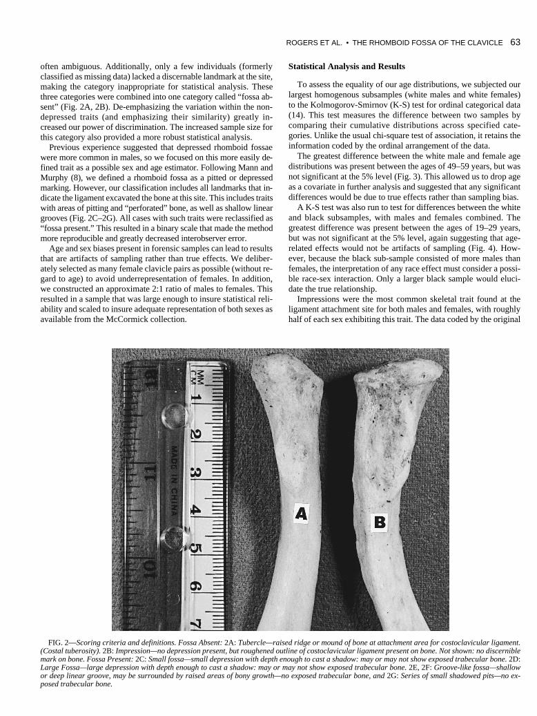
FIG. 2—Scoring criteria and definitions. Fossa Absent: 2A: Tubercle—raised ridge or mound of bone at attachment area for costoclavicular ligament.(Costal tuberosity). 2B: Impression—no depression present, but roughened outline of costoclavicular ligament present on bone. Not shown: no discerniblemark on bone. Fossa Present: 2C: Small fossa—small depression with depth enough to cast a shadow: may or may not show exposed trabecular bone. 2D:Large Fossa—large depression with depth enough to cast a shadow: may or may not show exposed trabecular bone. 2E, 2F: Groove-like fossa—shallowor deep linear groove, may be surrounded by raised areas of bony growth—no exposed trabecular bone, and 2G: Series of small shadowed pits—no ex-posed trabecular bone.
often ambiguous. Additionally, only a few individuals (formerlyclassified as missing data) lacked a discernable landmark at the site,making the category inappropriate for statistical analysis. Thesethree categories were combined into one category called “fossa ab-sent” (Fig. 2A, 2B). De-emphasizing the variation within the non-depressed traits (and emphasizing their similarity) greatly in-creased our power of discrimination. The increased sample size forthis category also provided a more robust statistical analysis.
Previous experience suggested that depressed rhomboid fossaewere more common in males, so we focused on this more easily de-fined trait as a possible sex and age estimator. Following Mann andMurphy (8), we defined a rhomboid fossa as a pitted or depressedmarking. However, our classification includes all landmarks that in-dicate the ligament excavated the bone at this site. This includes traitswith areas of pitting and “perforated” bone, as well as shallow lineargrooves (Fig. 2C–2G). All cases with such traits were reclassified as“fossa present.” This resulted in a binary scale that made the methodmore reproducible and greatly decreased interobserver error.
Age and sex biases present in forensic samples can lead to resultsthat are artifacts of sampling rather than true effects. We deliber-ately selected as many female clavicle pairs as possible (without re-gard to age) to avoid underrepresentation of females. In addition,we constructed an approximate 2:1 ratio of males to females. Thisresulted in a sample that was large enough to insure statistical reli-ability and scaled to insure adequate representation of both sexes asavailable from the McCormick collection.
Statistical Analysis and Results
To assess the equality of our age distributions, we subjected ourlargest homogenous subsamples (white males and white females)to the Kolmogorov-Smirnov (K-S) test for ordinal categorical data(14). This test measures the difference between two samples bycomparing their cumulative distributions across specified cate-gories. Unlike the usual chi-square test of association, it retains theinformation coded by the ordinal arrangement of the data.
The greatest difference between the white male and female agedistributions was present between the ages of 49–59 years, but wasnot significant at the 5% level (Fig. 3). This allowed us to drop ageas a covariate in further analysis and suggested that any significantdifferences would be due to true effects rather than sampling bias.
A K-S test was also run to test for differences between the whiteand black subsamples, with males and females combined. Thegreatest difference was present between the ages of 19–29 years,but was not significant at the 5% level, again suggesting that age-related effects would not be artifacts of sampling (Fig. 4). How-ever, because the black sub-sample consisted of more males thanfemales, the interpretation of any race effect must consider a possi-ble race-sex interaction. Only a larger black sample would eluci-date the true relationship.
Impressions were the most common skeletal trait found at theligament attachment site for both males and females, with roughlyhalf of each sex exhibiting this trait. The data coded by the original

64 JOURNAL OF FORENSIC SCIENCES
FIG. 2—(Continued).

four class ordinal scale illustrated an age distribution of the largest,deepest fossae most common in young males (Fig. 5). There is anotable trend for the largest fossae to be exhibited in youngermales, with the greatest number of large fossae found in males be-tween the ages of 20 and 30 years. This suggests that the largestfossae may be considered suggestive of “young” males, althoughthis must be corroborated by other methods because these largefossae are not limited to this age group. Only three females exhib-ited this largest class of fossae. The youngest (16 years) and oldest(62 years) females exhibited bilateral fossae, and the third “middle-aged” female (33 years) exhibited the trait on the left clavicle. Dueto the small number and discontinuous distribution of females withfossae, no age-related trend is suggested.
Once the categories were collapsed to the binary scoring system,the raw data again showed fossae were more common in males andon the right clavicle in both sexes (Table 2). The collapse to the bi-nary scoring system did not alter the age distribution for either clav-
icle: fossae were most common in males between the ages of 20 and30 years. This result remains suggestive of age rather than defini-tive, since individuals outside this age range also exhibited fossae.
Logistic regression for ordinal categorical data was performed totest the significance of multiple variables simultaneously (15). Thismethod fits a linear regression to the data using a logit link modelthat accommodates binary and ordinal response data. Regressionparameters are estimated using a maximum likelihood estimatemethod, and predictive equations can be generated (16). Our goalto produce a sexing method based upon discrete traits could havebeen attained simply by using posterior probabilities, but we antic-ipated an effect due to age. Logistic regression allows assessmentof possible effects due to covariates that are not possible using sim-ple probabilities.
The presence of a rhomboid fossa at the costoclavicular ligamentattachment site was modeled as dependent upon age, race and sex.Because logistic regression allows for continuous variables, agewas not coded as a categorical variable. The results confirm thatage and sex have a significant relationship presence of a rhomboidfossa for both right and left clavicles. Race was not significant foreither the right or the left clavicle when the other variables were inthe model (Table 3).
ROGERS ET AL. • THE RHOMBOID FOSSA OF THE CLAVICLE 65
FIG. 3—Results of K-S test: white males versus white females. D 50.126512, Critical value (a 5 .05) 5 0.16. Percentage survivorship plot-ted against age illustrates the differences between the cumulative distribu-tions for white males and females. The greatest difference occurs betweenthe ages of 49 and 59 years, but is not significant at the 5% level.
FIG. 4—Results of K-S test: white males/females versus black males/fe-males. D 5 0.08743, Critical value (a 5 .05) 5 0.23. Again, the percent-age survivorship for each sex plotted against age shows the differences be-tween the cumulative distributions for the black and white samples. Thegreatest difference is found between the ages of 19 and 29 years, but is notsignificant at the 5% level. However, these results are based upon a smallblack sample and should be viewed with caution.
FIG. 5—Age distribution of largest rhomboid fossae.
TABLE 2—Distribution of rhomboid fossae.
Left Right
Males 72/231 5 31% 83/231 5 36%Females 3/113 5 3% 9/113 5 8%
TABLE 3—Results of logistic regression.
Likelihood Ratio X2 p
Left Right Left Right
Age 14.66 21.89 0.0001 0.0001Sex 20.88 21.66 0.0001 0.0001Race 0.40 3.11 0.5273 0.0777*
* Near-significance should be interpreted with caution due to smallsample sizes and grouping of observations from non-white individuals.

66 JOURNAL OF FORENSIC SCIENCES
The posterior probabilities for sex based upon the type of skele-tal trait reflect what was seen in the raw data: males were morelikely to exhibit a fossa at the site of attachment (Table 4). For thissample, if a right clavicle exhibits a fossa at the site of attachment,the posterior probability of the individual being male is 81.7%; theprobability that the individual is female is 18.3%. If a rhomboidfossa is present on a left clavicle, the posterior probability of the in-dividual being male is 92.2%, while the probability that the indi-vidual is female is only 7.8%. This level of discrimination is notpossible when the rhomboid syndesmosis takes a form other than afossa. For the left or right clavicle, the posterior probabilities formales and females, if no fossa is present, are roughly 40% and60%, respectively. This reflects our methodology that grouped allnon-fossa skeletal traits into a single category.
When these probabilities are used to calculate odds ratios, theleft clavicle’s value for sex estimation is more apparent. The oddsthat a male exhibits a fossa on the right clavicle are roughly sixtimes greater than the odds that a female clavicle exhibits a fossa.For the left, the odds that a male exhibits a fossa are almost 17times greater than the odds that a female exhibits a fossa.
Due to the continuous scale for age, posterior probabilities can-not be easily calculated or applied for age estimation. However, thecalculated odds ratios of 1.05 (left clavicles) and 1.04 (right clavi-cles) show that with each year of age increase, the odds of exhibit-ing a fossa at the attachment site decrease. Therefore, younger in-dividuals are more likely to have fossae. While this effect is alsohighly significant, the application of predictive formulae for ageestimation would be neither efficient nor user-friendly. Coding ageas a continuous variable increased our ability to detect this age-re-lated trend. However, it prevented estimation of age from the rawdata as was possible from the discrete distribution of the sex variable.
The logistic regression calculates a score that measures how wellthe model fits the data with the current variables. The -2log Lscores of 48.65 and 62.97 (left and right clavicles, respectively) areeach highly significant (both p 5 0.0001), which indicates that theexisting variables do not fully explain the variation. This suggestsat least one important factor (and possibly several important fac-tors) affecting the presence or absence of a rhomboid fossa has notbeen included. Given the popular conception, the most obvious fac-tor to consider is mechanical usage. However, the association be-tween rhomboid fossae and increased muscle mass has been ques-tioned due to its presence in pre-pubescent and physically“average” individuals (9). Additionally, the highly symmetric ex-pression of the trait within each sex in our sample further chal-lenges this relationship. Since upwards of 80–85% of Americansare right-hand dominant, one would expect significantly greater ex-pression of fossae on the right clavicle if the trait was strongly cor-
related with habitual activity patterns. However, the slightly morecommon expression of the trait on the right clavicle and in male in-dividuals does suggest some relationship with mechanical loadingor activity. The etiology may be more complex than simple biome-chanics, involving biochemical and genetic factors in a complex re-lationship with patterns of mechanical force. This relationship re-mains unresolved.
Blind Testing of the Sexing Method
Two of the authors (LEF and NLR) tested the method on a sam-ple of 49 individuals (26 males, 23 females) from the William M.Bass Skeletal Collection housed at the University of Tennessee.The collection is composed of recently deceased individuals fromthe East Tennessee region. Therefore, the demographics and re-gional characteristics are comparable to the McCormick sample.All descriptive information was masked during observation andeach investigator scored the sample independently. Differences inprocessing between the original McCormick collection and the testsample from the Bass collection led to a few discrepancies in scor-ing between the two observers based upon our written definition.These problems prompted us to clarify our original description. Anobjective third observer was then able to successfully estimate sexfor these difficult cases based upon the revised description.
As with the McCormick collection, rhomboid fossae in the Basstest sample were more common in males than in females, and morecommon on the right clavicle. Of the 10 fossae observed on leftclavicles from the test sample, nine were from males, one from afemale. (This fossa was of the largest class.) These results closelymatch those predicted by the calculated posterior probabilities andconfirm that the rhomboid fossa trait is found more often on the leftclavicle of male individuals.
Conclusions
Our results indicate that age is a significant factor in the presenceof rhomboid fossae on the inferior border of the clavicle. However,exact age assignment based upon the presence of these fossae isproblematic. The presence of a rhomboid fossa is not an adequatecriterion for accurate age estimation because the trait is found in in-dividuals as young as 12 years and as old as 90 years. However,presence of an extremely large, deep fossa (on either side) is sug-gestive of a “young” male (,30 years), but this supposition mustbe corroborated by other methods.
Presence of a rhomboid fossa can be used as an indicator of sexbecause males exhibit fossae more frequently than females. It mustbe reiterated that our definition of rhomboid fossae includes land-marks characterized by shadowed pits, shallow grooves, a series ofsmall perforations, or other evidence that the ligament has exca-vated the bone and created actual depressions (see Fig. 2C–2G).Because tubercles are difficult to define and impressions are com-monly found in both males and in females, these traits are not usedfor estimation in this method. Similarly, the absence of a skeletaltrait is not appropriate for estimation due to small sample size.
While 93% of the fossae in the original sample were present onmale clavicles, the occurrence of this trait will vary from sample tosample. Sex ratios (and perhaps population activity patterns) areexpected to influence the frequency of rhomboid fossae. Therefore,an estimate of sex based upon this method for individuals fromother populations cannot be accompanied by a posterior probabil-ity of the estimate. However, given the preponderance of fossa onmale clavicles it can be said that if a fossa is present, the specimenis likely from a male. This is especially true for the left clavicle,
TABLE 4—Posterior probabilities.
Element Event Probability
Left clavicle P (male/fossa) 92.2%Left clavicle P (female/fossa) 7.8%Left clavicle P (male/no fossa) 41.4%Left clavicle P (female/no fossa) 58.6%Right clavicle P (male/fossa) 81.7%Right clavicle P (female/fossa) 18.3%Right clavicle P (male/no fossa) 41.0%Right clavicle P (female/no fossa) 59.0%

given the greater discrepancy of fossa expression between malesand females on the left bone. Our test of the sexing method con-firms it can be used to corroborate other estimates of sex or providea sex estimate for unknown individuals in the absence of otherskeletal indicators.
Acknowledgments
The authors thank the reviewers for excellent comments and sug-gestions. We are also grateful to John Garwood, Lyle Konigsberg,David Sylwester, Cathy Butler, Lee Meadows Jantz, Ann Ross,Mike Keene, Matt Ward, and Rick Snow for their helpful assistanceand comments on this paper. An earlier version of this study was pre-sented at the 50th Annual Meeting of the American Association ofForensic Sciences in San Francisco, California, Feb. 9–14, 1998.
References1. White TD. Human osteology. San Diego: Academic Press, 1991.2. Parsons FG. On the proportions and characteristics of the modern En-
glish clavicle. J Anat 1916;51:71–93.3. Pendergrass EP, Hodes PJ. The rhomboid fossa of the clavicle. Amer J
Roentgen 1937;38:152–5.4. Bearn JG. Direct observations on the function of the capsule of the ster-
noclavicular joint in clavicular support. J Anat 1967;101:159–70.5. Cave AJE. The nature and morphology of the costoclavicular ligament.
J Anat 1961;95:170–9.6. Goldenberg DB, Brogdon BG. Congenital anomalies of the pectoral gir-
dle demonstrated by chest radiography. J de l’association canadienne desradiologiestes. 1967;18:472–7.
7. Chawla S, Mukerjee P. Unilateral rhomboid fossa: a case report. IndianJ Radiology 1970;24:50–1.
8. Mann RW, Murphy SP. Regional atlas of bone disease: a guide to patho-logic and normal variation in the human skeleton. Springfield, IL: CCThomas, 1990.
9. Shauffer IA, Collins WV. The deep clavicular rhomboid fossa: clinicalsignificance and incidence in 10,000 routine chest photoflourograms.JAMA 1966;9:158–9.
10. Jit I, Kaur H. Rhomboid fossa in the clavicles of North Indians. AJPA1986;70:97–103.
11. American College of Radiology. Index of Radiologic Diagnoses, 4th ed.,Reston, VA: ACR, 1992.
12. Stirland AJ. Musculoskeletal evidence for activity: problems of evalua-tion. Int J Osteoarchaeol 1998;354–62.
13. Stirland AJ. Diagnosis of occupationally related paleopathology: can itbe done? In: DJ Ortner, AC Aufderheide, editors. Human paleopathol-ogy: current synthesis and future options. Washington, DC: SmithsonianInstitution, 1991;41–7.
14. Thomas DH. Figuring anthropology: first principles of probability andstatistics. New York: Holt, Rhinehart & Winston, 1976.
15. SAS: Stastistical Analysis System. SAS Institute, Inc. Cary, NC. Site li-cense for the University of Tennessee Computing Center (0001395011).
16. SAS Institute, Inc. SAS/STAT User’s Guide, Version 6, Fourth Edition,Vol. 2. Chapter 27: Logistic Regression. Cary, NC: SAS, 1989.
Additional information and reprint requests:Nikki L. RogersUniversity of TennesseeDepartment of Anthropology252 South Stadium HallKnoxville, TN [email protected]
ROGERS ET AL. • THE RHOMBOID FOSSA OF THE CLAVICLE 67