Stimulation of human red blood cells leads to Ca2+-mediated intercellular adhesion
Upload
independentCategory
view
0download
0
The maize transfer cell-specific type-A response regulatorZmTCRR-1 appears to be involved in intercellular signalling
Luis M. Muniz, Joaquın Royo, Elisa Gomez, Cristina Barrero, Diego Bergareche and Gregorio Hueros*
Departamento de Biologıa Celular y Genetica, Universidad de Alcala, 28871 Alcala de Henares, Madrid, Spain
Received 23 March 2006; revised 29 May 2006; accepted 31 May 2006.*For correspondence (fax þ34 91 885 4799; e-mail [email protected]).
Summary
Response regulators are signal-transduction molecules present in bacteria, yeast and plants, acting as relays
for environmental challenges. This paper reports the characterization of a Zea mays gene, ZmTCRR-1, that
codes for a member of the type-A response regulator class of proteins. The gene was found to be expressed
exclusively in the endosperm transfer-cell layer 8–14 days after pollination, when transfer-cell differentiation is
most active. The promoter of ZmTCRR-1 was strongly transactivated in heterologous systems by the transfer
cell-specific transcription factor ZmMRP-1. The ZmTCRR-1 protein was detected not only in the transfer-cell
layer, but also in the conductive tissue deep inside the endosperm, where there is no transcription of the gene.
This suggests that two-component systems might be involved in intercellular signal transmission, in contrast
to the generally held belief that these systems are involved only in cell-autonomous pathways.
Keywords: maize endosperm, transfer cells, endosperm development, response regulator, signal transduc-
tion.
Introduction
The transfer cells of maize make up a highly specialized
tissue on the placental side of the endosperm, forming an
interface between the filial and maternal tissues in the seed
(Becraft, 2001; Kiesselbach, 1949; Olsen, 2004; Thompson
et al., 2001). Their deep cell wall ingrowths afford a
remarkable increase in the membrane surface area, facilita-
ting nutrient uptake from the apoplastic space in the plac-
ento-chalaza area (Offler et al., 2002). In maize, immediately
above the transfer cells a region of prismatic cells (known as
conducting cells: Becraft, 2001) facilitates symplastic trans-
port for assimilates on their way to the upper part of the
endosperm, where they are used to build storage products.
Transfer cells also show a specific gene-expression pat-
tern, which has been the subject of much study in recent
years. Genes specifically expressed in transfer cells have
been identified in barley (Doan et al., 1996) and maize
(Gomez et al., 2002; Gutierrez-Marcos et al., 2004; Hueros
et al., 1995, 1999b). In maize, with the exception of ZmMRP-1
(Gomez et al., 2002), all these genes code for small hydro-
philic proteins of unknown function, although some of the
results obtained suggest they might be involved in defence
of the seed against pathogens harboured by the mother
plant (Serna et al., 2001). ZmMRP-1 is a transcription factor
exclusively expressed in the transfer-cell layer of maize from
the earliest stages of endosperm development. It has been
shown that ZmMRP-1 can transactivate various transfer cell-
specific promoters (Gomez et al., 2002; Gutierrez-Marcos
et al., 2004). The identification of regulatory pathways
operating in parallel with, or connected to, the pathway
specified by ZmMRP-1 would be a key contribution to the
study of transfer-cell biology.
In recent years, a signal-transduction system generically
known as the two-component system or phosphorelay
system, has been described in a number of higher plants
(Brandstatter and Kieber, 1998; Kakimoto, 1996; Sakakibara
et al., 1998). The system was found previously in eubacteria
(Parkinson and Kofoid, 1992) and yeast (Maeda et al., 1994;
reviewed by Stock et al., 2000). To date, no homologous
molecular mechanism has been identified in the archaea or
metazoa.
Plant-response regulators have been most extensively
described in Arabidopsis and maize (Hosoda et al., 2002;
Takei et al., 2002; Yamada et al., 1998). The Arabidopsis
genome contains about 40 genes with sequence similarity to
phosphorelay-type genes (D’Agostino et al., 2000; Hwang
et al., 2002). The involvement of two-component systems in
ª 2006 The Authors 17Journal compilation ª 2006 Blackwell Publishing Ltd
The Plant Journal (2006) 48, 17–27 doi: 10.1111/j.1365-313X.2006.02848.x

cytokinin- and ethylene-mediated signalling is being studied
intensively in this organism (D’Agostino et al., 2000; Lohr-
mann and Harter, 2002; Oka et al., 2002; Taniguchi et al.,
1998). In maize, these systems have been shown to react at
the systemic level to changes in inorganic nitrogen availab-
ility (Sakakibara et al., 1998, 2000).
The basic partners in two-component pathways include a
histidine kinase, which acts as a membrane or cytoplasmic
receptor of environmental signals; a response-regulator
protein, the effector molecule of the system; and a histidine
phosphotransfer (HPt), molecule which acts as a courier
between receptors and response regulators. Response reg-
ulators are divided into two classes, in plants known as type-
A (small size, short or non-existent C-terminal domain,
cytokinin-inducible, molecular targets presently unknown);
and type-B, belonging to the GARP family of transcriptional
activators (longer C-terminal extension with similarity to
Myb-related DNA-binding domains, not inducible by cytoki-
nin, involved in transcriptional regulation).
This paper describes the identification of ZmTCRR-1, a
type-A response-regulator gene specifically expressed in the
immature maize endosperm transfer-cell layer. We show
that the promoter of the gene is strongly transactivated by
the transfer cell-specific transcriptional activator ZmMRP-1.
Evidence is also provided that the ZmTCRR-1 protein moves
from the transfer cells inwards, where it accumulates in the
conducting tissue of the endosperm. These results suggest
that ZmTCRR-1 is involved in an inter-cellular signalling
pathway, a previously unsuspected role for the two-compo-
nent signalling system to which it belongs.
Results
Identification of a seed-specific response regulator-like gene
In an ongoing effort to isolate genes differentially expressed
during maize kernel development (this experiment will be
fully described elsewhere), an expression database was built
for over 6000 transcripts randomly selected from a 10-days
after pollination (DAP) endosperm cDNA library, using a
differential screening method. Clone 38-10 showed moder-
ate hybridization to a subtracted probe specific for the bot-
tom half of the kernel, but no signal with any other probe in
the set. This clone carries a 410-bp-long insert, encoding a
protein with significant similarity to several plant-response
regulators. The insert in clone 38-10 included the 45 C-ter-
minal residues of this protein and a 230-bp 3¢ untranslated
region (UTR).
RACE experiments were performed to obtain the 5¢-end of
the clone 38-10 transcript. The putative full-length cDNA
obtained was 667 bp long, encoding a 124-aa peptide with a
predicted molecular weight of 13.8 kDa. As shown in
Figure 1, this protein is similar to the receiver domain of
the type-A response regulator elements of plants (ARR10,
O49397; ARR6, NP_201097; ZmRR1, BAA85112; ZmRR8,
BAB41137.1) and bacteria (Spo0F, P06628; CheY, P96126).
The gene was therefore named ZmTCRR-1 (Zea mays
transfer cell response regulator-1).
A motif search using Pfam (http://www.sanger.ac.uk/
Software/Pfam/) indicated the presence of a response-regu-
lator domain in the 6-119-aa region of ZmTCRR-1
(E(0) ¼ 5e)8). The alignment of its sequence to those of
response regulators of plants and bacteria in the Swissprot
and National Center for Biotechnology Information (NCBI)
databases showed high sequence conservation in the
regions around the amino acids involved in phosphate
group transfer activity (acidic pocket; Figure 1). However,
one of the canonical aspartate residues in this structure, D13,
is changed to histidine in ZmTCRR-1. This variation of the
canonical motif distinguishes ZmTCRR-1 from the other
known plant response regulators, which show the classic
DDK triad. To ascertain whether this variation reflects a
mutant allele that arose in the maize variety under exam-
ination (A69Y), a region from the start codon to base 220
inside the first intron was sequenced in the inbred lines B73,
F2 and W64, and in the maize ancestor teosinte (Zea
diploperennis). Alignment of the sequences showed that
the change was present in all samples, which were 97.9%
identical in the exon region. The conservation of the amino-
acid sequence was complete (not shown).
The sequence was scanned for transmembrane domains
using TMpred (Hofmann and Stoffel, 1993); SOSUI (http://
sosui.proteome.bio.tuat.ac.jp); and signal peptides, employ-
ing TARGETP ver. 1.0 (Emanuelsson et al., 2000) and PREDO-
TAR ver. 0.5 (http://www.inra.fr/predotar) software. No
targeting sequence directing ZmTCRR-1 to any particular
cell compartment was found.
ZmTCRR-1 is a single-copy gene in maize
Primers designed to amplify the coding sequence of
ZmTCRR-1 were also used to amplify its genomic se-
quence from maize genomic DNA. A PCR product of
approximately 1200 bp was aligned to the cDNA, and four
introns were found with canonical GT/AG borders. An in-
verse-PCR procedure (Hueros et al., 1999a) was used to
isolate the 1200-bp sequence located immediately up-
stream of the translation start site of ZmTCRR-1. A pre-
liminary analysis of the promoter sequence showed no
significant homology to promoters from other known re-
sponse-regulator genes in maize (Deji et al., 2000; not
shown). The maize genome codes for at least 10 different
response regulators (Asakura et al., 2003), although they
share little sequence identity to ZmTCRR-1. The insert in
clone 38-10 was digoxigenin-labelled and used to deter-
mine the copy number by hybridization to genomic DNA
digested with different restriction endonucleases. The
probe hybridized to a single DNA fragment in all cases,
18 L. M. Muniz et al.
ª 2006 The AuthorsJournal compilation ª 2006 Blackwell Publishing Ltd, The Plant Journal, (2006), 48, 17–27

indicating that ZmTCRR-1 is a single-copy gene in maize
(not shown).
ZmTCRR-1 is expressed exclusively in the bottom half of the
maize kernel
Northern blot analyses were performed on total RNA from
unpollinated female flowers, the upper and lower halves of
the immature seeds at 16 DAP, the leaves, roots, coleoptiles,
tassel and silks (Figure 2a). Only the RNA sample extracted
from the lower part of the seeds showed positive hybrid-
ization to the ZmTCRR-1 probe. Tissue specificity was fur-
ther confirmed by RT–PCR, using the same RNA samples as
described above after DNAse treatment. No PCR product
was amplified from non-seed tissues (not shown).
Northern blot hybridization was also used to analyse the
expression of ZmTCRR-1 over grain development, using
total RNA from seeds at various developmental stages (from
3 to 32 DAP). In the case of seeds older than 8 DAP, the
samples were dissected into upper and lower halves prior to
RNA extraction. The results (Figure 2b) confirmed that the
gene is expressed only in the basal part of the seeds. The
gene was also found to be expressed over a very short
period: the hybridization signal was very strong at 8 and
11 DAP, but very weak at 6, 14 and 17 DAP, and almost
undetectable before and after this window.
ZmTCRR-1 is expressed exclusively in the transfer-cell layer
A digoxigenin-labelled antisense RNA probe was pro-
duced from the 38-10 insert and used for in situ hybrid-
ization experiments with seed sections at various
developmental stages (Figure 3 for results obtained at
11 DAP). The transfer-cell layer was the only kernel tissue
to show positive hybridization signals with the antisense
probe. In agreement with the results obtained in the
Northern analyses, the signal intensity in the transfer cells
reached a maximum at around 11 DAP (Figure 3a–c), and
it was not detectable before 5 DAP or after 16 DAP (not
shown).
Figure 1. Alignment of ZmTCRR-1 to response regulators in plants and bacteria.
Conserved residues are shadowed in black. Asterisks denote amino acids forming the acidic pocket. A variation in the canonical D13 aa (as numbered in the CheY
sequence) is underlined in the ZmTCRR-1 and ARR19 sequences. Only domains relevant to the alignment are shown for sequences other than ZmTCRR-1.
A new transfer cell response regulator in maize 19
ª 2006 The AuthorsJournal compilation ª 2006 Blackwell Publishing Ltd, The Plant Journal, (2006), 48, 17–27

Figure 2. Expression pattern of ZmTCRR-1 in different maize tissues and developmental stages.
(a) Total RNA (20 lg per lane) from different maize tissues were Northern blotted and hybridized with a ZmTCRR-1 probe. U, unpollinated flowers; T, top half and B,
bottom half from 16-DAP seeds; L, leaves; R, roots; C, coleoptiles; A, anthers; S, silks. The image below shows the ethidium bromide-stained gel before the transfer
of RNA.
(b) Total RNA (20 lg per lane) from different seed developmental stages were Northern blotted and hybridized with a ZmTCRR-1 probe. Numbers 3-32 refer to
number of days after pollination (DAP). From 8 DAP, seeds were dissected before RNA extraction into their upper (T) and lower (B) halves. The image below shows
the ethidium bromide-stained gel before the transfer of RNA.
Figure 3. Localization of ZmTCRR-1 mRNA and protein.
(a–c) 11-DAP seed sections were hybridized with a digoxigenin-labelled ZmTCRR-1 antisense RNA probe.
(a) Complete 11-DAP seed section showing specific hybridization in the transfer cell area; (b, c) higher magnification of the transfer layer. Greater signal intensity is
appreciated in cells at both edges of the transfer-cell layer (arrows in b) compared with the central cells of the tissue.
(d–g) 11-DAP (d, e, g) and 16-DAP (f) seed sections were reacted with the anti-ZmTCRR-1 antibody (d–f) or the pre-immune serum (g).
Bar, 1 mm (a, d, f, g); 500 lm (b, c, e). Em, embryo; En, endosperm; TCL, transfer-cell layer; Pd, pedicel. In all cases the signal was detected with an alkaline
phosphatase conjugate: the blue-purple precipitate formed on incubation with NBT/BCIP appears as an orange-gold signal in dark-field illumination.
20 L. M. Muniz et al.
ª 2006 The AuthorsJournal compilation ª 2006 Blackwell Publishing Ltd, The Plant Journal, (2006), 48, 17–27

In these samples, the highest accumulation of the tran-
script was observed in the small, immature cells on both
sides of the transfer-cell layer, and in the inner layers of the
tissue (Figure 3b). No signal was detected with a sense
probe used as a negative control (not shown).
Cellular localization of ZmTCRR-1
Immunolocalization experiments showed that the ZmTCRR-
1 protein accumulates in several cell layers inside the
endosperm (Figure 3d–f), rather than just in the transfer cells
where the transcript is produced. At 11 DAP, the protein was
distributed throughout the lower half of the endosperm
(Figure 3d). Interestingly, almost no signal was detected in
the most mature transfer cells (Figure 3d,e) where it was
originally produced, while the protein was readily detected
in the conducting cells above the transfer-cell layer and in
the immature cells on both sides of the transfer-cell layer
(Figure 3d). At 16 DAP (Figure 3f) the protein was still
detectable, although the intensity of the signal had de-
creased significantly. No signal was obtained when a pre-
immune serum was used with these sections (Figure 3g).
Western blot analyses of seed protein extracts further
confirmed the localization of ZmTCRR-1 (Figure 4). The anti-
ZmTCRR-1 antibody detected a band with the expected size
(Figure 4a, arrow) in protein extracts from both upper and
lower halves of the kernels. In agreement with the results
obtained in the Northern blot experiments, the protein was
undetectable by 5 DAP, peaked at 11 DAP, and subsequently
decayed, being almost undetectable by 20 DAP. No protein
was detected in the low molecular-weight area of the blot
using the pre-immune serum (not shown). The ZmTCRR-1
antibody also reacted (Figure 4a) with two additional prod-
ucts: a high molecular-weight protein (approximately
100 kDa) present at all developmental stages but signifi-
cantly absent from the lower seed-half protein extracts
(lanes 11B, 14B, 16B) and, although weakly, with the storage
products that accumulate in the endosperm after 14 DAP,
and can represent up to 50% of the total protein content at
the older developmental stages (Figure 4a, lanes 16T, 20T).
To rule out a possible contamination of the upper-half
extracts with proteins from the lower part of the kernels, the
purity of protein extracts was assessed by immunoreaction
of the blots with the basal kernel-specific antibodies for
BETL-1 (Figure 4b; Hueros et al., 1995) and BAP-2 (Figure 4c:
the antibody recognizes both the pre-protein and the mature
form; Serna et al., 2001). These controls showed the expec-
ted immunoreacting peptides only in extracts from the
bottom half of the seeds. The only evidence of cross-
contamination was the detection of a weak band corres-
ponding to the low-molecular weight, highly diffusible
mature product from BAP-2 in the 11-DAP top-half extracts
(Figure 4c, lane 11T); this slight contamination was not seen
for the other BETL marker proteins and was not observed for
any marker at older developmental stages (when the kernel
can be dissected more precisely).
The presence of His-tagged markers in the blots allowed
us to measure accurately the size of the immunoreacting
proteins. In the case of ZmTCRR-1 the estimated size was
12.1 kDa, well in agreement with that predicted from its
sequence (13.8 kDa), considering that the protein is strongly
acidic (pI 4.79) and tends therefore to migrate more quickly
than expected in neutral SDS-containing gels. For the
recombinant ZmTCRR-1 (Figure 4a, lane R) the predicted
size is 16.7 kDa and the estimated molecular weight is
16.3 kDa, in this case the pI is 5.89 and a smaller deviation
can thus be anticipated. The same effect, although in an
opposite direction, was observed for the strongly cationic
Figure 4. Western blot analyses of the localization of ZmTCRR-1.
Total protein extracts from 5-DAP whole kernels (K) or 11-, 14-, 16- and 20-DAP upper (T) or lower (B) seed halves were reacted with the anti-ZmTCRR-1 antibody
(diluted 1:500, a) or the antibodies against the transfer cell-specific proteins BETL-1 (1:2000, b) and BAP-2 (1:2000, c). Note that BAP-2 protein accumulation is
decreasing after 11 DAP; the precursor (upper band) is already undetectable at 16 DAP. R, 0.24 ng of the recombinant ZmTCRR-1 that was used to raise the antibody.
M, lanes containing His-tagged molecular weight markers (BenchMark His-tagged protein standard, Invitrogen, molecular weights indicated on the left).
Arrow (a) indicates the low molecular-weight product corresponding to the ZmTCRR-1 protein. Blots were sectioned to facilitate their reaction with the antibodies
indicated and the anti-His antibody used to visualize the molecular weight markers, indicated by dotted lines.
(a, b) Reacted blots exposed for 15 min; (c) blot exposed for 20 sec.
A new transfer cell response regulator in maize 21
ª 2006 The AuthorsJournal compilation ª 2006 Blackwell Publishing Ltd, The Plant Journal, (2006), 48, 17–27

peptides BETL-1 (pI 8.18) and BETL-2 (upper band, pI 8.95)
with a predicted MW of 7.6 and 7.8 kDa, versus a calculated
MW of 10.5 and 12.9 kDa, respectively.
The promoter of ZmTCRR-1 is activated by the transfer cell-
specific transcriptional activator ZmMRP-1
The transfer cell-specific genes BETL-1 and BETL-2 have a
TATC microsatellite sequence 40–80 bp upstream of their
TATA boxes (Hueros et al., 1999b). In the ZmTCRR-1 pro-
moter, a sequence containing five TATC repeats is located
40 bp upstream of the putative TATA box. This suggests that
factors controlling BETL-1 and BETL-2 expression, namely
ZmMRP-1 (Gomez et al., 2002), might also regulate
ZmTCRR-1. To test this hypothesis, a construct containing
1200 bp of the ZmTCRR-1 promoter fused to the GUS
reporter gene was assayed for transactivation by ZmMRP-1
in two transient expression systems. In onion epidermal
cells (Figure 5), the reporter construct ZmTCRR-1prom–GUS
was inactive in the absence of the ZmMRP-1 transcriptional
activator (Figure 5a). However, co-bombardment with a
construct overexpressing ZmMRP-1 under the control of the
maize ubiquitin promoter (Figure 5b) produced a strong
signal in terms of the number and intensity of blue spots in
the epidermal cells. The signal was even stronger than that
obtained with the ubiquitin promoter–GUS positive control
(Figure 5c). To quantify the effect of ZmMRP-1 on the
ZmTCRR-1 promoter, the same constructs were introduced
into tobacco protoplasts, along with a p35S-LUC construct
for transformation efficiency control. The presence of the
effector plasmid expressing ZmMRP-1 under the control of
the maize ubiquitine promoter increased the GUS activity
driven by the ZmTCRR-1 promoter by a factor of 9.01 com-
pared with control experiments (in which the effector plas-
mid was substituted by a plasmid containing the ubiquitin
promoter sequence but no ZmMRP-1). For comparison,
experiments using the BETL-1 promoter–GUS construct as a
reporter plasmid were performed in parallel, as the BETL-1
promoter is reported to be efficiently transactivated by
ZmMRP-1 in this transient expression system (Gomez et al.,
2002). In this case the transactivation factor was 9.89. The
ZmTCRR-1 promoter was thus transactivated by ZmMRP-1
at a level similar to that of the BETL-1 promoter.
Discussion
The relatively recent discovery of two-component signalling
systems in yeast and plants opens up a wide field of research
regarding the role these molecules might play in eukaryotic
cell biology. The plant-response regulators described to
date, and the molecules functionally associated with them,
are involved in cytokinin- and ethylene-mediated responses
(Brandstatter and Kieber, 1998; Sakai et al., 2001). Only
ZmRR3, a type-A response regulator isolated from maize
cobs, has been shown to be unresponsive to this kind of
stimulus (Asakura et al., 2003). Here we report the identifi-
cation of a maize response regulator, ZmTCRR-1, specifically
expressed in a very discrete region of the kernel, the endo-
sperm basal transfer layer. This gene is developmentally
regulated and, at least in in vitro-cultured kernels, was not
induced by cytokinin (not shown). BLASTX analysis showed
important similarities with response regulators from maize
and rice, particularly high in the functionally critical do-
mains. A pairwise sequence comparison of the seven maize
genes ZmRR 1-7 (all type-A molecules; Asakura et al., 2003)
with ZmTCRR-1 revealed identities of between 21.1% and
42.6%, supporting the identification of ZmTCRR-1 as a type-
A response regulator. Unfortunately there are no available
biochemical assays to prove or disprove type-A response-
regulator functionality, besides the non-conclusive assays to
test whether the protein can be phosphorylated in vitro. The
critical experiments to prove functionality would be feasible
only once the up- and downstream interacting partners were
identified.
The protein encoded by ZmTCRR-1 (Figure 1) is acidic and
small in size (124 aa, 13.8 kDa). The response-regulator
domain encompasses almost the whole protein, and no
secretion domain can be predicted for its structure. This
contrasts with the situation in other type-A response regu-
lators in plants where the response-regulator domain regu-
lates an adjacent C-terminal domain ranging from 30 to 100
amino acids (D’Agostino and Kieber, 1999) and suggests that
in ZmTCRR-1, the response-regulator motif itself is respon-
sible for the function of the protein. A similar structure is
found in Arabidopsis genes ARR16 and ARR17. The amino
acids in the response-regulatory domain involved in the
process of phosphorylation and conformational change
constitute the acidic pocket (Parkinson and Kofoid, 1992),
and are invariant in most of the response regulators
characterized to date (Cho et al., 2001; Imamura et al.,
1998). ZmTCRR-1 shows the conserved residues archetypi-
cal of type-A response-regulator acidic pockets, with one
exception: aspartate 13 is substituted by a histidine. Studies
on mutant forms of the bacterial CheY gene (Bourret et al.,
1990, 1993), which is the closest ZmTCRR-1 relative in
Figure 5. Transactivation of the ZmTCRR-1 promoter by ZmMRP-1.
(a–c) The ZmTCRR-1 promoter was fused to the GUS gene and co-bombarded
into onion epidermal cells along with a plasmid bearing the ubiquitin
promoter (a) or the ZmMRP-1 coding sequence under the control of the
ubiquitin promoter (b). A ubiquitin promoter–GUS construct (c) was intro-
duced as a positive control. An area 1–2 cm2 with the highest density of blue
spots is shown in each case.
22 L. M. Muniz et al.
ª 2006 The AuthorsJournal compilation ª 2006 Blackwell Publishing Ltd, The Plant Journal, (2006), 48, 17–27
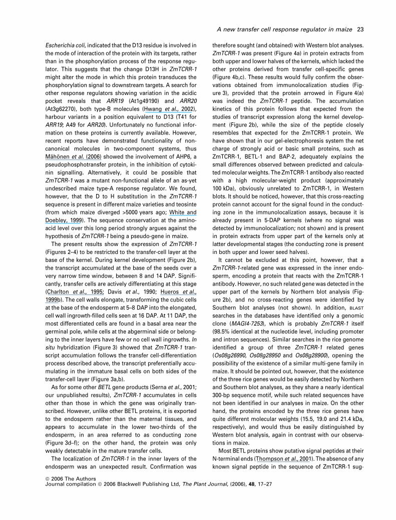
Escherichia coli, indicated that the D13 residue is involved in
the mode of interaction of the protein with its targets, rather
than in the phosphorylation process of the response regu-
lator. This suggests that the change D13H in ZmTCRR-1
might alter the mode in which this protein transduces the
phosphorylation signal to downstream targets. A search for
other response regulators showing variation in the acidic
pocket reveals that ARR19 (At1g49190) and ARR20
(At3g62270), both type-B molecules (Hwang et al., 2002),
harbour variants in a position equivalent to D13 (T41 for
ARR19; A49 for ARR20). Unfortunately no functional infor-
mation on these proteins is currently available. However,
recent reports have demonstrated functionality of non-
canonical molecules in two-component systems, thus
Mahonen et al. (2006) showed the involvement of AHP6, a
pseudophosphotransfer protein, in the inhibition of cytoki-
nin signalling. Alternatively, it could be possible that
ZmTCRR-1 was a mutant non-functional allele of an as-yet
undescribed maize type-A response regulator. We found,
however, that the D to H substitution in the ZmTCRR-1
sequence is present in different maize varieties and teosinte
(from which maize diverged >5000 years ago; White and
Doebley, 1999). The sequence conservation at the amino-
acid level over this long period strongly argues against the
hypothesis of ZmTCRR-1 being a pseudo-gene in maize.
The present results show the expression of ZmTCRR-1
(Figures 2–4) to be restricted to the transfer-cell layer at the
base of the kernel. During kernel development (Figure 2b),
the transcript accumulated at the base of the seeds over a
very narrow time window, between 8 and 14 DAP. Signifi-
cantly, transfer cells are actively differentiating at this stage
(Charlton et al., 1995; Davis et al., 1990; Hueros et al.,
1999b). The cell walls elongate, transforming the cubic cells
at the base of the endosperm at 5–8 DAP into the elongated,
cell wall ingrowth-filled cells seen at 16 DAP. At 11 DAP, the
most differentiated cells are found in a basal area near the
germinal pole, while cells at the abgerminal side or belong-
ing to the inner layers have few or no cell wall ingrowths. In
situ hybridization (Figure 3) showed that ZmTCRR-1 tran-
script accumulation follows the transfer cell-differentiation
process described above, the transcript preferentially accu-
mulating in the immature basal cells on both sides of the
transfer-cell layer (Figure 3a,b).
As for some other BETL gene products (Serna et al., 2001;
our unpublished results), ZmTCRR-1 accumulates in cells
other than those in which the gene was originally tran-
scribed. However, unlike other BETL proteins, it is exported
to the endosperm rather than the maternal tissues, and
appears to accumulate in the lower two-thirds of the
endosperm, in an area referred to as conducting zone
(Figure 3d–f); on the other hand, the protein was only
weakly detectable in the mature transfer cells.
The localization of ZmTCRR-1 in the inner layers of the
endosperm was an unexpected result. Confirmation was
therefore sought (and obtained) with Western blot analyses.
ZmTCRR-1 was present (Figure 4a) in protein extracts from
both upper and lower halves of the kernels, which lacked the
other proteins derived from transfer cell-specific genes
(Figure 4b,c). These results would fully confirm the obser-
vations obtained from immunolocalization studies (Fig-
ure 3), provided that the protein arrowed in Figure 4(a)
was indeed the ZmTCRR-1 peptide. The accumulation
kinetics of this protein follows that expected from the
studies of transcript expression along the kernel develop-
ment (Figure 2b), while the size of the peptide closely
resembles that expected for the ZmTCRR-1 protein. We
have shown that in our gel-electrophoresis system the net
charge of strongly acid or basic small proteins, such as
ZmTCRR-1, BETL-1 and BAP-2, adequately explains the
small differences observed between predicted and calcula-
ted molecular weights. The ZmTCRR-1 antibody also reacted
with a high molecular-weight product (approximately
100 kDa), obviously unrelated to ZmTCRR-1, in Western
blots. It should be noticed, however, that this cross-reacting
protein cannot account for the signal found in the conduct-
ing zone in the immunolocalization assays, because it is
already present in 5-DAP kernels (where no signal was
detected by immunolocalization; not shown) and is present
in protein extracts from upper part of the kernels only at
latter developmental stages (the conducting zone is present
in both upper and lower seed halves).
It cannot be excluded at this point, however, that a
ZmTCRR-1-related gene was expressed in the inner endo-
sperm, encoding a protein that reacts with the ZmTCRR-1
antibody. However, no such related gene was detected in the
upper part of the kernels by Northern blot analysis (Fig-
ure 2b), and no cross-reacting genes were identified by
Southern blot analyses (not shown). In addition, BLAST
searches in the databases have identified only a genomic
clone (MAGI4-7253), which is probably ZmTCRR-1 itself
(98.5% identical at the nucleotide level, including promoter
and intron sequences). Similar searches in the rice genome
identified a group of three ZmTCRR-1 related genes
(Os08g26990, Os08g28950 and Os08g28900), opening the
possibility of the existence of a similar multi-gene family in
maize. It should be pointed out, however, that the existence
of the three rice genes would be easily detected by Northern
and Southern blot analyses, as they share a nearly identical
300-bp sequence motif, while such related sequences have
not been identified in our analyses in maize. On the other
hand, the proteins encoded by the three rice genes have
quite different molecular weights (15.5, 19.0 and 21.4 kDa,
respectively), and would thus be easily distinguished by
Western blot analysis, again in contrast with our observa-
tions in maize.
Most BETL proteins show putative signal peptides at their
N-terminal ends (Thompson et al., 2001). The absence of any
known signal peptide in the sequence of ZmTCRR-1 sug-
A new transfer cell response regulator in maize 23
ª 2006 The AuthorsJournal compilation ª 2006 Blackwell Publishing Ltd, The Plant Journal, (2006), 48, 17–27

gests that protein movement is not mediated by secretion.
We suggest that the protein might translocate through a
symplast pathway, via the plasmodesmata. The transfer of
proteins and RNA between cells via specific interaction with
these organelles has already been documented (Imlaut
et al., 1999; Jackson, 2001; Lucas et al., 1995). Interestingly,
when tobacco protoplasts and onion epidermal cells were
transformed with a ZmTCRR-1–GFP fusion construct (not
shown), the GFP signal was concentrated in clumps asso-
ciated with the membrane system, very often forming a
punctate fluorescence pattern, which has been associated
with the plasmodesmatal localization of cell and viral
movement proteins (Lee et al., 2005; Medina-Escobar et al.,
2003). No evidence was found, however, for translocation of
the fusion protein to neighbouring cells. As the maize
endosperm transfer cells cannot be transiently transformed,
additional confirmation of ZmTCRR-1 translocation awaits
the generation of transgenic plants expressing ZmTCRR-1–
GFP fusion proteins.
Most known type-A response regulators are controlled by
cytokinin, which has, in at least one case, been shown to
exert its effect through a type-B molecule. Sakai et al. (2001)
showed the expression of ARR6 mRNA to be controlled by
ARR1, which mediates cytokinin signals. This model of
interaction, with type-B response regulators activating
accumulation of type-A response regulator mRNA in
response to stimuli, is the current hypothesis for the
functioning of two-component systems in plants (Oka et al.,
2002). No type-B response-regulator expression has been
described in the transfer cells, although an as-yet undiscov-
ered response regulator may govern ZmTCRR-1 expression.
We found instead that ZmTCRR-1 is regulated by ZmMRP-1
(Figure 5), a single-domain Myb-related transcriptional
regulator (Gomez et al., 2002). Interestingly, ZmMRP-1 con-
tains the SHAQKYF sequence in its DNA-binding domain,
which resembles the B-motif of the type-B response-regu-
lator proteins belonging to the GARP family of transcrip-
tional activators (Hosoda et al., 2002).
Very little information is available on the downstream
cellular targets of type-A response regulators, apart from the
well characterized interaction of the Arabidopsis cytokinin-
induced ARR4 with the plant photoreceptor phytochrome B
(Sweere et al., 2001). We are currently conducting two-
hybrid screenings to identify ZmTCRR-1 interactors in the
maize developing endosperm.
Experimental procedures
Plant material
DNA and RNA samples were obtained from glasshouse-grownmaize plants, inbred lines A69Y, B73, F2, W64, and the maizeancestor Z. diploperennis (teosinte). Tobacco (var. Petit Havana)protoplasts were obtained from leaves of plants grown underglasshouse conditions.
Differential screening
The lambda 10-DAP kernel library was described by Hueros et al.(1995). RNA obtained from 8-DAP seeds, 21-DAP seeds, and the topor bottom half of 10-DAP seeds was used to synthesize subtractedprobes employing the PCR-Select kit (Clontech, Mountain View, CA,USA).
Six thousand clones from the library were randomly selected andtheir inserts PCR-amplified with universal/reverse pBluescript prim-ers. The products were electrophoresed on 1.5% agarose gels andtransferred to charged nylon membranes (Roche Applied Science,Penzberg, Germany). These filters were hybridized with 32P-labelledprobes obtained by random priming (RediPrime II Labelling kit,Amersham, UK) of 8-DAP, 21-DAP, top-half 10-DAP and bottom-half10-DAP subtracted cDNA samples, and a mixed roots þ leavesunsubstracted cDNA sample. This identified transcripts with pref-erential expression in defined tissue/time frames. The hybridizationsignal of each clone with each probe was recorded in a range fromno signal ()) to very strong signal (þþþþ).
Preparation of full-length cDNA and a genomic sequence for
ZmTCRR-1
To obtain the 5¢ terminus of the cDNA, the Advantage2 cDNA PCR kit(Clontech) was used, following the manufacturer’s instructions.Once obtained, new primers were designed and the full-lengthcDNA was amplified from 10-DAP seed mRNA.
Primers encompassing the full coding sequence and part of the 3¢UTR of ZmTCRR-1 were used to isolate the genomic sequence of thegene by PCR. The amplified fragment was ligated into the EcoRVsite of pBluescript and sequenced. This sequence was aligned to thecDNA in order to locate intron/exon borders.
Southern and Northern blots
DNA (20 lg) was digested with BamHI, HindIII, EcoRI and EcoRVendonucleases, electrophoresed, and transferred to charged nylonmembranes (Roche). Total RNA (20 lg) from each of the followingsamples – unpollinated flowers, leaves, roots, coleoptiles, silks,anthers, whole seeds at 3, 5, 6 DAP, and the top and bottom halvesof 8-, 11-, 14-, 16-, 20-, 22-, 24- and 32-DAP seeds – were electro-phoresed in 1.5% agarose under denaturing conditions (6% for-maldehyde) and transferred to charged nylon membranes (Roche).Blotting procedures were as described by Hueros et al. (1995). 300bases at the 3¢-end of the cDNA were labelled with digoxigenin-dUTP (Roche) for Southern blots and 32P for Northern blots.Hybridization, washing of the membranes, and autoradiographicdetection for each technique were performed as described byHueros et al. (1999a).
In vitro protein synthesis and antibody production
The coding sequence of ZmTCRR-1 was cloned between the NcoI–BamHI sites of the pIVEX 2.4a vector, which adds a 6· histidine tailto the N-terminal end of the protein. This was used to produce thepeptide in an HY 500 in vitro transcription/translation system, basedon an E. coli extract (Roche). Protein solubility and integrity wastested by Western blotting, using a primary mouse anti-His anti-body (Qiagen GmBH, Hilden, Germany) to detect the His-taggedprotein, and a secondary anti-mouse antibody conjugated tohorseradish peroxidase (Sigma-Aldrich, St. Louis, MO, USA).Detection was based on the Super Signal West Pico Chemilumi-
24 L. M. Muniz et al.
ª 2006 The AuthorsJournal compilation ª 2006 Blackwell Publishing Ltd, The Plant Journal, (2006), 48, 17–27

nescent Substrate (Pierce Biotechnology, Rockford, IL, USA).The resulting protein was solubilized in 8 M urea, affinity-purifiedusing Ni-NTA agarose (Qiagen), and dialysed against 1 M urea 0.5%SDS. The protein yield of the procedure was quantified using theBradford reagent (Bradford et al., 1976; Sigma). Four 100-lg doseswere injected into rabbits over 80 days in order to obtain a poly-clonal serum.
Western blot experiments
The 5-DAP kernels and the top and bottom halves of 11-, 14-, 16- and20-DAP seeds were ground in liquid N. The powder was extractedwith protein loading buffer (Laemmli, 1970) containing 1 mM EDTA,1 mM phenylmethylsulphonylfluoride and 1 lg ml)1 proteinaseinhibitors (pepstatin, leupeptin, aprotinin; Roche). These proteins(30–50 lg per lane) were then separated on 12% pre-cast NuPAGENovex gels (Invitrogen, Carlsbad, CA, USA) using a 2-(N-morpho-line)-ethanesulphonic acid (MES) buffer system. Transference to apolyvinylidene fluoride filter (Millipore, Bedford, MA, USA) was alsoperformed using the Invitrogen apparatus and buffers. The filterwas then subjected to immunodetection with the ZmTCRR-1 anti-serum. Replicates of the filter were tested with pre-immune serum,rabbit anti-BETL-1 (Hueros et al., 1995) and anti-BAP-2 (Serna et al.,2001) polyclonal sera. The signal was detected using a chemilumi-nescent substrate (Super Signal West Pico ChemiluminescentSubstrate, Pierce).
In situ hybridization and immunolocalization
Seeds at 5–20 DAP were fixed in paraformaldehyde/glutaraldehyde,dehydrated in an ethanol series, embedded in Fibrowax (PlanoGmbH, Marburg, Germany), and cut into 8-lm sections as describedby Hueros et al. (1999b). For in situ hybridization, slides were pro-bed with digoxigenin riboprobes synthesized (using the Dig RNALabelling Mix kit, Roche) from the library clone 38–10. Immunolo-calization was performed using the UltraVision Detection System(Lab Vision Corporation, Suffolk, UK) following the manufacturer’sinstructions. PBS was used as the washing buffer. In situ hybrid-ization and immunolocalization signals were detected with NBT/BCIP (4-nitro blue tetrazolium/5-bromo-4-chloro-3-indolyl-phos-phate; Roche) dissolved in polyvinyl alcohol solution (10% polyvinylalcohol, 0.1 M Tris, 0. 1 M NaCl, 50 mM MgCl) as a substrate. Theresults were photographed under dark-field illumination.
Promoter isolation
An inverse PCR strategy was followed to obtain the promoter se-quence of ZmTCRR-1. Southern-blot analyses using severalrestriction enzymes led to the identification of HindIII as the enzymeproducing the most suitable fragment for promoter isolation. Ge-nomic DNA was digested with HindIII and fragments between 1 and2.0 kb were gel-purified, self-ligated and used as templates for a PCRreaction using a proofreading enzyme (KOD Hot-Start DNA Polym-erase, Novagen, Merck Darmstadt, Germany) and primers derivedfrom the transcribed sequence. The 1.4-kb PCR product (including200 bp of coding sequences in its termini to verify the sequenceidentity) was purified and cloned into a pBluescript vector.
Promoter transactivation assays
The 1200-bp promoter fragment isolated by I-PCR was fused to thestart codon of the GUS gene. Together with a ubiquitin promoter-
ZmMRP-1 expression vector or an empty plasmid (pUBI-MRP andpUBI-NOS, described by Gomez et al., 2002), this construct wasused to transform onion epidermal cells (by particle bombardment)or tobacco protoplasts (mediated by polyethylene glycol). In thetobacco protoplast experiments, a 35s-luciferase vector was inclu-ded as a transformation control. In the onion epidermis, the tissuewas incubated for 24 h in the dark, and the GUS expression signalwas developed for 24 h in a staining solution containing X-Gluc(Jefferson et al., 1987). Transformed protoplast were collected after2 days at 26�C culture in K3 buffer (Negrutiu et al., 1987) and dividedinto two aliquots that were assayed independently for GUS andluciferase activity. The results, presented as the GUS:Luc ratio, arethe average of five replicates.
Accession numbers
Sequence data were deposited in the EMBL and GenBank databasesunder accession numbers AM085299 for the ZmTCRR-1 cDNA;AM085300 for the ZmTCRR-1 genomic clone (cultivar A69Y);AM085301 for the ZmTCRR-1 genomic sequence from cultivar W64;AM085303 for the ZmTCRR-1 genomic sequence from cultivar F2;AM085304 for the ZmTCRR-1 genomic sequence from cultivar B73;and AM085302 for the ZmTCRR-1 genomic sequence from teosinte(Z. diploperennis).
Acknowledgements
This work was supported by grants to G.H.: QLK3-CT-2000-00302from the European Union, and BIO2003-03721 from the SpanishMinisterio de Ciencia y Tecnologıa. J.R. is supported by a ‘Ramon yCajal’ contract from the Spanish Ministerio de Ciencia y Tecnologıa.We thank Yolanda Sanz for providing technical support.
References
Asakura, Y., Hagino, T., Ohta, Y., Aoki, K., Yonekura-Sakakibara, K.,
Deji, A., Yamaya, T., Sugiyama, T. and Sakakibara, H. (2003)Molecular characterization of His–Asp phosphorelay signalingfactors in maize leaves: implications of the signal divergence bycytokinin-inducible response regulators in the cytosol and thenuclei. Plant Mol. Biol. 52, 331–341.
Becraft, P.W. (2001) Cell fate specification in the cereal endosperm.Semin. Cell Dev. Biol. 12, 387–394.
Bourret, R.B., Hess, J.F. and Simon, M.J. (1990) Conserved as-partate residues and phosphorylation in signal transduction bythe chemotaxis protein CheY. Proc. Natl Acad. Sci. USA, 87,41–45.
Bourret, R.B., Drakeli, S.K., Chervitzli, S.A., Simon, M.I. and Falkelill,
J.J. (1993) Activation of the phosphosignaling protein CheY. II.Analysis of activated mutants by 19F NMR and protein engineer-ing. J. Biol. Chem. 268, 13089–13096.
Bradford, M.M. (1976) A rapid and sensitive method for the quan-titation of microgram quantities of protein utilizing the principleof protein-dye binding. Anal. Biochem. 72, 248–254.
Brandstatter, I. and Kieber, J.J. (1998) Two genes with similarity tobacterial response regulators are rapidly and specifically inducedby cytokinin in Arabidopsis. Plant Cell, 10, 1009–1019.
Charlton, W.L., Keen, C.L., Merriman, C., Lynch, P., Greenland,
A.J. and Dickinson, H.G. (1995) Endosperm development inZea mays: implication of gametic imprinting and paternal ex-cess in regulation of transfer layer development. Development,121, 3089–3097.
A new transfer cell response regulator in maize 25
ª 2006 The AuthorsJournal compilation ª 2006 Blackwell Publishing Ltd, The Plant Journal, (2006), 48, 17–27

Cho, H.S., Pelton, J.G., Yan, D., Kustu, S. and Wemmer, D.E. (2001)Phosphoaspartates in bacterial signal transduction. Curr. Opin.Struct. Biol. 11, 679–684.
D’Agostino, I.B. and Kieber, J.J. (1999) Phosphorelay signal trans-duction: the emerging family of plant response regulators. TrendsBiochem. Sci. 24, 452–456.
D’Agostino, I.B., Deruere, J. and Kieber, J.J. (2000) Characterizationof the response of the Arabidopsis response regulator genefamily to cytokinin. Plant Physiol. 124, 1706–1717.
Davis, R.W., Smith, J.D. and Cobb, B.G. (1990) A light and electronmicroscopic investigation of the transfer cell region of maizecaryopsis. Can. J. Bot. 68, 471–479.
Deji, A., Sakakibara, H., Ishida, Y., Yamada, S., Komari, T., Kubo, T.
and Sugiyama, T. (2000) Genomic organization and transcrip-tional regulation of maize ZmRR1 and ZmRR2 encoding cyto-kinin-inducible response regulators. Biochim. Biophys. Acta 1492,216–220.
Doan, D.N., Linnestad, C. and Olsen, O.A. (1996) Isolationof molecular markers from the barley endosperm coenocyteand the surrounding nucellus cell layers. Plant Mol. Biol. 31,877–886.
Emanuelsson, O., Nielsen, H., Brunak, S. and von Heijne, G. (2000)Predicting subcellular localization of proteins based ontheir N-terminal amino acid sequence. J. Mol. Biol. 300, 1005–1016.
Gomez, E., Royo, J., Guo, Y., Thompson, R. and Hueros, G. (2002)Establishment of cereal endosperm expression domains: identi-fication and properties of a maize transfer cell-specific transcrip-tion factor, ZmMRP-1. Plant Cell, 14, 598–610.
Gutierrez-Marcos, J.F., Costa, L.M., Biderre-Petit, C., Khbaya, B.,
O’Sullivan, D.M., Wormald, M., Perez, P. and Dickinson, H.G.
(2004) maternally expressed gene1 is a novel maize endospermtransfer cell-specific gene with a maternal parent-of-origin pat-tern of expression. Plant Cell, 16, 1288–1301.
Hofmann, K. and Stoffel, W. (1993) TMBASE – a database of mem-brane spanning protein segments. Biol. Chem. Hoppe-Seyler,374, 166.
Hosoda, K., Imamura, A., Katoh, E., Hatta, T., Tachiki, M.,
Yamada, H., Mizuno, T. and Yamazaki, T. (2002) Molecularstructure of the GARP family of plant Myb-related DNA bindingmotifs of the Arabidopsis response regulators. Plant Cell, 14,2015–2029.
Hueros, G., Varotto, S., Salamini, F. and Thompson, R.D. (1995)Molecular characterization of BET1, a gene expressed in theendosperm transfer cells of maize. Plant Cell, 7, 747–757.
Hueros, G., Gomez, E., Cheikh, N., Edwards, J., Weldon, M., Sala-
mini, F. and Thompson, R.D. (1999a) Identification of a promotersequence from the BETL1 gene cluster able to confer transfer cell-specific expression in transgenic maize. Plant Physiol. 121, 1143–1152.
Hueros, G., Royo, J., Maitz, M., Salamini, F. and Thompson, R.D.
(1999b) Evidence for factors regulating transfer cell-specificexpression in maize endosperm. Plant Mol. Biol. 41, 403–414.
Hwang, I., Chen, H.-C. and Sheen, J. (2002) Two-component signaltransduction pathways in Arabidopsis. Plant Physiol. 129, 500–515.
Imamura, A., Hanaki, N., Umeda, H., Nakamura, A., Suzuki, T.,
Ueguchi, Ch. and Mizuno, T. (1998) Response regulators impli-cated in His-to-Asp phosphotransfer signalling in Arabidopsis.Proc. Natl Acad. Sci. USA, 95, 2691–2696.
Imlaut, A., Truernaut, E. and Sauer, N. (1999) Cell-to-cell and long-distance trafficking of the green fluorescent protein in the phloemand symplastic unloading of the protein into sink tissues. PlantCell, 11, 309–322.
Jackson, D. (2001) The long and the short of it: signallingdevelopment through plasmodesmata. Plant Cell, 13, 2569–2572.
Jefferson, R.A., Kavanagh, T.A. and Bevan, M. (1987) GUS fusions:b-glucuronidase as a sensitive and versatile gene fusion marker inhigher plants. EMBO J. 6, 3901–3907.
Kakimoto, T. (1996) CKI1, a histidine kinase homolog implicated incytokinin signal transduction. Science, 274, 982–985.
Kiesselbach, T.A. (ed.) (1949) The Structure and Reproduction ofCorn. Cold Spring Harbor, NY, USA: Cold Spring Harbor Labor-atory Press.
Laemmli, U.K. (1970) Cleavage of structural proteins during theassembly of the head of bacteriophage T4. Nature, 227, 680–685.
Lee, J.-Y., Taoka, K.-I., Yoo, B.-C., Ben-Nissan, G., Kim, D.-J. and
Lucas, W.J. (2005) Plasmodesmal-associated protein kinase intobacco and Arabidopsis recognizes a subset of non-cell-auton-omous proteins. Plant Cell, 17, 2817–2831.
Lohrmann, J. and Harter, K. (2002) Plant two-component signalingsystems and the role of response regulators. Plant Physiol. 128,363–369.
Lucas, W.J., Bouche-Pillon, S., Jackson, D.P., Nguyen, L., Baker, L.,
Ding, B. and Hake, S. (1995) Selective trafficking of KNOTTED1homeodomain protein and its mRNA through plasmodesmata.Science, 270, 1980–1983.
Maeda, T., Wurgler-Murphy, S.M. and Saito, H. (1994) A two-com-ponent system that regulates an osmosensing MAP kinase cas-cade in yeast. Nature, 369, 242–245.
Mahonen, A.P., Bishopp, A., Higuchi, M., Nieminen, K.M., Kinoshita,
K., Tormakangas, K., Ikeda, Y., Oka, A., Kakimoto, T. and Hel-
ariutta, Y. (2006) Cytokinin signaling and its inhibitor AHP6regulate cell fate during vascular development. Science, 311, 94–98.
Medina-Escobar, N., Haupt, S., Thow, G., Boevink, P., Chapman, S.
and Oparka, K. (2003) High-throughput viral expression of cDNA–green fluorescent protein fusions reveals novel subcellularaddresses and identifies unique proteins that interact with plas-modesmata. Plant Cell, 15, 1507–1523.
Negrutiu, I., Shillito, R., Potrykus, I., Biasini, G. and Sala, F. (1987)Hybrid genes in the analysis of transformation conditions. PlantMol. Biol. 8, 363–373.
Offler, C.E., McCurdy, D.W., Patrick, J.W. and Talbot, M.J. (2002)Transfer cells: cells specialized for a special purpose. Annu. Rev.Plant Biol. 54, 431–454.
Oka, A., Sakai, H. and Iwakoshi, S. (2002) His–Asp phosphorelaysignal transduction in higher plants: receptor and response reg-ulators for cytokinin signalling in Arabidopsis thaliana. GenesGenet. Syst. 77, 383–391.
Olsen, O.-A. (2004) Nuclear endosperm development in cereals andArabidopsis thaliana. Plant Cell, 16, S214–S227.
Parkinson, J.S. and Kofoid, E.C. (1992) Communication modules inbacterial signaling proteins. Annu. Rev. Genet. 26, 71–112.
Sakai, H., Honma, T., Aoyama, T., Sato, S., Kato, T., Tabata, S. and
Oka, A. (2001) ARR1, a transcription factor for genes immediatelyresponsive to cytokinins. Science, 294, 1519–1521.
Sakakibara, H., Suzuki, M., Takei, K., Deji, A., Taniguchi, M. and
Sugiyama, T. (1998) A response-regulator homologue possiblyinvolved in nitrogen signal transduction mediated by cytokinin inmaize. Plant J. 14, 337–344.
Sakakibara, H., Taniguchi, M. and Sugiyama, T. (2000) His-Aspphosphorelay signaling: a communication avenue betweenplants and their environment. Plant Mol. Biol. 42, 273–278.
Serna, A., Maitz, M., O’Connell, T., Santandrea, G. et al. (2001)Maize endosperm secretes a novel antifungal protein into adja-cent maternal tissue. Plant J. 25, 687–698.
26 L. M. Muniz et al.
ª 2006 The AuthorsJournal compilation ª 2006 Blackwell Publishing Ltd, The Plant Journal, (2006), 48, 17–27

Stock, A.M., Robinson, V.L. and Goudreau, P.N. (2000) Two-com-ponent signal transduction. Annu. Rev. Biochem. 69, 183–215.
Sweere, U., Eichenberg, K., Lohrmann, J., Mira-Rodado, V., Baurle,
I., Kudla, J., Nagy, F., Schafer, E. and Harter, K. (2001) Interactionof the response regulator ARR4 with phytochrome B in modula-ting red light signaling. Science, 294, 1108–1111.
Takei, K., Takahashi, T., Sugiyama, T., Yamaya, T. and Sakakibara,
H. (2002) Multiple routes communicating nitrogen availabilityfrom roots to shoots: a signal transduction pathway mediated bycytokinin. J. Exp. Bot. 53, 971–977.
Taniguchi, M., Kiba, T., Sakakibara, H., Ueguchi, C., Mizuno, T. and
Sugiyama, T. (1998) Expression of Arabidopsis response regula-
tor homologs is induced by cytokinins and nitrate. FEBS Lett. 429,259–262.
Thompson, R.D., Hueros, G., Becker, H.-A. and Maitz, M. (2001)Development and functions of seed transfer cells. Plant Sci. 160,775–783.
White, S.E. and Doebley, J.F. (1999) The molecular evolution ofterminal ear1, a regulatory gene in the genus Zea. Genetics, 153,1455–1462.
Yamada, H., Hanaki, N., Imamura, A., Ueguchi, C. and Mizuno, T.
(1998) An Arabidopsis protein that interacts with the cytokinin-inducible response regulator, ARR4, implicated in the His-Aspphosphorelay signal transduction. FEBS Lett. 436, 76–80.
A new transfer cell response regulator in maize 27
ª 2006 The AuthorsJournal compilation ª 2006 Blackwell Publishing Ltd, The Plant Journal, (2006), 48, 17–27
Copyright © 2022 FDOKUMEN