The developmental expression of the asparagus intracellular PR protein (AoPR1) gene correlates with...
13
The Plant Journal (1994) 6( 1 ), 31-43 The developmental expression of the asparagus intracellular PR protein (AoPR1) gene correlates with sites of phenylpropanoid biosynthesis Simon A.J. Warner, Aideen Gill and John Draper* Botany Department, Leicester University, Leicester LE1 7RH, UK Summary Previous reports have described the induction, by either wounding or attempted pathogen invasion, of an Asparagus officinalis intrscellular pathogenesis- related (AoPR1) promoter-GUS gene fusion in trans- genic tobacco. Here we describe the unexpected developmental expression pattern of the AoPR1-GUS gene which correlates well, temporally and spatially, with the developmental expression observed for GUS fusions with promoters derived from genes coding for enzymes in the 'core phenylpropanoid pathway'. Analysis of endogenous AoPR1 gene exprassion in asparagus and both AoPRI--GUS and AoPR1- Iuciferase gene fusions in transgenic tobacco sug- gests that the AoPR1 promoter directs similar cell- spacific transcription patterns in both asparagus and tranagenic tobacco. The AoPR1 promoter contains sequence motifs similar to those impliceted as important in the regulation of phenylpropanoid pathway genes and another 'intracellular' PR gene. Treatment with salicylic acid enhances AoPR1 promoter gene activity both in tobacco and in asparagus. Introduction Plants respond to stimuli applied externally such as wounding or pathogen attack by altering patterns of gene expression (Bowles, 1990; Dixon and Harrison, 1990). Several classes of 'defence-related' genes are known to be regulated developmentally as well as being induced in response to wounding or pathogen attack. Well character- ized examples include genes coding for enzymes involved in the phenylpropanoid pathway which catalyse the synthesis of cell wall structural molecules, pigments, UV- light protectants, chemical attractants and anti-microbial phytoalexins (Hahlbrock and Scheel, 1989). Analysis of the promoters of phenylalanine ammonia-lyase (PAL) and Received 17 December 1993; revised 10 March 1994; accepted 21 March 1994. * For correspondence (fax +44 533 522791). chalcone synthase (CHS) genes by fusion with a GUS reporter gene has allowed the histochemical localization of promoter activity in transgenic plants (Bevan et al., 1989; Fritze et al., 1991; Liang et al., 1989; Ohl et al., 1990; Schmid et al., 1990; Shufflebottom et al., 1993). The developmental expression patterns observed for these constructs correlate with the expected locations of phenyl- propanoid derivatives synthesized from later points of the pathway, such as in xylem parenchyma cells in lignifing stem tissue (PAL2), or anthocyanin-containing regions (Harborne, 1976; de Vlaming, 1976) of the petal and seed testa (CHS). Other genes that have been implicated in the defence response but which encode gene products of unknown function are also expressed developmentally. For example, the potato wun I gene promoter drives GUS reporter gene expression in mature pollen and in the stomium of anthers in transgenic tobacco (Siebertz eta/., 1989). Because the function of these proteins is unknown it has proved difficult to predict any possible develop- mental role for these defence-related genes. The AoPR1 gene (Warner et al., 1992) derived from an asparagus wound-induced messenger RNA population (Harikrishna et al., 1991) shares homology with a new class of pathogenesis-related proteins (Walter et al., 1990). Several lines of evidence suggest that this group of PR proteins are intraceliular (Somssich et al., 1988; Warner et al., 1992) and thus differ from the 'classical' acidic PR proteins described from tobacco which are generally extracellular (Bol et al., 1990). The AoPR1 promoter was fused to the I~-glucuronidase (GUS) reporter gene and used to transform tobacco. Initial studies confirmed that the AoPR1 promoter drives strong GUS expression at wound sites and at sites of pathogen invasion (Warner et al., 1993). Previous studies on other reported 'intracellular' PR (IPR) proteins have also observed induction at sites of wounding as well as pathogen attack (Constabel and Brisson, 1992; Crowell et al., 1992; Matton et al., 1993). The developmental regula- tion of IPR proteins has yet to be described in detail but reports exist of endogenous intracellular PR protein or transcript expression in developing pea seeds (Barratt and Clark, 1991), soybean roots (Crowell et al., 1992) and birch pollen (Breiteneder et al., 1989). Here we report the first detailed description of the developmentally regulated transcriptional activity of AoPR1 in asparagus and trans- genic tobacco. 31
-
Upload
independent -
Category
Documents
-
view
8 -
download
0
Transcript of The developmental expression of the asparagus intracellular PR protein (AoPR1) gene correlates with...

The Plant Journal (1994) 6( 1 ), 31-43
The developmental expression of the asparagus intracellular PR protein (AoPR1) gene correlates with sites of phenylpropanoid biosynthesis
Simon A.J. Warner, Aideen Gill and John Draper* Botany Department, Leicester University, Leicester LE1 7RH, UK
Summary
Previous reports have described the induction, by either wounding or attempted pathogen invasion, of an Asparagus officinalis intrscellular pathogenesis- related (AoPR1) promoter-GUS gene fusion in trans- genic tobacco. Here we describe the unexpected developmental expression pattern of the AoPR1-GUS gene which correlates well, temporally and spatially, with the developmental expression observed for GUS fusions with promoters derived from genes coding for enzymes in the 'core phenylpropanoid pathway'. Analysis of endogenous AoPR1 gene exprassion in asparagus and both AoPRI--GUS and AoPR1- Iuciferase gene fusions in transgenic tobacco sug- gests that the AoPR1 promoter directs similar cell- spacific transcription patterns in both asparagus and tranagenic tobacco. The AoPR1 promoter contains sequence motifs similar to those impliceted as important in the regulation of phenylpropanoid pathway genes and another 'intracellular' PR gene. Treatment with salicylic acid enhances AoPR1 promoter gene activity both in tobacco and in asparagus.
Introduction
Plants respond to stimuli applied externally such as wounding or pathogen attack by altering patterns of gene expression (Bowles, 1990; Dixon and Harrison, 1990). Several classes of 'defence-related' genes are known to be regulated developmentally as well as being induced in response to wounding or pathogen attack. Well character- ized examples include genes coding for enzymes involved in the phenylpropanoid pathway which catalyse the synthesis of cell wall structural molecules, pigments, UV- light protectants, chemical attractants and anti-microbial phytoalexins (Hahlbrock and Scheel, 1989). Analysis of the promoters of phenylalanine ammonia-lyase (PAL) and
Received 17 December 1993; revised 10 March 1994; accepted 21 March 1994. * For correspondence (fax +44 533 522791).
chalcone synthase (CHS) genes by fusion with a GUS reporter gene has allowed the histochemical localization of promoter activity in transgenic plants (Bevan et al., 1989; Fritze et al., 1991; Liang et al., 1989; Ohl et al., 1990; Schmid et al., 1990; Shufflebottom et al., 1993). The developmental expression patterns observed for these constructs correlate with the expected locations of phenyl- propanoid derivatives synthesized from later points of the pathway, such as in xylem parenchyma cells in lignifing stem tissue (PAL2), or anthocyanin-containing regions (Harborne, 1976; de Vlaming, 1976) of the petal and seed testa (CHS). Other genes that have been implicated in the defence response but which encode gene products of unknown function are also expressed developmentally. For example, the potato wun I gene promoter drives GUS reporter gene expression in mature pollen and in the stomium of anthers in transgenic tobacco (Siebertz eta/., 1989). Because the function of these proteins is unknown it has proved difficult to predict any possible develop- mental role for these defence-related genes.
The AoPR1 gene (Warner et al., 1992) derived from an asparagus wound-induced messenger RNA population (Harikrishna et al., 1991) shares homology with a new class of pathogenesis-related proteins (Walter et al., 1990). Several lines of evidence suggest that this group of PR proteins are intraceliular (Somssich et al., 1988; Warner et al., 1992) and thus differ from the 'classical' acidic PR proteins described from tobacco which are generally extracellular (Bol et al., 1990). The AoPR1 promoter was fused to the I~-glucuronidase (GUS) reporter gene and used to transform tobacco. Initial studies confirmed that the AoPR1 promoter drives strong GUS expression at wound sites and at sites of pathogen invasion (Warner et al., 1993). Previous studies on other reported 'intracellular' PR (IPR) proteins have also observed induction at sites of wounding as well as pathogen attack (Constabel and Brisson, 1992; Crowell et al., 1992; Matton et al., 1993). The developmental regula- tion of IPR proteins has yet to be described in detail but reports exist of endogenous intracellular PR protein or transcript expression in developing pea seeds (Barratt and Clark, 1991), soybean roots (Crowell et al., 1992) and birch pollen (Breiteneder et al., 1989). Here we report the first detailed description of the developmentally regulated transcriptional activity of AoPR1 in asparagus and trans- genic tobacco.
31

32 Simon A.J. Warner, Aideen Gill and John Draper
The role of salicylic acid in systemic acquired resist- ance (SAR) and the induction of PR protein genes such as tobacco PRt a, by exogenously supplied salicylic acid has been the focus of many recent studies (Enyedi et al., 1992; Malamy and Klessig, 1992; Raskin, 1992; Ward et a/., 1991; Yalpani and Raskin, 1993). Although there is no similarity at the predicted amino acid level, the mature 'classical' tobacco PRla protein and the IPR proteins share a similar isoelectric point, have a similar molecular weight and are both induced by pathogen infection. In this study we show that the AoPR1 promoter is inducible by exogenously supplied salicylic acid in asparagus seedlings and in transgenic tobacco leaf disks.
Results
The AoPR I-GUS gene fusion is strongly expressed at sites of active lignification in tobacco stems undergoing secondary thickening and in tobacco roots
AoPR 1-GUS tobacco plant stem tissue was examined for reporter gene activity using both fluorometric and histo- chemical analytical techniques at various times after seed germination. Faint histochemical staining was found localized to the vascular tissue in stem sections taken from seedlings up to 12 weeks old (data not shown). In contrast, intense GUS activity is found in stems taken
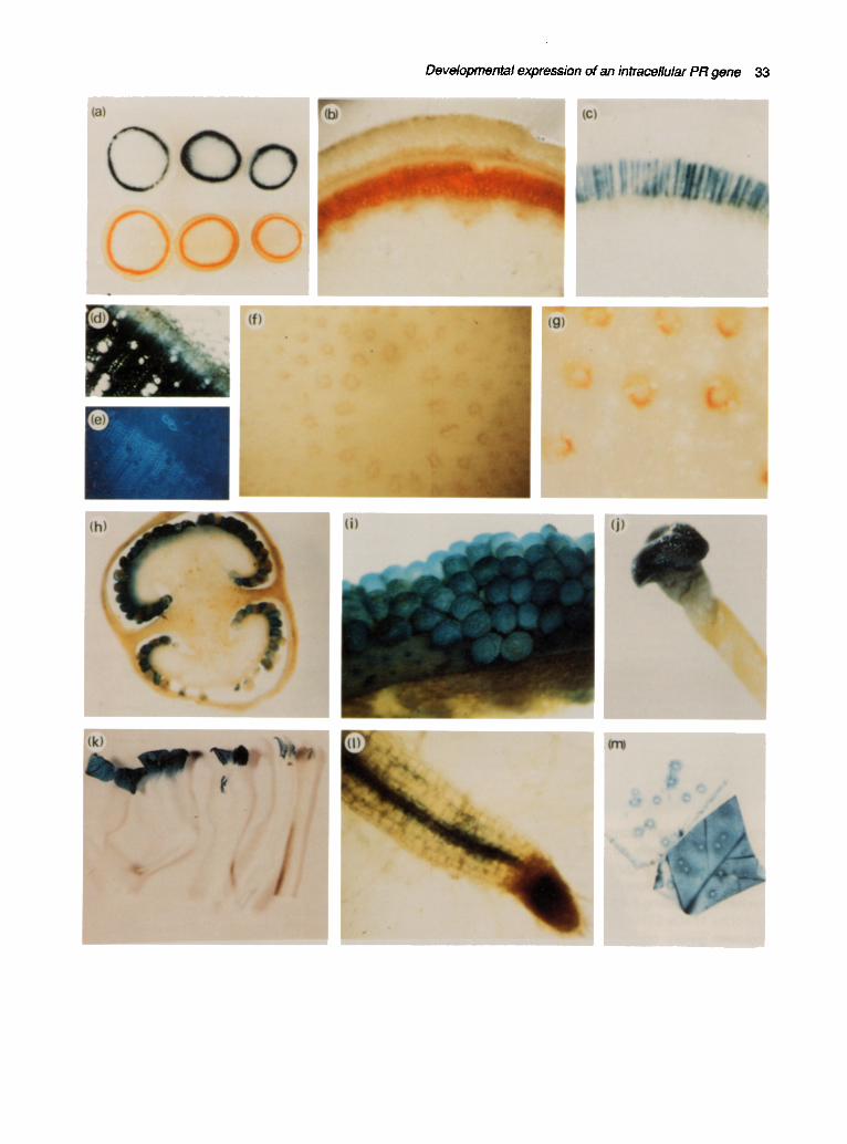
from mature plants which were undergoing increasing secondary thickening postflowering (Figure l a). More detailed histochemical analysis (Figure lc and d) revealed that AoPR1-GUS expression in stems was limited primar- ily to parenchyma ray cells of the developing secondary xylem. These cells lie radially between the highly lignifiod tracheary elements and are responsible for the production of the phenylpropanoid precursors required for xylem vessel lignification (Bevan et al., 1989). Autofluorescence of the stem section resulting from the presence of high concentrations of lignin and other cell wall-bound pheno- lics (Figure l e) and phloroglucinol staining for lignin (Figure la and b) showed clearly that AoPR1-GUS activity sharply mirrors the pattern of stem lignification.
Previous fluorometric analysis of AoPR1-GUS gene expression revealed a low level of expression in roots taken from 6-week-old plants and in seedlings stressed by growth in vitro on agar-containing medium, derived from certain transgenic tobacco lines (Firek et al., 1993; Ozcan et a/., 1993). Histochemical analysis of roots from mature soil grown-tobacco plants revealed that GUS activity was restricted to vascular tissue (Figure 11), whilst quantitative measurements showed that expression levels were relatively low in comparison with other tissues (see Figure 3a).
Expression of endogenous AoPR 1 protein in asparagus stems is associated with vascular tissue
Endogenous AoPR1 gene expression was studied in asparagus stems using a polyclonal antibody raised to AoPR1 protein that had been expressed in Escherichia coll. Protein extracts from 6-week-old, light-grown, unwounded asparagus seedlings, 6-week-old asparagus seedlings cut into 2 mm lengths 3 days postwounding, asparagus roots and mature greenhouse-grown aspara- gus spears were used for Western blot analysis. As pre- dicted from the experiments in transgenic tobacco, AoPR1 protein was detected by the AoPR1 antisera in wounded asparagus samples and in unwounded stem and root tis- sue (Figure 2a). Tissue prints performed on mature asparagus spears demonstrated that the AoPR1 antisera cross-reacted with protein associated with the vascular bundles, which again correlated with cells producing lignin as detected by phloroglucinol staining (Figure I f and g). Pre-immune antisera gave no signal on identically treated tissue prints.
Endogenous intracellular PR proteins are developmentally expressed in potato stems and tubers
We have demonstrated that the antibody raised against the AoPR1 protein will recognise the 17 kD STH-2 intra- cellular PR protein from potato (Gill et al., manuscript in preparation). Likewise an antibody prepared against the
Figure 1. Expression of AoPR1-GUS in transgenic tobacco and endogenous AoPRt gene expression in asparagus. (a) Adjacent stem sections from a flowering mature tobacco plant alternately stained for GUS activity or lignin deposition (X-Gluc staining above and phlero- glucinol staining below) (:<2). (b) Stem section from a mature tobacco plant stained with phloroglucinol (x30). (c) Stem section from a mature tobacco plant stained with X-Gluc for 3 h showing GUS activity in the xylem parenchyma rays (×30). (d) X-Gluc stained stem section from a tobacco plant (xt 50). (el Identical section to (d) showing UV autofluorence of phenolic compounds. (f) Tissue print of asparagus spears cross-reacting with the AoPR1 anti-sere around the vascular bundles (x30). (g) Phloroglucinol staining of asparagus stem showing the location of lignin surrounding the vascular bundles (x60). (h) Cross-section of a tobacco seed pod 5 day s postpollination showing staining of seeds (x7). (i) Part of a Iongtitudinai section of a tobacco seed pod 5 days postpollination showing GUS activity in the testa (x50). 0) GUS activity in a receptive tobacco stigma (x12). (k) Tobacco petals at various stages from prior to flower opening (right) to fully opened (left). (I) Root from tobacco seedling showing staining in the vascular tissue (x150}. (m) Wounded tobacco leaf disks placed on filter paper soaked in water (top) and 4 mM salicylic acid (bottom) for 48 h.
Developmental expression of an intracellular PR gene 33

34 Simon A.J. Warner, Aideen Gill and John Draper
Figure 2. Western and Northern analysis of AoPR1 gene expression in asparagus and potato. (a) Western blot of proteins detected with the AoPRt antisera. One micro- gram of total protein from E. coil expressing the pT7.7 AoPR1 construct and 10 p.g of total plant protein were loaded par lane. Lane 1, pT7.7 AoPR1 protein. Lane 2, untreated cladodes from 6-week'-old asparagus seedlings. Lane 3, roots from 6-week-old asparagus seedlings. Lane 4, asparagus spears from mature plants grown outdoors. Lane 5, etiolated 6-week-old asparagus seedlings sliced into 2 mm lengths and incubated on damp filter paper at 20°C for 3 days. Lane 6, etiolated 6-week-old asparagus seedlings 3 days postspraying with 4 mM salicylic acid. (b) Western blot of potato proteins detected with the STH-2 antisera. One microgram of total protein from E. coil expressing the pT7.7 AoPR'I con- struct and 10 Izg of total plant protein were loaded per lane. Treatment of potato material is as dascribed previously (Madneau etal., 1987). Lane 1, sliced potato tubers. Lane 2, sliced potato tubers treated with arachidonic acid. Lane 3, pT7.7 AoPR1 protein. Lane 4, untreated potato stem. (c) Northern blot of total RNA extracted from etiolated 6-week-old aspara- gus seedlings postspraying with 4 mM salicylic acid. Ten micrograms of total RNA was loaded per lane. Lane 1, unsprayed plants. Lane 2, 12 hours postspraying. Lane 3, 1 day postspraying. Lane 4, 3 days postspraying. Lane 5, 5 days postspraying.
potato STH-2 protein (Constabel and Brisson, 1992) will cross-react specifically to the AoPR1 fusion protein expressed in E. coil (Figure 2b, lane 3). Protein extracts from potato tuber slices (variety Desiree) analysed by Western blotting using the STH-2 antibody revealed a low level of the 17 kDa STH-2 protein (Figure 2b, lane 1 ) which increased greatly on elicitation with arachidonic acid (Figure 2b, lane 2). In contrast a second cross-reacting protein of approximately 18 kDa (Constabel and Brisson, 1992) was not inducible by elicitor treatment. Endogenous expression of the STH-2 intracellular PR protein was also detected in mature potato stems (Figure 2b, lane 4).
AoPR1 promoter activity is localized to cells producing phenylpropanoid derivatives in developing flowers
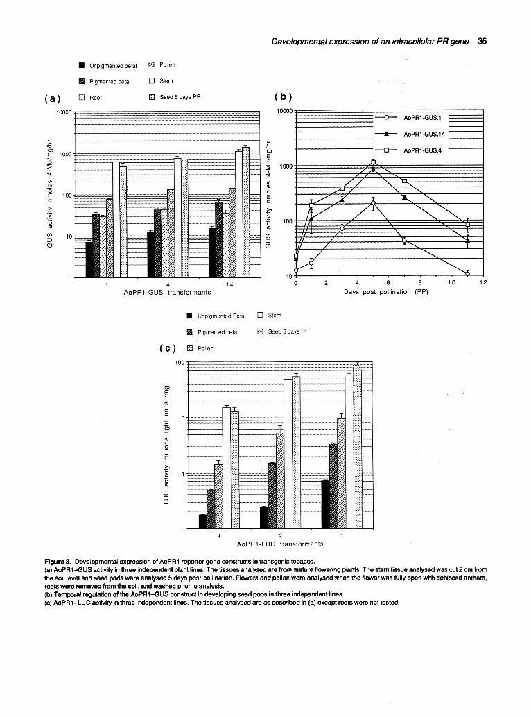
Mature pollen released from dissected anthers just prior to dehiscence exhibited positive staining with X-Gluc (Warner etaL, 1993). Expression levels of AoPR1-GUS in mature pollen were lower than in lignified stems (Figure 3a). Further analysis of AoPR1 expression revealed no detectable GUS activity in developing microspores and immature pollen. AoPR1-GUS expression in sporophytic floral tissues prepollination was observed in the nectaries (data not included), the stigma surface (Figure l j) and the anthocyanin-containing regions of developing petals (Figure l k). Staining for GUS activity in nectades was only undertaken in open flowers just prior to pollination. Histochemical staining was most intense at the sti~jma surface as it became 'receptive' for p?llinat~n. ~GUS activity in petals first appeared in buds that had just opened, which correlated with the onset of anthocyaRin synthesis. Quantitative analysis of GUS activity revealed relatively low levels of AoPR1 promoter activity in the anthocyanin-containing regions of developing petals (Figure 3a).
AoPR1-GUS expression isgreatly upregulated in developing seeds prior to the visible accumulation of pigments
Plants from progeny of three lines of AoPR1-GUS trans- genic tobacco were grown to the flowering stage. Selected flowers were emasculated just prior to anther dehiscence and poller applied from dehisced anthers of another flower on the same plant. Developing seed pods were har- vested at different days postpollination (PP) and GUS activity was measured using a fluorometric assay. From Figure 3(b) it can be seen that AoPR1-GUS gene expres- sion is temporally regulated during seed pod develop- ment, with GUS activity increasing to a maximum approxim~.tely 3-5 days postpollinatibn. Using a histo- chemical assay, GUS activity in seed pods was found to be located mainly in immature seed testas (Figure lh). Strong GUS activity was only found in immature seeds prior to visible pigment accumulation which started 9-10 days postpollination.
Transgenic tobacco transformed with AoPR 1-LUC constructs shows identical developmentally regulated reporter gene activity to plants transformed with AoPR1--GUS.
Recently, it has been suggested that artefactual expres- sion in transgenic plants may be associated with the use of GUS as a reporter gene, especially in pollen (Uknes e! al., 1993). For this reason we constructed an
Developmental expression of an intracellular PR gene 35
Figure 3. Developmental expression of AoPR1 reporter gene constructs in transgenic tobacco. (a) AoPR1--GUS activity in three independent plant lines. The tissues analysed are from mature flowering plants. The stem tissue analysed was cut 2 cm from the soil level and seed pods were analysed 5 days post-pollination. Flowers and pollen were analysed when the flower was fully open with dehisced anthers, roots were removed from the soil, and washed prior to analysis. (b) Temporal regulation of the AoPRI.--GUS construct in developing seed pods in three independent lines. (c) AoPR1-LUC activity in three independent lines. The tissues analysed are as described in (a) except roots were not tested.

36 Simon A.J. Warner, Aideen Gill and John Draper
AoPRl-luciferase translational fusion differing from the AoPR1-GUS construct only in the nature of the fusion. The AoPR1-LUC fusion was created so that the initiating ATG of the luc gene was an integral part of an Ncol site used in the fusion. Although firefly luciferase does not offer the same advantages as the GUS reporter gene, because no simple histochemical assay is available for this enzyme, luciferase activity may be simply, sensitively and quickly assayed using a photoluminometer. Standard tobacco transformation techniques were employed to transfer the AoPR1-LUC construct into the tobacco genome and eight independent transformed tobacco lines were obtained. The presence of the transgene was confirmed by PCR (data not shown). Protein extracts were prepared from various organs and tissues and these were assayed for luciferase activity. Figure 3(c) shows that the luciferase activity in the eight independently transformed tobacco lines mirrors the developmentally regulated GUS activity in transgenic tobacco harbouring the AoPR1-GUS constructs (Figure 3a). These data suggest that there is no artefactual expression of the AoPR1-GUS reporter gene in tobacco.
The AoPR1 promoter activity is enhanced by salicylic acid
Salicylic acid inducibility of the classical tobacco PR genes has been well characterized (Antoniw and White, 1986). More recently, the soybean IPR transcript, SAM 22, has been shown to be moderately induced following treatment of detached leaves with 10 mM salicylic acid (Crowell et a/., 1992). Initial experiments using tobacco leaf disks harbouring the AoPR1-GUS construct were performed to determine the optimum concentrations of salicylic acid. The leaf disks were placed on filter paper soaked in increasing concentrations of salicylic acid for 48 h at room temperature, harvested and assayed for GUS activity using the procedure described by van de Rhee et al. (1990). Figure 4(a) shows that the increase in
Figure 4. Induction of GUS activity in leaf disks in response to treatment With salicylic and 4-hydroxybenzoic acid. Error bars represent the standard deviation taken from measurements of a minimum of three independent samples. (a) Dose dependency of AoPR1-GUS expression following a 48 h time period post,spraying with 4 mM salicylic acid or 4-hydroxypenzoic acid. (b) Time course of GUS activity following wounding alone or following wounding and treatment with 4 mM salicylic acid. (c) A comparison of GUS activity following wounding alone or following wounding and treatment with 4 mM salicylic acid in three independent transgenic plant lines harbouring the AoPR1-GUS transgene (1,4 and 14) representing the range of expression levels found for this construct and two plants harbouring the CaMV35-GUS construct (121.13 and 121.85) that exhibit high and low expression levels.
(e)
s
"6 E
>
(n
( b )
-6 _E
u)
( c )
E
-6 E
u)
1000
-- Sa~y~ a~d i
8OO ~ 4-h~roxyoenzo¢ a=d , , ~
0 2 4 6 8 10
Concentration mM
12
500
Wounded / / / ~
~- 4 mM SA & W o u n d i n g /
2OO
0 10 20 30 40 51
Time h
1800"
1600.
1400.
1200'
1000"
800'
600,
400'
200'
0
[ ] Wounding
• 4 mM SA & Wounding
4 14 121.13
Transgenic Plam Line
121.85

Developmental expression of an intracellular PR gene 37
GUS activity is approximately linear up to a salicylic acid concentration of 4 mM. After incubation in 10 mM salicylic acid the leaf disks appeared less healthy and the mea- sured GUS activity ceased to increase linearly with con- centration. Treatment of disks with salicylic acid concentrations below 1 mM gave no detectable induction of GUS activity and treatment with 4-hydroxybenzoic acid, a biologically inactive analogue of salicylic acid, had no effect on measurable GUS activity in identically treated leaf disks (Figure 4a). Subsequent experiments were car- ried out using 4 mM salicylic acid. The induction kinetics of the AoPR1-GUS gene were similar following either wounding or treatment with salicylic acid (Figure 4b). Max- imal expression levels varied in independent transgenic lines but in all cases an approximate sixfold induction of the AoPR1-GUS gene was achieved following treatment of leaf explants with salicylic acid (Figure 4c).
Histochemical staining of leaf disks that had been treated with 4mM salicylic acid revealed GUS activity throughout the whole of the explant with stronger staining surrounding the wound sites (Figure l m). AoPR1-GUS expression in major leaf veins was greater than in sur- rounding non-wounded tissue in salicylic acid-treated leaf explants (Figure I m). Control leaf disks, floated on water, revealed characteristic staining only around the wound sites (Figure 1 m).
Endogenous AoPR1 protein was undetectable in asparagus cladode tissue using Western blotting (Figure 2a, lane 2). Spraying/with salicylic acid induced AoPR1 protein accumulation to levels similar to those achieved following gross wounding of etiolated seedling tissue (Fig- ure 2a, lane 6). The time course of AoPR1 transcript induction (Figure 2c) following spraying with salicylic acid was similar to that achieved after wounding seedlings (Warner et al., 1992, 1993).
The AoPR1 promoter contains elements related to nucleotide sequences known to be important for gene upregulation both in other intracellular PR proteins and in genes coding for enzymes of the phenylpropanoid pathway
PcPRI-1 is a parsley gene coding for a member of the intracellular PR group of proteins which has been shown to be strongly upregulated at sites of fungal attack and following microbial elicitor treatment of established suspension cultured cells (Meier et al., 1991 ; Somssich et al., 1988). Although there are no significant long stretches of homology between the AoPR1 and PcPRI-1 promoters there is a sequence from -396 to -388 in the AoPR1 promoter that is identical to an inducible footprinted
Rgure 5. Architecture of sequences that may possibly interact with trans- acting factors in the AoPR1 promoter.
sequence of the PcPRI-1 promoter from -244 to -235 which reads ATTTGACCG (Figure 5). The core sequence TGACCG is within the repeat 2 sequence of the AoPR1 promoter (Warner et al., 1992) but there are no inverted repeat sequences of this motif as in the parsley PcPRI-1 promoter.
Discussion
Previously we have shown that the AoPR1 promoter is strongly and locally induced adjacent to wounded regions or sites of attempted pathogen invasion in leaves of trans- genic tobacco plants (Warner et al., 1993). Additionally, as a birch protein related to AoPR1 (Betvl) was isolated originally as a pollen allergen, we were not surprised to find AoPR1-GUS expression in mature pollen (Wamer et al., 1993). Other members of the intracellular PR protein class exhibit developmental expression in other tissues. For example, the soybean SAM 22 transcript is detectable at high levels in roots, as well as being induced by chemical elicitors such as salicylic acid and methyl viologen (Crowell et al., 1992), whilst the ABR17 and ABR18 proteins from pea are strongly upregulated in developing pea seeds and induced by treatment with exogenous abscisic acid (Barratt and Clark, 1991; un- published cDNA sequence on GCG Wisconsin DNA database). These findings prompted us to examine the possible developmental expression of the AoPR1-GUS translational fusion in transgenic tobacco plants at various points in the life cycle and in response to exogenously- supplied salicylic acid. In addition, recent evidence of aberrant GUS enzyme expression driven by the tobacco PRla promoter has been described where ectopic GUS expression in pollen was shown in particular to be artefactual (Beilmann et al., 1992; Uknes et al., 1993). To investigate this possibility we analysed also endo- genous AoPR1 expression in asparagus and repeated the promoter analysis in tobacco, this time using an AoPR1 promoter-firefly luciferase reporter construct.
38 Simon A.J. Warner, Aideen Gift and John Draper
AoPR1-GUS expression is developmentally regulated with expression patterns similar to those documented for other IPR and genes coding for enzymes in the phenylpropanoid pathway
Many genes that are wound-, pathogen-, or elicitor- induced are not only activated following these external stimuli, but are often also subject to developmental con- trol, especially in floral organs. For example, studies on the pathogenesis-related (PR) proteins of tobacco revealed that some are expressed in floral organs (Lotan et aL, 1989; Neale et aL, 1990), whilst the proteinase inhibitor (Pin 2) promoter-GUS gene fusion demonstrated reporter gene activity in developing potato ovules (Pefia- Cortes et aL, 1991). The reasons for the developmental expression of these defence-related genes are currently unknown. In the case of phenylpropanoid pathway genes, the developmental expression observed for promoter-GUS fusions correlates with cell types that accumulate known phenylpropanoid pathway products. For example, histochemical analysis of the bean PAL-2 and parsley 4CL-1 promoter-GUS fusions in transgenic tobacco (Bevan et aL, 1989; Hauffe et aL, 1991; Liang et aL, 1989; Shufflebottom et aL, 1993) revealed develop- mental expression in the pigmented parts of the petal (antlhocyanins and other flavonoids) and developing seed coats (anthocyanins; Harborne, 1976), in mature pollen (4,2',4',6'-tetrahydroxychalcone; de Vlaming and Kho, 1976), in the secondary xylem parenchyma cells of the stem (lignin) and at the stigma surface where volatile phenylpropanoid compounds that attract pollinating insects are produced (Hauffe et aL, 1991 ).
The present data represent the first comprehensive analysis of developmental gene expression driven by an intracellular PR protein gene promoter in transgenic plants. It is clear from the temporal and spatial analysis of the AoPR1 promoter-GUS gene fusion during develop- ment in transgenic tobacco and endogenous expression in asparagus that specific expression of the AoPR1 gene occurs in the vast majority of cell types known to accumu- late different classes of phenylpropanoid derivatives. These data were somewhat unexpected as it has been shown previously that other promoters of specific genes coding for enzymes involved in the core phenylpropanoid biosynthesis pathway exhibit a much more restricted pattern of developmental expression. For example, the PAL2 gene promoter from bean gives strong expression in tobacco secondary xylem parenchyma but is only very weakly expressed in leaf cells, whilst the PAL1 gene is strongly induced by wounding but weakly expressed in stem tissue (Shufflebottom et aL, 1993). Promoters of genes coding for enzymes involved in branches of the phenylpropanoid pathway, such as chalcone synthase (flavonoid synthesis) show very little expression at sites of
lignin synthesis and can either be active in all cell types where flavonoids are found (as in the case of snapdragon and Arabidopsis, which only have a single CHS gene) or may show differential expression in a range of cell types in different plant species (e.g. bean) which contain several CHS genes (Schmid et al., 1990). Thus, in contrast to these previously analysed promoters the AoPRI--GUS fusion was expressed in the majority of cells producing products of all the main branches of the phenylpropanoid pathway.
Some data on the developmental expression of intra- cellular PR proteins have been previously presented but these data did not localize IPR protein expression to any particular cell type (Barratt and Clark, 1991 ; Breiteneder et a/., 1989; Crowell et al., 1992). In addition, it has been demonstrated by in situ hybridization that the parsley PcPRI-1 transcript is localized to the vascular tissue in the root (Dr Imre Somssich, personal communication) and the bean PvPR1 promoter is active in roots of transgenic tobacco (Dr Michael Waiters, personal communication). It was confirmed that the AoPR1 promoter was able to drive GUS and LUC expression in these cell types in transgenic tobacco. However, IPR gene expression in stems and flowers has not been described elsewhere and as such the possibility of unscheduled expression existed. This was examined by testing for endogenous IPR protein expression in asparagus and potato by either Northern or Western analysis. Western blots and tissue prints revealed low levels of endogenous AoPR1 protein in asparagus stem tissues localized to sites of active lignification. The lower amount of AoPR1 expression in asparagus stems as compared with tobacco correlates well with the structural differences between monocot and dicot stems and the corresponding differences in the degree of secondary thickening. Western blot analysis of lignified potato stem protein revealed endogenous STH-2 IPR protein. The IPR protein expression in potato stems or untreated potato tubers has not been reported in previous studies (Constabel and Brisson, 1992).
The observation that the AoPR1 promoter exhibits low expression levels in the pigmented regions of tobacco petals is difficult to correlate with expression patterns in asparagus since asparagus has very small unpigmented flowers. The level of AoPR1-GUS expression in petals correlates well with the activity of the phenylpropanoid pathway in these organs as anthocyanin accumulation only occurs in epidermal cells.
The AoPR1-GUS gene fusion shows by far the strongest developmental expression in the expanding secondary xylem of tobacco stems as they become lignified and in the seed coat, just prior to the appearance of anthocyanins. The PAL2 gene promoter from bean gives strong expression in xylem parenchyma, in an iden- tical manner to that exhibited by AoPR1-GUS fusion

Developmental expression of an intracellular Pit gene 39
whilst the time course for maximal AoPR1-GUS activity in developing seeds is in approximate agreement with that observed for the snapdragon CHS promoter-GUS fusion in transgenic tobacco (Fritze et aL, 1991; Shufflebottom et aL, 1993). This temporal and spatial regulation reflects the fact that these latter two genes are being activated in cells required to produce large amounts of lignins and antho- cyanins, respectively, and it would perhaps be interesting to speculate why the AoPR1 promoter is strongly acti- vated in both of these cell types, as well as being strongly induced at sites of wounding and attempted pathogen invasion (Warner et aL, 1992, 1993).
In conclusion, the AoPR1 promoter is active in trans- genic tobacco in all tissues and organs that have been associated with the expression of all the other reported IPR genes and is also expressed in additional cell types such as the pigmented regions of the petal and stems. It is not yet clear whether endogenous IPR genes are develop- mentally regulated similarly in other species, or whether some gene family members are confined to specific devel- opmental expression in one organ or cell type. Further promoter-reporter analysis in transgenic plants may help answer this question.
The AoPR1 promoter contains sequence elements similar to those identified in promoters of other IPR genes and in genes coding for enzymes of phenylpropanoid biosynthesis
Lignin and flavonoid production represent two separate branches of the phenylpropanoid pathway which diverge after the 'core' part of the pathway which ends at 4- hydroxycinnamoyl CoA. Thus, it is tempting to speculate that AoPR1 either has some function concerned with the core part of the phenylpropanoid pathway, or that perhaps the AoPR1 promoter could be generally activated by phenylpropanoid intermediates, as shown for the CHS15 gene from bean by Loake et aL (1991). Several elements in the AoPR1 promoter have been identified previously (Warner et aL, 1993) that are similar to the H-box sequences implicated to be important in the regulation of genes coding for enzymes in the early part of the phenyl- propanoid pathway (Loake et aL, 1991; Lois et aL, 1989; Yu et aL, 1993). Most of these sequences have been shown to be involved in the induction of transcription by microbial elicitors or UV light stress. However, in the case of the CHS15 gene from bean (Loake et aL, 1991; Yu et aL, 1993 ), a further promoter element has been identified which has been shown to increase transcription in response to externally supplied trans-p-coumaric acid (4- CA) and the proteins binding these sequences have been biochemically characterized. In preliminary experiments with AoPR1-GUS tobacco seedlings we have not found any evidence for increased GUS activity in UV-stressed
plants. Similarly we have no evidence for either up or downregulation of the AoPR1-GUS fusion by externally applied cinnamic or coumaric acid (Gill and Warner, unpublished observations). However, the significance of these latter data is not known as both of these compounds are known to cause non-specific effects on tobacco cell metabolism that are not directly related to gene transcrip- tion. It is possible that the elements similar to those described by Lois et aL (1989) are involved in AoPR1 induction at wound sites and at sites of pathogen invasion, but further work is required to substantiate any functional link. In this context it should be noted that there are no H-box sequences in the promoter of the patho- gen-inducible potato IPR homologue, pSTH2 (Matton et aL, 1990), so this sequence motif is not necessarily conserved between all the members of the IPR family.
Although the parsley PR-1 gene promoter sequence ATTTGACCG (shown by in vivo DNA footprinting to be protected in elicitor-treated parsley cells) is present in the AoPR1 promoter and a similar sequence is important in the regulation of the potato STH2 promoter (Matton et aL, 1993), the functional importance of this element in the AoPR1 promoter is as yet unknown. Other sequence ele- ments that have been shown by other workers to interact with nuclear factors are also present in the AoPR1 pro- moter. One of these elements is similar to the binding site of the common plant regulatory factors (CPRF) or bZlP proteins which recognize G-box elements (Weisshaar et aL, 1991; Williams et aL, 1992). These proteins have been shown to be important in the regulation of expression of a wide variety of genes showing tissue specificity or respon- siveness to environmental stimuli, such as light, or bio- logically active compounds such as ABA (Oeda et aL, 1991; Schindler et aL, 1992; Schmidt et aL, 1992). In this context it is interesting to note that preliminary experi- ments show AoPR1-GUS expression in developing tobacco seed, localized to the embryo (unpublished observations). This developmental expression data correlates well with the expression pattern of the pea IPR protein ABR18 in developing pea embryos (Barratt and Clark, 1991 ) which is ABA responsive.
The biochemical function of intracellular PR proteins in general is unknown and there have been no other reported detailed promoter-GUS analyses published for other members of this family to corroborate the current findings. The intracellular PR proteins have been described in many species, including both monocots and dicots and are generally transcribed at high levels when activated by elicitors, pathogen attack or (in some cases) wounding. The protein sequences of IPR proteins do not share significant homology with any known phenyl- propanoid biosynthesis enzymes or other classes of protein and so they are unlikely to represent a previously unrecognized enzyme of this pathway. The co-induction of

40 Simon A.J. Warner, Aideen Gill and John Draper
IPR genes and genes of the phenylpropanoid pathway is currently under investigation.
The AoPR1 promoter is responsive to exogenous salicylic acid
Experiments investigating the effects of salicylic acid on transgenic tobacco were carried out using procedures already published (van de Rhee et al., 1990) and demon- strated that the AoPR1 promoter is inducible by salicylic acid in tobacco leaf disks. The levels of induction compare directly with the levels of induction observed for a tobacco 'classical' acidic PRla promoter reported by van de Rhee et aL (1990) and Ohshima et aL (1990), but are two to ten- fold lower than those reported by Uknes et aL (1993) for essentially the same promoter in experiments where the leaves on an intact plant were painted with 50 mM salicylic acid. In our hands, when greenhouse-grown tobacco plants were sprayed with concentrations greater than 20- 30 mM of salicylic acid the leaves wilted within 6-12 h post-treatment rendering further analysis impossible. A possible explanation of why we observed wilting at these levels may relate to the fact that we were using SR1 tobacco, whereas Uknes et aL (1993) used Xanthi-nc. In preliminary experiments we found little induction of GUS activity after spraying 4 mM salicylic acid on intact AoPR1-GUS tobacco plants but, in contrast, using PRla antisera in a Western analysis on protein extracts we confirmed that the endogenous 'classic' secreted PRla protein accumulated to high levels following this treatment (data not shown). These preliminary observations suggest that classical PR proteins and intracellular PR proteins may be responding differently to the presence of exogenously supplied salicylic acid. Northern analyses confirmed that the AoPR1 transcript was weakly induced following spraying asparagus seedlings with salicylic acid, with message levels at their most abundant 2-3 days post-treatment. It has recently been confirmed that sali- cylic acid is a phenylpropanoid metabolite, synthesized directly from cinnamic acid via benzoic acid (Yalpani et aL, 1993) which again emphasizes the correlation between phenylpropanoid metabolism and AoPR1 expression. In summary, although the AoPR1 promoter is inducible by salicylic acid in tobacco leaf explants and in asparagus seedlings, the levels of induction remain low and are not very high compared with AoPR1 induction by wounding, or in comparison with expression levels observed in lignified stems or developing seeds.
Experimental procedures
Plant growth conditions and treatments
T2 seed was obtained from self-fertilized transgenic SR1 tobacco plants harbouring AoPR1-GUS constructs (Warner et al., 1993).
The seeds were germinated on MS0 media (Draper et al., 1989) supplemented with 50 mg ml -~ kanamycin and then transferred to compost and grown in greenhouse conditions to maturity. T1 SR1 tobacco plants were obtained for AoPR1-LUC constructs by standard transformation techniques (Draper et aL, 1988) and plants were grown to maturity in the greenhouse. Wounding assays were performed on leaf disks as described previously (Warner et aL, 1993). Asparagus (Asparagus officinalis cv. Conovers Colossal) seeds were germinated in vermiculite and grown in the green house for 6 weeks prior to treatment with 4 mM salicylic acid (Sigma). Mature 3-year-old or greater asparagus plants were grown outdoors in soil beds and spears cut as required. Potato (Solanum tuberosum cv. Desir~e) plants were grown in the greenhouse to maturity. Tubers were treated accord- ing to Marineau et aL (1987) and other plant material used for protein extraction with no special prior treatment.
Purification of AoPRI protein from E. coil and antibody production
The AoPR1 cDNA was cloned into the EcoRI site of the pT7-7 vector (Tabor and Richardson, 1985). The pT7-7 vector contains the T7 phage RNA polymerase promoter and the translational start site of the gene 10 protein. The pT7-7:AoPR1 construct was then transformed into competent E. coil K38 cells that were already harbouring the heat-inducible pGP1-2 plasmid which expresses T7 RNA polymerase. Cultures were grown overnight in LB media at 30°C diluted 1:50 in fresh LB and grown at 30°C to an ODsoo nm 0.8-1.0. The culture was then transferred to a 42°C shaker for 30 min followed by a further incubation of 30 min at 30°C. Inclusion bodies were isolated by the differential centrifuga- tion method as described by Marston (1987) and the inclusion body protein denatured and refoided according to Owen et al. (1992). The pT7-7:AoPR1 inclusion body protein products were separated by preparative SDS--polyacrylamide gel electrophore- sis and the 17 kDa AoPR1 protein product was excised. The AoPR1 protein was electroeluted from the gel slice, neutralized, dialysed against TBS and used to raise polyclonal antisera in rab- bits as described by Harlow and Lane (1988).
Western and Northern analysis of AoPR1 protein and transcript
Protein was extracted from relevantly treated asparagus or potato tissues in protein extraction buffer as described by Constabel and Brisson (1992) and Westerns were carried out using the 1:60 00 dilution of the AoPR1 antisera as described by Harlow and Lane (1988). Tissue prints were developed as for Westerns following pressing of freshly sliced asparagus spears directly on to nitro- cellulose. Northern analysis was carried out on asparagus RNA using the AoPR1 cDNA as a probe as described previously (Warner et al., 1992).
Construction of AoPRl-luciferase reporter gene and plant transformation
The pBiuescript based plasmid pNBL52 was a gift from Nell Bate and David Twell of Leicester University (manuscript in prepara- tion) and was used as the basis for the construction of the AoPR1 promoter luciferase fusions. The pNBL52 plasmid is engineered to create facile translational fusions between test promoters and the firefly luciferase gene where the initiating ATG codon of the

Developmental expression of an intracellular PR gene 41
luciferase gene is encoded in an Ncol site. Promoter fragments are ideally cloned into this vector as Sail and Ncol fragments. An oligonucleotide primer (5'- GAAGCCATGGTTCCTCTCTGTTGT- TATG-3') was designed to insert an Ncol site into the AoPR1 promoter such the Ncol site's ATG was in exactly the same position as the AoPR1 gene's natural initiating ATG. This primer was used in conjunction with the T7 sequencing primer in a PCR reaction with 100 ng of the AoPR1 promoter IPCR product cloned into the EcoRI and Sinai sites of pBluescript for 10 cycles using conditions described previously (Warner et al., 1993). The PCR product was digested with Ncol and Sail and cloned into the Ncol and SaA sites of pRTL2 creating a translational fusion between AoPR1 and luciferase. The pBI101.1-based plasmid AoPR1- GUS pBI101.1 (Warner et al. 1993) was digested with Ssll and Smal to remove the GUS reporter gene, the cohesive ends were then end-filled using Klenow in the presence of 2 mM dNTPs, the plasmid was recircularized using T4 DNA ligase, then transformed into E. coil and amplified. This plasmid was digested with BamHI and Sail releasing the AoPR1 promoter, gel purified and ligated to a BamHI /Sail AoPR1-LUC fragment so that a pBI101-based plasmid containing the AoPR1-LUC gene fusion was created. This was used to transform LBA4404 Agro- bacterium and sub-sequently transferred into tobacco by standard techniques (Draper et aL, 1988)
Luciferase analysis
Plant tissue was ground up in 0.5 ml of luciferase extraction buffer (100 mM phosphate buffer pH 7.5 and 1 mM dithiothreitol), the debris was spun down for 5 min and the supernatant removed to a fresh tube. Protein extract (50 p_l) (adjusted to 0.5 mg m1-1 pro- tein) was added to the bioluminescence photometer reaction tube and the tube placed into the photometer (Berthold Clinilumat from Berthold Instruments UK Ltd). ATP buffer (100 I~1) (50 mM HEPES pH 7.8, 20 mM MgCI2 and 10 mM ATP) and 100 I~1 of 0.05 mM D-luciferin (Sigma) made up in luciferase extraction buffer was then added by the machine and the light emission measured as light units. Luciferase activity was expressed as light units per mg of protein. Protein quantitation was carried out essentially as in Bradford (1976).
GUS analysis
Transformed tobacco plant lines 1, 4 and 14, harbouring the AoPR1-GUS construct, were chosen that represented low, medium and high levels of wound-induced GUS expression as determined from initial fluorometric assays. A minimum of three independent samples were assayed for each tissue sample. Fluorometric and histochemical analysis of GUS activity (Jef- ferson et aL, 1987) were carried out as described accord- ing to the modifications of Topping et aL (1991). Pollen was stained after removal from anthers and washing twice in staining buffer. Petals and stigmas were dissected prior to staining. Developing seeds were stained in situ following the bisection of the seed pod to allow entry of the X-Gluc.
Acknowledgements
We would like to thank Ray White and John Antoniw for the gift of the PRla antisera, Peter Constabel and Normand Brisson for the gift of the STH-2 antisera and Graham Benskin for growing and
maintaining transgenic plants. S.A.J.W. is supported by AFRC grant no. PG91/525 and A.G. is supported by a Gatsby Charitable Foundation studentship.
References
Antoniw, J.F. and White, R.F. (1986) Changes with time in the distribution of virus and PR protein around single local lesions of TMV infected tobacco. Plant Mol. BioL 6, 145-149.
Barrett, D.H.P. and Clerk, J.A. (1991 ) Proteins arising during the late stages of embryogenesis in Pisum sativum L. Planta, 184, 14-23.
Be,mann, A., Albrecht, K., Schultze, S., Wanner, G. and Pfitzner, U.M. (1992) Activation of a truncated PR-1 promoter by endogenous enhancers in transgenic plants. Plant MoL BioL 18, 65-78.
Bevan, M., Shufflebottom, D., Edwards, K., Jefferson, R. and Schuch, W. (1989) Tissue and cell-specific activity of a phenylalanine ammonia lyase promoter in transgenic plants. EMBO J. 8, 1899-1906.
Bol, J., Linthorst, H.J.M. and Corneliseen, B.J.C. (1990) Plant pathogenesis-related proteins induced by virus infection. Ann. Rev. PhytopathoL 28, 113-138.
Bowles, D.J. (1990) Defence-related proteins in higher plants. Ann. Rev. Biochem. 59, 873-907.
Bradford, M.M. (1976) A rapid and sensitive method for the quantification of microgram quantities utilising the principle of protein dye binding. Anal Biochem. 72, 248-254.
Breiteneder, H., Pettenburger, K., Bito, A., Valenta, R., Kraft, D., Rumpold, H., Scheiner, O. and Breitenbach, M. (1989) The gene coding for the major birch pollen allergen Betvl, is highly homologous to a pea disease resistance response gene. EMBOJ. 8, 1935-1938.
Constabel, P.C. and Briseon, N. (1992) The defence-related STH-2 gene product of potato shows race-specific accumula- tion after inoculation with low concentrations of Phytophtora infestans zoospores. Planta, 188, 289-295.
Crowell, D.N., John, M.E., Russell, D. and Amisino, R.M. (1992) Charactedsation of a stress-induced, developmentally regulated gene family from soybean. Plant MoL Biol. 18, 459-466.
Dixon, R.A. and Harrison, M.J. (1990) Activation and organisa- tion of genes involved in microbial defence in plants. Adv. GeneL 28, 165-235.
Draper, J., Scott, R., Armitage, P. and Walden, R. (1988) Plant Genetic Transformation and Gene Expression: A Laboratory Manual. Oxford: Blackwell Scientific Publications,
Enyedi, A.J., Yalpani, N., Silverman, P. and Raskin, I. (1992) Localisation, conjugation, and function of salicylic acid in tobacco during the hypersensitive reaction to tobacco mosaic virus. Proc. Natl Acad. Sci. USA, 89, 2480-2484.
Firek, S., (~zcan, S., Warner, S.A.J. and Draper, J. (1993) A wound-induced promoter ddving npt-II expression limited to dedifferentiated cells at wound sites is sufficient to allow selec- tion of transgenic shoots. Plant Mol. BioL 22, 129-142.
Fritze, K., Staiger, D., Czaja, I., Walden, R., Schell, J. and Wing, D. (1991) Developmental and UV light regulation of the snapdragon chalcone synthase promoter. Plant Cell, 3, 893-905.
Hahibrock, K. and Schsel, D. (1989) Physiology and molecular biology of phenylpropanoid metabolism. Ann. Rev. Plant Phys. Mol. BioL 40, 347-369.
Harborne, J.B. (1976) Functions of flavonoids in plants. In
42 Simon A.J. Warner, Aideen Gill and John Draper
Chemistry and Biochemistry of Plant Pigments, Volume 2 (Goodwin, T.W, ed.). London: Academic press, pp. 735-776.
Harikrishna, K., Paul, E., Darby, R. and Draper, J. (1991) Wound response in mechanically isolated asparagus meso- phyll cells: a model monocotyledon system. J. Exp. BoL 42, 791-799.
Harlow, E. and Lane, P. (1988) Antibodies. A Laboratory Manual. Cold Spring Harbor, NY: Cold Spring Harbor Press.
Hauffe, K.D., Paszkowskl, U., Schulze-Lefert, P., Hahlbrock, K., Dangl, J.L. and Douglas, C.J. (1991) A parsley 4CL-1 promoter fragment specifies complex expression patterns in transgenic tobacco. Plant Cell, 3, 435-443.
Jefferson, R.A., Kavanagh, T.A. and Bovan, M.W. (1987) GUS fusions: beta glucuronidase as a sensitive and versatile marker in plants. EMBOJ. 6, 3901-3907.
Llang, X., Dron, M., Crsmer, C.L., Dixon, R.A. and Lamb, C.J. (1989) Developmental and environmental regulation of a phenylalanine ammonia-lyase-~-glucuronidase gene fusion in transgenic tobacco plants. Proc. Natl Acad. Sci. USA, 86, 9284-9288.
Loake, G.J., Choudary, A.D., Harrison, M.J., Mavandad, M., Lamb, C.J. and Dixon, R.A. (1991) Phenylpropanoid pathway intermediates regulate transient expression of a chalcone synthase promoter. Plant Cell; 3, 829-840.
Lois, R., Dietrich, A., Hahlbrock, K. and Schulz, W.A. (1989) Phenylalanine ammonia-lyase gene from parsley: structure, regulation and identification of elicitor and light responsive cis- acting elements. EMBO J. 8, 1641-1648.
Lotan, T., Ori, N. and Ruhr, R. (1989) Pathogenesis-related proteins are developmentally regulated in tobacco flowers. Plant Cell, 1,969-979.
Malamy, J. and Klessig, D.F. (1992) Salicylic acid and plant disease resistance. PlantJ. 2, 643---654.
Marineau, C,, Matron, D.P. and Brisoon, N. (1987) Differential accumulation of potato mRNAs during the hypersensitive response induced by arachidonic acid elicitor. Plant MoL BioL 9, 335-342.
Marston, F.A.O. (1987) The purification of eukaryotic polypep- tides expressed in Escherisha coil. In DNA Cloning: A practical approach, Volume 3 (Glover, D. M., ed.). Oxford: RL Press, pp. 59--88.
Matron, D.P., Bell, B. and Brisoon, N. (1990) Nucleotide sequence of a pathogenesis-related gene of potato. P/antMol. Biol. 14, 863-865.
Matton, D.P., Prescott, G., Bertrand, C., Camirand, A. and Brisoon, N. (1993) Identification of c/s-acting elements involved in the regulation of the pathogenesis-related gene STH-2 in potato. PlantMol. Biol. 22, 279-291.
Meier, I., Hahlbrock, K. and Somssich, I. (1991) Elicitor- inducible and constitutive in vivo DNA footprints indicate novel cis acting elements in the promoter of a parsley gene encoding pathogenesis-related protein 1. Plant Cell, 3, 309-315.
Neale, A.D., Wahlelthner, J.A., Lund, M., Bonnett, H.T., Kelly, A., Meeks-Wagner, D.R., Peacock, W.J. and Dennis, E.S. (1990) Chitinase, ~-l,3-glucanase, osmotin and extensin are expressed in tobacco explants during flower formation. Plant Cell, 2, 673-684.
Oeda, K., Sallnas, J. and Chua, N,-H. (1991) A tobacco bZIP transcription factor (TAF-1) binds to a G-box-like motif con- served in plant genes. EMBO J. 10, 1793-1802.
Ohl, S., Hedrick, S.A., Cho W, J. and Lamb, C.J. (1990) Func- tional properties of the phenylalanine ammonia-lyase promoter from Arabidopsis. Plant Cell, 2, 837-648.
Ohshima, M., Itch, H., Matsuoka, M., Murakaml, T. and
Ohashi, Y. (1990) Analysis of stress-induced or salicylic acid- induced expression of the pathogenesis-related la protein gene in transgenic tobacco. Plant Cell, 2, 95-106.
Owen, M., Gandecha, A., Cockburn, B. and Whltelam, G. (1992) Synthesis of a functional anti-phytochrome single chain Fv protein in transgenic tobacco. Biotechnology, 10, 790-794.
Ozcan, S., Flrsk, S., Warner, S.A.J. and Draper, J. (1993) Selectable marker genes engineered for specific expression in target cell, for plant transformation. Biotechnology, 11, 218-221.
Pefla-Cort6s, H., Willmitzer, L. and S~nchez-Serrsno, J.J. (1991) Abscisic acid mediates wound induction but not devel- opmental-specific expression of the proteinase inhibitor II gene family. Plant Cell, 3, 963-972.
Raskin, I. (1992) Role of salicylic acid in plants. Ann. Rev. Plant Physiol. 43, 439-463.
Van de Rhso, M.D., Van Kan, J.A.L., Gonzalez-Json, M.T. and Bol, J. (1990) Analysis of regulatory elements involved in the induction of two tobacco genes by salicylate treatment and virus infection. Plant Cell, 2, 357-366.
Schindler, U., Terzaghi, W., Beckmann, H., Kadesch, T. and Cashmors, A.R. (1992) DNA binding site preferences and transcriptional activation properties of the Arabidopsis tran- scription factor GBF-1. EMBO J. 11, 1275-1289.
Schmid, J., Doemer, P.W., Clouso, S.D., Dixon, R.A. and Lamb, C.J. (1990) Developmental and environmental regula- tion of a bean chalcone synthase promoter in transgenic tobacco. Plant Cell, 2, 619---631.
Schmidt, R.J., Ketudat, M., Aukerman, M.J. and Hoschek, G. (1992) Opaque-2 is a transcriptional activator that recognises a specific target site in 22-kD Zein genes. Plant Cell, 4, 489-700.
Shufflebottom, D., Edwards, K., Schuch, W. and Bevan M. (1993) Transcription of two members of a gene family encod- ing phenylalanine ammonia-lyase leads to remarkably differ- ent cell specificities and induction patterns. Plant J. 3, 835-845.
Slebertz, B., Logemann, J., Wlllmltzer, L. and ScheU, J. (1989) Cis analysis of the wound-inducible promoter wun 1 in trans- genic tobacco plants and histochemical Iocalisation of its expression. Plant Cell, 1,961-968.
Somssich, I.E., Schmelzer, E., Bollmann, J. and Hahlbrock, K. (1988) Gene structure and in situ transcript Iocalisation of pathogenesis related protein 1 in parsley. Mol. Gen. Genet. 215, 200-208.
Tabor, S. and Richardson, C.C. (1985) A bacteriophage T7 RNA polymerase/promoter system for controlled exclusive expression of specific genes. Proc. Nat/Acad. ScL USA, 82, 1074--1078.
Topping, J.F., Wei, W. and Llndsoy, K. (1991) Functional tag- ging of regulatory elements in the plant genome. Development, 112, 1009-1019.
Uknes, S., Dincher, S., Fdeddch, L., et al. (1993) Regulation of pathogenesis-related protein-la gene expression in tobacco. Plant Cell, 5, 159-169.
de Vlaming, P. and Kho, K.F.F. (1976) 4,2',4',6'-Tetrahydrexy- chalcone in pollen of Petunia hybrida. Phytochemistry, 15, 348-349.
Walter, M.H., Jian-Wei, L., Grand, C., Lamb, C.J. and Hess, D. (1990) Bean pathogenesis-related (PR) proteins deduced from elicitor-induced transcripts are members of a ubiquitous new class of conserved PR proteins including pollen allergens. MoL Gen. Genet. 222, 353-360.
Ward, E.R., Uknes, S.J., Williams, S.C., et al. (1991)

Developmental expression of an intracellular PR gene 43
Co-ordinate gene activity in response to agents that induce systemic acquired resistance. Plant Cell, 3, 1085-1094.
Warner, S.A.J., Scott, R. and Draper, J. (1992) Characterisa- tion of a wound-induced transcript from the monocotyledon asparagus that shares homology with a new class of patho- genesis-related (PR) proteins. PlantMol. Biol. 10, 555-561.
Warner, S.A.J., Scott, R. and Draper, J. (1993) Isolation of an asparagus intracellular PR gene (AoPR1) wound-responsive promoter by the inverse polymerase chain reaction and its characterisation in transgenic tobacco. Plant J. 3, 191-201.
Weisshar, B., Armstrong, G.A., Block, A., da Costa • Silva, O., and Hahlbrock, K. (1991) Light-inducible and consti- tutively expressed DNA-binding proteins recognising a plant promoter element with functional relevance in light responsive-
ness. EMBOJ. 10, 1777-1786. Williams, M.E., Foster, R. and Chua, N.-H. (1992) Sequences
flanking the hexameric G-box core CACGTG affect the specificity of protein binding. Plant Cell, 4, 485-489.
Yalpani, N. and Raskln, I. (1993) Salicylic acid: a systemic signal in induced plant disease resistance. Trends Microbiol. 1, 88-92.
Yalpanl, N., Leon, J., Lawton, M.A. and Raskin, I. (1993) Pathway of salicylic acid biosynthesis in healthy and virus- inoculated tobacco. Plant Phys. 103, 315-321.
Yu, L.M., Lamb, C.J. and Dixon, R.A. (1993) Purification and biochemical characterization of proteins which bind to the H-box cis-element implicated in transcriptional activation of plant defence genes. Plant J. 3, 805-816.