The Biochemical and Genetic Basis for the Biosynthesis of ...
20
genes G C A T T A C G G C A T Review The Biochemical and Genetic Basis for the Biosynthesis of Bioactive Compounds in Hypericum perforatum L., One of the Largest Medicinal Crops in Europe Paride Rizzo, Lothar Altschmied, Beena M. Ravindran , Twan Rutten and John C. D’Auria * Leibniz Institute of Plant Genetics and Crop Plant Research (IPK), 06466 Gatersleben, Germany; [email protected] (P.R.); [email protected] (L.A.); [email protected] (B.M.R.); [email protected] (T.R.) * Correspondence: [email protected]; Tel.: +49-39-48-25-176 Received: 15 September 2020; Accepted: 13 October 2020; Published: 16 October 2020 Abstract: Hypericum perforatum L. commonly known as Saint John’s Wort (SJW), is an important medicinal plant that has been used for more than 2000 years. Although H. perforatum produces several bioactive compounds, its importance is mainly linked to two molecules highly relevant for the pharmaceutical industry: the prenylated phloroglucinol hyperforin and the naphtodianthrone hypericin. The first functions as a natural antidepressant while the second is regarded as a powerful anticancer drug and as a useful compound for the treatment of Alzheimer’s disease. While the antidepressant activity of SJW extracts motivate a multi-billion dollar industry around the world, the scientific interest centers around the biosynthetic pathways of hyperforin and hypericin and their medical applications. Here, we focus on what is known about these processes and evaluate the possibilities of combining state of the art omics, genome editing, and synthetic biology to unlock applications that would be of great value for the pharmaceutical and medical industries. Keywords: Hypericum perforatum; Saint John’s Wort; hypericin; hyperforin; dark glands; pale glands; biosynthesis 1. Introduction Hypericum perforatum (Figure 1) belongs to the order Malpighales, which includes more than 16,000 species. Representatives of the genus Hypericum (469 species) can be found in every temperate climate around the globe and comprise species with a wide variety of plant architecture ranging from small herbs to trees [1]. Extracts of Saint John’s Wort SJW have been used for centuries to treat anxiety, depression, sciatica, or even wounds [2–4]. In recent years, SJW extracts have been used mainly for their antidepressant properties. Numerous behavioral studies on humans and rats have been performed and have shown their efficacy [5–8]. This has resulted in a multi-billion dollar market for Hypericum-based products that consists of more than 13% of the total herbal supplement trade in Europe [1] and approximately 6 billion USD worldwide [9]. While the antidepressant properties of Hypericum extracts are linked to the presence of hyperforin, another major component, the naphtodianthrone hypericin has been identified as a promising anticancer agent and a potential treatment against neurodegenerative diseases including Alzheimers’s disease [10,11]. To harness the full medical potential of these compounds, identification of genes involved in their biosynthesis is required, since novel genome editing tools and approaches in synthetic biology provide routes to production in genetically modified microorganisms and plants. Several genes potentially involved in the biochemical pathways to hyperforin and hypericin have been Genes 2020, 11, 1210; doi:10.3390/genes11101210 www.mdpi.com/journal/genes
-
Upload
khangminh22 -
Category
Documents
-
view
4 -
download
0
Transcript of The Biochemical and Genetic Basis for the Biosynthesis of ...
genesG C A T
T A C G
G C A T
Review
The Biochemical and Genetic Basis for theBiosynthesis of Bioactive Compounds inHypericum perforatum L., One of the LargestMedicinal Crops in Europe
Paride Rizzo, Lothar Altschmied, Beena M. Ravindran , Twan Rutten and John C. D’Auria *
Leibniz Institute of Plant Genetics and Crop Plant Research (IPK), 06466 Gatersleben, Germany;[email protected] (P.R.); [email protected] (L.A.); [email protected] (B.M.R.);[email protected] (T.R.)* Correspondence: [email protected]; Tel.: +49-39-48-25-176
Received: 15 September 2020; Accepted: 13 October 2020; Published: 16 October 2020�����������������
Abstract: Hypericum perforatum L. commonly known as Saint John’s Wort (SJW), is an importantmedicinal plant that has been used for more than 2000 years. Although H. perforatum producesseveral bioactive compounds, its importance is mainly linked to two molecules highly relevant for thepharmaceutical industry: the prenylated phloroglucinol hyperforin and the naphtodianthronehypericin. The first functions as a natural antidepressant while the second is regarded asa powerful anticancer drug and as a useful compound for the treatment of Alzheimer’s disease.While the antidepressant activity of SJW extracts motivate a multi-billion dollar industry around theworld, the scientific interest centers around the biosynthetic pathways of hyperforin and hypericinand their medical applications. Here, we focus on what is known about these processes and evaluatethe possibilities of combining state of the art omics, genome editing, and synthetic biology to unlockapplications that would be of great value for the pharmaceutical and medical industries.
Keywords: Hypericum perforatum; Saint John’s Wort; hypericin; hyperforin; dark glands; paleglands; biosynthesis
1. Introduction
Hypericum perforatum (Figure 1) belongs to the order Malpighales, which includes more than16,000 species. Representatives of the genus Hypericum (469 species) can be found in every temperateclimate around the globe and comprise species with a wide variety of plant architecture ranging fromsmall herbs to trees [1].
Extracts of Saint John’s Wort SJW have been used for centuries to treat anxiety, depression, sciatica,or even wounds [2–4]. In recent years, SJW extracts have been used mainly for their antidepressantproperties. Numerous behavioral studies on humans and rats have been performed and have showntheir efficacy [5–8]. This has resulted in a multi-billion dollar market for Hypericum-based productsthat consists of more than 13% of the total herbal supplement trade in Europe [1] and approximately6 billion USD worldwide [9]. While the antidepressant properties of Hypericum extracts are linked to thepresence of hyperforin, another major component, the naphtodianthrone hypericin has been identifiedas a promising anticancer agent and a potential treatment against neurodegenerative diseases includingAlzheimers’s disease [10,11]. To harness the full medical potential of these compounds, identificationof genes involved in their biosynthesis is required, since novel genome editing tools and approaches insynthetic biology provide routes to production in genetically modified microorganisms and plants.Several genes potentially involved in the biochemical pathways to hyperforin and hypericin have been
Genes 2020, 11, 1210; doi:10.3390/genes11101210 www.mdpi.com/journal/genes
Genes 2020, 11, 1210 2 of 20
characterized, but functional proofs are missing in many cases and neither of the two pathways is fullyunderstood [12–14]. In this review, we describe the most current state of knowledge regarding thegenes and enzymes that participate in the biosynthesis of these important pharmaceuticals as well astheir medicinal uses.Genes 2020, 11, x FOR PEER REVIEW 2 of 21
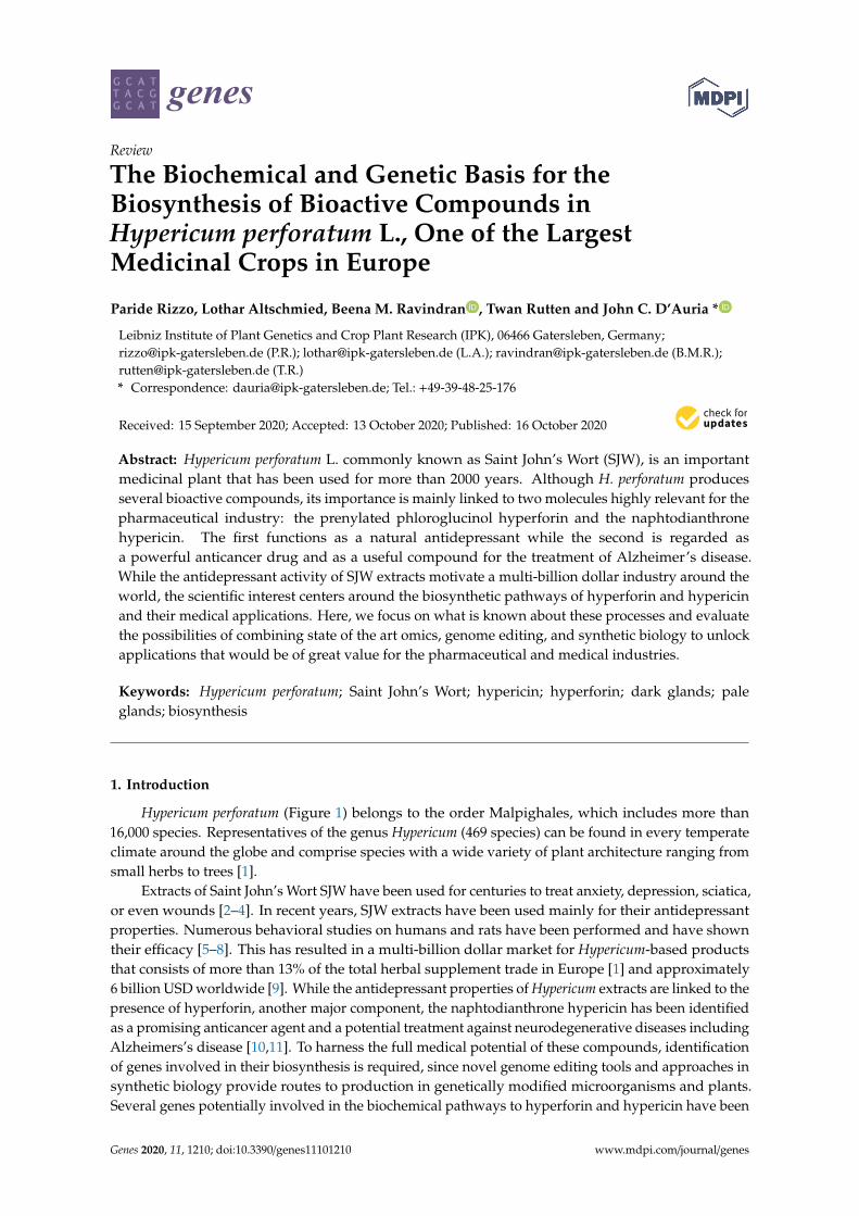
Figure 1. Canopy and appearance of plants of Hypericum perforatum. (A): Greenhouse cultivation of
a sexual diploid genotype (HyPR-01); (B): Single plant 7 months old in full flowering; (C): Detail of
open flowers; (D): Detail of adult leaves. Photos by Paride Rizzo.
Extracts of Saint John’s Wort SJW have been used for centuries to treat anxiety, depression,
sciatica, or even wounds [2–4]. In recent years, SJW extracts have been used mainly for their
antidepressant properties. Numerous behavioral studies on humans and rats have been performed
and have shown their efficacy [5–8]. This has resulted in a multi-billion dollar market for Hypericum-
based products that consists of more than 13% of the total herbal supplement trade in Europe [1] and
approximately 6 billion USD worldwide [9]. While the antidepressant properties of Hypericum
extracts are linked to the presence of hyperforin, another major component, the naphtodianthrone
hypericin has been identified as a promising anticancer agent and a potential treatment against
neurodegenerative diseases including Alzheimers’s disease [10,11]. To harness the full medical
potential of these compounds, identification of genes involved in their biosynthesis is required, since
novel genome editing tools and approaches in synthetic biology provide routes to production in
genetically modified microorganisms and plants. Several genes potentially involved in the
biochemical pathways to hyperforin and hypericin have been characterized, but functional proofs are
missing in many cases and neither of the two pathways is fully understood [12–14]. In this review,
we describe the most current state of knowledge regarding the genes and enzymes that participate
in the biosynthesis of these important pharmaceuticals as well as their medicinal uses.
Figure 1. Canopy and appearance of plants of Hypericum perforatum. (A): Greenhouse cultivation ofa sexual diploid genotype (HyPR-01); (B): Single plant 7 months old in full flowering; (C): Detail ofopen flowers; (D): Detail of adult leaves. Photos by Paride Rizzo.
2. The Relevance of Hyperforin, Hypericin and Other Bioactive Compounds fromHypericum perforatum L.
The composition of extracts from H. perforatum has been studied extensively and many secondarymetabolites including rutin, hyperforin, hyperoside, quercitrin, isoquercitrin, quercetin, hypericin, andchlorogenic acid, have been identified [3,15–17]. Nevertheless, two compounds have emerged as themost important for the pharmaceutical industry: hyperforin and hypericin.
Hyperforin is a prenylated phloroglucinol derivative that constitutes the most abundant lipophiliccomponent of the hydroalcoholic extracts of H. perforatum [6,18,19]. The antidepressant and neuro-activeeffects for which SJW extracts are known could eventually be attributed to the presence of this particularcomponent and pure hyperforin alone can reproduce most of them [6,18,19]. Many antidepressants relyon the capacity to inhibit the uptake of important neurotransmitters like serotonin, dopamine (DA), ornoradrenaline (NA) [20]. Hyperforin constitutes a natural synaptosomal inhibitor of neurotransmitter
Genes 2020, 11, 1210 3 of 20
uptake. Its efficiency is comparable to that of tricyclic antidepressants (TCA) or serotonin specificinhibitors (SSRI) but without the side-effects typical for these drugs [6].
Unlike many other anti-depressants, hyperforin can inhibit the synaptosomal uptake of the aminoacid transmitters, gamma-aminobutyric acid (GABA), and L-glutamate [21]. This is due to its mode ofaction that instead of being based on the competitive interactions for the transporter binding sites,relies on the increase of intracellular Na+ known to be critical for the regulation of neurotransmitteruptake [21–23]. These characteristics confer to hyperforin the status of a broad-spectrum uptakeinhibitor of neurotransmitters, whose effects have been reported in several behavioral studies on ratsand humans [5–8].
While SJW extracts are commonly sold as mild antidepressants, reports on their efficiency arecontradictory, with some studies finding no enhanced effect over placebo treatments [24,25]. Inaddition, there are reports regarding the existence of local bias being observed in German-speakingcountries [26]. Besides its use as a potential antidepressant, hyperforin is also considered a powerfulantibacterial compound, effective against all gram-positive bacteria and penicillin-resistant (PRSA)and methicillin-resistant (MRSA) Staphylococcus aureus [27].
SJW extracts are also the main source of natural hypericin. This naphtodianthrone is regardedas an efficient anticancer compound that induces an apoptotic response after specifically binding tomelanoma cancer cells [28]. The successful application of hypericin in cancer photodynamic therapy(PDT) takes advantage of the properties of hypericin as one of the most powerful photosensitizersknown in nature [11]. When exposed to light, hypericin can induce an apoptotic signal. This involvesthe formation of reactive oxygen species (ROS) that eventually lead to the killing of the tumor [29,30].Hypericin therapy also induces an increase in cytokine levels, leading to an inflammatory response andthe activation of immune cells [11,31]. This mechanism is known as Immunogenic Cell Death (ICD).Hypericin is classified as a type II immunogenic cell death inducer [32] since the apoptotic response itinduces is triggered at the level of the endoplasmic reticulum. Molecules capable of inducing suchimmunogenic cell death responses are classified as “damage-associated molecular patterns” (DAMPs)and have become a new avenue of treatment for cancer [32]. Hypericin is the most potent natural ICDinducer known to date, and its properties are being explored to find even more effective analogues.
The antiviral properties of hypericin were shown against a large number of viruses includingHIV, influenza virus A, herpes simplex, bovine diarrhea virus (BVDV), hepatitis C, duck reovirus, andbronchitis virus [33–40]. Hypericin seems to be particularly effective against enveloped viruses bytargeting and modifying viral proteins [34]. This effect is enhanced by light, which not only inactivatesthe virus, but can also prevent fusion of the virus with the cell membrane of the host [41].
Interest in SJW extracts has gone beyond the antidepressant, anti-cancer and antiviral applicationsafter it was discovered that these extracts can reduce both memory impairment and β-amyloid fibrildeposition in the brain of APP-transgenic mice [10]. These results are compliant with in vitro studiesshowing that hypericin can interfere and significantly inhibit the formation of β-amyloid fibrils byassociating with the precursors of these fibrils [42,43]. At high concentrations, hypericin can alter cellmembrane permeability and induce anomalous functioning in the affected cells [42,44,45]. Rats treatedwith SJW extracts also showed an increased expression of the ABCC1 transporter which is involvedin the clearance of brain tissues from the β-amyloid plaques [10]. Since this effect is independent ofhyperforin it suggests an additional role for hypericin [10].
In light of these recent studies, it is likely that there will be an increased pharmaceutical demandfor both hyperforin and hypericin. Since large-scale synthetic production is prohibitively expensive,SJW extracts remain the main source of these two compounds. Unraveling their biosynthetic pathwayswould not only allow the creation of multi-tasking SJW plants producing customized levels of bioactivecompounds, but also be used for engineering microorganisms producing these, and related, valuabledrugs on a large scale.
It is important to note that other compounds produced by H. perforatum are also regarded asbioactive compounds. These include phenolic derivatives such as tannins and xanthones. Further
Genes 2020, 11, 1210 4 of 20
examples include the flavonoids quercetin, hyperoside, rutin and isoquercitrin. Concentrations offlavonoids can range between 7 to 12% in flowers [3,9]. Flavonoids confer strong antioxidativeproperties to SJW extracts. When in high concentrations, these extracts can be referred to as FEHP(Flavonoids-rich extracts of H. perforatum). They are proposed to reduce the effects of oxidative stressthat acts upon such processes as aging, carcinogenesis, diabetes, and many other clinical conditions [46].Flavonoids like quercitin, kaemferol, and biapigenin also have gastro and neuroprotective functionsdue to their capacity of inhibiting the peroxidation of mitochondrial membranes. By maintainingmitochondrial transmembrane electric potential, the overload of calcium uptake in the mitochondrionis avoided [47].
3. Secretory Structures of Hypericum perforatum
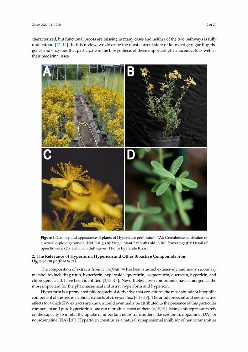
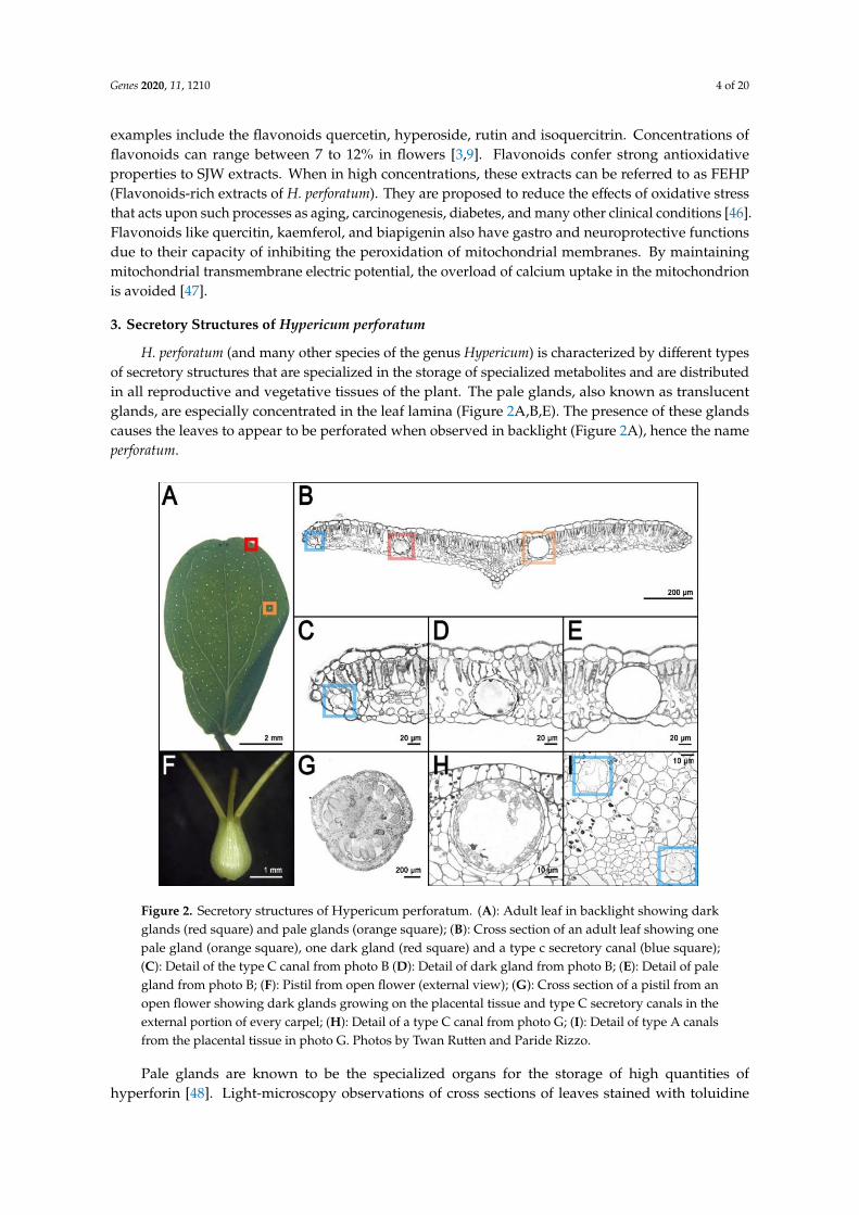
H. perforatum (and many other species of the genus Hypericum) is characterized by different typesof secretory structures that are specialized in the storage of specialized metabolites and are distributedin all reproductive and vegetative tissues of the plant. The pale glands, also known as translucentglands, are especially concentrated in the leaf lamina (Figure 2A,B,E). The presence of these glandscauses the leaves to appear to be perforated when observed in backlight (Figure 2A), hence the nameperforatum.
Genes 2020, 11, x FOR PEER REVIEW 4 of 21
hypericin can alter cell membrane permeability and induce anomalous functioning in the affected
cells [42,44,45]. Rats treated with SJW extracts also showed an increased expression of the ABCC1
transporter which is involved in the clearance of brain tissues from the β-amyloid plaques [10]. Since
this effect is independent of hyperforin it suggests an additional role for hypericin [10].
In light of these recent studies, it is likely that there will be an increased pharmaceutical demand
for both hyperforin and hypericin. Since large-scale synthetic production is prohibitively expensive,
SJW extracts remain the main source of these two compounds. Unraveling their biosynthetic
pathways would not only allow the creation of multi-tasking SJW plants producing customized levels
of bioactive compounds, but also be used for engineering microorganisms producing these, and
related, valuable drugs on a large scale.
It is important to note that other compounds produced by H. perforatum are also regarded as
bioactive compounds. These include phenolic derivatives such as tannins and xanthones. Further
examples include the flavonoids quercetin, hyperoside, rutin and isoquercitrin. Concentrations of
flavonoids can range between 7 to 12% in flowers [3,9]. Flavonoids confer strong antioxidative
properties to SJW extracts. When in high concentrations, these extracts can be referred to as FEHP
(Flavonoids-rich extracts of H. perforatum). They are proposed to reduce the effects of oxidative stress
that acts upon such processes as aging, carcinogenesis, diabetes, and many other clinical conditions
[46]. Flavonoids like quercitin, kaemferol, and biapigenin also have gastro and neuroprotective
functions due to their capacity of inhibiting the peroxidation of mitochondrial membranes. By
maintaining mitochondrial transmembrane electric potential, the overload of calcium uptake in the
mitochondrion is avoided [47].
3. Secretory Structures of Hypericum Perforatum
H. perforatum (and many other species of the genus Hypericum) is characterized by different types
of secretory structures that are specialized in the storage of specialized metabolites and are
distributed in all reproductive and vegetative tissues of the plant. The pale glands, also known as
translucent glands, are especially concentrated in the leaf lamina (Figure 2A,B,E). The presence of
these glands causes the leaves to appear to be perforated when observed in backlight (Figure 2A),
hence the name perforatum.
Figure 2. Secretory structures of Hypericum perforatum. (A): Adult leaf in backlight showing darkglands (red square) and pale glands (orange square); (B): Cross section of an adult leaf showing onepale gland (orange square), one dark gland (red square) and a type c secretory canal (blue square);(C): Detail of the type C canal from photo B (D): Detail of dark gland from photo B; (E): Detail of palegland from photo B; (F): Pistil from open flower (external view); (G): Cross section of a pistil from anopen flower showing dark glands growing on the placental tissue and type C secretory canals in theexternal portion of every carpel; (H): Detail of a type C canal from photo G; (I): Detail of type A canalsfrom the placental tissue in photo G. Photos by Twan Rutten and Paride Rizzo.
Pale glands are known to be the specialized organs for the storage of high quantities ofhyperforin [48]. Light-microscopy observations of cross sections of leaves stained with toluidine
Genes 2020, 11, 1210 5 of 20
blue reveal many pale glands embedded in the parenchyma and delimited by two layers of cells. Ofthese layers, the external one appears thicker due to modified parenchyma cells, while the layer thatsurrounds the lumen is made by flattened cells with very thin cell walls [49].
H. perforatum is also characterized by other translucent secretory structures called canals becauseof their oblong shape. There are three types of canals: A (Figure 2B,C,I), B, and C (Figure 2H). Type Acanals occur in every part of the plant but the stamens and they are characterized by a lumen that ismuch smaller than the one from the pale glands and is delimited by 4 polygonal cells (Figure 2I). TypeB canals are present in several floral organs and the stem. Their structure is similar to the pale glands,in which their main difference is their oblong shape. Finally, type C (Figure 2G,H) canals have onlybeen described in the carpels of H. perforatum. Type C canals show one or multiple layers of intenselystained secretory cells with thin walls that surround a large lumen [14,49].
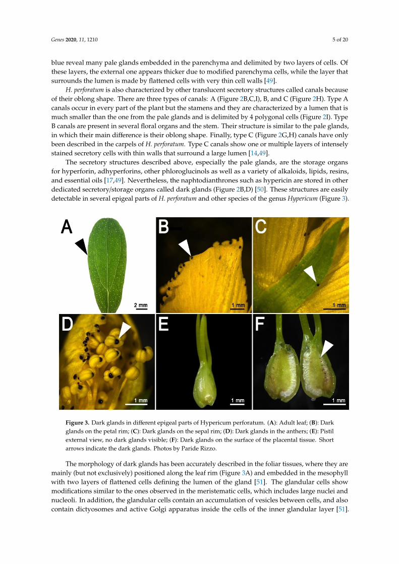
The secretory structures described above, especially the pale glands, are the storage organsfor hyperforin, adhyperforins, other phloroglucinols as well as a variety of alkaloids, lipids, resins,and essential oils [17,49]. Nevertheless, the naphtodianthrones such as hypericin are stored in otherdedicated secretory/storage organs called dark glands (Figure 2B,D) [50]. These structures are easilydetectable in several epigeal parts of H. perforatum and other species of the genus Hypericum (Figure 3).
Genes 2020, 11, x FOR PEER REVIEW 5 of 21
Figure 2. Secretory structures of Hypericum perforatum. (A): Adult leaf in backlight showing dark
glands (red square) and pale glands (orange square); (B): Cross section of an adult leaf showing one
pale gland (orange square), one dark gland (red square) and a type c secretory canal (blue square);
(C): Detail of the type C canal from photo B (D): Detail of dark gland from photo B; (E): Detail of pale
gland from photo B; (F): Pistil from open flower (external view); (G): Cross section of a pistil from an
open flower showing dark glands growing on the placental tissue and type C secretory canals in the
external portion of every carpel; (H): Detail of a type C canal from photo G; (I): Detail of type A canals
from the placental tissue in photo G. Photos by Twan Rutten and Paride Rizzo.
Pale glands are known to be the specialized organs for the storage of high quantities of
hyperforin [48]. Light-microscopy observations of cross sections of leaves stained with toluidine blue
reveal many pale glands embedded in the parenchyma and delimited by two layers of cells. Of these
layers, the external one appears thicker due to modified parenchyma cells, while the layer that
surrounds the lumen is made by flattened cells with very thin cell walls [49].
H. perforatum is also characterized by other translucent secretory structures called canals because
of their oblong shape. There are three types of canals: A (Figure 2B,C,I), B, and C (Figure 2H). Type
A canals occur in every part of the plant but the stamens and they are characterized by a lumen that
is much smaller than the one from the pale glands and is delimited by 4 polygonal cells (Figure 2I).
Type B canals are present in several floral organs and the stem. Their structure is similar to the pale
glands, in which their main difference is their oblong shape. Finally, type C (Figure 2G,H) canals have
only been described in the carpels of H. perforatum. Type C canals show one or multiple layers of
intensely stained secretory cells with thin walls that surround a large lumen [14,49].
The secretory structures described above, especially the pale glands, are the storage organs for
hyperforin, adhyperforins, other phloroglucinols as well as a variety of alkaloids, lipids, resins, and
essential oils [17,49]. Nevertheless, the naphtodianthrones such as hypericin are stored in other
dedicated secretory/storage organs called dark glands (Figure 2B,D) [50]. These structures are easily
detectable in several epigeal parts of H. perforatum and other species of the genus Hypericum (Figure
3).
Figure 3. Dark glands in different epigeal parts of Hypericum perforatum. (A): Adult leaf; (B): Dark
glands on the petal rim; (C): Dark glands on the sepal rim; (D): Dark glands in the anthers; (E): Pistil Figure 3. Dark glands in different epigeal parts of Hypericum perforatum. (A): Adult leaf; (B): Darkglands on the petal rim; (C): Dark glands on the sepal rim; (D): Dark glands in the anthers; (E): Pistilexternal view, no dark glands visible; (F): Dark glands on the surface of the placental tissue. Shortarrows indicate the dark glands. Photos by Paride Rizzo.
The morphology of dark glands has been accurately described in the foliar tissues, where they aremainly (but not exclusively) positioned along the leaf rim (Figure 3A) and embedded in the mesophyllwith two layers of flattened cells defining the lumen of the gland [51]. The glandular cells showmodifications similar to the ones observed in the meristematic cells, which includes large nuclei andnucleoli. In addition, the glandular cells contain an accumulation of vesicles between cells, and alsocontain dictyosomes and active Golgi apparatus inside the cells of the inner glandular layer [51].
Genes 2020, 11, 1210 6 of 20
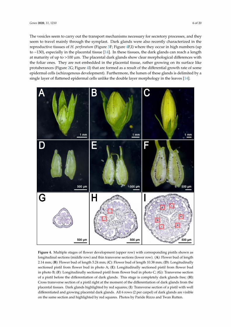
The vesicles seem to carry out the transport mechanisms necessary for secretory processes, and theyseem to travel mainly through the symplast. Dark glands were also recently characterized in thereproductive tissues of H. perforatum (Figure 3F; Figure 4F,I) where they occur in high numbers (upto ~130), especially in the placental tissue [14]. In these tissues, the dark glands can reach a lengthat maturity of up to >100 µm. The placental dark glands show clear morphological differences withthe foliar ones. They are not embedded in the placental tissue, rather growing on its surface likeprotuberances (Figure 2G; Figure 4I) that are formed as a result of the differential growth rate of someepidermal cells (schizogenous development). Furthermore, the lumen of these glands is delimited by asingle layer of flattened epidermal cells unlike the double layer morphology in the leaves [14].Genes 2020, 11, x FOR PEER REVIEW 7 of 21
Figure 4. Multiple stages of flower development (upper row) with corresponding pistils shown as
longitudinal sections (middle row) and thin transverse sections (lower row). (A): Flower bud of length
2.14 mm; (B): Flower bud of length 5.24 mm; (C): Flower bud of length 10.38 mm; (D): Longitudinally
sectioned pistil from flower bud in photo A; (E): Longitudinally sectioned pistil from flower bud in
photo B; (F): Longitudinally sectioned pistil from flower bud in photo C; (G): Transverse section of a
pistil before the differentiation of dark glands. This stage is completely dark glands free; (H): Cross
transverse section of a pistil right at the moment of the differentiation of dark glands from the
placental tissues. Dark glands highlighted by red squares; (I): Transverse section of a pistil with well
differentiated and growing placental dark glands. All 6 rows (2 per carpel) of dark glands are visible
on the same section and highlighted by red squares. Photos by Paride Rizzo and Twan Rutten.
These glands are larger than their foliar counterpart and their density is much higher than in the
leaf, which normally shows a maximum of ~20–30 glands [14]. The placental glands do not
differentiate until the flower buds reach an average length of ~5 mm [14] (Figure 4E,H; dark gland
differentiation stage). This means that during early flower development, the placentas are completely
glandless (Figure 4D,G) and can be used as pre-differentiation stages in the context of multi-stage
experiments dealing with the development of dark glands. Furthermore, a recent study which
characterized the floral development of 93 genotypes, identified a combination of perfectly
contrasting phenotypes in which some SJW accessions never revealed any dark glands in their pistils
(Figure 5A,C) in contrast to other accessions in which pistils were heavily glanded (Figure 5B,D) [14].
Figure 4. Multiple stages of flower development (upper row) with corresponding pistils shown aslongitudinal sections (middle row) and thin transverse sections (lower row). (A): Flower bud of length2.14 mm; (B): Flower bud of length 5.24 mm; (C): Flower bud of length 10.38 mm; (D): Longitudinallysectioned pistil from flower bud in photo A; (E): Longitudinally sectioned pistil from flower budin photo B; (F): Longitudinally sectioned pistil from flower bud in photo C; (G): Transverse sectionof a pistil before the differentiation of dark glands. This stage is completely dark glands free; (H):Cross transverse section of a pistil right at the moment of the differentiation of dark glands from theplacental tissues. Dark glands highlighted by red squares; (I): Transverse section of a pistil with welldifferentiated and growing placental dark glands. All 6 rows (2 per carpel) of dark glands are visibleon the same section and highlighted by red squares. Photos by Paride Rizzo and Twan Rutten.
Genes 2020, 11, 1210 7 of 20
4. The Frontier of Dark Gland and Hypericin Biosynthesis Research
The dark glands of H. perforatum caught the attention of generations of scientists who tried tounravel the mechanisms of hypericin biosynthesis. With the advent of omics technologies, it is possibleto study for the first time the transcriptome of a dark gland, and from that information, begin to inferthe genes involved in hypericin biosynthesis. Historically, the scientific community addressed the leafas a model tissue for this kind of study. Dark glands in the leaves are mainly (but not exclusively)distributed along the rim. However, it is difficult to find contrasting phenotypes of H. perforatumin which a glandless leaf can be compared with a glanded one. Furthermore, the foliar dark glandsdifferentiate already at very early stages of leaf development and this makes it difficult to addressstages without glands (pre-differentiation) with post-differentiation stages. These issues resulted instudies which compared the transcriptome of the leaf rim with the leaf lamina [13,52]. Althoughthese two tissues are part of the same organ, they have different anatomies and patterning. Thisconstitutes a bias for any comparison between these two different fractions of the leaf. Nevertheless,RNAseq experiments including the rim vs lamina comparison, represented the first glimpse into thetranscriptome of dark glands, and also identified putative candidate genes involved in the biosynthesisof hypericin. In the most recent years, the pistil has emerged as a more suitable model for this kind ofstudy [14]. The pistil of H. perforatum is in many cases heavily glanded, with up to >130 glands perpistil and is distributed along six rows on the surface of the placental tissue (Figure 4I).
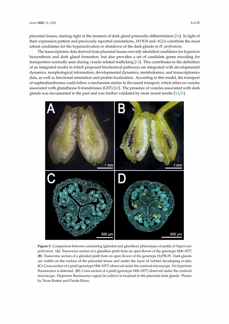
These glands are larger than their foliar counterpart and their density is much higher than in theleaf, which normally shows a maximum of ~20–30 glands [14]. The placental glands do not differentiateuntil the flower buds reach an average length of ~5 mm [14] (Figure 4E,H; dark gland differentiationstage). This means that during early flower development, the placentas are completely glandless(Figure 4D,G) and can be used as pre-differentiation stages in the context of multi-stage experimentsdealing with the development of dark glands. Furthermore, a recent study which characterized thefloral development of 93 genotypes, identified a combination of perfectly contrasting phenotypes inwhich some SJW accessions never revealed any dark glands in their pistils (Figure 5A,C) in contrast toother accessions in which pistils were heavily glanded (Figure 5B,D) [14].
These contrasting morphologies corresponds to equally contrasting metabolic phenotypes. Thismeans that the heavily glanded pistils show high levels of hypericin and related precursors, whilethe glandless pistils are characterized by the absence (or traces) of these compounds. In light of theseresults, the pistil represents a nearly ideal platform for the study of dark gland development andhypericin biosynthesis.
The gene expression results reported by Rizzo et al. identified clusters of genes following differenttemporal dynamics of gene expression [14]. Some genes coding for enzymes putatively involved in thebiosynthesis of hypericin are expressed mainly after the differentiation and growth of dark glands. Thecombination of spatio-temporal expression and annotation on heterologous systems makes these genesvaluable for the characterization of this complex biosynthetic pathway (see the following section).Another small cluster of genes was reported to have a peak of expression strictly synchronized withthe differentiation of the first dark gland primordia in the placenta (Figure 4H). Only two transcriptionfactors are part of this cluster. MYB38 is an R2R3-Myb gene that belongs to the subgroup 14 of theMyb family [53]. All the members of this subgroup are exclusively involved in cell differentiationand organ identity [54–57]. MYB38 is the orthologue of RAX2 from Arabidopsis thaliana, and is knownto be involved in developmental processes by regulating lateral patterning and lateral meristeminitiation [55]. This is a process that resembles the lateral patterning in placental tissues of H. perforatum,from which dark glands originate. The only other transcription factor with a nearly identical expressionpattern is an ortholog of A. thaliana AGL6 (Agamous Like 6), a MADS-box gene responsible for thedetermination of organ identity and meristem differentiation in the flower [58]. AGL6 is also known tobe the MADS-box gene with the highest rate of duplication and neofunctionalization in the evolutionaryhistory of dicotyledons [59]. These two genes have very low levels of expression in placental tissuesthat never differentiate dark glands, while they are 100–1000 times more highly expressed in glanded
Genes 2020, 11, 1210 8 of 20
placental tissues, starting right at the moment of dark gland primordia differentiation [14]. In light oftheir expression pattern and previously reported annotations, MYB38 and AGL6 constitute the mostrobust candidates for the hyperactivation or shutdown of the dark glands in H. perforatum.
The transcriptomic data derived from placental tissues not only identified candidates for hypericinbiosynthesis and dark gland formation, but also provides a set of candidate genes encoding fortransporters normally seen during vesicle-related trafficking [14]. This contributes to the definitionof an integrated model in which proposed biochemical pathways are integrated with developmentaldynamics, morphological information, developmental dynamics, metabolomics, and transcriptomicsdata, as well as functional annotation and protein localization. According to this model, the transportof naphtodianthrones could follow a mechanism similar to flavonoid transport, which relies on vesiclesassociated with glutathione-S-transferases (GST) [60]. The presence of vesicles associated with darkglands was documented in the past and was further validated by more recent results [14,51].
Genes 2020, 11, x FOR PEER REVIEW 8 of 21
Figure 5. Comparison between contrasting (glanded and glandless) phenotypes of pistils of
Hypericum perforatum. (A): Transverse section of a glandless pistil from an open flower of the genotype
H06-1877; (B): Transverse section of a glanded pistil from on open flower of the genotype HyPR-09.
Dark glands are visible on the surface of the placental tissue and under the layer of (white) developing
ovules. (C): Cross section of a pistil (genotype H06-1877) observed under the confocal microscope. No
hypericin fluorescence is detected. (D): Cross section of a pistil (genotype H06-1877) observed under
the confocal microscope. Hypericin fluorescence signal (in yellow) is localized in the placental dark
glands. Photos by Twan Rutten and Paride Rizzo.
These contrasting morphologies corresponds to equally contrasting metabolic phenotypes. This
means that the heavily glanded pistils show high levels of hypericin and related precursors, while
the glandless pistils are characterized by the absence (or traces) of these compounds. In light of these
results, the pistil represents a nearly ideal platform for the study of dark gland development and
hypericin biosynthesis.
The gene expression results reported by Rizzo et al. identified clusters of genes following
different temporal dynamics of gene expression [14]. Some genes coding for enzymes putatively
involved in the biosynthesis of hypericin are expressed mainly after the differentiation and growth
of dark glands. The combination of spatio-temporal expression and annotation on heterologous
systems makes these genes valuable for the characterization of this complex biosynthetic pathway
Figure 5. Comparison between contrasting (glanded and glandless) phenotypes of pistils of Hypericumperforatum. (A): Transverse section of a glandless pistil from an open flower of the genotype H06-1877;(B): Transverse section of a glanded pistil from on open flower of the genotype HyPR-09. Dark glandsare visible on the surface of the placental tissue and under the layer of (white) developing ovules.(C): Cross section of a pistil (genotype H06-1877) observed under the confocal microscope. No hypericinfluorescence is detected. (D): Cross section of a pistil (genotype H06-1877) observed under the confocalmicroscope. Hypericin fluorescence signal (in yellow) is localized in the placental dark glands. Photosby Twan Rutten and Paride Rizzo.
Genes 2020, 11, 1210 9 of 20
5. Hypericin Biosynthesis
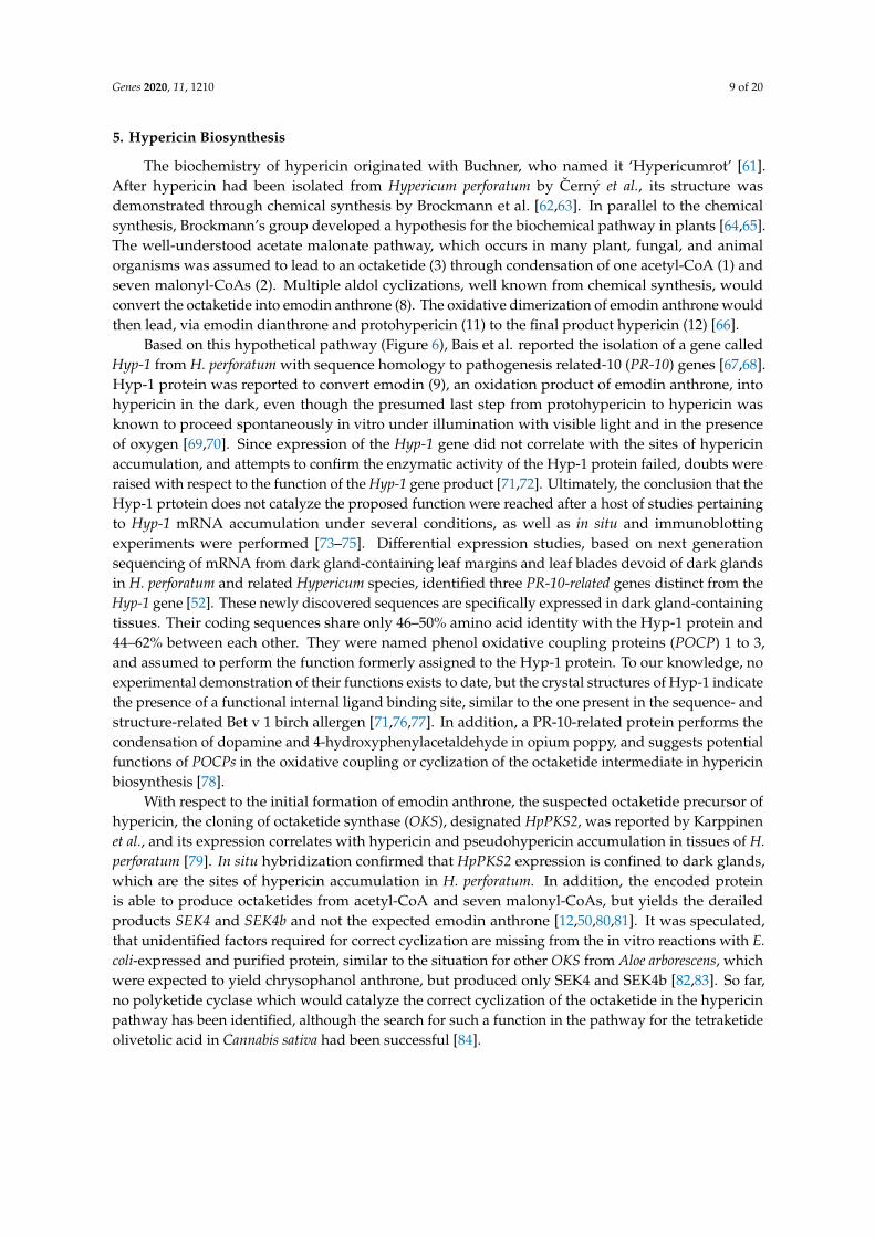
The biochemistry of hypericin originated with Buchner, who named it ‘Hypericumrot’ [61].After hypericin had been isolated from Hypericum perforatum by Cerný et al., its structure wasdemonstrated through chemical synthesis by Brockmann et al. [62,63]. In parallel to the chemicalsynthesis, Brockmann’s group developed a hypothesis for the biochemical pathway in plants [64,65].The well-understood acetate malonate pathway, which occurs in many plant, fungal, and animalorganisms was assumed to lead to an octaketide (3) through condensation of one acetyl-CoA (1) andseven malonyl-CoAs (2). Multiple aldol cyclizations, well known from chemical synthesis, wouldconvert the octaketide into emodin anthrone (8). The oxidative dimerization of emodin anthrone wouldthen lead, via emodin dianthrone and protohypericin (11) to the final product hypericin (12) [66].
Based on this hypothetical pathway (Figure 6), Bais et al. reported the isolation of a gene calledHyp-1 from H. perforatum with sequence homology to pathogenesis related-10 (PR-10) genes [67,68].Hyp-1 protein was reported to convert emodin (9), an oxidation product of emodin anthrone, intohypericin in the dark, even though the presumed last step from protohypericin to hypericin wasknown to proceed spontaneously in vitro under illumination with visible light and in the presenceof oxygen [69,70]. Since expression of the Hyp-1 gene did not correlate with the sites of hypericinaccumulation, and attempts to confirm the enzymatic activity of the Hyp-1 protein failed, doubts wereraised with respect to the function of the Hyp-1 gene product [71,72]. Ultimately, the conclusion that theHyp-1 prtotein does not catalyze the proposed function were reached after a host of studies pertainingto Hyp-1 mRNA accumulation under several conditions, as well as in situ and immunoblottingexperiments were performed [73–75]. Differential expression studies, based on next generationsequencing of mRNA from dark gland-containing leaf margins and leaf blades devoid of dark glandsin H. perforatum and related Hypericum species, identified three PR-10-related genes distinct from theHyp-1 gene [52]. These newly discovered sequences are specifically expressed in dark gland-containingtissues. Their coding sequences share only 46–50% amino acid identity with the Hyp-1 protein and44–62% between each other. They were named phenol oxidative coupling proteins (POCP) 1 to 3,and assumed to perform the function formerly assigned to the Hyp-1 protein. To our knowledge, noexperimental demonstration of their functions exists to date, but the crystal structures of Hyp-1 indicatethe presence of a functional internal ligand binding site, similar to the one present in the sequence- andstructure-related Bet v 1 birch allergen [71,76,77]. In addition, a PR-10-related protein performs thecondensation of dopamine and 4-hydroxyphenylacetaldehyde in opium poppy, and suggests potentialfunctions of POCPs in the oxidative coupling or cyclization of the octaketide intermediate in hypericinbiosynthesis [78].
With respect to the initial formation of emodin anthrone, the suspected octaketide precursor ofhypericin, the cloning of octaketide synthase (OKS), designated HpPKS2, was reported by Karppinenet al., and its expression correlates with hypericin and pseudohypericin accumulation in tissues of H.perforatum [79]. In situ hybridization confirmed that HpPKS2 expression is confined to dark glands,which are the sites of hypericin accumulation in H. perforatum. In addition, the encoded proteinis able to produce octaketides from acetyl-CoA and seven malonyl-CoAs, but yields the derailedproducts SEK4 and SEK4b and not the expected emodin anthrone [12,50,80,81]. It was speculated,that unidentified factors required for correct cyclization are missing from the in vitro reactions with E.coli-expressed and purified protein, similar to the situation for other OKS from Aloe arborescens, whichwere expected to yield chrysophanol anthrone, but produced only SEK4 and SEK4b [82,83]. So far,no polyketide cyclase which would catalyze the correct cyclization of the octaketide in the hypericinpathway has been identified, although the search for such a function in the pathway for the tetraketideolivetolic acid in Cannabis sativa had been successful [84].
Genes 2020, 11, 1210 10 of 20
Genes 2020, 11, x FOR PEER REVIEW 11 of 21
exceptions to this overlap are the early induced transcription factors AGL6 and MYB38, which have
been implicated in the differentiation of dark glands [14].
Figure 6. The hypothetical pathway for the biosynthesis of hypericin: Enzymes, represented in blue
are suggested by previous literature.: OKS, octaketide synthase; PKC, polyketide cyclase; TER,
thioesterase; POCP, phenoloxidative coupling protein; BBE, berberine bridge enzyme.
6. Hyperforin Biosynthesis
(+)-Hyperforin (27) (C35H52O4) is a polyprenylated acylphloroglucinol (PPAP) derivative with a
phloroisobutyrophenone bicyclic core [86–88]. It is a mixture of interconverting and stably co-existing
tautomers , yet it is unstable when exposed to light and oxygen [88–91]. This is due to the enolized β-
dicarbonyl system present in phloroglucinols [91–93]. It is a fairly stable molecule in protic solvents
Figure 6. The hypothetical pathway for the biosynthesis of hypericin: Enzymes, represented in blue aresuggested by previous literature.: OKS, octaketide synthase; PKC, polyketide cyclase; TER, thioesterase;POCP, phenoloxidative coupling protein; BBE, berberine bridge enzyme.
Using the newly discovered development of dark glands in placental tissue of some H. perforatumlines, which is completely absent in other lines, Rizzo et al. used the overlap of differentially expressedgenes (DEGs) between lines with and without dark glands, and DEGs between developmental stageswith and without dark glands in the same line, to restrict the number of candidate genes involved in darkgland development and hypericin biosynthesis [14]. Together with metabolites identified by Ultra HighPerformance Liquid Chromatography-Electrospray ionization-High Resolution Mass Spectrometry
Genes 2020, 11, 1210 11 of 20
(UHPLC-ESI-HRMS), which correlate with the presence of dark glands, a new biosynthetic pathwayfor hypericin was proposed. This new model suggests that penicilliopsin (10) is the first intermediatewith two condensed octaketides instead of emodin dianthrone [14]. Since penicilliopsin links the twooctaketide halves of hypericin via the same C-C bond as skyrin (13), this or a similar pathway seems tobe supported by the metabolite profiling of 17 Hypericum species by Kimáková et al. [85]. In that study,skyrin and skyrin glucosides, but not emodin or emodin anthrone, are correlated with the presence ofhypericin, suggesting skyrin as an intermediate in hypericin biosynthesis. A novel addition to thepathway proposed by Rizzo et al. is a gene encoding berberine-bridge enzyme (BBE), which washypothesized to convert penicilliopsin to protohypericin, the last step of hypericin synthesis for whichan enzyme is required [14]. Since this gene encodes an N-terminal signal peptide, the formation of theC-C double bond between the octaketide halves is likely to occur within the vacuole, in an ER-derivedvesicle, or outside of the cell. In addition, Rizzo et al. confirmed the highly correlated expression of theOKS gene and the POCP genes identified by Sotak et al. with dark gland development, and identifiednovel candidate genes for the cleavage of the CoA-thioester bond, the cyclization of the octaketide, andhydroxylation of penicilliopsin to hydroxypenicilliopsin for the formation of pseudohypericin [12–14].Furthermore, genes potentially involved in the transport of pathway intermediates across membranes(ABC transporter, UDP-glucosyl transferase, β-glucosidase, major facilitator protein for sugar import)and by vesicles (glutathione-S-transferases) were identified within the small set of DEGs highlyupregulated during dark gland development [14]. A comparison of DEGs provided for H. tomentosumleaf tissues with and without dark glands and DEGs for dark gland development [14] show that almostall gene functions in the smaller set of DEGs occur in the larger set [13,14,52]. The only exceptions tothis overlap are the early induced transcription factors AGL6 and MYB38, which have been implicatedin the differentiation of dark glands [14].
6. Hyperforin Biosynthesis
(+)-Hyperforin (27) (C35H52O4) is a polyprenylated acylphloroglucinol (PPAP) derivative with aphloroisobutyrophenone bicyclic core [86–88]. It is a mixture of interconverting and stably co-existingtautomers, yet it is unstable when exposed to light and oxygen [88–91]. This is due to the enolizedβ-dicarbonyl system present in phloroglucinols [91–93]. It is a fairly stable molecule in protic solventsand in in vivo systems [94]. Hyperforin occurs naturally in H. perforatum Linn. (Hypericaceae) in largeamounts, (6.01 and 13.59 mg g−1 dry weight in leaves and flowers respectively), and in several otherspecies, albeit at lower levels [95,96]. It is localized in pistils, flowers and fruits. In the development ofthe flowers, the hyperforin content increased from 2.47% to 8.48% (dry weight) from flower buds tofruits [97]. Such an increase was also reported by Maisenbacher and Kovar [98]. High concentrationsof hyperforin accumulate in the translucent glands [48]. While extracts of H. perforatum have beenused as an herbal supplement to treat depression as early as 1958, the determination that hyperforinwas the metabolite responsible for the pharmaceutical affects was reported in 1971 [99,100]. The partialmolecular structure was published four years later [86]. Further studies were able to focus on thestereochemistry of the molecule, and absolute configuration was ultimately resolved with the use ofx-ray data crystallization techniques [87,101].
The evidence for the incorporation of intermediates from primary metabolism into hyperforinwas ascertained via the use of feeding studies using labeled precursors. isobutyryl-CoA (17) hasbeen determined to be one of the initial primary metabolite starter molecules in the biosynthesis ofthe hyperforin core structure. Furthermore, isobutyryl-CoA is derived from an α-ketoisovalerateintermediate (15) produced from a combination of pyruvate and valine (16) [88,102,103]. In manycases, the addition of precursors/substrates to cell/tissue cultures can enhance the production ofspecialized metabolites [104]. The addition of labelled valine, leucine and isoleucine to shootcultures of H. perforatum revealed that valine was the most likely precursor [103]. QuantitativeNMR spectroscopy analysis of H. perforatum cuttings immersed in (1-13C) glucose revealed that thedimethylallyl moieties were derived from the non-mevalonate pathway (MEP), and hence established
Genes 2020, 11, 1210 12 of 20
the need for plastid-derived metabolites to be involved in supplying intermediates to the hyperforinbiosynthetic pathway [88,105].
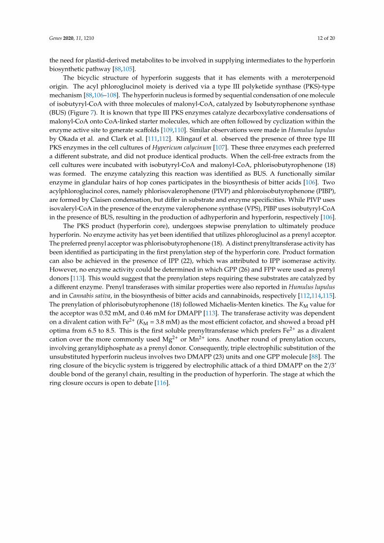
The bicyclic structure of hyperforin suggests that it has elements with a meroterpenoidorigin. The acyl phloroglucinol moiety is derived via a type III polyketide synthase (PKS)-typemechanism [88,106–108]. The hyperforin nucleus is formed by sequential condensation of one moleculeof isobutyryl-CoA with three molecules of malonyl-CoA, catalyzed by Isobutyrophenone synthase(BUS) (Figure 7). It is known that type III PKS enzymes catalyze decarboxylative condensations ofmalonyl-CoA onto CoA-linked starter molecules, which are often followed by cyclization within theenzyme active site to generate scaffolds [109,110]. Similar observations were made in Humulus lupulusby Okada et al. and Clark et al. [111,112]. Klingauf et al. observed the presence of three type IIIPKS enzymes in the cell cultures of Hypericum calycinum [107]. These three enzymes each preferreda different substrate, and did not produce identical products. When the cell-free extracts from thecell cultures were incubated with isobutyryl-CoA and malonyl-CoA, phlorisobutyrophenone (18)was formed. The enzyme catalyzing this reaction was identified as BUS. A functionally similarenzyme in glandular hairs of hop cones participates in the biosynthesis of bitter acids [106]. Twoacylphloroglucinol cores, namely phlorisovalerophenone (PIVP) and phloroisobutyrophenone (PIBP),are formed by Claisen condensation, but differ in substrate and enzyme specificities. While PIVP usesisovaleryl-CoA in the presence of the enzyme valerophenone synthase (VPS), PIBP uses isobutyryl-CoAin the presence of BUS, resulting in the production of adhyperforin and hyperforin, respectively [106].
The PKS product (hyperforin core), undergoes stepwise prenylation to ultimately producehyperforin. No enzyme activity has yet been identified that utilizes phloroglucinol as a prenyl acceptor.The preferred prenyl acceptor was phlorisobutyrophenone (18). A distinct prenyltransferase activity hasbeen identified as participating in the first prenylation step of the hyperforin core. Product formationcan also be achieved in the presence of IPP (22), which was attributed to IPP isomerase activity.However, no enzyme activity could be determined in which GPP (26) and FPP were used as prenyldonors [113]. This would suggest that the prenylation steps requiring these substrates are catalyzed bya different enzyme. Prenyl transferases with similar properties were also reported in Humulus lupulusand in Cannabis sativa, in the biosynthesis of bitter acids and cannabinoids, respectively [112,114,115].The prenylation of phlorisobutyrophenone (18) followed Michaelis-Menten kinetics. The KM value forthe acceptor was 0.52 mM, and 0.46 mM for DMAPP [113]. The transferase activity was dependenton a divalent cation with Fe2+ (KM = 3.8 mM) as the most efficient cofactor, and showed a broad pHoptima from 6.5 to 8.5. This is the first soluble prenyltransferase which prefers Fe2+ as a divalentcation over the more commonly used Mg2+ or Mn2+ ions. Another round of prenylation occurs,involving geranyldiphosphate as a prenyl donor. Consequently, triple electrophilic substitution of theunsubstituted hyperforin nucleus involves two DMAPP (23) units and one GPP molecule [88]. Thering closure of the bicyclic system is triggered by electrophilic attack of a third DMAPP on the 2’/3’double bond of the geranyl chain, resulting in the production of hyperforin. The stage at which thering closure occurs is open to debate [116].
Genes 2020, 11, 1210 13 of 20
Genes 2020, 11, x FOR PEER REVIEW 13 of 21
DMAPP [113]. The transferase activity was dependent on a divalent cation with Fe2+ (KM = 3.8 mM)
as the most efficient cofactor, and showed a broad pH optima from 6.5 to 8.5. This is the first soluble
prenyltransferase which prefers Fe2+ as a divalent cation over the more commonly used Mg2+ or Mn2+
ions. Another round of prenylation occurs, involving geranyldiphosphate as a prenyl donor.
Consequently, triple electrophilic substitution of the unsubstituted hyperforin nucleus involves two
DMAPP (23) units and one GPP molecule [88]. The ring closure of the bicyclic system is triggered by
electrophilic attack of a third DMAPP on the 2’/3’ double bond of the geranyl chain, resulting in the
production of hyperforin. The stage at which the ring closure occurs is open to debate [116].
Figure 7. The hypothetical pathway for the biosynthesis of hyperforin: Prenylations are shown in red.
Enzymes, represented in blue, are suggested by previous studies: PDH, pyruvate dehydrogenase;
ACC, acetyl-CoA carboxylase; BCDH, branched chain α-keto acid dehydrogenase complex; PIBP
transferase, phlorisobutyrophenone transferase; BUS, isobutyryl-CoA; DXS, 1-deoxy-D-xylulose-5-
phosphate synthase; ISPH, 1-hydroxy-2-methyl-butenyl 4-diphosphate reductase; IDI, isopentenyl
diphosphate isomerase; ISPA, Farnesyl pyrophosphate synthase.
Figure 7. The hypothetical pathway for the biosynthesis of hyperforin: Prenylations are shown in red.Enzymes, represented in blue, are suggested by previous studies: PDH, pyruvate dehydrogenase; ACC,acetyl-CoA carboxylase; BCDH, branched chain α-keto acid dehydrogenase complex; PIBP transferase,phlorisobutyrophenone transferase; BUS, isobutyryl-CoA; DXS, 1-deoxy-D-xylulose-5-phosphatesynthase; ISPH, 1-hydroxy-2-methyl-butenyl 4-diphosphate reductase; IDI, isopentenyl diphosphateisomerase; ISPA, Farnesyl pyrophosphate synthase.
7. Perspectives of Engineering the Biosynthetic Pathways of Relevant Compounds fromH. perforatum in Microorganisms and Plants
The active compounds produced by H. perforatum, especially hypericin and hyperforin, are of greatimportance to the pharma industry, and the elucidation of their biosynthetic pathways would lay thefoundations for engineering the production of these compounds in microorganisms. Nevertheless, theengineering of such compounds is a complex problem and multiple aspects must be taken into account.Some of the compounds discussed here are cytotoxic, and they are normally compartmentalized inside
Genes 2020, 11, 1210 14 of 20
specialized organs (glands), like in the case of hypericin. This implies that the sole knowledge of thegenes involved in the biosynthesis of these molecules is not enough for their large-scale production.Engineered microorganisms could die soon after starting the biosynthesis of molecules such as thenaphtodianthrones, because they cannot store them inside dedicated glands. There is at least one casereported in the literature of a fungus producing hypericin, and several other studies reporting theenhancement of hypericin production in the presence of arbuscular mycorrhizae or in response to theinfection with fungal pathogens [117–120]. Nevertheless, these organisms are endophytic fungi thatrely on the compartmentalization carried out by the plant. For this reason, a realistic perspective for thelarge scale production of bioactive compounds in SJW should rely on the biosynthesis genes, as wellas on the genes involved in the differentiation of dark glands (AGL6, MYB38), and the translocationand secretion mechanisms (GST). In this way, the generation of new varieties with hyperactivated (orknocked out) glands could be possible, and the production of these highly valuable compounds couldbe carried out in planta.
Possible means of synthesizing hyperforin in microorganisms have been attempted [121,122].However, a total biosynthesis of hyperforin using a bacterial system has not yet been successful.On the other hand, the production of phlorisovalerophenone, a key intermediate of humulone andadhyperforin biosynthesis, has been achieved in E. coli [102]. In this system, isovaleryl-CoA wasproduced employing a pathway involving hydroxyl-3-methylglutaryl CoA (HMG-CoA), an enzymethat is known to be involved in the mevalonate pathway leading to the biosynthesis of isoprenoids [123].The enzymes acetyl-CoA acetyltransferase and HMG-CoA synthase were used to yield HMG-CoA.Further steps included the addition of HMG-CoA dehydratase, MG-CoA decarboxylase and DMA-CoAreductase to produce isovaleryl-CoA. After generating isovaleryl CoA, the type III PKS valerophenonesynthase that was introduced into an engineered E. coli strain. The system ultimately producedphlorisovalerophenone at a concentration of 6.4 mgL-1. This system utilizes glucose as the main carbonsource for entry into the pathway leading to PIVP, which suggests that an economical biosyntheticproduction of these compounds is possible.
8. Conclusions
In recent years, Hypericum research has experienced a resurgence of scientific interest due to thenewly discovered potential of its bioactives, and their abilities to be utilized as an anticancer medicineand a possible treatment for neurodegenerative diseases. Hypericin and hyperforin are the metaboliteswith the highest value for the pharmaceutical industry. Therefore, the engineering of their biosynthesisin orthologous systems (especially microorganisms) would be advantageous for the developmentof new drugs. Challenges related to compound toxicity and storage may slow the current pace ofadvancement, however this may well be overcome by utilizing different strains of microorganisms oreven by using transformed plant systems.
The future of H. perforatum research as well as other related species will heavily depend on the useof large-scale omics methodologies for the identification of putative genes involved in the regulationand biosynthesis of pharmaceutically important compounds. Already, the recent discoveries using darkgland-bearing tissues have provided many sequences to test. While a few studies have been publishedregarding stable transformation of Hypericum, they are inconsistent and not yet optimized [124,125].Ongoing research using overexpression and knockout technologies, such as CRISPR-Cas9, will allowresearchers to test the current biosynthesis models in a systematic fashion. Functional validationwill open the doors to many promising possibilities and novel lines of research for the Hypericumcommunity. The discovery of putative transcription factors correlated with dark gland developmentmay be used in transgenic overexpression and knockout lines to further tease apart the links betweengland structure and metabolism. Overall, the future of H. perforatum is full of new possibilities that,with the help of modern synthetic and molecular biology techniques, will lead to more diversified andpowerful products based on SJW.
Genes 2020, 11, 1210 15 of 20
Author Contributions: Conceptualization, P.R. and J.C.D.; writing—original draft preparation, P.R., J.C.D., L.A.,B.M.R.; writing—review and editing, P.R., J.C.D., L.A., T.R.; original photos in Figures 1–5 by, P.R. and T.R.,Figures 6 and 7 designed by J.C.D., B.M.R., L.A. All authors have read and agreed to the published version ofthe manuscript.
Funding: This research was funded by the state of Sachsen-Anhalt and EFRE (Europäischer Fonds für regionaleEntwicklung), project HyperSpEED (Hypericum multi-species Exploration of Extracts Diversity)–grant numberZS/2019/07/99749. The project is part of the research cluster Autonomie im Alter (AiA).
Acknowledgments: We thank Isabel Mora-Ramirez for suggestions and help with formatting the manuscript andBenjamin Chavez for help with the final rendering of some pictures.
Conflicts of Interest: The authors declare no conflict of interest.
References
1. Crockett, S.L.; Robson, N.K.B. Taxonomy and Chemotaxonomy of the Genus Hypericum. Med. Aromat. PlantSci. Biotechnol. 2011, 5, 1–13. [PubMed]
2. Oliveira, A.I.; Pinho, C.; Sarmento, B.; Dias, A.C.P. Neuroprotective activity of hypericum perforatum and itsmajor components. Front. Plant Sci. 2016, 7, 1–15. [CrossRef] [PubMed]
3. Barnes, J.; Anderson, L.A.; Phillipson, J.D. St John’s wort (Hypericum perforatum L.): A Review of its Chemistry,Pharmacology and Clinical properties. J. Pharmacol. Sci. 2001, 583–600. [CrossRef]
4. Müller, W.E. St. John’s Wort and Its Active Principle in Depression and Anxiety; Springer Science & BusinessMedia: Berlin/Heidelberg, Germany, 2005.
5. Bhattacharya, S.K.; Chakrabarti, A.; Chatterjee, S.S. Activity profiles of two hyperforin-containing hypericumextracts in behavioral models. Pharmacopsychiatry 1998, 31, 22–29. [CrossRef] [PubMed]
6. Chatterjee, S.S.; Bhattacharya, S.K.; Wonnemann, M.; Singer, A.; Müller, W.E. Hyperforin as a possibleantidepressant component of Hypericum extracts. Life Sci. 1998, 63, 499–510. [CrossRef]
7. Schellenberg, R.; Sauer, S.; Dimpfel, W. Pharmacodynamic effects of two different hypericum extracts inhealthy volunteers measured by quantitative EEG. Pharmacopsychiatry 1998, 31, 44–53. [CrossRef]
8. Dimpfel, W.; Schober, F.; Mannel, M. Effects of a methanolic extract and a hyperforin-enriched CO2 extract of St.John’s Wort (Hypericum perforatum) on intracerebral field potentials in the freely moving rat (Tele-Stereo-EEG).Pharmacopsychiatry 1998, 31, 30–35. [CrossRef]
9. Klemow, M.K.; Bartlow, A.; Crawford, J.; Kocher, N.; Shah, J.; Ritsick, M. Herbal Medicine Biomolecularand Clinical Aspects, 2nd ed.; Benzie, I.F.F., Wachtel-Galor, S., Eds.; CRC Press: Boca Raton, FL, USA,2011; Volume 9, ISBN 9781439807132.
10. Hofrichter, J.; Krohn, M.; Schumacher, T.; Lange, C.; Feistel, B.; Walbroel, B.; Heinze, H.-J.; Crockett, S.;Sharbel, T.F.; Pahnke, J. Reduced Alzheimer’s disease pathology by St. John’s Wort treatment isindependent of hyperforin and facilitated by ABCC1 and microglia activation in mice. Curr. Alzheimer Res.2013, 10, 1057–1069. [CrossRef]
11. Garg, A.D.; Krysko, D.V.; Vandenabeele, P.; Agostinis, P. Hypericin-based photodynamic therapy inducessurface exposure of damage-associated molecular patterns like HSP70 and calreticulin. Cancer Immunol.Immunother. 2012, 61, 215–221. [CrossRef]
12. Karppinen, K.; Hokkanen, J.; Mattila, S.; Neubauer, P.; Hohtola, A. Octaketide-producing type III polyketidesynthase from Hypericum perforatum is expressed in dark glands accumulating hypericins. FEBS J.2008, 275, 4329–4342. [CrossRef]
13. Soták, M.; Czeranková, O.; Klein, D.; Jurcacková, Z.; Li, L.; Cellárová, E. Comparative transcriptomereconstruction of four hypericum species focused on Hypericin biosynthesis. Front. Plant Sci. 2016, 7, 1–14.[CrossRef]
14. Rizzo, P.; Altschmied, L.; Stark, P.; Rutten, T.; Gündel, A.; Scharfenberg, S.; Franke, K.; Bäumlein, H.;Wessjohann, L.; Koch, M.; et al. Discovery of key regulators of dark gland development and hypericinbiosynthesis in St. John’s Wort (Hypericum perforatum). Plant Biotechnol. J. 2019, 17, 2299–2312. [CrossRef][PubMed]
15. Chandrasekera, D.H.; Heinrich, M.; Ashton, D.; Welham, K.J.; Middleton, R. Quantitative analysis of themajor constituents of St John’s wort with HPLC-ESI-MS. J. Pharm. Pharmacol. 2005, 57, 1645–1652. [CrossRef]
16. Scotti, F.; Löbel, K.; Booker, A.; Heinrich, M. St. John’s Wort (Hypericum perforatum) Products – How VariableIs the Primary Material? Front. Plant Sci. 2019, 9, 1–12. [CrossRef]
Genes 2020, 11, 1210 16 of 20
17. Nahrstedt, A.; Butterweck, V. Biologically active and other chemical constituents of the herb of Hypericumperforation L. Pharmacopsychiatry 1997, 30, 129–134. [CrossRef] [PubMed]
18. Laakmann, G.; Schüle, C.; Baghai, T.; Kieser, M. St. John’s Wort in mild to moderate depression: Therelevance of hyperforin for the clinical efficacy. Pharmacopsychiatry 1998, 31, 54–59. [CrossRef] [PubMed]
19. Cervo, L.; Rozio, M.; Ekalle-Soppo, C.B.; Guiso, G.; Morazzoni, P.; Caccia, S. Role of hyperforin inthe antidepressant-like activity of Hypericum perforatum extracts. Psychopharmacology 2002, 164, 423–428.[CrossRef]
20. Obata, H. Analgesic mechanisms of antidepressants for neuropathic pain. Int. J. Mol. Sci. 2017, 18, 2483.[CrossRef]
21. Müller, W.E.; Singer, A.; Wonnemann, M. Hyperforin—Antidepressant activity by a novel mechanism ofaction. Pharmacopsychiatry 2001, 34, 98–102. [CrossRef]
22. Sneddon, J.M. Sodium-dependent accumulation of 5-hydroxytryptamine by rat blood platelets. Br. J.Pharmacol. 1969, 37, 680–688. [CrossRef]
23. Bogdanski, D.F.; Tissari, A.H.; Brodie, B. Effects of sodium and potassium on kinetics of 5-hydroxytryptamineand norepinephrine transport by rabbit synaptosomes. Biochim. Biophys. Acta 1970, 219, 189–199. [CrossRef]
24. Shelton, R.C.; Keller, M.B.; Gelenberg, A.; Dunner, D.L.; Hirschfeld, R.; Thase, M.E.; Russel, J.; Lydiard, R.B.;Crits-Cristoph, P.; Gallop, R.; et al. Effectiveness of St. John’s Wort in major depression: A randomizedcontrolled trial. J. Am. Med. Assoc. 2001, 285, 1978–1987. [CrossRef] [PubMed]
25. Hypericum Depression Trial Study Group Effects of Hypericum perforatum (St John’s Wort) in Major DepressiveDisorder. J. Am. Med. Assoc. 2002, 287, 1807–1814. [CrossRef] [PubMed]
26. Linde, K.; Berner, M.M.; Kriston, L. St John’s wort for major depression (Review). Wiley Cochrane Collab.2008, 4, 1–55.
27. Schempp, C.M.; Pelz, K.; Wittmer, A.; Schöpf, E.; Simon, J.C. Antibacterial activity of hyperforin from StJohn’s wort, against multiresistant Staphylococcus aureus and gram-positive bacteria. Lancet 1999, 353, 2129.[CrossRef]
28. Dudek-Peric, A.M.; Gołab, J.; Garg, A.D.; Agostinis, P. Melanoma targeting with the loco-regionalchemotherapeutic, Melphalan: From cell death to immunotherapeutic efficacy. Oncoimmunology 2015, 4, 5–7.[CrossRef]
29. Agostinis, P.; Assefa, Z.; Vantieghem, A.; Vandenheede, J.R.; Merlevede, W.; De Witte, P. Apoptotic andanti-apoptotic signaling pathways induced by photodynamic therapy with hypericin. Adv. Enzyme Regul.2000, 40, 157–182. [CrossRef]
30. Agostinis, P.; Vantieghem, A.; Merlevede, W.; De Witte, P.A.M. Hypericin in cancer treatment: More light onthe way. Int. J. Biochem. Cell Biol. 2002, 34, 221–241. [CrossRef]
31. Garg, A.D.; Nowis, D.; Golab, J.; Agostinis, P. Photodynamic therapy: Illuminating the road from cell deathtowards anti-tumour immunity. Apoptosis 2010, 15, 1050–1071. [CrossRef]
32. Krysko, O.; Løve Aaes, T.; Bachert, C.; Vandenabeele, P.; Krysko, D. V Many faces of DAMPs in cancertherapy. Cell Death Dis. 2013, 4, e631. [CrossRef]
33. Chen, H.; Muhammad, I.; Zhang, Y.; Ren, Y.; Zhang, R.; Huang, X.; Diao, L.; Liu, H.; Li, X.; Sun, X.; et al.Antiviral activity against infectious bronchitis virus and bioactive components of Hypericum perforatum L.Front. Pharmacol. 2019, 10, 1–22. [CrossRef]
34. Degar, S.; Prince, A.M.; Pascual, D.; Lavie, G.; Levin, B.; Mazur, Y.; Lavie, D.; Ehrlich, L.S.; Carter, C.;Meruelo, D. Inactivation of the Human Immunodeficiency Virus by Hypericin: Evidence for PhotochemicalAlterations of p24 and a Block in Uncoating. AIDS Res. Hum. Retroviruses 1992, 8, 1929–1936. [CrossRef][PubMed]
35. Hudson, J.B.; Harris, L.; Towers, G.H.N. The importance of light in the anti-HIV effect of hypericin. AntiviralRes. 1993, 20, 173–178. [CrossRef]
36. Tang, J.; Colacino, J.M.; Larsen, S.H.; Spitzer, W. Virucidal activity of hypericin against enveloped andnon-enveloped DNA and RNA viruses. Antiviral Res. 1990, 13, 313–325. [CrossRef]
37. Prince, A.M.; Pascual, D.; Meruelo, D.; Liebes, L.; Mazur, Y.; Dubovi, E.; Mandel, M.; Lavie, G. Strategies forEvaluation of Enveloped Virus Inactivation in Red Cell Concentrates Using Hypericin. Photochem. Photobiol.2000, 71, 188–195. [CrossRef]
Genes 2020, 11, 1210 17 of 20
38. Shih, C.M.; Wu, C.H.; Wu, W.J.; Hsiao, Y.M.; Ko, J.L. Hypericin inhibits hepatitis C virus replication viadeacetylation and down-regulation of heme oxygenase-1. Phytomedicine 2018, 46, 193–198. [CrossRef][PubMed]
39. Du, X.; Xiao, R.; Fu, H.; Yuan, Z.; Zhang, W.; Yin, L.; He, C.; Li, C.; Zhou, J.; Liu, G.; et al. Hypericin-loadedgraphene oxide protects ducks against a novel duck reovirus. Mater. Sci. Eng. C 2019, 105, 110052. [CrossRef][PubMed]
40. Mehjabin, R.; Xiong, L.; Huang, R.; Yang, C.; Chen, G.; He, L.; Liao, L.; Zhu, Z.; Wang, Y. Full-lengthtranscriptome sequencing and the discovery of new transcripts in the unfertilized eggs of Zebrafish (Daniorerio). G3 Genes Genomes Genet. 2019, 9, 1831–1838. [CrossRef]
41. Lenard, J.; Rabsont, A.; Vanderoef, R. Photodynamic inactivation of infectivity of human immunodeficiencyvirus and other enveloped viruses using hypericin and rose bengal: Inhibition of fusion and syncytiaformation (vesicular stomatitis virus/influenza virus/Sendai virus/hemolysis). Proc. Natl. Acad. Sci. USA1993, 90, 158–162. [CrossRef]
42. Sgarbossa, A.; Buselli, D.; Lenci, F. In vitro perturbation of aggregation processes in β-amyloid peptides: Aspectroscopic study. FEBS Lett. 2008, 582, 3288–3292. [CrossRef]
43. Bramanti, E.; Lenci, F.; Sgarbossa, A. Effects of hypericin on the structure and aggregation properties ofβ-amyloid peptides. Eur. Biophys. J. 2010, 39, 1493–1501. [CrossRef] [PubMed]
44. Talaga, P. Inhibitors of β-amyloid aggregation: Still an issue of structure and function? Drug Discov. Today2004, 1, 7–12. [CrossRef]
45. Chimon, S.; Shaibat, M.A.; Jones, C.R.; Calero, D.C.; Aizezi, B.; Ishii, Y. Evidence of fibril-likeβ-sheet structuresin a neurotoxic amyloid intermediate of Alzheimer’s β-amyloid. Nat. Struct. Mol. Biol. 2007, 14, 1157–1164.[CrossRef]
46. Zou, Y.; Lu, Y.; Wei, D. Antioxidant activity of a flavonoid-rich extract of Hypericum perforatum L. in vitro. J.Agric. Food Chem. 2004, 52, 5032–5039. [CrossRef] [PubMed]
47. Silva, B.; Oliveira, P.J.; Dias, A.; Malva, J.O. Quercetin, kaempferol and biapigenin from hypericum perforatumare neuroprotective against excitotoxic insults. Neurotox. Res. 2008, 13, 265–279. [CrossRef]
48. Soelberg, J.; Jørgensen, L.B.; Jäger, A.K. Hyperforin accumulates in the translucent glands of Hypericumperforatum. Ann. Bot. 2007, 99, 1097–1100. [CrossRef]
49. Ciccarelli, D.; Andreucci, A.C.; Pagni, A.M. Translucent Glands and Secretory Canals in Hypericum perforatumL. (Hypericaceae): Morphological, Anatomical and Histochemical Studies During the Course of Ontogenesis.Ann. Bot. 2001, 88, 637–644. [CrossRef]
50. Zobayed, S.M.A.; Afreen, F.; Goto, E.; Kozai, T. Plant-environment interactions: Accumulation of hypericinin dark glands of Hypericum perforatum. Ann. Bot. 2006, 98, 793–804. [CrossRef] [PubMed]
51. Onelli, E.; Rivetta, A.; Giorgi, A.; Bignami, M.; Cocucci, M.; Patrignani, G. Ultrastructural studies on thedeveloping secretory nodules of Hypericum perforatum. Flora 2002, 197, 92–102. [CrossRef]
52. Soták, M.; Czeranková, O.; Klein, D.; Nigutová, K.; Altschmied, L.; Li, L.; Adarsch, J.; Wurtele, E.S.;Cellarova, E. Differentially Expressed Genes in Hypericin-Containing Hypericum perforatum Leaf Tissues asRevealed by De Novo Assembly of RNA-Seq. Plant Mol. Biol. Rep. 2016, 1027–1041. [CrossRef]
53. Stracke, R.; Werber, M.; Weisshaar, B. The R2R3-MYBgene family in Arabidopsis thaliana. Cell Signal. GeneRegul. 2001, 447–456. [CrossRef]
54. Feng, C.; Andreasson, E.; Maslak, A.; Mock, H.P.; Mattsson, O.; Mundy, J. Arabidopsis MYB68 in developmentand responses to environmental cues. Plant Sci. 2004, 167, 1099–1107. [CrossRef]
55. Müller, D.; Schmitz, G.; Theres, K. Blind homologous R2R3 Myb genes control the pattern of lateral meristeminitiation in Arabidopsis. Plant Cell 2006, 18, 586–597. [CrossRef] [PubMed]
56. Fujiwara, S.; Mitsuda, N.; Nakai, Y.; Kigoshi, K.; Suzuki, K.; Ohme-Takagi, M. Chimeric repressor analysisidentifies MYB87 as a possible regulator of morphogenesis via cell wall organization and remodeling inArabidopsis. Biotechnol. Lett. 2014, 36, 1049–1057. [CrossRef] [PubMed]
57. Liberman, L.M.; Sparks, E.E.; Moreno-risueno, M.A.; Petricka, J.J.; Benfey, P.N. MYB36 regulates the transitionfrom proliferation to differentiation in the Arabidopsis root. Proc. Natl. Acad. Sci. USA 2015, 112, 12099–12104.[CrossRef] [PubMed]
58. Ohmori, S.; Kimizu, M.; Sugita, M.; Miyao, A.; Hirochika, H.; Uchida, E.; Nagato, Y.; Yoshida, H. MOSAICFLORAL ORGANS1, an AGL6-Like MADS Box Gene, Regulates Floral Organ Identity and Meristem Fate inRice. Plant Cell 2009, 21, 3008–3025. [CrossRef] [PubMed]
Genes 2020, 11, 1210 18 of 20
59. Viaene, T.; Vekemans, D.; Becker, A.; Melzer, S.; Geuten, K. Expression divergence of the AGL6 MADSdomain transcription factor lineage after a core eudicot duplication suggests functional diversification. BMCPlant Biol. 2010, 10, 148. [CrossRef]
60. Zhao, J. Flavonoid transport mechanisms: How to go, and with whom. Trends Plant Sci. 2015, 20, 576–585.[CrossRef]
61. Buchner, A. Buchner’s Report. Pharmacie 1830, 217.62. Cerný, C. Über das Hypericin (Hypericumrot). Zeitschrift für Physiol. Chemie 1911, 73, 371–381. [CrossRef]63. Brockmann, H.; Kluge, F.; Muxfeldt, H. Totalsynthese des Hypericins. Chem. Ber. 1957, 37, 2302–2318.
[CrossRef]64. Brockmann, H.; Pohl, F.; Maier, K.; Haschad, M. Über das Hypericin, den photodynamischen Farbstoff des
Johanniskrautes (Hypericum perforatum). Justus Liebigs Annalen der Chemie. Justus Liebigs Ann. Chem. 1942,553, 1–53. [CrossRef]
65. Brockmann, H.; Falkenhausen, E.; Neeff, R.; Dorlars, A.; Budde, G. Die Konstitution des Hypericins. Chem.Ber. 1951, 84, 865–887. [CrossRef]
66. Falk, H. From the Photosensitizer Hypericin to the Photoreceptor Stentorin- The Chemistry ofPhenanthroperylene Quinones. Angew. Chemie 1999, 38, 3116–3136. [CrossRef]
67. Agarwal, P.; Agarwal, P.K. Pathogenesis related-10 proteins are small, structurally similar but with diverserole in stress signaling. Mol. Biol. Rep. 2014, 599–611. [CrossRef] [PubMed]
68. Bais, H.P.; Vepachedu, R.; Lawrence, C.B.; Stermitz, F.R.; Vivanco, J.M. Molecular and biochemicalcharacterization of an enzyme responsible for the formation of hypericin in St. John’s wort (Hypericumperforatum L.). J. Biol. Chem. 2003, 278, 32413–32422. [CrossRef] [PubMed]
69. Brockmann, H.; Sanne, W. Zur Biosynthese des Hypericins. Naturwissenschaften 1953, 40, 509–510. [CrossRef]70. Gill, M.; Gimenez, A.; Mckenzie, R.W. Pigments of Fungi, Part 8. Bianthroquinones from Democybe
austroveneta. J. Nat. Prod. 1988, 51, 1251–1256. [CrossRef]71. Kosuth, J.; Katkovcinova, Z.; Olexova, P.; Cellarova, E. Expression of the hyp-1 gene in early stages of
development of Hypericum perforatum L. Plant Cell Rep. 2007, 211–217. [CrossRef]72. Michalska, K.; Fernandes, H.; Sikorski, M.; Jaskolski, M. Crystal structure of Hyp-1, a St. John’s wort protein
implicated in the biosynthesis of hypericin. J. Struct. Biol. 2010, 169, 161–171. [CrossRef]73. Kosuth, J.; Smelcerovic, A.; Borsch, T.; Zuehlke, S.; Karppinen, K.; Spiteller, M.; Hohtola, A.; Cellárová, E.
The hyp-1 gene is not a limiting factor for hypericin biosynthesis in the genus Hypericum. Funct. Plant Biol.2011, 35–43. [CrossRef]
74. Kosuth, J.; Hrehorova, D.; Jaskolski, M.; Cellarova, E. Stress-induced expression and structure of the putativegene hyp-1 for hypericin biosynthesis. Plant Cell Tissue Org. Cult. 2013, 114, 207–216. [CrossRef]
75. Karppinen, K.; Derzsó, E.; Jaakola, L.; Hohtola, A. Molecular Cloning and Expression Analysis of hyp-1 TypePR-10 Family Genes in Hypericum perforatum. Front. Plant Sci. 2016, 7, 1–12. [CrossRef]
76. Sliwiak, J.; Dauter, Z.; Jaskolski, M. Crystal Structure of Hyp-1, a Hypericum perforatum PR-10 Protein, inComplex with Melatonin. Front. Plant Sci. 2016, 7, 1–10. [CrossRef]
77. Mogensen, J.E.; Wimmer, R.; Larsen, J.N.; Spangfort, M.D.; Otzen, D.E. The Major Birch Allergen, Bet v1, Shows Affinity for a Broad Spectrum of Physiological Ligands. J. Biol. Chem. 2002, 277, 23684–23692.[CrossRef] [PubMed]
78. Lee, E.-J.; Facchini, P. Norcoclaurine Synthase Is a Member of the Pathogenesis-Related 10/Bet v1 ProteinFamily. Plant Cell 2010, 22, 3489–3503. [CrossRef]
79. Karppinen, K.; Hohtola, A. Molecular cloning and tissue-specific expression of two cDNAs encodingpolyketide synthases from Hypericum perforatum. J. Plant Physiol. 2008, 165, 1079–1086. [CrossRef]
80. Kusari, S.; Sezgin, S.; Nigutova, K.; Cellarova, E.; Spiteller, M. Spatial chemo-profiling of hypericin andrelated phytochemicals in Hypericum species using MALDI-HRMS imaging. Anal. Bioanal. Chem. 2015, 407,4779–4791. [CrossRef] [PubMed]
81. Kucharíková, A.; Kimáková, K.; Janfelt, C.; Cellárová, E. Interspecific variation in localization of hypericinsand phloroglucinols in the genus Hypericum as revealed by desorption electrospray ionization massspectrometry imaging. Physiol. Plant. 2016, 157, 2–12. [CrossRef]
82. Abe, I.; Oguro, S.; Utsumi, Y.; Sano, Y.; Noguchi, H. Engineered Biosynthesis of Plant Polyketides: ChainLength Control in an Octaketide-Producing Plant Type III Polyketide Synthase. J. Am. Chem. Soc.2005, 127, 12709–12716. [CrossRef] [PubMed]
Genes 2020, 11, 1210 19 of 20
83. Mizuuchi, Y.; Shi, S.; Wanibuchi, K.; Kojima, A.; Morita, H.; Noguchi, H.; Abe, I. Novel type III polyketidesynthases from Aloe arborescens. FEBS J. 2009, 276, 2391–2401. [CrossRef]
84. Gagne, S.J.; Stout, J.M.; Liu, E.; Boubakir, Z.; Clark, S.M.; Page, J.E. Identification of olivetolic acid cyclasefrom Cannabis sativa reveals a unique catalytic route to plant polyketides. Proc. Natl. Acad. Sci. USA 2012,109, 12811–12816. [CrossRef]
85. Kimáková, K.; Kimáková, A.; Idkowiak, J.; Stobiecki, M.; Rodziewicz, P. Phenotyping the genus Hypericum bysecondary metabolite profiling: Emodin vs. skyrin, two possible key intermediates in hypericin biosynthesis.Anal. Bioanal. Chem. 2018, 410, 7689–7699. [CrossRef]
86. Bystrov, N.S.; Chernov, B.K.; Dobrynin, V.N.; Kolosov, M.N. The structure of hyperforin. Tetrahedron Lett.1975, 16, 2791–2794. [CrossRef]
87. Brondz, I.; Greibrokk, T.; Groth, P.A.; Aasen, A.J. The relative stereochemistry of hyperforin—An antibioticfrom L. Tetrahedron Lett. 1982, 23, 1299–1300. [CrossRef]
88. Adam, P.; Arigoni, D.; Bacher, A.; Eisenreich, W. Biosynthesis of hyperforin in Hypericum perforatum.J. Med. Chem. 2002, 45, 4786–4793. [CrossRef] [PubMed]
89. Maisenbacher, P.; Kovar, K.-A. Analysis and Stability of Hyperici Oleum. Planta Med. 1992, 58, 351–354.[CrossRef]
90. Erdelmeier, C. Hyperforin, Possibly the Major Non-Nitrogenous Secondary Metabolite of Hypericumperforatum L. Pharmacopsychiatry 1998, 31, 2–6. [CrossRef] [PubMed]
91. Verotta, L.; Appendino, G.; Jakupovic, J.; Bombardelli, E. Hyperforin Analogues from St. John’s Wort(Hypericum perforatum). J. Nat. Prod. 2000, 63, 412–415. [CrossRef] [PubMed]
92. De Shan, M.; Hu, L.H.; Chen, Z.L. Three New Hyperforin Analogues from Hypericum perforatum. J. Nat.Prod. 2001, 64, 127–130. [CrossRef]
93. Liu, F.; Pan, C.; Drumm, P.; Ang, C.Y.W. Liquid chromatography–mass spectrometry studies of St. John’swort methanol extraction: Active constituents and their transformation. J. Pharm. Biomed. Anal. 2005, 37,303–312. [CrossRef] [PubMed]
94. Brahmachari, G. Biosynthetic and Total Synthetic Approaches for (+)-Hyperforin. In Discovery and Developmentof Neuroprotective Agents from Natural Products; Elsevier: Amsterdam, The Netherlands, 2018; pp. 435–456.ISBN 9780128097694.
95. Piovan, A.; Filippini, R.; Caniato, R.; Borsarini, A.; Maleci, L.B.; Cappelletti, E.M. Detection of hypericins inthe ‘red glands’ of Hypericum elodes by ESI-MS/MS. Phytochemistry 2004, 65, 411–414. [CrossRef] [PubMed]
96. Umek, A.; Kreft, S.; Kartnig, T.; Heydel, B. Quantitative Phytochemical Analyses of Six Hypericum SpeciesGrowing in Slovenia. Planta Med. 1999, 65, 388–390. [CrossRef]
97. Tekel’ová, D.; Repcák, M.; Zemková, E.; Tóth, J. Quantitative Changes of Dianthrones, Hyperforin andFlavonoids Content in the Flower Ontogenesis of Hypericum perforatum. Planta Med. 2000, 66, 778–780.[CrossRef]
98. Maisenbacher, P.; Kovar, K.-A. Adhyperforin: A Homologue of Hyperforin from Hypericum perforatum.Planta Med. 1992, 58, 291–293. [CrossRef] [PubMed]
99. Grigson, G. The Englishman’s Flora. In The Englishman’s Flora; Harper-Collins: New York, NY, USA, 1958;pp. 83–89. ISBN 0246108207.
100. Gurevich, A.I.; Dobrynin, V.N.; Kolosov, M.N.; Popravko, S.A.; Riabova, I.D. Antibiotic hyperforin fromHypericum perforatum L. Antibiotiki 1971, 16, 510–513. [PubMed]
101. Brondz, I.; Greibrokk, T.; Groth, P.; Aasen, A.J.; Seip, R.; Brunvoll, J. The Absolute Configuration ofHyperforin, an Antibiotic from Hypericum perforatum L., Based on the Crystal Structure Determination of itsp-Bromobenzoate Ester. Acta Chem. Scand. 1983, 37a, 263–265. [CrossRef]
102. Zhou, W.; Zhuang, Y.; Bai, Y.; Bi, H.; Liu, T.; Ma, Y. Biosynthesis of phlorisovalerophenone and4-hydroxy-6-isobutyl-2-pyrone in Escherichia coli from glucose. Microb. Cell Fact. 2016, 15, 149. [CrossRef]
103. Karppinen, K.; Hokkanen, J.; Tolonen, A. Biosynthesis of hyperforin and adhyperforin from amino acidprecursors in shoot cultures of Hypericum perforatum. Phytochemistry 2007, 68, 1038–1045. [CrossRef]
104. Smetanska, I. Production of secondary metabolites using plant cell cultures. In Food Biotechnology; Stahl, U.,Donalies, U.E.B., Nevoigt, E., Eds.; Springer: Berlin/Heidelberg, Germany, 2007; Volume 132, pp. 188–224.ISBN 9783540705352.
105. Lichtenthaler, H. Sterols and Isoprenoids. Biochem. Soc. Trans. 2000, 28, 785–789. [CrossRef]
Genes 2020, 11, 1210 20 of 20
106. Paniego, N.B.; Zuurbier, K.W.M.; Fung, S.Y.; Van Der Heijden, R.; Scheffer, J.J.C.; Verpoorte, R.Phlorisovalerophenone synthase, a novel polyketide synthase from hop (Humulus lupulus L.) cones.Eur. J. Biochem. 1999, 262, 612–616. [CrossRef] [PubMed]
107. Klingauf, P.; Beuerle, T.; Mellenthin, A.; El-moghazy, S.A.M. Biosynthesis of the hyperforin skeleton inHypericum calycinum cell cultures. Phytochemistry 2005, 66, 139–145. [CrossRef]
108. Hillwig, M.L. Understanding the Biological Activity of Hypericum Species through Metabolomics Studies.Ph.D. Thesis, Iowa State University, Ames, IA, USA, 2008.
109. Jez, J.M.; Bowman, M.E.; Noel, J.P. Expanding the biosynthetic repertoire of plant type III polyketide synthasesby altering starter molecule specificity. Proc. Natl. Acad. Sci. USA 2002, 99, 5319–5324. [CrossRef] [PubMed]
110. Abe, I.; Morita, H. Structure and function of the chalcone synthase superfamily of plant type III polyketidesynthases. Nat. Prod. Rep. 2010, 27, 809–838. [CrossRef]
111. Okada, Y.; Yamazaki, Y.; Suh, D.Y.; Sankawa, U.; Ito, K. Bifunctional activities of Valerophenone synthase inhop (Humulus lupulus L.). J. Am. Soc. Brew. Chem. 2001, 59, 163–166. [CrossRef]
112. Clark, S.M.; Vaitheeswaran, V.; Ambrose, S.J.; Purves, R.W.; Page, J.E. Transcriptome analysis of bitter acidbiosynthesis and precursor pathways in hop (Humulus lupulus). BMC Plant Biol. 2013, 13. [CrossRef]
113. Boubakir, Z.; Beuerle, T.; Liu, B.; Beerhues, L. The first prenylation step in hyperforin biosynthesis.Phytochemistry 2005, 66, 51–57. [CrossRef]
114. Zuurbier, K.W.M.; Fung, S.Y.; Scheffer, J.J.C.; Verpoorte, R. In-vitro prenylation of aromatic intermediates inthe biosynthesis of bitter acids in Humulus lupulus. Phytochemistry 1998, 49, 2315–2322. [CrossRef]
115. Taura, F.; Tanaka, S.; Taguchi, C.; Fukamizu, T.; Tanaka, H.; Shoyama, Y.; Morimoto, S. Characterization ofolivetol synthase, a polyketide synthase putatively involved in cannabinoid biosynthetic pathway. FEBS Lett.2009, 583, 2061–2066. [CrossRef]
116. Beerhues, L. Hyperforin. Phytochemistry 2006, 67, 2201–2207. [CrossRef]117. Kusari, S.; Lamshöft, M.; Zühlke, S.; Spiteller, M. An Endophytic Fungus from Hypericum perforatum that
Produces Hypericin. J. Nat. Prod. 2008, 71, 159–162. [CrossRef] [PubMed]118. Zubek, S.; Mielcarek, S.; Turnau, K. Hypericin and pseudohypericin concentrations of a valuable medicinal
plant Hypericum perforatum L. are enhanced by arbuscular mycorrhizal fungi. Mycorrhiza 2012, 149–156.[CrossRef] [PubMed]
119. Lazzara, S.; Militello, M.; Carrubba, A.; Napoli, E.; Saia, S. Arbuscular mycorrhizal fungi altered the hypericin,pseudohypericin, and hyperforin content in flowers of Hypericum perforatum grown under contrasting Pavailability in a highly organic substrate. Mycorrhiza 2017, 27, 345–354. [CrossRef] [PubMed]
120. Cirak, C.; Aksoy, H.M.; Ayan, A.K.; Saglam, B.; Kevseroglu, K. Enhanced Hypericin Production in Hypericumperforatum and Hypericum pruinatum in Response to Inoculation with Two Fungal Pathogens. Plant Prot. Sci.2004, 41, 109–114. [CrossRef]
121. Kraus, G.A.; Nguyen, T.H.; Jeon, I. Synthesis of the core bicyclic system of hyperforin and nemorosone.Tetrahedron Lett. 2003, 44, 659–661. [CrossRef]
122. Mehta, G.; Bera, M.K. Synthetic studies towards the phloroglucin natural product hyperforin: Constructionof the fully prenylated bicyclic core. Tetrahedron Lett. 2009, 50, 3519–3522. [CrossRef]
123. Li, Y.; Luxenburger, E.; Müller, R. An Alternative Isovaleryl CoA Biosynthetic Pathway Involving a PreviouslyUnknown 3-Methylglutaconyl CoA Decarboxylase. Angew. Chemie Int. Ed. 2013, 52, 1304–1308. [CrossRef]
124. Hou, W.; Shakya, P.; Franklin, G. A perspective on hypericum perforatum genetic transformation. Front.Plant Sci. 2016, 7, 1–12. [CrossRef]
125. Khan, S.A.; Verma, P.; Arbat, A.; Gaikwad, S.; Parasharami, V.A. Development of enhanced hypericin yieldingtransgenic plants and somaclones: High throughput direct organogenesis from leaf and callus explants ofHypericum perforatum. Ind. Crops Prod. 2018, 111, 544–554. [CrossRef]
Publisher’s Note: MDPI stays neutral with regard to jurisdictional claims in published maps and institutionalaffiliations.
© 2020 by the authors. Licensee MDPI, Basel, Switzerland. This article is an open accessarticle distributed under the terms and conditions of the Creative Commons Attribution(CC BY) license (http://creativecommons.org/licenses/by/4.0/).