Sleep in acute care units
10
Sleep Breath (2006) 10: 6–15 DOI 10.1007/s11325-005-0044-8 REVIEW Ahmed BaHammam Published online: 25 January 2006 # Springer-Verlag 2006 Sleep in acute care units Abstract Patients in the acute care units (ACU) are usually critically ill, making them more susceptible to the unfavorable atmosphere in the hospital. One of these unfavorable factors is sleep disruption and deprivation. Many factors may affect sleep in the ACU, including therapeutic interventions, diagnostic procedures, medications, the underlying disease process, and noise generated in the ACU environ- ment. Many detrimental physiological effects can occur secondary to noise and sleep deprivation, including car- diovascular stimulation, increased gastric secretion, pituitary and adrenal stimulation, suppression of the im- mune system and wound healing, and possible contribution to delirium. Over the past few years, many studies have endeavored to objectively assess sleep in the ACUs, as well as the effect of mechanical ventilation and circadian rhythm changes critically ill patients. At this time, therefore, it is important to review published data regarding sleep in ACUs, in order to improve the knowledge and recognition of this problem by health care professionals. We have therefore reviewed the meth- ods used to assess sleep in ACUs, factors that may affect sleep in the ACU environment, and the clinical implications of sleep disruption in the ACU. Keywords Sleep . Polysomnography . Bispectral . BIS . ICU . Intensive . Critical . Care . CCU . Deprivation Introduction The acute care units (ACU) in a hospital, which include the medical and surgical intensive care units (MICU and SICU), coronary care units (CCU) and post-anesthesia care units (PACU), are areas for patients who need special attention. Patients in ACUs are usually critically ill, making them more susceptible to the unfavorable atmosphere in the hospital. One of these unfavorable factors is sleep disruption and deprivation [1–4]. Frequently, however, sleep disruption and deprivation is not noticed by the medical staff in ACUs, and many of these professionals have limited knowledge regard- ing sleep disruption in ACUs and its impact on patient health [5]. In ACUs, many organ systems are aggressively monitor- ed for the development of dysfunction or failure, but fre- quently the function of sleep in acutely ill patients is ignored. Many factors may affect sleep in the ACU, including therapeutic interventions, diagnostic procedures, medications, the underlying disease process, and noise generated in the ACU environment. Many detrimental physiological effects can occur secondary to noise, including cardiovascular stim- ulation, increased gastric secretion, pituitary and adrenal stimulation, suppression of the immune system and wound healing, and possible contribution to delirium [6–10]. It is our opinion that physicians and nurses working in ACUs have a low level of awareness regarding sleep disruption in critically ill patients and the impact of this disruption on the health of these patients. Over the past few years, many studies have endeavored to objectively assess sleep in the ACUs, as well as the effect of mechanical ventilation and circadian rhythm changes in critically ill patients. At this time, therefore, it is important to review published data regarding sleep in ACUs to improve the knowledge and recognition of this problem by health care professionals. We have therefore reviewed the methods used to assess sleep in ACUs, factors that may affect sleep in the ACU environment, and the clinical implications of sleep disruption in the ACU. A. BaHammam (*) Sleep Disorders Center and Medical Intensive Care Unit, Department of Medicine 38, College of Medicine, King Saud University, Riyadh 11461, Saudi Arabia e-mail: [email protected] Tel.: +966-1-4671521 Fax: +966-1-4672558
Transcript of Sleep in acute care units

Sleep Breath (2006) 10: 6–15DOI 10.1007/s11325-005-0044-8 REVIEW
Ahmed BaHammam
Published online: 25 January 2006# Springer-Verlag 2006
Sleep in acute care units
Abstract Patients in the acute careunits (ACU) are usually critically ill,making them more susceptible to theunfavorable atmosphere in the hospital.One of these unfavorable factors issleep disruption and deprivation. Manyfactors may affect sleep in the ACU,including therapeutic interventions,diagnostic procedures, medications,the underlying disease process, andnoise generated in the ACU environ-ment. Many detrimental physiologicaleffects can occur secondary to noiseand sleep deprivation, including car-diovascular stimulation, increasedgastric secretion, pituitary and adrenalstimulation, suppression of the im-mune system and wound healing, andpossible contribution to delirium. Overthe past few years, many studies have
endeavored to objectively assess sleepin the ACUs, as well as the effect ofmechanical ventilation and circadianrhythm changes critically ill patients.At this time, therefore, it is important toreview published data regarding sleepin ACUs, in order to improve theknowledge and recognition of thisproblem by health care professionals.We have therefore reviewed the meth-ods used to assess sleep in ACUs,factors that may affect sleep in theACU environment, and the clinicalimplications of sleep disruption in theACU.
Keywords Sleep .Polysomnography . Bispectral . BIS .ICU . Intensive . Critical . Care .CCU . Deprivation
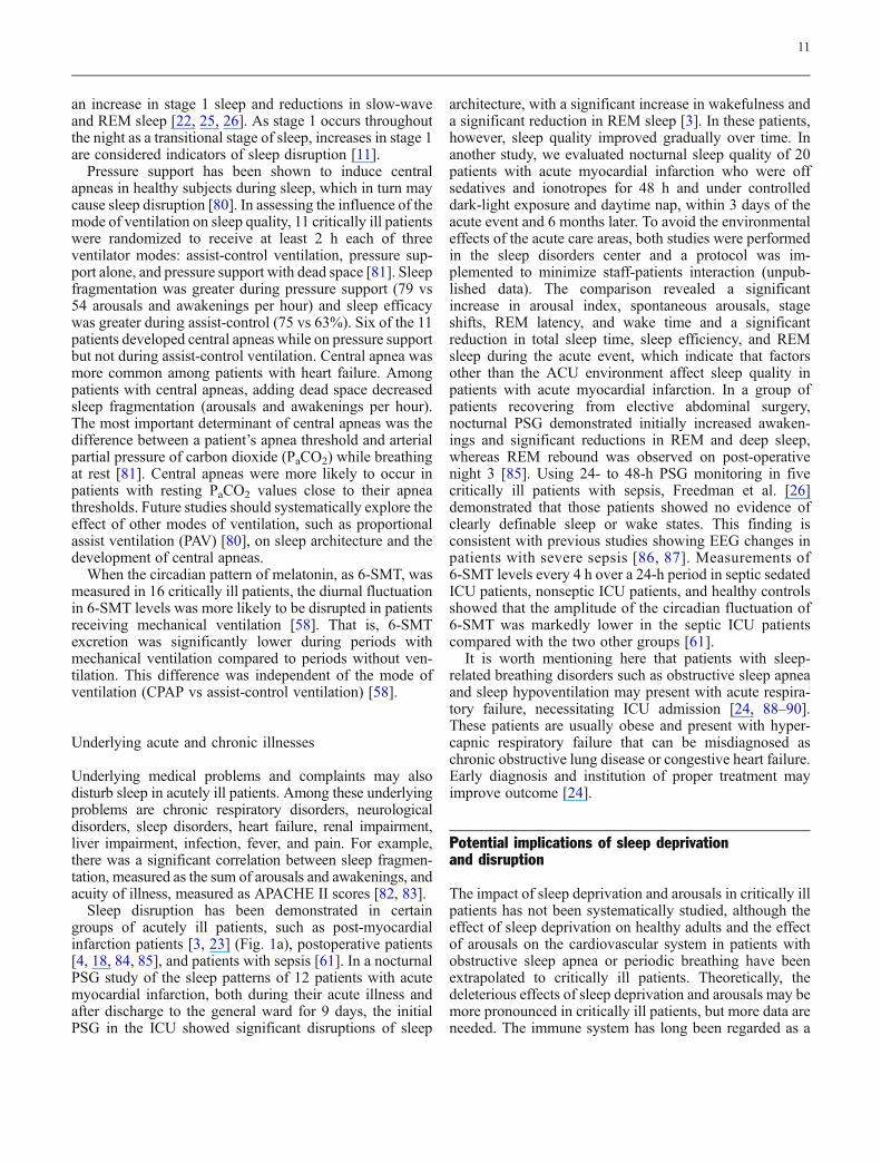
Introduction
The acute care units (ACU) in a hospital, which include themedical and surgical intensive care units (MICU and SICU),coronary care units (CCU) and post-anesthesia care units(PACU), are areas for patients who need special attention.Patients in ACUs are usually critically ill, making themmoresusceptible to the unfavorable atmosphere in the hospital.One of these unfavorable factors is sleep disruption anddeprivation [1–4]. Frequently, however, sleep disruption anddeprivation is not noticed by the medical staff in ACUs, andmany of these professionals have limited knowledge regard-ing sleep disruption in ACUs and its impact on patient health[5]. In ACUs, many organ systems are aggressively monitor-ed for the development of dysfunction or failure, but fre-quently the function of sleep in acutely ill patients is ignored.
Many factors may affect sleep in the ACU, includingtherapeutic interventions, diagnostic procedures, medications,the underlying disease process, and noise generated in the
ACU environment. Many detrimental physiological effectscan occur secondary to noise, including cardiovascular stim-ulation, increased gastric secretion, pituitary and adrenalstimulation, suppression of the immune system and woundhealing, and possible contribution to delirium [6–10].
It is our opinion that physicians and nurses working inACUs have a low level of awareness regarding sleepdisruption in critically ill patients and the impact of thisdisruption on the health of these patients. Over the past fewyears, many studies have endeavored to objectively assesssleep in the ACUs, as well as the effect of mechanicalventilation and circadian rhythm changes in critically illpatients. At this time, therefore, it is important to reviewpublished data regarding sleep in ACUs to improve theknowledge and recognition of this problem by health careprofessionals. We have therefore reviewed the methodsused to assess sleep in ACUs, factors that may affect sleepin the ACU environment, and the clinical implications ofsleep disruption in the ACU.
A. BaHammam (*)Sleep Disorders Center andMedical Intensive Care Unit,Department of Medicine 38,College of Medicine,King Saud University,Riyadh 11461, Saudi Arabiae-mail: [email protected].: +966-1-4671521Fax: +966-1-4672558

Normal sleep
Sleep is a physiological state that humans need to passthrough every day to repair and restore body functions.Typically, humans adapt to a 24-h circadian pattern, wherethey sleep at night and are awake during the day. This 24-hinternal clock (circadian pattern) is maintained byenvironmental factors, primarily light exposure, whichaffects melatonin secretion at night. Several physiologicaland biochemical body functions follow this circadianpattern. In normal individuals, sleep latency usually lastsabout 10–20 min and sleep lasts for 6–9 h on average,although variability among individuals can be significant.
While asleep, an individual passes through four to sixcycles, each of which consists of five sleep stages (Fig. 1b).One is called rapid eye movement (REM), and the rest(stages 1 through 4) are non-REM (NREM) sleep. Sleeponset begins with stage 1, which is a transitional stage,usually about 5% of normal sleep, and then progresses intostage 2, which can constitute as much as 50–60% of sleep.Thereafter, sleep progresses to stages 3 and 4, which areknown as delta, slow-wave, or deep sleep. These stagesoccur predominantly during the first half of the night andconstitute 15–20% of night sleep. Compared with lightsleep, slow-wave sleep is usually deeper and more restfuland requires a more intense stimulus to awaken the sleeper.REM sleep occurs approximately every 90 min in 4–6cycles, increasing in duration during the second half of thenight. During REM sleep, there is inhibition of spinalmotor neurons, which leads to paralysis of the majormuscle groups but spares the diaphragm and ocularmuscles. Although brain activity is increased duringREM sleep, this stage is considered to be restful, with avariable arousal threshold. Factors disturbing sleep, such asnoise, may affect the integrity of sleep. This may result inelectroencephalographic (EEG) arousals and awakenings,which may affect sleep architecture and prevent the normalprogress into the deeper stages of sleep [11].
Methods for sleep assessment in ACUs
Before discussing factors that affect sleep in ACUs, it isimportant to describe the methods used to assess sleep inACU settings. Among the methods used to assess sleep inACUs are staff observations [12], self-reporting, poly-somnography (PSG), and bispectral analysis.
Self-reporting
Several studies have subjectively assessed sleep in ACUsusing qualitative and quantitative methodologies. In anevaluation of physical and psychological stressors in 50intensive care unit (ICU) patients who completed the ICUEnvironmental Stressor Scale, insomnia was ranked as the
second most important stressor, second only to pain [13].Of patients interviewed 3 days after discharge from theICU, 61% reported sleep deprivation and 7% ratedinsomnia as their worst experience in the ICU [14]. Inanother study, 27% of ICU patients reported havinginsufficient sleep [15]. Furthermore, perceived ICU sleepquality was poor compared with baseline and did notchange over the course of patient stay in the ICU [16].However, self-reported data suffer from recall bias and alack of objective assessment of sleep quality.
Polysomnography
The standard diagnostic test for sleep orders is polysomno-graphy, during which neuro-cardio-respiratory parametersare usually monitored. Although PSG studies are usuallyperformed in a sleep laboratory, PSG has been used toassess sleep in critically ill patients in ACUs [2–4], [17–26]. In general, these studies were conducted in differentACUs and involved small groups of patients with differentunderlying medical and surgical problems, including thosewith acute coronary syndrome [3, 23], patients recoveringfrom open heart surgery [4], mechanically ventilatedpatients [22], trauma patients [19], patients with neurolog-ical and respiratory disorders [24], and mixed groups ofcritically ill patients. Previous studies have described thetechniques for performing PSG in critically ill patients inACUs attended by ICU nurses, but not by sleep techniciansor in a laboratory setting attended by a sleep technician[23–24]. Overall, sleep recording on ACU patientsrevealed sleep fragmentation, increases in stages 1 and 2sleep, reductions in slow-wave and REM sleep, andreductions in total sleep time and sleep efficiency. Mostof the previous studies only evaluated sleep during thenight, rather than over a 24-h period. However, thosestudies that did monitor PSG continuously for ≥24 hshowed that 40–50% of the total sleep time in ACUs occurduring the day [2, 18, 25, 26]. This disturbed sleep patternmay not improve over the course of a patient’s stay in theICU and may take several days to normalize after dischargefrom the ICU [21]. For example, repetitive whole-night in-hospital EEG showed that normal sleep patterns did notreturn to patients with acute myocardial infarction until 9days after discharge from the ICU [3].
Many factors in the ACUs may disrupt the typical EEGarchitecture seen during normal sleep, making the inter-pretation of sleep quality difficult. In a recent study, 24-hPSG monitoring of critically ill mechanically ventilatedpatients in the ICU revealed that eight patients haddisturbed sleep architecture [22]. The remaining patientshad either coma (seven patients) or atypical sleep (fivepatients), in which their EEGs were intermediate betweensleep and coma and characterized by a virtual absence ofstage 2 and REM sleep [22]. The authors thought that theseEEG changes were likely due to sedation.
7

The bispectral index (BIS)
Because of the difficulties of performing full PSG in theACUs environment and the need for certain expertise, analternative method, the bispectral index (BIS), has beenused to objectively assess sleep in ACU settings.Previously, the BIS was used extensively by anesthetiststo measure the depth of anesthesia and sedation. The BISmonitor displays a real-time EEG trace, acquired from afrontotemporal montage. It combines power spectral anal-ysis with interrogation of the phase relationship betweencomponent waves of different frequencies. The monitorgenerates a dimensionless number on a continuous scale of0–100, with 100 representing normal cortical electricalactivity and 0 indicating cortical electrical silence [27].
Several studies have used the BIS to assess sleep inACUs (Fig. 2). In one study, the investigators compared thechanges in the BIS with the conventional EEG stages ofsleep in five subjects during the early part of the night.They reported a good correlation between the BIS and the
level of natural sleep [28]. Light sleep occurred at BISvalues of 75–90, slow-wave sleep occurred at BIS values of20–70, and rapid eye movement sleep occurred at BISvalues of 75–92 [28]. In another study, which assessedsleep patterns in 27 ICU patients using the BIS andsubmental electromyogram, none of the patients showed acompletely normal sleep pattern [29]. In comparing the BISand spectral edge frequency (SEF) with PSG in ten patientswith mild apnea/hypopnea syndrome to test whether thesesignals could predict physiologic sleep stages, neither theBIS nor SEF reliably indicated conventionally determinedsleep stages [30]. Further studies are needed to access theutility of BIS as a monitor of sleep in patients in ACUs.
Skin potential levels of critically ill patients in the ICUhave been recorded to monitor the sleep-wake cycle of thesepatients [31]. Recently, actigraphy, a portable device thatrecords movement over extended periods of time and wornmost commonly on the wrist, [32] has been used to assessagitation and sleep in critically ill patients [33, 34]. In onestudy, actigraphy correlated well with observed and subjec-
Fig. 1 Hypnogram of a patient 2days after acute myocardial infarc-tion (a) and 6 months later (b). Thevertical axis represents sleep stagesand the horizontal axis time.a Significant sleep disruptions andawakenings and absence of slow-wave and REM sleep. b Clearlydefined sleep cycles with progres-sion to different sleep stages. S1Stage 1, S2 stage 2, S3 stage 3, S4stage 4, REM rapid eye movement,MVT movement, WK wake(BaHammam, unpublished data)
8

tive scores on agitation and sedation scales [34]. Furtherstudies are needed to assess the usefulness of this techniquein assessing circadian patterns in critically ill patients.
Factors affecting sleep in ACUs
Environmental factors
Environmental factors in ACUs include noise produced byphones, pagers, alarms, and staff and light, which maydisturb the normal light-dark cycle.
Noise Several studies have shown that the noise level inACUs is quite high [17, 35–41]. Excess noise mayincrease the mistakes by [42, 43] as well as impair theconcentration and mental efficiency of the staff [44, 45].Noise levels in ACUs range from 60 to 84 dB throughoutthe day and night [36]. As a reference, a busy office has anaverage noise level of 70 dB, and a pneumatic drill heardfrom around 15 m away has a noise level of 80 dB [16,37]. As noise measurements use a logarithmic scale, anincrease of 10 dB represents a doubling of the noise level.The US Environmental Protection Agency (EPA) hasrecommended that hospital noise levels not exceed 45 dBduring the day and 35 dB at night [46]. A study in anintermediate respiratory care unit reported a very strongcorrelation between the number of patient arousals and thenumber of sound peaks ≥80 dB [17]. In a study of 26 adultpatients in a PACU, the mean integrated sound pressurewas 67.1 dB [40]. Staff conversation caused 56% of the
recorded noise above 65 dB, while other sources of noise,such as alarms, telephones, and nursing care, accountedfor less than 10% each [40]. This finding agrees with thatof two other studies, both of which reported that staffconversation was one of the most disruptive environ-mental noises [16, 25]. Synchronous audio, video, andpolysomnographic recording in seven patients showed that20% of the recorded arousals and awakenings were relatedto noise and 10% to patient care activities, whereas thecauses of the remaining 70% were not identified, indicat-ing that other as yet unknown factors are involved [25].The above findings concur with those obtained byFreedman et al. [26] in a study of 22 critically ill patients.By simultaneously monitoring environmental noise andPSG, they found that environmental noise was responsiblefor 11.5 and 17% of the arousals and awakenings fromsleep, respectively.
Staff-patient interactions Despite the increased sophistica-tion of monitoring systems in ACUs, which shoulddecrease hands-on manipulations of sleeping patients,there are still frequent and repetitive staff interactions withcritically ill patients, thus reducing patient sleep time inACUs. Several studies have found that the mean numberof staff-patient interactions ranged from 40 to 60 per night[47–49]. Using a 24-h PSG monitoring, it was found thatthere were eight staff-patient interactions per hour ofpatient sleep, with most of these interactions due tonursing activities, such as wound dressing, adjustment ofintravenous drips, and administration of medications [25].Nurses were found to routinely provide daily baths forpatients between 02:00 and 05:00 on 55 of the 147 studynights [48]. Sleep disruptions caused by human interven-tions/diagnostic tests were found to be significantlydifferent in different ACUs [16]. For example, patientsin the MICU perceived interruptions caused by staffinteractions and diagnostic tests to be significantly moredisruptive to their sleep than did patients in the SICU andthe cardiac intermediate care unit. Based on these results,it has been recommended that nocturnal staff-patientinteractions be clustered, thus allowing patients unin-terrupted periods of sleep [50–52]. In addition, althoughmost of these studies assessed sleep interruptions and staffinteractions at night, future studies should assess thesevariables during both the day and night.
Circadian pattern disturbances About 40–50% of the totalsleep time in ACUs has been found to occur during theday [2, 18]. For example, using a 24-h PSG monitoring,approximately half the sleep of ventilated patients in theICU occurred during daytime (06:00–22:00), whereassleep in healthy controls tended to be more nocturnal [25].Sleep in humans follows a circadian pattern, in whichpeople usually sleep at night and are awake during the day.This circadian pattern, which is coupled to physiologiccycles, is maintained by internal and external cues called
Fig. 2 Normal changes in sleep stage and BIS in a sleeping healthyvolunteer throughout the night (−1, REM sleep; 0, awake; 1 and 2,light sleep; 3 and 4, slow-wave sleep) [29]. Reproduced withpermission from the publisher
9

Zeitgebers (makers or cues of time). Among these cues,the most important is exposure to light. Disturbances ofthe light-dark cycle and frequent environmental noises andinterventions in the ACUs may disturb the normalcircadian rhythm. Measurable markers for this circadianpattern include body temperature and the levels of certainhormones, such as melatonin and cortisol. Body temper-ature normally follows a circadian pattern, rising duringthe day and falling at night. In general, a falling bodytemperature induces sleep, whereas a rising temperatureprovokes wakefulness. As shown by recording the rectaltemperature of 15 patients who spent greater than 1 weekin the ICU, there was a marked disruption of circadianrhythms [53]. Similar findings were reported in otherwisehealthy patients following cardiac surgery [54].
Melatonin is considered to be the best marker ofcircadian rhythm [55, 56]. Individual melatonin profilesare highly reproducible and are less subject to maskingfactors than are other rhythm markers such as coretemperature and cortisol level. Melatonin secretion isnormally increased at bedtime and remains high untilearly morning. Several studies have shown that thecircadian release of melatonin is disturbed in critically illpatients [57–61]. For example, whenmelatonin levels in theblood and urine were measured over three consecutive daysin eight critically ill patients during deep sedation andmechanical ventilation, the circadian rhythm of melatoninsecretion was abolished in all but one patient, and there wasno correlation between melatonin levels and levels ofsedation [57]. This suggested that the impairment of thecircadian release of melatonin may play a role in sleepdisruption and delirium in the ICU. When the urineconcentrations of the melatonin metabolite 6-sulphatoxymelatonin (6-SMT) and free cortisol were monitored in 16patients for their entire stay in the ICU, the diurnal rhythmsof both hormones were consistently or periodicallydisturbed in 65–75% of the patients, despite controllingfor illumination [58]. Treatment with adrenergic drugs andbenzodiazepines increased 6-SMT excretion [58]. In addi-tion, there was a significant correlation between ICUpsychosis and an irregular melatonin circadian rhythm [59].
Hyposecretion of melatonin may have detrimental effectson critically ill patients. In animal models, hyposecretion ofmelatonin has been shown to impair mitochondrial oxidativephosphorylation [62] and the capacity to survive endotox-emia [63, 64].Moreover, the antioxidant effects of melatoninhave been observed to prevent ischemia-induced renaldamage [65, 66]. Although some investigators haverecommended that melatonin be given to patients in theICU to induce sleep and to resynchronize their biologicalclocks [67], there is little data to support that. Treatment withmelatonin may also regulate the secretion of growthhormones and prolactin [68]. Future studies should assessthe effect of exogenous melatonin administration on sleepand circadian pattern in acutely ill patients.
Medications
Many drugs are used frequently in ACU settings, includinghypnotics, narcotics, and ionotropic drugs. However,heavy sedation does not imply good sleep. For example,although the benzodiazepines decrease sleep latency andawakenings and increase sleep duration and efficiency(sleep duration/time in bed), these drugs also significantlyreduce slow-wave and REM sleep, increase spindles,increase cortical activity at low doses, and decrease EEGamplitude at high doses [69–71]. Narcotics also suppressdeep and REM sleep and increase arousals and stage 1sleep [72]. However, the effects of benzodiazepines andnarcotics on sleep in patients in the ACUs have not beenproperly investigated.
Many critically ill patients receive intravenous iono-tropic drugs, such as dopamine and norepinephrine. Bothof these drugs are important central neurotransmitters andare associated with cortical activation, thus possiblyincreasing arousals. Fortunately, both drugs do not appearto cross the blood-brain barrier under normal conditions[73]. Nevertheless, the administration of epinephrine topatients sedated with propofol increased their sedationscore and BIS values [74]. In rats, blood-brain barrierpermeability is increased by tumor necrosis factor alpha(TNF-α), which is secreted as part of the sepsis cascade[75]. Additional studies are needed to examine the effectsof these drugs on sleep architecture in critically ill patients.
Mechanical ventilation
Mechanical ventilation is an invasive treatment modalitythat may affect sleep quality in critically ill patients.Among the causes of sleep interruption in ventilatedpatients are the severity of the illness, nursing interventionsfor suctioning and other reasons [49], sedation and othermedications, mode of ventilation, asynchrony between thepatient and the machine [76], and failure of patients totrigger ventilators due to the use of improper settings,which may result in unloading of the respiratory muscles orintrinsic positive end-expiratory pressure [77]. As factorsother than mechanical ventilation may affect sleep in thisgroup of patients, it is difficult to assess the effects ofmechanical ventilation on sleep. To date, therefore, therehave been only a few studies subjectively or objectivelyevaluating sleep during mechanical ventilation.
In interviews with 158 survivors of critical illness 2months after discharge, 30% indicated having experiencedagony/panic during mechanical ventilation, which wasassociated with asynchrony between the patient’s breathingand the ventilator [78]. Using PSG, ventilated critically illpatients were found to have as many as 20–63 arousals andawakenings per hour [22, 23, 25, 79]. In general, PSGmonitoring of sleep during mechanical ventilation revealed
10
an increase in stage 1 sleep and reductions in slow-waveand REM sleep [22, 25, 26]. As stage 1 occurs throughoutthe night as a transitional stage of sleep, increases in stage 1are considered indicators of sleep disruption [11].
Pressure support has been shown to induce centralapneas in healthy subjects during sleep, which in turn maycause sleep disruption [80]. In assessing the influence of themode of ventilation on sleep quality, 11 critically ill patientswere randomized to receive at least 2 h each of threeventilator modes: assist-control ventilation, pressure sup-port alone, and pressure support with dead space [81]. Sleepfragmentation was greater during pressure support (79 vs54 arousals and awakenings per hour) and sleep efficacywas greater during assist-control (75 vs 63%). Six of the 11patients developed central apneas while on pressure supportbut not during assist-control ventilation. Central apnea wasmore common among patients with heart failure. Amongpatients with central apneas, adding dead space decreasedsleep fragmentation (arousals and awakenings per hour).The most important determinant of central apneas was thedifference between a patient’s apnea threshold and arterialpartial pressure of carbon dioxide (PaCO2) while breathingat rest [81]. Central apneas were more likely to occur inpatients with resting PaCO2 values close to their apneathresholds. Future studies should systematically explore theeffect of other modes of ventilation, such as proportionalassist ventilation (PAV) [80], on sleep architecture and thedevelopment of central apneas.
When the circadian pattern of melatonin, as 6-SMT, wasmeasured in 16 critically ill patients, the diurnal fluctuationin 6-SMT levels was more likely to be disrupted in patientsreceiving mechanical ventilation [58]. That is, 6-SMTexcretion was significantly lower during periods withmechanical ventilation compared to periods without ven-tilation. This difference was independent of the mode ofventilation (CPAP vs assist-control ventilation) [58].
Underlying acute and chronic illnesses
Underlying medical problems and complaints may alsodisturb sleep in acutely ill patients. Among these underlyingproblems are chronic respiratory disorders, neurologicaldisorders, sleep disorders, heart failure, renal impairment,liver impairment, infection, fever, and pain. For example,there was a significant correlation between sleep fragmen-tation, measured as the sum of arousals and awakenings, andacuity of illness, measured as APACHE II scores [82, 83].
Sleep disruption has been demonstrated in certaingroups of acutely ill patients, such as post-myocardialinfarction patients [3, 23] (Fig. 1a), postoperative patients[4, 18, 84, 85], and patients with sepsis [61]. In a nocturnalPSG study of the sleep patterns of 12 patients with acutemyocardial infarction, both during their acute illness andafter discharge to the general ward for 9 days, the initialPSG in the ICU showed significant disruptions of sleep
architecture, with a significant increase in wakefulness anda significant reduction in REM sleep [3]. In these patients,however, sleep quality improved gradually over time. Inanother study, we evaluated nocturnal sleep quality of 20patients with acute myocardial infarction who were offsedatives and ionotropes for 48 h and under controlleddark-light exposure and daytime nap, within 3 days of theacute event and 6 months later. To avoid the environmentaleffects of the acute care areas, both studies were performedin the sleep disorders center and a protocol was im-plemented to minimize staff-patients interaction (unpub-lished data). The comparison revealed a significantincrease in arousal index, spontaneous arousals, stageshifts, REM latency, and wake time and a significantreduction in total sleep time, sleep efficiency, and REMsleep during the acute event, which indicate that factorsother than the ACU environment affect sleep quality inpatients with acute myocardial infarction. In a group ofpatients recovering from elective abdominal surgery,nocturnal PSG demonstrated initially increased awaken-ings and significant reductions in REM and deep sleep,whereas REM rebound was observed on post-operativenight 3 [85]. Using 24- to 48-h PSG monitoring in fivecritically ill patients with sepsis, Freedman et al. [26]demonstrated that those patients showed no evidence ofclearly definable sleep or wake states. This finding isconsistent with previous studies showing EEG changes inpatients with severe sepsis [86, 87]. Measurements of6-SMT levels every 4 h over a 24-h period in septic sedatedICU patients, nonseptic ICU patients, and healthy controlsshowed that the amplitude of the circadian fluctuation of6-SMT was markedly lower in the septic ICU patientscompared with the two other groups [61].
It is worth mentioning here that patients with sleep-related breathing disorders such as obstructive sleep apneaand sleep hypoventilation may present with acute respira-tory failure, necessitating ICU admission [24, 88–90].These patients are usually obese and present with hyper-capnic respiratory failure that can be misdiagnosed aschronic obstructive lung disease or congestive heart failure.Early diagnosis and institution of proper treatment mayimprove outcome [24].
Potential implications of sleep deprivationand disruption
The impact of sleep deprivation and arousals in critically illpatients has not been systematically studied, although theeffect of sleep deprivation on healthy adults and the effectof arousals on the cardiovascular system in patients withobstructive sleep apnea or periodic breathing have beenextrapolated to critically ill patients. Theoretically, thedeleterious effects of sleep deprivation and arousals may bemore pronounced in critically ill patients, but more data areneeded. The immune system has long been regarded as a
11

vulnerable target for sleep deprivation. Cytokines synthe-sized by the immune system may play a role in normalsleep regulation, by increasing NREM sleep and decreas-ing REM sleep, and during inflammatory events, anincrease in cytokine levels may intensify their effects onsleep regulation [91]. Current evidence suggests that acuteand chronic sleep deprivation is associated with decreasedproportions of natural killer cells [92], lower antibody titersfollowing influenza virus immunization [93], reducedlymphokine-activated killer activity [94], and reducedinterleukin-2 production [94]. Moreover, sleep deprivationmay alter endocrine and metabolic functions, altering thenormal pattern of cortisol release and contributing toalterations of “glucocorticoid feedback regulation” [95],glucose tolerance, and insulin resistance [96]. In healthysubjects, sleep deprivation is associated with a negativeenergy balance [97]. Although the effect of sleep depriva-tion on ventilatory responses in critically ill patients is notknown, sleep deprivation per se in ten healthy subjects didnot reduce the sensitivity of central chemoreceptors orchange resting ventilation or metabolism [98]. In a recentstudy of cardiovascular autonomic modulation during 36 hof total sleep deprivation in 18 normal subjects, acute sleepdeprivation was associated with increased sympathetic anddecreased parasympathetic cardiovascular modulation anddecreased baroreflex sensitivity [99]. In addition, frequentarousals were associated with elevated catecholaminerelease and increased blood pressure [100].
Delirium, formerly known as ICU psychosis or the ICUsyndrome, is a serious problem facing the staff in ACUs.The incidence of delirium in a cohort of mechanicallyventilated patients in medical and coronary ICUs wasfound to be as high as 81% [101]. In addition, delirium wasan independent predictor of higher 6-month mortality andlonger hospital stay, even after adjusting for relevantcovariates [102]. Sleep deprivation has been suggested as a
risk factor for delirium in critically ill patients. Using staffobservations, without objective methods, there was ahigher prevalence of delirium among sleep-deprivedpatients [103, 104]. Delirious patients were reported tohave irregular patterns of melatonin release [59] anddisrupted circadian rhythms, resulting in fragmented sleep-wake cycles and nighttime awakenings [105]. Thesefindings suggest a strong association between deliriumand sleep deprivation but do not establish causality. It musttherefore be determined whether sleep deprivation is acause or a symptom of delirium.
Conclusions and future perspectives
Critically ill patients are prone to sleep deprivation, poorsleep quality, and disturbed circadian patterns. The causes ofsleep disruption in these patients seem to be multifactorialand include the ACU environment and staff, diagnostic andtherapeutic interventions, medications, mechanical ventila-tion, and the underlying illness. The role and contribution ofeach of these factors to sleep disruption in critically illpatients need to be clearly delineated. This will assist in thedevelopment of evidence-based guidelines regarding sleep-promoting interventions in ACUs. The effects of newermodes of ventilation on sleep architecture should also beexplored. Objective methods that are practical and easy tointerpret are needed to assess sleep in the ACUs. The use ofBIS may prove to be an easy and practical way to assesssleep in critically ill patients, but more research is needed.Staff working in ACUs should be educated about theimportance of sleep, the factors that may disturb sleep, andthe role of the ACU staff in promoting good sleep incritically ill patients. Finally, the impact of sleep deprivationand disturbances on patients’ stay in ACUs and on outcomeshould be determined.
References
1. Adam K, Oswald I (1983) Proteinsynthesis, bodily renewal and the sleep-wake cycle. Clin Sci 65:561–567
2. Hilton BA (1976) Quantity and qualityof patients’ sleep and sleep-disturbingfactors in a respiratory intensive careunit. J Adv Nurs 1:453–468
3. Broughton R, Baron R (1978) Sleeppatterns in the intensive care unit andon the ward after acute myocardialinfarction. Electroencephalogr ClinNeurophysiol 45:348–360
4. Orr WC, Stahl ML (1977) Sleepdisturbances after open heart surgery.Am J Cardiol 39:196–201
5. Christensen M (2005) What knowledgedo ICU nurses have with regard to theeffects of noise exposure in the inten-sive care unit? Intensive Crit Care Nurs21:199–207
6. Baker CF (1992) Discomfort to envi-ronmental noise: heart rate responses ofSICU patients. Crit Care Nurs Q 15:75–90
7. Tomei F, Papaleo B, Baccolo TP,Persechino B, Spabno G, Rosati MV(1994) Noise and gastric secretion. AmJ Ind Med 26:367–372
8. Haynes C (1999) Emergence delirium:a literature review. Br J Theatre Nurs9:502–510
9. Mc Carthy DO, Ouimet ME, Daun JM(1992) The effects of noise stress onleukocyte function in rats. Res NursHealth 15:131–137
10. Dinges DF, Douglas SD, Zaugg L,Campbell DE, McMann JM, WhitehouseWG, Orne BC, Kapoor SC, Icaza E,Orne MT (1994) Leukocytosis and nat-ural killer cell function parallel neurobe-havioral fatigue induced by 64 hours ofsleep deprivation. J Clin Invest 93:1930–1939
12

11. Carskadon MA, Dement WC (1999)Normal human sleep: an overview. In:Kryger M, Roth T, Dement WC (eds)Principles and practice of sleep medi-cine. WB Saunders, Philadelphia, pp15–26
12. Edwards GB, Schuring LM (1993)Pilot study: validating staff nurses’observations of sleep and wake statesamong critically ill patients, usingpolysomnography. Am J Crit Care2:125–131
13. Novaes MA, Aronovich A, Ferraz MB,Knobel E (1997) Stressors in ICU:patients’ evaluation. Intensive CareMed 23:1282–1285
14. Simini B (1999) Patients’ perceptionsof intensive care. Lancet 354:571–572
15. Granja C, Lopes A, Moreira S, Dias C,Costa-Pereira A, Carneiro A (2005)Patients’ recollections of experiences inthe intensive care unit may affect theirquality of life. Crit Care 9:R96–R109
16. Freedman NS, Kotzer N, Schwab RJ(1999) Patient perception of sleepquality and etiology of sleep disruptionin the intensive care unit. Am J RespirCrit Care Med 159:1155–1162
17. Aaron JN, Carlisle CC, Carscadon MA,Meyer TJ, Hill NS, Millman RP (1996)Environmental noise as a cause of sleepdisruption in an intermediate respirato-ry care unit. Sleep 19:707–710
18. Aurell J, Elmqvist D (1985) Sleep inthe surgical intensive care unit: contin-uous polygraphic recording of sleep innine patients receiving postoperativecare. Br Med J (Clin Res Ed)290:1029–1032
19. Fontaine DK (1989) Measurement ofnocturnal sleep patterns in traumapatients. Heart Lung 18:402–410
20. Buckle P, Pouliot Z, Millar T, Kerr P,Kryger MH (1992) Polysomnographyin acutely ill intensive care unitpatients. Chest 102:288–291
21. Richards KC, Bairnsfather L (1988) Adescription of night sleep patterns in thecritical care unit. Heart Lung 17:35–42
22. Cooper AB, Thornley KS, Young GB,Slutsky AS (2000) Sleep in critically illpatients requiring mechanical ventila-tion. Chest 117:809–818
23. BaHamman A, Al-Mobeireek A,Al-Nozha M, Al-Tahan A, Binsaeed A(2005) Behaviour and time-course ofsleep disordered breathing in patientswith acute coronary syndromes. Int JClin Pract 59:874–880
24. BaHamman A, Syed S, Al-Mughairy A(2005) Sleep-related breathing disor-ders in obese patients presenting withacute respiratory failure. Respir Med99:718–725
25. Gabor JY, Cooper AB, Crombach SA,Lee B, Kadikar N, Bettger HE, HanlyPJ (2003) Contribution of the intensivecare unit environment to sleep disrup-tion in mechanically ventilated patientsand healthy subjects. Am J Respir CritCare Med 167:708–715
26. Freedman NS, Gazendam J, Levan L,Pack AI, Schwab RJ (2001) Abnormalsleep/wake cycles and the effect ofenvironmental noise on sleep disruptionin the intensive care unit. Am J RespirCrit Care Med 163:451–457
27. Eke Z, Bell K (2004) Depth of anaes-thesia. CPD Anaesth 6:140–142
28. Sleigh JW, Andrzejowski J, Steyn-RossA, Steyn-Ross M (1999) The bispectralindex: a measure of depth of sleep?Anesth Analg 88:659–661
29. Nicholson T, Patel J, Sleigh JW (2001)Sleep patterns in intensive care unitpatients: a study using the bispectralindex. Crit Care Resusc 3:86–91
30. Nieuwenhuijs D, Coleman EL, DouglasNJ, Drummond GB, Dahan A (2002)Bispectral index values and spectraledge frequency at different stages ofphysiologic sleep. Anesth Analg94:125–129
31. Shiihara Y, Nogami T, Chigira M,Tanno Y, Sakai Y, Takahashi S, KodamaM, Kunimoto F (2001) Sleep-wakerhythm during stay in an intensive careunit: A week’s long-term recording ofskin potentials. Psychiatry Clin Neurosci55:279–280
32. Littner M, Kushida CA, AndersonWM, Bailey D, Berry RB, Davila DG,Hirshkowits M, Kapen S, Kramer M,Loube D, Wise M, Johnson SF (2003)Practice parameters for the role ofactigraphy in the study of sleep andcircadian rhythms: an update for 2002.Sleep 26:337–341
33. Redeker NS, Wykpisz E (1999) Pain,fatigue, activity, and sleep after coro-nary bypass (abstract). Better healththrough nursing research. State of theScience Congress, American Academyof Nursing, Washington, DC
34. Grap MJ, Borchers CT, Munro CL,Elswick RK Jr, Sessler CN (2005)Actigraphy in the critically ill: correla-tion with activity, agitation, and seda-tion. Am J Crit Care 14:52–60
35. Falk SA, Woods NF (1973) Hospitalnoise-levels and potential healthhazards. N Engl J Med 289:774–781
36. Meyer TJ, Eveloff SE, Bauer MS,Schwartz WA, Hill NS, Millman RP(1994) Adverse environmental condi-tions in the respiratory and medicalICU settings. Chest 105:1211–1216
37. Redding JS, Hargest TS, Minsky SH(1977) How noisy is intensive care?Crit Care Med 5:275–276
38. Gowan NJ (1979) The perceptual worldof the intensive care unit: an overviewof some environmental considerationsin the helping relationship. Heart Lung8:340–344
39. Soutar RL, Wilson JA (1986) Doeshospital noise disturb patients? Br MedJ (Clin Res Ed) 292:305
40. Allaouchiche B, Duflo F, Debon R,Bergeret A, Chassard D (2002) Noisein the postanaesthesia care unit. Br JAnaesth 88:369–373
41. Monsén MG, Edéll-Gustafsson UM(2005) Noise and sleep disturbancefactors before and after implementationof a behavioural modification pro-gramme. Intensive Crit Care Nurs21:208–219
42. Murthy VS, Malhotra SK, Bala I,Raghunathan M (1995) Detrimentaleffects of noise on anaesthetists. Can JAnaesth 42:608–611
43. Balogh D, Kittinger E, Benzer A, HacklJM (1993) Noise in the ICU. IntensiveCare Med 19:343–346
44. Smith A (1989) A review of the effectsof noise on human performance. ScandJ Psychol 30:185–206
45. Park SH, Song HH, Han JH, Park JM,Lee EJ, Park SM, Kang KJ, Lee JH,Hwang SS, Rho SC et al (1994) Effectof noise on the detection of rib fracturesby residents. Invest Radiol 29:54–58
46. Agency UEP (1974) Information onlevels of environmental noise requisiteto protect the public health and welfarewith an adequate margin of safety. USGovernment Printing Office,Washington, DC
47. Woods NF (1972) Patterns of sleep inpostcardiotomy patients. Nurs Res21:347–352
48. Tamburri LM, DiBrienza R, Zozula R,Redeker NS (2004) Nocturnal careinteractions with patients in critical careunits. Am J Crit Care 13:102–112
49. Celik S, Öztekin D, Akyolcu N, ÝsseverH (2005) Sleep disturbance: the patientcare activities applied at the night shift inthe intensive care unit. J Clin Nurs14:102–106
50. Sakallaris B, Orrel B (1997) Effects ofa 6-hour block of uninterrupted time onpostcardiac surgery patients’ perceivedsleep and pain. Am J Crit Care 6:243(Abstract)
51. Edwards JB, Schuring LM (1993)Sleep protocol: a research-based prac-tice change. Crit Care Nurse 13:84–88
52. Dines-Kalinowski CM (2002) Nature’snurse: promoting sleep in the ICU.Dimens Crit Care Nurs 21:32–34
13

53. Tweedie IE, Bell CF, Clegg A, CampbellIT, Minors DS, Waterhouse JM (1989)Retrospective study of temperaturerhythms of intensive care patients. CritCare Med 17:1159–1165
54. Nutall GA, Kumar M, Murray MJ(1998) No difference exists in thealteration of circadian rhythm betweenpatients with and without intensive careunit psychosis. Crit Care Med26:1351–1355
55. Arendt J (1995) Melatonin and themammalian pineal gland. Chapman &Hall, London, UK
56. Miles A, Thomas R (1988)Melatonin—a diagnostic markerin laboratory medicine? In: Miles A,Philbrick DRS, Thompson C (eds)Melatonin: clinical perspectives.Oxford University Press, Oxford, UK,pp 253–279
57. Olofsson K, Alling C, Lundberg D,Malmros C (2004) Abolished circadianrhythm of melatonin secretion in se-dated and artificially ventilated inten-sive care patients. Acta AnaesthesiolScand 48:679–684
58. Frisk U, Olsson J, Nylen P, Hahn RG(2004) Low melatonin excretion duringmechanical ventilation in the intensivecare unit. Clin Sci 107:47–53
59. Miyazaki T, Kuwano H, Kato H, AndoH, Kimura H, Inose T, Ohno T, SuzukiM, Nakjima M, Manda R, Fukuchi M,Tsukada K (2003) Correlation betweenserum melatonin circadian rhythm andintensive care unit psychosis after tho-racic esophagectomy. Surgery133:662–668
60. Shilo L, Dagan Y, Smorjik Y, WeinbergU, Dolev S, Komptel B, Balaum H,Shenkman L (1999) Patients in theintensive care unit suffer from severelack of sleep associated with loss ofnormal melatonin secretion pattern. AmJ Med Sci 317:278–281
61. Mundigler G, Delle-Karth G, KorenyM, Zehetgruber M, Steindl-Munda P,Marktl W, Ferti L, Siostrzonek P (2002)Impaired circadian rhythm of melatoninsecretion in sedated critically illpatients with severe sepsis. Crit CareMed 30:536–540
62. Martin M, Macias M, Leon J, EscamesG, Khaldy H, Acuna-Castrovieho D(2002) Melatonin increases the activityof the oxidative phosphorylation en-zymes and the production of ATP in ratbrain and liver mitochondria. Int JBiochem Cell Biol 34:348–357
63. Maestroni GJ (1996) Melatonin as atherapeutic agent in experimental en-dotoxic shock. J Pineal Res 20:84–89
64. Escames G, Leon J, Macias M, KhaldyH, Acuna-Castroviejo D (2003) Mela-tonin counteracts lipopolysaccharide-induced expression and activity ofmitochondrial nitric oxide synthase inrats. FASEB J 17:932–934
65. Tunez I, del Carmen Munoz M, FeijooM, Valdelvira ME, Rafael Munoz-Castaneda J, Montilla P (2003) Mela-tonin effect on renal oxidative stressunder constant light exposure. CellBiochem Funct 21:35–40
66. Kunduzova OR, Escourrou G, SeguelasMH, Delagrange P, De La Farge F,Cambon C, Parini A (2003) Preventionof apoptotic and necrotic cell death,caspase-3 activation, and renal dys-function by melatonin after ischemia/reperfusion. FASEB J 17:872–874
67. Shilo L, Dagan Y, Smorjik Y, WeinbergU, Dolev S, Komptel B, Shenkman L(2000) Effect of melatonin on sleepquality of COPD intensive carepatients: a pilot study. Chronobiol Int17:71–76
68. Petterborg LJ, Thalen BE, KjellmanBF, Wetterberg L (1991) Effect ofmelatonin replacement on serum hor-mone rhythms in a patient lackingendogenous melatonin. Brain Res Bull27:181–185
69. Qureshi A, Lee-Chiong T Jr (2004)Medications and their effects on sleep.Med Clin North Am 88:751–766
70. Borbély AA, Mattmann P, Loepfe M,Strauch I, Lehmann D (1985) Effect ofbenzodiazepine hypnotics on all-nightsleep EEG spectra. Hum Neurobiol4:189–194
71. Achermann P, Borbély AA (1987)Dynamics of EEG slow wave activityduring physiological sleep and afteradministration of benzodiazepine hyp-notics. Hum Neurobiol 6:203–210
72. Cronin AJ, Keifer JC, Davies MF, KingTS, Bixler EO (2001) Postoperativesleep disturbance: influences of opioidsand pain in humans. Sleep 24:39–44
73. Oldendorf WH (1971) Brain uptake ofradiolabeled amino acids, amines, andhexoses after arterial injection. Am JPhysiol 221:1629–1639
74. Andrzejowski J, Sleigh JW, JohnsonIA, Sikiotis L (2000) The effect ofintravenous epinephrine on the bispec-tral index and sedation. Anaesthesia55:761–763
75. Bourne RS, Mills GH (2004) Sleepdisruption in critically ill patients—pharmacological considerations. Anaesthesia 59:374–384
76. Flick GR, Bellamy PE, Simmons DH(1989) Diaphragmatic contraction dur-ing assisted mechanical ventilation.Chest 96:130–135
77. Hubmayr RD (1994) Setting the venti-lator. In: Tobin MI (ed) Principles andpractice of mechanical ventilation.McGraw-Hill, New York, pp 191–206
78. Bergbom-Engberg I, Haljamae H(1989) Assessment of patients’ experi-ence of discomforts during respiratortherapy. Crit Care Med 17:1068–1072
79. Gottschlich MM, Jenkins ME, MayesT, Khoury J, Kramer M, Warden GD,Kagan RJ (1994) The 1994 ClinicalResearch Award. A prospective clinicalstudy of the polysomnographic stagesof sleep after burn injury. J Burn CareRehabil 15:486–492
80. Meza S, Mendez M, Ostrowski M,Younes M (1998) Susceptibility toperiodic breathing with assisted venti-lation during sleep in normal subjects.J Appl Physiol 85:1929–1940
81. Partasarathy S, Tobin MJ (2002) Effectof ventilator mode on sleep quality incritically ill patients. Am J Respir CritCare Med 166:1423–1429
82. Parthasarathy S, Tobin MJ (2003) Issleep disruption related to severity ofcritical illness? Am J Respir Crit CareMed 167:A968 (Abstract)
83. Parthasarathy S (2004) Sleep duringmechanical ventilation. Curr OpinPulm Med 10:489–494
84. Kavey NB, Altshuler KZ (1983)Flurazepam and the sleep of hernior-rhaphy patients. J Clin Pharmacol23:199–208
85. Knill RL, Moote CA, Skinner ML,Rose EA (1990) Anesthesia with ab-dominal surgery leads to intense REMsleep during the first postoperativeweek. Anesthesiology 73:52–61
86. Bolton CF, Young GB, Zochodne DW(1993) The neurological complicationsof sepsis. Ann Neurol 33:94–100
87. Young GB, Bolton CF, Austin TW,Archibald YM, Gonder J, Wells GA(1990) The encephalopathy associatedwith septic illness. Clin Invest Med13:297–304
88. Fletcher EC, Shah A, Qian W, MillerCC (1991) “Near miss” death inobstructive sleep apnea: a critical caresyndrome. Crit Care Med 19:1158–1164
89. Buckle P, Pouliot Z, Millar T, Kerr P,Kryger MH (1992) Polysomnographyin acutely ill intensive care unitpatients. Chest 102:288–291
90. Resta O, Guido P, Foschimo BarbaroMP, Picca V, Talamo S, Lamorgese P(2000) Sleep-related breathing disor-ders in acute respiratory failure assistedby non-invasive ventilatory treatment:utility of portable polysomnographicsystem. Respir Med 94:128–134
14

91. Krueger JM, Majde JA (2003) Hu-moral links between sleep and theimmune system: research issues. AnnN Y Acad Sci 992:9–20
92. Osturk L, Pelin Z, Van Cauter E (1999)Effects of 48 hours sleep deprivation onhuman immune profile. Sleep Res On-line 2:107–111
93. Spiegel K, Sheridan JF, Van Cauter E(2002) Effect of sleep deprivation onresponse to immunization. JAMA288:1471–1472
94. Irwin M, McClintick C, Costlow C,Fortner M, White J, Gillin JC (1996)Partial night sleep deprivation reducesnatural killer and cellular immuneresponses in humans. FASEB J10:643–653
95. Ledproult R, Copinschi G, Buxton O,Van Cauter E (1997) Leproult R,Copinschi G, Buxton O, Van Cauter E.Sleep loss results in an elevation ofcortisol levels the next evening. Sleep20:865–870
96. Speigel K, Leproult R, Van Cauter E(1999) Impact of sleep debt on meta-bolic and endocrine function. Lancet354:1435–1439
97. Scrimshaw NS, Habicht JP, Pellet P,Piche ML, Cholakos B (1966) Effectsof sleep deprivation and reversal ofdiurnal activity on protein metabolismof young men. Am J Clin Nutr 19:313–319
98. Spengler CM, Shea SA (2000) Sleepdeprivation per se does not decrease thehypercapnic ventilatory response inhumans. Am J Respir Crit Care Med161:1124–1128
99. Zhong X, Hilton HJ, Gates GJ, Jelic S,Stern Y, Bartels MHN, DemeersmanRE, Basner RC (2005) Increased sym-pathetic and decreased parasympatheticcardiovascular modulation in normalhumans with acute sleep deprivation.J Appl Physiol 98:2024–2032
100. Loredo JS, Zeigler MG, Ancoli-IsraelS, Clausen JL (1999) Relationship ofarousals from sleep to sympatheticnervous system activity and BP inobstructive sleep apnea. Chest116:655–659
101. Ely EW, Gautam S, Margolin R,Francis J, May L, Speroff T, TrumanB, Dittus R, Bernard R, Inouye SK(2001) The impact of delirium in theintensive care unit on hospital lengthof stay. Intensive Care Med 27:1892–1900
102. Ely EW, Shintani A, Truman B,Speroff T, Gordon SSSM, Harrell FEJr, Inouye SK, Bernard GR, Dittus RS(2004) Delirium as a predictor ofmortality in mechanically ventilatedpatients in the intensive care unit.JAMA 291:1753–1762
103. Heller SS, Frank KA, Malm JR,Bowman FO Jr, Harris PD, CharltonMH, Kornfeld DS (1970) Psychiatriccomplications of open-heart surgery:a re-examination. N Engl J Med283:1015–1020
104. Helton MC, Gordon SH, Nunnery SL(1980) The correlation between sleepdeprivation and the intensive care unitsyndrome. Heart Lung 9:464–468
105. Mira S, GanZini L (2003) Delirium,depression, and anxiety. Crit Care Clin19:771–787
15