Carbohydrates - Moodle@Units
29
C hapter 7 Carbohydrates Sugar in the gourd and honey in the horn, I never was so happy since the hour I was born. Turkey in the Straw, stanza 6 (classic American folk tune) OUTLINE 7.1 ● Carbohydrate Nomenclature 7.2 ● Monosaccharides 7.3 ● Oligosaccharides 7.4 ● Polysaccharides 209 Carbohydrates are the single most abundant class of organic molecules found in nature. The name carbohydrate arises from the basic molecular formula (CH 2 O) n, which can be rewritten (C H 2 O) n to show that these substances are hydrates of carbon, where n 3 or more. Carbohydrates constitute a versatile class of molecules. Energy from the sun captured by green plants, algae, and some bacteria during photosynthesis (see Chapter 22) is stored in the form of carbohydrates. In turn, carbohydrates are the metabolic precursors of virtually all other biomolecules. Breakdown of carbohydrates provides the energy that sustains animal life. In addition, carbohydrates are covalently linked with a vari- ety of other molecules. Carbohydrates linked to lipid molecules, or glycolipids, are common components of biological membranes. Proteins that have cova- lently linked carbohydrates are called glycoproteins. These two classes of bio- molecules, together called glycoconjugates, are important components of cell walls and extracellular structures in plants, animals, and bacteria. In addition to the structural roles such molecules play, they also serve in a variety of “The Discovery of Honey”—Piero di Cosimo (1462). (Courtesy of the Worcester Art Museum)
-
Upload
khangminh22 -
Category
Documents
-
view
3 -
download
0
Transcript of Carbohydrates - Moodle@Units

Chapter 7
Carbohydrates
Sugar in the gourd and honey in the horn,I never was so happy since the hour I wasborn.
Turkey in the Straw, stanza 6 (classic American folk tune)
OUTLINE
7.1 ● Carbohydrate Nomenclature
7.2 ● Monosaccharides
7.3 ● Oligosaccharides
7.4 ● Polysaccharides
209
Carbohydrates are the single most abundant class of organic molecules foundin nature. The name carbohydrate arises from the basic molecular formula(CH2O)n, which can be rewritten (C � H2O)n to show that these substances arehydrates of carbon, where n � 3 or more. Carbohydrates constitute a versatileclass of molecules. Energy from the sun captured by green plants, algae, andsome bacteria during photosynthesis (see Chapter 22) is stored in the form ofcarbohydrates. In turn, carbohydrates are the metabolic precursors of virtuallyall other biomolecules. Breakdown of carbohydrates provides the energy thatsustains animal life. In addition, carbohydrates are covalently linked with a vari-ety of other molecules. Carbohydrates linked to lipid molecules, or glycolipids,are common components of biological membranes. Proteins that have cova-lently linked carbohydrates are called glycoproteins. These two classes of bio-molecules, together called glycoconjugates, are important components of cellwalls and extracellular structures in plants, animals, and bacteria. In additionto the structural roles such molecules play, they also serve in a variety of
“The Discovery of Honey”—Piero di Cosimo (1462). (Courtesy of the Worcester Art Museum)

processes involving recognition between cell types or recognition of cellularstructures by other molecules. Recognition events are important in normal cellgrowth, fertilization, transformation of cells, and other processes.
All of these functions are made possible by the characteristic chemical fea-tures of carbohydrates: (1) the existence of at least one and often two or moreasymmetric centers, (2) the ability to exist either in linear or ring structures,(3) the capacity to form polymeric structures via glycosidic bonds, and (4) thepotential to form multiple hydrogen bonds with water or other molecules intheir environment.
7.1 ● Carbohydrate Nomenclature
Carbohydrates are generally classified into three groups: monosaccharides (andtheir derivatives), oligosaccharides, and polysaccharides. The monosaccharidesare also called simple sugars and have the formula (CH2O)n. Monosaccharidescannot be broken down into smaller sugars under mild conditions. Oligo-saccharides derive their name from the Greek word oligo, meaning “few,” andconsist of from two to ten simple sugar molecules. Disaccharides are commonin nature, and trisaccharides also occur frequently. Four- to six-sugar-unitoligosaccharides are usually bound covalently to other molecules, includingglycoproteins. As their name suggests, polysaccharides are polymers of the sim-ple sugars and their derivatives. They may be either linear or branched poly-mers and may contain hundreds or even thousands of monosaccharide units.Their molecular weights range up to 1 million or more.
7.2 ● Monosaccharides
Classification
Monosaccharides consist typically of three to seven carbon atoms and aredescribed either as aldoses or ketoses, depending on whether the moleculecontains an aldehyde function or a ketone group. The simplest aldose is glyc-eraldehyde, and the simplest ketose is dihydroxyacetone (Figure 7.1). Thesetwo simple sugars are termed trioses because they each contain three carbonatoms. The structures and names of a family of aldoses and ketoses with three,four, five, and six carbons are shown in Figure 7.2 and 7.3. Hexoses are the mostabundant sugars in nature. Nevertheless, sugars from all these classes are impor-tant in metabolism.
Monosaccharides, either aldoses or ketoses, are often given more detailedgeneric names to describe both the important functional groups and the totalnumber of carbon atoms. Thus, one can refer to aldotetroses and ketotetroses,aldopentoses and ketopentoses, aldohexoses and ketohexoses, and so on. Sometimesthe ketone-containing monosaccharides are named simply by inserting the let-ters -ul- into the simple generic terms, such as tetruloses, pentuloses, hexuloses, hep-tuloses, and so on. The simplest monosaccharides are water-soluble, and mosttaste sweet.
Stereochemistry
Aldoses with at least three carbons and ketoses with at least four carbons con-tain chiral centers (Chapter 4). The nomenclature for such molecules mustspecify the configuration about each asymmetric center, and drawings of thesemolecules must be based on a system that clearly specifies these configurations.
FIGURE 7.1 ● Structure of a simple aldose(glyceraldehyde) and a simple ketose (dihy-droxyacetone).
210 Chapter 7 ● Carbohydrates
HO C H
CH2OH
H
or OHC
CH2OH
H
L-isomer D-isomer
Glyceraldehyde
CH2OH
CH2OH
Dihydroxy-acetone
OC
OC
H OC

As noted in Chapter 4, the Fischer projection system is used almost universallyfor this purpose today. The structures shown in Figures 7.2 and 7.3 are Fischerprojections. For monosaccharides with two or more asymmetric carbons, theprefix D or L refers to the configuration of the highest numbered asymmetriccarbon (the asymmetric carbon farthest from the carbonyl carbon). A mono-saccharide is designated D if the hydroxyl group on the highest numbered asymmetric carbon is drawn to the right in a Fischer projection, as in D-glyc-eraldehyde (Figure 7.1). Note that the designation D or L merely relates the
7.2 ● Monosaccharides 211
FIGURE 7.2 ● The structure and stereochemical relationships of D-aldoses having threeto six carbons. The configuration in each case is determined by the highest numberedasymmetric carbon (shown in gray). In each row, the “new” asymmetric carbon is shown inred.
HCOH
CHO
CH2OH
2
1
3
D-Glyceraldehyde
Carbonnumber
HCOH
CH2OH
3
4
D-Erythrose
HCOH
CHO
2
1
HCOH
CH2OH
D-Threose
HOCH
CHO
HCOH
CH2OH
4
5
D-Ribose (Rib)
HCOH3
HCOH
CHO
2
1
HCOH
CH2OH
D-Arabinose (Ara)
HCOH
HOCH
CHO
HCOH
CH2OH
D-Xylose (Xyl)
HOCH
HCOH
CHO
HCOH
CH2OH
D-Lyxose (Lyx)
HOCH
HOCH
CHO
HCOH
CH2OH
5
D-Allose
HCOH4
HCOH3
HCOH
CHO
2
1
Carbonnumber
Carbonnumber
Carbonnumber
6
HCOH
CH2OH
D-Altrose
HCOH
HCOH
HOCH
CHO
HCOH
CH2OH
D-Glucose(Glc)
HCOH
HOCH
HCOH
CHO
HCOH
CH2OH
D-Mannose(Man)
HCOH
HOCH
CHO
HOCH
HCOH
CH2OH
D-Gulose
HOCH
HCOH
CHO
HCOH
CH2OH
D-Idose
HCOH
HOCH
CHO
HCOH
HOCH
HCOH
CH2OH
D-Galactose(Gal)
HOCH
HCOH
CHO
HCOH
CH2OH
D-Talose
HOCH
CHO
HOCH
HOCH HOCH
ALDOTRIOSE
ALDOTETROSES
ALDOPENTOSES
ALDOHEXOSES
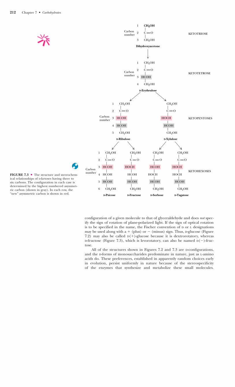
configuration of a given molecule to that of glyceraldehyde and does not spec-ify the sign of rotation of plane-polarized light. If the sign of optical rotationis to be specified in the name, the Fischer convention of D or L designationsmay be used along with a � (plus) or � (minus) sign. Thus, D-glucose (Figure7.2) may also be called D(�)-glucose because it is dextrorotatory, whereas D-fructose (Figure 7.3), which is levorotatory, can also be named D(�)-fruc-tose.
All of the structures shown in Figures 7.2 and 7.3 are D-configurations,and the D-forms of monosaccharides predominate in nature, just as L-aminoacids do. These preferences, established in apparently random choices earlyin evolution, persist uniformly in nature because of the stereospecificity of the enzymes that synthesize and metabolize these small molecules.
FIGURE 7.3 ● The structure and stereochem-ical relationships of D-ketoses having three tosix carbons. The configuration in each case isdetermined by the highest numbered asymmet-ric carbon (shown in gray). In each row, the“new” asymmetric carbon is shown in red.
212 Chapter 7 ● Carbohydrates
OC
CH2OH
1
2
Dihydroxyacetone
Carbonnumber
CH2OH
CH2OH
3
4
D-Erythrulose
Carbonnumber
CH2OH
HCOH
2
1
OC
3Carbonnumber
CH2OH
HCOH
2
1
CH2OH
4
5
D-Ribulose
HCOH
OC
CH2OH
HOCH
CH2OH
D-Xylulose
HCOH
OC
3Carbonnumber
CH2OH
HCOH
2
1
4 HCOH
CH2OH
5
6
D-Psicose
HCOH
OC
CH2OH
HOCH
HCOH
CH2OH
D-Fructose
HCOH
OC
CH2OH
HCOH
HOCH
CH2OH
D-Sorbose
HCOH
OC
CH2OH
HOCH
HOCH
CH2OH
D-Tagatose
HCOH
KETOHEXOSES
KETOPENTOSES
KETOTETROSE
KETOTRIOSEOC
CH2OH
3

L-Monosaccharides do exist in nature, serving a few relatively specialized roles.L-Galactose is a constituent of certain polysaccharides, and L-arabinose is aconstituent of bacterial cell walls.
According to convention, the D- and L-forms of a monosaccharide are mir-ror images of each other, as shown in Figure 7.4 for fructose. Stereoisomers thatare mirror images of each other are called enantiomers, or sometimes enan-tiomeric pairs. For molecules that possess two or more chiral centers, more thantwo stereoisomers can exist. Pairs of isomers that have opposite configurationsat one or more of the chiral centers but that are not mirror images of eachother are called diastereomers or diastereomeric pairs. Any two structures in agiven row in Figures 7.2 and 7.3 are diastereomeric pairs. Two sugars that dif-fer in configuration at only one chiral center are described as epimers. For exam-ple, D-mannose and D-talose are epimers and D-glucose and D-mannose areepimers, whereas D-glucose and D-talose are not epimers but merely diastere-omers.
Cyclic Structures and Anomeric Forms
Although Fischer projections are useful for presenting the structures of par-ticular monosaccharides and their stereoisomers, they ignore one of the mostinteresting facets of sugar structure—the ability to form cyclic structures with for-mation of an additional asymmetric center. Alcohols react readily with aldehydes toform hemiacetals (Figure 7.5). The British carbohydrate chemist Sir NormanHaworth showed that the linear form of glucose (and other aldohexoses) couldundergo a similar intramolecular reaction to form a cyclic hemiacetal. The result-ing six-membered, oxygen-containing ring is similar to pyran and is designateda pyranose. The reaction is catalyzed by acid (H�) or base (OH�) and is readily reversible.
FIGURE 7.4 ● D-Fructose and L-fructose, anenantiomeric pair. Note that changing the con-figuration only at C5 would change D-fructoseto L-sorbose.
7.2 ● Monosaccharides 213
HO C H
OC
CH2OH
H C OH
H C OH
CH2OH
D-Fructose
H C OH
OC
CH2OH
HO C H
HO C H
CH2OH
L-Fructose
Enantiomers
Mirror imageconfigurations
FIGURE 7.5
H
R' O
R O C
C
C
C
C
CH2OH
H OH
HO H
H OH
H OH
D-Glucose
C O
C C
C
H
H
HH
CH2OH H
OH
OHHO
C
C
C
CH2OH
HO H
H OH
H
CHO HO
CH OH
FISCHER PROJECTION FORMULAS
C
OH C O
C
H
OH
C
C C
H
H
HH
CH2OH
OH
OHHO
α-D-Glucopyranose
C O
C
H
C
C C
H
H
HH
CH2OH
OH
OHHO
β-D-Glucopyranose
HAWORTH PROJECTION FORMULAS
OH
H H
R' OH
C+
AldehydeAlcohol
R O
Hemiacetal
Cyclization
O
Pyran
C
H
O
β-D-Glucopyranose
1
2
3
4
5
6
1
23
4
5
6
C
C
C
CH2OH
H OH
H OH
H
CHO HO
CH OH
α-D-Glucopyranose
1
2
3
4
5
6
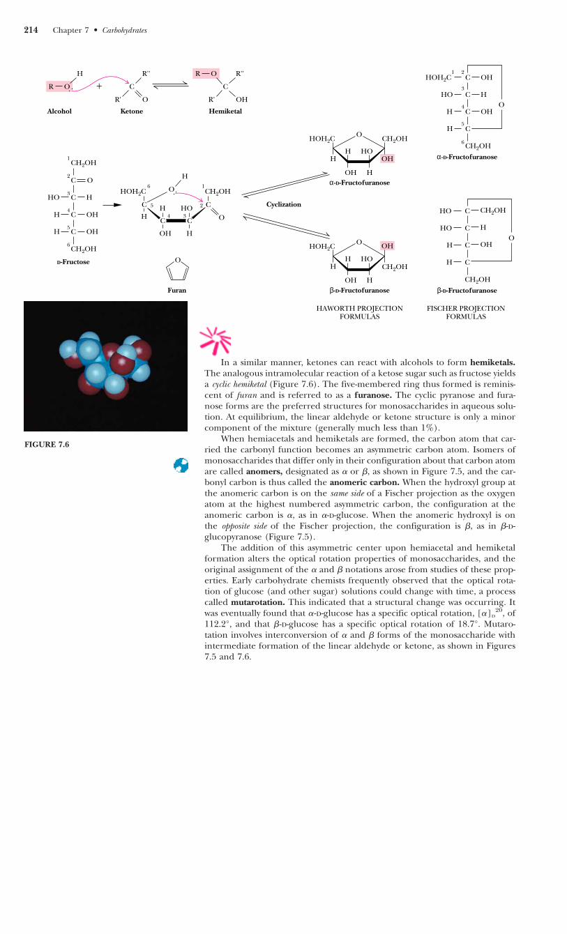
In a similar manner, ketones can react with alcohols to form hemiketals.The analogous intramolecular reaction of a ketose sugar such as fructose yieldsa cyclic hemiketal (Figure 7.6). The five-membered ring thus formed is reminis-cent of furan and is referred to as a furanose. The cyclic pyranose and fura-nose forms are the preferred structures for monosaccharides in aqueous solu-tion. At equilibrium, the linear aldehyde or ketone structure is only a minorcomponent of the mixture (generally much less than 1%).
When hemiacetals and hemiketals are formed, the carbon atom that car-ried the carbonyl function becomes an asymmetric carbon atom. Isomers ofmonosaccharides that differ only in their configuration about that carbon atomare called anomers, designated as � or �, as shown in Figure 7.5, and the car-bonyl carbon is thus called the anomeric carbon. When the hydroxyl group atthe anomeric carbon is on the same side of a Fischer projection as the oxygenatom at the highest numbered asymmetric carbon, the configuration at theanomeric carbon is �, as in �-D-glucose. When the anomeric hydroxyl is on the opposite side of the Fischer projection, the configuration is �, as in �-D-glucopyranose (Figure 7.5).
The addition of this asymmetric center upon hemiacetal and hemiketalformation alters the optical rotation properties of monosaccharides, and theoriginal assignment of the � and � notations arose from studies of these prop-erties. Early carbohydrate chemists frequently observed that the optical rota-tion of glucose (and other sugar) solutions could change with time, a processcalled mutarotation. This indicated that a structural change was occurring. Itwas eventually found that �-D-glucose has a specific optical rotation, [�]D
20, of112.2°, and that �-D-glucose has a specific optical rotation of 18.7°. Mutaro-tation involves interconversion of � and � forms of the monosaccharide withintermediate formation of the linear aldehyde or ketone, as shown in Figures7.5 and 7.6.
FIGURE 7.6
214 Chapter 7 ● Carbohydrates
O
R''
R' O
R O C
C
C
C
C
CH2OH
HO H
H OH
H OH
D-Fructose
H R''
R' OH
C+
KetoneAlcohol
R O
Hemiketal
CH2OH
H
O
C C
H
OH
HO
H
C C
CH2OHHOH2C
H O
Cyclization
OH
O CH2OH
H
OH
HO
H
H
HOH2C
α-D-Fructofuranose
OHO
H
OH
HO
H
H
HOH2C
β-D-Fructofuranose
CH2OHO
Furan
C
C
C
CH2OH
HOH2C OH
HO H
H
CH OHO
α-D-Fructofuranose
C
C
C
CH2OH
HO CH2OH
HO H
H
CH OHO
β-D-Fructofuranose
HAWORTH PROJECTIONFORMULAS
FISCHER PROJECTIONFORMULAS
1
2
3
4
5
6
2
34
5
6 1
1 2
3
4
5
6
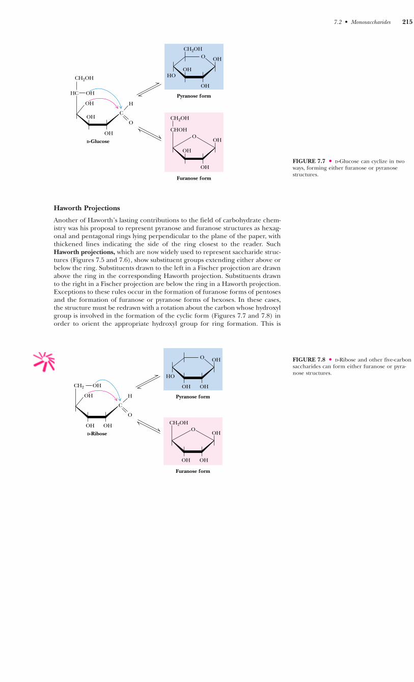
Haworth Projections
Another of Haworth’s lasting contributions to the field of carbohydrate chem-istry was his proposal to represent pyranose and furanose structures as hexag-onal and pentagonal rings lying perpendicular to the plane of the paper, withthickened lines indicating the side of the ring closest to the reader. SuchHaworth projections, which are now widely used to represent saccharide struc-tures (Figures 7.5 and 7.6), show substituent groups extending either above orbelow the ring. Substituents drawn to the left in a Fischer projection are drawnabove the ring in the corresponding Haworth projection. Substituents drawnto the right in a Fischer projection are below the ring in a Haworth projection.Exceptions to these rules occur in the formation of furanose forms of pentosesand the formation of furanose or pyranose forms of hexoses. In these cases,the structure must be redrawn with a rotation about the carbon whose hydroxylgroup is involved in the formation of the cyclic form (Figures 7.7 and 7.8) inorder to orient the appropriate hydroxyl group for ring formation. This is
7.2 ● Monosaccharides 215
FIGURE 7.7 ● D-Glucose can cyclize in twoways, forming either furanose or pyranosestructures.
FIGURE 7.8 ● D-Ribose and other five-carbonsaccharides can form either furanose or pyra-nose structures.
OCH2OH
OH
OHHO
Pyranose form
OH
OOH
OH
OH
CHOH
CH2OH
Furanose form
OH
OH
OH
HC
CH2OH
OH
C
O
H
D-Glucose
O
OH
HO
Pyranose form
OH
OOH
OH
CH2OH
Furanose form
OH
OH
CH2 OH
C
O
H
D-Ribose
OH
OH
OH
merely for illustrative purposes and involves no change in configuration of thesaccharide molecule.
The rules previously mentioned for assignment of �- and �-configurationscan be readily applied to Haworth projection formulas. For the D-sugars, theanomeric hydroxyl group is below the ring in the �-anomer and above the ringin the �-anomer. For L-sugars, the opposite relationship holds.
As Figures 7.7 and 7.8 imply, in most monosaccharides there are two ormore hydroxyl groups which can react with an aldehyde or ketone at the otherend of the molecule to form a hemiacetal or hemiketal. Consider the possi-bilities for glucose, as shown in Figure 7.7. If the C-4 hydroxyl group reactswith the aldehyde of glucose, a five-membered ring is formed, whereas if theC-5 hydroxyl reacts, a six-membered ring is formed. The C-6 hydroxyl does notreact effectively because a seven-membered ring is too strained to form a sta-ble hemiacetal. The same is true for the C-2 and C-3 hydroxyls, and thus five-and six-membered rings are by far the most likely to be formed from six-membered monosaccharides. D-Ribose, with five carbons, readily forms eitherfive-membered rings (�- or � -D-ribofuranose) or six-membered rings (�- or � -D-ribopyranose) (Figure 7.8). In general, aldoses and ketoses with five or morecarbons can form either furanose or pyranose rings, and the more stable formdepends on structural factors. The nature of the substituent groups on the car-bonyl and hydroxyl groups and the configuration about the asymmetric car-bon will determine whether a given monosaccharide prefers the pyranose orfuranose structure. In general, the pyranose form is favored over the furanosering for aldohexose sugars, although, as we shall see, furanose structures aremore stable for ketohexoses.
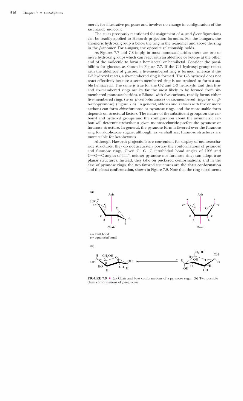
Although Haworth projections are convenient for display of monosaccha-ride structures, they do not accurately portray the conformations of pyranoseand furanose rings. Given COCOC tetrahedral bond angles of 109° andCOOOC angles of 111°, neither pyranose nor furanose rings can adopt trueplanar structures. Instead, they take on puckered conformations, and in thecase of pyranose rings, the two favored structures are the chair conformationand the boat conformation, shown in Figure 7.9. Note that the ring substituents
216 Chapter 7 ● Carbohydrates
FIGURE 7.9 ● (a) Chair and boat conformations of a pyranose sugar. (b) Two possiblechair conformations of �-D-glucose.
a
e
a
e
ea e
a
e
O
Axis
Chair
a = axial bonde = equatorial bond
a
e e
ea
O
Axis
Boat
ea
a
e
aa
109�
(a)
(b)
OHHO
OHCH2OH
HO
O H
HOH
H HOHOH
CH2OH
OH
H
HH
H
HO
H

in these structures can be equatorial, which means approximately coplanar withthe ring, or axial, that is, parallel to an axis drawn through the ring as shown.Two general rules dictate the conformation to be adopted by a given saccha-ride unit. First, bulky substituent groups on such rings are more stable whenthey occupy equatorial positions rather than axial positions, and second, chairconformations are slightly more stable than boat conformations. For a typicalpyranose, such as �-D-glucose, there are two possible chair conformations(Figure 7.9). Of all the D-aldohexoses, �-D-glucose is the only one that canadopt a conformation with all its bulky groups in an equatorial position. Withthis advantage of stability, it may come as no surprise that �-D-glucose is themost widely occurring organic group in nature and the central hexose in car-bohydrate metabolism.
Derivatives of Monosaccharides
A variety of chemical and enzymatic reactions produce derivatives of the sim-ple sugars. These modifications produce a diverse array of saccharide deriva-tives. Some of the most common derivations are discussed here.
Sugar Acids
Sugars with free anomeric carbon atoms are reasonably good reducing agentsand will reduce hydrogen peroxide, ferricyanide, certain metals (Cu2� andAg�), and other oxidizing agents. Such reactions convert the sugar to a sugaracid. For example, addition of alkaline CuSO4 (called Fehling’s solution) to analdose sugar produces a red cuprous oxide (Cu2O) precipitate:
and converts the aldose to an aldonic acid, such as gluconic acid (Figure 7.10).Formation of a precipitate of red Cu2O constitutes a positive test for an alde-hyde. Carbohydrates that can reduce oxidizing agents in this way are referredto as reducing sugars. By quantifying the amount of oxidizing agent reducedby a sugar solution, one can accurately determine the concentration of thesugar. Diabetes mellitus is a condition that causes high levels of glucose in urineand blood, and frequent analysis of reducing sugars in diabetic patients is animportant part of the diagnosis and treatment of this disease. Over-the-counterkits for the easy and rapid determination of reducing sugars have made thisprocedure a simple one for diabetics.
Monosaccharides can be oxidized enzymatically at C-6, yielding uronicacids, such as D-glucuronic and L-iduronic acids (Figure 7.10). L --Iduronic acidis similar to D-glucuronic acid, except for having an opposite configuration atC-5. Oxidation at both C-1 and C-6 produces aldaric acids, such as D-glucaricacid.
Sugar Alcohols
Sugar alcohols, another class of sugar derivative, can be prepared by the mildreduction (with NaBH4 or similar agents) of the carbonyl groups of aldosesand ketoses. Sugar alcohols, or alditols, are designated by the addition of -itolto the name of the parent sugar (Figure 7.11). The alditols are linear mole-cules that cannot cyclize in the manner of aldoses. Nonetheless, alditols arecharacteristically sweet tasting, and sorbitol, mannitol, and xylitol are widelyused to sweeten sugarless gum and mints. Sorbitol buildup in the eyes of dia-
OB
RC H � 2 Cu2� � 5 OH�
�
OB
RC O� � Cu2O
�
� 3 H2OAldehyde Carboxylate
7.2 ● Monosaccharides 217
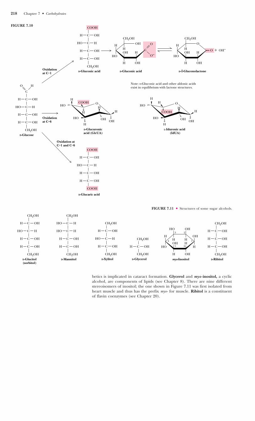
betics is implicated in cataract formation. Glycerol and myo -inositol, a cyclicalcohol, are components of lipids (see Chapter 8). There are nine differentstereoisomers of inositol; the one shown in Figure 7.11 was first isolated fromheart muscle and thus has the prefix myo - for muscle. Ribitol is a constituentof flavin coenzymes (see Chapter 20).
218 Chapter 7 ● Carbohydrates
Oxidation at C–1
O–O + OH–
H C OH
COOH
H
HO
HO
H
O
H
COOH
H
OHOH
H
D-Glucuronicacid (GlcUA)
H
HO
HO
H
OH
COOHH
OHOH
H
L-Iduronic acid (IdUA)
HO C H
H C OH
H C OH
CH2OHD-Gluconic acid
H C OH
COOH
HO C H
H C OH
H C OH
D-Glucaric acid
COOH
OH
CH
H
HH
CH2OH
OH
OHHO
D-Gluconic acid
O H
H
HH
CH2OH
OH
OHHO
D-δ-Gluconolactone
O
Note: D-Gluconic acid and other aldonic acids exist in equilibrium with lactone structures.
Oxidation at C–6
Oxidation at C–1 and C–6
H OH
C
C
O H
C
HO H
H OHC
H OHC
CH2OH
D-Glucose
FIGURE 7.10
FIGURE 7.11 ● Structures of some sugar alcohols.
H C OH
CH2OH
HO C H
H C OH
H C OH
D-Glucitol(sorbitol)
CH2OH
CH2OH
HO C H
H C OH
H C OH
D-Mannitol
CH2OH
HO C H CH2OH
H C OH
H C OH
D-Xylitol
CH2OH
HO C H CH2OH
H C OH
D-Glycerol
CH2OH
H C OH
CH2OH
H C OH
H C OH
D-Ribitol
CH2OHH OH
HOHHHO
HHOHH
OHHO
myo-Inositol
1
23
4
65

Deoxy Sugars
The deoxy sugars are monosaccharides with one or more hydroxyl groupsreplaced by hydrogens. 2-Deoxy-D-ribose (Figure 7.12), whose systematic nameis 2-deoxy-D-erythropentose, is a constituent of DNA in all living things (seeChapter 11). Deoxy sugars also occur frequently in glycoproteins and polysac-charides. L-Fucose and L-rhamnose, both 6-deoxy sugars, are components ofsome cell walls, and rhamnose is a component of ouabain, a highly toxic car-diac glycoside found in the bark and root of the ouabaio tree. Ouabain is usedby the East African Somalis as an arrow poison. The sugar moiety is not thetoxic part of the molecule (see Chapter 10).
Sugar Esters
Phosphate esters of glucose, fructose, and other monosaccharides are impor-tant metabolic intermediates, and the ribose moiety of nucleotides such as ATPand GTP is phosphorylated at the 5�-position (Figure 7.13).
7.2 ● Monosaccharides 219
FIGURE 7.12 ● Several deoxy sugars and ouabain, which contains �-L-rhamnose (Rha).Hydrogen atoms highlighted in red are “deoxy” positions.
FIGURE 7.13 ● Several sugar esters important in metabolism.
O H
OHHH
HOH
H
HOH2C
2-Deoxy-α-D-Ribose
HH
OH OH
H
HO
H
H
O
α-L-Rhamnose (Rha)
HOH
OH H
H
OH OH
H
O
α-L-Fucose (Fuc)
H
HO
CH3HH
OH OH
O
H
HO
H
H
O
Ouabain
CH2
HOOH
OH
OHCH3
OH
O
O
CH3 CH3
O
O
P
O–
O
O
P
O–
O
P
O–
HO
H H
OPO32–
OH
H
H
OHH
CH2OHO
α-D-Glucose-1-phosphate
H
2–O3PO H2C
OH
CH2 OPO32–O
H
H
α-D-Fructose-1,6-bisphosphate
OH
HOH
CH2
H
O
H
Adenosine-5'-triphosphate
OH
N
OH
H
–O O
N
N N
NH2

Amino Sugars
Amino sugars, including D-glucosamine and D-galactosamine (Figure 7.14), con-tain an amino group (instead of a hydroxyl group) at the C-2 position. Theyare found in many oligo- and polysaccharides, including chitin, a polysaccha-ride in the exoskeletons of crustaceans and insects.
Muramic acid and neuraminic acid, which are components of the polysac-charides of cell membranes of higher organisms and also bacterial cell walls,are glucosamines linked to three-carbon acids at the C-1 or C-3 positions. Inmuramic acid (thus named as an amine isolated from bacterial cell wall poly-saccharides; murus is Latin for “wall”), the hydroxyl group of a lactic acid moi-ety makes an ether linkage to the C-3 of glucosamine. Neuraminic acid (anamine isolated from neural tissue) forms a COC bond between the C-1 of N-acetylmannosamine and the C-3 of pyruvic acid (Figure 7.15). The N-acetyl andN-glycolyl derivatives of neuraminic acid are collectively known as sialic acidsand are distributed widely in bacteria and animal systems.
FIGURE 7.14 ● Structures of D-glucosamineand D-galactosamine.
220 Chapter 7 ● Carbohydrates
FIGURE 7.15 ● Structures of muramic acid and neuraminic acid and several depictionsof sialic acid.
HO
H OH
NH2
H
H
OHH
CH2OHO
β-D-Glucosamine
H H
OH
NH2
H
H
OHH
CH2OHO
β-D-Galactosamine
H
HO
CH3 CH COOH
O
Fischer projection
H C OH
CH2
C C H
HOOC C OH
C H
H C OH
H C OH
CH2OH
O
CH3
O
COOH
OH
HH
HOH
HN
H
CCH3
H
HCOH
HCOH
CH2OH
Haworth projection
N-Acetyl-D-neuraminic acid (NeuNAc), a sialic acid
COOH
OH
H
H
H
HOH
O
O
CCH3
H
OHHHOH2C
HHO
Chair conformation
HO
H
NH2
H
H
OH
CH2OHO H
OH
Muramic acid
O
O
N-Acetylmannosamine
H C OH
CH2
C NH
C H
C O
C H
H C OH
H C OH
CH2OH
CH3
HO
COOH
Pyruvic acid
NH
NH
N-Acetyl-D-neuraminic acid (NeuNAc)

Acetals, Ketals, and Glycosides
Hemiacetals and hemiketals can react with alcohols in the presence of acid toform acetals and ketals, as shown in Figure 7.16. This reaction is another exam-ple of a dehydration synthesis and is similar in this respect to the reactions under-gone by amino acids to form peptides and nucleotides to form nucleic acids.The pyranose and furanose forms of monosaccharides react with alcohols inthis way to form glycosides with retention of the �- or �-configuration at theC-1 carbon. The new bond between the anomeric carbon atom and the oxy-gen atom of the alcohol is called a glycosidic bond. Glycosides are namedaccording to the parent monosaccharide. For example, methyl-�-D-glucoside(Figure 7.17) can be considered a derivative of �-D-glucose.
7.3 ● Oligosaccharides
Given the relative complexity of oligosaccharides and polysaccharides in higherorganisms, it is perhaps surprising that these molecules are formed from rela-tively few different monosaccharide units. (In this respect, the oligo- and poly-saccharides are similar to proteins; both form complicated structures based ona small number of different building blocks.) Monosaccharide units includethe hexoses glucose, fructose, mannose, and galactose and the pentoses riboseand xylose.
Disaccharides
The simplest oligosaccharides are the disaccharides, which consist of two mono-saccharide units linked by a glycosidic bond. As in proteins and nucleic acids,each individual unit in an oligosaccharide is termed a residue. The disaccha-rides shown in Figure 7.18 are all commonly found in nature, with sucrose,maltose, and lactose being the most common. Each is a mixed acetal, with onehydroxyl group provided intramolecularly and one hydroxyl from the othermonosaccharide. Except for sucrose, each of these structures possesses one freeunsubstituted anomeric carbon atom, and thus each of these disaccharides isa reducing sugar. The end of the molecule containing the free anomeric car-bon is called the reducing end, and the other end is called the nonreducingend. In the case of sucrose, both of the anomeric carbon atoms are substituted,that is, neither has a free OOH group. The substituted anomeric carbons can-not be converted to the aldehyde configuration and thus cannot participate in
7.3 ● Oligosaccharides 221
Hemiacetal
O H
R' OH
C
R
+ OHR''
Acetal
O H
R' O
C
R
R''
Hemiketal
O R'''
R' OH
C
R
+ OHR''
Ketal
O R'''
R' O
C
R
R''
H2O+
+ H2O
HO
H H
O CH3
OH
H
H
OHH
CH2OHO
Methyl-α-D-glucoside
HO
H
H
OH
H
H
OHH
CH2OHO
Methyl-β-D-glucoside
O CH3
FIGURE 7.16 ● Acetals and ketals can be formed from hemiacetals and hemiketals,respectively.
FIGURE 7.17 ● The anomeric forms ofmethyl-D-glucoside.
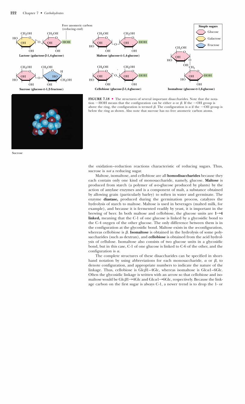
the oxidation–reduction reactions characteristic of reducing sugars. Thus,sucrose is not a reducing sugar.
Maltose, isomaltose, and cellobiose are all homodisaccharides because theyeach contain only one kind of monosaccharide, namely, glucose. Maltose isproduced from starch (a polymer of �-D-glucose produced by plants) by theaction of amylase enzymes and is a component of malt, a substance obtainedby allowing grain (particularly barley) to soften in water and germinate. Theenzyme diastase, produced during the germination process, catalyzes thehydrolysis of starch to maltose. Maltose is used in beverages (malted milk, forexample), and because it is fermented readily by yeast, it is important in thebrewing of beer. In both maltose and cellobiose, the glucose units are 1n4linked, meaning that the C-1 of one glucose is linked by a glycosidic bond tothe C-4 oxygen of the other glucose. The only difference between them is inthe configuration at the glycosidic bond. Maltose exists in the �-configuration,whereas cellobiose is �. Isomaltose is obtained in the hydrolysis of some poly-saccharides (such as dextran), and cellobiose is obtained from the acid hydrol-ysis of cellulose. Isomaltose also consists of two glucose units in a glycosidicbond, but in this case, C-1 of one glucose is linked to C-6 of the other, and theconfiguration is �.
The complete structures of these disaccharides can be specified in short-hand notation by using abbreviations for each monosaccharide, � or �, todenote configuration, and appropriate numbers to indicate the nature of thelinkage. Thus, cellobiose is Glc�1–4Glc, whereas isomaltose is Glc�1–6Glc.Often the glycosidic linkage is written with an arrow so that cellobiose and iso-maltose would be Glc�1n4Glc and Glc�1n6Glc, respectively. Because the link-age carbon on the first sugar is always C-1, a newer trend is to drop the 1- or
222 Chapter 7 ● Carbohydrates
FIGURE 7.18 ● The structures of several important disaccharides. Note that the nota-tion OHOH means that the configuration can be either � or �. If the OOH group isabove the ring, the configuration is termed �. The configuration is � if the OOH group isbelow the ring as shown. Also note that sucrose has no free anomeric carbon atoms.
OHOOH
OH
CH2OHO
OH
OH
CH2OH
O HOH
Lactose (galactose-β-1,4-glucose)
Free anomeric carbon(reducing end)
O
HOOH
OH
CH2OHO
OH
OH
CH2OH
OHOH
Maltose (glucose-α-1,4-glucose)
O
HOOH
OH
CH2OHO
HO
OH
O
Sucrose (glucose-α-1,2-fructose)
H
CH2OH
O
HOOH
OH
CH2OHO
OH
OH
CH2OH
O HOH
Cellobiose (glucose-β-1,4-glucose)
CH2
HOH
HO
OH
O
Isomaltose (glucose-α-1,6-glucose)
Glucose
Galactose
Fructose
Simple sugars
CH2OH
O
OH
CH2OH
HO
OH
O
OH
Sucrose

1n and describe these simply as Glc�4Glc and Glc�6Glc, respectively. Morecomplete names can also be used, however, so that maltose would be O-�-D-glucopyranosyl-(1n4)-D-glucopyranose. Cellobiose, because of its �-glycosidiclinkage, is formally O-�-D-glucopyranosyl-(1n4)-D-glucopyranose.
�-D-lactose (O-�-D-Galactopyranosyl-(1n4)-D-glucopyranose) (Figure 7.18)is the principal carbohydrate in milk and is of critical nutritional importanceto mammals in the early stages of their lives. It is formed from D-galactose andD-glucose via a �(1n4) link, and because it has a free anomeric carbon, it iscapable of mutarotation and is a reducing sugar. It is an interesting quirk ofnature that lactose cannot be absorbed directly into the bloodstream. It mustfirst be broken down into galactose and glucose by lactase, an intestinal enzymethat exists in young, nursing mammals but is not produced in significant quan-tities in the mature mammal. Most humans, with the exception of certaingroups in Africa and northern Europe, produce only low levels of lactase. Formost individuals, this is not a problem, but some cannot tolerate lactose andexperience intestinal pain and diarrhea upon consumption of milk.
Sucrose, in contrast, is a disaccharide of almost universal appeal and tol-erance. Produced by many higher plants and commonly known as table sugar,it is one of the products of photosynthesis and is composed of fructose andglucose. Sucrose has a specific optical rotation, [�]D
20, of �66.5°, but anequimolar mixture of its component monosaccharides has a net negative rota-tion ([�]D
20 of glucose is �52.5° and of fructose is �92°). Sucrose is hydrolyzedby the enzyme invertase, so named for the inversion of optical rotation accom-panying this reaction. Sucrose is also easily hydrolyzed by dilute acid, appar-ently because the fructose in sucrose is in the relatively unstable furanose form.Although sucrose and maltose are important to the human diet, they are nottaken up directly in the body. In a manner similar to lactose, they are firsthydrolyzed by sucrase and maltase, respectively, in the human intestine.
7.3 ● Oligosaccharides 223
A D E E P E R L O O K
Trehalose—A Natural Protectant for Bugs
Insects use an open circulatory system to circulate hemolymph(insect blood). The “blood sugar” is not glucose but rather tre-halose, an unusual, nonreducing disaccharide (see Figure).Trehalose is found typically in organisms that are naturally sub-ject to temperature variations and other environmental stresses—bacterial spores, fungi, yeast, and many insects. (Interestingly,honeybees do not have trehalose in their hemolymph, perhapsbecause they practice a colonial, rather than solitary, lifestyle. Beecolonies maintain a rather constant temperature of 18°C, pro-tecting the residents from large temperature changes.)
What might explain this correlation between trehalose uti-lization and environmentally stressful lifestyles? Konrad Bloch*suggests that trehalose may act as a natural cryoprotectant.Freezing and thawing of biological tissues frequently causes irre-versible structural changes, destroying biological activity. Highconcentrations of polyhydroxy compounds, such as sucrose andglycerol, can protect biological materials from such damage.
Trehalose is particularly well-suited for this purpose and has beenshown to be superior to other polyhydroxy compounds, especiallyat low concentrations. Support for this novel idea comes fromstudies by P. A. Attfield,† which show that trehalose levels in theyeast Saccharomyces cerevisiae increase significantly during exposureto high salt and high growth temperatures—the same conditionsthat elicit the production of heat-shock proteins!
* Bloch, K., 1994. Blondes in Venetian Paintings, the Nine-Banded Armadillo, and Other Essays in Biochemistry. NewHaven: Yale University Press.†Attfield, P. A., 1987. Trehalose accumulates in Saccharomyces cerevisiae during exposure to agents that induceheat shock responses. FEBS Letters 225:259.
OH
OHOHOH
HO
HO
CH2OH
CH2OH
HH
H
H H
HH
H H
H
O
O
O
224 Chapter 7 ● Carbohydrates
A D E E P E R L O O K
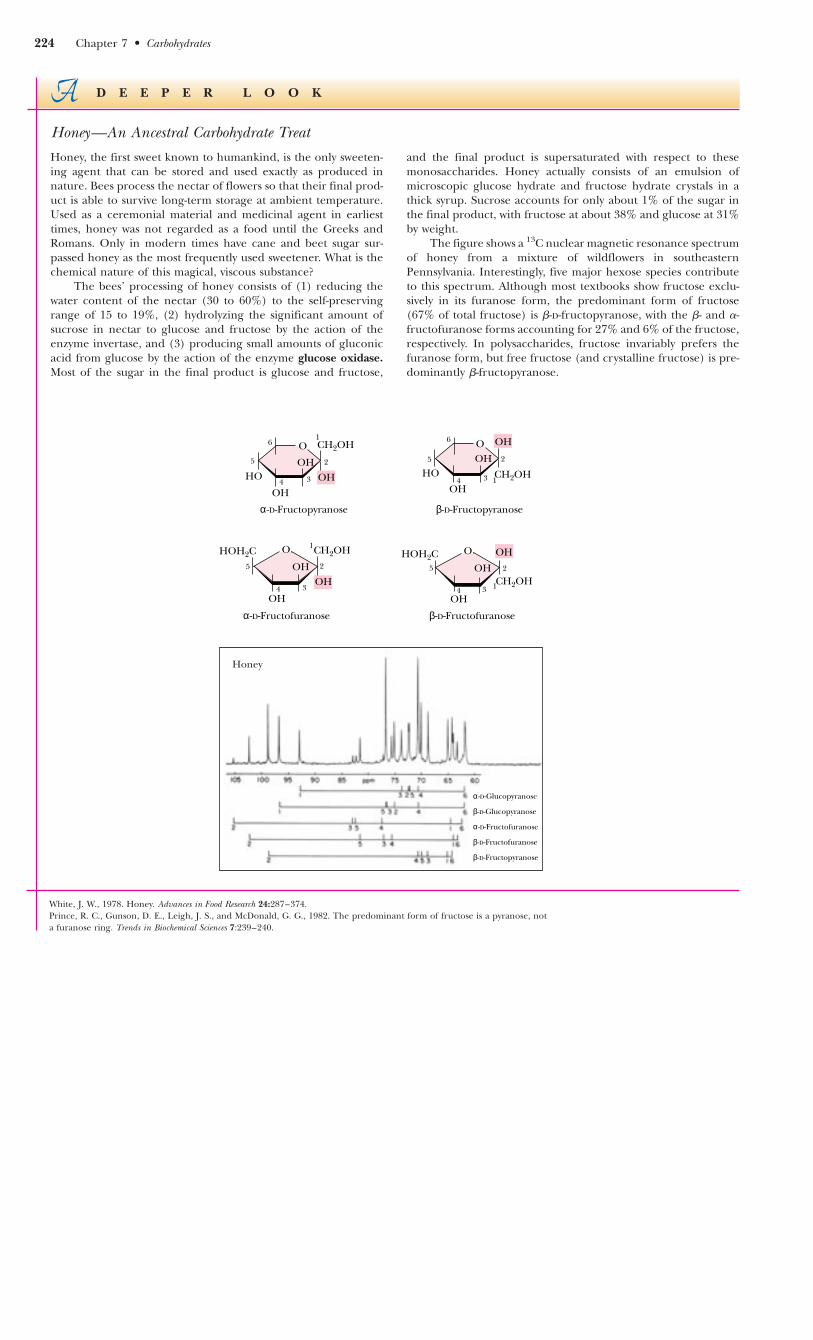
Honey—An Ancestral Carbohydrate Treat
Honey, the first sweet known to humankind, is the only sweeten-ing agent that can be stored and used exactly as produced innature. Bees process the nectar of flowers so that their final prod-uct is able to survive long-term storage at ambient temperature.Used as a ceremonial material and medicinal agent in earliesttimes, honey was not regarded as a food until the Greeks andRomans. Only in modern times have cane and beet sugar sur-passed honey as the most frequently used sweetener. What is thechemical nature of this magical, viscous substance?
The bees’ processing of honey consists of (1) reducing thewater content of the nectar (30 to 60%) to the self-preservingrange of 15 to 19%, (2) hydrolyzing the significant amount ofsucrose in nectar to glucose and fructose by the action of theenzyme invertase, and (3) producing small amounts of gluconicacid from glucose by the action of the enzyme glucose oxidase.Most of the sugar in the final product is glucose and fructose,
and the final product is supersaturated with respect to thesemonosaccharides. Honey actually consists of an emulsion ofmicroscopic glucose hydrate and fructose hydrate crystals in athick syrup. Sucrose accounts for only about 1% of the sugar inthe final product, with fructose at about 38% and glucose at 31%by weight.
The figure shows a 13C nuclear magnetic resonance spectrumof honey from a mixture of wildflowers in southeasternPennsylvania. Interestingly, five major hexose species contributeto this spectrum. Although most textbooks show fructose exclu-sively in its furanose form, the predominant form of fructose(67% of total fructose) is �-D-fructopyranose, with the �- and �-fructofuranose forms accounting for 27% and 6% of the fructose,respectively. In polysaccharides, fructose invariably prefers thefuranose form, but free fructose (and crystalline fructose) is pre-dominantly �-fructopyranose.
White, J. W., 1978. Honey. Advances in Food Research 24:287–374.Prince, R. C., Gunson, D. E., Leigh, J. S., and McDonald, G. G., 1982. The predominant form of fructose is a pyranose, nota furanose ring. Trends in Biochemical Sciences 7:239–240.
CH2OH
HOOH
OH
OH
O
CH2OHHOH2C
OH
CH2OHHOOH
OH
OH
O
α-D-Fructopyranose β-D-Fructopyranose
O
OH
OHCH2OH
HOH2C OHO
OH
OH
1
1
1
1
2 2
22
3 3
33
4 4
44
5 5
55
6 6
α-D-Fructofuranose β-D-Fructofuranose
Honey
α-D-Fructofuranose
β-D-Fructofuranose
β-D-Fructopyranose
β-D-Glucopyranose
α-D-Glucopyranose
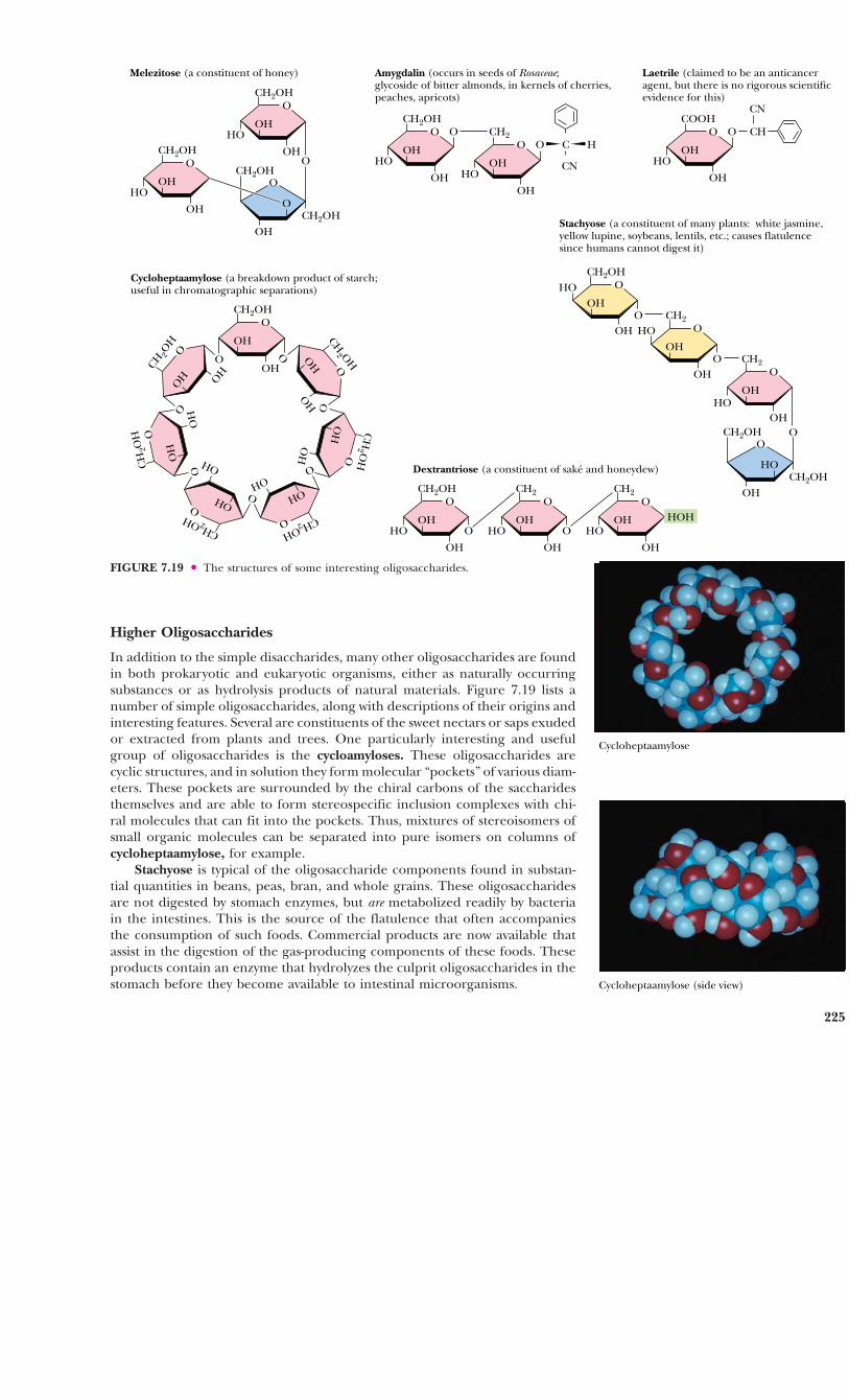
Higher Oligosaccharides
In addition to the simple disaccharides, many other oligosaccharides are foundin both prokaryotic and eukaryotic organisms, either as naturally occurringsubstances or as hydrolysis products of natural materials. Figure 7.19 lists anumber of simple oligosaccharides, along with descriptions of their origins andinteresting features. Several are constituents of the sweet nectars or saps exudedor extracted from plants and trees. One particularly interesting and usefulgroup of oligosaccharides is the cycloamyloses. These oligosaccharides arecyclic structures, and in solution they form molecular “pockets” of various diam-eters. These pockets are surrounded by the chiral carbons of the saccharidesthemselves and are able to form stereospecific inclusion complexes with chi-ral molecules that can fit into the pockets. Thus, mixtures of stereoisomers ofsmall organic molecules can be separated into pure isomers on columns ofcycloheptaamylose, for example.
Stachyose is typical of the oligosaccharide components found in substan-tial quantities in beans, peas, bran, and whole grains. These oligosaccharidesare not digested by stomach enzymes, but are metabolized readily by bacteriain the intestines. This is the source of the flatulence that often accompaniesthe consumption of such foods. Commercial products are now available thatassist in the digestion of the gas-producing components of these foods. Theseproducts contain an enzyme that hydrolyzes the culprit oligosaccharides in thestomach before they become available to intestinal microorganisms.
225
Melezitose (a constituent of honey)
CH2OH
HOOH
OH
O
O
O
OH
CH2OH
CH2OH
CH2OH
HOOH
OH
O
O
Stachyose (a constituent of many plants: white jasmine, yellow lupine, soybeans, lentils, etc.; causes flatulence since humans cannot digest it)
CH2OHHO
OH
OH
O
O CH2
HOOH
OH
O
O CH2
HOOH
OH
O
OO
HO
OH
CH2OH
CH2OH
Amygdalin (occurs in seeds of Rosaceae; glycoside of bitter almonds, in kernels of cherries,peaches, apricots)
CH2OH
HOOH
OH
O O CH2
HOOH
OH
O O C H
CN
Laetrile (claimed to be an anticanceragent, but there is no rigorous scientificevidence for this)
COOH
HOOH
OH
O O CH
CN
Dextrantriose (a constituent of saké and honeydew)
CH2OH
HOOH
OH
O
O
CH2
HOOH
OH
O
O
CH2
HOOH
OH
O
Cycloheptaamylose (a breakdown product of starch;useful in chromatographic separations)
CH2OH
OH
OH
O
O
CH 2O
HO
H OH
OO
CH
2OH
OH
OH
O
O
CH2OHOH
OH
OO
CH2OH
OH
OH
O
O
CH
2 OH
OH
OH O
OCH
2 OHOH
OH
O
O
HOH
FIGURE 7.19 ● The structures of some interesting oligosaccharides.
Cycloheptaamylose
Cycloheptaamylose (side view)

Another notable glycoside is amygdalin, which occurs in bitter almondsand in the kernels or pits of cherries, peaches, and apricots. Hydrolysis of thissubstance and subsequent oxidation yields laetrile, which has been claimed bysome to have anticancer properties. There is no scientific evidence for theseclaims, and the U.S. Food and Drug Administration has never approved laetrilefor use in the United States.
Oligosaccharides also occur widely as components (via glycosidic bonds)of antibiotics derived from various sources. Figure 7.20 shows the structures ofa few representative carbohydrate-containing antibiotics. Some of these antibi-otics also show antitumor activity. One of the most important of this type isbleomycin A2, which is used clinically against certain tumors.
226 Chapter 7 ● Carbohydrates
FIGURE 7.20 ● Some antibiotics are oligosaccharides or contain oligosaccharide groups.
O
OMe
CH3
NH2
H
O O
HN
NH2
NH2
OHN
NH
OHH
O
HO
S N
N S
CONH
S+
N N
H2NCH3
HN
HO
HN
N
HN
CH2OHO
OH OHO
O
H
CH2OHO
OOCNH2HO
HO
Bleomycin A2 (an antitumor agent used clinically against specific tumors) Aburamycin C (an antibiotic and antitumor agent)
OH3C
O OHO O
H3C
OMeHO
CH3
OH
OCH
OH
O
OH
H3C
O
OH3C
OH
O
OH3C
OH
O
OCH3
OOCCHHO
CH3
CH3
Streptomycin (a broad spectrum antibiotic)
H2NCNHNH
HOOH
NHCNH2
NH
HO
OO
OH
CHOH3C
OHO O
CH2OHCH3NH
OH
Sulfurmycin B (active against Gram-positive bacteria,mycobacteria, and tumors)
OCH3
OCH3O O
O
OCH3
O
HNCH3
O
COOCH3
CH2COCH3
OH
O
O
CH3
CH3
OH
OH
OH

7.4 ● Polysaccharides
Structure and Nomenclature
By far the majority of carbohydrate material in nature occurs in the form ofpolysaccharides. By our definition, polysaccharides include not only those sub-stances composed only of glycosidically linked sugar residues but also mole-cules that contain polymeric saccharide structures linked via covalent bonds toamino acids, peptides, proteins, lipids, and other structures.
Polysaccharides, also called glycans, consist of monosaccharides and theirderivatives. If a polysaccharide contains only one kind of monosaccharide mole-cule, it is a homopolysaccharide, or homoglycan, whereas those containingmore than one kind of monosaccharide are heteropolysaccharides. The mostcommon constituent of polysaccharides is D-glucose, but D-fructose, D-galactose,L-galactose, D-mannose, L-arabinose, and D-xylose are also common. Commonmonosaccharide derivatives in polysaccharides include the amino sugars (D-glucosamine and D-galactosamine), their derivatives (N-acetylneuraminic acidand N-acetylmuramic acid), and simple sugar acids (glucuronic and iduronicacids). Homopolysaccharides are often named for the sugar unit they contain,so that glucose homopolysaccharides are called glucans, while mannosehomopolysaccharides are mannans. Other homopolysaccharide names are justas obvious: galacturonans, arabinans, and so on. Homopolysaccharides of uni-form linkage type are often named by including notation to denote ring sizeand linkage type. Thus, cellulose is a (1n4)-�-D-glucopyranan. Polysaccharidesdiffer not only in the nature of their component monosaccharides but also inthe length of their chains and in the amount of chain branching that occurs.Although a given sugar residue has only one anomeric carbon and thus canform only one glycosidic linkage with hydroxyl groups on other molecules,each sugar residue carries several hydroxyls, one or more of which may be anacceptor of glycosyl substituents (Figure 7.21). This ability to form branchedstructures distinguishes polysaccharides from proteins and nucleic acids, whichoccur only as linear polymers.
7.4 ● Polysaccharides 227
FIGURE 7.21 ● Amylose and amylopectin are the two forms of starch. Note that the lin-ear linkages are �(1n4), but the branches in amylopectin are �(1n6). Branches in poly-saccharides can involve any of the hydroxyl groups on the monosaccharide components.Amylopectin is a highly branched structure, with branches occurring every 12 to 30residues.
CH2OHO
O
CH2OHO
O
CH2OHO
O
CH2OHO
O
CH2OHO
O
CH2OHO
O
CH2OHO
O
CH2OHO
Amylose
O
CH2OHO
O
CH2OHO
O
CH2O
O
CH2OHO
O
CH2OHO
O
Amylopectin
. . .
. . .

Polysaccharide Functions
The functions of many individual polysaccharides cannot be assigned uniquely,and some of their functions may not yet be appreciated. Traditionally, bio-chemistry textbooks have listed the functions of polysaccharides as storagematerials, structural components, or protective substances. Thus, starch, glyco-gen, and other storage polysaccharides, as readily metabolizable food, provideenergy reserves for cells. Chitin and cellulose provide strong support for the skele-tons of arthropods and green plants, respectively. Mucopolysaccharides, suchas the hyaluronic acids, form protective coats on animal cells. In each of thesecases, the relevant polysaccharide is either a homopolymer or a polymer ofsmall repeating units. Recent research indicates, however, that oligosaccharidesand polysaccharides with varied structures may also be involved in much moresophisticated tasks in cells, including a variety of cellular recognition and inter-cellular communication events, as discussed later.
Storage Polysaccharides
Storage polysaccharides are an important carbohydrate form in plants and ani-mals. It seems likely that organisms store carbohydrates in the form of poly-saccharides rather than as monosaccharides to lower the osmotic pressure ofthe sugar reserves. Because osmotic pressures depend only on numbers of mole-cules, the osmotic pressure is greatly reduced by formation of a few polysac-charide molecules out of thousands (or even millions) of monosaccharideunits.
Starch
By far the most common storage polysaccharide in plants is starch, which existsin two forms: � -amylose and amylopectin, the structures of which are shownin Figure 7.21. Most forms of starch in nature are 10 to 30% �-amylose and 70to 90% amylopectin. Typical cornstarch produced in the United States is about25% �-amylose and 75% amylopectin. �-Amylose is composed of linear chainsof D-glucose in �(1n4) linkages. The chains are of varying length, having mo-lecular weights from several thousand to half a million. As can be seen fromthe structure in Figure 7.21, the chain has a reducing end and a nonreducingend. Although poorly soluble in water, �-amylose forms micelles in which thepolysaccharide chain adopts a helical conformation (Figure 7.22). Iodine reactswith �-amylose to give a characteristic blue color, which arises from the inser-tion of iodine into the middle of the hydrophobic amylose helix.
In contrast to �-amylose, amylopectin, the other component of typicalstarches, is a highly branched chain of glucose units (Figure 7.21). Branchesoccur in these chains every 12 to 30 residues. The average branch length isbetween 24 and 30 residues, and molecular weights of amylopectin moleculescan range up to 100 million. The linear linkages in amylopectin are �(1n4),whereas the branch linkages are �(1n6). As is the case for �-amylose, amy-lopectin forms micellar suspensions in water; iodine reacts with such suspen-sions to produce a red-violet color.
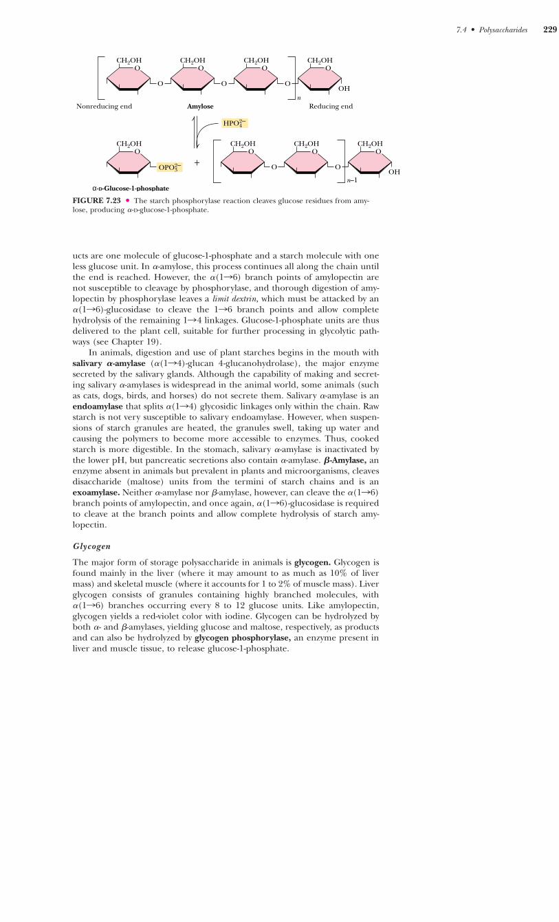
Starch is stored in plant cells in the form of granules in the stroma of plas-tids (plant cell organelles) of two types: chloroplasts, in which photosynthesistakes place, and amyloplasts, plastids that are specialized starch accumulationbodies. When starch is to be mobilized and used by the plant that stored it, itmust be broken down into its component monosaccharides. Starch is split intoits monosaccharide elements by stepwise phosphorolytic cleavage of glucoseunits, a reaction catalyzed by starch phosphorylase (Figure 7.23). This is for-mally an �(1n4)-glucan phosphorylase reaction, and at each step, the prod-
228 Chapter 7 ● Carbohydrates
FIGURE 7.22 ● Suspensions of amylose inwater adopt a helical conformation. Iodine (I2)can insert into the middle of the amylose helixto give a blue color that is characteristic anddiagnostic for starch.
I
I
I
I
I
I
7.4 ● Polysaccharides 229
ucts are one molecule of glucose-1-phosphate and a starch molecule with oneless glucose unit. In �-amylose, this process continues all along the chain untilthe end is reached. However, the �(1n6) branch points of amylopectin arenot susceptible to cleavage by phosphorylase, and thorough digestion of amy-lopectin by phosphorylase leaves a limit dextrin, which must be attacked by an�(1n6)-glucosidase to cleave the 1n6 branch points and allow completehydrolysis of the remaining 1n4 linkages. Glucose-1-phosphate units are thusdelivered to the plant cell, suitable for further processing in glycolytic path-ways (see Chapter 19).
In animals, digestion and use of plant starches begins in the mouth withsalivary �-amylase (�(1n4)-glucan 4-glucanohydrolase), the major enzymesecreted by the salivary glands. Although the capability of making and secret-ing salivary �-amylases is widespread in the animal world, some animals (suchas cats, dogs, birds, and horses) do not secrete them. Salivary �-amylase is anendoamylase that splits �(1n4) glycosidic linkages only within the chain. Rawstarch is not very susceptible to salivary endoamylase. However, when suspen-sions of starch granules are heated, the granules swell, taking up water andcausing the polymers to become more accessible to enzymes. Thus, cookedstarch is more digestible. In the stomach, salivary �-amylase is inactivated by the lower pH, but pancreatic secretions also contain �-amylase. �-Amylase, anenzyme absent in animals but prevalent in plants and microorganisms, cleavesdisaccharide (maltose) units from the termini of starch chains and is anexoamylase. Neither �-amylase nor �-amylase, however, can cleave the �(1n6)branch points of amylopectin, and once again, �(1n6)-glucosidase is requiredto cleave at the branch points and allow complete hydrolysis of starch amy-lopectin.
Glycogen
The major form of storage polysaccharide in animals is glycogen. Glycogen isfound mainly in the liver (where it may amount to as much as 10% of livermass) and skeletal muscle (where it accounts for 1 to 2% of muscle mass). Liverglycogen consists of granules containing highly branched molecules, with�(1n6) branches occurring every 8 to 12 glucose units. Like amylopectin,glycogen yields a red-violet color with iodine. Glycogen can be hydrolyzed byboth �- and �-amylases, yielding glucose and maltose, respectively, as productsand can also be hydrolyzed by glycogen phosphorylase, an enzyme present inliver and muscle tissue, to release glucose-1-phosphate.
CH2OHO
O
CH2OHO
O
CH2OHO
O
CH2OHO
HPO42–
Nonreducing end
CH2OHO
CH2OHO
O
CH2OHO
O
CH2OHO
+OPO3
2–
n
n–1
Reducing endAmylose
α-D-Glucose-1-phosphate
OH
OH
FIGURE 7.23 ● The starch phosphorylase reaction cleaves glucose residues from amy-lose, producing �-D-glucose-1-phosphate.
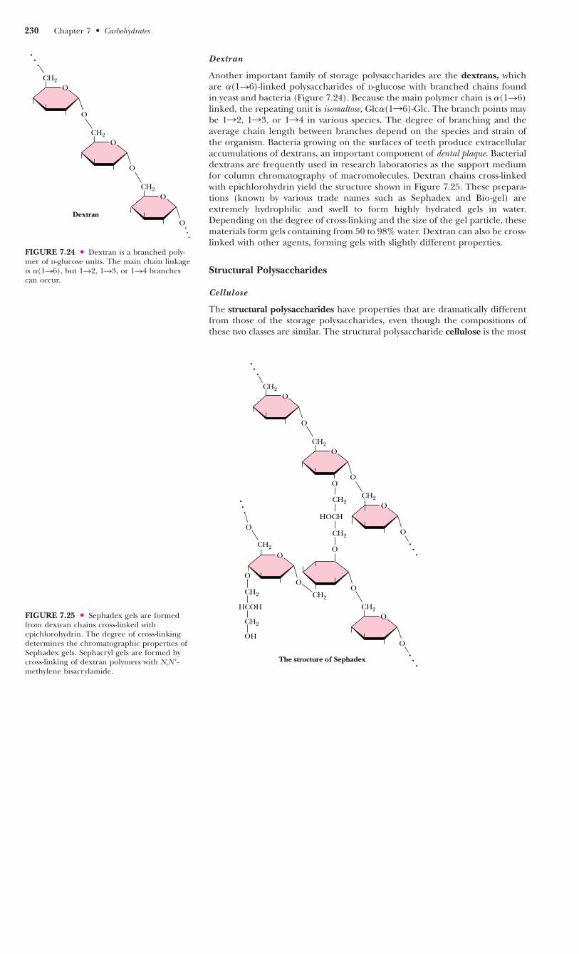
Dextran
Another important family of storage polysaccharides are the dextrans, whichare �(1n6)-linked polysaccharides of D-glucose with branched chains foundin yeast and bacteria (Figure 7.24). Because the main polymer chain is �(1n6)linked, the repeating unit is isomaltose, Glc�(1n6)-Glc. The branch points maybe 1n2, 1n3, or 1n4 in various species. The degree of branching and theaverage chain length between branches depend on the species and strain ofthe organism. Bacteria growing on the surfaces of teeth produce extracellularaccumulations of dextrans, an important component of dental plaque. Bacterialdextrans are frequently used in research laboratories as the support mediumfor column chromatography of macromolecules. Dextran chains cross-linkedwith epichlorohydrin yield the structure shown in Figure 7.25. These prepara-tions (known by various trade names such as Sephadex and Bio-gel) areextremely hydrophilic and swell to form highly hydrated gels in water.Depending on the degree of cross-linking and the size of the gel particle, thesematerials form gels containing from 50 to 98% water. Dextran can also be cross-linked with other agents, forming gels with slightly different properties.
Structural Polysaccharides
Cellulose
The structural polysaccharides have properties that are dramatically differentfrom those of the storage polysaccharides, even though the compositions ofthese two classes are similar. The structural polysaccharide cellulose is the most
FIGURE 7.24 ● Dextran is a branched poly-mer of D-glucose units. The main chain linkageis �(1n6), but 1n2, 1n3, or 1n4 branchescan occur.
230 Chapter 7 ● Carbohydrates
FIGURE 7.25 ● Sephadex gels are formedfrom dextran chains cross-linked withepichlorohydrin. The degree of cross-linkingdetermines the chromatographic properties ofSephadex gels. Sephacryl gels are formed bycross-linking of dextran polymers with N,N�-methylene bisacrylamide.
Dextran
. . .
CH2
O
O
CH2
O
O
CH2
O
O
. . .
The structure of Sephadex
CH2
HOCH
CH2
O
CH2
HCOH
CH2
OH
CH2
O
O
. . .
CH2
O
. . .O
. . .
O
O
CH2
O
O
CH2
. . .
O
CH2
O
O
O
O
O
CH2

abundant natural polymer found in the world. Found in the cell walls of nearlyall plants, cellulose is one of the principal components providing physical struc-ture and strength. The wood and bark of trees are insoluble, highly organizedstructures formed from cellulose and also from lignin (see Figure 27.35). It isawe-inspiring to look at a large tree and realize the amount of weight supportedby polymeric structures derived from sugars and organic alcohols. Cellulosealso has its delicate side, however. Cotton, whose woven fibers make some ofour most comfortable clothing fabrics, is almost pure cellulose. Derivatives ofcellulose have found wide use in our society. Cellulose acetates are producedby the action of acetic anhydride on cellulose in the presence of sulfuric acidand can be spun into a variety of fabrics with particular properties. Referredto simply as acetates, they have a silky appearance, a luxuriously soft feel, anda deep luster and are used in dresses, lingerie, linings, and blouses.
Cellulose is a linear homopolymer of D-glucose units, just as in �-amylose.The structural difference, which completely alters the properties of the poly-mer, is that in cellulose the glucose units are linked by �(1n4)-glycosidic bonds,whereas in �-amylose the linkage is �(1n4). The conformational differencebetween these two structures is shown in Figure 7.26. The �(1n4)-linkage sitesof amylose are naturally bent, conferring a gradual turn to the polymer chain,which results in the helical conformation already described (see Figure 7.22).The most stable conformation about the �(1n4) linkage involves alternating180° flips of the glucose units along the chain so that the chain adopts a fullyextended conformation, referred to as an extended ribbon. Juxtaposition ofseveral such chains permits efficient interchain hydrogen bonding, the basisof much of the strength of cellulose.
The structure of one form of cellulose, determined by X-ray and electrondiffraction data, is shown in Figure 7.27. The flattened sheets of the chainslie side by side and are joined by hydrogen bonds. These sheets are laid ontop of one another in a way that staggers the chains, just as bricks are stag-gered to give strength and stability to a wall. Cellulose is extremely resistantto hydrolysis, whether by acid or by the digestive tract amylases described ear-lier. As a result, most animals (including humans) cannot digest cellulose toany significant degree. Ruminant animals, such as cattle, deer, giraffes, andcamels, are an exception because bacteria that live in the rumen (Figure 7.28)secrete the enzyme cellulase, a �-glucosidase effective in the hydrolysis of cel-lulose. The resulting glucose is then metabolized in a fermentation processto the benefit of the host animal. Termites and shipworms (Teredo navalis) sim-ilarly digest cellulose because their digestive tracts also contain bacteria thatsecrete cellulase.
7.4 ● Polysaccharides 231
OOH
OHHO
O OO
OH
HOOH
OOH
OHHO
O O
β-1,4-Linked D-glucose units
(b)
O
OHOH
HOO
OO
OH
OOH
HO
α-1,4-Linked D-glucose units
(a)
FIGURE 7.26 ● (a) Amylose, composed exclusively of the relatively bent �(1n4) link-ages, prefers to adopt a helical conformation, whereas (b) cellulose, with �(1n4)-glycosidiclinkages, can adopt a fully extended conformation with alternating 180° flips of the glucoseunits. The hydrogen bonding inherent in such extended structures is responsible for thegreat strength of tree trunks and other cellulose-based materials.

Chitin
A polysaccharide that is similar to cellulose, both in its biological function andits primary, secondary, and tertiary structure, is chitin. Chitin is present in thecell walls of fungi and is the fundamental material in the exoskeletons of crus-taceans, insects, and spiders. The structure of chitin, an extended ribbon, isidentical to cellulose, except that the OOH group on each C-2 is replaced byONHCOCH3, so that the repeating units are N-acetyl-D-glucosamines in �(1n4)linkage. Like cellulose (Figure 7.27), the chains of chitin form extended rib-bons (Figure 7.29) and pack side by side in a crystalline, strongly hydrogen-bonded form. One significant difference between cellulose and chitin iswhether the chains are arranged in parallel (all the reducing ends together atone end of a packed bundle and all the nonreducing ends together at theother end) or antiparallel (each sheet of chains having the chains arrangedoppositely from the sheets above and below). Natural cellulose seems to occuronly in parallel arrangements. Chitin, however, can occur in three forms, some-times all in the same organism. �-Chitin is an all-parallel arrangement of thechains, whereas �-chitin is an antiparallel arrangement. In -chitin, the struc-ture is thought to involve pairs of parallel sheets separated by single antipar-allel sheets.
Chitin is the earth’s second most abundant carbohydrate polymer (aftercellulose), and its ready availability and abundance offer opportunities forindustrial and commercial applications. Chitin-based coatings can extend theshelf life of fruits, and a chitin derivative that binds to iron atoms in meat hasbeen found to slow the reactions that cause rancidity and flavor loss. Withoutsuch a coating, the iron in meats activates oxygen from the air, forming reac-tive free radicals that attack and oxidize polyunsaturated lipids, causing mostof the flavor loss associated with rancidity. Chitin-based coatings coordinate theiron atoms, preventing their interaction with oxygen.
FIGURE 7.27 ● The structure of cellulose,showing the hydrogen bonds (blue) betweenthe sheets, which strengthen the structure.Intrachain hydrogen bonds are in red andinterchain hydrogen bonds are in green.
232 Chapter 7 ● Carbohydrates
FIGURE 7.28 ● Giraffes, cattle, deer, andcamels are ruminant animals that are able tometabolize cellulose, thanks to bacterial cellu-lase in the rumen, a large first compartment inthe stomach of a ruminant.
Intrachainhydrogen bond
Intersheethydrogen bond
Interchainhydrogen bond
Esophagus
Omasum
Small intestine
Rumen
Abomasum
Reticulum
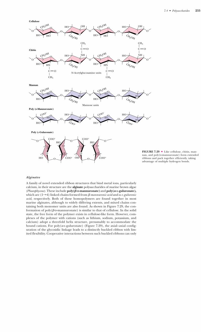
Alginates
A family of novel extended ribbon structures that bind metal ions, particularlycalcium, in their structure are the alginate polysaccharides of marine brown algae(Phaeophyceae). These include poly(�-D-mannuronate) and poly(�-L-guluronate),which are (1n4) linked chains formed from �-mannuronic acid and �-L-guluronicacid, respectively. Both of these homopolymers are found together in mostmarine alginates, although to widely differing extents, and mixed chains con-taining both monomer units are also found. As shown in Figure 7.29, the con-formation of poly(�-D-mannuronate) is similar to that of cellulose. In the solidstate, the free form of the polymer exists in cellulose-like form. However, com-plexes of the polymer with cations (such as lithium, sodium, potassium, andcalcium) adopt a threefold helix structure, presumably to accommodate thebound cations. For poly(�-L-guluronate) (Figure 7.29), the axial–axial config-uration of the glycosidic linkage leads to a distinctly buckled ribbon with lim-ited flexibility. Cooperative interactions between such buckled ribbons can only
FIGURE 7.29 ● Like cellulose, chitin, man-nan, and poly(D-mannuronate) form extendedribbons and pack together efficiently, takingadvantage of multiple hydrogen bonds.
7.4 ● Polysaccharides 233
Cellulose
C O
CH3
O
HOHO
CH2OH
O OOCH
2OH
OHHO O
HOHO
CH2OH
O O
OHHO
Chitin
O
HNHO
CH2OH
O O
NHHO O
HO
CH2OH
O O
HO
C O
CH3
HN
Mannan
OHO
HO
CH2OH
O O
HO O
HO
CH2OH
O O
HOHO
Poly (D-Mannuronate)
OHO
HO
COO–
O O
HO O
HO
COO–
O OCOO –
HOHO
Poly (L-Guluronate)
O
O
OH
HO O
O
HO
COO–
HOCOO–
O
OH
OH O
O
HO
COO–
HOCOO–
O
N–Acetylglucosamine units
Mannose units
O
O
O
O
C O
CH3
NH
C O
CH3
OCH2OH
OCH2OH
OCH2OH
OCH2OH
HOOCH
2OH
HO
OHO
COO –O
HO
O

be strong if the interstices are filled effectively with water molecules or metalions. Figure 7.30 shows a molecular model of a Ca2�-induced dimer of poly(�-L-guluronate).
Agaro s e
An important polysaccharide mixture isolated from marine red algae(Rhodophyceae) is agar, which consists of two components, agarose andagaropectin. Agarose (Figure 7.31) is a chain of alternating D-galactose and 3,6-anhydro-L-galactose, with side chains of 6-methyl-D-galactose. Agaropectin issimilar, but contains in addition sulfate ester side chains and D-glucuronic acid.The three-dimensional structure of agarose is a double helix with a threefoldscrew axis, as shown in Figure 7.31. The central cavity is large enough to accom-modate water molecules. Agarose and agaropectin readily form gels contain-ing large amounts (up to 99.5%) of water. Agarose can be processed to removemost of the charged groups, yielding a material (trade name Sepharose) use-ful for purification of macromolecules in gel exclusion chromatography. Pairsof chains form double helices that subsequently aggregate in bundles to forma stable gel, as shown in Figure 7.32.
Glycosaminoglycans
A class of polysaccharides known as glycosaminoglycans is involved in a varietyof extracellular (and sometimes intracellular) functions. Glycosaminoglycansconsist of linear chains of repeating disaccharides in which one of the mono-saccharide units is an amino sugar and one (or both) of the monosaccharideunits contains at least one negatively charged sulfate or carboxylate group. Therepeating disaccharide structures found commonly in glycosaminoglycans are
234 Chapter 7 ● Carbohydrates
A D E E P E R L O O K
Billiard Balls, Exploding Teeth, and Dynamite—The Colorful History of Cellulose
Although humans cannot digest it and most people’s acquain-tance with cellulose is limited to comfortable cotton clothing, cel-lulose has enjoyed a colorful and varied history of utilization. In1838, Théophile Pelouze in France found that paper or cottoncould be made explosive if dipped in concentrated nitric acid.Christian Schönbein, a professor of chemistry at the University ofBasel, prepared “nitrocotton” in 1845 by dipping cotton in a mix-ture of nitric and sulfuric acids and then washing the material toremove excess acid. In 1860, Major E. Schultze of the Prussianarmy used the same material, now called guncotton, as a propel-lant replacement for gunpowder, and its preparation in brass car-tridges soon made it popular for this purpose. The only problemwas that it was too explosive and could detonate unpredictably infactories where it was produced. The entire town of Faversham,England, was destroyed in such an accident. In 1868, Alfred Nobelmixed guncotton with ether and alcohol, thus preparing nitro-cellulose, and in turn mixed this with nitroglycerine and sawdustto produce dynamite. Nobel’s income from dynamite and also
from his profitable development of the Russian oil fields in Bakueventually formed the endowment for the Nobel Prizes.
In 1869, concerned over the precipitous decline (from hunt-ing) of the elephant population in Africa, the billiard ball man-ufacturers Phelan and Collander offered a prize of $10,000 forproduction of a substitute for ivory. Brothers Isaiah and JohnHyatt in Albany, New York, produced a substitute for ivory by mix-ing guncotton with camphor, then heating and squeezing it toproduce celluloid. This product found immediate uses wellbeyond billiard balls. It was easy to shape, strong, and resilient,and it exhibited a high tensile strength. Celluloid was used even-tually to make dolls, combs, musical instruments, fountain pens,piano keys, and a variety of other products. The Hyatt brotherseventually formed the Albany Dental Company to make false teethfrom celluloid. Because camphor was used in their production,the company advertised that their teeth smelled “clean,” but, asreported in the New York Times in 1875, the teeth also occasion-ally exploded!
Portions adapted from Burke, J., 1996. The Pinball Effect: How Renaissance Water Gardens Made the CarburetorPossible and Other Journeys Through Knowledge. New York: Little, Brown, & Company.
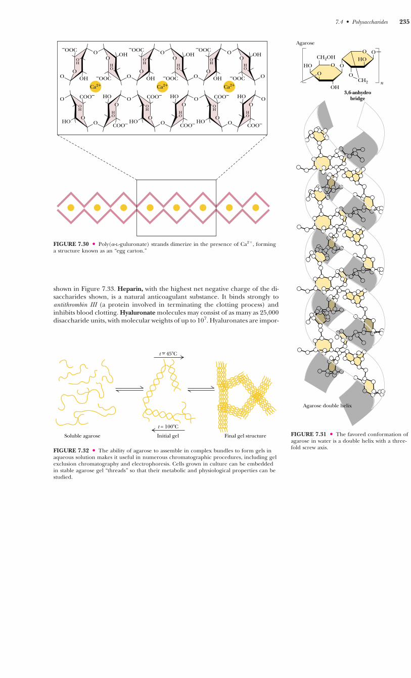
shown in Figure 7.33. Heparin, with the highest net negative charge of the di-saccharides shown, is a natural anticoagulant substance. It binds strongly toantithrombin III (a protein involved in terminating the clotting process) andinhibits blood clotting. Hyaluronate molecules may consist of as many as 25,000disaccharide units, with molecular weights of up to 107. Hyaluronates are impor-
FIGURE 7.31 ● The favored conformation ofagarose in water is a double helix with a three-fold screw axis.
7.4 ● Polysaccharides 235
FIGURE 7.30 ● Poly(�-L-guluronate) strands dimerize in the presence of Ca2�, forminga structure known as an “egg carton.”
FIGURE 7.32 ● The ability of agarose to assemble in complex bundles to form gels inaqueous solution makes it useful in numerous chromatographic procedures, including gelexclusion chromatography and electrophoresis. Cells grown in culture can be embeddedin stable agarose gel “threads” so that their metabolic and physiological properties can bestudied.
O
OO
OH
–OOCOH
–OOC O
O
OOH
–OOCOH
–OOC O
O
OOH
–OOCOH
–OOC O
O
O
HO
O
OCOO–
HOCOO–
O
HO
O
OCOO–
HOCOO–
O
HO
O
OCOO–
HOCOO–
Ca2+ Ca2+ Ca2+
O O O
O O O
OH
OH
OH
OH
OH
OH
HO
HO
HO
HO
HO
HO
Soluble agarose Initial gel Final gel structure
t = 100�C
t ~ 45�C
Agarose double helix
O
OH
HO OO
CH2OH
O
OOHO
CH2
3,6-anhydrobridge
n
Agarose

tant components of the vitreous humor in the eye and of synovial fluid, thelubricant fluid of joints in the body. The chondroitins and keratan sulfate arefound in tendons, cartilage, and other connective tissue, whereas dermatan sul-fate, as its name implies, is a component of the extracellular matrix of skin.Glycosaminoglycans are fundamental constituents of proteoglycans (discussedlater).
FIGURE 7.33 ● Glycosaminoglycans areformed from repeating disaccharide arrays.Glycosaminoglycans are components of theproteoglycans.
236 Chapter 7 ● Carbohydrates
H OH
O
OH HHH
H
COO–
14
βO
H NHCCH3
O
HH
–O3SO
H
CH2OH
14
H 3
D-GlucuronateN-Acetyl-
D-galactosamine-4-sulfate
Chondroitin-4-sulfate
H OSO3–
O
OH HHH H
COO–
14α
H NHSO3–
O
HH H
CH2OSO3–
14H
2 Oα
D-Glucuronate-2-sulfate
N-Sulfo-D-glucosamine-6-sulfate
Heparin
H OH
O
OH HHH
H
COO–
14
H NHCCH3
O
HHHO
H
CH2OSO3–
14
H
D-GlucuronateN-Acetyl-
D-galactosamine-6-sulfate
Chondroitin-6-sulfate
H OH
O
OH HHH
H
COO–
14
H NHCCH3
O
HHH
H
CH2OH
1
HO 3
D-GlucuronateN-Acetyl-
D-glucosamine
Hyaluronate
H OH
O
OH HCOO–H
H
H
14
H NHCCH3
O
HH
–O3SO
H
CH2OH
14
H 3
L-IduronateN-Acetyl-D-
galactosamine-4-sulfate
Dermatan sulfate
H OH
O
HHHO
H
CH2OHβ H NHCCH3
O
HHH
H
CH2OSO3–
14O
β
Oβ
β
O
O
β
Oβ
D-GalactoseN-Acetyl-
D-glucosamine-6-sulfate
Keratan sulfate
2OH
OH
H 3
6
O
O O
O O
βO
βO
βO O
O
PROBLEMS
1. Draw Haworth structures for the two possible isomers of D-altrose (Figure 7.2) and D-psicose (Figure 7.3).2. Give the systematic name for stachyose (Figure 7.19).3. Trehalose, a disaccharide produced in fungi, has the followingstructure:
a. What is the systematic name for this disaccharide?b. Is trehalose a reducing sugar? Explain.
OHOH
OH
OH
OH
H
H H
H
H
H
H
H
HO
HO
CH2OH
HOCH2
OO
4. Draw a Fischer projection structure for L-sorbose (D-sorbose isshown in Figure 7.3).5. �-D-Glucose has a specific rotation, [�]D
20, of �112.2°, whereas�-D-glucose has a specific rotation of �18.7°. What is the compo-sition of a mixture of �-D- and �-D-glucose, which has a specificrotation of 83.0°?6. A 0.2-g sample of amylopectin was analyzed to determine thefraction of the total glucose residues that are branch points in thestructure. The sample was exhaustively methylated and thendigested, yielding 50 mol of 2,3-dimethylglucose and 0.4 mol of1, 2, 3, 6-tetramethylglucose.a. What fraction of the total residues are branch points?b. How many reducing ends does this amylopectin have?

FURTHER READING
Aspinall, G. O., 1982. The Polysaccharides, Vols. 1 and 2. New York: AcademicPress.
Collins, P. M., 1987. Carbohydrates. London: Chapman and Hall.
Davison, E. A., 1967. Carbohydrate Chemistry. New York: Holt, Rinehart andWinston.
Feeney, R. E., Burcham, T. S., and Yeh, Y., 1986. Antifreeze glycoproteinsfrom polar fish blood. Annual Review of Biophysical Chemistry 15:59–78.
Jentoft, N., 1990. Why are proteins O-glycosylated? Trends in BiochemicalSciences 155:291–294.
Kjellen, L., and Lindahl, U., 1991. Proteoglycans: Structures and interac-tions. Annual Review of Biochemistry 60:443–475.
Lennarz, W. J., 1980. The Biochemistry of Glycoproteins and Proteoglycans. NewYork: Plenum Press.
Lodish, H. F., 1991. Recognition of complex oligosaccharides by the mul-tisubunit asialoglycoprotein receptor. Trends in Biochemical Sciences 16:374–377.
McNeil, M., Darvill, A. G., Fry, S. C., and Albersheim, P., 1984. Structureand function of the primary cell walls of plants. Annual Review of Biochemistry53:625–664.
Pigman, W., and Horton, D., 1972. The Carbohydrates. New York: AcademicPress.
Rademacher, T. W., Parekh, R. B., and Dwek, R. A., 1988. Glycobiology.Annual Review of Biochemistry 57:785–838.
Ruoslahti, E., 1989. Proteoglycans in cell regulation. Journal of BiologicalChemistry 264:13369–13372.
Sharon, N., 1980. Carbohydrates. Scientific American 243:90–102.
Sharon, N., 1984. Glycoproteins. Trends in Biochemical Sciences 9:198–202.
Further Reading 237