Influence of acute sleep loss on the neural correlates of alerting, orientating and executive...
11
Influence of acute sleep loss on the neural correlates of alerting, orientating and executive attention components VINCENZO MUTO*, ANAHITA SHAFFII-LE BOURDIEC*, LUCA MATARAZZO, ARIANE FORET, LAURA MASCETTI, MATHIEU JASPAR, GILLES VANDEWALLE, CHRISTOPHE PHILLIPS, CHRISTIAN DEGUELDRE, EVELYNE BALTEAU, ANDRE ´ LUXEN, FABIENNE COLLETTE and PIERRE MAQUET Cyclotron Research Centre, University of Lie ` ge, Belgium Keywords attention, functional magnetic resonance imaging, sleep loss Correspondence Pierre Maquet, Centre de Recherches du Cyclotron, Universite ´ de Lie ` ge, B30, Sart Tilman, B-4000 Lie ` ge, Belgium. Tel.: 32 43 66 36 87; fax: 32 43 66 29 46; e-mail: [email protected] *These authors equally contributed to this work. Accepted in revised form 16 March 2012; received 18 October 2011 DOI: 10.1111/j.1365-2869.2012.01020.x SUMMARY The Attention Network Test (ANT) is deemed to assess the alerting, orientating and executive components of human attention. Capitalizing on the opportunity to investigate three facets of attention in a single task, we used functional magnetic resonance imaging (fMRI) to assess the effect of sleep deprivation (SD) on brain responses associated with the three attentional components elicited by the ANT. Twelve healthy volunteers were scanned in two conditions 1 week apart, after a normal night of sleep (rested wakefulness, RW) or after one night of total sleep deprivation. Sleep deprivation was associated with a global increase in reaction times, which did not affect specifically any of the three attention effects. Brain responses associated with the alerting effect did not differ between RW and SD. Higher-order attention components (orientating and conflict effects) were associated with significantly larger thalamic responses during SD than during RW. These results suggest that SD influences different components of human attention non- selectively, through mechanisms that might either affect centrencephal- ic structures maintaining vigilance or ubiquitously perturb neuronal function. Compensatory responses can counter these effects transiently by recruiting thalamic responses, thereby supporting thalamocortical function. INTRODUCTION A single night of sleep deprivation is detrimental to a number of cognitive abilities, ranging from phasic alertness (Doran et al., 2001) to executive functions (Harrison et al., 2000). Deteriorating attention is usually believed to participate in this decline in cognitive performance. However, attention is a heterogeneous concept (Oken et al., 2006) and sleep depri- vation has been shown to affect various aspects of attention, such as phasic alertness (Drummond et al., 2005), selective (Horowitz et al., 2003) and divided attention (Drummond et al., 2001). A persistent issue is therefore whether sleep deprivation affects various components of attention selec- tively and differentially or whether the decrease in alertness is the core phenomenon that can explain the failure of the other attention systems (Lim and Dinges, 2010). Operational definitions of vigilance, phasic alertness and attention are detailed in Data S1. A cognitive model assumes that human attention is supported by three main functions which are associated specifically with independent brain circuits and neuromodu- lators (Posner and Petersen, 1990). Following this model, the alerting component is defined as the ability to prepare and sustain alertness to process high-priority signals (Posner and Petersen, 1990). It would involve thalamic, frontal and parietal areas (Fan et al., 2005). The orientating component would allow one to attend to target items overtly or covertly, thereby improving their processing efficiency (Posner and Petersen, 1990). Orientating would involve the superior parietal lobe, temporo–parietal junctions and superior frontal cortex (Fan et al., 2005). A third, executive attention compo- nent would be involved in conflict resolution and would recruit J. Sleep Res. (2012) 21, 648–658 fMRI and components of attention 648 ª 2012 European Sleep Research Society
Transcript of Influence of acute sleep loss on the neural correlates of alerting, orientating and executive...

Influence of acute sleep loss on the neural correlates of alerting,orientating and executive attention components
V I NCENZO MUTO * , ANAH I T A SHAFF I I - L E BOURD I EC * , L UCA MATARAZZO ,AR I ANE FORET , L AURA MASCETT I , MATH I EU JASPAR ,G I L L ES VANDEWAL LE , CHR I S TOPHE PH I L L I P S , CHR I S T I AN DEGUELDRE ,EVE LYNE BALTEAU , ANDR E LUXEN , F AB I ENNE COL LETTE andP I ERRE MAQUETCyclotron Research Centre, University of Liege, Belgium
Keywordsattention, functional magnetic resonanceimaging, sleep loss
CorrespondencePierre Maquet, Centre de Recherches duCyclotron, Universite de Liege, B30, SartTilman, B-4000 Liege, Belgium.Tel.: 32 43 66 36 87;fax: 32 43 66 29 46;e-mail: [email protected]*These authors equally contributed to this work.
Accepted in revised form 16 March 2012;received 18 October 2011
DOI: 10.1111/j.1365-2869.2012.01020.x
SUMMARYThe Attention Network Test (ANT) is deemed to assess the alerting,orientating and executive components of human attention. Capitalizingon the opportunity to investigate three facets of attention in a singletask, we used functional magnetic resonance imaging (fMRI) to assessthe effect of sleep deprivation (SD) on brain responses associated withthe three attentional components elicited by the ANT. Twelve healthyvolunteers were scanned in two conditions 1 week apart, after a normalnight of sleep (rested wakefulness, RW) or after one night of total sleepdeprivation. Sleep deprivation was associated with a global increase inreaction times, which did not affect specifically any of the threeattention effects. Brain responses associated with the alerting effect didnot differ between RW and SD. Higher-order attention components(orientating and conflict effects) were associated with significantly largerthalamic responses during SD than during RW. These results suggestthat SD influences different components of human attention non-selectively, through mechanisms that might either affect centrencephal-ic structures maintaining vigilance or ubiquitously perturb neuronalfunction. Compensatory responses can counter these effects transientlyby recruiting thalamic responses, thereby supporting thalamocorticalfunction.
INTRODUCTION
A single night of sleep deprivation is detrimental to a numberof cognitive abilities, ranging from phasic alertness (Doranet al., 2001) to executive functions (Harrison et al., 2000).Deteriorating attention is usually believed to participate in thisdecline in cognitive performance. However, attention is aheterogeneous concept (Oken et al., 2006) and sleep depri-vation has been shown to affect various aspects of attention,such as phasic alertness (Drummond et al., 2005), selective(Horowitz et al., 2003) and divided attention (Drummondet al., 2001). A persistent issue is therefore whether sleepdeprivation affects various components of attention selec-tively and differentially or whether the decrease in alertness isthe core phenomenon that can explain the failure of the otherattention systems (Lim and Dinges, 2010). Operational
definitions of vigilance, phasic alertness and attention aredetailed in Data S1.A cognitive model assumes that human attention is
supported by three main functions which are associatedspecifically with independent brain circuits and neuromodu-lators (Posner and Petersen, 1990). Following this model, thealerting component is defined as the ability to prepare andsustain alertness to process high-priority signals (Posnerand Petersen, 1990). It would involve thalamic, frontal andparietal areas (Fan et al., 2005). The orientating componentwould allow one to attend to target items overtly or covertly,thereby improving their processing efficiency (Posner andPetersen, 1990). Orientating would involve the superiorparietal lobe, temporo–parietal junctions and superior frontalcortex (Fan et al., 2005). A third, executive attention compo-nent would be involved in conflict resolution and would recruit
J. Sleep Res. (2012) 21, 648–658 fMRI and components of attention
648 ª 2012 European Sleep Research Society

the anterior cingulate cortex and the lateral prefrontal cortex(Fan et al., 2005).The attention network test (ANT) was designed to probe
the efficiency of these three attention networks within a singletask (Fan et al., 2005). Therefore, it would be a particularlyappropriate task to address whether sleep deprivation hasa differential and selective influence on the various compo-nents of attention. In this study, using functional magneticresonance imaging (fMRI) and a within-subject design, weassessed brain responses related to the three main effects(alerting, orientating and conflict effects) during restedwakefulness and after sleep deprivation.
MATERIALS AND METHODS
Subjects
Young, healthy subjects (n = 14, seven female; age range19–27 years; mean age = 21) gave their written informedconsent to participate in this study, which was approved bythe Ethics Committee of the Faculty of Medicine of theUniversity of Liege. They received financial compensation fortheir participation. An interview established the absence ofmedical, traumatic, psychiatric or sleep disorders. All volun-teers were right-handed (Oldfield, 1971), free from medica-tion, non-smokers and moderate caffeine and alcoholconsumers. None had worked on night shifts during theprevious year or travelled through more than one time zoneduring the last 2 months. Extreme morning and eveningtypes, as assessed by the Horne–Ostberg questionnaire(Horne and Ostberg, 1976), were not included. None com-plained of excessive daytime sleepiness as assessed by theEpworth Sleepiness Scale (score < 11) (Johns, 1991) or ofsleep disturbances as determined by the Pittsburgh SleepQuality Index Questionnaire (score < 7) (Buysse et al., 1989).
Protocol
Participants completed the protocol on two separate exper-imental days (Fig. 1a), 1 week apart. Before each visit, theyfollowed a 7-day regular and individual sleep schedule,including an 8-h sleep period, as assessed by sleep diariesand wrist actigraphy (Actiwatch, Cambridge Neuroscience,UK). Two volunteers were excluded because they did notcomply with this schedule. Volunteers were requested torefrain from caffeine- and alcohol-containing beverages andintense physical activity 7 days preceding each visit. At leasta week before the first visit, a short behavioural trainingsession outside the scanner familiarized the participants tothe task.During one of the visits [sleep deprivation (SD)], partic-
ipants came to the laboratory in the evening (19:00 h) andspent the entire night in a dim light environment (<10 lux)under constant supervision by two staff members. Theywere allowed one snack every 3 h. They were kept in asitting position except to visit the bathroom. In the morning,starting from 09:00 h, volunteers underwent an fMRI sessionduring which they carried out the ANT. The scanningsessions took place between 09:00 h and 17:00 h, corre-sponding to a sleep deprivation of 25–33 h, depending onthe volunteer. During this time, volunteers were monitoredcontinuously by one experimenter. For the other visit [restedwakefulness (RW)], participants followed exactly the sameprotocol but after a normal night of sleep at home. For eachvolunteer, fMRI sessions took place at the same time of dayduring both visits. Half the participants started with the SDsession, the other half with the RW session. Subjectivesleepiness was estimated immediately before scanningusing the Karolinska sleepiness scale (KSS; Akerstedt andGillberg, 1990).
(a)7d Actigraphy 7d Actigraphy
RW-fMRI
SD-fMRISD
sleep
RW-fMRI
SD-fMRISD
sleep
(b) Cue
No cue
200 ms
500 ± 250 ms
3300 ± 150 ms
<1250 ms
Center cue or
Spatial cues
*+
*
**
++
+
+
+
+
Target
congruent incongruent
Figure 1. (a) Experimental design. (b).Description of a trial.
Neural correlates of attention during sleep loss 649
ª 2012 European Sleep Research Society

Task
The Attentional Network Task (ANT), developed originally byFan and collaborators (Fan et al., 2005), was adapted tofMRI acquisitions (Fig. 1b). Stimuli were coded using cogent
version 2000 (http://www.vislab.ucl.ac.uk/cogent.php) imple-mented in matlab version 6.1 (Mathworks Inc., Natick, MA,USA).Participants were instructed to fixate the central cross for
the entire experiment. Stimuli consisted of a row of fivealigned horizontal black arrows (0.58� in size, separated by0.06�; the five arrows row covered a total of 3.27�), pointingeither to the left or right, presented against a grey back-ground, 1.06� above or below a fixation cross. The target, i.e.the central arrow, could point either in the same direction asthe other four flanker arrows (congruent condition) or in theopposite direction (incongruent condition). The participantswere asked to determine as quickly and accurately aspossible the direction of the central arrow of a stimulus bypressing one of two buttons on a keyboard. In one-third of thetrials, the stimulus was preceded by a visual cue (an asterisk)presented at the centre of the screen for 550 ± 250 ms(central cue). In another third of the trials, the stimulus waspreceded by a visual cue presented above or below thecentral fixation cross for 550 ± 250 ms (spatial cue), whichindicated that the five-arrow-stimulus would appear in theupper or lower part of the screen, respectively. The spatialcue was always predictive of the place where the targetwould appear. In the remaining third of the trials, the stimuluswas not preceded by any visual cue (no cue condition). Thestimulus was presented for a maximum of 1250 ms, disap-pearing when the response was made. The intertrial intervalwas set to 3300 ± 150 ms. A total of 252 trials (36 for eachtrial type) were presented in random order and the fMRIsession lasted approximately 21 min.According to Fan et al. (2005), three attention components
were evaluated behaviourally by measuring the influence ofcue and target conditions on reaction times (RTs), relative toa reference condition:
• the alerting effect, by subtracting the mean RTs of thecentre cue condition from the mean RT of the no cuecondition;
• the orientating effect, by subtracting the mean RTs of thespatial cue condition from the mean RTs of the centre cuecondition; and
• the conflict effect, by subtracting the mean RTs of all thecongruent trials from the mean RTs of all the incongruenttrials.The objective of the fMRI analysis was to characterize the
effects of SD on the alerting, orientating and conflict effects.Because SD was expected to affect attention processes, weconsidered the responses associated with optimal and globalperformance separately for RW and SD. Distributions ofreaction times were computed separately for each of the sixdifferent trial types. We defined optimal performance as thecondition corresponding to the trials associated with RTs
below the 30th percentile of the RT distribution during a givenstate (RW or SD). Global performance corresponded to thetrials for RTs which were between the 30th and 70thpercentiles. The slowest trials were above the 70th percentileof RTs. This strategy was adopted to eschew the problematicsituation in which trials with the fastest response times areselected mainly from the sleep group, as would be observedif the RT distribution was computed across RW and SDconditions. The strategy also avoided some trials, corre-sponding to optimal vigilance during SD, being comparedwith intermediate RT trials in the RW condition.
Behavioural analysis
Only accurate trials were included in the analyses. Trialswere split into the fastest RTs (below the 30th percentile of allRTs), slowest RTs (above the 70th percentile) and interme-diate RTs (between the 30th and 70th percentiles). For eachtrial class (percentile 30, intermediate, percentile 70), arepeated-measures analysis of variance (anova) was con-ducted with �condition� (RW versus SD), �cue� (no cue versuscentre cue versus spatial cue) and �target� (congruent versusincongruent) as within-subject factors. Degrees of freedomwere adjusted using the Greenhouse–Geisser method.Uncorrected F-values were reported together with theGreenhouse–Geisser epsilon and corrected P-values.In a second analysis, alerting, orientating and conflict
effects were derived by subtracting the mean RTs of eachcondition to its reference condition, as described above. Apaired t-test was used to assess the session effect.Inferences were conducted at P < 0.05 after Bonferronicorrection for multiple comparisons (n = 3).
fMRI data acquisition and analysis
Magnetic resonance (MT) imaging was performed on a 3TMR scanner (Allegra, Siemens, Erlangen, Germany). Multi-slice T2*-weighted fMRI images were obtained with agradient echo-planar sequence using axial slice orientation(32 transverse slices; voxel size = 3.4 · 3.4 · 3.0 mm3 witha 30% of interslice gap; matrix size = 64 · 64 · 32; TR ⁄TE:2130 ⁄40 ms; FoV: 220 · 220 mm2; flip angle = 90�). A high-resolution T1-weighted structural MR scan was obtained foreach participant (3D MDEFT; 176 sagittal slices; TR ⁄TE ⁄TI: 7.92 ⁄2.4 ⁄910 ms; flip angle = 15�, field of view 256 ·224 mm2, matrix size = 256 · 224 · 176, voxel size =1 · 1 · 1 mm3).Functional volumes were preprocessed and analysed
using Statistical Parametric Mapping (SPM8; http://www.fil.ion.ucl.ac.uk/spm) implemented in matlab version 7.4.0(Mathworks Inc.). The first three volumes were discarded toaccount for saturation effects. Images were realigned,coregistered to the structural image, normalized spatially toa template conforming to the Montreal Neurological Institute(MNI) space, and smoothed spatially with a Gaussian kernelof 8-mm full width at half maximum.
650 V. Muto et al.
ª 2012 European Sleep Research Society

The fMRI data analysis, based on a mixed-effects model,was conducted in two serial steps, taking into account fixedand random effects (respectively, intra- and interindividualvariance). At the first level, event types were defined accord-ing to the presence and position of the cue (no cue, centralcue, �spatial� cue), congruency of the target (congruent,incongruent) and the distribution of reaction times (<percentile30, >percentile 70, intermediate RTs). This resulted in 18different trial types. Two further trial types of no interest (errorand lapses) were also included in the design matrix.For each subject, changes in brain regional responses
were estimated using a general linear model, in which theactivity evoked by each trial type was modelled as a functionrepresenting its onset, convolved with a canonical haemo-dynamic response function. Movement parameters estimatedduring realignment and a constant vector were also includedin the matrix as variables of no interest. High-pass filteringwas implemented in the matrix design using a cutoff period of128 s to remove low-frequency drifts from the time–series.Serial correlations in fMRI signal were estimated using anautoregressive (order 1) plus a white noise model and arestricted maximum likelihood algorithm.Linear contrasts estimated the three main effects of interest
(alerting, orientating, executive effects) for each session(sleep versus sleep deprivation) separately as well as thedifferences in these effects between sessions (i.e. the sleepstatus · effect interaction) separately for each class of reac-tion times. We focused on the effects corresponding to thefastest and intermediate RTs. The slowest RTs, althoughmodelled explicitly, were not investigated further, given thatthey could result from several potential factors [sleepiness,perceptual, attentional or executive deficit, task disengage-ment (Drummond et al., 2005)]. With regard to behaviouralanalyses, trials associated with intermediate RTs (percentile30 < RTs < percentile 70) were considered to be correspond-ing to an average alertness level, to which we will refer as�global� alertness. In contrast, trials associated with the fastestRTs (RTs < percentile 30) relative to the intermediate RTswere denoted as an �optimal� alertness level (Schmidt et al.,2009). The resulting set of voxel values for each contrastconstituted maps of the t-statistics [SPM(T)]. Summary statis-tic images were then further smoothed (6-mm FWHM Gauss-ian kernel) and entered into the second-level analysis, whichcorresponded to one-sample t-tests probing the experimentaleffects tested at the first level. Statistical inferences wereperformed after correction for multiple comparisons on smallspherical volumes (SVC; 10-mm radius) at a threshold ofPsvc = 0.05, around a priori locations of activation in structuresof interest, taken from the literature (see Tables 2–4).
RESULTS
Behavioural results
Subjective sleepiness was increased significantly in SD,relative to the RW condition (KSS, RW: 2.41 ± 0.79; SD:
5.33 ± 1.55, paired t-test: t(11) = 7.7, P < 0.001). All volun-teers maintained a response accuracy of between 97 and98% (percentage of given responses) in both conditions.For the sake of completeness, we first detail the effects of
sleep, cue and target conditions on RTs for intermediate, fastand slow trials. In a second step, statistics on the alerting,orientating and conflict effects are summarized for the threecategories of RTs. Mean values and standard deviations ofeach trial type appear in Table 1.For intermediate RTs (Fig. 2a), a repeated-measures anova
with session (RW versus SD), cue (no cue, central or spatialcue) and target (congruent versus incongruent) as within-subject factors showed a main effect of session (F1 = 10.29,P = 0.008), cue (F2 = 10.75, P = 0.001) and target(F1 = 72.82, P < 0.001). The cue · target interaction wassignificant (F(2,22), F = 6.09, P = 0.008, � = 0.82), due to anunexpectedly smaller conflict effect in the no cue condition(F1,11 = 12.93,P = 0.004), independent of the sleep condition.The session · cue and session · target interactions were notsignificant (F2 = 0.42, P = 0.664; F1 = 0.73, P = 0.410,respectively), whereas the session · cue · target interactiontended to be significant (F2 = 3.80; P = 0.06, � = 0.67).For fast RTs (Fig. 2a), a repeated-measures anova with
session (RW versus SD), cue and target as within-subjectfactors showed a main effect of session (F1 = 5.1,P = 0.045), cue (F2 = 17.4, P < 0.001) and target(F1 = 191.5, P < 0.001). The session · target interactionwas significant (F1, F = 5.0, P = 0.048), due to a largerdifference in RTs between congruent and incongruent trialsduring SD, relative to the RW condition. The session · cue,the cue · target and the session · cue · target interactionswere not significant (F2 = 0.62, P = 0.48; F1 = 1.57,P = 0.23; F2 = 2.35, P = 0.13, � = 0.86).For slow RTs (not shown), a repeated-measures anova with
session, cue and target as within-subject factors showed amain effect of session (F1 = 19.71, P = 0.001), cue(F2 = 9.06, P < 0.001) and target (F1 = 20.96, P < 0.001).None of the interactions were significant.Essentially, these results showed a global slowing of RTs
after SD, relative to RW, irrespective of the cue or targetcondition. The only exception was a larger effect of targetincongruence during SD for the fastest responses. These
Table 1 Mean reaction times [and standard deviations (SD) inmilliseconds] for the different trial types during the two conditions
Trial type
Sleepdeprivation
Restedwakefulness
Mean SD Mean SD
No cue congruent 612 77 560 63No cue incongruent 674 66 616 58Central cue congruent 570 61 532 60Central cue incongruent 686 94 607 54Spatial cue congruent 575 87 515 59Spatial cue incongruent 632 48 601 65
Neural correlates of attention during sleep loss 651
ª 2012 European Sleep Research Society
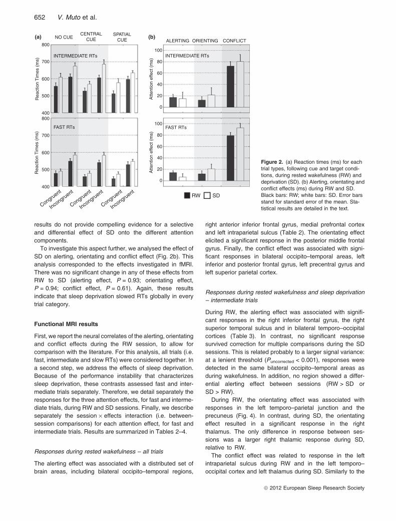
results do not provide compelling evidence for a selectiveand differential effect of SD onto the different attentioncomponents.To investigate this aspect further, we analysed the effect of
SD on alerting, orientating and conflict effect (Fig. 2b). Thisanalysis corresponded to the effects investigated in fMRI.There was no significant change in any of these effects fromRW to SD (alerting effect, P = 0.93; orientating effect,P = 0.94; conflict effect, P = 0.61). Again, these resultsindicate that sleep deprivation slowed RTs globally in everytrial category.
Functional MRI results
First, we report the neural correlates of the alerting, orientatingand conflict effects during the RW session, to allow forcomparison with the literature. For this analysis, all trials (i.e.fast, intermediate and slow RTs) were considered together. Ina second step, we address the effects of sleep deprivation.Because of the performance instability that characterizessleep deprivation, these contrasts assessed fast and inter-mediate trials separately. Therefore, we detail separately theresponses for the three attention effects, for fast and interme-diate trials, during RW and SD sessions. Finally, we describeseparately the session · effects interaction (i.e. between-session comparisons) for each attention effect, for fast andintermediate trials. Results are summarized in Tables 2–4.
Responses during rested wakefulness – all trials
The alerting effect was associated with a distributed set ofbrain areas, including bilateral occipito–temporal regions,
right anterior inferior frontal gyrus, medial prefrontal cortexand left intraparietal sulcus (Table 2). The orientating effectelicited a significant response in the posterior middle frontalgyrus. Finally, the conflict effect was associated with signi-ficant responses in bilateral occipito–temporal areas, leftinferior and posterior frontal gyrus, left precentral gyrus andleft superior parietal cortex.
Responses during rested wakefulness and sleep deprivation– intermediate trials
During RW, the alerting effect was associated with signifi-cant responses in the right inferior frontal gyrus, the rightsuperior temporal sulcus and in bilateral temporo–occipitalcortices (Table 3). In contrast, no significant responsesurvived correction for multiple comparisons during the SDsessions. This is related probably to a larger signal variance:at a lenient threshold (Puncorrected < 0.001), responses weredetected in the same bilateral occipito–temporal areas asduring wakefulness. In addition, no region showed a differ-ential alerting effect between sessions (RW > SD orSD > RW).During RW, the orientating effect was associated with
responses in the left temporo–parietal junction and theprecuneus (Fig. 4). In contrast, during SD, the orientatingeffect resulted in a significant response in the rightthalamus. The only difference in response between ses-sions was a larger right thalamic response during SD,relative to RW.The conflict effect was related to response in the left
intraparietal sulcus during RW and in the left temporo–occipital cortex and left thalamus during SD. Similarly to the
(a) (b)NO CUE
800100
80
60
40
20
0
700
600
Rea
ctio
n T
imes
(m
s)
Atte
ntio
n ef
fect
(m
s)
100
80
60
40
20
0
Atte
ntio
n ef
fect
(m
s)
500
400800
700
600
Rea
ctio
n T
imes
(m
s)
500
400
Congruent
Incongruent
Congruent
Incongruent
Congruent
Incongruent
INTERMEDIATE RTs
FAST RTs FAST RTs
RW SD
INTERMEDIATE RTs
ALERTING ORIENTING CONFLICT
CENTRALCUE
SPATIALCUE
Figure 2. (a) Reaction times (ms) for eachtrial types, following cue and target condi-tions, during rested wakefulness (RW) anddeprivation (SD). (b) Alerting, orientating andconflict effects (ms) during RW and SD.Black bars: RW; white bars: SD. Error barsstand for standard error of the mean. Sta-tistical results are detailed in the text.
652 V. Muto et al.
ª 2012 European Sleep Research Society

orientating effect, the only difference in responses betweensessions was a larger conflict-related activity in bilateralthalamic nuclei during SD, relative to RW (Fig. 5).
Responses during rested wakefulness and sleep deprivation– fast trials
Similarly to intermediate responses, fast responses corre-sponding to the alerting effect during RW were associatedwith increased activity in the bilateral occipito–temporal areas
and right superior temporal gyrus (Fig. 3, Table 4). Nosignificant response was observed during SD, except at alenient threshold (Puncorrected < 0.001) in the left occipito–temporal cortex. Alerting-related activity in the left precuneusand the right temporo–parietal junction was larger during RWthan SD (Fig. 3), whereas no region was more active duringSD than RW.In contrast, for the orientating effect, significant brain
activity was detected only during SD in the left temporo–parietal junction and the precuneus (Fig. 4). Intriguingly, these
Table 2 Results obtained during rested wakefulness (RW) (all trials included)
Area x y z Z-score PSVC References
Alerting effectR. lateral temporo–occipital cortex 48 )68 6 3.88 0.011 (Coull et al., 2001)L. lateral temporo–occipital cortex )42 )66 6 4.19 0.004 (Fan et al., 2005)R. superior medial frontal cortex 4 32 34 3.51 0.030 (Fan et al., 2005)R. inferior frontal gyrus 50 28 4 3.42 0.038 (Konrad et al., 2005)L. anterior intraparietal sulcus )30 )42 34 4.21 0.004 (Thiel et al., 2004)
Orientating effectLeft MFG )44 2 48 3.16 0.049 (Thiel et al., 2004)
Conflict effectR. occipito–temporal cortex 44 )60 )2 3.47 0.033 (Fan et al., 2005)L. occipito–temporal cortex )46 )68 )2 3.31 0.049 (Fan et al., 2005)L. inferior frontal sulcus )34 32 )4 3.25 0.05 (Konrad et al., 2005)L. precentral gyrus )36 )10 50 3.64 0.022 (Fan et al., 2005)L. superior parietal cortex )44 )36 52 3.31 0.049 (Coull et al., 2001)
Coordinates (x, y, z) are expressed in mm in the Montreal Neurological Institute (MNI) space. Psvc: probability of rejecting the null hypothesisof no activation after correction for multiple comparisons over small volumes of interest taken from the literature (reference in the last column).R., right; L., left; MFG, Middle Frontal Gyrus.
Table 3 Results obtained during rested wakefulness (RW) and deprivation (SD) (intermediate trials)
Area x y z Z-score PSVC References
Alerting effect RWR. inferior frontal gyrus 44 22 18 3.29 0.046 (Fan et al., 2005)R. superior temporal sulcus 52 )46 12 4.31 0.003 (Fan et al., 2005)R. lateral temporo–occipital cortex 50 )70 2 4.17 0.004 (Coull et al., 2001)L. lateral temporo–occipital cortex )42 )68 6 3.88 0.010 (Fan et al., 2005)
Orientating effect RWL. precuneus )16 )50 34 3.47 0.050 (Thiel et al., 2004)L. temporo–parietal junction )58 )60 35 3.45 0.050 (Coull et al., 2001)
Orientating effect SDR. thalamus 8 )16 0 3.24 0.036 (Fan et al., 2005)
Orientating effect SD > RWR. thalamus 8 )16 0 5.45 0.032 (Fan et al., 2005)
Conflict effect RWL. intraparietal sulcus )26 )48 50 3.40 0.039 (Coull et al., 2001)
Conflict effect SDL. temporo-occipital cortex )42 )68 )6 3.77 0.039 (Coull et al., 2001)L. thalamus )14 )28 8 3.24 0.05 (Fan et al., 2005)
Conflict effect SD > RWR. thalamus 18 )28 4 3.92 0.01 (Fan et al., 2005)L. thalamus )12 )26 4 3.28 0.05 (Fan et al., 2005)
Coordinates (x, y, z) are expressed in mm in the Montreal Neurological Institute (MNI) space. Psvc: probability of rejecting the null hypothesisof no activation after correction for multiple comparisons over small volumes of interest taken from the literature (reference in the last column).R., right; L., left.
Neural correlates of attention during sleep loss 653
ª 2012 European Sleep Research Society

areas correspond to those associated with intermediateresponses during RW. Responses in the left temporo–parietalarea were larger during SD than during RW, whereas noregion had a larger orientating-related activity during RW,relative to SD.Bold responses did not differ between congruent and
incongruent trials (conflict effect), either in RW or in SD.Thalamic responses associated with the fastest trials wereincreased significantly during SD relative to RW (Fig. 5).
DISCUSSION
We used the ANT to test whether sleep deprivation wouldinfluence differentially the alerting, orientating and executivecomponents of attention. Behavioural data did not supportthis hypothesis and essentially showed a global slowing ofRTs for all trial types. Only the conflict effect associated withthe fastest RTs was enhanced during SD, relative to RWcondition. Functional MRI data did not show significantbetween-session changes in responses related to the alertingeffect, except for those associated with the fastest RTs,which were characterized by increased activity during RW inthe precuneus and right temporo–parietal junction. For theorientating effect, the precuneus and left temporo–parietaljunction, which were activated during RW, could still berecruited during SD, but only when an optimal alertness levelcould be achieved. The orientating effect during SD wassupported by an enhanced recruitment of the thalamus(intermediate RTs) and right temporo–parietal region (fastestRTs). The conflict effect was also associated with increasedthalamic responses during SD relative to RW, for bothintermediate and fast RTs.
Maintaining phasic alertness during sleep loss
Sleep deprivation did not selectively modify the alertingcomponent and slow down responses to the same extent totrials with or without a central cue. Whereas the alertingeffect was associated with responses in occipital andtemporal areas during RW, no statistically significant brainalerting responses were detected after SD. Moreover, thealerting effect was not associated with any significantdifference in brain responses between SD and RW forintermediate RTs. These results do not support the view thatthe neural correlates of phasic alertness are modifiedspecifically by sleep deprivation. On the contrary, theysuggest that the main impact of sleep deprivation on brainfunction is related either to an impaired capacity of cent-rencephalic structures to maintain vigilance, or to theubiquitous influence of local sleep pressure on neuronalactivity (Krueger et al., 2008).Intriguingly, for the fastest trials, SD resulted in
decreased responses (relative to RW) in the precuneusand right temporo–parietal junction. In the absence of anybehavioural correlates, this finding indicates that the activityin these areas might reflect strategies that are adoptedprimarily under rested conditions but are not critical to thealerting effect. These strategies would buttress a steadycognitive output during RW. In contrast, SD would reducethese cognitive resources and the corresponding brainresponses.The current findings do not confirm recent behavioural
data showing, as well as global slowing in RTs, a significanteffect of sleep deprivation on alerting effect (Martella et al.,2011). This discrepancy is explained potentially by their
Table 4 Results obtained during rested wakefulness (RW) and deprivation (SD) (fast trials)
Area x y z Z-score PSVC References
Alerting effect RWR. lateral temporo–occipital cortex 42 )76 )4 3.53 0.025 (Coull et al., 2001)L. lateral temporo–occipital cortex )48 )60 6 3.49 0.028 (Fan et al., 2005)R. superior temporal gyrus 68 )36 16 3.65 0.019 (Fan et al., 2005)
Alerting effect RW > SDL. precuneus )16 )56 36 3.72 0.014 (Thiel et al., 2004)R. temporo–parietal junction 52 )66 18 3.53 0.024 (Thiel et al., 2004)
Orientating effect SDL. temporo-parietal junction )48 )64 24 4.3 0.002 (Coull et al., 2001)L. precuneus )10 )68 44 3.28 0.044 (Thiel et al., 2004)
Orientating effect SD > RWL. temporo-parietal junction )46 )62 20 3.75 0.015 (Coull et al., 2001)
Conflict effect SD > RWL. thalamus )6 )6 12 3.47 0.032 (Fan et al., 2005)
Coordinates (x, y, z) are expressed in mm in the Montreal Neurological Institute (MNI) space. Psvc: probability of rejecting the null hypothesisof no activation after correction for multiple comparisons over small volumes of interest taken from the literature (reference in the last column).R., right; L., left.
654 V. Muto et al.
ª 2012 European Sleep Research Society

experimental protocol in which SD sessions took place at04:00 h, a time of day associated with marked circadiandecline in alertness (Dijk et al., 1992). It should be notedthat this experimental design possibly underestimates thegenuine behavioural and neural effects of sleep deprivation,which can be detected more easily in the early morninghours after sleep deprivation. A discussion about thecontribution of increased sleep pressure and circadianrhythms on our results can be found in the supportingonline information.
Thalamic and cortical compensatory responses maintainhigher-order attention components during sleep loss
For higher-order (orientating and executive) attention com-ponents, some brain responses were increased after SD
relative to RW. Similar increases were reported for othercognitive tasks (Venkatraman et al., 2007) and are thought toreflect �compensatory� responses, i.e. brain attempts torecruit neural resources transiently to maintain cognitiveoutput despite increased sleep pressure and ⁄or circadianmisalignment (Drummond et al., 2001; Tomasi et al., 2009).Importantly, these �compensatory� responses were detectedin thalamic nuclei for the orientating and executive attentioncomponents. The thalamus constitutes a unique interfacebetween vigilance and cognition. Increased thalamic res-ponses were observed during sleep deprivation for workingmemory (Chee et al., 2008; Vandewalle et al., 2009) andattention tasks (Chee et al., 2008; Portas et al., 1998). These�compensatory� thalamic responses speak against a selectiveinfluence of SD on cortical circuits involved in orientating orexecutive attention components. In contrast, and in keeping
Alerting effect
RW
RW SD
per30
per30RW > SD
Left lateral occipital cortex
* *
*
°
°
Right lateral occipital cortex
Right superior temporal gyrus
Fast
–0.5
0
0.5
1
–0.5
0
0.5
1
–0.5
0
0.5
–0.5
–1
0
0.5
1
1.5
0
0.5
1
1.5
2
Intermediate Slow Fast Intermediate Slow
Fast Intermediate Slow
Fast Intermediate Slow
Fast Intermediate Slow
Right temporo-parietal junction
Left precuneusFigure 3. Brain responses associated withthe alerting effect. Row 1: bilateral lateraloccipital areas; row 2: right superior temporalgyrus; row 3: right temporo–parietal junction;row 4: left precuneus. Left panels: functionalresults are displayed at Puncorrected < 0.001,over the normalized structural magneticresonance (MR) scan of a typical subject.Right panels: activity estimates of each areafor fast, intermediate and slow reaction times(RTs) (arbitrary units ± standard error of themean). *Significant within-state effect(Psvc < 0.05); �significant state · effectinteraction (Psvc < 0.05).
Neural correlates of attention during sleep loss 655
ª 2012 European Sleep Research Society

with the conclusion drawn for alerting effects, these thalamicresponses would reflect non-specific mechanisms, whichtransiently maintain an optimal vigilance level, therebysupporting cortical function and maintaining steady perfor-mance.The only cortical area showing a compensatory response
was the left temporo–parietal junction. These findings speakfor a phasic recruitment of this area when an optimalvigilance level can be achieved during sleep loss, suggesting
again the importance of vigilance level for the adjustment ofcortical responses during sleep loss.
Does the ANT probe independent and segregatedattention components?
The ANT was designed to probe three allegedly independentattention networks, which were segregated in specific brainareas (Fan et al., 2005). The current data acquired during
Orienting effectLeft Precuneus
Arb
itrar
y un
its
Arb
itrar
y un
its
Arb
itrar
y un
itsA
rbitr
ary
units
Left temporo-parietal junction
RW intermediate
intermediate
per30SD
RW SD
SD > RW
Right Thalamus
Left Precuneus
*
Fast
–0.5
–1
–1.5
0
0.5
1
–0.5
–1
0
0.5
11.5
–0.5
–1
0
0.5
1
–0.5
0
0.5
1
1.5
Intermediate Slow Fast Intermediate Slow
Fast Intermediate Slow
Fast Intermediate Slow
*
* *°
*°
Figure 4. Brain responses associated with the orientating effect. Upper row: left precuneus; middle row: left temporo–parietal junction; lowerrow: right thalamus. Left panels: functional results are displayed at Puncorrected < 0.001, over the normalized structural magnetic resonance (MR)scan of a typical subject. Right panels: activity estimates of each area for fast, intermediate and slow reaction times (RTs) (arbitraryunits ± standard error of the mean). *Significant within-state effect; �Significant state · effect interaction (Psvc < 0.05).
Conflict effect
SD > RW per30 SD > RW intermediate
SDRW
°
Left Thalamus
2
1
0
–1
0
0.5
–0.5
–1–2Fast Intermediate Slow Fast Intermediate Slow
Left Thalamus
°
Figure 5. Differential thalamic responsesassociated with the conflict effect betweenrested wakefulness (RW) and deprivation(SD). Functional results are displayed atPuncorrected < 0.001, over the normalizedstructural magnetic resonance (MR) scan ofa typical subject. Lateral panels: activityestimates for fast, intermediate and slowreaction times (RTs) in each contrast (arbi-trary units ± standard error of the mean).�Significant state · effect interaction (Psvc<0.05).
656 V. Muto et al.
ª 2012 European Sleep Research Society

RW do not support the view that the ANT probes independentsets of brain areas. For instance, the alerting effect elicitedsignificant responses in occipito–temporal areas, in keepingwith previous findings, but also in the inferior frontal gyrus,which had been implicated in conflict resolution (Fan et al.,2005). These discrepancies may have various origins, suchas different versions of the tasks, the use of differentstatistical fMRI models or the procedures of statisticalinferences (which were conservative in the current work).To maintain short scanning sessions, we transformed theANT version for fMRI (Fan et al., 2005) in a compact form,whereas Fan and collaborators used a long version allowingthem to separate responses to cues from responses totargets (Fan et al., 2005). In the same vein, MacLeod et al.(2010) demonstrated recently that the three attention net-works do not operate independently at a cognitive level. Inany case, the ANT does not seem adapted to our originalobjective, which was to assess the effects of sleep depriva-tion on three independent aspects of human attention using acompact design based on a single task.
CONCLUSIONS
Behavioural and neural data do not support the view of aselective and differential influence of sleep deprivation onalerting, orientating or executive attention components. Therecruitment of thalamus during sleep loss to support higher-order (orientating and executive) attention components sug-gests that sleep deprivation influences attention globallythrough its impact on the ability to maintain vigilance.
ACKNOWLEDGEMENTS
This study was supported by the Belgian Fonds National dela Recherche Scientifique, the Queen Elisabeth MedicalFoundation, the University of Liege and the InteruniversitaryAttraction Pole program.
DISCLOSURE
The authors have no conflictes of interest to declare.
REFERENCES
Akerstedt, T. and Gillberg, M. Subjective and objective sleepiness inthe active individual. Int. J. Neurosci., 1990, 52: 29–37.
Buysse, D. J., Reynolds, C. F. I. I. I., Monk, T. H., Berman, S. R. andKupfer, D. J. The Pittsburgh Sleep Quality Index: a new instrumentfor psychiatric practice and research. Psychiatry Res., 1989, 28:193–213.
Chee, M. W., Tan, J. C., Zheng, H. et al. Lapsing during sleepdeprivation is associated with distributed changes in brain activa-tion. J. Neurosci., 2008, 28: 5519–5528.
Coull, J. T., Nobre, A. C. and Frith, C. D. The noradrenergic alpha2agonist clonidine modulates behavioural and neuroanatomicalcorrelates of human attentional orienting and alerting. Cereb.Cortex, 2001, 11: 73–84.
Dijk, D. J., Duffy, J. F. and Czeisler, C. A. Circadian and sleep ⁄wakedependent aspects of subjective alertness and cognitive perfor-mance. J. Sleep Res., 1992, 1: 112–117.
Doran, S. M., Van Dongen, H. P. and Dinges, D. F. Sustainedattention performance during sleep deprivation: evidence of stateinstability. Arch. Ital. Biol., 2001, 139: 253–267.
Drummond, S. P., Gillin, J. C. and Brown, G. G. Increased cerebralresponse during a divided attention task following sleep depriva-tion. J. Sleep Res., 2001, 10: 85–92.
Drummond, S. P., Bischoff-Grethe, A., Dinges, D. F., Ayalon, L.,Mednick, S. C. and Meloy, M. J. The neural basis of thepsychomotor vigilance task. Sleep, 2005, 28: 1059–1068.
Fan, J., McCandliss, B. D., Fossella, J., Flombaum, J. I. and Posner,M. I. The activation of attentional networks. Neuroimage, 2005, 26:471–479.
Harrison, Y., Horne, J. A. and Rothwell, A. Prefrontal neuropsycho-logical effects of sleep deprivation in young adults – a model forhealthy aging? Sleep, 2000, 23: 1067–1073.
Horne, J. A. and Ostberg, O. A self-assessment questionnaire todetermine morningness–eveningness in human circadian rhythms.Int. J. Chronobiol., 1976, 4: 97–110.
Horowitz, T. S., Cade, B. E., Wolfe, J. M. and Czeisler, C. A.Searching night and day: a dissociation of effects of circadianphase and time awake on visual selective attention and vigilance.Psychol. Sci., 2003, 14: 549–557.
Johns, M. W. A new method for measuring daytime sleepiness: theEpworth sleepiness scale. Sleep, 1991, 14: 540–545.
Konrad, K., Neufang, S., Thiel, C. M. et al. Development ofattentional networks: an fMRI study with children and adults.Neuroimage, 2005, 28: 429–439.
Krueger, J. M., Rector, D. M., Roy, S., Van Dongen, H. P., Belenky,G. and Panksepp, J. Sleep as a fundamental property of neuronalassemblies. Nat. Rev. Neurosci., 2008, 9: 910–919.
Lim, J. and Dinges, D. F. A meta-analysis of the impact of short-termsleep deprivation on cognitive variables. Psychol. Bull., 2010, 136:375–389.
MacLeod, J. W., Lawrence, M. A., McConnell, M. M., Eskes, G. A.,Klein, R. M. and Shore, D. I. Appraising the ANT: psychometric andtheoretical considerations of the Attention Network Test.Neuropsychology, 2010, 24: 637–651.
Martella, D., Casagrande, M. and Lupianez, J. Alerting, orienting andexecutive control: the effects of sleep deprivation on attentionalnetworks. Exp. Brain Res., 2011, 210: 81–89.
Oken, B. S., Salinsky, M. C. and Elsas, S. M. Vigilance, alertness, orsustained attention: physiological basis and measurement.Clin. Neurophysiol., 2006, 117: 1885–1901.
Oldfield, R. C. The assessment and analysis of handedness: theEdinburgh inventory. Neuropsychologia, 1971, 9: 97–113.
Portas, C. M., Rees, G., Howseman, A. M., Josephs, O., Turner, R.and Frith, C. D. A specific role for the thalamus in mediating theinteraction of attention and arousal in humans. J. Neurosci., 1998,18: 8979–8989.
Posner, M. I. and Petersen, S. E. The attention system of the humanbrain. Annu. Rev. Neurosci., 1990, 13: 25–42.
Schmidt, C., Collette, F., Leclercq, Y. et al. Homeostatic sleeppressure and responses to sustained attention in the suprachias-matic area. Science, 2009, 324: 516–519.
Thiel, C. M., Zilles, K. and Fink, G. R. Cerebral correlates of alerting,orienting and reorienting of visuospatial attention: an event-relatedfMRI study. Neuroimage, 2004, 21: 318–328.
Tomasi, D., Wang, R. L., Telang, F. et al. Impairment of attentionalnetworks after 1 night of sleep deprivation. Cereb. Cortex, 2009,19: 233–240.
Vandewalle, G., Archer, S. N., Wuillaume, C. et al. Functionalmagnetic resonance imaging-assessed brain responses during anexecutive task depend on interaction of sleep homeostasis,
Neural correlates of attention during sleep loss 657
ª 2012 European Sleep Research Society

circadian phase, and PER3 genotype. J. Neurosci., 2009, 29:7948–7956.
Venkatraman, V., Chuah, Y. M., Huettel, S. A. and Chee, M. W.Sleep deprivation elevates expectation of gains and attenuatesresponse to losses following risky decisions. Sleep, 2007, 30: 603–609.
SUPPORTING INFORMATION
Additional Supporting Information may be found in the onlineversion of this article:
Figure S1. Alerting, orienting and conflict effects duringrested wakefulness.Data S1. Operational definitions of vigilance, alertness,
arousal and attention.Please note: Wiley-Blackwell is not responsible for the
content or functionality of any supporting materials suppliedby the authors. Any queries (other than missing material)should be directed to the corresponding author for the article.
658 V. Muto et al.
ª 2012 European Sleep Research Society