Sensitivity and Specificity of a Novel Classifier for the Early ...
15
RESEARCH ARTICLE Sensitivity and Specificity of a Novel Classifier for the Early Diagnosis of Dengue Nguyen Minh Tuan 1 , Ho Thi Nhan 2 , Nguyen Van Vinh Chau 3 , Nguyen Thanh Hung 1 , Ha Manh Tuan 4 , Ta Van Tram 5 , Nguyen Le Da Ha 6 , Phan Loi 7 , Han Khoi Quang 8 , Duong Thi Hue Kien 2 , Sonya Hubbard 9 , Tran Nguyen Bich Chau 2 , Bridget Wills 2,10 , Marcel Wolbers 2,10 , Cameron P. Simmons 2,9,10,11 * 1 Children’s Hospital No. 1, Ho Chi Minh City, Vietnam, 2 Oxford University Clinical Research Unit, Hospital for Tropical Diseases, Ho Chi Minh City, Vietnam, 3 Hospital for Tropical Diseases, Ho Chi Minh City, Vietnam, 4 Children’s Hospital No. 2, Ho Chi Minh City, Vietnam, 5 Tien Giang Provincial Hospital, My Tho, Tien Giang Province, Vietnam, 6 Dong Nai Children’s Hospital, Bien Hoa, Dong Nai Province, Vietnam, 7 Long An Provincial Hospital, Tan An, Long An Province, Vietnam, 8 Binh Duong Provincial Hospital, Thu Dau Mot, Binh Duong Province, Vietnam, 9 Department of Microbiology and Immunology, University of Melbourne, Parkville, Victoria, Australia, 10 Centre for Tropical Medicine, Nuffield Department of Medicine, University of Oxford, Oxford, United Kingdom, 11 Nossal Institute of Global Health, University of Melbourne, Parkville, Victoria, Australia * [email protected] Abstract Background Dengue is the commonest arboviral disease of humans. An early and accurate diagnosis of den- gue can support clinical management, surveillance and disease control and is central to achiev- ing the World Health Organisation target of a 50% reduction in dengue case mortality by 2020. Methods 5729 children with fever of <72hrs duration were enrolled into this multicenter prospective study in southern Vietnam between 2010-2012. A composite of gold standard diagnostic tests identified 1692 dengue cases. Using statistical methods, a novel Early Dengue Classi- fier (EDC) was developed that used patient age, white blood cell count and platelet count to discriminate dengue cases from non-dengue cases. Results The EDC had a sensitivity of 74.8% (95%CI: 73.0-76.8%) and specificity of 76.3% (95%CI: 75.2-77.6%) for the diagnosis of dengue. As an adjunctive test alongside NS1 rapid testing, sensitivity of the composite test was 91.6% (95%CI: 90.4-92.9%). Conclusions We demonstrate that the early diagnosis of dengue can be enhanced beyond the current standard of care using a simple evidence-based algorithm. The results should support pa- tient management and clinical trials of specific therapies. PLOS Neglected Tropical Diseases | DOI:10.1371/journal.pntd.0003638 April 2, 2015 1 / 15 OPEN ACCESS Citation: Tuan NM, Nhan HT, Chau NVV, Hung NT, Tuan HM, Tram TV, et al. (2015) Sensitivity and Specificity of a Novel Classifier for the Early Diagnosis of Dengue. PLoS Negl Trop Dis 9(4): e0003638. doi:10.1371/journal.pntd.0003638 Editor: Scott B Halstead, Pediatric Dengue Vaccine Initiative, UNITED STATES Received: December 2, 2014 Accepted: February 23, 2015 Published: April 2, 2015 Copyright: © 2015 Tuan et al. This is an open access article distributed under the terms of the Creative Commons Attribution License, which permits unrestricted use, distribution, and reproduction in any medium, provided the original author and source are credited. Data Availability Statement: All relevant data are within the paper and its Supporting Information files. Funding: This study was supported by grants to CPS from the NHMRC GNT006549 and the Wellcome Trust 084368/Z/07/Z. The funders had no role in study design, data collection and analysis, decision to publish, or preparation of the manuscript. Competing Interests: The authors have declared that no competing interests exist.
-
Upload
khangminh22 -
Category
Documents
-
view
2 -
download
0
Transcript of Sensitivity and Specificity of a Novel Classifier for the Early ...

RESEARCH ARTICLE
Sensitivity and Specificity of a NovelClassifier for the Early Diagnosis of DengueNguyenMinh Tuan1, Ho Thi Nhan2, Nguyen Van Vinh Chau3, Nguyen Thanh Hung1,Ha Manh Tuan4, Ta Van Tram5, Nguyen Le Da Ha6, Phan Loi7, Han Khoi Quang8, Duong ThiHue Kien2, Sonya Hubbard9, Tran Nguyen Bich Chau2, Bridget Wills2,10, Marcel Wolbers2,10,Cameron P. Simmons2,9,10,11*
1 Children’s Hospital No. 1, Ho Chi Minh City, Vietnam, 2 Oxford University Clinical Research Unit, Hospitalfor Tropical Diseases, Ho Chi Minh City, Vietnam, 3 Hospital for Tropical Diseases, Ho Chi Minh City,Vietnam, 4 Children’s Hospital No. 2, Ho Chi Minh City, Vietnam, 5 Tien Giang Provincial Hospital, My Tho,Tien Giang Province, Vietnam, 6 Dong Nai Children’s Hospital, Bien Hoa, Dong Nai Province, Vietnam,7 Long An Provincial Hospital, Tan An, Long An Province, Vietnam, 8 Binh Duong Provincial Hospital, ThuDau Mot, Binh Duong Province, Vietnam, 9 Department of Microbiology and Immunology, University ofMelbourne, Parkville, Victoria, Australia, 10 Centre for Tropical Medicine, Nuffield Department of Medicine,University of Oxford, Oxford, United Kingdom, 11 Nossal Institute of Global Health, University of Melbourne,Parkville, Victoria, Australia
Abstract
Background
Dengue is the commonest arboviral disease of humans. An early and accurate diagnosis of den-
gue can support clinical management, surveillance and disease control and is central to achiev-
ing theWorld Health Organisation target of a 50% reduction in dengue casemortality by 2020.
Methods
5729 children with fever of<72hrs duration were enrolled into this multicenter prospective
study in southern Vietnam between 2010-2012. A composite of gold standard diagnostic
tests identified 1692 dengue cases. Using statistical methods, a novel Early Dengue Classi-
fier (EDC) was developed that used patient age, white blood cell count and platelet count to
discriminate dengue cases from non-dengue cases.
Results
The EDC had a sensitivity of 74.8% (95%CI: 73.0-76.8%) and specificity of 76.3% (95%CI:
75.2-77.6%) for the diagnosis of dengue. As an adjunctive test alongside NS1 rapid testing,
sensitivity of the composite test was 91.6% (95%CI: 90.4-92.9%).
Conclusions
We demonstrate that the early diagnosis of dengue can be enhanced beyond the current
standard of care using a simple evidence-based algorithm. The results should support pa-
tient management and clinical trials of specific therapies.
PLOS Neglected Tropical Diseases | DOI:10.1371/journal.pntd.0003638 April 2, 2015 1 / 15
OPEN ACCESS
Citation: Tuan NM, Nhan HT, Chau NVV, Hung NT,Tuan HM, Tram TV, et al. (2015) Sensitivity andSpecificity of a Novel Classifier for the EarlyDiagnosis of Dengue. PLoS Negl Trop Dis 9(4):e0003638. doi:10.1371/journal.pntd.0003638
Editor: Scott B Halstead, Pediatric Dengue VaccineInitiative, UNITED STATES
Received: December 2, 2014
Accepted: February 23, 2015
Published: April 2, 2015
Copyright: © 2015 Tuan et al. This is an openaccess article distributed under the terms of theCreative Commons Attribution License, which permitsunrestricted use, distribution, and reproduction in anymedium, provided the original author and source arecredited.
Data Availability Statement: All relevant data arewithin the paper and its Supporting Information files.
Funding: This study was supported by grants to CPSfrom the NHMRC GNT006549 and the WellcomeTrust 084368/Z/07/Z. The funders had no role instudy design, data collection and analysis, decision topublish, or preparation of the manuscript.
Competing Interests: The authors have declaredthat no competing interests exist.

Author Summary
Dengue is a very common acute infectious disease in the tropical world. Health care pro-fessionals are able to better care for dengue patients if they can make an early diagnosisand make a plan for case management. This current study investigated fever in 5729 chil-dren in Vietnam with 3 days or less of fever and identified 1692 dengue cases using ad-vanced, gold standard methods. We systematically collected a range of medical andlaboratory findings on each patient when they entered the study and used statistical toolsto determine if these medical and laboratory findings could enable early diagnosis, inde-pendent of sophisticated, gold-standard laboratory tests. Our results, called the Early Den-gue Classifier, had performance characteristics suggesting it could improve the diagnosticproficiency of health care professionals. However the performance of the Early DengueClassifier is not perfect and likely will not change the practice of experienced doctors indengue endemic settings. Our study highlights the need for 2nd generation, easy-to-userapid diagnostic tests that can accurately diagnose dengue in the first few days of fever.
IntroductionDengue is an acute, systemic viral infection and a public health problem in the tropical world[1]. The etiological agents of dengue are any of the four dengue viruses (DENV-1-4). In en-demic countries it is common for all four DENV serotypes to co-circulate. Late-stage trials of adengue vaccine with intermediate efficacy have recently been reported, offering hope of a pub-lic health intervention [2, 3].
The World Health Organisation (WHO) has a stated goal of reducing global dengue mortal-ity by 50% by 2020 [1]. Improvements in case diagnosis and management will be central toachieving this aim. Significant loss of intravascular plasma volume leading to hypovolemicshock (dengue shock syndrome (DSS)), usually between the 4th-6th day of illness, is the com-monest life-threatening complication of dengue [1, 4]. It’s widely held that the case-incidenceof DSS can be reduced via careful monitoring and the judicious use of parenteral fluids tomaintain an adequate intravascular volume [1]. Ideally, this case management approach is en-abled because the attending physician had made an early diagnosis and thus alerted clinicians,nurses and family caregivers to the signs and symptoms suggestive of clinical worsening. Addi-tional benefits of an early diagnosis include support to community level public health interven-tions and improvements in the sensitivity of case surveillance systems and disease burdenestimates. Furthermore, it is likely that the therapeutic window of opportunity for a dengue an-tiviral drug lies in the first 48–72 hours of illness [5]. Thus, programmatic use of therapeutic in-terventions in the future will likely go hand in hand with strategies for early diagnosis.
Yet there are numerous challenges for busy primary care clinicians in making a diagnosis ofdengue in the first few days of illness. Rapid lateral flow tests, based on the detection of theviral NS1 antigen, are available in some settings and can provide a confirmatory diagnosis [6–8]. The diagnostic performance of the WHO dengue case definition, which relies on non-specific signs and symptoms that overlap with other infectious diseases, is unknown in the firstfew days of illness [1]. Potts et al concluded that more prospective studies were needed to con-struct a valid and generalizable algorithm to guide the differential diagnosis of dengue in en-demic countries [9]. To this end, several prospective studies have described the creation ofclassifiers for the diagnosis of dengue [10–12]. However none of these studies have exclusivelyfocused on pediatric fever cases presenting to primary care facilities with short illness histories,a very common scenario in dengue endemic settings. Against this backdrop, the purpose of the
Early Diagnosis of Dengue
PLOS Neglected Tropical Diseases | DOI:10.1371/journal.pntd.0003638 April 2, 2015 2 / 15

current study was to prospectively derive a dengue diagnostic algorithm from routinely collect-ed clinical and laboratory findings in pediatric patients with<72 hours of illness history andcompare this approach against the diagnostic performance of a leading NS1 rapid test (BioRadNS1 Ag STRIP) in the same patients. The results provide pragmatic methods to enhance theearly diagnosis of dengue in primary care settings.
Materials and Methods
Human research ethicsThe study protocol was approved by the Hospital for Tropical Diseases scientific and ethicalcommittee and the Oxford University Tropical Research Ethical Committee (OXTREC 35–10).The accompanying parent/guardian of each child provided written informed consent.
Patient enrolmentRecruitment occurred in the public sector outpatient departments of Children’s Hospital No. 1(HCMC), Children’s Hospital No. 2 (HCMC), The Hospital for Tropical Diseases (HCMC),Tien Giang Provincial Hospital, Dong Nai Children’s Hospital, Binh Duong Provincial Hospi-tal and Long An Provincial Hospital. These outpatient departments function as primary careproviders to their local communities. A patient presenting to one of the study sites was eligiblefor enrolment if they met the following inclusion criteria—a) fever at presentation (or historyof fever) and less than 72 hours of symptom history, b) in the attending physicians opiniondengue was a possible diagnosis, c) 1–15 years of age inclusive, d) accompanying family mem-ber or guardian had a mobile phone and e) written informed consent for the child to partici-pate was provided by the parent/guardian. Patients were excluded if- a) the attending physicianbelieved they were unlikely to be able to attend follow-up or b) the attending physician believedanother (non-dengue) diagnosis was more likely. Patient enrolment occurred consecutivelyduring normal clinical hours on weekdays without restriction. All patients were enrolled intothe study before the attending physician received the results of any routine laboratory tests.
Clinical and laboratory investigations on the day of enrolmentAt the time of enrolment, information on the patient’s age, sex, illness history, presenting signsand symptoms were recorded in a case report form. The definitions used to support standard-ized data capture are shown in S1 Table. Blood samples were drawn for routine hematology,biochemistry and NS1 rapid test. All NS1 rapid tests (NS1 Ag STRIP, BioRad) were performedon the same day of patient enrolment by one of two trained laboratory technicians at the Hos-pital for Tropical Diseases. Routine hematology results, but not biochemistry or NS1 rapid testresults, were made available to the attending physician, who decided on the management ap-proach, i.e. hospitalization or ambulatory follow-up.
Patient follow-upA 2nd blood sample for the purposes of serology was collected around the time of defervescencefrom all patients that were hospitalized anytime during their acute illness. If the patient wasmanaged solely on an ambulatory basis for the duration of their illness, then a 2nd early conva-lescence blood sample for the purposes of serology was collected only from a randomly selected10% of this patient population. The randomization code to select ambulatory cases for follow-up was generated by software. All clinical and laboratory data were stored in an ICH-GCPcompliant, clinical data management platform called “CLIRES”. Demographic and clinicaldata were double entered. Electronic data files containing hematological results were uploaded
Early Diagnosis of Dengue
PLOS Neglected Tropical Diseases | DOI:10.1371/journal.pntd.0003638 April 2, 2015 3 / 15
directly to CLIRES. Independent study monitoring was performed by the Clinical Trials Unitof the Oxford University Clinical Research Unit which examined adherence to the trial proce-dures, data collection and recording and compliance with ICH-GCP.
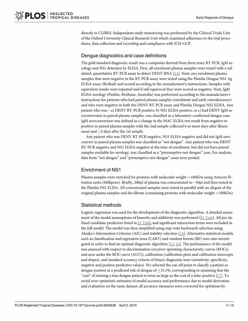
Dengue diagnostics and case definitionsThe gold standard diagnostic result was a composite derived from three tests; RT-PCR, IgM se-rology and NS1 detection by ELISA. First, all enrolment plasma samples were tested with a val-idated, quantitative RT-PCR assay to detect DENV RNA [13]. Next, any enrolment plasmasamples that were negative in the RT-PCR assay were tested using the Platelia Dengue NS1 AgELISA assay (BioRad) and scored according to the manufacturer's instructions. Samples withequivalent results were repeated and if still equivocal they were scored as negative. Next, IgMELISA serology (Panbio, Brisbane, Australia) was performed according to the manufacturer'sinstructions for patients who had paired plasma samples (enrolment and early convalescence)and who were negative in both the DENV RT-PCR assay and Platelia Dengue NS1 ELISA. Anypatient who was—a) DENV RT-PCR positive, b) NS1 ELISA positive, or c) had DENV IgM se-roconversion in paired plasma samples, was classified as a laboratory-confirmed dengue case.IgM seroconversion was defined as a change in the MAC ELISA test result from negative topositive in paired plasma samples with the 2nd sample collected 6 or more days after illnessonset and>2 days after the 1st sample.
Any patient who was DENV RT-PCR negative, NS1 ELISA negative and did not IgM sero-convert in paired plasma samples was classified as “not dengue”. Any patient who was DENVRT-PCR negative and NS1 ELISA negative at the time of enrolment, but did not have pairedsamples available for serology, was classified as a “presumptive not-dengue” case. For analysis,data from “not dengue” and “presumptive not-dengue” cases were pooled.
Enrichment of NS1Plasma samples were enriched for proteins with molecular weight>100kDa using Amicon fil-tration units (Millipore). Briefly, 200μl of plasma was concentrated to ~30μl and then tested inthe Platelia NS1 ELISA. All concentrated samples were tested in parallel with an aliquot of theoriginal plasma samples and the filtrate (containing proteins with molecular weight<100kDa).
Statistical methodsLogistic regression was used for the development of the diagnostic algorithm. A detailed assess-ment of the model assumptions of linearity and additivity was performed (S1 Text). All pre-de-fined candidate predictors listed in S1 Table and significant interaction terms were included inthe full model. The model was then simplified using step-wise backwards selection usingAkaike’s Information Criterion (AIC) and stability selection [14]. Alternative statistical modelssuch as classification and regression trees (CART) and random forests (RF) were also investi-gated in order to find an optimal diagnostic algorithm [15, 16]. The performance of the modelwas assessed with respect to discrimination (receiver operating characteristic curves (ROCs)and area under the ROC curve (AUC)), calibration (calibration plots and calibration interceptsand slopes), and standard accuracy criteria of binary diagnostic tests (sensitivity, specificity,negative and positive predictive values). We selected the cut-off point to classify a patient asdengue positive at a predicted risk of dengue of�33.3%, corresponding to assuming that the“cost” of missing a true dengue patient is twice as large as the cost of a false-positive [17]. Toavoid over-optimistic estimates of model accuracy and performance due to model derivationand evaluation on the same dataset, all accuracy measures were corrected for optimism by
Early Diagnosis of Dengue
PLOS Neglected Tropical Diseases | DOI:10.1371/journal.pntd.0003638 April 2, 2015 4 / 15

validation. Validation was performed for the whole model development process including vari-able selection. Two validation schemes were employed to mimic external validation:
a. “leave-one-site-out cross-validation”, i.e. repeatedly developing the algorithm on all but onesite and validation on the left-out site
b. Temporal validation with patients recruited before 15 June 2012 as the training set and pa-tients recruited thereafter as the evaluation set [18].
The final logistic model was also presented as a nomogram for direct clinical use. All statisti-cal analyses were performed using the statistical software R v3.1.1 (R foundation for statisticalcomputing, Vienna, Austria) and its companion packages c060 version 0.2–3 (for stability se-lection), randomForest version 4.6–7 (for random forest) and rpart version 4.1–8 (for CART).
Results
Study population5729 children with fever of less than 72 hours were enrolled at one of the seven clinical studysites in southern Vietnam between October 2010 and December 2012. A summary of the pa-tient screening, enrolment and diagnostic outcomes is shown in S1 Fig A total of 5707 patientswere included in the analyses. 1692 (29.6%) participants had laboratory-confirmed dengue.The baseline characteristics of the dengue and non-dengue cases are shown in Table 1. Notably,dengue cases were older than non-dengue cases. All four DENV serotypes were detected;DENV-1 was the commonest serotype, followed by DENV-4, -2 and -3.
Diagnostic accuracy of the NS1 Ag Strip test and association withviremiaEnrolment plasma samples (n = 5707) were tested for the presence of NS1 by NS1 Ag Strip testin a blinded, real-time fashion. Against the composite gold-standard reference diagnostic re-sult, the NS1 Ag Strip test had a sensitivity of 70.4% (95%CI: 68.2–72.6%), specificity of 99.2%(95%CI: 98.9–99.5%), positive predictive value (PPV) of 97.4% (95%CI: 96.3–98.2%), and neg-ative predictive value (NPV) of 88.9% (95%CI: 87.9–89.8%) for the diagnosis of dengue(Table 2). There was a striking difference in diagnostic performance by serotype, with NS1detection being less sensitive in DENV-2 infections irrespective of the serological response(primary vs secondary)(S2 Table). The detection of NS1 was strongly associated with the con-centration of DENV RNA in the same plasma sample; the odds of NS1 detection increased by1.8 (95%CI: 1.6–1.9) for each 10-fold higher DENV RNA concentration (Table 2). These datadefine the strengths and weaknesses of NS1 rapid testing; it is highly specific but is compro-mised by suboptimal sensitivity, especially for DENV-2 cases.
Manipulation of plasma specimens to improve NS1 detectionVolume enrichment of the plasma molecular weight fraction containing multimeric NS1(>100,000kDa) was performed on plasma samples from 21 viremic dengue cases enrolled inthis study. However, despite 5-10-fold concentration of plasma, this processing failed to mate-rially improve the diagnostic yield, with only 1 of 11 samples changing their status from nega-tive (original sample) to positive (concentrated sample) in the Platelia NS1 ELISA (S3 Table).
Early Diagnosis of Dengue
PLOS Neglected Tropical Diseases | DOI:10.1371/journal.pntd.0003638 April 2, 2015 5 / 15

The Early Dengue Classifier; A diagnostic rule based on clinical andsimple laboratory featuresMultivariate logistic regression analyses of clinical, demographic and laboratory data from5707 patients were performed to generate a practical dengue diagnostic classifier that could re-place or augment NS1-based diagnosis in the first 72 hours of illness. The most parsimoniousmodel, derived from stability selection, used the patient’s age, white cell count and plateletcount at the time of enrolment to classify dengue from non-dengue cases (Table 3). Alternativeapproaches to feature selection yielded models with only slightly higher performance but reliedon many more (more than ten) variables (S4 Table). The most parsimonious model, hereincalled the Early Dengue Classifier (EDC), had a sensitivity of 74.8% (95%CI: 73.0–76.8%), spec-ificity of 76.3% (95%CI: 75.2–77.6%), positive predictive value of 57.1% (95%CI: 56.2–59.0%),and negative predictive value of 87.8% (95%CI: 86.8–88.5%) for correctly classifying denguecases in the entire dataset at the pre-defined cut-off of 33.3%. Of note, this pre-defined cut-offreflecting clinical priorities was very close to the cut-off corresponding to the point on the
Table 1. Baseline characteristics of study participants.
Laboratory-confirmed dengue (n = 1692) Non-Dengue (n = 4015)
Demographic characteristics
Age (years) 9 (6–11) 5 (3–8)
Sex-Male (n, %) 945 (55.9%) 2249 (56.0%)
BMI (kg/(m)2) 16.4 (14.6–18.9) 15.6 (14.1–17.6)
History and clinical characteristics
Day of illness
1 361 (21.3%) 1232 (30.7%)
2 692 (40.9%) 1732 (43.1%)
3 639 (37.8%) 1051 (26.2%)
Temperature (°C) 38.5 (38–39) 38.4 (37.8–39.0)
Vomiting (n, %) 737 (43.6%) 1442 (35.9%)
Abdominal pain (n, %) 351 (20.7%) 702 (17.5%)
Skin bleeding (n, %) 243 (14.4%) 135 (3.4%)
Mucosal bleeding (n, %) 113 (6.7%) 99 (2.5%)
Flush (n, %) 399 (23.6%) 532 (13.3%)
Hepatomegaly (n, %) 6 (0.4%) 5 (0.1%)
Rash (n, %) 62 (3.7%) 79 (2.0%)
Conjunctival injection (n, %) 343 (20.3%) 385 (9.6%)
Laboratory results*
WBC (103/mm3) 4.74 (3.50–6.80) 8.90 (6.36–12.40)
PLT (103/mm3) 180 (141–227) 242 (200–292)
HCT (%) 38.6 (36.6–40.7) 37.4 (35.3–39.6)
ALB (g/L) 43.7 (41.7–45.6) 43.9 (42.0–45.7)
AST (U/l) 51 (40–67) 42 (35–49)
CK (U/l) 105 (82–140) 100 (76–131)
* All laboratory results were acquired on the day of enrolment.
Values are presented as median and interquartile range for continuous variables or frequency and
percentage for categorical variables.
BMI: body mass index; WBC: white blood cell count; PLT: platelet count; HCT: hematocrit; ALB: albumin;
AST: aspartate aminotransferase; CK: creatine kinas
doi:10.1371/journal.pntd.0003638.t001
Early Diagnosis of Dengue
PLOS Neglected Tropical Diseases | DOI:10.1371/journal.pntd.0003638 April 2, 2015 6 / 15

ROC curve closest to the upper left corner (perfect model), which was 34.2% (Fig 1A). The areaunder the ROC curve (AUC) was 0.829 (Fig 1B) and the predicted risk of dengue agreed wellwith the observed risk (Fig 1C). The EDC had sensitivity of 72.9% (95% CI: 69.6–76.6%) forDENV1, 74.7% (95%CI: 71.0–79.7%) for DENV2, 68.4% (95%CI: 59.2–74.5%) for DENV3 and78.2% (95%CI: 75.5–83.3%) for DENV4 infection. The overall performance characteristics ofthe EDC under temporal, leave-one-site-out validation or seasonality (rainy versus dry season),are summarized in S5 Table. These results suggest that, in settings where NS1 rapid tests arenot routinely available, the EDC could assist primary care physicians in dengue diagnosis.
In settings where NS1 rapid tests are routinely used, the EDC can be combined with the NS1rapid test as a composite test (classified as positive when either NS1 rapid test or EDC are posi-tive, and classified as negative when both NS1 rapid test and EDC are negative). This compositetest had sensitivity of 91.6% (95%CI: 90.4–92.9%) while the specificity was 75.7% (95%CI:74.5–77.0%). Corresponding positive and negative predictive values were 61.7% (95%CI:60.6–63.1%) and 95.5% (95%CI: 94.9–96.1%). If a higher specificity was desired, a higher cut-offvalue of the EDC could be used for the combined test instead, e.g. a cut-off of 50% would lead toa sensitivity of 86.0% (95%CI: 84.5–87.6%) and specificity of 89.6% (95%CI: 88.7–90.5%). Theseresults imply that the EDC is useful in settings with and without NS1 rapid testing.
User-friendly applications of the Early Dengue ClassifierFig 2 presents a nomogram of the EDC. The nomogram assigns points to all risk factors andtranslates the total point score to a predicted risk for dengue. For example, a 9-year-old patientwith platelet count 100x103/mm3, and white blood cell count 5x10³/mm3 has a total pointsscore of 15+32+84 = 131, and the corresponding risk of dengue is about 70%. The predicted riskof dengue is larger than 33.3% so the patient would be classified as dengue positive. Alternative-ly, the EDC could be implemented as a smartphone app. The exact formula for the estimatedrisk of dengue (p) is given by the following logistic equation: logit(p) = 1.236 + 0.139�age (inyears)– 0.254�white blood cell (in 103/mm3)– 0.006 �platelet (in 103/mm3).
Table 2. Diagnostic performance of NS1 rapid test in enrolment plasma samples and odds of NS1 detection in relation to plasma viremia.
Laboratory-confirmed dengue cases Non-denguecases
Total
NS1 rapid testpositive
1192 32 1224 PPV % = 97.4%(96.3–98.2%)
NS1 rapid testnegative
500 3983 4483 NPV % = 88.9%(87.9–89.8%)
Total 1692 4015 5707
Median (IQR) plasma viral RNA concentration(log10copies/ml)a
OR (95%CI) Sensitivity % (95%CI)
Specificity % (95%CI)
All serotypes(n = 1692)
7.3 (6.2–8.3) 1.8 (1.6–1.9) 70.4% (68.2–72.6%)
99.2% (98.9–99.5%)
DENV-1 (n = 629) 7.9 (6.6–8.7) 2.0 (1.8–2.3) 80.3 (77.0–83.3%) -
DENV-2 (n = 399) 7.0 (6.0–7.9) 1.8 (1.5–2.1) 46.4 (41.4–51.4%) -
DENV-3 (n = 154) 7.5 (6.4–8.6) 1.4 (1.1–1.9) 85.1 (78.4–90.3%) -
DENV-4 (n = 433) 6.9 (6.0–7.7) 1.5 (1.3–1.8) 75.8 (71.3–79.7%) -
a Viremia measurement in the enrolment plasma sample (the same sample was also used for NS1 testing).
PPV: positive predictive value; NPV: negative predictive value; DENV: dengue virus; OR: odds ratios for detecting NS1 for each 10-fold higher DENV
RNA concentration. There were 77 dengue cases where the infecting serotype was unknown.
doi:10.1371/journal.pntd.0003638.t002
Early Diagnosis of Dengue
PLOS Neglected Tropical Diseases | DOI:10.1371/journal.pntd.0003638 April 2, 2015 7 / 15

DiscussionThe early and accurate diagnosis of dengue on the grounds of clinical signs and symptomsalone is problematic [9]. Physicians need better tools to assist in early diagnosis if the WHOambition of a 50% reduction in global dengue mortality is to be achieved by 2020. This studycharacterized the performance of three diagnostic approaches; the NS1 rapid test, a stand-alone diagnostic classifier and the combination of NS1 rapid test and diagnostic classifier to-gether. Our results highlight the utility of NS1 rapid tests for an early specific diagnosis,yet also remind that 2nd generation tests are needed with improved sensitivity. The diagnosticclassifier described here could help guide diagnosis in endemic settings, or be used as an ad-junct to help exclude dengue in patients returning a negative NS1 rapid test result.
There is a body of literature describing the performance of NS1 rapid tests for the diagnosisof dengue [6–8, 19–22]. This current study extends that literature in several ways. First, by vir-tue of the large sample size we demonstrate with high precision the differential sensitivity ofthe NS1 Ag STRIP for different DENV serotypes. This test was sensitive (between 75–85%) forDENV-1, -3 and -4 infections, but poorly sensitive in DENV-2 infections (46.4%). Lower sensi-tivity was partially attributable to the great majority of DENV-2 infections being associatedwith secondary serological responses, although we note sensitivity was also low in primary
Table 3. Univariate andmultivariate analysis of candidate predictors of laboratory-confirmed dengue.
Univariate analysis Multivariate analysis
Full model with all candidatepredictors
Final model based on stabilityselection
OR 95% CI p OR 95% CI P OR 95% CI p
Demographic characteristics
Age (by + 1 year) 1.24 1.22–1.26 <0.001 1.21 1.18–1.24 <0.001 1.15 1.13–1.17 <0.001
Sex: Male 0.99 0.89–1.11 0.909 0.94 0.81–1.09 0.432 - - -
BMI (by +1) 1.09 1.07–1.11 <0.001 1.06 1.04–1.09 <0.001 - - -
History and clinical characteristics
Day of illness (+1 day) 1.45 1.34–1.56 <0.001 0.78 0.70–0.87 <0.001 - - -
Temperature (by +1°C) 1.23 1.14–1.32 <0.001 1.28 1.16–1.40 <0.001 - - -
Vomiting = Yes 1.37 1.22–1.54 <0.001 1.31 1.12–1.52 <0.001 - - -
Abdominal pain = Yes 1.24 1.07–1.42 0.004 0.91 0.76–1.10 0.349 - - -
Skin bleeding = Yes 4.82 3.87–5.99 <0.001 2.08 1.53–2.84 <0.001 - - -
Mucosal bleeding = Yes 2.83 2.15–3.73 <0.001 1.02 0.67–1.53 0.934 - - -
Flush = Yes 2.02 1.75–2.34 <0.001 1.37 1.11–1.69 0.003 - - -
Hepatomegaly = Yes 2.85 0.87–9.36 0.086 0.58 0.05–7.08 0.687 - - -
Rash = Yes 2.02 1.75–2.34 <0.001 1.23 0.78–1.93 0.381 - - -
Injection = Yes 2.40 2.05–2.81 <0.001 1.58 1.25–1.99 <0.001 - - -
Laboratory results
WBC (+103/mm3) 0.70 0.68–0.71 <0.001 0.77 0.75–0.80 <0.001 0.78 0.76–0.80 <0.001
PLT (+104/mm3) 0.87 0.86–0.88 <0.001 0.97 0.95–0.98 <0.001 0.94 0.93–0.95 <0.001
HCT (+1%) 1.12 1.10–1.14 <0.001 0.97 0.95–0.99 0.013 - - -
ALB (+1g/l) 0.97 0.95–0.99 0.004 1.00 0.98–1.03 0.787 - - -
AST (per 2-fold increase) 3.65 3.23–4.14 <0.001 3.50 2.97–4.13 <0.001 - - -
CK (per 2-fold increase) 1.33 1.24–1.43 <0.001 0.84 0.76–0.93 <0.001 - - -
BMI: body mass index; WBC: white blood cell count; PLT: platelet count; HCT: hematocrit; ALB: albumin; AST: aspartate aminotransferase; CK:
creatine kinase
doi:10.1371/journal.pntd.0003638.t003
Early Diagnosis of Dengue
PLOS Neglected Tropical Diseases | DOI:10.1371/journal.pntd.0003638 April 2, 2015 8 / 15

Fig 1. Performance of the Early Dengue Classifier (EDC) in all subjects. Figure A displays possiblesensitivity/specificity trade-offs for different cut-off values and the distance from the corresponding points onthe ROC curve to the upper left corner (perfect model). Figure B displays the receiver operating characteristic(ROC) curve. Figure C is a calibration plot. It displays a scatterplot-smoother of predicted versus observed
Early Diagnosis of Dengue
PLOS Neglected Tropical Diseases | DOI:10.1371/journal.pntd.0003638 April 2, 2015 9 / 15

DENV-2 infections. This suggests that there are particular virological (e.g. lower viral burdensin vivo) or intrinsic aspects of the NS1 test, that limit DENV-2 NS1 detection. [23–26]. Second,we make the novel observation that 5–10 fold enrichment of proteins with molecular weight>100kDa in plasma specimens (the NS1 hexamer has predicted molecular weight of 310kDa[27]) did not lead to improved NS1 detection rates. These data suggest that dengue patientswho return negative NS1 rapid test results in the first 3 days of illness have free plasma NS1concentrations substantially below the limit of sensitivity of existing assays and that 2nd genera-tion tests might need to be at least an order of magnitude more sensitive. Nonetheless, betterNS1 rapid diagnostic tests are needed if they are going to be widely adopted by clinical servicesin primary care settings. In malaria, HRP2 rapid diagnostic tests for Plasmodium falciparuminfection are an example of how improvements to assay performance can lead to recognition asa diagnostic standard of care [28]. Finally, although serum NS1 concentrations have been pro-posed to have prognostic value in a small study, this is yet to be independently validated and islikely to be difficult given that blood NS1 concentrations vary widely according to the infectingDENV serotype, serological response and day of illness [8, 24, 29, 30].
Previous studies have described clinical and/or routine laboratory findings that distinguishpatients with dengue from those with other febrile illnesses [12, 31–37]. What is striking in theliterature is that only three prospective studies have considered dengue diagnostic algorithmsexclusively in children and of these the largest contained 1227 patients, of who 614 had dengue[11, 38, 39]. More generally, most diagnostic studies failed to report positive and negative pre-dictive values for their diagnostic algorithms, thus making it difficult to assess their utility inroutine practice. Against this backdrop, a strength of the current study is the large sample size,the presence of all four DENV serotypes, robust statistical validation techniques and transpar-ent performance characteristics. The clinical signs and symptoms that make up the WHO casedefinition for dengue were not used in the final, parsimonious diagnostic EDC classifier. In-stead, we found that only three variables—patient age, white blood cell count and plateletcount, provided similar discriminatory information as alternative models that relied upon amuch larger set of clinical data.
The purpose of this study was to explore whether it was possible to develop any kind of sim-ple, evidence-based algorithm for early diagnosis—the results demonstrate this feasibility, albe-it the performance characteristics of the end-result algorithm are not so outstanding that theywill result in widespread adoption or change the practice of experienced clinicians. We concurwith Potts et al in the belief that diagnostic rules for dengue are not a replacement for goodclinical acumen and management [9]. Nonetheless, the EDC described here offers an evidence-based guide that can likely improve the prevailing diagnostic accuracy of most Vietnamesephysicians working in primary care who do not possess extensive experience in dengue diagno-sis and management. In particular, in settings where NS1 rapid tests are not routinely availableor affordable, or where DENV-2 is the most prevalent virus in circulation, the EDC could helpguide clinicians in making their differential diagnosis. An early diagnosis of dengue can assistin patient triage and management by directing clinical/caregiver attention to clinical warningsigns and/or the appearance of capillary permeability, for which supportive oral and/or paren-teral fluid therapy is recommended in order to prevent circulatory compromise. Additionally,in the first days of illness many dengue cases are infectious to Aedes aegyptimosquitoes and
risks (dotted line), predicted versus observed risks for ten patient strata of equal size grouped according topredicted risks (triangles) and the ideal identity line (dashed line). The rugs at the bottom of the graphscharacterize the distribution of predicted risks in true dengue and non-dengue cases, respectively.
doi:10.1371/journal.pntd.0003638.g001
Early Diagnosis of Dengue
PLOS Neglected Tropical Diseases | DOI:10.1371/journal.pntd.0003638 April 2, 2015 10 / 15

Fig 2. Nomogram of the Early Dengue Classifier (EDC) to predict the risk of dengue. A horizontal linefrom a predictor value to the “Points” axis assigns points to the 3 required variables age, platelet count (PLT),and white blood cell count (WBC). The sum of these points (total points) can then be translated to thecorresponding predicted risk of dengue. As an example, a 9-year-old patient with a PLT of 100x103/mm3, andaWBC of 5x10³/mm3 has a score of 15+32+84 = 131, and the corresponding risk of dengue is about 70%.Note: As<1% of patients had platelet (PLT) count>500x103/mm3 or white blood cell (WBC) count>30x103/mm3, for better visualization, PLT andWBC counts were truncated at 500x103/mm3 and 30x103/mm3 respectively.
doi:10.1371/journal.pntd.0003638.g002
Early Diagnosis of Dengue
PLOS Neglected Tropical Diseases | DOI:10.1371/journal.pntd.0003638 April 2, 2015 11 / 15

hence an early diagnosis could support measures to prevent further transmission, e.g. by use oftopical repellents and local mosquito control [40].
Our study has several design features and limitations that might preclude its wider gener-alizability. The EDC relies on routine hematology findings that are commonly accessible in pri-mary care settings in Vietnam but might not be available everywhere. By design, our studyfocused on patients with<72 hours of illness and hence our results might not be applicable topatients who present to medical care at later time-points. By using the age of the patient as acomponent of the EDC, it’s likely that the EDC would not perform well in settings where theburden of dengue falls on age-groups different from that in southern Vietnam. Nonetheless,this study has delivered the largest population-based and quantitative framework to guide earlydiagnosis of pediatric dengue. Further prospective validation in Vietnam and other endemiccountries with similar epidemiology will be needed to establish the clinical utility of the EDC.
Supporting InformationS1 Fig. STARD flow chart showing patient enrolment and classification.(DOC)
S2 Fig. Plots of estimated component smooth functions of a GAM for the risk of denguewhich included all candidate predictors listed in S1 Table and modeled continuous parame-ters as smooth terms.Only terms estimated to have a non-linear association with outcome aredisplayed. Dots correspond to individual partial residuals; solid lines correspond to smoothspline functions estimated by GAM; dashed lines correspond to the estimated smooth func-tions plus/minus one standard error.(DOCX)
S1 Table. Clinical and demographic features recorded at the time of study enrolment.(DOCX)
S2 Table. Sensitivity of NS1 rapid test according to serotype and serological response inhospitalized patients.(DOCX)
S3 Table. Detection of NS1 in viremic blood samples collected at the time of enrolment, be-fore and after volume concentration.(DOCX)
S4 Table. Performance of models for the risk of dengue in all subjects resulting from differ-ent statistical modeling approaches.(DOCX)
S5 Table. Performance of the Early Dengue Classifier (EDC).(DOCX)
S1 Text. Detailed statistical appendix including assessment of linearity and interactions.(DOCX)
S1 Dataset. Early Dengue classifier dataset. Clinical and laboratory results (line listings) byindividual patient, plus data dictionary.(XLS)
S1 Checklist. STARD checklist.(DOC)
Early Diagnosis of Dengue
PLOS Neglected Tropical Diseases | DOI:10.1371/journal.pntd.0003638 April 2, 2015 12 / 15

Author ContributionsConceived and designed the experiments: NMT HTN NVVC NTH HMT TVT NLDH PLHKQ DTHK SH TNBC BWMWCPS. Performed the experiments: NMT NVVC NTH HMTTVT NLDH PL HKQ DTHK SH TNBC. Analyzed the data: NMT HTN CPS. Wrote the paper:NMT HTN BW CPS.
References1. WHO. Dengue: guideline for diagnosis, treatment, prevention and control. Geneva, 2009. PMID:
23762963
2. Capeding MR, Tran NH, Hadinegoro SR, Ismail HI, Chotpitayasunondh T, Chua MN, Luong CQ, Rus-mil K, Wirawan DN, Nallusamy R, Pitisuttithum P, Thisyakorn U, Yoon IK, van der Vliet D, Langevin E,Laot T, Hutagalung Y, Frago C, Boaz M, Wartel TA, Tornieporth NG, Saville M, Bouckenooghe A, theCYDSG. Clinical efficacy and safety of a novel tetravalent dengue vaccine in healthy children in Asia: aphase 3, randomised, observer-masked, placebo-controlled trial. Lancet 2014.
3. Sabchareon A, Wallace D, Sirivichayakul C, Limkittikul K, Chanthavanich P, Suvannadabba S, Jiwar-iyavej V, Dulyachai W, Pengsaa K, Wartel TA, Moureau A, Saville M, Bouckenooghe A, Viviani S, Tor-nieporth NG, Lang J. Protective efficacy of the recombinant, live-attenuated, CYD tetravalent denguevaccine in Thai schoolchildren: a randomised, controlled phase 2b trial. Lancet 2012; 380(9853):1559–67. doi: 10.1016/S0140-6736(12)61428-7 PMID: 22975340
4. Phung KL, Dong THT, Tran VD, Cao TT, Nguyen THT, Nguyen TTK, Simmons C, Farrar J, Nga NTN,Phan Tu Qui, Nguyen Minh Dung, Marcel Wolbers, Wills B. Clinical characteristics of dengue shocksyndrome in Vietnamese children; a 10-year prospective study in a single hospital. Clinical InfectiousDiseases 2013; 57(11): 1577–86. doi: 10.1093/cid/cit594 PMID: 24046311
5. Simmons CP, Wolbers M, Nguyen MN, Whitehorn J, Shi PY, Young P, Petric R, Nguyen VV, Farrar J,Wills B. Therapeutics for dengue: recommendations for design and conduct of early-phase clinical tri-als. PLoS Negl Trop Dis 2012; 6(9): e1752. doi: 10.1371/journal.pntd.0001752 PMID: 23029567
6. Hang VT, Nguyet NM, Trung DT, Tricou V, Yoksan S, Dung NM, Van Ngoc T, Hien TT, Farrar J, WillsB, Simmons CP. Diagnostic accuracy of NS1 ELISA and lateral flow rapid tests for dengue sensitivity,specificity and relationship to viraemia and antibody responses. PLoS Negl Trop Dis 2009; 3(1): e360.doi: 10.1371/journal.pntd.0000360 PMID: 19156192
7. Ramirez AH, Moros Z, Comach G, Zambrano J, Bravo L, Pinto B, Vielma S, Cardier J, Liprandi F. Eval-uation of dengue NS1 antigen detection tests with acute sera from patients infected with dengue virusin Venezuela. Diagn Microbiol Infect Dis 2009; 65(3): 247–53. doi: 10.1016/j.diagmicrobio.2009.07.022 PMID: 19733994
8. Tricou V, Vu HT, Quynh NV, Nguyen CV, Tran HT, Farrar J, Wills B, Simmons CP. Comparison of twodengue NS1 rapid tests for sensitivity, specificity and relationship to viraemia and antibody responses.BMC Infect Dis 2010; 10: 142. doi: 10.1186/1471-2334-10-142 PMID: 20509940
9. Potts JA, Rothman AL. Clinical and laboratory features that distinguish dengue from other febrile ill-nesses in endemic populations. Trop Med Int Health 2008; 13(11): 1328–40. doi: 10.1111/j.1365-3156.2008.02151.x PMID: 18803612
10. Gregory CJ, Santiago LM, Arguello DF, Hunsperger E, Tomashek KM. Clinical and laboratory featuresthat differentiate dengue from other febrile illnesses in an endemic area—Puerto Rico, 2007–2008. AmJ Trop Med Hyg 2010; 82(5): 922–9. doi: 10.4269/ajtmh.2010.09-0552 PMID: 20439977
11. Kalayanarooj S, Vaughn DW, Nimmannitya S, Green S, Suntayakorn S, Kunentrasai N, ViramitrachaiW, Ratanachu-eke S, Kiatpolpoj S, Innis BL, Rothman AL, Nisalak A, Ennis FA. Early clinical and labo-ratory indicators of acute dengue illness. J Infect Dis 1997; 176(2): 313–21. PMID: 9237695
12. Tanner L, Schreiber M, Low JG, Ong A, Tolfvenstam T, Lai YL, Ng LC, Leo YS, Thi Puong L, Vasude-van SG, Simmons CP, Hibberd ML, Ooi EE. Decision tree algorithms predict the diagnosis and out-come of dengue fever in the early phase of illness. PLoS Negl Trop Dis 2008; 2(3): e196. doi: 10.1371/journal.pntd.0000196 PMID: 18335069
13. Hue KDT, Tuan TV, Thi HTN, Bich CTN, Anh HHL, Wills BA, Simmons CP. Validation of an internallycontrolled one-step real-time multiplex RT-PCR assay for the detection and quantitation of denguevirus RNA in plasma. Journal of virological methods 2011; 177(2): 168–73. doi: 10.1016/j.jviromet.2011.08.002 PMID: 21843553
14. Meinshausen N, Bühlmann P. Stability selection. Journal of the Royal Statistical Society: Series B (Sta-tistical Methodology) 2010; 72(4): 417–73.
15. Breiman L, Friedman JH, Olshen RA, Stone CJ, Wadsworth International Group B, CA. Classificationand Regression Trees. 1984.
Early Diagnosis of Dengue
PLOS Neglected Tropical Diseases | DOI:10.1371/journal.pntd.0003638 April 2, 2015 13 / 15

16. Breiman L. Random Forests. Machine Learning, 2001.
17. Steyerberg E. Clinical prediction models. A practical approach to development, validation, and updat-ing. New York: Springer, 2009.
18. Harrell F. Regression Modeling StrategiesWith Applications to Linear Models, Logistic Regression,and Survival Analysis. New York: Springer, 2001.
19. Osorio L, Ramirez M, Bonelo A, Villar LA, Parra B. Comparison of the diagnostic accuracy of commer-cial NS1-based diagnostic tests for early dengue infection. Virol J 2010; 7: 361. doi: 10.1186/1743-422X-7-361 PMID: 21134275
20. Najioullah F, Combet E, Paturel L, Martial J, Koulmann L, Thomas L, Hatchuel Y, Cabie A, Cesaire R.Prospective evaluation of nonstructural 1 enzyme-linked immunosorbent assay and rapid immunochro-matographic tests to detect dengue virus in patients with acute febrile illness. Diagn Microbiol Infect Dis2011; 69(2): 172–8. doi: 10.1016/j.diagmicrobio.2010.09.021 PMID: 21251561
21. Pok KY, Lai YL, Sng J, Ng LC. Evaluation of nonstructural 1 antigen assays for the diagnosis and sur-veillance of dengue in Singapore. Vector Borne Zoonotic Dis 2010; 10(10): 1009–16. doi: 10.1089/vbz.2008.0176 PMID: 20426686
22. Dussart P, Petit L, Labeau B, Bremand L, Leduc A, Moua D, Matheus S, Baril L. Evaluation of two newcommercial tests for the diagnosis of acute dengue virus infection using NS1 antigen detection inhuman serum. PLoS Negl Trop Dis 2008; 2(8): e280. doi: 10.1371/journal.pntd.0000280 PMID:18714359
23. Duong V, Ly S, Lorn Try P, Tuiskunen A, Ong S, Chroeung N, Lundkvist A, Leparc-Goffart I, Deubel V,Vong S, Buchy P. Clinical and virological factors influencing the performance of a NS1 antigen-captureassay and potential use as a marker of dengue disease severity. PLoS Negl Trop Dis 2011; 5(7):e1244. doi: 10.1371/journal.pntd.0001244 PMID: 21811645
24. Duyen HT, Ngoc TV, Ha do T, Hang VT, Kieu NT, Young PR, Farrar JJ, Simmons CP, Wolbers M, WillsBA. Kinetics of plasma viremia and soluble nonstructural protein 1 concentrations in dengue: differentialeffects according to serotype and immune status. J Infect Dis 2011; 203(9): 1292–300. doi: 10.1093/infdis/jir014 PMID: 21335562
25. Kumarasamy V, Chua SK, Hassan Z, Wahab AH, Chem YK, MohamadM, Chua KB. Evaluating thesensitivity of a commercial dengue NS1 antigen-capture ELISA for early diagnosis of acute denguevirus infection. Singapore Med J 2007; 48(7): 669–73. PMID: 17609831
26. Alcon S, Talarmin A, Debruyne M, Falconar A, Deubel V, Flamand M. Enzyme-linked immunosorbentassay specific to Dengue virus type 1 nonstructural protein NS1 reveals circulation of the antigen in theblood during the acute phase of disease in patients experiencing primary or secondary infections.J Clin Microbiol 2002; 40(2): 376–81. PMID: 11825945
27. Flamand M, Megret F, Mathieu M, Lepault J, Rey FA, Deubel V. Dengue virus type 1 nonstructural gly-coprotein NS1 is secreted frommammalian cells as a soluble hexamer in a glycosylation-dependentfashion. J Virol 1999; 73(7): 6104–10. PMID: 10364366
28. Murray CK, Gasser RA Jr., Magill AJ, Miller RS. Update on rapid diagnostic testing for malaria. ClinMicrobiol Rev 2008; 21(1): 97–110. doi: 10.1128/CMR.00035-07 PMID: 18202438
29. Libraty DH, Young PR, Pickering D, Endy TP, Kalayanarooj S, Green S, Vaughn DW, Nisalak A, EnnisFA, Rothman AL. High circulating levels of the dengue virus nonstructural protein NS1 early in dengueillness correlate with the development of dengue hemorrhagic fever. J Infect Dis 2002; 186(8): 1165–8.PMID: 12355369
30. Libraty DH, Endy TP, Houng HS, Green S, Kalayanarooj S, Suntayakorn S, ChansiriwongsW, VaughnDW, Nisalak A, Ennis FA, Rothman AL. Differing influences of virus burden and immune activation ondisease severity in secondary dengue-3 virus infections. J Infect Dis 2002; 185(9): 1213–21. PMID:12001037
31. Chadwick D, Arch B, Wilder-Smith A, Paton N. Distinguishing dengue fever from other infections on thebasis of simple clinical and laboratory features: application of logistic regression analysis. J Clin Virol2006; 35(2): 147–53. PMID: 16055371
32. Deparis X, Murgue B, Roche C, Cassar O, Chungue E. Changing clinical and biological manifestationsof dengue during the dengue-2 epidemic in French Polynesia in 1996/97—description and analysis in aprospective study. Trop Med Int Health 1998; 3(11): 859–65. PMID: 9855396
33. Karande S, Gandhi D, Kulkarni M, Bharadwaj R, Pol S, Thakare J, De A. Concurrent outbreak of lepto-spirosis and dengue in Mumbai, India, 2002. J Trop Pediatr 2005; 51(3): 174–81. PMID: 15831670
34. McBride WJ, Mullner H, LaBrooy JT, Wronski I. The 1993 dengue 2 epidemic in Charters Towers,North Queensland: clinical features and public health impact. Epidemiol Infect 1998; 121(1): 151–6.PMID: 9747766
Early Diagnosis of Dengue
PLOS Neglected Tropical Diseases | DOI:10.1371/journal.pntd.0003638 April 2, 2015 14 / 15

35. Phuong CX, Nhan NT, Kneen R, Thuy PT, van Thien C, Nga NT, Thuy TT, Solomon T, Stepniewska K,Wills B, Dong Nai Study G. Clinical diagnosis and assessment of severity of confirmed dengue infec-tions in Vietnamese children: is the world health organization classification system helpful? Am J TropMed Hyg 2004; 70(2): 172–9. PMID: 14993629
36. Sawasdivorn S, Vibulvattanakit S, Sasavatpakdee M, Iamsirithavorn S. Efficacy of clinical diagnosis ofdengue fever in paediatric age groups as determined byWHO case definition 1997 in Thailand. DengueBulletin, World Health Organization Regional Office for South East Asia; New Delhi, India 2001; 25:56–64.
37. Wilder-Smith A, Earnest A, Paton NI. Use of simple laboratory features to distinguish the early stage ofsevere acute respiratory syndrome from dengue fever. Clin Infect Dis 2004; 39(12): 1818–23. PMID:15578405
38. Biswas HH, Ortega O, Gordon A, Standish K, Balmaseda A, Kuan G, Harris E. Early clinical features ofdengue virus infection in nicaraguan children: a longitudinal analysis. PLoS Negl Trop Dis 2012; 6(3):e1562. doi: 10.1371/journal.pntd.0001562 PMID: 22413033
39. Potts JA, Thomas SJ, Srikiatkhachorn A, Supradish PO, Li W, Nisalak A, Nimmannitya S, Endy TP,Libraty DH, Gibbons RV, Green S, Rothman AL, Kalayanarooj S. Classification of dengue illness basedon readily available laboratory data. Am J Trop Med Hyg 2010; 83(4): 781–8. doi: 10.4269/ajtmh.2010.10-0135 PMID: 20889865
40. Nguyet MN, Duong TH, Trung VT, Nguyen TH, Tran CN, Long VT, Dui le T, Nguyen HL, Farrar JJ,Holmes EC, RabaaMA, Bryant JE, Nguyen TT, Nguyen HT, Nguyen LT, PhamMP, Nguyen HT, LuongTT, Wills B, Nguyen CV, Wolbers M, Simmons CP. Host and viral features of human dengue casesshape the population of infected and infectious Aedes aegypti mosquitoes. Proc Natl Acad Sci U S A2013; 110(22): 9072–7. doi: 10.1073/pnas.1303395110 PMID: 23674683
Early Diagnosis of Dengue
PLOS Neglected Tropical Diseases | DOI:10.1371/journal.pntd.0003638 April 2, 2015 15 / 15