Sensitivity and Specificity of Six Tests for the Diagnosis of Adult GH Deficiency
13
Sensitivity and Specificity of Six Tests for the Diagnosis of Adult GH Deficiency BEVERLY M. K. BILLER, MARY H. SAMUELS, ANTHONY ZAGAR, DAVID M. COOK, BAHA M. ARAFAH, VIVIEN BONERT, STAVROS STAVROU, DAVID L. KLEINBERG, JOHN J. CHIPMAN, AND MARK L. HARTMAN Massachusetts General Hospital (B.M.K.B.), Boston, Massachusetts 02114; Oregon Health Sciences University (M.H.S., D.M.C.), Portland, Oregon 97201; Lilly Research Laboratories (A.Z., J.J.C., M.L.H.), Eli Lilly & Co., Indianapolis, Indiana 46285; University Hospital of Cleveland (B.M.A.), Cleveland, Ohio 44106; Cedars Sinai Medical Center (V.B.), Los Angeles, California 90048; and New York University Medical Center (S.S., D.L.K.), New York, New York 10010 Although the use of the insulin tolerance test (ITT) for the diagnosis of adult GH deficiency is well established, diagnos- tic peak GH cut-points for other commonly used GH stimula- tion tests are less clearly established. Despite that fact, the majority of patients in the United States who are evaluated for GH deficiency do not undergo insulin tolerance testing. The aim of this study was to evaluate the relative utility of six different methods of testing for adult GH deficiency currently used in practice in the United States and to develop diagnostic cut-points for each of these tests. Thirty-nine patients (26 male, 13 female) with adult-onset hypothalamic-pituitary dis- ease and multiple pituitary hormone deficiencies were stud- ied in comparison with age-, sex-, estrogen status-, and body mass index-matched control subjects (n 34; 20 male, 14 fe- male). A third group of patients (n 21) with adult-onset hypothalamic-pituitary disease and no more than one addi- tional pituitary hormone deficiency was also studied. The pri- mary end-point was peak serum GH response to five GH stim- ulation tests administered in random order at five separate visits: ITT, arginine (ARG), levodopa (L-DOPA), ARG plus L- DOPA, and ARG plus GHRH. Serum IGF-I concentrations were also measured on two occasions. For purposes of anal- ysis, patients with multiple pituitary hormone deficiencies were assumed to be GH deficient. Three diagnostic cut-points were calculated for each test to provide optimal separation of multiple pituitary hormone deficient and control subjects ac- cording to three criteria: 1) to minimize misclassification of control subjects and deficient patients (balance between high sensitivity and high specificity); 2) to provide 95% sensitivity for GH deficiency; and 3) to provide 95% specificity for GH deficiency. The greatest diagnostic accuracy occurred with the ITT and the ARG plus GHRH test, although patients pre- ferred the latter (P 0.001). Using peak serum GH cut-points of 5.1 g/liter for the ITT and 4.1 g/liter for the ARG plus GHRH test, high sensitivity (96 and 95%, respectively) and specificity (92 and 91%, respectively) for GH deficiency were achieved. To obtain 95% specificity, the peak serum GH cut- points were lower at 3.3 g/liter and 1.5 g/liter for the ITT and ARG plus GHRH test, respectively. There was substantial overlap between patients and control subjects for the ARG plus L-DOPA, ARG, and L-DOPA tests, but test-specific cut- points could be defined for all three tests to provide 95% sen- sitivity for GH deficiency (peak GH cut-points: 1.5, 1.4 and 0.64 g/liter, respectively). However, 95% specificity could be achieved with the ARG plus L-DOPA and ARG tests only with very low peak GH cut-points (0.25 and 0.21 g/liter, respec- tively) and not at all with the L-DOPA test. Although serum IGF-I levels provided less diagnostic discrimination than all five GH stimulation tests, a value below 77.2 g/liter was 95% specific for GH deficiency. In conclusion, the diagnosis of adult GH deficiency can be made without performing an ITT, provided that test-specific cut-points are used. The ARG plus GHRH test represents an excellent alternative to the ITT for the diagnosis of GH deficiency in adults. (J Clin Endocrinol Metab 87: 2067–2079, 2002) G H DEFICIENCY (GHD) in adults is now a well-recog- nized condition, associated with a number of meta- bolic abnormalities, many of which are reversible with re- placement therapy (1). Current published consensus guidelines recommend that the diagnosis of adult GHD be established in patients with an appropriate clinical history by demonstrating a peak GH concentration of less than 3–5 g/liter following insulin-induced hypoglycemia (insulin tolerance test, ITT) (2, 3). However, this test is labor intensive, has potential risks, and is contraindicated in some patients. For these reasons, the ITT is not commonly performed in the United States. A recent study of over 800 patients undergoing evaluation for GHD in the United States reported that 77.7% of the subjects were tested with arginine (ARG), levodopa (l-DOPA), or ARG plus l-DOPA, with only 11.4% of patients undergoing the ITT (4). These other commonly employed GH stimulation tests have been interpreted using the same diagnostic cut-points as those reported for the ITT (3–5 g/ liter), despite lack of data supporting this approach. Al- though the combined ARG plus GHRH test has been pro- posed to be the best alternative to the ITT with a peak GH cut-point of 9 g/liter (3, 5), this test has been reported to be performed in fewer than 1% of patients in the United States (4). While the response to multiple pharmacologic agents in healthy adults has been evaluated in several studies (6 –12), comparison of diagnostic stimulation tests in patients with hypothalamic-pituitary disease has been limited to two or Abbreviations: ARG, Arginine; BMI, body mass index; CART, clas- sification and regression tree analysis; FN, false negative; FP, false pos- itive; GHD, GH deficiency; GHRP, GH-releasing peptides; ITT, insulin tolerance test; l-DOPA, levodopa; MPHD, multiple pituitary hormone deficiencies; PHD, pituitary hormone deficiencies; PPV, positive pre- dictive value; ROC, receiver-operating characteristic; SDS, sd scores; TN, true negative; TP, true positive. 0013-7227/02/$15.00/0 The Journal of Clinical Endocrinology & Metabolism 87(5):2067–2079 Printed in U.S.A. Copyright © 2002 by The Endocrine Society 2067
-
Upload
independent -
Category
Documents
-
view
0 -
download
0
Transcript of Sensitivity and Specificity of Six Tests for the Diagnosis of Adult GH Deficiency

Sensitivity and Specificity of Six Tests for the Diagnosisof Adult GH Deficiency
BEVERLY M. K. BILLER, MARY H. SAMUELS, ANTHONY ZAGAR, DAVID M. COOK,BAHA M. ARAFAH, VIVIEN BONERT, STAVROS STAVROU, DAVID L. KLEINBERG,JOHN J. CHIPMAN, AND MARK L. HARTMAN
Massachusetts General Hospital (B.M.K.B.), Boston, Massachusetts 02114; Oregon Health Sciences University (M.H.S.,D.M.C.), Portland, Oregon 97201; Lilly Research Laboratories (A.Z., J.J.C., M.L.H.), Eli Lilly & Co., Indianapolis, Indiana46285; University Hospital of Cleveland (B.M.A.), Cleveland, Ohio 44106; Cedars Sinai Medical Center (V.B.), Los Angeles,California 90048; and New York University Medical Center (S.S., D.L.K.), New York, New York 10010
Although the use of the insulin tolerance test (ITT) for thediagnosis of adult GH deficiency is well established, diagnos-tic peak GH cut-points for other commonly used GH stimula-tion tests are less clearly established. Despite that fact, themajority of patients in the United States who are evaluated forGH deficiency do not undergo insulin tolerance testing. Theaim of this study was to evaluate the relative utility of sixdifferent methods of testing for adult GH deficiency currentlyused in practice in the United States and to develop diagnosticcut-points for each of these tests. Thirty-nine patients (26male, 13 female) with adult-onset hypothalamic-pituitary dis-ease and multiple pituitary hormone deficiencies were stud-ied in comparison with age-, sex-, estrogen status-, and bodymass index-matched control subjects (n � 34; 20 male, 14 fe-male). A third group of patients (n � 21) with adult-onsethypothalamic-pituitary disease and no more than one addi-tional pituitary hormone deficiency was also studied. The pri-mary end-point was peak serum GH response to five GH stim-ulation tests administered in random order at five separatevisits: ITT, arginine (ARG), levodopa (L-DOPA), ARG plus L-DOPA, and ARG plus GHRH. Serum IGF-I concentrationswere also measured on two occasions. For purposes of anal-ysis, patients with multiple pituitary hormone deficiencieswere assumed to be GH deficient. Three diagnostic cut-pointswere calculated for each test to provide optimal separation ofmultiple pituitary hormone deficient and control subjects ac-cording to three criteria: 1) to minimize misclassification ofcontrol subjects and deficient patients (balance between high
sensitivity and high specificity); 2) to provide 95% sensitivityfor GH deficiency; and 3) to provide 95% specificity for GHdeficiency. The greatest diagnostic accuracy occurred withthe ITT and the ARG plus GHRH test, although patients pre-ferred the latter (P � 0.001). Using peak serum GH cut-pointsof 5.1 �g/liter for the ITT and 4.1 �g/liter for the ARG plusGHRH test, high sensitivity (96 and 95%, respectively) andspecificity (92 and 91%, respectively) for GH deficiency wereachieved. To obtain 95% specificity, the peak serum GH cut-points were lower at 3.3 �g/liter and 1.5 �g/liter for the ITT andARG plus GHRH test, respectively. There was substantialoverlap between patients and control subjects for the ARGplus L-DOPA, ARG, and L-DOPA tests, but test-specific cut-points could be defined for all three tests to provide 95% sen-sitivity for GH deficiency (peak GH cut-points: 1.5, 1.4 and 0.64�g/liter, respectively). However, 95% specificity could beachieved with the ARG plus L-DOPA and ARG tests only withvery low peak GH cut-points (0.25 and 0.21 �g/liter, respec-tively) and not at all with the L-DOPA test. Although serumIGF-I levels provided less diagnostic discrimination than allfive GH stimulation tests, a value below 77.2 �g/liter was 95%specific for GH deficiency. In conclusion, the diagnosis ofadult GH deficiency can be made without performing an ITT,provided that test-specific cut-points are used. The ARG plusGHRH test represents an excellent alternative to the ITT forthe diagnosis of GH deficiency in adults. (J Clin EndocrinolMetab 87: 2067–2079, 2002)
GH DEFICIENCY (GHD) in adults is now a well-recog-nized condition, associated with a number of meta-
bolic abnormalities, many of which are reversible with re-placement therapy (1). Current published consensusguidelines recommend that the diagnosis of adult GHD beestablished in patients with an appropriate clinical history bydemonstrating a peak GH concentration of less than 3–5�g/liter following insulin-induced hypoglycemia (insulintolerance test, ITT) (2, 3). However, this test is labor intensive,has potential risks, and is contraindicated in some patients.
For these reasons, the ITT is not commonly performed in theUnited States. A recent study of over 800 patients undergoingevaluation for GHD in the United States reported that 77.7%of the subjects were tested with arginine (ARG), levodopa(l-DOPA), or ARG plus l-DOPA, with only 11.4% of patientsundergoing the ITT (4). These other commonly employedGH stimulation tests have been interpreted using the samediagnostic cut-points as those reported for the ITT (3–5 �g/liter), despite lack of data supporting this approach. Al-though the combined ARG plus GHRH test has been pro-posed to be the best alternative to the ITT with a peak GHcut-point of 9 �g/liter (3, 5), this test has been reported to beperformed in fewer than 1% of patients in the United States(4). While the response to multiple pharmacologic agents inhealthy adults has been evaluated in several studies (6–12),comparison of diagnostic stimulation tests in patients withhypothalamic-pituitary disease has been limited to two or
Abbreviations: ARG, Arginine; BMI, body mass index; CART, clas-sification and regression tree analysis; FN, false negative; FP, false pos-itive; GHD, GH deficiency; GHRP, GH-releasing peptides; ITT, insulintolerance test; l-DOPA, levodopa; MPHD, multiple pituitary hormonedeficiencies; PHD, pituitary hormone deficiencies; PPV, positive pre-dictive value; ROC, receiver-operating characteristic; SDS, sd scores; TN,true negative; TP, true positive.
0013-7227/02/$15.00/0 The Journal of Clinical Endocrinology & Metabolism 87(5):2067–2079Printed in U.S.A. Copyright © 2002 by The Endocrine Society
2067

three tests in prior studies (5, 13–16). Drawbacks of earlierstudies included a lack of control subjects or matching con-trol subjects to hypopituitary patients by age and sex alone.While such reports are valuable, GH secretion is also influ-enced by other factors, such as body composition and es-trogen use (17). Therefore, evaluation of tests designed todiagnose GHD should control for such variables (18, 19).
We investigated the utility of five different stimulationtests used in clinical practice in the United States and de-veloped test-specific cut-points to improve the diagnosticaccuracy of these tests. Toward this end, we compared thepeak serum GH response in patients with adult-onset hy-pothalamic-pituitary disease and multiple (two or more) ad-ditional (other than GHD) pituitary hormone deficiencies(MPHD) with that in control subjects rigorously matched forage, sex, body mass index (BMI), and estrogen status. Thediagnostic usefulness of the GH-dependent biochemicalmarker, IGF-I was also evaluated. To define diagnostic cut-points for each test, we assumed that the MPHD patientswere GH deficient, based on prior studies demonstrating thatsuch patients have an approximately 90% chance of havingsevere GHD with the ITT (20, 21). Three diagnostic cut-pointswere calculated for each test, using two distinct statisticalmethods, to provide optimal separation of MPHD and con-trol subjects according to three criteria: 1) to minimize mis-classification of control subjects and MPHD patients (balancebetween high sensitivity and high specificity); 2) to provide95% sensitivity for GHD; and 3) to provide 95% specificity forGHD. These cut-points were then used to evaluate a thirdgroup of subjects with hypothalamic-pituitary disease andnot more than one additional pituitary hormone deficiency[0–1 pituitary hormone deficiencies (PHD)]. Previous studieshave shown that such patients have a lower probability ofhaving severe GHD (20, 21), but few data comparing differ-ent tests are available for this patient group.
Subjects and MethodsSubjects
Study subjects were recruited at five United States pituitary centers.The institutional review board at each site approved the study, and allpatients gave written informed consent. Subject characteristics areshown in Table 1. The primary study group consisted of 39 patients withadult-onset hypothalamic-pituitary disease and MPHD. The four PHDconsidered in this study were: 1) TSH deficiency; 2) ACTH deficiency;3) gonadotropin deficiency (LH and/or FSH deficiency were counted asone deficiency); and 4) AVP deficiency (central diabetes insipidus). PRLdeficiency was not considered a PHD in this study. Women under theage of 50 yr with untreated hypogonadism were excluded from thestudy. The MPHD group included 26 men and 13 women; ages rangedfrom 26.3 to 70.1 yr (mean: 48.9 � 11.1 [sd] yr, median: 49.2 yr). The mostcommon disorders leading to hypopituitarism in this group were pi-tuitary adenomas (74%) or other tumors (18%), as shown in Table 2.
Thirty-one percent of patients had two additional (other than GHD)PHD, 54% had three PHD, and 15% had four PHD (Table 2).
A second group of 21 patients (4 men, 17 women) with adult-onsethypothalamic-pituitary disease and 0–1 PHD was also studied. The agerange in these subjects was 26.5 to 65.4 yr (mean: 48.2 � 11.3 yr, median:49.2 yr), and 71% had a history of pituitary adenoma. Forty-three percentof this group had no additional PHD; 57% had one treated PHD (Table 2).
Intracranial lesions had been stable for at least 2 yr before study entry,and at least 3 months of stable treatment were required for those takinghormone replacement for hormone deficiencies other than GHD. GHtherapy was not administered for at least one month before study entry,based on data that demonstrate that serum IGF-I levels return to near-baseline within 48 h after cessation of GH (22). The protocol required thatall patients with hypogonadism be treated with sex steroid replacementtherapy, except for women over 50 yr of age. Patients undergoing con-current therapy with monoamine oxidase inhibitors or cabergoline wereexcluded. Patients taking other dopamine agonists were required todiscontinue therapy 7 d (pergolide) and 4 d (bromocriptine) before eachstimulation test. All subjects (both pituitary patients and control sub-jects) with a history of acromegaly, active Cushing’s disease, cardio-vascular or cerebrovascular disease, seizures, diabetes, malignancy, re-nal, or hepatic dysfunction or who were pregnant were excluded fromthe study.
The study also enrolled 34 control subjects (20 men, 14 women),matched to the MPHD patients for sex, age (�5 yr), BMI (�2 kg/m2),and estrogen status. Their ages ranged from 24.1–68.1 yr (mean: 47.2 �11.3 yr, median: 48.0 yr). For matching of estrogen status, women underthe age of 50 yr with MPHD who were on estrogen replacement therapywere matched to female control subjects who were also taking estrogen(as an oral contraceptive or for replacement). Women with MPHD overthe age of 50 yr who had untreated hypogonadism were matched tofemale control subjects who were not receiving estrogen. The controlsubjects were healthy, and had undergone normal growth and devel-opment. The female control subjects had a history of regular, age-appropriate menses. The male control subjects had normal serum tes-tosterone concentrations. Serum PRL concentrations were normal in allcontrol subjects.
Study procedures
All subjects were first evaluated at a screening visit for a completemedical history, physical examination and laboratory tests. They thenunderwent stimulation testing on five separate mornings; each visit wasseparated by 5–21 d. Subjects fasted overnight for at least 10 h before
TABLE 1. Subject characteristics
Patients withMPHDn � 39
Controlsubjectsn � 34
Patients with0–1 PHD
n � 21
Age (yr) 48.9 � 11.1 47.2 � 11.3 48.2 � 11.3BMI (kg/m2) 30.5 � 6.1 30.3 � 5.8 29.2 � 8.3Men 26 (67%) 20 (59%) 4 (10%)a
Women 13 (33%) 14 (41%) 17 (81%)a
a P � 0.01 vs. control subjects and MPHD patients.
TABLE 2. Hypothalamic-pituitary disease characteristics
Patients withMPHDn � 39
Patients with0–1 PHD
n � 21
Pituitary adenomas 29 (74%) 15 (71%)Craniopharyngiomas 6 (15%) 0Head trauma or
Sheehan’s syndrome2 (5%) 1 (5%)
Empty sella 0 2 (10%)Sellar cyst or
inflammation0 2 (10%)
Medulloblastoma 1 (3%) 0Surgical hypophysectomy 1 (3%) 0Idiopathic 0 1 (5%)Central hypogonadism 38a (97%) 7b (33%)Central hypothyroidism 35 (90%) 3 (14%)Central hypoadrenalism 31 (79%) 1 (5%)Central diabetes
insipidus7 (18%) 1 (5%)
a All 38 patients with MPHD who had 2° hypogonadism receivedgonadal steroid replacement. One patient who did not have 2° hypo-gonadism received estrogen for treatment of menopause.
b Of the 7 patients with 0–1 PHD who had 2° hypogonadism, 4received gonadal steroid replacement. Among the 14 patients with0–1 PHD who did not have 2° hypogonadism, 4 women receivedestrogen either for contraception or treatment of menopause.
2068 J Clin Endocrinol Metab, May 2002, 87(5):2067–2079 Biller et al. • Diagnostic Tests in Adult GH Deficiency

arrival and refrained from strenuous exercise on the morning of eachtest. The indwelling catheter was inserted 30–60 min before the initialbaseline blood sampling. Five GH stimulation tests were performed inrandom order, one at each study visit, according to the followingprocedures.
1) ITT. Regular human insulin 0.10–0.15 U/kg was administered iv witha target blood glucose less than 40 mg/dl. Additional insulin boluseswere administered if needed to achieve the target glucose value unlessthe investigator believed this to be unsafe. Administration of iv dextrosewas allowed if the subject developed signs of neuroglycopenia in as-sociation with hypoglycemia.
2) ARG test. Thirty grams of l-ARG hydrochloride (10% solution) wereinfused iv over 30 min.
3) l-DOPA. l-DOPA (500 mg) was administered PO.
4) Combined ARG plus l-DOPA test (ARG-l-DOPA). l-DOPA (500 mg)was given PO at initiation of the 30-min l-ARG (30 g) iv infusion.
5) Combined ARG plus GHRH test (ARG-GHRH). GHRH (Geref Diag-nostic, provided by Serono, Norwell, MA) 1 �g/kg was administered byan iv bolus, followed by a 30-min infusion of l-ARG (30 g).
All tests were performed in all subjects, except for the ITT, which wasomitted in subjects older than 55 yr (26 of the 94 study subjects). Atwelve-lead electrocardiogram was performed before each ITT to ex-clude active ischemia. Blood was sampled every 20–30 min for 2.5 h,beginning 30 min before the administration of the provocative agents.During each test, blood samples were centrifuged and frozen (�20 C).Following completion of all stimulation tests for an individual subject,the frozen samples were shipped in a single batch on ice to a centrallaboratory. At the conclusion of the last stimulation test, patients wereasked to rank the tests in order of preference from one (most preferred)to five (least preferred).
Serum IGF-I concentrations were measured at the screening visit andthe first stimulation test visit. sd scores (SDS) were calculated for allpatients with MPHD and for all control subjects, using the serum IGF-Imeans and sds, appropriate for age and sex, provided by the centrallaboratory (Esoterix Endocrinology, Calabasas Hills, CA).
Assays
Samples were analyzed in duplicate at a central laboratory (EsoterixEndocrinology, Calabasas Hills, CA) according to the followingprocedures.
GH assay. All serum samples from an individual subject were analyzedin one assay. Serum GH concentrations were measured using an im-munochemiluminometric assay specific for 22-kDa human GH with asensitivity of 0.05 �g/liter (23). The intra and interassay coefficients ofvariation ranges were 3.8–9.1% and 8–10%, respectively for a qualitycontrol range of 0.3–20 �g/liter. Samples higher than 20 �g/liter wererepeated on dilution. This assay is calibrated against the WHO 80/505international GH standard (human pituitary derived GH) but uses na-tive-sequence recombinant human GH as standard (Eli Lilly & Co.,Indianapolis, IN). This method yields results that are on average one halfof those obtained with a polyclonal RIA (Mark Stene, Esoterix Endo-crinology, personal communication).
IGF-I assay. Serum IGF-I concentrations were measured in a highlyspecific competitive binding RIA after acid-ethanol extraction (24). Theassay uses native sequence IGF-I (Bachem, Torrance, CA) as standardbut is standardized 16% higher than native sequence (mass correct) IGF-I(Genentech, Inc., South San Francisco, CA) because the normal rangeswere established before the availability of this standard. IGF-II is addedto each assay tube to eliminate potential interference from residual lowmolecular weight IGF binding proteins. The assay sensitivity was 12.9�g/liter. The intra and interassay coefficients of variation ranges were4.1–6.5% and 6.6–8.4% for a quality control range of 60 �g/liter-500�g/liter, respectively (Mark Stene, Esoterix Endocrinology, personalcommunication).
Statistical analysis
Data are presented throughout as mean � sd, except where notedotherwise. Serum GH values below the detection limit of less than 0.05�g/liter were set at 0.025 �g/liter for analysis. The peak serum GHresponse was used as the primary variable for analysis of stimulationtests. Group differences were analyzed by t tests or �2 tests. Significance(two-sided) was set at 0.05. Patient test preferences were analyzed byFriedman’s test and sign tests. The agreement between the two serumIGF-I measurements was assessed by Pearson’s correlation between thetwo values.
To define diagnostic cut-points for each test, we assumed that theMPHD patients were GH deficient, based on prior studies demonstrat-ing that such patients have an approximately 90% chance of havingsevere GHD based upon testing with the ITT (20, 21). Three diagnosticcut-points were calculated for each test, using two distinct statisticalmethods (described below), to provide optimal separation of MPHD andmatched control subjects according to three criteria: 1) to minimizemisclassification of control subjects and MPHD patients (balance be-tween high sensitivity and high specificity); 2) to provide 95% sensitivityfor GHD; and 3) to provide 95% specificity for GHD. Positive predictivevalue (PPV), sensitivity and specificity were calculated for each testusing the numbers of patients with true positive (TP), true negative (TN),false positive (FP) and false negative (FN) results (25). In this analysis,MPHD patients were classified as TP or FN, depending on whether theirpeak GH or IGF-I value was below or above the test-specific cut-point,respectively. Control subjects were classified as TN or FP, depending onwhether their peak GH or IGF-I value was above or below the test-specific cut-point, respectively. Sensitivity was defined as the percentageof patients with MPHD who had a peak GH below the test-specificcut-point (calculated as TP/[TP � FN]). Specificity was defined as thepercentage of control subjects with peak GH above the test-specificcut-point (calculated as TN/[TN � FP]). Positive predictive value wasdefined as the likelihood that a subject with a positive test (peak GHbelow the test-specific cut-point) was clinically GH-deficient, based onthe presence of MPHD (calculated as TP/[TP � FP]). After definingtest-specific cut-points based on the comparison of MPHD patients andmatched control subjects, the percentage of 0–1 PHD patients with peakGH values below these cut-points was calculated for each test.
Classification and regression tree analysis (CART) was performed(S-Plus 2000) to discriminate MPHD patients from the matched controlsubjects based upon the peak GH or IGF-I concentration, and upon IGF-ISDS. This computer algorithm calculated cut-point values for peak se-rum GH and IGF-I that minimized misclassification of patients withMPHD and control subjects. Diagnostic cut-points defined by CARTprovide a balance between high sensitivity and high specificity. Theimpact of age, sex, and BMI on cut-points calculated by CART wasexplored by including these factors in the model. Additionally, multiplelinear regression analysis was used to describe the effects of age, sex andBMI on peak GH values.
The diagnostic accuracy of each test was also investigated usingreceiver-operating characteristic (ROC) curves calculated with Accu-ROC (version 2.0) (26). Once again, the comparison of MPHD patientsand matched control subjects was used for this analysis, with PPV,sensitivity and specificity as defined above. The ROC curves plotted theTP rate (sensitivity) against the FP rate (1-specificity) for different cut-points of peak GH and serum IGF-I concentrations, and for IGF-I SDS.A test with perfect discrimination between the control and MPHDgroups (100% sensitivity and 100% specificity) would coincide with theupper left corner of the box, and be associated with a ROC area of 1.0(see Fig. 3). In contrast, a test providing no discrimination between thetwo groups would result in a diagonal line from the lower left to theupper right corner of the box (sensitivity � 1-specificity), and corre-spond to a ROC area of 0.5. Peak GH and serum IGF-I cut-pointscorresponding to 95% sensitivity and 95% specificity for GHD werecalculated using the ROC curves.
Adverse events that occurred in more than 5% of subjects within 48 hof each GH stimulation test are reported for all three groups of subjectscombined because they were similar in all groups. There were no seriousadverse events (defined by regulatory criteria) during this study.
Biller et al. • Diagnostic Tests in Adult GH Deficiency J Clin Endocrinol Metab, May 2002, 87(5):2067–2079 2069
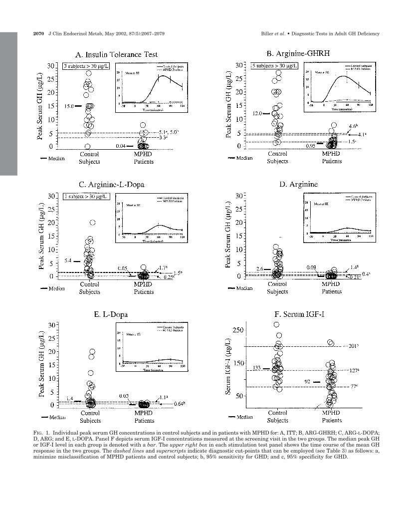
FIG. 1. Individual peak serum GH concentrations in control subjects and in patients with MPHD for: A, ITT; B, ARG-GHRH; C, ARG-L-DOPA;D, ARG; and E, L-DOPA. Panel F depicts serum IGF-I concentrations measured at the screening visit in the two groups. The median peak GHor IGF-I level in each group is denoted with a bar. The upper right box in each stimulation test panel shows the time course of the mean GHresponse in the two groups. The dashed lines and superscripts indicate diagnostic cut-points that can be employed (see Table 3) as follows: a,minimize misclassification of MPHD patients and control subjects; b, 95% sensitivity for GHD; and c, 95% specificity for GHD.
2070 J Clin Endocrinol Metab, May 2002, 87(5):2067–2079 Biller et al. • Diagnostic Tests in Adult GH Deficiency
Results
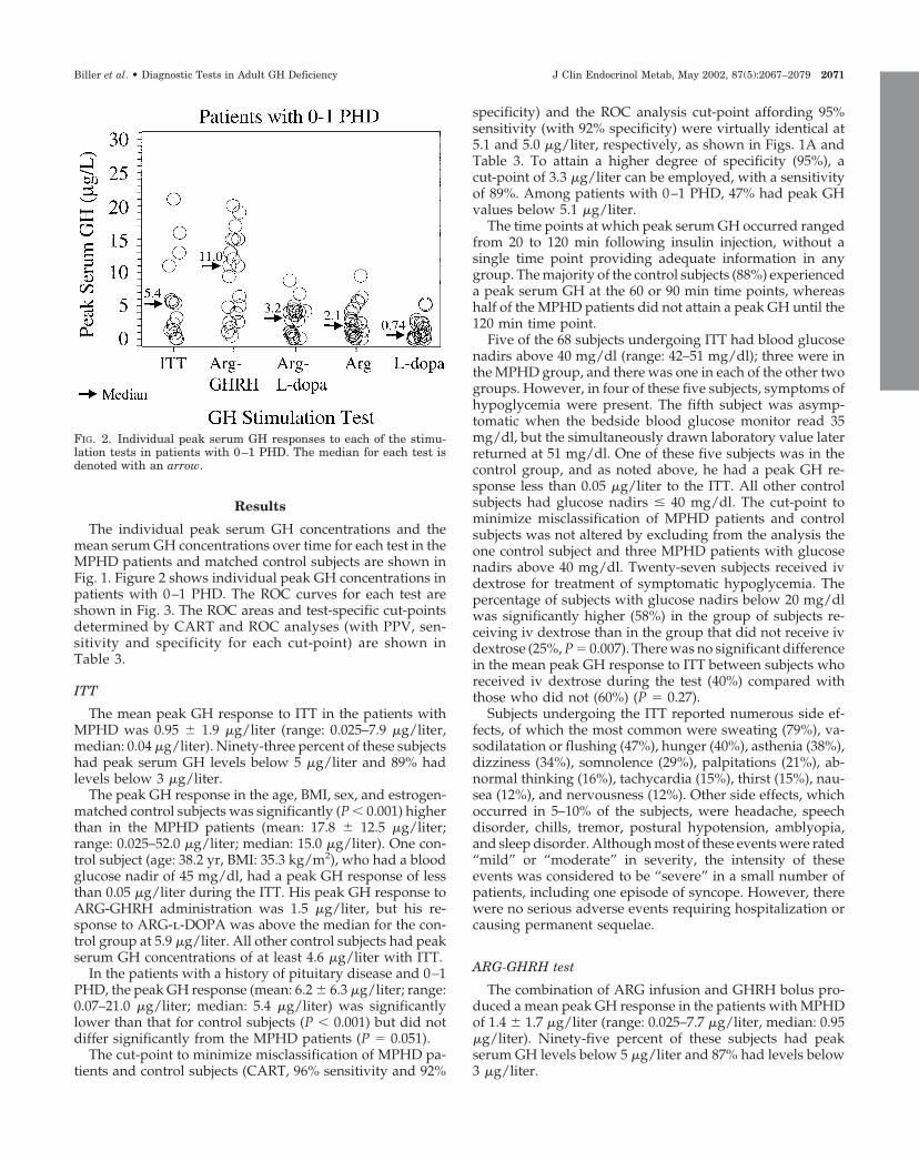
The individual peak serum GH concentrations and themean serum GH concentrations over time for each test in theMPHD patients and matched control subjects are shown inFig. 1. Figure 2 shows individual peak GH concentrations inpatients with 0–1 PHD. The ROC curves for each test areshown in Fig. 3. The ROC areas and test-specific cut-pointsdetermined by CART and ROC analyses (with PPV, sen-sitivity and specificity for each cut-point) are shown inTable 3.
ITT
The mean peak GH response to ITT in the patients withMPHD was 0.95 � 1.9 �g/liter (range: 0.025–7.9 �g/liter,median: 0.04 �g/liter). Ninety-three percent of these subjectshad peak serum GH levels below 5 �g/liter and 89% hadlevels below 3 �g/liter.
The peak GH response in the age, BMI, sex, and estrogen-matched control subjects was significantly (P � 0.001) higherthan in the MPHD patients (mean: 17.8 � 12.5 �g/liter;range: 0.025–52.0 �g/liter; median: 15.0 �g/liter). One con-trol subject (age: 38.2 yr, BMI: 35.3 kg/m2), who had a bloodglucose nadir of 45 mg/dl, had a peak GH response of lessthan 0.05 �g/liter during the ITT. His peak GH response toARG-GHRH administration was 1.5 �g/liter, but his re-sponse to ARG-l-DOPA was above the median for the con-trol group at 5.9 �g/liter. All other control subjects had peakserum GH concentrations of at least 4.6 �g/liter with ITT.
In the patients with a history of pituitary disease and 0–1PHD, the peak GH response (mean: 6.2 � 6.3 �g/liter; range:0.07–21.0 �g/liter; median: 5.4 �g/liter) was significantlylower than that for control subjects (P � 0.001) but did notdiffer significantly from the MPHD patients (P � 0.051).
The cut-point to minimize misclassification of MPHD pa-tients and control subjects (CART, 96% sensitivity and 92%
specificity) and the ROC analysis cut-point affording 95%sensitivity (with 92% specificity) were virtually identical at5.1 and 5.0 �g/liter, respectively, as shown in Figs. 1A andTable 3. To attain a higher degree of specificity (95%), acut-point of 3.3 �g/liter can be employed, with a sensitivityof 89%. Among patients with 0–1 PHD, 47% had peak GHvalues below 5.1 �g/liter.
The time points at which peak serum GH occurred rangedfrom 20 to 120 min following insulin injection, without asingle time point providing adequate information in anygroup. The majority of the control subjects (88%) experienceda peak serum GH at the 60 or 90 min time points, whereashalf of the MPHD patients did not attain a peak GH until the120 min time point.
Five of the 68 subjects undergoing ITT had blood glucosenadirs above 40 mg/dl (range: 42–51 mg/dl); three were inthe MPHD group, and there was one in each of the other twogroups. However, in four of these five subjects, symptoms ofhypoglycemia were present. The fifth subject was asymp-tomatic when the bedside blood glucose monitor read 35mg/dl, but the simultaneously drawn laboratory value laterreturned at 51 mg/dl. One of these five subjects was in thecontrol group, and as noted above, he had a peak GH re-sponse less than 0.05 �g/liter to the ITT. All other controlsubjects had glucose nadirs � 40 mg/dl. The cut-point tominimize misclassification of MPHD patients and controlsubjects was not altered by excluding from the analysis theone control subject and three MPHD patients with glucosenadirs above 40 mg/dl. Twenty-seven subjects received ivdextrose for treatment of symptomatic hypoglycemia. Thepercentage of subjects with glucose nadirs below 20 mg/dlwas significantly higher (58%) in the group of subjects re-ceiving iv dextrose than in the group that did not receive ivdextrose (25%, P � 0.007). There was no significant differencein the mean peak GH response to ITT between subjects whoreceived iv dextrose during the test (40%) compared withthose who did not (60%) (P � 0.27).
Subjects undergoing the ITT reported numerous side ef-fects, of which the most common were sweating (79%), va-sodilatation or flushing (47%), hunger (40%), asthenia (38%),dizziness (34%), somnolence (29%), palpitations (21%), ab-normal thinking (16%), tachycardia (15%), thirst (15%), nau-sea (12%), and nervousness (12%). Other side effects, whichoccurred in 5–10% of the subjects, were headache, speechdisorder, chills, tremor, postural hypotension, amblyopia,and sleep disorder. Although most of these events were rated“mild” or “moderate” in severity, the intensity of theseevents was considered to be “severe” in a small number ofpatients, including one episode of syncope. However, therewere no serious adverse events requiring hospitalization orcausing permanent sequelae.
ARG-GHRH test
The combination of ARG infusion and GHRH bolus pro-duced a mean peak GH response in the patients with MPHDof 1.4 � 1.7 �g/liter (range: 0.025–7.7 �g/liter, median: 0.95�g/liter). Ninety-five percent of these subjects had peakserum GH levels below 5 �g/liter and 87% had levels below3 �g/liter.
FIG. 2. Individual peak serum GH responses to each of the stimu-lation tests in patients with 0–1 PHD. The median for each test isdenoted with an arrow.
Biller et al. • Diagnostic Tests in Adult GH Deficiency J Clin Endocrinol Metab, May 2002, 87(5):2067–2079 2071
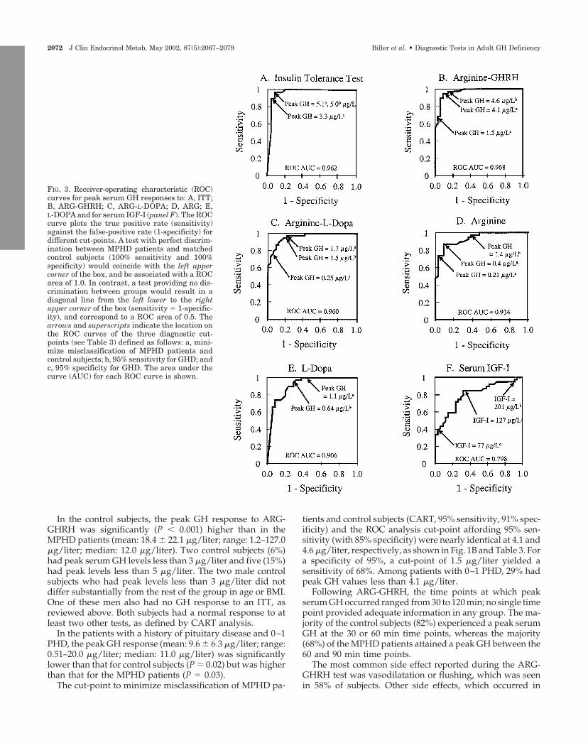
In the control subjects, the peak GH response to ARG-GHRH was significantly (P � 0.001) higher than in theMPHD patients (mean: 18.4 � 22.1 �g/liter; range: 1.2–127.0�g/liter; median: 12.0 �g/liter). Two control subjects (6%)had peak serum GH levels less than 3 �g/liter and five (15%)had peak levels less than 5 �g/liter. The two male controlsubjects who had peak levels less than 3 �g/liter did notdiffer substantially from the rest of the group in age or BMI.One of these men also had no GH response to an ITT, asreviewed above. Both subjects had a normal response to atleast two other tests, as defined by CART analysis.
In the patients with a history of pituitary disease and 0–1PHD, the peak GH response (mean: 9.6 � 6.3 �g/liter; range:0.51–20.0 �g/liter; median: 11.0 �g/liter) was significantlylower than that for control subjects (P � 0.02) but was higherthan that for the MPHD patients (P � 0.03).
The cut-point to minimize misclassification of MPHD pa-
tients and control subjects (CART, 95% sensitivity, 91% spec-ificity) and the ROC analysis cut-point affording 95% sen-sitivity (with 85% specificity) were nearly identical at 4.1 and4.6 �g/liter, respectively, as shown in Fig. 1B and Table 3. Fora specificity of 95%, a cut-point of 1.5 �g/liter yielded asensitivity of 68%. Among patients with 0–1 PHD, 29% hadpeak GH values less than 4.1 �g/liter.
Following ARG-GHRH, the time points at which peakserum GH occurred ranged from 30 to 120 min; no single timepoint provided adequate information in any group. The ma-jority of the control subjects (82%) experienced a peak serumGH at the 30 or 60 min time points, whereas the majority(68%) of the MPHD patients attained a peak GH between the60 and 90 min time points.
The most common side effect reported during the ARG-GHRH test was vasodilatation or flushing, which was seenin 58% of subjects. Other side effects, which occurred in
FIG. 3. Receiver-operating characteristic (ROC)curves for peak serum GH responses to: A, ITT;B, ARG-GHRH; C, ARG-L-DOPA; D, ARG; E,L-DOPA and for serum IGF-I (panel F). The ROCcurve plots the true positive rate (sensitivity)against the false-positive rate (1-specificity) fordifferent cut-points. A test with perfect discrim-ination between MPHD patients and matchedcontrol subjects (100% sensitivity and 100%specificity) would coincide with the left uppercorner of the box, and be associated with a ROCarea of 1.0. In contrast, a test providing no dis-crimination between groups would result in adiagonal line from the left lower to the rightupper corner of the box (sensitivity � 1-specific-ity), and correspond to a ROC area of 0.5. Thearrows and superscripts indicate the location onthe ROC curves of the three diagnostic cut-points (see Table 3) defined as follows: a, mini-mize misclassification of MPHD patients andcontrol subjects; b, 95% sensitivity for GHD; andc, 95% specificity for GHD. The area under thecurve (AUC) for each ROC curve is shown.
2072 J Clin Endocrinol Metab, May 2002, 87(5):2067–2079 Biller et al. • Diagnostic Tests in Adult GH Deficiency
5–10% of the subjects, were paresthesias, nausea, and ab-normal taste sensation.
ARG-L-DOPA test
The combined ARG-l-DOPA test produced a mean peakGH response in the patients with MPHD of 0.31 � 0.65�g/liter (range: 0.025–3.5 �g/liter, median: 0.05 �g/liter).All but one of these subjects had a peak serum GH level lessthan 3 �g/liter.
The peak GH response in the control subjects was signif-icantly (P � 0.001) higher than in the MPHD patients (mean:6.7 � 7.1 �g/liter; range: 0.16–37.0 �g/liter; median: 5.4�g/liter). However, a substantial number of control subjects(35%) had a peak serum GH level less than 3 �g/liter, and47% of this group had values less than 5 �g/liter.
In patients with 0–1 PHD, the peak GH response (mean:3.0 � 2.4 �g/liter; range: 0.09–8.8 �g/liter; median: 3.2 �g/liter) was significantly lower than that for control subjects(P � 0.004) but was higher than that for the MPHD patients(P � 0.04).
A cut-point of 1.7 �g/liter minimized the misclassificationof MPHD patients and control subjects and provided 97%sensitivity and 79% specificity (Fig. 1C and Table 3). Usingsensitivity set at 95% with ROC analysis, a cut-point of 1.5�g/liter yielded 79% specificity. To achieve a specificity of95%, a low cut-point of 0.25 �g/liter was required, resultingin a sensitivity of 75%. Among patients with 0–1 PHD, 40%had peak GH values less than 1.7 �g/liter.
The time to peak serum GH occurred at 30 or 60 min in 68%of the control subjects. In contrast, peak serum GH wasmeasured at the 120 min time point in 68% of the MPHDpatients.
The most common side effects during the ARG-l-DOPAtest were nausea (29%), vomiting (12%) and paresthesias(12%). Other side effects reported in 5–10% of subjects in-cluded asthenia, dizziness, abnormal taste sensation, and drymouth.
ARG test
Following ARG infusion, the mean peak serum GH re-sponse in the patients with MPHD was 0.3 � 0.51 �g/liter(range: 0.025–2.4 �g/liter, median: 0.09 �g/liter). All pa-tients had a peak serum GH level less than 3 �g/liter.
In control subjects, the peak GH response was significantly(P � 0.001) higher than in the MPHD patients (mean: 3.7 �3.3�g/liter; range: 0.08–11.0 �g/liter; median: 2.6 �g/liter).However, the majority of control subjects (59%) had peakserum GH levels less than 3 �g/liter and 68% of this grouphad values less than 5 �g/liter.
In the group of patients with 0–1 PHD, the peak serum GHresponse (mean: 2.6 � 2.5 �g/liter; range: 0.06–9.5 �g/liter;median: 2.1 �g/liter) was significantly higher than that forthe MPHD patients (P � 0.001) but did not differ significantlyfrom the control subjects (P � 0.12).
As shown in Fig. 1D and Table 3, a cut-point of 0.4 �g/literminimized the misclassification of MPHD patients and con-trol subjects and produced a sensitivity of 87% and a spec-ificity of 91%. For a higher sensitivity (95%), a cut-point of 1.4�g/liter resulted in a specificity of 62%. To achieve a highspecificity (95%), a lower cut-point of 0.21 �g/liter was re-quired, which was associated with a sensitivity of 74%.Among patients with 0–1 PHD, 19% had peak GH values lessthan 0.4 �g/liter.
Sixty-five percent of control subjects had a peak serum GHlevel measured at the 60 or 90 min time points, whereas thepeak occurred at 90 or 120 min in 74% of the MPHD patients.
Side effects were uncommon with the ARG test, but 5–10%of subjects reported paresthesias, dry mouth, and headache.
L-DOPA test
The mean peak serum GH response to l-DOPA in thepatients with MPHD was 0.13 � 0.22 �g/liter (range: 0.025–1.0 �g/liter, median: 0.03 �g/liter). None had peak serumGH levels above 3 �g/liter.
In control subjects, the peak GH response was significantly(P � 0.001) higher than in the MPHD patients (mean: 3.3 �4.9 �g/liter; range: 0.025–20.0 �g/liter; median: 1.4 �g/liter).Nevertheless, the majority of control subjects (68%) had peakserum GH levels below 3 �g/liter and most (79%) were lessthan 5 �g/liter.
In the patients with 0–1 PHD, the peak serum GH response(mean: 1.4 � 1.6 �g/liter; range: 0.025–5.3 �g/liter; median:0.74 �g/liter) was significantly lower than that for controlsubjects (P � 0.03) but did not differ significantly from theMPHD patients (P � 0.13).
A cut-point of 1.1 �g/liter minimized the misclassification
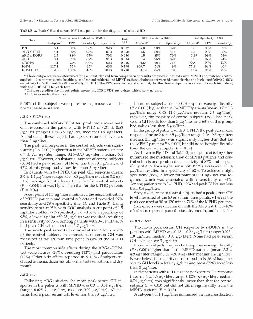
TABLE 3. Peak GH and serum IGF-I cut-pointsa for the diagnosis of adult GHD
TestMinimize misclassification (CART) ROC
AUC95% Sensitivity (ROC) 95% Specificity (ROC)
Cut-pointb PPV Sensitivity Specificity Cut-pointb PPV Specificity Cut-pointb PPV Sensitivity
ITT 5.1 93% 96% 92% 0.962 5.0 93% 92% 3.3 96% 89%ARG-GHRH 4.1 92% 95% 91% 0.968 4.6 88% 85% 1.5 96% 68%ARG-L-DOPA 1.7 84% 97% 79% 0.960 1.5 85% 79% 0.25 96% 75%ARG 0.4 92% 87% 91% 0.934 1.4 75% 62% 0.21 97% 74%L-DOPA 1.1 75% 100% 62% 0.906 0.64 78% 71% N/A N/A N/AIGF-I 127.1 75% 85% 68% 0.798 200.7 54% 9% 77.2 94% 40%IGF-I SDS �2.00 100% 46% 100% 0.790 0.12 54% 6% �1.94 95% 49%
a Three cut-points were determined for each test, derived from comparison of results obtained in patients with MPHD and matched controlsubjects: 1) to minimize misclassification of control subjects and MPHD patients (balance between high sensitivity and high specificity); 2) 95%sensitivity for GHD; and 3) 95% specificity for GHD. The PPV, sensitivity and specificity for the three cut-points are shown for each test, alongwith the ROC AUC for each test.
b Units are �g/liter for all cut-points except the IGF-I SDS cut-points, which have no units.AUC, Area under the curve.
Biller et al. • Diagnostic Tests in Adult GH Deficiency J Clin Endocrinol Metab, May 2002, 87(5):2067–2079 2073

of MPHD patients and control subjects and provided 100%sensitivity and a specificity of 62%, as shown in Fig. 1E andTable 3. A lower cut-point of 0.64 �g/liter increased thespecificity to 71%, while maintaining 95% sensitivity. How-ever, ROC analysis demonstrated that it was not possible toreach 95% specificity with this test. Among patients with 0–1PHD, 55% had peak GH values less than 1.1 �g/liter.
The peak serum GH was measured at 90 or 120 min in 74%of control subjects and in 87% of MPHD patients.
Twenty-six percent of subjects reported nausea with l-DOPA. Other side effects seen in 5–10% of subjects weredizziness, asthenia, and headache.
Comparison of GH stimulation test results
Results obtained with the five GH stimulation tests werecompared using the cut-points to minimize misclassificationof MPHD patients and control subjects (CART analysis) foreach test. Specifically, a patient was considered to have anormal response if the peak GH value exceeded the test-specific cut-point. Only two MPHD patients responded nor-mally on more than one test. Four other MPHD patientsresponded normally to one test but not to the other tests.Only one MPHD patient, who had a pituitary adenoma,responded normally to the ITT but subsequently withdrewfrom the study and did not have any other tests. Of theseseven patients, six had pituitary adenomas and 1 had a cra-niopharyngioma. The patient with the craniopharyngiomaresponded normally to ARG-GHRH and ARG but not to theITT (peak GH � 0.43 �g/liter), l-DOPA, or ARG-l-DOPA.The only other MPHD patient who responded normally toARG-GHRH had a pituitary adenoma and had abnormalresponses to ARG-l-DOPA, ARG, and l-DOPA; he did nothave an ITT due to his age (63.5 yr).
Effect of sex, age, and BMI on peak GH values
For the GH response to ITT in control subjects, BMI hada significant inverse relationship with peak serum GH whencontrolling for age (r � �0.43, P � 0.034); similar results wereobserved with ARG-GHRH (r � �0.36, P � 0.037). For everyincrease of 1 kg/m2, the peak serum GH level was 0.89�g/liter lower for the ITT and 1.4 �g/liter lower for theARG-GHRH test. In contrast, BMI had no significant effecton the peak GH response to the other three stimulation testsin the control subjects. Among MPHD patients, BMI had aninverse effect on peak GH when controlling for age withl-DOPA (r � �0.32, P � 0.05) but no significant effect withthe other stimulation tests. Age had no significant effect onpeak serum GH in control or MPHD subjects for any of thefive GH stimulation tests.
Sex had a significant effect on peak GH in control subjectswhen controlling for age with the ARG (P � 0.01), andARG-GHRH tests (P � 0.03). The female control subjects (n �4) had higher peak serum GH values than the male controlsubjects (n � 20) on the ARG test (5.2 � 3.3 vs. 2.6 � 3.0�g/liter), and on the ARG-GHRH test (27.1 � 30.5 vs. 12.3 �10.9 �g/liter). There was no statistically significant effect ofsex on the GH responses to the other tests in control subjects,or on the results of any stimulation test in the patients withMPHD.
Despite these relationships identified by regression anal-yses, the inclusion of sex, age, and BMI in the model did notalter the peak GH cut-points arrived at by CART analysis.
Patient test preferences
Table 4 shows subject preferences for the five stimulationtests. The ITT was the least preferred test by subjects in allthree study groups. ARG alone was ranked as the mostpreferred test. The ARG-GHRH test was preferred signifi-cantly more than the ITT (P � 0.001).
IGF-I
The serum IGF-I concentration at the screening visit washighly correlated with the level at the first stimulation testvisit (r � 0.87, P � 0.001). Correlations were similar amongthe subgroups (control subjects: r � 0.80; MPHD patients: r �0.87; 0–1 PHD patients: r � 0.89, P � 0.001 for all). There wereno significant differences between the IGF-I concentrations atvisit 1 compared with visit 2 for control subjects (141.6 � 44.6vs. 133.1 � 35.9 �g/liter, P � 0.074), MPHD patients (90.9 �45.6 vs. 92.7 � 48.5 �g/liter, P � 0.66) or 0–1 PHD patients(127.1 � 58.4 vs. 117.2 � 62.3 �g/liter, P � 0.14). Therefore,the screening visit IGF-I values were used in the evaluationof IGF-I as a diagnostic test using CART and ROC analyses.When all three groups were considered together, there werestatistically significant (P � 0.05) correlations between peakGH and serum IGF-I for each GH stimulation test (r valuesranging from 0.21 to 0.44), but this relationship only ac-counted for 4–19% of the variance in the serum IGF-I values.Within each group of patients, the correlations between peakGH and IGF-I were less consistent with relatively small sam-ple sizes.
Although the mean serum IGF-I levels were significantlydifferent in MPHD patients compared with carefullymatched control subjects (P � 0.001), there was substantialoverlap between the two groups (Fig. 1F). In the patients witha history of pituitary disease and 0–1 PHD, serum IGF-Iconcentrations were intermediate between the values for theother two groups. These values were significantly higherthan those observed in the MPHD patients (P � 0.007), butthey did not differ from the control group values.
Among control subjects, 3 of 34 subjects (8.8%) had IGF-Ivalues that were slightly below the normal range for age andsex (provided by the central laboratory) at the first office visit.All three of these control subjects had low normal IGF-I
TABLE 4. Patient preferences among the five GHstimulation tests
Patients withMPHD
Controlsubjects
Patients with0–1 PHD
ARGa 2.0 � 1.0 1.8 � 1.0 1.9 � 1.2L-DOPAa,b 2.4 � 1.3 2.4 � 1.2 2.5 � 1.3ARG-GHRHb,c 2.7 � 1.2 2.8 � 1.0 2.8 � 1.1ARG-L-DOPAc 3.2 � 1.2 3.2 � 1.1 3.1 � 0.9ITT 4.6 � 0.8 4.8 � 0.4 4.7 � 0.9
A lower number indicates higher patient preference (1 � mostpreferred, 5 � least preferred). Data are reported for subjects who hadall five tests.
a,b,c Superscripts indicate tests with rankings that were not sta-tistically different (P � 0.05).
2074 J Clin Endocrinol Metab, May 2002, 87(5):2067–2079 Biller et al. • Diagnostic Tests in Adult GH Deficiency

values at the second visit. Fifty-six percent of the MPHDpatients had IGF-I values that were below the normal rangefor age and sex. Among patients with 0–1 PHD, 38.1% hadIGF-I values that were below the normal range.
A cut-point of 127.1 �g/liter minimized the misclassifi-cation of MPHD patients and control subjects and provideda sensitivity of 85% and a specificity of 68% as shown in Fig.1F and Table 3. Among patients with 0–1 PHD, 57% hadserum IGF-I values below 127 �g/liter. For a higher sensi-tivity (95%), a cut-point of 200.7 �g/liter resulted in a spec-ificity of only 9%. However, a cut-point of 77.2 �g/literyielded 95% specificity and a sensitivity of 40%. Thus, anIGF-I level less than 77 �g/liter may be useful for identifi-cation of patients with a very high probability of GHD. Al-ternatively, an IGF-I SDS of �2.00 may be used, which pro-vided 100% specificity but only 46% sensitivity for GHD(Table 3).
Discussion
This study is the first to compare six methods of testing forGHD in adults with hypothalamic-pituitary disorders and incontrol subjects matched for age, sex, estrogen use, and BMI.We considered patients with MPHD to be at the greatest riskfor GHD, as has been previously demonstrated (4, 20–21,27–29). The GH response to ITT and the ARG-GHRH testproduced the sharpest separation between MPHD patientsand control subjects, but the ARG-GHRH test was preferredby patients. For all tests, the definition of test-specific cut-points improved the sensitivity and specificity for the diag-nosis of adult GHD. Peak serum GH occurred earlier in themajority of control subjects than in patients with pituitarydisease. However, there was a wide variation in the timingof peak GH in all three study groups, indicating that it is notadvisable to streamline stimulation test blood sampling toone or two time points.
The diagnosis of GHD in adults is challenging because ofthe lack of a single specific biologic end-point, such as growthfailure, which is the cardinal clinical sign in pediatric pa-tients. Therefore, the confirmation of GHD largely rests onlaboratory testing in the context of a history of childhoodGHD or adult-onset hypothalamic-pituitary disease. Thepeak GH response to an ITT is more specific than measure-ment of 24-h spontaneous GH release for the diagnosis ofadult GHD (30). The use of a cut-point or threshold fornormal GH response to a stimulation test is arbitrary; GHsecretion may not be completely absent in GHD, but mayrather reflect a continuum between normal and abnormal(28). Few studies to date have evaluated different peak GHcut-points for different stimulation tests (5, 16, 18). The cut-points of 3 or 5 �g/liter, which have been accepted as de-fining GHD, have often been applied in clinical practiceregardless of the pharmacologic agents used. Another im-portant variable to consider regarding the use of an arbitrarycut-point is the type of GH assay performed (18, 31). Whenthe same serum samples are tested in different assays, thereis wide variability in the absolute values reported. As aresult, the classification of individual subjects as normal orGH deficient may change (32, 33).
In the current study, a single GH assay was employed, and
two different statistical methods were used to define cut-points for the diagnosis of GHD. Using ROC analysis, 95%sensitivity and 95% specificity cut-points were calculated foreach test. High sensitivity cut-points maximize detection ofadult GHD, whereas high specificity cut-points minimizemisclassification of normal subjects as GH-deficient. Thus,clinicians can choose whether the priority for an individualpatient is to attain high sensitivity or high specificity, and usecorresponding cut-points. For example, in a patient withpanhypopituitarism, in whom there is a very high probabil-ity of GHD (4, 20, 21, 27–29), a clinician might prefer a testwith at least 95% sensitivity (limiting the chance of a falsenegative result), in order not to misclassify the patient ashaving normal GH secretion, and withhold potentially ben-eficial therapy. In contrast, in an asymptomatic patient with0–1 PHD, the risk of GHD is lower (4, 20–21, 27–29). In sucha patient, the goal might be high specificity (limiting thechance of a false positive test), to avoid the unnecessary useof GH replacement. However, if such a patient had symp-toms compatible with GHD, the use of the 95% sensitivitycut-point might be deemed most appropriate by some cli-nicians. For a balance between high sensitivity and highspecificity, the cut-point derived by CART analysis to min-imize misclassification of MPHD and control subjects may beused.
There are many variables affecting GH secretion and re-sponsiveness to provocative testing (17–19). GH release de-clines with age and is influenced by sex, estrogen use, andbody composition (17). Obesity suppresses GH release, with24-h GH levels in obese men reduced by 75% in comparisonwith age-matched normal weight subjects (34). Each one unitincrease in BMI has been shown to be associated with a 6%decrease in 24-h GH secretion (35). The amount of abdominalvisceral fat is a stronger predictor of 24-h GH release than istotal percentage body fat (36). In addition, decreased respon-siveness to stimulation tests such as GHRH, ITT, ARG, l-DOPA, and ARG-GHRH has been demonstrated in subjectswith obesity and/or abdominal adiposity (8, 10, 17). Thus,permanent GHD due to organic hypothalamic-pituitary dis-ease may be difficult to distinguish from the reversible blunt-ing of GH secretion in obesity (18). These findings under-score the importance of using a control population matchedfor age, sex, estrogen use, and BMI when evaluating GHstimulation tests for the diagnosis of GHD. No prior studyevaluating a number of GH stimulation test agents has con-trolled for all of these variables. In the present study, BMIwas inversely related to peak serum GH responses to the ITTand ARG-GHRH test in control subjects. This relationshipwas not seen in MHPD subjects (likely due to their overallvery low responses), nor was it seen for any of the other tests.Although the inclusion of age, sex, and BMI did not alter thecut-points arrived at by CART analysis, the present studywas not designed to evaluate whether different peak GHcut-points are necessary based on age, sex, or BMI. Theinclusion of control subjects matched for these variables re-sulted in lower peak GH cut-points for some of the tests thanare presently used in clinical practice. Therefore, the use ofthese new cut-points will improve the specificity of diag-nostic testing for adult GHD.
The ITT has been considered the diagnostic gold standard
Biller et al. • Diagnostic Tests in Adult GH Deficiency J Clin Endocrinol Metab, May 2002, 87(5):2067–2079 2075
in establishing the presence of GHD in adults (3). An ad-vantage of the test is that it allows evaluation of the completehypothalamic-somatotroph axis, making it useful in patientswith both hypothalamic and pituitary disease. However,there are a number of disadvantages to this test. Patients withcontraindications to hypoglycemia such as seizures and isch-emic heart disease are not considered candidates, and patientsafety requirements make it a labor-intensive procedure (37,38). In addition, the ITT has poor reproducibility for anindividual subject. Up to a 6-fold difference in peak GH hasbeen demonstrated on different days in healthy adults un-dergoing ITTs, regardless of the degree of hypoglycemia (16,39). Although some investigators have shown total separa-tion of patients from carefully matched control subjects usingthe ITT (30), others have reported overlap between thegroups (13). Our study did not evaluate reproducibility of theITT, but it did demonstrate near-complete separation be-tween control subjects and those patients considered at highrisk for GHD, based on of the presence of at least two otherPHD. Only one control subject had a peak GH of less than3 �g/liter following ITT; his failure to respond was likely dueto his morbid obesity and inadequate hypoglycemia. Thehigh diagnostic accuracy of this test is demonstrated by thearea under the ROC curve of 0.962 (1.00 would indicateperfect separation between the diseased and normal groups).A peak serum GH cut-point of 5.0 �g/liter provided 95%sensitivity and 92% specificity for the diagnosis of GHD.Although the test is clearly useful for separating MPHDpatients from control subjects, it was the least preferred stim-ulation test by patients.
The ARG-GHRH test performed equally well, as shown byan area under the ROC curve of 0.968, indicating that itprovides an ideal alternative to the ITT. Subjects preferredthis test to the ITT. The GH response to ARG-GHRH isindependent of age, and there is less inter and intraindi-vidual variability than with other stimulation tests. Thus, ithas been considered the best diagnostic alternative to the ITT(3, 40, 41). However, the GH response to ARG-GHRH isdecreased in healthy obese subjects (42). Interestingly, twostudies have reported higher peak GH levels to ARG-GHRHthan to ITT, a finding not seen in our study (5, 15). In thereport by Ghigo et al. (15), the control subjects were within15% of ideal body weight. Our control subjects had higherBMIs (mean: 30.3 � 5.8 kg/m2; highest value: 45.6 kg/m2)than in these previous reports, because they were intention-ally BMI-matched to the MPHD patients. If the peak GHcut-point of 9 �g/liter, suggested by previous studies, (5)were applied to our population, all patients with MPHDwould be classified as GH deficient, but many of our controlsubjects would fall below this threshold as well. The peakserum GH cut-point that minimized the misclassification ofMPHD and control subjects in the current study was 4.1�g/liter; this cut-point provided 95% sensitivity and 91%specificity. This excellent separation between patients withGHD and carefully matched control subjects, coupled withthe high degree of patient acceptability, suggests that theARG-GHRH test is the best alternative to ITT in ourpopulation.
Many investigators have evaluated ARG alone as a pro-vocative test of GH secretion in adults (11, 12, 43, 44). This
amino acid is believed to increase GH by suppressing en-dogenous somatostatin (11, 45, 46). The ARG test was rankedthe most preferred test by our study subjects. ARG has beenshown to have high intraindividual reproducibility, but nor-mal subjects often have peak serum GH values less than 3�g/liter, producing overlap with patients considered to havesevere GHD (44, 47). Indeed, over half of the control subjectsin the current study would be misclassified as GH-deficientif this criterion were used. A peak serum GH cut-point of 1.4�g/liter provided 95% sensitivity, but only 62% specificity.To achieve 91% specificity a very low cut-point of 0.4 �g/literwas required, but this resulted in a lower sensitivity (87%).The specificity of the ARG test can be substantially improvedby combining it with l-DOPA. Children have been shown tohave a greater response to the combination of these agentsthat to either alone, but there have been no controlled dataevaluating this combination in adults (48). In the currentstudy, a peak serum GH cut-point of 1.5 �g/liter for theARG-l-DOPA test provided 95% sensitivity and 79% spec-ificity. Although testing with l-DOPA alone has been sug-gested as an effective alternative to the ITT, it was the leastuseful of the five GH stimulation tests we evaluated. Thel-DOPA test had the lowest ROC area among the five stim-ulation tests, reflecting the fact that the majority of MPHDpatients overlapped with control subjects. The earlier, morepositive studies included only nonobese normal subjects, asyoung as 13 yr old, which may account for the higher serumGH responses to l-DOPA. These older studies also employedGH RIAs, producing higher absolute values than assays thatare now commonly used (6, 7). Considering the ease of ad-ministration of l-DOPA, which is the only oral GH stimu-lation agent, this could be used as a screening test for de-termining which patients do not need further testing. A peakGH response greater than 1.1 �g/liter, the cut-point to min-imize misclassification of MPHD and control subjects, couldbe used to identify patients who most likely do not haveGHD. All 16 of the control subjects with such a response tol-DOPA had peak GH responses greater than 5 �g/liter withITT. Patients with peak serum GH levels below this cut-pointwould need another test with a higher specificity, such asITT, ARG-GHRH, or ARG-l-DOPA to confirm the diagnosisof GHD.
Although the mean serum IGF-I concentrations were sig-nificantly different in MPHD patients compared with care-fully matched control subjects, there was substantial overlapbetween the two groups. This test had the lowest ROC areaamong the six diagnostic tests evaluated. This is not sur-prising, as numerous studies have demonstrated that somepatients with GHD have serum IGF-I concentrations withinthe normal range, as was the case for 44% of the MPHDpatients in the present study (4, 15, 16, 21, 27, 30, 47, 49–51).Because of the decline in serum IGF-I levels with age, thediagnostic utility of measuring this hormone is particularlylow in older patients (15, 44). An IGF-I level below a certaincut-point might be useful for the diagnosis of GHD, espe-cially in childhood-onset or young adult-onset GHD patients(50, 52). The most useful finding of the present study re-garding IGF-I measurement was that a cut-point of 77.2 �g/liter provided 95% specificity for GHD; only one controlsubject had an IGF-I value below this cut-point. Similarly,
2076 J Clin Endocrinol Metab, May 2002, 87(5):2067–2079 Biller et al. • Diagnostic Tests in Adult GH Deficiency

another study reported that an IGF-I value below 84 �g/literhad 95% PPV and 89% specificity for adult GHD (4). Thesefindings might allow a subgroup of patients to undergo asingle blood sample instead of a stimulation test. However,because different IGF-I assays may yield different results,and serum IGF-I concentrations may be decreased by a va-riety of causes, caution should be used in applying low IGF-Idiagnostic cut-points to the diagnosis of adult GHD in clin-ical practice (3). SDS offer the advantage of taking into ac-count age and sex. We found that in this population, an IGF-ISDS of �2.00 provided 100% specificity for the diagnosis ofGHD in the age range studied. Our study did not includenormal subjects over the age of 68 yr; it will be important forfuture studies to evaluate the diagnostic utility of IGF-I SDSin elderly patients with pituitary disease.
The severity of pituitary disease, marked by the numberof PHD present, is associated with the severity of GHD. Themean peak GH response to provocative testing and serumIGF-I concentrations decline progressively with increasingnumber of PHD (4, 20, 21, 27–29, 51). Two studies reportedthat 87–91% of patients with two or more PHD had severeGHD (20, 21). This was reproduced in the current study, as89% of the MPHD patients had a peak serum GH less than3 �g/liter on the ITT. The concordance between differenttests in an individual patient may be higher in patients withMPHD (28). In the current study, the group of subjects with0–1 PHD had IGF-I and peak GH values that were interme-diate between the MPHD and control subjects. Using thecut-points that minimized misclassification of MPHD pa-tients and control subjects, the percentage of patients with0–1 PHD that were classified as GHD varied from 19–55%depending on the GH stimulation test. With ITT and ARG-GHRH, the percentage of patients with 0–1 PHD who hadpeak GH less than 3 �g/liter was 40% and 24%, respectively.This probably reflects the biological heterogeneity of thispatient group and is consistent with the findings of previousstudies. Thus, for the diagnostic evaluation of such patients,the use of cut-points with high specificity may be needed toavoid treating those who are not GH deficient.
Limitations of this study must be considered. Because theITT was performed only in subjects under 55 yr of age, dataregarding the GH response to hypoglycemia are not avail-able for older participants. Borderline attainment of hypo-glycemia in five subjects may have blunted the magnitude ofthe peak GH response to ITT. However, the peak GH re-sponse to ITT does not correlate with the degree of hypo-glycemia in healthy adults (53). Furthermore, exclusion ofthese five subjects from the analysis did not change the peakGH cut-point for the ITT. Because of variability in GH assays(18, 31–33), the peak GH cut-points suggested here applyonly to the particular assay used. Another limitation to anystudy evaluating tests for the diagnosis of GHD lies in thedefinition of the disorder. Because there is no absolutemethod for distinguishing between GH sufficiency and de-ficiency, there must be an arbitrary determination used toevaluate the chosen diagnostic tests. Finally, pulsatile andstimulated GH levels may be higher in the preovulatory andluteal phase of the menstrual cycle than during the follicularphase (54). Although female control subjects were matchedto MPHD patients according to estrogen status, subjects with
regular menses were not studied at a particular phase of thecycle, which may have produced some variability betweensubjects.
Many pharmacologic agents other than those included inthis study have been used to stimulate GH secretion.Clonidine alone is clearly not adequate, as many normalindividuals have no detectable GH response, or show thesame response as to placebo (12, 47). Glucagon injection imis considered a useful alternative to ITT by some investiga-tors, but reported by others as being less effective than ARG(7, 12, 55). A number of agents have been used in combinationwith GHRH. Pyridostigmine plus GHRH cannot be usedacross the life span in the same way as ARG-GHRH, becausethe potentiating effect of pyridostigmine on GH release de-clines with age (40). The newest agents used in combinationwith GHRH for the diagnosis of GHD are the synthetic GH-releasing peptides (GHRP). The combination of GHRH withhexarelin, GHRP-6, or GHRP-2 is well tolerated and usefuldiagnostically. Subsequent studies will be needed to confirmthese promising initial findings and to evaluate the proposedcut-points, which are higher than with ITT (14, 56, 57).
In conclusion, this comparison of six tests for the diagnosisof GHD in adults with hypothalamic-pituitary disease andcarefully matched control subjects demonstrated that thegreatest diagnostic accuracy was obtained with the ITT andthe ARG-GHRH test. While there was more overlap betweenMPHD patients and control subjects for ARG, l-DOPA andARG-l-DOPA, test-specific cut-points were defined to im-prove the sensitivity and specificity of these tests. The ARG-l-DOPA test appears to be a reasonable third choice. Thesedata indicate that it is possible to diagnose GHD in adultswithout performing an ITT, provided that test-specific cut-points are employed. The ARG-GHRH test represents anexcellent alternative to the ITT for the diagnosis of GHD inadults.
Acknowledgments
We thank Marie Cook, Marilyn Miller-Collistro, Rhodora Enriquez,Yvonne Francis, Amir Hamrahian, and Karen Pulaski, who assisted theinvestigators with collection of data for this study. We also thank severalother individuals for their contributions to this study: Ray Carroll forstatistical advice; Anne Klibanski, Charmian Quigley, Armando Ama-dor, Amy Rosen, and Chien-Feng Chen for critical review of the manu-script; Mark Muhlhauser, Brian Oertel, Maurice Lunik, and Nekia Clay-ton for project management and technical support; Stan Holley, RobKulak, Laura Macione, Otis Mitchell, and Doreen Struble for field studymanagement. We also thank the many other individuals who contrib-uted to the conduct of this study.
Received October 15, 2001. Accepted February 14, 2002.Address all correspondence and requests for reprints to: Beverly
M. K. Biller, M.D., Neuroendocrine Unit, Bulfinch 457B, MassachusettsGeneral Hospital, Fruit Street, Boston, Massachusetts 02114. E-mail:[email protected].
This study was supported by Eli Lilly & Co.. The results of this studywere reported in part at the Growth Hormone Research Society Con-ference, Goteborg, Sweden, September 7–9, 2000, and at the 7th Inter-national Pituitary Congress, Phoenix, Arizona, June 23–25, 2001.
References
1. de Boer H, Blok GJ, Van der Veen EA 1995 Clinical aspects of growth hormonedeficiency in adults. Endocr Rev 16:63–86
2. American Association of Clinical Endocrinologists 1998 AACE clinical prac-
Biller et al. • Diagnostic Tests in Adult GH Deficiency J Clin Endocrinol Metab, May 2002, 87(5):2067–2079 2077

tice guidelines for growth hormone use in adults and children. Endocr Pract4:165–173
3. Growth Hormone Research Society 1998 Consensus guidelines for the diag-nosis and treatment of adults with growth hormone deficiency: summarystatement of the Growth Hormone Research Society workshop on adultgrowth hormone deficiency. J Clin Endocrinol Metab 83:379–381
4. Hartman ML, Crowe BJ, Biller BMK, Ho KKY, Clemmons DR, Chipman JJ,on behalf of the HypoCSS Advisory Board and The U.S. HypoCSS StudyGroup, 2002 Which patients do not require a growth hormone (GH) stimu-lation test for the diagnosis of adult GH deficiency? J Clin Endocrinol Metab,87:477–485
5. Aimaretti G, Corneli G, Razzore P, Bellone S, Baffoni C, Arvat E, CamanniF, Ghigo E 1998 Comparison between insulin-induced hypoglycemia andgrowth hormone (GH)-releasing hormone � arginine as provocative tests forthe diagnosis of GH deficiency in adults. J Clin Endocrinol Metab 83:1615–1618
6. Eddy RL, Gilliland PF, Ibarra Jr JD, Thompson JQ, MacMurry Jr JF 1974Human growth hormone release: comparison of provocative test procedures.Am J Med 56:179–185
7. Lin T, Tucci J 1974 Provocative tests of growth hormone release. Ann InternMed 80:464–469
8. Vizner B, Reiner Z, Sekso M 1983 Effect of l-DOPA on growth hormone,glucose, insulin, and cortisol response in obese subjects. Exp Clin Endocrinol81:41–48
9. Page M, Dieguez C, Valcavi R, Edwards C, Hall R, Scanlon MF 1988 Growthhormone (GH) responses to arginine and l-DOPA alone and after GHRHpretreatment. Clin Endocrinol 28:551–558
10. Cordido F, Dieguez C, Casanueva FA 1990 Effect of central cholinergic neu-rotransmission enhancement by pyrdostigmine on the growth hormone se-cretion elicited by clonidine, arginine, or hypoglycemia in normal and obesesubjects. J Clin Endocrinol Metab 70:1361–1370
11. Koppeschaar HPF, ten Horn CD, Thijssen JHH, Page MD, Dieguez C, Scan-lon MF 1992 Differential effects of arginine on growth hormone releasinghormone and insulin induced growth hormone secretion. Clin Endocrinol36:487–490
12. Rahim A, Toogood AA, Shalet SM 1996 The assessment of growth hormonestatus in normal young adult males using a variety of provocative agents. ClinEndocrinol 45:557–562
13. Fisker S, Jørgensen JO, Ørskov H, Christiansen JS 1998 l-Arginine andinsulin-tolerance tests in the diagnosis of adult growth hormone deficiency:influence of confounding factors. Clin Endocrinol 48:109–115
14. Mahajan T, Lightman SL 2000 A simple test for growth hormone deficiencyin adults. J Clin Endocrinol Metab 85:1473–1476
15. Ghigo E, Aimaretti G, Gianotti L, Bellone J, Arvat E, Camanni F 1996 Newapproach to the diagnosis of growth hormone deficiency in adults. Eur JEndocrinol 134:352–356
16. Hoeck HC, Vestergaard P, Jakobsen PE, Falhof J, Laurberg P 2000 Diagnosisof growth hormone (GH) deficiency in adults with hypothalamic-pituitarydisorders: comparison of test results using pyridostigmine plus GH-releasinghormone (GHRH), clonidine plus GHRH, and insulin-induced hypoglycemiaas GH secretagogues. J Clin Endocrinol Metab 85:1467–1472
17. Hartman ML 2000 Physiological regulators of growth hormone secretion. In:Juul A, Jørgensen JOL, eds. Growth hormone in adults, 2nd ed. Cambridge:Cambridge University Press; 3–53
18. Shalet SM, Toogood A, Rahim A, Brennan BM 1998 The diagnosis of growthhormone deficiency in children and adults. Endocr Rev 19:203–223
19. Fisker S, Jørgensen JO, Christiansen JS 1998 Variability in growth hormonestimulation tests. Growth Horm IGF Res 8 (Suppl):A31–A35
20. Toogood AA, Beardwell CG, Shalet SM 1994 The severity of growth hormonedeficiency in adults with pituitary disease is related to the degree of hypo-pituitarism. Clin Endocrinol 41:511–516
21. Bates AS, Evans AJ, Clayton RN 1995 Assessment of GH status in adults withGH deficiency using serum growth hormone, serum insulin-like growth fac-tor-I and urinary growth hormone excretion. Clin Endocrinol 42:425–430
22. Copeland KC, Underwood LE, Van Wyk JJ 1980 Induction of immunoreactivesomatomedin C in human serum by growth hormone: dose-response rela-tionships and effect on chromatographic profiles. J Clin Endocrinol Metab50:690–697
23. Stene MA, Moxness MS, Sinha YN, High sensitivity immunoassay for growthhormone: adult normal ranges and clinical utility for predicting acromegalywith single sample basal level testing. Program of the 80th Meeting of TheEndocrine Society, New Orleans, LA, 1998 (Abstract P2-181), pp 290–291
24. Blum WF, Brier BH 1994 Radioimmunoassays for IGFs and IGFBPs. GrowthRegul 4 (Suppl 1):11–19
25. Mausner JS, Bahn AK 1974 Epidemiology. Philadelphia: W. B. Saunders;237–263
26. Vida S 1993 A computer program for non-parametric receiver operating char-acteristic analysis. Comput Methods Programs Biomed 40:95–101
27. Svensson J, Johannson G, Bengtsson BÅ 1997 Insulin-like growth factor-I ingrowth hormone deficient adults: relationship to population-based normalvalues, body composition and insulin tolerance test. Clin Endocrinol 46:579 –586
28. Lissett CA, Thompson EG, Rahim A Brennan BM, Shalet SM 1999 How many
tests are required to diagnose growth hormone (GH) deficiency in adults? ClinEndocrinol 51:551–557
29. Colao A, Cerbone G, Pivonello R, Aimaretti G, Loche S, Di Somma C,Faggiano A, Corneli G, Ghigo E, Lombardi G 1999 The growth hormone (GH)response to the arginine plus GH-releasing hormone test is correlated to theseverity of lipid profile abnormalities in adult patients with GH deficiency.J Clin Endocrinol Metab 84:1277–1282
30. Hoffman DM, O’Sullivan AJ, Baxter RC, Ho KK 1994 Diagnosis of growth-hormone deficiency in adults. Lancet 343:1064–1068
31. Strasburger CJ 1998 Laboratory assessment of GH. Growth Horm IGF Res8(Suppl A):41–46
32. Andersson AM, Ørskov H, Ranke MB, Shalet S, Skakkebaek NE 1995 In-terpretation of growth hormone provocative tests: comparison of cut-off valuesin four European laboratories. Eur J Endocrinol 132:340–343
33. Granada ML, Sanmarti A, Lucas A, Salinas I, Carrascosa A, Foz M, Audi L1990 Assay-dependent results of immunoassayable spontaneous 24-hourgrowth hormone secretion in short children. Acta Paediatr Scand 370(Suppl):63–70
34. Veldhuis JD, Iranmanesh A, Ho KK, Waters MJ, Johnson ML, Lizarralde G1991 Dual defects in pulsatile growth hormone secretion and clearance sub-serve the hyposomatotropism of obesity in man. J Clin Endocrinol Metab72:51–59
35. Iranmanesh A, Lizarralde G, Veldhuis JD 1991 Age and relative adiposity arespecific negative determinants of the frequency and amplitude of growthhormone (GH) secretory bursts and the half-life of endogenous GH in healthymen. J Clin Endocrinol Metab 73:1081–1088
36. Clasey JL, Weltman A, Patrie J, Weltman JY, Pezzoli S, Bouchard C, ThornerMO, Hartman ML 2001 Abdominal visceral fat and fasting insulin are im-portant predictors of 24-hour growth hormone release independent of age,gender and other physiological factors. J Clin Endocrinol Metab 86:3845–3852
37. Fish HR, Chernow B, O’Brian JT 1986 Endocrine and neurophysiologic re-sponses of the pituitary to insulin-induced hypoglycemia: a review. Metab-olism 35:763–780
38. Jones SL, Trainer PJ, Perry L, Wass JA, Bessser GM, Grossman A 1994 Anaudit of the insulin tolerance test in adult subjects in an acute investigation unitover one year. Clin Endocrinol 41:123–128
39. Vestergaard P, Hoeck HC, Jakobsen PE, Laurberg P 1997 Reproducibility ofgrowth hormone and cortisol responses to the insulin tolerance test and theshort ACTH test in normal adults. Horm Metab Res 29:106–110
40. Valetto MR, Bellone J, Baffoni C, Savio P, Aimaretti G, Gianotti L, Arvat E,Camanni F, Ghigo E 1996 Reproducibility of the growth hormone response tostimulation with growth hormone-releasing hormone plus arginine duringlifespan. Eur J Endocrinol 135:568–572
41. Ghigo E, Aimaretti G, Corneli G, Bellone J, Arvat E, Maccario M, CamanniF 1998 Diagnosis of GH deficiency in adults. Growth Horm IGF Res 8(SupplA):55–58
42. Ghigo E, Procopio M, Boffano GM, Arvat E, Valente F, Maccario M, MazzaE, Camanni F 1992 Arginine potentiates but does not restore the bluntedgrowth hormone response to growth hormone-releasing hormone in obesity.Metabolism 41:560–563
43. Merimee T, Rabinowitz D, Fineberg SE 1969 Arginine-initiated release ofhuman growth hormone. N Engl J Med 280:1434–1438
44. Toogood AA, O’Neill PA, Shalet SM 1996 Beyond the somatopause: growthhormone deficiency in adults over the age of 60 years. J Clin Endocrinol Metab81:460–465
45. Alba-Roth J, Muller OA, Schopohl J, von Werder K 1988 Arginine stimulatesgrowth hormone secretion by suppressing endogenous somatostatin secretion.J Clin Endocrinol Metab 67:1186–1189
46. Ghigo E, Bellone J, Mazza E, Imperiale E, Procopio M, Valente F, Lala R, DeSanctis C, Camanni F 1990 Arginine potentiates the GHRH- but not thepyridostigmine-induced GH secretion in normal short children. Further evi-dence for a somatostatin suppressing effect of arginine. Clin Endocrinol 32:763–767
47. Baum HB, Biller BMK, Katznelson L, Oppenheim DS, Clemmons DR, Can-nistraro KB, Schoenfeld DA, Best SA, Klibanski A 1996 Assessment ofgrowth hormone (GH) secretion in men with adult-onset GH deficiency com-pared with that in normal men—a clinical research center study. J Clin En-docrinol Metab 81:84–92
48. Weldon VV, Gupta SK, Klingensmith G, Clarke WL, Duck SC, HaymondMW, Pagliara AS 1975 Evaluation of growth hormone release in children usingarginine and l-DOPA in combination. J Pediatr 87:540–544
49. Thissen JP, Ketelslegers JM, Maiter D 1996 Use of insulin-like growth factor-I(IGF-I) and IGF-binding protein-3 in the diagnosis of acromegaly and growthhormone deficiency in adults. Growth Regul 6:222–229
50. Hilding A, Hall K, Wivall-Helleryd I-L, Saaf M, Melin A-L, Thoren M 1999Serum levels of insulin-like growth factor I in 152 patients with growth hor-mone deficiency, aged 19–82 years, in relation to those in healthy subjects.J Clin Endocrinol Metab 84:2013–2019
51. Marzullo P, Di Somma C, Pratt KL, Marzullo P, Di Somma C, Pratt KL,Khosravi J, Diamandis A, Lombardi G, Colao A, Rosenfeld RG 2001 Use-fulness of different biochemical markers of the insulin-like growth factor (IGF)
2078 J Clin Endocrinol Metab, May 2002, 87(5):2067–2079 Biller et al. • Diagnostic Tests in Adult GH Deficiency
family in diagnosing growth hormone excess and deficiency in adults. J ClinEndocrinol Metab 86:3001–3008
52. Span JP, Pieters GF, Sweep CG, Swinkels LM, Smals AG 1999 Plasma IGF-Iis a useful marker of growth hormone deficiency in adults. J Endocrinol Invest22:446–450
53. Hoeck HC, Vestergaard P, Jakobsen PE, Laurberg P 1995 Test of growthhormone secretion in adults: poor reproducibility of the insulin tolerance test.Eur J Endocrinol 133:305–312
54. Faria AC, Bekenstein LW, Booth Jr RA, Vaccaro VA, Asplin CM, VeldhuisJD, Thorner MO, Evans WS 1992 Pulsatile growth hormone release in normalwomen during the menstrual cycle. Clin Endocrinol 36:591–596
55. Podolsky S, Sivaprasad R 1972 Assessment of growth hormone reserve: com-parison of intravenous arginine and subcutaneous glucagon stimulation tests.J Clin Endocrinol Metab 35:580–584
56. Gasperi M, Aimaretti G, Scarcello G, Corneli G, Cosci C, Arvat E, MartinoE, Ghigo E 1999 Low dose hexarelin and growth hormone (GH)-releasinghormone as a diagnostic tool for the diagnosis of GH deficiency in adults:comparison with insulin-induced hypoglycemia test. J Clin Endocrinol Metab84:2633–2637
57. Popovic V, Leal A, Micic D, Koppeschaar HP, Torres E, Paramo C, ObradovicS, Dieguez C, Casanueva FF 2000 GH-releasing hormone and GH-releasingpeptide-6 for diagnostic testing in GH-deficient adults. Lancet 356:1137–1142
American Board of Internal Medicine
2002 Certification Examination in Endocrinology, Diabetes, and Metabolism
Late registration period: April 2–June 1, 2002Examination date: November 6, 2002
Important note: The Board now offers all of its Subspecialty Certification Examinations annually.
2002 ABIM Recertification Examinations in Internal Medicine, its Subspecialties,and Added Qualifications
The ABIM Recertification Program, which has been renamed the Program for Continuous ProfessionalDevelopment (CPD), consists of an at-home, open-book Self-Evaluation Process (SEP) and an examinationthat will be administered twice each year in May and November. To register for the examination, Diplomatesmust be enrolled in the CPD Program and be in at least year 6 of their certification cycle.
CPD examination administration
May 7, 2002November 6, 2002
Deadline for submission of exam registration form
March 1, 2002September 1, 2002
For more information and application forms, please contact: Registration Section, American Board of InternalMedicine, 510 Walnut Street, Suite 1700, Philadelphia, Pennsylvania 19106-3699. Telephone: (800) 441-2246 or(215) 446-3500; Fax: (215) 446-3590; E-mail: [email protected]; Web Site: www.abim.org.
Biller et al. • Diagnostic Tests in Adult GH Deficiency J Clin Endocrinol Metab, May 2002, 87(5):2067–2079 2079