Review Article Recent Advancements in Ultrasound Transducer
19
Review Article Recent Advancements in Ultrasound Transducer: From Material Strategies to Biomedical Applications Jiapu Li, 1,2 Yuqing Ma, 1 Tao Zhang, 1 K. Kirk Shung, 3 and Benpeng Zhu 1,2 1 Wuhan National Laboratory for Optoelectronics, Optics Valley Laboratory, School of Optical and Electronic Information, Huazhong University of Science and Technology, Wuhan, China 430074 2 State Key Laboratory of Transducer Technology, Chinese Academy of Sciences, Shanghai 200050, China 3 NIH Resource Center for Medical Ultrasonic Transducer Technology, Department of Biomedical Engineering, University of Southern California, Los Angeles, CA 90089, USA Correspondence should be addressed to Benpeng Zhu; [email protected] Received 15 October 2021; Accepted 6 February 2022; Published 12 May 2022 Copyright © 2022 Jiapu Li et al. Exclusive Licensee Suzhou Institute of Biomedical Engineering and Technology, CAS. Distributed under a Creative Commons Attribution License (CC BY 4.0). Ultrasound is extensively studied for biomedical engineering applications. As the core part of the ultrasonic system, the ultrasound transducer plays a significant role. For the purpose of meeting the requirement of precision medicine, the main challenge for the development of ultrasound transducer is to further enhance its performance. In this article, an overview of recent developments in ultrasound transducer technologies that use a variety of material strategies and device designs based on both the piezoelectric and photoacoustic mechanisms is provided. Practical applications are also presented, including ultrasound imaging, ultrasound therapy, particle/cell manipulation, drug delivery, and nerve stimulation. Finally, perspectives and opportunities are also highlighted. 1. Introduction Due to its exclusive advantages, such as safety, low cost, and convenience, medical ultrasound plays an important role in the biomedical engineering field [1]. Ultrasound, when used with different intensities, identifies various physical character- istics. High-intensity ultrasound is able to induce a thermal effect, which is quite useful for tumor ablation [2–5]. With its intensity decreasing, ultrasound successively behaves with mechanical and wave characteristics. Ultrasound radiation force can be utilized for cell/particle manipulation, drug deliv- ery, and neuromodulation [6–9]. Additionally, ultrasound wave, which has the capability to obtain information of a tar- get through echoes, is most commonly used for biomedical imaging [10, 11]. An ultrasound transducer is indispensable for various ultrasonic biomedical applications. The traditional ultrasound device is a type of a piezoelectric transducer that converts elec- tricity into vibrations, thereby generating ultrasound. With the recent development of piezoelectric materials [12–19], a piezo- electric transducer’s performance has been enhanced. Further- more, some unique technologies, such as 3D printing and stretchable electrodes, provide new insights for ultrasound transducer fabrication [20, 21]. Specifically, ultrasound imag- ing resolution is directly proportional to its frequency. The higher the frequency, the better the ultrasound resolution. Commonly, clinical ultrasound’s frequency is set in the range of 2-15 MHz, and this can be a problem as this range can result in limited image resolution. To meet the requirement of precision medicine, one research trend for piezoelectric transducers is increasing its operational frequency [22]. Another problem lies in the fact that piezoelectric transduc- ers are easily affected by electromagnetic interference. As a vital supplement to the piezoelectric transducer, the optoa- coustic transducer has attracted much attention recently because of its antielectromagnetic interference and easy fab- rication process. Such a device converts pulse laser into ultra- sound, and its principle is the photoacoustic effect that is discovered by Bell in 1880. Laser-induced ultrasound is also being studied for biomedical imaging, nerve stimulation, cell manipulation, and other biomedical uses [10, 23, 24]. This review is aimed at summarizing and classifying recent advances in ultrasound transducer technology that use a variety of material strategies and device designs for AAAS BME Frontiers Volume 2022, Article ID 9764501, 19 pages https://doi.org/10.34133/2022/9764501
-
Upload
khangminh22 -
Category
Documents
-
view
0 -
download
0
Transcript of Review Article Recent Advancements in Ultrasound Transducer

Review ArticleRecent Advancements in Ultrasound Transducer: From MaterialStrategies to Biomedical Applications
Jiapu Li,1,2 Yuqing Ma,1 Tao Zhang,1 K. Kirk Shung,3 and Benpeng Zhu 1,2
1Wuhan National Laboratory for Optoelectronics, Optics Valley Laboratory, School of Optical and Electronic Information,Huazhong University of Science and Technology, Wuhan, China 4300742State Key Laboratory of Transducer Technology, Chinese Academy of Sciences, Shanghai 200050, China3NIH Resource Center for Medical Ultrasonic Transducer Technology, Department of Biomedical Engineering, University ofSouthern California, Los Angeles, CA 90089, USA
Correspondence should be addressed to Benpeng Zhu; [email protected]
Received 15 October 2021; Accepted 6 February 2022; Published 12 May 2022
Copyright © 2022 Jiapu Li et al. Exclusive Licensee Suzhou Institute of Biomedical Engineering and Technology, CAS. Distributedunder a Creative Commons Attribution License (CC BY 4.0).
Ultrasound is extensively studied for biomedical engineering applications. As the core part of the ultrasonic system, the ultrasoundtransducer plays a significant role. For the purpose of meeting the requirement of precision medicine, the main challenge for thedevelopment of ultrasound transducer is to further enhance its performance. In this article, an overview of recent developments inultrasound transducer technologies that use a variety of material strategies and device designs based on both the piezoelectric andphotoacoustic mechanisms is provided. Practical applications are also presented, including ultrasound imaging, ultrasound therapy,particle/cell manipulation, drug delivery, and nerve stimulation. Finally, perspectives and opportunities are also highlighted.
1. Introduction
Due to its exclusive advantages, such as safety, low cost, andconvenience, medical ultrasound plays an important role inthe biomedical engineering field [1]. Ultrasound, when usedwith different intensities, identifies various physical character-istics. High-intensity ultrasound is able to induce a thermaleffect, which is quite useful for tumor ablation [2–5]. Withits intensity decreasing, ultrasound successively behaves withmechanical and wave characteristics. Ultrasound radiationforce can be utilized for cell/particle manipulation, drug deliv-ery, and neuromodulation [6–9]. Additionally, ultrasoundwave, which has the capability to obtain information of a tar-get through echoes, is most commonly used for biomedicalimaging [10, 11].
An ultrasound transducer is indispensable for variousultrasonic biomedical applications. The traditional ultrasounddevice is a type of a piezoelectric transducer that converts elec-tricity into vibrations, thereby generating ultrasound.With therecent development of piezoelectricmaterials [12–19], a piezo-electric transducer’s performance has been enhanced. Further-more, some unique technologies, such as 3D printing and
stretchable electrodes, provide new insights for ultrasoundtransducer fabrication [20, 21]. Specifically, ultrasound imag-ing resolution is directly proportional to its frequency. Thehigher the frequency, the better the ultrasound resolution.Commonly, clinical ultrasound’s frequency is set in the rangeof 2-15MHz, and this can be a problem as this range canresult in limited image resolution. To meet the requirementof precision medicine, one research trend for piezoelectrictransducers is increasing its operational frequency [22].Another problem lies in the fact that piezoelectric transduc-ers are easily affected by electromagnetic interference. As avital supplement to the piezoelectric transducer, the optoa-coustic transducer has attracted much attention recentlybecause of its antielectromagnetic interference and easy fab-rication process. Such a device converts pulse laser into ultra-sound, and its principle is the photoacoustic effect that isdiscovered by Bell in 1880. Laser-induced ultrasound is alsobeing studied for biomedical imaging, nerve stimulation, cellmanipulation, and other biomedical uses [10, 23, 24].
This review is aimed at summarizing and classifyingrecent advances in ultrasound transducer technology thatuse a variety of material strategies and device designs for
AAASBME FrontiersVolume 2022, Article ID 9764501, 19 pageshttps://doi.org/10.34133/2022/9764501

biomedical applications. First, ultrasound generation mech-anism examples, including electroacoustic and photoacous-tic conversion, is discussed. Second, extensive studies ontwo types of ultrasound transducers from the perspectiveof material strategies (e.g., piezoelectric ceramics, singlecrystal, films, composites, and metal-polydimethylsiloxane(PDMS) composite, carbon nanomaterial-PDMS composite,and lead halide perovskite-PDMS composite), fabricationtechniques (e.g., 3D printing, dice-and-fill method, and flexi-ble technologies), and biomedical applications (e.g., imaging,cell/particle manipulation, drug delivery, and neuromodula-tion) are reviewed, as summarized in Figure 1. Finally, oppor-tunities and challenges are presented, and the outlook forfuture research will be highlighted in the conclusion section.
2. Principle of the Ultrasound Generation
2.1. Principle of the Piezoelectric Transducer. A piezoelectrictransducer belongs to a group of electric-driven devices andoften has a three-layer structure (i.e., piezoelectric layer, back-ing layer, and matching layer), as shown in Figure 2(a). Thepiezoelectric coefficient (d33) and the electromechanical cou-pling coefficient (kt) of the piezoelectric materials have drasticinfluence on the acoustic performance of a piezoelectric trans-ducer. In particular, the acoustic impedance of piezoelectricmaterials is ~30MRayl, which is much higher than that of bio-logical tissues (i.e., ~1.5MRayl). The acoustic impedance mis-match will cause ultrasound’s reflection at the interface; hence,the ultrasound wave cannot effectively travel through theinterface [28, 29]. Therefore, the acoustic matching layer isrequired, which can improve the sensitivity, bandwidth, andenergy transmission efficiency of the transducer. The pro-posed thickness of the impedance matching layer is λm/4(where λm is the sound wavelength in the matching layer).When the matching layer’s acoustic impedance satisfiesEquation (1), the theoretically forward-propagating soundwave can completely pass through the acoustic matchinglayer [28–30]:
Zm =ffiffiffiffiffiffiffiffiffiffiffiffiffiffiffiffiffiffiffiffi
Zn−m+1p Zm
cnþ1q
: ð1Þ
Here, Zp and Zc are the acoustic impedance of the pie-zoelectric material and coupling medium, respectively. Thenumber of the matching layers is n, and the acousticimpedance of the mth layer is Zm. The backing layer (e.g.,conductive epoxy) can be used on the rear of the trans-ducer to dampen the echo to reduce the pulse durationand absorb part of the energy from the backward soundwave [28–31]. Usually, an equivalent circuit (KLM) isemployed to design piezoelectric transducers [32]. In theKLM model, a piezoelectric transducer is described as aset of finite length transmission lines with a frequency-dependent electroacoustic coupling transformer; here, planeacoustic waves propagate in both directions (Figure 2(a)).
2.2. Principle of the Optoacoustic Transducer. The core partof an optoacoustic transducer is optoacoustic material,which usually comprises light absorption material and ther-
mal expansion material (Figure 2(b)). Light absorptionmaterial achieves photothermal conversion (ΔT) through anonradiative transition mechanism. Meanwhile, thermalexpansion material emits ultrasonic waves (P) through peri-odic thermal expansion based on the thermoelastic princi-ple. Thermal conductivity influences the heat transferbetween the light absorption and thermal expansion mate-rials and further impacts the optoacoustic energy conversionefficiency and frequency. Before saturation, the outputsound pressure is positively correlated with the laser energydensity. Furthermore, the pulse laser width determines theupper limit of bandwidth of the optoacoustic signal [33].The temperature change (ΔT) of an optoacoustic transducercaused by the pulse laser can be expressed as [10, 34, 35]
ΔT = αηEρC
, ð2Þ
where α, ρ, and C are the absorption coefficient, density, andspecific heat capacity of the material, respectively, E is thedensity of laser energy, and η is the nonradiative transitionprobability of the photon. According to the assumption ofRef. [10], for an optoacoustic transducer with a layeredstructure, when the thermal and stress confinement condi-tions of the optoacoustic transducer are met (that is, thewidth of the pulsed laser must be less than the time for theheat pulse and stress pulse to pass through the light absorp-tion area (La = 1/α) in the optoacoustic composite layer [34,36–38]), the sound pressure (P) of an optoacoustic trans-ducer can be expressed as [10]
P = KβΔT , ð3Þ
where β and K are the expansion coefficient and bulk mod-ulus of the medium, respectively.
Figure 1: Schematic diagram showing the main topics ofpiezoelectric/optoacoustic transducers [10, 22, 23, 25–27],reproduced with permission.
2 BME Frontiers

3. Ultrasound Transducer Designand Fabrication
3.1. Piezoelectric Transducer. Piezoelectric materials used forultrasound transducer fabrication contain lead-contentmaterials and lead-free materials. Their properties, includingthe piezoelectric coefficient (d33), dielectric properties, elec-tromechanical coupling coefficient (kt), and acoustic imped-ance, determine the performance of the transducers. Inaddition, the use of the piezoelectric composite has receivedextensive attention, because such material has the advantageof enhanced electromechanical coupling, which can helpbroaden bandwidths and increase energy transfer, resultingin a significant improvement in the signal-to-noise ratio(SNR). Generally, ultrasound transducers have two maintypes: single-element transducer and array (linear, circular,and 2D array). Compared to the array, the single-elementtransducer has a relatively low level of complexity in geom-etry. Meanwhile, the array possesses the capability ofdynamic focusing, high frame rate, and real-time measure-ment [39, 40]. In this section, the latest advancement inpiezoelectric materials and the design of transducers isreviewed. Table 1 shows the summary performance of differ-ent piezoelectric transducers.
3.1.1. Lead-Content Piezoelectric Transducers
(1) Lead Zirconate Titanate (PZT). Owing to its higher pie-zoelectric coefficient (d33 ~600 pCN-1) and electromechani-cal coupling coefficient (kt ~0.5), PZT with a perovskitecrystal structure has been widely used for fabricating ultra-
sound transducers [51]. The low mechanical and dielectriclosses of PZT benefit various applications in high powerultrasonic therapy [32]. Woo and Roh developed a 3MHzHIFU transducer based on the PZT-5A ceramic [52]. 3Dprinting technology has good potential for ultrasound trans-ducer fabrication due to its satisfactory properties, includingnear-net-shape forming, high green strength, and low binderconcentration [41]. Using this technology, Chen et al. fabri-cated an ~2.24MHz (-6 dB bandwidth: 35%) single-elementtransducer based on PZT-5H ceramic [41]. Recently, flexibletransducers have been investigated. The Jiang group used thePZT/PDMS 1-3 composite to fabricate a 1~5MHz flexibleultrasound transducer [53, 54]. Wang et al. designed a~7.5MHz flexible ultrasonic transducer based on the PZT-5H 1-3 composite (Figure 3(a)) [27]. Simultaneously, theirgroup described a low-profile membrane-based stretchableultrasonic transducer [55]. The high-performance PZT-5H1-3 composites are used to design ~3.5MHz 10 × 10 arraytransducer (-6 dB bandwidth: ~47%, high SNR: ~20.28 dB),multilayered serpentine metal traces as electrical intercon-nects, and low-modulus elastomer membranes as encapsula-tion materials [55]. The phased-array transducer can steerand focus the pulse ultrasound beam. Chiu et al. designeda 48-element 20MHz (-6 dB bandwidth: 42%) phased-arraytransducer using the PZT-5H ceramic 1-3 composite [56].With the operational frequency increasing, the thickness ofthe piezoelectric material is required to be thinner. As thelapping down process of bulk material is difficult and time-consuming, piezoelectric thick films are regarded as a goodcandidate for high-frequency transducers. Our groupreported a PZT thick film simple fabrication technology
VElectrical signal
Back
ing
laye
r
Piez
oele
ctric
laye
r
Mat
chin
g la
yer
Ultrasound waves
Opt
oaco
ustic
tran
sduc
er
Ultrasound waves
Pulse laser
……
(a) (b)
Piezoelectric transducer
KLM model Optoacoustic mechanism
Optoacoustic transducer
E
Laser-thermalconversion
Laser
P
Thermal-acoustictransform
Optoacoustic
Air TissueTissueWater
ΔT = 𝛼𝜂E
𝜌CP = K𝛽ΔT
Figure 2: The principle of (a) piezoelectric transducer and (b) optoacoustic transducer.
3BME Frontiers

using a hydrothermal method and obtained a 50MHz (-6 dBbandwidth: 40%) single-element ultrasound transducer(Figure 3(b)) [57]. In addition, the sol-gel and sol-infiltration technique is successfully used to fabricate a
high-frequency ultrasound transducer. Chen et al. builtsingle-element ultrahigh-frequency (100-300MHz) needle(~mm) ultrasound transducers based on PZT thick filmsprepared using this technique [42].
Table 1: Performance summary of representative piezoelectric transducers.
Piezoelectric material Categoryd33
(pC N-1)kt
Center frequency(MHz)
-6 dB BW(%)
StructureSize
(mm×mm)Reference
PZT-5H 3D printed ceramics ~350 0.55 2.24 358 × 8
2D array15 × 15 [41]
PZT-5H Film ~600 0.4 100-300 23 Single element — [42]
Sm-PMN-PT Ceramic 1-3 composite 1000 0.79 ~1 —16 × 162D array
17:48 × 17:48 [43]
PMN-PTSingle-crystal1-3 composite
~1500 0.76 40 82 50 circular array — [44]
KNN Ceramics 237 0.44 18.3 42 64 linear array 3 × 4:8 [40]
KNN Single crystal ~490 0.55 82 57.3 Single element 0:4 × 0:4 [45]
BT 3D printed ceramics ~160 0.47 6.28 41.3 Single element — [46]
LiNbO3 Single crystal ~40 ~0.5 100-300 >40 Single element 0:4 × 0:4 [47]
NBT Ceramics ~30 0.35 70.4 52.7 Single element 0:7 × 0:7 [48]
ZnO Film ~30 0.28 330 21 Single element — [49]
PLLA Fiber ~10 — 1 — Single element 5 × 5 [50]
(e)
(c)(b)
(d)
(a)
(f)
Figure 3: (a) Flexible ultrasound arrays based on the PZT-5H/epoxy 1-3 composites [27], reproduced with permission, copyright 2018,Springer Nature. (b) A 50MHz ultrasound transducer based on the hydrothermal PZT-5H thick film [57], reproduced with permission,copyright 2016, AIP Publishing. (c) 128-element 0.7PMN-0.3PT 1-3 composite-based circular array [58], reproduced with permission,copyright 2020, IEEE. (d) Miniaturized 64-element side-looking phased array [59], reproduced with permission, copyright 2019, IEEE.(e) High-performance ultrasound needle transducer based on modified PMN-PT ceramic with ultrahigh clamped dielectric permittivity[60], reproduced with permission, copyright 2018, IEEE. (f) 20MHz 48-element 0.27PIN-0.45PMN-0.28PT single-crystal phased array[61], reproduced with permission, copyright 2020, MDPI.
4 BME Frontiers

(2) Relaxor-PT. Lead-content perovskite relaxor-PT singlecrystals/ceramics, such as lead magnesium niobate-lead tita-nate (PMN-PT), exhibit significantly high performance interms of the electromechanical coupling coefficient (kt~0.94), piezoelectric coefficient (d33 ~4100 pCN-1), dielectricpermittivity (above 13,000), and low dielectric loss (sup-pressing internal heat generation) [12–18]. The featuresmake them suitable candidates for ultrasound transducerswith high sensitivity and broad bandwidth. Using the0.7PMN-0.3PT single crystal 1-3 composite, Zhang et al.developed a 6.9MHz (-6dB bandwidth: 64.6%) 128-elementcircular array transducer (Figure 3(c)) [58]; Cabrera-Munozet al. designed a miniaturized 15MHz (-6 dB bandwidth:52.2%) 64-element side-looking phased-array catheter (diam-eter: 3.3mm) (Figure 3(d)) [59]. Subsequently, Cabrera-Munoz et al. fabricated a 39MHz (-6dB bandwidth: 80%)0:4mm× 0:4mm needle-type transducer using modifiedPMN-PT ceramic with ultrahigh clamped permittivity(Figure 3(e)) [60]. Li et al. fabricated a ~40MHz (-6dB band-width: 82%) 50-element circular array needle-type 1-3composite transducer (diameter: 1.7mm) using 0.67PMN-0.33PT single crystal [44]. In recent years, PIN-PMN-PT sin-gle crystal has attracted much attention, because it can solvethe drawback of relatively low Tc of PMN-PT single crystal[61]. Our group prepared a 20MHz (-6dB bandwidth: 77%)48-element phased array based on the 0.27PIN-0.45PMN-0.28PT single crystal 1-3 composite (Figure 3(f)) [61]. In orderto further enhance the piezoelectric performance of PMN-PT,the Sm doped PMN-PT material has been successfully devel-
oped. Based on Sm-PMN-PT ceramic, Zhang et al. designedthe 1MHz 256-element 2D array [43].
3.1.2. Lead-Free Piezoelectric Transducer
(1) (K,Na)NbO3 (KNN). The KNN family is the most prom-ising candidate for lead-free piezoelectric ultrasound trans-ducer applications because of its stable piezoelectricproperties (~700 pCN-1), high kt (~0.6), and relatively highCurie temperature (~302°C). Therefore, KNN-based piezo-electric materials have been extensively investigated for thedevelopment of ultrasound transducers [17, 40, 62]. Zhenget al. further improved the piezoelectricity and electrome-chanical coupling factors and in situ temperature stabilityof KNN-based ceramics by phase structural engineering[63]. They built a 5MHz (-6 dB bandwidth: 81%) millimetertransducer based on high-performance KNN 1-3 composites(Figure 4(a)). So far, only few studies have been conductedon array ultrasound transducers with lead-free piezoelectricmaterials. Zhang et al. designed an 18.3MHz (-6 dB band-width: 42%) 64-element linear array based on KNNceramics, which has a 75μm pitch (<λ in water) and is inter-connected via a custom flexible circuit [40]. Chen et al. fab-ricated a 52.6MHz (-6 dB bandwidth: 64.4%) needletransducer (Figure 4(b)), whose high sensitivity is attributedto the internal microstructure (i.e., phase structure andnanodomains) of KNN ceramics [51]. Subsequently, theydeveloped a high sensitivity, broad -6 dB bandwidth (83%)16MHz ultrasound transducer based on KNN 1-3
(a)
(d) (e) (f)
(b) (c)
Figure 4: (a) KNN/epoxy 1-3 composite single-element ultrasonic transducer [63], reproduced with permission, copyright 2020, Elsevier.(b) KNN-based ceramic highly sensitive needle transducer [51], reproduced with permission, copyright 2019, IEEE. (c) KNN-based 1-3composite high-sensitivity ultrasound transducers [64], reproduced with permission, copyright 2019, AIP Publishing. (d) 100-300MHzLiNbO3 ultrasound transducers [47], reproduced with permission, copyright 2016, Springer Nature. (e) 3D printed BaTiO3 ultrasoundtransducer [46], reproduced with permission, copyright 2016, Elsevier. (f) PLLA nanofiber ultrasound transducers [50], reproduced withpermission, copyright 2020, Proceedings of the National Academy of Sciences.
5BME Frontiers

composites (Figure 4(c)) [64]. Quan et al. fabricated BaZrO3and (Bi0.5Na0.5) TiO3-modified KNN-based textured ceramic-susing the reactive template grain growth method and builtan 81MHz (-6 dB bandwidth: 52%) needle transducer [65].Compared with KNN ceramics, KNN single crystals havebetter piezoelectric properties owing to their higher degreeof orientation and grain-boundary free microstructures[66]. Recently, a large-sized KNN single crystal with a larged33 (670pCN-1) has been achieved by a seed-free solid statecrystal growth method. Using the synthesized KNN-basedsingle crystal, our group built a 38MHz (-6 dB bandwidth:56%) needle transducer (diameter: 1mm). Subsequently,our group fabricated a central frequency of an 82MHz(-6dB bandwidth: 57.3%) side-looking needle transducerbased on a KNN-based single-crystal thick film [45].
(2) LiNbO3. Among lead-free materials, LiNbO3 single crys-tal has some excellent material properties (d33 ~40 pCN-1, kt~0.6, Tc: ~1210°C) for its use as a transducer material. TheZhang group designed a 75MHz (-6 dB bandwidth: 92%)single-element transducer (diameter: 6mm) by using press-focused LiNbO3 single crystal [67]. Furthermore, their groupfabricated an ultrahigh-frequency (100-300MHz) single-element needle transducer (Figure 4(d)) [47]. Recently, multi-layer polymer-metal structures for acoustic impedance match-ing have been investigated to avoid reliance on the specificimpedance of the materials. Such multilayer structures entailusing polymers and metals with different impedances andachieve a specific matching effect by adjusting the thicknessof each layer. Yang et al. found that the matching effect of atriple-layer polymer-metal polymer can significantly improvethe high-frequency (~90MHz) broad bandwidth (86.6%) andsensitivity of the ultrasound transducer based on LiNbO3 singlecrystal [68]. Zhang et al. designed a 100MHz 10-element arrayof buffer-layer structure, and the ultrasound beam in the azi-muth plane in water could be electronically focused to obtaina spatial resolution of 86μm at the focal depth [69].
(3) BaTiO3 (BT). A BT-based ceramic system has variousadvantages, such as stable electrical properties, good electro-mechanical coupling (~0.47), and low dielectric loss, but itslow d33 (~160 pCN-1) limits application. BT-based ceramicswith improved piezoelectric properties have been achievedby novel structures or by optimizing preparation procedures[70]. Chen et al. fabricated a 6.28MHz (-6 dB bandwidth:41.3%) ultrasound transducer based on BT ceramics using3D printing technology (Figure 4(e)) [46].
(4) (Bi, Na)TiO3 (NBT). Although NBT ceramic has strongferroelectric properties, low dielectric loss, and relativelyhigh Curie temperature (Tc ~320°C), its piezoelectric coeffi-cient (d33 ~16 pCN-1) is low [48]. Fei et al. fabricated Co-doped NBT (NBT-Co) piezoelectric ceramics to improvethe piezoelectric properties of NBT ceramics and designeda 0:7 × 0:7mm needle ultrasound transducer with a centerfrequency of 72.7MHz (-6 dB bandwidth: 52.7%) and f-number close to 1 [48]. Liu et al. fabricated a 2 × 16 array82.84MHz (-6 dB bandwidth: 46.77%) millimeter transducerusing an NBT-based thick film [71].
(5) ZnO. Traditionally, ZnO (d33 ~30 pCN-1, kt ~0.28) hasbeen used for the ultrahigh-frequency transducer designbecause of its low relative dielectric constant (~4) and depo-sition in quite thin layers (~1μm) with excellent uniformityon a substrate [49]. Recently, Fei et al. fabricated a 330MHz(-6 dB bandwidth: 21%) transducer using ZnO film [49].
(6) Organic Piezoelectric Material. Organic piezoelectricmaterials, such as polyvinylidene difluoride (PVDF) (d33~20-28 pCN-1, kt ~0.16) and biopiezoelectric materials, haveattracted significant research interest in recent years owingto their low density, high flexibility, and low acoustic imped-ance [72]. PVDF has low acoustic impedance that is bettermatched to tissues and has outstanding broadband receivingperformance, even with a small sensing area. Therefore,PVDF is mainly used in acoustic detectors [73]. Recently,Curry et al. developed a biodegradable and biocompatiblepiezoelectric poly(L-lactic acid) (PLLA) nanofiber (d33~10pC N-1) and fabricated a 1MHz ultrasound transducergenerating an acoustic pressure of 0.3MPa (Figure 4(f)) [50].
3.2. Optoacoustic Transducer. The thermal expansion andlight absorption materials are the main components of theoptoacoustic transducer. Generally, an ideal thermal expan-sion material for the optoacoustic transducer is the polydi-methylsiloxane (PDMS) because of its high thermalexpansion coefficient, low specific heat capacity, and hightransparency. Light absorption materials include metal films,carbon nanomaterials, and perovskite. This section intro-duces research on metal-PDMS composite, carbon-PDMScomposite, and perovskite-PDMS composite optoacoustictransducers. Table 2 shows the summary performance of dif-ferent optoacoustic transducers.
3.2.1. Metal-PDMS Composite. Metal-PDMS-based optoa-coustic transducer, using gold (Au) [74, 78], chromium(Cr) [79, 80], or germanium (Ge) [81], has already beenextensively studied. It can generate high-frequency(~80MHz) ultrasound with broad bandwidth (-6 dB band-width: ~180%) [78–81]. Owing to the low light absorptionand high light reflectivity of the metal film, the optoacoustictransducer has low sound pressure [78–82]. Additionally,the metal-PDMS optoacoustic transducer has a low laserdamage threshold [78–82]. These reasons limit its biomedi-cal applications.
3.2.2. Carbon Nanomaterials-PDMS Composite. With in-depth research, carbon nanomaterials gradually havebecome the focus of researchers’ attention because of theirexcellent optical (broadband light absorption) and thermal(high photothermal conversion efficiency, excellent thermalconductivity, low specific heat capacity, and good thermaldiffusivity) properties, such as carbon nanotubes (CNTs),carbon black (CB) [83], graphene [84], carbon nanofibers(CNFs) [85], graphite [86], and candle-soot carbon nano-particles (CSNPs) [87]. Using carbon nanomaterial-PDMScomposites, the optoacoustic transducer has higher optoa-coustic conversion efficiency and laser damage thresholdthan the metal-PDMS optoacoustic transducer.
6 BME Frontiers

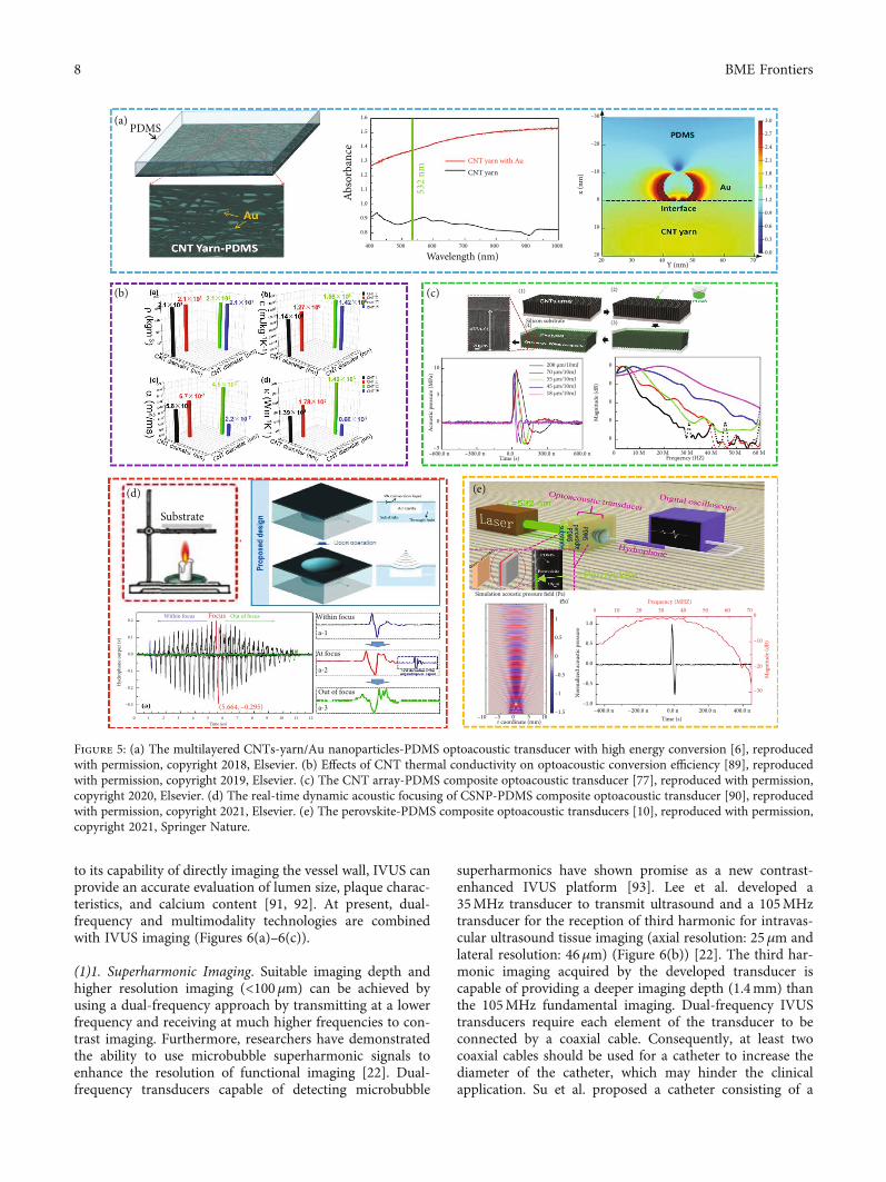
(1) CNT-PDMS Composite. CNTs exhibit a series of unusualproperties due to their unique structure and size, makingthem ideal light absorption materials for use in optoacoustictransducers. Baac et al. demonstrated a gold hybrid CNTs/PDMS optoacoustic transducer by depositing a thin layerof gold on the grown CNT film [88]. They have confirmedthat the damage threshold of the CNT-PDMS compositefilm is 10 times higher than that of the Cr film. In addition,the composite film can withstand extremely high laserenergy (>400mJ cm-2) without physical damage and gener-ate strong sound pressure. Our group fabricated high energyconversion (2:74 × 10−2) of the optoacoustic transducerusing the multilayered CNTs-yarn/Au nanoparticles-PDMScomposite, in which Au nanoparticles enhance light absorp-tion through local surface plasma effect (Figure 5(a)) [6].Subsequently, our group indicated that the great thermalconductivity of CNTs can generate a stronger optoacousticsignal in the 1-3 composite structure (Figure 5(b)) [89].Then, our group built an optoacoustic transducer based onthe CNT array-PDMS composite with anisotropic thermalconductivity. The frequency modulation (~20MHz) can beachieved by controlling this composite’s thickness(Figure 5(c)). Our results have proved that the working fre-quency of the transducer is inversely proportional to thethickness of the CNT array-PDMS composite [77]. Further-more, our group fabricated a self-healing optoacoustic trans-ducer based on CNT polymer. Even after 10 times of damageand healing, the output sound pressure still maintains at92.3% of the original value [25]. Silva et al. demonstratedan optoacoustic transducer that generated ~80MHz (-6 dBbandwidth: 170%) ultrasound pulses with functionalizedCNTs and 30ps pulse laser [35]. The functionalized CNTspromote ultrafast dissipation of heat to the PDMS. The pico-second pulse laser can improve the bandwidth of the optoa-coustic transducer.
(2) CSNP-PDMS Composite. CSNPs can be produced duringthe burning process of candles and have demonstrated theadvantages of a spherical shape (large surface-to-volumeratio), low specific heat capacity (~750 J kg-1K-1), low cost,and fabrication simplicity for developing highly efficient
optoacoustic transmitters [10, 75]. Under consistent experi-mental conditions, the optoacoustic conversion efficiency ofthe CSNP-PDMS composites was higher than that of theCB- or CNF-PDMS composites [37]. Using CSNP-PDMScomposites, our group and collaborators designed a noveloptoacoustic transmitter including the transducer and aircavity, which achieved real-time dynamic acoustic focus byadjusting the air pressure of the cavity (Figure 5(d)). Further-more, the negative optoacoustic pressure was significantlyimproved, which could provide additional advantages forultrasonic cavitation applications [90].
3.2.3. Lead Halide Perovskite-PDMS Composites. Takingadvantage of low specific heat capacity (~308 J kg-1K-1),low thermal conductivity (~0.5Wm-1K-1), small thermaldiffusion coefficient (0.145mm2 s−1), and high absorptioncoefficient (6.7μm-1) of perovskites, our group designed alead halide perovskite-PDMS-stack structure optoacoustictransducer. The theoretically calculated phonon spectrumshows that the overlap of optical phonons and acoustic pho-nons leads to the upconversion of acoustic phonons andthus results in a small thermal diffusion coefficient ofmethylamine lead iodine perovskite. The small thermal dif-fusion coefficient can generate a strong thermal localizationeffect on the perovskite layer, which enhances the energy con-version efficiency of the optoacoustic transducer. Figure 5(e)shows that the central frequency and -6dB bandwidth of theultrasound wave are at 29.2MHz and 40.8MHz, respectively.When the laser energy was 3mJ/pulse, the peak-to-peak valueof the acoustic pressure was 24.89MPa, and the optoacousticconversion efficiency was 2:97 × 10−2 [10].
4. Ultrasound Transducers forBiomedical Applications
4.1. Piezoelectric Transducer for Biomedical Applications
4.1.1. Medical Imaging
(1) Intravascular Ultrasound (IVUS) Imaging. IVUS imagingis an important tool in the diagnosis of cardiovascular dis-eases and has been widely used for clinical diagnosis. Owing
Table 2: Performance summary of representative optoacoustic transducers.
Light absorption materialsTransducer’saperture size
(mm)
d(μm)
CF(MHz)
-6 dB BW(%)
η(×10-2)
P(MPa)
h(mm)
Pulse laserparameters
ReferenceE
(mJ/pulse)τ (ns)
Au — 0.02 ~80 ~180 — 1.5 0 1 × 10⁻⁴ 5 [74]
CSNP ~10 6 10 210 0.441 +4.8/-1 4.2 3.57 6 [75]
CNTs-xylene 0.22 ~1 28.5 ~140 — 12.2 3 0.01 2 [76]
CNTs-yarn 10 ~5 11.8 179 2.74+33.6/-10
5 45 5 [6]
CNT array 5 18 20.2 152 0.251 +8.8 2 10 5 [77]
Functionalized CNTs 2.8 38 ~100 171 — 1.69 0 — 0.03 [35]
MAPbI3 5 0.34 29.2 140 2.97 +15/-10 2 3 5 [10]
Note: d: transducer thickness; CF: center frequency; BW: bandwidth; η: optoacoustic energy conversion efficiency; P: optoacoustic pressure; h: the distance ofhydrophone and transducer; E, τ: the energy and width of pulse laser.
7BME Frontiers
to its capability of directly imaging the vessel wall, IVUS canprovide an accurate evaluation of lumen size, plaque charac-teristics, and calcium content [91, 92]. At present, dual-frequency and multimodality technologies are combinedwith IVUS imaging (Figures 6(a)–6(c)).
(1)1. Superharmonic Imaging. Suitable imaging depth andhigher resolution imaging (<100μm) can be achieved byusing a dual-frequency approach by transmitting at a lowerfrequency and receiving at much higher frequencies to con-trast imaging. Furthermore, researchers have demonstratedthe ability to use microbubble superharmonic signals toenhance the resolution of functional imaging [22]. Dual-frequency transducers capable of detecting microbubble
superharmonics have shown promise as a new contrast-enhanced IVUS platform [93]. Lee et al. developed a35MHz transducer to transmit ultrasound and a 105MHztransducer for the reception of third harmonic for intravas-cular ultrasound tissue imaging (axial resolution: 25μm andlateral resolution: 46μm) (Figure 6(b)) [22]. The third har-monic imaging acquired by the developed transducer iscapable of providing a deeper imaging depth (1.4mm) thanthe 105MHz fundamental imaging. Dual-frequency IVUStransducers require each element of the transducer to beconnected by a coaxial cable. Consequently, at least twocoaxial cables should be used for a catheter to increase thediameter of the catheter, which may hinder the clinicalapplication. Su et al. proposed a catheter consisting of a
400 500 600 700 800 900 1000
Abso
rban
ce
CNT yarnCNT yarn with Au
532
nm
Wavelength (nm)
1.6
1.5
1.4
1.3
1.2
1.1
1.0
0.9
0.8
–30
–20
–10
0
10
2020 30 40 50 60 70
x (n
m)
Y (nm)
0.0
0.3
0.6
0.9
1.2
1.5
1.8
2.1
2.4
2.7
3.0
Substrate
(5.664, –0.295)
Within focus Out of focusFocus Within focus
At focus
Out of focus
a-1
a-2
a-3
PDMS(a)
(b)
(d) (e)
(c)
–600.0 n –300.0 n 0.0 300.0 n 600.0 nTime (s)
–5
0
5
10
Acou
stic p
ress
ure (
MPa
)
200 μm/10mJ70 μm/10mJ55 μm/10mJ45 μm/10mJ18 μm/10mJ
0 10 M 20 M 30 M 40 M 50 M 60 MFrequency (HZ)
0
0
0
0
0
Mag
nitu
de (d
B)
–30
–20
–10
0706050403020100
1.0
0.5
0.0
–0.5
–1.0–400.0 n –200.0 n 0.0 n 200.0 n 400.0 n
Time (s)
Mag
nitu
de (d
B)
Frequency (MHZ)
Nor
mal
ized
acou
stic p
ress
ure
Simulation acoustic pressure field (Pa)
0.5
1
0
–0.5
–1
–1.5–5–10 0 5 10r coordinate (mm)
Silicon substrate
(1) (2)
(3)(4)
Figure 5: (a) The multilayered CNTs-yarn/Au nanoparticles-PDMS optoacoustic transducer with high energy conversion [6], reproducedwith permission, copyright 2018, Elsevier. (b) Effects of CNT thermal conductivity on optoacoustic conversion efficiency [89], reproducedwith permission, copyright 2019, Elsevier. (c) The CNT array-PDMS composite optoacoustic transducer [77], reproduced with permission,copyright 2020, Elsevier. (d) The real-time dynamic acoustic focusing of CSNP-PDMS composite optoacoustic transducer [90], reproducedwith permission, copyright 2021, Elsevier. (e) The perovskite-PDMS composite optoacoustic transducers [10], reproduced with permission,copyright 2021, Springer Nature.
8 BME Frontiers

dual-frequency transducer for intravascular ultrasound. Bothultrasonic elements with different frequencies were connectedto one coaxial cable to simplify the connection [94].
(1)2. Acoustic Radiation Force (ARF) Elasticity Imaging. Thechallenge in predicting plaque ruptures depends on preciseknowledge of the mechanical properties of the arterial walland the plaque. Owing to the minimal contrast between dif-ferent types of soft tissues, the sensitivity and specificity ofIVUS for detecting the composition of a plaque are poor.Thus, ARF elasticity imaging has been developed byresearchers as an alternative to conventional ultrasoundelastography. It can be used to distinguish various compo-nents of plaques and assess the mechanical properties ofarterial walls. Shih et al. designed a dual-frequency IVUStransducer for elasticity imaging, where the propagation ofshear waves was induced by an 8.5MHz ultrasound trans-ducer, and the 31MHz imaging transducer monitored theelastic properties of plaques and vessels [95]. The stiffness dis-tributions of the atherosclerotic aorta from a rabbit and shearwave velocity of the lipid-rich plaques and arterial walls were0:38 ± 0:19ms-1 and 3:45 ± 0:45ms-1, respectively.
(1)3. Dual-Modality Photoacoustic and Ultrasound (PAUS)Imaging. In IVUS, the contrast between the lipid-rich regionand other soft tissues is confined. Intravascular photoacous-tic (IVPA) imaging seems to be an alternative method toaddress this issue to detect the lipid pool and atheroscleroticcalcification. In this hybrid imaging process, a tiny single-element ultrasound transducer integrated with a laser fiber
and driven by a rotational shaft is placed at an appropriateposition inside the blood vessel to detect the photoacousticsignals inside the tissue. As we all know, analyses of the pho-toacoustic spectrum have already been proven as an effectivemethod to expose significant differences between malignantand normal tissue regions [96]. Consequently, frequencydomain analyses seem to be a beneficial supplement in IVPAtechnology and could be potentially used for the characteri-zation of atherosclerotic plaques [66]. In addition, Li et al.designed a dual-modality PAUS system for endoscopicimaging [97], which showed enhanced bandwidths of theultrasound transducer and improved SNR of PAUS images(Figure 6(c)).
(2) Internal Organ Endoscopic Ultrasound Imaging (EUS).EUS is a diagnostic imaging method that uses ultrasoundto obtain images of internal organs in the human body, suchas the chest, abdomen, and colon. It can be used to visualizethe organ wall and surrounding structures. Zhang et al.developed a 6.8MHz 128-element endoscopic ultrasoundarray to obtain 3D imaging of a healthy swine intestine(Figure 6(d)) [58]. A newly emerging endoscopic technique,called photoacoustic endoscopy (PAE), could be an impor-tant complementing procedure in diagnosing GI tract dis-eases because it is well suited to provide high-resolutionmicrovasculature imaging with rich spectral and functionalinformation of the tissue. Yang et al. described a 3.8mmdiameter side-scanning PAE-EUS probe, which realizessimultaneous PAUS imaging of internal organs [99]. Wire-less capsule endoscopy enables remote diagnostic assessment
50 kHz Laser(a) (b)
(d) (e) (f)
(c)Coupler
Coupler
Circulator
Circulator
To ref arm
Balance detector
50kHz ultrasound pulser/receiver
Luer lock
Dig
itize
r
IVUS-OCT imagingcatheter
532 nm laser
Rotary joint and translation stageLens Connector
OCT
ARF-OCE
100 μm
0.30.50.70.9D
epth
(mm
)
–1 –0.5 0 0.5 1x (mm)
Submucosa
Circular muscle
Mucosa
SerosaLongitudinal muscle
10 mm
dB50
0
Figure 6: Piezoelectric ultrasound imaging. (a) Ultrafast IVUS-OCT system and miniaturized catheter for atherosclerotic plaque imaging[92], reproduced with permission, copyright 2015, Springer Nature. (b) 35MHz/105MHz dual-element focused transducer for IVUSimaging [22], reproduced with permission, copyright 2018, MDPI. (c) Dual-modality photoacoustic and ultrasound endoscopy imaging[97], reproduced with permission, copyright 2019, Elsevier. (d) 128-element 6.8MHz circular array for the 2D and 3D endoscopic imageof the swine intestine [58], reproduced with permission, copyright 2020, IEEE. (e) Ultrahigh-frequency (~300MHz) ultrasonictransducers for biomicroscopy imaging of zebrafish eyes [47], reproduced with permission, copyright 2016, Springer Nature. (f) ARF-OCE mapping for the retinal layers [98], reproduced with permission, copyright 2018, Association for Research in Vision andOphthalmology.
9BME Frontiers
of the gastrointestinal tract in a painless procedure. Wanget al. presented a mechanical scanning device incorporatinga 39MHz high-frequency transducer, which can offer goodimage resolution (∼60μm) for the lumen wall of the porcinesmall intestine [100].
(3) Ophthalmic Ultrasound Imaging. Ultrasound biomicro-scopy is used for real-time diagnosis of anterior segmentaldiseases with a relatively deep imaging and a large field ofview, regardless of whether the suspicious lesion is in opti-cally transparent or opaque media [51, 101]. Fei et al. usedthe ~300MHz single-element ultrasound transducer toobtain the ultrasonic biomicroscopy image of zebrafish eyes,whose resolution is up to ~10μm, comparable to the resolu-tion of OCT (Figure 6(e)) [47]. The elastic properties of thecornea are crucial for human vision. Therefore, measuringthe elasticity distribution of the cornea is important for eval-uating corneal pathologies and the efficacy of corneal treat-ment, particularly during the early stages of cornealsclerosis. Qu et al. used ARF optical coherence elastography(ARF-OCE) to map out the elasticity of retinal layers inhealthy and diseased in vivo rabbit models (Figure 6(f)) [98].
4.1.2. Acoustic Tweezers. Optical force, acoustic force, andmagnetic force and electrophoresis can be used to manipu-late single particles, cells, and organisms for many applica-tions in biology, chemistry, engineering, and physics.Compared with optical, electrical, and magnetic counter-parts, acoustic tweezers are relatively noninvasive to biolog-ical objects and applicable to most microparticles [102, 103].
Acoustic waves are capable of exerting acoustic radiationforces to levitate particles with a wide range of sizes andmaterials through air and water. As a promising microparti-cle manipulator, single beam acoustic tweezers (SBATs) areattractive [57]. They have the advantage of providing a sig-nificant trapping force and offering deep penetration, show-ing great potential for in vivo and clinical applications.
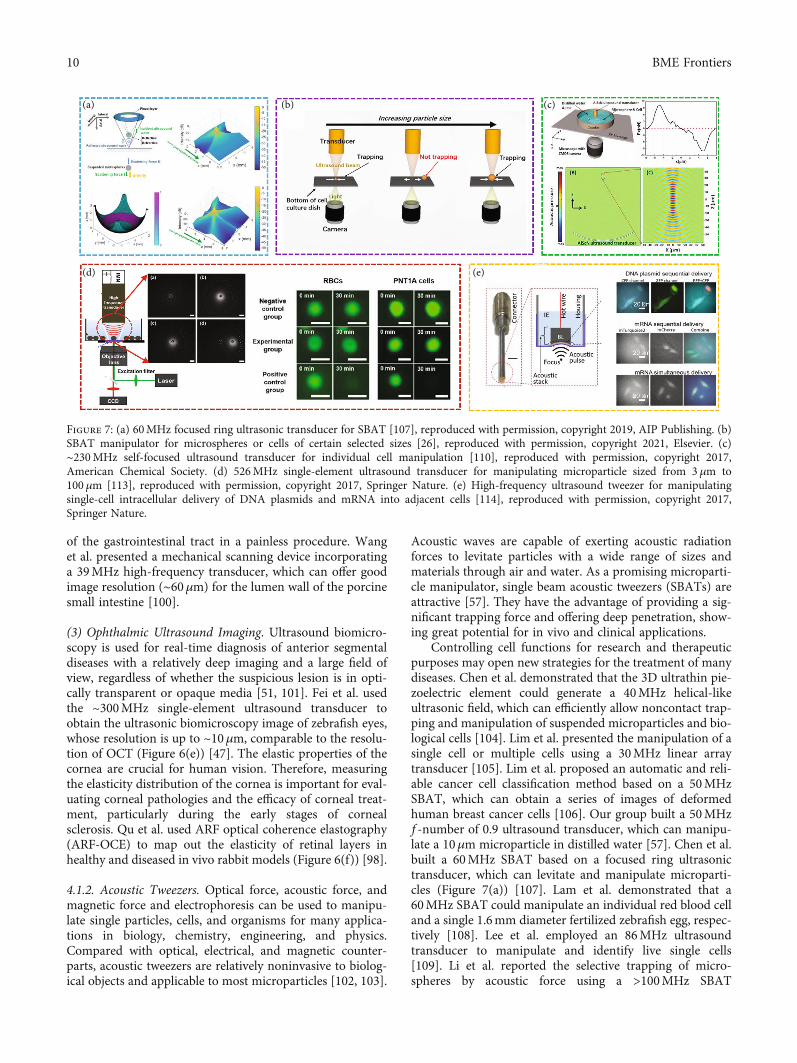
Controlling cell functions for research and therapeuticpurposes may open new strategies for the treatment of manydiseases. Chen et al. demonstrated that the 3D ultrathin pie-zoelectric element could generate a 40MHz helical-likeultrasonic field, which can efficiently allow noncontact trap-ping and manipulation of suspended microparticles and bio-logical cells [104]. Lim et al. presented the manipulation of asingle cell or multiple cells using a 30MHz linear arraytransducer [105]. Lim et al. proposed an automatic and reli-able cancer cell classification method based on a 50MHzSBAT, which can obtain a series of images of deformedhuman breast cancer cells [106]. Our group built a 50MHzf -number of 0.9 ultrasound transducer, which can manipu-late a 10μm microparticle in distilled water [57]. Chen et al.built a 60MHz SBAT based on a focused ring ultrasonictransducer, which can levitate and manipulate microparti-cles (Figure 7(a)) [107]. Lam et al. demonstrated that a60MHz SBAT could manipulate an individual red blood celland a single 1.6mm diameter fertilized zebrafish egg, respec-tively [108]. Lee et al. employed an 86MHz ultrasoundtransducer to manipulate and identify live single cells[109]. Li et al. reported the selective trapping of micro-spheres by acoustic force using a >100MHz SBAT
(a)
(d) (e)
(b) (c)
Figure 7: (a) 60MHz focused ring ultrasonic transducer for SBAT [107], reproduced with permission, copyright 2019, AIP Publishing. (b)SBAT manipulator for microspheres or cells of certain selected sizes [26], reproduced with permission, copyright 2021, Elsevier. (c)∼230MHz self-focused ultrasound transducer for individual cell manipulation [110], reproduced with permission, copyright 2017,American Chemical Society. (d) 526MHz single-element ultrasound transducer for manipulating microparticle sized from 3 μm to100μm [113], reproduced with permission, copyright 2017, Springer Nature. (e) High-frequency ultrasound tweezer for manipulatingsingle-cell intracellular delivery of DNA plasmids and mRNA into adjacent cells [114], reproduced with permission, copyright 2017,Springer Nature.
10 BME Frontiers

(Figure 7(b)) [26]. They found that setting the SBAT shapeand wavelength can manipulate microspheres or cells of cer-tain selected sizes. Our group fabricated an ~230MHz self-focused SBAT ultrasound transducer, which has a narrowlateral beam width (~8.2μm). The developed SBAT can con-tinuously manipulate individual 10μm epidermoid carci-noma cells (Figure 7(c)) [110]. Fei et al. fabricated a needleultrasonic transducer with a center frequency higher than300MHz (-6 dB bandwidth: >64%). The focused acousticmicrobeam produced by the transducer can manipulateindividual 3μm microspheres [111]. Chen et al. studiedadjustable multiscale SBAT based on a 526MHz single-element ultrasonic transducer that can flexibly change thesize of the “tweezers” in the same manner as ordinary metaltweezers [112].
The mechanical properties of cells play a key role in var-ious cellular functions, such as proliferation, migration, andgene expression. SBATs are a promising technology for thequantification of the mechanical performance of cells. Forthis purpose, Kim et al. developed a tightly focused153MHz ultrasound transducer to grab and separate a single
cell from a heterogeneous cell sample and to analyze thephysical and functional characteristics of cells (Figure 7(d))[113]. Hwang et al. demonstrated that a single-beam acous-tic trapping technique can be utilized to examine themechanical properties of breast cancer cells [115]. In addi-tion, the role of cell mechanics in cancer cells has resultedin the identification of new mechanisms of therapy resis-tance. Their group demonstrated that 30MHz SBAT canbe applied to quantify the deformability of adherent breastcancer cell lines [116] and to explore the elastic propertiesof pre-B acute lymphocytic leukemia cells [117]. Hwanget al. developed 193MHz acoustic tweezers to trap 5μmfibronectin- (FNT-) coated microbead and demonstratedits potential to study intracellular calcium signaling byFNT binding to human breast cancer cells [118].
There have been limited transfection techniques that candeliver multiple types of active molecules simultaneouslyinto single cells with high precision and low cytotoxicity.Yoon et al. introduced a new transfection technique thatutilizes the center frequency over 150MHz ultrasoundwithout any contrast agents inducing intracellular delivery
Figure 8: (a) All-optical ultrasound imaging of ex vivo human lymph node [122], reproduced with permission, copyright 2021, IEEE. (b)All-optical fiber ultrasound system for heart motion detection [123], reproduced with permission, copyright 2017, Springer Nature. (c) All-optical ultrasound technology for IVUS imaging [124], reproduced with permission, copyright 2019, Springer Nature. (d) All-opticalultrasound imaging system for fish eye imaging [10], reproduced with permission, copyright 2021, Springer Nature.
11BME Frontiers

of exogenous molecules [119]. Recently, they reportedthat a >150MHz high-frequency ultrasound-based remoteintracellular delivery technique capable of delivering mul-tiple DNA plasmids, messenger RNAs, and recombinantproteins has been developed. This technique allows highspatiotemporal visualization and analysis of gene and pro-tein expressions, as well as single-cell gene editing (acous-tic transfection). They found that ultrahigh-frequencyultrasound can directly deliver genes and proteins intothe cytoplasm without microbubbles (Figure 7(e)) [114].
4.1.3. Other Medical Applications. Recently, researchers havedeveloped implanted piezoelectric transducers to disrupt theblood-brain barrier and facilitate the delivery of drugs intothe brain [50, 120]. In addition, a 1.1MHz ultrasound trans-ducer was applied to modulate the cholinergic anti-inflammatory pathway and to reduce cytokine response to
endotoxin to the same levels as implant-based vagus nervestimulation [121]. In order to provide remarkable insightsinto the cardiovascular disease diagnosis and prognosis,Wang et al. fabricated a 7.5MHz flexible ultrasound trans-ducer to capture blood pressure waveforms at deeply embed-ded arterial and venous sites [27].
4.2. Optoacoustic Transducer for Medical Application
4.2.1. All-Optical Ultrasound Imaging. The optoacoustictransducer can generate ultrasound pulses with MPa-levelpeak pressures, offering the potential for improved imagequality and higher resolution for tissue imaging. Phamet al. presented a broadband all-optical plane-wave ultra-sound imaging system for high-resolution and high-fidelity3D ultrasound images of ex vivo human lymph nodes(Figure 8(a)) [122]. Finlay et al. built an optoacoustic
Carbon nanotube (CNT)-coated lens
Ultrasound
Water tankUltrasonic cutting
Nd:YAG laserLight
Carbon black/PDMS
Laser absorption Thermal expansion
Pulsed-shock waves
LGFUMicrogels
FixtureTest tube
Nd:YAG laserGlass water tank LGFU lens
LGFU lens
Beforea
c
b
After Max ΔF/F
50 m
s1
ms
Repe
at1
ms
3 ns
1.7 kHz
Pulsed laser
Diffuser: ZnO/epoxy mixture
Absorber: graphite/epoxy mixture
Figure 9: (a) Microscale fragmentation of the solid materials by the LGFU [125], reproduced with permission, copyright 2012, SpringerNature. (b) The optoacoustic transducer for thrombolysis [25], reproduced with permission, copyright 2020, Elsevier. (c) Theoptoacoustic transducer for cutting tissue [126], reproduced with permission, copyright 2017, John Wiley and Sons. (d) The optoacoustictransducer for microparticle movement control [6], reproduced with permission, copyright 2018, Elsevier. (e) The optoacoustictransducer for drug delivery [128], reproduced with permission, copyright 2015, Elsevier. (f) The optoacoustic transducer for neuronstimulation [23], reproduced with permission, copyright 2020, Springer Nature. (g) The optoacoustic nanotransducers for targetedneuromodulation [129], reproduced with permission, copyright 2021, Elsevier. (h) The optoacoustic transducer for stimulating singleneurons [130], reproduced with permission, copyright 2021, Springer Nature.
12 BME Frontiers

transducer with a center frequency of ~20MHz (-6 dB band-width: 132.5%), which was integrated within a custom innertransseptal needle to obtain all-optical ultrasound imaging ofmultiple locations within a swine heart (Figure 8(b)) [123].Colchester et al. designed an optoacoustic transducer witha center frequency of ~20MHz (-6 dB bandwidth: 156.5%)[124]. They built an IVUS system using all-optical ultra-sound technology to realize in vitro imaging of porcine aortasections (Figure 8(c)). Noimark et al. fabricated a 28.5MHzoptoacoustic transducer (-6 dB bandwidths: ~140%) at theend of 200μm optical fiber, which can achieve high-resolution all-optical ultrasound imaging of the porcineaorta in vitro [76]. In 2021, our group developed an all-optical ultrasound imaging system with a 29.2MHzperovskite-PDMS composite optoacoustic transducer. Usingthis system, the eye ultrasound imaging of a fish with highresolution and high SNR can be obtained, where the struc-tures of the cornea, iris, and lens surface are clearly visible(Figure 8(d)) [10].
4.2.2. Optoacoustic Medical Therapy
(1) Ultrasound Operation. With the application of an optoa-coustic lens, coating the optoacoustic composite material on
the concave surface to generate focused ultrasound, thesound pressure of an optoacoustic transducer has beengreatly improved. Baac et al. used laser-generated focusedultrasound (LGFU) to perform an experiment on an artifi-cial kidney-stone model [125]. They used LGFU to breakthe polymer coating, demonstrating its potential applicationin lithotripsy treatment (Figure 9(a)). Subsequently, ourgroup performed an ultrasound thrombolysis experimentusing a self-healing optoacoustic transducer (Figure 9(b))[25]. Lee et al. demonstrated that LGFU can be used forhigh-precision cavitation cutting that is applicable to morecomplex shapes [126]. They removed the choroid from thepig’s eye and then applied LGFU to the choroid surface.Two small ablation grooves are observed on the choroidafter drying the sample, which implies the achievement ofmechanical ablation (Figure 9(c)).
(2) Drug Delivery. High-intensity optoacoustic waves cangenerate acoustic cavitation which is effective for the controlof drug release [127]. Furthermore, the low duty cycle ofoptoacoustic waves can reduce ultrasound-induced heating,thereby avoiding the detrimental effects on surrounding tis-sues. Our group developed a new type of optoacoustic trans-ducer with a PDMS/Au-CNT yarn-PDMS structure using
The origin of the highpiezoelectricity in relaxor
ferroelectricsPrecision ultrasound
neuromodulationBiodegradable
piezoelectric transducer
Continous monitoringof deep-tissue
haemodynamics
Transparentferroelectric crystals
Self-navigated 3Dacoustic tweezers
Drug delivery Ultrasound operation Thrombolysis
All opticalultrasound
fish-eye imaging
Nervestimulation
Manipulating microparticles
Cell investigationLithotripsy
Hign-performancepiezoelectric materials;
High-performanceoptoacousticmaterials;
Materials
Struc
tures
Medical
applications
Materials
Struc
tures
Medical
applications
Optoacoustictransducer
Piezoelectricultrasoundtransducer
Enhanced integration with
optical diagnosis, etc methods;
Optimization of the effect of
ultrasound therapy;
Optim
izing
stru
cture
to
impr
ove b
andw
idth
;
High fr
eque
ncy a
rray
trans
duce
r;
High-resolution all-optical
ultrasound imaging;
Ultrasound therapy in
special environment;
Perfe
cting
opto
acou
stic
theo
ry;
Enha
ncin
g tra
nsdu
cer
stabil
ity;
2015 2016 2018 2019 2020 2021
2018 2019 2020 20212015 20172012
(a)
(b)
(c)
(b)
(d)
Monitoring of theblood pressure
High frequencyacoustic tweezers
trapping live singlecell
Figure 10: Summary of crucial developments in piezoelectric (a) and optoacoustic (b) [6, 10, 12, 18, 20, 23–25, 27, 103, 118, 120, 121, 125,126, 128] transducers, reproduced with permission. Research prospects of piezoelectric (c) and optoacoustic (d) transducers.
13BME Frontiers

multilayer CNT yarn and successfully applied it to manipu-late particles in a certain direction (Figure 9(d)) [6]. Di et al.used a focused optoacoustic transducer based on carbon-black/PDMS composite to promote drug release. As shownin Figure 9(e), when laser-generated ultrasound excites themicrogels, gradual release of the drug from poly(lactic-co-glycolic acid) nanoparticles is promoted due to the cavita-tion effects at the microgels and oscillation of the microgelshells [128]. These results provide guidelines for furtherin vivo potential clinical applications.
(3) Nerve Stimulation. Laser-generated ultrasound is anemerging modality for neuromodulation [126]. Jiang et al.reported an optoacoustic neural stimulator using a miniatur-ized fiber-optic converter, which could generate ~1MHzomnidirectional ultrasound wave (Figure 9(f)) [23]. Withstudies on living animals, the application of laser-generatedultrasound to neuromodulation and brain stimulation ofthe human body is expected to be available in the future.Subsequently, their group reported semiconducting polymernanoparticle-based targeted photoacoustic nanotransducers(PANs) for neural stimulation [129]. PANs can be surface-modified to selectively bind onto neurons; laser-generatedultrasound produced by PANs can modulate neuron activities(Figure 9(g)). Recently, their group also fabricated a taperedfiber optoacoustic emitter to generate an ultrasound field witha high spatial precision of 39.6μm, which can modulate singleneurons or subcellular structures (Figure 9(h)), such as axonsand dendrites [130].
5. Conclusions and Outlook
This review summarizes some recent advances in the field ofan ultrasound transducer that has been widely used in clin-ical diagnosis and treatments, such as biomedical imaging,thrombolysis, cell manipulation, drug delivery, and neuro-modulation. Plenty of researchers around the world, includ-ing the United States, the United Kingdom, China, andSouth Korea, are focusing on this field. Figure 10 showsthe summary and prospective crucial development for ultra-sound transducers recently. As explained in this review,ultrasound transducers could be divided into two major cat-egories: piezoelectric transducers and optoacoustic transduc-ers. A piezoelectric transducer which belongs to traditionalultrasound devices has been extensively investigated for awide range of biomedical engineering applications and isbeing revolutionized by advances in microelectronic tech-nologies. The optoacoustic transducer emerges as a promis-ing candidate for biomedical engineering applications, dueto its simple preparation processes, antielectromagneticinterference, and broad bandwidth. The progresses of ultra-sound transducers from the perspective of material strate-gies, design considerations, and biomedical engineeringapplications have been discussed systematically.
Because the material selection and structure design ofultrasound transducers have vital influences on its acousticperformance, seeking new material with excellent propertiesand optimizing device structure are two eternal themes forultrasound transducer development. Recent researches on
piezoelectric material have made a great breakthrough thatSm-PMN-PT single crystal with giant piezoelectricity andtransparent PMN-PT single crystal with ultrahigh piezoelec-tricity have been invented [15, 16, 18], providing a newstrategy for ultrasound transducer fabrication. From the per-spective of environmental protection, new nontoxic mate-rials with such super piezoelectricity are in demand fornext-generation piezoelectric transducers. Different frompiezoelectric devices, the optoacoustic transducer has a morecomplex energy conversion process. In order to preciselycontrol the performance of the optoacoustic transducer,device physics needs to be further explored. At present, car-bon nanomaterial-PDMS composites dominate optoacousticmaterials, but the energy conversion efficiency is still low.For next-generation optoacoustic transducer fabrication,novel materials with higher optoacoustic energy conversioncoefficients are required. For convenience in biomedicalengineering application, the development trend of the ultra-sound transducer is towards package miniaturization, arraydesign, and multifunctional integration. Moreover, the con-tinuous influx of new technologies, such as 3D printing, flex-ible electronics, and artificial intelligent, are expected tobring an innovation concept for transducer design.
Finally, we hope this work can provide a summary ofcurrent ultrasound transducer developments for further the-oretical study and inspire better structure design of the ultra-sound transducer for biomedical engineering applications inthe future.
Conflicts of Interest
The authors declare no competing interests.
Authors’ Contributions
Jiapu Li and Yuqing Ma contribute equally to this work.
Acknowledgments
This work was supported by the Natural Science Foundationof China (Grant nos. 11774117 and 12102140), ShenzhenBasic Science Research (JCYJ20200109110006136), andChina Postdoctoral Science Foundation (2021M70130).
References
[1] D. K. Piech, B. C. Johnson, K. Shen et al., “A wirelessmillimetre-scale implantable neural stimulator with ultrason-ically powered bidirectional communication,” Nature Bio-medical Engineering, vol. 4, no. 2, pp. 207–222, 2020.
[2] S. B. Devarakonda, M. R. Myers, M. Lanier, C. Dumoulin,and R. K. Banerjee, “Assessment of gold nanoparticlemediated-enhanced hyperthermia using MR-guided high-intensity focused ultrasound ablation procedure,” Nano Let-ters, vol. 17, no. 4, pp. 2532–2538, 2017.
[3] S. A. Quadri, M. Waqas, I. Khan et al., “High-intensityfocused ultrasound: past, present, and future in neurosur-gery,” Neurosurgical Focus, vol. 44, no. 2, p. E16, 2018.
[4] T. Liu, N. Zhang, Z. Wang et al., “Endogenous catalytic gen-eration of O2bubbles forin situultrasound-guided high
14 BME Frontiers

intensity focused ultrasound ablation,” ACS Nano, vol. 11,no. 9, pp. 9093–9102, 2017.
[5] J. MacDonell, N. Patel, S. Rubino et al., “Magnetic resonance–guided interstitial high-intensity focused ultrasound for braintumor ablation,” Neurosurgical Focus, vol. 44, no. 2, p. E11,2018.
[6] Z. Chen, Y. Wu, Y. Yang et al., “Multilayered carbon nano-tube yarn based optoacoustic transducer with high energyconversion efficiency for ultrasound application,” NanoEnergy, vol. 46, pp. 314–321, 2018.
[7] X. Wang, F. Yan, X. Liu et al., “Enhanced drug delivery usingsonoactivatable liposomes with membrane-embedded por-phyrins,” Journal of Controlled Release, vol. 286, pp. 358–368, 2018.
[8] P. Zhu, H. Peng, L. Mao, and J. Tian, “Piezoelectric singlecrystal ultrasonic transducer for endoscopic drug release ingastric mucosa,” IEEE Transactions on Ultrasonics, Ferroelec-trics, and Frequency Control, vol. 68, no. 4, pp. 952–960, 2021.
[9] K. Yu, X. Niu, E. Krook-Magnuson, and B. He, “Intrinsicfunctional neuron-type selectivity of transcranial focusedultrasound neuromodulation,” Nature Communications,vol. 12, no. 2519, pp. 1–17, 2021.
[10] X. Du, J. Li, G. Niu et al., “Lead halide perovskite for efficientoptoacoustic conversion and application toward high-resolution ultrasound imaging,” Nature Communications,vol. 12, no. 3348, pp. 1–9, 2021.
[11] Y. Wang, X. Ge, H. Ma, S. Qi, G. Zhang, and Y. Yao, “Deeplearning in medical ultrasound image analysis: a review,”IEEE Access, vol. 9, pp. 54310–54324, 2021.
[12] F. Li, S. Zhang, T. Yang et al., “The origin of ultrahigh piezo-electricity in relaxor-ferroelectric solid solution crystals,”Nature Communications, vol. 7, no. 1, pp. 1–9, 2016.
[13] F. Li, D. Lin, Z. Chen et al., “Ultrahigh piezoelectricity in fer-roelectric ceramics by design,” Nature Materials, vol. 17,no. 4, pp. 349–354, 2018.
[14] H. Pan, F. Li, Y. Liu et al., “Ultrahigh–energy density lead-freedielectric films via polymorphic nano-domain design,” Sci-ence, vol. 365, no. 6453, pp. 578–582, 2019.
[15] F. Li, M. J. Cabral, B. Xu et al., “Giant piezoelectricity of Sm-doped Pb(Mg1/3Nb2/3)O3-PbTiO3single crystals,” Science,vol. 364, no. 6437, pp. 264–268, 2019.
[16] Z. Chen, F. Li, Q. Huang et al., “Giant tuning of ferroelectric-ity in single crystals by thickness engineering,” ScienceAdvances, vol. 6, no. 42, 2020.
[17] X. Gao, Z. Cheng, Z. Chen et al., “The mechanism for theenhanced piezoelectricity in multi-elements doped(K,Na)NbO3 ceramics,” Nature Communications, vol. 12,no. 1, pp. 1–9, 2021.
[18] C. Qiu, B. Wang, N. Zhang et al., “Transparent ferroelectriccrystals with ultrahigh piezoelectricity,” Nature, vol. 577,no. 7790, pp. 350–354, 2020.
[19] S. Yang, J. Li, Y. Liu et al., “Textured ferroelectric ceramicswith high electromechanical coupling factors over a broadtemperature range,” Nature Communications, vol. 12, no. 1,pp. 1–10, 2021.
[20] C. Wang, B. Qi, M. Lin et al., “Continuous monitoring ofdeep-tissue haemodynamics with stretchable ultrasonicphased arrays,” Nature Biomedical Engineering, vol. 5, no. 7,pp. 749–758, 2021.
[21] Y. Yang, Z. Chen, X. Song et al., “Three dimensional printingof high dielectric capacitor using projection based stereo-
lithography method,” Nano Energy, vol. 22, pp. 414–421,2016.
[22] J. Lee, J. Y. Moon, and J. H. Chang, “A 35 MHz/105 MHzdual-element focused transducer for intravascular ultrasoundtissue imaging using the third harmonic,” Sensors, vol. 18,2018.
[23] Y. Jiang, H. J. Lee, L. Lan et al., “Optoacoustic brain stimula-tion at submillimeter spatial precision,” Nature Communica-tions, vol. 11, no. 1, 2020.
[24] Y. C. Chen, H. W. Baac, K. T. Lee et al., “Selective photome-chanical detachment and retrieval of divided sister cells fromenclosed microfluidics for downstream analyses,” ACS Nano,vol. 11, no. 5, pp. 4660–4668, 2017.
[25] J. Li, Y. Yang, Z. Chen et al., “Self-healing: a new skillunlocked for ultrasound transducer,” Nano Energy, vol. 68,article 104348, 2020.
[26] Z. Li, D. Wang, C. Fei et al., “The forbidden band and sizeselectivity of acoustic radiation force trapping,” Iscience,vol. 24, no. 1, article 101988, 2021.
[27] C. Wang, X. Li, H. Hu et al., “Monitoring of the central bloodpressure waveform via a conformal ultrasonic device,”Nature Biomedical Engineering, vol. 2, no. 9, pp. 687–695,2018.
[28] Q. Zhou, S. Lau, D. Wu, and K. Kirk Shung, “Piezoelectricfilms for high frequency ultrasonic transducers in biomedicalapplications,” Progress in Materials Science, vol. 56, no. 2,pp. 139–174, 2011.
[29] Q. Zhou, K. H. Lam, H. Zheng, W. Qiu, and K. K. Shung,“Piezoelectric single crystal ultrasonic transducers for bio-medical applications,” Progress in Materials Science, vol. 66,pp. 87–111, 2014.
[30] T. E. G. Alvarez-Arenas, “Acoustic impedance matching ofpiezoelectric transducers to the air,” IEEE Transactions onUltrasonics, Ferroelectrics, and Frequency Control, vol. 51,no. 5, pp. 624–633, 2004.
[31] S. Zhang, F. Li, X. Jiang, J. Kim, J. Luo, and X. Geng, “Advan-tages and challenges of relaxor-PbTiO3 ferroelectric crystalsfor electroacoustic transducers - a review,” Progress in Mate-rials Science, vol. 68, pp. 1–66, 2015.
[32] M. Castillo, P. Acevedo, and E. Moreno, “KLM model forlossy piezoelectric transducers,” Ultrasonics, vol. 41, no. 8,pp. 671–679, 2003.
[33] T. Lee, H. W. Baac, Q. Li, and L. J. Guo, “Efficient photo-acoustic conversion in optical nanomaterials and compos-ites,” Advanced Optical Materials, vol. 6, no. 24, article1800491, 2018.
[34] F. Gao, R. Kishor, X. Feng et al., “An analytical study of pho-toacoustic and thermoacoustic generation efficiency towardscontrast agent and film design optimization,” Photoacoustics,vol. 7, pp. 1–11, 2017.
[35] A. D. Silva, C. A. Henriques, D. V. Malva et al., “Photoacous-tic generation of intense and broadband ultrasound pulseswith functionalized carbon nanotubes,” Nanoscale, vol. 12,no. 40, pp. 20831–20839, 2020.
[36] S. Noimark, R. J. Colchester, R. K. Poduval et al., “Polydi-methylsiloxane composites for optical ultrasound generationand multimodality imaging,” Advanced Functional Materials,vol. 28, no. 9, 2018.
[37] J. Kim, H. Kim, W. Y. Chang, W. Huang, X. Jiang, and P. A.Dayton, “Candle-soot carbon nanoparticles in photoacous-tics: advantages and challenges for laser ultrasound
15BME Frontiers

transmitters,” IEEE Nanotechnology Magazine, vol. 13, no. 3,pp. 13–28, 2019.
[38] S. L. Chen, “Review of laser-generated ultrasound transmit-ters and their applications to all-optical ultrasound transduc-ers and imaging,” Applied Sciences, vol. 7, no. 1, p. 25, 2017.
[39] C. M.Wong, Y. Chen, H. Luo, J. Dai, K. H. Lam, and H. L. W.Chan, “Development of a 20-MHz wide-bandwidth PMN-PTsingle crystal phased-array ultrasound transducer,” Ultrason-ics, vol. 73, pp. 181–186, 2017.
[40] Z. Zhang, R. Chen, B. Wang et al., “Development of a KNNceramic based lead-free linear array ultrasonic transducer,”IEEE Transactions on Ultrasonics, Ferroelectrics, and Fre-quency Control, vol. 65, no. 11, pp. 2113–2120, 2018.
[41] Y. Chen, X. Bao, C. M. Wong et al., “PZT ceramics fabricatedbased on stereolithography for an ultrasound transducerarray application,” Ceramics International, vol. 44, no. 18,pp. 22725–22730, 2018.
[42] X. Chen, C. Fei, Z. Chen et al., “Simulation and fabrication of0-3 composite PZT films for ultrahigh frequency (100–300MHz) ultrasonic transducers,” Journal of Applied Physics,vol. 119, no. 9, article 094103, 2016.
[43] Z. Zhang, M. Su, F. Li et al., “New Sm-PMN-PT ceramicbased 2-D array for low-intensity ultrasound therapy applica-tion,” IEEE Transactions on Ultrasonics, Ferroelectrics, andFrequency Control, vol. 67, no. 10, pp. 2085–2094, 2020.
[44] S. Li, J. Tian, and X. Jiang, “A micromachined Pb(Mg1/3Nb2/3)O3-PbTiO3 single crystal composite circular array for intra-vascular ultrasound imaging,” Journal of Engineering and Sci-ence in Medical Diagnostics and Therapy, vol. 2, no. 2, 2019.
[45] T. Zhang, J. Ou-Yang, X. Yang, W. Wei, and B. Zhu, “Highperformance KNN-based single crystal thick film for ultra-sound application,” Electronic Materials Letters, vol. 15,no. 1, pp. 1–6, 2019.
[46] Z. Chen, X. Song, L. Lei et al., “3D printing of piezoelectricelement for energy focusing and ultrasonic sensing,” NanoEnergy, vol. 27, pp. 78–86, 2016.
[47] C. Fei, C. Chiu, X. Chen et al., “Ultrahigh frequency (100MHz-300 MHz) ultrasonic transducers for optical resolutionmedical imagining,” Scientific Reports, vol. 6, no. 1, pp. 1–8,2016.
[48] C. Fei, T. Zhao, D. Wang et al., “High frequency needle ultra-sonic transducers based on lead-free co doped Na0.5Bi4.5-Ti4O15 piezo-ceramics,” Micromachines, vol. 9, no. 6, p. 291,2018.
[49] C. Fei, H. Hsu, A. Vafanejad et al., “Ultrahigh frequency ZnOsilicon lens ultrasonic transducer for cell-size microparticlemanipulation,” Journal of Alloys and Compounds, vol. 729,pp. 556–562, 2017.
[50] E. J. Curry, T. T. Le, R. Das et al., “Biodegradable nanofiberbased piezoelectric transducer,” Proceedings of the NationalAcademy of Sciences of the United States of America,vol. 117, no. 1, pp. 214–220, 2020.
[51] R. Chen, L. Jiang, T. Zhang et al., “Eco-friendly highly sensi-tive transducers based on a new KNN–NTK–FM lead-freepiezoelectric ceramic for high-frequency biomedical ultra-sonic imaging applications,” IEEE Transactions on Biomedi-cal Engineering, vol. 66, no. 6, pp. 1518–1587, 2019.
[52] J. Woo and Y. Roh, “Design and fabrication of an annulararray high intensity focused ultrasound transducer with anoptimal electrode pattern,” Sensors and Actuators A,vol. 290, pp. 156–161, 2019.
[53] T. Kim, Z. Cui, W. Y. Chang, H. Kim, Y. Zhu, and X. Jiang,“Flexible 1-3 composite ultrasound transducers with silvernanowire-based stretchable electrodes,” IEEE Transactionson Industrial Electronics, vol. 67, no. 8, pp. 6955–6962, 2020.
[54] C. Peng, M. Chen, H. K. Sim, Y. Zhu, and X. Jiang, “Noninva-sive and nonocclusive blood pressure monitoring via a flexi-ble piezo-composite ultrasonic sensor,” IEEE SensorsJournal, vol. 21, no. 3, pp. 2642–2650, 2021.
[55] H. Hu, X. Zhu, C. Wang et al., “Stretchable ultrasonic trans-ducer arrays for three-dimensional imaging on complex sur-faces,” Science Advances, vol. 4, no. 3, article eaar3979, 2018.
[56] C. T. Chiu, B. J. Kang, P. Eliahoo, T. Abraham, and K. K.Shung, “Fabrication and characterization of a 20-MHzmicro-linear phased-array transducer for intervention guid-ance,” IEEE Transactions on Ultrasonics, Ferroelectrics, andFrequency Control, vol. 64, no. 8, pp. 1261–1268, 2017.
[57] B. Zhu, J. Xu, Y. Li et al., “Micro-particle manipulation by sin-gle beam acoustic tweezers based on hydrothermal PZT thickfilm,” AIP Advances, vol. 6, no. 3, p. 035102, 2016.
[58] Q. Zhang, Y. Li, J. Liu et al., “A PMN-PT composite-basedcircular array for endoscopic ultrasonic imaging,” IEEETransactions on Ultrasonics, Ferroelectrics, and FrequencyControl, vol. 67, no. 11, pp. 2354–2362, 2020.
[59] N. E. Cabrera-Munoz, P. Eliahoo, R. Wodnicki et al., “Fabri-cation and characterization of a miniaturized 15-MHz side-looking phased-array transducer catheter,” IEEE Transac-tions on Ultrasonics, Ferroelectrics, and Frequency Control,vol. 66, no. 6, pp. 1079–1092, 2019.
[60] Z. Zhang, F. Li, R. Chen et al., “High-performance ultrasoundneedle transducer based on modified PMN-PT ceramic withultrahigh clamped dielectric permittivity,” IEEE Transactionson Ultrasonics, Ferroelectrics, and Frequency Control, vol. 65,no. 2, pp. 223–230, 2018.
[61] W. Zhou, T. Zhang, J. Ou-Yang, X. Yang, D. Wu, and B. Zhu,“PIN-PMN-PT single crystal 1-3 composite-based 20 MHzultrasound phased array,” Micromachines, vol. 11, no. 5,p. 524, 2020.
[62] P. Li, J. Zhai, B. Shen et al., “Ultrahigh piezoelectric propertiesin textured (K,Na)NbO3-based lead-free ceramics,”Advanced Materials, vol. 30, no. 8, article 1705171, 2018.
[63] T. Zheng, Y. Zhang, Q. Ke et al., “High-performance potas-sium sodium niobate piezoceramics for ultrasonic trans-ducer,” Nano Energy, vol. 70, article 104559, 2020.
[64] L. Jiang, R. Chen, J. Xing et al., “Fabrication of a(K,Na)NbO3-based lead-free 1-3 piezocomposite for high-sensitivity ultrasonic transducers application,” Journal ofApplied Physics, vol. 125, no. 21, article 214501, 2019.
[65] Y. Quan, C. Fei, W. Ren et al., “Lead-free KNN-based tex-tured ceramics for high-frequency ultrasonic transducerapplication,” IEEE Transactions on Ultrasonics, Ferroelec-trics, and Frequency Control, vol. 68, no. 5, pp. 1979–1987, 2021.
[66] B. Zhu, Y. Zhu, J. Yang et al., “New potassium sodium niobatesingle crystal with thickness-independent high- performancefor photoacoustic angiography of atherosclerotic lesion,” Sci-entific Reports, vol. 6, no. 1, 2016.
[67] T. Zhang, R. Chen, Z. Zhang et al., “High frequency singlecrystal ultrasonic transducers up to 100 MHz for high resolu-tion ophthalmic imaging applications,” in 2017 IEEE Interna-tional Ultrasonics Symposium (IUS), pp. 1–4, Washington,DC, USA, 2017.
16 BME Frontiers

[68] X. Yang, C. Fei, D. Li et al., “Multi-layer polymer-metal struc-tures for acoustic impedance matching in high- frequencybroadband ultrasonic transducers design,” Applied Acoustics,vol. 160, article 107123, 2020.
[69] J. Y. Zhang, W. J. Xu, J. Carlier et al., “Modelling and simula-tion of high-frequency (100 MHz) ultrasonic linear arraysbased on single crystal LiNbO3,” Ultrasonics, vol. 52, no. 1,pp. 47–53, 2012.
[70] H. Wei, H. Wang, Y. Xia et al., “An overview of lead-free pie-zoelectric materials and devices,” Journal of Materials Chem-istry C, vol. 6, no. 46, pp. 12446–12467, 2018.
[71] H. Liu, W. Ren, J. Zhao et al., “Design and fabrication of highfrequency BNT film based linear array transducer,” CeramicsInternational, vol. 41, no. 1, pp. S631–S637, 2015.
[72] A. Baba, C. T. Searfass, and B. R. Tittmann, “High tempera-ture ultrasonic transducer up to 1000 ° C using lithium nio-bate single crystal,” Applied Physics Letters, vol. 97, no. 23,article 232901, 2010.
[73] V. Daeichin, C. Chen, Q. Ding et al., “A broadband polyviny-lidene difluoride-based hydrophone with integrated readoutcircuit for intravascular photoacoustic imaging,” Ultrasoundin Medicine & Biology, vol. 42, no. 5, pp. 1239–1243, 2016.
[74] Y. Hou, J. S. Kim, S. Ashkenazi, M. O’Donnell, and L. J. Guo,“Optical generation of high frequency ultrasound using two-dimensional gold nanostructure,” Applied Physics Letters,vol. 89, no. 9, article 093901, 2006.
[75] W. Y. Chang, W. Huang, J. Kim, S. Li, and X. Jiang, “Candlesoot nanoparticles-polydimethylsiloxane composites for laserultrasound transducers,” Applied Physics Letters, vol. 107,no. 16, article 161903, 2015.
[76] S. Noimark, R. J. Colchester, B. J. Blackburn et al., “Carbonnanotube–PDMS composite coatings on optical fibers forall-optical ultrasound imaging,” Advanced Functional Mate-rials, vol. 26, no. 46, pp. 8390–8396, 2016.
[77] J. Li, J. Xu, X. Liu et al., “A novel CNTs array-PDMS compos-ite with anisotropic thermal conductivity for optoacoustictransducer applications,” Composites Part B: Engineering,vol. 196, article 108073, 2020.
[78] Y. Hou, J. S. Kim, S. Ashkenazi, S. W. Huang, L. J. Guo, andM. O’Donnell, “Broadband all-optical ultrasound transduc-ers,” Applied Physics Letters, vol. 91, no. 7, article 073507,2007.
[79] Y. S. Chen, Y. Zhao, S. J. Yoon, S. S. Gambhir, andS. Emelianov, “Miniature gold nanorods for photoacousticmolecular imaging in the second near-infrared optical win-dow,” Nature Nanotechnology, vol. 14, no. 5, pp. 465–472,2019.
[80] S. Kang, Y. Yoon, J. Kim, and W. Kim, “Thermoelasticresponse of thin metal films and their adjacent materials,”Applied Physics Letters, vol. 102, no. 2, article 021908,2013.
[81] T. Lee and L. J. Guo, “Highly efficient photoacoustic conver-sion by facilitated heat transfer in ultrathin metal film sand-wiched by polymer layers,” Advanced Optical Materials,vol. 5, no. 2, article 1600421, 2017.
[82] Y. Liang, H. Sun, L. Cheng, L. Jin, and B. O. Guan, “High spa-tiotemporal resolution optoacoustic sensing with photo-thermally induced acoustic vibrations in optical fibres,”Nature Communications, vol. 12, no. 4139, pp. 1–10, 2021.
[83] T. Buma, M. Spisar, and M. O’Donnell, “High-frequencyultrasound array element using thermoelastic expansion in
an elastomeric film,” Applied Physics Letters, vol. 79, no. 4,pp. 548–550, 2001.
[84] S. Hwan Lee, M. A. Park, J. J. Yoh et al., “Reduced grapheneoxide coated thin aluminum film as an optoacoustic trans-mitter for high pressure and high frequency ultrasound gen-eration,” Applied Physics Letters, vol. 101, no. 24, article241909, 2012.
[85] B. Y. Hsieh, J. Kim, J. Zhu, S. Li, X. Zhang, and X. Jiang, “Alaser ultrasound transducer using carbon nanofibers–polydi-methylsiloxane composite thin film,” Applied Physics Letters,vol. 106, no. 2, article 021902, 2015.
[86] L. Lan, Y. Xia, R. Li et al., “A fiber optoacoustic guide withaugmented reality for precision breast- conserving sur-gery,” Light: Science & Applications, vol. 7, no. 1, pp. 1–11, 2018.
[87] C. Peng, M. Chen, J. B. Spicer, and X. Jiang, “Acoustics at thenanoscale (nanoacoustics): a comprehensive literaturereview. Part I: materials, devices and selected applications,”Sensors and Actuators A: Physical, vol. 332, article 112719,2021.
[88] H. W. Baac, J. G. Ok, T. Lee, and L. J. Guo, “Nano-structuralcharacteristics of carbon nanotube–polymer composite filmsfor high-amplitude optoacoustic generation,” Nanoscale,vol. 7, no. 34, pp. 14460–14468, 2015.
[89] J. Li, X. Lan, S. Lei, J. Ou-Yang, X. Yang, and B. Zhu, “Effectsof carbon nanotube thermal conductivity on optoacoustictransducer performance,” Carbon, vol. 145, pp. 112–118,2019.
[90] Q. Li, J. Li, H. Zhu, Y. Chen, B. Zhu, and H. Yu, “Dynamicacoustic focusing in photoacoustic transmitter,” Photoacous-tics, vol. 21, article 100224, 2021.
[91] Z. Chen, L. Zheng, W. Cao et al., “High-frequency ultrasonicimaging with lead-free (Na,K)(Nb,Ta)O3single crystal,”Ultrasonic Imaging, vol. 39, no. 6, pp. 348–356, 2017.
[92] J. Li, T. Ma, D. Mohar et al., “Ultrafast optical-ultrasonic sys-tem andminiaturized catheter for imaging and characterizingatherosclerotic plaques _in vivo_,” Scientific Reports, vol. 5,no. 1, 2015.
[93] J. Ma, X. Jiang, K. H. Martin, P. A. Dayton, Y. Li, andQ. Zhou, “Dual frequency transducers for intravascular ultra-sound super-harmonic imaging and acoustic angiography,”in 2014 IEEE International Ultrasonics Symposium,pp. 675–678, Chicago, IL, USA, 2014.
[94] M. Su, Z. Zhang, J. Hong et al., “Cable-shared dual-frequencycatheter for intravascular ultrasound,” IEEE Transactions onUltrasonics, Ferroelectrics, and Frequency Control, vol. 66,no. 5, pp. 849–856, 2019.
[95] C. C. Shih, P. Y. Chen, T. Ma, Q. Zhou, K. K. Shung, and C. C.Huang, “Development of an intravascular ultrasound elastog-raphy based on a dual-element transducer,” Royal SocietyOpen Science, vol. 5, no. 4, 2018.
[96] L. Lin, P. Hu, J. Shi et al., “Single-breath-hold photoacousticcomputed tomography of the breast,” Nature Communica-tions, vol. 9, no. 1, pp. 1–9, 2018.
[97] Y. Li, G. Lu, J. J. Chen et al., “PMN-PT/epoxy 1-3 compositebased ultrasonic transducer for dual-modality photoacousticand ultrasound endoscopy,” Photoacoustics, vol. 15, article100138, 2019.
[98] Y. Qu, Y. He, A. Saidi et al., “In vivo elasticity map-ping of posterior ocular layers using acoustic radiationforce optical coherence elastography,” Investigative
17BME Frontiers

Ophthalmology & Visual Science, vol. 59, no. 1,pp. 455–461, 2018.
[99] J. M. Yang, C. Favazza, J. Yao et al., “Three-dimensional pho-toacoustic endoscopic imaging of the rabbit esophagus,”PLoS One, vol. 10, no. 4, 2015.
[100] X. Wang, V. Seetohul, R. Chen et al., “Development of amechanical scanning device with high-frequency ultrasoundtransducer for ultrasonic capsule endoscopy,” IEEE Transac-tions on Medical Imaging, vol. 36, no. 9, pp. 1922–1929, 2017.
[101] J. H. Cha and J. H. Chang, “Development of 15 MHz 2-2piezo-composite ultrasound linear array transducers for oph-thalmic imaging,” Sensors and Actuators A: Physical, vol. 217,pp. 39–48, 2014.
[102] X. Ding, S. C. S. Lin, B. Kiraly et al., “On-chip manipulationof single microparticles, cells, and organisms using surfaceacoustic waves,” Proceedings of the National Academy of Sci-ences of the United States of America, vol. 109, no. 28,pp. 11105–11109, 2012.
[103] Y. Yang, T. Ma, S. Li et al., “Self-navigated 3D acoustic twee-zers in complex media based on time reversa1,” Research,vol. 2021, article 9781394, 13 pages, 2021.
[104] X. Chen, X. Qian, K. H. Lam et al., “Helical-like 3D ultrathinpiezoelectric element for complicated ultrasonic field,”Advance Function Material, vol. 29, no. 32, article 1902912,2019.
[105] H. G. Lim, H. H. Kim, C. Yoon, and K. K. Shung, “A one-sided acoustic trap for cell immobilization using 30-MHzarray transducer,” IEEE Transactions on Ultrasonics, Ferro-electrics, and Frequency Control, vol. 67, no. 1, pp. 167–172,2020.
[106] H. G. Lim, O. J. Lee, K. K. Shung, J. T. Kim, and H. H. Kim,“Classification of breast cancer cells using the integration ofhigh-frequency single-beam acoustic tweezers and convolu-tional neural networks,” Cancers, vol. 12, no. 5, p. 1212, 2020.
[107] X. Chen, K. H. Lam, R. Chen et al., “Acoustic levitation andmanipulation by a high-frequency focused ring ultrasonictransducer,” Applied Physics Letters, vol. 114, no. 5, article054103, 2019.
[108] K. H. Lam, Y. Li, Y. Li, H. G. Lim, Q. Zhou, and K. K. Shung,“Multifunctional single beam acoustic tweezer for non-invasive cell/organismmanipulation and tissue imaging,” Sci-entific Reports, vol. 6, no. 1, 2016.
[109] C. Lee, H. Jung, K. H. Lam, C. Yoon, and K. K. Shung,“Ultrasonic scattering measurements of a live single cellat 86 MHz,” IEEE Transactions on Ultrasonics, Ferroelec-trics, and Frequency Control, vol. 62, no. 11, pp. 1968–1978, 2015.
[110] B. Zhu, C. Fei, C. Wang et al., “Self-focused AlScN film ultra-sound transducer for individual cell manipulation,” ACS Sen-sors, vol. 2, no. 1, pp. 172–177, 2017.
[111] C. Fei, Y. Li, B. Zhu et al., “Contactless microparticle controlvia ultrahigh frequency needle type single beam acoustictweezers,” Applied Physics Letters, vol. 109, no. 17,p. 173509, 2016.
[112] X. Chen, K. H. Lam, R. Chen et al., “An adjustable multi-scalesingle beam acoustic tweezers based on ultrahigh frequencyultrasonic transducer,” Biotechnology and Bioengineering,vol. 114, no. 11, pp. 2637–2647, 2017.
[113] M. G. Kim, J. Park, H. G. Lim et al., “Label-free analysis of thecharacteristics of a single cell trapped by acoustic tweezers,”Scientific Reports, vol. 7, no. 1, article 14092, 2017.
[114] S. Yoon, P. Wang, Q. Peng, Y. Wang, and K. K. Shung,“Acoustic-transfection for genomic manipulation of single-cells using high frequency ultrasound,” Scientific Reports,vol. 7, no. 1, p. 5275, 2017.
[115] J. Y. Hwang, J. Kim, J. M. Park et al., “Cell deformation bysingle-beam acoustic trapping: a promising tool for mea-surements of cell mechanics,” Scientific Reports, vol. 6,no. 1, 2016.
[116] H. C. Liu, E. J. Gang, H. N. Kim et al., “Characterizingdeformability of drug resistant patient-derived acute lympho-blastic leukemia (ALL) cells using acoustic tweezers,” Scien-tific Reports, vol. 8, no. 1, article 15708, 2018.
[117] H. C. Liu, E. J. Gang, H. N. Kim et al., “Integrin antibodydecreases deformability of patient-derived pre-B acute lym-phocytic leukemia cells as measured by high-frequencyacoustic tweezers,” Journal of Ultrasound in Medicine,vol. 39, no. 3, pp. 589–595, 2020.
[118] J. Y. Hwang, C. W. Yoon, H. G. Lim et al., “Acoustic tweezersfor studying intracellular calcium signaling in SKBR-3 humanbreast cancer cells,” Ultrasonics, vol. 63, pp. 94–101, 2015.
[119] S. Yoon, M. G. Kim, C. T. Chiu et al., “Direct and sustainedintracellular delivery of exogenous molecules usingacoustic-transfection with high frequency ultrasound,” Scien-tific Reports, vol. 6, no. 1, 2016.
[120] H. Zhang, T. Wang, W. Qiu et al., “Monitoring the openingand recovery of the blood−brain barrier with noninvasivemolecular imaging by biodegradable ultrasmall Cu2−xSenanoparticles,” Nano Letters, vol. 18, no. 8, pp. 4985–4992,2018.
[121] V. Cotero, Y. Fan, T. Tsaava et al., “Noninvasive sub-organultrasound stimulation for targeted neuromodulation,”Nature Communications, vol. 10, no. 1, p. 952, 2019.
[122] K. Pham, S. Noimark, N. Huynh et al., “Broadband all-opticalplane-wave ultrasound imaging system based on a Fabry-Perot scanner,” IEEE Transactions on Ultrasonics, Ferroelec-trics, and Frequency Control, vol. 68, no. 4, pp. 1007–1016,2021.
[123] M. C. Finlay, C. A. Mosse, R. J. Colchester et al., “Through-needle all-optical ultrasound imaging in vivo: a preclinicalswine study,” Light: Science & Applications, vol. 6, no. 12, arti-cle e17103, 2017.
[124] R. J. Colchester, C. Little, G. Dwyer et al., “All-optical rota-tional ultrasound imaging,” Scientific Reports, vol. 9, no. 1,p. 5576, 2019.
[125] H. W. Baac, J. G. Ok, A. Maxwell et al., “Carbon-nanotubeoptoacoustic lens for focused ultrasound generation andhigh-precision targeted therapy,” Scientific Reports, vol. 2,no. 1, 2012.
[126] T. Lee, W. Luo, Q. Li, H. Demirci, and L. J. Guo, “Laser-Induced focused ultrasound for cavitation treatment: towardhigh-precision invisible sonic scalpel,” Small, vol. 13, no. 38,2017.
[127] H. W. Baac, T. Lee, J. G. Ok, T. Hall, and L. Jay Guo, “Dual-frequency focused ultrasound using optoacoustic and piezo-electric transmitters for single-pulsed free-field cavitation inwater,” Applied Physics Letters, vol. 103, no. 23, article234103, 2013.
[128] J. Di, J. Kim, Q. Hu, X. Jiang, and Z. Gu, “Spatiotemporaldrug delivery using laser-generated-focused ultrasound sys-tem,” Journal of Controlled Release, vol. 220, pp. 592–599,2015.
18 BME Frontiers

[129] Y. Jiang, Y. Huang, X. Luo et al., “Neural stimulation in vitroand in vivo by photoacoustic nanotransducers,” Matter,vol. 4, no. 2, pp. 654–674, 2021.
[130] L. Shi, Y. Jiang, F. R. Fernandez et al., “Non-genetic photo-acoustic stimulation of single neurons by a tapered fiberoptoacoustic emitter,” Light: Science & Applications, vol. 10,no. 1, p. 143, 2021.
19BME Frontiers