Platelet-derived growth factor-induced Akt phosphorylation ...
Regional hippocampal differences in AKT survivalsignaling across the lifespan: implications for CA1vulnerability with aging
TC Jackson1, A Rani1, A Kumar1 and TC Foster*,1
Distinct neuronal populations differ by the degree of damage caused from cellular stress. Hippocampal neurons of area CA1 areespecially vulnerable to several stressors that increase as age advances. We show here that survival signaling, as measured byactivated protein kinase B (AKT), was significantly reduced in the nuclear CA1 region across the lifespan compared with CA3. Inagreement with these findings, the pro-apoptotic protein and AKT nuclear substrate, forkhead box O3a transcription factor(FOXO3a), were significantly higher in CA1. Further, regional differences in PH domain and leucine-rich repeat proteinphosphatase 1 (PHLPP1), a recently discovered inhibitor of AKT, inversely correlated with nuclear phosphorylated AKT atSer473. Altogether, our data suggest that regional differences in nuclear levels of activated AKT may contribute to regionaldifferences in hippocampal vulnerability and implicate PHLPP1 as a potential target for therapeutic intervention to improvehippocampal health.Cell Death and Differentiation (2009) 16, 439–448; doi:10.1038/cdd.2008.171; published online 28 November 2008
Aging is associated with increased vulnerability to hippocam-pal cell death.1 Furthermore, aging-dependent diseases cantarget vulnerable neuron populations. Under these conditions,loss of select hippocampal neuron populations can compro-mise hippocampal function, contributing to age associatedmemory impairments.
Despite the similarity in cell type between the CA1 and CA3regions, evidence suggests that these regions show differentvulnerabilities to cell death depending on the stressor.2 Forexample aging-dependent complications, such as cardiovas-cular disease,3 stroke,4 and decreased cerebral blood flow5
can increase neuronal ischemic damage in the hippocampus.However, area CA1 is the most affected hippocampal regionto ischemic insult.6 Numerous in vivo studies using rodentmodels of hippocampal acute/severe ischemia,7 or chronic/mild hypoperfusion,8 showed targeted damage to the areaCA1, while sparing area CA3.
In addition to ischemic insult, differences in hippocampalvulnerability to stressor-induced damage are observed indebilitating age-dependent diseases, such as Alzheimer’sdisease. Studies on the human brain show that area CA1 ofthe hippocampus is one of the earliest brain regions todevelop the pathological markers associated with Alzheimer’sdisease,9 and the rodent models have correlated diseasepathology to CA1 neuronal loss.10 Understanding whatmediates regional differences in hippocampal vulnerabilitymay provide novel solutions for treating aging-dependentdecline in hippocampal function caused by decreasedneuronal health and survival.
Regional differences in hippocampal vulnerability mightresult from intrinsic CA1/CA3 differences in the regulation ofcell-survival pathways. The phosphoinositide kinase-3 path-way, through the activation of protein kinase B (AKT), isparticularly important for neuron survival and has been shownto protect neurons against a vast variety of stressors,including ischemia,11 b-amyloid,12 and tau pathology.13 AsAKT activation can protect neurons against stressors knownto increase with aging, we sought to determine whethermeasures of AKT activity differed between areas CA1 andCA3 of the hippocampus across the lifespan.
Results
Nuclear active AKT differs between CA3 and CA1regions. Accumulation of nuclear phosphorylated AKT(pAKT) is critical to AKT’s antiapoptotic effects. To deter-mine regional hippocampal differences in both nuclear pAKTlevels and regulators of pAKT, nuclear and cytoplasmic-enriched fractions were prepared from CA1 and CA3 hippo-campal homogenates and used for western blot analysis.Cytoplasmic and nuclear fractions were probed for thenuclear protein, TATA box binding protein (TBP), to verifyseparation. As expected, TBP was observed primarily in thenuclear protein enriched fractions (Figure 1a) and served asa nuclear loading control in subsequent analyses. NuclearCA1 and CA3 samples were then probed for AKTphosphorylated at Ser473 (pAKT473, B60 kDa; Figure 1b),
Received 01.8.08; revised 02.10.08; accepted 21.10.08; Edited by N Bazan; published online 28.11.08
1Department of Neuroscience, McKnight Brain Institute, University of Florida, Gainesville, FL, USA*Corresponding author: TC Foster, Department of Neuroscience, McKnight Brain Institute, University of Florida, PO Box 100244, Gainesville, FL 32610-0244, USA.Tel: 352 392 4359; Fax: 352 846 0185; E-mail: [email protected]: hippocampus; PHLPP1; aging; AKT; FOXO3aAbbreviations: AKT, Protein Kinase B; FOXO3a, Forkhead Box O3a Transcription Factor; CA3, Ammon’s horn (area CA3); CA1, Ammon’s horn (area CA1); TBP, TataBox Binding Protein; PTEN, Phosphatase and Tensin Homolog; IGF-1, Insulin-like Growth Factor 1; PP2A-A, Protein phosphatase 2A, subunit A; PHLPP1, PH Domainand Leucine-Rich Repeat Protein Phosphatase 1; PKC, Protein Kinase C; PI3K, Phosphoinositide Kinase-3
Cell Death and Differentiation (2009) 16, 439–448& 2009 Macmillan Publishers Limited All rights reserved 1350-9047/09 $32.00
www.nature.com/cdd
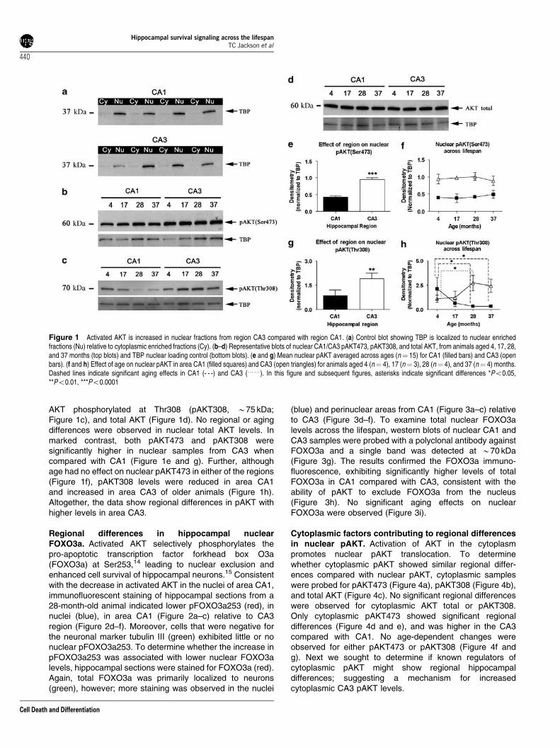
AKT phosphorylated at Thr308 (pAKT308, B75 kDa;Figure 1c), and total AKT (Figure 1d). No regional or agingdifferences were observed in nuclear total AKT levels. Inmarked contrast, both pAKT473 and pAKT308 weresignificantly higher in nuclear samples from CA3 whencompared with CA1 (Figure 1e and g). Further, althoughage had no effect on nuclear pAKT473 in either of the regions(Figure 1f), pAKT308 levels were reduced in area CA1and increased in area CA3 of older animals (Figure 1h).Altogether, the data show regional differences in pAKT withhigher levels in area CA3.
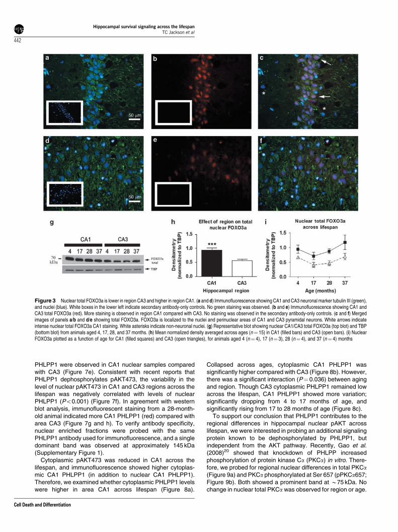
Regional differences in hippocampal nuclearFOXO3a. Activated AKT selectively phosphorylates thepro-apoptotic transcription factor forkhead box O3a(FOXO3a) at Ser253,14 leading to nuclear exclusion andenhanced cell survival of hippocampal neurons.15 Consistentwith the decrease in activated AKT in the nuclei of area CA1,immunofluorescent staining of hippocampal sections from a28-month-old animal indicated lower pFOXO3a253 (red), innuclei (blue), in area CA1 (Figure 2a–c) relative to CA3region (Figure 2d–f). Moreover, cells that were negative forthe neuronal marker tubulin III (green) exhibited little or nonuclear pFOXO3a253. To determine whether the increase inpFOXO3a253 was associated with lower nuclear FOXO3alevels, hippocampal sections were stained for FOXO3a (red).Again, total FOXO3a was primarily localized to neurons(green), however; more staining was observed in the nuclei
(blue) and perinuclear areas from CA1 (Figure 3a–c) relativeto CA3 (Figure 3d–f). To examine total nuclear FOXO3alevels across the lifespan, western blots of nuclear CA1 andCA3 samples were probed with a polyclonal antibody againstFOXO3a and a single band was detected at B70 kDa(Figure 3g). The results confirmed the FOXO3a immuno-fluorescence, exhibiting significantly higher levels of totalFOXO3a in CA1 compared with CA3, consistent with theability of pAKT to exclude FOXO3a from the nucleus(Figure 3h). No significant aging effects on nuclearFOXO3a were observed (Figure 3i).
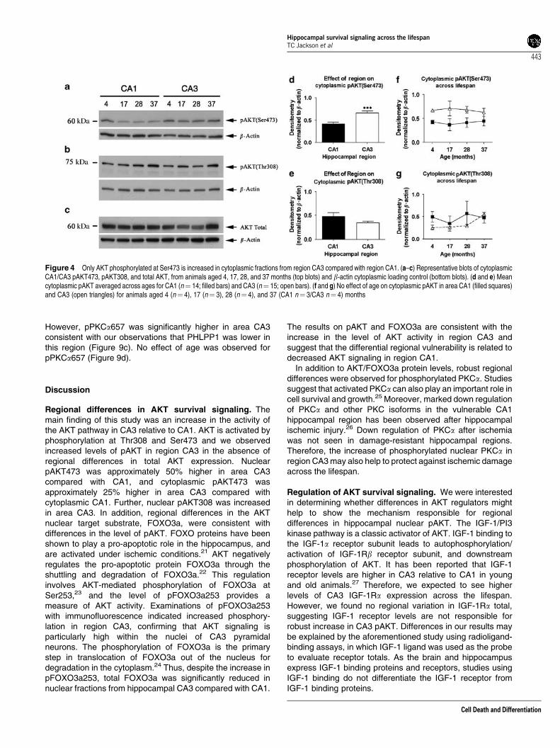
Cytoplasmic factors contributing to regional differencesin nuclear pAKT. Activation of AKT in the cytoplasmpromotes nuclear pAKT translocation. To determinewhether cytoplasmic pAKT showed similar regional differ-ences compared with nuclear pAKT, cytoplasmic sampleswere probed for pAKT473 (Figure 4a), pAKT308 (Figure 4b),and total AKT (Figure 4c). No significant regional differenceswere observed for cytoplasmic AKT total or pAKT308.Only cytoplasmic pAKT473 showed significant regionaldifferences (Figure 4d and e), and was higher in the CA3compared with CA1. No age-dependent changes wereobserved for either pAKT473 or pAKT308 (Figure 4f andg). Next we sought to determine if known regulators ofcytoplasmic pAKT might show regional hippocampaldifferences; suggesting a mechanism for increasedcytoplasmic CA3 pAKT levels.
Figure 1 Activated AKT is increased in nuclear fractions from region CA3 compared with region CA1. (a) Control blot showing TBP is localized to nuclear enrichedfractions (Nu) relative to cytoplasmic enriched fractions (Cy). (b–d) Representative blots of nuclear CA1/CA3 pAKT473, pAKT308, and total AKT, from animals aged 4, 17, 28,and 37 months (top blots) and TBP nuclear loading control (bottom blots). (e and g) Mean nuclear pAKT averaged across ages (n¼ 15) for CA1 (filled bars) and CA3 (openbars). (f and h) Effect of age on nuclear pAKT in area CA1 (filled squares) and CA3 (open triangles) for animals aged 4 (n¼ 4), 17 (n¼ 3), 28 (n¼ 4), and 37 (n¼ 4) months.Dashed lines indicate significant aging effects in CA1 (- - -) and CA3 (yy). In this figure and subsequent figures, asterisks indicate significant differences *Po0.05,**Po0.01, ***Po0.0001
Hippocampal survival signaling across the lifespanTC Jackson et al
440
Cell Death and Differentiation

The insulin-like growth factor 1 (IGF-1) receptor tyrosinekinase is a robust activator of AKT signaling in hippocampalneurons. Cytoplasmic activation of PI3 kinase by IGF-1promotes AKT phosphorylation and enhances its nucleartranslocation.16 To investigate whether expression of IGF-1receptors contributes to the differential activation of AKT,cytoplasmic CA1 and CA3 samples were probed for IGF-1Ra,and a band was detected at B150 kDa (Figure 5a). No regionor age differences in IGF-1Ra were observed suggesting thatthe total receptor expression does not underlie regionaldifferences in cytoplasmic pAKT (Figure 5c).
Next, we investigated whether the activity of IGF-1receptors might reflect regional differences in pAKT. Activa-tion of IGF-1 receptors requires ligand-dependent autopho-sphorylation of the IGF-1Rb (95 kDa) subunit. CA1 and CA3cytoplasmic samples were probed for phosphorylated IGF-1Rb (Tyr1135/1136) and a dominant band was detected atB95 kDa (Figure 5b). The existence of multiple bands above95 kDa may be because of additional post-transcriptionalmodification of IGF-1Rb. Although no significant regionaldifferences in pIGF-1Rb were observed, there was asignificant interaction (P¼ 0.016) between region and aging(Figure 5d). Activated IGF-1 receptors remained stableacross the lifespan in CA1. However in CA3, activatedIGF-1 receptors were much higher compared with CA1 inyoung animals and decreased significantly across thelifespan, falling well below CA1 levels at 37 months of age.
Phosphatase and tensin homolog (PTEN) can act as anegative regulator of AKT at the cell membrane.17 Totalcytoplasmic PTEN was detected as a single band at B50 kDaand levels were not different across regions (Figure 6a). In
contrast, PTEN levels varied as a function of age (Po0.001)with the highest levels observed between 17 and 28 months(Figure 6c). PTEN is regulated by multiple phosphorylationsites, including Ser370 such that the phosphorylated form(pPTEN370) is less active than the dephosphorylated form.Therefore, cytoplasmic samples were probed with an antibodyspecific for pPTEN370, and a band was detected at B55 kDa(Figure 6b). The level of pPTEN370 was not influencedby age, (Figure 6d) however, pPTEN370 was significantlyhigher in cytoplasmic CA1 samples relative to region CA3(Figure 6e). As pPTEN370 should enhance the level of pAKT,the activity of PTEN does not seem to explain the reducedlevel of nuclear pAKT in region CA1.
Phosphatases directly acting on AKT contribute toregional differences in nuclear pAKT. The serine/threonine phosphatase protein phosphatase 2A (PP2A)turns off AKT activity by dephosphorylation of AKT atThr308.18 CA1 and CA3 nuclear fractions were probed withan antibody against the PP2A-A subunit, and a single bandwas detected at B60 kDa (Figure 7a). Comparing CA1 andCA3 hippocampi across age groups showed no significantregional or age differences in total PP2A-A, suggesting thatnuclear PP2A-A levels do not underlie the robust regionaldifferences in active AKT (Figure 7c).
The PH domain and leucine-rich repeat protein phospha-tase 1 (PHLPP1) dephosphorylates AKT at Ser473 in a highlyspecific fashion.19 Examination of nuclear fractions for totalPHLPP1 (Figure 7b) indicated that PHLPP1 tended(P¼ 0.096) to increase with age from 17 months to 38 months(Figure 7d). Furthermore, significantly higher levels of
Figure 2 Nuclear pFOXO3a253 is higher in region CA3 and lower in region CA1. (a and d) Immunofluorescence showing CA1 and CA3 neuronal marker tubulin III(green), and nuclei (blue). White boxes in the lower left indicate secondary antibody-only controls. No green staining was observed. (b and e) Immunofluorescence showingCA1 and CA3 pFOXO3a253 (red). More staining is observed in region CA3 compared with CA1. No staining was observed in the secondary antibody-only controls. (c and f)Merged images of panels a/b and d/e showing pFOXO3a. pFOXO3a is localized to the nuclei of CA1 and CA3 pyramidal neurons. White arrows indicate intense nuclearpFOXO3a253 CA3 staining. White asterisks indicate non-neuronal nuclei
Hippocampal survival signaling across the lifespanTC Jackson et al
441
Cell Death and Differentiation
PHLPP1 were observed in CA1 nuclear samples comparedwith CA3 (Figure 7e). Consistent with recent reports thatPHLPP1 dephosphorylates pAKT473, the variability in thelevel of nuclear pAKT473 in CA1 and CA3 regions across thelifespan was negatively correlated with levels of nuclearPHLPP1 (Po0.001) (Figure 7f). In agreement with westernblot analysis, immunofluorescent staining from a 28-month-old animal indicated more CA1 PHLPP1 (red) compared witharea CA3 (Figure 7g and h). To verify antibody specificity,nuclear enriched fractions were probed with the samePHLPP1 antibody used for immunofluorescence, and a singledominant band was observed at approximately 145 kDa(Supplementary Figure 1).
Cytoplasmic pAKT473 was reduced in CA1 across thelifespan, and immunofluorescence showed higher cytoplas-mic CA1 PHLPP1 (in addition to nuclear CA1 PHLPP1).Therefore, we examined whether cytoplasmic PHLPP1 levelswere higher in area CA1 across lifespan (Figure 8a).
Collapsed across ages, cytoplasmic CA1 PHLPP1 wassignificantly higher compared with CA3 (Figure 8b). However,there was a significant interaction (P¼ 0.036) between agingand region. Though CA3 cytoplasmic PHLPP1 remained lowacross the lifespan, CA1 PHLPP1 showed more variation;significantly dropping from 4 to 17 months of age, andsignificantly rising from 17 to 28 months of age (Figure 8c).
To support our conclusion that PHLPP1 contributes to theregional differences in hippocampal nuclear pAKT acrosslifespan, we were interested in probing an additional signalingprotein known to be dephosphorylated by PHLPP1, butindependent from the AKT pathway. Recently, Gao et al.(2008)20 showed that knockdown of PHLPP increasedphosphorylation of protein kinase Ca (PKCa) in vitro. There-fore, we probed for regional nuclear differences in total PKCa(Figure 9a) and PKCa phosphorylated at Ser 657 (pPKCa657;Figure 9b). Both showed a prominent band at B75 kDa. Nochange in nuclear total PKCa was observed for region or age.
Figure 3 Nuclear total FOXO3a is lower in region CA3 and higher in region CA1. (a and d) Immunofluorescence showing CA1 and CA3 neuronal marker tubulin III (green),and nuclei (blue). White boxes in the lower left indicate secondary antibody-only controls. No green staining was observed. (b and e) Immunofluorescence showing CA1 andCA3 total FOXO3a (red). More staining is observed in region CA1 compared with CA3. No staining was observed in the secondary antibody-only controls. (c and f) Mergedimages of panels a/b and d/e showing total FOXO3a. FOXO3a is localized to the nuclei and perinuclear areas of CA1 and CA3 pyramidal neurons. White arrows indicateintense nuclear total FOXO3a CA1 staining. White asterisks indicate non-neuronal nuclei. (g) Representative blot showing nuclear CA1/CA3 total FOXO3a (top blot) and TBP(bottom blot) from animals aged 4, 17, 28, and 37 months. (h) Mean normalized density averaged across ages (n¼ 15) in CA1 (filled bars) and CA3 (open bars). (i) NuclearFOXO3a plotted as a function of age for CA1 (filled squares) and CA3 (open triangles), for animals aged 4 (n¼ 4), 17 (n¼ 3), 28 (n¼ 4), and 37 (n¼ 4) months
Hippocampal survival signaling across the lifespanTC Jackson et al
442
Cell Death and Differentiation
However, pPKCa657 was significantly higher in area CA3consistent with our observations that PHLPP1 was lower inthis region (Figure 9c). No effect of age was observed forpPKCa657 (Figure 9d).
Discussion
Regional differences in AKT survival signaling. Themain finding of this study was an increase in the activity ofthe AKT pathway in CA3 relative to CA1. AKT is activated byphosphorylation at Thr308 and Ser473 and we observedincreased levels of pAKT in region CA3 in the absence ofregional differences in total AKT expression. NuclearpAKT473 was approximately 50% higher in area CA3compared with CA1, and cytoplasmic pAKT473 wasapproximately 25% higher in area CA3 compared withcytoplasmic CA1. Further, nuclear pAKT308 was increasedin area CA3. In addition, regional differences in the AKTnuclear target substrate, FOXO3a, were consistent withdifferences in the level of pAKT. FOXO proteins have beenshown to play a pro-apoptotic role in the hippocampus, andare activated under ischemic conditions.21 AKT negativelyregulates the pro-apoptotic protein FOXO3a through theshuttling and degradation of FOXO3a.22 This regulationinvolves AKT-mediated phosphorylation of FOXO3a atSer253,23 and the level of pFOXO3a253 provides ameasure of AKT activity. Examinations of pFOXO3a253with immunofluorescence indicated increased phosphory-lation in region CA3, confirming that AKT signaling isparticularly high within the nuclei of CA3 pyramidalneurons. The phosphorylation of FOXO3a is the primarystep in translocation of FOXO3a out of the nucleus fordegradation in the cytoplasm.24 Thus, despite the increase inpFOXO3a253, total FOXO3a was significantly reduced innuclear fractions from hippocampal CA3 compared with CA1.
The results on pAKT and FOXO3a are consistent with theincrease in the level of AKT activity in region CA3 andsuggest that the differential regional vulnerability is related todecreased AKT signaling in region CA1.
In addition to AKT/FOXO3a protein levels, robust regionaldifferences were observed for phosphorylated PKCa. Studiessuggest that activated PKCa can also play an important role incell survival and growth.25 Moreover, marked down regulationof PKCa and other PKC isoforms in the vulnerable CA1hippocampal region has been observed after hippocampalischemic injury.26 Down regulation of PKCa after ischemiawas not seen in damage-resistant hippocampal regions.Therefore, the increase of phosphorylated nuclear PKCa inregion CA3 may also help to protect against ischemic damageacross the lifespan.
Regulation of AKT survival signaling. We were interestedin determining whether differences in AKT regulators mighthelp to show the mechanism responsible for regionaldifferences in hippocampal nuclear pAKT. The IGF-1/PI3kinase pathway is a classic activator of AKT. IGF-1 binding tothe IGF-1a receptor subunit leads to autophosphorylation/activation of IGF-1Rb receptor subunit, and downstreamphosphorylation of AKT. It has been reported that IGF-1receptor levels are higher in CA3 relative to CA1 in youngand old animals.27 Therefore, we expected to see higherlevels of CA3 IGF-1Ra expression across the lifespan.However, we found no regional variation in IGF-1Ra total,suggesting IGF-1 receptor levels are not responsible forrobust increase in CA3 pAKT. Differences in our results maybe explained by the aforementioned study using radioligand-binding assays, in which IGF-1 ligand was used as the probeto evaluate receptor totals. As the brain and hippocampusexpress IGF-1 binding proteins and receptors, studies usingIGF-1 binding do not differentiate the IGF-1 receptor fromIGF-1 binding proteins.
Figure 4 Only AKT phosphorylated at Ser473 is increased in cytoplasmic fractions from region CA3 compared with region CA1. (a–c) Representative blots of cytoplasmicCA1/CA3 pAKT473, pAKT308, and total AKT, from animals aged 4, 17, 28, and 37 months (top blots) and b-actin cytoplasmic loading control (bottom blots). (d and e) Meancytoplasmic pAKT averaged across ages for CA1 (n¼ 14; filled bars) and CA3 (n¼ 15; open bars). (f and g) No effect of age on cytoplasmic pAKT in area CA1 (filled squares)and CA3 (open triangles) for animals aged 4 (n¼ 4), 17 (n¼ 3), 28 (n¼ 4), and 37 (CA1 n¼ 3/CA3 n¼ 4) months
Hippocampal survival signaling across the lifespanTC Jackson et al
443
Cell Death and Differentiation
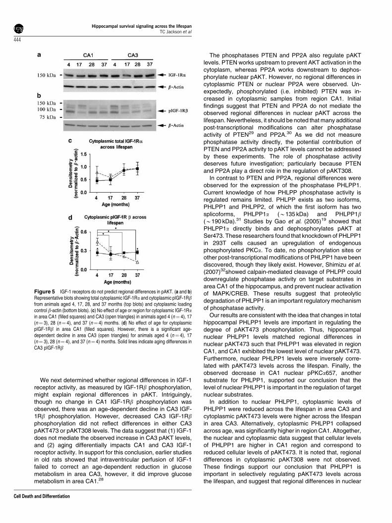
We next determined whether regional differences in IGF-1receptor activity, as measured by IGF-1Rb phosphorylation,might explain regional differences in pAKT. Intriguingly,though no change in CA1 IGF-1Rb phosphorylation wasobserved, there was an age-dependent decline in CA3 IGF-1Rb phosphorylation. However, decreased CA3 IGF-1Rbphosphorylation did not reflect differences in either CA3pAKT473 or pAKT308 levels. The data suggest that (1) IGF-1does not mediate the observed increase in CA3 pAKT levels,and (2) aging differentially impacts CA1 and CA3 IGF-1receptor activity. In support for this conclusion, earlier studiesin old rats showed that intraventricular perfusion of IGF-1failed to correct an age-dependent reduction in glucosemetabolism in area CA3, however, it did improve glucosemetabolism in area CA1.28
The phosphatases PTEN and PP2A also regulate pAKTlevels. PTEN works upstream to prevent AKT activation in thecytoplasm, whereas PP2A works downstream to dephos-phorylate nuclear pAKT. However, no regional differences incytoplasmic PTEN or nuclear PP2A were observed. Un-expectedly, phosphorylated (i.e. inhibited) PTEN was in-creased in cytoplasmic samples from region CA1. Initialfindings suggest that PTEN and PP2A do not mediate theobserved regional differences in nuclear pAKT across thelifespan. Nevertheless, it should be noted that many additionalpost-transcriptional modifications can alter phosphataseactivity of PTEN29 and PP2A.30 As we did not measurephosphatase activity directly, the potential contribution ofPTEN and PP2A activity to pAKT levels cannot be addressedby these experiments. The role of phosphatase activitydeserves future investigation; particularly because PTENand PP2A play a direct role in the regulation of pAKT308.
In contrast to PTEN and PP2A, regional differences wereobserved for the expression of the phosphatase PHLPP1.Current knowledge of how PHLPP phosphatase activity isregulated remains limited. PHLPP exists as two isoforms,PHLPP1 and PHLPP2, of which the first isoform has twosplicoforms, PHLPP1a (B135 kDa) and PHLPP1b(B190 kDa).31 Studies by Gao et al. (2005)19 showed thatPHLPP1a directly binds and dephosphorylates pAKT atSer473. These researchers found that knockdown of PHLPP1in 293T cells caused an upregulation of endogenousphosphorylated PKCa. To date, no phosphorylation sites orother post-transcriptional modifications of PHLPP1 have beendiscovered, though they likely exist. However, Shimizu et al.(2007)32showed calpain-mediated cleavage of PHLPP coulddownregulate phosphatase activity on target substrates inarea CA1 of the hippocampus, and prevent nuclear activationof MAPK/CREB. These results suggest that proteolyticdegradation of PHLPP1 is an important regulatory mechanismof phosphatase activity.
Our results are consistent with the idea that changes in totalhippocampal PHLPP1 levels are important in regulating thedegree of pAKT473 phosphorylation. Thus, hippocampalnuclear PHLPP1 levels matched regional differences innuclear pAKT473 such that PHLPP1 was elevated in regionCA1, and CA1 exhibited the lowest level of nuclear pAKT473.Furthermore, nuclear PHLPP1 levels were inversely corre-lated with pAKT473 levels across the lifespan. Finally, theobserved decrease in CA1 nuclear pPKCa657, anothersubstrate for PHLPP1, supported our conclusion that thelevel of nuclear PHLPP1 is important in the regulation of targetnuclear substrates.
In addition to nuclear PHLPP1, cytoplasmic levels ofPHLPP1 were reduced across the lifespan in area CA3 andcytoplasmic pAKT473 levels were higher across the lifespanin area CA3. Alternatively, cytoplasmic PHLPP1 collapsedacross age, was significantly higher in region CA1. Altogether,the nuclear and cytoplasmic data suggest that cellular levelsof PHLPP1 are higher in CA1 region and correspond toreduced cellular levels of pAKT473. It is noted that, regionaldifferences in cytoplasmic pAKT308 were not observed.These findings support our conclusion that PHLPP1 isimportant in selectively regulating pAKT473 levels acrossthe lifespan, and suggest that regional differences in nuclear
Figure 5 IGF-1 receptors do not predict regional differences in pAKT. (a and b)Representative blots showing total cytoplasmic IGF-1Ra and cytoplasmic pIGF-1Rbfrom animals aged 4, 17, 28, and 37 months (top blots) and cytoplasmic loadingcontrol b-actin (bottom blots). (c) No effect of age or region for cytoplasmic IGF-1Rain area CA1 (filled squares) and CA3 (open triangles) in animals aged 4 (n¼ 4), 17(n¼ 3), 28 (n¼ 4), and 37 (n¼ 4) months. (d) No effect of age for cytoplasmicpIGF-1Rb in area CA1 (filled squares). However, there is a significant age-dependent decline in area CA3 (open triangles) for animals aged 4 (n¼ 4), 17(n¼ 3), 28 (n¼ 4), and 37 (n¼ 4) months. Solid lines indicate aging differences inCA3 pIGF-1Rb
Hippocampal survival signaling across the lifespanTC Jackson et al
444
Cell Death and Differentiation

CA1/CA3 pAKT308 depend upon regulation (or dysregula-tion) by mechanisms distinct from PHLPP1.
Despite our data supporting PHLPP1’s role in mediatingregional differences in hippocampal pAKT473, additionalregulation seems very likely; primarily because CA1 pAKT473levels were remarkably stable across the lifespan in boththe nucleus and the cytoplasm whereas CA1 PHLPP1showed much more variability. One possibility is that regionaldifferences in growth factor-mediated AKT activation, otherthan IGF-1, help to regulate AKT across the lifespan.Immunohistochemical studies in rat brain showed that BDNFlevels are extremely high in area CA3 and extremely low inarea CA1.33 Further, these differences are maintained acrossthe lifespan.34 As BDNF can activate AKT, and has beenshown to rapidly inhibit FOXO3a nuclear localization,35 BDNFsignaling may also contribute to the observed maintenance ofpAKT across the lifespan. However, comparative studies ofgrowth factor-mediated survival signaling in hippocampal cellculture show that IGF-1 strongly induces AKT although itweakly induces ERK, whereas BDNF strongly induces ERKand only weakly induces AKT.36 Therefore, whether BDNFcontributes to sustained AKT activity in vivo and whether itscontribution is large or small remains unknown and deservesfurther investigation.
AKT activity across the lifespan. Aging-dependentdysregulation of proteins that mediate survival signalingmay contribute to increased hippocampal vulnerability duringaging. The results of this study indicate that the level ofnuclear pAKT308 declines in CA1 region as age advances.Alternatively, CA1 pAKT473 was stable across the lifespan.This raises the question of whether the loss in dual pAKTexacerbates vulnerability of CA1 region as age advances.Recent in vitro studies examining the regulation of AKTphosphorylation and AKT activity provided evidence that dualphosphorylation of pAKT308 and pAKT473 are temporally
associated/coordinated, and together predicted AKTactivity.37 In addition, studies in NIH3T3 cells showed thata pAKT308 mutant failed to activate when treated withgrowth factor, whereas a pAKT473 mutant showed reducedactivity; thus dual phosphorylation seems to be required forfull AKT kinase activity.38 In aged animals, decreased CA1pAKT308 levels would predict reduced total AKT activityeven though pAKT473 remained stable.
It is unclear what mechanism(s) underlie the age-relateddecline in nuclear CA1 pAKT308. One possible explanationmay be that aging differentially affects nuclear CA1/CA3membrane phosphatidylinositol (3,4,5)-trisphosphate (PIP3)levels. AKT can be activated in the nucleus by identicalsignaling mechanisms observed at the cell membrane.39
However, before AKT can be phosphorylated at Thr308, PI3kinase must be activated upstream. When activated, PI3kinase will phosphorylate membrane bound PIP2 to PIP3. PIP3
will then allow both AKT and PIP3-dependent kinase 1 (PDK-1) to bind the inner membrane through their PH domains.PDK-1 is responsible for phosphorylating AKT at Thr308, andbinding of both enzymes to PIP3 is critical to allowing PDK-1access to AKT’s active site at Thr308. Therefore, changes inthe nuclear CA1 PIP3 levels across the lifespan could alter thenuclear CA1 AKT308 levels. However, little is known aboutthe regulation and control of nuclear PIP3, let alone how agingmight influence nuclear PIP3 levels.
Alternatively, Ca2þ /calmodulin-dependent protein kinasekinase (CaM-KK) can selectively and directly phosphorylateAKT at Thr308 but not on Ser473.40 Intriguingly, CaM-KKa isfound only in the nucleus, where it acts to regulate its betterknown substrate Ca2þ /calmodulin-dependent protein kinaseIV. As Ca2þ imbalance plays an important role in hippocam-pal CA1 loss of function with aging, altered hippocampal CaM-KK activity with age may also play a role in selectively alteringnuclear pAKT308 phosphorylation across the lifespan in areaCA1. This idea remains to be tested.
Figure 6 PTEN does not predict regional differences in pAKT. (a and b) Representative blots showing total PTEN, and pPTEN370 in animals aged 4, 17, 28, and 37months (top blots) and cytoplasmic loading control b-actin (bottom blots). (c) CA1 (filled squares) and CA3 (open triangles) cytoplasmic total PTEN shows no significantregional effects but significant aging effects in animals aged 4 (n¼ 4), 17 (n¼ 3), 28 (n¼ 4), and 37 (n¼ 4) months. Solid lines indicate aging differences in CA1/CA3 totalPTEN . (d) CA1 (filled squares) and CA3 (open triangles) cytoplasmic pPTENSer370 was not different across the lifespan in animals aged 4 (n¼ 4), 17 (n¼ 3), 28 (n¼ 4), and37 (n¼ 4) months, however, (e) elevated levels of pPTEN370 were observed (n¼ 15) in region CA1 (filled bars) relative to CA3 (open bars)
Hippocampal survival signaling across the lifespanTC Jackson et al
445
Cell Death and Differentiation

Altogether, the results suggest that the regional hippocam-pal differences in the AKT pathway may contribute to regionaldifferences in neuronal vulnerability to certain stressors, andhelp protect CA3 neurons from pathology associated withage-dependent disease. In support for these conclusions, the
pro-apoptotic protein FOXO3a was higher in area CA1 acrosslifespan and actively inhibited in pyramidal neuron nuclei ofregion CA3. Further, nuclear PHLPP1 was higher in CA1
Figure 7 Nuclear PHLPP1 but not PP2A-A correlate with regional differences innuclear pAKTSer473. (a and b) Representative blots showing nuclear total PP2A-Aand PHLPP1 in animals aged 4, 17, 28, and 37 months (top blots) and TBP (bottomblots) (c) CA1 (filled squares) and CA3 (open triangles) nuclear PP2A-A across thelifespan. (d) CA1 (filled squares) and CA3 (open triangles) nuclear PHLPP1 had atendency to increase with age, and (e) significant regional differences (n¼ 15) wereobserved in CA1 (filled bars) relative to CA3 (open bars). (f) Linear regression ofcombined CA1 (dark circles)/CA3 (open circles) samples across the lifespanshowing PHLPP1 levels correlate with pAKTSer473 (r2¼ 0.27). (g and h)Immunofluorescence showing CA1 and CA3 PHLPP1 (red) and nuclei stained withDAPI (blue). White border squares in the bottom left indicate secondary antibody-only controls. White arrow heads indicate nuclear and perinuclear regions of intensePHLPP1 staining in area CA1. Animals aged 4 (n¼ 4), 17 (n¼ 3), 28 (n¼ 4), and37 (n¼ 4) months were used for lifespan graphs
Figure 8 Cytoplasmic PHLPP1 is increased in area CA1. (a) Representativeblot showing cytoplasmic total PHLPP1 in animals aged 4, 17, 28, and 37 months(top blot) and b-actin (bottom blot) (b) Mean normalized densitometry averagedacross ages (n¼ 15) in CA1 (filled bars) and CA3 (open bars). Significant regionaldifferences in cytoplasmic PHLPP1 (n¼ 15) were observed in area CA1 (filled bars)relative to area CA3 (open bars). (c) CA1 PHLPP1 show significant variation acrossthe lifespan (filled squares). Solid lines indicate aging differences in CA1 PHLPP1.Alternatively CA3 (open triangles) cytoplasmic PHLPP1 showed reduced and stablelevels across the lifespan. Animals aged 4 (n¼ 4), 17 (n¼ 3), 28 (n¼ 4), and 37(n¼ 4) months were used for lifespan graph
Figure 9 Regional differences in nuclear pPKCa657. (a and b) Representativeblots showing nuclear CA1/CA3 total PKCa and pPKCa657 levels in animals aged4, 17, 28, and 37 months (top blots) normalized to TBP (bottom blots). (c) Meannormalized densitometry averaged across ages (n¼ 15) in CA1 (filled bars) andCA3 (open bars). Significantly higher levels of pPKCa657 are observed in thenucleus of area CA3. (d) CA1 (filled squares) and CA3 (open triangles) nuclearpPKCa657 shows no significant aging effects. Animals aged 4 (n¼ 4), 17 (n¼ 3),28 (n¼ 4), and 37 (n¼ 4) months were used for lifespan graph
Hippocampal survival signaling across the lifespanTC Jackson et al
446
Cell Death and Differentiation

region and correlated with levels of nuclear pAKT473. To thebest of our knowledge, these findings are novel and suggest anew hypothesis to explain the mechanisms for CA1 vulner-ability to certain kinds of stressors.
Materials and MethodsAged animals. NIA Fischer 344/Brown Norway rats were kept in specificpathogen free (SPF) housing and maintained on a 12 h light/dark cycle. Animalswere aged 4 (n¼ 4), 17 (n¼ 3), 28 (n¼ 4), and 37 (n¼ 4) months, and fed astandard ad libitum diet. Animals were killed by CO2 asphyxiation and decapitated.Hippocampi were removed, separated into CA1 and CA3 sections, and flash frozenin liquid nitrogen. Samples were stored at �801C until further processing.
Homogenization & nuclear enrichment. Tissues were homogenizedusing NER nuclear separation kit (PIERCE) according to the manufacturer’sinstructions. Briefly, frozen samples (B30–40 mg of tissue/sample) were removedfrom �801C freezer and immediately placed in a 2 ml glass dounce homogenizer(Kimble–Kontes) containing ice-cold CERI buffer (for cytoplasmic fraction) with1.5� protease inhibitors, 1� EDTA, and 2� phosphatase inhibitors (PIERCE).Samples were then homogenized using 11 strokes with pestle B and 10 strokes withpestle A, and transferred to 0.5 ml tubes. After sitting on ice for 10 min, sampleswere briefly vortexed and spun at 16 000� g at 41C for 5 min. Samples were thenplaced back on ice and supernatant was collected and saved for cytoplasmic proteinanalysis. Next, NERI buffer (for nuclear fraction), containing 1.5� proteaseinhibitors, 1� EDTA, and 2� phosphatase inhibitors (PIERCE) was added to thecell pellet. Samples were vortexed every 10 min for 40 min and spun at 16 000� gat 41C for 10 min. The supernatant (containing nuclear enriched protein fraction)was collected and stored for later analysis.
Western blot. Protein concentrations were determined using BSA method(PIERCE). Kaleidoscope protein standards (Bio-Rad) and CA1/CA3 samples(20mg/lane) were loaded on 4–15% gradient gels (Bio-Rad) and run for 1 h at120 V. Proteins were then transferred to PDVF membranes (Amersham) overnightat 50 V/41C. Blots were then stained with Ponceau S and photocopied. Blots werewashed 5 min in T-BST and subsequently blocked in T-BST (7% milk) for 1 h.Primary antibodies were then applied to blots overnight at 41C (Table 1), washedthree times with TBS, and secondary antibodies applied for 2 h at roomtemperature. Blots were then developed using ECL Plus Western Blot DetectionKit (Amersham) on biomax film (Kodak). Blots were scanned using 6500 scanner(Bio-Rad), and densitometry determined using UNSCAN IT software (silk scientific).
Immunofluorescence. A 28-month-old Fisher 344/BN rat was anesthetizedand perfused with 4% paraformaldehyde. The brain was removed, placed inparaformaldehyde for 1 h, and transferred to 30% sucrose solution for 72 h. Afterfixation, the brain was embedded in optimal cutting compound and 8 mm coronalsections were made using a cryostat. Slices were collected on Superfrost Plus glassslides (Fisher), and allowed to air dry for 1 h. Slides were then washed with PBS andpermeabilized with 0.05% TritonX-100/PBS for 15 min. Slides were washed again inPBS and treated with 1% SDS/PBS for 5 min for mild antigen retrieval. Sectionswere then washed in PBS, blocked with 20% goat serum (in 1% BSA PBS) for 2 h,and incubated overnight (at 41C) in 3% goat serum PBS containing rabbit anti-phospho or total FOXO3aþmouse anti-neuronal tubulin III or rabbit anti-PHLPP1.Slides were then washed with PBS and incubated with secondary antibodies (AlexaFluor goat anti-rabbit 594/goat anti-mouse 488; Invitrogen) for 1.5 h. Slides werethen stained with 3% Sudan Black B in 70% ethanol for 10 min to remove lipofuscinautofluorescence. Finally, slides were mounted (Prolong Gold Anti-Fade with DAPI;Invitrogen) and images were taken on an Axiovert 40 CFR fluorescent microscope(Zeiss).
Statistical analysis. Western blot densitometry was analyzed using three wayANOVA (NCSS Statistical Software); data significant at Po0.05. Post hoc analysiswas carried out using Fisher LSD test. All graphs were produced using Prismsoftware (Silk Scientific).
Acknowledgements. This research was supported by the NIH: (1)Neurobiology of Aging Training Grant 2-T32-AG000196-16A1 (2) RO1-AG014979-09 and (3) Evelyn F. McKnight Brain Research Grant.
1. Hof PR, Morrison JH. The aging brain: morphomolecular senescence of cortical circuits.Trends Neurosci 2004; 27: 607–613.
2. Kadar T, Dachir S, Shukitt-Hale B, Levy A. Sub-regional hippocampal vulnerabilityin various animal models leading to cognitive dysfunction. J Neural Transm 1998; 105:987–1004.
3. Korf ES, White LR, Scheltens P, Launer LJ. Midlife blood pressure and the risk ofhippocampal atrophy: the Honolulu Asia aging study. Hypertension 2004; 44: 29–34.
4. Back T, Hemmen T, Schuler OG. Lesion evolution in cerebral ischemia. J Neurol 2004;251: 388–397.
5. Noda A, Ohba H, Kakiuchi T, Futatsubashi M, Tsukada H, Nishimura S. Age-relatedchanges in cerebral blood flow and glucose metabolism in conscious rhesus monkeys.Brain Res 2002; 936: 76–81.
6. Schmidt-Kastner R, Freund TF. Selective vulnerability of the hippocampus in brainischemia. Neuroscience 1991; 40: 599–636.
Table 1 Antibody list
Antibody Concentration Host Clonality Company
TBP 1 : 1000 Mouse Monoclonal ABRb-Actin 1 : 8000 Chicken Polyclonal Abcamp(Thr308)AKT 1 : 1000 Rabbit Monoclonal Cell signalingp(Ser473)AKT 1 : 1000 Rabbit Polyclonal Cell signalingAKT total 1 : 1000 Rabbit Polyclonal Cell signalingFOXO3a 1 : 1000 Rabbit Polyclonal ABRpFOXO3a253 (Immuno-Fluo) 2 mg/ml Rabbit Polyclonal AbcamFOXO3a (Immuno-Fluo) 1 : 100 Rabbit Monoclonal Cell signalingIGF-1Ra 1 : 200 Rabbit Polyclonal Santa CruzpIGF-1Rb 1 : 1000 Rabbit Monoclonal Cell signalingp(Ser370)PTEN 1 : 1000 Rabbit Polyclonal AbcamPTEN total 1 mg/ml Rabbit Polyclonal AbcamPP2A-A 1 : 1000 Mouse Monoclonal Cell signalingPHLPP1 1 : 200 Goat Polyclonal Santa CruzPHLPP1 (Immuno-Fluo) 1 : 50 Rabbit Polyclonal Caymanp(Ser657)PKCa 0.5mg/ml Rabbit Polyclonal UpstatePKCa total 0.4mg/ml Mouse Monoclonal UpstateNeuronal b-tubulin III 1 : 1000 Mouse Monoclonal Abcam
AKT, protein kinase B; pAKT, phosphorylated AKT; FOXO3a, forkhead box O3a transcription factor; pFOXO3a, phosphorylated FOXO3a; IGF-1, insulin-like growthfactor 1; pIGF-1, phosphorylated IGF-1; pPTEN, phosphorylated PTEN; PHLPP1, PH domain and leucine-rich repeat protein phosphatase 1; PKCa, protein kinaseCa; pPKCa, phosphorylated PKCa; PTEN, phosphatase and tensin homolog; TBP, TATA box binding protein
Hippocampal survival signaling across the lifespanTC Jackson et al
447
Cell Death and Differentiation

7. Ouyang YB, Voloboueva LA, Xu LJ, Giffard RG. Selective dysfunction of hippocampal CA1astrocytes contributes to delayed neuronal damage after transient forebrain ischemia.J Neurosci 2007; 27: 4253–4260.
8. De Jong GI, Farkas E, Stienstra CM, Plass JR, Keijser JN, de la Torre JC et al. Cerebralhypoperfusion yields capillary damage in the hippocampal CA1 area that correlates withspatial memory impairment. Neuroscience 1999; 91: 203–210.
9. West MJ, Kawas CH, Stewart WF, Rudow GL, Troncoso JC. Hippocampal neurons in pre-clinical Alzheimer’s disease. Neurobiol Aging 2004; 25: 1205–1212.
10. Spires TL, Orne JD, SantaCruz K, Pitstick R, Carlson GA, Ashe KH et al. Region-specificdissociation of neuronal loss and neurofibrillary pathology in a mouse model of tauopathy.Am J Pathol 2006; 168: 1598–1607.
11. Fukunaga K, Kawano T. Akt is a molecular target for signal transduction therapy in brainischemic insult. J Pharmacol Sci 2003; 92: 317–327.
12. Martin D, Salinas M, Lopez-Valdaliso R, Serrano E, Recuero M, Cuadrado A. Effect of theAlzheimer amyloid fragment Abeta(25–35) on Akt/PKB kinase and survival of PC12 cells.J Neurochem 2001; 78: 1000–1008.
13. Xu GG, Deng YQ, Liu SJ, Li HL, Wang JZ. Prolonged Alzheimer-like tau hyper-phosphorylation induced by simultaneous inhibition of phosphoinositol-3 kinase and proteinkinase C in N2a cells. Acta Biochim Biophys Sin (Shanghai) 2005; 37: 349–354.
14. Brunet A, Park J, Tran H, Hu LS, Hemmings BA, Greenberg ME. Protein kinase SGKmediates survival signals by phosphorylating the forkhead transcription factor FKHRL1(FOXO3a). Mol Cell Biol 2001; 21: 952–965.
15. Zheng WH, Kar S, Quirion R. Insulin-like growth factor-1-induced phosphorylation oftranscription factor FKHRL1 is mediated by phosphatidylinositol 3-kinase/Akt kinase androle of this pathway in insulin-like growth factor-1-induced survival of cultured hippocampalneurons. Mol Pharmacol 2002; 62: 225–233.
16. Borgatti P, Martelli AM, Bellacosa A, Casto R, Massari L, Capitani S et al. Translocation ofAkt/PKB to the nucleus of osteoblast-like MC3T3-E1 cells exposed to proliferative growthfactors. FEBS Lett 2000; 477: 27–32.
17. Wan X, Helman LJ. Levels of PTEN protein modulate Akt phosphorylation on serine 473,but not on threonine 308, in IGF-II-overexpressing rhabdomyosarcomas cells. Oncogene2003; 22: 8205–8211.
18. Trotman LC, Alimonti A, Scaglioni PP, Koutcher JA, Cordon-Cardo C, Pandolfi PP et al.Identification of a tumour suppressor network opposing nuclear Akt function. Nature 2006;441: 523–527.
19. Gao T, Furnari F, Newton AC. PHLPP: a phosphatase that directly dephosphorylates Akt,promotes apoptosis, and suppresses tumor growth. Mol Cell 2005; 18: 13–24.
20. Gao T, Brognard J, Newton AC. The phosphatase PHLPP controls the cellular levels ofprotein kinase C. J Biol Chem 2008; 283: 6300–6311.
21. Kawano T, Morioka M, Yano S, Hamada J, Ushio Y, Miyamoto E et al. Decreased aktactivity is associated with activation of forkhead transcription factor after transient forebrainischemia in gerbil hippocampus. J Cereb Blood Flow Metab 2002; 22: 926–934.
22. Plas DR, Thompson CB. Akt activation promotes degradation of tuberin and FOXO3a viathe proteasome. J Biol Chem 2003; 278: 12361–12366.
23. Zheng WH, Kar S, Quirion R. Insulin-like growth factor-1-induced phosphorylation ofthe forkhead family transcription factor FKHRL1 is mediated by Akt kinase in PC12 cells.J Biol Chem 2000; 275: 39152–39158.
24. Brunet A, Bonni A, Zigmond MJ, Lin MZ, Juo P, Hu LS et al. Akt promotes cell survivalby phosphorylating and inhibiting a Forkhead transcription factor. Cell 1999; 96:857–868.
25. Michie AM, Nakagawa R. The link between PKC alpha regulation and cellulartransformation. Immunol Lett 2005; 96: 155–162.
26. Ziemka-Nalecz M, Zalewska T, Zajac H, Domanska-Janik K. Decrease of PKC precedesother cellular signs of calpain activation in area CA1 of the hippocampus after transientcerebral ischemia. Neurochem Int 2003; 42: 205–214.
27. Dore S, Kar S, Rowe W, Quirion R. Distribution and levels of [125I]IGF-I, [125I]IGF-II and[125I]insulin receptor binding sites in the hippocampus of aged memory-unimpaired and-impaired rats. Neuroscience 1997; 80: 1033–1040.
28. Lynch CD, Lyons D, Khan A, Bennett SA, Sonntag WE. Insulin-like growth factor-1selectively increases glucose utilization in brains of aged animals. Endocrinology 2001;142: 506–509.
29. Das S, Dixon JE, Cho W. Membrane-binding and activation mechanism of PTEN. Proc NatlAcad Sci USA 2003; 100: 7491–7496.
30. Janssens V, Goris J. Protein phosphatase 2A: a highly regulated family of serine/threoninephosphatases implicated in cell growth and signalling. Biochem J 2001; 353 (Part 3):417–439.
31. Brognard J, Sierecki E, Gao T, Newton AC. PHLPP and a second isoform, PHLPP2,differentially attenuate the amplitude of Akt signaling by regulating distinct Akt isoforms.Mol Cell 2007; 25: 917–931.
32. Shimizu K, Phan T, Mansuy IM, Storm DR. Proteolytic degradation of SCOP in thehippocampus contributes to activation of MAP kinase and memory. Cell 2007; 128:1219–1229.
33. Yan Q, Rosenfeld RD, Matheson CR, Hawkins N, Lopez OT, Bennett L et al. Expression ofbrain-derived neurotrophic factor protein in the adult rat central nervous system.Neuroscience 1997; 78: 431–448.
34. Newton IG, Forbes ME, Legault C, Johnson JE, Brunso-Bechtold JK, Riddle DR. Caloricrestriction does not reverse aging-related changes in hippocampal BDNF. Neurobiol Aging2005; 26: 683–688.
35. Zhu W, Bijur GN, Styles NA, Li X. Regulation of FOXO3a by brain-derived neurotrophicfactor in differentiated human SH-SY5Y neuroblastoma cells. Brain Res Mol Brain Res2004; 126: 45–56.
36. Zheng WH, Quirion R. Comparative signaling pathways of insulin-like growth factor-1 andbrain-derived neurotrophic factor in hippocampal neurons and the role of the PI3 kinasepathway in cell survival. J Neurochem 2004; 89: 844–852.
37. Kumar N, Afeyan R, Sheppard S, Harms B, Lauffenburger DA. Quantitative analysis of Aktphosphorylation and activity in response to EGF and insulin treatment. Biochem BiophysRes Commun 2007; 354: 14–20.
38. Bellacosa A, Chan TO, Ahmed NN, Datta K, Malstrom S, Stokoe D et al. Akt activation bygrowth factors is a multiple-step process: the role of the PH domain. Oncogene 1998; 17:313–325.
39. Wang R, Brattain MG. AKT can be activated in the nucleus. Cell Signal 2006; 18:1722–1731.
40. Yano S, Tokumitsu H, Soderling TR. Calcium promotes cell survival through CaM-K kinaseactivation of the protein-kinase-B pathway. Nature 1998; 396: 584–587.
Supplementary Information accompanies the paper on Cell Death and Differentiation website (http://www.nature.com/cdd)
Hippocampal survival signaling across the lifespanTC Jackson et al
448
Cell Death and Differentiation
Copyright © 2022 FDOKUMEN