Quantum Chemical and Microwave Spectroscopic ...
389
Quantum Chemical and Microwave Spectroscopic Investigations on Phenyl Ring Containing Molecules Von der Fakult¨at f¨ ur Mathematik, Informatik und Naturwissenschaften der RWTH Aachen University zur Erlangung des akademischen Grades einer Doktorin der Naturwissenschaften genehmigte Dissertation vorgelegt von Master of Science Lynn Ferres aus Esch/Alzette, Luxemburg Berichter: Univ.-Prof. Dr. rer. nat. Wolfgang Stahl Univ.-Prof. Dr. rer. nat. Arne L¨ uchow Tag der m¨ undlichen Pr¨ ufung: 18.04.2019 Diese Dissertation ist auf den Internetseiten der Universit¨ atsbibliothek verf¨ ugbar.
-
Upload
khangminh22 -
Category
Documents
-
view
1 -
download
0
Transcript of Quantum Chemical and Microwave Spectroscopic ...

Quantum Chemical and MicrowaveSpectroscopic Investigations on Phenyl
Ring Containing Molecules
Von der Fakultat fur Mathematik, Informatik undNaturwissenschaften der RWTH Aachen University zur Erlangungdes akademischen Grades einer Doktorin der Naturwissenschaften
genehmigte Dissertation
vorgelegt vonMaster of Science
Lynn Ferresaus Esch/Alzette, Luxemburg
Berichter: Univ.-Prof. Dr. rer. nat. Wolfgang StahlUniv.-Prof. Dr. rer. nat. Arne Luchow
Tag der mundlichen Prufung: 18.04.2019
Diese Dissertation ist auf den Internetseiten der Universitatsbibliothek verfugbar.


七転び、八起き。
Japanese Proverb”Fall down seven times and get up eight.”
i

The experimental work presented in this thesis was conducted in the time betweenOctober 2015 and August 2018 at the Institute of Physical Chemistry, RWTHAachen University, under the supervision of Prof. Dr. Wolfgang Stahl.
ii
Parts of this thesis have been published in scientific journals:
1. L. Ferres, W. Stahl, H. V. L. Nguyen, The Molecular Structure of Phene-tole studied by Microwave Spectroscopy and Quantum Chemical Calculations,Mol. Phys. 114, 2788-2793, (2016).DOI: 10.1080/00268976.2016.1177217
2. L. Ferres, H. Mouhib, W. Stahl, H. V. L. Nguyen, Methyl Internal Rota-tion in the Microwave Spectrum of o-Methyl Anisole, ChemPhysChem 18, 14,1855-1859, (2017). DOI: 10.1002/cphc.201700276
3. L. Ferres, H. Mouhib, W. Stahl, H. V. L. Nguyen, Inside Cover: MethylInternal Rotation in the Microwave Spectrum of o-Methyl Anisole, ChemPhys-Chem 14, (2017). DOI: 10.1002/cphc.201700700
4. L. Ferres, H. Mouhib, W. Stahl, M. Schwell; H. V. L. Nguyen, MolecularStructure and Ring Tunneling of Phenyl Formate as Observed by MicrowaveSpectroscopy and Quantum Chemistry, J. Mol. Spectrosc. 337, 59-64, (2017).DOI: 10.1016/j.jms.2017.04.017
5. L. Ferres, W. Stahl, I. Kleiner, H. V. L. Nguyen, The Effect of Inter-nal Rotation in p-Methyl Anisole Studied by Microwave Spectroscopy, J. Mol.Spectrosc. 343, 44-49, (2017). DOI:10.1016/j.jms.2017.09.008
6. L. Ferres, W. Stahl, H. V. L. Nguyen, Conformational Effects on theTorsional Barriers in m-Methylanisole Studied by Microwave Spectroscopy, J.Chem. Phys. 148, 12, 124304, (2018). DOI: 10.1063/1.5016273
7. L. Ferres, K.-N. Truong, W. Stahl, H. V. L. Nguyen, Interplay Be-tween Microwave Spectroscopy and X-ray Diffraction: The Molecular Structureand Large Amplitude Motions of 2,3-Dimethylanisole, ChemPhysChem 19, 14,1781-1788, (2018). DOI: 10.1002/cphc.201800115
8. L. Ferres, J. Cheung, W. Stahl, H. V. L. Nguyen, Conformational Effecton the Large Amplitude Motions of 3,4-Dimethylanisole Explored by MicrowaveSpectroscopy, J. Phys. Chem. A 123, 3497–3503, (2019).DOI: 10.1021/acs.jpca.9b00029
Parts of this thesis were used for conference contributions:
24th Colloquium on High Resolution Molecular Spectroscopy (HRMS) in Di-jon, France, 24. – 28.08.2015, Poster: L. Ferres, W. Stahl, H. V. L. Nguyen,Microwave Spectroscopic and Quantum Chemical Investigations on Phenyl For-mate And Phenetole.
iii

Groupement de recherche international (GDRI) High Resolution Microwave,Infrared and Raman Molecular Spectroscopy (HIResMIR) Workshop in Paris,France, 15. – 16.10.2015, Poster: R. Kannengießer, K. Eibl, L. Ferres, W.Stahl, H. V. L. Nguyen, Poster: Microwave Spectroscopy as a Tool to Deter-mine Very Low Torsional Barriers.
71st International Symposium on Molecular Spectroscopy (ISMS), Urbana,USA, 20. – 24.06.2016, Talk: L. Ferres, W. Stahl, H. V. L. Nguyen, The Molec-ular Structure of Phenetole studied by Microwave Spectroscopy and QuantumChemical Calculations.
25th Colloquium on High Resolution Molecular Spectroscopy (HRMS) in Helsinki,Finland, 20. – 25.08.2017, Poster: L. Ferres, W. Stahl, H. V. L. Nguyen, Cou-pled Internal Rotations in Dimethylanisoles.
The 25th international Conference on High Resolution Molecular Spectroscopy(HRMS), in Bilbao, Spain, 03. – 07.09.2018, Talk: L. Ferres, J. Cheung,W. Stahl, H. V. L. Nguyen, Microwave Spectroscopic and Quantum ChemicalStudies of the Coupled Large Amplitude Motions in S-Phenyl Thioacetate.
Parts of this thesis are the results of supervised students’ projects:
Viktoria Siebert (bachelor thesis): Microwave Spectroscopic Investigations andQuantum Chemical Calculations of p-Methyl Anisole, RWTH Aachen Univer-sity, (2016).
Jenny Cheung (bachelor thesis): Analysis of Internal Rotation in 2,5-Dimethyl-anisole by the Use of Quantum Chemical Calculations and Microwave Spec-troscopy, RWTH Aachen University, (2017).
Joshua Spautz (bachelor thesis): J. Spautz, Quantum Chemical Calculationsand Microwave Spectroscopic Investigations on 2,6-Dimethylanisole, RWTHAachen University, (2018).
Dan Zhao (research report): Microwave Spectroscopic and Quantum ChemicalInvestigations on S-Phenyl Thioacetate, (2018).
Jenny Cheung (research report): A Study of S-Phenyl Thioaceatate by Means ofQuantum Chemical Calculations and Microwave Spectroscopy, RWTH AachenUniversity, (to be submitted in 2019).
iv

Acknowledgments
Firstly, I would like to express my sincere gratitude to my advisor Prof. Dr. WolfgangStahl for the opportunity to do research in a warm-hearted surrounding. I amvery grateful for his continuous support, his patience, motivation, and immenseknowledge. I especially enjoyed testing his newly developed programs while he wasadopting the source codes on my data sets.
Moreover I am very grateful for the support and proofreading of several scientificpapers from Dr. Ha Vinh Lam Nguyen. Her guidance helped me in all the timeof research and writing. I also want to acknowledge Prof. Dr. Arne Luchow forhis assistance as co-adviser of this dissertation and Dr. Isabelle Kleiner for theopportunity to spend two months in Paris while giving me an introduction to theprogram BELGI.
A very special gratitude goes to Halima Mouhib, Vinh Van, and Raphaela Kan-nengießer for numerous useful advice. I also acknowledge my colleagues KonradEibl, Maike Andresen, and Christina Dindic for a wonderful ambition in our work-ing group and the numerous funny moments at conferences and meetings. I wouldalso like to thank Daniela Lucht for her great help in administration.
I am deeply grateful to my family and friends, who have provided me moral andemotional support in my life.
I thank the RWTH Start-up program for funds and the IT centre of the RWTHAachen University for free computing time.
I would also thank the students I supervised for their contributions to my projects:Viktoria Siebert (bachelor thesis), Jenny Cheung (bachelor thesis and student’s re-search), Dan Zhao (student’s research), and Joshua Spautz (bachelor thesis). Fi-nally, I thank Khai Nghi-Truong for the collaboration in the 2,3-dimethylanisoleproject.
v

vi

Contents Contents
Contents
1. Introduction 1
2. Methods 32.1. Quantum Chemical Calculations . . . . . . . . . . . . . . . . . . . . . 32.2. Experimental . . . . . . . . . . . . . . . . . . . . . . . . . . . . . . . 4
I. A semi-rigid rotor case study 7
3. Theory 93.1. Symmetric and Asymmetric Tops . . . . . . . . . . . . . . . . . . . . 10
3.1.1. Symmetric Top . . . . . . . . . . . . . . . . . . . . . . . . . . 103.1.2. Asymmetric Top . . . . . . . . . . . . . . . . . . . . . . . . . 13
3.2. Centrifugal Distortion . . . . . . . . . . . . . . . . . . . . . . . . . . 143.3. Computer Programs . . . . . . . . . . . . . . . . . . . . . . . . . . . 16
4. Phenetole 174.1. Introduction . . . . . . . . . . . . . . . . . . . . . . . . . . . . . . . . 174.2. Quantum Chemical Calculations . . . . . . . . . . . . . . . . . . . . . 184.3. Microwave Spectroscopy . . . . . . . . . . . . . . . . . . . . . . . . . 234.4. Discussion and Conclusion . . . . . . . . . . . . . . . . . . . . . . . . 24
II. The series of the Methylanisole isomers 27
5. Theory 295.1. Internal Rotation . . . . . . . . . . . . . . . . . . . . . . . . . . . . . 295.2. Computer Programs . . . . . . . . . . . . . . . . . . . . . . . . . . . 33
5.2.1. XIAM . . . . . . . . . . . . . . . . . . . . . . . . . . . . . . . 335.2.2. aixPAM . . . . . . . . . . . . . . . . . . . . . . . . . . . . . . 345.2.3. BELGI-Cs . . . . . . . . . . . . . . . . . . . . . . . . . . . . . 36
6. o-Methylanisole 376.1. Introduction . . . . . . . . . . . . . . . . . . . . . . . . . . . . . . . . 376.2. Quantum Chemical Calculations . . . . . . . . . . . . . . . . . . . . . 386.3. Microwave Spectroscopy . . . . . . . . . . . . . . . . . . . . . . . . . 406.4. Discussion . . . . . . . . . . . . . . . . . . . . . . . . . . . . . . . . . 42
7. m-Methylanisole 477.1. Introduction . . . . . . . . . . . . . . . . . . . . . . . . . . . . . . . . 477.2. Quantum Chemical Calculations . . . . . . . . . . . . . . . . . . . . . 48
7.2.1. Conformational Analysis . . . . . . . . . . . . . . . . . . . . . 487.2.2. Methyl Internal Rotations . . . . . . . . . . . . . . . . . . . . 50
vii

Contents Contents
7.3. Microwave Spectroscopy . . . . . . . . . . . . . . . . . . . . . . . . . 527.3.1. The trans-conformer . . . . . . . . . . . . . . . . . . . . . . . 537.3.2. The cis-conformer . . . . . . . . . . . . . . . . . . . . . . . . 537.3.3. The aixPAM Fits . . . . . . . . . . . . . . . . . . . . . . . . . 54
7.4. Results and Discussion . . . . . . . . . . . . . . . . . . . . . . . . . . 56
8. p-Methylanisole 598.1. Introduction . . . . . . . . . . . . . . . . . . . . . . . . . . . . . . . . 598.2. Quantum Chemical Calculations . . . . . . . . . . . . . . . . . . . . . 608.3. Microwave Spectroscopy . . . . . . . . . . . . . . . . . . . . . . . . . 638.4. Results and Discussion . . . . . . . . . . . . . . . . . . . . . . . . . . 67
9. Conclusion - Methylanisoles 71
III.The overall spectroscopic investigation on Dimethy-lanisoles 75
10.Theory 7710.1. Internal Rotation in Two-Top Molecules . . . . . . . . . . . . . . . . 7710.2. Computer Programs . . . . . . . . . . . . . . . . . . . . . . . . . . . 79
10.2.1. XIAM . . . . . . . . . . . . . . . . . . . . . . . . . . . . . . . 7910.2.2. NTOP . . . . . . . . . . . . . . . . . . . . . . . . . . . . . . . 79
11.2,3-Dimethylanisole 8111.1. Introduction . . . . . . . . . . . . . . . . . . . . . . . . . . . . . . . . 8111.2. Quantum Chemical Calculations . . . . . . . . . . . . . . . . . . . . . 82
11.2.1. 2.1. Conformational analysis . . . . . . . . . . . . . . . . . . . 8211.2.2. 2.2. Methyl internal rotations . . . . . . . . . . . . . . . . . . 83
11.3. Results . . . . . . . . . . . . . . . . . . . . . . . . . . . . . . . . . . . 8611.4. Discussion . . . . . . . . . . . . . . . . . . . . . . . . . . . . . . . . . 90
12.2,4-Dimethylanisole 9312.1. Introduction . . . . . . . . . . . . . . . . . . . . . . . . . . . . . . . . 9312.2. Quantum Chemical Calculations . . . . . . . . . . . . . . . . . . . . . 9412.3. Results . . . . . . . . . . . . . . . . . . . . . . . . . . . . . . . . . . . 9712.4. Discussion . . . . . . . . . . . . . . . . . . . . . . . . . . . . . . . . . 100
13.2,5-Dimethylanisole 10313.1. Introduction . . . . . . . . . . . . . . . . . . . . . . . . . . . . . . . . 10313.2. Quantum Chemical Calculations . . . . . . . . . . . . . . . . . . . . . 10413.3. Results . . . . . . . . . . . . . . . . . . . . . . . . . . . . . . . . . . . 10713.4. Discussion . . . . . . . . . . . . . . . . . . . . . . . . . . . . . . . . . 110
14.3,4-Dimethylanisole 11314.1. Introduction . . . . . . . . . . . . . . . . . . . . . . . . . . . . . . . . 113
viii

Contents Contents
14.2. Quantum Chemical Calculations . . . . . . . . . . . . . . . . . . . . . 11414.3. Results . . . . . . . . . . . . . . . . . . . . . . . . . . . . . . . . . . . 11814.4. Discussion . . . . . . . . . . . . . . . . . . . . . . . . . . . . . . . . . 121
15.3,5-Dimethylanisole 12515.1. Introduction . . . . . . . . . . . . . . . . . . . . . . . . . . . . . . . . 12515.2. Quantum Chemical Calculations . . . . . . . . . . . . . . . . . . . . . 12615.3. Results . . . . . . . . . . . . . . . . . . . . . . . . . . . . . . . . . . . 13015.4. Results and Discussion . . . . . . . . . . . . . . . . . . . . . . . . . . 133
16.2,6-Dimethylanisole 13516.1. Introduction . . . . . . . . . . . . . . . . . . . . . . . . . . . . . . . . 13516.2. Quantum Chemical Calculations . . . . . . . . . . . . . . . . . . . . . 13616.3. Results . . . . . . . . . . . . . . . . . . . . . . . . . . . . . . . . . . . 14116.4. Discussion . . . . . . . . . . . . . . . . . . . . . . . . . . . . . . . . . 144
17.Conclusion - Dimethylanisoles 147
IV.Substituted aromatic systems featuring a double min-imum potential 153
18.Introduction 15518.1. Computer Programs . . . . . . . . . . . . . . . . . . . . . . . . . . . 157
19.Phenyl Formate 15919.1. Introduction . . . . . . . . . . . . . . . . . . . . . . . . . . . . . . . . 15919.2. Quantum Chemical Calculations . . . . . . . . . . . . . . . . . . . . . 16019.3. Microwave spectroscopy . . . . . . . . . . . . . . . . . . . . . . . . . 163
19.3.1. 3.1. Spectral assignment of the ground state vt = 0 . . . . . . 16319.3.2. 3.3. Calculations of low-lying tunneling states . . . . . . . . . 16419.3.3. Fits of the ground state vt = 0 . . . . . . . . . . . . . . . . . 16619.3.4. Global fits of the vt = 0 and vt = 1 states . . . . . . . . . . . 166
19.4. Results and Discussion . . . . . . . . . . . . . . . . . . . . . . . . . . 167
20.Phenyl Acetate 16920.1. Introduction . . . . . . . . . . . . . . . . . . . . . . . . . . . . . . . . 16920.2. Quantum Chemical Calculations . . . . . . . . . . . . . . . . . . . . . 17020.3. Results . . . . . . . . . . . . . . . . . . . . . . . . . . . . . . . . . . . 174
20.3.1. Assignment of the vt = 0, A species . . . . . . . . . . . . . . . 17420.3.2. Assignment of the vt = 1, A species . . . . . . . . . . . . . . . 17520.3.3. Assignment of the vt = 1, E species . . . . . . . . . . . . . . . 175
20.4. Discussion . . . . . . . . . . . . . . . . . . . . . . . . . . . . . . . . . 176
21.S -Phenyl Thioacetate 17921.1. Introduction . . . . . . . . . . . . . . . . . . . . . . . . . . . . . . . . 180
ix

Contents Contents
21.2. Quantum Chemical Calculations . . . . . . . . . . . . . . . . . . . . . 18121.3. Results . . . . . . . . . . . . . . . . . . . . . . . . . . . . . . . . . . . 18621.4. Discussion . . . . . . . . . . . . . . . . . . . . . . . . . . . . . . . . . 188
22.Conclusion 191
23.Bibliography 193
V. Appendix: Experimental and Theoretical Data 203
24.Phenetole 205
25.Methylanisoles 21225.1. OMA . . . . . . . . . . . . . . . . . . . . . . . . . . . . . . . . . . . . 21225.2. MMA . . . . . . . . . . . . . . . . . . . . . . . . . . . . . . . . . . . 22025.3. PMA . . . . . . . . . . . . . . . . . . . . . . . . . . . . . . . . . . . . 236
26.Dimethylanisoles 24726.1. 2,3-DMA . . . . . . . . . . . . . . . . . . . . . . . . . . . . . . . . . . 24726.2. 2,4-DMA . . . . . . . . . . . . . . . . . . . . . . . . . . . . . . . . . . 26626.3. 2,5-DMA . . . . . . . . . . . . . . . . . . . . . . . . . . . . . . . . . . 28326.4. 3,4-DMA . . . . . . . . . . . . . . . . . . . . . . . . . . . . . . . . . . 29326.5. 3,5-DMA . . . . . . . . . . . . . . . . . . . . . . . . . . . . . . . . . . 31326.6. 2,6-DMA . . . . . . . . . . . . . . . . . . . . . . . . . . . . . . . . . . 325
27.Non-Planar Aromatic Systems 34927.1. Phenylformate . . . . . . . . . . . . . . . . . . . . . . . . . . . . . . . 34927.2. Phenyl Acetate . . . . . . . . . . . . . . . . . . . . . . . . . . . . . . 35627.3. S -phenyl thioacetate . . . . . . . . . . . . . . . . . . . . . . . . . . . 364
x

1 INTRODUCTION
1. Introduction
The exact molecular structure is essential for almost all chemical reactions and pro-cesses. Due to the high accuracy, microwave spectroscopy is a well-suited tool todetermine molecular geometry parameters such as the rotational and centrifugalconstants [1]. Dynamical parameters e.g. the torsional barrier height [2] and theCoriolis coupling terms [3] can also be determined using this spectroscopic method.Therefore, microwave spectroscopy became more and more popular over time.
Several aromatic molecules with internal rotation as for example o-cresol [4], m-xylene [5], and p-fluorotoluene [6] were analyzed using microwave spectroscopy.However, only a few π-system molecules with more than one internal rotor are pub-lished, as for instance the survey of the three isomers of dimethylbenzaldehyde [7].Furthermore, a few case studies on furans and thiophenes were reported, as for ex-ample 2,5-dimethylfuran [8], 2-acetyl-5-methylfuran [9], and 2,5-dimethylthiophene[10].
This dissertation is divided into several parts, each dealing with another class ofmolecules, as summarized in Figure 1.1. Using a combination of microwave spec-troscopy and quantum chemical calculations a total of thirteen molecules were ex-amined. The aim of this work is the structure determination (part I), the analysisof the influence on the rotational behavior by the substituents’ positions (part II),the clarification of the questions whether and how multiple rotors affect each other(part III), and finally the exploration of a rotational-vibrational (ro-vibrational)molecular system (part IV). All molecules under investigation possess one similar-ity: the aromatic phenyl-ring structure featuring a π-electron system. This inducesrather heavy molecules with low vapor pressures for gas phase spectroscopy.
The first part describes the investigations on phenetole, a semi-rigid rotor molecule,by means of quantum chemical calculations and microwave spectroscopy. Thismolecule serves as a reference for the subsequent molecules, especially concerningthe intensities of the observed rotational transitions in the microwave spectra, andthe molecular parameters, e. g. the dipole moment components and the inertialdefects.
Part II brings into focus the internal rotation. For this purpose, the three isomers ofmethyl-anisole were chosen, differing in the positioning of the methyl group relativeto the methoxy group. The experimentally determined rotational barriers will becompared in order to yield more clarity concerning the interaction of the rotor’sposition and the torsional potential. Moreover, this chapter is the preliminary stageto the following part dealing with two rotors.
1

1 INTRODUCTION
part Isemi−rigid rotor
part IIone internal rotor
part IIItwo internal rotors
part IVro−vibrational
Phenetole
o−Methylanisolem−Methylanisole
p−Methylanisole
3,4−Dimethylanisole2,4−Dimethylanisole2,3−Dimethylanisole2,5−Dimethylanisole3,5−Dimethylanisole2,6−Dimethylanisole
Phenyl FormatePhenyl AcetateS−Phenyl Thioacetate
Figure 1.1 Parts and topics of this dissertation with the respective molecules underinvestigation.
As mentioned above, part III describes the series of the six dimethylanisole isomers,a two-rotor chemical compound. Again, a connection between the torsional barrierand the rotor position is the main focus of this part. Additionally, the barrier heightswithin the individual molecules will be a crucial point of interest in this chapter.The previously determined values for the respective methylanisoles (part II) will beconsidered to achieve a better understanding of the results.
The scope of this thesis is completed by the fourth part, concerning three non-planarmolecules, namely phenyl formate, phenyl acetate and S -phenyl thioacetate. Allthree molecules undergo a tunneling process caused by the rotation of the formate,acetate, and thioacetate fragments, which is a very rarely observed phenomenon inmicrowave spectroscopy.
Finally a conclusion will be drawn by comparing the results obtained for the fourindividual molecular model classes, providing a basis for future investigations onπ-electron systems, coupled internal rotations, and the analysis of ro-vibrationalmicrowave spectra.
2

2 METHODS
2. Methods
2.1. Quantum Chemical Calculations
All quantum chemical calculations mentioned in this work were performed using theGAUSSIAN09 [11] suite of programs. For most projects, Møller plesset perturba-tion theory of second order (MP2) and density functional theory (DFT) methodswere applied to perform a conformer analysis. The conformers were optimized usingthe 6-311++G(d,p) basis set, comprising polarization and diffuse functions. Sub-sequently harmonic frequency calculations were performed to verify the energeticminimum. For some molecules, several method-basis set combinations were exe-cuted, with the purpose to screen for an optimal level of theory for a given type ofmolecular geometries.
Moreover, scan calculations were performed by varying step-wise a particular dihe-dral angle and optimizing the newly obtained structure. Potential energy curveswere drawn through the calculated energy data points, visualizing the torsional po-tential as a function of the scanned dihedral angle. Transition states are located atthe curve maxima, providing starting values for the molecular geometries which areoptimized in the following with the Berny algorithm [12] to the molecular structureof the transition state. By subtracting the transition state energy from the minimumenergy, the potential barrier is obtained.
Furthermore, two-dimensional potential energy surfaces were calculated and mappedas color contour plots. These surfaces are mainly used to complete the conformeranalysis or to study internal rotation coupling.
In addition, anharmonic frequency calculations delivered theoretical values for thecentrifugal distortion constants. As these calculations are extensive and time-consuming, mainly calculations at the B3LYP/6-311++G(d,p) level of theory wereused. These calculations are also very important for the fourth part of this disser-tation, as they yield the equilibrium rotational constants for the vibrational groundstate vt = 0.
Simulations were performed with computing resources granted by RWTH AachenUniversity under projects <thes0248> and <thes0394>.
3

2.2 Experimental 2 METHODS
2.2. Experimental
This chapter is a modified and revised version of the corresponding chapter in mymaster’s thesis [13] and research report [14].
All spectra in this dissertation were recorded using a molecular beam Fourier trans-form microwave (MB-FTMW) spectrometer, operating in the frequency range from2.0 to 26.5 GHz [15]. In the following, the set-up and the operating modes are ex-plained.
single sideband modulator
microwave synthesizer
transient recorder & computer
ν
IF 1
IF 2
powermeter
molecular beam
ν
ν + 160 MHz
ν + 160 MHz + δ
160 MHz + δ
2.5 MHz + δ
PIN diode switch
XX
X
X
160 MHz
Figure 2.2.1 Scheme of the microwave frequency circuit and MB-FTMW spectrom-eter instrumentation.
The computer-controlled microwave signal ν is generated and emitted from a mi-crowave synthesizer. Subsequently, in a single-sideband modulator, this signal isup-converted to a frequency ν + 160 MHz (see Figure 2.2.1). In the next step,the amplified signal is cut into pulses by a PIN diode switch. These pulses are ledinto a vacuum chamber with a pressure of 10−6 to 10−7 mbar. This ultra-high vac-uum is achieved by using a diffusion pump. In the vacuum chamber, the irradiatedfrequency is sent via antenna to the microwave resonator, also called Fabry-Perotresonator, which consists of two adjustable parallel mirrors able to create a standingwave with the desired frequency. This frequency is used to polarize the moleculesduring the measurements, yielding a macroscopic dipole moment. By switchingoff the microwave pulse, the molecules return to their initial state and thus, themacroscopic dipole moment collapses. During this process, microwave signals with
4

2 METHODS 2.2 Experimental
an energy equal to the difference between the rotational transition levels, are emit-ted. This decaying molecular emission is collected by the the antenna, yielding anoff-resonance of the polarized molecules denoted as δ. Thus, the altered frequencyν + 160 MHz + δ is led into an intermediate frequency (IF) mixer and down-converted to 160 MHz + δ. In a second IF mixer, the signal is again down-convertedto 2.5 MHz + δ and finally digitized in the transient recorder and converted by aFourier transform into a microwave spectrum.
All chemical compounds analyzed in this dissertation were measured using the pipecleaner method. For this purpose, a pipe cleaner segment carrying the substancewas introduced into a stainless steel tube and mounted upstream the nozzle.Heliumwas used as a carrier gas and thus the helium-substance mixture enters the vacuumchamber at a backing pressure of 150 to 220 kPa. Due to the Joule-Thompson effectthe inlet gas stream is cooled down to a rotational temperature of approximately1-3 K during expansion.
Table 2.2.1 Properties of the investigated molecules in this dissertation.
substancea purity p / Torrb supplierPhO >99% 2.01 Sigma AldrichOMA >98% 1.90 TCIMMA >98% 1.80 TCIPMA >98% 1.65 TCI
34DMA 99% 0.402 TCI24DMA 97% 0.696 Sigma Aldrich23DMA >98% 0.582 TCI25DMA n.a.c 0.778 Merck35DMA 99% 0.616 Alfa Aesar26DMA >98% 1.18 TCI
PhF 95% 2.49 Alfa AesarPhAc 99% 0.418 Sigma Aldrich
PhSAc >98% 0.0434 TCI
aabbreviations: PhO: phenetole, OMA: o-methylanisole, MMA: m-methylanisole,PMA: p-methylanisole, 34DMA: 3,4-dimethylanisole, 24DMA: 2,4-dimethylanisole,23DMA: 2,3-dimethylanisole, 25DMA: 2,5-dimethylanisole, 35DMA: 3,5-dimethyl-anisole, 26DMA: 2,6-dimethylanisole, PhF: phenyl formate, PhAc: phenyl acetate,PhSAc: S -phenyl thioacetate. bpredicted values by SciFinder at 25°C [16]. cpurityindicated as ’for synthesis ’.
First, a broadband scan is recorded by overlaying several spectra in 250 kHz steps.This is very useful for the first assignment, as the estimated position of the rota-tional transitions is given. In a second phase, the signals are re-measured at higherresolution, visualizing the doublet structure of each signal, due to the Doppler effect.The transition frequency used in the assignments always is the center frequency of
5

2.2 Experimental 2 METHODS
the doublets. The estimated accuracy is 2 kHz [17].
All chemicals were purchased either from TCI Europe (Eschborn), Sigma Aldrich(Munich), Alfa Aesar (Karlsruhe), or Merck KGaA (Darmstadt). As the vapor pres-sures are rather low, the pipe cleaner method has been applied successfully for allmolecules, as explained previously in this chapter. Table 2.2.1 gives an overview ofthe properties of the investigated chemical substances. As all compounds exhibit ahigh degree of purity, no further purification steps were necessary.
6

Part I.A semi-rigid rotor case study
O Me
7

8

3 THEORY
3. Theory
This chapter focuses on basic theoretical knowledge of semi-rigid rotors and usesparts of my master’s thesis (2015)[13] and research report (2014) [14]. Detailed in-formation on this topic is given in reference [18].
The angular momentum ~J describing a rotational motion, is defined in Equation (1)as the product of the angular velocity ~ω and the moment of inertia I.
~J = I~ω (1)
For a three-dimensional analysis, the principal moments of inertia are denoted by Ia,Ib, and Ic. By definition, Ia≤ Ib≤ Ic, with a, b, and c designating the principal axesof inertia. Ia, Ib, and Ic are obtained carrying out a principal axis-transformationof the inertia tensor I. Ia, Ib, and Ic thus refer to the molecular-fixed inertial axissystem, while Ifg with f , g ∈ {x, y, z} indicate the moments of inertia in an arbitrarycenter-of-mass and molecular-fixed coordinate system.
I =
Ixx Ixy IxzIyx Iyy IyzIzx Izy Izz
principal axis-−−−−−−−−→transformation
Ia 0 00 Ib 00 0 Ic
Usually, the molecular-fixed inertial axis system is applied, where the center of massis often chosen as the origin of the coordinate system. The individual expressionsfor the Ifg in the molecular fixed coordinate system as well as the three expressionsfor Ia, Ib, and Ic in the principal axis system are given below. N designates thenumber of nuclei in the molecule and mi the atomic masses.
Ixx =∑N
i=1mi(y2i + z2
i ); Ixy = Iyx = −∑N
i=1 mixiyiIyy =
∑Ni=1mi(x
2i + z2
i ); Ixz = Izx = −∑N
i=1 mixiziIzz =
∑Ni=1mi(x
2i + y2
i ); Iyz = Izy = −∑N
i=1 miyiziIa =
∑Ni=1mi(b
2i + c2
i ); Ib =∑N
i=1 mi(a2i + c2
i )
Ic =∑N
i=1mi(a2i + b2
i )
The different rotor-types are classified into asymmetric tops (Ia < Ib < Ic), prolatetops (Ia < Ib = Ic), oblate tops (Ia = Ib < Ic) , spherical tops (Ia = Ib = Ic), andlinear molecules (Ia ≈ 0, Ib = Ic).
oblate top A = B > Cprolate top A > B = C
asymmetric top A > B > C
9

3.1 Symmetric and Asymmetric Tops 3 THEORY
Considering prolate tops, the energy increases with | Ka | for a given J , while con-trarily, the energy decreases with | Kc | in case of oblate tops. This is due to the factthat the rotational constants A, B and C are inversely proportional to the momentsof inertia.
Rotating systems in the gas phase are often described using the very simple quantummechanical model called the rigid rotor. For a rigid rotor, the rotational energy isequal to the kinetic energy (see Equation (2).
T =1
2~ω†I~ω (2)
In Equation (2) ~ω is the angular velocity vector, I the inertia tensor and ~ω† thetransposed angular velocity vector. The respective definitions are given below:
~ω =
ωaωbωc
I =
Ia 0 00 Ib 00 0 Ic
~ω† =(ωa ωb ωc
).
By multiplying the angular velocity with the inertia tensor I and the correspondingtransposed angular velocity, following expression in Equation (3) is obtained for thekinetic energy T :
T =1
2(Iaω
2a + Ibω
2b + Icω
2c ). (3)
The Hamiltonian of the rigid rotor includes only kinetic energy. Replacing theangular velocities by the angular momenta Ja = Iaωa, Jb = Ibωb, and Jc = Icωcyield the Hamiltonian function (Equation (4)).
H =J2a
2Ia+J2b
2Ib+J2c
2Ic(4)
The angular momenta are replaced by their operators, finally delivering the expres-sion for the rigid rotor Hamiltonian operator H (Equation (5)).
H =Ja
2
2Ia+Jb
2
2Ib+Jc
2
2Ic(5)
3.1. Symmetric and Asymmetric Tops
3.1.1. Symmetric Top
For oblate symmetric tops Ia is equal to Ib, leading to a modified Hamiltonian(Equation (6)):
H =Ja
2+ Jb
2
2Ib+Jc
2
2Ic(6)
10

3 THEORY 3.1 Symmetric and Asymmetric Tops
Subsequently, applying the following equation, Ja2
+ Jb2
= J2− Jc
2, another ex-
pression is obtained as given in Equation (7).
H =1
2IbJ2 + (
1
2Ic− 1
2Ib)Jc
2(7)
By combination with the Schrodinger Equation (8), Equation (9) is obtained:
HΨ = EΨ (8)
1
2IbJ
2ΨJ,K,M + (
1
2Ic− 1
2Ib)Jc
2ΨJ,K,M = EJ,K,MΨJ,K,M (9)
Simultaneous eigenfunctions ΨJ,K,M exist as a consequence to the commutation of
the operators l2 and lc. The corresponding expressions are given below with J beingthe angular momentum quantum number , Kc the projection quantum number onthe c-axis in the principal axis system.
l2Ψ = ~2J(J + 1)Ψ ,with J = 0, 1, 2, 3, ... (10)
lcΨ = ~KcΨ ,with Kc = 0,±1,±2, ...± J (11)
Combination with the relations (9), (10), and (11), yields Equation (12):
~2
2IbJ(J + 1)ΨJ,K,M + (
~2
2lc− ~2
2lb)K2
cΨJ,K,M = EJ,K,MΨJ,K,M . (12)
With the corresponding eigenvalues EJ,K,M , Equation (12) is transformed to Equa-tion (13). Equation (14) is obtained by defining the rotational constants B and Cas indicated below. The eigenvalues EJ,K,M in Equation (14) are now in frequencyunits.
EJ,K,M =~2
2IbJ(J + 1) + (
~2
2Ic− ~2
2Ib)K2
c (13)
B =h
8π2IbC =
h
8π2Ic
EJ,K,Mh
= EJ,K,M = BJ(J + 1) + (C −B)K2c (14)
11
3.1 Symmetric and Asymmetric Tops 3 THEORY
There are three possibilities to attribute the quantization axis to the principal axesa, b and c. Those three choices correspond to the representations I, II and III,respectively. The remaining two axes are assigned to the b- and c -axes, yielding aright or left-handed coordinate system, indicated by a superscript r or l. Thus, sixdifferent combinations are obtained.[19]
For oblate tops in I -representation as well as for prolate tops in III -representation,By is equal to Bx and consequently, also to B.
The transition dipole moment µmn is given by the following integral (15) containingthe conjugated complex wave function Ψ∗m for the initial state m, the dipole momentoperator µ, and the wave function Ψn of the final state n over the whole space dτ .
µmn =
∫Ψ∗mµΨndτ (15)
If this integral yields values different from zero, the rotational transition is allowed.From this, the selection rules for symmetric tops are given as:
∆J = ±1
∆K = 0 with K ∈ {Ka, Kc}µpermanent 6= 0.
12

3 THEORY 3.1 Symmetric and Asymmetric Tops
3.1.2. Asymmetric Top
Most molecules are asymmetric tops. As mentioned above, for asymmetric tops thethree moments of inertia differ: Ia 6= Ib 6= Ic. The asymmetry of a molecule isindicated by Ray’s asymmetry parameter κ: [20]
κ =2B − A− CA− C
. (16)
For prolate tops, κ = −1, while for oblate tops κ = 1. All other κ-values describeasymmetric tops.
The selection rules for asymmetric tops are the following:
∆J = 0 Q-branch∆J = +1 R-branch∆J = −1 P-branch.
Depending on the values for ∆J , the rotational transitions are categorized in severalbranches, which exhibit individual patterns in the microwave spectrum.
Depending on the size of the projected dipole vector on the respective principal axes,a, b, and/or c-type transitions are observable with different intensities.
The selection rules for the different types of transitions are summarized below:
a-type: µa 6= 0 ; ∆Ka = ±0,±2,±4...∆Kc = ±1,±3,±5...
b-type: µb 6= 0 ; ∆Ka = ±1,±3,±5...∆Kc = ±1,±3,±5...
c-type: µc 6= 0 ; ∆Ka = ±1,±3,±5...∆Kc = ±0,±2,±4... .
An asymmetric top features 2J + 1 energy sub-levels (see Figure 3.1), only differingby the quantum number K. This K-splitting leads to further energy levels whichare partly degenerated for symmetric tops. For a given J value, the K values fromthe oblate top are linked to those of the prolate top as indicated in Figure 3.1, cre-ating the energy level scheme for asymmetric tops with JKaKc-energy levels situatedbetween those of the two symmetric top limiting cases.
13

3.2 Centrifugal Distortion 3 THEORY
JKaKc
Ka = 3
J = 3
J = 2
J = 1
J = 0
prolate top
κ = -1
J = 3
J = 2
J = 1
J = 0
Kc = 0
Kc = 1
Kc = 2
Kc = 3
Kc = 0
Kc = 1
Kc = 2
Kc = 0
Kc = 1Kc = 0
oblate top
κ = 1
331Ka = 2
Ka = 0
Ka = 1
Ka = 2
Ka = 1
Ka = 0
Ka = 1
Ka = 0
Ka = 0
321
322
312
313
303
220
221
211
212
202
110
111
101
000
330
{ {{{
{ {
near oblatetopκ ≈ 1
near prolatetopκ≈ -1
Figure 3.1 Energy levels for oblate, prolate, and non symmetric tops, labeled withquantum numbers Ka and Kc from the prolate and oblate side, respectively. Thisfigure is a modified version from [18].
3.2. Centrifugal Distortion
In reality, molecules are not perfectly rigid as assumed in the rigid rotor model.During rotation, centrifugal forces lead to a distortion of the molecular structure,which causes changes in bond lengths and angles.
Therefore the model of the rigid rotor is expanded to a new model called semi-rigidrotor. The adapted Hamiltonian splits in a pure rigid rotor Hr and a centrifugaldistortion term Hcd:
H = Hr + Hcd. (17)
The rigid rotor Hamiltonian Hr can be written as in Equation 18 and the centrifugaldistortion term Hcd is given by the expression in Equation (19).
Hr = AJ2a +BJ2
b + CJ2c (18)
Hcd =~2
4
∑α,β,γ,δ
τα,β,γ,δJαJβJγJδ , with α, β, γ, δ ∈ {a, b, c}. (19)
14

3 THEORY 3.2 Centrifugal Distortion
The distortion constants are defined as τα,β,γ,δ =∑µiα,β(f−1)ji · µjγ,δ, with (f−1)ji
designating the elements of the inverse force constant matrix. The indices j indicatethe row and i the column of the matrix. The term µiα,β indicates for every i thecomponent of the inverse inertia tensor I−1 of the deflection coordinates α and β,while µjγ,δ is the same for every j and for the deflection coordinates γ and δ, respec-tively. Centrifugal distortion constants can be obtained by at least two methods:Watson’s A or Watson’s S reduction. In a preliminary step, the Van-Vleck trans-formation [21] delivers a complex expression (20), which can be simplified in thefollowing with Watson’s reduction to the expressions (21) or (22), depending on themolecular symmetry.
H = (Bx − 4R6)J2x + (By − 4R6)J2
y + (Bz − 4R6)J2z
−DJ J4 −DJK J
2J2z −DK J
4z − δJJ2[J2
+ + J2−]
+R5{J2z [J2
+ + J2−] + [J2
+ + J2−]J2
z }+R6[J4+ + J4
−]
(20)
In equation (20) above, the following expressions for R5, R6, J+ and J− were used:
R5 = − 132{τxxxx − τyyyy − 2(τxxzz + 2τxzxz) + 2(τyyzz + 2τyzyz)}~4
R6 = 164{τxxxx + τyyyy − 2(τxxyy + 2τxyxy)}~4
J± = (Jx ± iJy)
J2 = J2x + J2
y + J2z
Watson’s A reduction yields the Hamiltonian for asymmetric tops:
HA =1
2(BA
x +BAy )J2 + [BA
z −1
2(BA
x +BAy )]J2
z +1
2(BA
x −BAy )(J2
x − J2y )
−∆J J4 −∆JK J
2J2z −∆K J
4z − 2δJ J
2(J2x − J2
y )− δK [J2z (J2
x − J2y )
+ (J2x − J2
y )J2z ]
(21)
Watson’s S reduction delivers the following expression of the Hamiltonian in case ofsymmetric tops:
HS =1
2(BS
x +BSy )J2 + [BS
z −1
2(BS
x +BSy )]J2
z +1
4(BS
x −BSy )(J2
+ + J2−)
−DJ J4 −DJK J
2J2z −DK J
4z + d1J
2(J2+ + J2
−) + d2(J4+ + J4
−).(22)
15

3.3 Computer Programs 3 THEORY
3.3. Computer Programs
XIAM is a very important program applied for fitting torsional parameters e.g. therotational - and quartic and/or sextic centrifugal distortion constants, the torsionalbarrier height, and the angle between the rotor and the principal axes ∠(i, a). De-pending on the desired molecular models, XIAM runs in several modes. This way,rigid rotors, one top and two top molecules can be treated with this user-friendlyand fast-calculating program. Therefore, in this work, all first fit-attempts were car-ried out with XIAM. The starting point of the assignment always was the reductionof the molecule to a semi-rigid rotor, by fitting only the A species. The theoreticalexplanations [22][23] of the XIAM code are given in chapter 5.2.1 in the second partof this thesis.
For most molecules, the rather simple program XIAM sufficiently describes themolecular structure, which is indicated by a standard deviation in the range of themeasurement accuracy. However, especially for low barriers, higher-order parame-ters are necessary to fit the rotational transitions of the E symmetry species moreadequately. The programs aixPAM [24] and BELGI-Cs[25], as described in chapters5.2.2 and 5.2.3, respectively, render the implementation of such higher-order termspossible. Both programs are able to perform semi-rigid rotor and one-top fits.
16

4 Phenetole
4. Phenetole
Preliminary studies on this chapter’s topic were carried out in my master’s the-sis [13]. The following chapter uses parts already published in the Journal of Molec-ular Physics [26].
L. Ferres, W. Stahl, and H. V. L. NguyenThe molecular structure of phene-tole studied by microwave spec-troscopy and quantum chemical cal-culationsJ. Mol. Phys. 114, (2016), 2788-2793.
L. Ferres performed the measurements, the quantum chemical calculations, the as-signment and fitting of rotational transitions, as well as co-writing the manuscript.
4.1. Introduction
Phenetole (C6H5OC2H5), also called ethyl phenyl ether or ethoxybenzene, is acolourless liquid with typical aromatic smell, and is mainly used as a solvent oras a volatile liquid compound in heating rate counter of radiators. Because thephenyl ring structure is known to be planar [27], the conformational landscape ofphenetole is completely determined by the orientations of the phenyl ring and theethyl group. In the literature, phenyl rings are sometimes reported to tilt out ofthe plane spanned by neighbouring heavy-toms due to sterical hindering, e.g. inthe cases of N -phenylformamide [28] and phenylalanine [29]. On the other hand,they were also found to be located in the molecular plane in many molecules such asbenzaldehyde [30], acetophenone [31], and anisole [32]. In acetanilide, both struc-tures with in-plane and out-of-plane phenyl ring were observed in the experimentalspectrum [33].
The most favorable orientation of the ethyl group is also not obvious. Often, for amolecule with unknown structure, a plane of symmetry is expected, i.e. if the ethylgroup is located in the molecular plane. This assumption is true in many moleculesstudied before such as methyl propionate [34], ethyl methyl ketone [35], diethyl ke-tone [36], and diethyl amine [37]. On the other hand, for the most stable conformerof N -ethylacetamide [38], and N,N -diethylacetamide [39] an out-of-plane tilt angleof 70° was found for the ethyl group.
It is an interesting question, whether the structure of phenetole shows a plane ofsymmetry or the phenyl group and/or the ethyl group are twisted out of the C-O-Cplane. For structural determination, microwave spectroscopy is ideally suited. How-
17

4 Phenetole
ever, with nine heavy atoms and ten protons in phenetole, the traditional methodusing isotopic substitutions turns out to be difficult. Another possibility consistsin comparing the experimentally deduced molecular parameters with results fromquantum chemical calculations, as the combination of both methods was appliedsuccessfully in many previous studies [10],[35],[40].
4.2. Quantum Chemical Calculations
By rotating the phenyl ring and the ethyl group, several starting geometries can becreated. For a conformational analysis, a two-dimensional potential energy surface(PES) was calculated as a function of the dihedral angles α = ∠(C2−C3−O12−C13)and β = ∠(C3−O12−C13−C16), which correspond to the rotation of the phenyl ringabout the C3−O12 bond and the ethyl group about the O12−C13 bond, respectively(for atom numbering see Figure 4.1). These angles were varied in a grid of 10°, whileall other geometry parameters were optimized at the MP2/6–311++G(d,p) level oftheory using the GAUSSIAN program package [11]. This level of theory was chosen,since from previous experience [41,42] it is known to be a sufficiently robust methodfor the purposes of this study.
15
17
α
β
γ10 9
4
7 8
3
21
6
5 4
12
1314 18
1619
I
14
5
1
6
1918
17
2
8
3
15
II7
4
10
13
9
12
14
16
11
Figure 4.1 Molecular geometries of the trans (I) and gauche (II) conformersof phenetole optimized at the MP2/6–311++G(d,p) level of theory. The transconformer features Cs symmetry; the gauche conformer exhibits an ethyl grouptilted out of the frame plane by 70.0°, and is about 4.11 kJ·mol−1 higher in en-ergy than the trans conformer. The dihedral angles α = ∠(C2−C3−O12−C13),β = ∠(C3−O12−C13−C16), and γ = ∠(O12−C13−C16−H18) correspond to a rota-tion of the phenyl ring about the C3−O12 bond, the ethyl group about the O12−C13
bond, and the internal rotation of the methyl group, respectively.
To define the dihedral angle α or β = ∠(A−B−C−D) we look from C along the CBbond onto B. If BCD spans the same plane as ABC, α or β is 180°. If the BCDplane is rotated counter clockwise against the ABC plane, α or β is positive, fora clockwise rotation of the BCD plane against the ABC plane, α or β is negative.With this definition, the geometries represented by (α,β), (–α,–β), (180+α,β), and(180–α,–β) have the same potential energy due to the C2v symmetry of the phenyl
18

4 Phenetole
ring. Therefore, only a quarter of the full PES calculations are needed. The calcu-lated energies were parameterized using a two-dimensional Fourier expansion basedon terms with the correct symmetry of the angles α and β. The correspondingcoefficients are given in Table 24.3 in the appendix section. Using these Fouriercoefficients, the PES was drawn as a contour plot depicted in Figure 4.2.
α / °
β/
°
0
10
20
30
40
50
60
70
80
90
100
36018000
180
360
Figure 4.2 The potential energy surface of phenetole calculated at the MP2/6–311++G(d,p) level of theory obtained by rotating the phenyl ring and the ethylgroup. The dihedral angles α and β are defined as in Figure 4.1. The energiesare indicated (in per cent) relative to the energetic minimum Emin = −385.0444525Hartree (0%) and the energetic maximum Emax = −385.02509670 Hartree (100%).The black line indicates a potential energy cut, which will be illustrated later inFigure 4.3.b.
Two equivalent broad energy minima exist in the region centred at (α,β) = (0°,180°)and (180°, 180°), corresponding to a completely planar conformation. To study thisin detail, a one-dimensional energy plot along β = 180° from α = 0° to 360° wascalculated and parameterized (Figure 4.3.a). The Fourier coefficients are availablein Table 24.3. Correspondingly, two minima can be found at α = 0° and 180° how-ever, they are surprisingly narrow. On the other hand, the two equivalent maximaat approximately α = 90° and 270° are extremely broad. The potential energy ofapproximately 626 cm−1 of these two maxima represents the V2 torsional barrier ofthe phenyl ring.
Four other energy minima are observed at (α,β) = (16.7°,70.9°), (−16.7°,−70.9°),(196.7°,70.9°), and (163.3°,−70.9°), which are all equivalent. From the Fourier co-efficients of the PES given in Table 24.2, a potential energy cut from β = 0°−360°was calculated, connecting the three minima at (163.3°,−70.9°), (180.0°,180.0°),and (196.7°,70.9°) as illustrated in Figure 4.3.b. The two equivalent minima at
19

4 Phenetole
β = ±70.92° correspond to an enantiomeric pair featuring the ethyl group tiltedout of the frame plane by 70°. This is the same tilt angle as those found inN -ethylacetamide [12] and N,N -diethylacetamide [13] already mentioned in the in-troduction. The barrier between the trans and gauche conformations of approxi-mately 883 cm−1 (10.56 kJ · mol−1), lies in the same order of magnitude than thebarrier to internal rotation of the methyl group (735 cm−1) [43] in anisole calculatedat the same level of theory.
III II*
II II*
0
1000
2000
3000
4000
0 60 120 180 240 300 360
β / °
0
250
500
750
0 60 120 180 240 300 360
α /°
Ene
rgy
/ cm−
1
a b
Figure 4.3.a The potential energy curve corresponding to a rotation of the phenylring about the C3-O12 bond at a start value of 180° for β. The dihedral angle α is var-ied in a grid of 10°. Energy values relative to the energetically lowest conformationwith Emin = −385.0444428 Hartree are used. The phenyl torsion is V2 ≈ 626 cm−1.Figure 4.3.b A potential energy cut from β = 0° to 360° calculated using the Fouriercoefficients of the PES in Figure 4.2, connecting the three minima at (163.3°,−70.9°),(180.0°,180.0°), and (196.7°,70.9°). The energy path is shown as a line on the PESin Figure 4.2. The β-dependence of α is given as α = −0.153 β + 207.541. Theequivalent gauche conformers at β= ±70.9° are about 387 cm−1 (4.63 kJ mol−1)higher in energy than the trans conformer at β = 180°. The barrier between thetrans and gauche conformers is approximately 883 cm−1 (10.56 kJ mol−1).
The energy minima found on the PES were re-optimized under full geometry relax-ation to two stable conformers (called trans and gauche) visualized in Figure 4.1.The trans conformer is completely planar. For the gauche conformer, a combina-tion of (α,β) = (16.0°,–70.0°) was found, which is similar to the results from thePES given in Figure 4.2. It is surprising that the phenyl ring is twisted with asmall angle against the ethyl group, probably due to the relatively low V2 torsionalbarrier of 626 cm−1. The gauche conformer is about 344 cm−1 (4.11 kJ·mol−1)higher in energy than the trans conformer, which is in reasonable agreement withthe results observed in Figure 4.3.b (4.63 kJ·mol−1). Therefore, we do not expectto observe the gauche conformer in the microwave spectrum, where the rotationaltemperature is very low (about 2 K), and thus, for the experimental part, the focus
20

4 Phenetole
lies on the trans conformer. The calculated rotational constants of the trans con-former are A = 4.837 GHz, B = 0.922 GHz, and C = 0.782 GHz. Noticeable isthe high dipole moment component of 1.27 D in b-direction, which suggests a mi-crowave spectrum with intense b-type transitions. The dipole moment componentin a-direction is 0.69 D, i.e. a-type transitions are also present in the spectrum. Thedipole moment component in c-direction is zero, and therefore no c-type transitionsare expected. Harmonic frequency calculations at the MP2/6-311++G(d,p) levelyielded one imaginary vibrational mode, which is a bending vibration of the phenylring. Stating stable planar ring systems as unstable is a well-known behavior foundat this level of theory, which has been reported for benzene and arenes [44]. For thegauche conformer, the rotational constants are A = 3.717 GHz, B = 1.089 GHz,and C = 0.931 GHz; the dipole moment components µa = 0.83 D, µb = 1.32 D, andµc = 0.24 D. The atomic coordinates in the principal axis system for both conform-ers are available in Table 24.1 in the appendix section.
Afterwards, several methods such as Møller–Plesset perturbation theory of secondorder (MP2), Hartree–Fock (HF), and density functional theory (DFT) in combi-nation with different basis sets were used to re-optimize the geometry of the transconformer given in Figure 4.1 in order to check for convergence and to comparewith the experimental rotational constants. The calculated rotational constants aresummarized in Table 4.1.
Finally, a potential energy curve for the methyl internal rotation was calculated atthe MP2/6–311++G(d,p) level of theory, by a step-wise variation of the dihedralangle γ = ∠(O12−C13−C16−H18) of the trans conformer, whereas all other param-eters were allowed to relax. Only a range of 0°−120° has to be considered due tothe C3v symmetry of the methyl group. Calculations were carried out at differentangles γ within a grid of 10° and the data were parameterized with the followingexpansion V = V0 + (V3/2)cos3α + (V6/2)cos6α. The offset V0 was determined tobe −385.041897 Hartree, V3 is 1168 cm−1, V6 is 42.5 cm−1. The V6 contributionto the three-fold V3 potential is about 4%. The V3 barrier of 1168 cm−1 is quitehigh. From our experience, no splittings or only very small splittings due to methylinternal rotation can be observed for a barrier that high with the resolution of ourspectrometer [41].
21

4 Phenetole
Table 4.1 Rotational constants of phenetole in GHz optimized using the MP2,B3LYP, and HF methods in combination with different basis sets and their de-viations to the experimental values (obs.−wecalc.) in MHz. Frequency calcula-tion is denoted as nfi. The imaginary frequency found at the MP2/6–311G+(d,p),MP2/6–311G++(d,p), and MP2/6–311G++(df,pd) levels corresponds to the vibra-tion around the C3−O12 bond (for atom numbering see Figure 4.1).
Basis set nfi A ∆A B ∆B C ∆CMP26-31G(d,p) 0 4.856 1 0.924 1 0.784 06-311G(d,p) 0 4.845 10 0.924 1 0.784 06-311+G(d,p) 1 4.838 17 0.922 1 0.782 26-311++G(d,p) 1 4.837 18 0.922 1 0.782 26-311++G(df,pd) 1 4.870 15 0.930 7 0.788 46-311G(2df,2pd) 0 4.890 35 0.932 9 0.791 76-311+G(2df,2pd) 0 4.885 30 0.931 8 0.789 56-311++G(3d,3p) 0 4.857 2 0.926 3 0.785 1cc-PVDZ 0 4.800 55 0.918 5 0.778 6B3LYP6-31G(d,p) 0 4.867 12 0.916 7 0.779 56-31+G(d,p) 0 4.882 27 0.916 7 0.779 56-311++G(d,p) 0 4.882 27 0.916 7 0.779 56-311++G(df,pd) 0 4.899 44 0.919 4 0.781 36-311++G(2df,2pd) 0 4.907 52 0.920 3 0.782 26-311++G(3df,3pd) 0 4.909 54 0.920 3 0.782 2aug-cc-PVTZ 0 4.907 52 0.920 3 0.782 2HF6-31G(d,p) 0 4.965 110 0.928 5 0.790 66-31+G(3d,3p) 0 4.966 111 0.929 6 0.790 66-31G(3df,3pd) 0 4.983 128 0.931 8 0.792 86-311+G(d,p) 0 4.969 114 0.928 5 0.789 56-311G(df,pd) 0 4.988 133 0.931 8 0.792 86-311G(3df,3pd) 0 4.998 143 0.933 10 0.794 106-311++G(2df,2pd) 0 4.995 140 0.932 9 0.793 9Experiment 4.855 0.923 0.784
22

4 Phenetole
4.3. Microwave Spectroscopy
Using the calculated rotational constants of the trans conformer given in section 4.2,a theoretical spectrum of phenetole was predicted using the program XIAM [22] andcompared to the experimental broadband scan. Although the dipole moment compo-nent in a-direction is calculated to be smaller than that in b-direction, the R-branchJ = 6 � 5 and 7 � 6 a-type transitions are the most intense lines in the scan, whichcould be easily assigned on the basis of their characteristic pattern. To complete theassignment, b-type transitions in the scan were also identified. Rotational constantsfrom this preliminary fit were used to predict the spectrum in the frequency rangefrom 2 to 26.5 GHz, which enabled to measure the predicted transitions directly inthe high resolution mode.
Table 4.2 Molecular parameters of phenetole obtained by the program XIAM.
Parametera Unit Observed Calculated Obs.–Calc.A MHz 4855.37115(16) 4.837 0.018B MHz 0923.288562(38) 0.922 0.001C MHz 0783.852629(34) 0.782 0.002DJ kHz 0.01863(14)DJK kHz 0.1077(10)DK kHz 0.739(12)d1 kHz −0.003540(57)d2 kHz −0.000449(18)N b 186σc kHz 2.3
a All parameters refer to the principal axis system. Watson’s S reduction and Ir representation
were used.b Number of lines.c Standard deviation of the fit.
In total, 186 lines were fitted by floating only three rotational constants A, B, Cand five quartic centrifugal distortion constants to a standard deviation of 2.3 kHz,close to the measurement accuracy of 2 kHz. The molecular parameters are sum-marized in Table 4.2. A list of all fitted transitions is given in Table 24.4 in thesupplementary material. A comparison of the experimental and fitted spectra canbe found in Figure 4.4, where the most intense transitions are labeled by their re-spective quantum numbers.
23

4 Phenetole
Inte
nsity
/ a.
u.
Frequency / GHz
6 06←
5 05
4 14 ←
303
8 26 ←
817 6 2
4 ←
523
7 25 ←
616
6 15 ←
514
6 24 ←
615
8 08 ←
717
5 23 ←
514
4 22 ←
413
7 17 ←
616
515 ←
404 3 2
1 ←
312
7 07 ←
606 2 2
0 ←
211
7 26 ←
625
6 25 ←
524
10.0 11.0 12.0
Figure 4.4 A section from 10 to 12 GHz of the broadband scan (upper trace)of phenetole compared to the theoretical spectrum reproduced with the programXIAM (lower trace) using the molecular parameters given in Table 4.2. The mostintense transitions are marked by their respective quantum numbers J ′K′aK′c ← JKaKc .
4.4. Discussion and Conclusion
The microwave spectrum of phenetole was measured in a supersonic jet and success-fully assigned. From the experimental data, the rotational and centrifugal distortionconstants were determined. The experimental parameters were compared to thoseobtained by quantum chemistry. Using the MP2/6–311++G(d,p) level of theory,the calculated B and C rotational constants of the trans conformer are almost inexact agreement with the experimental values. The A rotational constant shows asomewhat larger deviation of 18 MHz (0.4 %). However, it should be mentionedthat the calculated rotational constants refer to the equilibrium internuclear dis-tances, whereas the experimental constants are effective constants averaged by thezero point vibration. Therefore, an agreement of better than 1 % is not expected.The inertial defect of the observed conformer ∆c = (Ic–Ia–Ib) = −6.718 uA2 con-firms that the heavy atom skeleton is planar with two pairs of hydrogen atomsout of plane. This inertial defect is almost the same as those found in other transconformers of planar molecules containing an ethyl group, e.g. trans ethyl formate(∆c = −6.514 uA2) [45] and trans ethyl nitrate (∆c = −6.503 uA2) [46].
All signals in the broadband scan could be assigned to the trans conformer, whichconfirms that the gauche conformer cannot be observed under our measurementconditions. In agreement with the rather high torsional barrier of the methyl group(V3 = 1168 cm−1) calculated by quantum chemical methods, all assigned lines ap-peared sharp and no signs of splittings were observed for the methyl internal rotation.
24

4 Phenetole
Harmonic frequency calculations show that the MP2 method in combination with the6–311+G(d,p), 6–311++G(d,p), and 6–311++G(df,pd) basis sets yields one imagi-nary vibrational mode, which is a bending vibration of the phenyl ring (see section4.2). This behavior does not occur in other combinations listed in Table 4.1. Therotational constants calculated at the MP2/6–31G(d,p) level of theory match theexperimental values best, followed by calculations at the MP2/6–311G++(3d,3p)level. Other basis sets combined with the MP2 and B3LYP method overestimatethe A rotational constants, which are even worse by applying the HF method (de-viations of more than 100 MHz larger than the experimental value). However,since the calculated constants refer to the equilibrium structure, whereas the ex-perimental constants are effective constants, the accurately calculated values at theMP2/6–31G(d,p) level are probably due to error compensations. This level of the-ory yields also reasonable agreement between the calculated and the experimentalresults in the case of anisole. No statement is available for the question whetherthese error compensations appear in general for other molecular systems similar tophenetole.
25

4 Phenetole
26

Part II.The series of the Methylanisoleisomers
O
Me
O
Me
O
Me
Me
Me
Me
27

28

5 Theory
5. Theory
5.1. Internal Rotation
Parts of this chapter are revised versions of the corresponding chapters in my mas-ter’s thesis (2015) [13] and research report (2014) [14]. The principles given in thefollowing text and figures are taken from reference [18], were more detailed informa-tion can be found.
In most cases, the rigid rotor model is insufficient because some additive internalmotions in the molecule are occurring during rotation. Those large amplitude mo-tions, also called internal rotations, describe the rotation of at least two molecularparts against each other. Often the rotor is the smaller and therefore the lighterunit, e.g. a methyl group in frequent cases, and the frame is the heavier part of themolecule. As the rotation of the rotor reminds of a top, molecules containing onlyone internal rotor are called one-top molecules.
This part of the present dissertation deals with a series of three mono-substitutedmethylanisoles: o, m-, and p-methylanisole. The three isomers only differ in theposition of the aryl-methyl group, which is the internal rotor of the molecule. Theaim of this part was to to analyze the influence of the intra-molecular surroundingon the torsional barrier height.
The energetic difference between the highest and lowest energetic conformation ofthe methyl top is called rotational barrier [22]. The total potential energy V of therotation of a methyl group (change of the corresponding dihedral angle α) is givenby the sum of several n−fold torsional potentials (with n = 3,6,..) as defined by Linand Swalen [47]
V =V3
2(1− cos(3α)) +
V6
2(1− cos(6α)) + ... . (23)
This expression can be simplified, as the V6-term is often negligible in comparisonto the V3-term. If the torsional barrier is zero, the methyl group behaves like a freerotor (Figure 5.1). In contrast to this, an infinite rotational barrier would split thetorsional potential into three individual independent harmonic oscillators. For allother cases, the rotational barrier has a finite value and the internal rotation can bedescribed with a periodic potential as shown in Figure 5.1.
29

5 Theory
π/3 2π/3 4π/3π 5π/3 2π 7π/30
Torsional angle α [rad]
Tor
sion
alp
oten
tial
ener
gyV
V (α)Vharm(α)
V3
Vfree rotor
v=0
v=1
v=2
Figure 5.1 Threefold torsional potential V (α) marked in magenta and torsionalenergy levels of the harmonic oscillator potential Vharm for different v-values. Thecyan colored curves of Vharm describe the torsional potential of three independentharmonic oscillators with infinite rotational barriers and the green line characterizesthe free rotor potential with a rotational barrier of zero. This figure is a modifiedversion from [18].
π/3 2π/3 4π/3π 5π/3 2π 7π/30
Torsional angle α [rad]
Tor
sion
alp
oten
tial
ener
gyV
V(α)A
E
V3
v = 0
v = 1
v = 2
Figure 5.2 Threefold torsional potential V (α) and torsional energy levels. Theenergy levels split into a twofold degenerate E level and a non-degenerate A level.This figure is a modified version from [18].
As illustrated in Figure 5.2, the torsional potential of a methyl group has three en-ergetic equivalent minima and maxima for a full rotation of 360°. Each vibrationalv energy level splits into an A level and a twofold degenerated E level. A formalsolution Uvσ(α) of the torsional equation is given in Equation 24, where the integer
30

5 Theory
σ indicates the symmetry or the periodicity of the torsional wave functions.
Uvσ(α) = Σ∞k=−∞Avkei(3k+σ)α, with v = 0, 1, 2, ..., σ = 0,±1 , and k ∈ Z.
(24)
The non-degenerate A level describes an oscillating motion around the minima. Thismotion resembles a harmonic oscillator for σ = 0. Contrarily, the twofold degenerateE levels describe a rotation of the methyl group, as in the free rotor model. Becausetwo directions are possible, σ equals ± 1.
m
±5
±4
±3
±2
±1
0
±6
v
3
2
1
0
V3 = 0free rotor
V3 = ∞harmonicoscillator
V3 = finite
A
AA
A
A
EE
EE
Figure 5.3 Splitting of torsional levels in the non-degenerate A and twofold degen-erate E sublevels. On the left hand-side, the free rotor energy levels are plotted as afunction of the quantum number m and on the right hand-side the energy levels ofthe harmonic oscillator depending on v are illustrated. The drawing is not to scale.This figure is a modified version from [18].
The free rotor model with a torsional potential of zero and the quantum numberm = 0, ±1, ±2,... describes the free internal rotation (Figure 5.3). The m-energylevels (except m = 0) are twofold degenerated, while the v-energy levels are assignedto harmonic torsional oscillations. For barriers of a finite value, some of the degen-eracy is lifted. As explained in the previous paragraph, each v energetic level splitsinto one A and two E levels. Altogether the number of energetic levels sums up to3(v + 1). For m = 0, there is only one connection to v = 0. With m a multiple
31
5 Theory
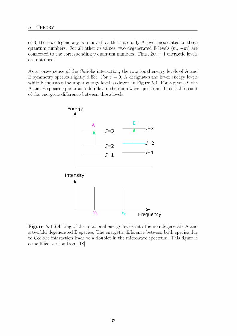
of 3, the ±m degeneracy is removed, as there are only A levels associated to thosequantum numbers. For all other m values, two degenerated E levels (m, −m) areconnected to the corresponding v quantum numbers. Thus, 2m + 1 energetic levelsare obtained.
As a consequence of the Coriolis interaction, the rotational energy levels of A andE symmetry species slightly differ. For v = 0, A designates the lower energy levelswhile E indicates the upper energy level as drawn in Figure 5.4. For a given J , theA and E species appear as a doublet in the microwave spectrum. This is the resultof the energetic difference between those levels.
A E
J=3
J=2
J=1 J=1
J=2
J=3
Energy
Frequency
Intensity
νA νE
Figure 5.4 Splitting of the rotational energy levels into the non-degenerate A anda twofold degenerated E species. The energetic difference between both species dueto Coriolis interaction leads to a doublet in the microwave spectrum. This figure isa modified version from [18].
32

5 Theory
5.2. Computer Programs
5.2.1. XIAM
The theoretical explanations of the XIAM code are given in references [22] and [23].Three possible ways of defining a molecule fixed coordinate system were describedin ref. [25] by Hougen et al.: i) the principal inertial axis system (PAM), ii) the in-ternal axis system (IAM), and iii) the rho axis system (RAM). The program XIAM(eXtended Internal Axis Method) calculates in several axis systems: For each top anew rho axis system is defined and eigenvalues are calculated. Afterwards, eigenval-ues in the principal axis system are calculated. Therefore, the method is designatedas the combined axis method, abbreviated CAM, based on the internal axis methoddeveloped by Woods [48,49] and extended by Vacherand et al. [50].
The Hamiltonian used for the description of a molecule containing one internal rotoris given by the sum of the rigid rotor Hamiltonian part Hrr and a torsional Hamil-tonian Hi:
H = Hrr + Hi (25)
with Hrr = BxP2x + ByP
2y+ BzP
2z:
Hi = F (pi − ~ρi†P )2 +
V3,i
2(1− cos3αi) +
V6,i
2(1− cos6αi) (26)
F designates the rotational constant of the torsional motion, α stands for the angledescribing the torsion of top i relative to the frame, pi = −i δ
δαis the angular mo-
mentum operator of the internal rotation, ~ρi = I−1xi is a vector with xi = Iα,i~λg,iand ~λg,i are the direction cosines and g the principal inertia axes.
By applying a rotation operator D which aligns the z-axis parallel to the vector ~ρ,the Coriolis coupling terms ρxPxp and ρyPyp are eliminated. In a second step, the
opposite transformation D−1 is applied to yield the Hamiltonian in the principal axissystem. The resulting Hamiltonian matrix diagonal in K, is diagonalized, yielding anew set of eigenfunctions, which are reused as torsional wave functions in a seconddiagonalization step. The second step is necessary to treat internal rotation, overallrotation and rotation-torsion coupling.
The numerical advantage of using the CAM method is the neglect of the off-diagonalmatrix elements in v. Furthermore, for ground state calculations, the energy matrixis truncated at 2J+1. This is sufficient, as no matrix off-diagonal elements in v occur.
33

5 Theory
The empirical internal rotation-overall rotation distortion operators is programmedinto XIAM as terms Dpi2J , Dpi2K and Dpi2−. These terms become very importantfor fitting molecules with a low barrier to internal rotation.
5.2.2. aixPAM
The aixPAM code is based on the rigid frame−rigid top model. It will be brieflydescribed here using a notation given in Ref. [23]. The Hamiltonian includes theoverall rotation, the internal rotation, and Coriolis-like interaction terms. It can bewritten as
H =1
2~P †I−1 ~P + V (27)
with the (transposed) 4-dimensional angular momentum vector ~P † = (Px, Py, Pz, p)and the inertia tensor
Ix 0 0 λixIα0 Iy 0 λiyIα0 0 Iz λizIα
λixIα λiyIα λizIα Iα
(28)
and the torsional potential
V = ΣnV3n
2(1− cos(3nα)). (29)
Pg with g ε {x, y, z} are the Cartesian components of the overall angular momentum,p the angular momentum of the internal rotor, Ig the principal moments of inertia,Iα the moment of inertia of the internal rotor, and the λig the direction cosinesbetween the internal rotor axis i and the principal axes of inertia g. Numericalinversion of I yields
1
2I−1 =
B′x Zxy Zxz −Qx
Zxy B′y Zyz −Qy
Zxz Zyz B′z −Qz
−Qx −Qy −Qz F
(30)
and the rigid frame-rigid top Hamiltonian used in aixPAM can be written as
H = B′JP2 +B′KP
2z +B′−(P 2
x −P 2y )+Σg,g′Zgg′{Pg, Pg′}+Fp2−2ΣgQgPgp+V (31)
with BJ ′ =B′x+B′y
2, B′K = B′z −
B′x+B′y2
, B′− =B′x−B′y
2, and the anti-commutator
{Pg,Pg′} =PgPg′+Pg′Pg. Beyond this basic model, centrifugal distortion and effec-tive interaction terms can be added. The Hamiltonian matrix is set up in the prin-cipal axis system without pre-diagonalization. A product basis |σ, k〉 · |J,K〉 of free
34

5 Theory
rotor functions (2π)−12 exp((i(3k+σ)α) and symmetric top functions is used. Both,
real and complex matrix elements are allowed. The matrix size is (2J+1)(2kmax+1)with mmax = 3kmax + σ. The matrices are block-diagonal in the torsional state σ;therefore, one matrix is used for each σ. The matrices are quite large for high J andkmax, for example they are of the size 697 x 697 for J = 20 and kmax = 8. However,with modern computers the time needed for diagonalizing such matrices is only afew seconds. Except for the truncation of the matrix, aixPAM does not neglect anyother matrix elements and kmax is increased until the fit converges.
Table 5.1 Some operators and their definitions in the aixPAM input file.
Parameter Operatora Definitionb
B′J P 2 BJ 1.0 P2B′K P 2
z BK 1.0 Pz PzB′– P 2
x − P 2y B– 0.5 P+ P+
B– 0.5 P– P–Zxy {Px, Pz} Zxz 0.5 P+ Pz
Zxz 0.5 P– PzZxz 0.5 Pz P+Zxz 0.5 Pz P–
∆J −P 4 DJ –1.0 P2 P2∆JK −P 2P 2
z DJK –1.0 P2 Pz Pz∆K –P 4
z DK –1.0 Pz Pz Pz Pzδj –2P 2(P 2
x − P 2y ) dJ –1.0 P2 P+ P+
dJ –1.0 P2 P− P−δk –{P 2
z , (P2x–P 2
y )} dk –0.5 Pz Pz P+ P+dk –0.5 Pz Pz P– P–dk –0.5 P+ P+ Pz Pzdk –0.5 P– P– Pz Pz
V312(1–cos3α) V3 0.5 e0
V3 –0.25 e+3V3 –0.25 e–3
aAnti-commutators are written as {u,v} = uv + vu. bThe fundamental operatorsare given in the text. Example: The operator associated with V3 translates to0.5 − 0.25ei3α– 0.25e−i3α = 0.5(1−cos3α).
An important feature of the aixPAM code is the possibility to add effective Hamil-tonian terms from the input file. These terms are given as a sum of products of thefundamental operators P 2 (P2), Pz (Pz), P+ = Px + iPy (P+), P− = Px− iPy (P−),p (p), 1 (e0), e+ = ei3α (e+3), e− = e−i3α (e−3). The operator codes as they are usedin the input file are given in parentheses. P are the angular momenta of the overallrotation with its components Px, Py, Pz, p the angular momentum of the internalrotation about the angle α. The operators 1, ei3α, and e−i3α are needed to code thepotential operators Vn with n = 3, 6, 9, . . . Some examples of effective operators
35

5 Theory
with their corresponding operator descriptions in the input file are given in Table5.1. We emphasize that in contrast to the BELGI code, where all parameters are inthe rho axis system, all parameters in aixPAM refer to principal axis coordinates.
5.2.3. BELGI-Cs
BELGI-Cs [25] by I. Kleiner et al. is another program developed for fitting molec-ular parameters of molecules with Cs-symmetry containing an internal rotor. ForC1-symmetric molecules, another version BELGI-C1 [51] exists. The program isbased on theoretical ideas from Kirtman [52] and Lees and Baker [53]. The imple-mented two step Hamiltonian diagonalization was inspired by Herbst’s publications[54]. The program is able to produce a global fit of A and E species correspondingto several (up to 9) torsional levels vt.
BELGI-Cs uses the rho axis method (RAM) to built up and diagonalize the tor-sional Hamiltonian, given by the following expression:
Htors = V (α) + F (pα − ρPa)2 (32)
with
Vα =V3
2(1− cos3α) +
V6
2(1− cos6α) + ... (33)
The coupling terms except −2FρapαPa vanish, as a consequence to the rho axissystem. Thus, the torsional Hamiltonian only contains matrix elements of the type|Kvtσ〉 = exp[i(3k+σ)α] which respect ∆K = 0, but still comprise internal-rotation-global-rotation Coriolis interaction. The significance of K and σ was already ex-plained in the XIAM chapter. The matrix is also truncated at ktronc = 10, thereforethe matrix size is 21 x 21 (2k+1) x (2k+1).
In a second diagonalization step using the torsional eigenvalues obtained via thefirst step (matrix of the size vt(2J+1), with vt = 0−8), rotational and centrifugaldistortion terms and higher order coupling terms between internal and global rota-tion angular momenta are obtained.
The advantages of this program is the high amount of rotation-torsion coupling pa-rameters, often needed for molecules with low torsional barrier to internal rotation.Reference [55] contains an overview of most implementable parameters in BELGI-Cs.
36

6 O-METHYLANISOLE
6. o-Methylanisole
A part of this chapter has already been published in the Journal of Chemical Physicsand Physical Chemistry [56].
L. Ferres, H. Mouhib, W. Stahl, and H. V. L.Nguyen
Methyl Internal Rotation in theMicrowave Spectrum of o-Methyl Anisole
ChemPhysChem 18, (2017), 1855−1859.
V3 = 444 cm−1
10GHz
14GHz
experimental
simulated
EA
12.61731 GHz12.61676
616 ← 505
E A
533 ← 432
12.99765 12.99832 GHz
15
17
L. Ferres performed the measurements, quantum chemical calculations, assignments,fitting of rotational transitions, and helped co-writing the manuscript.
6.1. Introduction
The gas phase structures of toluene and its derivatives have been the subjects ofmany rotational spectroscopic studies for decades, especially in the microwave andUV frequency regions. Many investigations by UV spectroscopy have been thor-oughly carried out for example in Ref. [57−59], giving information on gas-phasestructures in the electronic ground and excited states of these molecules. In mi-crowave spectroscopy, halogen atoms [60], aldehyde [61] or alcohol groups [4] are themost common substituents, also providing sufficiently large dipole moment allowingstrong rotational transitions. Surprisingly, almost no investigations were performedwith the methoxy group as substituent. These compounds are of special interest,because the internal rotation of methyl groups attached at the benzene ring is dif-ficult to predict from studies in the literature. In toluene, a pure V6 potential isfound [62]. In some cases, the V3 potential barriers are very low with a large V6
contribution [61]. In some other cases, only V3 contributions remain [4]. Currently,no study is available to systematically explain this observation.
In the case of mono-substituted methoxy toluenes, which can also be viewed asmonomethyl substituted anisoles, the three relative positions are ortho, meta, andpara, and thus three isomers exist. In the present work, the gas phase structureof o-methyl anisole (OMA) is studied with a view towards highly accurate deter-mination of the barriers to internal rotation. So far, the barrier height of the ringmethyl group has only been determined by Alvarez-Valtierra et al. with fluorescencespectroscopy, where the authors found for the electronic ground state a V3 potentialof 345 cm−1 [63].
The structures of substituted anisoles are also interesting. Anisole itself has a planarheavy atom frame [32] like almost all other related compounds such as phenetole[26], p-fluoroanisole [64], and p-anisaldehyde [65]. However, nonplanar structures
37

6 o-Methylanisole
cannot be excluded, because, for example, Lister et al. reported on a second isomerwith the methylsulfanyl (CH3-S) group in a nearly perpendicular orientation [66] forp-fluorothioanisole. Some other molecules containing a phenyl ring also have non-planar structures, as in the cases of phenyl formate [67] and N -phenylformamide [28].
6.2. Quantum Chemical Calculations
Reasonable starting values of molecular parameters such as rotational constantsare important for spectral assignment, because reliable predictions of the spectrumconsiderably simplify the assignment process. Quantum chemistry is a powerfultool for this purpose. The Gaussian09 suite of programs [11] was employed forall calculations in this work. If not stated otherwise, the MP2/6–311++G(d,p)level of theory is used, because it yields quite reasonable results for some othermolecules containing aromatic rings like phenetole [26] 2,5-dimethylthiophene [10],and 2-acetyl-5-methylfuran [9].
Because the rotations of two methyl groups about the C4−C16 and the O11−C12 bondby varying the dihedral angles β = ∠(C5−C4−C16−H19) and γ = ∠(C3,O11,C12,H14),respectively, (for atom numbering see Figure 6.1) do not create new conforma-tions, the conformational preferences of OMA are solely determined by the rota-tion of the methoxy group about the O11-C3 bond by varying the dihedral angleα = ∠(C12,O11,C3,C4). A potential curve was calculated where α was varied withina 10° step width while all other geometry parameters were optimized. The calcu-lated energies were parameterized using a Fourier expansion with the correspondingcoefficients given in Table 25.1.1 in the appendix section. Using these Fourier coef-ficients, the potential energy curve was drawn as depicted in Figure 6.2.a), showingthat there is only one Cs conformer for OMA at α = 180°. Because the minimumof the potential curve is very shallow, this region was recalculated within a grid of1° and plotted in Figure 6.2. at an enlarged scale.
15
17
14
10
12
11
13
1816
4
3
27
19
6
5
9
1
8
Figure 6.1 Molecular structure of the only conformer of o-methyl anisole optimizedat the MP2/6–311++G(d,p) level of theory. The protons H15 and H17 are located
38

6 o-Methylanisole
behind H13 and H18, respectively.
α / °
Ene
rgy
/ kJ
mol−
1
20
30
00 60 240180120 360300
10
0
1.0
160 180 200
1.5
40
Ene
rgy
/ cm−
1 β / °
0 60 240180120 3603000
100
200
300
400
500a) b)
Figure 6.2 a). The potential energy curve of o-methyl anisole obtained by rotatingthe methoxy group about the C3−O11 bond (for atom numbering, see Figure 6.1 ina grid of 10°. The relative energies with respect to the lowest energy conformationwith E = −385.0454020 Hartree are given. The minimum region was recalculatedin a grid of 1°, as depicted with an enlarged scale. Figure 6.2 b). The potentialenergy curve obtained by rotating the ring methyl group about the C4−C16 bond.The dihedral angle β = ∠(C5−C4−C16−H19) was varied in a grid of 10°, while allother molecular parameters were optimized at the MP2/6–311++G(d,p) level. Rel-ative energies with respect to the lowest energy conformations with the absoluteenergies E = −385.0454073 Hartree are used.
Full geometry optimizations and frequency calculations were performed afterwardsfor the minimum (see Figure 6.1). The Cartesian coordinates are given in Table25.1.2 in the appendix section. One imaginary vibrational mode was found in theharmonic frequency calculations, which is a bending vibration of the phenyl ring.Stating stable planar ring systems as unstable is a well-known behavior found atthe MP2/6-311++G(d,p) level of theory, which has been reported for benzene andarenes [44].
The only stable conformer of OMA is predicted to possess the rotational constantsA = 2.481 GHz, B = 1.559 GHz, and C = 0.969 GHz and the dipole moment com-ponents |µa| = 0.79 D, |µb| = 1.01 D, and |µc| = 0.00 D. Consequently, no c-typetransitions are observable in the microwave spectrum and both, a- and b-type tran-sitions, are intense.
In spite of its relatively simple structure, the internal dynamics of OMA are chal-lenging. There are two inequivalent methyl internal rotations. The barrier heightsof the ring methyl and the methoxy methyl group were calculated by varying the di-hedral angles β and γ, respectively, in a grid of 10°. A rotation of 120° was sufficientdue to the three-fold symmetry of the methyl groups. A V3 potential of 447.71 cm−1
39

6 o-Methylanisole
was found for the ring methyl and 1118.18 cm−1 for the methoxy methyl groupwith no significant V6 contributions. Simple two-top predictions using the programXIAM [22] indicate that torsional splittings arising from the methoxy methyl groupare smaller than 2 kHz. Therefore, with the resolution of the spectrometer in use,only splittings arising from the ring methyl group were expected. The potentialenergy curve of the ring methyl group is shown in Figure 6.2 b); the correspondingcoefficients in Table 25.1.3 in the appendix section.
6.3. Microwave Spectroscopy
The microwave spectrum of the only conformer of OMA was calculated from theab initio predictions with the program XIAM. As a first step, the internal rotationeffect was neglected and the rigid-rotor mode of XIAM was applied to calculatethe theoretical spectra. The dipole moment components suggest a spectrum withintense a- and b-type, but no c-type transitions. The assignment was started withthe a-type R-branch J = 6 � 5, especially the transitions 606 � 505 and 616 � 515,and then the J = 5 � 4 transitions such as 505 � 404. The assignment of theselines fixed the B and C rotational constants. Afterwards, some b-type transitionswere also assigned and the A rotational constant was determined.
Frequency / GHz
Inte
nsity
/ a.
u.
simulated
experimental
10 1211 1413
Figure 6.3 A broadband scan of o-methyl anisole from 10 to 14 GHz. The ex-perimental spectrum is the upper trace. The lower trace indicates the predictedtheoretical spectrum using the molecular parameters deduced from a one-top fit bythe program XIAM, showing that (i) a simple one-top model can reasonably repro-duce the microwave spectrum of o-methyl anisole, and (ii) only some weak linesremained unassigned in the spectrum.
Characteristic patterns of transitions appeared close to those of the A species, whichwere assumed to be the E internal rotation symmetry species of the same rotationaltransitions. In the next step, the internal rotation of the ring methyl group wastaken into consideration and the spectrum was predicted with torsional componentsusing the rotational constants from the rigid rotor fit. The angles δ between the
40

6 o-Methylanisole
internal rotor axis and the principal a axis as well as the V3 potential were calcu-lated by ab initio. In doing so, the E species frequencies of many transitions werepredicted sufficiently accurate, especially for a-type transitions, leading to theirstraightforward assignment. Finally, 30 torsional lines were assigned and fitted inthe 10−14 GHz broadband spectrum (shown in Figure 6.3). Using the predictionrefined from this fit, high resolution measurements were performed in the frequencyrange 2−26.5 GHz, leading to an expanded assignment of 125 A and 119 E linesand a standard deviation of 2.9 kHz. Figure 6.4. shows typical transitions of OMAmeasured at high resolution. The fitted molecular parameters are summarized inTable 6.1. All fitted transitions are listed in Table 25.1.4 in the appendix section.
E
A
12617.19912616.832
616 ← 505
E A533 ← 432
12998.19812997.789
Figure 6.4 Typical b-type and a-type transitions (in MHz) of o-methyl anisolemeasured at high resolution. The measurement accuracy is 2 kHz, the typical ex-perimental line width 20 kHz. The doublets marked by brackets are due to theDoppler effect. The torsional A-E splittings are also indicated. For both spectra, 50free-induction decays were co-added.
The three linear combinations BJ = 12(B+C), BK = A − 1
2(B+C), B− = 1
2(B−C)
of the rotational constants and the quartic centrifugal distortion constants were de-termined with very high accuracy by fitting 244 torsional components in a one-rotorfit. The V3 potential, the angle δ between the internal rotor axis and the principal aaxis, and two higher order parameters Dpi2J and Dpi2– were also obtained. The in-ternal rotation constant F0 of the methyl top could not be fitted because the presentdata set contains only information on the torsional ground state with relatively lowJ and K values, and because the torsional barrier is rather high (444 cm−1).
41

6 o-Methylanisole
Table 6.1 Molecular parameters of o-methyl anisole obtained by the programXIAM.
Parametera Unit Obs. Calc.b Obs.−Calc.A MHz 2489.335(87) 2481.1078 8.23B MHz 1557.68(17) 1558.5760 −0.90C MHz 970.516(37) 968.9111 1.60DJ kHz 0.03845(63) 0.03655 0.0019DJK kHz 0.1583(23) 0.17364 0.0153DK kHz 0.1091(28) 0.03958 0.0695d1 kHz −0.01792(33) −0.01802 0.0001d2 kHz −0.006296(92) −0.00668 0.0004V3 cm−1 444.05(41) 459.25 −15.20F0 GHz 158.0c 157.9223 0.1Dpi2J kHz 60.0(7.0)Dpi2− kHz 43.6(6.4)∠(i,a) ° 70.996(95) 68.8415 2.154∠(i,b) ° 19.004(95) 21.1586 −2.155∠(i,c) ° 90.0d 89.970 0.030NA/N e
E 125/119σf kHz 2.8
aAll parameters refer to the principal axis system. Watson’s S reduction and Ir representation
were used. b Centrifugal distortion constants obtained from anharmonic frequency calculations at
the B3LYP/6–311++G(d,p) level of theory, all other values from geometry optimizations at the
MP2/6–311++G(d,p) level. c Corresponds to Iα = 3.2 uA2, a value often found for methyl groups.d Fixed due to Cs symmetry. e Number of A and E species lines. f Standard deviation of the fit.
6.4. Discussion
The experimentally deduced rotational constants are compared with those fromquantum chemical calculations (see Table 6.1). The experimental and calculated Band C rotational constants agree well and the deviation in the A rotational constantis very small (about 8 MHz). The calculated V3 potential of 459 cm−1 is also quiteclose to the experimental value of 444 cm−1. The centrifugal distortion constantsobtained by anharmonic frequency calculations at the B3LYP/6-311++G(d,p) levelof theory are in good agreement with the experimental values. Because the benzeneframe of OMA is quite rigid, all centrifugal distortion constants are small. Calcu-lations with the MP2 and B3LYP methods in combination with various basis setswere performed in addition, which confirm that the MP2/6-311++G(d,p) level issufficiently suitable for OMA, as can be seen in Table 6.2 in the appendix section.It is noteworthy that the structures obtained from calculations at all levels of theoryare consistent, which is not obvious if compared to the case of, for example, diethylketone [36].
42

6 o-Methylanisole
Table 6.2 Rotational constants of o-methyl anisole in GHz and their deviationsto the experimental values (obs. − calc.) in MHz optimized using the MP2 andB3LYP methods in combination with different basis sets.
Basis set A ∆A B ∆B C ∆CMP26-31G(d,p) 2.496 −7 1.560 −2 0.972 −16-311G(d,p) 2.488 1 1.561 −3 0.971 06-311+G(d,p) 2.481 8 1.558 −1 0.969 26-311++G(d,p) 2.481 8 1.559 −1 0.969 26-311G(df,pd) 2.505 −15 1.574 −16 0.978 −86-311+G(df,pd) 2.497 −8 1.572 −14 0.976 −66-311++G(df,pd) 2.497 −8 1.572 −14 0.976 −56-311G(2df,2pd) 2.511 −22 1.576 −18 0.980 −96-311+G(2df,2pd) 2.503 −13 1.574 −16 0.978 −76-311++G(2df,2pd) 2.503 −13 1.574 −16 0.978 −76-311G(3df,3pd) 2.509 −19 1.573 −15 0.978 −86-311+G(3df,3pd) 2.502 −13 1.572 −14 0.977 −66-311++G(3df,3pd) 2.502 −13 1.572 −14 0.977 −6cc-pVDZ 2.456 33 1.545 13 0.960 11B3LYP6-31G(d,p) 2.487 −2 1.550 −8 0.967 −46-31+G(d,p) 2.480 −9 1.545 −13 0.964 −76-31++G(d,p) 2.480 −9 1.545 −13 0.964 −76-311G(d,p) 2.491 1 1.552 −5 0.968 −36-311+G(d,p) 2.484 −6 1.548 −10 0.965 −66-311++G(d,p) 2.484 −6 1.548 −10 0.965 −66-311G(d,p) 2.495 6 1.551 −7 0.968 −36-311+G(d,p) 2.491 1 1.550 −8 0.967 −46-311++G(d,p) 2.491 1 1.550 −8 0.967 −46-311G(df,pd) 2.504 14 1.556 −2 0.971 16-311+G(df,pd) 2.499 9 1.555 −3 0.970 −16-311++G(df,pd) 2.499 9 1.555 −3 0.970 −16-311G(2df,2pd) 2.498 9 1.558 0 0.971 06-311+G(2df,2pd) 2.500 11 1.557 0 0.971 06-311++G(2df,2pd) 2.500 11 1.557 0 0.971 06-311G(3df,3pd) 2.506 16 1.558 1 0.972 26-311+G(3df,3pd) 2.501 11 1.558 0 0.972 16-311++G(3df,3pd) 2.501 11 1.558 0 0.972 1cc-pVDZ 2.484 −5 1.545 −13 0.964 −7aug-cc-pVDZ 2.476 −13 1.546 −11 0.963 −7cc-pVTZ 2.502 12 1.558 0 0.972 1aug-cc-pVTZ 2.500 10 1.557 0 0.971 0Experiment 2.489 1.558 0.971
43

6 o-Methylanisole
In 2006 Alvarez-Valtierra et al. determined the following rotational constants:A = 2.4900(1) GHz, B = 1.5589(1) GHz, and C = 0.9707(1) GHz, as well as aV3 potential of 345 cm−1 for OMA by fluorescence spectroscopy [63]. The rotationalconstants are not as accurate as in the present work, but they are in agreementwith those deduced from the XIAM fit. On the other hand, the rotational barrierdetermined by fluorescence spectroscopy is 99 cm−1 lower than the value determinedby microwave spectroscopy, which is a significant difference. We assume that thebarrier height given by microwave spectroscopy is more reliable, since this parameterwas only indirectly determined in fluorescence spectroscopy.
Table 6.3 Torsional barriers of ortho substituted toluene derivatives.
Derivate V3/cm−1 Ref.o-tolunitrile 187.699(3) [70]o-fluorotoluene 227.28(2) [69]2,4-difluorotoluene 203.91(24) [60]anti-o-cresol 371.05(41) [4]syn-o-cresol 669.10(51) [4]o-methyl anisole 444.48(42) this workanti -3,4-dimethylbenzaldehyde 454.1(14),480.6(44) [7]syn-3,4-dimethylbenzaldehyde 508.1(11),550.7(88) [7]37Cl-o-chlorotoluene 507.2(83) [71]35Cl-o-chlorotoluene 513.8(27) [71]o-xylene 518.3(32) [72]syn-2,5-dimethylbenzaldehyde 566(16) [7]
It is interesting to compare the V3 potential of the ring methyl group in OMAwith those of other toluene derivatives, where the substituents are also in the or-tho position with respect to each other (see Table 6.3). In the case of acetates,both the steric and electronic effects might affect the methyl barrier height, wherethe electronic effect has a larger influence than the steric effect [68]. In the caseof ortho substituted toluene, the influence of the substituent on the barrier heightseems to be predominantly of a steric nature. Small or slim substituents such asfluorine atoms (in o-fluorotoluene [69] or 2,4-difluorotoluene [60]) or cyano groups(o-tolunitrile [70]) hinder the internal rotation much less than voluminous chlorineatoms (o-chlorotoluene [71]), hydroxyl groups (o-cresol [4]), methyl groups (o-xylene[72] or 3,4-dimethylbenzaldehyde [7]), methoxy groups (OMA), or aldehyde groups(2,5-dimethylbenzaldehyde [7]). In molecules where syn- and anti -conformers co-exist in the spectrum, the anti -conformer has lower internal rotation barrier, becausethe ortho substituent is much closer to the methyl group, and the steric hinderingincreases, while the electronic contribution should be similar for both conformers.An electronic effect is, however, not excluded, as can be seen from the different V3
potential of o-fluorotoluene [69] and 2,4-difluorotoluene [60] or o-xylene [72] and3,4-dimethyl-benzaldehyde [7]. Nevertheless, these differences are less significant.
44

6 o-Methylanisole
The planar moment of inertia Pcc = −2(Ic−Ia−Ib) = 13.396 uA2 of OMA confirmsthat the heavy atom skeleton is planar with two pairs of hydrogen atoms out of plane.These values are very similar as that found in other planar molecules containing twomethyl groups such as the trans and cis conformers of 2-acetyl-5-methylfuran [9](Pcc = 12.938 and 13.160 uA2, respectively) and the syn and anti conformers of3,4-dimethylbenzaldehyde [7] (Pcc = 12.904 and 12.938 uA2, respectively).
After the spectral assignment, only very few weak lines remained unassigned in thebroadband scan, as shown in Figure 6.3. We thus concluded that water complexesor dimers were not present under our measurement conditions.
45

6 o-Methylanisole
Inside Cover of the ChemPhysChem Journal, Issue 14/2017 [73]. Thepicture shows the excellent agreement between the experimental (cyan) and theo-retical rotational spectrum (red) of o-methyl anisole in the gas phase. The analysisof torsional splittings from the internal rotation of the ring methyl group yields athree-fold potential with a barrier height of 444 cm−1.
46

7 m-Methylanisole
7. m-Methylanisole
A part of this chapter has already been published in the Journal of ChemicalPhysics [24].
L. Ferres, W. Stahl, and H. V. L. Nguyen
Conformational Effects on the Torsional Barriersin m-Methylanisole Studied by Microwave Spec-troscopy
J. Chem.Phys 148, (2018), 124304.
L. Ferres performed measurements, quantum chemical calculations, assignment andfitting of rotational transitions, and co-writing the manuscript.
7.1. Introduction
Rotational transitions of a molecule with internal rotation of a methyl group featuresplittings into torsional components, and can no longer be treated using a rigid-rotormodel. A number of programs have been developed to treat this problem, amongthem are IAM [49], XIAM [22], BELGI-Cs [25] and BELGI-C1 [51], Erham [74],and RAM36 [62]. All of them use a rigid frame-rigid top model [47] as a theoreticalbackground, which is supplemented by a number of higher order (effective) terms,including centrifugal distortion of both the frame and the top in the Hamiltonian.On the other hand, they differ from each other by their methods which are namedafter the coordinates in use; e.g., the XIAM code uses the combined axis method;Erham sets up and solves an Effective Rotational HAMiltonian; and BELGI-Cs,BELGI-C1, as well as RAM36 use the rho axis method (RAM). The numericaltechniques are also different: The Hamiltonian matrices are pre-diagonalized, smallmatrix elements are neglected, and the matrices are truncated at a given limit.
The program XIAM was mostly used in previous investigations on molecules ,asfor example methyl butyrate [75], N,N -diethylacetamide [39], and methyl isobutylketone [42], of different chemical classes undergoing internal rotation, because it isuser-friendly and offers a reasonable compromise of accuracy and calculation speed.On the other hand, XIAM has its weaknesses in treating internal rotations withlow barrier heights, e.g. in the cases of 3-pentin-1-ol [76], allyl acetate [77], andN -ethylacetamide [38] where standard deviations much larger than measurementaccuracy have been observed.
For numerous molecular systems, fits carried out with XIAM and BELGI in itsCs or C1 version were compared, where in almost all cases, BELGI yielded a lowerstandard deviation than XIAM with the same data set [78-80]. There were somediscussions about the reasons for this observation. A frequently used argument is
47

7 m-Methylanisole
that only a limited number of parameters can be fitted using XIAM in comparisonwith BELGI, and that the torsional interactions between different vt states are nottaken into account explicitly. Higher order coupling terms between the internal ro-tation and the overall rotation cannot be easily implemented in the XIAM code toimprove the quality of the fit. Sometimes, it is also argued that even if there weremore effective terms in XIAM, the standard deviation would still be worse thanwith BELGI. The reason is that XIAM uses a two-step diagonalization procedure,whereby only one J -block is considered in the second diagonalization step and somematrix elements are thus neglected. This truncation could additionally affect theaccuracy of the fit.
These discussions induced the authors to write another internal rotation program,called aixPAM, where aix stands for Aix-la-Chapelle, the French name of Aachenwhere the program was written. MMA (m-methylanisole) was chosen for the firstapplication of aixPAM, since two conformers with rather extensive data sets havebeen recently measured and assigned, and because the XIAM fits of these data havenot achieved measurement accuracy.
Our recent studies on the microwave spectra of o-methylanisole (OMA) [56] and p-methylanisole (PMA) [81] have shown that the ring methyl group undergoes internalrotation with quite different barrier heights of about 444 cm−1 and 50 cm−1, respec-tively, whereas the effect arising from the methoxy methyl group is negligible. It isinteresting to compare the respective torsional barriers when the ring methyl groupis in m-position, because depending on the relative position of the substituents, con-siderable effects on the torsional barrier height can be observed. There are studiesin the literature on the isomers of fluorotoluene [69, 82, 6], cresol [4, 83, 84], andmethylbenzaldehyde [61],[85] stating very different barrier heights for the o-, m-,and p-isomers.
A comparison with the barriers of other toluene derivatives with a methyl groupin m-position is also very interesting. Some previous investigations [56,81,85] haveshown that the o- and p-isomers often possess only one stable conformer because ofthe sterical hindrance in the o-isomer and the structural symmetry of the p-isomer.In m-substituted toluenes, two conformers are often observed [61], making theseisomers ideally suited to study conformational effects on torsional barriers.
7.2. Quantum Chemical Calculations
7.2.1. Conformational Analysis
Before recording the microwave spectrum, quantum chemical calculations were per-formed to determine the possible conformers of MMA.
By rotating the entire −OCH3 group about the C4−O11 bond (for atom number-ing see Figure 7.1), a potential curve was calculated by varying the dihedral an-
48

7 m-Methylanisole
gle β = ∠(C5−C4−O11−C12) in a grid of 10°, while all other geometry parame-ters were optimized. Calculations were carried out at the B3LYP/6-311++G(d,p)level of theory using the Gaussian09 program package [11], if not stated other-wise. In almost all recent investigations the MP2/6-311++G(d,p) level was appliedsuccessfully. However, for molecules containing aromatic rings like phenetole [26],2,5-dimethylthiophene [10], 2-acetyl-5-methylfuran [9], as well as the the isomerOMA [56] of MMA, harmonic frequency calculations yielded one imaginary vibra-tional mode, which is a bending vibration of the phenyl ring. It is well-known thatthe MP2/6-311++G(d,p) level of theory often yields an imaginary frequency forstable planar ring systems, which has been reported for benzene and arenes [44].This effect did not occur in calculations using the B3LYP method. Variation of themethods and basis sets in our previous studies on OMA [56] and PMA [81] confirmsthat the structural parameters do not change significantly and the rotational con-stants are calculated with sufficient accuracy at the B3LYP/6-311++G(d,p) level.
I17
II
13
11
8
4
3
2
1816
19
1
7
10 6
5
9
1215
α
β γ
13
11 1214
21
6
9
5 4
810 3
716
19
18
Figure 7.1 The molecular structures of the cis (I) and the trans (II) conformerof MMA, optimized at the B3LYP/6-311++G(d,p) level of theory. The dihedralangles are defined as α = ∠(C3−C2−C16−H18), β = ∠(C5−C4−O11−C12), andγ = ∠(C4−O11−C12−H13).
The potential curve as a function of β is given in Figure 7.2; the Fourier coefficientsof the potential function are available as appendix (Table 25.2.1). Calculations yieldtwo possible conformers of MMA: the energetically more favorable trans conformerat β = 0° and the cis conformer at β = 180°. Their fully optimized structuresare illustrated in Figure 7.1; the Cartesian coordinates are given in Table 25.2.4.Frequency calculations state that both of them are true minima with an energy dif-ference of only 0.40 kJ/mol. Therefore, both conformers are expected to be visiblein the experimental spectrum.
49

7 m-Methylanisole
Optimizations predict the rotational constants A = 2756.8 MHz, B = 1286.6 MHz,and C = 887.0 MHz, as well as the dipole moment components µa = −0.08 D,µb = −1.64 D, and µc = 0.00 D for the cis conformer. The respective values fortrans-MMA are A = 3537.9 MHz, B = 1116.2 MHz, C = 857.7 MHz, µa = 0.67 D,µb = 0.73 D, and µc = 0.00 D. Accordingly, for cis-MMA, only b-type transitionsare expected in the microwave spectrum, while a-type transitions should also beobservable for trans-MMA. Finally, anharmonic frequency calculations were carriedout to obtain the centrifugal distortion constants.
0
2
4
6
8
10
14
-90
12
0 90 180 270β / °
Ene
rgy
/ kJ
mol−
1
II I
Figure 7.2 The potential energy curve of m-methylanisole obtained by a 360° ro-tation of the methoxy group about the C4−O11 bond by varying the dihedral angleβ = ∠(C5−C4−O11−C12) in a grid of 10° at the B3LYP/6−311++G(d,p) level oftheory. The relative energies with respect to the lowest energy conformation withE = −386.1953453 Hartree are given. Two stable Cs conformers found at β = 0°(trans) and 180° (cis) differ by only 0.40 kJ mol−1 in energy.
7.2.2. Methyl Internal Rotations
Potential energy scans generated barrier heights of the ring- and the methoxy methylgroups, obtained by variation of the dihedral angles α = ∠(C3−C2−C16−H18) andγ = ∠(C4−O11−C12−H13) in a grid of 10°, corresponding to rotations about theC2−C16 and O11−C12 bonds, respectively. All other geometry parameters were op-timized. Due to the three-fold symmetry of the methyl groups, a rotation of 120° wassufficient. The potential energy curves showing the rotation of the ring methyl groupare presented in Figure 7.3; the corresponding Fourier coefficients are available inTable 25.2.2 in the appendix. For the cis conformer, V3 potentials of 50.13 cm−1 forthe ring methyl group and 1085.35 cm−1 for the methoxy methyl group were found.The respective values for the trans conformer are 32.77 cm−1 and 1079.11 cm−1.
Figure 7.3 indicates that the ring methyl group prefers different orientations in thetwo conformers of MMA. Taking H8 as the reference (see Figure 7.1), the lowestenergy conformations of cis-MMA locate two of three hydrogen atoms of the ring
50

7 m-Methylanisole
methyl group staggered to H8, corresponding to α = 60°, 180°, or 300°. On the con-trary, trans-MMA favours a conformation with one of the hydrogen atoms eclipsedto H8, corresponding to α = 0°, 120°, or 240°.
α / °
Ene
rgy
/ cm−
1
0 60 120 180 240 300 3600
10
20
30
40
50 I
II
Figure 7.3 The potential energy curves for the cis (I, black) and the trans (II, blue)conformer of MMA obtained by rotating the ring methyl group about the C2−C16
bond. The dihedral angle α = ∠(C3−C2−C16−H18) was varied in a grid of 10°, whileall other molecular parameters were optimized at the B3LYP/6-311++G(d,p) levelof theory. Relative energies with respect to the lowest energy conformations withthe absolute energies E = −386.1951917 and −386.1953453 Hartree, respectively,are used.
360
180
090 270−90β / °
α /
°
0 %
20
40
60
80270
90
100
0 180
III
Figure 7.4 The potential energy surface (PES) depending on the dihedral anglesα and β of MMA calculated at the B3LYP/6-311++G(d,p) level of theory. α andβ were varied in a grid of 10°, while all other parameters were optimized. Thenumbers in the color code indicate the energy (in percent) relative to the ener-getic minimum Emin = −386.1905225 Hartree (0%) and the energetic maximumEmax = −386.1953453 Hartree (100%). Note that there are significantly more col-ors in the lower 50% section. The PES indicates that (i) two conformers of MMA
51

7 m-Methylanisole
exist (I = cis-MMA, II = trans-MMA); (ii) conformer I is higher in energy thanconformer II; and (iii) the orientation of the ring methyl group is different in thetwo conformers.
The V3 potential of the methoxy methyl group exceeds 1000 cm−1 in both, the cisand trans conformers. Trial two-top calculations using the program XIAM predictthat torsional splittings arising from the internal rotation of this methyl group aresmaller than 5 kHz and thus not resolvable with the spectrometer in use.
Furthermore, a 2D potential energy surface (PES) depending on α and β was calcu-lated by varying these dihedral angles in a grid of 10°. The PES was parameterizedwith a 2D Fourier expansion based on terms representing the correct symmetry ofα and β. The Fourier coefficients are also listed in Table 25.2.3 in the appendix sec-tion. The PES shown in Figure 7.4 clearly indicates the following points: (i) Twoconformers of MMA exist (I = cis-MMA, II = trans-MMA); (ii) conformer I ishigher in energy than conformer II; and (iii) the orientation of the ring methylgroup depends on the position of the methoxy group.
7.3. Microwave Spectroscopy
Quantum chemical calculations have shown that the trans conformer is lower in en-ergy than the cis conformer (see section 7.2). Additionally, only for this conformer,a-type transitions, which can often be assigned easily due to their characteristic pat-terns, are predicted to be observable. Therefore, the assignment was started withthe spectrum of trans-MMA.
trans
8.5 9.5 10.5 11.5 12.5Frequency / GHz
Inte
nsity
/ a.
u.
cissim.
exp.
13207.9513207.70 13262.98 13263.23
707← 616
AE
MHz
a) b)
Figure 7.5 a) A portion of the broadband scan of MMA from 8.5 to 12.5 GHz. Theexperimental spectrum is the upper trace. The lower trace indicates the theoreticalspectrum of the cis (in blue) and the trans conformer (in red) predicted using themolecular parameters deduced from the program aixPAM. Figure 7.5 b) A typicalspectrum of the 707 � 616 transition of the cis conformer of MMA with its A and Etorsional species. The A-E splitting is approximately 55 MHz. The splittings indi-cated by brackets are due to the Doppler effect. For each of these spectra, 50 decays
52

7 m-Methylanisole
were co-added.
A portion of the broadband scan is shown in Figure 7.5 a), a typical measurementat high resolution in Figure 7.5 b). Some intense lines can be measured with anaccuracy of 2 kHz, but in most cases the line widths are larger due to unresolvedsplittings arising from the internal rotation of the methoxy methyl group and alsodue to proton spin-spin and spin-rotation coupling, resulting in a measurement ac-curacy of about 4 kHz. In the indicated fits, all lines were equally weighted.
7.3.1. The trans-conformer
As a first step, the methyl internal rotation was neglected and trans-MMA wastreated as an effective rigid-rotor. A spectrum containing A species transitions waspredicted with the program XIAM in its rigid-rotor mode using the rotational con-stants given in chapter 7.2, and compared with the experimental broadband scan.The very intense R-branch J = 6 � 5 and 7 � 6 a-type transitions with Ka = 0 and1 were firstly identified in the scan, yielding the B and C rotational constants. After-wards, b-type transitions were also assigned, which fixed the A rotational constant.This enabled a prediction of the whole rigid-rotor spectrum with sufficient accuracyto find all the remaining A species lines in the frequency range of 2-26.5 GHz. Atthis stage, 183 lines were fitted with the three rotational constants A, B, C, and fivequartic centrifugal distortion constants.
As a next step, the methyl internal rotation was taken into account and both theA and E species transitions were predicted. The initial V3 potential and the anglebetween the internal rotor axis and the principal a-axis were taken from quantumchemical calculations. By comparing the theoretical and experimental broadbandscan, the assignment was straightforward for the a-type R-branch transitions men-tioned above, where the A-E splittings were less than 30 MHz. The assignment ofb-type lines with much larger splittings (up to 3.5 GHz) was more difficult, however,finally successful. In total, 183 A species and 137 E species lines were fitted usingthe program XIAM with a standard deviation of 32.1 kHz. The molecular parame-ters are summarized as Fit I in Table 7.1. A list including all the fitted transitionsis given in Table 25.2.6 in the appendix section.
7.3.2. The cis-conformer
After the trans conformer was assigned, a large number of lines remained in the scan,which belong to the cis conformer. Because only b-type transitions are present, theassignment was more difficult, nevertheless, possible by trial and error. 92 A speciesand 131 E species transitions up to J = 13 could be measured and fitted with theprogram XIAM to a standard deviation of 27.0 kHz. This fit is also given in Ta-ble 7.1; the fitted transitions are presented in Table 25.2.5 in the appendix section.Note that for both conformers, some c-type transitions are available in the frequency
53

7 m-Methylanisole
lists (Tables 25.2.5 and 25.2.6), which are all E species lines. It is known that for theE species, forbidden transitions can be observed [25], as found e.g. in ethyl acetate[86] and butadienyl acetate [68].
7.3.3. The aixPAM Fits
The program aixPAM was written to check the accuracy of XIAM. It is a veryflexible code in which effective terms can easily be added in the Hamiltonian. Thefollowing two sample calculations were made:
(1) We took the same MMA data set and the same set of parameters mentionedin chapters 7.3.1 and 7.3.2 for comparative fits with XIAM and aixPAM. TheseaixPAM fits are given as Fit II in Table 7.1. The Dpi2 parameters often used
in XIAM are also floated in aixPAM, where Dpi2J multiplies 2(pα ~ρ† ~P )2 ~P 2, Dpi2Kmultiplies {(pα − ~ρ† ~P )2, P 2
z }, and Dpi2− multiplies {(pα − ~ρ† ~P )2, (P 2x − P 2
y )}. The
anti-commutator is defined as {u,v} = uv + vu; ~ρ† is the transposed rho vector.The complete definitions in terms of aixPAM ’s fundamental operators are extensiveand therefore given in Table 5.1 in the theoretical part.
The standard deviations are very similar in both the fits. The one obtained from aix-PAM is only slightly better than that from XIAM by about 4 kHz, indicating thatthe matrix elements neglected in XIAM do not significantly limit its accuracy. Theobserved-minus-calculated values of Fit II are listed in Table 25.2.5 for trans-MMAand in Table Table 25.2.6 for cis-MMA in the appendix section.
(2) We added in a second aixPAM fit some effective terms not available in XIAM.This enabled us to achieve a standard deviation close to the experimental accuracy.For cis-MMA, three additional parameters VJ multiplying P 2(1−cos3α), DmK mul-tiplying P 2
z p2α, and V− multiplying (P 2
x − P 2y )(1−cos3α) lowered the standard devi-
ation to 3.7 kHz, which is the measurement accuracy. For trans-MMA, the stan-dard deviations were reduced to 4.1 kHz by adding the parameters VK multiplyingP 2z (1−cos3α), DmJ multiplying P 2p2
α, and V−, where in both cases, the operatorscontaining the (1−cos3α) term led to most significant changes of the fits. It shouldbe noted that the RAM equivalent versions of these operators are also available inBELGI. These aixPAM fits are summarized as Fit III in Table 7.1.
54

7m-M
ethylaniso
le
Table 7.1 Molecular parameters of m-methylanisole in the principal axis system obtained by the programs XIAM and aixPAM.
cis-MMA trans-MMAPar.a Unit Fit Ib Fit IIc Fit IIId Calc.e Fit Ib Fit IIc Fit IIId Calc.e
Af MHz 2755.8636(70) 2755.997(18) 2755.304(73) 2756.845 3529.454(40) 3528.242(61) 3521.824(85) 3537.943Bf MHz 1293.7072(25) 1293.3105(60) 1291.236(45) 1286.597 1122.3350(17) 1122.3847(26) 1121.498(45) 1116.215Cf MHz 890.60188(47) 890.55131(66) 890.88(16) 886.992 861.12564(46) 861.14249(47) 862.060(45) 857.656∆J kHz 0.0421(31) 0.0493(26) 0.05137(42) 0.05173 0. 0310(33) 0.0285(29) 0.02397(43) 0.02326∆JK kHz −0.027(22) −0.076(19) −0.0951(31) −0.06667 0.071(21) 0.034(19) 0.0451(28) 0.09015∆K kHz 0.601(99) 0.542(85) 0.446(14) 0.39097 −0.478(94) 0.299(84) 0.954(15) 0.57448δJ kHz 0.0129(14) 0.0169(12) 0.01833(19) 0.01869 0.0091(17) 0.0085(15) 0.00609(22) 0.00598δK kHz 0.045(16) 0.101(13) 0.0846(21) 0.04213 0.119(57) 0.134(51) 0.0857(75) 0.00370Dpi2J kHz 93.10(87) 97.68(85) 157.8(59) − −34.80(46) −34.64(42) −64.99(65) −Dpi2K MHz −0.2333(43) −0.1864(43) −0.4450(33) − 0.904(22) 0.996(21) 1.2828(85) −Dpi2− kHz 66.34(84) 75.72(83) 161.4(66) − −20.56(48) −18.01(44) 49.9(36) −V3 cm−1 55.784(34) 55.469(34) 55.7693(90) 50.128 36.548(32) 36.392(32) 36.6342(84) 32.768F0 GHz 157.074(96) 156.190(96) 157.192(28) 160.898 158.10(15) 157.45(14) 158.825(38) 160.855VJ MHz − − 1.41(12) − − − − −DmK MHz − − 0.4122(59) − − − − −VK MHz − − − − − − 9.97(12) −V− MHz − − 1.92(13) − − − 1.265(61) −DmJ MHz − − − − − − 0.0491(12) −∠(i,a) ° 51.6451(1) 51.65520(20) 51.6942(12) 52.140 146.8215(1) 146.83013(53) 146.8552(14) 146.887∠(i,b) ° 141.6451(1) 141.65520(20) 141.6942(12) 142.140 123.1785(1) 123.16987(53) 123.1448(14) 123.113∠(i,c) ° 90.0g 90.0g 90.0g 89.996 90.0g 90.0g 90.0g 89.995NA/Nh
E − 92/131 92/131 92/131 − 183/137 183/137 183/137 −σi kHz 27.0 23.0 3.7 − 32.1 28.3 4.1 −
aAll parameters refer to the principal axis system. Watson’s A reduction and Ir representation were used. bFit with the program XIAM. cFit with theprogram aixPAM using the same parameters as in Fit I. dFit with the program aixPAM using three additional parameters. eCentrifugal distortionconstants obtained from anharmonic frequency calculations; all other values from geometry optimisations at the B3LYP/6-311++G(d,p) level of theory.fDerived from the linear combinations BJ = 1
2 (B + C ), BK = A − 12 (B + C ), B− = 1
2 (B − C ). The standard errors of A, B, and C were obtained
from σA =√σ2BJ
+ σ2BK
+ 2ρ(BJ , BK)σBJσBK
, σB =√σ2BJ
+ σ2B−
+ 2ρ(BJ , B−)σBJσB− , and σC =
√σ2BJ
+ σ2B−− 2ρ(BJ , B−)σBJ
σB− , respectively,
with the correlation coefficients ρ(BJ , BK) and ρ(BJ , B−). gFixed due to symmetry. hNumber of the A and E species lines. iStandard deviation of thefit. All fits were carried out with a basis size kmax = 8.
55

7 m-Methylanisole
7.4. Results and Discussion
While no significant differences between Fit I and Fit II are observed, Fit III in Ta-ble 7.1 shows that aixPAM improves the standard deviations for both the trans andcis conformers of MMA, to measurement accuracy by adding three parameters inthe fits. Therefore, we assumed that additional effective parameters in XIAM wouldallow fits with standard deviations close to those obtained with aixPAM or BELGI.The torsional barriers of the ring methyl group of 36.6342(84) cm−1 (trans-MMA)and 55.7693(90) cm−1 (cis-MMA), according to Fit III, are significantly different.Compared with the barrier heights of 444 cm−1 and 50 cm−1 observed for OMA[56] and PMA [81], respectively, it is deduced that the V3 potential changes for eachisomer because the methyl rotor encounters different local environments. The sameeffect was observed in other toluene derivatives, as indicated in Table 7.2.
Table 7.2 Torsional barriers of toluene derivatives in cm−1.
Molecule ortho cis-meta trans-meta paraTolunitrile 187.699(3)[70] 14.1960(3)a[87] − b
Fluorotoluene 227.28(2)[69] 15.8(1.1)a[82] 4.8298(64)b[6]35Cl-chlorotoluene 513.8(27)[71] −a 4.872(14)b,c[87]Xylene 518.3(32)[72] 4.49(14)a[5] −Cresol 371.05(41)d[4] 22.44(7)[83] 3.2(2)[83] 18.39(3)[84]Methylbenzaldehyde − 35.925(3)[61] 4.64(3)[61] 28.37[85]Methylanisole 444.48(42)[56] 55.7693(90)e 36.6342(84)e 49.6370(1)[81]
aOnly one conformer due to symmetry of the m-substituent.bOnly V6 potential exists due to symmetry.cValue determined by initial defect.dValue for the anti conformer. The respective value for the syn conformer is 669.10(51) cm−1.eThis work.
The torsional barriers of the o-isomers are largest, because the barrier heights aredominated by steric hindrance, since the substituents are adjacent to each other inthe benzene ring. The barriers decrease in m-isomers, because the substituents arefurther apart, which creates a symmetric local environment near the methyl group,even though the global frame of the molecule is still asymmetric. The p-isomersoften possess the smallest barriers [81,84,85] because the molecule is electronicallyand structurally symmetric, as already discussed in Ref. [81].
In OMA, only one conformer exists with the methoxy methyl group pointing awayfrom the methyl group, since steric effects predominate any stabilisation due to six-membered ring interaction [56]. Conversely, the distance between the substituents ofMMA is large enough to negate steric hindrance, leading to two stable conformers.In PMA, the symmetry of the molecule allows for only one conformer [81]. Onlyfew examples are reported in the literature concerning the conformational effect ontorsional barriers. The studies on m-cresol, m-methylbenzaldehyde, and MMA (this
56

7 m-Methylanisole
work) have explored significant differences in V3 potentials between the rotationalconformers, where in all cases the barrier for the trans conformer is lower. As dis-cussed in Ref. [68], there are two factors that affect the height of a methyl rotortorsional barrier: Steric hindrance and electronic configuration. In the mentionedm-substituted toluenes, steric effects are absent and the far distance between thetwo substituents implies that electronic properties are more likely responsible forthe different barrier heights.
In the case of o-cresol, where exceptionally both anti (trans) and syn (cis) con-formers exist because of the low steric hindrance of the OH group, the barrier of371.05(41) cm−1 found for anti -o-cresol is also much lower than that of 669.10(51)cm−1 found for syn-o-cresol. However, because the hydroxy and the ring methylgroup are close in syn-o-cresol, this observation can still be explained by steric ef-fects.
Table 7.3 Comparison of the rotational constants A, B, and C (in MHz) as well asthe V3 potential (in cm−1) of m-methylanisole observed by microwave spectroscopy(Fit III in Table 7.1) and fluorescence spectroscopy (Fit F)[63].
Par. Fit F Fit III Fit III − Fit Fcis-MMAA 2766.7(1) 2755.304(73) −11.4B 1297.5(1) 1291.236(45) −6.3C 890.7(1) 890.88(16) 0.2V3 57.07 55.7693(90) −1.30trans-MMAA 3573.1(1) 3521.824(85) −51.3B 1124.2(1) 1121.498(45) −2.7C 861.1(1) 862.060(45) 1.0V3 30.35 36.6342(84) 6.28
Alvarez-Valtierra et al. have already reported on the internal rotation of the ringmethyl group in MMA with V3 potentials of 57.07 cm−1 (cis-MMA) and 30.35 cm−1
(trans-MMA) using fluorescence spectroscopy [63]. In comparison with the mi-crowave spectroscopic data, for cis-MMA, the values are consistent within a smalldifference of 1.3 cm−1. For trans-MMA, the barrier height obtained from fluores-cence spectroscopy differs by more than 6 cm−1 from that determined by microwavespectroscopy [63]. Not only the torsional barriers but also the rotational constantsdo not match in studies by microwave and fluorescence spectroscopy, as indicated inTable 7.3. We think that molecular parameters obtained by microwave spectroscopyare more accurate and reliable because of the higher resolution.
57

7 m-Methylanisole
Structural parameters such as rotational constants and the angles between the inter-nal rotor axis and the principal axes as well as the V3 barrier height calculated at theB3LYP/6-311++G(d,p) level of theory agree well with the experimental values fortwo conformers of MMA (see Table 7.2). Centrifugal distortion constants predictedby anharmonic frequency calculations are in the same order of magnitude with theexperimental values. Therefore it is concluded that the B3LYP/6-311++G(d,p)level is sufficiently suited for optimizing the structures of MMA.
Finally, after the spectra of cis- and trans-MMA were assigned, no intense linesremain in the broadband scan (see Figure 7.5), implying that 13C isotopologues orwater complexes are not observable for this compound under our measurement con-ditions.
58

8 p-Methylanisole
8. p-Methylanisole
Preliminary studies on this topic were carried out in the bachelor thesis (2016) ofVictoria Siebert [89]. A major part of this chapter has already been published inthe Journal of Molecular Spectroscopy [81].
L. Ferres, W. Stahl, I. Kleiner, and H. V. L.NguyenThe effect of internal rotation inp-methyl anisole studied bymicrowave spectroscopy
J. Mol. Spectrosc. 343, (2018),44-49.
L. Ferres performed measurements and quantum chemical calculations, contributedto the assignment and fit of the rotational transitions, and co-wrote the manuscript.
8.1. Introduction
p-Methyl anisole (PMA, also known as 4-methyl anisole, 4-methoxy toluene, or 1-methoxy-4-methyl benzene) is a colorless liquid with an almond-similar smell and avapor pressure of 2 hPa (20 °C). The molecule is of spectroscopic interest due to itslarge amplitude motions (LAM). Methyl anisole exists as three structural isomerswith the ring methyl group in ortho, meta, and para position relative to the methoxygroup. Although some chemical properties such as vapor pressure, color, smell, andacidity are similar, the effect of methyl internal rotation which often depends onthe steric and electronic surroundings can be completely different for these isomers.This has been frequently observed in previous rotational spectroscopic investigationson aromatic ring containing molecules. For example, the barrier to internal rotationof the acetyl methyl group differs by about 100 cm−1 in two conformers (cis andtrans) of 2-acetyl-5-methylfuran [9]. In o-cresol, Welzel et al. reported barriers ofthe V3 potentials of 371.05(41) and 669.10(51) cm−1 for the anti and syn conformers,respectively [4]. Quite different barrier heights were also found for three isomers ofdimethylbenzaldehyde [7].
Our recent studies on the microwave spectrum of o-methylanisole [56] have shownthat the ring methyl group undergoes internal rotation with a barrier of the V3
potential of approximately 444 cm−1, whereas the effect arising from the methoxymethyl group could not be resolved. Several investigations in literature pointed out,that the barrier to internal rotation is drastically lower if the ring methyl groupmoves from the ortho to the para position. Back to the case of o-cresol, intermedi-ate barrier heights with dominating V3 potentials and negligible V6 components were
59

8 p-Methylanisole
found [4]. Compared to the very low V3 term of 18.39(3) cm−1 and a significant V6
contribution of −7.3(6) cm−1 found in p-cresol [84], the V3 value decreases by oneorder of magnitude. Therefore, the aim of the present work on PMA is an accuratedetermination of its structure and the barrier to internal rotation of the ring methylgroup and subsequently a comparison with the torsional barriers of o-methyl anisoleand other toluene derivatives possessing methyl groups in para position.
8.2. Quantum Chemical Calculations
For the structure determination, a combination of microwave spectroscopy and quan-tum chemistry has lately developed into a powerful and reliable tool. With 9 heavyatoms and 10 protons, PMA is yet a relatively small aromatic substance, at the sametime being a medium-sized molecule in the realm of precise spectroscopic studies.Determining its structure by the atom-by-atom substitution method is already be-coming a tedious task for the heavy atom backbone while - in natural abundance -the positions of the hydrogen atoms cannot be determined at all. Combining theexperimentally deduced molecular parameters with quantum chemical results, a de-tailed and precise statement on the three-dimensional structure of PMA becomespossible. This combination was applied successfully in many of our previous studies,e.g. in Ref. [78], [80], [40].
All quantum chemical calculations were carried out using the program package Gaus-sian09 [11]. Because calculations for some other molecules containing aromatic ringslike phenetole [26], o-methyl anisole [56], and 2,5-dimethylthiophene [10] have shownthat the MP2/6-311++G(d,p) level of theory, which we frequently used for geom-etry optimizations, yielded geometry parameters in reasonable agreement with theexperimental values, we also used this level in all calculations for PMA if not statedotherwise.
18
11
6
5
1
10 7
1312
154
9
3
8
2 16
19
18
Figure 8.1 Molecular structure of the only conformer of p-methyl anisole optimizedat the MP2/6-311++G(d,p) level of theory. The protons H14 is located behind H15.
60

8 p-Methylanisole
All conformations of PMA can be created by rotating the methoxy group about theO11−C5 bond (for atom numbering see Figure 8.1). We calculated a potential energycurve by varying the dihedral angle α = ∠(C12−O11−C5−C4) in 10° steps while allother geometry parameters were optimized. Because of the (heavy atom) linearityof the methoxy and the (heavy atom) planarity of the tolyl moieties, calculationsfor α values from 0° to 180° were sufficient. The calculated energies were parame-terized using a Fourier expansion based on terms with the correct symmetry of αwith the corresponding coefficients given in Table 25.3.1 in the appendix section.Using these Fourier coefficients, the potential energy curve was drawn as illustratedin Figure 8.2, showing that PMA has two stable Cs conformers at α = 0° (Ia) and180° (Ib) which are equivalent and can be transformed into each other by a rotationof 180° of the tolyl group with a torsional barrier of about 634 cm−1.
100
200
300
400
500
600
700
0−90 90 270
α / °
Ene
rgy
/ cm−
1
0 180
Ia Ib
a)
200
400
600
800
1000
00 60 120 180 240 300 360
β / °
Ene
rgy
/ cm−
1
b)
Figure 8.2 a): The potential energy curve of p-methyl anisole obtained by rotatingthe methoxy group about the O11−C5 bond (for atom numbering, see Figure 8.1)by varying the dihedral angle α = ∠(C12−O11−C5−C4) in a grid of 10° at theMP2/6-311++G(d,p) level of theory. The relative energies with respect to that ofthe lowest energy conformations with E = −385.0433328 Hartree are given. Thetwo stable Cs conformers at α = 0° (Ia) and 180° (Ib) are equivalent and can betransformed into each other by a rotation of 180° of the tolyl group with a tor-sional barrier of about 634 cm−1 (7.58 kJ mol−1). Figure 8.2 b): The potentialenergy curve obtained by rotating the methoxy methyl group about the C12−O11
bond. The dihedral angle β = ∠(H13−C12−O11−C5) was varied in a grid of 10°,while all other molecular parameters were optimized at the MP2/6-311++G(d,p)level. Relative energies with respect to that of the lowest energy conformations withE = −385.0433319 Hartree are used. The barrier of the V3 potential is 1034.5 cm−1
(12.38 kJ mol−1).
The fully optimized geometry for the minimum Ia was calculated afterwards and de-picted in Figure 8.1. The Cartesian coordinates are available in Table 25.3.2 in theappendix. The predicted rotational constants are A = 4.756 GHz, B = 0.983 GHz,and C = 0.823 GHz and the dipole moment components µa =−0.09 D, µb =−1.38 D,and µc = −0.01 D. The signs are given with respect to the coordinates given in Ta-ble 25.3.2. Therefore, intense b-type, very weak a-type, and no c-type transitions
61

8 p-Methylanisole
are expected in the microwave spectrum. The Ray’s asymmetry parameter [20]κ = −0.91 indicates that PMA is a near prolate top. Frequency calculations con-firm that the structure given in Figure 8.1 is a true minimum and not a saddlepoint. This is surprising because the MP2/6-311++G(d,p) level of theory oftenyields one imaginary vibrational mode, which is a bending vibration of the phenylring [56],[10],[44], for this kind of molecules.
MP2B3LYP
0 60 120 180 240 300 360 γ / °
0
10
20
30
40
50
60
Ene
rgy
/ cm
−1
Figure 8.3 The potential energy curve obtained by rotating the ring methyl groupabout the C2−C16 bond. The dihedral angle γ = ∠(C1−C2−C16−H19) was variedin a grid of 10° at starting values of −1.2° and 180.6° for α and β, respectively(values obtained from the geometry given in Figure 8.1), while all other molecu-lar parameters including α and β were optimized at the MP2/6-311++G(d,p) andB3LYP/6-311++G(d,p) levels. Relative energies with respect to the lowest energyconformations with the absolute energies E = −385.0433328 and −386.1944856Hartree, respectively, are used.
While the structure of PMA is relatively simple because of the planarity of the phenylring, the LAM of this molecule with two inequivalent methyl internal rotations ismore challenging. The barrier heights of the methoxy methyl and the ring methylgroup were calculated by varying the dihedral angles β = ∠(H13−C12−O11−C5) andγ = ∠(C1−C2−C16−H19), respectively, in a grid of 10°. A rotation of 120° was suffi-cient due to the threefold symmetry of the methyl groups. For the methoxy methylgroup, a barrier of the V3 potential of 1034.5 cm−1 was found; however, the potentialform is remarkably asymmetric, as shown in Figure 8.3. The barrier of the V3 poten-tial for the ring methyl group is 58.8 cm−1 with significant V6 contributions (givenin Figure 8.3), as can also be recognized from the Fourier coefficients listed in Table25.3.3a in the appendix section. In Figure 8.3, the blue curve (calculated at theMP2/6-311++G(d,p) level of theory) shows that double minimum potentials existin the region of γ = 0−120° as well as 120−240° and 240−360° with local maximaat γmax = 60°, 180°, and 300°. These local maxima correspond to the conformationI depicted in Figure 8.4. The minima of the blue curve in Figure 8.3 at γmin− γmax± 21° correspond to the conformations II and II* in Figure 8.4.
62

8 p-Methylanisole
The optimizations were repeated using density functional theory at the B3LYP/6-311++G(d,p) level of theory. Here, in contrary to the results obtained at theMP2/6-311++G(d,p) level, a V3 potential without significant V6 contributions wasfound(see also the Fourier coefficients given in Table 25.3.3b). In the black potentialcurve in Figure 8.3, simple global energy minima exist at γmax, whereas the minimaat γmax ± 21° do not occur.
17
16
151
7
10
2
6
3
4
17
16
151
10
7
2
6
4
I
1719
17
18 18
19
1
3
16 16
1716
151
10
7
6
2
41617
19
18
1 1
3
7 7 7
8 8 815 14 15 1514 14
II*IIγ = 60°γ = 39° γ = 81°
Figure 8.4 View along the C2−C16 axis, showing three conformations of p-methylanisole with different positions of the ring methyl group. Conformation I corre-sponds to the local maxima γmax between the regions of γ = 0−120° as well as120−240° and 240−360° in Figure 8.3 calculated at the MP2/6-311++G(d,p) level,which are the global minima calculated at the B3LYP/6-311++G(d,p) level. Con-formations II and II* correspond to the local minima at γmin ≈ γmax ± 21° inFigure 8.3 calculated at the MP2/ 6-311++G(d,p) level, which do not occur in cal-culations at the B3LYP/6-311++G(d,p) level.
8.3. Microwave Spectroscopy
The calculated barrier of the V3 potential of the ring methyl group is 58.8 cm−1,while that of the methoxy methyl group is 1034.5 cm−1 (see Section 8.2). Simpletwo-top predictions using the program XIAM [22] indicate that torsional splittingsarising from the methoxy methyl group are smaller than 2 kHz, whereas those fromthe ring methyl group are much larger, up to 11.3 GHz (torsional splitting of the423 ← 312 transition). Therefore, the spectrum of PMA is expected to be that of aone-top molecule with resolvable splittings of only the ring methyl group into thetorsional A and E species [90].
As a first step, the methyl internal rotation was neglected and PMA was treatedas an effective rigid-rotor, i.e. only the A species was considered. Because onlythe dipole moment component in b-direction is sufficiently large, the assignmentwas started with searching for b-type transitions. Using the calculated rotationalconstants given in Section 8.2, a theoretical spectrum between 10 and 14 GHz was
63

8 p-Methylanisole
predicted using the program XIAM and compared to the experimental broadbandscan mentioned in Figure 8.5. The characteristic butterfly pattern of the R branchKa = 2 ← 1 was recognized in the spectrum, leading to a straightforward assign-ment which fixed all the rotational constants. This enabled a prediction of the wholerigid-rotor spectrum with sufficient accuracy to find further A species lines in thefrequency range from 2 to 26.5 GHz. Surprisingly, some a-type transitions couldbe measured. Those lines are more intense than expected (see the calculated dipolemoment component µa = 0.09 D in Section 8.2). At this stage, 164 lines were fittedwith the three rotational constants A, B, C and five quartic centrifugal distortionconstants to a root-mean-square (rms) deviation of 2.4 kHz, which is within themeasurement accuracy.
10 1311 12 14Frequency / GHz
Inte
nsity
/ a.
u.
Figure 8.5 A broadband scan of p-methyl anisole from 10 to 14 GHz. The ex-perimental spectrum is the upper trace. The lower trace indicates the theoreticalspectrum (A species in red and E species in blue) predicted using the molecular pa-rameters deduced from a one-top fit using the program XIAM. The average value ofthe line widths was 25 kHz, the line positions could be determined with an accuracyof about 2.5 kHz.
As a next step, the methyl internal rotation was taken into account and both A andE species transitions were predicted. The initial barrier of the V3 potential and theangle between the internal rotor axis and the principal a-axis were adopted fromab initio results in Section 8.2. The rotational constant of the methyl group F0 wasfixed to the value of 160.16 GHz also calculated by ab initio. By comparing thetheoretical and experimental broadband scan, the assignment was straightforwardfor some b-type lines with low Ka values and small A-E splittings. Other transitionswith larger splittings were assigned successfully afterwards by trial and error.
The program XIAM was used to fit 164 A species and 183 E species lines in the mi-crowave spectrum of PMA to a rms deviation of 82.2 kHz, which is much larger thanthe measurement accuracy (2.5 kHz). The molecular parameters are summarized as
64

8 p-Methylanisole
Fit A/E. in Table 8.1. A list including all fitted transitions is given in Table 25.3.4in the appendix section.
Table 8.1 Molecular parameters of p-methyl anisole in the principal axis systemobtained by the programs XIAM and BELGI-Cs.
Par.a Unit Fit A/E Fit Ab Fit BELGI Calc.c
A MHz 4783.9078(68) 4783.91962(21) 4784.8097(19) 4756.0299B MHz 983.7432(15) 983.741406(37) 983.64740(25) 982.6510C MHz 824.7913(14) 824.789801(36) 824.74834(18) 822.8805∆J kHz 0.0262(37) 0.01996(16) 0.02130(11) 0.01961217∆JK kHz −0.083(33) −0.0307(11) −0.04565(92)d −0.03237823∆K kHz — 1.382(16) 0.715(19)d 0.89904515δJ kHz 0.0046(12) −0.004047(46) 4.521(37)d 0.00396224δK kHz — −0.000218(16) 0.0625(26)d 0.04729138V3 cm−1 49.6370(1) 49.6370 48.7400(93) 58.8Dpi2J kHz −5.30(36) −5.30 — —Dpi2K MHz 0.3618(23) 0.3618 — —Dpi2− kHz −4.69(13) −4.69 — —∠(i, a) ° 6.9597(2) 6.9597 7.01600(32) 6.96∠(i, b) ° 83.0403(2) 83.0403 82.98400(32) 83.10∠(i, c) ° 90.00e 90.00 90.00e 89.11sf — 4.00752 — 3.97574 —NA/N
gE — — 164/0 164/183 —
rmsh kHz 82.2 2.4 2.1 —
aAll parameters refer to the principal axis system. Watson’s A reduction and Ir representation
were used. bIncludes only A species transitions from the internal rotation of the ring methyl
rotor, while the internal rotational parameters are fixed to the values obtained from the global
Fit A/E. cCentrifugal distortion constants obtained from anharmonic frequency calculations at
the B3LYP/6-311++G(d,p) level of theory, all other values from geometry optimizations at the
MP2/6-311++G(d,p) level. dValues in the rho axis system. eFixed due to the planarity of all
heavy atoms. fReduced barrier, defined as s = 4V3/9F . gNumber of the A and E species lines
(NA/NE). hRoot-mean-square deviation of the fit.
From previous investigations on molecules of different chemical classes undergoinginternal rotation with low barrier height such as 3-pentin-1-ol [76], allyl acetate [77],and N -ethylacetamide [38], it is known that E species transitions are often not fittedwell with XIAM, because no higher order coupling terms between the internal rota-tion and the overall rotation can be easily implemented in this program to improvethe quality of the fit.
65

8 p-Methylanisole
Table 8.2 Spectroscopic constants of p-methyl anisole in the rho axis system ob-tained with the program BELGI-Cs.
Operatora Par.b Unit Valuec
P2a A GHz 4.7823783(19)
P2b B GHz 0.98607894(12)
P2c C GHz 0.82474835(18)
12(1-cos 3γ) V3 cm−1 48.7400(93)
PaPγ ρ − 0.0296818(14)−P4 DJ kHz 0.02130(11)−P2P2
a DJK kHz −0.04565(92)−P4
a DK kHz 0.715(19)−2P2(P2
b −P2c) δJ Hz 4.521(37)
−{P2a,(P
2b −P2
c)} δJ kHz 0.0625(26){ P a,Pb } Dab GHz 0.0961078(44){Pa,Pb}(1-cos 3γ) dab MHz −1.5508(91)P2{Pa,Pb} DabJ kHz −0.0346(13)P2γ F GHz 163.346(20)
P2(1-cos 3γ) FV MHz 0.06162(28)P3aPγ k1 MHz 0.01074(76)
2P2γ (P2
b −P2c) c1 kHz −2.470(12)
− NA/NE − 164/183− rms kHz 2.3/1.9
a All constants refer to the rho axis system. Therefore, the inertia tensor is not diagonal and
the constants cannot be directly compared to those referring to the principal axis system. Pa,
Pb, and Pc are the components of the overall rotational angular momentum. Pγ is the angular
momentum of the internal rotor rotating around the internal rotor axis by an angle γ. {u, v} is
the anti-commutator uv + vu.b Parameters. The product of the parameter and operator from a given row yields the term actu-
ally used in the vibration-rotation-torsion Hamiltonian, except for F, ρ, and A, which occur in the
Hamiltonian in the form F (Pγ − ρPa)2 + APa.c Values of the parameters from the present fit. Statistical uncertainties are shown as one standard
uncertainty in unit of the last digit.
As an alternative, a fit with the program BELGI-Cs [25] was carried out. A compar-ison of the two programs was described elsewhere [86] [79] and will not be repeatedhere. The same data set was fitted to measurement accuracy with rms deviationsof 2.1 kHz. Parameters, which can be transformed into the principal axis systemby a rotation about the c-axis, are given as Fit BELGI in Table 8.1; all BELGI-Cs
parameters in the rho axis system in Table 8.2. All fitted transitions along withtheir residuals are also listed in Table 25.3.4 in the appendix.
66

8 p-Methylanisole
8.4. Results and Discussion
The XIAM Fit A/E in Table 8.1 can be improved by fixing the internal rotationalparameters to the values obtained from the global fit and fitting only A speciestransitions. This fit is given as Fit A in Table 8.1. The standard deviation reducessignificantly to 2.4 kHz, which shows that the program XIAM can fit A speciestransitions of PMA perfectly, but not the E species.
The program BELGI-Cs can fit the same data set using 17 parameters to a rmsdeviation of 2.1 kHz. The rotational constants A, B, C, centrifugal distortion con-stants, the barrier of the V3 potential, the reduced rotational constant of the internalrotor F , and the Dab parameter, which multiplies the PaPb + PbPa operator and isdue to the use of the rho axis system, are floated. Finally, some higher-order termsbetween the internal rotation and the global overall rotation (Fv, k1, c1, and DabJ)are required to obtain an rms deviation very close to measurement accuracy. TheBELGI-Cs fit also provides better predictive power than the XIAM fit. However,for assignment purposes the program XIAM is more convenient to use.
The barrier of the V3 potential of the ring methyl group is 49.6370(1) cm−1 accord-ing to the XIAM fit. The values obtained by BELGI-Cs agree within 1.3%. Thereduced barriers s = 4V3/9F as well as the methyl rotor angles in the BELGI-Cs
and the XIAM fits are also very similar (see Table 8.1). With the program XIAM,a strong correlation between V3 and Iγ is present, because the analysis only includestransitions in the ground torsional state. Therefore, Iγ was fixed to the calculatedvalue corresponding to F0 = 160.16 GHz (see Section 8.3). In the case of BELGI-Cs,the reduced rotational constant of the internal rotor F could be fitted. Please notethat F = F0/r with r = 1− IγΣgλ
2g/Ig. The Ig are the principal moments of inertia
and the λg the direction cosines between the principal axes of inertia gε{a, b, c} andthe internal rotor axis. The sixfold contribution to the barrier is assumed to benegligible. However, with only ground state torsional transitions, it is impossibleto test the validity of this assumption, as V6 cannot be determined. On the otherhand, contrary to the situation found for p-cresol [5] and to the calculations at theMP2/6-311++G(d,p) level of theory, the BELGI-Cs fit is quite good if only the V3
term is fitted. Therefore, we can determine the barrier of PMA without any ambi-guities.
As mentioned in Section 8.2, calculations of the potential barrier of the ring methylgroup at the MP2/6-311++G(d,p) level indicate double minimum potentials arisingfrom a significant V6 contribution, while the B3LYP/6-311++G(d,p) level predicts anormal threefold potential. This kind of inconsistency between the MP2 and B3LYPmethods in combination with the 6-311++G(d,p) basis set has been quite often ob-served in previous studies such as diethyl ketone [36] and ethyl methyl ketone [35].The calculations were repeated at various combinations of the MP2 and B3LYPmethods with different basis sets to check for convergence. The results are summa-rized in Table 8.3. None of the calculations using the B3LYP method shows doubleminimum potentials. In combination with the aug-cc-pVDZ and aug-cc-pVTZ basis
67

8 p-Methylanisole
sets, the barrier of the V3 potential is calculated relatively well with deviation ofabout 3.5 cm−1 to the experimental value. In the contrary, all calculations usingthe MP2 method yield double minimum potentials. This effect did not occur ino-methyl anisole [56], where all levels of theory predicted a threefold potential forthe internal rotation of the ring methyl group.
Table 8.3 Rotational constants (in GHz) and barrier of the V3 potential of the ringmethyl group (in cm−1) of p-methyl anisole calculated using the MP2 and B3LYPmethods in combination with different basis sets and their deviations to the exper-imental values (obs.−calc.) in MHz and cm−1, respectively.
Basis set A ∆A B ∆B C ∆C V3 ∆V3
MP26-31G(d,p) 4.7747 8.2 0.9851 1.4 0.8250 0.2 50.2 0.86-311G(d,p) 4.7633 19.6 0.9843 0.6 0.8242 0.6 51.5 2.16-311+G(d,p) 4.7563 26.6 0.9826 1.1 0.8229 1.9 59.0 9.66-311++G(d,p) 4.7560 26.9 0.9827 0.1 0.8229 1.9 58.8 9.46-311G(df,pd) 4.7936 10.7 0.9923 8.6 0.8307 5.9 53.3 3.96-311+G(df,pd) 4.7857 2.8 0.9906 6.9 0.8293 4.5 86.9 37.56-311++G(df,pd) 4.7854 2.5 0.9906 6.9 0.8293 4.5 72.0 22.66-311G(2df,2pd) 4.8076 24.7 0.9934 9.7 0.8318 7.0 42.1 7.36-311+G(2df,2pd) 4.8030 20.1 0.9915 7.8 0.8304 5.6 42.9 6.56-311++G(2df,2pd) 4.8004 17.5 0.9920 8.3 0.8306 5.8 41.3 8.1aug-cc-pVDZ 4.6952 87.7 0.9744 9.3 0.8153 9.5 44.8 4.6aug-cc-pVTZ 4.7896 6.7 0.9903 6.6 0.8291 4.3 – –B3LYP6-31G(d,p) 4.7983 15.4 0.9775 6.2 0.8204 4.4 41.5 7.96-311G(d,p) 4.8186 35.7 0.9783 5.4 0.8216 3.2 44.2 5.26-311+G(d,p) 4.8158 32.9 0.9774 6.3 0.8209 3.9 45.0 4.46-311++G(d,p) 4.8155 32.6 0.9775 6.2 0.8209 3.9 45.1 4.36-311G(df,pd) 4.8357 52.8 0.9815 2.2 0.8243 0.5 44.3 5.16-311+G(df,pd) 4.8330 50.1 0.9806 3.1 0.8236 1.2 44.9 4.56-311++G(df,pd) 4.8327 49.8 0.9807 3.0 0.8237 1.1 45.7 3.76-311G(2df,2pd) 4.8441 61.2 0.9827 1.0 0.8254 0.6 41.3 8.16-311+G(2df,2pd) 4.8405 57.6 0.9817 2.0 0.8245 0.3 42.1 7.36-311++G(2df,2pd) 4.8404 57.5 0.9817 2.0 0.8246 0.2 41.3 8.1aug-cc-pVDZ 4.7794 3.5 0.9746 9.1 0.8179 6.9 45.9 3.5aug-cc-pVTZ 4.8388 55.9 0.9817 2.0 0.8245 0.3 45.6 3.8Experimental 4.7829 0.9837 0.8248 49.4
68

8 p-Methylanisole
The V6 contribution in the potential would generate additional splittings of the Aand E torsional species of the threefold rotor if the lowest torsional level (18.85 cm−1
in the case of PMA, value calculated by the program BELGI-Cs) lies below the localmaxima, as in the case of pinacolone [91]. Because no additional splittings are ob-served experimentally, this is most likely not the case. This is further supported bysample calculations of the torsional energy levels based on the calculated potentialparameters. Therefore, the absence of additional splittings cannot give any state-ment on the accuracy of the MP2 or B3LYP method.
Moreover, the barrier of the V3 potential of 49.6370(1) cm−1 obtained for PMA is anorder of magnitude lower than that found in o-methyl anisole (444.05(41) cm−1) [56].A similar situation was observed for the anti and syn conformers of o-cresol withbarriers of the V3 potentials of 371.05(41) and 669.10(51) cm−1, respectively [4],vs. the much lower V3 term of 18.39(3) cm−1 and a significant V6 contribution of−7.3(6) cm−1 found in p-cresol [84]. In p-tolualdehyde, the authors reported a verylow barrier of the V3 potential of 28.37 cm−1 with V6 contribution of −5.33 cm−1 [85].We believe that there is an intuitive explanation for the low barrier of the ringmethyl group in para substituted toluenes. If toluene or a molecule with symmetricsubstituent at the para position like p-fluorotoluene is considered, the frame hasa perfect C2v symmetry. In combination with the local C3v symmetry of the ringmethyl group, the V3 contribution of the potential would be zero, and only a smallV6 term remains, which is 4.9 cm−1 in the case of toluene [92] and 4.8298(63) cm−1
for p-fluorotoluene [6]. If the substituent is slightly asymmetric, such as an alcoholgroup in p-cresol, an aldehyde group in p-tolualdehyde, or a methoxy methyl groupin PMA, the C2v symmetry of the frame is slightly broken, causing a V3 contribu-tion. The smaller the substituent (OH < CHO < OCH3), the less the C2v symmetryof the frame is broken, and consequently the lower the barrier of the V3 potential(18.39(3) cm−1 < 28.37 cm−1 < 49.6370(1) cm−1, respectively). In ortho substitutedtoluene the frame has no longer C2v symmetry. The V3 potential term is thereforedominant; the V6 potential becomes negligible.
Alvarez-Valtierra et al. have already reported on the LAM of PMA using fluores-cence spectroscopy [93] with a barrier of the V3 potential of 48.9(2) cm−1 for theinternal rotation of the ring methyl group. In the case of o-methyl anisole, thebarrier height obtained from fluorescence spectroscopy differs by 99 cm−1 from thatdetermined by microwave spectroscopy [56]. For PMA, the value of this parame-ter is much more consistent, and only a small difference of less than 1 cm−1 wasfound. On the other hand, the barrier height given by microwave spectroscopy isdetermined with much higher accuracy and is more reliable, since this parameterwas only indirectly determined in fluorescence spectroscopy.
69

8 p-Methylanisole
The rotational constants obtained from the XIAM and BELGI-Cs fits are similar;however, they do not agree within the errors, because different parameter sets werefitted (see Tables 8.1 and 8.2) and the correlations among these parameters are dif-ferent. The values calculated at the MP2/6-31G(d,p) level of theory (see Table 8.3)match the experimental values best. However, since the calculated constants referto the equilibrium structure, whereas the experimental constants are effective con-stants, the accurately calculated values at the MP2/6-31G(d,p) level are probablydue to error compensations. This level of theory also yields quite good agreementbetween the calculated and the experimental results in the case of two very similarmolecules, anisole and o-methyl anisole [56]. All levels of theory in use predict the Band C rotational constants quite well and overestimate the value of the A rotationalconstant.
70

9 Conclusion - Methylanisoles
9. Conclusion - Methylanisoles
In Part II of this thesis, o-, m-, and p-methylanisole [56], [24], [81] is examined usinga combination of microwave spectroscopy and quantum chemical calculations. Asthe position of the internal rotor is the only difference between the three isomers,the methylanisole series is a perfect system for investigating the mesomeric effecton the torsional barrier height. The congruence with computational methods willalso be discussed. In this chapter the main focus is the question if and how theposition of the internal rotor affects the rotational barrier height. A short summaryof the results of the investigation on the methylanisole series is given, as well as acomparison and discussion of the newly gained insights on this specific topic.
Quantum chemical calculations yielded a planar molecular structure for all threemethylanisoles as the most favored conformation. This is validated by the exami-nation of the respective microwave spectra, by attributing the observed frequenciesto the rotational transitions and fitting molecular parameters to obtain a standarddeviation in the same order of magnitude as the measurement accuracy. In thethree microwave spectra of the methylanisole isomers, mainly a- and b-type signalsoccured. Due to the planarity of the molecule, almost no c-type lines were observed.Only a few forbidden E species c-type lines were detected in the broadband scansof MMA and PMA.
Signal splittings resulting of the internal rotation of the methyl rotor are observedin the microwave spectrum of OMA and thus, the internal rotation was examinedin detail. Steric hindrance of the methyl rotor in the adjacent o-position leads toone single possible orientation of the methoxy group. In this planar conformer atorsional potential of 444.05(41) cm−1 [56] was found, accompanied by narrow split-tings of the symmetry species in the microwave spectrum. This potential is thehighest observed within the methylanisole family of molecules (see Figure 9.1). Theprogram XIAM was applied successfully and the fit containing 244 signals convergedto a standard deviation of 2.9 kHz.
MMA occurs as two conformers, differing in the orientation of the methoxy group.Compared to OMA, the internal methyl rotor is located far enough from the methoxy-substituent, to reduce the steric hindrance caused by the latter thus allowing thesecond conformer. For cis-MMA, a torsional barrier height of 55.7693(90) cm−1 wasobtained by fitting 223 rotational transitions to 3.7 kHz with the program aixPAM.The respective V3 value for the trans-conformer barrier is 36.6342(84) cm−1 for 320signals and a standard deviation of 4.1 kHz [24]. These low barrier heights lead tolarge splittings of the symmetry species in the microwave spectrum, which makesthe assignment process a challenging task. The low barrier also causes some prob-lems while fitting the E species with XIAM, so another program, namely aixPAMwas applied. The main advantage of aixPAM, same as in BELGI-Cs, is an extendedchoice of higher order parameters.
71

9 Conclusion - Methylanisoles
Due to the molecular symmetry in PMA, only one conformer exists at a temperatureof 2 K in the molecular beam. A torsional potential of 48.7400(93) cm−1 [81] wasfound by fitting 347 signals to a standard deviation of 2.1 kHz, by using the programBELGI-Cs. Here, an acceptable XIAM fit is hard to achieve, as the barrier heightsare once again rather low. Thus higher order effective fitting parameters are needed.
O
Me
Me
Me
Me
Me
OMA
trans−MMA
PMA
cis−MMA
444 cm−1
37 cm−156 cm−1
49 cm−1
Figure 9.1 Recapitulation of the via microwave spectroscopy determined rotationbarriers found in o- (OMA) [56], cis-m- (cis-MMA) [24], trans-m- (trans-MMA)[24], and p-methylansiole (PMA) [81]. The counter-clockwise arrows indicate thesequential arrangement regarding the barrier height from high to low.
As illustrated in Figure 9.1., the potential barrier height is strongly influenced bythe position of the internal rotor. In OMA, the highest potential of 444 cm−1 wasfound, probably due to the steric hindrance of the methoxy substituent in the ad-jacent ortho-position. This remains true for other o-substituted molecules such aso-cresol (1 and 2) [4] and o-tolunitrile (6) [70], as recapitulated in Figure 9.2. InMMA, two conformers occur, as the internal rotor is now in meta-position and thus,further away from the methoxy group. Consequently, the barrier to internal rota-tion decreases. However, the barrier height of 37 cm−1 found for trans-MMA slightlydiffers from the value for cis-MMA of 56 cm−1 [24]. This can be explained by thenon-symmetry of the methoxy group: for the m-methyl rotor, the intra-molecularenvironment is different depending on the cis- or trans-conformation of the methoxygroup. Nevertheless, the difference is rather small compared to the change duringthe ortho↔meta substitution. Contrarily, the position change from meta to para
72

9 Conclusion - Methylanisoles
positions and vice versa only creates a small change in the potential barrier. Therotational barrier found for PMA lies in between the two values for cis- and trans-MMA.
1 2
5
7 8
10
9
6
43
11 12
669 cm−1 371 cm−1 188 cm−1
14 cm−1
22 cm−1 3 cm−1
18 cm−1
5 cm−1
28 cm−1
36 cm−1
5 cm−1 5 cm−1
Me
Me Me
Me
OO
O
O
Me
CN
MeMe
CN
Me
O O
OH
ortho-
meta-
para-
Me
H H
H
H
H H
OH
MeMeMe
F
Figure 9.2 Recapitulation of the rotational barriers found in other aromatics,such as syn-o-cresol (1) [4], anti-o-cresol(2) [4], cis-m-cresol (3) [83], trans-m-cresol (4) [83], p-cresol (5) [84], o-tolunitrile (6) [70], m-tolunitrile (7) [87], cis-m-methylbenzaldehyde (8) [61], trans-m-methylbenzaldehyde (9) [61], p-methyl-benz-aldehyde (10) [85], p-fluorotoluene (11) [6], and toluene (12) [92].
The potential curves of the methyl rotor in MMA and PMA calculated at theMP2/6-311++G(d,p) level of theory, show a mixing of V6 contributions to the mainV3 potential. This can be explained by the same arguments explaining the lowbarrier height: The direct neighbourhood (adjacent sp2-carbons, π-electrons andhydrogens) of the methyl rotor is symmetric and therefore a small V6 residue is ob-servable in the calculated potentials. In Figure 9.2. the lowest barriers to internalrotation of only 5 cm−1 belong to toluene [92](12) and p-fluorotoluene (11) [6]. Bothtorsional potentials originate from a pure six-fold symmetry, as the environment ofthe internal rotor is perfectly symmetric. In case of OMA, the methoxysubstituentin the directly adjacent surrounding is responsible for the asymmetry, which resultsin a pure V3 torsional potential. As a conclusion, the following trend was formu-lated for MMA and PMA: the greater the angle between the methyl- and the methyl
73

9 Conclusion - Methylanisoles
methoxy internal rotor, the lower the rotational barrier.
Moreover, if applied to the cresol series, the trend given in the precedent paragraphis confirmed. As a consequence of the steric hindrance, the rotational barriers foundin o-substituted isomers (compounds 1, 2 [4], and 6 [70]) are always noticeablylarger than the barriers found in m-(compounds 3, 4 [83], 7 [87], 8, and 9 [61]) andp- substituted aromates (compounds 5 [84], 11 [6], 12 [92], and 10 [85]).
When several conformers are allowed for the same isomer, the barrier to internalrotation is lower in the trans-m-conformer than in the cis-m-conformer, as also ob-served for m-methylansiole (see Figure 9.1.). Thus, the trans-m-cresol (4) barrieris lower than the cis-m-cresol (3) barrier [83]. The same applies for the methyl-benzaldehydes (9) and (8) [61]. This is also true for o-substituted aromates, as forexample in syn and anti o-cresol [4], corresponding to the structures (1) and (2) inFigure 9.2, respectively.
Taking a closer look at the p-substituted cresol (5) [84] and p-methylbenzaldehyde(10) [85], the barrier to internal rotation is once again drastically lower than inthe o-substituted analogue. If compared to the m-isomers, it is found that forall these chemical compounds, the barrier of the p-isomer (5, 10) [84][85] liesin between the barrier values for the cis-m and trans-m isomers. The followingtrend is obtained, additionally confirmed by the here analyzed methylanisole series:Vortho > Vmeta,cis > Vpara > Vmeta,trans.
As a final conclusion, the analysis of this series of molecules clearly demonstratesthat the barrier height to internal rotation is drastically influenced by the positionof the internal rotor relative to the substituent. Thus, the second question of thisthesis being answered, the next task is to analyze the influence of a second rotor inthe dimethylanisoles in part III.
74

Part III.The overall spectroscopicinvestigation on Dimethylanisoles
O
Me
Me
Me
O
Me
Me
Me
O
Me
Me
Me
O
Me
MeMe
O
Me
MeMe
O
Me
Me
Me
75

76

10 Theory
10. Theory
10.1. Internal Rotation in Two-Top Molecules
Molecules containing more than one internal rotor often are challenging projects inmicrowave spectroscopy, as they require some patience during the assignment pro-cess due to the numerous symmetry species splitting, as indicated in Figure 10.1.Also, the group theory for such molecules is rather complex [94]. In Part III of thisdissertation, five two-tops (23DMA, 24DMA, 25DMA, 34DMA, and 35DMA) andone three-top molecule with two equivalent rotors (26DMA) are examined in detail.As already known from the methylanisoles OMA, MMA and PMA in Part II, themicrowave signals of a one-top molecule split into two symmetry components due tointernal rotation. According to Figure 10.1, the microwave spectra of the two-topdimethylanisoles contain five symmetry species per transition, while in the spectrumof 26DMA ten signal doublets are expected to occur. The scheme in Figure 10.1pictures the signal splitting in the microwave spectrum depending on the number ofinternal rotors.
(0)
(1)
(00)
(01)
(10)
(11)
(12)
(110)(111)
(112)
(100)(101)(102)
(010)
(011)
(000)(001)
(120)(121)(122)
(110)(111)
(112)
(102), (012)
(010), (100)
(011), (101)
(000)
(001)
(120)
(122), (121)
(012)rigid
1 top
2 tops3 tops 2 eq. tops + 1 top
Figure 10.1 Symmetry splitting of one rotational transition as a consequence tointernal rotation for i) a rigid rotor, ii) a molecule containing one internal rotor, iii)a two-top molecule, iv) a three-top molecule, v) and a three-top molecule with twoequivalent internal rotors.
77

10 Theory
In the six dimethylanisole isomers the rotors are, without exception, methyl groups,leading to pure three- and/or three and six-fold mixed torsional potentials. Thetorsional potential given by J. D. Swalen and C. C. Constain [95] for acetone dependson the torsional barrier heights V3,n, the torsional angles αn, and the top-top couplingparameters Vcc and Vss. The V6 are neglected in this equation.
V (α1, α2) =V3,1
2(1− cos(3α1)) +
V3,2
2(1− cos(3α2))
+ Vcccos(3α1)cos(3α2) + Vsssin(3α1)sin(3α2) + ... (34)
In equation 34, sixfold and higher order terms are neglected. Thus, the Hamiltoniansplits into the rotational Hamiltonian Hr, the torsional Hamiltonian Ht, the couplingof two internal rotors H
′t and the coupling of rotation and torsion Hrt:
H = Hr +Ht +H ′t +Hrt (35)
with the following expressions for two equivalent rotors (hence V3,1 = V3,2 andF1 = F2):
Hr = AzP2z +BxP
2x + CyP
2y (36)
Ht = F1p21 +
V3,1
2(1− cos3α1) + F2p
22 +
V3,2
2(1− cos3α2) (37)
H ′t = F12(p1p2 + p2p1) + Vcccos3α1cos3α2 + Vsssin3α1sin3α2 + ... (38)
Hrt = −2QxPx(p1 + p2)− 2QzPz(p1 − p2) (39)
In the equations above Az, Bx, and Cy are the adapted rotational constants fortreating internal rotation. The angular momentum of top i is designated by pi. Fis the reduced rotational constant and F ′ is a kinetic interaction constant. Thecorresponding expressions for these parameters are given below:
rz =
[1− 2λ2
xIαIx
](40)
Az =~2
2rzIz(41)
Cy =~2
2Iy(42)
F =~2
4Iα
(1
rx+
1
rz
)(43)
F12 =~2
4Iα
(1
rx− 1
rz
)(44)
Qz = λzAz (45)
78

10 Theory
Bx, Qx and rx can be obtained analogously to Az, Qz and rz, respectively. For twonon-equivalent rotors, Hr and Hrt have to be extended, as shown in the cases ofN -methylethylidenimine [96], [97], and silyl methyl ether [98]. As these expressionare very complex, they will not be shown here, but can be found in the respectivepublications. Further information about internal rotation and coupling of gearedrotors can be found in reference [99].
10.2. Computer Programs
10.2.1. XIAM
As already mentioned before in Part II, Chapter 5.2.1, the program XIAM [22] canalso be used for two- and three- top molecules. Thus the Hamiltonian is modified,as described in the theoretical part with a sum for the rigid rotor and respectiveinternal rotation Hamiltonian terms and extended by an additional top-top inter-action part H12 including the Vcc and Vss coefficients of the coupling terms. Thesecoupling coefficients are implemented into the XIAM program and can be fittedwhen inserting the frequencies of the symmetry species describing the internal ro-tations of at least two rotors (e. g. (11) and (12) species for a two-top molecule).The corresponding matrix elements are again calculated in the rho axis system first,and then transformed into the principal axis system. If torsional excited states aretreated, the ground state torsional basis functions vK = 0 are connected to the ex-cited state functions vK = 1 and the off-diagonal elements in v are not negligiblysmall anymore. Therefore the number of used basis functions has to be increased to2(2J+1). Here, the use of the combined axis method has a great advantage as theprincipal axis method would require 36(2J+1) basis functions.
10.2.2. NTOP
Sometimes, higher order terms are necessary in order to accordingly describe certainmolecules. The disadvantage of XIAM is, that only the implemented parameterscan be fitted. If higher order parameters, as for example VJ and DmK in case ofMMA are needed, another program is necessary. For this purpose, the programNTOP was written by prof. W. Stahl. In NTOP, any desired parameter can beimplemented as a linear combination of the matrix elements in the source code andfitted in the following by attributing the rotational transitions. Other than XIAM,NTOP calculates using the PAM. Another advantage is, that only a certain type ofsymmetry species can be fitted, without modifying the data sets. In this disserta-tion, this was frequently done in order to compare the residues for the fits withouttop-top coupling (symmetry species (11) and (12)) to the residues of the global fit(all symmetry species).
79

10 Theory
80

11 2,3-Dimethylanisole
11. 2,3-Dimethylanisole
Parts of this chapter have already been published in the Journal ChemPhysChem:a European Journal of Physical Chemistry and Chemical Physics [100].
L. Ferres, K.-N. Truong, W. Stahl, and H. V. L.Nguyen
Interplay between Microwave Spec-troscopy and X-ray Diffraction: TheMolecular Structure and Large AmplitudeMotions of 2,3-Dimethylanisole
ChemPhysChem 19, (2018), 1781-1788.
L. Ferres performed measurements, quantum chemical calculations, assignment andfitting of rotational transitions, and co-wrote the manuscript.
11.1. Introduction
The knowledge of the exact molecular structures of compounds is indispensable inmany research fields, especially in chemistry. For decades, this is proven by the factthat many analysis methods have been developed to determine the three-dimensionalstructure of a molecule. In the gas phase, molecular jet Fourier transform microwave(FTMW) spectroscopy is the most suitable tool for this purpose. In this chapter thestructural properties of 2,3-dimethylanisole (23DMA, also known as 1-methoxy-2,3-dimethylbenzene or 3-methoxy-o-xylene) in the gas phase are reported. Moreover,X-Ray measurements were carried out in order to determine the molecular structurein the solid state. These results can be found in the respective publication [100].This combination of X-ray diffraction and microwave spectroscopy is ideally suitedto determine the structures of sizeable molecules as only quantum chemical calcu-lations are often insufficient [101] [102].
The commercially available substance 23DMA is the only isomer of the dimethyl-anisoles which solidifies at room temperature and, thus, X-ray diffraction measure-ments were carried out (see ref. [100]). Results from recent gas phase investigationson o-methyl anisole (OMA)[56] and m-methyl anisole (MMA)[24] suggest that split-tings resulting from the internal rotations of the two methyl groups attached to thephenyl ring (called the o- and m-methyl groups from now on) can be resolved,whereas the effect arising from the methoxy methyl group is negligible. Only a verylimited number of microwave spectroscopic investigations on similar aromatic sys-tems with two internal rotors are reported in the literature, e.g. 2,5-dimethylfurane[8], 2,5-dimethylthiophene [10], 2-acetyl-5-methylfuran [9] and dimethylbenzalde-
81
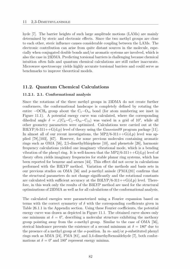
11 2,3-Dimethylanisole
hyde [7]. The barrier heights of such large amplitude motions (LAMs) are mainlydetermined by steric and electronic effects. Since the two methyl groups are closeto each other, steric influence causes considerable coupling between the LAMs. Theelectronic contribution can arise from quite distant sources in the molecule, espe-cially when conjugated double bonds and/or aromatic systems are involved, which isalso the case in 23DMA. Predicting torsional barriers is challenging because chemicalintuition often fails and quantum chemical calculations are still rather inaccurate.Microwave spectroscopy yields highly accurate torsional barriers and could serve asbenchmarks to improve theoretical models.
11.2. Quantum Chemical Calculations
11.2.1. 2.1. Conformational analysis
Since the rotations of the three methyl groups in 23DMA do not create furtherconformers, the conformational landscape is completely defined by rotating theentire −OCH3 group about the C1−O14 bond (for atom numbering see inset inFigure 11.1). A potential energy curve was calculated, where the correspondingdihedral angle δ = ∠(C6−C1−O14−C15) was varied in a grid of 10°, while allother geometry parameters were optimized. Calculations were carried out at theB3LYP/6-311++G(d,p) level of theory using the Gaussian09 program package [11].In almost all of our recent investigations, the MP2/6-311++G(d,p) level was ap-plied [78],[103], [67]. However, for some previous molecules containing aromaticrings such as OMA [56], 2,5-dimethylthiophene [10], and phenetole [26], harmonicfrequency calculations yielded one imaginary vibrational mode, which is a bendingvibration of the phenyl ring. It is well-known that the MP2/6-311++G(d,p) level oftheory often yields imaginary frequencies for stable planar ring systems, which hasbeen reported for benzene and arenes [44]. This effect did not occur in calculationsperformed with the B3LYP method. Variation of the methods and basis sets inour previous studies on OMA [56] and p-methyl anisole (PMA)[81] confirms thatthe structural parameters do not change significantly and the rotational constantsare calculated with sufficient accuracy at the B3LYP/6-311++G(d,p) level. There-fore, in this work only the results of the B3LYP method are used for the structuraloptimizations of 23DMA as well as for all calculations of the conformational analysis.
The calculated energies were parameterized using a Fourier expansion based onterms with the correct symmetry of δ with the corresponding coefficients given inTable 26.1.1 in the Appendix section. Using these Fourier coefficients, the potentialenergy curve was drawn as depicted in Figure 11.1. The obtained curve shows onlyone minimum at δ = 0°, describing a molecular structure exhibiting the methoxygroup pointing away from the o-methyl group. Similar to the case of OMA [56],sterical hindrance prevents the existence of a second minimum at δ = 180° due tothe presence of a methyl group at the o-position. In m- and/or p-substituted phenylrings such as MMA [24], PMA [81], and 3,4-dimethylbenzaldehyde [7], both confor-mations at δ = 0° and 180° represent energy minima.
82

11 2,3-Dimethylanisole
The geometry at the minimum was fully optimized to a planar conformer depictedin the inset of Figure 11.1. The Cartesian coordinates are given in Table 26.1.2in the Appendix section. The calculated rotational constants A = 2208.2 MHz,B = 1103.2 MHz, and C = 745.9 MHz come along with a value of κ = −0.51for Ray’s asymmetry parameter. The predicted dipole moment components areµa = 0.89 D, µb = 0.80 D, and µc = 0.00 D, leading to the expectation that onlya- and b-type, but no c-type transitions are observable in the microwave spectrum.Frequency calculations without imaginary frequencies confirm the stability of theoptimized conformer.
0−180 −120 −60 0 60 120 180
10
20
30
δ / °
Ene
rgy
/ kJ
mol−
1
40
14
20
16
1013
12
2019
21
23
4
1
65
9
8 7
141516
17
α
β γ
δ
Figure 11.1 The potential energy curve of 23DMA obtained by rotating the methoxygroup about the C1−O14 bond in a grid of 10°. The relative energies with re-spect to the lowest energy conformation with E = −425.514898170 Hartree aregiven. Inset: Molecular structure of the only conformer of 23DMA fully opti-mized at the B3LYP/6-311++G(d,p) level of theory. The hydrogen atoms H11,H22, and H18 are located behind H12, H21, and H16, respectively. The dihedral anglesα = ∠(C4−C3−C10−H12), β = ∠(C1−C2−C19−H21), and γ = ∠(C1−O14−C15−H17)correspond to methyl internal rotations of the m-, o-, and methoxy methyl groupsabout the C3−C10, C2−C19, and O14−C15 bonds, respectively.
11.2.2. 2.2. Methyl internal rotations
To study the internal rotations of the methyl groups, the MP2/6-311++G(d,p) andB3LYP/6-311++G(d,p) levels of theory were applied, because the results obtainedusing these two methods are not always in agreement, and it is not possible to con-firm by the experiments which method is correct. Therefore, it is necessary to reportthe results of both methods in order to avoid a wrong interpretation or incompleteinformation.
The LAMs in 23DMA are complex because of three inequivalent methyl internal ro-tations. For the m-methyl group, a V3 potential of 426.26 cm−1 with no significant
83

11 2,3-Dimethylanisole
V6 contributions was found by varying the dihedral angle α = ∠(C4−C3−C10−H12)in a grid of 10° under full relaxation of all other geometry parameters, as illustratedin Figure 11.2. The corresponding Fourier coefficients are given in Tables 26.1.3aand 26.1.3b.
20
15
10
5
0−180 −120 −60 0 60 120 180
β / °
100
75
50
25
0
β / °
−180 −120 −60 0 60 120 180α / °
0
100
200
300
400
Ene
rgy
/ cm−
1E
nerg
y / c
m−
1
MP2 B3LYP
B3LYPMP2
300
200
100
0
400
500
−180 −120 −60 0 60 120 180α / °
−180 −120 −60 0 60 120 180
a)
b)
Figure 11.2 a) Upper trace The potential energy curve of 23DMA obtainedby rotating the m-methyl group about the C3−C10 bond by varying the dihedralangle α in a grid of 10°. The relative energies with respect to the lowest energy con-formations with E = −424.2443679 Hartree (MP2) and E = −425.5205477 Hartree(B3LYP) are given. Left hand-side Double minima with small V6 contributionat α around 0° and ±120° calculated at the MP2/6-311++G(d,p) level of theory.The estimated barrier height is 527.57 cm−1. Right hand-side V3 potential with-out V6 contribution calculated at the B3LYP/6-311++G(d,p) level. The barrierheight is 426.26 cm−1. b) Lower trace: The potential energy curve obtainedby rotating the o-methyl group about the C2−C19 bond by varying the dihedralangle β in a grid of 10°. The relative energies with respect to the lowest energyconformations with E = −424.10748540 Hartree (MP2) and E = −425.52054770Hartree (B3LYP) are given. Left hand-side Double minima with significant V6
contribution (about 25 %) at β around βmax± 30° with βmax = 0° and ±120° calcu-lated at the MP2/6-311++G(d,p) level of theory. The estimated barrier height is
84

11 2,3-Dimethylanisole
95.76 cm−1. Right hand-side V3 potential without V6 contribution calculated atthe B3LYP/6-311++G(d,p) level. The barrier height is approximately 17.03 cm−1.
For the o-methyl group, the barrier height was calculated by varying the dihe-dral angle β = ∠(C1−C2−C19−H21). The results obtained from calculations atthe MP2/6-311++G(d,p) and B3LYP/6-311++G(d,p) levels of theory are contro-versial. Using the B3LYP method, three energetic minima with no significant V6
contributions at β = 0° and ±120° were obtained, indicating that the o- and m-methyl groups are staggered (see Figure 11.2). Furthermore, local minima exist inthe maximum regions around β = ±60° and 180°. The potentials obtained with theMP2 method show V6 contributions of about 25 %, resulting in double minimumpotentials with local maxima at βmax = 0° and ±120° and minima at βmax ± 30°(see Figure 11.2). These observations are probably due to the coupling between theLAMs of the two neighboring o- and m-methyl groups.
β / °
α /
°
0 18090 2700
180
360
270
90
100
80
60
40
20
0 %360
Figure 11.3 The potential energy surface in dependence on the dihedral angles αand β of 23DMA calculated at the B3LYP/6-311++G(d,p) level of theory. Theangles α and β were varied in a grid of 10° while all other geometry parameters wereoptimized. The numbers in the color code indicate the energy (in percent) relativeto the energetic minimum Emin = −425.52054770 Hartree (0 %) and the energeticmaximum Emax = −425.5183019 Hartree (100 %). Note that there are significantlymore colors in the lower 50 % section.
To study this coupling, a two-dimensional potential energy surface (2D-PES) de-pending on the dihedral angles α and β was calculated. Due to symmetry, only datapoints in the range from α and β = 0 − 120° are needed. The potential energieswere parameterized with a 2D Fourier expansion based on terms representing thecorrect symmetry of the angles α and β. The corresponding coefficients are avail-able in Table 26.1.3c in the Appendix. The PES given in Figure 11.3 leads to the
85

11 2,3-Dimethylanisole
assumption that almost no potential coupling terms between α and β exist.
For the methoxy methyl group, Reinhold et al. reported a torsional barrier of about1200 cm−1 in the case of anisole [104], which should be similar in 23DMA. Torsionalsplittings arising from a methyl group with such high barrier to internal rotation arenot observable with our experimental resolution, which has been proven by previousinvestigations on monomethylanisoles [56], [24], [81].
11.3. Results
The results of the X-ray experiments can be found in the published version of thischapter in reference [100].
At the beginning, the internal rotation effects were neglected and 23DMA wastreated as a semi-rigid rotor. Using the rotational constants and dipole momentcomponents obtained from quantum chemistry (see Section 11.2.2) a theoretical mi-crowave spectrum was calculated with the program XIAM [22] in its rigid-rotormode for a comparison with the experimental data. The J = 7 ← 6 a-type transi-tions with Ka = 0, 1 were assigned first. Afterwards, more a-type lines were foundprogressively. Even though quantum chemistry predicted almost the same value fordipole moment components in a- and b-directions (µa = 0.89 D and µb = 0.80 D),b-type transitions appeared much weaker in intensity, but could be assigned never-theless, yielding a fit of 115 rigid-rotor lines (indicated as Fit 00 in Table 11.1). Thestandard deviation is in agreement with the measurement accuracy; the rotationaland centrifugal distortion constants are determined very accurately.
Each rotational transition of 23DMA splits into five torsional components (σ1σ2) =(00), (01), (10), (11), and (12), due to the LAMs of two methyl groups. The nota-tions σ1 and σ2 [10] refer to the methyl rotor at the m- and o-position, respectively.Figure 11.4 illustrates a typical spectrum. In the second step, the internal rotationof the m-methyl rotor was taken into account and a one-top spectrum was predictedusing the rotational constants from the rigid rotor fit and the calculated V3 potentialas well as the angle between the internal rotor axis and the principal a-axis. Be-cause of the strong correlation between V3 and the moment of inertia of the methylgroup F0, the latter was fixed to 158 GHz, corresponding to Iα = 3.2 uA2, a valuefrequently found for methyl groups. The LAM splittings between the (00) and (10)species are in most transitions smaller than 1 MHz, which simplified the assignment.At this stage, 232 lines with J ≤ 13 and Ka ≤ 5 were fitted to a standard deviationwithin the measurement accuracy using the program XIAM. The results are givenas Fit 00/10 in Table 11.1.
The third step concerns attempts to assign the torsional species (01) arising fromthe o-methyl rotor with the previously applied method for the m-methyl group. Theassignments were more difficult because the barrier height is very low, causing largesplittings in the microwave spectrum. First, the (01) species of some a-type transi-
86

11 2,3-Dimethylanisole
tions with small splittings was identified. Gradually the number of (01) lines in thefit was increased until finally, 188 lines were assigned and included in Fit 00/01 alsogiven in Table 11.1. The standard deviation of 90.4 kHz of this fit is much largerthan the measurement accuracy. This observation has been found previously inother low barrier internal rotation problems treated by XIAM, such as ethyl acetate(101.606(23) cm−1, 85.3 kHz) [86], allyl acetate (98.093(12) cm−1, 54.0 kHz) [77],vinyl acetate (151.492(34) cm−1, 92.3 kHz)[79], and 3-pentyn-1-ol (9.4552(94) cm−1,20.7 kHz)[76]. In our recent study on two conformers of MMA, the results of thefits obtained by XIAM were compared to those obtained by a newly developed pro-gram, aixPAM, where further parameters in addition to those fitted in XIAM wereincluded [24]. Without these parameters, the standard deviations are similar in bothcases for the same set of transitions and parameters (27 kHz vs. 23 kHz for the cisconformer with V3= 56 cm−1 and 32.1 kHz vs. 28.3 kHz for the trans conformerwith V3 = 37 cm−1). With three additional parameters, the standard deviation ofaixPAM decreases to 3.7 kHz and 4.1 kHz for the cis and the trans conformer, re-spectively. Because the V3 potential of the o-methyl rotor is only about 27 cm−1, thelarge standard deviation obtained from XIAM for the o-methyl group of 23DMAmight also arise from the limited number of fitted parameters. Due to the largestandard deviation, the centrifugal distortion constants could not be fitted well andtherefore were fixed to the values obtained from Fit 00/10.
MHz12221.198 12221.49710889.34710888.700
(00)
(10)
(11)
(01)(12)
624 ← 523
Figure 11.4 An exemplary a-type rotational transition (in MHz) measured athigh resolution, showing splittings into five torsional species. The brackets describeDoppler pairs. The spectra are normalized. For the left hand-side spectrum, 780decays were co-added and the polarization frequency was 10888.85 MHz. For theright hand-side spectrum, the number of co-added decays was 50 and the polariza-tion frequency 12221.50 MHz.
87

11 2,3-Dimethylanisole
At this stage, the program aixPAM was used to fit the same set of (00) and (01)species lines. By adding 5 parameters, which are VJ multiplying P 2[1−cos(3α)], VKmultiplying P 2
z [1−cos(3α)], V− multiplying (P 2x −P 2
y )[1−cos(3α)], Pzp3 multiplyingPzp
3, and D2p2 multiplying (P 2x − P 2
y )p2, a standard deviation of 3.6 kHz wasachieved, which is much closer to measurement accuracy. This fit is given as FitaixPAM in Table 11.1, and clearly confirms that the (01) species assignment iscorrect and the large standard deviation of Fit 00/01 arises from the lack of higherorder term parameters. The added parameters are effective parameters, which takeinto account the higher order terms of the V3 potentials as well as the quartic higherorder combinations.
Frequency / GHz10 11 12 13
Inte
nsity
/ a.
u.
(00) (01)(10) (11)
(12)
sim.
exp.
Figure 11.5 The discontinuous broadband scan of 23DMA from 10 to 13.5 GHz.The experimental spectrum is the upper trace. The lower trace indicates the theo-retical spectrum predicted using the molecular parameters deduced from the GlobalFit using the program XIAM, showing that no intense lines remain unassigned inthe spectrum.
Finally, a two-top spectrum was simulated using the program XIAM in order toassign the (11) and (12) species, which describe the coupling between the LAMs oftwo methyl groups. For transitions with large torsional splittings, the assignmentswere evident. On the other hand, there are some transitions with splittings in thesame order of the standard deviation of Fit 00/01 (a few 100 kHz), causing thedistinction of the (11) and (12) species of those lines much more uncertain. In aglobal XIAM fit containing 465 lines, a standard deviation of 118 kHz was found byfloating 14 parameters, which are the rotational constants, five quartic centrifugaldistortion constants, the two V3 potentials, the angles between the internal rotor axesand the principal a-axis, and three higher order parameters Dpi2J , Dpi2K , Dpi2− ofthe o-methyl rotor. The results of this fit are summarized as Global Fit in Table 11.1.
88

11
2,3-D
imethylaniso
le
Table 11.1 Molecular parameters of 23DMA in the principal axis system obtained by the XIAM and aixPAM codes.
Par.a Unit Fit 00b Fit 00/10c Fit 00/01d Fit 00/01e Global Fitf Calc.g
XIAM XIAM aixPAM XIAMA MHz 2204.62202(26) 2204.60333(50) 2204.5825(63) 2246.7(15) 2204.603(13) 2208.1534B MHz 1114.47007(16) 1114.47150(12) 1107.7520(16) 1103.06(40) 1107.7338(26) 1103.2195C MHz 748.079761(88) 748.077482(68) 748.07934(96) 749.02(32) 748.0765(17) 745.9185∆J kHz 0.03147(68) 0.03169(45) 0.03169h 0.03685(82) −0.072(11) 0.0200599∆JK kHz 0.1366(43) 0.1351(29) 0.1351h 0.0269(54) 0.212(93) 0.1010498∆K kHz — −0.116(41) −0.116h −0.176(58) 3.93(95) 0.0022304δJ kHz 0.01197(36) 0.01206(24) 0.01206h 0.01466(43) −0.0434(61) 0.0064910δK kHz 0.1428(63) 0.1395(51) 0.1395h 0.0994(82) — 0.0395049V3 cm−1 — 520.08(39) 26.9068(7) 26.756(16) 518.7(1.2)/26.9047(5)i 426.26/16.97Dpi2J MHz — — 0.18440(87) 0.302(14) 0.18513(66) —Dpi2K MHz — — −0.3149(55) −3.234(96) −0.3125(47) —Dpi2− MHz — — 0.2143(12) 0.168(36) 0.21593(90) —VJ MHz — — — 2.31(25) — —V− MHz — — — 3.49(40) — —VK GHz — — — −0.0549(17) — —Pzp3 MHz — — — −0.2797(40) — —D2p2 GHz — — — 0.472(29) — —∠(i, a) ° — 23.46(52) 87.79(1) 89.323(21) 21.8(1.1)/87.79(1)i 29.42/86.15i
∠(i, b) ° — 66.54(52) 2.21(1) 0.677(21) 68.2(1.1)/2.21(1)i 60.58/3.85i
∠(i, c) ° — 90.0k 90.0k 90.0k 90.0/90.0i,k 89.95/90.00i
N l — 115 115/117 115/73 115/73 115/117/81/77/75 —σm kHz 3.2 3.0 90.4 3.6 117.6 —
aAll parameters refer to the principal axis system. Watson’s A reduction and Ir representation were used. bRigid rotor fit including only (00) species.cIncluding only the (00) and (10) torsional species of the m-methyl rotor, fitted using the program XIAM. dIncluding only the (00) and (01) torsional
species of the o-methyl rotor, fitted using the program XIAM. eIncluding only the (00) and (01) torsional species of the o-methyl rotor, fitted using the
program aixPAM. fGlobal fit using the program XIAM. gCalculated at the B3LYP/6-311++G(d,p) level of theory. hFixed to the values obtained from
Fit 00/10. iLeft and right hand-side: values obtained for the m- and o-methyl groups, respectively. kFixed due to symmetry. lNumber of lines. Fit 00:
(00) species, Fit 00/10: (00)/(10) species, Fit 00/01: (00)/(01) species, Global Fit: (00)/(01)/(10)/(11)/(12) species. mStandard deviation of the fit.
89

11 2,3-Dimethylanisole
A list of all frequencies is available in Table 26.1.4, a comparison between the exper-imental and the theoretical spectrum simulated by XIAM is depicted in Figure 11.5.
11.4. Discussion
The rotational- and quartic centrifugal distortion constants were determined withvery high accuracy by fitting 115 torsional transitions in a rigid rotor fit. The stan-dard deviation of 3.2 kHz is close to the measurement accuracy. The m-methylgroup with an intermediate V3 potential of 523.39(39) cm−1 does not cause fittingproblems. The one-top XIAM Fit 00/10 reaches the same quality as that of therigid rotor Fit 00 by additionally including only two parameters, namely the barrierheight V3 and the angle between the m-methyl rotor axis and the principal a-axis.
On the contrary, the internal rotation of the o-methyl group cannot be capturedcorrectly by XIAM. With a V3 potential of only 27.0750(6) cm−1, the addition ofthree higher-order parameters Dpi2J , Dpi2K , and Dpi2− was not sufficient to reducethe standard deviation to measurement accuracy. With a standard deviation of90.4 kHz, the centrifugal distortion constants in Fit 00/01 were not determined veryprecisely.
The torsional barrier found for the o-methyl group in OMA is rather high (459 cm−1)and the program XIAM yielded excellent fitting results for this LAM problem. Thesterical hindrance between the methoxy and the o-methyl group is the main reasonfor this intermediate value of V3, which has been confirmed by the results obtainedfor a number of o-substituted toluenes [56]. In MMA, the methyl group at the m-position is further apart from the methoxy group, which creates a symmetric localenvironment near the methyl group and decreases the ring methyl barrier height byan order of magnitude (36.6342(84) and 55.7693(90) cm−1 for the trans and the cisconformer, respectively) [24]. However, in 23DMA the intermediate barrier of about519 cm−1 of the m-methyl group is not surprising because the adjacento-methylgroup causes steric hindrance. The very low barrier of the o-methyl group can beexplained in a similar way as has been described in Refs. [105] and [68]. Assumingthat the methoxy methyl group and the two other methyl groups are similar, theo-methyl group, which has a C3v symmetry, experiences potentials based on a C2v
frame symmetry as in toluene CH3−C6H5 (V6 = 4.8 cm−1) [62] or nitromethaneCH3NO2 (V6 = 4.9 cm−1) [106], where only a V6 term but no V3 contribution exists.In the case of 23DMA, the frame symmetry is slightly out-of-balance, causing thesmall V3 potential term of about 27 cm−1 observed for the o-methyl group. Unfor-tunately, the V6 term cannot be fitted because only data in the ground state areavailable, i.e. the attempts to fit a combined V3 − V6 function as suggested by theMP2 method (see chapter 11.2.) failed due to experimental limitation. Thus, nostatement was obtained from the experiment to decide whether MP2 or B3LYP isbetter suited to describe the internal rotations in 23DMA.
90
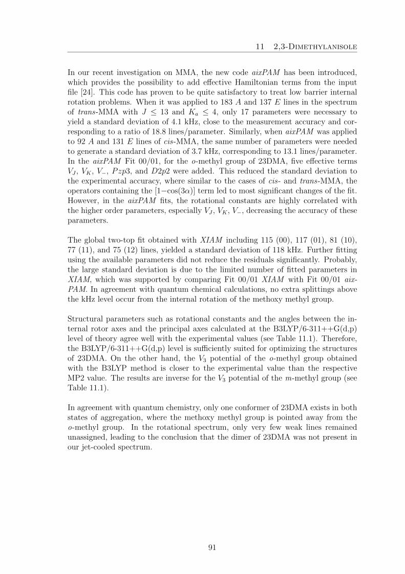
11 2,3-Dimethylanisole
In our recent investigation on MMA, the new code aixPAM has been introduced,which provides the possibility to add effective Hamiltonian terms from the inputfile [24]. This code has proven to be quite satisfactory to treat low barrier internalrotation problems. When it was applied to 183 A and 137 E lines in the spectrumof trans-MMA with J ≤ 13 and Ka ≤ 4, only 17 parameters were necessary toyield a standard deviation of 4.1 kHz, close to the measurement accuracy and cor-responding to a ratio of 18.8 lines/parameter. Similarly, when aixPAM was appliedto 92 A and 131 E lines of cis-MMA, the same number of parameters were neededto generate a standard deviation of 3.7 kHz, corresponding to 13.1 lines/parameter.In the aixPAM Fit 00/01, for the o-methyl group of 23DMA, five effective termsVJ , VK , V−, Pzp3, and D2p2 were added. This reduced the standard deviation tothe experimental accuracy, where similar to the cases of cis- and trans-MMA, theoperators containing the [1−cos(3α)] term led to most significant changes of the fit.However, in the aixPAM fits, the rotational constants are highly correlated withthe higher order parameters, especially VJ , VK , V−, decreasing the accuracy of theseparameters.
The global two-top fit obtained with XIAM including 115 (00), 117 (01), 81 (10),77 (11), and 75 (12) lines, yielded a standard deviation of 118 kHz. Further fittingusing the available parameters did not reduce the residuals significantly. Probably,the large standard deviation is due to the limited number of fitted parameters inXIAM, which was supported by comparing Fit 00/01 XIAM with Fit 00/01 aix-PAM. In agreement with quantum chemical calculations, no extra splittings abovethe kHz level occur from the internal rotation of the methoxy methyl group.
Structural parameters such as rotational constants and the angles between the in-ternal rotor axes and the principal axes calculated at the B3LYP/6-311++G(d,p)level of theory agree well with the experimental values (see Table 11.1). Therefore,the B3LYP/6-311++G(d,p) level is sufficiently suited for optimizing the structuresof 23DMA. On the other hand, the V3 potential of the o-methyl group obtainedwith the B3LYP method is closer to the experimental value than the respectiveMP2 value. The results are inverse for the V3 potential of the m-methyl group (seeTable 11.1).
In agreement with quantum chemistry, only one conformer of 23DMA exists in bothstates of aggregation, where the methoxy methyl group is pointed away from theo-methyl group. In the rotational spectrum, only very few weak lines remainedunassigned, leading to the conclusion that the dimer of 23DMA was not present inour jet-cooled spectrum.
91

11 2,3-Dimethylanisole
92

12 2,4-Dimethylanisole
12. 2,4-Dimethylanisole
Understanding the Rotational Behaviorin 2,4-Dimethylanisole
L. Ferres performed measurements, quantum chemical calculations, assignments ofthe microwave spectra and prepared the manuscript.
12.1. Introduction
In microwave spectroscopy, it is of common use to describe the investigated moleculesas semi-rigid tops. This was implemented in many programs as e.g. IAM [49],XIAM [22]; BELGI-Cs [25], BELGI-C1 [51], ERHAM [74], RAM36 [62], and aix-PAM [24], written to treat molecules using a rigid-frame - rigid-top model, which isexpanded by higher order terms, as for example, the centrifugal distortion constants,leading to the above mentioned semi-rigid top model.
The choice of the axis system is not as straight-forward as it might seem, in fact sev-eral methods are applicable: BEGLI and RAM36 use the rho-axis method, XIAMthe combined axis method, and aixPAM calculates in the principal axis system.Regarding the numerical solutions, a few alternatives are executable: the Hamil-
93

12 2,4-Dimethylanisole
tonian can be pre-diagonalized resulting in a two-step diagonalization while otherprograms only need one step of diagonalization. Often, small and off-diagonal matrixelements are neglected or the matrices are truncated, simplifying the calculationsbut forfeiting flexibility and accuracy, in return. Another crucial point is the numberof parameters which are implementable in the program source codes.
2,4-Dimethylanisole (C6H3OCH3(CH3)2), in the following abbreviated as 24DMA, isalso known as 1-methoxy-2,4-dimethylbenzene or 4-methoxy-m-xylene. The chemi-cal compound under investigation occurs as colourless liquid with an aromatic smell.24DMA can be synthesized via electrochemical oxidation of p-xylene in methanol[107]. The molecular structure is a composition of a methoxyphenol part and twomethyl groups attached to the aromatic ring.
The two ring-methyl groups are able to undergo internal rotation, which inducesmeasurable signal splittings in the microwave spectrum. Only few methyl substi-tuted phenyl rings as well as two-top molecules have been investigated so far usingrotational spectroscopy, e.g. 4,5-dimethylthiazol [108] or dimethylbenzaldehyde [7].The methyl anisole family (ortho-, meta- and para-methylanisoles [56], [24], [81])has also already been examined. Therefore, it seems plausible to determine exper-imentally the rotational barriers of 24DMA in order to compare them to the bar-riers of the methylanisoles and to analyze the influence of the substitution site onthe rotational barriers in aromatic systems. As previous projects state, microwavespectroscopy is a well-suited tool for internal rotation examination, yielding highlyaccurate rotational and centrifugal distortion constants [26] [67], as well as torsionalbarriers [100] determined with high accuracy. In this work a new program calledN-TOP is introduced: a program code developed to render the implementation ofhigher order terms possible with respect to any number of internal rotors.
12.2. Quantum Chemical Calculations
All quantum chemical calculations were performed using the Gaussian09 suite ofprograms [11]. If not stated otherwise, the calculations were carried out at theMP2/6-311++G(d,p) level of theory. The molecular structure was optimized underfull relaxation and featuring one stable conformer showing all heavy atoms locatedin a plane, as illustrated in Figure 12.1.
Furthermore, to check for alternative stable structures, a scan calculation of therotation about the C1−O14 bond was performed under full relaxation of all othercoordinates, by varying the dihedral angle γ = ∠(C2−C1−O14−C15). The resultingenergies are summarized in a potential energy curve, which was generated by Fourierexpansion (Figure 12.1). The corresponding Fourier terms are given in Table 26.2.2.This curve indicates the existence of only one single stable planar conformer, whichis already depicted in Figure 12.1. Due to steric hindrance only this conformer fea-turing the C15 methyl group pointing away from the C10 methyl group, and thus
94

12 2,4-Dimethylanisole
resulting in a planar trans-structure with γ = 180°, is allowed.
11
18
22
1920
213
8
2
6
4
10
12
13
1
7 9
5
14
1517
16α
β
γ
δ
Figure 12.1 Optimized structures of 24DMA with α = −19.69°, β = −179.85°,γ = −179.90° , and δ = 179.81° at the MP2/6-311++G(d,p) level of theory. Thedihedral angles are defined as α = ∠(C5−C4−C19−H22), β = ∠(C1−C2−C10−H12),γ = ∠(C2−C1−O14−C15), and δ = ∠(C1-O14-C15-H16). The corresponding atomcoordinates are listed in Table 26.2.1. By varying the dihedral angle γ, a potentialenergy curve relative to Emin = −424.2452163 Hartree was drawn using Fourier ex-pansion given in Table 26.2.2.
As the main spectroscopic interest in this molecule lies in the determination of thetorsional barriers, the dihedral angle α was also varied in 10° steps. For each step theenergy of the newly generated molecular geometry was optimized (see Figure 12.2).Two levels of theory were used for the same basic set, yielding different moleculargeometries. The MP2 method optimizes α to ±19.69° while B3LYP optimizes to0.03°. As depicted in Figure 12.1, the only stable conformer obtained via MP2 fea-tures no Cs-symmetry as the hydrogen atoms of the C19 methyl group do not lie inthe molecular plane. Contrarily, considering the B3LYP method, 24DMA exhibitsCs-symmetry. The barrier height was determined by calculating the difference be-tween the energies of the optimized minima and transition state structures. Thetorsional barriers were found to be 61.61 cm−1 (MP2) and 57.68 cm−1 (B3LYP). ForMP2 a V6 contribution of 8.32 cm−1 was obtained.
The second methyl internal rotation of the ortho-rotor is realized by changing theangle β = ∠(C1−C2−C10−H12) which equals to a rotation about the C2−C10 axis.For this purpose, both the MP2 and B3LYP methods were applied, yielding similarresults depicted in Figure 12.2.
95

12 2,4-Dimethylanisole
60
45
30
15
0
75
−180 −120 −60 0 60 120 180
Ene
rgy
/ cm−
1
α / °
MP2B3LYP
β / °
500
400
300
200
100
00 60 120 180 240 300 360
a) b)
Figure 12.2 a) Potential energy curve of the variation of α = ∠(C5−C4−C19−H22)under fully relaxation of all other coordinates. Two levels of theory were usedfor the same basic set, yielding different molecular geometries. The MP2 methodoptimizes α to ±19.69° while B3LYP optimizes to 0.03°. Figure 12.2 b) Variationof the dihedral angle β = ∠ (C1−C2−C10−H12) using the basis set 6-311++G(d,p)in combination with the MP2 (black) and B3LYP (blue) methods. The potentialenergy curves differ only slightly in their height, modeled by the torsional barrierV3. For both methods the minima occur at β = 60°/180°/300°.
10.51669810.51647210.466100 10.466399GHz
707 ← 606
(00)
(10)(01)
(12)
(11)
Figure 12.3 Two normalized high resolution spectra with 400 co-added decays each,show the signal splitting of the 707 ← 606 a-type transition due to internal rotation.Doppler pairs are marked with brackets and labeled with the corresponding symme-try species. The spectra were recorded at polarization frequencies of 10.46625 and
96

12 2,4-Dimethylanisole
10.51650 GHz and at 400 repetitions, each.
The low calculated torsional barriers of 453.24 cm−1 (MP2) and 393.98 cm−1 (B3LYP)suggest large splittings in the microwave spectrum. The torsional barrier of themethoxy methyl group was calculated to 1112.41 cm−1, and therefore no observabletorsional splittings are expected in the microwave spectrum. Quantum chemical op-timization calculations yielded the following rotational constants: A= 2410.28 MHz,B = 961.89 MHz, and C = 696.54 MHz, and dipole moments: µa = 0.26 D, µb =−1.07 D, and µc = 0.01 D, implying strong b-type transitions in the recorded broad-band scan. Harmonic frequency calculations at the MP2/6-311++G(d,p) level oftheory yielded an imaginary vibrational mode, which describes a bending motion ofthe phenyl ring, often reported for aromatic structures [44].
12.3. Results
First, three b-type transitions from the 707 ← 616, and 717 ← 606 branches wereassigned to the frequencies observed in the 10 to 14 GHz broadband scan, fittingthe rotational constants A, B and C with the program XIAM. Then, more accuratefrequencies obtained from high resolution measurements were used for verificationof the assignment. As the signal splittings of the (00) and (10) species are verynarrow, a single measurement often was sufficient to gather both frequencies. Thus,more a- and b-type lines as well as fitting parameters were added subsequently, untilapproximately 50 (00) and (10) species lines were fitted to a standard deviation of3 kHz yielding a torsional barrier of 439.61(36) cm−1.
10 11 12 13 14
Frequency / GHz
Inte
nsity
/ a.
u.
Figure 12.4 The broadband scan of 2,4-dimethylanisole reaching from 10 to 14GHz. The upper trace shows the experimental spectrum, while the lower trace in-dicates the theoretical spectrum using the molecular parameters obtained from theglobal XIAM Fit.
97

12
2,4-D
imethylaniso
le
Table 12.1. Molecular parameters of 2,4-dimethylanisole in the principal axis system obtained by the programs XIAM and N-TOP.
Par.a Unit Fit XIAM Fit 00/10/01 N-TOP Fit NTOP Calc.b
A MHz 2419.1283(17) 2420.155(86) 2419.133(55) 2420.2832B MHz 963.36414(45) 964.405(77) 964.151(41) 961.8944C MHz 698.24270(30) 697.62(15) 698.828(65) 696.5443∆J kHz 0.0175(15) 0.01483(23) 0.01290(17) 0.01644c
∆JK kHz 0.0431(90) 0.0274(13) 0.03200(99) 0.02951c
∆K kHz 1.02(13) 0.3797(59) 0.3986(48) 0.27809c
δJ kHz 0.00475(77) 0.00408(12) 0.003153(86) 0.004986c
δK kHz 0.048(16) 0.0319(23) 0.0270(19) 0.035971c
V d3 cm−1 447.75(14) 59.0121(1) 431.29(36) 49.572(33) 435.649(20) 48.192(25) 453.24 61.61Ddpi2J MHz — 2.877(66) — — —
Ddpi2K MHz — 83.75(32) — — —
Ddpi2− MHz — −2.665(64) — — —
F d0 GHz 160h 160h 158h 158h 158h 158h —F12 GHz 3.05(18) — — —HK kHz 0.0268(37) — — —Vcc GHz — −157.18(58) −181.50(45) —DdmJ MHz — −0.0505(43) 0.0211(19) −0.1101(17) 0.00591(86) —
DdmK MHz — −0.124(17) 0.4565(29) 0.0968(88) 0.4605(19) —
ddm MHz — −0.0586(47) −0.077(21) −0.1285(17) 0.1036(86) —V dK MHz — — −1.258(33) — −1.007(18) —V dJ MHz — — 0.150(20) — −0.0116(88) —V d− MHz — — −0.74(22) — 1.089(88) —∠(i, a)d ° 62.998(39) 1.9330(2) 64.70(12) 1.9351(2) 63.0266(5) 1.9351(2) 61.50 2.03∠(i, b)d ° 27.002(39) 88.0670(2) 25.30(12) 88.0649(2) 26.9734(5) 88.0649(2) 28.50 88.26∠(i, c)d ° 90.00f 90.00f 90.00f 90.00f 90.00f 90.00f 90.10 88.99Ng — 590 384 590 —σe kHz 18.9 3.7 4.6 —
aAll parameters refer to the principal axis system. Watson’s A reduction and Ir representation were used. bCalculated at the MP2/6-311++G(d,p)level of theory. cCalculated at the B3LYP/6-311++G(d,p) level of theory dLeft and right hand side: values obtained for the o- and p-methyl groups,respectively. eStandard deviation of the fit. fFixed due to symmetry. gNumber of lines. hCorresponds to Iα = 3.2 uA2.
98

12 2,4-Dimethylanisole
The assignment of the wider torsional splittings was a more challenging task, as thequantum chemical calculation are not always applicable to low hindered internal ro-tors, as already known from m-[24] and p- methylanisole [81]. For this purpose, a fewstrong (00) transitions with an elevated Ka of 4 or 5 were used, as these transitionsfeature wider splittings concerning the (01) (11) and (12) symmetry species (seeFigure 12.4). This way, the torsional barrier of 58.297(15) cm−1 was determined. Aglobal fit containing all five symmetry species was established, as given in Table 12.1.
In the end, some forbidden c-type transitions were identified and added to the fit,leading to a fit comprising 590 rotational transitions. Using the full set of rotationaland quartic centrifugal constants expanded by the higher order Dpi parameters, F12
and HJ , a standard deviation of 19 kHz was obtained using the XIAM program(Fit XIAM). As Figure 12.4 shows, no intensive lines remained unassigned in themicrowave broadband scan.
Afterwards, the newly developed program NTOP was applied to the same data set.At first, the standard deviation was of the same order of magnitude as the XIAMfit, but by adding additional parameters among them VK , VJ and V−, the standarddeviation decreased to 4.6 kHz. A separate fit was realized, using only the (00), (01),and (10) symmetry species, as they are translated by signals with higher intensityin the microwave spectrum. Therefore, these lines have an increased measurementaccuracy, compared to the (11) and (12) symmetry species, which often appear asvery weak broad signals. This is also shown by the lower standard deviation of 3.7kHz of the Fit (00)/(01)/(10) given in Table 12.1.
99

12 2,4-Dimethylanisole
12.4. Discussion
The rotational and centrifugal distortion constants of the three fits are well de-termined, showing a high accuracy. The values are in good agreement with thetheoretical ones given in the calc. column in Table 12.1. The XIAM program ob-viously has some difficulties fitting the rotational transitions caused by the signalsplitting of two internal rotors. NTOP enables to fit the same data set by using 3further parameters, thus reducing the standard deviation by 14 kHz, yielding a fitvery close to measurement accuracy. In p- and m- methylanisole, a similar problemwas observed, which could be solved by fitting the higher order parameters Fv, k1, c1
(corresponding to FJ , QzK , and dm), and FJ , DmK , FK , F−, and DmJ , respectively.
The sixfold contribution to the barrier to internal rotation of the p-methyl rotorin 24DMA was neglected, as previously done in case of p-methylanisole. Neverthe-less, the fit is satisfactory, fitting a threefold V3 potential with high accuracy. Forthe o-methyl rotor, no sixfold potentials were observed by use of quantum chemicalcalculations. The fitted rotational barriers of 435.649(20) and 48.192(25) cm−1 arein good agreement with the calculated values of 453.24 and 61.61 cm−1 obtainedvia quantum chemical calculations. The high amount of lines in the fits is also anindicator for an accurate description of 2,4-dimethylanisole using the set of fittingparameters given in Table 12.1.
When compared to other o-substituted toluene derivates, the potential barrier of435.649(20) cm−1 is very close to the values found for o-methylanisole and anti -3,4-dimethyl-benzaldehyde (see Table 12.2). This is not a surprising result, as anti -3,4-dimethyl-benzaldehyde conformers and o-methylanisole are structurally very similarto 24DMA. In 24DMA, no syn-conformation was observed, as shown in Figure 12.1by the torsional potential energy curve. Normally, the amplitude of syn-torsional po-tentials are more elevated than anti -conformational potentials, due to an increasedsteric hindrance. This was observed for 3,4-dimethylbenzaldehyde and for o-cresol.o-chlorotoluene, o-fluorotoluene and o-xylene also possess rotational barriers in therange of several hundreds of wavenumbers, which is in agreement with the expec-tations and the results of this work. The respective references are given in Table 12.2.
All para-substituted toluenes reveal torsional barriers significantly lower than inthe o-substituted isomers. This is also true for 24DMA: the barrier to internalrotation of the p-methyl group is fitted to 48.192(25) cm−1, which is an order ofmagnitude smaller than the barrier for the o-methyl internal rotation. The valueis very similar to the p-methylanisole rotational barrier of 49.6370(1) cm−1. Allvalues found for p-substituted aromatic systems lie in the range from roughly 4 to50 cm−1. The smallest ones were determined for the halogene containing moleculesp-fluorotoluene and 35Cl-p-chlorotoluene, followed by p-cresol and p-tolualdehyde.The greatest value was found in p-methylanisole.
100

12 2,4-Dimethylanisole
Table 12.2. Torsional barriers of ortho- and para substituted toluene derivatives.
ortho paraV3/cm−1 Ref. V3/cm−1 Ref.
fluorotoluene 227.28(2) [69] 4.8298(63) [6]anti -cresol 371.05(41) [4] 18.39(3) [84]syn-cresol 669.10(51) [4] 18.39(3) [84]methylanisole 444.05(41) [56] 49.6370(1) [81]anti -3,4-DMBAa 454.1(14),480.6(44) [7] – –syn-3,4-DMBAa 508.1(11),550.7(88) [7] – –37Cl-chlorotoluene 507.2(83) [71] – –35Cl-chlorotoluene 513.8(27) [71] 4.872(14) [88]xylene 518.3(32) [72] – –syn-2,5-DMBAa 566(16) [7] – –tolualdehyde – – 28.37 [85]
aDMBA: abbreviation for dimethylbenzaldehyde.
In summary, comparing the rotational barriers of the methylanisoles to the 24DMAtorsional barriers, no significant deviance was found. This is probably due to thespatial arrangement of the substituents, which are not directly adjacent to each otherin the phenyl ring. However, the necessary fitting of the cosine coupling parameterVcc suggests that both methyl rotors influence each other.
101

12 2,4-Dimethylanisole
102

13 2,5-Dimethylanisole
13. 2,5-Dimethylanisole
A Microwave Study on a Combination ofIntermediate and Low Internal Rotation Barrier
Molecule: 2,5-Dimethylanisole
00
01
10
1211
716← 625
21
13
16
0.293 MHz 1.001 MHz
{ {
J. Cheung contributed to this chapter by carrying out a part of the quantum chemi-cal calculations and by measuring the majority of the rotational transitions [109]. L.Ferres supervised her during her research project, completed the quantum chemicalcalculations and the microwave spectroscopic measurements, assisted in the assign-ment process, performed fits with numerous data sets and programs, and preparedthe manuscript.
13.1. Introduction
The molecular structure of molecules is of great interest in many chemical pro-cesses found in the laboratories and in nature: especially in bio-chemical processes,the knowledge of the exact molecular structure is of great importance in order tounderstand many selective chemical reaction mechanisms. Over the past decades,microwave spectroscopy was proven as an important tool to determine the threedimensional molecular structure of a molecule [26]. Additionally, information about
103

13 2,5-Dimethylanisole
internal motions [91] such as internal rotation [100] or rotation-vibrational modes[67] can be deduced from microwave spectroscopic data.
Previously, ortho-, meta- and para-methylanisole [56], [24], [81] were examined viamicrowave spectroscopy and quantum chemical calculations. As 2,5-dimethylansiole,in the following abbreviated as 25DMA, features two ring-attached methyl groups,namely one in ortho- and one in para-position relative to the methoxy-substituent,great similarities to o- and p-methylansiole are expected. As already observed inp-methyl-anisole, low rotational barriers in methylanisoles lead to several compli-cations in the assigning and fitting process. It is an interesting question how thiswill influence a two-rotor molecule. In p-methylanisole, higher order fitting param-eters were necessary to fit the rotational transitions to measurement accuracy. Foro-methyl anisole, no problems of this type occured. In dimethylbenzaldehyde [7],another two-rotor molecule, a global fit containing five symmetry species could beachieved without further complications.
In this chapter, the theoretical molecular parameters are calculated using quantumchemical calculations, which will be used to simulate a rotational spectrum. Thesimulated spectrum will then be compared with the experimental broadband scan,fitting the rotational transitions to the observed frequencies, thus yielding the molec-ular parameters of 2,5-dimethylanisole.
13.2. Quantum Chemical Calculations
All quantum chemical calculations were performed using the Gaussian09 programpackage [11]. First, the molecular structure was optimized using the B3LYP/6-311++G(d,p) level of theory. The optimized planar structure is given in Fig-ure 13.1. The dihedral angles were optimized to α = −0.02°, β = 0.00°, γ = 179.88°,and δ = −61.23°. The MP2 method delivered nearly identical results, except forH12, which is tilting out of plane by about 20° (γ = −159.90°). Harmonic frequencycalculations yielded no imaginary frequencies.
Thereafter, the rotational constants of A = 1842.71 MHz, B = 1160.63 MHz, andC = 721.62 MHz were obtained. The calculated dipole moments of µa = 0.04 D, µb= −1.44 D, and µc = 0.00 D suggest occurrence of strong b-type signals in the mi-crowave broadband scan. Morevoer, the dihedral angle α was optimized to −0.02°,resulting in the trans-conformation for the planar conformer of 25DMA. This is notsurprising, as it is well known from literature [27], that phenyl rings are planar. Thedihedral angle δ, describing the rotation of the methoxy-methyl group, is optimizedto −61.23°, exhibiting H17 lying in the molecular plane spanned by the heavy atomsof 25DMA. The optimized values for β and γ, describing the orientation of the o-and m-methyl groups, respectively, indicate that hydrogens H21 and H12 also lie inthe molecular heavy-atom plane. Thus, in the molecular structure of 25DMA, H12
points towards H7, and H21 towards H8.
104

13 2,5-Dimethylanisole
12
24
36
0−180 −120 −60 0 60 120 180
α / °
Ene
rgy
/ kJ
mol−
1
21
13
16
1
6
2
3
4
5
9
8
7
15
14
19
10
16 17
20
21
11
12
α
β
γ
δ
a) b)
Figure 13.1 a): Optimized molecular structure of 25DMA with atom numbering.The molecular structure optimization was carried out at the B3LYP/ 6-311++G(d,p)level of theory and resulted in a planar structure with Emin = −425.5229577 Hartree.The four dihedral angles α = ∠(C6−C1−O14−C15), β = ∠(H21−C19−C2−C3),γ = ∠(C6−C5−C10−H12), δ = ∠(C1−O14−C15−H18), is also indicated in the fig-ure above. Atom coordinates are given in Table 26.3.1 in the appendix section.Figure 13.1 a): Variation of the dihedral angle α of 2,5-dimethylanisole. A po-tential energy curve matching the calculated energies was drawn using the Fouriercoefficients given in Table 26.3.2. The calculations were carried out at the B3LYP/6-311++G(d,p) level of theory. The conformer analysis yields one stable conformerin the minimum of the curve at α = 0°.
Varying the dihedral angle α in 10° steps permits to check for the existence for sec-ondary conformers. For each calculation step, the molecular geometry is optimizedby floating all other molecular coordinates. However, as the potential energy curveshown in Figure 13.1 reveals, only one minimum exists at α = 0°, which belongs tothe planar conformer given in Figure 13.1. This value is consistent with the opti-mization results given above. Due to steric hindrance, the methoxy group pointsaway from the o-methyl group, which results in the trans-conformation given atα = 0°.
However, regarding torsional barriers, MP2 was also considered, as it often yieldsmatching torsional potentials in comparison to B3LYP. For the other previouslyanalyzed methylanisoles a basis set and method variation was carried out. Unfortu-nately, no method proved to be ideally suited for calculating low torsional barriersand therefore both methods (MP2 and B3LYP) were applied.
105

13 2,5-Dimethylanisole
Variation of the dihedral angle β is equivalent to the rotation of the o-methylgroup about the C2−C19 axis. This scan-calculation was carried out at the B3LYPand MP2 levels of theory, in combination with the previously applied basis set6-311++G(d,p). The optimized molecular structure was inserted as input struc-ture, with β set to −180°. A threefold torsional potential resulted, as depicted inFigure 13.2. Both methods yielded three equivalent minima at β = −120°, 0°, and120°, in agreement to the optimized value of 0.00°.
0
20
40
60
80
0 60 120 180 300240 360
γ / °
B3LYP
MP2
0
100
200
300
400
500
−180 −120 −60 0 60 120 180
Ene
rgy
/ cm
−1
β / °
a) b)
Figure 13.2 a): The dihedral angle β = ∠ (H21−C19−C2−C3) was varied stepwise, resulting in a threefold torsional potential. Potential energy curves were fittedto the calculated energies, relative to Emin = −425.5229577 Hartree (B3LYP), and−424.2456292 Hartree (MP2), respectively. The corresponding Fourier coefficientsare listed in Table 26.1.3. Figure 13.2 b): Step wise variation of the dihedral angleγ = ∠ (C6−C5−C10−H12), resulting in a threefold torsional potential for B3LYP.MP2 yields a mixed potential, containing V3 and V6 terms. Using the correspondingFourier coefficients listed in Table 26.1.4, two potential energy curves were fittedto the calculated energies relative to Emin = −425.5229577 Hartree (B3LYP), and−424.2456292 Hartree (MP2), respectively.
Applying the same approach again, the torsional barrier of the o-methyl rotor wasalso calculated: MP2 yields a barrier of 454.73 cm−1, while B3LYP calculates thepotential barrier to 396.55 cm−1. As a consequence, no large splittings are expectedas a result to the hindered internal rotation of the o-methyl rotor in the broadbandscan.
Contrarily, for the m-methyl rotor, low calculated torsional barriers of 73.68 cm−1
(MP2) and 54.72 cm−1 (B3LYP) were found. The MP2 method also yields small V6
terms mixed to the dominating V3 potential, which is shown by the double minimafor the blue curve in Figure 13.2. Optimization to the transition state yielded a V6
contribution of 10.34 cm−1. In agreement with the optimization results, the minimaoccur at γ = 60°, 180°, and 300° for the B3LYP method. In contrast to this, forMP2 the double minima appear at γ = 40°/80°, 160°/200°, and 280°/320°, while localmaxima were obtained for the B3LYP minima coordinates at γ = 60°, 180°, and 300°.
106

13 2,5-Dimethylanisole
Applying the MP2 method, the methoxy methyl group features a potential barrierof 1155.97 cm−1 (1139.23 cm−1 for B3LYP), which causes very narrow splittingsin the microwave spectrum. As a consequence, with the instrumentation in use,the torsional splittings cannot be resolved and the LAM of this methyl rotor is notobservable in the microwave spectrum. This has also been reported for other methy-lanisoles [56], [24], [81], [100]. A more detailed analysis of this internal rotation isgiven in the study of anisole by Onda et al. [32].
180
0
−180
90
−90
180 3600 270
β /
°
γ / °
0 %
20
40
60
80
100
90
Figure 13.3 Two dimensional potential energy surface of 2,5-dimethylanisole. Thelinear colour code indicates the energies in percent, relative to the lowest confor-mation of Emin = −425.5229577 Hartree (0%), and the highest conformation ofEmax = −425.5209890 Hartree (100%).
To analyze the influence of the rotors on each other, a two dimensional potentialenergy surface (PES) was generated. For this purpose, the dihedral angles γ and βwere varyied in a grid of 10°. Thus the obtained energies were parameterized withsymmetry-adapted Fourier terms given in Table 26.1.5 and plotted as a colour con-tour plot (see Figure 13.3). The rotation of both methyl groups by 120° leads to nineminima occuring at (γ, β) = (60±120°, 0±120°), in agreement with the previouslyoptimized planar conformation of 25DMA.
13.3. Results
Using the results obtained by quantum chemistry, a theoretical spectrum was simu-lated by inserting the calculated rotational constants and dipole moments given insection 13.1 into the program XIAM [22]. As only the dipole moment componentalong the b-axis is sufficiently elevated, mainly b-type signals are expected in themicrowave spectrum. In a first step, the molecule was treated as a semi-rigid rotor.The rotational transitions belonging to the R-branches of 616 ←505 and 606 ←515
were assigned first. Progressively, the Ka quantum number was increased to 3. For
107

13 2,5-Dimethylanisole
the (00) species, three rotational and five centrifugal distortion constants were fit-ted, leading to a fit containing 75 (00) species lines, with a standard deviation of3.2 kHz. No a- nor c-type transitions were identified in the microwave spectrum.In the next step, the (01) symmetry species, corresponding to the rotation of theo-methyl rotor, was assigned, as the splittings generated by the elevated torsionalbarrier are rather narrow. This way, often both symmetry species (00) and (01) ap-peared in the same high resolution measurement, as indicated in Figure 13.4. Thisfit also reached a standard deviation in agreement with the measurement accuracy.Totally, 298 lines were fitted in a global fit with the program NTOP, using the sameset of parameters expanded by the torsional barrier V3, and the angle ∠(i,a) to theinternal rotor axis.
12.14953312.149240 12.154496 12.155497 GHz
(00)
(01)
(10)
(12)(11)
716← 625
Figure 13.4 The spectrum of the 716 ←625 rotational transition, showing a sig-nal splitting into five symmetry species. As a consequence to the Doppler effect,each signal additionally splits in two lines indicated by brackets in this Figure. Thespectra were recorded at 12149.50 MHz, and 12155.00 MHz, respectively and at 50co-added decays each. Due to unresolved splittings arising from the internal rota-tion of the methoxy methyl group and also to proton spin-spin and spin-rotationcoupling, some lines widths are larger than the measurement accuracy of 2 kHz [15].
For the m-methyl rotor, larger (00)-(10) splittings are expected, because of the lowertorsional barrier of 50−80 cm−1. The remaining (10) (11) and (12) symmetry specieswere assigned altogether, as they are situated close to each other, and thus a correctattribution based on the order of the signal appearance is guaranteed.
108

13
2,5-D
imethylaniso
le
Table 13.1 Fitted and calculated molecular parameters of 2,5-dimethylanisole, given in the pricipal axis system.
Par.a Unit Fit 00/01/10 b Global Fit Calc.c
A MHz 1847.95362(69) 1847.95517(56) 1842.0713B MHz 1165.15934(26) 1165.16427(23) 1160.6284C MHz 724.514355(51) 724.514414(42) 721.6219∆J kHz 0.0331(13) 0.04101(81) 0.03138∆JK kHz −0.0148(47) −0.0559(40) −0.01551∆K kHz 0.161(27) 0.16124d 0.17943δJ kHz 0.01285(60) 0.01672(40) 0.01215δK kHz 0.0516(48) 0.0547(32) 0.02118V3 cm−1 442.33(43) 65.71355(12) 466.428(73) 65.71452(11) 396.55 54.72Dpi2J MHz / 0.041205(45) / 0.04076(38) / /Dpi2K MHz / −0.12569(30) / −0.12492(24) / /Dpi2− MHz / 0.027486(46) / 0.027048(40) / /F0 GHz 160.00d 160.00d 160.00d 160.00d 162.02 162.25∠(i,a) ° 34.59(20) 327.56284(5) 33.962(40) 327.562050(45) 31.6044(13) 333.9236(52)∠(i,b) ° 55.41(20) 57.56284(5) 56.038(40) 57.56205(45) 58.3956(13) 63.9236(52)∠(i,c) ° 90.00d 90.00d 90.00d 90.00d 90.00 90.00Ne / 206 298 /σf kHz 4.0 21.2 /
aAll parameters are given in the principal axis system using Ir representation. Watson’s A reduction was used.bThis N-TOP Fit contains only (00),(01) and (10) species transitions.cCalculations were carried out at the B3LYP/6-311++G(d,p) level of theory. Other values deduced from MP2 calculations are given in section 13.2.dfixed due to Cs symmetry.eNumber of lines.f standard deviation of the fit.
109

13 2,5-Dimethylanisole
Some of the most intense lines in the microwave broadband scan are forbidden(10)/(11)/(12) c-type transitions. The J -quantum number of these lines is ratherlow, which generates the strong intensity of the signals. This feature led to someconfusion during the assignment process. Finally, the lines were assigned, but ex-cluded from the fit, as forbidden c-type transitions, especially of the (11) and (12)symmetry species, lead to an increased standard deviation. Probably an additionalparameter is necessary for fitting these transitions, but at the present stage of thiswork, the correct solution to this problem has not been worked out yet.
A high number of rotational transitions could be found for Jmax= 15 and Ka,max
= 3. The program NTOP has several advantages, and was therefore applied in thescope of this project, but the search for additional higher order parameters decreas-ing the standard deviation was unsuccessful. However, by fitting only the (00),(10)and (01) symmetry species, it became clear that the fitting problems arise from the(11) and (12) species, which describe the coupling of the large amplitude motionsof both rotors. Therefore, Vcc fitting attemps were carried out, but the standarddeviation could not be reduced.
13.4. Discussion
The rotational constants are determined very accurately using the program NTOP.There are no great differences between the (00)/(10)/(01) and the global Fit (Ta-ble 13.1). This is also true for the centrifugal distortion constants, which are alsofitted with sufficient accuracy. Compared to the calculated data, the fitted valuesseem plausible. Regarding the rotational barriers, a small deviation of 24 cm−1 oc-curs for V3(o-Me), when fitting additionally (11) and (12) symmetry species (GlobalFit versus Fit (00)/(10)/(01)). This is not surprising, as the splitting into (10),(11)and (12) is parameterized with this torsional potential. Accordingly, V3(m-Me)nearly stays invariant, while fitting (11) and (12) species, as it is mostly determinedby the splitting of the (00) and (01) species. However, as this torsional potential isvery low, higher order parameters such as Dpi2J , Dpi2K , and Dpi2− are necessary tominimize the standard deviation. This was also reported for other methylanisoleswith low potential barriers, as for example MMA [24], PMA [81], 23DMA [100], and24DMA [chapter 12]. The fitted potential barrier of the o-Me rotor is closer to thetheoretical values obtained by MP2 of 454.73 cm−1. The B3LYP-modeled poten-tial of 396.55 cm−1 is roughly 50 cm−1 lower than the experimental one. For thisreason, it is confirmed that besides the B3LYP method, MP2 should also be con-sidered for torsional barrier calculations. Due to the dipole moment components of25DMA, the fit contains only b-type transitions. Therefore, in direct comparison to2,4-dimethylanisole, which has also a low and and intermediate potential barrier, itis a more challenging task to obtain a fit with a low standard deviation for 25DMA.The fit of 2,4-dimethylanisole comprises about 600 signals of a- and b-type natureand thus disposes of more information than the 25DMA Fit. As a consequence, itis easier to fit the molecular parameters of 24DMA in a global fit listing all five
110

13 2,5-Dimethylanisole
symmetry species.
Due to the steric hindrance created by the adjacent methoxy group, which usu-ally favors an orientation pointing away from the ortho-substituent, the barrier ofthe ortho methyl groups are always larger than the barriers of m- and p- methylgroups [56], [24], [81]. Normally, values of about 400 cm−1 are found for thetorsional barriers in ortho-substituted methylanisoles: 444.05(41) cm−1 (OMA),447.74(14) cm−1 (24DMA)[chapter 12], and 466.428(73) cm−1 (25DMA, this work).For 2,5-dimethylfuran, the potential barrier was found to be 439.1461(83) cm−1
[8], which is also very similar to the value determined in this work. Those fourtorsional potentials are in excellent agreement within each other. Per contra, themicrowave study of 2,5-dimethylbenzaldehayde [7] reveals a larger barrier heightof 565.66(15.97) cm−1 for the o-methyl rotor, in comparison to the methylanisolebarrier series. As the splittings caused by this LAMs are always very narrow, the de-termined barriers have an elevated error, compared to low barrier potentials. There-fore, the accuracy normally applies until the second or third decimal place.
The V3-value of 65.71452(11) cm−1 for the second rotor in m-position, relative to themethoxy substituent, is also compared to the potentials found for m- and p- methylrotors in other methylanisoles: 55.7693(90) cm−1 (cis-MMA), 36.6342(84) cm−1
(trans-MMA), 49.6370(1) cm−1 (PMA), and 58.953(57) cm−1 (24DMA). All thesevalues were found to be in the range of 36−65 cm−1. For 2,5-dimethylbenzaldehyde,the m-methyl barrier height was found to be 5.35(0.30) cm−1, which is also quitedifferent from the 25DMA m-methyl barrier height.
For the methylanisoles MMA and PMA, higher order parameters were necessaryto reach a standard deviation in agreement to the measurement accuracy. Thiswas achieved using other fitting programs called aixPAM [24] and BELGI-Cs [25],respectively. Contrarily, 24DMA did not need such parameters. Unfortunately,in the fit of 25DMA, these parameters also were of no use. However, in othermolecules also exhibiting a π-system, as for example 2-acetyl-5-methylfuran [9] or2,5-dimethylthiophene [10], no problems occurred during the fitting routine. There-fore, in case of 25DMA, the elevated standard deviation probably is related to thevibrations of the methoxy substitutent during the methyl internal rotation. Anotherreason for the problems during fitting could be the nearly identical absolute anglesto the internal rotor axis ∠(i,a) of 33.96° and 327.56° (= −32.44°) and ∠(i,b) of56.04° and 57.56° for the two rotors. Probably the fitting programs have some issuesto distinguish both rotors and therefore yield less accurate values for the barrierpotentials V3 and the angles ∠(i,a) and ∠(i,b).
111

13 2,5-Dimethylanisole
8.5 9.5 10.5 11.5 12.5 13.5
Frequency / GHz
Inte
nsity
/ a.
u.
Figure 13.5 Broadband scan of 25DMA from 8.5 to 14 GHz. The experimentalspectrum is the upper trace, while the lower trace indicates the simulated spectrumby use of the fitted molecular parameters deduced from the program NTOP.
Thus, it is concluded that the molecule under investigation in this work can be welldescribed by microwave spectroscopy and quantum chemical calculations using themolecular parameters given in Table 13.1. The standard deviation of the global fitis still a little elevated, but this seems normal as no additional coupling parameterswere used. As expected, only b-type lines were detected in the microwave spectrum,and no intensive signals remained unassigned in the microwave spectrum (see Fig-ure 13.5).
112

14 3,4-Dimethylanisole
14. 3,4-Dimethylanisole
Coupled Large Amplitude Motions:Communication between two Methyl Rotors
through the Benzene Ring in3,4-Dimethylanisole observed by Microwave
Spectroscopy
I II
468 cm−1
430 cm−1
534 cm−1
500 cm−1
111← 000 00
1101
1012
3.299487 GHz 3.299838
L. Ferres performed measurements, quantum chemical calculations, assignments ofthe microwave spectra and prepared the manuscript. J. Cheung performed somefurther high-resolution measurements.
14.1. Introduction
During the last decades, numerous leaps in technology were realized regarding spec-troscopic instrumentation, leading to a new range of possibilities for signal detec-tion and the analysis of minor scale effects. Therefore, high-resolution microwavemeasurements carried out nowadays are able to depict signal splittings due to theDoppler Effect and thus high barrier internal rotation (even for two or three topmolecules) became observable in the spectra. This is a crucial step in data acquisi-tion, because, as a consequence to the newly acquired accuracy of the measurement,new theoretical models and fitting programs (XIAM [22], BELGI-C1 [25], SPFIT[110], RAM36 [62], aixPAM [24]) are required to accordingly describe the molecularstructure and large amplitude motions (LAMs), as for example in phenyl formate[67], isopropenyl acetate [111], or pinacolone [91].
113
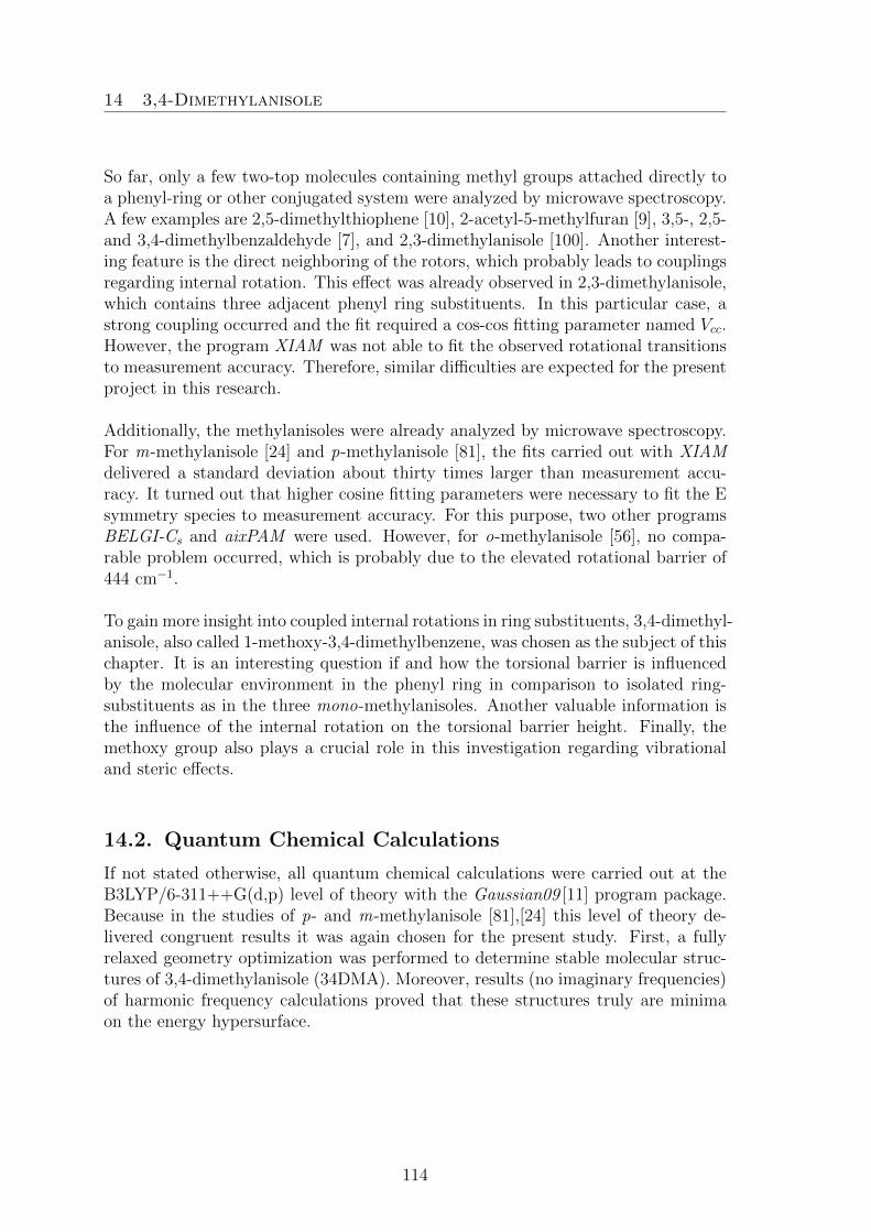
14 3,4-Dimethylanisole
So far, only a few two-top molecules containing methyl groups attached directly toa phenyl-ring or other conjugated system were analyzed by microwave spectroscopy.A few examples are 2,5-dimethylthiophene [10], 2-acetyl-5-methylfuran [9], 3,5-, 2,5-and 3,4-dimethylbenzaldehyde [7], and 2,3-dimethylanisole [100]. Another interest-ing feature is the direct neighboring of the rotors, which probably leads to couplingsregarding internal rotation. This effect was already observed in 2,3-dimethylanisole,which contains three adjacent phenyl ring substituents. In this particular case, astrong coupling occurred and the fit required a cos-cos fitting parameter named Vcc.However, the program XIAM was not able to fit the observed rotational transitionsto measurement accuracy. Therefore, similar difficulties are expected for the presentproject in this research.
Additionally, the methylanisoles were already analyzed by microwave spectroscopy.For m-methylanisole [24] and p-methylanisole [81], the fits carried out with XIAMdelivered a standard deviation about thirty times larger than measurement accu-racy. It turned out that higher cosine fitting parameters were necessary to fit the Esymmetry species to measurement accuracy. For this purpose, two other programsBELGI-Cs and aixPAM were used. However, for o-methylanisole [56], no compa-rable problem occurred, which is probably due to the elevated rotational barrier of444 cm−1.
To gain more insight into coupled internal rotations in ring substituents, 3,4-dimethyl-anisole, also called 1-methoxy-3,4-dimethylbenzene, was chosen as the subject of thischapter. It is an interesting question if and how the torsional barrier is influencedby the molecular environment in the phenyl ring in comparison to isolated ring-substituents as in the three mono-methylanisoles. Another valuable information isthe influence of the internal rotation on the torsional barrier height. Finally, themethoxy group also plays a crucial role in this investigation regarding vibrationaland steric effects.
14.2. Quantum Chemical Calculations
If not stated otherwise, all quantum chemical calculations were carried out at theB3LYP/6-311++G(d,p) level of theory with the Gaussian09 [11] program package.Because in the studies of p- and m-methylanisole [81],[24] this level of theory de-livered congruent results it was again chosen for the present study. First, a fullyrelaxed geometry optimization was performed to determine stable molecular struc-tures of 3,4-dimethylanisole (34DMA). Moreover, results (no imaginary frequencies)of harmonic frequency calculations proved that these structures truly are minimaon the energy hypersurface.
114

14 3,4-Dimethylanisole
The calculations mentioned above yielded two energetically stable and planar con-formers, which only differ by the orientation of the methoxy group. Phenyl ringsare known to be planar [27] which was also proven by numerous previous experi-ments e.g. phenetole [26], acetophenone [31], and toluene [92]. The co-existence oftwo conformers with a different orientation of the substituent also occurred in othermolecules such as phenylalanine [29], N -phenylformamide [28], [3] or m-methylanisole[24]. Therefore, the conformers were named cis- and trans-3,4 dimethylanisole, orconformer I and II, respectively. For trans-3,4-dimethylanisole the rotational con-stants A = 2882.189955 MHz, B = 850.787600 MHz, and C = 665.055255 MHz wereobtained. The following dipole moments were calculated: µa = 0.18 D, µb =−0.90 D,and µc = 0.00 D. For cis-34DMA the rotational constants A = 2618.548868 MHz,B = 903.649096 MHz, and C = 680.361605 MHz and the following dipole momentsµa = −0.15 D, µb = 1.58 D, and µc = 0.00 D were calculated. Therefore, mainlyb-type transition signals are expected in the microwave spectrum. The optimizeddihedral angles are α = 0.15° ,β = 0.02°, γ = 0.04°, δ = 179.88° for cis-34DMA (I),and α = 179.97°,β = 0.01°, γ = 0.01°, δ = −179.96° for trans-34DMA (II). Fur-thermore, anharmonic frequency calculations were carried out to provide theoreticalcentrifugal distortion constants.
12
14
2
5
16
18
1
3
10
15
4
22
13
69
8
7
19
20
14
15
16
17
1
2
3
4
5
6
9
8
131012
7
19
22
20
I II
α
β
γ
δ
Figure 14.1. Molecular structures with atom labeling of 3,4-dimethylanisole. Carte-sian coordinates of these configurations are listed in Table 26.4.1. Quantum chemicalcalculations were carried out at the B3LYP/6-311++G(d,p) level of theory. The cis-and trans-conformers possess energies of Ecis = −425.5211543 Hartree (with ZPE-correction: EZPE = −425.334008 Hartree) and Etrans = −425.5215701 Hartree (withZPE-correction: EZPE = −425.334355 Hartree), respectively. The cis-conformer is1.09 kJ/mol higher in energy than the trans-conformer (see also the potential in Fig-ure 14.2). The dihedral angles α = ∠(C2−C1−O14−C15), β = ∠(C2−C3−C19−H22),γ = ∠(C5−C4−C10−H13), and δ = ∠(H16−C15−O14−C1) correspond to rotationsabout the C1−O14, C3−C19, C4−C10, and C15−O14 axes, respectively. For con-former I the optimized dihedral angles are α= 0.15° ,β = 0.02°, γ = 0.04°, δ = 179.88°,
115
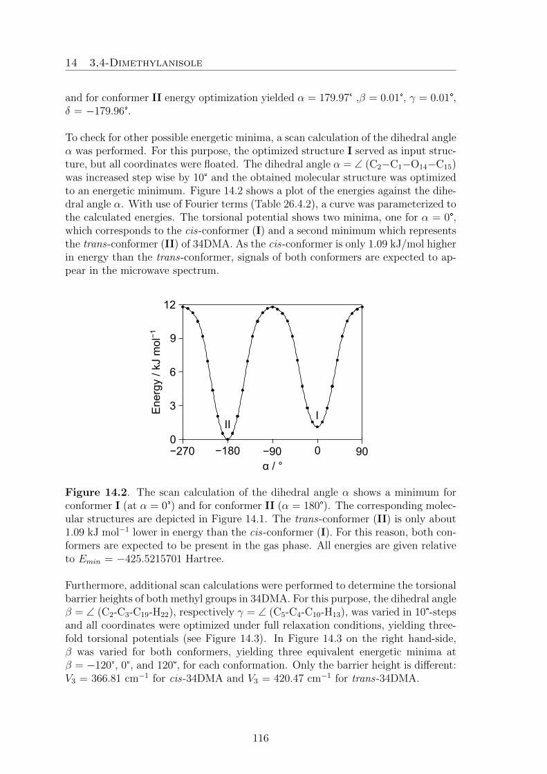
14 3,4-Dimethylanisole
and for conformer II energy optimization yielded α = 179.97° ,β = 0.01°, γ = 0.01°,δ = −179.96°.
To check for other possible energetic minima, a scan calculation of the dihedral angleα was performed. For this purpose, the optimized structure I served as input struc-ture, but all coordinates were floated. The dihedral angle α = ∠ (C2−C1−O14−C15)was increased step wise by 10° and the obtained molecular structure was optimizedto an energetic minimum. Figure 14.2 shows a plot of the energies against the dihe-dral angle α. With use of Fourier terms (Table 26.4.2), a curve was parameterized tothe calculated energies. The torsional potential shows two minima, one for α = 0°,which corresponds to the cis-conformer (I) and a second minimum which representsthe trans-conformer (II) of 34DMA. As the cis-conformer is only 1.09 kJ/mol higherin energy than the trans-conformer, signals of both conformers are expected to ap-pear in the microwave spectrum.
−90 0 90−180−270α / °
0
3
6
9
12
Ene
rgy
/ kJ
mol−
1
III
Figure 14.2. The scan calculation of the dihedral angle α shows a minimum forconformer I (at α = 0°) and for conformer II (α = 180°). The corresponding molec-ular structures are depicted in Figure 14.1. The trans-conformer (II) is only about1.09 kJ mol−1 lower in energy than the cis-conformer (I). For this reason, both con-formers are expected to be present in the gas phase. All energies are given relativeto Emin = −425.5215701 Hartree.
Furthermore, additional scan calculations were performed to determine the torsionalbarrier heights of both methyl groups in 34DMA. For this purpose, the dihedral angleβ = ∠ (C2-C3-C19-H22), respectively γ = ∠ (C5-C4-C10-H13), was varied in 10°-stepsand all coordinates were optimized under full relaxation conditions, yielding three-fold torsional potentials (see Figure 14.3). In Figure 14.3 on the right hand-side,β was varied for both conformers, yielding three equivalent energetic minima atβ = −120°, 0°, and 120°, for each conformation. Only the barrier height is different:V3 = 366.81 cm−1 for cis-34DMA and V3 = 420.47 cm−1 for trans-34DMA.
116

14 3,4-Dimethylanisole
The variation of the other dihedral angle γ, representing a rotation of the methylgroup about the C4−C10 axis, delivers the threefold torsional potential shown inFigure 14.3 a). The minima occur at γ=−120°, 0°, and 120°, while all other coor-dinates stay invariant, as in the scan calculation of β. Again, the torsional barrierheights are different for the two conformers: V3 = 394.66 cm−1 for cis-34DMA (I)and V3 = 447.05 cm−1 for trans-34DMA (II). For the rotation of the para-methylgroup (C10,H11,H12,H13) both theoretical values are higher than for the meta-methylgroup rotation (C19,H20,H21,H22) (see Figure 14.3 b)).
−180 0−120 −60 60 120 1800
100
200
300
400
500
Ene
rgy
/ cm−
1
γ / °−180 1800−120 −60 60 1200
100
200
300
400
500
III
β / °
a) b)
Figure 14.3 a). Potential energy curves for conformers I and II obtained by varyingthe dihedral angle γ at the B3LYP/6-311++G(d,p) level of theory. CorrespondingFourier terms are given in Table 26.4.3. The threefold torsional potentials reach en-ergetic minima at γ = −120°, 0°, and 120°. The calculated torsional potentials areV3 = 394.66 cm−1 (I) and V3 = 447.09 cm−1 (II). Energies relative to the energeticminima of E = −425.5211543 Hartree (I) and E = −425.5215701 Hartree (II) aregiven. Figure 14.3 b). Threefold torsional potentials obtained by variation of thedihedral angle β, which is translated by a rotation about the C3−C19 axis. Threeenergetic equivalent minima occur for both conformers at β = −120°, 0°, and 120°,corresponding to the orientation of the three Hydrogen atoms H20, H21, and H22 tothe rest of the molecule. The other dihedral angles α, γ, and δ stay almost invariantduring the scan calculation. Fourier terms are given in Table 26.4.3. For cis-34DMAthe barrier height V3 is 366.90 cm−1 and for trans-34DMA V3 = 420.56 cm−1. Rel-ative energies to the lowest conformations of I and II with the absolute energies ofE = −425.5211543 Hartree and E = −425.5215700 Hartree are used.
117

14 3,4-Dimethylanisole
Finally, the torsional potential of the methoxy methyl group (C15,H16,H17,H18) wascalculated. Another scan calculation of the dihedral angle δ = ∠(H16−C15−O14−C1)was performed. Again, both conformers possess three equivalent minima at the samecoordinates: δ = 60°, 180°, and 300° (Figure 26.4.2). During the variation of δ, themethoxy group turns out of plane (variation of α by up to 35°) until a maximumin energy is reached. Then, with the next 10° step, it suddenly flips back into thering plane (α = 0° or 180°). Therefore, the torsional potentials are not symmetric.The corresponding Fourier terms are given in Table 26.4.3. The barrier heights ofV3 = 1055.63 cm−1 for cis-34DMA and V3 = 1072.59 cm −1 for trans-34DMA weredetermined. From previous studies on methylanisoles [56], [24],[81], it is known thatsuch elevated torsional barrier heights lead to very narrow signal splittings in themicrowave spectra, often not resolvable by the instrumentation in use.
14.3. Results
Because of the very small dipole moment along the a-axis of only 0.2 D, only a fewa-type transitions were measurable and often required more than 10000 repetitionsto resolve all the signal splittings. The signals were also very weak, which led to abroadened line-shape and consequently, to an elevated measurement accuracy.
To assign the rotational transitions to the measured frequencies, the program XIAMwas applied. First, the trans-conformer was treated as a rigid rotor problem. A spec-trum was predicted using the calculated rotational constants and dipole momentsgiven in the chapter about the quantum chemical calculation. A few lines wereassigned by fitting the linear combinations of the rotational constants A, B andC, starting with the intensive (00) species transitions of Ka= 0 and Ka=1. In thenext step, the molecule was treated as a two-top problem. Because of the smallsplittings (see Figure 14.4), it was very difficult to assign the different symmetryspecies correctly to the signals. The remaining (01), (10), (11), and (12) symmetryspecies were assigned in an unusual manner beginning with transitions of higher Ka.causing larger splittings in the recorded signals. Thus the five symmetry species aremore easily distinguishable. Later on, also lower Ka transitions were included inthe fit. Finally, also a-type lines were also fitted, but due to the very low dipolemoment of 0.2 D, only a few a-type lines could be resolved and a very large numberof data acquisition signals was necessary. Still those transitions remained very weakin intensity, and therefore were hard to find and to assign, because sometimes not allfive symmetry species were observable. Finally 61 (00), 65 (01), 64 (10) 59 (11), and64 (12) species lines were successfully assigned and fitted to 4.2 kHz. The symmetryspecies label (σ1σ2) corresponds to (Me4,Me3). The molecular parameters of this fitare given in Table 14.1.
118

14 3,4-Dimethylanisole
(12)
(01) (11)
(10)
(00)
3299.487 3299.662 3299.838
111 ← 000
Figure 14.4. A typical spectrum of the b-type rotational transition 111 ← 000 ofthe cis-conformer of 34DMA. Due to the Doppler effect, each species splits into twolines embraced by a bracket labeled with the symmetry species. The signal splitsinto five symmetry species named (00), (01), (10), (11), and (12), corresponding tothe notation (σ1σ2) designating Me4 and Me3, respectively. The overall splitting isabout 350 kHz. For this spectrum, roughly 3000 decays were co-added.
The assigning and fitting approach for the cis-conformer (I) was similar. The (00)species of the rigid rotor were assigned straightforward. For the two-top model, thesmall symmetry species splittings also caused some difficulties, but after a while, theassignment was successful. In a final step, some a-type transitions were collectedand inserted in the fit. The global XIAM fit with a standard deviation of 3.2 kHzcontains 84 (00), 84 (01), 82 (10) 82 (11), and 83 (12) species lines. For this con-former, the lables are inversed: (σ1σ2) correspond to Me3 and Me4, respectively.
119

14
3,4-D
imethylaniso
le
Table 14.1. Molecular parameters of 3,4-dimethylanisole in the principal axis system obtained by the program XIAM. Calculatedvalues were obtained with the B3LYP method. The values on the left- and right hand-side correspond to Me3 and Me4, respectively.
Par.a Unit Fit conf. I Calc.b Fit conf. II Calc.b
A MHz 2616.26783(33) 2618.5487 2880.65412(30) 2882.1896B MHz 908.83471(19) 903.6490 855.06382(10) 850.7875C MHz 683.427404(67) 680.3615 667.855484(60) 665.0552∆J kHz 0.0177(16) 0.01628 0.01214(64) 0.01131∆JK kHz 0.0163(64) 0.00835 0.0365(29) 0.03646∆K kHz 0.42(16) 0.24893 0.401(25) 0.34957δJ kHz 0.00487(87) 0.00464 0.00308(37) 0.00269δK kHz 0.063(16) 0.04495 0.032(11) 0.01386V3 cm−1 424.16(41) 455.58(19) 366.81 394.66 491.29(35) 519.68(28) 420.47 447.05F0 GHz 160.00c 160.00c 160.00c 160.00c 160.00c 160.00c 160.00c 160.00c
∠(i, a) ° 62.86(15) 11.45(53) 59.62 2.50 48.83(19) 23.85(36) 44.42 17.50∠(i, b) ° 27.14(15) 78.55(53) 30.38 87.50 41.17(19) 66.15(36) 45.58 72.50∠(i, c) ° 90.00c 90.00c 90.00 89.98 90.00c 90.00c 89.99 90.00Nd / 312 / 426 /σe kHz 4.6 / 4.4 /
aAll parameters refer to the principal axis system. Watson’s A reduction and Ir representation were used.bCentrifugal distortion constants obtained from anharmonic frequency calculations at the B3LYP/6-311++G(d,p) level of theory,all other values from geometry optimizations at the MP2/6-311++G(d,p) level.cFixed due to Cs symmetry.dNumber of lines.eStandard deviation of the fit.
120

14 3,4-Dimethylanisole
A total of 728 lines (313 lines for cis-34DMA and 415 lines for trans-34DMA) werefitted to measurement accuracy, yielding trustworthy values for the fitting parame-ters. No intense lines remained unassigned in the broadband scan (Figure 14.5).
9.0 10.5 12.0 13.5
Frequency / GHz
Inte
nsity
/ a.
u.
III
exp.
sim.
Figure 14.5. The upper trace recapitulates a recorded part of the broadband scan,while the lower trace shows the simulations performed with XIAM. No intensivelines remained unassigned. Weak vapor pressure and dipole moments induced anelevated signal to noise ratio. With an increased number of repetitions for the highresolution measurements, lines of weak intensity could also be resolved and fitted.
A total of 728 lines (313 lines for cis-34DMA and 415 lines for trans-34DMA) werefitted to measurement accuracy, yielding trustworthy values for the fitting parame-ters. No intense lines remained unassigned in the broadband scan (Figure 14.5).
14.4. Discussion
In summary, the molecule under investigation is well described with fitting param-eters in XIAM such as the rotational constants A, B, and C, which are determinedwith high accuracy. The fitted and calculated values are in agreement, with a max-imal deviation of 0.6%. The centrifugal distortion constants ∆J , ∆JK , ∆K , δJ ,and δJ are not as accurately determined as other parameters, but for high torsionalbarriers, this is a known effect [34]. Nevertheless, the errors are about one or twoorders of magnitude smaller than the fitted values, which are consistent with thetheoretical centrifugal distortion constants obtained by anharmonic frequency cal-culations at the B3LYP method. Only for δK , the fitted values differ from the
121

14 3,4-Dimethylanisole
calculated values. The fitted angles ∠(i,a), ∠(i,b), and ∠(i,c) also are in agreementwith the results from quantum chemical calculations. F0 was fixed to theoreticalvalues obtained by ab-initio calculations. In 34DMA, experimental torsional barrierheights between 430 cm−1 and 534 cm−1 were obtained. The two conformers possessdifferent values for V3, which means that the orientation of the methoxy group is in-fluencing the torsional barrier height. In the past, this has also been observed for themono-substituted m-methylanisole [24]. The rotational barrier heights for the cis-conformation are about 70 cm−1 lower in energy than for trans-3,4-dimethylanisole.In both conformers, V3 of the para-methyl rotor is also larger than for the methylgroup in meta position (by approx. 35 cm−1).
The experimental values are significantly different from the calculated values. Forcis-34DMA, V3 = 430.00(37) cm−1 (Me3) is about 65 cm−1 larger than the calculatedvalue. This is also the case for the second rotor (Me4) with V3 = 467.90(17) cm−1,which surpassed the theoretical value by roughly 70 cm−1. In case of trans-34DMA,the deviances are even greater: the torsional barrier height for the meta-methylrotor of V3 = 499.64(26) cm−1 is 80 cm−1 larger than expected, and for the para-methyl group the barrier height of 533.54(22) cm−1 surpasses the theoretical valueby 85 cm −1.
Table 14.2. Summary of torsional barriers in cm−1 of methyl rotors in methylanisoleand benzaldehyde isomers.
molecule cis-meta trans-meta param-methylbenzaldehyde 35.925(3)[61] 4.64(3)[61] —p-methylbenzaldehyde — — 28.37[85]cis-3,4-dimethylbenzaldehyde 508.12(1.08)[7] — 550.74(8.82)[7]trans-3,4-dimethylbenzaldehyde — 454.06(1.34)[7] 480.64(4.35)[7]m-methylanisole 55.769(16)[24] 36.615(12)[24] —p-methylanisole — — 49.6370(1)[81]cis-3,4-dimethylanisole 424.16(41)a — 455.58(19)a
trans-3,4-dimethylanisole — 491.29(35)a 519.68(28)a
aResults from this work.
Compared to the one-top molecule m-methylanisole (see Table 14.2), the barrier ofinternal rotation of the methyl group in meta position increases from 55.769(16) cm−1
to 430.00(37) cm−1, when a second methyl group is placed in para position. As aconsequence to the induced steric hindrance, the barrier height of this second methylgroups rotation also increases from 49.6370(1) cm−1 to 467.90(17) cm−1. The sametrend is observed in the second conformer, where the meta and para barriers increaseby 463 cm−1 and 480 cm−1, respectively.
122

14 3,4-Dimethylanisole
This work shows that rotational barrier heights are not only influenced by theirposition in the benzene ring, but also by the orientation and position of other sub-stituents (methoxy group and second methyl rotor). These trends were also observedin 3,4-dimethylbenzaldehyde (Table 14.2), and thus match the experimental resultsof this project.
123

14 3,4-Dimethylanisole
124

15 3,5-Dimethylanisole
15. 3,5-Dimethylanisole
Two Coupled Low-Barrier Large AmplitudeMotions in 3,5-Dimethylanisole studied by
Microwave Spectroscopy
180
0
−180180 3600
β / °
γ / °
0 %
20
40
60
80
100
10
3
2
4
6
16
8
10
3
2
4
6
γ
β
L. Ferres performed measurements, quantum chemical calculations, assignments ofthe microwave spectra and prepared the manuscript.
15.1. Introduction
Over the time, many investigations on internal rotation problems were carried outusing a combination of quantum chemical calculations and microwave spectroscopy.Thus, the microwave spectra of semi-rigid rotors like phenetole [26], one-tops for ex-ample o-methylanisole [56], or two tops e.g. pinacolone [91] were analyzed. However,only a few molecules with two rotors attached directly to π-systems were studied,among them three isomers of dimethylbenzaldehyde [7], 2-acetyl-5-methylfuran [9],and 2,5-dimethylfuran [8]. Moreover, several dimethylanisole isomers were exam-ined.
125

15 3,5-Dimethylanisole
The barrier to internal rotation varies in a very broad range. Thus, many combi-nations are possible. Some examples for the combination of high and low barriersare 2,4-dimethylanisole (chapter 12), 2,5-dimethylansiole (chapter 13), 2,3-dimethyl-ansiole [100], and 2,5-dimethylbenzaldehyde [7]. Molecules featuring two high tor-sional barriers also are common: syn- and anti -3,4-dimethylanisole (chapter 14),syn- and anti -3,4-dimethylbenzaldehyde [7], 2,5-dimethylfuran [8], and 2-acetyl-5-methylfuran [9]. Additionally, also a pair of low barriers has been found in somemolecules e.g. 3,5-dimethylbenzaldehyde [7] and 3,5-dimethylanisole (this chapter).A pair of intermediate barriers is also possible, as for instance in 2,5-dimethyl-thiophene [10].
In 2,6- (chapter 16) and 2,3-dimethylanisole [100], the three substituents are all di-rectly adjacent and as a consequence influence each other. Those top-top couplingsare not well understood yet and very little knowledge is available in literature. Inmost cases, the fits showed an elevated standard deviation, which was explainedvia the vibrations of the methoxy group. However, in 2,4- and 3,4-dimethylanisole,(chapters 12and 14) the rotational transitions were fitted to measurement accuracy.2,5-dimethylanisole (chapter 13) features two substituents separated by one sp2 car-bon atom, similar as in 3,5-dimethylanisole, the molecule under investigation in thischapter. In 25DMA, the (11) and (12) symmetry species led to complications inthe fitting process, yielding a standard deviation ten times larger than measurementaccuracy. Hence, investigations on a further dimethylanisole isomer are of greatimportance.
15.2. Quantum Chemical Calculations
All quantum chemical calculations presented in this paper were carried out with theprogram package Gaussian09 [11]. From previous investigations on other dimethyl-anisoles, it is known that the MP2/- and B3LYP/6-311++G(d,p) levels of theoryare well-suited for this type of molecule. Hence a geometry optimization was carriedout using the B3LYP method. The calculation converged in a planar molecularstructure with Emin= −425.5228388 Hartree, given in Figure 15.1. As no imaginaryfrequencies were obtained from harmonic frequency calculations, the true minimumis confirmed for this molecule. The hydrogen atoms H21, H12 and H17 lie in themolecular plane spanned by the carbon atoms, resulting in a geometry featuringH21 pointing away from O14, H12 pointing towards O14 and H17 pointing towardsthe C10 methyl group. A view along the ring-plane reveals that the methyl groupsare staggered.
To check for other existing conformers in the molecular beam, the dihedral angle αwas varied, corresponding to a rotation about the O14−C15 axis. At each step, theenergy was minimized, by optimizing the newly obtained molecular geometry. Fig-ure 15.2 shows the resulting energies of this scan-calculation, in combination witha fitted potential energy curve. Two minima are observable at α = 0° (I) and at
126

15 3,5-Dimethylanisole
α = 180° (I∗), describing the same molecular structure, as given in Figure 15.1. ForI∗, the methoxy group lies again in the molecular plane, but this time, with C15
pointing towards C10. Accordingly, the in-plane hydrogens were rotated about 60°,resulting in a superposable structure to I. No other conformer than the one shownin Figure 15.1 was found for 3,5-dimethylanisole.
1
2
3
4
5
6 141516
17
7
8
9
192221
1011
12
α
β
γ
δ
Figure 15.1 Molecular structure of 3,5-dimethylanisole with atom labels obtainedby geometry optimization at the B3LYP/6-311++G(d,p) level of theory. The atomcoordinates are given in Table 26.5.1. The following dihedral angles are com-monly used in this chapter: α = ∠(C2−C1−O14−C15), β = ∠(C2−C3−C19−H21),γ = ∠(C4−C5−C10−H12), and δ = ∠(C1−O14−C15−H17).
α / °
Ene
rgy
/ kJ
mol−
1
0 90 180−90 270
5
10
15
0I I*
Figure 15.2. Potential energy curve obtained by varying the dihedral angle α, yield-ing two equivalent minima I/I∗ at α = 0°/180°. B3LYP/6-311++G(d,p) was chosenas the level of theory to perform this calculation. The corresponding Fourier termsare given in Table 26.5.2. Energies are relative to Emin = − 425.5228388 Hartree.
127

15 3,5-Dimethylanisole
Next, a closer look on the internal rotors was taken. 3,5-dimethylansiole featurestwo methyl groups undergoing internal rotation, creating signal splittings in the mi-crowave spectrum. To analyze the rotational behavior in detail, scan calculationsof the dihedral angles β and γ were performed. As the experimental value for lowtorsional potentials is neither congruent with MP2 nor B3LYP theoretical values,both methods were considered in the matter of internal rotation.
The left hand-side of Figure 15.3 depicts the fitted potential energy curve matchingthe energies obtained by the pointwise optimization for the redundant dihedral an-gle β. B3LYP yields a threefold potential with three equivalent minima at β = 60°,180° and 300°. On the contrary, MP2 yields three non-symmetric double minimaat β = 50°/80°, 170°/200°, and 290°/320°, separated by a V6 of 6.72 cm−1. In thiscase, the left minimum a(Figure 15.3 left hand-side) is always slightly preferred.This is the only structural isomer of dimethylanisole exhibiting this effect; all otherring-methyl potentials were symmetric without exception. The absolute minima areobtained for β = 50°, 170° and 290°. The corresponding molecular structures aredepicted in Figure 15.4. The potential barriers were calculated by subtracting theenergy for the optimized transition state from the minimum structure energy. Thusa torsional barrier of 67.16 cm−1 was obtained for B3LYP, while MP2 estimated thevalue at 78.31 cm−1.
0 60 120 180 240 300 360
β / °
0
20
40
60
80
Ene
rgy
/ cm−
1
a
b
c
γ / °0 60 120 180 240 300 360
0
10
20
30
40
50B3LYPMP2
a* cb
a) b)
Figure 15.3 a) Scan of the dihedral angle β corresponding to a rotation of the cis-methyl group about the C3−C19 axis. Energies relative toEmin =−424.2441433 Har-tee (MP2) and Emin = −425.5228388 Hartree (B3LYP) are given. Figure 15.3 b)Potential energy curve obtained by rotation of the C10 methyl (trans) group aboutthe C5−C10 axis (variation of γ). The indicated energies are relative to the minimumenergies Emin = −424.2441390 Hartee (MP2) and Emin = −425.5228388 Hartree(B3LYP). Both diagrams were obtained by using the MP2- (blue curve) and theB3LYP/6-311++G(d,p) (black curve) levels of theory. The applied Fourier co-efficients for the parameterization of the potential energy curve are given in Ta-bles 26.5.3 and 26.5.4.
128

15 3,5-Dimethylanisole
Concerning the torsional barrier of the trans-methyl group, similar results were ob-tained: B3LYP yielded a threefold potential with minima at γ = 60°, 180°, and 300°.MP2 calculations resulted again in a non-symmetric double minimum potential withminima at γ = 40°/80°, 160°/220°, 280°/320°. The structures are also depicted inFigure 15.4. For this large amplitude motion (LAM), the barriers to internal rota-tion were calculated to 43.35 (B3LYP) cm−1 and 49.43 (MP2) cm−1.
10
3
2
4
6
17
8
10
3
2
4
1
6
17
8
10
3
2
4
6
1518 16
17
9
2
4
19
20
21
22
107
1311
17
1518 16
9
2
192122
20
7
13 1110
1518 16
17
9
2
19
2122
20
7
1311
10
4
a b c
16
8
10
3
2
4
6
16
8
10
3
2
4
1
6
a* c*
17
1618 15
9
2
19
2122
20
7
1113
10
4
18
17
15 16
9
2
2119
22
20
710
1311
Figure 15.4 View along the C3−C6 axis, revealing five geometries of 3,5-dimethyl-anisole, differing in the orientation of the cis- and trans- methyl groups. The struc-tural geometries a/a∗ (staggered methyl groups) and c/c∗ (eclipsed methyl groups)were obtained by the use of the MP2 method, structure b is the result of the B3LYP-optimization.
In order to analyze internal rotation coupling, a two dimensional potential energysurface (see Figure 15.5) was calculated at the MP2/6-311++G(d,p) level of the-ory. This level of theory was chosen, as it yields asymmetric double minimumone-dimensional potential energy curves (Figure 15.3). Therefore, a colour contourplot was chosen to map the energies. A bobbin like pattern is obtained for theenergetically lowest 10 % domain. In each bobbin-shaped 10 % minimum area, twodiagonally opposed minima are visualized by increasing the resolution. Note thatthe energy scale is a non-linear scale. The 2D-PES also clarifies that the “left” aminimum in Figure 15.3 is always the lowest one, but appears in the 2D-PES alsoas a∗ on the “right hand-side” of the double minimum.
The enantiomeric pair a/a∗ features two staggered methyl groups, while the otherpair c/c∗ possesses eclipsed methyl groups. Thus, it is not surprising that the a/a∗
conformation is lower in energy than the c/c∗ conformation.
129

15 3,5-Dimethylanisole
180
0
−180
90
−90
180 3600 270
β / °
γ / °
0 %
20
40
60
80
100
90
ca
c*a*
180
0
−180
90
−90
180 3600 270
β / °
γ / °
90
b
MP2 B3LYPa) b)
Figure 15.5 Two-dimensional potential energy surfaces (2D-PES) calculated at theMP2/6-11++G(d,p) (left hand-side) and B3LYP/6-31++G(d,p) (right hand-side)levels of theory depending on the dihedral angles β and γ. The colour code indicatesthe energy in percent, relative to Emin = −424.244140 and Emax = −424.2436281Hartree, respectively to Emin = −425.5224753 Hartree and Emax = −425.5228387Hartree, for MP2 and B3LYP. The minima obtained via optimization calculationsare marked as a/a∗(global) and c/c∗(local). The corresponding Fourier terms aregiven in Table 26.5.5.
A scan calculation of the dihedral angle δ was also carried out, in order to deter-mine the barrier height to internal rotation of the methoxy methyl group. Vari-ation of this dihedral angle forces the methyl group out of plane. This has alsobeen observed for other di- and mono-methylansioles and was studied in detail byOnda et al. [32]. Thus, the barrier was calculated to be 1055.63 cm−1 (MP2) and1099.70 cm−1 (B3LYP), causing no resolvable splitting in the microwave spectrumof 3,5-dimethylanisole.
The rotational constants were calculated to A = 1736.1460 MHz, B = 1089.1004 MHz,and C = 677.7738 MHz. The theoretical dipole moments of µa =−0.48 D, µb = 1.18 D,and µc = 0.00 D led to the suggestion that strong b-type and weaker a-type signalsappear in the microwave broadband scan.
15.3. Results
First, the molecule was treated as a semi-rigid rotor, searching only for (00) specieslines in the microwave spectrum and fitting the set of the three rotational constantsA, B, and C, as well as the five centrifugal distortion constants with the programXIAM [22]. This fit converged without further complications and yielded well de-termined rotational parameters, coming along with a standard deviation of 3.3 kHz
130

15 3,5-Dimethylanisole
for 97 signals.
Next, the molecule was considered as a one-top problem. The cis-methyl groupwas assigned first, using the barrier obtained from quantum chemical calculationsas support. The low barrier to internal rotation causes very large splittings in themicrowave spectrum, causing a challenging assignment process. Starting the as-signment with the stronger b-type lines, the search for the 606 ← 515 branch waseffective, as this branch has the lowest splitting of all b-type lines. More lines andparameters were added progressively into the fit, floating the rotational barrier pa-rameters and the angle ∠(i,a) to the internal rotor axis. Finally, a data set with 203signals was fitted to 3.9 kHz and a torsional barrier height of 65.882570(37) cm−1
was determined.
For the second methyl-rotor, the same procedure was applied. Again, the B3LYPvalue of 43.34624 cm−1 served as the starting point. Again, large splittings occurredand the assignment was complicated. The higher order parameters Dpi2J , Dpi2K
and Dpi2− were of great help. Eventually, a standard deviation of 5.5 kHz was ob-tained for a fit containing 202 (00) and (10) signals. The V3 parameter was fittedto 45.844971(12) cm−1.
(12) (11) (01) (10) (00)
717 ← 616
(01)
707 ← 616
120kHz
10172.374
kHz kHz kHz
10189.105
121
10189.822
121
10190.903
121
kHz
10194.605
121
Figure 15.6. Signal splitting into the five expected symmetry species as a conse-quence due to two low-barrier internal rotations. This figure shows five individualhigh-resolution measurements with the assigned symmetry species appearing in theincreasing frequency order. For the 707 ← 616 rotational transition, the (11), (01)and (10) signals are situated close to each other, in comparison to the more distant(12) and (00) Doppler pairs. All five species show the same splitting amplitude of120 kHz.
131

15
3,5-D
imethylaniso
le
Table 15.1 Fitted and calculated molecular parameters of 3,5-dimethylanisole. The corresponding frequencies are given in Table24.5.6.
Par.a Unit Fit 00/01/10b Global Fit Calc.c
A MHz 1737.0865(15) 1737.05299(54) 1736.1460B MHz 1095.42369(95) 1095.40422(21) 1089.1004C MHz 680.545948(54) 680.546121(41) 677.7738∆J kHz 0.01166(50) 0.01163(50) 0.021055∆JK kHz 0.1004(30) 0.0982(30) 0.076055∆K kHz 0.106(13) 0.109(12) 0.009315δJ kHz 0.00276(25) 0.00277(25) 0.007545δK kHz 0.0442(21) 0.0435(21) 0.046307Vcc GHz / −297.5227(46) /V3 cm−1 37.204(48) 59.357(34) 36.10945(15) 58.57733(11) 43.34624 67.15924Dpi2J MHz −0.03856(18) 0.017046(81) 0.034832(26) 0.018629(23) / /Dpi2K MHz 0.20389(26) −0.06578(47) 0.20027(18) −0.05471(15) / /Dpi2− MHz −0.03526(18) 0.010347(80) −0.031455(27) 0.011953(24) / /F0 GHz 160.00d 160.00d 160.00d 160.00d 162.19d 161.95d
∠(i, a) ° 41.843080(54) 281.870452(39) 41.842641(53) 281.870948(36) 41.73 281.65∠(i, b) ° 131.84308(54) 11.870452(39) 131.842641(53) 11.870948(36) 131.73 11.65∠(i, c) ° 90.00d 90.00d 90.00d 90.00d 90.00 90.00N e / 308 370 /σf kHz 5.0 8.7 /
aAll parameters are given in the pricipal axis system using Ir representation. Watson’s A reduction was used. bThis Fit containsonly (00),(01) and (10) species transitions. cCalculations were carried out at the B3LYP/6-311++G(d,p) level of theory. Othervalues deduced from MP2 calculations are given in section 15.2. dFixed due to Cs symmetry. eNumber of lines. fStandarddeviation of the fit.
132

15 3,5-Dimethylanisole
With the newly obtained values for the barriers to internal rotation, a two top fitwith the program NTOP was achieved, predicting the frequencies for the (11) and(12) symmetry species. As expected, including these signals into the fit led to adrastically increased standard deviation. Here, fitting the cosine coupling parame-ter Vcc was essential, and decreased the standard deviation to 8.7 kHz. For a bettercomparison, another NTOP fit was established, containing only (00), (10) and (01)signals and no Vcc parameter (see Table 15.1).
15.4. Results and Discussion
In this chapter, 3,5-dimethylanisole was analyzed by means of quantum chemicalcalculations and microwave spectroscopy. Table 15.1 summarizes the newly gainedinsights into the rotational behavior of this compound.
The fitted molecular parameters of both fits are consistent and also in agreementwith the calculated values. The rotational constants A, B, and C, the higher or-der parameters Dpi2J , Dpi2K , and Dpi2−, and the angle to the internal rotation axis∠(i,a) are determined with very high accuracy. The centrifugal distortion constantsare also fitted accurately, given the problems occurring from the coupled LAM’s,causing the error of each fitted line to increase.
3,5-dimethylanisole features two meta-substituents with rather low rotational bar-riers of 36.10945(15) cm−1 and 58.57733(11) cm−1. These values are well con-sistent with the potential barrier heights of V3(trans) = 25.2504(41) cm−1 andV3(cis) = 53.0068(62) cm−1 observed for the analogous 3,5-dimethylbenzaldehydes [7].Compared to the conformers of the one-top molecule m-methylanisole [24] with55.7693(90) cm−1 (cis) and 36.6342(84) cm−1 (trans), this seems to be a reasonablevalue. In other dimethylanisoles, as for example 2,5-dimethylanisole (chapter 13),the m-methyl group has a barrier height of 65.71355(12) cm−1. These values are allin perfect agreement.
However, if another substituent is attached directly to the ring-methyl group, the V3
value changes significantly. This is the case for 2,3-dimethylanisole [100] and cis- andtrans- 3,4-dimethylaniolse (chapter 14), where it increases due to steric hindrancecaused by the adjacent rotors. Here, the values found for V3 are 518.7(1.2) cm−1
(23DMA), 430.00(37) cm−1 (cis-34DMA), and 499.64(26) cm−1 (trans-34DMA).In contrast to 3,5-dimethylanisole, the cis-conformer of 3,4-dimethylaniole pos-sesses lower barriers to internal rotations than the trans-conformer. Neverthelessin m-methylanisole, the same trend as in 3,5-dimethylansiole is observable: the cis-conformer features the higher torsional potential compared to the trans-conformer.
133

15 3,5-Dimethylanisole
9.5 10.5 11.5 12.5 13.5
Frequency / GHz
Inte
nsity
/ a.
u.
Figure 15.7 The upper trace shows a part of the recorded microwave broadbandscan reaching from 9.5 to 13.5 GHz. The lower trace depicts the simulated spectrumcontaining all five symmetry species marked with different colors: red for (00), greenfor (10), blue for (01), cyan for (11), and magenta for (12). Fitted molecular parame-ters deduced from the global fit in Table 15.1 served as input data for the simulation.
No intense signals remained unassigned in the microwave spectrum, as shown inFigure 15.7. However, more signals could be inserted into the fit, but this wouldalso drastically increase the standard deviation drastically. Thus, these attributionscannot be verified with absolute certainty and were therefore not included in theglobal fit given in Table 15.1. A solution for this problem could possibly be anothercoupling parameter or a parameter implementing the correction (higher order) onthe very large Vcc.
In summary, the microwave spectrum of 3,5-dimethylanisole was analyzed and ledto well-determined barriers to internal rotation of the ring methyl groups. However,the coupling motions complicated the fitting of the (11) and (12) signals. Therefore,some changes to the applied model are essential.
134

16 2,6-Dimethylanisole
16. 2,6-Dimethylanisole
2,6-Dimethylanisole – a Complex Three-RotorMolecule with Coupled Internal Rotations
L. Ferres performed the broadband scan measurements, a part of the quantumchemical calculations, assignments of the microwave spectra and preparation themanuscript. J. Spautz carried out further measurements to complete the final fitand calculated at various levels of theories in the scope of his bachelor thesis [112].
16.1. Introduction
The last remaining molecule of the dimethylanisole series is 2,6-dimethylanisole, inthe following abbreviated as 26DMA. As in 23DMA [100], the three substituents areadjacently attached to the phenyl ring. However, the order is different: in 26DMAthe methoxy group is situated in between the two methyl rotors, forcing the oxy-gen bound methyl group to tilt out of the plane formed by the heavy atoms of thephenyl ring. Compared to the mono methylanisole analogs, a tremendous change inbarrier heights was found in 23DMA. Clearly, the three substituents influence each
135

16 2,6-Dimethylanisole
other, leading to geared or coupled torsional motions, causing great difficulties inthe fitting routine. Thus a standard deviation of 120 kHz was obtained, exceedingthe measurement accuracy of 3 kHz. 26DMA is expected to behave in a similar way,resulting in a higher standard deviation because of the coupled large amplitude mo-tions (LAM) which occur during internal rotation.
So far, only a few two-rotor aromatic molecules were analyzed by microwave spec-troscopy, and even fewer featuring a rotor directly connected to the π-system. Oneexample is 23DMA, already mentioned in the paragraph above. Furthermore fourdimethylanisole isomers (chapter 14), (chapter 13), (chapter 12), (chapter 15) wereanalyzed via microwave spectroscopy and quantum chemical calculations. The ob-tained results were compared to the three methylanisoles [56], [24], and [81]. Theseinvestigations confirmed that in two-rotor anisoles the large amplitude motions ofthe individual rotors influence each other and that the positions of the substituentsplay a crucial role regarding the torsional barrier heights.
For three rotors, even less microwave spectroscopy studies exist. Most of them in-vestigated smaller molecules, as for example trimethylchlorosilane [113], trimethylio-dosilane [114], or (CH3)72
3 Ge79Br [115].
16.2. Quantum Chemical Calculations
As the positioning of three neighbored substituents on the phenyl ring is expectedto result in complex inter-molecular motions, quantum chemical calculations wereperformed to get a first idea of the molecular structure and dynamics in 26DMA.All calculations were carried out with the Gaussian09 [11] suite of programs. Themethods MP2 and B3LYP were combined with the basis set 6-311++G(d,p), be-cause it is known from previous investigations [116],[117] that those combinationsoften yield reasonable results. Moreover, other method-basis set combinations wereapplied, given the complexness of the molecule.
First, optimization calculations were carried out, in order to yield the energeticmost favorable conformation for 26DMA. Here, B3LYP and MP2 were both appliedand yielded contrary results, as shown in Figure 16.1. For both optimized struc-tures, the methoxy group is forced out of the phenyl ring plane, due to the sterichindrance caused by the two methyl groups on both neighboring ortho-positions.For B3LYP, the molecular structure Ia featuring a perpendicular mirror plane wasfound, with the methoxy group being tilted out of the ring-plane by about 92°.For MP2 however, the tilt-angle was found to be −100° and −83°, leading to twonon-superimposable structures, named Ib and Ib* in the Figure 16.1. Also, the ori-entation of both the methyl groups is not the same anymore; for Ib the hydrogenH11 lies in the ring-plane, while for Ib* hydrogen H20 is part of the molecular plane.For the Cv structure, both methyl groups are positioned in a symmetric orientation,more precisely rotated against the phenyl ring by ±14°.
136

16 2,6-Dimethylanisole
To verify that the optimized structures represent real minima in the energy surfaceof 26DMA, frequency calculations were carried out. The B3LYP/6-311++G(d,p)and B3PW91/6-311++G(d,p) levels of theory yielded both one imaginary frequencyfor Ia, which describes a vibration of the methoxy group, coupled to symmetric os-cillations of both o-methyl groups. Contrarily, for the B3LYP/6-31G(d,p) levelof theory, no imaginary frequency was obtained. However, as no other molecularconformations were found by using DFT methods, structure Ia must represent theminimum structure, even if the use of some basis sets yields imaginary frequen-cies. For the MP2/6-311++G(d,p) and MP2/cc-pVDZ levels of theory however, noimaginary frequencies were obtained. The MP2/cc-pVDZ level of theory yielded amolecular structure similar to conformation Ia.
968 75 13
11
16
96 78 15 3
11
20
Ia Ib Ib*
5
7
17
9
8
6
41
2 3
12
18
10
11
15
16
14
13
1921
20
142 6 191011
12
13
16 17
18 21
2022
56
41
2 3
7
9
8
192221
20
17
18 15
1614
10
11
12
19
21
2022
614
1817
16
210
12
11
13
56
41
2 3
7
9
8
10
1112
13
1417
18 15
16
19
20
21
968 75 13
11
10 19
21
20
22
6142
1311
1218
1716
α
β
γ
δ
Figure 16.1 Non-planar optimized molecular structures of 26DMA at the B3LYP/6-311++G(d,p) and MP2/6-311++G(d,p) levels of theory. The calculations yieldedthe conformations Ia for B3LYP and the enantiomers Ib/Ib* for MP2. The orien-tation of both o-methyl substituents is different, depending on the applied method.The four dihedral angles are defined as following: α = ∠(C5−C6−C19−H21), β= ∠(C3−C2−C10−H13), γ = ∠(C1−O14−C15−H16), and δ =∠(C2−C1−O14−C15).The atom coordinates in the principal axis system are given in Table 26.6.1.
In the next step, scan calculations were carried out. During a scan calculation, onedihedral angle is increased stepwise and the structure is optimized for each step.The obtained energies are plotted and fitted using a Fourier expansion, as shownin Figure 16.2. First the dihedral angle β was varied, corresponding to a rotationof the methyl group about the C2−C10 bond. As for this rotation, a variation ofother dihedral angles α, γ, and δ was observed, they were also examined in detailand plotted below the potential energy trace. For these potential energy curves,the B3LYP and MP2 methods were combined with the basis set 6-311++G(d,p).A main difference occurred for the minima regions: MP2 yielded double minimawith a V6 contribution of 27.81 cm−1, while B3LYP only calculated a single minima
137

16 2,6-Dimethylanisole
potential. This is in agreement with the results observed from the previously givenoptimizations. The double minima describe the enantiomeric pair structures Ib andIb*, while the single B3LYP minimum corresponds to the δ = 90° Ia conformation.The torsional barriers were calculated by taking the difference of the energies de-livered by optimizing the minimum and the transitions state structures, yielding215.44 cm−1 (MP2) and 183.48 cm−1 (B3LYP).
0
50
100
150
200
−180 −120 −60 0 60 120 180
Ene
rgy
/ cm
−
1
−105
−95
−85
−75
γ / °
57606366
δ / °
80
95
110
125
α /
°
β / °
−180 −120 −60 0 60 120 180
−180 −120 −60 0 60 120 180
−180 −120 −60 0 60 120 180
−35
−20
−5
10
MP2B3LYP
Figure 16.2 Scan calculation of the dihedral angle β in 26DMA. The correlationof the remaining three dihedral angles α, δ, and γ is given in the traces below. Thedefinition of the angles is given in Figure 16.1. The torsional barrier was calculatedto 183.48 cm−1 (B3LYP) and 215.44 cm−1 (MP2). The corresponding Fourier termsare listed in Table 26.6.2.
138

16 2,6-Dimethylanisole
0
100
200
300
400
500
0 60 120 180 240 300 360
Ene
rgy
/ cm
−
1
−115−100−85−70
0 60 120 180 240 300 360
δ / °
115125135145
0 60 120 180 240 300 360
β /
°
8595
105115125
0 60 120 180 240 300 360
α /
°
γ / °
MP2B3LYP
Figure 16.3 Variation of the dihedral angle γ. The other dihedral angles α, β, andδ correlate with the rotation of the methoxy methyl group, which is indicated in thelower traces in the Figure above. The definitions of the dihedral angles are givenin Figure 16.1. The torsional barriers of 413.40 cm−1 (B3LYP) and 473.10 cm−1
(MP2) for the methoxy methyl internal rotation were found. The correspondingFourier terms are listed in Table 16.6.3.
As can be deduced from Figure 16.2, all four dihedral angles correlate strongly.Thus, by varying β, the dihedral angle γ also oscillates by 10-20°. Additionally, achange of the dihedral angles δ and α by roughly 5° and 20-35°, respectively, wasobserved. The strongest correlation was found for the dihedral angles β and α, de-scribing the two methyl internal rotations. For α, two different scales were used, asthe orientation of the second methyl substituent relative to the other methyl groupdiffers tremendously, depending on the applied calculation method (for comparisonsee Figure 16.1, structures Ia and Ib/Ib*). In general, the variations of the otherdihedral angles are a little smaller for the B3LYP method, compared to the resultsobtained by MP2 calculations.
139

16 2,6-Dimethylanisole
Analogously, a scan calculation of the methoxy-methyl internal rotation was per-formed. The results are shown in Figure 16.3.
Obviously, the four dihedral angles also correlate when the methoxy methyl grouprotates. This is indicated by the variations of 30-40° for δ, 15-25° for β, and 15-30°for α. The coupling of γ and δ was also suggested for all the other dimethylanisoles,however, the rotational barrier of >1000 cm−1 is too elevated to resolve the signalsplitting in the microwave spectrum. For this reason, this coupling was not ana-lyzed experimentally, but quantum chemical calculations always suggested a changein the dihedral angle δ, causing the methoxy methyl group to tilt out of plane whenthe dihedral angle γ (corresponding to a rotation of the methoxy methyl group)is varied. For 26DMA, the internal rotation of the methoxy methyl group with abarrier height of ≈450 cm−1 will lead to larger splittings, which will be visible inthe microwave spectrum. Therefore, the correlation of γ and δ plays a crucial rolein this project. The angles observed for α and β differ for MP2 and B3LYP, whichis a consequence of the differing optimized molecular structures, depicted in Fig-ure 16.1. The torsional potentials are quite similar for both methods; no doubleminima were obtained for the methoxy methyl rotation. The barriers to internalrotation of V3 = 473.10 cm−1 (MP2) and 413.40 cm−1 (B3LYP) were determined.
180
0
−180
90
−90
0 180−180 −60 60
α /
°
β / °
0 %
20
40
60
80
100
180 3600 90 270
α /
°
γ / °
180
0
−180
90
−90
a) b)
Figure 16.4 a) 2D-PES of the rotation of both o-methyl groups calculated atMP2/6-311++G(d,p) level of theory. Figure 16.4 b) 2D-PES of the rotation ofone o-methyl and the methoxy methyl group calculated at MP2/6-311++G(d,p)level of theory. Non-linear color scales were used. The maximum and minimumenergies refer to −424.2408952 Hartree, and −424.2428499 Hartree for the left PES,and −424.2395022 Hartree and −424.2428465 Hartree for the right PES, respec-tively. The corresponding Fourier terms are given in Tables 16.6.4 and 16.6.5.
140

16 2,6-Dimethylanisole
Another method to visualize the coupling of several internal rotations is a two-dimensional potential energy surface (2D-PES). This was done for MP2 and B3LYP/6-311++G(d,p) levels of theory. However, only the MP2 results are presented here,as they are more interesting regarding the double minimum structure. To obtainsuch a 2D-PES, several scan calculations were repeated by fixing a second coordinateto a certain value (here the dihedral angles β and γ), while scanning the remainingcoordinate and calculating the energy for each molecular geometry. The energy isindicated by a color contour plot (Figure 16.4), mapped using a non-linear colorscale, in order to resolve the double minimum structure. Thus, the coupling of botho-methyl groups (α, and β, left hand-side in Figure 16.4) and the coupling betweenthe o-methyl and the methoxy methyl group (α and γ, Figure 16.4 right hand-side)were examined. No other conformers were found, and the results gained by the2D-PES are in agreement with the scan calculation results previously discussed.
16.3. Results
To perform first assignment attempts, calculated values from quantum chemistrywere inserted into XIAM [22] to yield a prediction for the rotational transitions inthe microwave spectrum. For starting values, B3LYP results were used, as theyoften proved to be well-suited for other dimethylanisoles. As the dipole momentswere calculated to µa = −0.44 D, µb = 0.00 D, and µc = 1.16 D, mainly c-typetransitions are expected in the microwave broadband scan. Moreover, the rotationalconstants A = 1651.566043 MHz, B = 1473.958677 MHz, and C = 829.558295 MHzwere inserted into XIAM to produce a simulated spectrum which is compared tothe experimental broadband scan. The broadband scan shows a very individual pat-tern, with alternating regions rich in a- and c-type lines followed by regions withoutsignals, as shown in Figure 16.5.
109 11 12
Frequency / GHz
13 14 15 16
Inte
nsity
/ a.
u.
10750.00 10760.00 10770.00 MHz
a) b)
Figure 16.5 a) The upper trace shows the recorded parts of the broadband scanin the frequency region reaching from 9 to 16 GHz. The spectrum contains concen-
141

16 2,6-Dimethylanisole
trated c-type (high intensity) and a-type (low intensity) regions. The lower traceshows the simulated spectra obtained by using the fitted molecular parameters fromthe two-top fit of the methoxy methyl and one o-methyl rotor. Figure 16.5 b)enlarged part of the microwave broadband scan showing two a-type rotational tran-sitions splitting into ten symmetry species. The signals appear very close to eachother and even overlap.
12580.100 12580.249 12580.399
432 ← 322
(100)
(120)(102)
(101)
Figure 16.6 A typical high-resolution measurement of a c-type rotational transi-tion of 2,6-dimethylanisole. The brackets indicate Doppler doublets. In one mea-surement, 4 Doppler pairs are occurring, stating that the symmetry species splittingis very narrow. The polarization frequency was 12580.250 MHz and the spectrumwas obtained by 50 co-added decays.
In the following, the quantum numbers were assigned to the measured frequencies,starting with the (000) symmetry species, treating 26DMA as a semi-rigid rotor. Toverify the assignment, high-resolution measurements were inserted into the fit, andthe centrifugal distortion constants DJ , DJK , DK , dJ and dK were fitted, thus de-livering a standard deviation of 2 kHz. Next, the molecule was treated as a one-topmolecule. Two assignments were achieved, one for the o-methyl internal rotationand one for the methoxy methyl rotor. Thus, the calculated barriers to internalrotation obtained via quantum chemical calculations together with the angles of therotor to the principal axis system ∠(I,a) and ∠(I,c) were inserted in XIAM and fittedin the following. Thus, two fits with a low standard deviation and well-determinedparameters were obtained. In the following, those fits were expanded by a secondmethyl rotor, by fitted for each rotor the rotational barrier and the angles of theinternal rotor axis to the principal axes separately, except for both equivalent methylgroups, which possess the same value for the torsional potential. Taking a look at
142

16 2,6-Dimethylanisole
the broadband scan section given in Figure 16.5 on the right hand-side, it becameobvious that both o-methyl groups are identical. If they were not, 14 symmetryspecies instead of 10 species would be expected to occur in the spectrum, leading toa further signal densification in the microwave spectrum.
The last step consisted in a three rotor contemplation of 26DMA. This was notso evident, as the symmetry species appeared close to each other in the microwavespectrum, and some signals in the previous fits were assigned incorrectly, which onlybecame clear in the very last assignment step where all the symmetry species weretaken into account. Finally, all signals were re-measured in the high-resolution mode(see Figure 16.6) and further rotational transitions were gathered outside the broad-band scan area to yield a fit comprising 837 signals fitted to a standard deviationof 7 kHz. The fit is given in Table 16.1 and a comparison of the fitted values tothe calculated ones will be drawn. An extended version of XIAM was used, able toimport 2000 transitions.
Table 16.1 Fitted and calculated molecular parameters of 2,6-dimethylanisole. Thecalculations were carried out at the B3LYP/6-311++G(d,p) level of theory. The Fitwas carried out with the program XIAM. The methyl rotors are indicated by theircarbon atom number. The frequencies as well as the residues of the XIAM fit canbe found in Table 26.6.6.
Par.a Unit Fit C19/C10/C15 Calc.b
A MHz 1650.99793(17) 1651.5659B MHz 1488.87977(17) 1473.9585C MHz 835.98819(82) 829.5582∆J kHz 0.0580(15) 0.055635∆JK kHz 0.3369(90) 2.35120∆K kHz −0.3073(91) −2.29214δJ kHz 0.01904(73) 0.018243δK kHz 0.1313(30) 0.55747F d
0 GHz 160.0 160.0 160.0 162.07 162.07 158.69V cm−1 199.0778(11) 457.440(31) 183.48 413.40∠(i, a) ° 61.6254(6) 52.902(23) 60.39 60.51 54.00∠(i, b) ° 150.7058(7) 29.2960(8) 90.111(90) 149.66 30.22 90.02∠(i, c) ° 96.6875(5) 96.6942(6) 37.098(24) 96.01 96.01 36.00Dpi2J MHz -17.30(16) — —Dpi2K MHz -19.24(17) — —Dpi2− MHz 0.12058(50) — —N e — 837 —σc kHz 5.8 —
aAll parameters refer to the principal axis system. Watson’s A reduction and Ir representation
were used. bCalculated at the B3LYP/6-311++G(d,p) level of theory. cStandard deviation of the
fit. dFixed due to symmetry. eNumber of lines.
143

16 2,6-Dimethylanisole
16.4. Discussion
Given the correlation determined in the quantum chemical part of this chapter, ahigher standard deviation was expected for the global fit of all ten symmetry species.However, a very high amount of lines (837 signals) were assigned and fitted to a stan-dard deviation of only 6 kHz. This is a very good result if the complexness of thelarge amplitude motions is taken into account. Probably, the measurement accuracyis also larger due to narrow and partially overlapping signal splittings.
The rotational constants were determined very accurately, coming along with a fullset of quartic centrifugal distortion constants, which are also fitted very precisely.This is probably a consequence of the large data set with a Jmax of 12 and a Ka,max
of 7. The barriers to internal rotation were found to be 199.0778(11) cm−1 and457.440(31) cm−1. Often, with such large or/and intermediate barrier heights, somemolecular parameters cannot be determined very accurately, because of the narrowsignal splitting. This is not the case for the 26DMA fit. All angles describing theaxis of the internal rotor to the principal axis are determined with high accuracy.The only parameters with an elevated error are the Dpi constants. This is not sur-prising, as a high barrier to internal rotation often does not allow a fitting of theDpi parameters. Here, the standard deviation is reduced from 62 to 6 kHz by fittingthe Dpi constants, and therefore they are included in the fit, even if they come alongwith a large error.
Overall, the calculated values are in agreement with the fitted parameters. Onlythe calculated centrifugal distortion constants DK and DJK differ by one order ofmagnitude. This probably is a consequence to the coupled LAMs in 26DMA. Thebarrier to internal rotation also differs, which is a known problem. For this reason,the torsional barriers were additionally calculated with the MP2 method in section16.2. The calculated angles to the internal rotor axis are in agreement with thefitted values, and therefore permit an identification of the three rotors.
If the torsional barriers are compared to other o-methyl barriers, as for exam-ple in o-methylanisole, no similar values were found. The barrier in 26DMA of199.0778(11)cm−1 is significantly smaller than the barrier in o-methylanisole of444.05(41) cm−1, or in anti -o-cresol of 371.05(41) cm−1. The methoxy barrier of457.440(31) cm−1 cannot be compared to other experimental methylanisole values,because they do not exist. The same applies to the methoxy methyl internal ro-tations in the remaining five dimethylanisoles. However, the barriers were alwayscalculated to be larger than 1000 cm−1. Clearly, the direct neighboring of the threesubstituents in 26DMA nearly halves all three barriers to internal rotation.
2,3-dimethylanisole (23DMA) [100] is another study case were three substituentsare attached adjacently to the phenyl ring. For 23DMA, it was found that the bar-rier to internal rotation in the o-position was reduced to 26.9047(5) cm−1, while them-methyl torsional barrier increased from 36.6342(84) cm−1 to 518.7(12) cm−1. Themethoxy methyl barrier was still too elevated to be resolved in the measurements.
144

16 2,6-Dimethylanisole
Therefore it is concluded that the orientation of the methoxy group also plays acrucial role, as the barrier of the methoxy methyl group was always >1000 cm−1
for a planar orientation, but decreased to ≈450 cm−1 for a perpendicular orientation.
Another molecule with two equivalent internal rotors is 2,5-dimethylthiophene [10].The determined barrier height is V3 = 247.95594(30) cm−1. As a further example fora two-top π-system molecule, 2,5-dimethylfuran [8] was analyzed, and the barrierto internal rotation for both equivalent methyl groups is V3 = 439.1461(83) cm−1.However, in both molecules, the two equivalent rotors are not directly adjacent toa third substituent or rotor. Nevertheless, no other three-rotor benzene was foundin literature, and therefore these molecules represent the most appropriate compar-ison. Regarding the value for the internal rotations, 2,5-dimethylthiophene shows asimilar torsional potential of 247.95594(30) cm−1 for the methyl groups as the onefound in 26DMA of 199.0778(11) cm−1. For the methoxy methyl internal rotation,no comparison was found, as the orientation in 26DMA represents an exceptionalcase.
In summary, the rotational transitions were well measured and fitted, especially con-cerning all the couplings of the large amplitude motions. The standard deviations of6 kHz is slightly increased, but if compared to the standard deviation in 23DMA, itis quite good. This is probably due to the more elevated barrier to internal rotationin 26DMA, causing narrow splittings in the spectrum and thus only small shifts dueto tunneling in the measured frequencies.
145

16 2,6-Dimethylanisole
146

17 Conclusion - Dimethylanisoles
17. Conclusion - Dimethylanisoles
The third part of this dissertation concerns the analysis of six dimethylanisol isomersusing a combination of quantum chemical calculations and microwave spectroscopy.The aim is to analyze the impact of a second rotor on the barriers to internal rota-tion and to compare this to the one-top methylanisoles (see Figure 17.1) reportedin part II. A short overview of the results and a thorough comparison is presentedin the following paragraphs.
As the six dimethylanisoles under investigation are constitutional isomers, similarmolecular properties are expected. However, this is not always the case. For in-stance, there are some significant differences regarding the vapor pressure of theindividual isomers. However, the vapor pressure values in Table 2.2.1 are only the-oretical values. While measuring, it was observed that some substances are morevolatile than others and therefore needed to be refilled more frequently, which con-firms the existence of different vapor pressures. In particular, 2,3-dimethylanisole isa great example as it solidifies at room temperature. This was not observed for theother five dimethylanisoles. Thus, the measurements of 2,3-dimethylanisole werecarried out using a heated nozzle, to avoid a crystallization.
O O OO444 cm−1
36 cm−1 56 cm−1
49 cm−1
Me
Me Me Me Me
Me Me
Me
100%
100% 100%100%
13 14 15 16
Figure 17.1 Recapitulation of the molecular structures and barriers to internalrotation of o-, m-, and p-methylanisole (13, 14 and 15). The corresponding chap-ters form the second part of this thesis. The rotational barriers of the individualmethylanisole conformers serve as a reference for the subsequent comparisons withthe dimethylanisole isomers, and therefore are each arbitrary fixed to 100%. Thecolors indicate the site of substitution and are helpful for the following comparisons.
2,4-dimethylanisole features, same as 2,5-dimethylanisole, one high and one low tor-sional barrier. A large amount of lines were measured (590 signals for 24DMA, and298 signals for 25DMA) and fitted with the program XIAM. However the standarddeviation was 10 times larger than the measurement accuracy. Therefore anotherprogram called N-TOP was applied, in order to find a necessary fitting parameter.Using these fitting parameters, 2,4-dimethylanisole can be described slightly betterthan 2,5-dimethylanisole. This is indicated by the standard deviations of 4.6 kHzand 21.2 kHz, respectively. The reasons for this are not clear and can only be
147

17 Conclusion - Dimethylanisoles
guessed. The fitting problems in 25DMA probably arise from the very similar angu-lar components of the internal rotation axis to the principal axes of both the methylrotors, which consequently become more difficult to distinguish for the programs.
The next molecule in this part is 3,5-dimethylanisole with two low-barrier internalrotors. None of them is orientated along an axis and both possess low barrier heightswhich complicates the assignment. Finally a fit of 3 MHz was reached. Again, NTOPwas applied to find an elementary fitting parameter. While excluding many linesfrom the fit, using the parameter Vcc helped greatly, leading to a standard deviationof 9 kHz for 270 signals. For this molecule, a model adjustment would probablybe the only measure to decrease the standard deviation and to fit all the assignedtransitions appropriately.
The experimentally determined barriers heights vary in a broad range. For 2,4-di-methylanisole (17), 2,5-dimethylanisole (18), and 3,5-dimethylanisole (19) the bar-riers to internal rotation only change slightly when compared to the correspond-ing mono-methylanisole torsional potentials (see Figure 17.2). Thus, the ortho-substituted barrier heights in 2,4- and 2,5-dimethylansiole as well as both meta-methyl torsional barriers in 3,5-dimethylanisole stay nearly invariant (∆V = 0−5%).For the para- and the cis-meta rotors in 24DMA and 25DMA, respectively, an in-crease of 27% and 18% was found, corresponding to only a few wavenumbers. There-fore these changes have a minuscule impact on the signal positions in the microwavespectra. Nevertheless, the values for the barriers to internal rotation of 62 cm−1 and66 cm−1 are in agreement with the respective barriers in similar molecules such asMMA (14, 15), PMA (16), and 35DMA (19). It is concluded that a second rotorin methylanisoles does not have a great impact on the remaining torsional barrier,if both rotors are separated at least by one sp2 carbon atom in the benzene.
O O OMe
Me
Me
Me
Me Me
Me Me
Me
436 cm−1 466 cm−1
− 2% + 5%
62 cm−1
+ 27%
66 cm−1
+ 18%
59 cm−1
+ 5%
36 cm−1
+ 0%
17 18 19
Figure 17.2 Molecular structures and experimentally determined barriers to in-ternal rotation of 24DMA (17), 25DMA (18), and 35DMA (19). The barriers arecompared to the methylanisole analogs (13), (14), (15) and (16) and the variationis indicated in percent. The colors indicate the substitution position (blue for ortho,green for cis-meta, magenta for trans-meta and red for para).
148

17 Conclusion - Dimethylanisoles
O OOOMe
Me
Me Me
Me
Me Me
Me
Me
Me
Me
Me
456 cm−1 520 cm−1
424 cm−1
491 cm−1 519 cm−1
27 cm−1 199 cm−1
457 cm−1
+ 930% + 1060%
+ 760%
+ 1360% + 1440%
− 94% − 55%
− 60%
20 21 22 23
Figure 17.3 Molecular structures and experimentally determined rotational barri-ers of cis-34DMA (20), trans-34DMA (21), 23DMA (22), and 26DMA (23). Theexperimentally determined rotational barriers are indicated in wavenumbers. Devi-ations relative to the methylanisoles in Figure 17.1 are indicated in percent.
In 3,4-dimethylanisole, two conformers occur, similar to m-methylanisole. This is aconsequence of the unsubstituted ortho- position, which allows the methoxy group toadopt two planar orientations, resulting in a cis(20) and a trans-conformation (21).Moreover, rather high torsional barriers were found for both conformers. However,as they are directly adjacent to each other in the phenyl ring, it is not quite pre-dictable how they interfere with each other. Clearly, a strong coupling occurs, asthe barriers to internal rotation drastically increase by up to one order of magnitude(760 - 1360 %). The assignment process was quite challenging, as the signal split-tings are very narrow and therefore complicate the assignment of the right symmetryspecies. In the end, no problems occurred during the fitting routines, as the barriersto internal rotation are high.
Considering 2,3-dimethylanisole (22), three adjacent substituents are attached tothe phenyl ring. The rotational barrier of the middle one decreases to 27 cm−1, cor-responding to a change of −94% if compared to OMA. The barrier of the m-methylgroup drastically increases by 1440%, compared to the trans-MMA barrier (14).Also, quantum chemical calculations state that the three methyl groups can notrotate independently, causing an elevated standard deviation of 118 kHz. Addition-ally, it remains unknown if and how the vibrations of the methoxy group affect thelarge amplitude motions of an adjacent rotor with a low barrier, as in all other di-and methylanisoles the directly adjacent rotor succumbs to a rather high torsionalbarrier (see 24DMA, 25DMA, and OMA).
The last molecule of this part is 2,6-dimethylanisole, which represents a three-rotorproblem. Unlike the remaining five dimethylanisoles, it is a non-planar molecule,as both o-methyl rotors next to the methoxy group force the latter out-of-plane toa perpendicular position, relative to the phenyl ring. Two of the three rotors areequivalent due to the molecular Cs symmetry. The barriers to internal rotation of
149

17 Conclusion - Dimethylanisoles
199 cm−1 and 457 cm−1 were found. For this purpose 10 very narrow-lying symmetryspecies are assigned in the microwave spectrum. The large amplitude motions of allthree rotors strongly correlate, which complicates the fitting process. Nevertheless,837 signals were fitted to a standard deviation of 6 kHz, using the program XIAM.26DMA constitutes a further exception as it is the only dimethylanisole isomer witha non-planar minimum structure. Consequently the molecular environment of theperpendicular methoxy group is symmetric to both sides and reduces the barrier tointernal rotation. Moreover, the barriers of the o-methyl groups are nearly halved,compared to the mono-substituted anisole OMA (13). This is a strong contrast tothe m-methyl barrier height in 23DMA (22), which increased by 1440%, as a con-sequence to the addition of a second adjacent rotor.
It is concluded that in the three dimethylanisoles 34DMA (20), 23DMA(22) and26DMA(23), the adjacent positioning of the methyl rotors has a very strong influenceon the barriers to internal rotation. Thus very high deviations to the methylanisolebarriers in Figure 17.1 were determined. The molecules with an intermediate ro-tational barrier, e.g. 34DMA and 26DMA, can be fitted to a standard deviationalmost in the range of the measurement accuracy. For 23DMA however, major fit-ting problems occur, due to the lowest torsional barrier of all the dimethylanisolesof 27 cm−1. As mentioned before, the problem occurs in all three molecules but dueto the higher barriers to internal rotation, the effect is negligible in 34DMA and26DMA.
CO H
CO H
CO H
CO H
CO H
CO H
CO H
Me
Me
Me
Me
Me
Me
MeMe
Me
Me Me
8 9 10 24
25 2627
36 cm−1 5 cm−1
28 cm−1
5 cm−1
566 cm−1
25 cm−1508 cm−1 53 cm−1454 cm−1
551 cm−1 481 cm−1
100% 100%
100%
−76%
+1410% −31%
+1970% +1720%
+9080% +1060%
Figure 17.4 Experimentally determined rotational barriers of several isomers ofmethylbenzaldehyde and dimethylbenzaldehyde. Deviances in the dimethylbenz-
150

17 Conclusion - Dimethylanisoles
aldehyde barriers relative to the methylbenzaldehyde barriers are indicated in per-cent.
The determined barrier heights in methylbenzaldehyde, confirm the postulated trendsin this chapter. If the methyl rotors are separated by at least one sp2 carbonatom, only a slight change occurs for the torsional potential as for example in2,5-dimethylbenzaldehyde (24) and 3,5-dimethylbenzaldehyde (27)(see Figure 17.4)compared to the mono-methylbenzaldehydes (8) and (9). However, if the rotor aredirectly adjacent to each other, the barrier heights change tremendously, as observedfor cis- and trans-3,4-dimethylbenzaldehyde (25) and (26). For the dimethylbenz-aldehydes, the increase was even greater than in the corresponding dimethylanisoles(20) and (21).
23DMA 24DMA 25DMA cis-34DMA
trans-34DMA
35DMA 26DMA
o− o− m− p− cism−
transm−
m− p−
m−
p−
o−
m−o−
OMe
ΔE
rel /
cm−
1
−20
−40
0
20
−60
−80
−100
40
60
80
MP2
B3LYP
exp
rotor position
Figure 17.5 Deviations of experimental and calculated rotational barriers of theseven dimethylanisole conformers. The barriers are designated with the labels o-,m-, p-, and OMe to distinguish between ortho, meta, para, and methoxy-methylrotors.
Regarding the quantum chemical calculations, mainly the MP2/6-311++G(d,p) andB3LYP/6-311++G(d,p) levels of theory were used for the dimethylanisole studies.For the assignment process, good starting values of the barriers to internal rotationare necessary, especially if low barriers are expected. However, Figure 17.5 showsthat it is not possible to predict which method works best for each molecule. Thebarriers were over- and underestimated by up to 100 cm−1, which has a great effecton the positions of the signals in the microwave spectra. For the dimethylanisoles in
151

17 Conclusion - Dimethylanisoles
Figure 17.2, with non-adjacent internal rotors, the best match between the resultsobtained by quantum chemical calculations and the experimental values is achieved.In case of the adjacent-rotor-molecules, the quantum chemical calculations obvi-ously again have problems to describe the torsional potentials accordingly, probablydue to the couplings of the large amplitude motions. The barriers were over- andunderestimated in equal measure and therefore it is of great importance to considerseveral levels of theory before starting the assignment of the microwave spectra.
In summary, the six dimethylanisoles were successfully analyzed by using microwavespectroscopy and quantum chemical calculations. The barriers to internal rota-tion were determined and the whole system, especially with the respective mono-methylanisoles, delivers an excellent comparison of the barriers to internal rotation.
152

Part IV.Substituted aromatic systemsfeaturing a double minimumpotential
O
O
O
O
Me S
O
MeH
153

154

18 Introduction
18. Introduction
This part focuses on quantum chemical calculations and microwave spectroscopicinvestigations on molecules with torsional large amplitude motions. For this pur-pose, the three molecules phenyl formate, phenyl acetate, and S -phenylthioacetatewere studied over several years.
J, K
J+1, K
+
+
-
- A
E
A
E
A
E
A
E
asymmetricrigid rotor
asymmetricrigid rotor with double minimumpotential
phenyl formate
asymmetric rigid rotor with double minimum potential and high barrier internalrotation
phenyl acetate
signals infrequency domain
a-type transitions
phenetole
Figure 18.2 Energy levels and microwave signal splittings for different types ofmolecular symmetry. Rotational a-type transitions are indicated as black arrows.
155

18 Introduction
Without an internal rotor, phenyl formate is the easiest project of the molecules inthe fourth part of this thesis. Therefore, the investigation was started by recordinga broadband scan of this molecule using microwave spectroscopy. During the assign-ment it turned out that the energy levels are shifted due to the tunneling motionsof the formate group. This is also supported by the results from quantum chemicalcalculations, which yield an enantiomeric pair as the most stable structure. Thus,the tunneling from one enantiomer to the other is allowed by quantum mechanics.To treat such a problem, a different approach has to be applied, as the Hamiltonianhas to be set up a in a more complex manner. For this purpose, the programs SP-FIT and SPCAT [110] were used. The Hamiltonian differs for each molecule, and istherefore not indicated here but in the respective chapters. Globally, it is importantto fit the energy difference ∆E as well as at least one Coriolis interaction parameterFab, Fbc or Fac.
Even if only the torsional ground state is observable, the energy levels are influencedby the tunneling motion and therefore the rotational constants slightly differ for eachtorsional state. Therefore, the difference between both energy levels is essential inorder to assign and fit the rotational transitions of the vt = 0 level adequately. Thiswas a very complicated procedure, as no similar study case can be found in literaturewith such a high ∆E value.
Ene
rgy
ϕ / °
vt = 0
vt = 1
vt = 2
+−+−
Figure 18.1 Double minima potential of a tunneling process. The torsional energylevels vt are not drawn to scale. Due to internal rotation, the levels split into A andE species. The A−E splitting is much smaller than the tunneling splitting betweenvt = 0 and vt =1. Therefore only rotational transitions with v′t ← vt of 0 ← 0 and1 ← 1 are detectable with microwave spectroscopy.
156

18 Introduction
Phenyl acetate and phenyl thioacetate were chosen to extend this row of moleculesas they supposedly exhibit a similar tunneling motion as the acetate and thioacetateparts. However, both molecules feature a methyl internal rotor, which additionallysplits the microwave signals into A and E symmetry species. Thus a pair of A/Especies signals is expected in the spectrum for the torsional level vt = 0. This split-ting is shown in the energy level scheme 18.2. More detailed information can befound in reference [18].
The energy levels of molecules featuring such an inversion motion, furthermore splitinto a (+) and a (−) state, due to a symmetric and anti-symmetric wave function, asfor example in ammonia [118] or in diethylamine [37]. Hence for phenyl formate, theenergy level difference is too large (2∆E = 96 GHz), rendering the (+)↔(−) tunnel-ing splitting non-observable in the frequency range accessible by the spectrometerin use. However, rotational transitions in the (+) level, referred to as vt = 0, and inthe (−) level, referred to as vt = 1, are observable in the microwave spectrum andfitted with the SPFIT program.
Unfortunately, no current program is able to fit a vt = 1 species, and therefore, theserotational transitions could not be fitted at the present state of this research.
18.1. Computer Programs
In this chapter, molecules with vibration-rotation interactions are examined. Theprograms Spin Fitting (SPFIT ) and Spin Cataloging (SPCAT ) [110], written byHerbert M. Pickett and published in 1990 through NASA COSMIC program ex-change [119], are well-suited to examine this kind of molecule. The main advantageof this program is the possibility to build up an individual Hamilton operator. Itenables thus to define any desired parameter and the program calculates very fast.Moreover several J-blocks permit the fitting and prediction of vt = 1 transition fre-quencies.The disadvantage of the fits comprising more than one torsional energy level is thehigh number of necessary parameters, because the splitting of the energy levels ∆Eand Coriolis coupling have to be taken into account. Unfortunately, only A speciesrotational transitions can be fitted, because low barrier internal rotation is hardlyfeasible with this program. For an A/E species fit, XIAM was used, as in the studiesbefore. However, a large standard deviation is expected for these fits.
157

18 Introduction
158

19 Phenyl Formate
19. Phenyl Formate
A part of this chapter has already been published in the Journal of Molecular Spec-troscopy [67].
L. Ferres, H. Mouhib, W. Stahl, M. Schwell,and H. V. L. Nguyen,Molecular structure and ring tunnel-ing of phenyl formate as observed bymicrowave spectroscopy and quantumchemistryJ. mol. spectrosc. 337,(2017), 59-64.
L. Ferres performed measurements, quantum chemical calculations, assignment andfitting of rotational transitions, and helped co-writing the manuscript.
19.1. Introduction
Phenyl formate (also called formic acid phenyl ester), H(C=O)OC6H5, with thestructure illustrated in Figure 19.1, is a molecule of chemical, biological, quantumchemical, and spectroscopic interest. Chemically, phenyl formate belongs to theclass of small esters, which are widely used in organic chemistry as a reagent for theformylation of amines [120]. In addition, small esters contain a number of commonodorant molecules. Many of these have been the object of our microwave studies,for example in Refs. [105], [80], [121], with a view toward determining dynamicaland/or conformational properties that might correlate with processes involved inthe sense of smell. Phenyl formate itself has a typical aromatic smell. It is knownthat odorants are carried by transport proteins which contain several amino acidsin their binding pocket, i.e. tryptophan, phenylanaline, and tyrosine [122]. Theseamino acids can bind the phenyl formate as an odorant over π - π interactions [123].The orientiation of the phenyl ring in odorants is strongly related to the bindingforce to those amino acids. Therefore, the exact molecular structure of phenyl for-mate also plays an import role in biology.
Quantum chemically, the relatively small empirical formular of C7H6O2 and thewell-known planarity of the phenyl ring [27] and the formate group [124], [125],[126] suggest that structure optimizations carried out for phenyl formate are withinthe capabilities of our computational resources. Because the traditional method byisotopic substitutions is not always possible, the support of quantum chemistry be-comes a helpful tool, whereby ab initio structures can be taken as references for acomparison of the experimental and calculated molecular parameters. As mentionedabove, the orientation of the phenyl group is important in many biological processes,but it is difficult to predict. Whenever possible, we often assume a plane of symme-
159

19 Phenyl Formate
try, i.e. the phenyl group is located in the plane formed by the heavy atoms. This isthe situation found for many phenyl ring containing molecules investigated by mi-crowave spectroscopy so far, such as anisole [32], phenetole [26], acetophenone [31],benzoyl fluoride [127], and benzaldehyde [30]. On the other hand, in some casesphenyl rings are reported to tilt out of the plane spanned by its neighboring heavyatoms, e.g. in cis-formanilide [3], N -phenylformamide [28] and acetanilide [33].Such tilt angle of a molecule fragment is not always easy to believe when they con-flict with chemical intuition and when they are based on the rather modest quantumchemistry calculations carried out in experimental spectroscopic laboratories, as e.g.in our investigations on allyl acetate [77]. As the present study unfolded, the phenylgroup is also tilted out of the H(C=O)O plane in phenyl formate.
Spectroscopically, phenyl formate is a derivative of formic acid, where the proton inthe acid group has been replaced by the much heavier phenyl group. When startingthis work, the spectrum of phenyl formate was expected to be essentially that of arigid-rotor with centrifugal distortion correction. However, this simple rigid-rotorexpectation turned out to be completely incorrect.
19.2. Quantum Chemical Calculations
All calculations were carried out at the MP2/6-311++G(d,p) level of theory withthe GAUSSIAN program package [11]. For a conformational analysis, the dihedralangles α = ∠(C13−O12−C3−C2) and β = ∠(H14−C13−O12−C3) were varied in agrid of 10° (for atom numbering see Figure 19.1), corresponding to the rotation ofthe phenyl ring about the O12−C3 bond and the rotation of the formyl group HC=Oabout the C13−O12 bond, respectively.
α = 72°
Ia Ia*
Ib Ib*
α = 112°
α =247° α = 288°
123
49510
11
71
1413
15 6
28 12
15
1314
3
8 2
4
1
9
6117
5 10
13
15
3
96
1
11105
21214 8
74
13
12
158
4102
93
14
176
11
5
Figure 19.1 Four energetically equivalent minima of the most stable trans con-former I of phenyl formate. The structures were tight-optimized at the MP2/6-311++G(d,p) level of theory. The dihedral angles α = ∠(C13−O12−C3−C2) are
160

19 Phenyl Formate
given. Note that the pairs Ia/I∗a and Ib/I∗b are enantiomers, while Ia and Ib as well asI∗a and I∗b can be transformed into each other by a rotation of 180° of the phenyl ring.
The two-dimensional potential energy surface (2D-PES) depending on α and β re-vealed two stable conformers; each of which appears as four equivalent minima.Because of the planarity of both the phenyl ring and the formyl group, the geome-tries represented by (α,β), (α+180°,β), (−α,−β), and (180°−α,−β) have the samepotential energy, and only a quarter of the full 2D-PES calculations are necessary.The calculated energies were parameterized using a 2D Fourier expansion based onterms with the correct symmetry of the angles α and β. The coefficients from thisparameterization are given in Table 27.1.1 in the supplementary material. Usingthese Fourier coefficients, the 2D-PES was drawn as a contour plot illustrated inFigure 19.2.
β / °
α /
°
-90 0 18090 2700
180
360
270
90
100
90
80
70
60
50
40
30
20
10
0 %
IIb*
IIb
IIa*
IIa
Ib*
Ib
Ia*
Ia
Figure 19.2 The potential energy surface of phenyl formate calculated at theMP2/6-311++G(d,p) level of theory depending on the dihedral angles α = ∠(C13−O12−C3−C2) and β = ∠(H14−C13−O12−C3). The color code indicates the en-ergy (in percent) relative to the energetically lowest conformations with an energyEmin = −419.7514621 Hartree (0%). The energy maximum corresponding to 100%is Emax = −419.7327553 Hartree.
If α is increased from 0° to 360° along the vertical lines at β = 0° or 180°, a series offour minima is found showing offset alternately lying slightly above and slightly be-low the α = 90° and α = 270° lines. The trans-conformer (conformer I) lying alongthe β = 180° line, occurs in four different versions, Ia (α ≈ 72°), I∗a (α ≈ 112°),Ib (α ≈ 247°), and I∗b (α ≈ 288°) on the PES. The closer lying adjacent minima(with ∆α ≈ 40°) are enantiomers, which are separated by a lower barrier of about28 cm−1, while the more distant adjacent minima (with ∆α ≈ 18°) arising from the180° rotation of the phenyl ring are separated by a higher barrier of 726 cm−1. This
161

19 Phenyl Formate
situation is depicted in Figure 19.3, where a one-dimensional “cut” along β = 180°of the PES is plotted. This cut is calculated by varying α from 0° to 360° at astarting value of β = 180°, while all molecular geometry parameters including β areoptimized. The coefficients from this potential curve are given in Table 27.1.2. Allfour versions of conformer I are shown in Figure 19.1. From a microwave spectro-scopic point of view, they are identical and possess the same rotational constants.The geometry of conformer Ia is subsequently re-optimized under full geometry op-timization, resulting in the rotational constants A = 3805.6 MHz, B = 1186.5 MHz,and C = 1039.9 MHz, and dipole moment components µa = 0.6 D, µb = −0.7 D,and µc = −1.3 D (in the principal axis system). The Cartesian coordinates are givenin Table 27.1.3 in the appendix chapter.
0 α / °
60 120 180 240 300 3600
200
400
600
800
Ener
gy /
cm
−1
Ia Ia* Ib Ib*
Figure 19.3 A one-dimensional “cut” of the potential surface in Figure 19.2 alongthe vertical line at β = 180° calculated by varying the dihedral angle α from 0° to360° at a starting value of β = 180°. All molecular geometry parameters including βare optimized at the MP2/6-311++G(d,p) level of theory. The closer lying adjacentminima (with ∆α ≈ 40°) are separated by a lower barrier of about 28 cm−1, whilethe more distant adjacent minima (with ∆α ≈ 180°) arising from the 180° rotationof the phenyl ring are separated by a higher barrier of 726 cm−1.
The dihedral angle α of the minima in Figure 19.1 is not 0° as expected for Cs sym-metry, i.e. the phenyl ring does not share a plane of symmetry with the H−(C=O)Oframe. The tilt angle is ± 72°, resulting in double energy potentials within the re-gions α = (60°−120°) and (240°−300°) (see also Figure 19.3). A similar tilt angleout of the (C=O)O plane was found for the isopropenyl group in isopropenyl ac-etate [111]. Another molecule with a double minimum potential is cis-formanilide.The phenyl group is rotated by 35° out of the plane containing the pseudo-peptidicgroup. The barrier to planarity is 152 cm−1[3]. A further example is acetanilide,which exists simultaneously as a planar and non-planar conformation. The non-planar compound has a phenyl ring tilted by 59° out of the C−(C=O)N plane,
162

19 Phenyl Formate
with almost exactly the same tunneling barrier of 151 cm−1 as in the case of cis-formanilide [3].
The barrier of 28 cm−1 separating Ia and I∗a as well as Ib and I∗b are very small com-pared to those found in cis-formanilide and acetanilide. Tunneling of the phenyl ringis therefore very probable. In spite of these quantum chemistry indications of po-tential trouble, the spectroscopic analysis was started, as described in Section 19.3,under the assumption that this tunneling effect arise from the phenyl ring motioncould be completely ignored, because the phenyl ring is heavy. When all attemptsat obtaining a good rigid-rotor fit to the spectral measurements failed, a closer lookwas taken at the quantum chemistry calculations. This will be discussed in detailin Sections 19.3 and 19.4.
The cis-conformer (conformer II), lying along the β = 0° line, can also be found infour different versions IIa, II∗a, IIb, and II∗b on the PES with essentially the sameα value as that of conformer I for each version. The geometry of conformer IIa isre-optimized under full geometry optimization, resulting in the rotational constantsA = 4885.9 MHz, B = 978.2 MHz, and C = 843.8 MHz, dipole moment compo-nents µa = −4.53 D, µa = 0.82 D, and µa = 1.37 D, and (α,β) = (60.5°, 5.6°).The Cartesian coordinates are also given in Table 27.1.3 in the appendix section.The cis-conformer is much higher in energy than the trans conformer (7.96 kJ/mol).This is in agreement with the results from previous investigations on esters [86], [79],[68]. Therefore, this conformer is not expected under the molecular jet conditions,where the rotational temperature is very low (approximately 2 K), and thus thepresent chapter focuses on the trans-conformer. Notably, the trans conformationsare defined as in Figure 19.1, where the phenyl group is in trans position to theproton of the formate group.
19.3. Microwave spectroscopy
19.3.1. 3.1. Spectral assignment of the ground state vt = 0
The calculated dipole moment components of the trans-conformer given in Sec-tion 19.2 suggest that all a-, b-, and c-type transitions are present in the microwavespectrum. First, it was searched for a-type transitions in the broadband scan us-ing a theoretical rigid-rotor spectrum predicted with the rotational constants alsogiven in Section 19.2, since these transitions follow typical patterns which can beoften recognized easily. Some lines following a-type selection rules were identified.The first fit attempts were made with the program XIAM [22] in its rigid-rotormode. Surprisingly, the assigned transitions could not be fitted well and the root-mean-square (rms) deviation was up to 3 MHz. Including the quartic centrifugaldistortion constants in the fit did not decrease the rms deviation, and moreoverthese parameters could not be determined well. This is unusual, because rms de-viations close to measurement accuracy can often be achieved in the fits of otherrigid-rotor molecules [41], [128], [129], [130]. Because of the typical a-type pattern
163

19 Phenyl Formate
in the microwave spectrum, the assignment was assumed to be correct. Using therotational constants of the preliminary fit, b- and c-type transitions were assignedby comparing the predicted and experimental spectrum. However, all attempts toreduce the rms deviation failed.
As mentioned in Section 19.2, it is very probable that some state other than theground state is populated in our 2 K molecular beam, which comes from the effectof ring tunneling. The primary support for the existence of a low-lying state justabove the vt = 0 ground state is the fact that after an exhaustive assignment oflines, a considerable number of transitions remained unassigned in the spectrum.They are much weaker than the assigned transitions, on the other hand alreadyquite strong that they are unlikely from the 13C isotopologues. This low-lying statewould then be a plausible candidate for a perturbation partner of the ground state.If we assume good thermal equilibrium in our jet, then the intensity of lines fromthis extra state suggests that it lies only about 10 cm−1 above the ground state,which in turn suggests that it is some tunneling component arising from the phenylring. Therefore the eigenvalues of the four lowest energy levels that would occurfrom the potential energy curve given in Figure 19.3 were calculated in an attemptto determine whether this assumption is correct.
19.3.2. 3.3. Calculations of low-lying tunneling states
The torsion of the phenyl ring against the formate group might cause low-lyingexcited torsional states which could be observed in the experiment. In order toestimate the energy separation between the vt = 0 and 1 states and the ratio ofthe population numbers of both states under our experimental conditions, a simplemodel of two rigid tops rotating against each other was applied. The expression forthe implemented Hamiltonian (in cm−1 units) is the following:
H=−F ∂2
∂α2 +V0+Σ7n=1[V2n cos(2nα)]
with the effective torsional constant F= h8π2Ic
=0.35101 cm−1. Here, c is the speed
of light and I=IpIfIp+If
=48.027 uA2 the effective moment of inertia calculated from
the moments of inertia of the phenyl group Ip=102.738 uA2 and the formate groupIf=90.186 u A2. Both of them refer to a rotation about the common torsional axisO12−C3 by the dihedral angle α. For the potential terms V2, V4,. . . , V14 the values340.99, 115.28, 4.16, 10.03, 2.67, 3.27, and 0.52 cm−1, respectively, were derivedfrom the parameterized potential curve given in Figure 19.3. The potential offsetwas chosen to be V2 = 247.79 cm−1, so that all potential minima are located at0 cm−1. The energy eigenvalues were obtained by a direct diagonalization of theHamilton matrix set up in the plane wave basis Ψm= 1√
2πeimα with m ε 0, ±1, ±2,. . . .
The matrix was truncated at |m| = 50, corresponding to a size of 101x101, whereconvergence for the lowest energy levels in the kHz range was achieved.
164

19 Phenyl Formate
The lowest energy levels are at 12.819 (0), 14.429 (1), 33.484 (2), and 44.344 cm−1
(3), all of them doubly degenerate. The torsional vt quantum numbers are given inparentheses. The degeneracy arises from the fact that the potential function consistsof a pair of double minimum potentials (see Figure 19.3) separated from each otherby high potential walls of 726 cm−1. No tunneling across these walls is observed andthe energy levels remain degenerated. The potential functions in the range from 0to 180° along with the lowest torsional levels are shown in Figure 19.4.
Ener
gy /
cm
−1
0
20
40
60
60 69.82 90 110.18 120
Ia Ia*
vt = 0
vt = 1
vt = 2
vt = 3
α / °
Figure 19.4 An enlargement in the range of 0 to 180° of the potential energy curvegiven in Figure 19.3, describing the torsion of the phenyl ring against the formategroup about the angle α. Horizontal lines indicate the lowest torsional energy levelsvt = 0, 1, 2, 3, which are doubly degenerate.
The vt difference for the two lowest torsional states is only 1.610 cm−1 or 48.27 GHz.This enables an estimation of the N1/N0 population ratio using the Boltzmann distri-
bution N1
N0=e
−∆EkT with the Boltzmann constant k and the temperature T. Assumed
that the torsional temperature is in the same temperature range of 1 - 2 K as therotational temperature in the jet, the population of the excited state vt = 1 liesbetween 9.9 and 31.4% with respect to the ground state vt = 0.
165

19 Phenyl Formate
19.3.3. Fits of the ground state vt = 0
At this point, it is convincing that the low-lying vt = 1 tunneling state is still pop-ulated in the jet-cooled spectrum that perturbs the ground state spectrum. There-fore the program SPFIT/SPCAT [110] is applied, available at the PROSPE website[131], which permits the user to choose Coriolis interaction terms for use in thefitting Hamiltonian. By trial and error a number of tunneling parameters and cen-trifugal distortion corrections were tested. At the beginning, the SPFIT/SPCATprogram did not provide a better rms deviation than the XIAM program (closeto 3 MHz). However, with gradually increasing number of parameters, especiallythe by including the tunneling parameters E, EJ , Fbc, Fac, and five sextic centrifu-gal distortion constants, among them the momentum cross terms Fbc and Fac weremost helpful, the situation improved. Molecular parameters were determined moreaccurately and rms deviation decreased to 3.7 kHz for a fit including 116 vt = 0rotational transitions with J 6 13 and Ka 6 2.
Including eleven a-type R-branch lines with Ka = 3 in the fit increased the rmsdeviation to 14.8 kHz. The fitted parameters of this fit consisting of 128 lines arecollected as Fit 0 in Table 19.1. A list of all fitted transitions is given in Table27.1.4; the Ka = 3 lines are marked by asterix. Attemps to improve the situationfor theses transitions by including more parameters were not sucessful.
19.3.4. Global fits of the vt = 0 and vt = 1 states
After the vt = 0 transitions were reasonably fitted using the program SPFIT/SPCAT,many weaker lines with intensity of about 1/5 to 1/10 located close to the most in-tense vt = 0 lines were found(see Figure 19.5). From a previous investigation onpinacolone [91], it is known that the low-lying vt = 1 excited state, if still populatedin the jet-cooled spectrum, is not far separated from the ground state. From theintensity calculations described in Section 19.3, the intensity of those weaker lines isin the correct order of magnitude as that predicted from the calculations. Therefore,these lines were assigned to the vt = 1 excited state transitions of the same quantumnumbers as those of the closest neighboring intense lines and thus 33 further linescould be identified. The global fit including 128 ground state vt = 0 lines and 33vt = 1 lines has a rms deviation of 54.5 kHz. The fitted molecular parameters aresummarized as Fit 01 also in Table 19.1. The frequencies along with their residuesare collected in Table 27.1.4. It is remarkable that a nearly complete set of quarticand sextic centrifugal distortion constants are needed separately for the excited statein sprite of the very limited number of lines in the fit. Including more parametersdid not help to reduce the rms deviation.
166

19 Phenyl Formate
Table 19.1. Fitted and calculated molecular parameters of phenyl formate.
Par.a Unit Fit 0 Fit 01 Calc.b
vt = 0 vt = 0,1A MHz 3838.160(12) 3899.83(22) 3836.97(22) 3733.074B MHz 1157.037(12) 1181.770(24) 1161.371(17) 1188.168C MHz 1034.813(15) 1047.539(11) 1045.8128(99) 1058.306∆J kHz −0.8837(38) −1.9010(34) −4.5174(69) 0.2530∆JK MHz −0.10513(31) −0.70918(59) 0.48479(62) 0.002649∆K MHz −0.9830(37) 3.3248(30) 2.086(16) −0.0004919δJ kHz −1.5880(14) −1.3566(32) 3.4393(22) −0.04523δK kHz −288.5(1.1) 65.83(39) −1.08(51) −0.1168E GHz 43.4233(80) 46.2231(25) 48.27c
EJ MHz 16.670(12) — —Fbc MHz −34.562(16) 34.3872(95) —Fac MHz 163.335(30) 168.201(19) —FbcK MHz — 1.8247(22) —FacK MHz — −3.8755(32) —Ga GHz — 1.3164(38) —Gb GHz — 0.64351(67) —HJ Hz −9.1462(96) — −0.0001499HJK kHz 0.36501(35) −0.90338(81) −0.7635(44) 0.00008028HK MHz −0.21843(77) 0.57522(69) 0.4811(30) 0.3106·10−6
HKJ kHz 1.6060(55) −41.872(42) 34.950(69) −0.3908·10 −6
hJ Hz — −3.9443(24) −0.1325·10−3
hJK kHz −1.2453(12) 3.5970(14) —Nd — 128 128/33 —rmse kHz 14.8 54.5 —
aAll parameters refer to the principal axis system. Watson’s A reduction and Ir representation
were used. bAnharmonic frequency calculations at the MP2/6-311++G(d,p) level of theory. The
rotational constants are the B0 ground state constants and are different from the Be equilibrium
constants given in Section 19.2. cSee Section 19.2. dNumber of lines. eRoot-mean-square deviation
of the fit.
19.4. Results and Discussion
The rms deviation of the ground state fit including 128 lines given as Fit 0 in Ta-ble 19.1 is 14.8 kHz. This deviation, which is seven times the measurement accuracy,is already quite successful compared to the fit using only a rigid-rotor model. Thekey parameters are Fbc = Pb,Pc and Fac = Pa,Pc, which have non-negligible values of−33.6 and 163.3 MHz, respectively. The Coriolis splitting ∆E of 43.4233(80) GHzis relatively close to the estimated value of 48.27 GHz (see Section 19.2).
167

19 Phenyl Formate
Though the fitted value of ∆E in the global Fit 01 is even closer to the calculatedone, the rms deviation of 54.5 kHz of this fit is unreasonably large comparing to themeasurement accuracy of 2 kHz or to that of the ground state Fit 0. A large numberof centrifugal distortion constants are needed for the fit, which is quite unusual fora fit of only low J and K transitions (i.e. J 6 13, Ka 6 3). The difference be-tween the rotational constants A, B, and C of the ground and the excited state areremarkable. Also almost all centrifugal distortion constants are in different ordersof magnitude. In other molecules, where similar tunneling problems were reported,e.g. benzyl alcohol [132], cis-formanilide [16], and acetanilide [33], these differencesfound for the rotational constants are significantly smaller, and the same set of cen-trifugal distortion constants can be used for both states.
These problems probably arise because the tunneling motion of phenyl formate isnot correctly captured by the present set of fitted parameters. Moreover the limitednumber of excited state lines, the relatively low tunneling barrier, as well as thelarge tilt angle of the phenyl ring (see Section 19.2) lead to further complications.Assigning and including more lines of the vt = 1 excited state might improve thefit quality. However, the measurements of these transitions under our jet-cooledconditions are rather surpassed because of the low line intensity and yet unreliableprediction. After excluding all assigned vt = 0 and vt = 1 lines, no intense linesremained in the broadband scan, as can be recognized in Figure 19.5.
8 10 12 1614
Inte
nsi
ty /
a.u
.
Frequency / GHz12.1 12.4
vt = 0
413 ← 303
62 MHz
vt = 1
Figure 19.5 A section from 8−16 GHz of the broadband scan (upper trace) ofphenyl formate compared to the theoretical spectrum reproduced with the SP-FIT/SPCAT program (lower trace). Transitions of the vt = 0 ground state aremarked in blue, vt = 1 excited state in red. Right hand side: A section from 12.1to 12.4 GHz of the broadband scan showing the vt = 0 and vt = 1 components ofthe 413 303 transition with a torsional splitting of 62 MHz.
168

20 Phenyl Acetate
20. Phenyl Acetate
Coupled Large Amplitude Motionsin Phenyl Acetate
α / °
β /
°
60 1800
180
Ia*
Ia
20.1. Introduction
Phenyl acetate (C6H5OCOCH3) is a colorless liquid with a plastic-like odor. It is of-ten used as a solvent in chemical reactions. Other names are phenyl ethanoate, phe-nol acetate, acetyloxybenzene or acetoxybenzene. It can be synthesized by addingacetic anhydride to phenole. [133] or via Baeyer-Villiger oxidation of acetophenone[134], or by decarboxylation of aspirine [135]. Currently, it is a drug studied in thetreatment of cancer. Naturally occurring in mammals, phenyl acetate induces differ-entiation, growth inhibition, and apoptosis in tumor cells. [136] As this compoundis of great interest in medicine, the knowledge of its exact molecular structure seemscrucial.
169

20 Phenyl Acetate
In the research group of prof. Stahl, many acetates have already been measured us-ing microwave spectroscopy. a few examples are n-propyl acetate [80], allyl acetate[77] or ethyl acetate [86]. For those acetates, a rotational barrier of approximately100 cm−1 was determined. Surprisingly, vinyl acetate [79] features a higher rota-tional barrier compared to the other acetates. The molecular structure of phenylacetate is very similar to the one of vinyl acetate, thus determining the rotationalbarrier of phenyl acetate is of high interest.
The previously studied phenyl formate [67] is structurally highly related to phenylacetate, and thus a very interesting project regarding the impressive phenyl-ringtilt angle of 72°. In the corresponding microwave spectrum, rotational transitionswere assigned for the, vt=0 and vt=1 torsional states, yielding a torsional energylevel splitting of ∆E = 46.2231(25) GHz, leading to a row of complications in thefurther assignment and fitting process. In the following, the molecular structure ofphenyl acetate is calculated using quantum chemical calculations yielding rotationalparameters. These parameters are used for a simulation of the microwave spectrum,which is compared to the recorded spectrum and assigned in the following using theprograms XIAM and SPFIT.
20.2. Quantum Chemical Calculations
Using the program package Gaussian09 [11], the molecular structure of phenyl ac-etate was optimized at the MP2/6-311G++(d,p) level of theory. Contrarily to allexpectations, the resulting structure is not planar (see Figure 20.1). Indeed, theacetate fragment is tilted out of the phenyl ring plane by ≈70°. In summary, fourconformers exist at (α, β ) = (121.6°, 72.2°), (120.3°, 111.5°), (121.6°, 248.6°), and(120.3, 287.8)°, yielding two pairs of enantiomers Ia/I∗a and Ib/I∗b , described by adouble minimum in the potential energy curve, as shown in Figure 20.2. Compar-ing the four optimized geometries to each other, the dihedral angle α stays nearlyinvariant.
Frequency calculations were carried out at the same level of theory, yielding animaginary frequency describing a ring bending motion. This has previously beenreported for the MP2 method [44]. Contrarily, harmonic B3LYP frequency calcu-lations state no imaginary frequencies for this conformer. The rotational constantsA = 3592.4315 MHz, B= 813.8922 MHz, and C = 744.3161 MHz, along with thethree dipole moments: µa = −0.41 D, µb = 0.90 D, and µc = −1.46 D were cal-culated. Thus, mainly c- and b-type transitions are expected in the microwavespectrum, possibly accompanied by some weak a-type signals.
170

20 Phenyl Acetate
9
12
42 57
1
10
8
11
16
18
13
17
156
14
3
β = 72.2° β = 111.5°
9
16
18
17
78
12
11
15
13
12
14
10
3
4
56
β = 248.6°1417
13 10
1518
11
16
9
12
8 712 3
6
54
β = 287.8°17
13
1518
11
16
9
12
8 71
2
10
14
4
3 56
IaIa
*
IbIb
*
Figure 20.1. Fully geometry optimization at the MP2/6-311++G(d,p) level oftheory yielded four stable C1-structures as depicted in this figure. The dihedralangles α = ∠(C2−C1−C3−C6) and β = ∠C1−O7−C8−C10) were optimized underfully relaxation of all other parameters. The four energetically equivalent structureswith E = −458.9579885 Hartree differ by the orientation of the acetate part withrespect to the phenyl ring. The corresponding atom coordinates for structure Ib aregiven in Table 27.2.1.
200
0
400
600
800
120 180600 240 300 360
β / °
Ene
rgy
/ cm
−1
Ia Ia* Ib Ib
*
Figure 20.2. Potential energy surface (PES) obtained by variation of the dihedralangle β = ∠C1−O7−C8−C10) in 10° steps. The curve shows two double minima, de-scribing the enantiomeric pair Ia/I∗a and Ib/I∗b* given in Figure 20.1. The rotationalbarrier to transform Ia in I∗a is about 26.71 cm−1, which is a small value for torsionalpotentials, suggesting wide splittings in the microwave spectra. The minima areobtained for β = 70°, 110°, 250°, and 290°. Those values are consistent with theresults of the optimizations. Energies are given relative to Emin = −458.9579885Hartree.
171

20 Phenyl Acetate
80
40
0
120
0 60 120 180−60−120−180
Ene
rgy
/ cm−
1
α / °
Figure 20.3. The threefold potential obtained by rotating the methyl group aboutthe C3−C1 axis (variation of the dihedral angle α = ∠(C2−C1−C3−C6). The op-timized structure of I∗a served as input structure. Three equivalent minima wereobtained for α = −120°, 0°, and 120°, which are in agreement with the optimizedvalue of 120.3°. Again, the torsional splittings in the microwave spectrum are ex-pected to be large, as the torsional barrier is low. Energies are given relative toEmin = −458.9577608 Hartree.
To check for further geometries, the dihedral angle α = ∠(C2−C1−C3−C6) was var-ied in 10° steps, while the total energy was optimized. Figure 20.2 shows a plot ofthe calculated energies, which are parameterized by Fourier expansion. The appliedFourier coefficients are listed in the supplementary material in Table 27.2.2. Fourminima in the potential energy curve were identified, which can be assigned to themolecular structures in Figure 20.1. The torsional barrier for transforming Ia intoIb is V3 = 837.45 cm−1.
The dihedral angle α was also varied in a 10° grid, yielding the potential energysurface in Figure 20.3. This curve does not create any new conformation but revealsthe torsional barrier height of 113.40 cm−1 corresponding to the internal rotation ofthe methyl group in phenyl acetate. Corresponding Fourier terms are given in Table27.2.3.
Furthermore, a potential energy surface depending on α and β was calculated. Thecalculated energies were parameterized using a Fourier expansion given in Table27.2.4. The PES was drawn as colour contour plot, see Figure 20.4. Note that thereare more colours in the lower 50% region.
Along the α-axis, the threefold symmetry given by the methyl group is clearly dis-tinguishable. For a given α minimum (for example α = 0°), the four structuresalong the β-axis are also recognizable. The minima regions of this 2D-PES are verybroad, therefore more colors were applied in the lower 50% area in order to permitresolution and distinction of the four minima. For the calculations of the low lying
172

20 Phenyl Acetate
tunneling states, a two rigid top model was applied, as described in ref. [67], usingthe following Hamiltonian in units of cm−1: H = −F ∂2
∂α2 + V0 + Σ7n=1[V2ncos(2nα)].
x
x
x
x
x
x
xx
x
x
II III
360
180
0180 3600α / °
β /
°
0 %
20
40
60
80
100
Figure 20.4 shows the potential energy surface of phenyl acetate depending on thedihedral angles α = ∠(C2−C1−C3−C6) and β = ∠C1−O7−C8−C10). The ener-gies calculated at the MP2/6-311++G(d,p) level of theory are given in percentagedcolour code, relative to Emin= −458.9579660 Hartree (0%), and Emax −458.9533836Hartree (100%).
Ene
rgy
/ cm−
1
β / °68 10850 13090
0
20
40
60
vt = 0
vt = 1
vt = 2
vt = 3
Ia Ia*
Figure 20.5. Cut out of the potential energy surface given in Figure 20.2. Thedoubly degenerate lowest torsional energy levels vt = 0, 1, 2, 3 are indicated byhorizontal lines.
The effective torsional constant F of 0.32635 cm−1 , the moments of inertia forthe phenyl frame Ip and of the acetate top Iac of 121.034 uA2 and 90.112 uA2,leading to an effective moment of inertia of I = 51.654 uA2, were implemented, aswell as the Fourier expansion terms already applied for the potential energy curvein Figure 20.2. Direct diagonalization of the so build-up Hamilton matrix yields thefollowing eigenvalues, which represent the energies of the degenerate torsional states:
173

20 Phenyl Acetate
13.801(0), 14.823 (1), 31.931 (2), and 42.330 (3) cm−1. The potential function inthe range from 50° to 130° and a plot of the torsional energy states are shown inFigure 20.5.
The difference of the first two energy levels is only 1.022 cm−1, corresponding to30.64 GHz. With this value for ∆E, the Boltzmann-distrubution can be calculated,yielding an N1/N0 ratio of 0.23−0.48 for a temperature of 1−2 K. Thus, the popu-lation of the first excited torsional state is expected to be 23−48% with respect tothe ground state.
20.3. Results
20.3.1. Assignment of the vt = 0, A species
First fit attempts with XIAM [22] using the calculated values for the rotationalconstants yielded a standard deviation of 1.7 MHz for the 514 ←404 and 523 ←515
branches. More broadband scan transitions of higher J and Ka were included inthe fit, five centrifugal distortion constants were floated, and the standard deviationdecreased to 1.24 MHz (20 lines), which is extremely high. However, when simulat-ing the spectrum with the fitted rotational and centrifugal distortion constants andcomparing to the measurements, it became evident that the assignment was correct(see Figure 20.6). More lines were added, using predictions outside the 8−16 GHzarea, resulting from the previously fitted parameters. Finally 36 A species werefitted to 1.3 MHz using 8 parameters.
8 91
0
1
A Esim.
exp.
10 11Frequency / GHz
12 13 14
Inte
nsity
/ a.
u.
Figure 20.6. In the upper trace: the recorded broadband scan of phenyl acetatereaching from 8 to 16 GHz. In the lower trace: the reproduced theoretical spectrumwith the parameters obtained from the XIAM A/E Fit in Table 20.1. A Speciestransitions are marked in red, E species in blue.
174

20 Phenyl Acetate
As the double minima potential suggests, Coriolis coupling is strong in phenyl ac-etate. Therefore, a new fit was carried out using the SPFIT program [110]. SPFIThas the advantage, that Coriolis cross terms, as e.g. here, Fbc, Fac, E and EJ can beimplemented. The standard deviation decreased and led to a fit with 63 A specieslines and an RMS of 2.6 kHz. (see Table 27.2.5)
20.3.2. Assignment of the vt = 1, A species
In a second step, 13 vt=1 lines could be identified and included in the fit, increasingthe standard deviation to 62.4 kHz. This has also been reported for phenyl formate,which has a very similar structure. The effective Hamiltonian
H = Σ1v=0 |v〉 (Hv
r +Hv∆) 〈v|+ (|0〉 〈1|+ |1〉 〈0|)Hc
where |0〉 and |1〉 represent the symmetric and the anti-symmetric torsional state,respectively, was applied, unifying the following applied operators:
i) the operator describing the overall rotation including 5 quartic and 3 sextic cen-trifugal distortion constants:
Hvr = AvJ
2a + BvJ
2b + CvJ
2c − ∆J,vJ
4 − ∆JK,vJ2J2
a − ∆K,vJ4a − δJ,vJ
2(J2+ + J2
−) −12δK,v[J
2a , (J
2+ + J2
−)]+ +HJ,vJ6 +HJK,vJ
4J2a +HKJ,vJ
2J4a)
ii) the operator expressing the torsional splitting between the |0〉 and the |1〉 energylevels:
Hv∆ = v(E + EJJ
2)
iii) and the Coriolis operator:Hc = Fac(JaJc + JcJa) + Fbc(JbJc + JcJb)
Because of the very weak lines and the inaccuracy of the predicted frequencies, itwas very difficult to gather more lines outside the broadband scan area.
20.3.3. Assignment of the vt = 1, E species
Unfortunately, SPFIT cannot treat internal rotation. Therefore, XIAM was used,keeping in mind that a standard deviation of a few MHz seems appropriate for thismolecule. As shown in Figure 20.6, the assignment of 42 E species lines, depictedin blue, was successful, delivering a standard deviation of 1.6 MHz for 78 lines.Adding the E species lines did not increase the standard deviation significantly,which is also in favour of a correct assignment. The fit yielded a rotational barrierof V3 = 137.098(99) cm−1, which is in agreement with the calculated value of 113.8cm−1. The fitted angle ∠(i,a) to internal rotor axis of 22.83(23)°, is also in agree-ment with the theoretical value of 23.30°.
175

20 Phenyl Acetate
20.4. Discussion
The XIAM fits presented in Table 20.1, yield similar values for the rotational andcentrifugal distortion constants. The V3 value of 137.098(99) cm−1 is also consistentwith the theoretical results. As pictured in Figure 20.6, the assignment was success-ful and no signals of high intensity remained unassigned in the microwave spectrum.The angles of the internal rotor axis to the principal axes are also in agreement withthe results obtained via quantum chemical calculations at the MP2/6-311++G(d,p)level of theory. In Fit A, only 36 A species were fitted. Note that it would havebeen possible to include more A species lines (as done in SPFIT Fit vt=0), butthe aim was to maintain an equilibered A/E signal ratio in Fit A/E. In order tocompare both standard deviations, only transitions from the broadband scan wereincluded in the XIAM fits. Adding E species lines, the standard deviation onlyslightly increased from 1.3 to 1.4 MHz. For low and intermediate torsional barriers[24][81], it is well known that E species lines require more higher-order parameterse.g. Dpi2J , Dpi2K , and Dpi2−, or some BELGI constants, in order to fit the frequen-cies to measurement accuracy of 3 kHz. As the increased standard deviation in thisfits has other origins, those parameters had no effect on the presented fits.
To treat the large amplitude tunneling motion caused by the floppiness of the ac-etate group, the SPFIT program was considered. Two fits were perfomed, as listedin Table 20.1. In the first fit, only torsional ground state transitions were included.In fact, the data set is partly identical to the XIAM data set, but expanded bylines in the whole measurable frequency domain reaching from 2 to 26 GHz. Thedifference between the ground and excited torsional levels vt=0 and vt=1 as wellas the Coriolis cross term parameter Fac were floated, decreasing the standard de-viation by several hundreds of kHz. Afterwards the molecular parameter set wasexpanded by the corrected parameter EJ , and the second Coriolis coupling constantFbc, leading to a standard deviation of 2.3 kHz, lying in the measurement accuracymargins. This fit proves that the assignment given in the XIAM fit was mostly cor-rect, and leads to the assumption that the A/E XIAM fit might also be correct, andthe increased standard deviation is only due to a lack of parameters and a wronglyassumed molecular model given in XIAM. As some vt=1 transitions were found inthe microwave spectrum of the structurally related molecule phenyl formate, thespectrum of phenyl acetate was also searched for these transitions. A total of 13vt=1 signals could be identified and were fitted in a second SPFIT fit, assigninga new set of rotational- and three of five quartic centrifugal constants to the vt=1species. For this global fit, the standard deviation increased to 48 kHz. For phenylformate, an RMS of 62 kHz was observed for the fit comprising vt = 1 transitions.Obviously, for these fits, more vt=1 lines and fitting parameters are necessary to de-scribe the measured spectrum accordingly. Using further parameters as for exampleFacK , FbcK and Gb, the standard deviation can be reduced to 36 kHz. Nevertheless,the trustworthiness of this fit is questioned, as the parameter/line ratio would beincreased drastically.
176

20
PhenylAcetate
Table 20.1. Fitted and theoretical molecular parameters of phenyl acetate. The corresponding frequencies are listed in Table27.2.5 in the supplementary material.
Par.a Unit Fit Ab Fit A / E Fit A vt = 0 Fit A vt = 1 Calc.b
XIAM XIAM SPFIT SPFITA MHz 3636.25(44) 3626.56(22) 3636.458(14) 3636.46507(36) 3641.797(12) 3567.709B MHz 800.76(69) 800.17(20) 802.469(17) 801.9824(13) 803.0920(19) 808.714C MHz 754.05(76) 754.58(21) 749.707(15) 750.1484(12) 749.9792(17) 739.389∆J kHz −0.3(20) −2.04(77) 0.1177(47) −0.3728(15) −3.060(11) 0.03796∆JK MHz 0.269(21) 0.3010(52) 0.02192(75) 0.1096(43) −0.17290(72) 0.0001329∆K MHz −0.514(42) −0.521(23) −0.01879(85) −0.12477(48) 0.4056(17) 0.0003617δJ kHz −2.31(70) −2.65(40) 7.56(98) 0.01093(32) — −0.0003792δK MHz −1.49(36) −1.802(86) 128.6(35) −0.34424(56) — −0.00001267HJ kHz — — 0.0222(62) −0.002711(46) 0.3604 10-8HJK kHz — — −0.0233(12) 0.03868(89) 0.1565 10-5HKJ kHz — — 0.23333) 0.1967(31) −0.1226 10-4V3 cm−1 — 137.17(13) — — 113.40f
E GHz — — 34.527(43) 34.585288(60) 30.64f
EJ MHz — — 1.344(14) 1.3436(00)c —Fbc MHz — — 24.055(39) 23.6140(25) —Fac MHz — — 75.44(11) 62.029(47) —∠(i,a) ° — 22.67(29) — — 23.301∠(i,b) ° — 78.07(15) — — 77.029∠(i,c) ° — 71.02(24) — — 70.991NA/ Nd
B — 20 20/42 63 63 13 —RMSe kHz 1144.2 1352.8 2.6 46.8 —
aAll parameters refer to the principal axis system. Watson’s A reduction and Ir representation were used. bAnharmonic frequency calculations at theB3LYP/6-311++G(d,p) level of theory. Rotational constants from the ground state B0 differ from the equilibrium constants Be given in Section 20.2.cFixed. dNumber of lines. eRoot-mean square deviation of the fit. f see section 20.2.
177

20 Phenyl Acetate
Unfortunately, a global fit including vt=0 and vt=1 A and E species of phenyl ac-etate is not realizable, as there exists no program for this purpose. Therefore, novt=1 E species lines were assigned.
Compared to phenyl formate, which exhibits the same molecular structure minus themethyl group, the results of both molecules are similar and thus match the expec-tations. Both molecules yielded a phenyl tilt angle of 72° and an energy differenceof several GHz. Other molecules which required also the fitting of Coriolis couplingterms, as for example, benzyl alcohol [132], cis- and trans-formanilide [3],[28], andacetanilide [33], feature an energy difference in the megahertz-order of magnitudeand thus could be fitted more easily, using the same set of centrifugal distortionconstants for both torsional states.
In summary, this project shows that a model, often successful, as implemented inXIAM can fail in some specific cases. Even other programs such as SPFIT seem tofeature a lack of parameters when the excited state is considered. In the literature,no results of comparable molecules with an ∆E value of similar order of magnitudewere reported, which renders the interpretation of the here-presented results moredifficult. A new model or a model expansion are necessary to treat this molecule infull extent, with respect to internal rotation of a low hindered rotor in combinationwith rotational vibrational spectra.
178

21 S -Phenyl Thioacetate
21. S -Phenyl Thioacetate
Molecular Structure and Coupled LargeAmplitude Motions of S -Phenyl Thioacetate
observed by Microwave Spectroscopy
L. Ferres recorded the broadband scan, performed a part of the quantum chemicalcalculations, assigned the microwave spectra and prepared the manuscript. J. Che-ung carried out further high-resolution measurements and extended the quantumchemical calculation data.
179

21 S -Phenyl Thioacetate
21.1. Introduction
The third and last molecule of this part is S -phenyl thioacetate. The aim was to an-alyze if and how the exchange of the hetero atom affects the molecular structure, thebarrier to internal rotation and the energy difference between vibrational ground andfirst excited states. It is known from previous studies on 2,5-dimethylthiophene [10]and 2,5-dimethylfuran [8], that a substitution of the oxygen ring-heteroatom witha sulfur results in a decrease of the rotational barrier height from 439.176(11) cm−1
to 242.148(41) cm−1. Therefore, with the previously determined barrier height of137.098(99) cm−1 for phenyl acetate [chapter 20], a low torsional barrier height caus-ing large splittings in the microwave spectra will be expected for S -phenyl thioac-etate, in the following abbreviated as PhSAc. Because PhSAc is the first sulfurcompound of this dissertation, a short introduction to thioesters will be given in thefollowing paragraph.
Thioesters are a very important class of molecules because they are indispensablein biochemical processes, as for example in reactions with coenzyme A [137]. Afew of them have already been examined spectroscopically. The smallest one is S -methylthioacetate, which has been investigated using gas electron diffraction [138]and microwave spectroscopy [139]. In the gas electron diffractogram and microwavespectrum, only the syn-conformer was observed. The barrier of the methyl groupin methyl thiolformate was found to be 149(3) cm−1 [140]. In S -ethyl thioacetate,anti, gauche and anti, anti conformations were confirmed by infrared and Ramanspectroscopy. Large amplitude motions in ethyl thioacetate have also been studied.Thus, the barriers to internal rotation of the out-of-plane ethyl-methyl rotor V3 =1320 cm−1 have been calculated in 1975 by S. Nagata et al. [141] using semi-empirical SCF calculations. More state-of-the-art quantum chemical calculationsindicated that the ethyl part of the anti -gauche conformer is tilted out-of plane-by about ±79° and ±86° [142] (2015). For the S -methyl α-halothioacetates, thedata were collected [143] in order to establish the following observation: the heavierthe halogen atom, the more the halogen atom tilts out of the plane defined by thegauche thioacetate heavy atom backbone chain. Moreover, a series of the S-n-propylthioesters (cyano, chloro, fluoro thioformate, and trifluorothioacetate) [144] shows,that for these molecules, several conformations (compact anti, intermediate anti,and intermediate gauche) are allowed. The compact-anti and the intermediate-anticonformations were found in all four compounds, except for S-n-propyl fluoroth-ioformate, which only occurred in compact-anti form. The intermediate-gaucheconformation was only observed in S-n-propyl cyanothioformate. Xuan et al. previ-ously reported on a few quantum chemical calculations carried out with Gaussian03[145] on S -phenyl thioacetate. The following chapter of this dissertation reportsa study on extended quantum chemical calculations and a more detailed analysisof the results. Moreover, S-phenyl thioacetate is for the first time analyzed usingmicrowave spectroscopy. Finally a comparison to the results of reference [146] willbe drawn.
180

21 S -Phenyl Thioacetate
21.2. Quantum Chemical Calculations
All quantum chemical calculations were performed using the Gaussian09 suite ofprograms [11]. First, optimization calculations were carried out at the MP2 andB3LYP/6-311++G(d,p) levels of theory. Taking the phenyl ring as reference, bothmethods yielded a non-planar molecular structure I, featuring a nearly perpendicularthioacetate part. The out-of-plane tilt angle is different for both methods, B3LYPyields β = 86.78° and MP2 β = 99.36°. Another major difference between MP2 andB3LYP is the orientation of the methyl group, as shown in Figure 21.1. For B3LYPand MP2, the corresponding dihedral angles α of 130.91° and 143.81° were calcu-lated, respectively. The dihedral angle γ stays nearly invariant for both methods(178.19° for B3LYP and 177.24° for MP2). Moreover, a second conformer II with Cs
symmetry was found, featuring a perpendicular orientated thioacetate group relativeto the phenyl ring (β = 91.71°, B3LYP), with the methyl group pointing towardsthe ring. However, as it is known from the previously analyzed phenyl formate andphenyl acetate, this conformer is unlikely to be detected in the molecular jet andtherefore, no additional computational investigations were carried out for this con-former.
B3LYPI
MP2I
1
10
3
4
2
5
9
611
87
12
13
16
17
18
1514
8711 1 2
13 1514
17
16
10
45
9
1
611
7
3
2
8
14 13
16
171815
18
16
1713
14
12821711
α
γβ
α
γβ
B3LYPII
18
18
17
16
13 143
4
2
8
910
6
5
1
7
11
1516
17
1413
122 81711
Figure 21.1 Top and lateral views of the optimized conformers I and II. Forthe calculations the basis set 6-311++G(d,p) was used. The three dihedral anglesα= ∠(H18−C15−C13−S12), β = ∠(C4−C3−C12−C13), and γ = ∠(C3−S12−C13−C15)correspond to rotations about the C13−C15, C3−S12, and S12−C13 bonds, respec-tively.
Furthermore, scan calculations were performed by increasing step-wise a dihedralangle by 10° and optimizing the newly obtained structure by floating all the remain-ing coordinates. The dihedral angle α was varied first, leading to the potentialsshown on the left hand-side in Figure 21.2. As the optimized structures with theMP2 and B3LYP methods differ, both methods yielding double minima potentialswere considered for the potential energy curve calculation. For B3LYP, the slightlyasymmetric double minimum is caused by the enantiomeric pair formed by mir-roring the thioacetate group via the plane spanned by the atoms C3−S12−C13 in
181

21 S -Phenyl Thioacetate
Figure 21.1. For more clarity, both minimum structures, named Ia and Ib are de-picted in Figure 21.3. Clearly, a rotation of the methyl group induces oscillationsof the phenyl ring to the second enantiomeric structure and vice versa. This isdescribed by the variation of β in dependence of α of ±6° (B3LYP) as indicatedin the lower trace in Figure 21.2. As the minimum region in the potential is veryflat and broad, it was recalculated using steps of 1°, confirming that it truly is adouble minimum potential. The V3 of 72.21 cm−1 and the V6 of 1.51 cm−1 were thusestimated. The minima obtained by the MP2 method were named Ic and Id, andare not enantiomers, contrarily to the B3LYP minima (see Figure 21.3). For MP2the barriers were found to be smaller (V3 = 49.73 cm−1 and V6 = 1.51 cm−1). Thelower traces in Figure 21.2 indicating the correlation of β, show that the phenyl ringdoes not flip in the α double minimum region, but slightly oscillates about ±3.2°.These double minima possess a higher degree in asymmetry, which is caused by thedifferent orientations of the methyl group in the molecular geometries Ic and Id.
80
60
40
20
0−180 −120 −60 0 60 120 180
Ene
rgy
/ cm−
1
α / °
β /
°
−180 −120 −60 0 60 120 18080
110
B3LYPMP2
Ia IbIc
Id
−180 −120 −60 0 60 120 180
−180 −120 −60 0 60 120 180
α /
°
β / °
−60
−90
−30
0
500
1000
1500
Ene
rgy
/ cm−
1
B3LYPMP2
IaIb IdIe
a) b)
Figure 21.2 a) Torsional potential of the rotation of the methyl group calculatedusing the 6-311++G(d,p) basis set in combination with MP2 and B3LYP methods.Both levels of theory show controversial results. The correlation of the varied angle(upper trace) and the second dihedral angle is indicated in the lower trace. Themolecular structures corresponding to the minima Ia, Ib, Ic, Id, Ie and If are de-picted in Figure 21.3. Figure 21.2 b) Torsional potential of the rotation of thephenyl group calculated using the 6-311++G(d,p) basis set in combination withMP2 and B3LYP methods.
Analogously, the rotation of the phenyl ring corresponding to a change of the di-hedral angle β was analyzed. Again, a double minimum potential was obtained,given in Figure 21.2 on the right hand-side. For B3LYP, the minima are equivalentto the previously identified minima structures Ia and Ib in the α-scan calculation,
182

21 S -Phenyl Thioacetate
separated by 1.58 cm−1. By applying MP2, however, a further minimum was ob-tained, named Ie, forming the enantiomer of Ic. B3LYP states a strong variationof β for the double minimum area, while for MP2, the dihedral angle stays almostinvariant. If the maximum in the MP2 potential energy curve is passed, the methylgroup performs a 60° rotation, as indicated by the sudden increase in the β-curve.For a better understanding, the minima structures Ia/Ib and Ic/Id for the B3LYPand MP2 methods, respectively, are indicated in Figure 21.3. The correspondingangles are given in Table 21.1. The V2 torsional potential of the phenyl ring istremendously lower for B3LYP (639.40 cm−1) than for MP2 (1471.36 cm−1).
12
B3LYPIa Ib
1414 17
16
18
16
17
18
MP2
12
9
1414
17
1618
13
1313
13 18
16
17
Ic Id
Ie If
1313 141418
1817
17
1616
MP2
Figure 21.3 Molecular structures of the minima in the potential curves given inFigure 21.2. The molecular structures Ia and Ib are enantiomers, while Ic and Iddiffer in the orientation of the methyl group. Ie and If are enantiomers to the struc-tures Ic and Id, respectively.
183

21 S -Phenyl Thioacetate
Table 21.1 Optimized dihedral angles for the different conformations of S -phenylthioacetate calculated at the B3LYP and MP2/6-311++G(d,p) levels of theory.
Conf. α / ° γ / ° β / ° MethodIa −130.88 −178.19 94.74 B3LYPIb 130.91 178.19 86.78 B3LYPIc −147.40 −175.55 −75.64 MP2Id 143.81 177.24 −79.61 MP2Ie 147.43 175.55 −104.08 MP2If −143.81 −177.24 −99.39 MP2
180
0
−180
90
−90
180 3600 270
β / °
α /
°
0 %
20
40
60
80
100
90
180
0
−180
90
−90
180 3600 270
β / °
90
α /
°
0 %
20
40
60
80
100
Ic
If
If*
Ic*
a) b)
Figure 21.4 2D-PES calculated at the MP2/6-311++G(d,p) level of theory at var-ious color scales. As can be seen on the right hand-side (Figure 21.4 a)), onlyconformers Ic and Ie are perceptible in this Figure as the enantiomers Id and Ifcorrespond to much broader and less deep minima regions in the PES. Moreover,the coupled motions of α and β become visible by the very broad minima, even atthe on the right hand-side (Figure 21.4 b)) indicated non-linear color scale.
To gain more insight into this coupled dynamics, and the contrary results regardingthe tunneling of the thioacetate group, two-dimensional potential energy surfaces(2D-PES) were calculated. For this purpose, one dihedral angle was fixed to acertain value, while the other one was varied, similar to the 1D potential energycurves (1D-PEC) in Figure 21.2. The fixed angle was then increased by 10°, andthe calculation was repeated, and so on, until the surface was completed. Becausethese calculations are very time-consuming and computing time was limited, the2D-PES in Figure 21.5 were build up by mirroring and translation of a quarter (lefthand-side 2D-PES) and a third (2D-PES on the right hand-side) of the data points.In Figure 21.4, the α-dependence of β was examined. As can be seen on the lefthand-side, very broad minima regions for the lowest 10% in energy were found. Thisasymmetric energy domains contain both minima structures Ic and Id, stating thatboth can be easily transformed into each other. For clarification, a non-linear scale
184

21 S -Phenyl Thioacetate
was chosen for the right hand-side 2D-PES, resolving the lowest 10% in energy usingmore colors. As already stated by the potential on the left hand-side in Figure 21.2,the Ic conformation is much more stable than the Id conformation, which becomesvisible via the disappearance of the second pink minima for the non-linear colorscale. Nevertheless, it should be kept in mind that both conformations are very lowin energy and tunneling between these structures is occurring.
As the coupling motions in S -phenyl thioacetate appear to be very complex, thedihedral angle γ was also studied in detail. Therefore, also γ-dependent 2D-PESsurfaces were calculated as indicated in Figure 21.5. For these surfaces, the calcula-tions were carried out at the B3LYP/6-311++G(d,p) level of theory. The surface onthe left hand-side shows conformers I and II, while the surface on the right hand-sideindicates the coupling of the rotations of the methyl group (dihedral angle α) andthe orientation of the thioactetate fragment (dihedral angle γ). Both angles seem tocorrelate slightly. This suggests that in S -phenylthioactate, all three dihedral anglesα, β and γ correlate, and that the problem is in reality a 3D-problem, which cannotbe illustrated accordingly by only using 2D-PES or 1D-PEC.
−270
−90
90
−180
0
180 3600 270
β / °
γ / °
0 %
20
40
60
80
100
90
180
0
−180
90
−90
−90 90−270 0
γ / °
−180
α /
°
II II*
II
IaIb
Ia
Ib
a) b)
Figure 21.5 a): 2D-PES calculated at the B3LYP/6-311++G(d,p) level of theoryby varying the dihedral angles γ and β. This PES indicates the conformer analysis.Conformer II is as expected higher in energy than conformer I and conformer I ex-ists as enantiomeric pair of Ia and Ib. Conformer II does not exhibit double minimapotentials, as the thioacetate group is perfectly perpendicular to the phenyl ring.Figure 21.5 b) 2D-PES calculated at the B3LYP/6-311++G(d,p) level of theoryin order to analyze the coupling between the dihedral angles γ and α. The slightzig-zag minimum path suggests a slight coupling of both the dihedral angles, butsmaller in amplitude than the previously shown α-β coupling in Figure 21.4.
185

21 S -Phenyl Thioacetate
21.3. Results
A broadband scan reaching from 8 to 16 GHz was recorded using an MB-FTMWspectrometer. The rotational constants yielded via quantum chemical calculationsgiven in the respective chapter above, were used to simulate a spectrum. Bothspectra were compared and the assignment of the A species was started. However,with a Ray’s asymmetry parameter κ of −0.99, the molecule is almost a prolate top.Consequently, the b- and c-type branches almost have the same transition frequen-cies, which complicates the assignment tremendously. Only very small splittingsof several kHz were found and it was impossible to decide which signal belongs tothe c-type and which one belongs to the b-type branch. Because of the tunnelingmotion of the phenyl ring, the standard deviation of the A species XIAM Fit wasexpected to be in the range of several MHz, three orders of magnitude larger thanthe splittings between b-type and c-type transitions. A further complication is thevery low vapor pressure, resulting in a low number of rotational transitions in thebroadband scan of 8 GHz length. The position of the lines outside the broadbandscan area was predicted very imprecisely due to the elevated standard deviation ofthe fit and therefore, several short scans were necessary to detect the signals. A dis-advantage of the scan mode is that these measurements are time-consuming and thenumber of repetitions is limited. Moreover, the polarization frequency is increasedstep wise, so that weak signals might not become visible because the polarizationfrequency might not be close enough to the average frequency of the Doppler doublet.
Inte
nsity
a.u
.
8 12 10GHz
14 16
exp.
sim.
Figure 21.6 The upper trace shows a broadband scan of S -phenyl thioacetate reach-ing from 8 GHz to 16 GHz. The broadband scan was created by overlapping spectraof 50 co-added decays each. The lower trace indicates a simulation calculated byusing the fitting parameters of the XIAM Fit given in Table 21.2. The A speciesare depicted in red, the E species in blue, respectively.
In a second assignment step, the methyl rotation was taken into account, requiringthe distinction of the signal splitting into A and E species in the microwave spec-trum. The barrier to internal rotation was varied manually, guided by the values
186
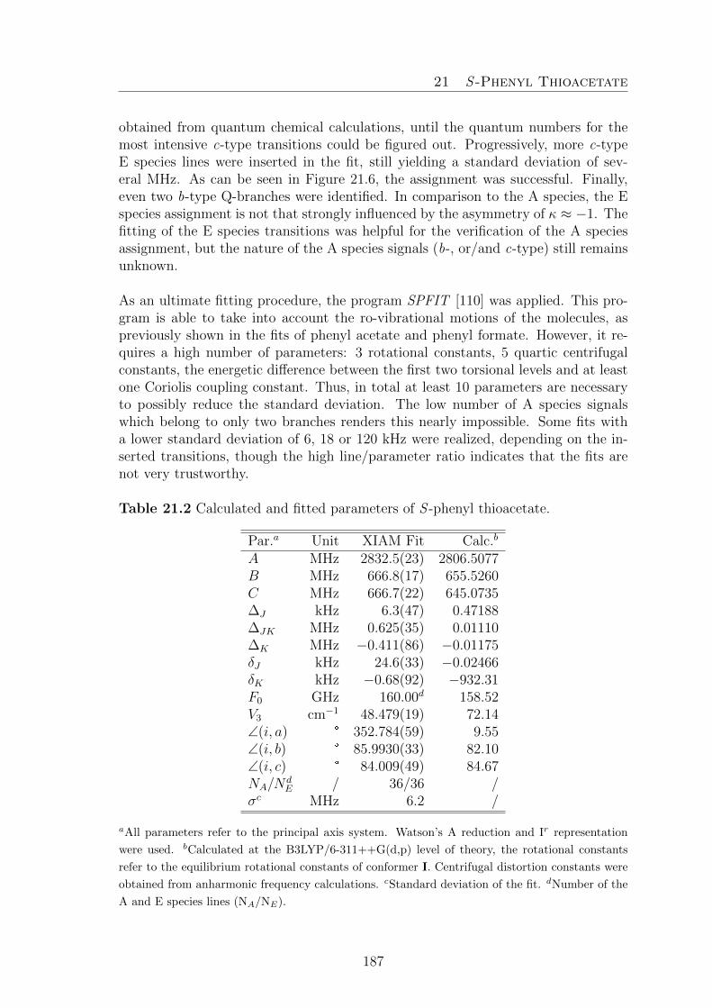
21 S -Phenyl Thioacetate
obtained from quantum chemical calculations, until the quantum numbers for themost intensive c-type transitions could be figured out. Progressively, more c-typeE species lines were inserted in the fit, still yielding a standard deviation of sev-eral MHz. As can be seen in Figure 21.6, the assignment was successful. Finally,even two b-type Q-branches were identified. In comparison to the A species, the Especies assignment is not that strongly influenced by the asymmetry of κ ≈ −1. Thefitting of the E species transitions was helpful for the verification of the A speciesassignment, but the nature of the A species signals (b-, or/and c-type) still remainsunknown.
As an ultimate fitting procedure, the program SPFIT [110] was applied. This pro-gram is able to take into account the ro-vibrational motions of the molecules, aspreviously shown in the fits of phenyl acetate and phenyl formate. However, it re-quires a high number of parameters: 3 rotational constants, 5 quartic centrifugalconstants, the energetic difference between the first two torsional levels and at leastone Coriolis coupling constant. Thus, in total at least 10 parameters are necessaryto possibly reduce the standard deviation. The low number of A species signalswhich belong to only two branches renders this nearly impossible. Some fits witha lower standard deviation of 6, 18 or 120 kHz were realized, depending on the in-serted transitions, though the high line/parameter ratio indicates that the fits arenot very trustworthy.
Table 21.2 Calculated and fitted parameters of S -phenyl thioacetate.
Par.a Unit XIAM Fit Calc.b
A MHz 2832.5(23) 2806.5077B MHz 666.8(17) 655.5260C MHz 666.7(22) 645.0735∆J kHz 6.3(47) 0.47188∆JK MHz 0.625(35) 0.01110∆K MHz −0.411(86) −0.01175δJ kHz 24.6(33) −0.02466δK kHz −0.68(92) −932.31F0 GHz 160.00d 158.52V3 cm−1 48.479(19) 72.14∠(i, a) ° 352.784(59) 9.55∠(i, b) ° 85.9930(33) 82.10∠(i, c) ° 84.009(49) 84.67NA/N
dE / 36/36 /
σc MHz 6.2 /
aAll parameters refer to the principal axis system. Watson’s A reduction and Ir representation
were used. bCalculated at the B3LYP/6-311++G(d,p) level of theory, the rotational constants
refer to the equilibrium rotational constants of conformer I. Centrifugal distortion constants were
obtained from anharmonic frequency calculations. cStandard deviation of the fit. dNumber of the
A and E species lines (NA/NE).
187

21 S -Phenyl Thioacetate
21.4. Discussion
The rotational constants obtained from the XIAM Fit given in Table 21.2 show largeerrors. This is normal, as the standard deviation of 6 MHz is also very high. Inspite of this, the potential barrier is determined quite well. However, the centrifugaldistortion constants could barely be determined. They are however necessary to fitthe rotational transitions of Ka = 2, and therefore cannot be ignored in this fit. Theangles of the internal rotor axis to the principal axes were determined well, and arein agreement with the calculated values.
The rotational constants B and C were both fitted to a very similar value, differingin the first digit. This is also an explanation for the enlarged standard deviation: asdescribed in the chapter above, the A species b- and c-type branches occur at nearlythe same frequencies for a prolate top. With a Ray’s asymmetry parameter κ of−0.99, S -phenyl acetate is a near-prolate top. If compared to the calculated values,the rotational constants seem plausible. However, the theoretical values are not thetorsional ground state rotational constants but equilibrium constants, and thereforeare expected to differ. Regarding the rotational barrier height, MP2 seems to bebetter suited, as shown in Figure 21.2 on the left hand-side. The potential heightof the methyl internal rotation was calculated to 48.34 cm−1, which is congruent tothe fitted value of 48.479(19) cm−1.
Clearly, the molecule features at least one tunneling motion, causing problems dur-ing the fitting process. A model dealing with Coriolis coupling terms would be bettersuited to describe this molecule accordingly. Thus, a standard deviation of 6 MHzis not unusual, if compared to phenyl acetate and phenyl formate, where the XIAMstandard deviations were 1.4 MHz and 3 MHz, respectively. The sulfur seems to en-hance this effect, which is not surprising given the very broad and even flatter doubleminima potentials due to the increased flexibility of the angles given by the large sizeof the sulfur atom, leading to less polarized and softer bonds in S -phenyl thioacetate.
In contrary to phenyl acetate and phenyl formate, the microwave spectrum of S -phenyl thioacetate does not show vt = 1 transitions. This is probably due to thevery low vapor pressure of 0.045 Torr [16]. This also complicates a fit with SPFIT,as no information from the first excited torsional state is present and therefore, thedifference of the excited and ground torsional states cannot be fitted very reliably.
Regarding the SPFIT fitting attempts, only an extended data set can lead to clarifi-cation, but unfortunately at the present stage of this project, no further informationcan be gained from the so-far gathered data. A further complication is the unknownvalue of the parameter E indicating the torsional energy level difference. E can becalculated, if the torsional potential is determined accurately, which is not the casefor the here presented molecule. Moreover, if the calculation methods are switched,the potentials change drastically, making it impossible to decide which one is themost congruent in reality. However, it is confirmed from the elevated standard de-viations, that at least one tunneling large amplitude motion in PhSAc exists.
188

21 S -Phenyl Thioacetate
Finally, the observation that a substitution of oxygen with sulfur reduces the tor-sional barrier heights significantly is confirmed. Thus, the methyl group in phenylacetate requires 137.098(99) cm−1 to perform a full rotation, while in the sulfuranalogue presented here in this work only 48.479(19) cm−1 are sufficient. Thesame observation has been made previously in 2,5-dimethylthiophene [10] and 2,5-dimethylfuran [8], where the barrier decreased from 439.176(11) cm−1 to 242.148(41)cm−1, respectively. In methyl thiolformate, the barrier also decreased to 149 cm−1
[140] when compared to methyl formate (V3 = 416 cm−1) [147].
189

21 S -Phenyl Thioacetate
190

22 Conclusion
22. Conclusion
In this part, the three molecules phenyl formate, phenyl acetate and S -phenylthio-acetate were analyzed. Phenyl formate (28) is the most simple project as it has nomethyl group which could undergo internal rotation. Quantum chemical calculationsyield a pair of enantiomers as the most stable structures. Although the molecularstructure does not contain a methyl group which could cause an A-E splitting, theassignment of this spectrum was very complicated. During the transformation of theenantiomers into each other, the dipole moment in a- and b- direction varies, whichmakes the assignment of these two line types difficult. For this reason, an assign-ment of the c-type lines was carried out at first. Afterwards, the a-type assignmentfollowed. Finally, the first excited vibrational state was also partially observed andfitted. The assignment was successful, although a standard deviation of the fit of3 MHz is too high. The difference ∆E of the first to torsional states was fitted to46 GHz. As this effect is caused by the specific molecular structure, phenyl acetatewas chosen as second project.
O
O
O
O
Me S
O
MeH
φ = 72° φ = 72° φ = 87°
φ φ φ
28 29 30
Figure 22.1. Out-of-plane tilt angles found in phenyl formate (28), phenyl acetate(29) and S -phenyl thioacetate (30).
The structure of phenyl acetate (29) differs from the one of phenyl formate (28)only by the methyl group instead of a single hydrogen atom. The rotational bar-rier of the acetyl methyl group is expected to be approximately 112 cm−1. Thus, asplitting of the rotational transitions into an A and an E species is observable. Thissplitting complicates the assignment of the spectrum. Moreover, quantum chemicalcalculations predicted a stable pair of enantiomers, which can be transformed intoeach other by a tunneling process. The assignment was successful, but again, astandard deviation of 2 MHz was obtained. A reasonable explanation of this effectwould be the tunneling process mentioned above. Unfortunately no program ableto fit vt = 1 E species lines exist. For phenyl acetate, a ∆E of 35 GHz was found.If compared to the ∆E value of phenyl formate, which lies in the same order ofmagnitude, this result seems reasonable. The acetate fragment is heavier than theformate group, implying that less energy is needed for the tunneling motion. Thecalculated out-of-plane tilt-angle ϕ of 72° is the same as for phenyl formate (seeFigure 22.1).
191

22 Conclusion
The last molecule of this part is S -phenylthioacetate (30), featuring the smallestout-of-plane angle for the phenyl ring. Quantum chemical calculations again yieldeda pair of enantiomers as the most stable structures, although the tilt angle varieswith the applied method. Moreover, the simultaneous inversion of two dihedralangles occurs while tunneling between the enantiomeric structures. This can beexplained by the increased size of the sulfur hetero atom, forming less strong bondsthan oxygen. As the molecules also contains a methyl group, A-E splitting is occur-ring in the microwave spectrum. Even with the previously gathered information andexperience, the assignment was still difficult. Finally, A and E species signals wereassigned with the program XIAM, but no trustworthy fits for the pure A species as-signment with SPFIT were obtained. This is due to the near-prolate top symmetryof the molecule, causing very narrow lying b and c type signals in the microwavespectrum. In combination with a standard deviation larger than this splitting, theassignment appears to be unfeasible. However the barrier to internal rotation couldbe determined surprisingly accurately, because the assigment of the E species tran-sitions is clear.
O
O
Me
S
O
Me
S
O
Me Me
Me Me
H
O
O
Me
H
O
S
Me
148.8(26) cm−1
416(14) cm−1 439.176(11) cm−1
242.148(41) cm−1 48.479(19) cm−1
137.17(13) cm−129
3031 32
33 34
Figure 22.2. Barriers to internal rotation of methyl formate (31), methyl thio-formate (33), 2,5-dimethylthiophene (32), 2,5-dimethylfurane (34), phenyl acetate(30), and S- phenyl thioacetate (29).
Compared to the barrier found in phenyl acetate (29), the values match the trendsfound in 2,5-dimethylthiophene (32) [10], and 2,5-dimethylfurane (34) [8], wherethe barrier to internal rotation decrease when the oxygen atom is replaced by asulfur atom. Another example for this trend are the rotational barriers reportedfor methyl formate (33) [147] and methyl thioformate (31) [140], as indicated inFigure 22.2.
192

23 Bibliography
23. Bibliography
[1] B. Tercero, I. Kleiner, J. Cernicharo, H. V. L. Nguyen, A. Lopez, G. M. MunozCaro, Astrophis. J. Lett. A 770:L13 (6pp), (2013).
[2] L. Tulimat, H. Mouhib, I. Kleiner, W. Stahl, J. Mol. Spectrosc. 312, 46, (2015).
[3] S. Blanco, J. C. Lopez, A. Lesarri, W. Caminati, J. L. Alonso, Mol. Phys. 103,1473, (2005).
[4] A. Welzel, A. Hellweg, I. Merke, W. Stahl, J. Mol. Spectrosc., 215, 58, (2002).
[5] C. Thomsen and H. Dreizler, Z. Naturforsch. 56a, 635, (2001).
[6] J. Rottstegge, H. Hartwig, H. DreizlerJ. Mol. Struct. 478, 37, (1999).
[7] M. Tudorie, I. Kleiner, M. Jahn, J.-U. Grabow, M. Goubet, O. Pirali, J. Phys.Chem. A 117, 13636, (2013).
[8] V. Van, J. Bruckhuisen, W. Stahl, V. Ilyushin, H. V. L. Nguyen, J. Mol. Spec-trosc. 343, 121, (2018).
[9] V. Van, W. Stahl, H. V. L. Nguyen, ChemPhysChem, 17, 3223, (2016).
[10] V. Van, W. Stahl, H. V. L. Nguyen, Phys. Chem. Chem. Phys. 17, 32111,(2015).
[11] M. J. Frisch, G. W. Trucks, H. B. Schlegel, G. E. Scuseria, M. A. Robb, J. R.Cheeseman, G. Scalmani, V. Barone, B. Mennucci, G. A. Petersson, H. Nakatsuji,M. Caricato, X. Li, H. P. Hratchian, A. F. Izmaylov, J. Bloino, G. Zheng, J. L.Sonnenberg, M. Hada, M. Ehara, K. Toyota, R. Fukuda, J. Hasegawa, M. Ishida,T. Nakajima, Y. Honda, O. Kitao, H. Nakai, T. Vreven, J. A., Jr. Montgomery, J.E. Peralta, F. Ogliaro, M. Bearpark, J. J. Heyd, E. Brothers, K. N. Kudin, V. N.Staroverov, R. Kobayashi, J. Normand, K. Raghavachari, A. Rendell, J. C. Burant,S. S. Iyengar, J. Tomasi, M. Cossi, N. Rega, J. M. Millam, M. Klene, J. E. Knox,J. B. Cross, V. Bakken, C. Adamo, J. Jaramillo, R. Gomperts, R. E. Stratmann,O. Yazyev, A. J. Austin, R. Cammi, C. Pomelli, J. W. Ochterski, R. L. Martin,K. Morokuma, V. G. Zakrzewski, G. A. Voth, P. Salvador, J. J. Dannenberg, S.Dapprich, A. D. Daniels, O. Farkas, J. B. Foresman, J. V. Ortiz, J. Cioslowski, D.J. Fox, Gaussian 09, Revision A.02, Gaussian, Inc., Wallingford CT, 2009.
[12] H. B. Schlegel, J. Comput. Chem. 3 214, (1982).
[13] L. Ferres, Quantum Chemical and Microwave Spectroscopic Investigations onPhenyl Acetate and Structurally related Molecules, RWTH Aachen University, mas-
193

23 Bibliography
ter thesis, (2015).
[14] L. Ferres, Quantum Chemical and Microwave Spectroscopic Investigations onPhenyl Acetate, RWTH Aachen University, research report, (2014).
[15] J.-U. Grabow, W. Stahl, H. Dreizler, Rev. Sci. Instrum. 67, 4072, (1996).
[16] SciFinder Scholar, version 2018; Chemical Abstracts Service: Columbus, OH,2018 (accessed May 5, 2018); calculated using ACD/Labs software, V11.02; ACD/Labs1994-2018.
[17] J.-U. Grabow, W. Stahl, Z. Naturforsch. A 45, 1043, (1990).
[18] W. Gordy, R. L. Cook, Microwave molecular spectra, Vol. XVIII, Wiley, NewYork, (1984).
[19] C. Di Lauro, Rotational Structure in Molecular Infrared Spectra, Newnes, (2013).
[20] D. S. Ray, Z. Physik 78, 74, (1932).
[21] J. H. van Vleck, Phys. Rev. 47, 487, (1935).
[22] H. Hartwig, H. Dreizler, Z. Naturforsch. A, 51, 923, (1996).
[23] H. Dreizler, Mikrowellenspektroskopische Bestimmung von Rotationsbarrierenfreier Molekule, Springer Berlin Heidelberg, Berlin, Heidelberg, 59, (1968).
[24] L. Ferres, W. Stahl, H. V. L. Nguyen, J. Chem. Phys. 148, 124304, (2018).
[25] J. T. Hougen, I. Kleiner, and M. Godefroid, J. Mol. Spectrosc. 163, 559, (1994).
[26] L. Ferres, W. Stahl, H.V.L. Nguyen, Mol. Phys. 114, 2788, (2016).
[27] J. March, Advanced Organic Chemistry, 4th Ed., J. Wiley and Sons, New York,(1992).
[28] J.-R. A. Moreno, D. Petitprez and T. R. Huet, Chem. Phys. Lett. 419, 411,(2006).
[29] C. Perez, S. Mata, S. Blanco, J.C. Lopez and J. L. Alonso, J. Phys. Chem. A115, 9653, (2011).
[30] R.K. Kakar, E.A. Rinehart, C.R. Quade and T. Kojima, J. Chem. Phys. 52,3803, (1970).
194

23 Bibliography
[31] M. Onda, Y. Kohama, K. Suga and I. Yamaguchi, J. Mol. Struct. 442, 19,(1998).
[32] M. Onda, A. Toda, S. Mori and I. Yamaguchi, J. Mol. Struct. 144, 47, (1986).
[33] C. Cabezas, M. Varela, W. Caminati, S. Mata, J.C. Lopez and J.L. Alonso, J.Mol. Spectrosc. 268, 42, (2011).
[34] H. V. L. Nguyen, W. Stahl and I. Kleiner, Mol. Phys. 110, 2035, (2012).
[35] H. V. L. Nguyen, V. Van, W. Stahl and I. Kleiner, J. Chem. Phys. 140, 214303,(2014).
[36] H. V. L. Nguyen and W. Stahl, ChemPhysChem. 12, 1900, (2011).
[37] H. V. L. Nguyen and W. Stahl, J. Chem. Phys. 135, 024310, (2011).
[38] R. Kannengießer, M. J. Lach, W. Stahl and H. V. L. Nguyen, ChemPhysChem.16, 1906, (2015).
[39] R. Kannengießer, S. Klahm, H. V. L. Nguyen, A. Luchow and W. Stahl, J.Chem. Phys. 141, 204308, (2014).
[40] H. V. L. Nguyen, H. Mouhib, S. Klahm, W. Stahl and I. Kleiner, Phys. Chem.Chem. Phys. 15, 10012, (2013).
[41] V. Van, C. Dindic, H. V. L. Nguyen and W. Stahl, ChemPhysChem. 16, 291,(2015).
[42] Y. Zhao, W. Stahl and H. V. L. Nguyen, Chem. Phys. Lett. 545, 9, (2012).
[43] S. Tsuzuki, H. Houjou, Y. Nagawa and K. Hiratani, J. Phys. Chem. A 104,1332, (2000).
[44] D. Moran, A. C. Simmonett, F.E. Leach, W.D. Allen, P.v.R. Schleyer and H.F.Schaefer, J. Am. Chem. Soc. 128, 9342, (2006).
[45] J. M. Riveros and E. B. Wilson, J. Chem. Phys. 46, 4605, (1967).
[46] D. G. Scroggin, J. M. Riveros and E.B. Wilson, J. Chem. Phys. 60, 1376,(1974).
[47] C. C. Lin and J. D. Swalen, Rev. Mod. Phys. 31, 841, (1959).
[48] R. C. Woods, J. Mol. Spectrosc. 21, 4, (1966).
195

23 Bibliography
[49] R. C. Woods, J. Mol. Spectrosc. 22, 49, (1967).
[50] J. M. Vacherand, B. P. van Eijck, J. Burie, and J. Demaison, J. Mol. Spectrosc.118, 355, (1986).
[51] I. Kleiner, J. T. Hougen, J. Chem. Phys. 119, 5505, (2003).
[52] B. Kirtman, J. Chem. Phys. 37, 2516, (1962).
[53] R. M. Lees and J. G. Baker, J. Chem. Phys. 48, 5299, (1968).
[54] E. Herbst, J. K. Messer, F. C. De Lucia and P. Helminger, J. Mol. Spectrosc.108, 42, (1984).
[55] V. Ilyushin, I. Kleiner, and F. J. Lovas, J. Phys. Chem. Ref. Data 37, 97,(2008).
[56] L. Ferres, H. Mouhib, W. Stahl, and H. V. L. Nguyen, ChemPhysChem 18,1855, (2017).
[57] I. Kalkman, C. Vu, M. Schmitt, W. L. Meerts, Phys. Chem. Chem. Phys.11, 4311, (2009).
[58] M. Bohm, C. Ratzer, M. Schmitt, J. Mol. Struct. 800, 55, (2006).
[59] R. M. Helm, H.-P. Vogel, H. J. Neusser, Chem. Phys. Lett. 270, 285, (1997).
[60] S. Maiti, A. I. Jaman, R. N. Nandi, J. Mol. Spectrosc. 177, 29, (1996).
[61] A. J. Shirar, D. S. Wilcox, K. M. Hotopp, G. L. Storck, I. Kleiner, B. C. Dian,J. Phys. Chem. A 114, 12187, (2010).
[62] V. Ilyushin, Z. Kisiel, L. Pszczolkowski, H. Mader, J. T. Hougen, J. Mol.Spectrosc. 259, 26, (2010).
[63] L. Alvarez-Valtierra, J. T. Yi, D. W. Pratt, J. Phys. Chem. B 110, 19914,(2006).
[64] D. G. Lister, J. Mol. Struct. 68, 33, (1980).
[65] R. K. Bohn, M. S. Farag, C. M. Ott, J. Radhakrishnan, S. A. Sorenson, N. S.True, J. Mol. Struct. 268, 107, (1992).
196

23 Bibliography
[66] D. G. Lister, P. Palmieri, C. Zauli, J. Mol. Struct. 35, 299, (1976).
[67] L. Ferres, H. Mouhib, W. Stahl, M. Schwell, H. V. L. Nguyen, J. Mol. Spec-trosc. 337, 59, (2017).
[68] A. Jabri, V. Van, H. V. L. Nguyen, W. Stahl, I. Kleiner, ChemPhysChem 17,2660, (2016).
[69] S. Jacobsen, U. Andresen, H. Mader, Struct. Chem. 14, 217, (2003).
[70] N. Hansen, H. Mader, T. Bruhn, Mol. Phys. 97, 587, (1999).
[71] K. P. R. Nair, J. Demaison, G. Wlodarczak, I. Merke, J. Mol. Spectrosc. 237,137, (2006).
[72] H. D. Rudolph, K. Walzer, I. Krutzik, J. Mol. Spectrosc. 47, 314, (1973).
[73] L. Ferres, H. Mouhib, W. Stahl, H. V. L. Nguyen, ChemPhysChem 14, 1855,(2017).
[74] P. Groner, J. Chem. Phys. 107, 4483, (1997).
[75] A. O. Hernandez-Castillo, C. Abeysekera, B. M. Hays, I. Kleiner, H. V. L.Nguyen, and T. S. Zwier, J. Mol. Spectrosc. 337, 51, (2017).
[76] K. Eibl, R. Kannengießer, W. Stahl, H. V. L. Nguyen, and I. Kleiner, Mol.Phys. 114, 3483, (2016).
[77] H. V. L. Nguyen, H. Mouhib, W. Stahl, and I. Kleiner, Mol. Phys. 108, 763,(2010).
[78] R. Kannengießer, W. Stahl, H. V. L. Nguyen, and I. Kleiner, J. Phys. Chem.A 120, 3992, (2016).
[79] H. V. L. Nguyen, A. Jabri, V. Van, and W. Stahl, J. Phys. Chem. A 118,12130, (2014).
[80] L. Sutikdja, W. Stahl, V. Sironneau, H. V. L. Nguyen, and I. Kleiner, Chem.Phys. Lett. 663, 145, (2016).
[81] L. Ferres, W. Stahl, I. Kleiner, and H. V. L. Nguyen, J. Mol. Spectrosc. 343,44, (2018).
[82] H. D. Rudolph and A. Trinkaus, Z. Naturforsch. 23a, 68, (1968).
[83] A. Hellweg, C. Hattig, I. Merke, and W. Stahl, J. Chem. Phys. 124, 204305,
197

23 Bibliography
(2006).
[84] A. Hellweg and C. Hattig, J. Chem. Phys. 127, 024307, (2007).
[85] H. Saal, J.-U. Grabow, A. Hight-Walker, J. T. Hougen, W. Caminati, I.Kleiner, presentation ]WH07, 65th Ohio State University International Symposiumon Molecular Spectroscopy, Columbus, Ohio, U.S.A., June 2010.
[86] D. Jelisavac, D. C. Cortes-Gomez, H. V. L. Nguyen, L. W. Sutikdja, W. Stahl,and I. Kleiner, J. Mol. Spectrosc. 257, 111, (2009).
[87] T. Bruhn and H. Mader, J. Mol. Spectrosc. 200, 151, (2000).
[88] G. E. Herberich, Z. Naturforsch. 22a, 761, (1967).
[89] V. Siebert, Microwave Spectroscopic Investigations and Quantum ChemicalCalculations of p-Methyl Anisole, RWTH Aachen University, bachelor thesis, (2016).
[90] H. Dreizler, Z. Naturforsch. 16a, 1354, (1961).
[91] Y. Zhao, H. V. L. Nguyen, W. Stahl, J. T. Hougen, J. Mol. Spectrosc. 318,91, (2015).
[92] H. D. Rudolph, H. Dreizler, A. Saeschke, P. Z. Wendling, Z. Naturforsch. 22a,940, (1967).
[93] L. Alvarez-Valtierra, J. T. Yi and D. W. Pratt, Phys. Chem. Chem. Phys 12,8323, (2010).
[94] P. Groner, J. R. Durig, J. Chem. Phys. 66, 1856, (1977).
[95] J. D. Swalen, C. C. Constain, J. Chem. Phys. 31, 1562, (1959).
[96] J. Meier, A. Bauder, Hs. H. Gunthard, J. Chem. Phys. 57, 1219, (1972).
[97] W. Bossert, A. Bauder, Hs. H. Gunthard, Chem. Phys. 39, 367, (1979).
[98] C. D. LeCroix, R. F. Curl, P. M. McKinney, R. J. Myers, J. Mol. Spectrosc.53, 250, (1974).
[99] K. N. Rao, Molecular Spectroscopy: Modern Research, Elsevier Science, NewYork, 59, (2012).
[100] L. Ferres, K.-N. Truong, W. Stahl, H. V. L. Nguyen, ChemPhysChem 19,1781, (2018).
198

23 Bibliography
[101] H. Mouhib, D. Jelisavac, W. Stahl, R. Wang, I. Kalf, U. Englert, ChemPhysChem12, 761, (2011).
[102] C. Merkens, T. Stadtmuller, U. Englert, H. Mouhib, W. Stahl, Z. Naturforsch.69a, 303, (2014).
[103] V. Van, W. Stahl, M. Schwell, H. V. L. Nguyen, J. Mol. Struct. 1156, 348,(2018).
[104 B. Reinhold, I. A. Finneran, S. T. Shipman, J. Mol. Spectrosc. 270, 89, (2011).
[105] H. Mouhib, D. Jelisavac, L. W. Sutikdja, E. Isaak, W. Stahl, J. Phys. Chem.A 115, 118, (2011).
[106] G. O. Sørensen, T. Pedersen, H. Dreizler, A. Guarnieri, A. P. Cox, J. Mol.Struct. 97, 77, (1983).
[107] F. Chen, H. Ma, B. Wang, J. Hazard. Mater. 162, 668, (2009).
[108] V. Vanh, Structures and internal dynamics of cyclic molecules studied bymicrowave spectroscopy and quantum chemistry, dissertation, RWTH Aachen Uni-versity, (2017).
[109] J. Cheung, Analysis of Internal Rotation in 2,5-Dimethylanisole by the Useof Quantum Chemical Calculations and Microwave Spectroscopy, bachelor thesis,RWTH Aachen University, (2017).
[110] H. M. Pickett, J. Mol. Spectrosc. 148, 371, (1991).
[111] H. V. L. Nguyen, W. Stahl, J. Mol. Spectrosc. 264, 120, (2010).
[112] J. Spautz, Quantum Chemical Calculations and Microwave Spectroscopic Inves-tigations on 2,6-Dimethylanisole, RWTH Aachen University, bachelor thesis, (2018).
[113] I. Merke, A. Luchow, W. Stahl, J. of Mol. Struct. 295, 780, (2006).
[114] I. Merke, W. Stahl, S. Kassi, D. Petitprez, G. Wlodarczak, J. of Mol. Spec-trosc. 216, 437, (2002).
[115] M. Schnell, J.-U. Grabow, Chem. Phys. 343, 121, (2008).
[116] Y. Zhao, H. Mouhib, G. Li, I. Kleiner, W Stahl, J. Mol. Spectrosc. 322, 38,(2016).
199

23 Bibliography
[117] K.-N. Truong, L. Weger, W. Stahl, H. Mouhib, ChemPhysChem 18, 2631,(2017).
[118] B. Bleaney, R. P. Penrose, Nature 157, 339, (1946).
[119] Program NPO-17907, NASA COSMIC Program Exchange, Univ. of Georgia,382 Broad St., Athens, GA 30602.
[120] H. Yale, J. Org. Chem. 36, 3238, (1971).
[121] L.W. Sutikdja, D. Jelisavac, W. Stahl, I. Kleiner, Mol. Phys. 110, 2883,(2012).
[122] S. Firestein, Nature 413, 211, (2001).
[123] K. Herrmann, Alkaloidhaltige Genussmittel, Gewurze, Kochsalz, Springer 632,(1979).
[124] R. F. Curl, J. Chem. Phys. 30 1529, (1959).
[125] W. Pyckhout, C.V. Alsenoy, H.J. Geise, J. Mol. Struct. 147, 85, (1986).
[126] G.I.L. Jones, D.G. Lister, N.L. Owen, J. Chem. Soc. Faraday Trans. 2, 71,1330, (1975).
[127] R.K. Kakar, J. Chem. Phys. 56, 1189, (1972).
[128] R. Kannengießer, W. Stahl, H.V.L. Nguyen, W.C. Bailey, J. Mol. Spectrosc.317, 50, (2015).
[129] V. Van, W. Stahl, H.V.L. Nguyen, J. Mol. Struct. 1123, 24, (2016).
[130] R. Kannengießer, W. Stahl, H.V.L. Nguyen, J. Phys. Chem. A 120, 5979,(2016).
[131] http://www.ifpan.edu.pl/ kisiel/prospe.htm.
[132] K.A. Utzaz, R.K. Bohn, J.A. Montgomery Jr, H.H. Michels, W. Caminati, J.Phys. Chem. A 114, 6913, (2010).
[133] F. Rajabi, R. Luque, Catal. Commun. 45, 129, (2014).
[134] M. E. Gonzalez-Nunez, R. Mello, A. Olmos, and G. Asensio, J. Org. Chem.70, 10879, (2005).
200

23 Bibliography
[135] S. Seo, J. B. Taylor, and M. F. Greaney, Chem. Commun. 48, 8270, (2012).
[136] https://ncit.nci.nih.gov/ncitbrowser/ConceptReport.jsp?dictionary=NCIThesaurus&version=18.02d&ns=ncit&code=C1501&key=n555999419&b=1&n=
null.
[137] P. W. Sadler, Chem. Rev. 60, 575, (1960).
[138] C. O. Della Vedova, R. M. Romano, H. Oberhammer, J. Org. Chem. 69,5395, (2004).
[139] G. I. L. Jones, D. G. Lister, N. L. Owen, J. Mol. Spectrosc. 60, 348, (1976).
[140] W. Caminati, B. P. Van Eijck, D. G. Lister, J. Mol. Spectrosc. 90, 15, (1981).
[141] S. Nagata, T. Yamabe, K. Fukui, J. Phys. Chem. 79, 2335, (1975).
[142] M. E. D. Lestard, M. E. Tuttolomondo, A. D. Altabef, SPECTROCHIM ACTAA 135, 907, (2015).
[143] P. R. Olivato, R. Nanartonis, J. C. D. Lopes, Y. Hase, Phosphorus Sulfur 71,107, (1992).
[144] C. J. Silvia, N. S. True, R. K. Bohn, J. Phys. Chem. 82, 483, (1978).
[145] Gaussian 03, Revision C.02, M. J. Frisch, G. W. Trucks, H. B. Schlegel, G. E.Scuseria, M. A. Robb, J. R. Cheeseman, J. A. Montgomery, Jr., T. Vreven, K. N.Kudin, J. C. Burant, J. M. Millam, S. S. Iyengar, J. Tomasi, V. Barone, B. Men-nucci, M. Cossi, G. Scalmani, N. Rega, G. A. Petersson, H. Nakatsuji, M. Hada,M. Ehara, K. Toyota, R. Fukuda, J. Hasegawa, M. Ishida, T. Nakajima, Y. Honda,O. Kitao, H. Nakai, M. Klene, X. Li, J. E. Knox, H. P. Hratchian, J. B. Cross,V. Bakken, C. Adamo, J. Jaramillo, R. Gomperts, R. E. Stratmann, O. Yazyev,A. J. Austin, R. Cammi, C. Pomelli, J. W. Ochterski, P. Y. Ayala, K. Morokuma,G. A. Voth, P. Salvador, J. J. Dannenberg, V. G. Zakrzewski, S. Dapprich, A. D.Daniels, M. C. Strain, O. Farkas, D. K. Malick, A. D. Rabuck, K. Raghavachari, J.B. Foresman, J. V. Ortiz, Q. Cui, A. G. Baboul, S. Clifford, J. Cioslowski, B. B.Stefanov, G. Liu, A. Liashenko, P. Piskorz, I. Komaromi, R. L. Martin, D. J. Fox,T. Keith, M. A. Al-Laham, C. Y. Peng, A. Nanayakkara, M. Challacombe, P. M. W.Gill, B. Johnson, W. Chen, M. W. Wong, C. Gonzalez, and J. A. Pople, Gaussian,Inc., Wallingford CT, 2004.
[146] X. Xuan, Y. Wang, N. Wang, SPECTROCHIM ACTA A 81, 236, (2011).
[147] R. F. Curl JR. J. Chem. Phys. 30, 1529, (1959).
201

23 Bibliography
202

Part V.Appendix: Experimental andTheoretical Data
203

204

24 Phenetole
24. Phenetole
Table 24.1 Nuclear coordinates in the principal inertial axes of the trans and gaucheconformers of phenetole calculated at the MP2/6-311++G(d.p) level of theory. Theatoms are numbered according to Figure 4.1.
trans gauchea/ A b/ A c/ A a/ A b/ A c/ A
C1 −2.265585 −1.061578 0.000121 −2.220235 −0.879222 0.328487C2 −0.890475 −1.291831 −0.000083 −0.901853 −1.297247 0.145270C3 0.005682 −0.210267 −0.000082 0.090021 −0.371645 −0.216036C4 −0.485691 1.103685 −0.000177 −0.247416 0.980568 −0.378181C5 −1.872270 1.320713 0.000090 −1.574102 1.390329 −0.180889C6 −2.766592 0.248647 0.000061 −2.564101 0.471301 0.175503H7 −2.947982 −1.907405 0.000136 −2.977433 −1.607858 0.605753H8 −0.484541 −2.299240 0.000006 −0.615878 −2.338515 0.263273H9 0.184851 1.955141 −0.000140 0.497472 1.716753 −0.658772H10 −2.245467 2.341557 0.000103 −1.824863 2.440559 −0.306158H11 −3.837571 0.428042 0.000270 −3.588816 0.798697 0.324114O12 1.330217 −0.543892 −0.000154 1.338590 −0.895833 −0.413945C13 2.269222 0.528068 −0.000002 2.442775 0.011723 −0.436324H14 2.118621 1.151602 0.891129 2.278459 0.793046 −1.187842H15 2.118981 1.151600 −0.891183 3.281057 −0.600683 −0.774517C16 3.652871 −0.089107 0.000188 2.730558 0.599940 0.936949H17 3.793194 −0.709082 −0.888169 2.914994 −0.205789 1.652012H18 4.410404 0.700164 0.000432 3.620050 1.236209 0.889555H19 3.792837 −0.709292 0.888453 1.893686 1.200533 1.299771
Table 24.2. Coefficients of the one-dimensional Fourier expansion for the energypotential curve calculated at the MP2/6-311++G(d,p) level of theory obtained byrotating the phenyl ring (α) at β = 180° (Figure 4.3). The potentials are expandedas V(ϕ) = a0 +
∑10n=1 an cos(nα + π) and V(ϕ) = b0 +
∑8n=1 bn cos(nβ + π).
Hartree cm−1 Hartree cm−1
a0 −385.0429113 b0 -385.0374041a1 −0.0000342 −7.51 b1 0.0071496 1569.16a2 −0.0013721 −301.15 b2 0.0036056 791.34a3 0.0000161 3.52 b3 0.0033164 727.86a4 −0.0002944 −64.61 b4 −0.0009341 −205.00a5 0.0000336 7.38 b5 −0.0007022 −154.11a6 0.0001020 22.40 b6 0.0000979 21.49a7 −0.0000124 −2.73 b7 0.0001653 36.29a8 0.0000436 9.58 b8 0.0000946 20.76a10 −0.0000240 −5.26a13 0.0000143 3.13
205

24 Phenetole
Table 24.3. Coefficients of the two-dimensional Fourier expansion for the energypotential surface calculated at the MP2/6-311++G(d,p) level of theory obtained byrotating the phenyl ring (α) and the ethyl group (β) (Figure 4.2).
N° Coefficient Energy / Hartree Energy / cm−1
1 1 −385.03842892 cos(2α) 0.0008718 191.3383 cos(4α) 0.0000466 10.2284 cosβ 0.0041311 906.6725 cos(2β) 0.0017416 382.2376 cos(3β) 0.0018345 402.6267 cos(4β) −0.0001926 −42.2718 cos(5β) −0.0000736 −16.1539 cos(2α) cosβ 0.0028013 614.81410 sin(2α) sinβ −0.0023093 −506.83311 cos(2α) cos(2β) 0.0015028 329.82612 sin(2α) sin(2β) −0.0019335 −424.35413 cos(2α) cos(3β) 0.0007234 158.76814 sin(2α) sin(3β) −0.0011560 −253.71315 cos(2α) cos(4β) −0.0003341 −73.32616 sin(2α) sin(4β) 0.0004830 106.00617 cos(2α) cos(5β) −0.0002168 −47.58218 sin(2α) sin(5β) 0.0002400 52.67419 cos(4α) cosβ 0.0000845 18.54620 sin(4α) sinβ −0.0001998 −43.85121 cos(4α) cos(2β) 0.0003429 75.25822 sin(4α) sin(2β) −0.0002987 −65.55723 cos(4α) cos(3β) 0.0007468 163.90424 sin(4α) sin(3β) −0.0007112 −156.09025 cos(4α) cos(4β) −0.0002838 −62.28726 sin(4α) sin(4β) 0.0002728 59.87327 cos(4α) cos(5β) −0.0002464 −54.07928 sin(4α) sin(5β) 0.0002326 51.05029 cos(6α) cos(3β) 0.0001131 24.82330 sin(6α) sin(3β) −0.0001111 −24.38431 cos(6α) cos(4β) −0.0000616 −13.52032 sin(6α) sin(4β) 0.0000614 13.47633 cos(6α) cos(5β) −0.0001175 −25.78834 sin(6α) sin(5β) 0.0001160 25.459
206

24 Phenetole
Table 24.4 Observed frequencies vo (in GHz) of 186 rotational transitions of phene-tole. The difference from calculated values (vo− vc in kHz) as obtained after a fitwith the program XIAM, are given in the last column.
N J Ka Kc J ′ K ′a K ′c vo vo − vc1 1 1 1 0 0 0 5.6392218 −1.02 2 1 2 1 0 1 7.2069284 1.23 2 2 0 1 1 1 15.4930291 −1.84 2 2 0 2 1 1 11.7998764 −1.45 2 2 0 3 1 3 7.3081654 −0.46 2 2 1 1 1 0 15.3499499 −2.27 2 2 1 2 1 2 12.2145380 −4.48 3 0 3 2 0 2 5.1068664 0.29 3 1 2 2 1 1 5.3282673 0.210 3 1 3 2 0 2 8.7063058 −2.111 3 1 3 2 1 2 4.9100198 0.312 3 2 1 2 1 2 17.3541592 −0.513 3 2 1 3 1 2 11.6075813 −3.714 3 2 2 2 1 1 16.9176517 −2.215 3 2 2 3 1 3 12.4259413 −0.616 4 0 4 3 0 3 6.7922797 0.117 4 1 3 3 1 2 7.0999623 −0.318 4 1 3 4 0 4 4.7436774 −2.419 4 1 4 3 0 3 10.1420179 −1.420 4 1 4 3 1 3 6.5425779 0.321 4 2 2 3 1 3 19.3061433 1.022 4 2 2 4 1 3 11.3696234 −1.223 4 2 3 3 1 2 18.4151089 −0.724 4 2 3 3 2 2 6.8257229 0.125 4 2 3 4 1 4 12.7090892 2.126 4 3 1 4 2 2 19.9670826 7.627 4 3 1 5 2 4 11.4939567 1.728 4 3 2 4 2 3 20.0211273 −8.929 4 3 2 5 2 3 11.3669421 −8.430 5 0 5 4 0 4 8.4635790 −0.231 5 0 5 4 1 4 5.1138419 2.432 5 1 4 4 1 3 8.8677109 −0.133 5 1 5 4 0 4 11.5215211 −1.634 5 1 5 4 1 4 8.1717833 0.435 5 2 3 4 1 4 21.3632749 2.236 5 2 3 4 2 2 8.5997084 0.437 5 2 3 5 1 4 11.1016219 0.338 5 2 4 4 1 3 19.8427431 −1.4
207

24 Phenetole
Table 24.4 (continued)
N J Ka Kc J ′ K ′a K ′c vo vo − vc39 5 2 4 4 2 3 8.5275977 0.140 5 2 4 5 1 5 13.0649038 2.141 5 3 2 4 3 1 8.5488376 −5.542 5 3 2 5 2 3 19.9162121 2.043 5 3 3 4 3 2 8.5476004 5.344 5 3 3 5 2 4 20.0411303 −3.545 5 3 3 6 2 4 9.5632923 −2.446 5 4 1 4 4 0 8.5437797 −1.247 5 4 2 4 4 1 8.5437730 −1.448 6 0 6 5 0 5 10.1179916 0.449 6 0 6 5 1 5 7.0600497 1.950 6 1 5 5 1 4 10.6302967 −0.351 6 1 6 5 0 5 12.8549523 −1.152 6 2 4 5 1 5 23.5427443 3.653 6 2 4 5 2 3 10.3512507 −0.254 6 2 4 6 1 5 10.8225756 0.155 6 2 5 5 1 4 21.2014728 −1.556 6 2 5 5 2 4 10.2264413 0.557 6 2 5 6 1 6 13.4943340 1.558 6 3 3 5 3 2 10.2645447 −1.859 6 3 3 6 2 4 19.8295092 3.560 6 3 3 7 2 6 8.1592490 2.061 6 3 4 5 3 3 10.2612254 1.262 6 3 4 7 2 5 7.7066487 3.763 6 4 2 5 4 1 10.2552014 −14.964 6 4 3 5 4 2 10.2551833 −4.065 6 5 1 5 5 0 10.2519082 −0.966 6 5 2 5 5 1 10.2519082 −0.867 7 0 7 6 0 6 11.7536484 0.568 7 0 7 6 1 6 9.0166874 1.769 7 1 6 6 1 5 12.3863512 −0.170 7 1 6 7 0 7 6.2928173 −3.571 7 1 7 6 0 6 14.1547257 −1.072 7 1 7 6 1 6 11.4177649 0.473 7 2 5 6 2 4 12.1178740 0.074 7 2 5 7 1 6 10.5540981 −0.175 7 2 6 6 1 5 22.4928355 1.376 7 2 6 6 2 5 11.9216572 0.377 7 2 6 7 1 7 13.9982262 1.378 7 3 4 6 3 3 11.9840417 −0.3
208

24 Phenetole
Table 24.4 (continued)
N J Ka Kc J ′ K ′a K ′c vo vo − vc79 7 3 4 7 2 5 19.6956746 0.980 7 3 4 8 2 7 6.5306283 1.081 7 3 5 6 3 4 11.9765879 0.482 7 3 5 7 2 6 20.1308500 2.383 7 4 3 6 4 2 11.9681336 1.484 7 4 4 6 4 3 11.9680348 −0.885 7 4 4 8 3 5 14.2666624 −0.486 7 5 2 6 5 1 11.9628558 −1.787 7 5 3 6 5 2 11.9628558 −1.188 7 6 1 6 6 0 11.9601121 0.189 7 6 2 6 6 1 11.9601121 0.190 8 0 8 7 0 7 13.3699773 0.791 8 0 8 7 1 7 10.9688993 1.592 8 1 7 8 0 8 7.0571660 −2.593 8 1 7 7 1 6 14.1343243 0.094 8 1 8 7 0 7 15.4347770 −0.595 8 1 8 7 1 7 13.0336995 0.896 8 2 6 7 2 5 13.8995090 −0.297 8 2 6 8 1 7 10.3192827 −0.498 8 2 7 7 1 6 23.7191436 −1.099 8 2 7 7 2 6 13.6126620 0.3100 8 2 7 8 1 8 14.5771878 −0.1101 8 3 5 7 3 4 13.7084207 −0.8102 8 3 6 7 3 5 13.6935744 −2.0103 8 3 6 8 2 7 20.2117630 0.6104 8 4 5 7 4 4 13.6825589 21.0105 8 4 5 9 3 6 12.5102224 −4.9106 8 5 3 7 5 2 13.6748669 −0.4107 8 5 4 7 5 3 13.6748669 1.8108 8 6 2 7 6 1 13.6707727 −0.4109 8 6 3 7 6 2 13.6707727 −0.4110 9 0 9 8 0 8 14.9679381 0.8111 9 0 9 8 1 8 12.9031376 1.2112 9 1 8 8 1 7 15.8724687 0.0113 9 1 8 8 2 7 6.2876468 −1.6114 9 1 9 8 0 8 16.7094183 −0.4115 9 1 9 8 1 8 14.6446184 0.5116 9 2 7 8 2 6 15.6945305 0.0117 9 2 7 9 1 8 10.1413456 0.7118 9 2 8 8 2 7 15.2988897 0.1119 9 2 8 9 1 9 15.2314608 1.2120 9 3 6 8 3 5 15.4389738 0.4
209

24 Phenetole
Table 24.4 (continued)
N J Ka Kc J ′ K ′a K ′c vo vo − vc121 9 3 6 9 2 7 19.2490319 3.0122 9 3 7 8 3 6 15.4119284 −0.1123 9 3 7 9 2 8 20.3248016 0.3124 9 4 5 8 4 4 15.3995255 −4.3125 9 4 6 8 4 5 15.3988978 3.6126 9 4 6 10 3 7 10.7319398 −1.9127 9 5 4 8 5 3 15.3880888 −3.7128 9 5 5 8 5 4 15.3880888 3.4129 9 6 3 8 6 2 15.3822619 −2.2130 9 6 4 8 6 3 15.3822619 −2.2131 10 0 10 9 0 9 16.5499506 0.7132 10 0 10 9 1 9 14.8084692 0.7133 10 1 9 9 1 8 17.5988414 0.6134 10 1 9 9 2 8 8.5876017 2.2135 10 1 9 10 0 10 9.0105908 0.1136 10 1 10 9 1 9 16.2504806 0.8137 10 2 8 9 2 7 17.4998128 0.3138 10 2 8 10 1 9 10.0423160 −0.7139 10 2 9 9 2 8 16.9798030 0.7140 10 2 9 10 1 10 15.9607821 0.0141 10 3 7 9 3 6 17.1771802 0.4142 10 3 7 1 0 2 18.9263984 2.3143 10 3 8 9 3 7 17.1312290 −0.4144 10 3 8 10 2 9 20.4762309 2.4145 10 4 6 9 4 5 17.1186477 −2.6146 10 4 7 9 4 6 17.1172796 2.8147 10 6 4 9 6 3 17.0946882 −1.3148 10 6 5 9 6 4 17.0946882 −1.1149 11 0 11 10 0 10 18.1194676 −0.4150 11 0 11 10 1 10 16.6774573 0.7151 11 1 10 10 1 9 19.3113356 0.3152 11 1 10 10 2 9 10.9191303 −2.3153 11 1 10 11 0 11 10.2024559 −2.1154 11 1 11 10 0 10 19.2933960 0.2155 11 1 11 10 1 10 17.8513851 0.7156 11 2 9 10 2 8 19.3111435 0.9157 11 2 9 11 1 10 10.0421219 −2.1158 11 2 10 10 2 9 18.6548994 0.3159 11 2 10 11 1 11 16.7642962 −0.5160 11 3 9 11 2 10 20.6722563 3.7161 12 0 12 11 0 11 19.6803246 0.9162 12 1 11 11 1 10 21.0077703 0.3
210

24 Phenetole
Table 24.4 (continued)
N J Ka Kc J ′ K ′a K ′c vo vo − vc163 12 1 11 11 2 10 13.2720035 0.0164 12 1 11 12 0 12 11.5299063 2.0165 12 1 12 11 0 11 20.6214834 0.0166 12 1 12 11 1 11 19.4475552 −0.4167 12 2 10 12 1 11 10.1582076 −1.8168 12 2 11 11 2 10 20.3237304 1.3169 12 3 9 12 2 10 18.0992663 1.1170 12 3 10 12 2 11 20.9188584 4.0171 13 0 13 12 0 12 21.2360840 0.8172 13 0 13 12 1 12 20.2949234 −0.1173 13 1 12 12 1 11 22.6860393 −0.2174 13 1 12 13 0 13 12.9798572 −3.4175 13 1 13 12 0 12 21.9804756 −1.5176 13 1 13 12 1 12 21.0393168 −0.6177 13 2 11 12 2 10 22.9334571 0.3178 13 2 11 13 1 12 10.4056267 0.0179 13 2 12 12 2 11 21.9859054 0.5180 13 2 12 13 1 13 18.5870561 −1.6181 14 0 14 13 0 13 22.7896124 −0.9182 14 1 13 14 0 14 14.5345980 3.3183 14 1 14 13 1 13 22.6270643 −1.7184 14 2 12 14 1 13 10.7973102 −1.5185 14 2 13 14 1 14 19.6011068 −0.7186 15 2 13 15 1 14 11.3441852 0.4
211

25 Methylanisoles
25. Methylanisoles
25.1. OMA
Table 25.1.1 Fourier expansion of the potential energy curve of of o-methyl anisolecalculated at the MP2/6-311++G(d,p) level of theory. The data were obtained byrotating the methoxy group about the O11−C3 bond by varying the dihedral an-gle α = ∠(C12−O11−C3−C4) in a grid of 10°, while all other molecular parameterswere optimized. The potential is expanded as V(α)=Σ15
i=0(aifi). The barrier heightis given by V=2Σ7
k=0(a2k+1) = 0.0062953 Hartree ≈ 2763.3 cm−1 ≈ 33.057 kJ/mol .
i fi ai / Hartree0 1 −385.04056231 cos(α) 0.00481452 cos(2α) 0.00111653 cos(3α) 0.00125514 cos(4α) 0.00004155 cos(5α) 0.00013836 cos(6α) 0.00015187 cos(7α) 0.00007568 cos(8α) 0.00012419 cos(9α) −0.000026210 cos(10α) −0.000023011 cos(11α) 0.000033112 cos(12α) 0.000046013 cos(13α) 0.000013214 cos(14α) −0.000005915 cos(15α) −0.0000083
Table 25.1.2 Nuclear coordinates in the principal inertial axes of the only con-former of o-methyl anisole calculated at the MP2/6-311++G(d,p) level of theory.The atoms are numbered according to Figure 6.1.
a / A b / A c / AC1 –1.396084 –1.644268 –0.000648C2 –0.012967 –1.409367 0.000513C3 0.466066 –0.092368 –0.000609C4 –0.426219 1.001421 0.000873C5 –1.798826 0.736218 –0.000612C6 –2.293347 –0.575857 0.000682H7 –1.760326 –2.668187 0.000048H8 0.667965 –2.252895 –0.000360
212

25 Methylanisoles
Table 25.1.2 (continued)
H9 –2.490822 1.575967 0.000221H10 –3.364337 –0.756664 –0.000291O11 1.791698 0.245179 –0.000629C12 2.723279 –0.826245 0.000448H13 2.612159 –1.449120 –0.894313H14 3.708651 –0.360955 0.000400H15 2.611504 –1.447749 0.896094C16 0.117139 2.405293 0.000085H17 0.743910 2.581594 0.879943H18 0.743708 2.580352 –0.880161H19 –0.700634 3.130817 –0.000310
Table 25.1.3 Fourier expansion of the potential energy curve given in Figure 6.2.The potential is expanded as V(β)=Σ5
i=0(aifi). The barrier heights are given asV3 = 2a1 = 445.36 cm−1 and V6 = 2 a2 = 57.72 cm−1.
i fi ai / Hartree ai / cm−1
0 1 −385.04451311 cos(3β) 0.0010146 222.682 cos(6β) 0.0001315 28.863 cos(9β) 0.0000233 5.114 cos(12β) 0.0000155 3.405 cos(15β) 0.0000071 1.56
Table 25.1.4 Observed frequencies in GHz (νo) of 244 torsional transitions ofo-methyl anisole with 125 A and 119 E species. νo − νc values in kHz as obtainedafter a fit with the program XIAM (see Table 6.1).
N J Ka Kc J ′ K ′a K ′c S vo vo–vc1 2 2 0 1 1 1 A 9.2326401 −6.32 2 2 0 1 1 1 E 9.2325940 3.03 2 2 1 1 1 0 A 8.4409836 −5.84 2 2 1 1 1 0 E 8.4409358 −0.45 3 2 1 2 0 2 A 13.5889728 −3.46 3 2 1 2 0 2 E 13.5888102 −0.47 3 2 1 2 1 2 A 13.0427560 3.28 3 2 1 2 1 2 E 13.0425533 0.79 3 3 0 2 2 1 A 13.8736289 −2.6
213

25 Methylanisoles
Table 25.1.4 (continued)
N J Ka Kc J ′ K ′a K ′c S vo vo–vc10 3 3 0 2 2 1 E 13.8737918 1.911 3 3 1 2 2 0 A 13.6230176 −3.712 3 3 1 2 2 0 E 13.6227319 2.313 4 0 4 3 0 3 A 8.7932356 −1.114 4 0 4 3 0 3 E 8.7932154 −0.615 4 0 4 3 1 3 A 8.5484063 −0.716 4 0 4 3 1 3 E 8.5483586 0.017 4 1 3 3 1 2 A 10.7346386 0.018 4 1 3 3 1 2 E 10.7345157 0.419 4 1 3 3 2 2 A 8.6654049 7.720 4 1 3 3 2 2 E 8.6651807 1.421 4 1 4 3 0 3 A 8.8840252 −6.422 4 1 4 3 0 3 E 8.8840252 −0.623 4 1 4 3 1 3 A 8.6392010 −0.924 4 1 4 3 1 3 E 8.6391686 0.125 4 2 2 3 0 3 E 17.9641169 0.026 4 2 2 3 1 3 E 17.7192591 −0.527 4 2 2 3 2 1 A 11.2725017 1.228 4 2 2 3 2 1 E 11.2722868 0.229 4 2 3 3 1 2 A 12.0251434 7.330 4 2 3 3 1 2 E 12.0251249 0.031 4 2 3 3 2 2 A 9.9558949 0.232 4 2 3 3 2 2 E 9.9557895 0.633 4 3 1 3 2 2 A 16.9640698 −1.634 4 3 1 3 2 2 E 16.9639286 1.435 4 3 1 3 3 0 A 10.6795791 1.536 4 3 1 3 3 0 E 10.6791987 1.337 4 3 2 3 2 1 A 15.7637084 −1.538 4 3 2 3 2 1 E 15.7636261 1.739 4 3 2 3 3 1 A 10.4219623 5.340 4 3 2 3 3 1 E 10.4219889 0.941 4 4 1 3 3 0 A 18.7000311 −2.442 4 4 1 3 3 0 E 18.6980199 4.443 4 4 1 4 3 2 A 8.3252914 −4.644 4 4 1 4 3 2 E 8.3236750 1.045 5 0 5 4 0 4 A 10.6881655 −1.446 5 0 5 4 0 4 E 10.6881451 −0.247 5 0 5 4 1 4 A 10.5973709 −1.148 5 0 5 4 1 4 E 10.5973357 0.349 5 1 4 4 1 3 A 12.8336477 0.250 5 1 4 4 1 3 E 12.8335581 0.051 5 1 4 4 2 3 A 11.5431516 1.6
214

25 Methylanisoles
Table 25.1.4 (continued)
N J Ka Kc J ′ K ′a K ′c S vo vo–vc52 5 1 4 4 2 3 E 11.5429489 0.353 5 1 5 4 0 4 A 10.7182592 −0.654 5 1 5 4 0 4 E 10.7182441 −0.655 5 1 5 4 1 4 A 10.6274636 −1.356 5 1 5 4 1 4 E 10.6274349 0.157 5 2 3 4 2 2 E 14.0935864 0.558 5 2 3 4 2 2 A 14.0938227 0.659 5 2 4 4 1 3 A 13.4918336 0.860 5 2 4 4 1 3 E 13.4918218 −8.261 5 2 4 4 2 3 E 12.2012234 0.162 5 2 4 4 2 3 A 12.2013323 −0.363 5 2 4 5 0 5 A 8.4722658 −3.964 5 2 4 5 0 5 E 8.4720355 1.165 5 2 4 5 1 5 A 8.4421678 −9.066 5 2 4 5 1 5 E 8.4419351 0.167 5 3 2 4 3 1 A 13.7316507 3.768 5 3 2 4 3 1 E 13.7313441 −0.769 5 3 3 4 2 2 A 17.4892740 3.270 5 3 3 4 2 2 E 17.4892417 1.671 5 3 3 4 3 2 A 12.9980628 1.472 5 3 3 4 3 2 E 12.9979013 −1.073 5 4 1 4 4 0 A 13.1636484 4.974 5 4 1 4 4 0 E 13.1619279 −4.075 5 4 2 4 4 1 A 13.0957510 0.876 5 4 2 4 4 1 E 13.0970377 2.177 5 4 2 5 3 3 A 8.4229812 −3.778 5 4 2 5 3 3 E 8.4228114 4.179 6 0 6 5 0 5 A 12.6077316 −1.080 6 0 6 5 0 5 E 12.6077094 0.381 6 0 6 5 1 5 A 12.5776387 −1.082 6 0 6 5 1 5 E 12.5776101 0.483 6 1 5 5 1 4 A 14.7076496 −1.584 6 1 5 5 1 4 E 14.7075853 0.185 6 1 5 5 2 4 A 14.0494702 1.786 6 1 5 5 2 4 E 14.0493096 −0.887 6 1 5 6 0 6 A 9.9139979 −7.788 6 1 5 6 0 6 E 9.9136329 −2.789 6 1 5 6 1 6 A 9.9046954 1.690 6 1 5 6 1 6 E 9.9043217 0.491 6 1 6 5 0 5 A 12.6170436 −0.992 6 1 6 5 0 5 E 12.6170236 0.293 6 1 6 5 1 5 A 12.5869505 −1.0
215

25 Methylanisoles
Table 25.1.4 (continued)
N J Ka Kc J ′ K ′a K ′c S vo vo–vc94 6 1 6 5 1 5 E 12.5869244 0.395 6 2 4 5 2 3 A 16.6290002 −0.596 6 2 4 5 2 3 E 16.6287839 0.297 6 2 4 5 3 3 A 13.2335572 5.298 6 2 4 5 3 3 E 13.2331287 −0.899 6 2 5 5 1 4 A 14.9885421 −3.6100 6 2 5 5 1 4 E 14.9885343 −0.6101 6 2 5 5 2 4 A 14.3303625 −0.6102 6 2 5 5 2 4 E 14.3302605 0.4103 6 2 5 6 0 6 E 10.1945869 1.5104 6 2 5 6 1 6 A 10.1855865 −1.9105 6 2 5 6 1 6 E 10.1852722 1.1106 6 3 3 5 3 2 A 16.8669087 0.6107 6 3 3 5 3 2 E 16.8665610 −0.6108 6 3 4 5 3 3 A 15.4728351 0.0109 6 3 4 5 3 3 E 15.4726453 0.1110 6 3 4 6 1 5 A 8.9234288 −3.3111 6 3 4 6 1 5 E 8.9232974 1.2112 6 3 4 6 2 5 A 8.6425352 −2.3113 6 3 4 6 2 5 E 8.6423490 2.5114 6 4 2 5 4 1 A 16.0612968 0.1115 6 4 2 5 4 1 E 16.0608093 −1.4116 6 4 3 5 4 2 A 15.7896720 2.0117 6 4 3 5 4 2 E 15.7896028 −2.3118 6 4 3 6 3 4 A 8.7398171 −2.6119 6 4 3 6 3 4 E 8.7397710 3.9120 6 5 1 5 5 0 A 15.7249280 −0.5121 6 5 2 5 5 1 A 15.7101311 −1.4122 6 5 2 5 5 1 E 15.7129692 −1.6123 6 6 0 6 5 1 A 12.9649363 1.7124 6 6 1 6 5 2 A 12.9810358 7.4125 7 0 7 6 0 6 A 14.5409162 −1.1126 7 0 7 6 0 6 E 14.5408936 0.6127 7 0 7 6 1 6 A 14.5316044 −1.1128 7 0 7 6 1 6 E 14.5315793 0.6129 7 1 6 6 1 5 A 16.5449191 −1.4130 7 1 6 6 1 5 E 16.5448550 0.2131 7 1 6 6 2 5 A 16.2640253 −0.6132 7 1 6 6 2 5 E 16.2639057 0.6133 7 1 6 7 0 7 A 11.9180119 3.0134 7 1 6 7 0 7 E 11.9176002 2.7135 7 1 6 7 1 7 A 11.9152532 −3.6
216

25 Methylanisoles
Table 25.1.4 (continued)
N J Ka Kc J ′ K ′a K ′c S vo vo–vc136 7 1 6 7 1 7 E 11.9148454 0.9137 7 1 7 6 0 6 A 14.5436686 −0.8138 7 1 7 6 0 6 E 14.5436466 0.7139 7 1 7 6 1 6 A 14.5343565 −1.0140 7 1 7 6 1 6 E 14.5343327 1.1141 7 2 5 6 2 4 A 18.8051150 0.6142 7 2 5 6 2 4 E 18.8049561 0.5143 7 2 5 6 3 4 A 16.5658373 6.0144 7 2 5 6 3 4 E 16.5654392 −0.7145 7 2 5 7 1 6 A 8.9443512 8.3146 7 2 5 7 1 6 E 8.9438805 −0.8147 7 2 5 7 2 6 A 8.8385295 −8.8148 7 2 5 7 2 6 E 8.8380510 0.8149 7 2 6 6 1 5 A 16.6507223 −2.8150 7 2 6 6 1 5 E 16.6506868 0.9151 7 2 6 6 2 5 A 16.3698298 −0.7152 7 2 6 6 2 5 E 16.3697365 0.3153 7 2 6 7 0 7 A 12.0238149 1.4154 7 2 6 7 0 7 E 12.0234299 1.3155 7 2 6 7 1 7 A 12.0210576 −3.9156 7 2 6 7 1 7 E 12.0206765 0.9157 7 3 4 6 3 3 A 19.8475003 0.1158 7 3 4 6 3 3 E 19.8471384 −0.9159 7 3 5 6 3 4 A 17.8156434 −0.7160 7 3 5 6 3 4 E 17.8154538 0.7161 7 3 5 7 1 6 A 10.1941531 −2.6162 7 3 5 7 1 6 E 10.1938942 −0.4163 7 3 5 7 2 6 A 10.0883511 0.0164 7 3 5 7 2 6 E 10.0880637 0.2165 7 4 3 6 4 2 A 19.1732232 1.1166 7 4 3 6 4 2 E 19.1727794 −3.8167 7 4 4 6 4 3 A 18.4443693 0.0168 7 4 4 6 4 3 E 18.4441253 −0.2169 7 4 4 7 3 5 A 9.3685438 −1.1170 7 4 4 7 3 5 E 9.3684427 3.2171 7 5 2 6 5 1 A 18.5184680 0.3172 7 5 2 6 5 1 E 18.5168634 −3.8173 7 5 2 7 4 3 A 9.5512780 −1.9174 7 5 3 6 5 2 A 18.4431089 0.6175 7 5 3 6 5 2 E 18.4440900 2.2176 7 6 1 6 6 0 A 18.3124225 0.9177 7 6 1 6 6 0 E 18.3108106 −1.8
217

25 Methylanisoles
Table 25.1.4 (continued)
N J Ka Kc J ′ K ′a K ′c S vo vo–vc178 7 6 1 7 5 2 A 12.7588967 8.2179 7 6 2 6 6 1 A 18.3095418 1.2180 7 6 2 6 6 1 E 18.3105672 −2.6181 8 0 8 7 0 7 A 16.4794152 −0.8182 8 0 8 7 0 7 E 16.4793926 1.0183 8 0 8 7 1 7 A 16.4766631 −0.8184 8 0 8 7 1 7 E 16.4766399 1.2185 8 1 7 7 1 6 A 18.4233751 −0.5186 8 1 7 7 1 6 E 18.4233042 0.7187 8 1 7 7 2 6 A 18.3175706 −0.4188 8 1 7 7 2 6 E 18.3174731 0.7189 8 1 7 8 0 8 A 13.8619682 −0.3190 8 1 7 8 0 8 E 13.8615103 0.9191 8 1 7 8 1 8 A 13.8611788 −2.7192 8 1 7 8 1 8 E 13.8607233 1.2193 8 1 8 7 0 7 A 16.4802023 −0.7194 8 1 8 7 0 7 E 16.4801798 0.9195 8 1 8 7 1 7 A 16.4774502 −0.7196 8 1 8 7 1 7 E 16.4774270 1.1197 8 2 6 7 3 5 A 19.4318291 −0.3198 8 2 6 7 3 5 E 19.4315190 −0.6199 8 2 6 8 1 7 A 11.2026146 5.1200 8 2 6 8 1 7 E 11.2021097 −1.0201 8 2 6 8 2 7 A 11.1659045 −0.5202 8 2 6 8 2 7 E 11.1654036 8.5203 8 2 7 7 1 6 A 18.4600796 −0.6204 8 2 7 7 1 6 E 18.4600188 −0.3205 8 2 7 7 2 6 A 18.3542744 −1.1206 8 2 7 7 2 6 E 18.3541880 0.0207 8 2 7 8 0 8 A 13.8986721 −0.9208 8 2 7 8 0 8 E 13.8982257 0.7209 8 2 7 8 1 8 A 13.8978844 −1.6210 8 2 7 8 1 8 E 13.8974380 0.3211 8 3 6 8 2 7 A 11.7565655 −6.3212 8 3 6 8 2 7 E 11.7561927 −0.2213 8 4 5 8 3 6 A 10.3520628 −3.6214 8 4 5 8 3 6 E 10.3518595 0.2215 8 6 2 8 5 3 A 12.3665978 4.0216 8 6 3 8 5 4 A 12.7023260 2.9217 9 0 9 8 0 8 A 18.4197639 0.4218 9 0 9 8 0 8 E 18.4197406 1.2
218

25 Methylanisoles
Table 25.1.4 (continued)
N J Ka Kc J ′ K ′a K ′c S vo vo–vc219 9 0 9 8 1 8 A 18.4189799 3.4220 9 0 9 8 1 8 E 18.4189553 3.2221 9 1 8 9 0 9 E 15.7776678 −1.4222 9 1 8 9 1 9 E 15.7774574 7.8223 9 1 9 8 0 8 A 18.4199800 −3.0224 9 1 9 8 0 8 E 18.4199573 −1.7225 9 1 9 8 1 8 A 18.4191957 −0.4226 9 1 9 8 1 8 E 18.4191727 1.0227 9 2 7 9 1 8 A 13.3272325 −6.5228 9 2 7 9 1 8 E 13.3267152 −0.8229 9 2 7 9 2 8 A 13.3152124 0.1230 9 2 7 9 2 8 E 13.3146867 1.6231 9 3 7 9 1 8 A 13.5722129 3.5232 9 3 7 9 1 8 E 13.5717527 −2.3233 9 3 7 9 2 8 A 13.5601838 1.1234 9 3 7 9 2 8 E 13.5597240 −0.1235 9 6 4 9 5 5 A 12.6112654 5.8236 9 8 2 9 7 3 A 17.6131069 −10.6237 10 1 9 10 0 10 A 17.6826484 −1.1238 10 1 9 10 0 10 E 17.6820829 −0.1239 10 1 9 10 1 10 A 17.6825927 3.2240 10 1 9 10 1 10 E 17.6820226 −0.3241 10 2 9 10 0 10 A 17.6864241 −3.4242 10 2 9 10 0 10 E 17.6858605 −2.0243 10 2 9 10 1 10 A 17.6863724 5.0244 10 2 9 10 1 10 E 17.6858006 −1.8
219

25 Methylanisoles
25.2. MMA
Table 25.2.1 Fourier expansion of the potential energy curve of m-methylanisolecalculated at the B3LYP/6-311++G(d,p) level of theory (Figure 7.2). The datawere obtained by rotating the methoxy group about the C4−O11 bond by varyingthe dihedral angle β = ∠(C5−C4−O11−C12) in a grid of 10°, while all other molec-ular parameters were optimized. The potential is expanded as V(α)=Σ12
i=0(bifi).
i fi bi / Hartree bi / cm−1
0 1 −386.19257141 cos(β) −0.0001673 −36.712 cos(2β) −0.0023769 −521.663 cos(3β) 0.0000828 18.184 cos(4β) −0.0003932 −86.295 cos(5β) 0.0000178 3.916 cos(6β) 0.0000501 10.997 cos(7β) −0.0000082 −1.818 cos(8β) 0.0000370 8.139 cos(9β) −0.0000028 −0.6210 cos(10β) −0.0000152 −3.3311 cos(11β) 0.0000031 0.6712 cos(12β) −0.0000034 −0.75
Table 25.2.2 Fourier expansion of the potential energy curves given in Figure 7.3calculated at the B3LYP2/6-311++G(d,p) level of theory, obtained by rotatingthe ring methyl group of cis-MMA and trans-MMA. The potential is expandedas V=b0+Σ4
n=1bn cos(nα).
cis-MMA (I) trans-MMA (II)Hartree cm−1 Hartree cm−1
b0 −386.1950867 −386.1952792b3 0.0001125 24.69 −0.0000730 −16.02b6 0.0000085 1.87 0.0000090 1.98b9 0.0000015 0.33 −0.0000006 −0.13b12 0.0000011 0.24 −0.0000016 −0.35
220
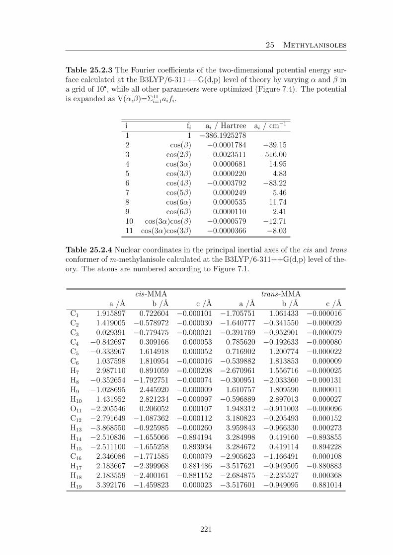
25 Methylanisoles
Table 25.2.3 The Fourier coefficients of the two-dimensional potential energy sur-face calculated at the B3LYP/6-311++G(d,p) level of theory by varying α and β ina grid of 10°, while all other parameters were optimized (Figure 7.4). The potentialis expanded as V(α,β)=Σ11
i=1aifi.
i fi ai / Hartree ai / cm−1
1 1 −386.19252782 cos(β) −0.0001784 −39.153 cos(2β) −0.0023511 −516.004 cos(3α) 0.0000681 14.955 cos(3β) 0.0000220 4.836 cos(4β) −0.0003792 −83.227 cos(5β) 0.0000249 5.468 cos(6α) 0.0000535 11.749 cos(6β) 0.0000110 2.4110 cos(3α)cos(β) −0.0000579 −12.7111 cos(3α)cos(3β) −0.0000366 −8.03
Table 25.2.4 Nuclear coordinates in the principal inertial axes of the cis and transconformer of m-methylanisole calculated at the B3LYP/6-311++G(d,p) level of the-ory. The atoms are numbered according to Figure 7.1.
cis-MMA trans-MMAa /A b /A c /A a /A b /A c /A
C1 1.915897 0.722604 −0.000101 −1.705751 1.061433 −0.000016C2 1.419005 −0.578972 −0.000030 −1.640777 −0.341550 −0.000029C3 0.029391 −0.779475 −0.000021 −0.391769 −0.952901 −0.000079C4 −0.842697 0.309166 0.000053 0.785620 −0.192633 −0.000080C5 −0.333967 1.614918 0.000052 0.716902 1.200774 −0.000022C6 1.037598 1.810954 −0.000016 −0.539882 1.813853 0.000009H7 2.987110 0.891059 −0.000208 −2.670961 1.556716 −0.000025H8 −0.352654 −1.792751 −0.000074 −0.300951 −2.033360 −0.000131H9 −1.028695 2.445920 −0.000009 1.610757 1.809590 0.000011H10 1.431952 2.821234 −0.000097 −0.596889 2.897013 0.000027O11 −2.205546 0.206052 0.000107 1.948312 −0.911003 −0.000096C12 −2.791649 −1.087362 −0.000112 3.180823 −0.205493 0.000152H13 −3.868550 −0.925985 −0.000260 3.959843 −0.966330 0.000273H14 −2.510836 −1.655066 −0.894194 3.284998 0.419160 −0.893855H15 −2.511100 −1.655258 0.893934 3.284672 0.419114 0.894228C16 2.346086 −1.771585 0.000079 −2.905623 −1.166491 0.000108H17 2.183667 −2.399968 0.881486 −3.517621 −0.949505 −0.880883H18 2.183559 −2.400161 −0.881152 −2.684875 −2.235527 0.000368H19 3.392176 −1.459823 0.000023 −3.517601 −0.949095 0.881014
221

25 Methylanisoles
Table 25.2.5 Observed A and E species frequencies (νo) of 223 rotational transi-tions of cis-MMA. The residues νo − νc (in kHz) are indicated as Fit I with theprogram XIAM as well as Fit II and III with the program aixPAM.
N° J Ka Kc J ′ Ka’ Kc’ Species νo /GHz Fit I Fit II Fit III1 1 1 1 0 0 0 E 2.9161173 −11.2 −10.5 1.62 3 1 3 2 1 1 E 3.4355118 −59.0 −48.8 2.73 2 1 1 2 0 2 E 3.7158559 84.7 65.4 −5.54 2 0 2 1 0 1 E 4.0337647 −21.7 −18.4 −0.95 2 2 1 2 1 2 E 4.0710712 −28.9 −24.3 −9.36 3 1 2 3 0 3 E 4.2363323 69.6 52.8 −2.47 4 1 3 4 0 4 A 4.4585285 −3.8 0.4 −1.48 4 1 4 3 1 2 E 4.5642978 −36.5 −29.4 4.59 3 0 3 2 1 2 E 4.9638191 −49.3 −38.9 −2.610 3 2 2 3 1 3 E 5.0537761 9.2 5.6 −0.611 3 1 2 2 2 1 E 5.1290768 45.8 34.6 0.812 3 0 3 2 1 2 A 5.1493335 2.6 3.4 0.713 2 1 2 1 0 1 E 5.2208816 16.6 10.5 0.414 5 1 5 4 1 3 E 5.4342942 −11.4 −8.3 1.815 2 1 2 1 0 1 A 5.4384815 −6.6 −2.7 0.216 4 1 3 3 2 2 A 5.5434310 12.4 11.2 2.717 2 2 1 2 1 2 A 5.6281840 −18.9 −11.6 −5.418 6 1 6 5 1 4 E 5.9057422 10.7 11.4 3.919 3 1 3 2 1 2 E 5.9642514 −12.0 −11.7 −3.520 5 1 4 5 0 5 E 5.9723023 7.7 0.5 −1.521 2 2 1 1 1 0 E 6.0901840 −94.1 −79.8 −16.722 3 0 3 2 0 2 E 6.1509362 −10.8 −10.0 −1.123 3 2 2 3 1 3 A 6.2808721 −17.2 −10.0 −4.924 4 2 3 4 1 4 E 6.4299941 24.3 15.0 −1.225 4 3 2 4 2 3 E 6.5454061 −6.2 1.8 1.726 2 1 1 1 1 1 E 6.7088208 47.4 36.8 −2.627 3 3 1 3 2 2 E 6.7254967 0.6 9.0 0.828 5 3 3 5 2 4 E 6.8409441 0.4 3.6 0.729 3 2 2 2 2 1 E 6.9469465 16.2 8.5 −4.630 4 1 3 3 2 2 E 6.9630852 −10.5 −10.4 −2.231 3 1 3 2 0 2 A 7.0472855 −6.3 −2.2 1.032 4 0 4 3 1 3 E 7.1216639 −39.8 −31.4 −2.633 3 1 3 2 0 2 E 7.1513715 29.5 20.4 1.034 4 2 3 4 1 4 A 7.1619599 −14.0 −6.8 −3.135 4 0 4 3 1 3 A 7.3657758 0.1 2.1 0.436 6 2 4 5 3 3 A 7.3722525 16.3 17.0 6.537 6 1 5 6 0 6 E 7.5427575 −9.2 −13.7 −3.4
222

25 Methylanisoles
Table 25.2.5 (continued)
N° J Ka Kc J ′ Ka’ Kc’ Species νo /GHz Fit I Fit II Fit III38 6 3 4 6 2 5 E 7.7749852 6.6 4.0 −1.739 5 2 4 5 1 5 E 7.8932484 25.4 13.6 −1.040 6 1 5 6 0 6 A 8.0362670 −5.9 0.3 −2.841 4 0 4 3 0 3 E 8.1220986 0.0 −1.6 −1.042 5 2 4 5 1 5 A 8.2628641 −10.8 −4.0 −1.543 5 1 4 4 2 3 A 8.3946707 9.9 10.8 3.644 3 3 1 3 2 2 A 8.4055115 −19.8 −10.7 −3.045 3 2 2 2 1 1 E 8.4892882 −49.6 −42.9 2.446 3 3 1 3 1 2 E 8.5433721 −23.3 −11.4 1.047 4 1 4 3 0 3 A 8.5758329 −8.0 −4.0 −0.848 4 3 2 4 2 3 A 8.5879992 −14.8 −6.8 −0.749 4 1 4 3 0 3 E 8.8006308 33.9 24.0 2.850 5 1 4 4 2 3 E 8.8027816 −42.0 −34.5 −2.251 5 2 3 4 3 2 E 8.8797396 60.4 42.7 −5.852 5 3 3 5 2 4 A 8.9273059 −9.0 −2.1 2.053 9 4 5 9 3 6 A 8.9512384 22.4 10.0 3.854 4 2 2 3 2 1 E 9.0623242 0.9 0.9 −1.055 4 3 1 4 2 2 E 9.0844067 −16.8 −26.9 1.256 7 3 5 7 2 6 E 9.1415482 7.8 0.6 −1.057 4 2 3 3 2 2 E 9.1764116 6.9 1.1 −1.958 2 2 1 1 1 0 A 9.1906043 −15.8 −7.2 −0.659 3 1 2 2 1 2 E 9.2001514 20.3 13.7 −5.160 7 4 4 7 3 5 E 9.2253951 70.9 65.9 −1.961 5 0 5 4 1 4 E 9.2604730 −25.8 −20.4 −2.362 2 2 1 1 0 1 E 9.2919630 −2.1 −3.6 1.363 6 2 5 6 1 6 E 9.3477827 21.6 9.8 −1.064 8 4 5 8 3 6 E 9.4012444 43.1 37.4 −3.165 5 0 5 4 1 4 A 9.4399310 −1.0 1.7 1.266 7 1 6 7 0 7 E 9.4607873 −8.3 −11.7 −1.667 6 3 4 6 2 5 A 9.4639897 −4.7 1.3 2.968 6 4 3 6 3 4 E 9.5451345 89.4 86.6 3.269 6 2 5 6 1 6 A 9.5600321 −3.1 3.1 4.870 5 1 5 4 1 4 E 9.6509606 −5.8 −6.1 −1.171 2 2 0 1 1 1 A 9.6708952 −13.7 −4.8 1.172 8 4 4 8 3 5 A 9.7151436 9.4 0.9 −1.373 5 0 5 4 0 4 E 9.9390029 5.9 2.9 −0.774 5 4 2 5 3 3 E 9.9644348 97.9 96.0 −1.475 7 1 6 7 0 7 A 10.0497077 3.2 9.4 7.876 9 2 7 9 1 8 A 10.0816307 −6.7 6.3 1.477 5 1 5 4 0 4 A 10.1246129 −6.3 −2.5 0.478 7 3 5 7 2 6 A 10.2231408 0.8 6.1 5.279 4 4 1 4 3 2 E 10.2297858 110.0 107.7 −0.1
223

25 Methylanisoles
Table 25.2.5 (continued)
N° J Ka Kc J ′ Ka’ Kc’ Species νo /GHz Fit I Fit II Fit III80 9 4 6 9 3 7 E 10.2603072 −0.2 −3.7 −3.081 5 1 5 4 0 4 E 10.3294882 23.6 14.9 −1.882 7 4 3 7 3 4 A 10.4585702 7.6 4.2 4.883 2 2 0 1 1 0 E 10.4679248 15.5 5.8 0.884 8 3 6 8 2 7 E 10.6249567 8.8 −1.9 0.385 7 2 5 6 3 4 A 10.6536607 5.5 9.8 2.086 7 2 6 7 1 7 E 10.8347996 17.6 7.1 −1.287 6 1 5 5 2 4 E 10.9361577 −45.3 −35.4 −0.488 3 2 2 2 1 1 A 10.9718115 −11.0 −2.6 4.189 4 2 3 3 1 2 E 10.9942893 −14.7 −17.0 0.790 7 2 6 7 1 7 A 11.0175288 0.2 5.3 7.091 3 2 2 2 1 2 E 11.0180331 2.8 −0.4 1.592 6 4 2 6 3 3 A 11.0236642 −0.6 0.6 2.693 6 2 4 5 3 3 E 11.0425637 38.3 28.7 −0.894 5 2 4 4 2 3 E 11.1142156 −4.1 −6.8 −0.295 6 1 5 5 2 4 A 11.1652862 3.4 5.7 0.796 8 3 6 8 2 7 A 11.2105471 1.5 6.0 3.297 6 0 6 5 1 5 E 11.2866453 −14.0 −11.4 −1.498 5 4 1 5 3 2 A 11.3687478 −7.6 −2.8 0.299 6 0 6 5 1 5 A 11.3918816 −3.2 −0.3 0.3100 6 1 6 5 1 5 E 11.4875533 −5.1 −5.9 −2.4101 8 1 7 8 0 8 E 11.5041310 −3.1 −5.4 −2.2102 4 4 0 4 3 1 A 11.5413878 −13.0 −5.7 −1.7103 9 4 5 9 3 6 E 11.5603574 −31.0 −9.1 2.6104 5 4 2 5 3 3 A 11.5846409 −7.2 −1.9 0.0105 4 4 1 4 3 2 A 11.5985726 −13.7 −6.3 −2.6106 6 4 3 6 3 4 A 11.6121592 −2.6 0.4 −0.1107 10 4 7 10 3 8 E 11.6190229 −34.2 −36.4 0.5108 10 4 6 10 3 7 E 11.6585809 −65.7 −16.9 0.3109 6 0 6 5 0 5 E 11.6771471 20.1 17.1 14.0110 8 4 4 8 3 5 E 11.7161635 −26.4 −24.7 0.7111 7 4 4 7 3 5 A 11.7274919 2.7 3.4 −0.1112 6 1 6 5 0 5 A 11.7449811 −3.5 −0.2 2.4113 6 1 6 5 0 5 E 11.8780433 17.3 10.7 1.2114 11 4 7 11 3 8 E 11.8801055 −105.6 −36.1 −0.9115 8 4 5 8 3 6 A 11.9824318 9.1 8.0 1.0116 4 1 3 3 1 3 E 12.0168621 −0.5 −4.0 −2.0117 7 4 3 7 3 4 E 12.0301690 −37.4 −46.3 0.6118 8 1 7 8 0 8 A 12.0362175 5.4 10.1 11.0119 9 3 7 9 2 8 E 12.0896745 14.6 1.6 2.1120 10 2 8 10 1 9 A 12.2383171 −19.8 −6.4 −9.3
224
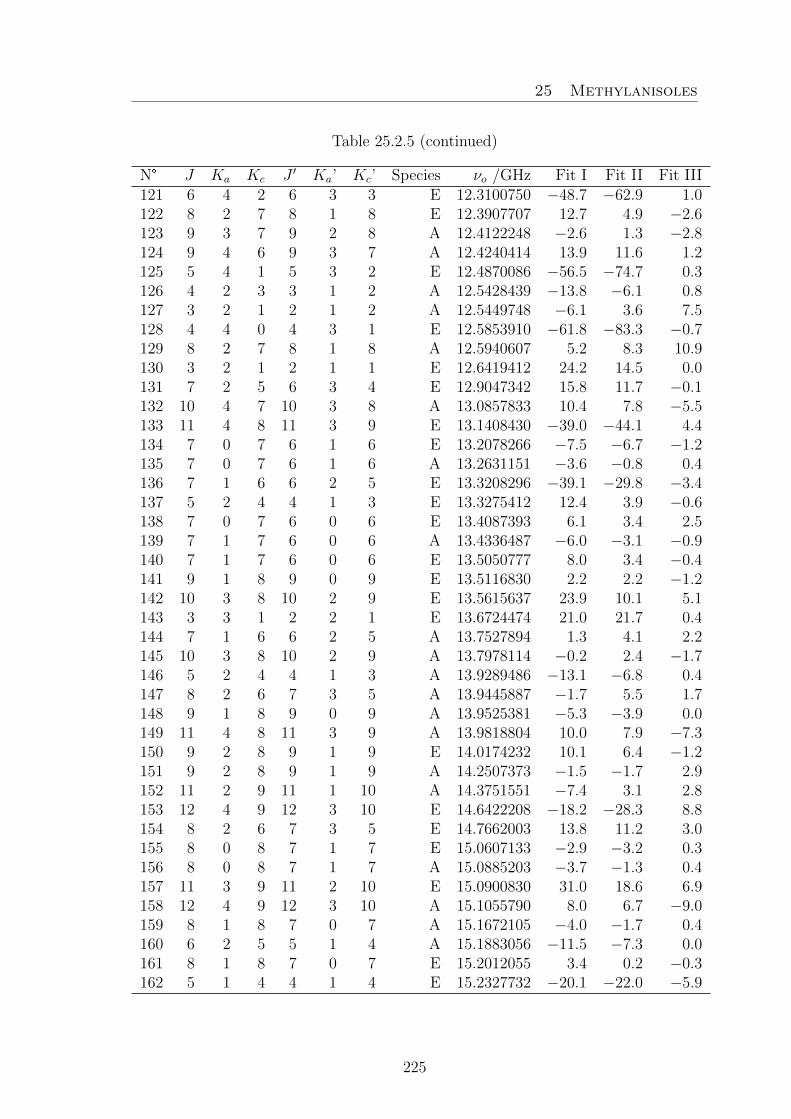
25 Methylanisoles
Table 25.2.5 (continued)
N° J Ka Kc J ′ Ka’ Kc’ Species νo /GHz Fit I Fit II Fit III121 6 4 2 6 3 3 E 12.3100750 −48.7 −62.9 1.0122 8 2 7 8 1 8 E 12.3907707 12.7 4.9 −2.6123 9 3 7 9 2 8 A 12.4122248 −2.6 1.3 −2.8124 9 4 6 9 3 7 A 12.4240414 13.9 11.6 1.2125 5 4 1 5 3 2 E 12.4870086 −56.5 −74.7 0.3126 4 2 3 3 1 2 A 12.5428439 −13.8 −6.1 0.8127 3 2 1 2 1 2 A 12.5449748 −6.1 3.6 7.5128 4 4 0 4 3 1 E 12.5853910 −61.8 −83.3 −0.7129 8 2 7 8 1 8 A 12.5940607 5.2 8.3 10.9130 3 2 1 2 1 1 E 12.6419412 24.2 14.5 0.0131 7 2 5 6 3 4 E 12.9047342 15.8 11.7 −0.1132 10 4 7 10 3 8 A 13.0857833 10.4 7.8 −5.5133 11 4 8 11 3 9 E 13.1408430 −39.0 −44.1 4.4134 7 0 7 6 1 6 E 13.2078266 −7.5 −6.7 −1.2135 7 0 7 6 1 6 A 13.2631151 −3.6 −0.8 0.4136 7 1 6 6 2 5 E 13.3208296 −39.1 −29.8 −3.4137 5 2 4 4 1 3 E 13.3275412 12.4 3.9 −0.6138 7 0 7 6 0 6 E 13.4087393 6.1 3.4 2.5139 7 1 7 6 0 6 A 13.4336487 −6.0 −3.1 −0.9140 7 1 7 6 0 6 E 13.5050777 8.0 3.4 −0.4141 9 1 8 9 0 9 E 13.5116830 2.2 2.2 −1.2142 10 3 8 10 2 9 E 13.5615637 23.9 10.1 5.1143 3 3 1 2 2 1 E 13.6724474 21.0 21.7 0.4144 7 1 6 6 2 5 A 13.7527894 1.3 4.1 2.2145 10 3 8 10 2 9 A 13.7978114 −0.2 2.4 −1.7146 5 2 4 4 1 3 A 13.9289486 −13.1 −6.8 0.4147 8 2 6 7 3 5 A 13.9445887 −1.7 5.5 1.7148 9 1 8 9 0 9 A 13.9525381 −5.3 −3.9 0.0149 11 4 8 11 3 9 A 13.9818804 10.0 7.9 −7.3150 9 2 8 9 1 9 E 14.0174232 10.1 6.4 −1.2151 9 2 8 9 1 9 A 14.2507373 −1.5 −1.7 2.9152 11 2 9 11 1 10 A 14.3751551 −7.4 3.1 2.8153 12 4 9 12 3 10 E 14.6422208 −18.2 −28.3 8.8154 8 2 6 7 3 5 E 14.7662003 13.8 11.2 3.0155 8 0 8 7 1 7 E 15.0607133 −2.9 −3.2 0.3156 8 0 8 7 1 7 A 15.0885203 −3.7 −1.3 0.4157 11 3 9 11 2 10 E 15.0900830 31.0 18.6 6.9158 12 4 9 12 3 10 A 15.1055790 8.0 6.7 −9.0159 8 1 8 7 0 7 A 15.1672105 −4.0 −1.7 0.4160 6 2 5 5 1 4 A 15.1883056 −11.5 −7.3 0.0161 8 1 8 7 0 7 E 15.2012055 3.4 0.2 −0.3162 5 1 4 4 1 4 E 15.2327732 −20.1 −22.0 −5.9
225

25 Methylanisoles
Table 25.2.5 (continued)
N° J Ka Kc J ′ Ka’ Kc’ Species νo /GHz Fit I Fit II Fit III163 6 2 5 5 1 4 E 15.2535224 29.8 18.7 0.4164 11 3 9 11 2 10 A 15.3273385 −2.6 −2.3 −5.3165 5 2 3 4 2 3 E 15.4251472 55.8 46.0 −2.6166 10 1 9 10 0 10 E 15.4326392 0.7 5.4 −2.0167 10 2 9 10 1 10 E 15.6979538 3.9 6.2 −1.9168 4 3 2 3 2 2 E 15.7218191 2.1 4.3 1.3169 8 1 7 7 2 6 E 15.7300439 −24.4 −16.5 −1.5170 10 1 9 10 0 10 A 15.8056110 −2.6 −6.3 1.3171 10 2 9 10 1 10 A 15.9564342 5.6 0.7 8.2172 8 1 7 7 2 6 A 16.1072052 −2.3 −0.3 0.6173 7 2 6 6 1 5 A 16.4149024 −7.9 −6.2 0.8174 12 3 10 12 2 11 E 16.6927630 36.6 27.8 9.7175 7 2 6 6 1 5 E 16.7971179 32.9 22.4 −0.1176 9 0 9 8 1 8 E 16.8759080 −2.3 −3.2 −0.6177 9 0 9 8 1 8 A 16.8902579 −3.5 −1.9 0.0178 9 1 9 8 0 8 A 16.9253971 −3.6 −2.1 −0.1179 9 1 9 8 0 8 E 16.9396690 0.9 −1.5 −0.4180 9 2 7 8 3 6 E 16.9554860 24.0 21.0 6.8181 12 3 10 12 2 11 A 16.9593890 −5.5 −8.9 −9.7182 9 2 7 8 3 6 A 17.1198283 −12.8 −4.3 −3.2183 11 1 10 11 0 11 E 17.2807750 −2.3 9.5 0.7184 11 2 10 11 1 11 E 17.4124802 −2.2 8.2 −0.2185 11 1 10 11 0 11 A 17.6161183 −3.6 −14.3 −2.5186 11 2 10 11 1 11 A 17.6893438 1.7 −9.8 1.8187 8 2 7 7 1 6 A 17.7115548 −10.7 −11.5 −5.3188 6 2 4 5 2 4 E 17.8835072 38.1 31.8 −0.7189 9 1 8 8 2 7 E 17.9968183 −14.8 −7.9 −1.2190 8 2 7 7 1 6 E 18.1311900 25.5 17.8 −0.2191 9 1 8 8 2 7 A 18.2487456 −3.7 −3.8 −0.5192 10 0 10 9 1 9 E 18.6722036 −0.9 −2.2 0.2193 10 0 10 9 1 9 A 18.6805483 −1.9 −1.3 0.5194 10 1 10 9 0 9 A 18.6958581 −2.3 −1.7 0.1195 10 1 10 9 0 9 E 18.7003218 −0.2 −2.3 −0.5196 6 1 5 5 1 5 E 18.8294023 −23.8 −25.6 −5.2197 12 1 11 12 0 12 E 19.0823565 −16.0 5.6 −2.9198 12 2 11 12 1 12 E 19.1452835 −14.5 6.3 −1.7199 12 1 11 12 0 12 A 19.4014661 1.7 −17.8 −0.8200 12 2 11 12 1 12 A 19.4359221 2.1 −17.9 −1.2201 9 2 8 8 1 7 E 19.4529614 14.2 10.5 0.8202 10 2 8 9 3 7 E 19.4840653 30.6 27.9 6.5203 10 2 8 9 3 7 A 20.0615164 −18.0 −11.5 −5.5204 10 1 9 9 2 8 E 20.0874203 −9.6 −2.6 0.0
226

25 Methylanisoles
Table 25.2.5 (continued)
N° J Ka Kc J ′ Ka’ Kc’ Species νo /GHz Fit I Fit II Fit III205 10 1 9 9 2 8 A 20.2354214 −3.6 −6.5 −1.7206 11 0 11 10 1 10 E 20.4596381 −0.6 −2.2 0.0207 11 0 11 10 1 10 A 20.4655676 −0.9 −1.6 0.1208 11 1 11 10 0 10 E 20.4717746 0.0 −2.0 0.1209 11 1 11 10 0 10 A 20.4721116 −0.7 −1.5 0.2210 7 2 5 6 2 5 E 20.6797207 23.7 17.0 −0.5211 10 2 9 9 1 8 E 20.8865935 2.4 2.6 −0.3212 11 1 10 10 2 9 E 22.0424557 −10.3 −2.5 −0.9213 11 2 9 10 3 8 E 22.1166584 30.2 30.4 5.6214 11 1 10 10 2 9 A 22.1252605 −1.3 −7.7 −1.8215 12 0 12 11 1 11 E 22.2431463 −0.5 −2.4 −0.4216 12 0 12 11 1 11 A 22.2482604 2.7 0.3 1.7217 12 1 12 11 0 11 E 22.2482971 −0.1 −2.2 −0.3218 12 1 12 11 0 11 A 22.2510131 1.2 −1.2 0.2219 11 2 10 10 1 9 E 22.4516132 −5.3 −1.6 −0.5220 11 2 9 10 3 8 A 22.7025932 −19.5 −18.0 −8.2221 12 1 11 11 2 10 E 23.9130250 −11.8 −2.5 −0.6222 13 1 13 12 0 12 E 24.0271699 0.9 −1.4 0.2223 13 1 13 12 0 12 A 24.0311061 4.8 0.5 1.5
Table 25.2.6 Observed A and E species frequencies (νo) of 320 rotational transi-tions of trans-MMA. νo−νc values (in kHz) as obtained after Fit I with the programXIAM as well as Fit II and III with the program aixPAM.
N° J Ka Kc J ′ Ka’ Kc’ Species νo /GHz Fit I Fit II Fit III1 1 1 0 1 0 1 A 2.7116996 16.9 18.1 6.72 2 1 1 2 0 2 A 2.9948123 13.4 14.6 4.23 3 1 2 3 0 3 A 3.4558352 6.1 7.2 −1.44 2 0 2 1 0 1 E 3.6295306 −15.5 −9.6 −0.65 3 0 3 2 1 2 A 3.6706874 −14.7 −13.3 1.06 2 0 2 1 0 1 A 3.9505226 −3.6 −2.0 −0.67 2 2 1 1 1 0 E 4.0340835 3.0 −0.3 1.08 2 1 2 1 1 1 E 4.0465785 −5.3 −7.7 1.79 3 0 3 2 1 2 E 4.0699138 −91.4 −66.6 −3.010 4 1 3 4 0 4 A 4.1328129 1.0 2.1 −4.011 2 1 1 1 1 0 A 4.2336349 −7.5 −5.9 −3.412 5 1 4 5 0 5 A 5.0625575 0.7 1.7 −1.213 2 1 2 1 0 1 E 5.1674668 67.9 52.1 4.014 6 1 5 6 1 6 A 5.4448549 −1.2 0.3 10.3
227

25 Methylanisoles
Table 25.2.6 (continued)
N° J Ka Kc J ′ Ka’ Kc’ Species νo /GHz Fit I Fit II Fit III15 3 1 3 2 1 2 A 5.5492272 −3.6 −1.6 −0.516 2 2 1 2 1 2 E 5.5640164 17.4 21.0 −10.317 3 0 3 2 0 2 E 5.6078489 −9.3 −6.1 0.518 3 1 3 2 1 2 E 5.7865202 −14.9 −14.5 2.019 4 0 4 3 1 3 E 5.8450141 −73.7 −49.2 −0.420 4 0 4 3 1 3 A 5.8685856 −17.0 −14.8 −0.621 3 0 3 2 0 2 A 5.8763653 −4.5 −2.2 −0.622 3 2 2 3 1 3 E 5.8906275 37.3 35.7 −8.423 3 2 2 2 2 1 A 5.9558998 8.4 9.9 12.624 3 2 1 2 2 0 A 6.0354262 6.7 8.3 11.525 3 1 2 2 1 1 E 6.0909318 10.5 8.2 3.026 3 2 2 2 2 1 E 6.1131315 5.3 0.5 4.127 2 1 2 1 0 1 A 6.1562028 8.9 11.3 0.028 6 1 5 6 0 6 A 6.2654895 −3.0 −1.9 −1.829 3 1 2 2 1 1 A 6.3373940 −5.9 −3.7 −0.430 6 2 4 6 1 5 A 6.4353089 21.5 21.5 −0.131 5 2 3 5 1 4 A 6.5256579 24.0 24.8 0.332 7 2 5 7 1 6 A 6.5508021 19.1 18.4 0.133 4 2 3 4 1 4 E 6.6374148 57.6 50.1 −2.134 6 1 5 5 2 4 A 6.6508122 −34.2 −31.5 −0.535 4 2 2 4 1 3 A 6.7594806 23.8 25.5 −0.936 3 2 1 3 1 2 A 7.0640203 23.7 26.2 −1.037 7 1 6 7 1 7 A 7.1711539 −17.4 −15.7 −5.838 3 1 3 2 0 2 E 7.3244535 65.4 44.2 3.639 4 1 4 3 1 3 A 7.3776813 −4.6 −2.1 −0.840 4 1 4 3 1 3 E 7.5007481 −18.8 −17.5 −0.141 4 0 4 3 0 3 E 7.5616174 −0.2 0.0 1.542 9 2 7 9 1 8 A 7.5848881 15.2 14.3 −0.543 7 1 6 7 0 7 A 7.7329525 −3.9 −2.7 −0.644 4 0 4 3 0 3 A 7.7471270 −4.4 −1.6 −0.545 3 1 3 2 0 2 A 7.7549066 8.1 11.0 −0.746 5 2 4 5 1 5 E 7.7569559 39.0 28.9 −19.547 5 0 5 4 1 4 E 7.8065675 −46.6 −25.9 1.948 4 2 3 3 2 2 A 7.9255453 −5.2 −3.4 −0.249 4 3 2 3 3 1 A 7.9785873 −6.8 −6.2 −2.350 4 3 1 3 3 0 A 7.9843355 −1.3 −0.7 3.251 4 3 2 3 3 1 E 8.0140673 2.6 −1.5 1.052 4 2 2 3 2 1 E 8.0390019 4.3 3.8 3.053 4 1 3 3 1 2 E 8.0403926 6.3 4.8 1.754 5 0 5 4 1 4 A 8.0469760 −16.7 −13.8 −0.655 4 2 2 3 2 1 A 8.1195685 −5.8 −3.9 0.5
228

25 Methylanisoles
Table 25.2.6 (continued)
N° J Ka Kc J ′ Ka’ Kc’ Species νo /GHz Fit I Fit II Fit III56 2 2 1 2 1 2 A 8.1350522 17.5 20.9 −4.057 4 2 3 3 2 2 E 8.2475354 1.4 −3.1 6.258 9 2 7 8 3 6 A 8.3231118 −22.6 −15.8 −3.559 4 1 3 3 1 2 A 8.4241076 −6.6 −3.9 −0.260 3 2 2 3 1 3 A 8.5417189 23.7 26.6 3.161 10 2 8 10 1 9 A 8.5659508 18.6 18.5 1.562 2 1 1 1 0 1 E 8.7444853 −28.8 −44.2 −0.663 8 1 7 8 1 8 A 9.0545257 −12.5 −10.8 −2.564 4 2 3 4 1 4 A 9.0895779 18.0 20.3 −1.365 6 2 5 6 1 6 E 9.1060592 48.2 38.8 5.366 5 1 5 4 1 4 A 9.1905442 −5.4 −2.6 −1.267 4 1 4 3 0 3 E 9.2173509 54.2 31.1 1.368 5 1 5 4 1 4 E 9.2348625 −14.7 −14.0 −0.169 4 1 4 3 0 3 A 9.2562227 8.0 11.0 −0.770 4 2 2 4 1 3 E 9.2839328 −8.3 −16.9 −10.171 3 2 1 3 1 2 E 9.2853196 −10.4 −20.0 −15.472 7 1 6 6 2 5 A 9.3225058 −33.4 −29.7 −1.073 8 1 7 8 0 8 A 9.4246325 −4.0 −2.9 −0.474 5 2 3 5 1 4 E 9.4294938 −12.4 −17.8 −10.375 5 0 5 4 0 4 E 9.4623014 8.3 5.7 2.276 5 0 5 4 0 4 A 9.5560717 −4.3 −1.2 −0.877 6 2 4 6 1 5 E 9.6997892 −14.6 −12.4 0.278 5 2 4 5 1 5 A 9.7809211 18.5 20.0 0.479 11 3 8 11 2 9 A 9.8052256 24.4 13.2 3.880 12 3 9 12 2 10 A 9.8132654 27.8 16.6 2.781 6 3 4 6 2 5 E 9.8622943 −77.4 −64.4 −1.082 6 0 6 5 1 5 E 9.8652957 −20.9 −5.3 2.383 5 2 4 4 2 3 A 9.8818869 −5.5 −3.6 −0.184 7 3 5 7 2 6 E 9.9155281 −55.4 −41.4 1.985 5 1 4 4 1 3 E 9.9477068 3.9 1.3 −0.886 5 4 2 4 4 1 A 9.9710227 −3.6 −5.4 −0.387 5 4 1 4 4 0 A 9.9713195 −11.1 −12.9 −7.888 5 4 2 4 4 1 E 9.9840881 0.6 −2.9 −3.089 5 3 3 4 3 2 A 9.9859205 −4.2 −3.8 0.790 5 3 2 4 3 1 E 9.9994146 1.7 2.6 0.391 5 3 2 4 3 1 A 10.0058576 −4.7 −4.3 0.392 7 2 5 7 1 6 E 10.0290718 −41.3 −27.8 1.793 5 3 3 4 3 2 E 10.0526946 1.5 −2.2 0.994 10 3 7 10 2 8 A 10.0555892 22.8 12.8 3.695 5 3 3 5 2 4 E 10.0773520 −98.4 −82.8 1.296 5 2 3 4 2 2 E 10.0932743 6.3 6.8 5.597 6 0 6 5 1 5 A 10.1662429 −13.7 −10.5 0.8
229

25 Methylanisoles
Table 25.2.6 (continued)
N° J Ka Kc J ′ Ka’ Kc’ Species νo /GHz Fit I Fit II Fit III98 5 2 3 4 2 2 A 10.2519894 −8.7 −6.5 −1.199 4 2 3 3 1 2 E 10.2567241 101.3 97.6 −7.3100 8 2 6 8 1 7 E 10.3439282 −79.0 −55.2 3.8101 5 2 4 4 2 3 E 10.3544263 −10.5 −12.5 5.1102 4 3 2 4 2 3 E 10.3790858 −108.3 −91.0 7.5103 5 1 4 4 1 3 A 10.4858147 −6.2 −3.1 0.3104 9 3 6 9 2 7 A 10.4916263 23.5 15.7 4.4105 7 2 6 7 1 7 E 10.5437704 27.5 20.2 6.7106 3 3 1 3 2 2 E 10.6125522 −111.2 −94.3 11.0107 9 2 7 9 1 8 E 10.6139626 −117.3 −91.5 −1.8108 6 2 5 6 1 6 A 10.6158316 17.0 17.6 0.2109 5 1 5 4 0 4 A 10.6996382 5.3 8.3 −3.1110 5 1 5 4 0 4 E 10.8905964 40.1 17.6 0.1111 10 2 8 10 1 9 E 10.9026993 −118.9 −105.3 −2.1112 6 1 6 5 1 5 A 10.9868863 −6.7 −3.9 −2.5113 6 1 6 5 1 5 E 10.9897290 −8.8 −8.7 0.6114 8 3 5 8 2 6 A 11.0274913 21.6 16.3 2.7115 3 1 2 2 0 2 E 11.2058856 −3.7 −27.4 2.1116 9 1 8 9 0 9 A 11.2770061 −3.1 −2.5 −1.4117 6 0 6 5 0 5 E 11.2935926 12.9 8.5 2.2118 6 0 6 5 0 5 A 11.3098099 −3.7 −0.6 −1.0119 11 2 9 11 1 10 E 11.3445849 −65.9 −82.8 −1.2120 6 2 5 6 0 6 A 11.4364803 29.3 29.5 2.2121 7 3 4 7 2 5 A 11.5753217 18.4 15.8 0.8122 2 2 1 1 1 0 A 11.5795664 20.5 25.2 0.3123 7 2 6 7 1 7 A 11.5911300 18.3 17.7 2.4124 3 2 2 2 1 2 E 11.6771456 20.4 19.2 −8.5125 6 2 5 5 2 4 A 11.8217966 −8.3 −6.5 −2.9126 2 2 0 1 1 1 A 11.8626828 19.5 24.2 0.4127 6 1 5 5 1 4 E 11.8985497 15.9 10.3 4.4128 4 1 3 3 1 3 E 11.9218242 −63.4 −67.4 −0.4129 7 0 7 6 1 6 E 11.9321614 0.8 11.5 3.9130 6 5 2 5 5 1 A 11.9619984 −5.7 −11.8 −5.3131 6 5 1 5 5 0 A 11.9620143 −3.5 −9.5 −3.0132 6 5 2 5 5 1 E 11.9642552 −4.1 −4.8 −11.4133 6 4 2 5 4 1 E 11.9786691 1.1 2.7 −4.7134 6 4 3 5 4 2 A 11.9795440 −1.4 −4.0 1.4135 6 4 2 5 4 1 A 11.9809089 0.4 −2.2 3.2136 8 2 7 8 1 8 E 11.9886746 6.9 1.7 5.1137 8 1 7 7 2 6 A 11.9964021 −31.6 −27.2 −2.4138 6 3 4 5 3 3 A 11.9971864 −4.9 −4.8 0.0139 6 4 3 5 4 2 E 12.0013702 1.5 −1.1 −1.7
230
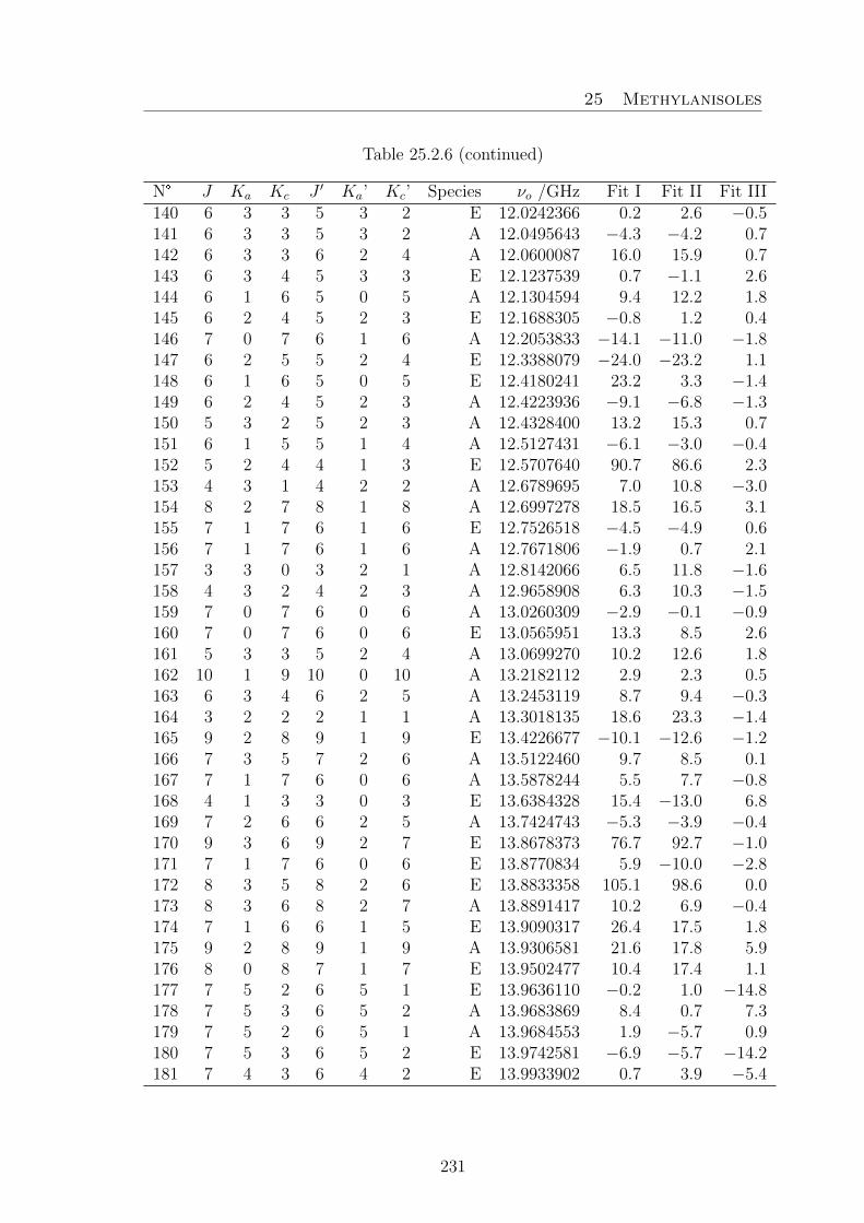
25 Methylanisoles
Table 25.2.6 (continued)
N° J Ka Kc J ′ Ka’ Kc’ Species νo /GHz Fit I Fit II Fit III140 6 3 3 5 3 2 E 12.0242366 0.2 2.6 −0.5141 6 3 3 5 3 2 A 12.0495643 −4.3 −4.2 0.7142 6 3 3 6 2 4 A 12.0600087 16.0 15.9 0.7143 6 3 4 5 3 3 E 12.1237539 0.7 −1.1 2.6144 6 1 6 5 0 5 A 12.1304594 9.4 12.2 1.8145 6 2 4 5 2 3 E 12.1688305 −0.8 1.2 0.4146 7 0 7 6 1 6 A 12.2053833 −14.1 −11.0 −1.8147 6 2 5 5 2 4 E 12.3388079 −24.0 −23.2 1.1148 6 1 6 5 0 5 E 12.4180241 23.2 3.3 −1.4149 6 2 4 5 2 3 A 12.4223936 −9.1 −6.8 −1.3150 5 3 2 5 2 3 A 12.4328400 13.2 15.3 0.7151 6 1 5 5 1 4 A 12.5127431 −6.1 −3.0 −0.4152 5 2 4 4 1 3 E 12.5707640 90.7 86.6 2.3153 4 3 1 4 2 2 A 12.6789695 7.0 10.8 −3.0154 8 2 7 8 1 8 A 12.6997278 18.5 16.5 3.1155 7 1 7 6 1 6 E 12.7526518 −4.5 −4.9 0.6156 7 1 7 6 1 6 A 12.7671806 −1.9 0.7 2.1157 3 3 0 3 2 1 A 12.8142066 6.5 11.8 −1.6158 4 3 2 4 2 3 A 12.9658908 6.3 10.3 −1.5159 7 0 7 6 0 6 A 13.0260309 −2.9 −0.1 −0.9160 7 0 7 6 0 6 E 13.0565951 13.3 8.5 2.6161 5 3 3 5 2 4 A 13.0699270 10.2 12.6 1.8162 10 1 9 10 0 10 A 13.2182112 2.9 2.3 0.5163 6 3 4 6 2 5 A 13.2453119 8.7 9.4 −0.3164 3 2 2 2 1 1 A 13.3018135 18.6 23.3 −1.4165 9 2 8 9 1 9 E 13.4226677 −10.1 −12.6 −1.2166 7 3 5 7 2 6 A 13.5122460 9.7 8.5 0.1167 7 1 7 6 0 6 A 13.5878244 5.5 7.7 −0.8168 4 1 3 3 0 3 E 13.6384328 15.4 −13.0 6.8169 7 2 6 6 2 5 A 13.7424743 −5.3 −3.9 −0.4170 9 3 6 9 2 7 E 13.8678373 76.7 92.7 −1.0171 7 1 7 6 0 6 E 13.8770834 5.9 −10.0 −2.8172 8 3 5 8 2 6 E 13.8833358 105.1 98.6 0.0173 8 3 6 8 2 7 A 13.8891417 10.2 6.9 −0.4174 7 1 6 6 1 5 E 13.9090317 26.4 17.5 1.8175 9 2 8 9 1 9 A 13.9306581 21.6 17.8 5.9176 8 0 8 7 1 7 E 13.9502477 10.4 17.4 1.1177 7 5 2 6 5 1 E 13.9636110 −0.2 1.0 −14.8178 7 5 3 6 5 2 A 13.9683869 8.4 0.7 7.3179 7 5 2 6 5 1 A 13.9684553 1.9 −5.7 0.9180 7 5 3 6 5 2 E 13.9742581 −6.9 −5.7 −14.2181 7 4 3 6 4 2 E 13.9933902 0.7 3.9 −5.4
231

25 Methylanisoles
Table 25.2.6 (continued)
N° J Ka Kc J ′ Ka’ Kc’ Species νo /GHz Fit I Fit II Fit III182 10 3 7 10 2 8 E 13.9936396 9.8 63.9 −0.4183 7 4 4 6 4 3 A 13.9952151 −1.1 −4.7 0.7184 7 4 3 6 4 2 A 13.9997254 −1.1 −4.7 0.7185 7 3 5 6 3 4 A 14.0094077 −5.0 −5.5 −0.7186 7 3 4 7 2 5 E 14.0174240 113.1 97.2 4.1187 7 4 4 6 4 3 E 14.0307413 −1.9 −2.6 −3.7188 7 3 4 6 3 3 E 14.0642005 −1.5 3.2 −0.7189 7 3 4 6 3 3 A 14.1242996 −4.3 −4.7 0.1190 8 0 8 7 1 7 A 14.1628979 −11.0 −8.4 −1.3191 7 2 6 6 2 5 E 14.1903629 −25.3 −23.6 2.0192 3 2 1 2 1 2 A 14.1905463 18.6 23.5 2.0193 6 3 3 6 2 4 E 14.1915341 110.6 92.3 4.4194 7 2 5 6 2 4 E 14.2383152 0.5 2.9 4.2195 7 3 5 6 3 4 E 14.2435955 −4.5 −1.7 3.7196 5 3 2 5 2 3 E 14.3361316 113.1 94.6 8.9197 5 1 4 4 1 4 E 14.3687851 −38.6 −46.4 1.1198 9 3 7 9 2 8 A 14.3912702 14.2 8.6 2.0199 4 3 1 4 2 2 E 14.4299872 113.6 94.7 9.9200 7 1 6 6 1 5 A 14.4934927 −5.0 −2.2 −0.9201 8 1 8 7 1 7 E 14.5138436 −2.4 −2.9 −0.1202 8 1 8 7 1 7 A 14.5330032 −4.1 −2.0 −0.6203 7 2 5 6 2 4 A 14.6089868 −6.5 −4.3 0.2204 9 1 8 8 2 7 A 14.6273683 −24.7 −20.3 −0.5205 8 0 8 7 0 7 A 14.7246917 −2.3 −0.2 −0.9206 8 0 8 7 0 7 E 14.7707414 8.5 4.3 1.1207 10 2 9 10 1 10 E 14.8620267 −19.6 −17.5 −8.4208 4 2 3 3 1 2 A 14.8899662 20.7 24.9 0.2209 6 2 5 5 1 4 E 14.9618682 65.9 65.2 7.3210 8 1 8 7 0 7 A 15.0947973 5.0 6.5 0.1211 10 2 9 10 1 10 A 15.2697243 19.3 13.5 2.5212 8 1 8 7 0 7 E 15.3343332 −8.4 −20.1 −4.2213 3 2 1 2 1 1 E 15.3762544 3.1 −8.8 −9.3214 8 2 7 7 2 6 A 15.6416009 −3.9 −3.3 −0.1215 9 0 9 8 1 8 E 15.8986334 19.8 24.2 5.0216 8 1 7 7 1 6 E 15.9427192 41.1 29.6 1.1217 8 2 7 7 2 6 E 15.9587466 −24.2 −22.6 −2.9218 8 5 4 7 5 3 A 15.9806052 8.0 −1.6 4.7219 8 5 3 7 5 2 A 15.9809028 7.7 −1.9 4.4220 8 5 4 7 5 3 E 15.9918671 −9.0 −4.8 −15.6221 8 4 4 7 4 3 E 16.0169055 −0.2 5.5 −6.0222 8 4 5 7 4 4 A 16.0183301 2.1 −2.9 2.1
232

25 Methylanisoles
Table 25.2.6 (continued)
N° J Ka Kc J ′ Ka’ Kc’ Species νo /GHz Fit I Fit II Fit III223 8 3 6 7 3 5 A 16.0184970 −3.0 −4.5 −0.2224 8 4 4 7 4 3 A 16.0305928 1.9 −3.0 1.8225 9 0 9 8 1 8 A 16.0500846 −8.5 −6.8 −1.4226 8 4 5 7 4 4 E 16.0757427 −5.8 −3.2 −5.2227 8 3 5 7 3 4 E 16.1234895 −2.6 5.6 1.2228 8 3 5 7 3 4 A 16.2391444 −3.2 −4.2 −0.5229 8 2 6 7 2 5 E 16.2575761 3.9 2.6 3.6230 9 1 9 8 1 8 E 16.2685251 0.3 −0.1 0.9231 9 1 9 8 1 8 A 16.2866544 −2.8 −1.7 −0.2232 5 2 4 4 1 3 A 16.3477450 21.3 24.7 −0.2233 8 3 6 7 3 5 E 16.4146989 −4.6 6.4 16.6234 8 1 7 7 1 6 A 16.4163712 −2.9 −0.9 −1.2235 9 0 9 8 0 8 A 16.4201904 −1.0 0.1 −0.2236 9 0 9 8 0 8 E 16.4622256 3.3 0.2 0.1237 9 1 9 8 0 8 A 16.6567587 3.2 3.7 −0.6238 11 2 10 11 1 11 A 16.7006922 20.5 12.0 1.5239 4 2 2 3 1 3 A 16.7608835 12.3 17.2 −1.1240 8 2 6 7 2 5 A 16.7869766 −4.6 −2.9 −0.6241 9 1 9 8 0 8 E 16.8321183 −15.2 −23.1 −3.0242 6 1 5 5 1 5 E 17.0324693 −11.0 −25.2 2.6243 10 1 9 9 2 8 A 17.1705335 −20.2 −16.7 −2.3244 7 2 6 6 1 5 E 17.2536830 26.4 32.9 6.5245 9 4 5 9 3 6 A 17.2612425 −37.3 −47.0 4.9246 9 2 8 8 2 7 A 17.5175824 −2.1 −2.7 0.2247 8 4 4 8 3 5 A 17.5840317 −42.6 −47.6 2.3248 6 2 5 5 1 4 A 17.6837304 22.6 24.9 0.1249 9 2 8 8 2 7 E 17.7025196 −15.2 −13.1 −4.0250 10 0 10 9 1 9 E 17.7805928 18.8 21.7 3.5251 7 4 3 7 3 4 A 17.7925818 −49.2 −50.2 −1.5252 10 0 10 9 1 9 A 17.8829772 −4.8 −4.4 −0.1253 6 4 2 6 3 3 A 17.9171537 −54.7 −52.5 −4.4254 9 1 8 8 1 7 E 17.9513422 53.5 42.0 2.6255 8 4 5 8 3 6 A 17.9801099 −43.0 −47.2 2.4256 7 4 4 7 3 5 A 17.9802767 −48.1 −48.9 0.1257 5 4 1 5 3 2 A 17.9858124 −56.1 −51.2 −3.6258 6 4 3 6 3 4 A 17.9944690 −52.3 −50.0 −1.7259 9 4 6 9 3 7 A 18.0088522 −38.6 −46.7 3.4260 5 4 2 5 3 3 A 18.0121088 −58.4 −53.4 −5.8261 10 1 10 9 1 9 E 18.0153966 0.2 −0.1 0.1262 9 3 7 8 3 6 A 18.0197100 1.0 −1.8 1.8263 4 4 0 4 3 1 A 18.0203402 −60.1 −52.9 −5.8
233

25 Methylanisoles
Table 25.2.6 (continued)
N° J Ka Kc J ′ Ka’ Kc’ Species νo /GHz Fit I Fit II Fit III264 4 4 1 4 3 2 A 18.0270028 −62.8 −55.6 −8.5265 10 1 10 9 1 9 A 18.0306684 −2.1 −2.3 −0.6266 9 4 6 8 4 5 A 18.0484502 3.2 −3.4 0.6267 9 4 5 8 4 4 E 18.0508893 −3.2 5.8 −8.0268 9 4 5 8 4 4 A 18.0773895 3.9 −2.5 0.9269 10 0 10 9 0 9 A 18.1195458 −0.3 −0.5 −0.2270 9 4 6 8 4 5 E 18.1419489 −10.9 −3.1 −6.1271 10 0 10 9 0 9 E 18.1504845 −0.7 −2.6 −0.6272 9 3 6 8 3 5 E 18.2058839 −7.4 5.7 1.8273 9 2 7 8 2 6 E 18.2213796 18.2 8.8 0.0274 10 1 10 9 0 9 A 18.2672371 2.5 1.7 −0.6275 9 1 8 8 1 7 A 18.2725647 0.6 1.2 −0.5276 10 1 10 9 0 9 E 18.3852899 −17.7 −22.8 −2.3277 9 3 6 8 3 5 A 18.4001812 1.2 −0.6 0.8278 9 3 7 8 3 6 E 18.5896762 −30.4 −10.8 7.8279 3 3 1 2 2 0 A 18.8486609 2.2 8.9 −1.1280 3 3 0 2 2 1 A 18.8696938 0.7 7.5 −2.4281 7 2 6 6 1 5 A 18.9134624 24.3 24.8 0.9282 9 2 7 8 2 6 A 18.9360451 −1.8 −1.0 −1.9283 8 2 7 7 1 6 E 19.3034023 −19.9 −2.7 6.2284 10 2 9 9 2 8 A 19.3697385 −0.4 −2.7 −0.1285 11 0 11 10 1 10 E 19.6096170 15.4 17.5 2.2286 5 2 3 4 1 4 A 19.6351917 8.4 13.0 −1.2287 11 0 11 10 1 10 A 19.6769551 −2.3 −3.8 −0.1288 11 1 11 10 1 10 E 19.7550586 −0.8 −0.7 −0.8289 11 1 11 10 1 10 A 19.7674663 2.6 0.7 2.7290 11 0 11 10 0 10 A 19.8246477 1.7 −0.3 0.8291 11 0 11 10 0 10 E 19.8444218 −2.2 −3.3 −0.1292 10 1 9 9 1 8 E 19.8954171 53.7 46.5 2.7293 11 1 11 10 0 10 A 19.9151551 2.9 0.4 −0.1294 7 1 6 6 1 6 E 19.9517705 22.6 −0.5 2.2295 11 1 11 10 0 10 E 19.9898637 −18.1 −21.2 −2.8296 10 3 8 9 3 7 A 20.0081512 4.1 −0.5 2.0297 10 1 9 9 1 8 A 20.0607490 3.8 2.4 −0.2298 8 2 7 7 1 6 A 20.0615698 24.6 22.9 1.0299 10 4 7 9 4 6 A 20.0842209 7.9 −0.7 1.6300 10 4 6 9 4 5 A 20.1454225 8.8 0.6 1.5301 10 2 8 9 2 7 E 20.1841505 48.8 29.5 −0.9302 10 3 7 9 3 6 E 20.3099558 −15.1 3.6 2.6303 10 3 7 9 3 6 A 20.6057728 4.7 1.8 −0.8304 10 3 8 9 3 7 E 20.6822769 −44.5 −20.9 3.0305 4 3 2 3 2 1 A 20.7918353 1.9 7.7 −1.8
234

25 Methylanisoles
Table 25.2.6 (continued)
N° J Ka Kc J ′ Ka’ Kc’ Species νo /GHz Fit I Fit II Fit III306 4 3 1 3 2 2 A 20.8981424 3.9 9.8 1.1307 10 2 8 9 2 7 A 21.0418092 4.7 4.0 −0.7308 9 2 8 8 1 7 E 21.0631964 −82.5 −51.7 −5.2309 9 2 8 8 1 7 A 21.1627802 24.6 20.3 1.6310 11 2 10 10 2 9 A 21.1984328 2.4 −2.1 0.4311 11 2 10 10 2 9 E 21.2208648 −0.3 7.9 −3.8312 12 0 12 11 1 11 E 21.4004635 12.6 14.2 2.4313 12 0 12 11 1 11 A 21.4444606 0.6 −3.2 0.4314 12 1 12 11 1 11 E 21.4889003 0.0 0.3 0.3315 12 0 12 11 0 11 E 21.5459057 −3.0 −3.4 0.1316 11 1 10 10 1 9 A 21.7904584 5.5 1.3 −1.1317 5 3 3 4 2 2 A 22.6581885 4.7 9.0 −0.4318 6 2 4 5 1 5 A 22.8670405 4.0 8.2 −1.9319 5 3 2 4 2 3 A 22.9784525 2.2 6.7 −0.6320 6 3 4 5 2 3 A 24.4033844 7.3 9.5 −0.4
235
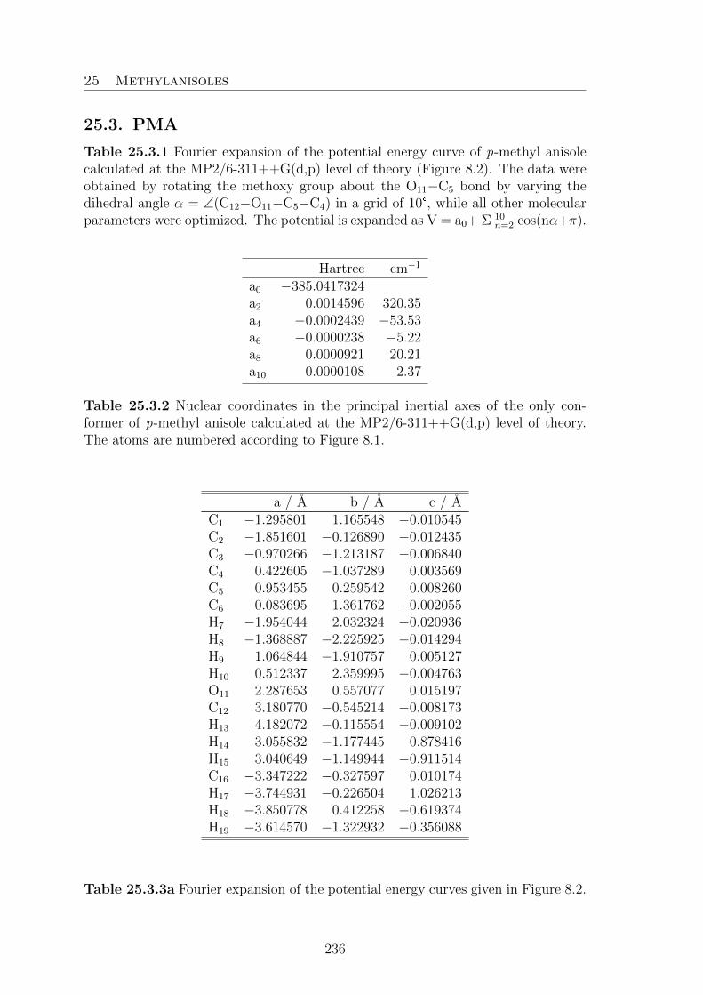
25 Methylanisoles
25.3. PMA
Table 25.3.1 Fourier expansion of the potential energy curve of p-methyl anisolecalculated at the MP2/6-311++G(d,p) level of theory (Figure 8.2). The data wereobtained by rotating the methoxy group about the O11−C5 bond by varying thedihedral angle α = ∠(C12−O11−C5−C4) in a grid of 10°, while all other molecularparameters were optimized. The potential is expanded as V = a0+ Σ 10
n=2 cos(nα+π).
Hartree cm−1
a0 −385.0417324a2 0.0014596 320.35a4 −0.0002439 −53.53a6 −0.0000238 −5.22a8 0.0000921 20.21a10 0.0000108 2.37
Table 25.3.2 Nuclear coordinates in the principal inertial axes of the only con-former of p-methyl anisole calculated at the MP2/6-311++G(d,p) level of theory.The atoms are numbered according to Figure 8.1.
a / A b / A c / AC1 −1.295801 1.165548 −0.010545C2 −1.851601 −0.126890 −0.012435C3 −0.970266 −1.213187 −0.006840C4 0.422605 −1.037289 0.003569C5 0.953455 0.259542 0.008260C6 0.083695 1.361762 −0.002055H7 −1.954044 2.032324 −0.020936H8 −1.368887 −2.225925 −0.014294H9 1.064844 −1.910757 0.005127H10 0.512337 2.359995 −0.004763O11 2.287653 0.557077 0.015197C12 3.180770 −0.545214 −0.008173H13 4.182072 −0.115554 −0.009102H14 3.055832 −1.177445 0.878416H15 3.040649 −1.149944 −0.911514C16 −3.347222 −0.327597 0.010174H17 −3.744931 −0.226504 1.026213H18 −3.850778 0.412258 −0.619374H19 −3.614570 −1.322932 −0.356088
Table 25.3.3a Fourier expansion of the potential energy curves given in Figure 8.2.
236

25 Methylanisoles
The potential is expanded as V(β)=Σ8i=0 bi fi.
MP2i fi bi / Hartree bi / cm−1
0 1 −385.04058591 cos(3β) 0.0022272 488.812 cos(6β) −0.0004268 −93.673 cos(9β) 0.0001935 42.474 cos(12β) 0.0000014 0.315 sin(3β) 0.0002251 49.406 sin(6β) −0.0000170 −3.737 sin(9β) −0.0001974 −43.328 sin(12β) 0.0002555 56.08
Table 25.3.3b Fourier expansion of the potential energy curves calculated at theMP2/6-311++G(d,p) and B3LYP/6-311++G(d,p) levels of theory given in Fig-ure 8.3. The potential is expanded as V(γ)=Σ4
i=0 bi fi.
B3LYP MP2i fi bi / Hartree bi / cm−1 bi / Hartree bi / cm−1
0 1 −386.1943875 −385.04325061 cos 3γ 0.0001018 22.34 0.0001023 22.452 cos 6γ 0.0000058 1.27 0.0000701 15.393 cos 9γ 0.0000013 0.29 0.0000054 1.194 cos 12γ −0.0000009 −0.20 0.0000052 1.14
237

25 Methylanisoles
Table 25.3.4 Observed 164 A and 183 E species frequencies (νo) in GHz of 347rotational transitions of p-methyl anisole. Differences to calculated values νo − νcare given in kHz as obtained after fits with the programs XIAM as X(vo–vc) andwith BELGI-Cs as B(vo–vc), respectively.
N J Ka Kc J ′ K ′a K ′c S vo X(vo–vc) B(vo–vc)1 5 0 5 4 1 4 A 5.7099652 −25.6 02 6 0 6 5 1 5 A 7.7735930 −26.6 03 7 0 7 6 1 6 A 9.8408191 −24.9 04 8 0 8 7 1 7 A 11.8940673 −22.0 05 9 0 9 8 1 8 A 13.9184379 −18.1 06 10 0 10 9 1 9 A 15.9032426 −12.6 07 11 0 11 10 1 10 A 17.8426703 −5.6 18 12 0 12 11 1 11 A 19.7354768 2.6 09 13 0 13 12 1 12 A 21.5839640 13.0 110 14 0 14 13 1 13 A 23.3926862 23.4 111 1 1 1 0 0 0 A 5.7037664 11.1 112 2 1 2 1 0 1 A 7.3533526 7.8 113 3 1 3 2 0 2 A 8.9252782 5.5 114 4 1 4 3 0 3 A 10.4256495 3.4 015 5 1 5 4 0 4 A 11.8642401 2.1 016 6 1 6 5 0 5 A 13.2542359 0.7 017 7 1 7 6 0 6 A 14.6115503 0.5 018 8 1 8 7 0 7 A 15.9535581 0.7 019 9 1 9 8 0 8 A 17.2973246 2.3 020 10 1 10 9 0 9 A 18.6576597 3.7 021 11 1 11 10 0 10 A 20.0455572 6.4 −122 12 1 12 11 0 11 A 21.4674665 10.2 −123 13 1 13 12 0 12 A 22.9255321 16.8 024 14 1 14 13 0 13 A 24.4185254 22.9 −225 1 1 0 1 0 1 A 4.0541734 8.0 −426 2 1 1 2 0 2 A 4.2179307 12.8 127 6 1 5 6 0 6 A 5.8963181 2.7 028 7 1 6 7 0 7 A 6.6384205 −1.0 −129 8 1 7 8 0 8 A 7.5363848 −5.5 −230 9 1 8 9 0 9 A 8.5978251 −5.8 031 10 1 9 10 0 10 A 9.8236049 −5.8 232 11 1 10 11 0 11 A 11.2068128 −8.4 −133 12 1 11 12 0 12 A 12.7329383 −3.6 134 13 1 12 13 0 13 A 14.3810690 2.0 135 14 1 13 14 0 14 A 16.1258833 10.4 136 15 1 14 15 0 15 A 17.9400243 24.1 237 8 1 7 7 2 6 A 5.2285636 −37.4 −238 9 1 8 8 2 7 A 7.6519306 −38.6 −7
238

25 Methylanisoles
Table 25.3.4. continued
N J Ka Kc J’ K ′a K ′c S vo X(vo–vc) B(vo–vc)39 10 1 9 9 2 8 A 10.1142344 −24.9 040 11 1 10 10 2 9 A 12.6035082 −14.6 241 12 1 11 11 2 10 A 15.1056162 −6.7 042 13 1 12 12 2 11 A 17.6045159 8.7 243 14 1 13 13 2 12 A 20.0829165 24.2 144 2 2 0 2 1 1 A 11.6902005 29.7 545 3 2 1 3 1 2 A 11.4737952 29.7 646 4 2 2 3 1 3 A 19.7055882 2.1 −547 5 2 3 4 1 4 A 21.9248788 0.4 −148 6 2 4 5 1 5 A 24.2916810 −6.2 −249 2 2 1 1 1 0 A 15.4615541 19.2 250 3 2 2 2 1 1 A 17.1111400 15.9 251 4 2 3 3 1 2 A 18.6805487 12.4 152 5 2 4 4 1 3 A 20.1703623 9.8 053 6 2 5 5 1 4 A 21.5818404 7.6 −154 7 2 6 6 1 5 A 22.9171195 6.6 −155 8 2 7 7 1 6 A 24.1794622 8.5 056 2 2 0 1 1 1 A 15.6253126 25.2 957 3 2 1 2 1 2 A 17.6119029 14.2 258 4 2 2 4 1 3 A 11.2093439 22.1 −159 5 2 3 5 1 4 A 10.9171843 21.8 160 6 2 4 6 1 5 A 10.6217704 18.2 061 7 2 5 7 1 6 A 10.3504518 13.8 −162 8 2 6 8 1 7 A 10.1317050 8.2 −163 9 2 7 9 1 8 A 9.9932243 2.2 −164 10 2 8 10 1 9 A 9.9603566 −4.6 065 11 2 9 11 1 10 A 10.0552886 −17.3 −466 12 2 10 12 1 11 A 10.2970368 −22.1 167 13 2 11 13 1 12 A 10.7017589 −31.6 168 14 2 12 14 1 13 A 11.2829931 −41.3 169 15 2 13 15 1 14 A 12.0512589 −55.0 −370 2 2 1 2 1 2 A 12.1623838 28.3 671 3 2 2 3 1 3 A 12.4037788 9.6 −1172 4 2 3 4 1 4 A 12.7273158 13.7 −573 5 2 4 5 1 5 A 13.1341214 13.0 −374 6 2 5 6 1 6 A 13.6253337 11.0 −275 7 2 6 7 1 7 A 14.2018892 10.7 176 8 2 7 8 1 8 A 14.8643251 7.3 077 9 2 8 9 1 9 A 15.6126092 2.7 −278 10 2 9 10 1 10 A 16.4459800 5.7 279 11 2 10 11 1 11 A 17.3627962 3.0 −180 12 2 11 12 1 12 A 18.3605166 5.8 181 13 2 12 13 1 13 A 19.4356505 7.1 −2
239

25 Methylanisoles
Table 25.3.4. continued
N J Ka Kc J’ K ′a K ′c S vo X(vo–vc) B(vo–vc)82 14 2 13 14 1 14 A 20.5838485 14.9 083 15 2 14 15 1 15 A 21.7999869 22.3 −284 3 3 0 3 2 1 A 19.8518726 −3.8 485 4 3 1 4 2 2 A 19.8179595 −16.0 −786 5 3 2 5 2 3 A 19.7518245 −15.9 −587 6 3 3 6 2 4 A 19.6397586 −11.5 288 7 3 4 7 2 5 A 19.4683292 −13.8 289 8 3 5 8 2 6 A 19.2266786 −20.3 −290 9 3 6 9 2 7 A 18.9089880 −21.8 091 10 3 7 10 2 8 A 18.5165214 −23.5 292 11 3 8 11 2 9 A 18.0587613 −29.4 193 12 3 9 12 2 10 A 17.5533628 −36.1 294 13 3 10 13 2 11 A 17.0250552 −47.2 −195 14 3 11 14 2 12 A 16.5038567 −58.3 −196 15 3 12 15 2 13 A 16.0228667 −72.1 −197 3 3 1 3 2 2 A 19.8755967 −2.1 698 4 3 2 4 2 3 A 19.8885664 −17.7 −799 5 3 3 5 2 4 A 19.9146339 −17.6 −4100 6 3 4 6 2 5 A 19.9598626 −16.8 1101 7 3 5 7 2 6 A 20.0310719 −25.2 −2102 8 3 6 8 2 7 A 20.1356262 −27.9 1103 9 3 7 9 2 8 A 20.2811363 −36.0 0104 10 3 8 10 2 9 A 20.4752497 −44.0 0105 11 3 9 11 2 10 A 20.7253804 −52.9 0106 12 3 10 12 2 11 A 21.0384881 −61.1 2107 13 3 11 13 2 12 A 21.4208645 70.6 2108 4 3 2 5 2 3 A 10.6910211 13.3 7109 5 4 2 6 3 3 A 16.9245748 −94.3 −1110 3 0 3 2 0 2 A 5.4066669 −9.5 −1111 4 0 4 3 0 3 A 7.1868557 −11.1 0112 5 0 5 4 0 4 A 8.9487586 −11.6 0113 6 0 6 5 0 5 A 10.6890759 −11.5 1114 7 0 7 6 0 6 A 12.4059799 −11.9 0115 8 0 8 7 0 7 A 14.0996374 −9.8 0116 9 0 9 8 0 8 A 15.7723592 −7.0 0117 10 0 10 9 0 9 A 17.4282083 −3.0 0118 11 0 11 10 0 10 A 19.0721218 1.3 0119 3 1 3 2 1 2 A 5.1842985 −9.1 −1120 4 1 4 3 1 3 A 6.9070388 −11.0 0121 5 1 5 4 1 4 A 8.6254465 −12.1 0122 6 1 6 5 1 5 A 10.3387593 −8.1 5123 7 1 7 6 1 6 A 12.0463896 −12.4 0124 8 1 8 7 1 7 A 13.7479869 −12.5 −1
240

25 Methylanisoles
Table 25.3.4. continued
N J Ka Kc J’ K ′a K ′c S vo X(vo–vc) B(vo–vc)125 9 1 9 8 1 8 A 15.4434039 −8.3 1126 10 1 10 9 1 9 A 17.1326943 −5.5 0127 11 1 11 10 1 10 A 18.8161063 0.1 1128 12 1 12 11 1 11 A 20.4940319 5.9 0129 3 1 2 2 1 1 A 5.6611606 −9.8 0130 4 1 3 3 1 2 A 7.5424359 −12.8 0131 5 1 4 4 1 3 A 9.4184870 −14.4 0132 6 1 5 5 1 4 A 11.2876628 −15.0 0133 7 1 6 6 1 5 A 13.1480833 −14.6 0134 8 1 7 7 1 6 A 14.9976026 −13.5 0135 9 1 8 8 1 7 A 16.8337981 −8.7 1136 10 1 9 9 1 8 A 18.6539875 −3.6 1137 11 1 10 10 1 9 A 20.4553341 3.1 1138 4 2 2 3 2 1 A 7.2779930 −12.0 2139 5 2 3 4 2 2 A 9.1263248 −17.3 −1140 6 2 4 5 2 3 A 10.9922499 −17.6 0141 7 2 5 6 2 4 A 12.8767650 −18.7 0142 8 2 6 7 2 5 A 14.7788561 −18.8 −1143 9 2 7 8 2 6 A 16.6953184 −13.7 3144 10 2 8 9 2 7 A 18.6211167 −13.4 −1145 4 2 3 3 2 2 A 7.2305698 −12.8 0146 5 2 4 4 2 3 A 9.0322504 −14.5 0147 6 2 5 5 2 4 A 10.8299671 −14.6 1148 7 2 6 6 2 5 A 12.6229416 −16.3 −1149 8 2 7 7 2 6 A 14.4104252 −13.5 1150 9 2 8 8 2 7 A 16.1916905 −10.4 1151 10 2 9 9 2 8 A 17.9660600 −7.6 −1152 11 2 10 10 2 9 A 19.7329240 −1.2 0153 6 3 3 5 3 2 A 10.8801767 −20.5 −1154 7 3 4 6 3 3 A 12.7053353 −1.3 0155 8 3 5 7 3 4 A 14.5372108 −20.0 1156 9 3 6 8 3 5 A 16.3776232 −19.8 0157 10 3 7 9 3 6 A 18.2286469 −18.4 −2158 6 3 4 5 3 3 A 10.8751915 −18.0 2159 7 3 5 6 3 4 A 12.6941551 −20.5 0160 8 3 6 7 3 5 A 14.5149762 −19.5 1161 9 3 7 8 3 6 A 16.3371995 −19.7 −1162 10 3 8 9 3 7 A 18.1601715 −17.4 −3163 7 4 4 6 4 3 A 12.6836424 −23.8 2164 8 4 5 7 4 4 A 14.5018188 −31.1 −4165 2 0 2 1 1 1 E 3.4244060 −36.8 −3166 3 0 3 2 1 2 E 5.1315580 −28.7 0167 4 0 4 3 1 3 E 6.8388250 −30.6 0
241

25 Methylanisoles
Table 25.3.4. continued
N J Ka Kc J’ K ′a K ′c S vo X(vo–vc) B(vo–vc)168 5 0 5 4 1 4 E 8.5458078 −36.8 −1169 6 0 6 5 1 5 E 10.2526189 −42.5 1170 7 0 7 6 1 6 E 11.9591784 −49.6 1171 8 0 8 7 1 7 E 13.6647958 −55.5 1172 9 0 9 8 1 8 E 15.3681067 −58.9 1173 10 0 10 9 1 9 E 17.0674502 −60.4 0174 11 0 11 10 1 10 E 18.7614026 −64.3 −7175 12 0 12 11 1 11 E 20.4491618 −49.1 2176 13 0 13 12 1 12 E 22.1305343 −39.9 2177 14 0 14 13 1 13 E 23.8058754 −30.3 0178 3 1 3 2 0 2 E 5.6630370 16.1 0179 4 1 4 3 0 3 E 7.4942186 15.3 0180 5 1 5 4 0 4 E 9.2785977 18.8 0181 6 1 6 5 0 5 E 11.0118719 23.7 −1182 7 1 7 6 0 6 E 12.6964233 32.2 0183 8 1 8 7 0 7 E 14.3412425 39.2 −1184 9 1 9 8 0 8 E 15.9592739 45.6 0185 10 1 10 9 0 9 E 17.5635686 50.4 0186 11 1 11 10 0 10 E 19.1643725 53.7 0187 12 1 12 11 0 11 E 20.7681263 55.2 0188 13 1 13 12 0 12 E 22.3779694 58.2 1189 14 1 14 13 0 13 E 23.9948018 59.1 0190 1 1 0 1 0 1 E 7.8070425 206.4 2191 2 1 1 2 0 2 E 7.8021748 −117.7 0192 3 1 2 3 0 3 E 7.8826326 −97.9 −2193 4 1 3 4 0 4 E 8.0051469 −48.5 0194 5 1 4 5 0 5 E 8.2008478 −4.5 0195 6 1 5 6 0 6 E 8.5087838 24.8 0196 7 1 6 7 0 7 E 8.9671969 44.6 0197 8 1 7 8 0 8 E 9.6062114 57.3 0198 9 1 8 9 0 9 E 10.4425448 63.2 −1199 10 1 9 10 0 10 E 11.4773963 65.2 0200 11 1 10 11 0 11 E 12.6976864 62.8 2201 12 1 11 12 0 12 E 14.0793787 50.6 −1202 13 1 12 13 0 13 E 15.5914988 36.5 1203 14 1 13 14 0 14 E 17.2001100 13.6 −2204 15 1 14 15 0 15 E 18.8721569 −9.7 −1205 4 1 3 3 2 2 E 7.1889999 −23.7 0206 5 1 4 4 2 3 E 8.9892692 −135.0 0207 6 1 5 5 2 4 E 10.7743017 −61.6 2208 7 1 6 6 2 5 E 12.5453609 −13.3 1209 8 1 7 7 2 6 E 14.3045152 19.3 1210 9 1 8 8 2 7 E 16.0543327 40.8 −1
242

25 Methylanisoles
Table 25.3.4. continued
N J Ka Kc J’ K ′a K ′c S vo X(vo–vc) B(vo–vc)211 10 1 9 9 2 8 E 17.7978054 54.3 −1212 11 1 10 10 2 9 E 19.5378843 59.1 −1213 12 1 11 11 2 10 E 21.2770027 60.1 3214 2 2 1 2 1 2 E 7.6379529 169.3 0215 3 2 2 3 1 3 E 7.6549722 144.9 0216 4 2 3 4 1 4 E 7.7573815 88.8 −5217 5 2 4 5 1 5 E 7.9871025 45.3 1218 6 2 5 6 1 6 E 8.3810155 9.3 0219 7 2 6 7 1 7 E 8.9664923 −7.3 0220 8 2 7 8 1 8 E 9.7563159 −39.4 −2221 9 2 8 9 1 9 E 10.7470414 −49.1 1222 10 2 9 10 1 10 E 11.9212175 −47.8 8223 11 2 10 11 1 11 E 13.2515404 −55.9 1224 12 2 11 12 1 12 E 14.7053165 −53.5 0225 13 2 12 13 1 13 E 16.2484845 −48.2 −1226 14 2 13 14 1 14 E 17.8493671 −41.4 −1227 15 2 14 15 1 15 E 19.4816977 −36.6 0228 4 2 3 3 1 2 E 7.3689709 205.4 0229 5 2 4 4 1 3 E 9.2605530 112.3 0230 6 2 5 5 1 4 E 11.1920419 39.8 1231 7 2 6 6 1 5 E 13.1541237 −17.9 −8232 8 2 7 7 1 6 E 15.1303663 −40.0 2233 9 2 8 8 1 7 E 17.1001057 −59.0 2234 10 2 9 9 1 8 E 19.0422304 −71.6 −2235 11 2 10 10 1 9 E 20.9385120 −72.1 −3236 12 2 11 11 1 10 E 22.7757589 −58.6 0237 2 2 0 1 1 0 E 19.0139176 −250.3 1238 3 2 1 2 1 1 E 20.8431902 261.2 0239 4 2 2 3 1 2 E 22.6493182 237.6 0240 5 2 3 4 1 3 E 24.4523783 192.5 1241 2 2 0 2 1 1 E 15.4543999 61.4 −8242 3 2 1 3 1 2 E 15.4017612 239.5 −5243 4 2 2 4 1 3 E 15.3859066 197.9 −2244 5 2 3 5 1 4 E 15.3575469 158.9 −1245 6 2 4 6 1 5 E 15.3073560 133.2 0246 7 2 5 7 1 6 E 15.2278500 110.8 2247 8 2 6 8 1 7 E 15.1171344 79.6 0248 9 2 7 9 1 8 E 14.9828097 51.0 0249 11 2 9 11 1 10 E 14.7321190 33.9 1250 12 2 10 12 1 11 E 14.6847229 44.5 0251 13 2 11 13 1 12 E 14.7457591 66.9 −1252 14 2 12 14 1 13 E 14.9592790 103.3 3253 15 2 13 15 1 14 E 15.3641996 137.4 −3
243

25 Methylanisoles
Table 25.3.4. continued
N J Ka Kc J’ K ′a K ′c S vo X(vo–vc) B(vo–vc)254 2 0 2 1 0 1 E 3.5643743 1.3 0255 3 0 3 2 0 2 E 5.3609640 −5.3 0256 4 0 4 3 0 3 E 7.1408981 −9.0 0257 5 0 5 4 0 4 E 8.8991297 −11.1 0258 6 0 6 5 0 5 E 10.6320869 −12.6 0259 7 0 7 6 0 6 E 12.3389643 −12.4 0260 8 0 8 7 0 7 E 14.0222544 −11.3 0261 9 0 9 8 0 8 E 15.6870951 −8.2 1262 10 0 10 9 0 9 E 17.3396307 −4.8 1263 11 0 11 10 0 10 E 18.9853498 0.2 1264 3 1 3 2 1 2 E 5.4336312 −7.0 0265 4 1 4 3 1 3 E 7.1921458 −5.9 0266 5 1 5 4 1 4 E 8.9252783 −4.4 1267 6 1 6 5 1 5 E 10.6324058 −4.3 1268 7 1 7 6 1 6 E 12.3166375 −4.9 0269 8 1 8 7 1 7 E 13.9837843 −4.7 1270 9 1 9 8 1 8 E 15.6402851 −5.5 0271 10 1 10 9 1 9 E 17.2913892 −4.1 0272 11 1 11 10 1 10 E 18.9404323 −3.8 −1273 2 1 1 1 1 1 E 11.2265790 −156.3 −4274 3 1 2 2 1 2 E 13.0141917 −125.5 0275 4 1 3 3 1 3 E 14.8439720 −78.9 1276 5 1 4 4 1 4 E 16.7466568 −40.1 1277 6 1 5 5 1 5 E 18.7614025 −17.9 0278 7 1 6 6 1 6 E 20.9263755 −4.9 1279 8 1 7 7 1 7 E 23.2710074 1.9 1280 1 1 0 1 1 1 E 7.6670701 164.2 −4281 2 1 1 2 1 2 E 7.5727595 −150.3 −8282 3 1 2 3 1 3 E 7.5805587 −120.2 −2283 4 1 3 4 1 4 E 7.6518280 −71.2 2284 7 1 7 6 1 5 E 4.1876374 5.4 −2285 8 1 8 7 1 6 E 5.3740452 −5.8 −1286 9 1 9 8 1 7 E 6.3530630 −11.1 0287 3 1 2 2 1 1 E 5.4414270 19.7 3288 4 1 3 3 1 2 E 7.2634105 38.5 0289 5 1 4 4 1 3 E 9.0948300 32.3 0290 6 1 5 5 1 4 E 10.9400236 17.3 0291 7 1 6 6 1 5 E 12.7973774 7.4 0292 8 1 7 7 1 6 E 14.6612690 1.5 0293 9 1 8 8 1 7 E 16.5234290 −1.8 0294 10 1 9 9 1 8 E 18.3744805 −4.5 0295 4 2 2 3 2 1 E 7.2475537 −5.3 1296 4 2 3 3 2 2 E 7.2945601 −57.0 1
244

25 Methylanisoles
Table 25.3.4. continued
N J Ka Kc J’ K ′a K ′c S vo X(vo–vc) B(vo–vc)297 5 2 4 4 2 3 E 9.1549933 −53.9 0298 6 2 5 5 2 4 E 11.0263149 −44.2 −3299 7 2 6 6 2 5 E 12.9021132 −32.6 −1300 8 2 7 7 2 6 E 14.7736108 −23.9 1301 9 2 8 8 2 7 E 16.6310095 −16.3 2302 10 2 9 9 2 8 E 18.4655570 −11.1 0303 2 2 0 2 2 1 E 15.3892221 −242.6 −1304 3 2 1 3 2 2 E 15.3273558 −17.5 1305 4 2 2 4 2 3 E 15.2803470 31.8 −1306 5 2 3 5 2 4 E 15.1918273 82.2 2307 6 2 4 6 2 5 E 15.0553399 112.9 2308 7 2 5 7 2 6 E 14.8710942 126.6 0309 8 2 6 8 2 7 E 14.6480392 123.2 1310 8 0 8 7 2 6 E 4.6983039 −37.8 1311 9 0 9 8 2 7 E 5.6117864 −23.9 −1312 2 2 1 2 0 2 E 7.8673608 194.5 1313 3 2 2 3 0 3 E 7.9570455 166.7 0314 4 2 3 4 0 4 E 8.1107084 119.5 1315 5 2 4 5 0 5 E 8.3665729 77.7 2316 4 3 2 3 3 1 E 7.2473965 −15.2 1317 5 3 3 4 3 2 E 9.0664326 −18.3 1318 6 3 4 5 3 3 E 10.8907426 −18.3 2319 7 3 5 6 3 4 E 12.7224148 −19.9 −5320 8 3 6 7 3 5 E 14.5641590 −1.5 2321 9 3 7 8 3 6 E 16.4177419 −0.5 0322 10 3 8 9 3 7 E 18.2838394 −5.7 1323 3 3 1 3 1 2 E 15.4452349 40.9 1324 4 3 2 4 1 3 E 15.4292211 −12.6 1325 5 3 3 5 1 4 E 15.4008203 −66.5 −1326 6 3 4 6 1 5 E 15.3515403 −101.2 1327 7 3 5 7 1 6 E 15.2765845 −121.7 2328 8 3 6 8 1 7 E 15.1794711 −128.1 1329 5 3 2 6 3 4 E 12.1372769 −874.6 1330 1 1 0 0 0 0 E 9.5313574 95.0 1331 2 1 1 1 0 1 E 11.3665483 −117.2 0332 3 1 2 2 0 2 E 13.2435988 −101.0 0333 4 1 3 3 0 3 E 15.1460432 −59.3 −2334 5 1 4 4 0 4 E 17.0999776 −15.5 1335 6 1 5 5 0 5 E 19.1408712 12.7 1336 7 1 6 6 0 6 E 21.3061608 31.8 0337 2 2 1 1 0 1 E 11.4317364 197.1 2338 3 2 2 2 0 2 E 13.3180094 161.2 0339 4 2 3 3 0 3 E 15.2516054 109.4 0
245

25 Methylanisoles
Table 25.3.4. continued
N J Ka Kc J’ K ′a K ′c S vo X(vo–vc) B(vo–vc)340 5 2 4 4 0 4 E 17.2657005 64.5 1341 6 2 5 5 0 5 E 19.3928886 34.2 0342 7 2 6 6 0 6 E 21.6629167 16.1 1343 2 2 0 1 1 0 E 19.0139171 −250.8 2344 2 2 1 1 1 1 E 11.2917697 160.7 2345 3 2 2 2 1 2 E 13.0886042 138.7 1346 4 2 3 3 1 3 E 14.9495316 87.2 0347 5 2 4 4 1 4 E 16.9123810 41.1 2
246

26 Dimethylanisoles
26. Dimethylanisoles
26.1. 2,3-DMA
Table 26.1.1 Fourier expansion of the potential energy curve of 2,3-dimethylanisolecalculated at the B3LYP/6-311++G(d,p) level of theory (Figure 11.1). The datawere obtained by rotating the methoxy group about the O11−C5 bond by varyingthe dihedral angle δ = ∠(C6−C1−O14−C15) in a grid of 10°, while all other molec-ular parameters were optimized. The potential is expanded as the following: V=c0+Σ8
n=2cos(n δ + π).
Hartree cm−1
c0 −425.5148982c1 0.0056582 1241.8314c2 0.0013797 302.81c3 0.0014314 314.16c5 0.0001195 26.23c6 0.0000401 8.80c7 0.0000549 12.05c8 0.0000542 11.90
Table 26.1.2 Nuclear coordinates in the principal inertial axes of the only conformerof 2,3-dimethylanisole calculated at the B3LYP/6-311++G(d,p) level of theory. Theatoms are numbered according to Figure 11.1.
a /A b /A c /AC1 0.803520 0.040985 0.000076C2 −0.390443 −0.708375 −0001219C3 −1.614992 0.023735 −0.000247C4 −1.624368 1.378889 −0.000017C5 −0.438526 2.099111 −0.000315C6 0.787601 1.435619 −0.000108H7 1.706978 2.005126 0.000379H8 −2.573731 1.903155 0.000529H9 −0.459295 3.183255 −0.000289C10 −2.932790 −0.766950 0.000969H11 −3.042174 −1.403298 −0.882645H12 −3.035744 −1.411982 0.878943H13 −3.768040 0.064468 0.007238O14 1.953824 −0.703699 0.002020C15 3.204003 −0.031032 −0.000641H16 3.326185 0.592137 0.892060H17 3.962688 −0.812441 −0.000324
247

26 Dimethylanisoles
Table 26.1.2 (continued)a /A b /A c /A
H18 3.323864 0.589342 −0.895607C19 −0.289031 −2.214003 −0.001177H20 −1.267427 −2.689423 −0.045600H21 0.223971 −2.571434 0.897164H22 0.301958 −2.565088 −0.852018
Table 26.1.3a Fourier expansion of the potential energy curves calculated at theMP2/6-311++G(d,p) and B3LYP/6-311++G(d,p) levels of theory given in Fig-ure 11.2.a). The data were obtained by rotating the m-methyl group about theC3−C10 bond by varying the dihedral angle α = ∠(C4−C3−C10−H12) in a grid of10°, while all other molecular parameters were optimized. The potential is expandedas V(α)=Σ4
i=0aifi.
B3LYP MP2i fi ai / Hartree ai / cm−1 ai / Hartree ai / cm−1
0 1 −425.5196335 −424.24342051 cos 3α 0.0009751 214.01 0.0011719 257.202 cos 6α 0.0000517 11.35 0.0002270 49.823 cos 9α −0.0000019 −0.42 −0.0000063 −1.384 cos 12α 0.0000026 0.57 0.0000480 10.53
Table 26.1.3b Fourier expansion of the potential energy curves calculated at theMP2/6-311++G(d,p) and B3LYP/6-311++G(d,p) levels of theory given in Figure11.2b). The data were obtained by rotating the o-methyl group about the C2-C19 bond by varying the dihedral angle β = ∠(C1,C2,C19,H21) in a grid of 10°,while all other molecular parameters were optimized. The potential is expanded asV(β)=Σ4
i=0bifi.
B3LYP MP2i fi bi / Hartree bi / cm−1 bi / Hartree bi / cm−1
0 1 −425.5204968 −424.10735911 cos 3β 0.0000373 8.19 0.0001361 29.872 cos 6β −0.0000146 −3.20 0.0001336 29.323 cos 9β −0.0000014 −0.31 0.0000211 4.634 cos 12β 0.0000008 0.18 0.0000112 2.46
248

26 Dimethylanisoles
Table 26.1.3c Coefficients of the two-dimensional Fourier expansion for the poten-tial energy surface depending on α and β given in Figure 11.3. The potential isexpanded as V(α,β)=Σ12
i=0Vifi.
i fi Vi / Hartree Vi / cm−1
0 1 −425.51946491 cos(3α) −0.0000983 −21.572 cos(3β) −0.0010323 −226.563 cos(6α) −0.0000340 −7.464 cos(6β) 0.0000117 2.575 cos(3α) cos(3β) 0.0000597 13.106 sin(3α) sin(3β) −0.0001591 −34.927 cos(6α) cos(3β) 0.0000110 2.418 sin(6α) sin(3β) −0.0000123 −2.709 cos(3α) cos(6β) 0.0000058 1.2710 sin(3α) sin(6β) −0.0000110 −2.4111 cos(6α) cos(6β) 0.0000010 0.2212 sin(6α) sin(6β) −0.0000048 −1.05
249

26
Dim
ethylaniso
les
Table 26.1.4 Observed frequencies (νo) of 465 rotational transitions of 23DMA including 115 (00), 117 (10), 81 (01), 77 (11), and 75 (12) torsionalspecies. νo − νc values as obtained after fits with the programs XIAM and aixPAM. Observed frequencies νo are given in GHz and the residues in kHz.
N J Ka Kc J Ka Kc Species νo Fit 00 Fit 00/01 Fit 00/10 Fit 00/10 global Fitupper level lower level XIAM XIAM XIAM aixPAM XIAM
1 2 0 2 1 0 1 00 3.6472216 0.0 0.0 −3.2 0.0 25.310 3.6472181 0.1 25.201 2.7949212 −238.2 3.2 −167.111 2.7950681 −214.012 2.7945845 −299.1
2 3 0 3 2 0 2 00 5.2968683 0.5 0.6 −1.8 0.6 19.610 5.2968599 −0.8 18.201 4.7405229 −162.7 1.4 −86.011 4.7406515 −119.412 4.7402311 −206.5
3 4 0 4 3 0 3 00 6.8200677 1.4 1.5 0.6 1.5 10.410 6.8200549 −1.6 7.501 6.6282897 −65.5 −0.9 −2.311 6.6283824 −23.012 6.6280802 −88.8
4 5 0 5 4 0 4 00 8.2856066 0.6 0.7 0.7 0.8 5.810 8.2855941 −1.4 4.001 8.3845656 22.8 −0.9 54.211 8.3845968 45.412 8.3844810 22.0
5 6 0 6 5 0 5 00 9.7485807 0.7 0.6 1.2 0.8 7.610 9.7485700 −0.3 6.901 9.9651820 52.5 −0.1 50.811 9.9651532 57.112 9.9652205 71.8
6 7 0 7 6 0 6 00 11.2241567 0.6 0.5 1.5 0.8 10.710 11.2241473 0.2 10.5
250

26
Dim
ethylaniso
les
Table 26.1.4 (continued)
No. J Ka Kc J Ka Kc Species νo Fit 00 Fit 00/01 Fit 00/10 Fit 00/10 global Fitupper level lower level XIAM XIAM XIAM aixPAM XIAM
01 11.4140822 18.6 −1.2 6.211 11.4140285 20.012 11.4141771 55.1
7 8 0 8 7 0 7 00 12.7098083 0.5 0.4 1.8 0.7 13.510 12.7098001 0.7 13.901 12.8306921 −24.4 −1.1 −28.311 12.8306444 −17.612 12.8307677 10.6
8 9 0 9 8 0 8 00 14.2010137 1.2 1.1 3.0 1.4 17.010 14.2010049 0.5 16.501 14.2690474 −52.5 −1.8 −44.611 14.2690155 −36.512 14.2690926 −19.5
9 10 0 10 9 0 9 00 15.6948856 0.8 0.8 3.1 1.1 19.610 15.6948774 0.6 19.501 15.7325325 −68.7 −1.4 −51.311 15.7325120 −45.012 15.7325567 −35.8
10 11 0 11 10 0 10 00 17.1899640 0.0 0.0 2.9 0.3 22.410 17.1899575 1.4 23.901 17.2114762 −81.8 −1.6 −56.611 17.2114575 −55.812 17.2114940 −41.1
11 12 0 12 11 0 11 00 18.6855782 0.4 0.4 3.9 0.8 27.210 18.6855708 0.9 27.801 18.6983979 −94.1 −1.4 −60.311 18.6984070 −35.612 18.6983826 −74.6
12 13 0 13 12 0 12 00 20.1814276 −2.9 −2.8 1.2 −2.5 29.710 20.1814169 −5.7 26.9
13 2 1 1 1 1 0 00 4.0914893 1.8 1.9 −4.8 1.9 50.0
251

26
Dim
ethylaniso
les
Table 26.1.4 (continued)
No. J Ka Kc J Ka Kc Species νo Fit 00 Fit 00/01 Fit 00/10 Fit 00/10 global Fitupper level lower level XIAM XIAM XIAM aixPAM XIAM
10 4.0914843 3.1 50.814 3 1 2 2 1 1 00 6.0821688 0.5 0.6 −7.9 0.6 59.9
10 6.0821596 −1.4 57.301 5.1687436 −233.4 5.0 −189.811 5.1688112 −334.112 5.1684637 −206.1
15 4 1 3 3 1 2 00 7.9922002 −0.1 0.0 −8.5 0.0 55.810 7.9921876 −1.5 53.601 6.7227301 −177.7 3.0 −109.211 6.7228514 −180.612 6.7223978 −214.0
16 5 1 4 4 1 3 00 9.7770409 0.3 0.4 −6.0 0.3 37.310 9.7770230 −0.4 36.201 8.5303415 −93.4 −0.2 −7.211 8.5305063 −57.612 8.5299476 −161.2
17 6 1 5 5 1 4 00 11.4050747 0.2 0.3 −2.9 0.2 10.410 11.4050497 −0.3 9.901 10.5100062 55.9 −0.5 136.911 10.5101966 89.112 10.5095707 −42.1
18 7 1 6 6 1 5 00 12.8977608 −0.4 −0.5 −0.9 −0.6 −11.310 12.8977347 3.1 −7.101 12.5439292 237.9 2.8 272.211 12.5440837 226.512 12.5435568 118.4
19 8 1 7 7 1 6 00 14.3280109 −0.6 −0.7 0.3 −0.8 −21.410 14.3279852 4.3 −15.601 14.4632708 335.2 8.3 299.111 14.4633436 280.312 14.4630762 224.7
252

26
Dim
ethylaniso
les
Table 26.1.4 (continued)
No. J Ka Kc J Ka Kc Species νo Fit 00 Fit 00/01 Fit 00/10 Fit 00/10 global Fitupper level lower level XIAM XIAM XIAM aixPAM XIAM
20 9 1 8 8 1 7 00 15.7587355 −1.5 −1.7 0.1 −1.9 −27.410 15.7587100 2.0 −23.001 16.1528345 256.1 9.2 169.411 16.1527970 174.512 16.1528353 174.0
21 10 1 9 9 1 8 00 17.2111468 −1.5 −1.7 0.8 −2.1 −33.510 17.2111232 1.9 −29.301 17.6177783 64.7 6.9 −28.911 17.6176646 −10.612 17.6179303 53.1
22 11 1 10 10 1 9 00 18.6825236 −0.4 −0.6 2.8 −1.2 −41.210 18.6824996 1.2 −39.001 18.9775157 −75.8 2.2 −155.6
23 3 1 3 2 1 2 00 4.9936313 −1.6 −1.7 −3.6 −1.5 21.210 4.9936313 0.2 22.901 4.5403835 −170.3 4.3 −119.111 4.5404909 −123.912 4.5401443 −232.7
24 4 1 4 3 1 3 00 6.5884271 −3.2 −3.3 −4.9 −3.1 19.710 6.5884271 0.3 23.101 6.2251377 −130.6 0.6 −76.911 6.2252237 −90.412 6.2249444 −160.8
25 5 1 5 4 1 4 00 8.1466901 −0.2 −0.4 −1.3 −0.1 20.610 8.1466854 0.0 20.901 7.8962229 −86.8 −1.7 −38.411 7.8962864 −50.112 7.8960781 −99.5
26 6 1 6 5 1 5 00 9.6776507 0.3 0.2 0.0 0.4 18.810 9.6776442 −0.1 18.601 9.5292755 −52.3 −2.4 −13.2
253

26
Dim
ethylaniso
les
Table 26.1.4 (continued)
No. J Ka Kc J Ka Kc Species νo Fit 00 Fit 00/01 Fit 00/10 Fit 00/10 global Fitupper level lower level XIAM XIAM XIAM aixPAM XIAM
11 9.5293153 −22.012 9.5291800 −51.5
27 7 1 7 6 1 6 00 11.1913542 −0.1 −0.2 0.4 0.1 16.910 11.1913478 0.4 17.601 11.1179155 −34.5 −2.0 −4.611 11.1179372 −7.612 11.1178596 −25.7
28 8 1 8 7 1 7 00 12.6955562 −1.0 −1.1 0.1 −0.8 15.710 12.6955505 0.6 17.601 12.6679511 −33.7 −2.1 −10.011 12.6679581 −10.012 12.6679256 −16.5
29 9 1 9 8 1 8 00 14.1950777 0.3 0.2 2.0 0.5 17.910 14.1950693 −0.5 17.301 14.1908494 −42.6 −0.9 −21.111 14.1908494 −17.212 14.1908379 −22.8
30 10 1 10 9 1 9 00 15.6924878 0.7 0.6 2.9 0.9 20.310 15.6924797 0.3 20.101 15.6978090 −57.5 −0.9 −34.811 15.6978007 −33.712 15.6978007 −37.8
31 11 1 11 10 1 10 00 17.1890170 −0.5 −0.5 2.4 −0.2 22.210 17.1890114 1.7 24.601 17.1967994 −72.5 0.1 −46.611 17.1967878 −46.512 17.1967994 −42.9
32 12 1 12 11 1 11 00 18.6852122 1.3 1.3 4.7 1.6 28.310 18.6852039 0.8 28.001 18.6924004 −87.5 0.6 −57.7
254

26
Dim
ethylaniso
les
Table 26.1.4 (continued)
No. J Ka Kc J Ka Kc Species νo Fit 00 Fit 00/01 Fit 00/10 Fit 00/10 global Fitupper level lower level XIAM XIAM XIAM aixPAM XIAM
11 18.6923862 −59.712 18.6924004 −54.2
33 13 1 13 12 1 12 00 20.1812921 1.7 1.8 5.8 2.2 34.410 20.1812837 1.2 33.9
34 3 2 1 2 2 0 00 5.8784254 4.3 4.3 −5.4 4.4 75.110 5.8783734 5.5 78.401 6.5493517 −133.611 6.5504746 302.612 6.5484430 −361.0
35 4 2 2 3 2 1 00 8.0178586 −4.9 −5.0 −17.3 −4.8 83.910 8.0178511 −2.5 85.501 8.2051260 −245.4 −3.6 −181.211 8.2052020 −359.212 8.2049373 −47.0
36 5 2 3 4 2 2 00 10.1580815 −2.5 −2.5 −15.0 −2.5 81.510 10.1580755 −1.1 81.501 9.4906432 −140.612 9.4903857 −37.4
37 6 2 4 5 2 3 00 12.2213613 −0.4 −0.5 −10.3 −0.5 56.810 12.2213484 −1.0 54.901 10.8891458 −91.9 1.1 −26.111 10.8892227 −175.012 10.8888120 −59.1
38 7 2 5 6 2 4 00 14.1603939 0.0 −0.2 −5.1 −0.2 16.710 14.1603733 0.0 15.801 12.5171238 50.5 −3.6 98.811 12.5172514 16.312 12.5167468 −14.8
39 8 2 6 7 2 5 00 15.9394681 −1.5 −2.1 −1.0 −2.0 −26.910 15.9394396 1.3 −23.801 14.3407318 276.6 −2.2 282.4
255

26
Dim
ethylaniso
les
Table 26.1.4 (continued)
No. J Ka Kc J Ka Kc Species νo Fit 00 Fit 00/01 Fit 00/10 Fit 00/10 global Fitupper level lower level XIAM XIAM XIAM aixPAM XIAM
11 14.3409067 215.212 14.3402821 111.3
40 9 2 7 8 2 6 00 17.5413219 −1.0 −2.3 4.2 −1.8 −55.610 17.5412839 3.5 −49.2
41 10 2 8 9 2 7 00 18.9990136 −0.9 −2.9 6.8 −2.1 −68.010 18.9989657 1.0 −62.9
42 3 2 2 2 2 1 10 5.5876921 0.4 42.401 5.4749467 55.711 5.4738941 −200.712 5.4758190 166.5
43 4 2 3 3 2 2 00 7.3889396 2.7 2.6 −4.2 2.8 51.710 7.3889396 −0.2 47.701 7.0214774 −82.711 7.0210851 −391.512 7.0217177 109.0
44 5 2 4 4 2 3 00 9.1403534 −0.1 −0.1 −6.5 0.0 45.210 9.1403473 −0.2 44.501 8.6362893 −129.0 6.6 −69.211 8.6364229 −3.012 8.6359984 −258.2
45 6 2 5 5 2 4 00 10.8354119 0.6 0.5 −4.6 0.5 36.010 10.8354003 −0.4 34.601 10.3079643 −72.3 −0.2 −18.211 10.3080808 −6.212 10.3076999 −146.8
46 7 2 6 6 2 5 00 12.4740258 0.6 0.4 −2.9 0.4 22.210 12.4740112 0.3 21.901 12.0126046 1.2 −3.0 37.511 12.0126985 26.412 12.0123698 −63.7
47 8 2 7 7 2 6 00 14.0629733 −0.6 −0.8 −2.1 −0.9 6.0
256

26
Dim
ethylaniso
les
Table 26.1.4 (continued)
No. J Ka Kc J Ka Kc Species νo Fit 00 Fit 00/01 Fit 00/10 Fit 00/10 global Fitupper level lower level XIAM XIAM XIAM aixPAM XIAM
10 14.0629560 −0.4 6.301 13.7176999 68.0 −2.8 77.511 13.7177732 63.912 13.7175079 0.2
48 9 2 8 8 2 7 00 15.6137476 0.2 −0.1 0.3 −0.3 −8.210 15.6137277 0.2 −7.901 15.3942315 109.3 0.4 88.911 15.3942789 78.012 15.3940881 33.3
49 10 2 9 9 2 8 00 17.1388374 −0.4 −0.7 1.3 −1.1 −23.010 17.1388163 0.0 −22.101 17.0250515 113.6 2.7 66.211 17.0250515 39.212 17.0249613 36.3
50 11 2 10 10 2 9 00 18.6486690 3.4 3.1 6.4 2.5 −32.910 18.6486428 −0.2 −35.901 18.6076113 87.6 4.5 19.411 18.6076113 19.412 18.6075638 9.8
51 4 3 1 3 3 0 00 7.6409517 4.0 4.0 −6.5 4.3 68.910 7.6401079 5.4 113.701 8.0431793 −629.511 8.0447802 −35.0
52 5 3 2 4 3 1 00 9.6897861 −5.4 −5.4 −18.1 −5.1 69.910 9.6896773 2.8 83.301 10.4446128 −355.2 −6.9 −225.811 10.4464091 455.812 10.4431885 −577.4
53 6 3 3 5 3 2 00 11.8450872 2.6 2.6 −10.6 2.8 74.110 11.8450574 −3.5 67.201 12.4331042 −302.0 13.9 −201.8
257

26
Dim
ethylaniso
les
Table 26.1.4 (continued)
No. J Ka Kc J Ka Kc Species νo Fit 00 Fit 00/01 Fit 00/10 Fit 00/10 global Fitupper level lower level XIAM XIAM XIAM aixPAM XIAM
11 12.4335960 −124.712 12.4326190 −217.7
54 7 3 4 6 3 3 00 14.0732460 −0.4 −0.4 −10.8 −0.4 39.310 14.0732428 2.4 39.701 13.9056209 −163.111 13.9057464 −369.112 13.9056007 275.6
55 8 3 5 7 3 4 00 16.2855558 0.3 0.3 −3.1 0.1 −21.801 15.2259642 −89.8 7.6 4.311 15.2261395 −124.912 15.2257873 262.4
56 9 3 6 8 3 5 00 18.3964776 1.6 1.1 7.7 0.9 −97.110 18.3964595 −0.2 −100.801 16.6957360 266.8 −9.2 291.411 16.6957360 44.012 16.6953478 250.4
57 10 3 7 9 3 6 00 20.3531022 −1.8 −3.2 14.2 −3.0 −168.010 20.3530778 3.3 −163.001 18.3596686 212.911 18.3598832 195.012 18.3594544 303.0
58 4 3 2 3 3 1 00 7.5791710 6.1 6.1 −3.5 6.4 64.910 7.5800119 7.6 21.1
59 5 3 3 4 3 2 00 9.4883277 0.6 0.6 −9.6 0.9 61.410 9.4884380 −0.9 52.001 9.5086763 −93.7 −4.6 −91.611 9.5070952 −335.912 9.5100225 −32.8
60 6 3 4 5 3 3 00 11.3753885 −3.7 −3.9 −13.2 −3.6 49.010 11.3754120 1.1 51.401 11.1226008 −240.6 1.2 −188.5
258

26
Dim
ethylaniso
les
Table 26.1.4 (continued)
No. J Ka Kc J Ka Kc Species νo Fit 00 Fit 00/01 Fit 00/10 Fit 00/10 global Fitupper level lower level XIAM XIAM XIAM aixPAM XIAM
11 11.1219395 −463.112 11.1230893 −38.0
61 7 3 5 6 3 4 00 13.2213999 0.4 0.0 −6.9 0.2 35.610 13.2213966 0.0 34.101 12.7552403 62.2 −0.6 116.511 12.7550293 −50.412 12.7550293 −85.9
62 8 3 6 7 3 5 00 15.0117033 1.4 0.7 −2.6 0.9 12.010 15.0116879 −1.8 8.401 14.4183744 −14.5 −4.6 13.011 14.4183744 −55.612 14.4180524 −188.1
63 9 3 7 8 3 6 00 16.7384422 1.0 0.0 0.8 0.2 −16.610 16.7384213 −1.2 −18.601 16.1197017 130.9 −5.6 111.711 16.1198147 118.012 16.1194018 −32.5
64 10 3 8 9 3 7 00 18.4015616 0.0 −1.4 3.6 −1.3 −46.310 18.4015386 1.1 −44.201 17.8475845 265.9 −2.6 186.211 17.8476841 174.112 17.8473106 70.0
65 11 3 9 10 3 8 00 20.0082781 −1.0 −2.7 6.2 −2.6 −74.510 20.0082533 3.0 −69.0
66 5 4 1 4 4 0 00 9.4944950 12.1 12.1 0.0 12.7 64.610 9.4921628 5.7 101.6
67 6 4 2 5 4 1 00 11.4684003 0.3 0.4 −12.0 0.9 45.210 11.4671148 −1.9 104.1
68 7 4 3 6 4 2 00 13.5060807 −1.8 −1.6 −12.8 −1.3 19.310 13.5058039 0.9 35.8
69 8 4 4 7 4 3 00 15.6372474 −2.5 −2.0 −9.5 −2.1 −32.4
259

26
Dim
ethylaniso
les
Table 26.1.4 (continued)
No. J Ka Kc J Ka Kc Species νo Fit 00 Fit 00/01 Fit 00/10 Fit 00/10 global Fitupper level lower level XIAM XIAM XIAM aixPAM XIAM
10 15.6371804 −2.0 −31.370 9 4 5 8 4 4 00 17.8741689 2.9 3.7 4.5 3.1 −117.3
10 17.8741494 −1.9 −125.671 10 4 6 9 4 5 00 20.1748886 0.4 1.4 15.9 0.2 −247.4
10 20.1748886 2.1 −250.672 5 4 2 4 4 1 00 9.4850894 3.4 3.4 −8.5 3.9 55.7
10 9.4874056 1.7 8.373 6 4 3 5 4 2 00 11.4276714 1.1 1.0 −10.8 1.6 46.3
10 11.4289481 2.1 −16.874 7 4 4 6 4 3 00 13.3789373 −2.3 −2.5 −12.6 −2.0 24.5
10 13.3792127 −0.2 9.075 8 4 5 7 4 4 00 15.3241080 9.2 8.8 2.5 9.3 5.4
10 15.3241634 1.1 −8.376 9 4 6 8 4 5 00 17.2438831 −1.6 −2.4 −2.9 −1.9 −46.6
10 17.2438976 1.4 −45.977 10 4 7 9 4 6 00 19.1189267 0.4 −1.0 5.8 −0.5 −92.1
10 19.1189184 −0.7 −94.078 6 4 2 5 4 2 10 11.4810456 −0.9 −65.179 6 4 3 5 4 1 10 11.4150178 1.6 152.980 6 5 1 5 5 0 00 11.3747498 −7.9 −7.8 −21.7 −6.8 20.1
10 11.3741587 −8.3 20.081 7 5 2 6 5 1 00 13.3242373 1.2 1.3 −11.6 2.2 6.1
10 13.3221456 0.8 32.182 8 5 3 7 5 2 00 15.3055364 1.2 1.4 −8.3 2.2 −35.3
10 15.3035069 0.1 42.483 9 5 4 8 5 3 00 17.3344178 0.2 0.6 −3.3 1.0 −105.9
10 17.3337621 −4.3 −77.284 6 5 2 5 5 1 00 11.3735496 −6.8 −6.7 −20.7 −5.8 21.3
10 11.3741427 5.5 30.585 7 5 3 6 5 2 00 13.3177939 −1.9 −1.8 −14.5 −0.9 4.4
10 13.3198770 0.0 −23.7
260

26
Dim
ethylaniso
les
Table 26.1.4 (continued)
No. J Ka Kc J Ka Kc Species νo Fit 00 Fit 00/01 Fit 00/10 Fit 00/10 global Fitupper level lower level XIAM XIAM XIAM aixPAM XIAM
86 8 5 4 7 5 3 00 15.2807882 −0.8 −0.8 −10.3 0.1 −30.910 15.2828058 −2.1 −115.3
87 9 5 5 8 5 4 00 17.2587818 −1.2 −1.4 −5.3 −0.6 −84.110 17.2594283 1.7 −119.7
88 1 1 1 0 0 0 00 2.9527017 0.0 0.9 −0.1 0.3 −34.610 2.9526715 0.7 −33.401 2.9468269 −36.3 −10.2 −69.411 2.9469229 −55.612 2.9466894 −70.1
89 2 1 2 1 0 1 00 4.4488591 −1.7 −0.8 −1.4 −1.4 −35.110 4.4488298 −2.5 −35.601 4.9188763 72.9 1.6 28.411 4.9188763 114.312 4.9190156 135.3
90 3 1 3 2 0 2 00 5.7952769 4.9 5.6 6.2 5.2 −31.010 5.7952420 −3.4 −38.901 6.6643370 139.2 1.1 74.811 6.6641851 90.412 6.6645750 201.3
91 4 1 4 3 0 3 00 7.0868231 −11.4 −10.8 −9.5 −11.1 −43.610 7.0868153 3.9 −27.901 8.1489519 171.4 0.4 83.911 8.1487569 119.012 8.1492888 247.5
92 5 1 5 4 0 4 00 8.4134584 −0.1 0.2 1.6 0.2 −20.510 8.4134401 −0.2 −20.201 9.4168865 151.5 1.1 49.211 9.4166125 43.512 9.4172842 234.3
93 6 1 6 5 0 5 00 9.8055037 0.8 0.9 2.1 1.0 −6.310 9.8054891 0.0 −6.7
261

26
Dim
ethylaniso
les
Table 26.1.4 (continued)
No. J Ka Kc J Ka Kc Species νo Fit 00 Fit 00/01 Fit 00/10 Fit 00/10 global Fitupper level lower level XIAM XIAM XIAM aixPAM XIAM
01 10.5615962 76.1 −0.6 −18.411 10.5613808 25.912 10.5619837 161.3
94 7 1 7 6 0 6 00 11.2482792 2.0 2.1 3.2 2.3 5.010 11.2482665 0.3 3.601 11.7143295 −11.1 −2.8 −74.011 11.7141632 −40.412 11.7146241 65.0
95 8 1 8 7 0 7 00 12.7196794 1.2 1.1 2.6 1.4 10.810 12.7196679 −1.0 8.901 12.9681980 −63.9 −4.0 −90.611 12.9680924 −70.712 12.9683717 −7.6
96 3 0 3 2 1 2 00 4.4952267 −2.0 −2.7 −7.6 −2.1 75.910 4.4952474 0.9 78.2
97 4 0 4 3 1 3 00 6.3216598 −2.3 −2.9 −6.7 −2.4 61.710 6.3216712 −0.6 63.0
98 5 0 5 4 1 4 00 8.0188401 2.3 1.9 −0.3 2.3 48.710 8.0188401 −0.5 45.801 6.8639004 −217.1 −5.2 −35.011 6.8642236 −95.312 6.8633841 −202.6
99 6 0 6 5 1 5 00 9.6207261 −1.4 −1.7 −2.5 −1.4 31.110 9.6207261 0.6 33.201 8.9328618 −75.4 −1.3 56.511 8.9330890 10.512 8.9324195 −138.2
100 7 0 7 6 1 6 00 11.1672333 0.1 −0.2 0.2 0.2 24.210 11.1672287 0.4 24.601 10.8176691 −3.8 0.4 76.511 10.8177971 47.4
262

26
Dim
ethylaniso
les
Table 26.1.4 (continued)
No. J Ka Kc J Ka Kc Species νo Fit 00 Fit 00/01 Fit 00/10 Fit 00/10 global Fitupper level lower level XIAM XIAM XIAM aixPAM XIAM
12 10.8174111 −37.1101 8 0 8 7 1 7 00 12.6856835 −3.3 −3.5 −2.3 −3.1 16.8
10 12.6856750 −5.4 14.901 12.5304454 6.0 1.0 52.511 12.5305079 41.012 12.5303202 0.2
102 9 0 9 8 1 8 00 14.1911391 −3.0 −3.1 −1.3 −2.8 16.210 14.1911391 4.2 23.601 14.1315423 −12.2 1.9 18.511 14.1315616 10.812 14.1314860 −3.9
103 6 1 5 5 2 4 00 9.8690976 1.8 1.1 −10.6 1.2 132.210 9.8691264 −7.4 119.901 6.9430868 −86.1 −1.4 94.411 6.9436111 32.3
104 7 1 6 6 2 5 00 11.9314409 −4.8 −5.4 −12.5 −5.5 79.310 11.9314637 −1.0 81.101 9.1790527 225.1 2.6 385.812 9.1779542 −121.7
105 8 1 7 7 2 6 00 13.7854322 0.2 −0.3 −3.1 −0.5 41.910 13.7854322 −2.6 38.111 11.6297198 −20.4
106 2 2 1 1 1 0 00 7.3619329 −9.3 −8.0 −10.0 −8.7 −68.910 7.3617902 −1.7 −55.4
107 3 2 2 2 1 1 00 8.8580919 −5.6 −4.4 −6.1 −5.0 −67.910 8.8580044 2.0 −57.401 8.3309689 −110.711 8.3312004 −166.912 8.3306070 −46.8
108 4 2 3 3 1 2 00 10.1648615 −4.7 −3.6 −3.6 −4.1 −77.310 10.1647800 −1.3 −71.3
263

26
Dim
ethylaniso
les
Table 26.1.4 (continued)
No. J Ka Kc J Ka Kc Species νo Fit 00 Fit 00/01 Fit 00/10 Fit 00/10 global Fitupper level lower level XIAM XIAM XIAM aixPAM XIAM
01 10.1837020 0.3 −2.7 −4.311 10.1837020 3.312 10.1835139 −78.7
109 5 2 4 4 1 3 00 11.3130280 8.7 9.6 11.7 9.2 −74.610 11.3129362 −3.4 −83.901 12.0972610 48.7 0.8 35.511 12.0970853 −7.312 12.0974214 183.9
110 6 2 5 5 1 4 00 12.3713954 5.4 6.0 9.6 5.9 −79.510 12.3713142 −2.7 −84.701 13.8748842 70.2 1.1 24.811 13.8746516 36.012 13.8751755 200.2
111 7 2 6 6 1 5 00 13.4403440 3.3 3.6 7.1 3.6 −70.110 13.4402773 −0.5 −71.201 15.3774830 15.9 −1.0 −74.211 15.3771499 −30.412 15.3779734 177.4
112 8 2 7 7 1 6 00 14.6055525 −0.9 −0.7 1.8 −0.8 −56.810 14.6055039 1.4 −52.5
113 3 3 1 2 2 0 00 11.9209231 0.9 −1.6 −1.8 0.7 58.710 11.9197292 3.8 127.1
114 4 3 2 3 2 1 00 13.6216633 −2.7 −5.1 −5.4 −2.7 43.210 13.6213620 0.2 64.001 12.4996037 −59.7 −1.1 110.611 12.5018478 −127.8
115 5 3 3 4 2 2 00 15.0921302 0.6 −1.8 0.1 0.8 18.510 15.0919392 −8.0 20.801 13.8031544 92.5 −1.7 200.7
116 3 3 0 2 2 1 00 12.0094321 1.6 −0.8 −2.3 1.5 68.710 12.0103708 4.4 21.8
264

26
Dim
ethylaniso
les
Table 26.1.4 (continued)
No. J Ka Kc J Ka Kc Species νo Fit 00 Fit 00/01 Fit 00/10 Fit 00/10 global Fitupper level lower level XIAM XIAM XIAM aixPAM XIAM
117 4 3 1 3 2 2 00 14.0627379 2.6 0.1 −5.6 2.6 89.010 14.0627861 8.9 92.601 15.1393969 −205.9 −0.9 −320.0
118 5 3 2 4 2 3 00 16.3635915 1.6 −0.8 −12.3 1.8 114.310 16.3635131 1.2 117.4
265

26 Dimethylanisoles
26.2. 2,4-DMA
Table 26.2.1. Nuclear coordinates in the principal inertial axes of 2,4-dimethylanisolecalculated at the MP2- and B3LYP/6-311++G(d,p) levels of theory. The atoms arenumbered according to Figure 12.1.
MP2 B3LYPa /A b /A c /A a /A b /A c /A
C1 0.865882 −0.120332 0.006813 0.859193 −0.120559 −0.000093C2 0.122051 1.078746 −0.000737 0.114703 1.076301 −0.000007C3 −1.272568 0.993164 −0.007270 −1.272926 0.983798 0.000042C4 −1.954933 −0.236257 −0.012141 −1.953217 −0.243448 0.000030C5 −1.189216 −1.405801 −0.007282 −1.187228 −1.404477 −0.000030C6 0.213426 −1.359308 −0.004772 0.210229 −1.352625 −0.000113H7 −1.684000 −2.374981 −0.017962 −1.675938 −2.373436 −0.000026H8 −1.846643 1.919536 −0.016285 −1.848623 1.905584 0.000091H9 0.772419 −2.288466 −0.003657 0.772797 −2.276794 −0.000215C10 0.840432 2.401886 −0.003376 0.823322 2.405536 0.000020H11 1.480332 2.496692 −0.886515 1.469202 2.510541 −0.876683H12 0.122193 3.226220 0.000855 0.104422 3.227023 0.000090H13 1.488729 2.495533 0.873609 1.469212 2.510462 0.876741O14 2.225168 0.039817 0.011843 2.221046 0.033229 −0.000175C15 3.004407 −1.146437 −0.002167 3.034754 −1.129808 0.000229H16 4.043538 −0.818057 −0.000812 4.064226 −0.774348 0.000559H17 2.812670 −1.758046 0.886872 2.861755 −1.739562 0.894002H18 2.808020 −1.738739 −0.903191 2.862413 −1.739749 −0.893548C19 −3.463403 −0.282178 0.015040 −3.462940 −0.288691 0.000056H20 −3.846894 −0.122444 1.029101 −3.878404 0.211451 0.880870H21 −3.890683 0.493551 −0.627884 −3.878405 0.210550 −0.881273H22 −3.831685 −1.251662 −0.332880 −3.827030 −1.318323 0.000571
266
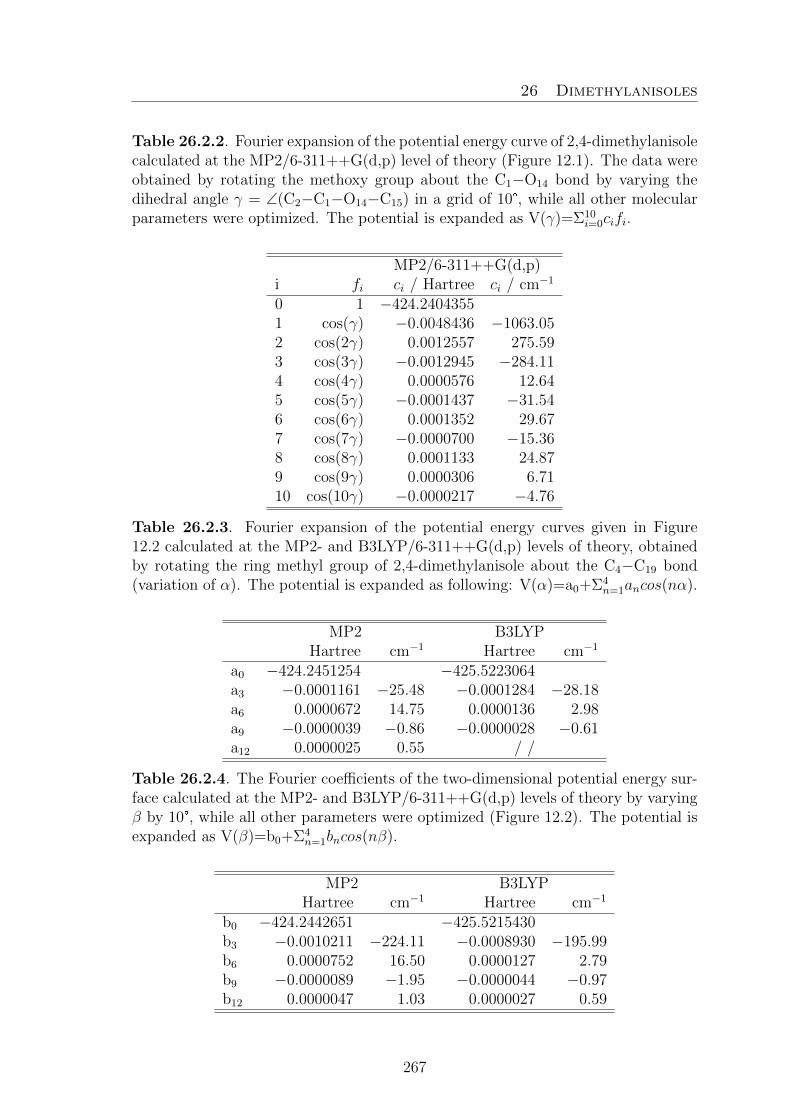
26 Dimethylanisoles
Table 26.2.2. Fourier expansion of the potential energy curve of 2,4-dimethylanisolecalculated at the MP2/6-311++G(d,p) level of theory (Figure 12.1). The data wereobtained by rotating the methoxy group about the C1−O14 bond by varying thedihedral angle γ = ∠(C2−C1−O14−C15) in a grid of 10°, while all other molecularparameters were optimized. The potential is expanded as V(γ)=Σ10
i=0cifi.
MP2/6-311++G(d,p)i fi ci / Hartree ci / cm−1
0 1 −424.24043551 cos(γ) −0.0048436 −1063.052 cos(2γ) 0.0012557 275.593 cos(3γ) −0.0012945 −284.114 cos(4γ) 0.0000576 12.645 cos(5γ) −0.0001437 −31.546 cos(6γ) 0.0001352 29.677 cos(7γ) −0.0000700 −15.368 cos(8γ) 0.0001133 24.879 cos(9γ) 0.0000306 6.7110 cos(10γ) −0.0000217 −4.76
Table 26.2.3. Fourier expansion of the potential energy curves given in Figure12.2 calculated at the MP2- and B3LYP/6-311++G(d,p) levels of theory, obtainedby rotating the ring methyl group of 2,4-dimethylanisole about the C4−C19 bond(variation of α). The potential is expanded as following: V(α)=a0+Σ4
n=1ancos(nα).
MP2 B3LYPHartree cm−1 Hartree cm−1
a0 −424.2451254 −425.5223064a3 −0.0001161 −25.48 −0.0001284 −28.18a6 0.0000672 14.75 0.0000136 2.98a9 −0.0000039 −0.86 −0.0000028 −0.61a12 0.0000025 0.55 / /
Table 26.2.4. The Fourier coefficients of the two-dimensional potential energy sur-face calculated at the MP2- and B3LYP/6-311++G(d,p) levels of theory by varyingβ by 10°, while all other parameters were optimized (Figure 12.2). The potential isexpanded as V(β)=b0+Σ4
n=1bncos(nβ).
MP2 B3LYPHartree cm−1 Hartree cm−1
b0 −424.2442651 −425.5215430b3 −0.0010211 −224.11 −0.0008930 −195.99b6 0.0000752 16.50 0.0000127 2.79b9 −0.0000089 −1.95 −0.0000044 −0.97b12 0.0000047 1.03 0.0000027 0.59
267

26 Dimethylanisoles
Table 26.2.5. Observed frequencies (νo) of 149 rotational transitions of 2,4-di-methylanisole. νo − νc values (in kHz) as obtained after Fit I with the programXIAM (global Fit) as well as Fit II (Fit 00/10/01) and III (global Fit) with theprogram NTOP.
No. J Ka Kc J Ka Kc Species νo Fit I Fit II Fit IIIupper level lower level XIAM NTOP NTOP
1 3 0 3 2 0 2 00 4.857652 −2 −1 03 0 3 2 0 2 10 4.857627 −2 0 0
2 4 0 4 3 0 3 00 6.346463 −2 0 04 0 4 3 0 3 10 6.346440 −2 0 04 0 4 3 0 3 01 6.281608 −9 −1 −24 0 4 3 0 3 12 6.281583 −2 −24 0 4 3 0 3 11 6.281621 1 1
3 5 0 5 4 0 4 00 7.766289 −2 1 25 0 5 4 0 4 10 7.766268 −2 −1 05 0 5 4 0 4 01 7.689111 −7 0 −25 0 5 4 0 4 12 7.689015 −1 −35 0 5 4 0 4 11 7.689199 2 0
4 6 0 6 5 0 5 00 9.146790 −3 1 16 0 6 5 0 5 10 9.146774 −2 0 06 0 6 5 0 5 01 9.078245 −6 0 −16 0 6 5 0 5 12 9.078156 2 06 0 6 5 0 5 11 9.078325 2 0
5 7 0 7 6 0 6 00 10.516598 −3 0 17 0 7 6 0 6 10 10.516582 −1 0 07 0 7 6 0 6 01 10.466261 −4 0 07 0 7 6 0 6 12 10.466194 1 07 0 7 6 0 6 11 10.466309 1 0
6 8 0 8 7 0 7 00 11.890302 −2 0 18 0 8 7 0 7 10 11.890287 −1 0 08 0 8 7 0 7 01 11.856966 −3 1 08 0 8 7 0 7 12 11.856920 2 08 0 8 7 0 7 11 11.856991 2 0
7 9 0 9 8 0 8 00 13.271258 −1 0 19 0 9 8 0 8 10 13.271241 −1 −1 −29 0 9 8 0 8 01 13.249962 −3 0 09 0 9 8 0 8 12 13.249932 4 29 0 9 8 0 8 11 13.249975 5 4
8 10 0 10 9 0 9 00 14.658238 0 0 110 0 10 9 0 9 10 14.658222 0 0 −110 0 10 9 0 9 01 14.644378 −1 1 010 0 10 9 0 9 12 14.644358 8 610 0 10 9 0 9 11 14.644378 4 2
9 11 0 11 10 0 10 00 16.049220 1 1 1
268

26 Dimethylanisoles
Table 26.2.5 (continued)
No. J Ka Kc J Ka Kc Species νo Fit I Fit II Fit IIIupper level lower level XIAM NTOP NTOP
11 0 11 10 0 10 10 16.049204 2 0 −111 0 11 10 0 10 01 16.039608 0 2 211 0 11 10 0 10 11 16.039598 1 −1
10 3 1 3 2 1 2 00 4.567965 0 1 23 1 3 2 1 2 10 4.567948 −3 −1 −1
11 4 1 4 3 1 3 00 6.058005 −2 −1 04 1 4 3 1 3 10 6.057988 −1 0 04 1 4 3 1 3 01 6.267722 9 2 24 1 4 3 1 3 12 6.267704 2 04 1 4 3 1 3 11 6.267674 3 1
12 5 1 5 4 1 4 00 7.526604 −2 0 05 1 5 4 1 4 10 7.526585 −1 0 05 1 5 4 1 4 01 7.673592 7 2 25 1 5 4 1 4 12 7.673637 0 15 1 5 4 1 4 11 7.673486 1 1
13 6 1 6 5 1 5 00 8.974955 −2 0 16 1 6 5 1 5 10 8.974935 −1 0 06 1 6 5 1 5 01 9.066067 4 1 16 1 6 5 1 5 12 9.066108 −1 06 1 6 5 1 5 11 9.065972 0 0
14 7 1 7 6 1 6 00 10.406303 −2 0 17 1 7 6 1 6 10 10.406284 −1 0 07 1 7 6 1 6 01 10.458298 1 1 07 1 7 6 1 6 12 10.458319 −3 −37 1 7 6 1 6 11 10.458228 −2 −2
15 8 1 8 7 1 7 00 11.824778 −2 −1 08 1 8 7 1 7 10 11.824760 −1 0 −18 1 8 7 1 7 01 11.852252 −1 0 −18 1 8 7 1 7 12 11.852261 1 08 1 8 7 1 7 11 11.852207 0 0
16 9 1 9 8 1 8 00 13.234347 −1 0 19 1 9 8 1 8 10 13.234329 1 1 09 1 9 8 1 8 01 13.247332 −2 −1 −19 1 9 8 1 8 12 13.247332 2 19 1 9 8 1 8 11 13.247305 4 3
17 10 1 10 9 1 9 00 14.638211 2 2 310 1 10 9 1 9 10 14.638190 −1 −2 −210 1 10 9 1 9 01 14.642969 1 2 210 1 10 9 1 9 12 14.642952 −4 −610 1 10 9 1 9 11 14.642937 −5 −6
18 11 1 11 10 1 10 00 16.038648 3 3 3
269

26 Dimethylanisoles
Table 26.2.5 (continued)
No. J Ka Kc J Ka Kc Species νo Fit I Fit II Fit IIIupper level lower level XIAM NTOP NTOP
11 1 11 10 1 10 10 16.038629 3 0 011 1 11 10 1 10 01 16.038871 0 1 011 1 11 10 1 10 12 16.038850 −7 −911 1 11 10 1 10 11 16.038833 −17 −19
19 3 1 2 2 1 1 00 5.360774 −2 −1 −13 1 2 2 1 1 10 5.360723 −3 −1 −13 1 2 2 1 1 01 5.109632 −13 2 13 1 2 2 1 1 12 5.109871 −9 13 1 2 2 1 1 11 5.109306 −10 1
20 4 1 3 3 1 2 00 7.103443 0 1 14 1 3 3 1 2 10 7.103378 −3 0 04 1 3 3 1 2 01 6.876988 −26 3 34 1 3 3 1 2 12 6.877596 −19 24 1 3 3 1 2 11 6.876249 −21 3
21 5 1 4 4 1 3 00 8.800739 0 1 15 1 4 4 1 3 10 8.800665 −2 1 05 1 4 4 1 3 01 8.618380 −32 2 25 1 4 4 1 3 12 8.619174 −25 15 1 4 4 1 3 11 8.617439 −27 3
22 6 1 5 5 1 4 00 10.433941 −1 1 16 1 5 5 1 4 10 10.433865 −2 1 06 1 5 5 1 4 01 10.280728 −28 0 −16 1 5 5 1 4 12 10.281314 −22 −36 1 5 5 1 4 11 10.280014 −22 0
23 7 1 6 6 1 5 00 11.985795 −1 1 17 1 6 6 1 5 10 11.985723 −2 1 07 1 6 6 1 5 01 11.836158 −20 −1 −27 1 6 6 1 5 12 11.836369 −11 −47 1 6 6 1 5 11 11.835847 −13 −5
24 8 1 7 7 1 6 00 13.450576 −1 1 28 1 7 7 1 6 10 13.450513 −1 1 08 1 7 7 1 6 01 13.292409 −16 −4 −48 1 7 7 1 6 12 13.292344 −3 −48 1 7 7 1 6 11 13.292409 −2 −3
25 9 1 8 8 1 7 00 14.843477 −1 1 29 1 8 8 1 7 10 14.843422 −1 0 −19 1 8 8 1 7 01 14.687680 −12 −4 −39 1 8 8 1 7 12 14.687510 4 −19 1 8 8 1 7 11 14.687799 3 −1
26 10 1 9 9 1 8 00 16.196299 −4 −2 −210 1 9 9 1 8 10 16.196254 1 1 −1
270

26 Dimethylanisoles
Table 26.2.5 (continued)
No. J Ka Kc J Ka Kc Species νo Fit I Fit II Fit IIIupper level lower level XIAM NTOP NTOP
27 11 1 10 10 1 9 00 17.539865 −1 0 011 1 10 10 1 9 10 17.539821 3 1 −1
28 12 1 11 11 1 10 00 18.891444 0 0 012 1 11 11 1 10 10 18.891399 4 1 −1
29 4 2 3 3 2 2 00 6.621075 3 4 54 2 3 3 2 2 10 6.621068 2 4 44 2 3 3 2 2 01 6.871147 26 0 −24 2 3 3 2 2 12 6.870448 17 −34 2 3 3 2 2 11 6.871742 20 −3
30 5 2 4 4 2 3 00 8.235806 1 2 25 2 4 4 2 3 10 8.235766 0 2 25 2 4 4 2 3 01 8.612772 32 0 −15 2 4 4 2 3 12 8.611884 23 −35 2 4 4 2 3 11 8.613526 26 −3
31 6 2 5 5 2 4 00 9.824626 −1 1 16 2 5 5 2 4 10 9.824573 −1 1 06 2 5 5 2 4 01 10.269982 30 3 26 2 5 5 2 4 12 10.269301 21 16 2 5 5 2 4 11 10.270503 22 1
32 7 2 6 6 2 5 00 11.384245 −1 0 17 2 6 6 2 5 10 11.384186 −1 1 07 2 6 6 2 5 01 11.815396 24 5 57 2 6 6 2 5 12 11.815080 11 27 2 6 6 2 5 11 11.815536 9 −2
33 8 2 7 7 2 6 00 12.913114 −1 0 08 2 7 7 2 6 10 12.913053 −1 1 08 2 7 7 2 6 01 13.264270 17 5 68 2 7 7 2 6 12 13.264219 5 48 2 7 7 2 6 11 13.264158 6 4
34 9 2 8 8 2 7 00 14.411820 −4 −3 −39 2 8 8 2 7 10 14.411762 0 1 09 2 8 8 2 7 01 14.659914 9 1 49 2 8 8 2 7 12 14.659960 −2 19 2 8 8 2 7 11 14.659721 1 3
35 10 2 8 9 2 7 00 17.609620 1 2 210 2 8 9 2 7 10 17.609489 −1 1 −1
36 11 2 9 10 2 8 00 19.141852 −1 1 011 2 9 10 2 8 10 19.141738 1 2 0
37 4 2 2 3 2 1 00 6.920959 −5 −4 −44 2 2 3 2 1 10 6.920860 −4 −2 −24 2 2 3 2 1 01 6.738402 −3 3 2
271

26 Dimethylanisoles
Table 26.2.5 (continued)
No. J Ka Kc J Ka Kc Species νo Fit I Fit II Fit IIIupper level lower level XIAM NTOP NTOP4 2 2 3 2 1 12 6.738384 −2 24 2 2 3 2 1 11 6.738318 −3 0
38 5 2 3 4 2 2 00 8.776374 2 2 25 2 3 4 2 2 10 8.776274 −2 0 05 2 3 4 2 2 01 8.476878 −8 2 25 2 3 4 2 2 12 8.476906 −7 05 2 3 4 2 2 11 8.476716 −3 3
39 6 2 4 5 2 3 00 10.641866 1 1 16 2 4 5 2 3 10 10.641756 −1 1 16 2 4 5 2 3 01 10.255366 −16 2 16 2 4 5 2 3 12 10.255559 −10 26 2 4 5 2 3 11 10.254999 −10 1
40 7 2 5 6 2 4 00 12.480276 1 1 27 2 5 6 2 4 10 12.480151 −2 1 07 2 5 6 2 4 01 12.072164 −27 4 37 2 5 6 2 4 12 12.072707 −17 47 2 5 6 2 4 11 12.071396 −19 4
41 8 2 6 7 2 5 00 14.265785 2 2 28 2 6 7 2 5 10 14.265650 −3 0 −18 2 6 7 2 5 01 13.896631 −37 5 58 2 6 7 2 5 12 13.897578 −28 28 2 6 7 2 5 11 13.895414 −30 4
42 9 2 7 8 2 6 00 15.980607 2 2 39 2 7 8 2 6 10 15.980470 −2 1 −19 2 7 8 2 6 01 15.672168 −43 1 19 2 7 8 2 6 12 15.673209 −33 −29 2 7 8 2 6 11 15.670866 −33 2
43 10 2 9 9 2 8 00 15.883082 1 2 210 2 9 9 2 8 10 15.883015 −5 −5 −710 2 9 9 2 8 01 16.040427 5 2 410 2 9 9 2 8 12 16.040472 −3 110 2 9 9 2 8 11 16.040242 −5 −1
44 11 2 10 10 2 9 00 17.331219 0 0 011 2 10 10 2 9 10 17.331160 1 1 −111 2 10 10 2 9 01 17.422564 −1 −2 211 2 10 10 2 9 12 17.422586 −1 411 2 10 10 2 9 11 17.422421 −6 −2
45 5 3 3 4 3 2 00 8.396137 −2 −2 −15 3 3 4 3 2 10 8.396880 4 7 75 3 3 4 3 2 01 8.471703 10 0 −15 3 3 4 3 2 12 8.471523 7 −1
272

26 Dimethylanisoles
Table 26.2.5 (continued)
No. J Ka Kc J Ka Kc Species νo Fit I Fit II Fit IIIupper level lower level XIAM NTOP NTOP5 3 3 4 3 2 11 8.471739 5 −2
46 6 3 4 5 3 3 00 10.087222 1 1 16 3 4 5 3 3 10 10.087335 −3 −1 −16 3 4 5 3 3 01 10.246329 15 −1 −26 3 4 5 3 3 12 10.245951 8 −56 3 4 5 3 3 11 10.246524 9 −4
47 7 3 5 6 3 4 00 11.770755 0 0 07 3 5 6 3 4 10 11.770725 −2 0 07 3 5 6 3 4 01 12.059805 25 −3 −47 3 5 6 3 4 12 12.059057 14 −67 3 5 6 3 4 11 12.060336 16 −6
48 8 3 6 7 3 5 00 13.438716 1 1 28 3 6 7 3 5 10 13.438640 −2 1 08 3 6 7 3 5 01 13.883166 37 −2 −28 3 6 7 3 5 12 13.882012 20 −78 3 6 7 3 5 11 13.884070 25 −7
49 9 3 7 8 3 6 00 15.083597 −1 0 09 3 7 8 3 6 10 15.083504 −1 1 09 3 7 8 3 6 01 15.656674 42 1 39 3 7 8 3 6 12 15.655443 25 −19 3 7 8 3 6 11 15.657629 29 −2
50 10 3 8 9 3 7 00 16.699349 −4 −3 −310 3 8 9 3 7 10 16.699250 −1 2 010 3 8 9 3 7 01 17.322765 34 3 6
51 5 3 2 4 3 1 00 8.447561 3 3 35 3 2 4 3 1 01 8.398317 −5 1 05 3 2 4 3 1 12 8.398271 −5 05 3 2 4 3 1 11 8.398240 −3 0
52 6 3 3 5 3 2 00 10.218814 1 0 16 3 3 5 3 2 10 10.218531 −3 −2 −26 3 3 5 3 2 01 10.112047 −6 2 16 3 3 5 3 2 12 10.112000 −6 16 3 3 5 3 2 11 10.111947 3 7
53 7 3 4 6 3 3 00 12.046533 1 0 07 3 4 6 3 3 10 12.046362 −1 2 17 3 4 6 3 3 01 11.846326 −8 2 27 3 4 6 3 3 12 11.846298 3 137 3 4 6 3 3 11 11.846175 −9 −3
54 8 3 5 7 3 4 00 13.931789 4 3 38 3 5 7 3 4 10 13.931620 −5 −2 −2
273
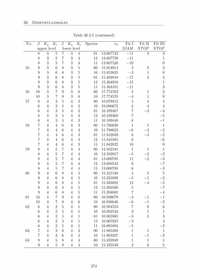
26 Dimethylanisoles
Table 26.2.5 (continued)
No. J Ka Kc J Ka Kc Species νo Fit I Fit II Fit IIIupper level lower level XIAM NTOP NTOP8 3 5 7 3 4 01 13.607745 −11 3 38 3 5 7 3 4 12 13.607739 −11 18 3 5 7 3 4 11 13.607526 −10 0
55 9 3 6 8 3 5 00 15.854014 3 3 39 3 6 8 3 5 10 15.853835 −3 1 09 3 6 8 3 5 01 15.404818 −17 3 39 3 6 8 3 5 12 15.404916 −15 19 3 6 8 3 5 11 15.404451 −11 3
56 10 3 7 9 3 6 00 17.774763 2 1 210 3 7 9 3 6 10 17.774570 −4 1 0
57 6 4 3 5 4 2 00 10.078812 5 4 46 4 3 5 4 2 10 10.080672 2 4 46 4 3 5 4 2 01 10.109487 7 −3 −46 4 3 5 4 2 12 10.109369 7 −56 4 3 5 4 2 11 10.109448 8 −1
58 7 4 4 6 4 3 00 11.786630 1 1 17 4 4 6 4 3 10 11.788623 −6 −2 −27 4 4 6 4 3 01 11.842049 8 −4 −57 4 4 6 4 3 12 11.841885 6 −67 4 4 6 4 3 11 11.842022 10 0
59 8 4 5 7 4 4 00 13.502181 1 1 18 4 5 7 4 4 10 13.502917 −5 −2 −28 4 5 7 4 4 01 13.600785 11 −2 −38 4 5 7 4 4 12 13.600542 6 −78 4 5 7 4 4 11 13.600788 6 −3
60 9 4 6 8 4 5 00 15.221548 4 5 59 4 6 8 4 5 10 15.221699 −5 −1 −29 4 6 8 4 5 01 15.393892 12 −4 −59 4 6 8 4 5 12 15.393500 5 −79 4 6 8 4 5 11 15.394002 7 −4
61 10 4 7 9 4 6 00 16.938679 −3 −1 −110 4 7 9 4 6 10 16.938646 −6 −1 −2
62 6 4 2 5 4 1 00 10.084553 7 6 66 4 2 5 4 1 10 10.082534 3 1 16 4 2 5 4 1 01 10.065985 −3 3 36 4 2 5 4 1 12 10.065925 −3 46 4 2 5 4 1 11 10.065894 −5 −2
63 7 4 3 6 4 2 00 11.805388 1 1 17 4 3 6 4 2 10 11.803207 1 −1 −2
64 9 4 5 8 4 4 00 15.335949 1 1 19 4 5 8 4 4 10 15.335539 2 6 5
274

26 Dimethylanisoles
Table 26.2.5 (continued)
No. J Ka Kc J Ka Kc Species νo Fit I Fit II Fit IIIupper level lower level XIAM NTOP NTOP9 5 5 8 5 4 00 15.168271 −1 −1 −19 5 5 8 5 4 01 15.217047 10 −4 −59 5 5 8 5 4 12 15.216867 10 −69 5 5 8 5 4 11 15.216980 7 −4
65 3 0 3 2 1 2 00 3.613653 −7 −11 −103 0 3 2 1 2 10 3.613632 −3 −4 −43 0 3 2 1 2 01 4.755446 59 5 93 0 3 2 1 2 12 4.755738 2 23 0 3 2 1 2 11 4.754888 3 1
66 4 0 4 3 1 3 00 5.392164 4 1 24 0 4 3 1 3 10 5.392128 2 2 24 0 4 3 1 3 01 6.221824 40 2 64 0 4 3 1 3 12 6.222286 −7 04 0 4 3 1 3 11 6.221138 −7 −1
67 5 0 5 4 1 4 00 7.100446 3 1 15 0 5 4 1 4 10 7.100407 0 0 05 0 5 4 1 4 01 7.643213 24 −1 25 0 5 4 1 4 12 7.643597 −9 −35 0 5 4 1 4 11 7.642661 −9 −4
68 6 0 6 5 1 5 00 8.720631 1 0 16 0 6 5 1 5 10 8.720597 1 1 16 0 6 5 1 5 01 9.047866 11 3 −26 0 6 5 1 5 12 9.048117 −8 −46 0 6 5 1 5 11 9.047499 −9 −5
69 7 0 7 6 1 6 00 10.262273 0 0 07 0 7 6 1 6 10 10.262244 1 1 17 0 7 6 1 6 01 10.448062 6 −2 −17 0 7 6 1 6 12 10.448203 −6 −37 0 7 6 1 6 11 10.447838 −6 −4
70 8 0 8 7 1 7 00 11.746272 0 0 18 0 8 7 1 7 10 11.746247 1 1 08 0 8 7 1 7 01 11.846730 2 −1 −18 0 8 7 1 7 12 11.846803 −3 −28 0 8 7 1 7 11 11.846595 −7 −7
71 9 0 9 8 1 8 00 13.192752 0 0 19 0 9 8 1 8 10 13.192730 2 1 09 0 9 8 1 8 01 13.244439 −1 −2 −29 0 9 8 1 8 12 13.244470 −3 −49 0 9 8 1 8 11 13.244362 −2 −3
72 10 0 10 9 1 9 00 14.616642 −1 −1 010 0 10 9 1 9 10 14.616623 2 0 0
275

26 Dimethylanisoles
Table 26.2.5 (continued)
No. J Ka Kc J Ka Kc Species νo Fit I Fit II Fit IIIupper level lower level XIAM NTOP NTOP
10 0 10 9 1 9 01 14.641483 −2 −1 −210 0 10 9 1 9 12 14.641494 1 −110 0 10 9 1 9 11 14.641438 0 −1
73 11 0 11 10 1 10 00 16.027654 2 1 111 0 11 10 1 10 10 16.027636 3 1 011 0 11 10 1 10 01 16.038123 −2 −1 −111 0 11 10 1 10 12 16.038123 3 111 0 11 10 1 10 11 16.038123 31 29
74 12 0 12 11 1 11 00 17.431614 6 3 412 0 12 11 1 11 10 17.431593 4 0 −1
75 2 1 2 1 0 1 00 4.534653 −8 −2 −22 1 2 1 0 1 10 4.534636 −3 1 1
76 3 1 3 2 0 2 01 4.880322 −60 −6 −113 1 3 2 0 2 12 4.880000 −3 −33 1 3 2 0 2 11 4.880823 −3 −2
77 4 1 4 3 0 3 00 7.012301 −12 −5 −54 1 4 3 0 3 10 7.012301 −4 −1 04 1 4 3 0 3 12 6.327001 7 04 1 4 3 0 3 11 6.328154 7 0
78 5 1 5 4 0 4 00 8.192446 −8 −1 05 1 5 4 0 4 10 8.192446 −4 0 05 1 5 4 0 4 01 7.719489 −25 2 −25 1 5 4 0 4 12 7.719055 10 25 1 5 4 0 4 11 7.720021 10 3
79 6 1 6 5 0 5 00 9.401113 −7 −1 06 1 6 5 0 5 10 9.401113 −3 0 06 1 6 5 0 5 01 9.096446 −13 4 16 1 6 5 0 5 12 9.096147 9 36 1 6 5 0 5 11 9.096797 9 4
80 7 1 7 6 0 6 00 10.660624 −8 −4 −37 1 7 6 0 6 10 10.660624 −2 1 17 1 7 6 0 6 01 10.476497 −8 3 17 1 7 6 0 6 12 10.476313 8 47 1 7 6 0 6 11 10.476704 9 5
81 8 1 8 7 0 7 00 11.968808 −4 0 18 1 8 7 0 7 10 11.968800 −2 −1 −18 1 8 7 0 7 01 11.862490 −4 4 28 1 8 7 0 7 12 11.862377 5 28 1 8 7 0 7 11 11.862599 5 3
82 9 1 9 8 0 8 00 13.312853 −2 0 19 1 9 8 0 8 10 13.312841 −1 −1 −1
276

26 Dimethylanisoles
Table 26.2.5 (continued)
No. J Ka Kc J Ka Kc Species νo Fit I Fit II Fit IIIupper level lower level XIAM NTOP NTOP9 1 9 8 0 8 01 13.252857 −2 3 29 1 9 8 0 8 12 13.252788 4 29 1 9 8 0 8 11 13.252911 5 3
83 10 1 10 9 0 9 00 14.679804 −1 1 110 1 10 9 0 9 10 14.679790 0 −1 −110 1 10 9 0 9 01 14.645861 −1 2 210 1 10 9 0 9 12 14.645817 4 210 1 10 9 0 9 11 14.645883 4 2
84 11 1 11 10 0 10 00 16.060212 1 1 111 1 11 10 0 10 10 16.060197 2 0 −111 1 11 10 0 10 01 16.040354 0 2 111 1 11 10 0 10 12 16.040325 5 311 1 11 10 0 10 11 16.040354 −1 −4
85 9 1 8 9 0 9 00 10.222716 −5 −2 −69 1 8 9 0 9 10 10.222372 −6 3 −19 1 8 9 0 9 01 10.460415 23 4 99 1 8 9 0 9 12 10.460453 −10 −59 1 8 9 0 9 11 10.459627 −7 −3
86 10 1 9 10 0 10 00 11.760788 3 7 310 1 9 10 0 10 10 11.760403 −6 2 −210 1 9 10 0 10 01 11.878299 14 −1 610 1 9 10 0 10 12 11.878194 −8 −510 1 9 10 0 10 11 11.877628 −6 −3
87 6 1 5 5 2 4 00 7.427626 9 2 26 1 5 5 2 4 10 7.427522 3 5 46 1 5 5 2 4 12 10.134809 12 66 1 5 5 2 4 11 10.134916 29 15
88 7 1 6 6 2 5 00 9.588788 1 −5 −67 1 6 6 2 5 10 9.588669 0 2 27 1 6 6 2 5 01 11.701348 92 9 167 1 6 6 2 5 12 11.701878 −20 27 1 6 6 2 5 11 11.700252 −16 1
89 8 1 7 7 2 6 00 11.655128 10 5 58 1 7 7 2 6 10 11.654995 −1 1 08 1 7 7 2 6 01 13.178361 52 0 68 1 7 7 2 6 12 13.179140 −35 −58 1 7 7 2 6 11 13.177118 −32 −6
90 9 1 8 8 2 7 00 13.585490 9 5 59 1 8 8 2 7 10 13.585365 1 2 19 1 8 8 2 7 01 14.601772 25 −8 −29 1 8 8 2 7 12 14.602433 −35 −9
277

26 Dimethylanisoles
Table 26.2.5 (continued)
No. J Ka Kc J Ka Kc Species νo Fit I Fit II Fit IIIupper level lower level XIAM NTOP NTOP
91 10 1 9 9 2 8 00 15.369967 8 4 410 1 9 9 2 8 10 15.369859 3 3 110 1 9 9 2 8 01 16.004122 7 −12 −710 1 9 9 2 8 12 16.004565 −30 −1110 1 9 9 2 8 11 16.003417 −30 −13
92 11 1 10 10 2 9 00 17.026749 5 2 111 1 10 10 2 9 10 17.026658 3 2 011 1 10 10 2 9 01 17.401132 −3 −12 −711 1 10 10 2 9 12 17.401389 −25 −1011 1 10 10 2 9 11 17.400676 −24 −12
93 12 1 11 11 2 10 00 18.586974 5 2 112 1 11 11 2 10 10 18.586896 6 3 112 1 11 11 2 10 01 18.797131 −7 −11 −512 1 11 11 2 10 12 18.797262 −18 −712 1 11 11 2 10 11 18.796844 −19 −9
94 3 2 2 2 1 1 00 9.414395 −12 −1 −13 2 2 2 1 1 10 9.414287 −3 2 1
95 4 2 3 3 1 2 00 10.674692 −11 0 14 2 3 3 1 2 10 10.674624 −6 −2 −2
96 5 2 4 4 1 3 00 11.807045 −19 −9 −85 2 4 4 1 3 10 11.807012 −2 2 15 2 4 4 1 3 12 8.765681 −56 −65 2 4 4 1 3 11 8.762547 −67 −3
97 6 2 5 5 1 4 00 12.830947 −5 5 66 2 5 5 1 4 10 12.830918 −4 −1 −26 2 5 5 1 4 12 10.415807 −12 −7
98 7 2 6 6 1 5 00 13.781240 −15 −5 −57 2 6 6 1 5 10 13.781240 −2 0 −17 2 6 6 1 5 01 11.950204 −91 −7 −147 2 6 6 1 5 12 11.949573 21 −27 2 6 6 1 5 11 11.951139 19 0
99 8 2 7 7 1 6 00 14.708572 −2 7 88 2 7 7 1 6 10 14.708572 1 1 18 2 7 7 1 6 01 13.378318 −52 1 −48 2 7 7 1 6 12 13.377422 37 58 2 7 7 1 6 11 13.379447 35 7
100 9 2 8 8 1 7 00 15.669806 −15 −7 −79 2 8 8 1 7 10 15.669806 −14 −14 −159 2 8 8 1 7 01 14.745823 −27 6 39 2 8 8 1 7 12 14.745038 38 109 2 8 8 1 7 11 14.746759 38 13
278

26 Dimethylanisoles
Table 26.2.5 (continued)
No. J Ka Kc J Ka Kc Species νo Fit I Fit II Fit IIIupper level lower level XIAM NTOP NTOP
101 10 2 9 9 1 8 00 16.709417 −7 −1 010 2 9 9 1 8 10 16.709417 1 0 −110 2 9 9 1 8 01 16.098567 −13 8 810 2 9 9 1 8 12 16.098001 32 1310 2 9 9 1 8 11 16.099205 32 15
102 11 2 10 10 1 9 00 17.844339 −2 3 311 2 10 10 1 9 10 17.844320 −3 −4 −611 2 10 10 1 9 01 17.458868 −4 10 1211 2 10 10 1 9 12 17.458491 24 1311 2 10 10 1 9 11 17.459250 23 14
103 12 2 11 11 1 10 00 19.065809 −2 1 012 2 11 11 1 10 10 19.065786 3 2 −112 2 11 11 1 10 01 18.830726 −3 6 1012 2 11 11 1 10 12 18.830487 20 1612 2 11 11 1 10 11 18.830903 −17 −20
104 3 2 1 2 1 2 00 10.369569 −11 −1 −13 2 1 2 1 2 10 10.369537 1 6 5
105 4 2 2 3 1 3 00 12.722560 −19 −10 −104 2 2 3 1 3 10 12.722444 −5 0 0
106 5 2 3 4 1 4 00 15.440913 −30 −23 −245 2 3 4 1 4 10 15.440727 −9 −3 −4
107 6 2 4 5 1 5 00 18.556212 10 16 156 2 4 5 1 5 10 18.555898 −9 −2 −3
108 8 2 7 8 1 8 00 9.829988 −4 3 18 2 7 8 1 8 10 9.829736 −6 0 −28 2 7 8 1 8 01 9.103065 −19 −6 −78 2 7 8 1 8 12 9.102491 24 2
109 9 2 8 9 1 9 00 11.007466 −2 6 29 2 8 9 1 9 10 11.007169 −6 1 −29 2 8 9 1 9 01 10.515664 9 13 159 2 8 9 1 9 12 10.515124 24 69 2 8 9 1 9 11 10.515646 22 6
110 11 2 9 11 1 10 00 9.676665 10 8 711 2 9 11 1 10 10 9.676243 −8 0 011 2 9 11 1 10 11 10.398247 −18 −2
111 3 3 0 2 2 1 00 13.051921 −4 0 13 3 0 2 2 1 10 13.055527 2 2 3
112 4 3 1 3 2 2 00 14.786850 −3 1 24 3 1 3 2 2 10 14.787879 3 −1 −1
113 5 3 2 4 2 3 00 16.613338 −2 1 2
279
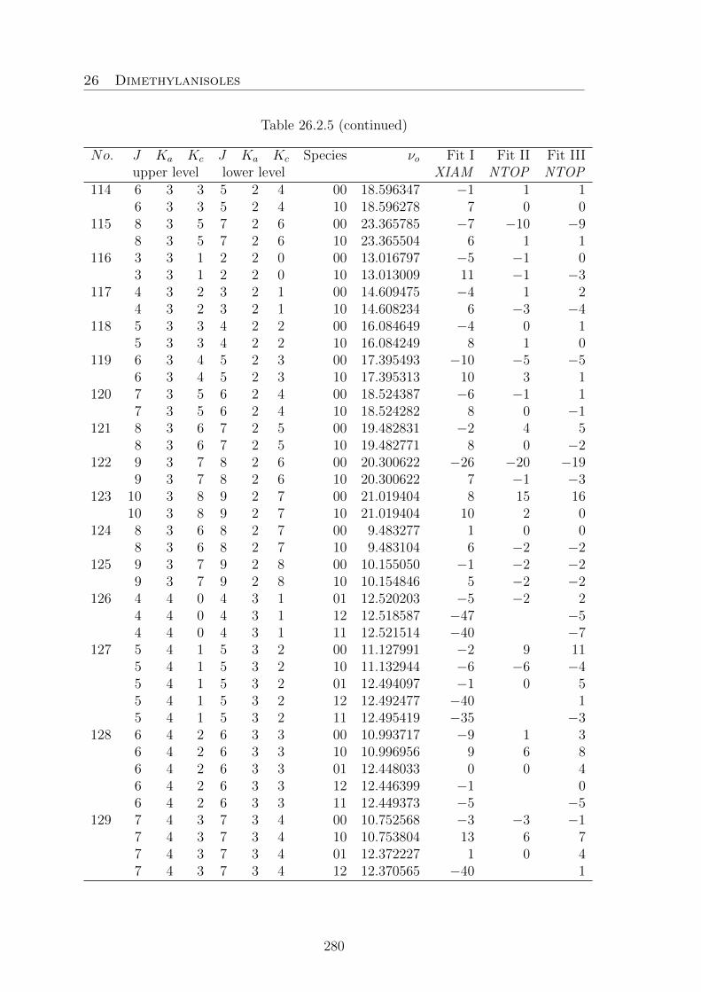
26 Dimethylanisoles
Table 26.2.5 (continued)
No. J Ka Kc J Ka Kc Species νo Fit I Fit II Fit IIIupper level lower level XIAM NTOP NTOP
114 6 3 3 5 2 4 00 18.596347 −1 1 16 3 3 5 2 4 10 18.596278 7 0 0
115 8 3 5 7 2 6 00 23.365785 −7 −10 −98 3 5 7 2 6 10 23.365504 6 1 1
116 3 3 1 2 2 0 00 13.016797 −5 −1 03 3 1 2 2 0 10 13.013009 11 −1 −3
117 4 3 2 3 2 1 00 14.609475 −4 1 24 3 2 3 2 1 10 14.608234 6 −3 −4
118 5 3 3 4 2 2 00 16.084649 −4 0 15 3 3 4 2 2 10 16.084249 8 1 0
119 6 3 4 5 2 3 00 17.395493 −10 −5 −56 3 4 5 2 3 10 17.395313 10 3 1
120 7 3 5 6 2 4 00 18.524387 −6 −1 17 3 5 6 2 4 10 18.524282 8 0 −1
121 8 3 6 7 2 5 00 19.482831 −2 4 58 3 6 7 2 5 10 19.482771 8 0 −2
122 9 3 7 8 2 6 00 20.300622 −26 −20 −199 3 7 8 2 6 10 20.300622 7 −1 −3
123 10 3 8 9 2 7 00 21.019404 8 15 1610 3 8 9 2 7 10 21.019404 10 2 0
124 8 3 6 8 2 7 00 9.483277 1 0 08 3 6 8 2 7 10 9.483104 6 −2 −2
125 9 3 7 9 2 8 00 10.155050 −1 −2 −29 3 7 9 2 8 10 10.154846 5 −2 −2
126 4 4 0 4 3 1 01 12.520203 −5 −2 24 4 0 4 3 1 12 12.518587 −47 −54 4 0 4 3 1 11 12.521514 −40 −7
127 5 4 1 5 3 2 00 11.127991 −2 9 115 4 1 5 3 2 10 11.132944 −6 −6 −45 4 1 5 3 2 01 12.494097 −1 0 55 4 1 5 3 2 12 12.492477 −40 15 4 1 5 3 2 11 12.495419 −35 −3
128 6 4 2 6 3 3 00 10.993717 −9 1 36 4 2 6 3 3 10 10.996956 9 6 86 4 2 6 3 3 01 12.448033 0 0 46 4 2 6 3 3 12 12.446399 −1 06 4 2 6 3 3 11 12.449373 −5 −5
129 7 4 3 7 3 4 00 10.752568 −3 −3 −17 4 3 7 3 4 10 10.753804 13 6 77 4 3 7 3 4 01 12.372227 1 0 47 4 3 7 3 4 12 12.370565 −40 1
280

26 Dimethylanisoles
Table 26.2.5 (continued)
No. J Ka Kc J Ka Kc Species νo Fit I Fit II Fit IIIupper level lower level XIAM NTOP NTOP7 4 3 7 3 4 11 12.373606 −34 −5
130 8 4 4 8 3 5 00 10.372965 −12 −2 18 4 4 8 3 5 10 10.373400 10 1 28 4 4 8 3 5 01 12.252468 3 −2 28 4 4 8 3 5 12 12.250740 −41 08 4 4 8 3 5 11 12.253932 −31 −4
131 9 4 5 9 3 6 00 9.854902 −12 −1 29 4 5 9 3 6 10 9.855098 9 0 09 4 5 9 3 6 01 12.068653 8 −2 39 4 5 9 3 6 12 12.066753 −41 09 4 5 9 3 6 11 12.070314 −33 −9
132 10 4 6 10 3 7 00 9.249617 −4 8 1110 4 6 10 3 7 10 9.249717 −6 −14 −1410 4 6 10 3 7 01 11.798215 18 −2 310 4 6 10 3 7 12 11.795891 −40 0
133 5 4 2 5 3 3 00 11.195483 −19 −8 −65 4 2 5 3 3 10 11.190375 5 −5 −65 4 2 5 3 3 01 9.390311 5 1 −35 4 2 5 3 3 12 9.391945 84 25 4 2 5 3 3 11 9.388742 77 6
134 6 4 3 6 3 4 00 11.187078 −10 0 26 4 3 6 3 4 10 11.183714 12 2 26 4 3 6 3 4 01 9.253469 −3 0 −46 4 3 6 3 4 12 9.255364 84 26 4 3 6 3 4 11 9.251663 74 6
135 7 4 4 7 3 5 00 11.202953 −9 −1 17 4 4 7 3 5 10 11.201611 7 −1 −17 4 4 7 3 5 01 9.035716 −17 3 −17 4 4 7 3 5 12 9.038193 76 37 4 4 7 3 5 11 9.033346 63 9
136 8 4 5 8 3 6 00 11.266415 −12 −4 −28 4 5 8 3 6 10 11.265890 6 −1 08 4 5 8 3 6 01 8.753337 −41 4 08 4 5 8 3 6 12 8.756720 60 18 4 5 8 3 6 11 8.750063 44 12
137 9 4 6 9 3 7 00 11.404364 −8 0 29 4 6 9 3 7 10 11.404086 3 −3 −1
138 10 4 7 10 3 8 00 11.643691 −10 0 210 4 7 10 3 8 10 11.643488 4 1 3
139 4 4 1 3 3 1 10 17.913316 8 4 4140 5 4 2 4 3 1 00 19.574091 −8 3 5
281

26 Dimethylanisoles
Table 26.2.5 (continued)
No. J Ka Kc J Ka Kc Species νo Fit I Fit II Fit IIIupper level lower level XIAM NTOP NTOP5 4 2 4 3 1 10 19.567544 14 2 0
141 6 4 3 5 3 2 00 21.205340 −8 2 56 4 3 5 3 2 10 21.201521 11 3 1
142 5 5 1 5 4 2 01 12.634603 −1 3 0143 6 5 2 6 4 3 01 12.589908 −2 −2 −5144 7 5 3 7 4 4 01 12.516545 2 −1 −5145 8 5 4 8 4 5 01 12.401019 1 −4 −8146 3 2 1 2 1 1 01 11.248635 87 5 5
3 2 1 2 1 1 12 11.246977 36 03 2 1 2 1 1 11 11.249993 43 −3
147 4 2 2 3 1 2 01 12.877403 96 5 54 2 2 3 1 2 12 12.875488 41 −14 2 2 3 1 2 11 12.879004 50 −4
148 3 3 1 2 2 1 10 13.046153 11 0 −23 3 1 2 2 1 01 11.366256 −49 1 −33 3 1 2 2 1 12 11.367773 24 23 3 1 2 2 1 11 11.364747 16 5
149 4 3 2 3 2 2 10 14.768167 7 −2 −34 3 2 3 2 2 01 12.997860 −56 0 −44 3 2 3 2 2 12 12.999603 21 34 3 2 3 2 2 11 12.996103 12 6
282

26 Dimethylanisoles
26.3. 2,5-DMA
Table 26.3.1. Principal inertial axes system nuclear coordinates of the planar2,5-dimethylanisole conformer calculated at the B3LYP/6-311++G(d.p) level of the-ory. The atom numbering refers to Figure 13.1.
a / A b / A c / AC1 −0.664126 −0.212608 0.000085C2 −0.633222 1.196868 0.000043C3 0.614558 1.809310 −0.000091C4 1.801826 1.071127 −0.000160C5 1.765090 −0.319748 −0.000048C6 0.514468 −0.956131 0.000049H7 2.755843 1.587192 −0.000317H8 0.662464 2.893796 −0.000180H9 0.478626 −2.038506 0.000085C10 3.031354 −1.142872 0.000118H11 3.086995 −1.788190 0.882705H12 3.916836 −0.504203 −0.002096H13 3.085002 −1.791686 −0.880006O14 −1.916220 −0.767155 0.000153C15 −2.039267 −2.181542 −0.000214H16 −1.585593 −2.624095 0.893497H17 −3.108709 −2.387509 −0.000406H18 −1.585345 −2.623639 −0.894022C19 −1.915808 1.986056 0.000060H20 −2.527011 1.752688 0.877013H21 −1.708096 3.057833 −0.000008H22 −2.527095 1.752594 −0.876811
Table 26.3.2 Coefficients of the one-dimensional Fourier expansion for the poten-tial energy curve obtained by rotating the methoxy group about the axis C1−O14
(variation of α, see Figure 13.1 right hand-side). The calculations were performed atthe B3LYP/6-311++G(d,p) level of theory. The expansion is defined as following:V =a0+Σ15
n=1cos(nα + π).
Fourier Term Hartree cm−1
1 −425.5172689cos( α) −0.0047500 −1042.50cos(2α) 0.0002597 57.00cos(3α) −0.0011979 −262.91cos(4α) −0.0000564 −12.38cos(6α) 0.0001059 23.24cos(8α) 0.0000359 7.88cos(10α) −0.0000264 −5.79
283

26 Dimethylanisoles
Table 26.3.3. Coefficients of the one-dimensional Fourier expansion for the poten-tial energy curve obtained by varying the dihedral angle β = ∠(H21−C19−C2−C3)(rotation about the C2−C19 axis, Figure 13.2 left hand-side). The calculations werecarried out at the MP2- and B3LYP/6-311++G(d,p) levels of theory. The Fourierexpansion is definded as: V =b0+Σ15
n=1cos(nβ + π).
MP2 B3LYPHartree cm−1 Hartree cm−1
b0 −424.2447286 −425.5220805b3 −0.0010119 −222.09 −0.0009011 −197.77b6 0.0001256 27.57 0.0000267 5.86b9 −0.0000181 −3.97 −0.0000019 −0.42b12 0.0000093 2.04 −0.0000018 −0.40
Table 26.3.4. Coefficients of the one-dimensional Fourier expansion of the poten-tial energy surface obtained by varying the dihedral angle γ=∠(C6−C5−C10−H12)(rotation about the C5−C10 axis, Figure 13.2 right hand-side). The calculationswere carried out at the MP2- and B3LYP/6-311++G(d,p) levels of theory. TheFourier expansion is given as: V =c0+Σ15
n=1cos(nγ + π).
MP2 B3LYPHartree cm−1 Hartree cm−1
c0 −424.2455187 −425.5228398c3 0.0001406 30.86 −0.0001256 −27.57c6 0.0000766 16.81 0.0000069 1.51c9 0.0000010 0.22 / /c12 0.0000034 0.75 −0.0000013 −0.29c15 0.0000025 0.55 / /
284

26 Dimethylanisoles
Table 26.3.5. Coefficients of the two-dimensional Fourier expansion of the poten-tial energy surface obtained by varying the dihedral angles β = ∠(H21−C19−C2−C3)and γ=∠(C6−C5−C10−H12) in a grid of 10° (see Figure 13.3). The calculations werecarried out at the B3LYP/6-311++G(d,p) level of theory. The potential is expandedas: V(β,γ) =Σ13
n=1Vifi.
i fi Vi / Hartree Vi / cm−1
1 1 −425.52198252 cos(3β) 0.0001059 23.243 cos(3γ) −0.0008790 −192.924 cos(6β) 0.0000064 1.405 cos(6γ) 0.0000253 5.556 cos(3β) cos(3γ) 0.0000215 4.727 sin(3β) sin(3γ) 0.0000132 2.908 cos(6β) cos(3γ) 0.0000007 0.159 sin(6β) sin(3γ) −0.0000006 −0.1310 cos(3β) cos(6γ) −0.0000015 −0.3311 sin(3β) sin(6γ) −0.0000005 −0.1112 cos(6β) cos(6γ) 0.0000008 0.1813 sin(6β) sin(6γ) 0.0000001 0.02
Table 26.3.6. Observed frequencies (νo) of 75 rotational transitions of 25DMA.Totally 298 signals were assigned and fitted to a standard deviation of 21.2 kHz.νo − νc values are given in kHz as obtained after a fit with the program N-TOP.Fit I only includes the (00)(10)(01) species and Fit II is the global Fit.
No. J Ka Kc J ′ Ka’ Kc’ Species νobs /GHz Fit I Fit II1 3 0 3 2 1 2 00 4.747844 5 1
3 0 3 2 1 2 01 4.793870 −3 −33 0 3 2 1 2 10 4.747844 0 −23 0 3 2 1 2 11 4.794522 / −75
2 4 0 4 3 1 3 00 6.385439 1 −14 0 4 3 1 3 01 6.394333 −1 14 0 4 3 1 3 10 6.385431 −1 −44 0 4 3 1 3 11 6.394601 / −33
3 5 0 5 4 1 4 00 7.912885 1 15 0 5 4 1 4 01 7.914376 0 25 0 5 4 1 4 10 7.912872 −1 −35 0 5 4 1 4 11 7.914452 / −235 0 5 4 1 4 12 7.914274 / 17
4 6 0 6 5 1 5 00 9.389994 1 26 0 6 5 1 5 01 9.389457 −2 16 0 6 5 1 5 10 9.389978 −1 −46 0 6 5 1 5 11 9.389465 / −156 0 6 5 1 5 12 9.389427 / 15
285

26 Dimethylanisoles
Table 26.3.6. (continued)
No. J Ka Kc J ′ Ka’ Kc’ Species νobs /GHz Fit I Fit II5 7 0 7 6 1 6 00 10.848035 0 2
7 0 7 6 1 6 01 10.846507 3 67 0 7 6 1 6 10 10.848019 −1 −47 0 7 6 1 6 11 10.846504 / 57 0 7 6 1 6 12 10.846486 / 5
6 8 0 8 7 1 7 00 12.299691 1 38 0 8 7 1 7 01 12.297517 −2 18 0 8 7 1 7 10 12.299674 −1 −38 0 8 7 1 7 11 12.297511 / 58 0 8 7 1 7 12 12.297495 / −8
7 9 0 9 8 1 8 00 13.749401 2 49 0 9 8 1 8 01 13.746803 0 39 0 9 8 1 8 10 13.749383 −1 −39 0 9 8 1 8 11 13.746795 / 69 0 9 8 1 8 12 13.746780 / −9
8 10 0 10 9 1 9 00 15.198571 0 310 0 10 9 1 9 01 15.195695 0 410 0 10 9 1 9 10 15.198555 −1 −210 0 10 9 1 9 11 15.195673 / −710 0 10 9 1 9 12 15.195685 / 4
9 11 0 11 10 1 10 00 16.647619 4 711 0 11 10 1 10 01 16.644556 0 411 0 11 10 1 10 10 16.647600 0 −111 0 11 10 1 10 11 16.644546 / 511 0 11 10 1 10 12 16.644546 / 4
10 12 0 12 11 1 11 00 18.096642 0 312 0 12 11 1 11 01 18.093463 −2 212 0 12 11 1 11 10 18.096626 −1 −212 0 12 11 1 11 11 18.093443 / −712 0 12 11 1 11 12 18.093454 / 3
11 13 0 13 12 1 12 00 19.545680 4 713 0 13 12 1 12 01 19.542429 3 813 0 13 12 1 12 10 19.545663 1 113 0 13 12 1 12 11 19.542407 / −313 0 13 12 1 12 12 19.542407 / −4
12 14 0 14 13 1 13 00 20.994721 0 414 0 14 13 1 13 01 20.991427 0 514 0 14 13 1 13 10 20.994707 1 114 0 14 13 1 13 11 20.991405 / −614 0 14 13 1 13 12 20.991405 / −6
13 15 0 15 14 1 14 00 22.443773 0 515 0 15 14 1 14 01 22.440458 0 5
286

26 Dimethylanisoles
Table 26.3.6. (continued)
No. J Ka Kc J ′ Ka’ Kc’ Species νobs /GHz Fit I Fit II15 0 15 14 1 14 10 22.443759 1 215 0 15 14 1 14 11 22.440442 / 115 0 15 14 1 14 12 22.440442 / 0
14 2 1 2 1 0 1 00 4.029018 0 52 1 2 1 0 1 01 3.910388 17 202 1 2 1 0 1 10 4.028982 −1 −52 1 2 1 0 1 12 3.910937 / −59
15 3 1 3 2 0 2 00 5.327303 −1 33 1 3 2 0 2 01 5.329879 2 63 1 3 2 0 2 10 5.327273 −1 −4
16 4 1 4 3 0 3 00 6.629016 −1 24 1 4 3 0 3 01 6.642922 1 44 1 4 3 0 3 10 6.628992 −1 −44 1 4 3 0 3 12 6.643279 / 68
17 5 1 5 4 0 4 00 7.999760 0 25 1 5 4 0 4 01 8.005479 −1 25 1 5 4 0 4 10 7.999741 −1 −35 1 5 4 0 4 11 8.005298 / −355 1 5 4 0 4 12 8.005625 / 34
18 6 1 6 5 0 5 00 9.418036 1 26 1 6 5 0 5 01 9.419037 0 26 1 6 5 0 5 10 9.418018 −1 −36 1 6 5 0 5 11 9.418957 / −206 1 6 5 0 5 12 9.419083 / 15
19 7 1 7 6 0 6 00 10.856538 1 37 1 7 6 0 6 01 10.855467 −3 07 1 7 6 0 6 10 10.856521 0 −37 1 7 6 0 6 11 10.855427 / −137 1 7 6 0 6 12 10.855483 / 12
20 8 1 8 7 0 7 00 12.302160 0 28 1 8 7 0 7 01 12.300116 −2 18 1 8 7 0 7 10 12.302144 −1 −38 1 8 7 0 7 11 12.300089 / −98 1 8 7 0 7 12 12.300117 / 8
21 9 1 9 8 0 8 00 13.750096 1 39 1 9 8 0 8 01 13.747532 −1 29 1 9 8 0 8 10 13.750079 −1 −39 1 9 8 0 8 11 13.747508 / −89 1 9 8 0 8 12 13.747527 / 6
22 10 1 10 9 0 9 00 15.198762 0 210 1 10 9 0 9 01 15.195894 −1 310 1 10 9 0 9 10 15.198747 0 −2
287

26 Dimethylanisoles
Table 26.3.6. (continued)
No. J Ka Kc J ′ Ka’ Kc’ Species νobs /GHz Fit I Fit II10 1 10 9 0 9 11 15.195868 / −1110 1 10 9 0 9 12 15.195868 / −14
23 11 1 11 10 0 10 00 16.647667 0 311 1 11 10 0 10 01 16.644609 −1 311 1 11 10 0 10 10 16.647651 −1 −211 1 11 10 0 10 11 16.644588 / −611 1 11 10 0 10 12 16.644588 / −8
24 12 1 12 11 0 11 00 18.096657 1 412 1 12 11 0 11 01 18.093480 1 512 1 12 11 0 11 10 18.096642 1 012 1 12 11 0 11 11 18.093463 / −112 1 12 11 0 11 12 18.093463 / −2
25 13 1 13 12 0 12 00 19.545680 0 313 1 13 12 0 12 01 19.542429 0 413 1 13 12 0 12 10 19.545663 −2 −313 1 13 12 0 12 11 19.542407 / −613 1 13 12 0 12 12 19.542407 / −7
26 14 1 14 13 0 13 00 20.994721 −1 314 1 14 13 0 13 01 20.991427 −1 414 1 14 13 0 13 10 20.994707 0 014 1 14 13 0 13 11 20.991405 / −714 1 14 13 0 13 12 20.991405 / −7
27 15 1 15 14 0 14 00 22.443773 0 515 1 15 14 0 14 01 22.440458 −1 515 1 15 14 0 14 10 22.443759 1 215 1 15 14 0 14 11 22.440442 / 015 1 15 14 0 14 12 22.440442 / 0
28 4 1 3 3 2 2 01 7.021955 −8 −44 1 3 3 2 2 10 6.497177 3 3
29 5 1 4 4 2 3 00 8.639566 −1 −105 1 4 4 2 3 01 8.795779 24 275 1 4 4 2 3 10 8.639581 4 −2
30 6 1 5 5 2 4 00 10.502481 2 −16 1 5 5 2 4 01 10.538844 1 66 1 5 5 2 4 10 10.502462 0 −76 1 5 5 2 4 11 10.539541 / −916 1 5 5 2 4 12 10.538089 / 75
31 7 1 6 6 2 5 00 12.149411 0 17 1 6 6 2 5 01 12.155080 0 67 1 6 6 2 5 10 12.149380 1 −67 1 6 6 2 5 12 12.154764 / 43
32 8 1 7 7 2 6 00 13.678794 −1 2
288

26 Dimethylanisoles
Table 26.3.6. (continued)
No. J Ka Kc J ′ Ka’ Kc’ Species νobs /GHz Fit I Fit II8 1 7 7 2 6 01 13.675963 1 88 1 7 7 2 6 10 13.678754 −1 −88 1 7 7 2 6 11 13.676002 / −378 1 7 7 2 6 12 13.675840 / 29
33 9 1 8 8 2 7 00 15.156367 −1 39 1 8 8 2 7 01 15.150203 0 79 1 8 8 2 7 10 15.156325 0 −69 1 8 8 2 7 11 15.150170 / −279 1 8 8 2 7 12 15.150151 / 21
34 10 1 9 9 2 8 00 16.614139 −1 310 1 9 9 2 8 01 16.606102 −1 710 1 9 9 2 8 10 16.614095 −1 −710 1 9 9 2 8 12 16.606045 / −11
35 11 1 10 10 2 9 00 18.065185 −1 411 1 10 10 2 9 01 18.055917 −1 711 1 10 10 2 9 10 18.065141 0 −7
36 12 1 11 11 2 10 00 19.514227 −1 412 1 11 11 2 10 01 19.504111 −2 612 1 11 11 2 10 10 19.514183 0 −612 1 11 11 2 10 12 19.504053 / −21
37 13 1 12 12 2 11 00 20.962803 0 613 1 12 12 2 11 01 20.952091 −3 613 1 12 12 2 11 10 20.962757 −1 −6
38 14 1 13 13 2 12 00 22.411353 −1 514 1 13 13 2 12 01 22.400219 −2 714 1 13 13 2 12 10 22.411309 0 −514 1 13 13 2 12 12 22.400166 / −14
39 2 2 0 1 1 1 00 6.887080 0 1140 3 2 1 2 1 2 00 9.744346 4 1041 4 2 2 3 1 3 00 13.252689 8 942 5 2 3 4 1 4 00 17.342154 −6 −7
5 2 3 4 1 4 01 17.646046 −2 2443 2 2 1 1 1 0 00 6.290604 1 15
2 2 1 1 1 0 10 6.290384 11 −5544 3 2 2 2 1 1 00 7.739712 0 13
3 2 2 2 1 1 01 6.597252 −6 −113 2 2 2 1 1 10 7.739585 −2 −24
45 4 2 3 3 1 2 00 8.964602 0 134 2 3 3 1 2 01 8.549355 3 94 2 3 3 1 2 10 8.964497 −3 −16
46 5 2 4 4 1 3 00 10.059331 0 125 2 4 4 1 3 01 9.992769 0 9
289

26 Dimethylanisoles
Table 26.3.6. (continued)
No. J Ka Kc J ′ Ka’ Kc’ Species νobs /GHz Fit I Fit II5 2 4 4 1 3 10 10.059240 −3 −135 2 4 4 1 3 11 9.991498 / −55 2 4 4 1 3 12 9.993916 / 17
47 6 2 5 5 1 4 00 11.179905 10 196 2 5 5 1 4 01 11.197386 2 116 2 5 5 1 4 10 11.179820 −3 −116 2 5 5 1 4 11 11.196410 / −91
48 7 2 6 6 1 5 00 12.425444 1 77 2 6 6 1 5 01 12.437655 3 117 2 6 6 1 5 10 12.425383 −1 −97 2 6 6 1 5 11 12.437115 / −737 2 6 6 1 5 12 12.438068 / 60
49 8 2 7 7 1 6 00 13.779480 1 68 2 7 7 1 6 01 13.780575 −1 58 2 7 7 1 6 10 13.779426 −1 −98 2 7 7 1 6 11 13.780315 / −488 2 7 7 1 6 12 13.780736 / 38
50 9 2 8 8 1 7 00 15.190453 0 59 2 8 8 1 7 11 15.185594 / −35
51 10 2 9 9 1 8 00 16.625086 0 510 2 9 9 1 8 01 16.617506 −2 610 2 9 9 1 8 10 16.625040 0 −710 2 9 9 1 8 11 16.617415 / −2510 2 9 9 1 8 12 16.617506 / 15
52 11 2 10 10 1 9 00 18.068564 −1 411 2 10 10 1 9 01 18.059430 −1 711 2 10 10 1 9 10 18.068519 −1 −711 2 10 10 1 9 11 18.059358 / −2011 2 10 10 1 9 12 18.059410 / 10
53 12 2 11 11 1 10 00 19.515239 0 512 2 11 11 1 10 01 19.505160 −1 712 2 11 11 1 10 10 19.515195 1 −512 2 11 11 1 10 11 19.505097 / −1612 2 11 11 1 10 12 19.505134 / 9
54 13 2 12 12 1 11 00 20.963098 0 613 2 12 12 1 11 01 20.952397 −2 713 2 12 12 1 11 10 20.963052 −1 −713 2 12 12 1 11 11 20.952339 / −1313 2 12 12 1 11 12 20.952339 / −21
55 14 2 13 13 1 12 00 22.411439 0 714 2 13 13 1 12 01 22.400308 0 914 2 13 13 1 12 10 22.411393 −1 −5
290

26 Dimethylanisoles
Table 26.3.6. (continued)
No. J Ka Kc J ′ Ka’ Kc’ Species νobs /GHz Fit I Fit II14 2 13 13 1 12 11 22.400261 / −114 2 13 13 1 12 12 22.400261 / −7
56 5 2 3 4 3 2 00 7.226073 12 −175 2 3 4 3 2 01 9.254431 −9 115 2 3 4 3 2 10 7.226318 −8 56
57 6 2 4 5 3 3 00 9.935203 3 −186 2 4 5 3 3 01 10.924008 −7 46 2 4 5 3 3 10 9.935303 1 9
58 7 2 5 6 3 4 00 12.412723 2 −87 2 5 6 3 4 01 12.757685 −1 107 2 5 6 3 4 10 12.412755 2 0
59 8 2 6 7 3 5 00 14.537288 −20 −208 2 6 7 3 5 01 14.629292 6 20
60 9 2 7 8 3 6 00 16.344577 −1 59 2 7 8 3 6 01 16.362406 7 229 2 7 8 3 6 10 16.344531 0 −79 2 7 8 3 6 11 16.362964 / −769 2 7 8 3 6 12 16.361732 / 77
61 10 2 8 9 3 7 00 17.953487 1 910 2 8 9 3 7 01 17.950194 4 1910 2 8 9 3 7 10 17.953422 −1 −910 2 8 9 3 7 11 17.950348 / −4710 2 8 9 3 7 12 17.949906 / 46
62 11 2 9 10 3 8 00 19.464216 1 1011 2 9 10 3 8 01 19.453654 2 1711 2 9 10 3 8 10 19.464142 −3 −1111 2 9 10 3 8 11 19.453633 / −3911 2 9 10 3 8 12 19.453527 / 31
63 12 2 10 11 3 9 00 20.933399 1 1112 2 10 11 3 9 10 20.933322 −2 −11
64 3 3 0 2 2 1 00 10.342912 2 2165 4 3 1 3 2 2 00 12.661219 −3 11
4 3 1 3 2 2 11 15.772991 / 866 5 3 2 4 2 3 00 15.499654 10 16
5 3 2 4 2 3 10 15.499519 −12 −2467 6 3 3 5 2 4 11 20.579275 / −6668 3 3 1 2 2 0 00 10.152112 1 2369 4 3 2 3 2 1 00 11.748617 4 2570 5 3 3 4 2 2 00 13.033174 4 26
5 3 3 4 2 2 01 11.002289 −7 −155 3 3 4 2 2 10 13.032942 −3 −44
71 6 3 4 5 2 3 00 14.059125 4 26
291

26 Dimethylanisoles
Table 26.3.6. (continued)
No. J Ka Kc J ′ Ka’ Kc’ Species νobs /GHz Fit I Fit II6 3 4 5 2 3 01 13.159791 −3 66 3 4 5 2 3 10 14.058931 −5 −30
72 7 3 5 6 2 4 00 14.944467 5 247 3 5 6 2 4 01 14.705645 0 147 3 5 6 2 4 10 14.944296 −7 −26
73 8 3 6 7 2 5 00 15.858624 6 218 3 6 7 2 5 01 15.849348 −2 118 3 6 7 2 5 10 15.858481 −3 −218 3 6 7 2 5 12 15.850729 / 43
74 9 3 7 8 2 6 00 16.937706 2 139 3 7 8 2 6 01 16.954355 −4 79 3 7 8 2 6 10 16.937593 −2 −179 3 7 8 2 6 12 16.955163 / 61
75 10 3 8 9 2 7 00 18.190581 2 1110 3 8 9 2 7 01 18.193038 −4 710 3 8 9 2 7 10 18.190485 −2 −1610 3 8 9 2 7 11 18.192507 / −6710 3 8 9 2 7 12 18.193386 / 41
76 11 3 9 10 2 8 00 19.551542 2 1011 3 9 10 2 8 01 19.543743 0 1211 3 9 10 2 8 10 19.551457 −1 −1311 3 9 10 2 8 11 19.543460 / −4811 3 9 10 2 8 12 19.543854 / 27
77 12 3 10 11 2 9 00 20.963722 3 1212 3 10 11 2 9 01 20.950710 16 2912 3 10 11 2 9 10 20.963640 −1 −1112 3 10 11 2 9 11 20.950521 / −3512 3 10 11 2 9 12 20.950692 / 5
292

26 Dimethylanisoles
26.4. 3,4-DMA
Table 26.4.1. Principal inertial axes system nuclear coordinates of conformers I andII calculated at the B3LYP/6-311++G(d,p) level of theory. The atom numberingrefers to Figure 14.1.
Conformer I (cis) Conformer II (trans)a /A b /A c /A a /A b /A c /A
C1 1.178015 −0.357965 −0.000604 1.164573 0.177836 −0.000099C2 0.439961 0.827128 −0.000680 0.136102 1.126743 −0.000067C3 −0.961127 0.803703 −0.000366 −1.202085 0.747473 −0.000104C4 −1.639318 −0.426578 0.000207 −1.536132 −0.626045 0.000042C5 −0.879053 −1.602221 0.000200 −0.499886 −1.555825 −0.000092C6 0.508488 −1.583065 −0.000191 0.845920 −1.178246 −0.000194H7 0.941495 1.786486 −0.000992 0.410887 2.175727 0.000027H8 −1.390019 −2.559964 0.000579 −0.740700 −2.614327 −0.000068H9 1.086715 −2.499100 −0.000070 1.613746 −1.940110 −0.000296C10 −3.147880 −0.492468 0.000764 −2.975852 −1.080003 0.000133H11 −3.577423 0.000185 0.879901 −3.517921 −0.715151 0.879347H12 −3.578124 −0.001384 −0.878888 −3.517987 −0.715399 −0.879143H13 −3.491398 −1.528687 0.001823 −3.040623 −2.169888 0.000277O14 2.545232 −0.419622 −0.001065 2.436863 0.681490 −0.000238C15 3.280708 0.794237 0.001692 3.523016 −0.232390 0.000389H16 4.331030 0.506360 0.002881 4.425899 0.376536 0.000893H17 3.071535 1.393126 −0.891868 3.513403 −0.866127 0.894325H18 3.068737 1.390679 0.896217 3.514466 −0.866193 −0.893518C19 −1.724798 2.106478 −0.000320 −2.280295 1.804019 0.000115H20 −2.372167 2.192658 −0.879409 −2.927531 1.717160 −0.878955H21 −2.372322 2.192616 −0.878631 −2.927419 1.717013 0.879241H22 −1.047066 2.962190 −0.000263 −1.848516 2.806212 0.000165
Table 26.4.2. Coefficients of the one-dimensional Fourier expansion for the poten-tial energy curve obtained by rotating the methoxy group about the axis C1−O14
(variation of α, see Figure 14.2). The calculations were carried out at the B3LYP/6-311++G(d,p) level of theory. The expansion is defined as V =a0+Σ15
n=1cos(nα+π).
Fourier Term Hartree cm−1
1 −425.5188791cos(2α) 0.0021699 476.24cos(4α) −0.0003798 −83.36cos(6α) −0.0000465 −10.21cos(8α) 0.0000336 7.37cos(10α) 0.0000134 2.94sin(1β) −0.0001091 −23.94sin(3β) 0.0000765 16.79sin(5β) −0.0000234 −5.14
293

26 Dimethylanisoles
Table 26.5.3. Coefficients of the one-dimensional Fourier expansion for the poten-tial energy curve obtained by varying the dihedral angles β = ∠(C2−C3−C19−H22)and γ = ∠(C5−C4−C10−H13), (rotations of the C3- and the C4- methyl groups)in cis- and trans-34DMA (Figure 14.3). The calculations were carried out at theB3LYP/6-311++G(d,p) level of theory. The expansions were chosen as V = b0 +Σ15n=1cos(nβ + π) and c0+Σ15
n=1cos(nγ + π).
cis-34DMA (I) trans-34DMA (II)Hartree cm−1 Hartree cm−1
b0 −425.5203440 −425.5206408b3 0.0008297 182.10 0.0009601 210.72b6 0.0000255 5.60 0.0000323 7.09b9 0.0000039 0.86 −0.0000030 −0.66b12 0.0000003 0.07 −0.0000051 −1.12c0 −425.5202829 −425.5205825c3 0.0008958 196.61 0.0010131 222.35c6 0.0000263 5.77 0.0000318 6.98c9 0.0000031 0.68 0.0000041 0.90c12 / / 0.0000006 0.13
294

26 Dimethylanisoles
Table 26.4.4. Observed frequencies (νo) of 89 rotational transitions of trans-34DMA. Totally 426 signals were assigned and fitted to a standard deviation of4.4 kHz. νo − νc values are given in kHz as obtained after a fit with the programXIAM. The symmetry species (ab) correspond to a: Me4 and b: Me3.
N° J Ka Kc J ′ Ka’ Kc’ Species Fit νo /GHz1 2 0 2 1 0 1 00 −0.4 3.0334595
10 1.5 3.033459501 −2.1 3.033450212 −0.4 3.033450211 −0.1 3.0334502
2 3 0 3 2 0 2 00 −1.5 4.519583010 1.7 4.519583001 −3.8 4.519570112 −0.7 4.519570111 −0.4 4.5195701
3 4 0 4 3 0 3 00 −0.7 5.970977010 −1.7 5.970971101 −4.9 5.970960312 −0.1 5.970960311 0.2 5.9709603
4 5 0 5 4 0 4 00 1.1 7.381768210 8.1 7.381768201 −8.0 7.381745612 −1.1 7.381745611 −0.9 7.3817456
5 6 0 6 5 0 5 00 1.0 8.753386310 −5.0 8.753371401 −0.3 8.753371412 −1.0 8.753361811 −0.9 8.7533618
6 7 0 7 6 0 6 00 2.3 10.094739310 −1.1 10.094725501 1.7 10.094725512 −1.0 10.094712511 −0.9 10.0947125
7 8 0 8 7 0 7 00 2.4 11.418370510 −0.1 11.418356901 1.5 11.418356912 −1.0 11.418343411 −0.9 11.4183434
8 3 0 3 2 1 2 00 1.4 2.6687616
295

26 Dimethylanisoles
Table 26.4.4. (continued)
N° J Ka Kc J ′ Ka’ Kc’ Species Fit νo /GHz10 −0.7 2.668808201 −18.4 2.668761612 −11.7 2.668808211 −29.1 2.6688082
9 4 0 4 3 1 3 00 0.1 4.359279710 −1.6 4.359320601 2.2 4.359292512 −8.3 4.359320611 −16.4 4.3593206
10 5 0 5 4 1 4 00 6.1 6.047076710 −1.3 6.047106101 2.6 6.047076712 −2.7 6.047106111 −7.0 6.0471061
11 6 0 6 5 1 5 00 −0.6 7.702924310 −0.9 7.702954001 1.3 7.702924312 2.3 7.702954011 −0.2 7.7029540
12 7 0 7 6 1 6 00 −1.8 9.307387110 −1.5 9.307410301 4.1 9.307387112 5.2 9.307410311 3.7 9.3074103
13 8 0 8 7 1 7 00 −8.6 10.853570510 1.7 10.853596801 −0.1 10.853570512 3.5 10.853589611 2.6 10.8535896
14 9 0 9 8 1 8 00 −10.5 12.344984010 2.3 12.345006701 −0.4 12.344984012 0.7 12.344994611 0.1 12.3449946
15 10 0 10 9 1 9 00 7.1 13.790822010 2.3 13.790822001 −0.6 13.790803312 3.9 13.790812411 3.5 13.7908124
16 11 0 11 10 1 10 00 1.1 15.201701910 0.3 15.201701901 0.9 15.2016902
296
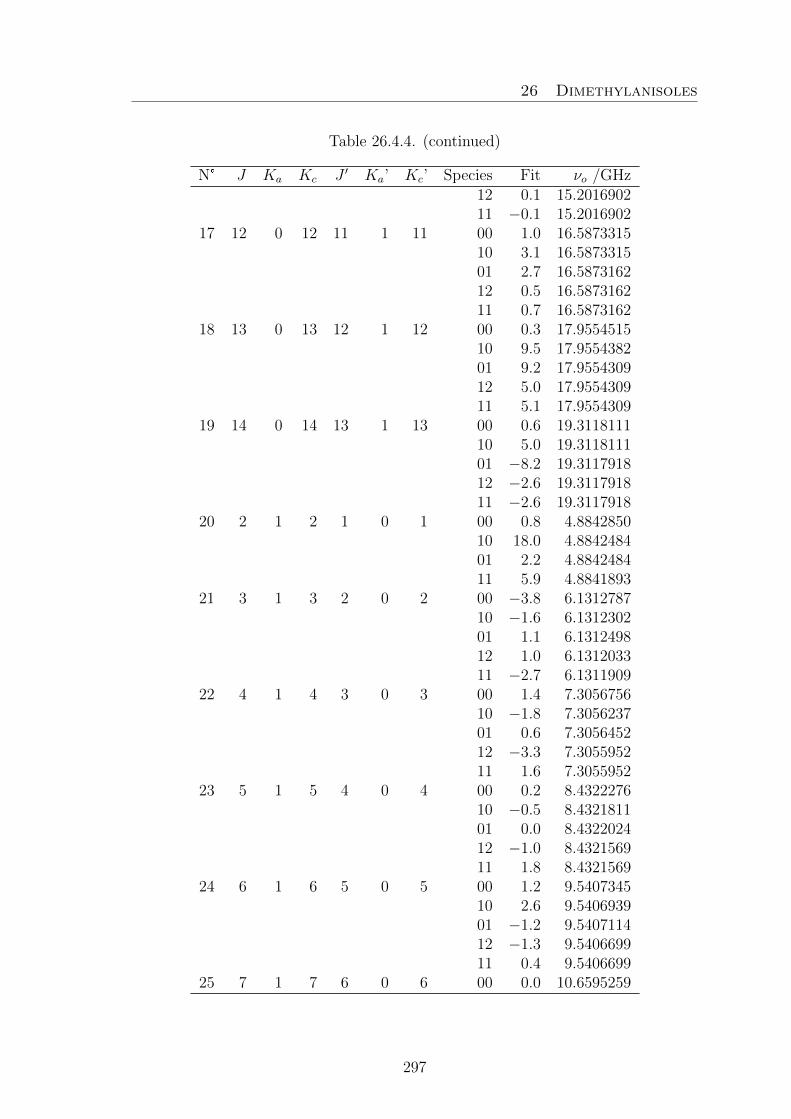
26 Dimethylanisoles
Table 26.4.4. (continued)
N° J Ka Kc J ′ Ka’ Kc’ Species Fit νo /GHz12 0.1 15.201690211 −0.1 15.2016902
17 12 0 12 11 1 11 00 1.0 16.587331510 3.1 16.587331501 2.7 16.587316212 0.5 16.587316211 0.7 16.5873162
18 13 0 13 12 1 12 00 0.3 17.955451510 9.5 17.955438201 9.2 17.955430912 5.0 17.955430911 5.1 17.9554309
19 14 0 14 13 1 13 00 0.6 19.311811110 5.0 19.311811101 −8.2 19.311791812 −2.6 19.311791811 −2.6 19.3117918
20 2 1 2 1 0 1 00 0.8 4.884285010 18.0 4.884248401 2.2 4.884248411 5.9 4.8841893
21 3 1 3 2 0 2 00 −3.8 6.131278710 −1.6 6.131230201 1.1 6.131249812 1.0 6.131203311 −2.7 6.1311909
22 4 1 4 3 0 3 00 1.4 7.305675610 −1.8 7.305623701 0.6 7.305645212 −3.3 7.305595211 1.6 7.3055952
23 5 1 5 4 0 4 00 0.2 8.432227610 −0.5 8.432181101 0.0 8.432202412 −1.0 8.432156911 1.8 8.4321569
24 6 1 6 5 0 5 00 1.2 9.540734510 2.6 9.540693901 −1.2 9.540711412 −1.3 9.540669911 0.4 9.5406699
25 7 1 7 6 0 6 00 0.0 10.6595259
297

26 Dimethylanisoles
Table 26.4.4. (continued)
N° J Ka Kc J ′ Ka’ Kc’ Species Fit νo /GHz10 3.9 10.659492501 −3.0 10.659505612 −1.3 10.6594705
26 8 1 8 7 0 7 00 0.6 11.808733310 3.1 11.808703801 −3.8 11.808714112 −1.0 11.808685311 −0.3 11.8086853
27 9 1 9 8 0 8 00 1.2 12.997160410 1.5 12.997133901 −1.3 12.997144712 −1.0 12.997118511 −0.6 12.9971185
28 10 1 10 9 0 9 0 8.2 14.224043910 10.1 14.224023801 13.6 14.224037112 11.2 14.224012811 −1.9 14.2239994
29 11 1 11 10 0 10 00 2.3 15.483248910 −0.8 15.483227601 0.9 15.483235912 −2.0 15.483214811 −1.8 15.4832148
30 12 1 12 11 0 11 00 1.9 16.767129110 2.7 16.767114601 −1.1 16.767114612 −1.3 16.767099211 −1.2 16.7670992
31 13 1 13 12 0 12 00 1.3 18.068654310 0.8 18.068640601 −1.0 18.068640612 −1.1 18.068627411 −1.0 18.0686274
32 14 1 14 13 0 13 00 4.2 19.382259710 −0.5 19.382243401 −0.7 19.382243412 3.9 19.382236411 3.9 19.3822364
33 3 1 3 2 1 2 00 1.5 4.280459810 0.4 4.280459801 4.9 4.2804598
298

26 Dimethylanisoles
Table 26.4.4. (continued)
N° J Ka Kc J ′ Ka’ Kc’ Species Fit νo /GHz12 8.4 4.280459811 −0.6 4.2804598
34 4 1 4 3 1 3 00 0.0 5.693976110 1.0 5.693976101 −1.4 5.693968312 1.3 5.693968311 5.6 5.6939761
35 5 1 5 4 1 4 00 −9.3 7.097521710 −7.1 7.097521701 −1.2 7.097521712 1.8 7.097521711 0.1 7.0975217
36 6 1 6 5 1 5 00 −0.5 8.490272510 2.7 8.490272501 −2.3 8.490261512 1.3 8.490261511 0.4 8.4902615
37 7 1 7 6 1 6 00 −0.3 9.872177510 3.8 9.872177501 −3.8 9.872163912 0.6 9.872163911 0.1 9.8721639
38 8 1 8 7 1 7 00 0.3 11.243944110 0.4 11.243939201 6.1 11.243939212 3.0 11.243931011 2.7 11.2439310
39 9 1 9 8 1 8 00 0.4 12.606794910 −1.8 12.606786901 3.5 12.606786912 0.6 12.606778211 0.4 12.6067782
40 3 1 2 2 1 1 00 −8.4 4.841551710 −2.1 4.841551701 0.9 4.841541712 2.7 4.841541711 11.7 4.8415417
41 4 1 3 3 1 2 00 −9.7 6.439714210 −3.3 6.439714201 3.7 6.439704112 8.3 6.439704111 11.9 6.4397041
299

26 Dimethylanisoles
Table 26.4.4. (continued)
N° J Ka Kc J ′ Ka’ Kc’ Species Fit νo /GHz42 5 1 4 4 1 3 00 −0.2 8.0228674
10 −2.4 8.022857401 2.6 8.022842012 0.5 8.022833211 2.5 8.0228332
43 6 1 5 5 1 4 00 −1.3 9.585311310 −2.3 9.585300501 6.9 9.585287012 2.1 9.585273311 3.7 9.5852733
44 7 1 6 6 1 5 00 −3.2 11.120344410 −8.1 11.120327101 3.6 11.120315312 2.3 11.120302211 3.6 11.1203022
45 2 2 1 1 1 0 00 2.2 9.309981510 −0.1 9.309118101 −1.4 9.309003212 −1.1 9.309728511 0.1 9.3069575
46 3 2 2 2 1 1 00 3.0 10.645697910 −2.9 10.645367401 −1.1 10.645430212 3.2 10.645449711 0.5 10.6447727
47 4 2 3 3 1 2 00 5.1 11.886182610 0.2 11.885971001 −2.5 11.886020712 1.5 11.885933311 0.1 11.8857016
48 5 2 4 4 1 3 00 −15.8 13.033531910 1.2 13.033377001 −1.1 13.033427212 0.3 13.033307111 −2.1 13.0332040
49 5 2 4 4 1 3 00 −11.5 13.033536210 0.9 13.033376701 −0.4 13.033427912 −16.5 13.033290311 −04.0 13.0332021
50 6 2 5 5 1 4 00 −0.4 14.092643610 0.3 14.0924868
300

26 Dimethylanisoles
Table 26.4.4. (continued)
N° J Ka Kc J ′ Ka’ Kc’ Species Fit νo /GHz01 −0.1 14.092542111 −6.8 14.092403612 −3.0 14.0923560
51 7 2 6 6 1 5 00 −1.1 15.072169310 −0.6 15.072020301 −0.5 15.072080912 −2.9 15.071943511 2.2 15.0719196
52 8 2 7 7 1 6 00 2.4 15.985968810 0.3 15.985823701 0.4 15.985889112 −1.3 15.985753011 −7.4 15.9857296
53 9 2 8 8 1 7 00 1.5 16.853968210 2.8 16.853833201 −0.5 16.853899112 3.1 16.853771711 −5.6 16.8537524
54 10 2 9 9 1 8 00 1.6 17.702050210 0.0 17.701920501 −0.5 17.701990612 0.1 17.701866411 −3.9 17.7018558
55 11 2 10 10 1 9 00 4.8 18.559795610 −0.2 18.559672701 −0.5 18.559741012 −3.4 18.559622311 0.7 18.5596223
56 12 2 11 11 1 10 00 1.8 19.455784710 −0.6 19.455676601 −0.8 19.455739312 0.3 19.455635911 2.9 19.4556359
57 2 2 0 1 1 1 00 −4.2 9.509564910 −0.5 9.510223401 −3.0 9.510246312 −0.4 9.509317611 −0.2 9.5120899
58 3 2 1 2 1 2 00 10.1 11.268886810 −19.0 11.268886801 3.0 11.268927812 0.6 11.2686148
301

26 Dimethylanisoles
Table 26.4.4. (continued)
N° J Ka Kc J ′ Ka’ Kc’ Species Fit νo /GHz11 0.1 11.2692885
59 4 2 2 3 1 3 00 −1.4 13.191151010 4.2 13.191067601 9.5 13.191090012 −2.3 13.190874011 9.7 13.1911163
60 5 2 3 4 1 4 00 −12.2 15.316956410 2.7 15.316848601 0.1 15.316848612 4.5 15.316679611 −1.4 15.3167749
61 6 2 4 5 1 5 00 −8.4 17.684899910 3.3 17.684776701 −4.7 17.684748912 1.7 17.684594111 8.1 17.6846532
62 3 3 0 2 2 1 00 −1.1 15.172989310 −2.3 15.178218201 −0.5 15.178013312 3.3 15.172658111 5.0 15.1846539
63 4 3 1 3 2 2 00 −0.7 16.722280810 2.8 16.725643101 −2.2 16.725491912 2.2 16.721876511 5.9 16.7304946
64 5 3 2 4 2 3 00 −2.2 18.302955910 −0.1 18.304289001 0.0 18.304229012 1.9 18.302537511 5.0 18.3075665
65 6 3 3 5 2 4 00 −4.0 19.936177310 −1.3 19.936514101 4.2 19.936527411 −0.4 19.9378911
66 7 3 4 6 2 5 00 −5.9 21.650206310 −0.7 21.6502063
67 3 3 1 2 2 0 00 −0.5 15.160091910 2.6 15.154374801 −1.8 15.154726512 0.7 15.159598011 −0.7 15.1476024
302

26 Dimethylanisoles
Table 26.4.4. (continued)
N° J Ka Kc J ′ Ka’ Kc’ Species Fit νo /GHz68 4 3 2 3 2 1 00 −0.7 16.6571295
10 0.3 16.653280001 2.8 16.653572012 2.0 16.656699311 2.7 16.6480855
69 5 3 3 4 2 2 00 −1.5 18.106427710 −0.4 18.104604201 0.2 18.104802712 1.7 18.106004111 2.3 18.1009786
70 6 3 4 5 2 3 00 −2.2 19.478770710 −0.6 19.477942901 0.4 19.478068812 2.2 19.478347911 3.7 19.4762083
71 7 3 5 6 2 4 00 3.2 20.747071410 0.8 20.746577901 0.4 20.746679212 4.0 20.746644911 1.8 20.7457416
72 8 3 6 7 2 5 00 −4.9 21.893294110 3.3 21.892935101 −0.5 21.893030712 −1.0 21.892872811 1.0 21.8924557
73 3 3 0 3 2 1 10 −9.5 10.546895212 0.4 10.542336711 −4.0 10.5509536
74 4 3 1 4 2 2 00 −2.4 10.457978910 0.4 10.461003501 −0.8 10.461109712 −1.4 10.457593311 −0.8 10.4653317
75 5 3 2 5 2 3 00 0.1 10.300908410 0.9 10.302157101 8.4 10.302141412 −1.1 10.300513411 1.6 10.3052262
78 6 3 3 6 2 4 00 2.2 10.055757310 1.0 10.056049801 0.1 10.056104812 −0.6 10.0553629
303

26 Dimethylanisoles
Table 26.4.4. (continued)
N° J Ka Kc J ′ Ka’ Kc’ Species Fit νo /GHz11 −0.2 10.0573609
79 7 3 4 7 2 5 00 −0.2 9.723302810 2.0 9.723274301 −3.4 9.723360212 −2.5 9.722915211 −2.3 9.7237406
80 8 3 5 8 2 6 00 1.1 9.325368010 −0.5 9.325218501 0.8 9.325327612 −2.6 9.324989011 2.2 9.3253680
81 9 3 6 9 2 7 00 0.3 8.903185310 6.7 8.903000201 1.0 8.903108612 −2.8 8.902820411 −8.6 8.9030002
82 3 3 1 3 2 2 10 −8.6 10.598971912 0.3 10.603233011 3.6 10.5946149
83 4 3 2 4 2 3 00 −1.9 10.636651810 −1.6 10.633036501 3.7 10.633320712 0.0 10.636239211 0.5 10.6285015
84 5 3 3 5 2 4 00 −1.4 10.701591210 0.5 10.699858001 0.6 10.700055812 0.7 10.701187511 −1.3 10.6964749
85 6 3 4 6 2 5 00 0.6 10.811765310 0.4 10.810983901 −1.8 10.811103612 0.9 10.811360511 0.8 10.8093619
86 7 3 5 7 2 6 00 −3.1 10.980688610 −0.5 10.980235601 −0.1 10.980324612 2.6 10.980287411 1.2 10.9794598
87 8 3 6 8 2 7 00 −2.5 11.221223210 −0.9 11.220890701 1.2 11.2209628
304

26 Dimethylanisoles
Table 26.4.4. (continued)
N° J Ka Kc J ′ Ka’ Kc’ Species Fit νo /GHz12 −2.8 11.220812611 1.6 11.2204418
88 9 3 7 9 2 8 00 −2.9 11.544687110 −0.5 11.544407801 0.9 11.544464112 −1.0 11.544273611 2.7 11.5440912
89 10 3 8 10 2 9 00 −3.2 11.960166110 −3.0 11.959910401 1.3 11.959954512 6.3 11.959753311 2.9 11.9596507
Table 26.4.5. Observed frequencies (νObs.) of 64 rotational transitions of cis-34DMA. Totally 312 signals were assigned and fitted to a standard deviation of4.6 kHz. νObs. − νCalc. values are given in kHz as obtained after a fit with theprogram XIAM. The symmetry species (ab) correspond to a: Me3 and b: Me4.
N° J Ka Kc J ′ Ka’ Kc’ Species Fit νo /GHz1 1 1 1 0 0 0 00 −1.8 3.2997794
10 −0.4 3.299732201 −2.7 3.299634212 −0.5 3.299649911 −0.7 3.2995253
2 2 1 2 1 0 1 00 7.9 4.666650010 0.5 4.666608001 −2.5 4.666527512 −1.3 4.666514511 0.4 4.6664752
3 3 1 3 2 0 2 00 −0.5 5.929178810 2.9 5.929157701 0.2 5.929076212 −5.4 5.929055911 2.0 5.9290437
4 4 1 4 3 0 3 00 −0.6 7.114618010 −0.8 7.114604301 3.2 7.114524512 −1.1 7.114511911 9.6 7.1145119
5 5 1 5 4 0 4 00 −7.2 8.262631910 −2.2 8.2626319
305

26 Dimethylanisoles
Table 26.4.5. (continued)
N° J Ka Kc J ′ Ka’ Kc’ Species Fit νo /GHz01 −0.5 8.262548912 1.4 8.262548911 7.6 8.2625489
6 6 1 6 5 0 5 00 −1.8 9.414907410 −0.7 9.414907401 1.5 9.414832012 0.7 9.414832011 4.5 9.4148320
7 7 1 7 6 0 6 00 −1.9 10.601504710 −0.1 10.601504701 −0.1 10.601441012 0.6 10.601441011 2.9 10.6014410
8 8 1 8 7 0 7 00 −6.5 11.833978110 −1.0 11.833978101 −0.2 11.833931812 4.7 11.833931811 6.1 11.8339318
9 9 1 9 8 0 8 00 −1.8 13.109108510 −2.4 13.109097801 1.2 13.109069812 0.0 13.109058911 0.8 13.1090589
10 10 1 10 9 0 9 00 −1.3 14.417109310 −2.0 14.417094301 9.6 14.417086812 1.6 14.417064811 2.0 14.4170648
11 11 1 11 10 0 10 00 −6.4 15.747772010 1.8 15.747762701 0.6 15.747751512 1.7 15.747735311 2.0 15.7477353
12 4 0 4 3 1 3 00 −1.1 4.935581010 −2.9 4.935537701 −4.3 4.935665212 1.3 4.935620311 3.9 4.9356406
13 5 0 5 4 1 4 00 −4.5 6.644502310 0.5 6.644456901 −0.4 6.644576411 3.4 6.6445346
306
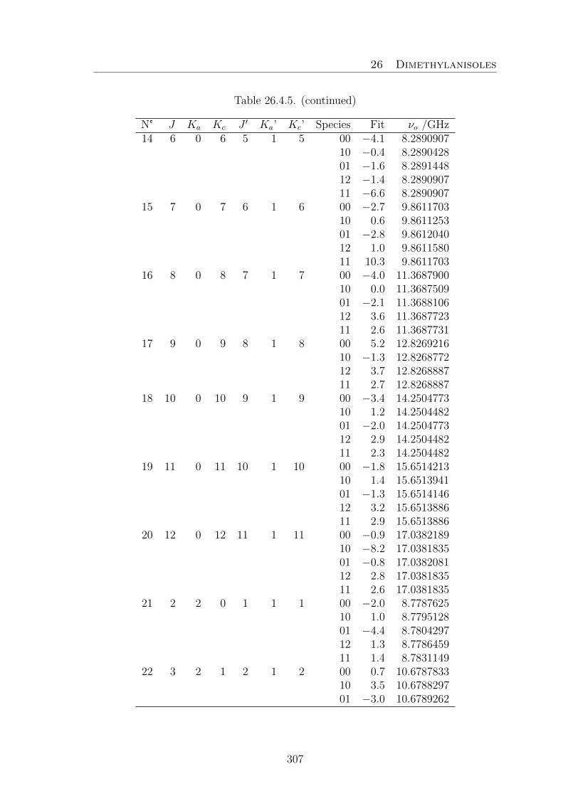
26 Dimethylanisoles
Table 26.4.5. (continued)
N° J Ka Kc J ′ Ka’ Kc’ Species Fit νo /GHz14 6 0 6 5 1 5 00 −4.1 8.2890907
10 −0.4 8.289042801 −1.6 8.289144812 −1.4 8.289090711 −6.6 8.2890907
15 7 0 7 6 1 6 00 −2.7 9.861170310 0.6 9.861125301 −2.8 9.861204012 1.0 9.861158011 10.3 9.8611703
16 8 0 8 7 1 7 00 −4.0 11.368790010 0.0 11.368750901 −2.1 11.368810612 3.6 11.368772311 2.6 11.3687731
17 9 0 9 8 1 8 00 5.2 12.826921610 −1.3 12.826877212 3.7 12.826888711 2.7 12.8268887
18 10 0 10 9 1 9 00 −3.4 14.250477310 1.2 14.250448201 −2.0 14.250477312 2.9 14.250448211 2.3 14.2504482
19 11 0 11 10 1 10 00 −1.8 15.651421310 1.4 15.651394101 −1.3 15.651414612 3.2 15.651388611 2.9 15.6513886
20 12 0 12 11 1 11 00 −0.9 17.038218910 −8.2 17.038183501 −0.8 17.038208112 2.8 17.038183511 2.6 17.0381835
21 2 2 0 1 1 1 00 −2.0 8.778762510 1.0 8.779512801 −4.4 8.780429712 1.3 8.778645911 1.4 8.7831149
22 3 2 1 2 1 2 00 0.7 10.678783310 3.5 10.678829701 −3.0 10.6789262
307

26 Dimethylanisoles
Table 26.4.5. (continued)
N° J Ka Kc J ′ Ka’ Kc’ Species Fit νo /GHz12 1.4 10.679521211 2.9 10.6784208
23 4 2 2 3 1 3 00 7.8 12.804129410 2.5 12.803998901 4.7 12.803996212 4.9 12.803681111 3.7 12.8040597
24 2 2 1 1 1 0 00 1.7 8.532480610 2.9 8.531546601 −9.1 8.530224912 0.1 8.531836011 0.9 8.5273677
25 3 2 2 2 1 1 00 7.1 9.899345710 1.0 9.899073201 −7.0 9.898612512 0.4 9.898908511 1.4 9.8978077
26 4 2 3 3 1 2 00 7.7 11.150937510 0.5 11.150788101 4.7 11.150499812 0.4 11.150542911 2.4 11.1501661
27 5 2 4 4 1 3 00 2.2 12.291620010 −0.7 12.291522801 4.3 12.291270612 0.0 12.291255311 3.3 12.2910920
28 6 2 5 5 1 4 00 −5.3 13.331556310 −1.0 13.331499001 5.9 13.331249412 −0.4 13.331224011 0.8 13.3311402
29 7 2 6 6 1 5 00 −0.2 14.289240710 −1.2 14.289206601 6.7 14.288948612 1.6 14.288934011 −1.7 14.2888835
30 8 2 7 7 1 6 00 −10.8 15.193434510 −1.7 15.193434501 7.5 15.193171012 3.0 15.1931710
31 9 2 8 8 1 7 00 6.8 16.0826132
308

26 Dimethylanisoles
Table 26.4.5. (continued)
N° J Ka Kc J ′ Ka’ Kc’ Species Fit νo /GHz10 0.0 16.082613201 −3.8 16.082342111 −2.4 16.0823421
32 10 2 9 9 1 8 00 9.6 16.999015910 −2.2 16.999015901 5.0 16.998778412 −11.8 16.998778411 −2.0 16.9987784
33 11 2 10 10 1 9 00 6.8 17.978149610 0.2 17.978149601 −0.5 17.977942312 −10.0 17.977942311 −4.0 17.9779423
34 3 3 1 2 2 0 00 −10.0 13.869479110 −1.4 13.862725001 −2.6 13.857795912 2.6 13.865628111 1.9 13.8485970
35 4 3 2 3 2 1 00 −9.5 15.417757710 −1.4 15.414036201 −5.1 15.410355512 3.6 15.415802711 0.4 15.4034809
36 5 3 3 4 2 2 00 −10.8 16.887430510 −1.1 16.886069901 −4.3 16.884138112 3.4 16.886415311 2.1 16.8799008
37 6 3 4 5 2 3 00 −5.0 18.237905010 1.4 18.237338601 −3.4 18.236393212 3.1 18.237133911 5.7 18.2345660
38 7 3 5 6 2 4 00 −0.6 19.441536910 1.7 19.441235001 −4.8 19.440605211 6.3 19.4397775
39 8 3 6 7 2 5 00 −8.4 20.491261210 2.1 20.491088401 2.6 20.490556412 4.7 20.490626711 8.6 20.4901279
309

26 Dimethylanisoles
Table 26.4.5. (continued)
N° J Ka Kc J ′ Ka’ Kc’ Species Fit νo /GHz40 3 3 0 2 2 1 00 −9.1 13.8915703
10 −1.7 13.898039501 −6.6 13.902304112 2.1 13.894185011 0.9 13.9112142
41 4 3 1 3 2 2 00 −9.5 15.529433110 −1.0 15.532843401 −6.9 15.535882312 2.7 15.530125811 3.8 15.5424451
42 5 3 2 4 2 3 00 −10.9 17.222898810 1.0 17.223927701 −6.0 17.225241612 4.9 17.222630111 5.9 17.2291434
43 6 3 3 5 2 4 00 −12.2 19.010750510 −0.3 19.010956001 −5.8 19.011310012 8.1 19.010211511 8.6 19.0127807
44 7 3 4 6 2 5 10 3.1 20.944381001 −8.8 20.944449612 −9.8 20.943805911 9.9 20.9448958
45 4 4 1 4 3 2 00 12.7 12.706902710 11.2 12.701694701 −9.5 12.699575112 −1.3 12.702680811 −1.4 12.6969720
46 4 4 0 4 3 1 00 5.0 12.698492510 7.6 12.703399801 −9.8 12.704441512 1.0 12.701053111 1.1 12.7067617
47 2 0 2 1 0 1 00 −3.1 3.163663210 −0.7 3.163639801 −1.0 3.163663212 0.8 3.163639811 1.9 3.1636398
48 3 0 3 2 0 2 00 −1.6 4.694471510 0.3 4.694441001 −2.2 4.6944656
310

26 Dimethylanisoles
Table 26.4.5. (continued)
N° J Ka Kc J ′ Ka’ Kc’ Species Fit νo /GHz12 −0.4 4.694435411 0.6 4.6944354
49 4 0 4 3 0 3 00 −1.4 6.170286910 0.9 6.170255701 −2.7 6.170274912 0.1 6.170244611 0.9 6.1702446
50 5 0 5 4 0 4 00 −4.3 7.588832810 −0.3 7.588806301 −0.9 7.588819612 2.4 7.588792711 3.0 7.5887927
51 6 0 6 5 0 5 00 −3.3 8.962893610 −0.8 8.962869701 −5.7 8.962869712 1.4 8.962850711 1.7 8.9628507
52 7 0 7 6 0 6 00 −7.8 10.313177510 −1.2 10.313161001 −0.9 10.313161012 −6.2 10.313132711 −5.9 10.3131327
53 8 0 8 7 0 7 10 −0.6 11.657092901 1.0 11.6570929
54 3 1 2 2 1 1 00 −0.6 5.101217310 0.8 5.101158001 −4.0 5.101202912 1.5 5.101158011 4.4 5.1011404
55 4 1 3 3 1 2 00 0.5 6.774304010 0.7 6.774227901 −4.2 6.774290812 4.8 6.774227911 13.5 6.7742279
56 5 1 4 4 1 3 00 −5.2 8.420286710 2.8 8.420204501 −5.1 8.420275812 11.0 8.420204511 2.2 8.4201902
57 6 1 5 5 1 4 00 6.4 10.028198610 1.7 10.028094701 −4.6 10.0281710
311

26 Dimethylanisoles
Table 26.4.5. (continued)
N° J Ka Kc J ′ Ka’ Kc’ Species Fit νo /GHz12 1.2 10.028079711 5.5 10.0280797
58 7 1 6 6 1 5 00 −7.7 11.585738010 0.1 11.585644001 −3.9 11.585716112 1.7 11.585621611 5.0 11.5856216
59 3 1 3 2 1 2 00 7.0 4.426210510 −4.7 4.426183201 0.2 4.426210512 −1.3 4.426183211 5.7 4.4262105
60 4 1 4 3 1 3 00 −0.3 5.879912210 −3.8 5.879887101 −0.8 5.879912212 −0.4 5.879887111 −8.2 5.8798871
61 5 1 5 4 1 4 00 −3.6 7.318305110 −0.9 7.318283001 −0.7 7.318305112 3.9 7.318283011 0.2 7.3182830
62 6 1 6 5 1 5 00 −8.9 8.741098310 1.1 8.741081901 −3.1 8.741098312 7.8 8.741081911 5.9 8.7410819
63 7 1 7 6 1 6 00 −1.6 10.149492710 1.8 10.149469101 −3.2 10.149482812 0.1 10.149458611 −0.9 10.1494586
64 8 1 8 7 1 7 00 −2.0 11.545661310 1.7 11.545638101 −3.9 11.545648912 −0.1 11.545625511 −0.7 11.5456255
312

26 Dimethylanisoles
26.5. 3,5-DMA
Table 26.5.1. Nuclear coordinates of 3,5-dimethylanisole given in the principalinertial axes system. Calculations were carried out at the B3LYP/6-311++G(d.p)level of theory. The atom numbering refers to Figure 15.1.
a /A b /A c /AC1 −0.921567 −0.412847 −0.000028C2 −0.633253 0.949502 −0.000003C3 0.703131 1.382118 0.000022C4 1.723931 0.437753 0.000036C5 1.442684 −0.939035 −0.000005C6 0.117838 −1.353773 −0.000051H7 2.757756 0.769718 0.000070H8 −0.140971 −2.406398 −0.000096H9 −1.425956 1.686761 −0.000020C10 2.569761 −1.944390 0.000044H11 3.207787 −1.820808 −0.880566H12 2.191285 −2.968266 −0.000513H13 3.207055 −1.821519 0.881289O14 −2.185051 −0.935756 −0.000100C15 −3.289959 −0.044179 0.000113H16 −3.294336 0.589442 −0.893820H17 −4.179610 −0.672236 0.000232H18 −3.294035 0.589366 0.894099C19 1.008245 2.861547 −0.000035H20 0.584086 3.353725 0.880957H21 2.084420 3.044736 0.000273H22 0.584639 3.353517 −0.881415
Table 26.5.2 Coefficients used for the potential energy curve obtained by rotatingthe methoxy group about the axis C1−O14 (variation of α, see Figure 15.2). Thecalculations were performed at the B3LYP/6-311++G(d,p) level of theory. Theexpansion is defined as following: V(α) =a0+Σ15
n=1cos(nα + π).
Fourier Term Hartree cm−1
1 −425.5201055cos(2α) −0.0023972 −526.12cos(4α) −0.0004008 −87.97cos(6α) 0.0000414 9.09cos(8α) 0.0000366 8.03cos(10α) −0.0000199 −4.37
313

26 Dimethylanisoles
Table 26.5.3. Coefficients of the one-dimensional Fourier expansion for the po-tential energy curve on the left hand-side in Figure 15.3, obtained by variation ofthe dihedral angle β = ∠(C2−C3−C19−H21). This corresponds to a rotation aboutthe C3−C19 axis. The calculations were carried out at the MP26-311++G(d,p) andB3LYP/6-311++G(d,p) levels of theory. The potential is expanded as: V(β) =Σ7i=0bifi.
MP2 B3LYPi fi bi/Hartree bi/cm−1 bi/Hartree bi/cm−1
0 1 −424.24402 −425.52269241 cos(3β) −0.0001027 −22.54 −0.0001525 −33.472 cos(6β) −0.0000015 −0.33 0.0000071 1.563 cos(9β) 0.0000001 0.02 −0.0000007 −0.154 cos(12β) / / −0.0000003 −0.075 sin(3β) 0.0001274 27.96 / /6 sin(6β) −0.0000617 −13.54 / /7 sin(9β) −0.0000001 −0.02 / /
Table 26.5.4. Coefficients of the one-dimensional Fourier expansion of the potentialenergy surface obtained by varying the dihedral angle γ = ∠(C4−C5−C10−H12)(rotation about the C5−C10 axis, Figure 15.3, right hand-side). The calculationswere carried out at the MP2/6-311++G(d,p) and B3LYP/6-311++G(d,p) levels oftheory. The Fourier expansion is given as: V(γ) =Σ7
i=0cifi.
MP2 B3LYPi fi ci/Hartree ci/cm−1 ci/Hartree ci/cm−1
0 1 −424.2440595 −425.52274801 cos(3γ) −0.0000227 −4.98 −0.0000981 −21.532 cos(6γ) −0.0000368 −8.08 0.0000076 1.673 cos(9γ) −0.0000010 −0.22 0.0000002 0.044 cos(12γ) / / 0.0000011 0.245 sin(3γ) 0.0000761 16.70 / /6 sin(6γ) −0.0000467 −10.25 / /7 sin(9γ) 0.0000010 0.22 / /
314

26 Dimethylanisoles
Table 26.5.5. Coefficients of the two-dimensional Fourier expansions of the poten-tial energy surfaces of 3,5-Dimethylanisole obtained by varying the dihedral anglesβ = ∠(C2−C3−C19−H21) and γ = ∠(C4−C5−C10−H12) in a grid of 10° (see Figure15.5). The calculations were carried out at the MP2- and B3LYP/6-311++G(d,p)levels of theory. The potentials are expanded as: V(β,γ) =Σ13
n=1aifi.
MP2 B3LYPi fi ai / Hartree ai / cm−1 ai / Hartree ai / cm−1
1 1 −424.2439469 −425.52263842 cos(3β) 0.0001540 33.80 0.0001165 25.573 cos(3γ) 0.0000604 13.26 0.0000630 13.834 cos(6β) 0.0000644 14.13 0.0000080 1.765 cos(6γ) 0.0000602 13.21 0.0000084 1.846 cos(3β) cos(3γ) −0.0000301 −6.61 −0.0000350 −7.687 sin(3β)·sin(3γ) 0.0000143 3.14 0.0000010 0.228 cos(6β) cos(3γ) −0.0000010 −0.22 / /9 sin(6β)·sin(3γ) 0.0000010 0.22 / /10 cos(3β) cos(6γ) −0.0000007 −0.15 0.0000008 0.1811 sin(3β)·sin(6γ) −0.0000050 −1.10 / /12 cos(6β) cos(6γ) 0.0000022 0.48 / /13 sin(6β)·sin(6γ) 0.0000004 0.09
Table 26.5.6 Observed frequencies (νo) of 146 rotational transitions of 3,5-dimethyl-anisole. Totally 370 signals were assigned and fitted to a standard deviation of8.7 kHz. νo − νc values are given in kHz as obtained after a fit with the programNTOP.
N° J Ka Kc J ′ Ka’ Kc’ Species νo /GHz Fit I Fit II1 6 0 6 5 1 5 00 8.825494 1 1
6 0 6 5 1 5 01 8.809982 −1 06 0 6 5 1 5 10 8.809768 −1 06 0 6 5 1 5 11 8.822865 / −10
2 7 0 7 6 1 6 00 10.194605 0 07 0 7 6 1 6 01 10.189822 0 07 0 7 6 1 6 10 10.190903 0 07 0 7 6 1 6 11 10.189105 / 17 0 7 6 1 6 12 10.172374 / −4
3 8 0 8 7 1 7 00 11.557964 −5 −58 0 8 7 1 7 01 11.555988 −1 08 0 8 7 1 7 10 11.556936 0 18 0 8 7 1 7 11 11.551253 / 38 0 8 7 1 7 12 11.554295 / −4
4 9 0 9 8 1 8 00 12.919605 0 0
315
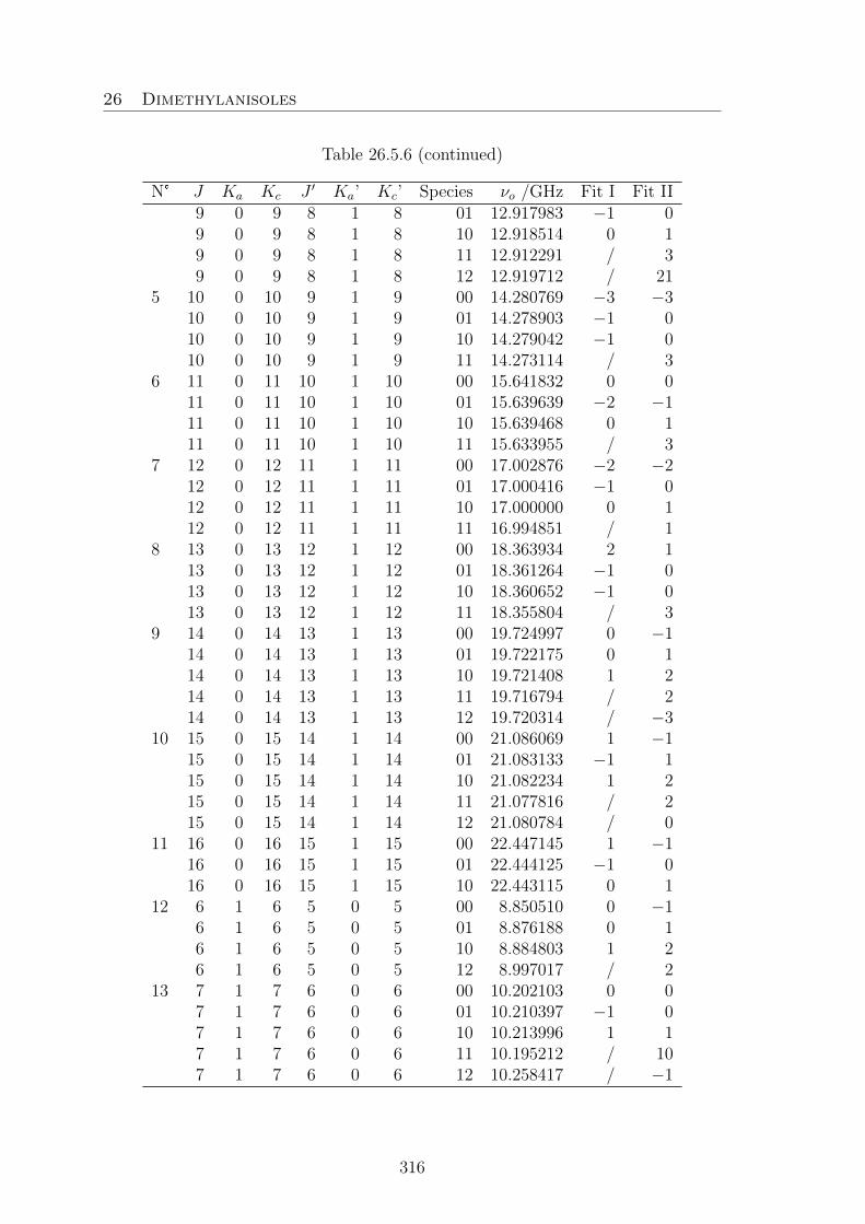
26 Dimethylanisoles
Table 26.5.6 (continued)
N° J Ka Kc J ′ Ka’ Kc’ Species νo /GHz Fit I Fit II9 0 9 8 1 8 01 12.917983 −1 09 0 9 8 1 8 10 12.918514 0 19 0 9 8 1 8 11 12.912291 / 39 0 9 8 1 8 12 12.919712 / 21
5 10 0 10 9 1 9 00 14.280769 −3 −310 0 10 9 1 9 01 14.278903 −1 010 0 10 9 1 9 10 14.279042 −1 010 0 10 9 1 9 11 14.273114 / 3
6 11 0 11 10 1 10 00 15.641832 0 011 0 11 10 1 10 01 15.639639 −2 −111 0 11 10 1 10 10 15.639468 0 111 0 11 10 1 10 11 15.633955 / 3
7 12 0 12 11 1 11 00 17.002876 −2 −212 0 12 11 1 11 01 17.000416 −1 012 0 12 11 1 11 10 17.000000 0 112 0 12 11 1 11 11 16.994851 / 1
8 13 0 13 12 1 12 00 18.363934 2 113 0 13 12 1 12 01 18.361264 −1 013 0 13 12 1 12 10 18.360652 −1 013 0 13 12 1 12 11 18.355804 / 3
9 14 0 14 13 1 13 00 19.724997 0 −114 0 14 13 1 13 01 19.722175 0 114 0 14 13 1 13 10 19.721408 1 214 0 14 13 1 13 11 19.716794 / 214 0 14 13 1 13 12 19.720314 / −3
10 15 0 15 14 1 14 00 21.086069 1 −115 0 15 14 1 14 01 21.083133 −1 115 0 15 14 1 14 10 21.082234 1 215 0 15 14 1 14 11 21.077816 / 215 0 15 14 1 14 12 21.080784 / 0
11 16 0 16 15 1 15 00 22.447145 1 −116 0 16 15 1 15 01 22.444125 −1 016 0 16 15 1 15 10 22.443115 0 1
12 6 1 6 5 0 5 00 8.850510 0 −16 1 6 5 0 5 01 8.876188 0 16 1 6 5 0 5 10 8.884803 1 26 1 6 5 0 5 12 8.997017 / 2
13 7 1 7 6 0 6 00 10.202103 0 07 1 7 6 0 6 01 10.210397 −1 07 1 7 6 0 6 10 10.213996 1 17 1 7 6 0 6 11 10.195212 / 107 1 7 6 0 6 12 10.258417 / −1
316

26 Dimethylanisoles
Table 26.5.6 (continued)
N° J Ka Kc J ′ Ka’ Kc’ Species νo /GHz Fit I Fit II14 8 1 8 7 0 7 00 11.560121 −1 −1
8 1 8 7 0 7 01 11.562061 0 18 1 8 7 0 7 10 11.563674 0 18 1 8 7 0 7 11 11.553013 / 68 1 8 7 0 7 12 11.580981 / −2
15 9 1 9 8 0 8 00 12.920204 −1 −19 1 9 8 0 8 01 12.919712 −1 09 1 9 8 0 8 10 12.920410 0 19 1 9 8 0 8 11 12.912783 / 49 1 9 8 0 8 12 12.927549 / −3
16 10 1 10 9 0 9 00 14.280935 0 010 1 10 9 0 9 01 14.279382 −1 010 1 10 9 0 9 10 14.279562 −1 010 1 10 9 0 9 12 14.282630 / −3
17 11 1 11 10 0 10 00 15.641875 0 −111 1 11 10 0 10 01 15.639770 −1 011 1 11 10 0 10 10 15.639609 1 211 1 11 10 0 10 12 15.640774 / −2
18 12 1 12 11 0 11 00 17.002891 1 112 1 12 11 0 11 01 17.000450 −2 012 1 12 11 0 11 10 17.000036 0 112 1 12 11 0 11 11 16.994851 / −812 1 12 11 0 11 12 17.000137 / −2
19 13 1 13 12 0 12 00 18.363934 −1 −213 1 13 12 0 12 01 18.361274 0 113 1 13 12 0 12 10 18.360664 1 213 1 13 12 0 12 11 18.355804 / 0
20 14 1 14 13 0 13 00 19.724997 0 −114 1 14 13 0 13 01 19.722175 −2 −114 1 14 13 0 13 10 19.721408 −1 014 1 14 13 0 13 11 19.716794 / 114 1 14 13 0 13 12 19.720314 / −15
21 15 1 15 14 0 14 00 21.086069 0 −115 1 15 14 0 14 01 21.083133 −1 015 1 15 14 0 14 10 21.082234 0 115 1 15 14 0 14 11 21.077816 / 215 1 15 14 0 14 12 21.080784 / −3
22 16 1 16 15 0 15 00 22.447145 1 −116 1 16 15 0 15 01 22.444125 −1 016 1 16 15 0 15 10 22.443115 0 016 1 16 15 0 15 11 22.438859 / 1
23 6 1 5 5 2 4 00 9.891193 3 1
317

26 Dimethylanisoles
Table 26.5.6 (continued)
N° J Ka Kc J ′ Ka’ Kc’ Species νo /GHz Fit I Fit II6 1 5 5 2 4 01 9.565264 2 26 1 5 5 2 4 10 9.527971 −5 −4
24 7 1 6 6 2 5 00 11.432510 1 07 1 6 6 2 5 01 11.286662 1 27 1 6 6 2 5 10 11.271267 0 17 1 6 6 2 5 12 10.740102 / −18
25 8 1 7 7 2 6 00 12.865762 0 −18 1 7 7 2 6 01 12.810362 0 18 1 7 7 2 6 10 12.807660 1 28 1 7 7 2 6 11 12.855445 / −408 1 7 7 2 6 12 12.580525 / −16
26 9 1 8 8 2 7 00 14.252070 0 −19 1 8 8 2 7 01 14.232254 0 19 1 8 8 2 7 10 14.233580 1 29 1 8 8 2 7 11 14.233457 / −5
27 10 1 9 9 2 8 00 15.620695 0 −110 1 9 9 2 8 01 15.612199 −1 010 1 9 9 2 8 10 15.613888 0 1
28 11 1 10 10 2 9 00 16.983409 1 −111 1 10 10 2 9 01 16.977347 0 111 1 10 10 2 9 10 16.978349 0 111 1 10 10 2 9 12 16.978002 / −17
29 12 1 11 11 2 10 00 18.344399 1 012 1 11 11 2 10 01 18.338057 0 112 1 11 11 2 10 10 18.338249 0 1
30 13 1 12 12 2 11 00 19.705006 2 113 1 12 12 2 11 01 19.697788 0 113 1 12 12 2 11 10 19.697287 0 1
31 14 1 13 13 2 12 01 21.057536 1 214 1 13 13 2 12 10 21.056486 4 5
32 3 2 1 2 1 2 00 9.190323 6 43 2 1 2 1 2 01 9.804323 23 283 2 1 2 1 2 10 10.341665 −4 −1
33 4 2 2 3 1 3 01 12.995952 7 134 2 2 3 1 3 10 13.273132 7 11
34 5 2 4 4 1 3 00 9.450552 2 25 2 4 4 1 3 01 9.850557 3 55 2 4 4 1 3 10 9.889555 13 15
35 6 2 5 5 1 4 00 10.505317 −3 −36 2 5 5 1 4 01 10.872584 1 36 2 5 5 1 4 10 10.939370 7 86 2 5 5 1 4 12 11.613948 / −10
318

26 Dimethylanisoles
Table 26.5.6 (continued)
N° J Ka Kc J ′ Ka’ Kc’ Species νo /GHz Fit I Fit II36 7 2 6 6 1 5 00 11.679637 −1 −2
7 2 6 6 1 5 01 11.889404 0 27 2 6 6 1 5 10 11.935772 4 57 2 6 6 1 5 12 12.533360 / −8
37 8 2 7 7 1 6 00 12.954805 −1 −18 2 7 7 1 6 01 13.044453 0 18 2 7 7 1 6 10 13.066617 1 1
38 9 2 8 8 1 7 00 14.281856 0 09 2 8 8 1 7 01 14.314054 0 19 2 8 8 1 7 10 14.323566 0 1
39 10 2 9 9 1 8 00 15.630148 1 010 2 9 9 1 8 01 15.638955 0 110 2 9 9 1 8 10 15.643061 0 1
40 11 2 10 10 1 9 00 16.986292 0 −111 2 10 10 1 9 01 16.985706 0 111 2 10 10 1 9 10 16.987372 0 1
41 12 2 11 11 1 10 00 18.345251 0 −112 2 11 11 1 10 01 18.340580 0 112 2 11 11 1 10 10 18.340945 1 1
42 13 2 12 12 1 11 00 19.705250 1 −113 2 12 12 1 11 01 19.698529 0 113 2 12 12 1 11 10 19.698070 0 1
43 14 2 13 13 1 12 01 21.057749 1 214 2 13 13 1 12 10 21.056704 0 1
44 4 2 3 3 1 2 01 8.599948 −1 045 5 2 4 4 1 3 12 10.392889 / −2146 9 2 8 8 1 7 12 14.487792 / −1547 5 2 3 4 1 4 01 16.338724 5 7
5 2 3 4 1 4 10 16.378385 13 1548 6 2 4 5 1 5 01 20.055195 5 549 6 0 6 5 0 5 00 8.844695 0 −1
6 0 6 5 0 5 01 8.860332 0 06 0 6 5 0 5 10 8.866962 1 16 0 6 5 0 5 11 8.838453 / 17
50 7 0 7 6 0 6 00 10.200421 0 07 0 7 6 0 6 10 10.208744 0 17 0 7 6 0 6 12 10.237844 0 −1
51 8 0 8 7 0 7 00 11.559651 0 −18 0 8 7 0 7 01 11.560708 −1 08 0 8 7 0 7 10 11.562186 0 18 0 8 7 0 7 11 11.552627 / 58 0 8 7 0 7 12 11.574868 / −3
319

26 Dimethylanisoles
Table 26.5.6 (continued)
N° J Ka Kc J ′ Ka’ Kc’ Species νo /GHz Fit I Fit II52 9 0 9 8 0 8 00 12.920064 −12 −12
9 0 9 8 0 8 01 12.919334 −2 −19 0 9 8 0 8 10 12.920001 0 09 0 9 8 0 8 12 12.925800 / −3
53 10 0 10 9 0 9 10 14.279450 −3 −254 11 0 11 10 0 10 00 15.641874 9 8
11 0 11 10 0 10 11 15.633990 / 1011 0 11 10 0 10 12 15.640641 / −3
55 12 0 12 11 0 11 00 17.002890 4 312 0 12 11 0 11 01 17.000450 6 712 0 12 11 0 11 10 17.000036 7 812 0 12 11 0 11 11 16.994863 / 5
56 13 0 13 12 0 12 00 18.363934 −1 −257 14 0 14 13 0 13 00 19.724997 0 −158 15 0 15 14 0 14 00 21.086069 1 −159 6 1 6 5 1 5 00 8.831308 0 0
6 1 6 5 1 5 01 8.825839 0 06 1 6 5 1 5 10 8.827610 1 16 1 6 5 1 5 11 8.827610 / 106 1 6 5 1 5 12 8.807249 / −4
60 7 1 7 6 1 6 00 10.196288 0 07 1 7 6 1 6 01 10.194541 −1 −17 1 7 6 1 6 10 10.196155 0 17 1 7 6 1 6 11 10.190480 / 37 1 7 6 1 6 12 10.192948 / −3
61 8 1 8 7 1 7 00 11.558440 1 08 1 8 7 1 7 01 11.557340 −1 08 1 8 7 1 7 10 11.558421 −1 08 1 8 7 1 7 11 11.551640 / 4
62 9 1 9 8 1 8 01 12.918361 0 19 1 9 8 1 8 10 12.918923 0 19 1 9 8 1 8 12 12.921437 / −3
63 11 1 11 10 1 10 00 15.641831 −9 −1064 12 1 12 11 1 11 00 17.002876 −4 −565 13 1 13 12 1 12 00 18.363934 1 066 14 1 14 13 1 13 00 19.724997 1 067 15 1 15 14 1 14 00 21.086069 1 068 5 1 4 4 1 3 00 9.017062 0 −1
5 1 4 4 1 3 01 8.973341 3 45 1 4 4 1 3 10 8.951450 0 2
69 6 1 5 5 1 4 00 10.324676 −2 −26 1 5 5 1 4 01 10.442479 2 3
320

26 Dimethylanisoles
Table 26.5.6 (continued)
N° J Ka Kc J ′ Ka’ Kc’ Species νo /GHz Fit I Fit II6 1 5 5 1 4 10 10.466072 3 56 1 5 5 1 4 12 10.421085 / −17
70 7 1 6 6 1 5 00 11.613149 −1 −27 1 6 6 1 5 01 11.716767 0 17 1 6 6 1 5 10 11.744564 3 47 1 6 6 1 5 11 11.587043 / 847 1 6 6 1 5 12 11.932964 / −12
71 8 1 7 7 1 6 00 12.932249 −1 −18 1 7 7 1 6 01 12.982998 0 18 1 7 7 1 6 10 12.998866 0 18 1 7 7 1 6 11 12.908767 / 398 1 7 7 1 6 12 13.180920 / −12
72 9 1 8 8 1 7 10 14.301331 1 273 10 1 9 9 1 8 10 15.636124 0 174 11 1 10 10 1 9 10 16.985286 0 175 5 2 4 4 2 3 00 8.581528 2 1
5 2 4 4 2 3 01 8.474046 2 276 6 2 5 5 2 4 00 10.071833 1 0
6 2 5 5 2 4 01 9.995369 1 26 2 5 5 2 4 10 10.001269 −1 −1
77 7 2 6 6 2 5 00 11.498997 1 07 2 6 6 2 5 01 11.459299 1 27 2 6 6 2 5 10 11.462474 0 17 2 6 6 2 5 11 11.496908 / −69
78 8 2 7 7 2 6 00 12.888317 0 −18 2 7 7 2 6 01 12.871817 0 18 2 7 7 2 6 10 12.875410 1 18 2 7 7 2 6 11 12.873660 / −14
79 9 2 8 8 2 7 00 14.259300 0 −19 2 8 8 2 7 01 14.252599 0 19 2 8 8 2 7 10 14.255816 1 2
80 10 2 9 9 2 8 00 15.622917 0 −110 2 9 9 2 8 01 15.618610 0 110 2 9 9 2 8 10 15.620828 3 4
81 11 2 10 10 2 9 00 16.984071 1 011 2 10 10 2 9 01 16.979295 0 111 2 10 10 2 9 10 16.980435 0 1
82 12 2 11 11 2 10 10 18.338853 −5 −483 5 2 3 4 2 2 00 9.939825 3 1
5 2 3 4 2 2 01 9.367454 −7 −105 2 3 4 2 2 10 9.127866 −7 −7
84 6 2 4 5 2 3 00 11.709817 0 −2
321

26 Dimethylanisoles
Table 26.5.6 (continued)
N° J Ka Kc J ′ Ka’ Kc’ Species νo /GHz Fit I Fit II6 2 4 5 2 3 01 11.157948 5 46 2 4 5 2 3 10 11.010944 −4 −3
85 7 2 5 6 2 4 00 13.218693 −1 −37 2 5 6 2 4 01 13.023599 8 87 2 5 6 2 4 10 12.962891 2 4
86 8 2 6 7 2 5 00 14.520943 −2 −38 2 6 7 2 5 01 14.633730 1 3
87 9 2 7 8 2 6 00 15.766629 −1 −29 2 7 8 2 6 01 15.938991 −3 −2
88 10 2 8 9 2 7 01 17.151716 −2 −110 2 8 9 2 7 10 17.175568 −2 −1
89 11 2 9 10 2 8 01 18.411556 −1 011 2 9 10 2 8 10 18.423421 −1 −1
90 12 2 10 11 2 9 01 19.722176 −4 −391 6 2 4 5 3 3 00 9.401919 −1 −4
6 2 4 5 3 3 01 8.679673 −6 −1092 7 2 5 6 3 4 01 10.883298 −2 −493 8 2 6 7 3 5 01 13.113507 1 194 9 2 7 8 3 6 01 15.085690 −2 −195 10 2 8 9 3 7 01 16.764414 −4 −296 11 2 9 10 3 8 01 18.259242 −3 −297 12 2 10 11 3 9 01 19.667282 −2 −198 13 2 11 12 3 10 01 21.040817 −2 −199 6 2 4 5 3 3 10 8.965692 5 0100 7 2 5 6 3 4 10 10.901646 7 6101 8 2 6 7 3 5 10 13.070565 9 10102 9 2 7 8 3 6 10 15.058090 7 9103 10 2 8 9 3 7 10 16.755367 4 5104 11 2 9 10 3 8 10 18.258297 1 2105 12 2 10 11 3 9 10 19.668228 −1 0106 13 2 11 12 3 10 10 21.041355 −1 0107 3 3 0 2 2 1 00 9.731425 −3 −3108 4 3 1 3 2 2 00 11.926478 1 0
4 3 1 3 2 2 01 12.640335 −21 −27109 5 3 2 4 2 3 00 14.622299 −6 −9
5 3 2 4 2 3 01 15.447659 −20 −17110 6 3 3 5 2 4 00 17.957137 −10 −16
6 3 3 5 2 4 01 18.687154 −13 −6111 7 3 4 6 2 5 01 22.049809 −6 −1112 3 3 1 2 2 0 00 9.546990 −4 −3113 4 3 2 3 2 1 00 11.045784 −11 −11
4 3 2 3 2 1 01 10.344830 −26 −27
322

26 Dimethylanisoles
Table 26.5.6 (continued)
N° J Ka Kc J ′ Ka’ Kc’ Species νo /GHz Fit I Fit II4 3 2 3 2 1 10 9.071865 9 10
114 5 3 3 4 2 2 00 12.247707 −13 −125 3 3 4 2 2 01 11.845734 8 95 3 3 4 2 2 10 11.173120 −13 −8
115 6 3 4 5 2 3 00 13.205548 −15 −156 3 4 5 2 3 01 13.298249 14 166 3 4 5 2 3 10 13.072186 −11 −7
116 7 3 5 6 2 4 00 14.034087 3 37 3 5 6 2 4 01 14.543823 8 107 3 5 6 2 4 10 14.533718 −2 0
117 8 3 6 7 2 5 00 14.896436 0 08 3 6 7 2 5 01 15.487031 0 38 3 6 7 2 5 10 15.554974 −1 1
118 9 3 7 8 2 6 00 15.918524 4 29 3 7 8 2 6 01 16.326293 −1 09 3 7 8 2 6 10 16.391872 −3 −2
119 10 3 8 9 2 7 00 17.102867 1 010 3 8 9 2 7 01 17.304030 0 110 3 8 9 2 7 10 17.340691 −5 −4
120 11 3 9 10 2 8 00 18.385619 1 −111 3 9 10 2 8 01 18.466454 1 211 3 9 10 2 8 10 18.482536 −3 −2
121 12 3 10 11 2 9 01 19.740855 1 212 3 10 11 2 9 10 19.747274 −3 −2
122 13 3 11 12 2 10 10 21.067722 −2 −1123 5 3 3 4 3 2 01 9.252722 26 27
5 3 3 4 3 2 10 9.592730 −14 −10124 6 3 4 5 3 3 01 10.819968 −3 −4
6 3 4 5 3 3 10 11.026935 −2 −3125 7 3 5 6 3 4 01 12.403523 0 −1
7 3 5 6 3 4 10 12.472474 3 2126 8 3 6 7 3 5 01 13.966807 0 0
8 3 6 7 3 5 10 13.984144 2 3127 9 3 7 8 3 6 01 15.472992 0 1
9 3 7 8 3 6 10 15.478292 2 3128 10 3 8 9 3 7 01 16.916729 −1 0
10 3 8 9 3 7 10 16.920489 0 1129 11 3 9 10 3 8 01 18.314140 −1 0
11 3 9 10 3 8 10 18.317412 0 1130 5 3 2 4 3 1 10 9.517944 −5 6131 6 3 3 5 3 2 01 11.713542 9 13
6 3 3 5 3 2 10 11.372518 −12 −9
323

26 Dimethylanisoles
Table 26.5.6 (continued)
N° J Ka Kc J ′ Ka’ Kc’ Species νo /GHz Fit I Fit II132 7 3 4 6 3 3 01 13.358025 10 8
7 3 4 6 3 3 10 13.031851 −10 −11133 8 3 5 7 3 4 01 15.101222 0 −1
8 3 5 7 3 4 10 14.890548 −2 −1134 9 3 6 8 3 5 01 17.023468 16 16
9 3 6 8 3 5 10 16.916323 5 6135 10 3 7 9 3 6 01 18.785031 1 3
10 3 7 9 3 6 10 18.764374 −7 −6136 11 3 8 10 3 7 10 20.208972 −5 −4137 4 4 1 3 3 0 00 13.107045 4 4138 5 4 2 4 3 1 00 14.802743 4 4139 6 4 3 5 3 2 00 16.234987 10 10140 7 4 4 6 3 3 00 17.322459 −10 −10141 8 4 5 7 3 4 00 18.117517 1 0142 9 4 6 8 3 5 00 18.753328 8 7143 4 4 0 3 3 1 00 13.149525 4 4144 5 4 1 4 3 2 00 15.090257 6 5145 6 4 2 5 3 3 00 17.273211 7 4146 7 4 3 6 3 4 00 19.929217 2 −3
324

26 Dimethylanisoles
26.6. 2,6-DMA
Table 26.6.1 Nuclear coordinates in the principal inertial axes system of conform-ers Ia and Ib of 2,6-dimethylansiole calculated at the B3LYP/6-311++G(d.p) levelof theory. The atoms are numbered as indicated in Figure 16.1.
Ia / B3LYP Ib / MP2a /A b /A c /A a /A b /A c /A
C1 1.696484 −1.206417 0.128917 1.479841 −1.447418 0.117589C2 0.315135 −1.227591 −0.084776 0.107928 −1.246948 −0.101786C3 −0.357332 0.000530 −0.171073 −0.359554 0.077108 −0.178694C4 0.317853 1.227166 −0.084544 0.504191 1.182949 −0.082627C5 1.699131 1.202958 0.129146 1.866433 0.941701 0.141701C6 2.385484 −0.002507 0.243407 2.354055 −0.365247 0.254376H7 2.234471 −2.146551 0.194885 1.861111 −2.465138 0.175329H8 2.239179 2.141895 0.195305 2.546922 1.786427 0.226304H9 3.457563 −0.003707 0.405321 3.413494 −0.539019 0.422808O10 −1.719342 0.002234 −0.433071 −1.698975 0.324503 −0.428672C11 −2.558622 0.001686 0.721964 −2.482021 0.248081 0.761233H12 −2.391138 0.891390 1.338538 −2.124259 0.965260 1.509944H13 −3.586537 0.004024 0.358847 −3.506522 0.494803 0.477779H14 −2.394157 −0.890651 1.335533 −2.456466 −0.758692 1.192449C15 −0.430399 −2.529941 −0.242822 −0.829645 −2.415252 −0.280568H16 −1.019862 −2.772570 0.647683 −1.259847 −2.738357 0.674439H17 −1.125208 −2.484750 −1.084197 −1.652809 −2.151832 −0.949653H18 0.266282 −3.353865 −0.408575 −0.292353 −3.267709 −0.705482C19 −0.424955 2.531064 −0.242600 −0.039193 2.580201 −0.242138H20 −1.116820 2.488696 −1.086566 −0.503318 2.706363 −1.224902H21 −1.017347 2.772813 0.646178 −0.806810 2.798572 0.506920H22 0.273650 3.354175 −0.404198 0.762829 3.315683 −0.135361
325

26 Dimethylanisoles
Table 26.6.2 Fourier coefficients of the potential energy curve given in Figure 16.2obtained by varying the dihedral angle β = ∠(C3−C2−C10−H13) in a grid of 10°,while all other parameters were optimized at the B3LYP and MP2/6-311++G(d,p)levels of theory. The B3LYP potential is expanded as V (β) = a0 +Σ14
n=2(cos(nβ+π),and the MP2 potential as V (β) = Σ9
i=0aifi.
MP2 B3LYPi fi Hartree cm−1 Hartree cm−1
1 1 −424.2424990 - a0 −425.5166892 —2 cos(3β) 0.0004535 99.532 a3 0.0004261 93.5183 cos(6β) 0.0001578 34.633 a6 0.0000556 12.2034 cos(9β) −0.0000023 −0.505 — — —5 cos(12β) 0.0000061 1.3396 sin(3β) −0.0000598 −13.1257 sin(6β) −0.0000355 −7.7918 sin(9β) 0.0000301 6.6069 sin(12β) −0.0000122 −2.678
Table 26.6.3 Coefficients of the two-dimensional Fourier expansion of the potentialenergy surface given in Figure 16.3 on the left hand-side.
N° Fourier term Coefficient / Hartree Coefficient / cm−1
1 1 −424.24204892 cos(3β) −0.0003427 −75.2143 cos(3α) −0.0004064 −89.1944 cos(6β) 0.0001204 26.4255 cos(6α) 0.0000253 5.5536 cos(3β)cos(3α) 0.0000987 21.6627 sin(3β)sin(3α) −0.0001210 −26.5568 cos(6β)cos(3α) 0.0000292 6.4099 sin(6β)sin(3α) 0.0000462 10.14010 cos(3β)cos(6α) 0.0000068 1.49211 sin(3β)sin(6α) −0.0000571 −12.532
326

26 Dimethylanisoles
Table 26.6.4 Coefficients of the two-dimensional Fourier expansion of the potentialenergy surface depending on α and β given in Figure 16.4 on the left hand-side.
N° Fourier term Coefficient / Hartree Coefficient / cm−1
1 1 −424.2413154 –2 cos(3β) −0.0011681 −256.3683 sin(3β) 0.0000067 1.474 cos(3α) −0.0004518 −99.1595 sin(3α) 0.0002408 52.8496 cos(6β) −0.0000196 −4.3027 sin(6β) −0.0000034 −0.7468 cos(6α) 0.0000856 18.7879 sin(6α) −0.0000873 −19.16010 cos(3β)cos(3α) 0.0001109 24.34011 sin(3β)sin(3α) −0.0000356 −7.81312 cos(3β)sin(3α) 0.0000583 12.79513 sin(3β)cos(3α) −0.0000478 −10.49114 cos(6β)cos(3α) −0.0000297 −6.51815 sin(6β)sin(3α) 0.0000016 0.35116 cos(6β)sin(3α) −0.0000516 −11.32517 sin(6β)cos(3α) −0.0000002 −0.04418 cos(3β)cos(6α) 0.0000078 1.71219 sin(3β)sin(6α) −0.0000102 −2.23920 cos(3β)sin(6α) −0.0000528 −11.58821 sin(3β)cos(6α) −0.0000102 −2.23922 cos(6β)cos(6α) −0.0000161 −3.53423 sin(6β)sin(6α) 0.0000028 0.61524 cos(6β)sin(6α) −0.0000008 −0.17625 sin(6β)cos(6α) −0.0000014 −0.307
Table 26.6.5 Coefficients of the two-dimensional Fourier expansion of the poten-tial energy surface depending on α and γ given in Figure 16.4 on the right hand-side.
N° Fourier term Coefficient / Hartree Coefficient / cm−1
1 1 −424.24204892 cos(3β) −0.0003427 −75.2143 cos(3α) −0.0004064 −89.1944 cos(6β) 0.0001204 26.4255 cos(6α) 0.0000253 5.5536 cos(3β)cos(3α) 0.0000987 21.6627 sin(3β)sin(3α) −0.0001210 −26.5568 cos(6β)cos(3α) 0.0000292 6.4099 sin(6β)sin(3α) 0.0000462 10.14010 cos(3β)cos(6α) 0.0000068 1.49211 sin(3β)sin(6α) −0.0000571 −12.532
327

26 Dimethylanisoles
Table 26.6.6 Observed frequencies (νo) of 103 rotational transitions of 2,6-dimethylanisole.The residues νo − νc were obtained by performing a XIAM fit.
N° J Ka Kc J ′ Ka’ Kc’ Species νo /GHz Fit1 3 0 3 2 0 2 (000) −0.7 5.7589751
(100) −0.5 5.7505587(120) −0.9 5.7601220(001) 1.5 5.7576583(101) 1.7 5.7521436(102) −0.5 5.7490128(111) −4.9 5.7439472(112) −10.8 5.7416475(121) 3.8 5.7588837
2 4 0 4 3 0 3 (000) −1.4 7.4198502(100) −0.5 7.4142450(110) −6.3 7.4096344(120) 0.6 7.4192080(001) 1.5 7.4181224(101) 1.3 7.4150156(102) 0.3 7.4133885(111) −2.4 7.4103920(112) −7.1 7.4087860(121) 2.4 7.4173974
3 5 0 5 4 0 4 (000) −2.2 9.0905209(100) −0.7 9.0859294(110) −5.4 9.0813768(120) 8.5 9.0895538(001) 1.1 9.0895641(101) 1.3 9.0866664(102) 0.3 9.0850760(111) −2.1 9.0821160(112) −6.9 9.0805208(121) 1.7 9.0885774
4 6 0 6 5 0 5 (000) −1.9 10.7623483(100) −0.1 10.7578326(110) −5.0 10.7532811(120) 9.2 10.7612896(001) 0.9 10.7614773(101) 2.2 10.7585573(102) 0.9 10.7569683(111) −1.5 10.7540076(112) −5.4 10.7524153
328

26 Dimethylanisoles
Table 26.6.6 (continued)
N° J Ka Kc J ′ Ka’ Kc’ Species νo /GHz Fit(121) 1.5 10.7604137
5 7 0 7 6 0 6 (000) −2.6 12.4343015(100) −0.1 12.4297791(110) −4.6 12.4252252(120) 2.6 12.4331957(001) 0.9 12.4334275(101) 2.4 12.4304912(102) 1.3 12.4289037(111) −0.4 12.4259397(112) −4.8 12.4243485(121) 2.0 12.4323182
6 8 0 8 7 0 7 (000) −2.3 14.1062752(100) 0.1 14.1017416(110) −4.5 14.0971839(120) 4.2 14.1051385(001) 1.7 14.1053909(101) 2.8 14.1024415(102) 1.8 14.1008553(111) 0.1 14.0978861(112) −4.1 14.0962964(121) 3.0 14.1042490
7 9 0 9 8 0 8 (000) −1.9 15.7782533(100) 0.7 15.7737104(110) −2.2 15.7691496(120) 4.0 15.7770901(001) 4.4 15.7773598(101) 3.8 15.7743981(102) 3.3 15.7728136(111) 1.9 15.7698388(112) −2.7 15.7682500(121) 2.7 15.7761892
8 10 0 10 9 0 9 (000) −1.7 17.4502307(100) 2.2 17.4456809(110) −1.9 17.4411138(120) 3.1 17.4490445(001) 3.1 17.4493245(101) 1.0 17.4463517(102) 1.4 17.4447694(111) 1.6 17.4417898(112) −2.5 17.4402028(121) 3.1 17.4481337
329

26 Dimethylanisoles
Table 26.6.6 (continued)
N° J Ka Kc J ′ Ka’ Kc’ Species νo /GHz Fit9 11 0 11 10 0 0 (000) −2.0 19.1222050
(100) 1.7 19.1176474(110) −0.4 19.1130771
10 12 0 12 11 0 1 (000) 0.7 20.7941782(001) 1.7 20.7932458
11 3 1 3 2 1 2 (000) −1.7 5.7353267(100) −0.9 5.7430873(110) 8.8 5.7495628(120) −0.8 5.7334860(001) −3.7 5.7365715(101) −2.3 5.7414366(102) 0.2 5.7445566(111) 4.6 5.7483633(112) −3.8 5.7346540
12 4 1 4 3 1 3 (000) −1.3 7.4173474(100) −1.3 7.4220045(110) 5.2 7.4254176(120) −1.7 7.4163438(001) −1.9 7.4189815(101) −1.6 7.4211415(102) −0.2 7.4227647(111) 3.3 7.4245703(112) 7.0 7.4261665(121) −2.5 7.4180595
13 5 1 5 4 1 4 (000) −1.3 9.0903050(100) −1.7 9.0938480(110) 4.7 9.0972215(120) −11.3 9.0893050(001) −1.8 9.0911447(101) −1.6 9.0929943(102) −0.6 9.0945822(111) 2.7 9.0963662(112) 6.7 9.0979555(121) −1.5 9.0901645
14 6 1 6 5 1 5 (000) −1.1 10.7623322(100) −2.0 10.7657476(110) 4.2 10.7691183(120) 6.6 10.7612894(001) −1.7 10.7630616(101) −1.6 10.7648828(102) −0.8 10.7664697
330

26 Dimethylanisoles
Table 26.6.6 (continued)
N° J Ka Kc J ′ Ka’ Kc’ Species νo /GHz Fit(111) 2.1 10.7682512(112) 7.0 10.7698411(121) −1.4 10.7620057
15 7 1 7 6 1 6 (000) −1.3 12.4343016(100) −2.2 12.4376913(110) 3.6 12.4410564(120) −4.6 12.4331963(001) −1.7 12.4350109(101) −1.6 12.4368150(102) −0.6 12.4384013(111) 2.0 12.4401780(112) 6.2 12.4417668(121) −1.3 12.4339087
16 8 1 8 7 1 7 (000) −1.8 14.1062756(100) −2.4 14.1096512(110) 4.0 14.1130109(120) −3.7 14.1051385(001) −1.6 14.1069725(101) −1.8 14.1087635(102) −0.7 14.1103490(111) 1.7 14.1121202(112) 6.4 14.1137087(121) −1.1 14.1058376
17 9 1 9 8 1 8 (000) −1.6 15.7782536(100) −2.9 15.7816169(110) 2.8 15.7849691(120) −3.9 15.7770900(001) −0.9 15.7789384(101) −3.8 15.7807162(102) −0.7 15.7823026(111) 1.6 15.7840680(112) 0.8 15.7856501
18 10 1 10 9 1 9 (000) −1.2 17.4502313(100) −2.1 17.4535847(110) 2.7 17.4569290(120) −3.1 17.4490462(001) −1.0 17.4526746(101) −1.0 17.4542567
19 11 1 11 10 1 0 (000) −2.0 19.1222050(100) −1.0 19.1255508(001) 1.7 19.1228676
331

26 Dimethylanisoles
Table 26.6.6 (continued)
N° J Ka Kc J ′ Ka’ Kc’ Species νo /GHz Fit(101) −0.4 19.1246290(102) 3.7 19.1262139
20 12 1 12 11 1 1 (000) 0.7 20.794178221 2 1 1 1 1 0 (000) −9.5 5.3056673
(110) −0.5 5.2967800(120) −14.1 5.2663366(001) −1.0 5.3056670(102) 6.4 5.2934385(111) 0.8 5.2966486(112) 0.2 5.2968961(121) −1.8 5.2663366
22 3 1 2 2 1 1 (000) −10.7 7.3345592(100) 1.6 7.3329702(110) 1.9 7.3271626(120) 1.3 7.3347963(001) −1.9 7.3345116(101) 6.5 7.3330486(102) −0.5 7.3327846(111) −0.1 7.3273640(112) −2.5 7.3268454(121) 1.0 7.3347437
23 4 1 3 3 1 2 (000) 1.0 8.9397349(100) 2.1 8.9352361(110) −8.8 8.9217560(120) 2.1 8.9457657(001) 2.5 8.9394072(101) 1.5 8.9367264(102) 2.0 8.9335180(111) −4.2 8.9234745(112) −15.8 8.9199279(121) −1.3 8.9454651
24 5 1 4 4 1 3 (000) −0.6 10.5624247(100) 2.4 10.5522598(110) −2.4 10.5447572(120) −0.8 10.5616072(001) 2.5 10.5603734(101) 5.5 10.5536160(102) 2.5 10.5509303(112) −4.2 10.5437441(121) 4.2 10.5596203
25 6 1 5 5 1 4 (000) −1.4 12.2252216
332

26 Dimethylanisoles
Table 26.6.6 (continued)
N° J Ka Kc J ′ Ka’ Kc’ Species νo /GHz Fit(100) 2.9 12.2188827(110) −0.6 12.2131725(120) 0.4 12.2226831(001) 1.2 12.2235773(101) 4.2 12.2196186(102) 2.9 12.2180075(111) 2.6 12.2139051(112) −2.6 12.2122986(121) 1.7 12.2209411
26 7 1 6 6 1 5 (000) −2.2 13.8956607(100) 2.9 13.8900663(110) 0.7 13.8844077(001) 1.0 13.8946967(101) 4.0 13.8907790(102) 3.4 13.8891882(111) 4.6 13.8851246(112) −1.1 13.8835265(121) 1.6 13.8917341
27 8 1 7 7 1 6 (000) 5.1 15.5672288(100) 3.7 15.5616706(110) 2.0 15.5560239(120) −9.9 15.5641031(001) 1.4 15.5663288(101) 6.0 15.5623711(102) 4.0 15.5607818
28 9 1 8 8 1 7 (000) −1.7 17.2390011(100) 4.5 17.2334331(110) 2.8 17.2277905(120) 2.0 17.2358136(001) 1.2 17.2381038
29 10 1 9 9 1 8 (000) −1.1 18.9108548(100) 2.8 18.9052682(001) 3.2 18.9099484(101) 11.3 18.9059489(102) 8.0 18.9043625
30 11 1 10 10 1 9 (000) −0.1 20.5827437(100) 6.1 20.5771466(001) 3.0 20.5818249
31 3 2 1 2 2 0 (000) −8.1 8.1963820(100) 15.8 8.1767548(110) 2.3 8.1694039
333

26 Dimethylanisoles
Table 26.6.6 (continued)
N° J Ka Kc J ′ Ka’ Kc’ Species νo /GHz Fit(120) −15.8 8.1463969(001) −1.2 8.1963833(101) 18.0 8.1764073(102) 11.9 8.1770889(111) 5.7 8.1687676(112) −0.3 8.1700352(121) −4.7 8.1463960
32 4 2 2 3 2 1 (000) −4.9 10.4907922(100) 6.8 10.4813227(110) 1.8 10.4791939(120) 1.0 10.4642493(001) −1.4 10.4907499(101) 4.8 10.4812482(102) 1.4 10.4813007(111) 0.7 10.4790982(112) −1.5 10.4791941(121) 0.6 10.4642063
33 5 2 3 4 2 2 (000) −0.7 12.1702067(100) 1.3 12.1701066(110) −0.2 12.1569794(120) 3.1 12.1825082(101) 0.7 12.1705406(102) 2.0 12.1694032(111) −0.6 12.1578739(112) −7.0 12.1558453(121) −0.8 12.1823679
34 6 2 4 5 2 3 (000) 0.5 13.7282142(100) 9.8 13.7179533(110) −0.3 13.7030540(120) −0.1 13.7302917(001) 1.8 13.7273247(101) 11.5 13.7201715(102) 8.4 13.7156340(111) 7.0 13.7048318(112) −5.6 13.7011981(121) 0.9 13.7294912
35 7 2 5 6 2 4 (000) −1.1 15.3663357(100) 6.0 15.3563813(110) 4.8 15.3490349(001) 3.6 15.3637431(101) 7.6 15.3573028
334

26 Dimethylanisoles
Table 26.6.6 (continued)
N° J Ka Kc J ′ Ka’ Kc’ Species νo /GHz Fit(102) 6.3 15.3553548
36 8 2 6 7 2 5 (000) −3.9 17.0314419(100) 6.5 17.0244707(001) 0.9 17.0300691(101) 6.8 17.0251751(102) 7.0 17.0235760
37 9 2 7 8 2 6 (000) −2.3 18.7015565(100) 6.0 18.6949657(001) 3.1 18.7006071(101) 7.7 18.6956547(102) 9.2 18.6940664
38 10 2 8 9 2 7 (000) −12.4 20.3726826(001) 2.6 20.3717810
39 3 2 2 2 2 1 (000) −1.4 6.9776776(100) −9.6 6.9782170(110) −3.6 6.9935624(120) −1.0 6.9640997(001) 2.1 6.9776877(101) −9.2 6.9783982(102) −7.1 6.9780476(111) 1.2 6.9938842(112) 2.5 6.9932557(121) −2.4 6.9641012
40 4 2 3 3 2 2 (000) −2.6 8.8310267(100) −3.5 8.8341799(110) 8.8 8.8439269(120) −1.2 8.8246399(001) −1.0 8.8312611(101) −3.9 8.8326184(102) −2.0 8.8357764(111) 5.1 8.8421648(112) 13.0 8.8456036(121) −1.4 8.8248407
41 5 2 4 4 2 3 (000) 0.3 10.5456508(100) −3.8 10.5532738(110) 3.2 10.5571616(120) 0.5 10.5424695(101) −3.8 10.5518069(102) −3.3 10.5544761(111) −0.6 10.5561315(112) 4.4 10.5580397
335

26 Dimethylanisoles
Table 26.6.6 (continued)
N° J Ka Kc J ′ Ka’ Kc’ Species νo /GHz Fit(121) −5.7 10.5443346
42 6 2 5 5 2 4 (000) −2.6 12.2233261(100) −2.7 12.2267249(001) −3.4 12.2248282(101) −3.9 12.2258489(102) −3.9 12.2274507(111) −3.0 12.2281077(112) 1.1 12.2296977(121) −2.7 12.2220936
43 7 2 6 6 2 5 (100) −4.9 13.8979648(110) −1.5 13.9002128(120) −14.8 13.8925056(001) −2.3 13.8962784(101) −4.2 13.8970887(102) −3.7 13.8986742(111) −3.0 13.8993355(112) 0.5 13.9009214(121) −3.2 13.8933215
44 8 2 7 7 2 6 (100) −4.1 15.5695598(110) −3.5 15.5718067(120) 6.4 15.5641232(001) −2.2 15.5679106(101) −4.7 15.5686713(102) −4.4 15.5702552
45 9 2 8 8 2 7 (000) −1.2 17.2390004(100) −5.2 17.2413128(110) −3.7 17.2435572(120) −4.0 17.2358155(001) −0.2 17.2396862(101) −4.9 17.2404138
46 10 2 9 9 2 8 (000) −1.0 18.9108548(100) −6.6 18.9131416(110) −6.9 18.9153784(120) −6.8 18.9076081(001) −1.2 18.9115262(101) −4.7 18.9122329
47 11 2 10 10 2 9 (000) −0.1 20.5827437(001) −2.2 20.5834002
48 4 3 1 3 3 0 (000) −6.0 10.9751465(110) 12.4 10.9069153(001) −2.4 10.9751465(111) 5.4 10.9052373
336

26 Dimethylanisoles
Table 26.6.6 (continued)
N° J Ka Kc J ′ Ka’ Kc’ Species νo /GHz Fit(112) 5.8 10.9086130(121) −3.7 10.9385404
49 5 3 2 4 3 1 (000) −5.8 13.5581373(100) 9.4 13.5361725(110) −1.4 13.5372527(120) −0.3 13.4924442(001) −2.6 13.5581077(101) 12.2 13.5359807(102) 8.1 13.5363020(111) −1.9 13.5369130(112) −2.1 13.5375276(121) −0.7 13.4924097
50 6 3 3 5 3 2 (000) −2.8 15.4306580(100) 4.1 15.4293424(110) 1.8 15.4173210(120) 2.5 15.4371328(001) −5.2 15.4305390(101) 1.4 15.4293492(102) −0.1 15.4291007(111) −5.6 15.4174435(112) −3.6 15.4169568(121) −1.2 15.4370212
51 7 3 4 6 3 3 (000) 3.3 16.9374238(100) 14.0 16.9329970(110) 6.1 16.9143925(120) −0.3 16.9469037(001) 2.7 16.9370785(101) 7.6 16.9344591(102) 12.6 16.9311212(111) 8.8 16.9163649(112) −2.7 16.9121762(121) −3.9 16.9465852
52 8 3 5 7 3 4 (000) −0.0 18.5221809(100) 15.1 18.5082724(110) 9.9 18.4961307(120) −2.7 18.5198694(001) 3.9 18.5201983(101) 16.0 18.5102034(102) 9.6 18.5063537(111) 12.4 18.4972939(112) 6.3 18.4948310
337

26 Dimethylanisoles
Table 26.6.6 (continued)
N° J Ka Kc J ′ Ka’ Kc’ Species νo /GHz Fit(121) 2.8 18.5180211
53 9 3 6 8 3 5 (000) −1.7 20.1724537(100) 8.2 20.1630862(110) 13.9 20.1550595(120) −3.2 20.1666775(101) 9.5 20.1638234(102) 10.5 20.1621485(111) 11.5 20.1557626
54 4 3 2 3 3 1 (000) 20.3 9.8820314(110) −15.8 9.9307692(120) 12.9 9.8624737(001) 1.5 9.8820298(111) −2.3 9.9324056(112) −2.8 9.9291528(121) −0.0 9.8624736
55 5 3 3 4 3 2 (000) −2.2 11.8859204(100) −5.7 11.8845027(110) 3.7 11.8935541(120) −1.6 11.8727586(001) −1.6 11.8859503(101) −3.5 11.8840529(102) −3.7 11.8849918(111) 3.4 11.8927377(112) 6.0 11.8943687(121) −1.2 11.8727797
56 6 3 4 5 3 3 (000) −3.5 13.6653695(100) −9.4 13.6720987(110) 1.9 13.6807249(120) −0.5 13.6589748(001) −2.4 13.6661137(101) −10.8 13.6697475(102) −9.4 13.6742541(111) −2.1 13.6788310(112) 4.8 13.6824022(121) −3.1 13.6596261
57 7 3 5 6 3 4 (000) −1.5 15.3572338(100) 2.4 15.3625813(110) −3.7 15.3641014(120) −1.1 15.3525249(001) −5.6 15.3596574(102) −6.5 15.3634227(112) −2.9 15.3648452
338

26 Dimethylanisoles
Table 26.6.6 (continued)
N° J Ka Kc J ′ Ka’ Kc’ Species νo /GHz Fit(121) −7.0 15.3549789
58 8 3 6 7 3 5 (000) −1.6 17.0303855(100) −6.8 17.0323479(110) −8.4 17.0334694(120) −15.1 17.0256023(001) −2.8 17.0315694(101) −5.9 17.0314566(102) −7.2 17.0330456(112) −6.4 17.0341659(121) −4.2 17.0268717
59 9 3 7 8 3 6 (000) −2.0 18.7014491(100) −7.3 18.7028391(110) −10.4 18.7039692(120) −19.8 18.6964429(001) −2.7 18.7021873(101) −8.2 18.7019380(102) −8.4 18.7035214(112) −9.1 18.7046520(121) −2.3 18.6971948
60 10 3 8 9 3 7 (000) 8.6 20.3726936(100) −9.4 20.3739593(001) −3.2 20.3733589
61 2 1 1 1 0 1 (000) −7.7 6.1210414(100) −0.5 6.1169731(110) −2.9 6.1182311(120) −0.9 6.1081293(001) 2.4 6.1210414(101) −0.9 6.1169728(111) 1.2 6.1182495(112) 1.0 6.1182003(121) −1.6 6.1081167
62 3 1 2 2 0 2 (000) −6.4 9.3226016(100) −8.1 9.3181400(110) 6.3 9.3214956(120) −2.1 9.3049177(001) 1.2 9.3226606(101) 4.0 9.3175749(102) −7.5 9.3187524(111) 5.6 9.3206663(112) 11.2 9.3222943(121) −1.0 9.3049618
339

26 Dimethylanisoles
Table 26.6.6 (continued)
N° J Ka Kc J ′ Ka’ Kc’ Species νo /GHz Fit63 4 1 3 3 0 3 (000) −8.1 12.5033579
(100) −1.5 12.5028214(110) 7.1 12.5004433(120) 5.8 12.4905663(001) −1.7 12.5044057(101) 5.3 12.5021593(102) 5.5 12.5032681(111) 6.6 12.5001939(112) 6.3 12.5005748(121) −3.7 12.4915457
64 5 1 4 4 0 4 (000) −4.5 15.6459353(100) 15.0 15.6408497(110) 9.0 15.6355642(120) −3.3 15.6329576(001) −0.8 15.6466565(101) 9.9 15.6407599(102) 0.4 15.6408025(111) 10.9 15.6354829(112) 10.2 15.6355338(121) −2.0 15.6337684
65 6 1 5 5 0 5 (000) 2.4 18.7806422(100) 10.6 18.7737951(110) 17.6 18.7673637(120) −7.8 18.7660904(101) 8.5 18.7737079(111) 9.3 18.7672657(112) 13.1 18.7673103(121) −2.7 18.7661314
66 2 2 0 1 1 0 (000) 0.3 6.3089235(100) −10.2 6.3157036(110) −2.1 6.3239110(120) 0.3 6.3205170(001) −0.3 6.3088986(101) −11.6 6.3160258(102) −8.6 6.3153318(111) 0.0 6.3245448(112) −0.4 6.3232271(121) 0.4 6.3204940
67 3 2 1 2 1 1 (000) −0.2 9.1996363(100) −2.5 9.1990778(110) 1.4 9.1965356
340
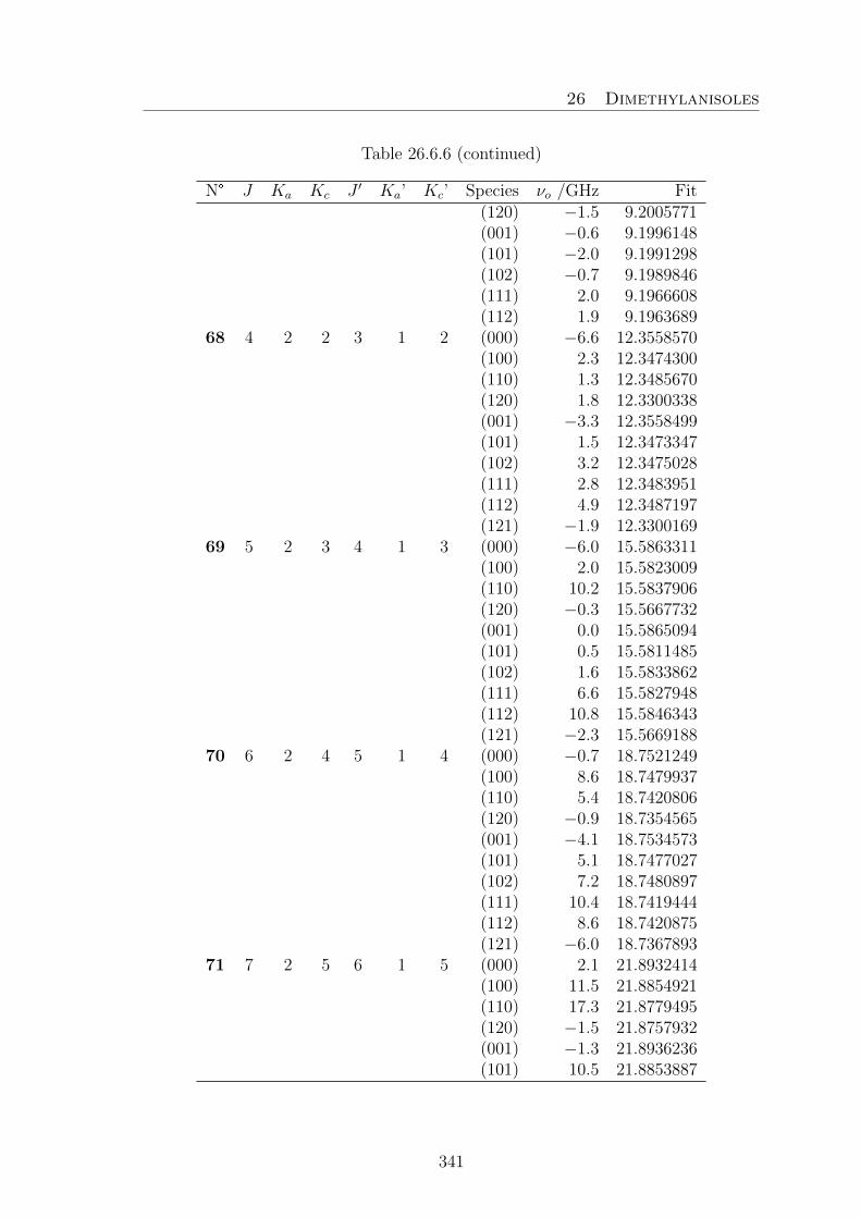
26 Dimethylanisoles
Table 26.6.6 (continued)
N° J Ka Kc J ′ Ka’ Kc’ Species νo /GHz Fit(120) −1.5 9.2005771(001) −0.6 9.1996148(101) −2.0 9.1991298(102) −0.7 9.1989846(111) 2.0 9.1966608(112) 1.9 9.1963689
68 4 2 2 3 1 2 (000) −6.6 12.3558570(100) 2.3 12.3474300(110) 1.3 12.3485670(120) 1.8 12.3300338(001) −3.3 12.3558499(101) 1.5 12.3473347(102) 3.2 12.3475028(111) 2.8 12.3483951(112) 4.9 12.3487197(121) −1.9 12.3300169
69 5 2 3 4 1 3 (000) −6.0 15.5863311(100) 2.0 15.5823009(110) 10.2 15.5837906(120) −0.3 15.5667732(001) 0.0 15.5865094(101) 0.5 15.5811485(102) 1.6 15.5833862(111) 6.6 15.5827948(112) 10.8 15.5846343(121) −2.3 15.5669188
70 6 2 4 5 1 4 (000) −0.7 18.7521249(100) 8.6 18.7479937(110) 5.4 18.7420806(120) −0.9 18.7354565(001) −4.1 18.7534573(101) 5.1 18.7477027(102) 7.2 18.7480897(111) 10.4 18.7419444(112) 8.6 18.7420875(121) −6.0 18.7367893
71 7 2 5 6 1 5 (000) 2.1 21.8932414(100) 11.5 21.8854921(110) 17.3 21.8779495(120) −1.5 21.8757932(001) −1.3 21.8936236(101) 10.5 21.8853887
341

26 Dimethylanisoles
Table 26.6.6 (continued)
N° J Ka Kc J ′ Ka’ Kc’ Species νo /GHz Fit(102) 9.9 21.8854361(111) 16.5 21.8778433(112) 14.5 21.8778961(121) −3.8 21.8762426
72 2 2 1 1 1 1 (100) 10.6 6.4391808(110) 5.5 6.4231344(120) 0.5 6.4455186(001) 1.0 6.4439766(101) 1.3 6.4388245(102) −1.3 6.4394196(111) 1.1 6.4225199(112) 0.4 6.4236467(121) −0.4 6.4454697
73 3 2 2 2 1 2 (000) −9.0 9.4237862(100) −4.2 9.4183925(110) 1.8 9.4091914(120) 8.9 9.4178390(001) −10.7 9.4236678(101) 4.8 9.4188757(102) −5.3 9.4177522(111) −4.4 9.4098745(112) −9.8 9.4083952(121) −0.6 9.4177209
74 4 2 3 3 1 3 (000) −0.9 12.5194953(100) 1.1 12.5094930(110) 0.9 12.5035545(120) −3.4 12.5089811(001) 2.2 12.5183675(101) 3.1 12.5100574(102) −6.1 12.5089734(111) −6.2 12.5036736(112) −7.0 12.5033579(121) 2.3 12.5079082
75 5 2 4 4 1 4 (000) 3.5 15.6478014(100) −9.0 15.6407546(110) −8.9 15.6352908(120) 4.3 15.6351123(001) −1.2 15.6469648(111) −7.5 15.6352374(112) −9.8 15.6352308(121) −1.3 15.6341829
342

26 Dimethylanisoles
Table 26.6.6 (continued)
N° J Ka Kc J ′ Ka’ Kc’ Species νo /GHz Fit76 6 2 5 5 1 5 (000) 6.6 18.7808269
(100) −3.4 18.7736383(110) −6.7 18.7670527(120) 11.3 18.7663002(001) −7.6 18.7806435(101) −0.2 18.7735787(102) −10.9 18.7735550(111) −12.5 18.7669796(112) −14.9 18.7669734(121) −2.5 18.7661119
77 7 2 6 6 1 6 (000) 7.3 21.9139756(100) −4.8 21.9058568(110) −13.1 21.8981464(120) −12.5 21.8975140(001) −9.7 21.9138587(111) −18.3 21.8980632(121) −3.7 21.8974283
78 3 3 0 2 2 0 (000) 1.9 9.5249897(110) −0.9 9.5658179(120) 3.3 9.5274206(001) −0.7 9.5249508(111) 2.2 9.5671385(112) 1.5 9.5643933(121) 0.2 9.5273812
79 4 3 1 3 2 1 (000) −0.1 12.3037501(100) −4.8 12.3077310(110) 4.5 12.3033245(120) −2.3 12.3195561(001) 0.4 12.3037164(101) −5.3 12.3078596(102) −3.1 12.3075340(111) 3.3 12.3036095(112) 4.8 12.3029681(121) −1.0 12.3195235
80 5 3 2 4 2 2 (000) −1.8 15.3710944(100) −1.5 15.3625815(110) 1.5 15.3613834(120) −3.3 15.3477512(001) −1.6 15.3710734(101) 3.2 15.3625932(102) 2.1 15.3625338(111) 3.6 15.3614272
343

26 Dimethylanisoles
Table 26.6.6 (continued)
N° J Ka Kc J ′ Ka’ Kc’ Species νo /GHz Fit(112) 3.4 15.3613009(121) −2.6 15.3477267
81 6 3 3 5 2 3 (000) 7.0 18.6315567(100) −0.4 18.6218156(110) 2.4 18.6217238(120) −4.1 18.6023757(001) 1.4 18.6315598(101) 2.1 18.6214001(102) −0.2 18.6222311(111) 2.9 18.6210011(121) −3.3 18.6023798
82 7 3 4 6 2 4 (000) −2.9 21.8407535(100) 2.4 21.8368579(110) 11.5 21.8330651(120) −3.4 21.8189885(001) −5.5 21.8413059(101) −0.1 21.8356893(102) 1.9 21.8377163(111) 11.9 21.8325413(112) 10.1 21.8333908(121) −8.1 21.8194738
83 3 3 1 2 2 1 (000) 3.3 9.6813665(110) 10.8 9.6319924(120) 3.8 9.6819230(001) −1.1 9.6812791(111) −0.9 9.6305811(112) −1.5 9.6332511(121) −1.2 9.6818374
84 4 3 2 3 2 2 (000) 2.7 12.5856979(100) 7.1 12.5802922(110) −0.0 12.5692007(120) −0.8 12.5802785(001) 0.2 12.5856232(101) 3.9 12.5802070(102) 3.1 12.5802279(111) −3.6 12.5691034(112) −5.7 12.5691493
85 5 3 3 4 2 3 (000) 1.2 15.6405897(100) 4.1 15.6306141(110) −7.8 15.6188251(120) 0.3 15.6283986
344

26 Dimethylanisoles
Table 26.6.6 (continued)
N° J Ka Kc J ′ Ka’ Kc’ Species νo /GHz Fit(001) 1.6 15.6403144(101) 4.2 15.6316414(102) 2.1 15.6294439(111) −6.5 15.6196752(112) −12.1 15.6179150(121) −1.4 15.6281461
86 6 3 4 5 2 4 (000) 0.7 18.7603118(100) −1.4 18.7494392(110) 0.2 18.7423977(120) 2.1 18.7449067(001) 2.2 18.7588470(101) −3.1 18.7495815(102) −3.4 18.7492225(111) 6.8 18.7423895(112) −13.3 18.7422758(121) 0.4 18.7434368
87 7 3 5 6 2 5 (000) −1.7 21.8942159(100) −4.4 21.8852875(110) −14.7 21.8775113(120) −0.8 21.8769328(001) 1.4 21.8936775(102) −6.8 21.8851937(111) −15.6 21.8774402(112) −20.1 21.8774206(121) −4.3 21.8763217
88 4 4 0 3 3 0 (000) 3.0 12.7951251(110) 3.5 12.8701812(120) 4.8 12.7883276(001) −2.9 12.7950899(111) 5.5 12.8718334(112) 4.9 12.8683427(121) −2.0 12.7882869
89 5 4 1 4 3 1 (000) 1.7 15.4475424(100) −6.5 15.4543432(110) 12.9 15.4560399(120) −0.3 15.4654189(001) 0.6 15.4474888(101) −8.2 15.4546412(102) −4.7 15.4539389(111) 14.4 15.4566638(112) 13.6 15.4553057
345

26 Dimethylanisoles
Table 26.6.6 (continued)
N° J Ka Kc J ′ Ka’ Kc’ Species νo /GHz Fit(121) −0.4 15.4653694
90 6 4 2 5 3 2 (000) −2.8 18.3993892(100) −2.7 18.3951598(110) 4.5 18.3892294(120) −6.5 18.3930933(001) 0.3 18.3993602(101) −0.3 18.3952197(102) 0.9 18.3950382(111) 8.3 18.3893834(112) 6.9 18.3890162(121) −4.2 18.3930584
91 7 4 3 6 3 3 (000) −6.3 21.6361160(100) −0.8 21.6226571(110) −1.0 21.6210161(120) −3.4 21.5980993(001) −4.0 21.6361007(101) 0.0 21.6225506(102) 1.3 21.6227295(111) 6.7 21.6208330(112) 3.6 21.6211732(121) −5.6 21.5980766
92 4 4 1 3 3 1 (000) 3.4 12.9335777(110) 8.7 12.8475806(120) 4.5 12.9322442(001) −2.2 12.9334271(111) −7.0 12.8457415(112) −7.8 12.8492284(121) −3.2 12.9320973
93 5 4 2 4 3 2 (000) 3.8 15.7647626(100) 11.2 15.7567567(110) −4.7 15.7371864(120) 1.7 15.7600022(001) 0.1 15.7646725(101) 7.7 15.7563784(102) 5.0 15.7569549(111) −12.3 15.7365344(112) −12.9 15.7376571(121) −3.1 15.7599121
94 6 4 3 5 3 3 (000) 1.7 18.7655975(100) 5.6 18.7571617(110) −5.5 18.7446923
346

26 Dimethylanisoles
Table 26.6.6 (continued)
N° J Ka Kc J ′ Ka’ Kc’ Species νo /GHz Fit(120) −1.4 18.7528754(001) 0.5 18.7654747(101) 3.3 18.7574300(102) 2.3 18.7566657(111) −5.2 18.7452355(112) −10.7 18.7439592(121) −3.5 18.7527572
95 7 4 4 6 3 4 (000) 1.0 21.8670721(100) 1.0 21.8527304(110) −13.0 21.8413594(120) −3.1 21.8492622(001) −0.0 21.8663738(101) 1.4 21.8537406(102) −1.0 21.8517416(112) −18.7 21.8409120(121) −3.4 21.8486299
96 8 4 5 7 3 5 (000) 0.4 25.000426797 5 5 0 4 4 0 (100) 3.3 16.1018243
(110) 8.0 16.1914703(120) 4.0 16.0911105(001) −8.4 16.1018589(111) 6.1 16.1928499(112) 5.8 16.1898397(121) −5.9 16.0911315
98 6 5 1 5 4 1 (000) 5.5 18.6458521(100) −8.1 18.6553526(120) 2.4 18.6517117(001) 0. 2 18.6457722(101) −13.4 18.6560439(102) −8.1 18.6544990(121) −1.1 18.6516373
99 7 5 2 6 4 2 (000) −0.9 21.4632571(100) −4.4 21.4652332(120) −10.2 21.4781666(001) 0.8 21.4632094(101) −3.2 21.4653605(102) 0.9 21.4650104(121) −4.4 21.4781206
100 6 6 0 5 5 0 (000) −0.7 19.4203562(110) 8.5 19.5033970(120) 0.1 19.4093566(111) 2.8 19.5045369
347

26 Dimethylanisoles
Table 26.6.6 (continued)
N° J Ka Kc J ′ Ka’ Kc’ Species νo /GHz Fit(112) 2.5 19.5019526
101 7 6 1 6 5 1 (000) 7.8 21.9052432(100) −14.4 21.9238160(120) 3.6 21.8956043(001) −1.8 21.9051432(102) −13.8 21.9221056(121) −4.4 21.8955060
102 8 6 2 7 5 2 (000) 1.9 24.5772828(100) −4.1 24.5835920(001) 7.3 24.5772141
103 7 7 0 6 6 0 (000) −8.3 22.7368505(110) 5.8 22.8086633(120) −6.4 22.7262765(111) −3.7 22.8097004(112) 0.0 22.8072682
348

27 Non-Planar Aromatic Systems
27. Non-Planar Aromatic Systems
27.1. Phenylformate
Table 27.1.1 Coefficients of the two-dimensional Fourier expansion for the poten-tial energy surface given in Figure 19.2.
N° Fourier term Coefficient / Hartree Coefficient / cm−1
1 1 −419.74158242 cos(β) 0.0013197 289.6413 cos(2β) −0.0069515 −1525.6784 cos(2α) 0.0007989 175.3385 cos(3β) 0.0008317 182.5376 cos(4β) 0.0003076 67.5107 cos(4α) 0.0003424 75.1488 cos(5β) −0.0000784 −17.2079 cos(6β) −0.0000192 −4.21410 cos(6α) −0.0000309 −6.78211 cos(β)cos(2α) 0.0006256 137.30312 sin(β)sin(2α) −0.0015639 −343.23613 cos(2β)cos(2α) 0.0013272 291.28714 sin(2β)sin(2α) −0.0011546 −253.40515 cos(3β)cos(2α) 0.0000875 19.20416 sin(3β)sin(2α) −0.0001649 −36.19117 cos(4β)cos(2α) 0.0001404 30.81418 sin(4β)sin(2α) −0.0001078 −23.65919 cos(5β)cos(2α) −0.0000216 −4.74120 sin(5β)sin(2α) 0.0000414 9.08621 cos(6β)cos(2α) −0.0000121 −2.65622 sin(6β)sin(2α) 0.0000003 0.06623 cos(β)cos(4α) 0.0002258 49.55724 sin(β)sin(4α) −0.0002738 −60.09225 cos(2β)cos(4α) 0.0005617 123.27926 sin(2β)sin(4α) −0.0005520 −121.15027 cos(3β)cos(4α) 0.0002332 51.18128 sin(3β)sin(4α) −0.0002158 −47.36329 cos(4β)cos(4α) 0.0000602 13.21230 sin(4β)sin(4α) −0.0000522 −11.45731 cos(5β)cos(4α) −0.0000043 −0.94432 sin(5β)sin(4α) −0.0000024 −0.52733 cos(6β)cos(4α) 0.0000120 2.63434 sin(6β)sin(4α) −0.0000138 −3.02935 cos(β)cos(6α) 0.0000152 3.336
349

27 Non-Planar Aromatic Systems
Table 27.1.1 (continued)
N° Fourier term Coefficient / Hartree Coefficient / cm−1
36 sin(β)sin(6α) −0.0000095 −2.08537 cos(2β)cos(6α) 0.0000355 7.79138 sin(2β)sin(6α) −0.0000377 −8.27439 cos(3β)cos(6α) 0.0000246 5.39940 sin(3β)sin(6α) −0.0000290 −6.36541 cos(4β)cos(6α) 0.0000458 10.05242 sin(4β)sin(6α) −0.0000447 −9.81143 cos(5β)cos(6α) 0.0000061 1.33944 sin(5β)sin(6α) −0.0000046 −1.01045 cos(6β)cos(6α) 0.0000079 1.73446 sin(6β)sin(6α) −0.0000085 −1.866
Table 27.1.2 Fourier coefficients of the potential energy curve given in Figure 19.3obtained by varying the dihedral angle α = ∠(C13−O12−C3−C2) in a grid of 10°at a starting value of β = ∠(H14−C13−O12−C3) = 180°, while all other parametersincluding β were optimized at the MP2/6-311++G(d,p) level of theory. The poten-tial is expanded as V =a0+Σ14
n=2[cos(nα + π)].
Hartree cm−1
a0 −419.7503357a2 0.0015537 341.00a4 0.0005253 115.29a6 0.0000189 4.15a8 0.0000457 10.03a10 0.0000122 2.68a12 0.0000149 3.27a14 0.0000024 0.53
350

27 Non-Planar Aromatic Systems
Table 27.1.3 Nuclear coordinates in the principal inertial axes of conformers Ia andIIa of phenyl formate calculated at the MP2/6-311++G(d.p) level of theory. Theatoms are numbered as indicated in Figure 19.1.
Conformer Ia Conformer IIaa /A b /A c /A a /A b /A c /A
C1 −1.676176 1.306502 −0.158794 −1.857351 1.320952 0.000996C2 −0.347589 1.048899 −0.512283 −0.490684 1.125547 −0.233724C3 0.140267 −0.250959 −0.384032 0.015618 −0.176585 −0.236736C4 −0.664967 −1.298690 0.057502 −0.812308 −1.281981 −0.033592C5 −1.994052 −1.030091 0.404312 −2.177680 −1.074899 0.186921C6 −2.503213 0.268820 0.289211 −2.701075 0.224555 0.211353H7 −2.068131 2.315581 −0.249468 −2.260824 2.329578 0.005659H8 0.306210 1.840882 −0.864097 0.169111 1.965227 −0.434385H9 −0.247004 −2.297933 0.133584 −0.380986 −2.278165 −0.055893H10 −2.632322 −1.838417 0.749777 −2.830820 −1.928686 0.343881H11 −3.534840 0.474437 0.559545 −3.761651 0.380531 0.385206O12 1.449679 −0.554406 −0.786004 1.363692 −0.411679 −0.498344C13 2.445111 −0.073656 0.008298 2.246554 0.166851 0.370862H14 3.403612 −0.379919 −0.428925 1.738849 0.659681 1.216321O15 2.302592 0.569072 1.014188 3.432005 0.112192 0.206678
Table 27.1.4 Observed frequencies νo of the vt = 0 and vt = 1 states of phenylformate. νo − νc values as obtained after fits with the SPFIT /SPCAT program.
N J Ka Kc v J ′ K ′a K ′c v’ vo vo–vc vo–vc1 2 0 2 0 1 0 1 0 4444.6340 0.0226 0.00082 3 0 3 0 2 0 2 0 6655.4477 0.0221 −0.00193 4 0 4 0 3 0 3 0 8852.5568 0.0126 −0.00574 5 0 5 0 4 0 4 0 11031.8667 −0.0017 −0.00795 6 0 6 0 5 0 5 0 13190.2352 −0.0151 −0.00726 7 0 7 0 6 0 6 0 15326.1659 −0.0211 −0.00357 8 0 8 0 7 0 7 0 17440.3516 −0.0165 0.00118 9 0 9 0 8 0 8 0 19535.6879 −0.0045 0.00239 10 0 10 0 9 0 9 0 21616.5866 0.0074 −0.000210 11 0 11 0 10 0 10 0 23687.8482 0.0142 0.003211 2 1 2 0 1 1 1 0 4322.5909 0.0122 −0.005812 3 1 3 0 2 1 2 0 6480.1904 0.0133 −0.004513 4 1 4 0 3 1 3 0 8633.5243 0.0107 0.0006
351

27 Non-Planar Aromatic Systems
Table 27.1.4. (continued)
N J Ka Kc v J ′ K ′a K ′c v’ vo vo–vc vo–vc14 5 1 5 0 4 1 4 0 10781.4452 0.0054 0.007515 6 1 6 0 5 1 5 0 12923.0283 −0.0004 0.013216 7 1 7 0 6 1 6 0 15057.6046 −0.0043 0.014217 8 1 8 0 7 1 7 0 17184.7797 −0.0049 0.008218 9 1 9 0 8 1 8 0 19304.4293 −0.0041 −0.006419 10 1 10 0 9 1 9 0 21416.6846 0.0009 −0.019320 11 1 11 0 10 1 10 0 23521.8778 0.0084 −0.019121 12 1 12 0 11 1 11 0 25620.5037 0.0337 0.0252*22 2 1 1 0 1 1 0 0 4567.9946 0.0158 0.009223 3 1 2 0 2 1 1 0 6847.9856 0.0154 0.007224 4 1 3 0 3 1 2 0 9122.9101 0.0095 0.001025 5 1 4 0 4 1 3 0 11390.6265 −0.0006 −0.008226 6 1 5 0 5 1 4 0 13648.5760 −0.0087 −0.014527 7 1 6 0 6 1 5 0 15893.6415 −0.0108 −0.012928 8 1 7 0 7 1 6 0 18122.0147 −0.0064 −0.001529 9 1 8 0 8 1 7 0 20329.1086 0.0015 0.017030 10 1 9 0 9 1 8 0 22509.5902 0.0030 0.0230*31 11 1 10 0 10 1 9 0 24657.7059 0.0056 −0.0165*32 2 2 1 0 2 0 2 0 10972.4382 −0.0007 −0.009633 3 2 2 0 3 0 3 0 10965.4732 −0.0239 −0.012434 4 2 3 0 4 0 4 0 10969.7323 −0.0354 −0.011435 5 2 4 0 5 0 5 0 10996.2462 −0.0263 −0.003836 6 2 5 0 6 0 6 0 11057.4214 −0.0062 0.001137 7 2 6 0 7 0 7 0 11165.3994 0.0176 0.005438 8 2 7 0 8 0 8 0 11329.8548 0.0227 0.002939 9 2 8 0 9 0 9 0 11555.8704 0.0095 0.005140 2 2 0 0 1 0 1 0 15421.9832 0.0246 −0.006441 4 2 2 0 3 0 3 0 19895.8395 −0.0091 −0.006342 3 2 2 0 2 2 1 0 6648.4841 0.0003 −0.003243 4 2 3 0 3 2 2 0 8856.8167 0.0019 −0.003944 5 2 4 0 4 2 3 0 11058.3779 0.0046 −0.003145 6 2 5 0 5 2 4 0 13251.4107 0.0053 −0.002046 7 2 6 0 6 2 5 0 15434.1426 0.0013 −0.000647 8 2 7 0 7 2 6 0 17604.8090 −0.0094 0.000648 9 2 8 0 8 2 7 0 19761.7028 −0.0185 0.003849 10 2 9 0 9 2 8 0 21903.2373 −0.0017 0.006950 3 2 1 0 2 2 0 0 6668.1242 0.0037 −0.000651 4 2 2 0 3 2 1 0 8905.8141 0.0076 −0.000152 5 2 3 0 4 2 2 0 11155.7391 0.0114 0.000753 6 2 4 0 5 2 3 0 13419.5140 0.0094 −0.000554 7 2 5 0 6 2 4 0 15696.9850 −0.0015 −0.001955 8 2 6 0 7 2 5 0 17985.6204 −0.0197 −0.0004
352

27 Non-Planar Aromatic Systems
Table 27.1.4. (continued)
N J Ka Kc v J ′ K ′a K ′c v’ vo vo–vc vo–vc56 9 2 7 0 8 2 6 0 20280.4119 −0.0340 0.002357 10 2 8 0 9 2 7 0 22574.4507 −0.0154 −0.0158*58 3 0 3 0 2 1 1 0 3727.3004 0.0240 0.000059 4 0 4 0 3 1 2 0 5731.8719 0.0215 −0.012560 6 0 6 0 5 1 4 0 9440.4380 −0.0033 −0.019661 7 0 7 0 6 1 5 0 11118.0280 −0.0157 −0.008662 8 0 8 0 7 1 6 0 12664.7385 −0.0210 0.005863 9 0 9 0 8 1 7 0 14078.4109 −0.0199 0.008864 10 0 10 0 9 1 8 0 15365.8896 −0.0134 −0.007865 11 0 11 0 10 1 9 0 16544.1464 −0.0033 −0.028866 12 0 12 0 11 1 10 0 17640.0689 0.0139 0.0244*67 1 1 0 0 0 0 0 0 5029.4026 0.0176 −0.010468 2 1 1 0 1 0 1 0 7372.7805 0.0199 −0.001969 3 1 2 0 2 0 2 0 9776.1329 0.0135 0.005270 4 1 3 0 3 0 3 0 12243.5944 −0.0000 0.007271 5 1 4 0 4 0 4 0 14781.6641 −0.0132 0.004672 6 1 5 0 5 0 5 0 17398.3733 −0.0203 −0.002073 7 1 6 0 6 0 6 0 20101.7792 −0.0165 −0.008174 8 1 7 0 7 0 7 0 22897.6291 −0.0007 −0.005075 9 1 8 0 8 0 8 0 25786.3847 0.0160 0.009776 6 1 6 0 5 2 4 0 3842.0801 0.0134 0.008477 7 1 7 0 6 2 5 0 5648.2739 0.0037 0.024678 9 1 9 0 8 2 7 0 9098.5330 0.0044 0.028079 10 1 10 0 9 2 8 0 10753.5146 0.0236 0.004780 11 1 11 0 10 2 9 0 12372.1553 0.0338 −0.021081 12 1 12 0 11 2 10 0 13964.6059 −0.0447 −0.0066*82 5 1 4 0 4 2 2 0 3738.3814 0.0085 0.005283 6 1 5 0 5 2 3 0 6231.2189 −0.0110 −0.009484 7 1 6 0 6 2 4 0 8705.3469 −0.0307 −0.021385 8 1 7 0 7 2 5 0 11130.3776 −0.0348 −0.020086 9 1 8 0 8 2 6 0 13473.8659 −0.0134 −0.002587 10 1 9 0 9 2 7 0 15703.0461 0.0254 0.020088 11 1 10 0 10 2 8 0 17786.2985 0.0436 0.016589 12 1 11 0 11 2 9 0 19694.4229 0.0122 −0.018990 13 1 12 0 12 2 10 0 21402.3379 −0.0326 0.000191 2 2 0 0 2 1 2 0 8417.3383 0.0092 0.018992 3 2 1 0 3 1 3 0 8605.2720 −0.0004 0.022793 4 2 2 0 4 1 4 0 8877.5620 −0.0034 0.022294 5 2 3 0 5 1 5 0 9251.8559 0.0027 0.015495 6 2 4 0 6 1 6 0 9748.3418 0.0127 0.002096 7 2 5 0 7 1 7 0 10387.7227 0.0160 −0.013797 9 2 7 0 9 1 9 0 12164.5446 −0.0300 −0.0148
353

27 Non-Planar Aromatic Systems
Table 27.1.4. (continued)
N J Ka Kc v J ′ K ′a K ′c v’ vo vo–vc vo–vc98 2 2 1 0 1 1 1 0 12735.0214 0.0220 0.013999 3 2 2 0 2 1 2 0 15060.9133 0.0088 0.0152100 4 2 3 0 3 1 3 0 17437.5391 −0.0030 0.0152101 5 2 4 0 4 1 4 0 19862.3928 −0.0090 0.0116102 6 2 5 0 5 1 5 0 22332.3589 −0.0085 0.0027103 7 2 6 0 6 1 6 0 24843.4727 −0.0072 −0.0115104 2 2 0 0 1 1 0 0 12617.1969 0.0201 0.0044105 3 2 1 0 2 1 1 0 14717.3257 0.0072 −0.0062106 4 2 2 0 3 1 2 0 16775.1540 −0.0009 −0.0137107 5 2 3 0 4 1 3 0 18807.9831 0.0012 −0.0138108 6 2 4 0 5 1 4 0 20836.8706 0.0113 −0.0060109 7 2 5 0 6 1 5 0 22885.2800 0.0189 0.0069110 8 2 6 0 7 1 6 0 24977.2587 0.0098 0.0193111 4 2 3 0 4 1 3 0 7578.6978 −0.0196 −0.0211112 5 2 4 0 5 1 4 0 7246.4489 −0.0147 −0.0163113 6 2 5 0 6 1 5 0 6849.2834 −0.0009 −0.0040114 7 2 6 0 7 1 6 0 6389.7837 0.0105 0.0075115 8 2 7 0 8 1 7 0 5872.5778 0.0074 0.0094116 9 2 8 0 9 1 8 0 5305.1724 −0.0123 −0.0035117 10 2 9 0 10 1 9 0 4698.8200 −0.0164 −0.0190118 4 3 2 0 3 3 1 0 8829.8959 0.0128 0.0383*119 5 3 3 0 4 3 2 0 11034.8128 0.0039 0.0245*120 6 3 4 0 5 3 3 0 13237.4610 −0.0231 0.0139*121 7 3 5 0 6 3 4 0 15436.8696 −0.0616 0.0131*122 8 3 6 0 7 3 5 0 17631.8561 −0.0711 0.0163*123 9 3 7 0 8 3 6 0 19821.0534 0.0507 −0.0138*124 4 3 1 0 3 3 0 0 8830.7270 0.0204 0.0259*125 5 3 2 0 4 3 1 0 11037.7426 0.0262 −0.0113*126 6 3 3 0 5 3 2 0 13245.3408 0.0273 −0.0516*127 7 3 4 0 6 3 3 0 15454.7496 0.0239 −0.0595*128 8 3 5 0 7 3 4 0 17667.8872 0.0338 0.0410*129 4 0 4 1 3 0 3 1 8856.8167 −0.1689 /130 3 1 3 1 2 1 2 1 6489.9278 −0.0251 /131 4 1 4 1 3 1 3 1 8653.4358 −0.1699 /132 5 1 5 1 4 1 4 1 10817.7828 −0.1662 /133 6 1 6 1 5 1 5 1 12983.8817 0.0871 /134 7 1 7 1 6 1 6 1 15152.9217 0.3535 /135 8 1 8 1 7 1 7 1 17326.2393 −0.3274 /136 9 1 9 1 8 1 8 1 19509.2203 0.0811 /137 3 1 2 1 2 1 1 1 6861.1823 0.0191 /138 4 1 3 1 3 1 2 1 9147.5755 −0.0061 /139 5 1 4 1 4 1 3 1 11433.3046 0.0248 /
354

27 Non-Planar Aromatic Systems
Table 27.1.4. (continued)
N J Ka Kc v J ′ K ′a K ′c v’ vo vo–vc vo–vc140 6 1 5 1 5 1 4 1 13717.8558 0.0801 /141 7 1 6 1 6 1 5 1 16000.3870 0.0794 /142 8 1 7 1 7 1 6 1 18279.7248 −0.0174 /143 9 1 8 1 8 1 7 1 20554.3499 −0.1206 /144 10 1 9 1 9 1 8 1 22822.3472 0.0631 /145 4 2 2 1 3 2 1 1 8976.6809 0.0379 /146 5 2 3 1 4 2 2 1 11253.6549 0.0437 /147 6 2 4 1 5 2 3 1 13548.4608 −0.0241 /148 7 2 5 1 6 2 4 1 15860.3897 0.0087 /149 6 2 5 1 5 2 4 1 13422.0305 0.0811 /150 7 2 6 1 6 2 5 1 15665.0980 −0.0601 /151 7 0 7 1 6 1 5 1 11076.0565 0.0050 /152 5 1 4 1 4 0 4 1 14882.4925 −0.0634 /153 3 1 2 1 2 0 2 1 9812.3648 0.0690 /154 2 2 0 1 1 1 0 1 12660.1409 0.0445 /155 3 2 1 1 2 1 1 1 14802.0176 −0.0368 /156 4 2 2 1 3 1 2 1 16917.5155 −0.0187 /157 5 2 3 1 4 1 3 1 19023.5949 0.0311 /158 6 2 4 1 5 1 4 1 21138.7521 −0.0168 /159 2 2 0 1 2 1 2 1 8457.3775 −0.0853 /160 3 2 1 1 3 1 3 1 8683.7063 −0.0769 /161 2 2 1 1 1 1 1 1 12780.3671 0.1587 /
* Not fitted transitions in Fit 0, Ka 6 2, given Table 19.1.
355

27 Non-Planar Aromatic Systems
27.2. Phenyl Acetate
Table 27.2.1. Nuclear coordinates in the principal inertial axes of conformer Ib ofphenyl acetate calculated at the MP2/6-311++G(d,p) level of theory. The atomsare numbered according to Figure 20.1.
a /A b /A c /AC1 −2.048428 −0.090306 0.185998O2 −1.765125 −0.742914 1.159689C3 −3.430315 0.246504 −0.304588H4 −3.565594 −0.146130 −1.315149H5 −4.168358 −0.185816 0.369378H6 −3.549137 1.331532 −0.349219O7 −1.114437 0.469936 −0.653461C8 0.224578 0.225051 −0.333328C9 0.771955 −1.042572 −0.530908C10 0.999498 1.295734 0.109160C11 2.129450 −1.241971 −0.259741H12 0.140800 −1.855184 −0.877385C13 2.357977 1.086871 0.373650H14 0.536384 2.268731 0.243770C15 2.924949 −0.179501 0.187634H16 2.566113 −2.225941 −0.406045H17 2.971502 1.914671 0.718058H18 3.979207 −0.339289 0.394552
Table 27.2.2. Fourier expansion of the potential energy curve of phenyl acetatecalculated at the MP2/6-311++G(d,p) level of theory (Figure 20.2). The data wereobtained by rotating phenyl ring about the C8−O7 bond by varying the dihedralangle β = ∠(C1−O7−C8−C10) in a grid of 10°, while all other molecular parameterswere optimized. The potential is expanded as: V (β) = b0 + Σ7
n=1bncos(nβ).
Hartree cm−1
b0 −448.9579885b2 0.0017257 378.75b4 0.0006207 136.23b6 0.0000659 14.46b8 0.0000508 11.15b10 0.0000167 3.67b12 0.0000281 6.17b14 0.0000142 3.12
356
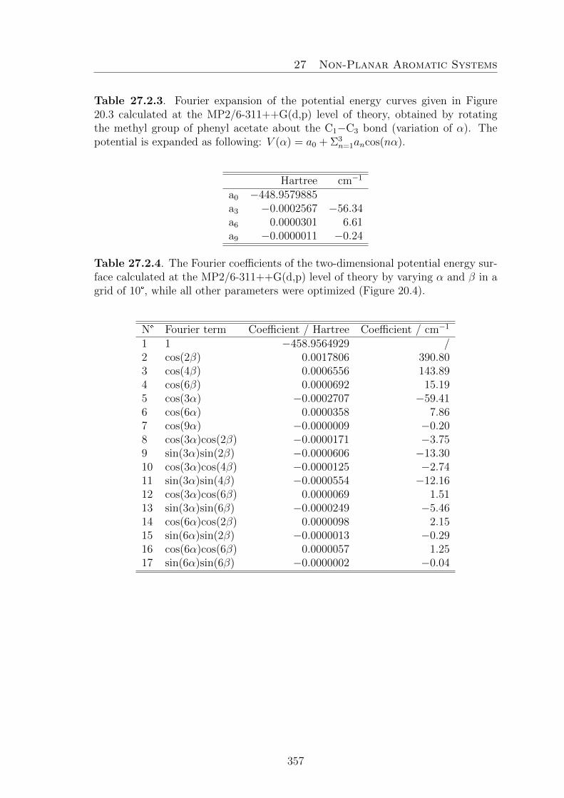
27 Non-Planar Aromatic Systems
Table 27.2.3. Fourier expansion of the potential energy curves given in Figure20.3 calculated at the MP2/6-311++G(d,p) level of theory, obtained by rotatingthe methyl group of phenyl acetate about the C1−C3 bond (variation of α). Thepotential is expanded as following: V (α) = a0 + Σ3
n=1ancos(nα).
Hartree cm−1
a0 −448.9579885a3 −0.0002567 −56.34a6 0.0000301 6.61a9 −0.0000011 −0.24
Table 27.2.4. The Fourier coefficients of the two-dimensional potential energy sur-face calculated at the MP2/6-311++G(d,p) level of theory by varying α and β in agrid of 10°, while all other parameters were optimized (Figure 20.4).
N° Fourier term Coefficient / Hartree Coefficient / cm−1
1 1 −458.9564929 /2 cos(2β) 0.0017806 390.803 cos(4β) 0.0006556 143.894 cos(6β) 0.0000692 15.195 cos(3α) −0.0002707 −59.416 cos(6α) 0.0000358 7.867 cos(9α) −0.0000009 −0.208 cos(3α)cos(2β) −0.0000171 −3.759 sin(3α)sin(2β) −0.0000606 −13.3010 cos(3α)cos(4β) −0.0000125 −2.7411 sin(3α)sin(4β) −0.0000554 −12.1612 cos(3α)cos(6β) 0.0000069 1.5113 sin(3α)sin(6β) −0.0000249 −5.4614 cos(6α)cos(2β) 0.0000098 2.1515 sin(6α)sin(2β) −0.0000013 −0.2916 cos(6α)cos(6β) 0.0000057 1.2517 sin(6α)sin(6β) −0.0000002 −0.04
357
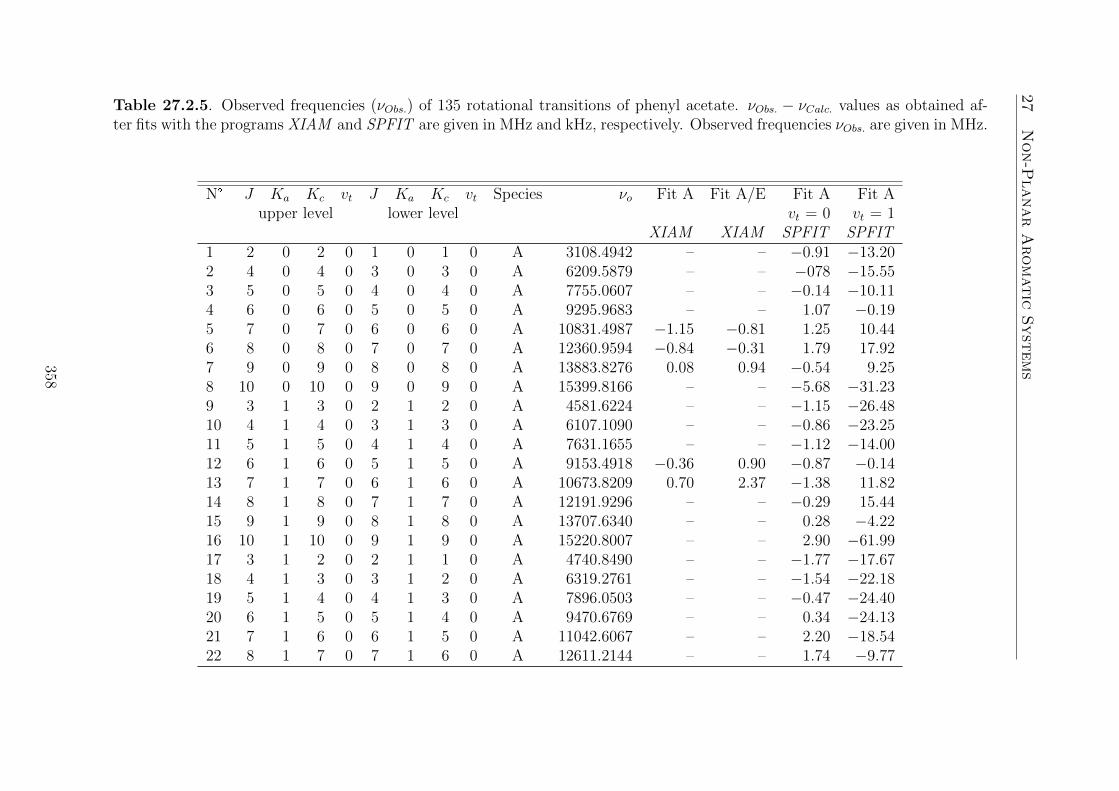
27
Non-P
lanarAromatic
Syst
ems
Table 27.2.5. Observed frequencies (νObs.) of 135 rotational transitions of phenyl acetate. νObs. − νCalc. values as obtained af-ter fits with the programs XIAM and SPFIT are given in MHz and kHz, respectively. Observed frequencies νObs. are given in MHz.
N° J Ka Kc vt J Ka Kc vt Species νo Fit A Fit A/E Fit A Fit Aupper level lower level vt = 0 vt = 1
XIAM XIAM SPFIT SPFIT1 2 0 2 0 1 0 1 0 A 3108.4942 – – −0.91 −13.202 4 0 4 0 3 0 3 0 A 6209.5879 – – −078 −15.553 5 0 5 0 4 0 4 0 A 7755.0607 – – −0.14 −10.114 6 0 6 0 5 0 5 0 A 9295.9683 – – 1.07 −0.195 7 0 7 0 6 0 6 0 A 10831.4987 −1.15 −0.81 1.25 10.446 8 0 8 0 7 0 7 0 A 12360.9594 −0.84 −0.31 1.79 17.927 9 0 9 0 8 0 8 0 A 13883.8276 0.08 0.94 −0.54 9.258 10 0 10 0 9 0 9 0 A 15399.8166 – – −5.68 −31.239 3 1 3 0 2 1 2 0 A 4581.6224 – – −1.15 −26.4810 4 1 4 0 3 1 3 0 A 6107.1090 – – −0.86 −23.2511 5 1 5 0 4 1 4 0 A 7631.1655 – – −1.12 −14.0012 6 1 6 0 5 1 5 0 A 9153.4918 −0.36 0.90 −0.87 −0.1413 7 1 7 0 6 1 6 0 A 10673.8209 0.70 2.37 −1.38 11.8214 8 1 8 0 7 1 7 0 A 12191.9296 – – −0.29 15.4415 9 1 9 0 8 1 8 0 A 13707.6340 – – 0.28 −4.2216 10 1 10 0 9 1 9 0 A 15220.8007 – – 2.90 −61.9917 3 1 2 0 2 1 1 0 A 4740.8490 – – −1.77 −17.6718 4 1 3 0 3 1 2 0 A 6319.2761 – – −1.54 −22.1819 5 1 4 0 4 1 3 0 A 7896.0503 – – −0.47 −24.4020 6 1 5 0 5 1 4 0 A 9470.6769 – – 0.34 −24.1321 7 1 6 0 6 1 5 0 A 11042.6067 – – 2.20 −18.5422 8 1 7 0 7 1 6 0 A 12611.2144 – – 1.74 −9.77
358

27
Non-P
lanarAromatic
Syst
ems
Table 27.2.5. (continued)
N° J Ka Kc vt J Ka Kc vt Species νo Fit A Fit A/E Fit A Fit Aupper level lower level vt = 0 vt = 1
XIAM XIAM SPFIT SPFIT23 9 1 8 0 8 1 7 0 A 14175.7923 – – 0.16 3.3824 10 1 9 0 9 1 8 0 A 15735.5306 – – −0.52 20.2025 4 2 2 0 3 2 1 0 A 6215.6765 – – 2.48 −7.7326 5 2 3 0 4 2 2 0 A 7773.6888 – – 1.69 −10.4027 4 2 3 0 3 2 2 0 A 6204.7701 – – 0.99 0.3628 5 2 4 0 4 2 3 0 A 7751.8940 – – 0.02 4.5829 8 0 8 0 7 1 6 0 A 8704.3787 1.13 2.13 2.02 21.3930 2 1 1 0 1 0 1 0 A 6048.1584 – – −1.01 57.0131 3 1 2 0 2 0 2 0 A 7680.5136 – – −1.46 52.9432 4 1 3 0 3 0 3 0 A 9339.3629 1.49 0.96 −0.40 48.6833 5 1 4 0 4 0 4 0 A 11025.8248 0.82 0.28 −0.59 39.3334 6 1 5 0 5 0 5 0 A 12741.4407 −0.56 −1.01 −0.41 25.0235 7 1 6 0 6 0 6 0 A 14488.0778 – – −0.58 5.3736 8 1 7 0 7 0 7 0 A 16267.7935 – – −0.09 −14.8537 9 1 8 0 8 0 8 0 A 18082.6308 – – 2.68 −24.9938 10 1 9 0 9 0 9 0 A 19934.3311 – – 0.00 −16.7439 11 1 10 0 10 0 10 0 A 21824.0082 – – −0.79 41.0840 2 2 0 0 1 1 0 0 A 11663.0142 0.47 0.30 −4.10 −46.0841 3 2 1 0 2 1 1 0 A 13161.6347 −1.25 −0.93 1.13 −38.5042 4 2 2 0 3 1 2 0 A 14636.4606 – – 3.78 −30.1643 5 2 3 0 4 1 3 0 A 16090.8723 – – 6.01 −19.3944 6 2 4 0 5 1 4 0 A 17529.1857 – – 4.77 −10.8145 7 2 5 0 6 1 5 0 A 18956.6266 – – −0.62 −7.3446 8 2 6 0 7 1 6 0 A 20379.2607 – – −6.60 −6.44
359

27
Non-P
lanarAromatic
Syst
ems
Table 27.2.5. (continued)
N° J Ka Kc vt J Ka Kc vt Species νo Fit A Fit A/E Fit A Fit Aupper level lower level vt = 0 vt = 1
XIAM XIAM SPFIT SPFIT47 9 2 7 0 8 1 7 0 A 21803.8708 – – −7.88 0.9148 10 2 8 0 9 1 8 0 A 23237.7920 – – 7.15 40.7749 2 2 1 0 1 1 1 0 A 11715.0204 0.72 0.42 −3.99 −37.9750 3 2 2 0 2 1 2 0 A 13315.4571 −1.02 −0.98 −2.09 −18.7551 4 2 3 0 3 1 3 0 A 14938.6052 – – 0.46 8.4852 5 2 4 0 4 1 4 0 A 16583.3903 – – 1.44 36.4253 6 2 5 0 5 1 5 0 A 18248.5244 – – −0.34 55.7554 7 2 6 0 6 1 6 0 A 19932.4812 – – −2.63 56.7355 8 2 7 0 7 1 7 0 A 21633.4639 – – −3.35 24.0156 9 2 8 0 8 1 8 0 A 23349.3822 – – 3.08 −62.0357 3 2 1 0 3 1 3 0 A 8739.2871 0.95 0.32 −1.08 2.3758 4 2 2 0 4 1 4 0 A 8847.8553 0.28 −0.33 2.96 18.6059 5 2 3 0 5 1 5 0 A 8990.3760 −0.16 −0.63 3.17 19.5960 6 2 4 0 6 1 6 0 A 9171.2491 −1.42 −1.58 3.54 5.1061 7 2 5 0 7 1 7 0 A 9395.5471 0.21 0.61 0.96 −26.2762 8 2 6 0 8 1 8 0 A 9668.8574 0.93 2.26 −3.43 −60.2463 3 3 0 0 2 2 0 0 A 18956.4609 – – −0.47 19.0664 4 3 2 0 4 2 3 0 A 14307.1339 0.06 0.21 – –65 2 2 0 1 1 1 0 1 A 11674.5391 – – – −63.8566 3 2 1 1 2 1 1 1 A 13187.6276 – – – 171.1967 2 2 1 1 1 1 1 1 A 11727.2715 – – – 41.8368 3 2 2 1 2 1 2 1 A 13344.6781 – – – 185.1669 6 2 5 1 6 1 5 1 A 8287.3647 – – – −3.7970 4 1 3 1 3 0 3 1 A 9358.0513 – – – −23.96
360

27
Non-P
lanarAromatic
Syst
ems
Table 27.2.5. (continued)
N° J Ka Kc vt J Ka Kc vt Species νo Fit A Fit A/E Fit A Fit Aupper level lower level vt = 0 vt = 1
XIAM XIAM SPFIT SPFIT71 5 1 4 1 4 0 4 1 A 11057.8825 – – – 57.3072 6 1 5 1 5 0 5 1 A 12792.5630 – – – 48.7873 7 1 6 1 6 0 6 1 A 14565.1298 – – – −42.3174 8 1 7 1 7 0 7 1 A 16378.9919 – – – −122.0175 9 1 8 1 8 0 8 1 A 18237.8357 – – – 82.3376 4 2 2 1 3 1 2 1 A 14721.6597 – – – 96.2677 4 2 3 1 3 1 3 1 A 15032.7522 – – – −84.3778 6 1 6 0 5 1 5 0 E 9193.8192 – 1.07 – –79 7 1 7 0 6 1 6 0 E 10705.5461 – 2.77 – –80 9 1 9 0 8 1 8 0 E 13720.5353 – −0.97 – –81 6 1 5 0 5 1 4 0 E 9431.3367 – −3.05 – –82 7 1 6 0 6 1 5 0 E 11019.3634 – 2.34 – –83 8 1 7 0 7 1 6 0 E 12591.6676 – −2.59 – –84 6 0 6 0 5 0 5 0 E 9291.0819 – −1.78 – –85 7 0 7 0 6 0 6 0 E 10824.9537 – −1.74 – –86 8 0 8 0 7 0 7 0 E 12352.2719 – −1.18 – –87 9 0 9 0 8 0 8 0 E 13872.3882 – 0.27 – –88 6 2 4 0 5 2 3 0 E 9317.2539 – 1.35 – –89 7 2 5 0 6 2 4 0 E 10866.5382 – −1.39 – –90 8 2 6 0 7 2 5 0 E 12421.1201 – 0.60 – –91 6 2 5 0 5 2 4 0 E 9317.2539 – 0.70 – –92 7 2 6 0 6 2 5 0 E 10866.2252 – −1.10 – –93 8 2 7 0 7 2 6 0 E 12415.8307 – −0.20 – –94 9 0 9 0 8 1 8 0 E 11936.0769 – 0.66 – –
361

27
Non-P
lanarAromatic
Syst
ems
Table 27.2.5. (continued)
N° J Ka Kc vt J Ka Kc vt Species νo Fit A Fit A/E Fit A Fit Aupper level lower level vt = 0 vt = 1
XIAM XIAM SPFIT SPFIT95 10 0 10 0 9 1 9 0 E 13595.6574 – −0.04 – –96 4 1 3 0 3 0 3 0 E 9542.8732 – 0.44 – –97 5 1 4 0 4 0 4 0 E 11182.1519 – −0.31 – –98 2 2 1 0 1 1 1 0 E 11244.5069 – 1.05 – –99 2 2 0 0 1 1 1 0 E 12849.6516 – −0.11 – –100 2 2 0 0 1 1 0 0 E 12044.4987 – 0.46 – –101 3 2 1 0 2 1 1 0 E 13588.1770 – 0.44 – –102 3 2 2 0 2 1 1 0 E 11981.7998 – 0.06 – –103 3 2 1 0 3 1 3 0 E 9766.2308 – −0.95 – –104 2 2 1 0 2 1 2 0 E 8143.1451 – 1.54 – –105 3 2 2 0 3 1 3 0 E 8161.3736 – 0.19 – –106 4 2 3 0 4 1 4 0 E 8206.3684 – −1.53 – –107 5 2 4 0 5 1 5 0 E 8291.4121 – 0.33 – –108 6 2 5 0 6 1 6 0 E 8415.3340 – 0.45 – –109 2 2 0 0 2 1 1 0 E 8929.5822 – 0.91 – –110 3 2 1 0 3 1 2 0 E 8903.2291 – −0.35 – –111 6 2 4 0 6 1 5 0 E 8648.6208 – −0.91 – –112 7 2 5 0 7 1 6 0 E 8500.7451 – 0.31 – –113 4 2 3 0 4 1 3 0 E 7246.5858 – −0.29 – –114 5 2 4 0 5 1 4 0 E 7162.4452 – −0.87 – –115 4 5 4 0 4 3 4 0 E 13826.5077 – 1.01 – –
362

27
Non-P
lanarAromatic
Syst
ems
Table 27.2.5. (continued)
N° J Ka Kc vt J Ka Kc vt Species νo Fit A Fit A/E Fit A Fit Aupper level lower level vt = 0 vt = 1
XIAM XIAM SPFIT SPFIT116 5 5 5 0 5 3 5 0 E 13809.1332 – −0.29 – –117 6 5 6 0 6 3 6 0 E 13785.2917 – −1.06 – –118 7 5 7 0 7 3 7 0 E 13753.9204 – −0.83 – –119 8 5 8 0 8 3 8 0 E 13714.3058 – 0.72 – –
363

27 Non-Planar Aromatic Systems
27.3. S -phenyl thioacetate
Table 27.3.1 Nuclear coordinates in the principal inertial axes system of con-formers Ib and Id of S -phenyl thioacetate calculated at the B3LYP- and MP2/6-311++G(d.p) levels of theory. The atoms are numbered as indicated in Figure 21.1.
Ib / B3LYP Id / MP2a /A b /A c /A a /A b /A c /A
C1 −2.524812 −0.742234 0.955831 2.538119 −0.690436 0.933460C2 −1.206813 −1.066745 0.641882 1.225015 −1.086078 0.656249C3 −0.520700 −0.325820 −0.322801 0.482237 −0.397116 −0.313022C4 −1.153944 0.734545 −0.974668 1.047621 0.688108 −0.997331C5 −2.471024 1.055502 −0.654409 2.358171 1.082114 −0.710276C6 −3.157317 0.318396 0.309451 3.103650 0.397042 0.257093H7 −3.055222 −1.317306 1.706320 3.114085 −1.224689 1.684313H8 −0.710145 −1.888958 1.142631 0.775488 −1.922240 1.184657H9 −0.617532 1.303710 −1.723997 0.462440 1.216028 −1.745208H10 −2.959905 1.881594 −1.158256 2.794497 1.926025 −1.237857H11 −4.182707 0.569857 0.555774 4.122169 0.704676 0.477460S12 1.163245 −0.765676 −0.751406 −1.190195 −0.884880 −0.654477C13 2.122519 0.343049 0.331494 −2.043249 0.362594 0.326514O14 1.621116 1.139043 1.076775 −1.463412 1.197432 0.982506C15 3.615762 0.134966 0.190865 −3.549651 0.276552 0.206845H16 3.991847 −0.293695 1.123738 −3.874692 1.023539 −0.523919H17 3.872315 −0.527404 −0.636332 −3.879674 −0.709780 −0.125135H18 4.092970 1.107119 0.051145 −3.993404 0.519697 1.174243
Table 27.3.2 Nuclear coordinates in the principal inertial axes system of con-former II of S -phenyl thioacetate calculated at the B3LYP/6-311++G(d,p) levelof theory. The atoms are numbered as indicated in Figure 21.1.
II / B3LYPa /A b /A c /A
C1 2.494672 0.212138 −1.205928C2 1.175197 −0.235875 −1.210618C3 0.506364 −0.454708 −0.001809C4 1.175515 −0.246404 1.208659C5 2.495006 0.201596 1.207586C6 3.154945 0.434676 0.001733H7 3.007504 0.381335 −2.146051H8 0.662960 −0.420711 −2.147516H9 0.663297 −0.439124 2.143985H10 3.008046 0.362949 2.148979H11 4.182322 0.780515 0.003088
364

27 Non-Planar Aromatic Systems
Table 27.3.2 (continued)
II / B3LYPa /A b /A c /A
S12 −1.176625 −1.087880 −0.004364C13 −2.255380 0.388821 0.001555O14 −3.440806 0.190275 0.001084C15 −1.623306 1.760434 0.006747H16 −0.990132 1.897880 −0.872141H17 −2.421589 2.502126 0.010057H18 −0.989760 1.890823 0.886494
Table 27.3.3 Fourier coefficients of the potential energy curve given in Figure 21.2on the left hand-side obtained by varying the dihedral angle α= ∠(H18−C15−C13−S12)in a grid of 10°, while all other parameters were optimized at the B3LYP andMP2/6-311++G(d,p) levels of theory. The indicated B3LYP potential is expandedas V (α) = Σ7
i=0aifi, and the MP2 potential as V (α) = Σ6i=0bifi.
B3LYP MP2i fi ai / Hartree ai / cm−1 bi / Hartree bi /cm−1
1 1 −783.2203659 — −781.5448779 —2 cos(3α) 0.0001591 34.918 0.0000614 13.4763 cos(6α) 0.0000524 11.500 0.0000519 11.3914 cos(9α) 0.0000045 0.988 0.0000039 0.8565 sin(3α) 0.0000172 3.775 −0.0000531 −11.6546 sin(6α) 0.0000124 2.721 −0.0000090 −1.9727 sin(12α) 0.0000011 0.241 — —
Table 27.3.4 Fourier coefficients of the potential energy curve given in Figure 21.2on the right hand-side obtained by varying the dihedral angle β = ∠(C4−C3−S12−C13)in a grid of 10°, while all other parameters were optimized at the B3LYP andMP2/6311++G(d,p) levels of theory. The potential calculated by B3LYP is ex-panded as V (β) = Σ7
i=0cifi, and the MP2 potential as V (β) = Σ6i=0difi.
B3LYP MP2i fi ci / Hartree ci / cm−1 di / Hartree di /cm−1
1 1 −781.54258 — −783.2192272 —2 cos(2β) 0.0032266 708.157 0.0015628 342.9952 cos(4β) 0.0009288 203.848 0.0002237 49.0963 cos(6β) 0.0000688 15.100 −0.0001042 −22.8693 cos(8β) 0.0000488 10.710 −0.0000196 −4.3023 cos(10β) 0.0000274 6.014 0.0000274 6.0143 cos(12β) 0.0000370 8.121 — —5 sin(2β) — — −0.0001514 −33.2286 sin(4β) — — −0.0000447 −9.8117 sin(6β) — — 0.0000320 7.023
365

27 Non-Planar Aromatic Systems
Table 27.3.5 Coefficients of the two-dimensional Fourier expansion of the potentialenergy surface given in Figure 21.4 calculated at the MP2/6-311++G(d,p) level oftheory. The Fourier terms are not symmetry adapted.
i fi Hartree cm−1
1 1 −780.0931390 –2 cos(1β) 0.0001171 25.7003 sin(1β) −0.0000015 −0.3294 cos(1α) −0.0000035 −0.7685 sin(1α) −0.0000018 −0.3956 cos(2β) 0.0025010 548.9067 sin(2β) −0.0000023 −0.5058 cos(2α) −0.0000018 −0.3959 sin(2α) −0.0000021 −0.46110 cos(3β) −0.0000783 −17.18511 sin(3β) −0.0000017 −0.37312 cos(3α) 0.0000011 0.24113 sin(3α) −0.0000134 −2.94114 cos(4β) 0.0004429 97.20515 sin(4β) −0.0000009 −0.19816 cos(4α) −0.0000034 −0.74617 sin(4α) −0.0000036 −0.79018 cos(5β) −0.0001094 −24.01119 sin(5β) 0.0000009 0.19820 cos(5α) −0.0000018 −0.39521 sin(5α) 0.0000008 0.17622 cos(6β) −0.0000792 −17.38223 sin(6β) 0.0000017 0.37324 cos(6α) −0.0000356 −7.81325 sin(6α) −0.0000445 −9.76726 cos(1β)cos(1α) 0.0000008 0.17627 sin(1β)sin(1α) −0.0000014 −0.30728 cos(1β)sin(1α) −0.0000008 −0.17629 sin(1β)cos(1α) −0.0000023 −0.50530 cos(2β)cos(1α) −0.0000051 −1.11931 sin(2β)sin(1α) −0.0000021 −0.46132 cos(2β)sin(1α) 0.0000023 0.50533 sin(2β)cos(1α) −0.0000052 −1.14134 cos(3β)cos(1α) 0.0000049 1.07535 sin(3β)sin(1α) −0.0000016 −0.35136 cos(3β)sin(1α) 0.0000015 0.32937 sin(3β)cos(1α) −0.0000026 −0.57138 cos(4β)cos(1α) −0.0000009 −0.198
366

27 Non-Planar Aromatic Systems
Table 27.3.5 (continued)
i fi Hartree cm−1
39 sin(4β)sin(1α) −0.0000006 −0.13240 cos(4β)sin(1α) 0.0000015 0.32941 sin(4β)cos(1α) −0.0000008 −0.17642 cos(5β)cos(1α) 0.0000065 1.42743 sin(5β)sin(1α) 0.0000006 0.13244 cos(5β)sin(1α) 0.0000025 0.54945 sin(5β)cos(1α) 0.0000013 0.28546 cos(6β)cos(1α) −0.0000021 −0.46147 sin(6β)sin(1α) 0.0000015 0.32948 cos(6β)sin(1α) 0.0000004 0.08849 sin(6β)cos(1α) 0.0000024 0.52750 cos(1β)cos(2α) 0.0000019 0.41751 sin(1β)sin(2α) −0.0000021 −0.46152 cos(1β)sin(2α) −0.0000008 −0.17653 sin(1β)cos(2α) −0.0000007 −0.15454 cos(2β)cos(2α) −0.0000022 −0.48355 sin(2β)sin(2α) −0.0000022 −0.48356 cos(2β)sin(2α) −0.0000007 −0.15457 sin(2β)cos(2α) −0.0000018 −0.39558 cos(3β)cos(2α) 0.0000038 0.83459 sin(3β)sin(2α) −0.0000027 −0.59360 cos(3β)sin(2α) 0.0000026 0.57161 sin(3β)cos(2α) −0.0000012 −0.26362 cos(4β)cos(2α) −0.0000018 −0.39563 sin(4β)sin(2α) −0.0000018 −0.39564 cos(4β)sin(2α) 0.0000013 0.28565 sin(4β)cos(2α) −0.0000001 −0.02266 cos(5β)cos(2α) 0.0000044 0.96667 sin(5β)sin(2α) 0.0000008 0.17668 cos(5β)sin(2α) 0.0000038 0.83469 sin(5β)cos(2α) 0.0000004 0.08870 cos(6β)cos(2α) −0.0000026 −0.57171 sin(6β)sin(2α) 0.0000026 0.57172 cos(6β)sin(2α) 0.0000005 0.11073 sin(6β)cos(2α) 0.0000011 0.24174 cos(1β)cos(3α) 0.0000007 0.15475 sin(1β)sin(3α) −0.0000030 −0.65876 cos(1β)sin(3α) 0.0000058 1.27377 sin(1β)cos(3α) −0.0000029 −0.63678 cos(2β)cos(3α) −0.0000422 −9.26279 sin(2β)sin(3α) −0.0000249 −5.46580 cos(2β)sin(3α) 0.0000622 13.651
367

27 Non-Planar Aromatic Systems
Table 27.3.5 (continued)
i fi Hartree cm−1
81 sin(2β)cos(3α) −0.0000470 −10.31582 cos(3β)cos(3α) 0.0000034 0.74683 sin(3β)sin(3α) −0.0000003 −0.06684 cos(3β)sin(3α) 0.0000021 0.46185 sin(3β)cos(3α) 0.0000037 0.81286 cos(4β)cos(3α) −0.0000121 −2.65687 sin(4β)sin(3α) 0.0000111 2.43688 cos(4β)sin(3α) 0.0000035 0.76889 sin(4β)cos(3α) 0.0000254 5.57590 cos(5β)cos(3α) 0.0000041 0.90091 sin(5β)sin(3α) −0.0000001 −0.02292 cos(5β)sin(3α) 0.0000016 0.35193 sin(5β)cos(3α) −0.0000016 −0.35194 cos(6β)cos(3α) −0.0000097 −2.12995 sin(6β)sin(3α) −0.0000024 −0.52796 cos(6β)sin(3α) −0.0000030 −0.65897 sin(6β)cos(3α) −0.0000091 −1.99798 cos(1β)cos(4α) 0.0000027 0.59399 sin(1β)sin(4α) −0.0000005 −0.110100 cos(1β)sin(4α) 0.0000016 0.351101 sin(1β)cos(4α) 0.0000007 0.154102 cos(2β)cos(4α) −0.0000014 −0.307103 sin(2β)sin(4α) −0.0000007 −0.154104 cos(2β)sin(4α) −0.0000020 −0.439105 sin(2β)cos(4α) 0.0000008 0.176106 cos(3β)cos(4α) 0.0000017 0.373107 sin(3β)sin(4α) −0.0000008 −0.176108 cos(3β)sin(4α) 0.0000029 0.636109 sin(3β)cos(4α) 0.0000008 0.176110 cos(4β)cos(4α) −0.0000030 −0.658111 sin(4β)sin(4α) −0.0000005 −0.110112 cos(4β)sin(4α) −0.0000010 −0.219113 sin(4β)cos(4α) 0.0000005 0.110114 cos(5β)cos(4α) 0.0000016 0.351115 sin(5β)sin(4α) 0.0000002 0.044116 cos(5β)sin(4α) 0.0000033 0.724117 sin(5β)cos(4α) −0.0000002 −0.044118 cos(6β)cos(4α) −0.0000026 −0.571119 sin(6β)sin(4α) 0.0000008 0.176120 cos(6β)sin(4α) −0.0000019 −0.417121 sin(6β)cos(4α) −0.0000002 −0.044122 cos(1β)cos(5α) 0.0000020 0.439
368

27 Non-Planar Aromatic Systems
Table 27.3.5 (continued)
i fi Hartree cm−1
123 sin(1β)sin(5α) 0.0000001 0.022124 cos(1β)sin(5α) 0.0000024 0.527125 sin(1β)cos(5α) 0.0000002 0.044126 cos(2β)cos(5α) 0.0000001 0.022127 sin(2β)sin(5α) 0.0000010 0.219128 cos(2β)sin(5α) −0.0000033 −0.724129 sin(2β)cos(5α) 0.0000013 0.285130 cos(3β)cos(5α) 0.0000011 0.241131 sin(3β)sin(5α) −0.0000001 −0.022132 cos(3β)sin(5α) 0.0000023 0.505133 sin(3β)cos(5α) 0.0000001 0.022134 cos(4β)cos(5α) −0.0000016 −0.351135 sin(4β)sin(5α) −0.0000009 −0.198136 cos(4β)sin(5α) −0.0000022 −0.483137 sin(4β)cos(5α) −0.0000009 −0.198138 cos(5β)cos(5α) 0.0000014 0.307139 sin(5β)sin(5α) −0.0000001 −0.022140 cos(5β)sin(5α) 0.0000025 0.549141 sin(5β)cos(5α) −0.0000002 −0.044142 cos(6β)cos(5α) −0.0000016 −0.351143 sin(6β)sin(5α) 0.0000006 0.132144 cos(6β)sin(5α) −0.0000023 −0.505145 sin(6β)cos(5α) 0.0000001 0.022146 cos(1β)cos(6α) 0.0000007 0.154147 sin(1β)sin(6α) −0.0000006 −0.132148 cos(1β)sin(6α) 0.0000037 0.812149 sin(1β)cos(6α) 0.0000002 0.044150 cos(2β)cos(6α) −0.0000090 −1.975151 sin(2β)sin(6α) −0.0000036 −0.790152 cos(2β)sin(6α) −0.0000133 −2.919153 sin(2β)cos(6α) 0.0000038 0.834154 cos(3β)cos(6α) 0.0000018 0.395155 sin(3β)sin(6α) −0.0000008 −0.176156 cos(3β)sin(6α) 0.0000047 1.032157 sin(3β)cos(6α) 0.0000003 0.066158 cos(4β)cos(6α) −0.0000016 −0.351159 sin(4β)sin(6α) −0.0000017 −0.373160 cos(4β)sin(6α) −0.0000055 −1.207161 sin(4β)cos(6α) 0.0000019 0.417162 cos(5β)cos(6α) 0.0000021 0.461163 sin(5β)sin(6α) −0.0000003 −0.066164 cos(5β)sin(6α) 0.0000048 1.053
369

27 Non-Planar Aromatic Systems
Table 27.3.5 (continued)
i fi Hartree cm−1
165 sin(5β)cos(6α) 0.0000005 0.110166 cos(6β)cos(6α) −0.0000011 −0.241167 sin(6β)sin(6α) −0.0000023 −0.505168 cos(6β)sin(6α) −0.0000047 −1.032169 sin(6β)cos(6α) 0.0000031 0.680
Table 27.3.6 Coefficients of the two-dimensional Fourier expansion of the poten-tial energy surface given in Figure 21.5 on the left hand-side calculated at theB3LYP/6-311++G(d,p) level of theory.
i fi Hartree cm−1
1 1 −783.211055220 –2 cos(γ) −0.0003439 −75.4773 sin(γ) −0.0022922 −503.0804 cos(2β) 0.0023826 522.9205 sin(2β) −0.0013821 −303.3366 cos(2γ) 0.0057337 1258.4027 sin(2γ) 0.0003584 78.6608 cos(3γ) 0.0000988 21.6849 sin(3γ) 0.0008374 183.78810 cos(4β) 0.0001730 37.96911 sin(4β) −0.0001723 −37.81512 cos(4γ) 0.0004225 92.72813 sin(4γ) 0.0001078 23.65914 cos(5γ) 0.0000860 18.87515 sin(5γ) 0.0000917 20.12616 cos(6β) −0.0000212 −4.65317 sin(6β) −0.0000065 −1.42718 cos(6γ) 0.0000352 7.72619 sin(6γ) 0.0000277 6.07920 cos(2β)cos(1γ) 0.0001499 32.89921 sin(2β)sin(1γ) 0.0007325 160.76522 cos(2β)sin(1γ) −0.0016951 −372.03123 sin(2β)cos(1γ) 0.0000362 7.94524 cos(4β)cos(1γ) 0.0000361 7.92325 sin(4β)sin(1γ) 0.0003068 67.33526 cos(4β)sin(1γ) −0.0000672 −14.74927 sin(4β)cos(1γ) −0.0000085 −1.86628 cos(6β)cos(1γ) 0.0000053 1.16329 sin(6β)sin(1γ) 0.0000330 7.24330 cos(6β)sin(1γ) −0.0000200 −4.389
370

27 Non-Planar Aromatic Systems
Table 27.3.6 (continued)
i fi Hartree cm−1
31 sin(6β)cos(1γ) −0.0000073 −1.60232 cos(2β)cos(2γ) −0.0009805 −215.19533 sin(2β)sin(2γ) −0.0001183 −25.96434 cos(2β)sin(2γ) −0.0001627 −35.70935 sin(2β)cos(2γ) −0.0010148 −222.72336 cos(4β)cos(2γ) −0.0003500 −76.81637 sin(4β)sin(2γ) −0.0000195 −4.28038 cos(4β)sin(2γ) −0.0000443 −9.72339 sin(4β)cos(2γ) −0.0001125 −24.69140 cos(6β)cos(2γ) −0.0000493 −10.82041 sin(6β)sin(2γ) 0.0000035 0.76842 cos(6β)sin(2γ) −0.0000102 −2.23943 sin(6β)cos(2γ) 0.0000369 8.09944 cos(2β)cos(3γ) −0.0000570 −12.51045 sin(2β)sin(3γ) 0.0007210 158.24146 cos(2β)sin(3γ) 0.0001629 35.75247 sin(2β)cos(3γ) −0.0001680 −36.87248 cos(4β)cos(3γ) −0.0000154 −3.38049 sin(4β)sin(3γ) 0.0002779 60.99250 cos(4β)sin(3γ) 0.0000992 21.77251 sin(4β)cos(3γ) −0.0000526 −11.54452 cos(6β)cos(3γ) −0.0000090 −1.97553 sin(6β)sin(3γ) 0.0000249 5.46554 cos(6β)sin(3γ) 0.0000580 12.73055 sin(6β)cos(3γ) −0.0000087 −1.90956 cos(2β)cos(4γ) −0.0000844 −18.52457 sin(2β)sin(4γ) 0.0001068 23.44058 cos(2β)sin(4γ) −0.0000359 −7.88059 sin(2β)cos(4γ) 0.0002555 56.07660 cos(4β)cos(4γ) −0.0000873 −19.16061 sin(4β)sin(4γ) 0.0000412 9.04262 cos(4β)sin(4γ) −0.0000228 −5.00463 sin(4β)cos(4γ) 0.0000858 18.83164 cos(6β)cos(4γ) −0.0000056 −1.22965 sin(6β)sin(4γ) 0.0000143 3.13866 cos(6β)sin(4γ) −0.0000002 −0.04467 sin(6β)cos(4γ) 0.0000507 11.12768 cos(2β)cos(5γ) −0.0000392 −8.60369 sin(2β)sin(5γ) 0.0000756 16.59270 cos(2β)sin(5γ) 0.0000870 19.09471 sin(2β)cos(5γ) 0.0000066 1.44972 cos(4β)cos(5γ) −0.0000273 −5.992
371

27 Non-Planar Aromatic Systems
Table 27.3.6 (continued)
i fi Hartree cm−1
73 sin(4β)sin(5γ) 0.0000201 4.41174 cos(4β)sin(5γ) 0.0000857 18.80975 sin(4β)cos(5γ) 0.0000000 0.00076 cos(6β)cos(5γ) −0.0000078 −1.71277 sin(6β)sin(5γ) −0.0000089 −1.95378 cos(6β)sin(5γ) 0.0000274 6.01479 sin(6β)cos(5γ) 0.0000057 1.25180 cos(2β)cos(6γ) −0.0000154 −3.38081 sin(2β)sin(6γ) 0.0000130 2.85382 cos(2β)sin(6γ) −0.0000020 −0.43983 sin(2β)cos(6γ) −0.0000198 −4.34684 cos(4β)cos(6γ) −0.0000277 −6.07985 sin(4β)sin(6γ) 0.0000159 3.49086 cos(4β)sin(6γ) −0.0000029 −0.63687 sin(4β)cos(6γ) 0.0000292 6.40988 cos(6β)cos(6γ) 0.0000041 0.90089 sin(6β)sin(6γ) 0.0000055 1.20790 cos(6β)sin(6γ) 0.0000050 1.09791 sin(6β)cos(6γ) 0.0000125 2.743
Table 27.3.7 Coefficients of the two-dimensional Fourier expansion of the poten-tial energy surface given in Figure 21.5 on the right hand-side calculated at theB3LYP/6-311++G(d,p) level of theory.
i fi Hartree cm−1
1 1 −783.21290 –2 cos(γ) −0.0005058 −111.0103 cos(2γ) 0.0063755 1399.2614 sin(2γ) 0.0004513 99.0495 cos(3γ) 0.0000743 16.3076 sin(3γ) 0.0010999 241.4007 cos(3α) −0.0005735 −125.8698 cos(4γ) 0.0004552 99.9059 sin(4γ) 0.0001608 35.29210 cos(5γ) 0.0001005 22.05711 sin(5γ) 0.0000595 13.05912 cos(6γ) 0.0000755 16.57013 sin(6γ) 0.0000424 9.30614 cos(6α) 0.0000081 1.77815 cos(γ)cos(3α) 0.0000471 10.337
372

27 Non-Planar Aromatic Systems
Table 27.3.7 (continued)
i fi Hartree cm−1
16 cos(γ)sin(3α) −0.0000191 −4.19217 sin(γ)cos(3α) 0.0001124 24.66918 sin(γ)sin(3α) 0.0000634 13.91519 cos(2γ)cos(3α) −0.0004523 −99.26820 cos(2γ)sin(3α) 0.0006698 147.00421 sin(2γ)cos(3α) −0.0000086 −1.88722 sin(2γ)sin(3α) 0.0000114 2.50223 cos(3γ)cos(3α) 0.0000249 5.46524 cos(3γ)sin(3α) 0.0000388 8.51625 sin(3γ)cos(3α) −0.0003312 −72.69026 sin(3γ)sin(3α) −0.0002846 −62.46227 cos(4γ)cos(3α) −0.0000809 −17.75528 cos(4γ)sin(3α) 0.0001599 35.09429 sin(4γ)cos(3α) −0.0000355 −7.79130 sin(4γ)sin(3α) −0.0000239 −5.24531 cos(5γ)cos(3α) −0.0000073 −1.60232 cos(5γ)sin(3α) 0.0000655 14.37633 sin(5γ)cos(3α) 0.0000089 1.95334 sin(5γ)sin(3α) −0.0000028 −0.61535 cos(6γ)cos(3α) −0.0000103 −2.26136 cos(6γ)sin(3α) 0.0000680 14.92437 sin(6γ)cos(3α) 0.0000072 1.58038 sin(6γ)sin(3α) −0.0000027 −0.59339 cos(γ)cos(6α) 0.0000209 4.58740 cos(γ)sin(6α) 0.0000069 1.51441 sin(γ)cos(6α) 0.0000046 1.01042 sin(γ)sin(6α) −0.0000232 −5.09243 cos(2γ)cos(6α) −0.0000001 −0.02244 cos(2γ)sin(6α) 0.0000222 4.87245 sin(2γ)cos(6α) −0.0000163 −3.57746 sin(2γ)sin(6α) −0.0000432 −9.48147 cos(3γ)cos(6α) 0.0000255 5.59748 cos(3γ)sin(6α) −0.0000073 −1.60249 sin(3γ)cos(6α) −0.0000050 −1.09750 sin(3γ)sin(6α) −0.0000325 −7.13351 cos(4γ)cos(6α) −0.0000030 −0.65852 cos(4γ)sin(6α) 0.0000109 2.39253 sin(4γ)cos(6α) −0.0000036 −0.79054 sin(4γ)sin(6α) −0.0000386 −8.47255 cos(5γ)cos(6α) −0.0000073 −1.60256 cos(5γ)sin(6α) −0.0000003 −0.06657 sin(5γ)cos(6α) −0.0000076 −1.668
373

27 Non-Planar Aromatic Systems
Table 27.3.7 (continued)
i fi Hartree cm−1
58 sin(5γ)sin(6α) −0.0000330 −7.24359 cos(6γ)cos(6α) −0.0000208 −4.56560 cos(6γ)sin(6α) −0.0000034 −0.74661 sin(6γ)cos(6α) 0.0000023 0.50562 sin(6γ)sin(6α) −0.0000361 −7.923
Table 27.3.8 Observed frequencies νo of 72 rotational transitions of S -phenyl thioac-etate. νo − νc values in MHz as obtained after fits with the program XIAM.
N° J Ka Kc J Ka Kc Species νo νo − νcupper level lower level
1 5 1 4 4 0 4 A 8.8398408 1.96582 6 1 5 5 0 5 A 10.1650456 −6.94123 7 1 6 6 0 6 A 11.5209628 6.06444 5 1 5 4 0 4 A 8.8398408 7.21285 6 1 6 5 0 5 A 10.1452925 5.35616 7 1 7 6 0 6 A 11.4320294 −1.70097 2 2 0 1 1 1 A 9.2617488 0.03128 3 2 1 2 1 2 A 10.5806358 −1.04489 4 2 2 3 1 3 A 11.8948768 −1.805310 5 2 3 4 1 4 A 13.2023087 −2.357711 6 2 4 5 1 5 A 14.5085521 0.307312 7 2 5 6 1 6 A 15.8169604 −3.645913 2 2 0 1 1 0 A 9.2597328 0.411214 3 2 1 2 1 1 A 10.5759790 0.309815 4 2 2 3 1 2 A 11.8890743 0.885216 5 2 3 4 1 3 A 13.1996385 1.283517 6 2 4 5 1 4 A 14.5003731 −2.624718 7 2 5 6 1 5 A 15.7861702 −2.385719 2 2 1 1 1 0 A 9.2597283 0.414820 3 2 2 2 1 1 A 10.5759691 0.331221 4 2 3 3 1 2 A 11.8890670 0.940822 5 2 4 4 1 3 A 13.1996385 1.359923 6 2 5 5 1 4 A 14.5003731 −2.581124 7 2 6 6 1 5 A 15.7861702 −2.385525 2 2 1 1 1 1 A 9.2617399 0.030326 3 2 2 2 1 2 A 10.5806278 −1.021527 4 2 3 3 1 3 A 11.8948716 −1.747528 5 2 4 4 1 4 A 13.2023087 −2.281429 6 2 5 5 1 5 A 14.5085521 0.350930 7 2 6 6 1 6 A 15.8169604 −3.645731 3 3 1 2 2 1 A 14.9813146 4.1616
374

27 Non-Planar Aromatic Systems
Table 27.3.8 (continued)
N° J Ka Kc J Ka Kc Species νo νo − νcupper level lower level
32 3 3 0 2 2 1 A 14.9813146 4.161533 3 3 0 2 2 0 A 14.9805992 3.454234 3 3 1 2 2 0 A 14.9805992 3.454335 4 3 1 3 2 1 A 16.2734181 −4.074136 4 3 2 3 2 2 A 16.2770098 −0.513737 3 K 2 2 K 1 E 8.1958908 −6.037738 4 K 2 3 K 1 E 9.5435549 0.034539 5 K 2 4 K 1 E 10.8899115 7.276240 6 K 2 5 K 1 E 12.2332018 14.954541 7 K 2 6 K 1 E 13.5427657 −7.341842 7 K 0 6 K 1 E 9.7667780 −15.314743 8 K 0 7 K 1 E 11.1841444 7.348244 4 K 2 3 K 0 E 9.2597328 −9.566545 5 K 2 4 K 0 E 10.5434805 −2.000146 6 K 2 5 K 0 E 11.8223914 6.691047 7 K 2 6 K 0 E 13.0916789 11.934248 3 K -1 2 K 1 E 8.6209729 12.084049 4 K -1 3 K 1 E 9.9728066 8.667850 5 K -1 4 K 1 E 11.3241740 3.558651 6 K -1 5 K 1 E 12.6731716 −4.315252 2 K -2 1 K -1 E 11.4265714 3.150053 3 K 3 2 K 2 E 12.4774776 −5.459254 4 K 3 3 K 2 E 13.7788200 −1.341355 5 K 3 4 K 2 E 15.0715670 1.644556 6 K -1 5 K 0 E 12.2623610 −12.578857 7 K -1 6 K 0 E 13.5721294 7.742358 3 K 3 3 K 2 E 8.5065719 −7.154259 4 K 3 4 K 2 E 8.4861152 −2.832360 5 K 3 5 K 2 E 8.4610528 3.459261 6 K 3 5 K 2 E 16.3543346 2.542562 3 K -1 2 K 0 E 8.3966209 2.858163 4 K -1 3 K 0 E 9.6892559 −0.661964 5 K -1 4 K 0 E 10.9777431 −5.717765 2 K -2 2 K -1 E 8.7764534 6.231766 3 K -2 3 K -1 E 8.7644956 6.171567 4 K -2 4 K -1 E 8.7467600 4.415268 5 K -2 5 K -1 E 8.7218429 −0.348169 6 K -2 6 K -1 E 8.6882226 −9.508470 3 K -2 2 K -1 E 12.7386227 0.978871 4 K -2 3 K -1 E 14.0436364 −3.580272 5 K -2 4 K -1 E 15.3397887 −12.0929
375