Pulsatile Blood Flow in Anatomically Accurate Vessels with Multiple Aneurysms: A Medical...
14
Flow, Turbulence and Combustion 71: 333–346, 2003. © 2004 Kluwer Academic Publishers. Printed in the Netherlands. 333 Pulsatile Blood Flow in Anatomically Accurate Vessels with Multiple Aneurysms: A Medical Intervention Planning Application of Computational Haemodynamics IORDANIS CHATZIPRODROMOU 1 , VINCENT D. BUTTY 1 , VINOD B. MAKHIJANI 2 , DIMOS POULIKAKOS 1 and YIANNIS VENTIKOS 1, 1 Laboratory for Thermodynamics in Emerging Technologies, Department of Mechanical and Process Engineering, Swiss Federal Institute of Technology, CH-8092 Zurich, Switzerland 2 CFD Research Corporation, Biomedical Technology Branch, Huntsville, AL, U.S.A. Received 8 October 2002; accepted in revised form 8 June 2003 Abstract. In the present study, we are investigating computationally the pulsatile flow in an anatom- ically accurate cerebral arterial segment exhibiting two saccular aneurysms. Our focus is on the haemodynamic patterns observed within the two aneurysms, in terms of inflow-outflow regions, emergence and disappearance of coherent structures and mixing throughout the cardiac cycle. The results obtained carry interesting features, important for thrombosis, pharmacokinetics and particu- larly for interventional planning for aneurysm treatment. For the latter, being the center-point of this study, we show that the two aneurysms behave in a dissimilar manner, since the blood inflow region oscillates significantly for one of them and practically does not oscillate at all for the second. This information can guide the medical interventionist in designing the optimal approach, particularly in cases where total obliteration of the aneurysm neck opening is impossible. Key words: blood flow, cerebral aneurysm, computational haemodynamics, pulsatility. 1. Introduction Cerebral saccular aneurysms are balloon-like deformations of the arterial wall of the vessels that bring blood to the brain (Figure 1). The rupture of aneurysms of this type often has particularly harmful consequences, since it leads to hemorrhage in the subarachnoidal region and accounts for almost 7% of all strokes. Moreover, approximately 30% of all aneurysm ruptures are lethal immediately, and an addi- tional 40% lead to death within 4 weeks, if the rupture is not treated effectively. The sheer number of studies of this nature done over the last 30 years [1, 2, 7, 8, 11, 16, 17, 20], only underlines the importance of such demographics, importance which is Author for correspondence. Currently at Department of Engineering Science, Univer- sity of Oxford, Parks Road, Oxford OX1 3PJ, U.K.; E-mail: yiannis.ventikos@engineering- science.oxford.ac.uk.
Transcript of Pulsatile Blood Flow in Anatomically Accurate Vessels with Multiple Aneurysms: A Medical...
Flow, Turbulence and Combustion 71: 333–346, 2003.© 2004 Kluwer Academic Publishers. Printed in the Netherlands.
333
Pulsatile Blood Flow in Anatomically AccurateVessels with Multiple Aneurysms: A MedicalIntervention Planning Application ofComputational Haemodynamics
IORDANIS CHATZIPRODROMOU1, VINCENT D. BUTTY1,VINOD B. MAKHIJANI2, DIMOS POULIKAKOS1 andYIANNIS VENTIKOS1,�
1Laboratory for Thermodynamics in Emerging Technologies, Department of Mechanical andProcess Engineering, Swiss Federal Institute of Technology, CH-8092 Zurich, Switzerland2CFD Research Corporation, Biomedical Technology Branch, Huntsville, AL, U.S.A.
Received 8 October 2002; accepted in revised form 8 June 2003
Abstract. In the present study, we are investigating computationally the pulsatile flow in an anatom-ically accurate cerebral arterial segment exhibiting two saccular aneurysms. Our focus is on thehaemodynamic patterns observed within the two aneurysms, in terms of inflow-outflow regions,emergence and disappearance of coherent structures and mixing throughout the cardiac cycle. Theresults obtained carry interesting features, important for thrombosis, pharmacokinetics and particu-larly for interventional planning for aneurysm treatment. For the latter, being the center-point of thisstudy, we show that the two aneurysms behave in a dissimilar manner, since the blood inflow regionoscillates significantly for one of them and practically does not oscillate at all for the second. Thisinformation can guide the medical interventionist in designing the optimal approach, particularly incases where total obliteration of the aneurysm neck opening is impossible.
Key words: blood flow, cerebral aneurysm, computational haemodynamics, pulsatility.
1. Introduction
Cerebral saccular aneurysms are balloon-like deformations of the arterial wall ofthe vessels that bring blood to the brain (Figure 1). The rupture of aneurysms ofthis type often has particularly harmful consequences, since it leads to hemorrhagein the subarachnoidal region and accounts for almost 7% of all strokes. Moreover,approximately 30% of all aneurysm ruptures are lethal immediately, and an addi-tional 40% lead to death within 4 weeks, if the rupture is not treated effectively. Thesheer number of studies of this nature done over the last 30 years [1, 2, 7, 8, 11, 16,17, 20], only underlines the importance of such demographics, importance which is
� Author for correspondence. Currently at Department of Engineering Science, Univer-sity of Oxford, Parks Road, Oxford OX1 3PJ, U.K.; E-mail: [email protected].
334 I. CHATZIPRODROMOU ET AL.
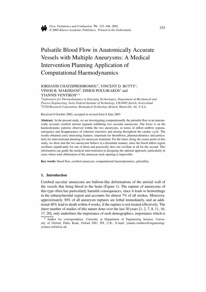
Figure 1. Domain of interest and control cross-sections through the two aneurysms. (Originalin colour)
further reinforced by the fact that, at least according to some estimates, 1–5% of thegeneral population exhibits cerebral aneurysms. The above figures do not includethe side-impact that cerebral aneurismal sacs may have in thrombus creation, sincethe recirculating, sometimes almost stagnant, blood inside the aneurysm presentswith an increased potential for clotting [15].
Computational Fluid Dynamics, when combined with proper insight on thephysiology of the human body system under investigation, can provide us withvaluable information concerning both the mechanisms at play and the planningof mission-critical medical interventions. Computational simulation of aneurismalflows has already allowed for a substantial increase of our understanding of the pre-vailing haemodynamics [3, 9, 13]. Visual inspection of the simulated flow patternsallows for valuable insight on the blood behavior near the orifice, or neck, of theaneurysm, but also within the sac. Moreover, the use of haemodynamic analysisalso allows for quantities like strain and wall shear stress to be mapped onto thevascular surface. This may contribute to better planning of endovascular treatment,and to our understanding of aneurysm rupture risk and patterns.
When numerical tools involving the solution of the Navier–Stokes equations areto be utilized routinely within a clinical environment, sufficient preparatory workhas to be conducted in order to pre-establish all the parameters and requirementsin a concrete way that covers all the possible situations and configurations theinterventionist might encounter. This element, being of critical importance whendesigning an intervention planning decision support methodology, is partly drivingthe present effort.
PULSATILE BLOOD FLOW IN ANATOMICALLY ACCURATE VESSELS 335
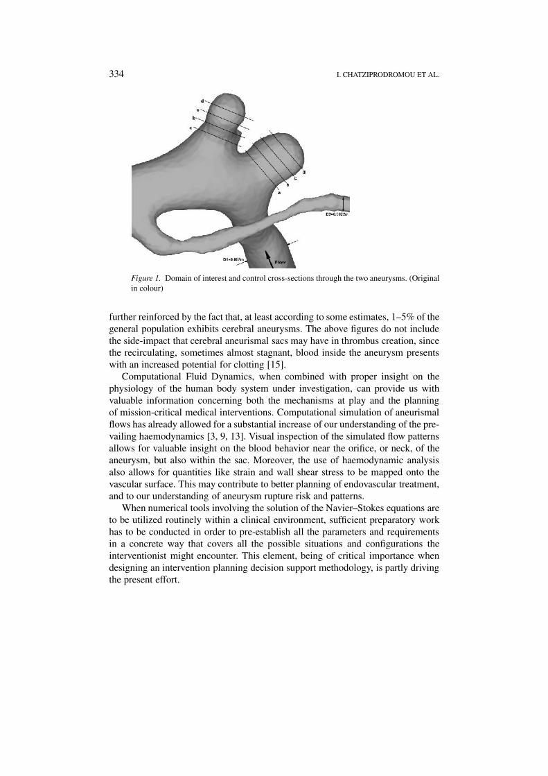
Figure 2. Grid independence study: (a) Influence of mesh resolution on the computation ofthe normal velocity profiles along a line on the cross-sectional plane (a) at the neck of the firstaneurysm. (b) Influence of mesh resolution on the computation of the inflow/outflow regionsseparating line (zero normal velocity contour line) on the cross-sectional plane (a) at the neckof the first aneurysm.
We shall present results from a computational simulation of the pulsatile flowwithin a patient’s right internal carotid artery exhibiting two saccular aneurysmsvery close to each other. It is often the case that such aneurysms are obliteratedthrough coil embolization, i.e. through the introduction (via a catheter) of a thinplatinum wire of adequate length, within the aneurysm. This coil forms a denselypacked grid and acts as a porous medium, thus diminishing or eliminating com-pletely the blood flow within the aneurysm and leading to the formation of a stablethrombus. Both the desired location of coil insertion, but also the thrombotic fea-tures of any particular aneurysm depend strongly on its haemodynamics, as theyare governed by the pulsatile blood flow characteristics and the geometric specificsof the sac and parent vessel.
2. Procedure and Numerical Techniques
There are a number of steps that have to be completed when patient-specific haemo-dynamic computations are to be performed. The procedure, briefly, has to go throughseveral stages [3], including:
• The acquisition of a suitable set of medical images that adequately describethe region of interest. These might be Computer Tomography data, MagneticResonance Imaging data, 3D Angiography data, etc.

336 I. CHATZIPRODROMOU ET AL.
Figure 3. Sinusoidal waveform of the inflow boundary condition.
• The processing of the images to extract the geometric features of the regionof interest.
• The discretization of the volume enclosed within the extracted surfaces witha suitable mesh generation technique.
• The numerical solution of the pertinent equations for the blood motion.
In the present study, we are utilizing CT data for a subject with two saccularaneurysms at the supraclinoid portion of the right internal carotid artery. The imagestack used has a resolution of 512 × 512 pixels at 74 slices with a pitch of 0.5 mm× 0.5 mm × 1 mm and was acquired using a Toshiba Helical CT scanner [6]. Theimages are processed [5, 18], and the extracted surface geometry is imported ina grid generator, (Hypermesh by Altair Eng.), where an unstructured, triangularsurface description and a volume tetrahedral-based grid are created.
Subsequently, the flow of blood through this geometry is obtained. For thenumerical solution of the flow in question, the CFD-ACE solver, by CFD Re-search Corporation is used. This solver employs a finite volume approach ona fully unstructured geometry for the solution of the incompressible, unsteady,three-dimensional Navier–Stokes equations, allowing for the incorporation of non-Newtonian fluid models, as well as for interaction of the fluid with the solidwall [10, 19]. Algebraic multigrid acceleration was employed in all simulationsperformed in order to enhance the speed of convergence of the solution.
A significant number of steady and pulsatile flow computations, with a varietyof numerical parameters, has been performed in order to eliminate, as much aspossible, the influence of numerics on the solution. For this reason, computationswith grids ranging from approximately 100,000 to 3,500,000 elements and severalnumerical discretization schemes were conducted. We should reiterate at this pointthat for all practical purposes, i.e. for the application of computational haemo-dynamics in a clinical environment, very strict quality criteria in terms of griddependence and numerical sensitivity have to be pre-established. This means that

PULSATILE BLOOD FLOW IN ANATOMICALLY ACCURATE VESSELS 337
Figure 4a. Evolution of the inflow-outflow patterns and cross-flow vectors for thecross-section ‘a’ of the first aneurysm. (Original in colour)
for every possible patient geometry, the requirements for an accurate solution mustbe known a priori, since neither the time nor the resources are usually available fora detailed study in each clinical case. In effect, the grid independence analysis ofthis and similar geometries, critical for establishing such rules and guidelines, isa topic of paramount importance that has begun attracting attention recently [14].Therefore, we shall present a detailed investigation defining a numerical accuracyprotocol for a critical cerebral pathology in a different publication [4]. For com-pleteness however, we are briefly presenting an indicative grid independence studyfor a quantity of great interest to this work, i.e. the normal velocity at the neck ofone of the aneurysms. Figure 2a depicts this normal velocity along a line on thecross-sectional plane (a) at the neck of the first aneurysm (Figure 1). It is apparent,that for this velocity all grids but the coarsest one yield similar results. The errorin maximum positive and negative velocity, normalized with the local value, isless than 4%, when our reference grid (the one with approximately half a millioncells) and the finest grid are compared. Figure 2b shows an even more convincingtrend: the lines depicted correspond to the contour of zero normal velocity, thusdelineating the regions of inflow and outflow for this particular aneurysm (for the
338 I. CHATZIPRODROMOU ET AL.
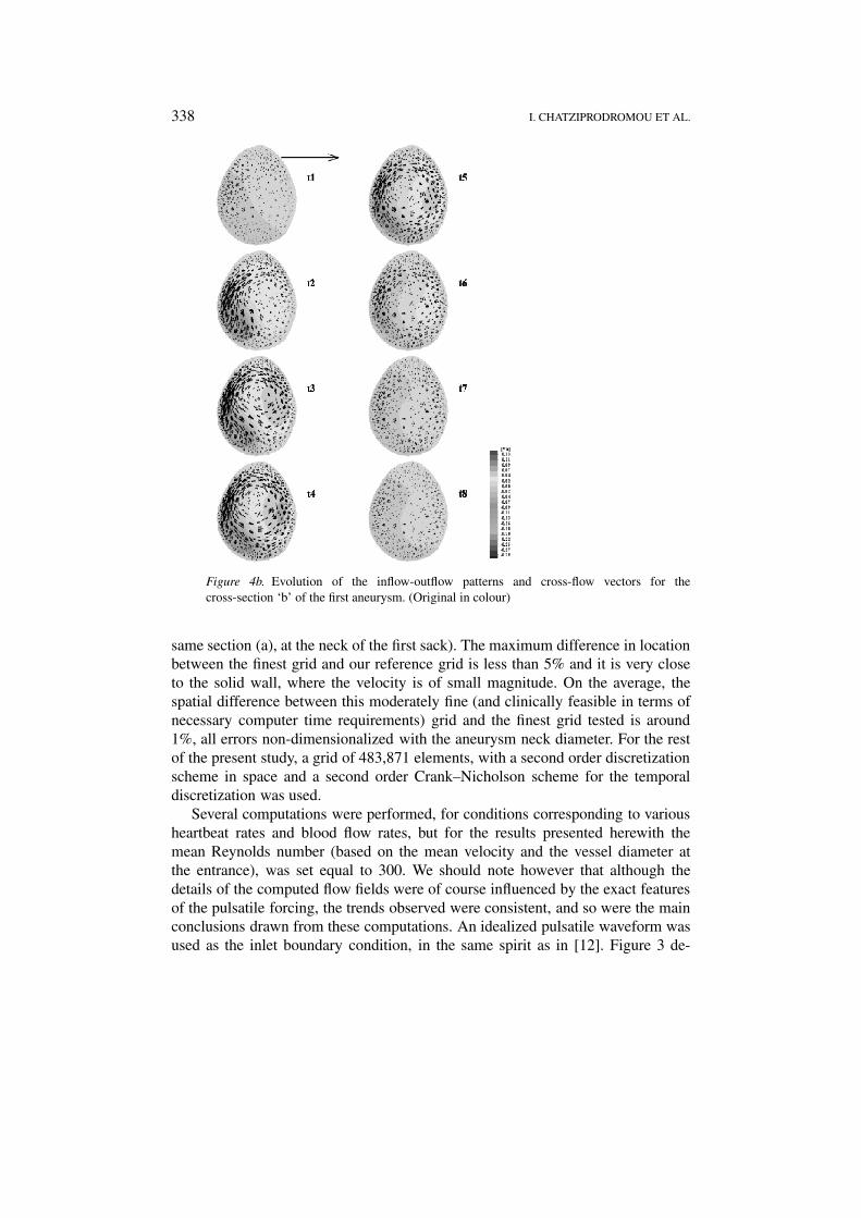
Figure 4b. Evolution of the inflow-outflow patterns and cross-flow vectors for thecross-section ‘b’ of the first aneurysm. (Original in colour)
same section (a), at the neck of the first sack). The maximum difference in locationbetween the finest grid and our reference grid is less than 5% and it is very closeto the solid wall, where the velocity is of small magnitude. On the average, thespatial difference between this moderately fine (and clinically feasible in terms ofnecessary computer time requirements) grid and the finest grid tested is around1%, all errors non-dimensionalized with the aneurysm neck diameter. For the restof the present study, a grid of 483,871 elements, with a second order discretizationscheme in space and a second order Crank–Nicholson scheme for the temporaldiscretization was used.
Several computations were performed, for conditions corresponding to variousheartbeat rates and blood flow rates, but for the results presented herewith themean Reynolds number (based on the mean velocity and the vessel diameter atthe entrance), was set equal to 300. We should note however that although thedetails of the computed flow fields were of course influenced by the exact featuresof the pulsatile forcing, the trends observed were consistent, and so were the mainconclusions drawn from these computations. An idealized pulsatile waveform wasused as the inlet boundary condition, in the same spirit as in [12]. Figure 3 de-
PULSATILE BLOOD FLOW IN ANATOMICALLY ACCURATE VESSELS 339
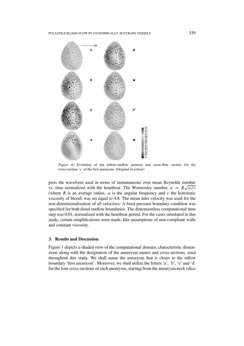
Figure 4c. Evolution of the inflow-outflow patterns and cross-flow vectors for thecross-section ‘c’ of the first aneurysm. (Original in colour)
picts the waveform used in terms of instantaneous over mean Reynolds numbervs. time normalized with the heartbeat. The Womersley number, a = R
√ω/ν
(where R is an average radius, ω is the angular frequency and ν the kinematicviscosity of blood) was set equal to 4.8. The mean inlet velocity was used for thenon-dimensionalization of all velocities. A fixed pressure boundary condition wasspecified for both distal outflow boundaries. The dimensionless computational timestep was 0.01, normalized with the heartbeat period. For the cases simulated in thisstudy, certain simplifications were made, like assumptions of non-compliant wallsand constant viscosity.
3. Results and Discussion
Figure 1 depicts a shaded view of the computational domain, characteristic dimen-sions along with the designation of the aneurysm names and cross-sections, usedthroughout this study. We shall name the aneurysm that is closer to the inflowboundary ‘first aneurysm’. Moreover, we shall utilize the letters ‘a’, ‘b’, ‘c’ and ‘d’for the four cross-sections of each aneurysm, starting from the aneurysm neck (slice

340 I. CHATZIPRODROMOU ET AL.
Figure 4d. Evolution of the inflow-outflow patterns and cross-flow vectors for thecross-section ‘d’ of the first aneurysm. (Original in colour)
‘a’) and going towards the aneurysm fundus (slice ‘d’). The time instants whereresults will be presented are as follows: t1 = 0.00, t2 = 0.13, t3 = 0.26, t4 = 0.39,t6 = 0.52, t6 = 0.65, t7 = 0.78 and t8 = 0.91, a convention used throughout thisstudy. As mentioned before, the basic period (i.e. the cardiac cycle) is one time unitlong, and all times mentioned are non-dimensionalized with respect to this time.
We have computed a total of five cardiac cycles and we observed no difference(i.e. full periodicity) after the second cycle. The results presented correspond tothe last cycle simulated and are thus clearly past all initial transients. Figures 4a,4b, 4c and 4d show contours of the normal velocity vector for the four control sec-tions of the first aneurysm. Moreover, cross-flow vectors are depicted, showing theinstantaneous secondary motion patterns. We can readily observe that significantsecondary motion prevails, for section ‘a’ in particular, that is damped as we movetowards the fundus (section ‘d’). Moreover, the inflow-outflow patterns observedoscillate significantly, with an actual switching of the inflow and the outflow areasbeen very apparent in the first section. Whereas we see a consistent secondarymotion (clockwise in the figures) for sections located in the middle of this firstaneurysm, this motion is much more complex on the entry section, but also at thefundus section.
PULSATILE BLOOD FLOW IN ANATOMICALLY ACCURATE VESSELS 341
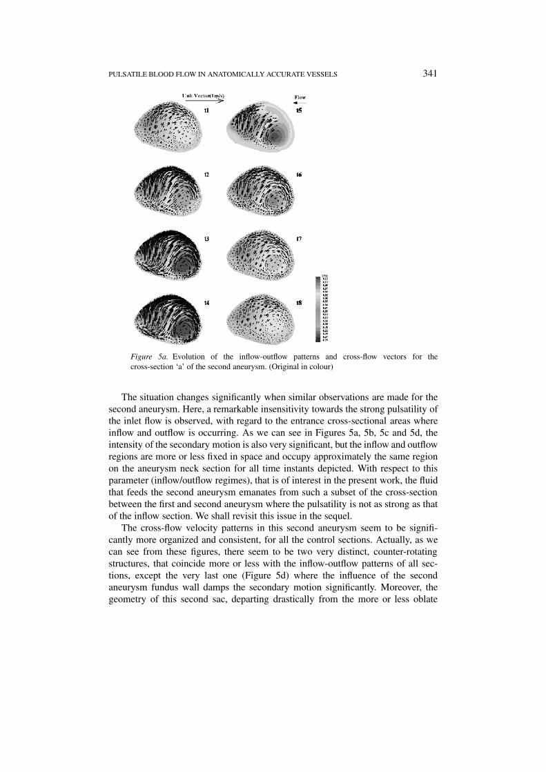
Figure 5a. Evolution of the inflow-outflow patterns and cross-flow vectors for thecross-section ‘a’ of the second aneurysm. (Original in colour)
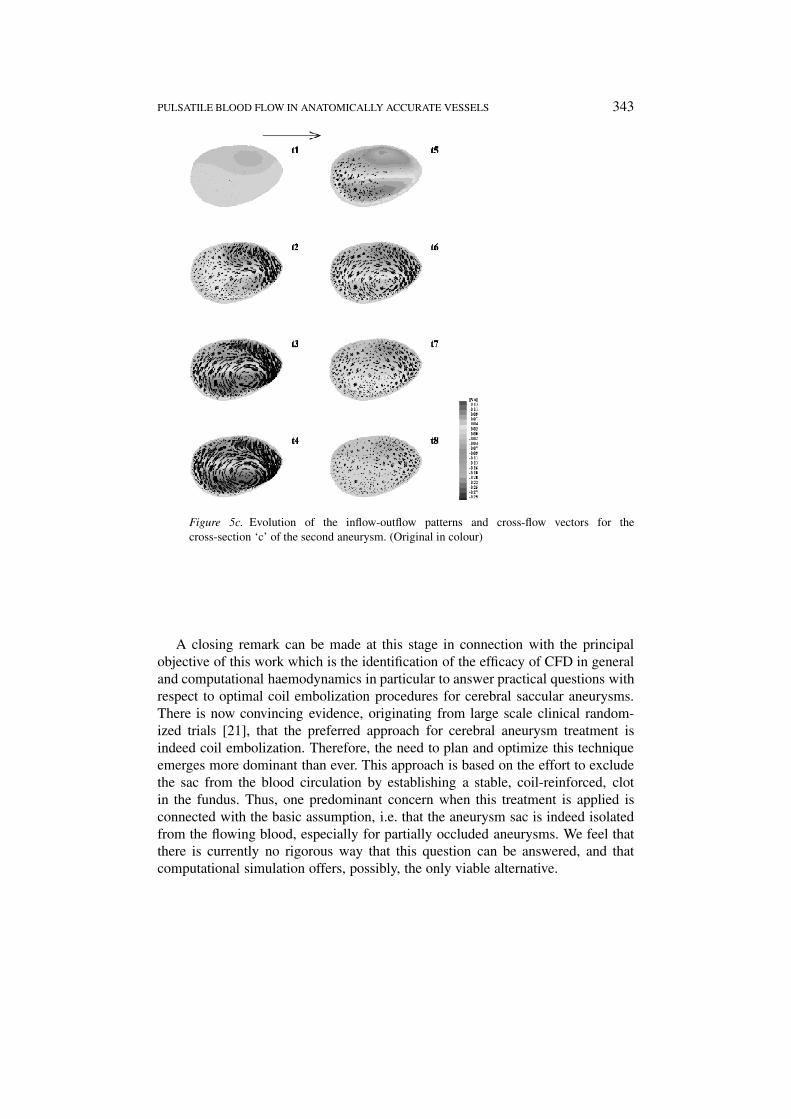
The situation changes significantly when similar observations are made for thesecond aneurysm. Here, a remarkable insensitivity towards the strong pulsatility ofthe inlet flow is observed, with regard to the entrance cross-sectional areas whereinflow and outflow is occurring. As we can see in Figures 5a, 5b, 5c and 5d, theintensity of the secondary motion is also very significant, but the inflow and outflowregions are more or less fixed in space and occupy approximately the same regionon the aneurysm neck section for all time instants depicted. With respect to thisparameter (inflow/outflow regimes), that is of interest in the present work, the fluidthat feeds the second aneurysm emanates from such a subset of the cross-sectionbetween the first and second aneurysm where the pulsatility is not as strong as thatof the inflow section. We shall revisit this issue in the sequel.
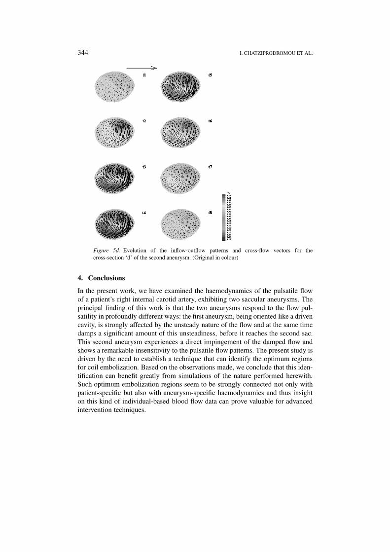
The cross-flow velocity patterns in this second aneurysm seem to be signifi-cantly more organized and consistent, for all the control sections. Actually, as wecan see from these figures, there seem to be two very distinct, counter-rotatingstructures, that coincide more or less with the inflow-outflow patterns of all sec-tions, except the very last one (Figure 5d) where the influence of the secondaneurysm fundus wall damps the secondary motion significantly. Moreover, thegeometry of this second sac, departing drastically from the more or less oblate

342 I. CHATZIPRODROMOU ET AL.
Figure 5b. Evolution of the inflow-outflow patterns and cross-flow vectors for thecross-section ‘b’ of the second aneurysm. (Original in colour)
spheroid we see at the first aneurysm, affects the emerging flow patterns, somethingthat is quite obvious in Figure 5b.
The profoundly different nature of the flow in the two aneurysms can possiblybe attributed to the fundamentally different locations and orientations they occupywith respect to the parent vessel: As we can see in Figure 1, the first aneurysmneck is oriented almost parallelly to the mean flow. In this respect, this aneurysmis acting like a cavity, driven by the oscillatory flow, and at the same time dampingthrough the intense secondary motion it is developing some of the pulsatility that isconvected downstream. Contrarily to this, the second aneurysm is located almostat a direct impingement angle with respect to the parent vessel. The main argumentthat can be made at this point is that the blood flow particulars for the cerebralvasculature pathology studied are not only patient-specific, something that has beenidentified for quite some time now and has diverted the computational efforts ofresearchers from generic idealized geometries to image-guided configurations, butthere is also great differentiation for the same patient. Thus, for medical interven-tion planning procedures to be effective, it is obvious that the maximum possibleamount of patient-specific information must be taken into account.
PULSATILE BLOOD FLOW IN ANATOMICALLY ACCURATE VESSELS 343
Figure 5c. Evolution of the inflow-outflow patterns and cross-flow vectors for thecross-section ‘c’ of the second aneurysm. (Original in colour)
A closing remark can be made at this stage in connection with the principalobjective of this work which is the identification of the efficacy of CFD in generaland computational haemodynamics in particular to answer practical questions withrespect to optimal coil embolization procedures for cerebral saccular aneurysms.There is now convincing evidence, originating from large scale clinical random-ized trials [21], that the preferred approach for cerebral aneurysm treatment isindeed coil embolization. Therefore, the need to plan and optimize this techniqueemerges more dominant than ever. This approach is based on the effort to excludethe sac from the blood circulation by establishing a stable, coil-reinforced, clotin the fundus. Thus, one predominant concern when this treatment is applied isconnected with the basic assumption, i.e. that the aneurysm sac is indeed isolatedfrom the flowing blood, especially for partially occluded aneurysms. We feel thatthere is currently no rigorous way that this question can be answered, and thatcomputational simulation offers, possibly, the only viable alternative.
344 I. CHATZIPRODROMOU ET AL.
Figure 5d. Evolution of the inflow-outflow patterns and cross-flow vectors for thecross-section ‘d’ of the second aneurysm. (Original in colour)
4. Conclusions
In the present work, we have examined the haemodynamics of the pulsatile flowof a patient’s right internal carotid artery, exhibiting two saccular aneurysms. Theprincipal finding of this work is that the two aneurysms respond to the flow pul-satility in profoundly different ways: the first aneurysm, being oriented like a drivencavity, is strongly affected by the unsteady nature of the flow and at the same timedamps a significant amount of this unsteadiness, before it reaches the second sac.This second aneurysm experiences a direct impingement of the damped flow andshows a remarkable insensitivity to the pulsatile flow patterns. The present study isdriven by the need to establish a technique that can identify the optimum regionsfor coil embolization. Based on the observations made, we conclude that this iden-tification can benefit greatly from simulations of the nature performed herewith.Such optimum embolization regions seem to be strongly connected not only withpatient-specific but also with aneurysm-specific haemodynamics and thus insighton this kind of individual-based blood flow data can prove valuable for advancedintervention techniques.
PULSATILE BLOOD FLOW IN ANATOMICALLY ACCURATE VESSELS 345
Acknowledgements
We would like to thank: Professor E. Hoffman (University of Iowa) for sharingthe CT data used in this study with us; Professor D.A. Steinman (John P. RobartsResearch Institute & University of Western Ontario) for providing his expertise inthe surface reconstruction of the geometry used and for insightful discussions; CFDResearch Corporation (Huntsville AL) for allowing the use of their software suite;Dr. S. Kollias, Dr. P. Summers and Professor A. Valavanis (Neuroradiology Insti-tute, University Hospital Zürich) for insightful discussions. The financial supportof the ETH Forschungskommission is kindly acknowledged.
References
1. Bannermann, R.M., Ingall, R.M. and Graf, C.J., The familial occurrence of intracranialaneurysms. Neurology 20 (1970) 283–292.
2. Böcker, W., Denk, H. and Heitz, Ph.U., Pathologie, Urban & Schwarzenberg (1997).3. Butty, V.D., Gudjonsson, K., Buchel, P., Makhijani, V.B., Ventikos, Y. and Poulikakos, D.,
Residence times and basins of attraction for a realistic right internal carotid artery with twoaneurysms. Biorheology 239 (2002) 387–393.
4. Chatziprodromou, I., Ventikos, Y. and Poulikakos, D., Numerical prerequisites for the uti-lization of computational haemodynamics in medical intervention planning: Application oncerebral vasculature pathologies. (2003) submitted.
5. Gill, J.D., Ladak, H.M., Steinman, D.A. and Fenster, A., Accuracy and variability assessment ofa semi-automatic technique for segmentation of the carotid arteries from 3D ultrasound images.Medical Physics 27 (2000) 1333–1342.
6. Hoffman, E., Private communication (2001).7. Johansen, K.H., Aneurysms. Scientific American 247 (1982) 110–125.8. Kassell, N.F. and Torner, J.C., Epidemiology of intracranial aneurysms. Internat. Anesthesiol.
Clin. 20 (1982) 13–17.9. Löw, M., Perktold, K. and Raunig, R., Hemodynamics in rigid and distensible saccu-
lar aneurysms: A numerical study of pulsatile flow characteristics. Biorheology 30 (1993)287–298.
10. Makhijani, V.B., Yang, H.Q., Dionne, P.J. and Thubrikar, M.J., Three-dimensional coupledfluid-structure simulation of pericardial bioprosthetic aortic valve function. ASAIO J. 43 (1997)387–391.
11. Mumenthaler, M. and Mattle, H., Neurologie. Thieme (1997).12. Myers, J.G., Moore, J.A., Ojha, M., Johnston, K.W. and Ethier, C.R., Factors influencing blood
flow patterns in the human right coronary artery. Ann. Biomed. Engrg. 29 (2001) 109–120.13. Perktold, K., On the paths of fluid particles in an axisymmetrical aneurysm. J. Biomech. 20
(1987) 311–317.14. Prakash, S. and Ethier, C.R., Requirements of mesh resolution in 3D computational hemody-
namics. J. Biomech. Engrg. Trans. ASME 123 (2001) 134–144.15. Reininger, A.J., Reininger, C.B., Heinzmann, U. and Wurzinger, L.J., Residence time in niches
of stagnant flow determines fibrin clot formation in an arterial branching model – Detailedflow-analysis and experimental results. Thrombosis and Haemostasis 74 (1995) 916–922.
16. Rinkel, G.J.E., Djibuti, M., Algra, A. and van Gijn, J., Prevalence and risk of rupture ofintracranial aneurysms. A systematic review. Stroke 29 (1998) 251–256.
17. Steiger, H.-J., Pathophysiology of Development and Rupture of Cerebral Aneurysms. ActaNeurochirurgica Supplementum, Vol. 48, Springer, Wien (1990).

346 I. CHATZIPRODROMOU ET AL.
18. Steinman, D.A., Private communication (2001).19. Sukumar, R., Athavale, M.M., Makhijani, V.B. and Przekwas, A.J., Application of computa-
tional fluid dynamics techniques to blood pumps. Artificial Organs 20 (1996) 529–533.20. Weir, B., Aneurysms Affecting the Nervous System. Williams and Wilkins, Baltimore, MD
(1987).21. International Subarachnoid Aneurysm Trial (ISAT) Collaborative Group, International sub-
arachnoid aneurysm trial (ISAT) of neurosurgical clipping versus endovascular coiling in 2143patients with ruptured intracranial aneurysms: A randomized trial. The Lancet 360 (2002)1267–1274.