Decoding complex multicomponent chromatograms by fourier analysis
Upload
independentCategory
view
4download
0
REVIEW ARTICLE
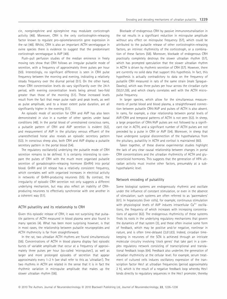
Encoding and Decoding Mechanisms of Pulsatile Hormone SecretionJ. J. Walker*�, J. R. Terry*�, K. Tsaneva-Atanasova�, S. P. Armstrong*, C. A. McArdle* and S. L. Lightman*
*Henry Wellcome Laboratories for Integrative Neuroscience and Endocrinology, University of Bristol, Bristol, UK.
�Bristol Centre for Applied Nonlinear Mathematics, Department of Engineering Mathematics, University of Bristol, Bristol, UK .
Introduction
Rhythms are fundamental in nature and endocrine rhythms span
time frames ranging from milliseconds to years. Many of these are
organised by endogenous mechanisms, including the circadian
(approximately 24-h period) cycles of many hormones that free-run
under constant conditions (1), and even slower circannual (approxi-
mately yearly) endocrine rhythms that are maintained under photo-
periodic clamping (2). Hormone secretion is also rhythmic over
much shorter time frames, as illustrated by neuroendocrine systems
where oscillators controlling neuronal membrane potential, with
time frames of milliseconds to seconds, control the coordinated
activity of hypothalamic neuronal networks. These characteristically
secrete hormone releasing factors into hypophysial portal blood in
a pulsatile fashion with ultradian (period < 24 h) time frames of
minutes to hours, which in turn drive the secretion of hormones
from the anterior pituitary with similar time frames. The pituitary
hormones act on target tissues, including the adrenals and gonads,
which synthesise and secrete glucocorticoid hormones and gonadal
steroids in a pulsatile fashion. The magnitude of this pulsatile
hypothalamic-pituitary target gland activity is modulated by slower
rhythms. Inputs from the suprachiasmatic nucleus (SCN), for exam-
ple, result in a circadian variation in glucocorticoid pulse amplitude.
In addition to daily changes, reproductive hormones also show oes-
trus ⁄ menstrual temporal profiles as well as circannual cycles in
seasonal breeders.
Oscillatory signals are well described for both intracellular and
cell-to-cell communication in many biological systems (3). When
compared with steady-state signalling, pulsatile signalling allows
greater control, is more robust to degradation, and is generally
more energy efficient (4). Moreover, pulsatile signalling provides
target sites with a quiescent interpulse interval allowing target
receptor recovery, and is therefore essential for maintaining tissue
responsiveness (5). Pulsatile signals may also be more ‘information
rich’ in the sense that pulse amplitude, pulse duration, pulse shape
and interpulse interval can all theoretically provide information to
the target cell, in comparison to amplitude alone provided by a
steady-state input. Most importantly, pulsatile signalling permits not
only amplitude, but also frequency modulation, and the potential
advantage of this is amply illustrated by comparing the information
Journal ofNeuroendocrinology
Correspondence to:
Jamie J. Walker, Henry Wellcome
Laboratories for Integrative
Neuroscience and Endocrinology,
University of Bristol, Dorothy Hodgkin
Building, Whitson Street, Bristol BS1
3NY, UK
(e-mail: [email protected]).
Ultradian pulsatile hormone secretion underlies the activity of most neuroendocrine systems,
including the hypothalamic-pituitary adrenal (HPA) and gonadal (HPG) axes, and this pulsatile
mode of signalling permits the encoding of information through both amplitude and frequency
modulation. In the HPA axis, glucocorticoid pulse amplitude increases in anticipation of waking,
and, in the HPG axis, changing gonadotrophin-releasing hormone pulse frequency is the primary
means by which the body alters its reproductive status during development (i.e. puberty). The
prevalence of hormone pulsatility raises two crucial questions: how are ultradian pulses encoded
(or generated) by these systems, and how are these pulses decoded (or interpreted) at their tar-
get sites? We have looked at mechanisms within the HPA axis responsible for encoding the pul-
satile mode of glucocorticoid signalling that we observe in vivo. We review evidence regarding
the ‘hypothalamic pulse generator’ hypothesis, and describe an alternative model for pulse gen-
eration, which involves steroid feedback-dependent endogenous rhythmic activity throughout
the HPA axis. We consider the decoding of hormone pulsatility by taking the HPG axis as a
model system and focussing on molecular mechanisms of frequency decoding by pituitary
gonadotrophs.
Keywords: endocrine signalling, ultradian rhythm, pulsatile hormone secretion, glucocorticoid
hormones, gonadotrophin-releasing hormone, mathematical modelling.
doi: 10.1111/j.1365-2826.2010.02087.x
Journal of Neuroendocrinology 22, 1226–1238
ª 2010 The Authors. Journal of Neuroendocrinology ª 2010 Blackwell Publishing Ltd
Journal of NeuroendocrinologyFrom Molecular to Translational Neurobiology

provided by a black and white television image (where each pixel
has a given signal amplitude) and a colour television image (where
each pixel has a given frequency as well as amplitude).
Ultradian pulsatility underlies the secretion of most hormones
(6). Despite this, in many neuroendocrine systems, the physiological
implication of these rhythms and the mechanisms underlying their
generation (encoding) and interpretation (decoding) at target cells
are still unclear. Here, we begin to address both of these questions.
Specifically, we have studied mechanisms within the hypothalamic-
pituitary-adrenal (HPA) axis responsible for encoding glucocorticoid
pulsatility. We review the ‘hypothalamic pulse generator’ hypothesis,
and also discuss an alternative model for pulse generation that
involves steroid feedback-dependent endogenous rhythmic activity
throughout the HPA axis. We also consider the second question
concerning the decoding of hormone pulsatility, taking the hypo-
thalamic-pituitary-gonadal (HPG) axis as a model system and
focussing on molecular mechanisms of frequency decoding by pitu-
itary gonadotrophs.
Encoding glucocorticoid pulsatility
A pulsatile mode of signalling underlies the activity of the HPA axis,
a system crucial for maintaining basal and stress-related homeosta-
sis by regulating the circulating levels of vital glucocorticoid
hormones (cortisol in man, corticosterone in rodents). Glucocortic-
oids govern a broad range of physiological functions, including the
regulation of cardiovascular, metabolic, cognitive and immuno-
logical activity (7–10). The regulation of HPA activity depends on
multiple inputs (Fig. 1). In the hypothalamus, the paraventricular
nucleus (PVN) receives an indirect input from the SCN which regu-
lates circadian variation in HPA activity, as well as afferent infor-
mation from brainstem nuclei responding to physical stressors such
as hypotension and inflammation, and from limbic areas of the
central nervous system that respond to cognitive and emotional
stressors (11). The PVN regulates corticotroph activity in the
anterior pituitary via two neuropeptides [corticotrophin-releasing
hormone (CRH) and arginine vasopressin (AVP)], which are released
into the pituitary portal circulation and hence the anterior pituitary.
Upon stimulation, corticotrophs secrete adrenocorticotrophic
hormone (ACTH) into the general circulation, through which it
accesses cells of the adrenal cortex initiating the synthesis and
secretion of glucocorticoids.
Glucocorticoid hormones act at target sites via their two cognate
receptors [the glucocorticoid receptor (GR) and mineralocorticoid
receptor (MR)], the expression of which is widespread in areas reg-
ulating HPA activity (12–14). The classical effect of glucocorticoids
is via activation of their receptors that translocate to the nucleus
acting as ligand dependent transcription factors binding to gluco-
corticoid response elements at promoter regions of glucocorticoid
sensitive genes (15–18). In addition to these classic genomic effects,
there has been considerable recent interest in rapid nongenomic
mechanisms through which glucocorticoids can also act (19,20).
In the basal state, glucocorticoids are released in an ultradian
pulsatile fashion from the adrenal cortex, which results in rapidly
changing levels of hormone concentration observable in blood
plasma (21–24) (Fig. 2), as well as within target tissues such as the
brain (25,26). The classic circadian glucocorticoid rhythm, which
peaks in the morning in man (27) and evening in the rodent (28),
is the result of amplitude (and to a lesser extent frequency) modu-
lation of the underlying ultradian rhythm (22,23,29–33). A particu-
CNSSCN
PVN
ACTH
CRHCORT
CORT
Hypothalamus
Median eminence
Portal circulation
Anterior pituitary
Corticotroph cells
Adrenal cortex
Fig. 1. Glucocorticoid release is regulated by the hypothalamic-pituitary
adrenal (HPA) axis. Neurones of the paraventricular nucleus (PVN) receive
circadian signals from the suprachiasmatic nucleus (SCN), as well as infor-
mation from brainstem nuclei responding to physical stressors, and from
limbic areas of the central nervous system (CNS) that respond to cognitive
and emotional stressors. These neurones project to the median eminence
where they release corticotrophin-releasing hormone (CRH) and arginine
vasopressin (AVP) into the portal blood. These peptides act on corticotroph
cells in the anterior pituitary to secrete adrenocorticotrophic hormone
(ACTH) into the general circulation, which in turn stimulates the synthesis
and release of glucocorticoid hormones (CORT) from cells of the adrenal
cortex. Glucocorticoids feed back at the pituitary and hypothalamus to inhi-
bit ACTH and CRH ⁄ AVP secretion, respectively.
ACTH
Cortisol
Clock time (h)
Seru
m c
ortis
ol (n
M) Plasm
a AC
TH (pg/m
l)
01.00
200
20
10
30
40
400
600
0 005.00 09.00 13.00 17.00
Fig. 2. Human data for adrenocorticotrophic hormone (ACTH) and cortisol
from a healthy male sampled at 10-min intervals, adapted from (24). Con-
centration time series demonstrates strong concordance between rhythmic
secretion of ACTH and cortisol, as well as a short time lag in ACTH-induced
cortisol secretion.
Encoding and decoding mechanisms of ultradian pulsatility 1227
ª 2010 The Authors. Journal of Neuroendocrinology ª 2010 Blackwell Publishing Ltd, Journal of Neuroendocrinology, 22, 1226–1238

larly striking feature of glucocorticoid pulsatility is the appearance
of a more distinct ultradian rhythm during the peak of the circa-
dian cycle, which operates at a frequency of approximately one
pulse per hour in rats (see shaded region 2 of Fig. 3). Variations in
amplitude and frequency of the ultradian rhythm not only compose
the circadian rhythm, but also characterise changes in HPA activity
that occur during early life programming (34), chronic stress (35),
lactation and ageing (36), and a number of other physiological and
pathological conditions (37).
The importance of glucocorticoid pulsatility resides in its ability
to provide a digital signalling system that can respond rapidly to
stress or changes in environmental conditions. A recent study by
Stavreva et al. (38) clearly shows that the pattern of glucocorticoid
presented to a tissue is critical for its transcriptional response, and
this effect is seen both in vivo and in vitro. The details of the
mechanisms underlying both genomic and nongenomic signalling
of glucocorticoid hormones are beyond the scope of this review,
but they not only act directly on multiple genes throughout the
body, but also have major effects on the ‘clock genes’ and through
them on circadian physiology (39,40).
Despite the significance of glucocorticoid pulsatility (36), surpris-
ingly little is known about the mechanisms that encode this
rhythm. Although the role of the SCN is well founded in regulating
circadian HPA activity (41,42), evidence suggests it is not required
for the generation of ultradian rhythmicity. Indeed, lesioning of the
SCN completely abolishes the circadian glucocorticoid rhythm in
both the adrenals and blood plasma (43–45), although it has little
effect on the ultradian rhythm, which persists at amplitudes and
frequencies comparable to those observed at the peak of the native
circadian rhythm (E. Waite, personal communication). Furthermore,
pulsatility in the HPA axis appears to be regulated independently of
the ultradian rhythmicity found in other neuroendocrine systems.
Glucocorticoids and luteinising hormone (LH) are both secreted in
pulses with similar frequencies, for example, although their release
is not concurrent (37).
Given the wealth of evidence showing that the circadian pace-
maker resides within the hypothalamus (46), it is perhaps not sur-
prising that it has also been assumed that the ultradian rhythm
was the result of a hypothalamic neural pulse generator. We review
this hypothesis, covering what we feel to be the most important
experimental studies and go on to discuss recent developments
from theoretical studies, which have led us to propose a new model
for the encoding mechanisms of glucocorticoid pulsatility.
The ‘hypothalamic pulse generator’ hypothesis
Neural signalling to the anterior pituitary is encoded in the dynamic
patterns of hypothalamic neuropeptides released into the portal
circulation (47). Both in man and the rat, the major ACTH
secretagogues are CRH and to a lesser extent AVP, although oxyto-
MorningLow mean CRH
EveningHigh mean CRH
07.00
07.00
0
50
100
0
5
10
15
1
2
07.00
08.00 09.00 10.00 17.00 18.00 19.00
19.0013.00 01.00
20.00
Clock time (h)
Clock time (h)
Cor
ticos
tero
ne (n
g/m
l)
CRH
-41
(pg/
5 m
in f
ract
ion)
Fig. 3. Different ways in which the hypothalamic-pituitary adrenal axis encodes glucocorticoid pulsatility. In region 1, where mean corticotrophin-releasing
hormone (CRH) levels are low, glucocorticoid pulsatility may well reflect the irregular high-frequency fluctuations in CRH and other corticotrophin-releasing
factors acting on the anterior pituitary. In region 2, higher mean levels of CRH drive on the anterior pituitary are sufficient to excite the intrinsic rhythmicity
of the pituitary-adrenal loop, which gives rise to the more distinct approximately hourly rhythm in glucocorticoid secretion. CRH data are adapted from a pre-
vious study (51); glucocorticoid data adapted from a previous study (21). All data were obtained from male Sprague–Dawley rats.
1228 J. J. Walker et al.
ª 2010 The Authors. Journal of Neuroendocrinology ª 2010 Blackwell Publishing Ltd, Journal of Neuroendocrinology, 22, 1226–1238
cin, norepinephrine and epinephrine may modulate corticotroph
activity (48). Moreover, CRH is the only corticotrophin-releasing
factor known to regulate pro-opiomelanocortin gene expression in
the rat (48). Whilst, CRH is also an important ACTH secretagogue in
ovine species there is evidence to suggest that the predominant
corticotroph secretagogue is AVP (49).
Push–pull perfusion studies of the median eminence in freely
moving rats show that CRH follows an irregular pulsatile mode of
secretion, with a frequency of approximately three pulses per hour
(50). Interestingly, no significant difference is seen in CRH pulse
frequency between the morning and evening, indicating a relatively
steady frequency over the diurnal period (51). On the other hand,
mean CRH concentration levels do vary significantly over the 24-h
period, with evening concentration levels being almost two-fold
greater than those of the morning (51). These increased levels
result from the fact that mean pulse nadir and peak levels, as well
as pulse amplitude, and to a lesser extent pulse duration, are all
significantly higher in the evening (51).
An episodic mode of secretion for CRH and AVP has also been
demonstrated in vivo in a number of other species under basal
conditions (48). In the portal blood of unrestrained conscious rams,
a pulsatile pattern of CRH and AVP secretion is evident (52),
and measurement of AVP in the pituitary venous effluent of the
unanesthetised horse also reveals an episodic secretory pattern
(53). In conscious sheep also, both CRH and AVP display a pulsatile
secretory pattern in the portal blood (54).
The regulatory oscillator(s) underlying the pulsatile mode of CRH
secretion remains to be defined. It is certainly interesting to com-
pare the pulses of CRH with the much more organised pulsatile
secretion of gonadotrophin-releasing hormone (GnRH) into portal
blood. GnRH and LH release has a relatively consistent frequency,
which correlates well with organised increases in electrical activity
in networks of GnRH-producing neurones (55). By contrast, the
irregularity of episodic CRH secretion not only suggests a different
underlying mechanism, but may also reflect an inability of CRH-
producing neurones to effectively synchronise with one another in
a coherent way (51).
ACTH pulsatility and its relationship to CRH
Given this episodic release of CRH, it was not surprising that pulsa-
tile patterns of ACTH measured in blood plasma were also found in
many species (4). What has been more surprising however, is that
in most cases, the relationship between pulsatile neuropeptides and
ACTH rhythmicity is far from straightforward.
In the rat, two ultradian ACTH rhythms are found simultaneously
(56). Concentrations of ACTH in blood plasma display fast episodic
bursts of variable amplitude that occur at a frequency of approxi-
mately three pulses per hour (so-called ‘micropulses’), as well as
larger and more prolonged episodes of secretion that appear
approximately every 1–2 h (we shall refer to this as ‘ultradian’). The
two rhythms in ACTH are related in the sense that it is in fact the
rhythmic variation in micropulse amplitude that makes up the
slower ultradian rhythm (56).
Blockade of endogenous CRH by passive immunoneutralisation in
the rat results in a significant reduction in micropulse amplitude
without any effect on micropulse frequency (57), which could be
attributed to the pulsatile release of other corticotrophin-releasing
factors, an intrinsic rhythmicity of the corticotroph, or a combina-
tion of these factors (58). Moreover, blockade of endogenous CRH
practically completely destroys the slower ultradian rhythm (57),
which has prompted speculation that the slower ultradian rhythm
in ACTH is driven by rhythmic secretion of CRH (57). However, there
are currently no solid data that support this hypothesis. In fact, this
hypothesis is actually contradictory to data on the frequency of
pulsatile CRH measured in rats of the same strain (male Sprague-
Dawley), which was three pulses per hour across the circadian cycle
(50,51,59), and which clearly correlates well with the ACTH micro-
pulse frequency.
In larger species, which allow for the simultaneous measure-
ments of portal blood and blood plasma, a straightforward connec-
tion between pulsatile CRH ⁄ AVP and pulses of ACTH is also absent.
In rams, for example, a clear relationship between portal levels of
AVP ⁄ CRH and temporal patterns of ACTH is not seen (52). In sheep,
a large proportion of CRH ⁄ AVP pulses are not followed by a signifi-
cant rise in ACTH, and a significant number of ACTH pulses are not
preceded by a pulse in CRH or AVP (54). Moreover, in sheep that
have undergone surgical disconnection of the hypothalamus from
the pituitary, pulsatility in ACTH and cortisol is maintained (60).
Taken together, of these diverse experimental studies highlight
the lack of any clear causal relationship between changes in portal
CRH concentrations and the ultradian rhythmicity of ACTH and glu-
cocorticoid hormones. This suggests that the generation of HPA ult-
radian activity must involve other factors, presumably at a sub-
hypothalamic level.
Network encoding of pulsatility
Some biological systems are endogenously rhythmic and oscillate
under the influence of constant stimulation, or even in the absence
of stimulation; such systems are often referred to as ‘pacemakers’
(61). In hepatocytes (liver cells), for example, continuous stimulation
with physiological levels of AVP induces intracellular Ca2+ oscilla-
tions, the frequency of which increases with increasing concentra-
tions of agonist (62). The endogenous rhythmicity of these systems
finds its roots in the underlying regulatory mechanisms that govern
the dynamics of that system (3), and these often involve some form
of feedback, which may be positive and ⁄ or negative, nonlinear in
nature, and is often time-delayed (3,61,63). Indeed, circadian time-
keeping in neurones of the SCN is achieved through an intricate
molecular circuitry involving ‘clock genes’ that take part in a com-
plex regulatory network consisting of transcriptional and transla-
tional feedback loops (64). Feedback also underlies the generation of
ultradian rhythmicity at the cellular level. For example, serum treat-
ment of cultured cells induces oscillatory expression of the tran-
scription factor Hes1 at ultradian frequencies (period approximately
2 h), which is the result of a negative feedback loop whereby Hes1
binds directly to regulatory sequences in the Hes1 promoter, thereby
Encoding and decoding mechanisms of ultradian pulsatility 1229
ª 2010 The Authors. Journal of Neuroendocrinology ª 2010 Blackwell Publishing Ltd, Journal of Neuroendocrinology, 22, 1226–1238

repressing transcription of its own gene (65). The network compris-
ing the HPA axis also features some of the properties that are com-
mon to pacemaker circuitry. In particular, the anterior pituitary, PVN
and higher centres are all targets for negative feedback by circulat-
ing glucocorticoids (66–69). In addition to feedback, delays are
inherent in the HPA network, which arise from transmission times
through the blood, as well as delayed response times.
The existence of pacemaker traits in the HPA network has
prompted speculation that the generation of pulsatile glucocorticoid
secretion is not necessarily a result of the neural pulse generator in
the hypothalamus, but may actually arise from the complex net-
work of excitatory and inhibitory interactions between CRH, ACTH
and the glucocorticoids. This view is supported by a number of
studies that have developed mathematical models characterising
the dynamic interactions between HPA hormones (70–72). Most of
these studies assume that feedback at the level of both the pitui-
tary and the hypothalamus gives rise to the ultradian rhythm. If
this were the case, then CRH, ACTH and glucocorticoids would all
oscillate over the same time frame. However, there is no good evi-
dence for this. Specifically, in the rat, the approximately hourly
rhythm observed in ACTH (56) and corticosterone (22) is not obser-
vable in CRH during either the nadir or peak of the circadian
rhythm (51) and, if glucocorticoid feedback at the hypothalamic
level was important in generating ultradian rhythmicity, then
removal of this feedback should ablate CRH pulsatility. However, in
cultured explants of the macaque hypothalamus, the pulsatile
release of CRH persists both in the presence and absence of gluco-
corticoids (73) and, in conscious rats, CRH pulsatility in the median
eminence is maintained following adrenalectomy (59).
We have recently taken a mathematical approach to explore the
encoding mechanisms underlying glucocorticoid pulsatility. Our
mathematical model builds upon one describing dynamic interac-
tions within the HPA axis (74), and focuses on the effects of non-
linear feedback of glucocorticoids mediated by GR at the level of
the anterior pituitary. Furthermore, we incorporate the delayed
response of glucocorticoid secretion following ACTH stimulation
(75), which results from the lack of releasable pools of glucocortic-
oids and the need to synthesise the hormone before secretion.
Our theoretical results do not discount the possibility of a hypo-
thalamic pulse generator but suggest that ultradian pulsatility in
ACTH and glucocorticoids can also occur even in the absence of
pulsatile input from the hypothalamus, providing that the mean
levels of hypothalamic drive are within a certain range. Thus, for
very low or high levels of constant hypothalamic stimulation, the
mathematical model predicts a steady-state response in ACTH and
glucocorticoid levels, whereas, for intermediate levels of hypotha-
lamic drive, sustained oscillations in ACTH and glucocorticoid levels
occur (76). These oscillations are born out of the excitatory–inhibi-
tory loop formed by the interactions between the anterior pituitary
and the adrenal gland. Implicit in this idea is that there is a close
coupling between ultradian rhythms in ACTH and glucocorticoids,
and there is indeed good experimental evidence supporting such a
relationship (Fig. 2).
Although yet to be tested in vivo, it follows from our work that
the HPA axis may have multiple ways of encoding glucocorticoid
pulsatility (Fig. 3). During the nadir of the circadian rhythm, when
mean CRH levels are low, our mathematical results suggest that
fluctuations in ACTH and glucocorticoids most likely reflect the
activity of episodic CRH and other corticotrophin-releasing factors.
During the peak of the circadian cycle, however, when mean CRH
levels are significantly higher, the theoretical model predicts that
these higher mean CRH levels are sufficient to generate the endog-
enous hourly rhythm in ACTH and glucocorticoids, an intrinsic
property of the pituitary-adrenal system (76).
Under both basal and nonbasal conditions, the HPA axis functions
as a closed-loop control system, heavily influenced by negative
feedback from circulating glucocorticoids. The regulation of pulsatili-
ty in the HPA axis likely involves a number of factors. The complex
network of excitatory and inhibitory connections coupled with the
pulsatile activity of hypothalamic-releasing factors renders the task
of unravelling mechanisms regulating glucocorticoid pulsatility
extremely difficult. Experimental studies have so far been unsuccess-
ful in identifying either the mechanistic or anatomical origin of the
ultradian rhythm. We have been able to demonstrate, using a math-
ematical approach, that glucocorticoid negative feedback at the level
of the anterior pituitary may not just be important for the homeo-
static regulation of optimal levels of ACTH and glucocorticoids, but
also could actually be involved in generating ultradian HPA activity.
The implications of this finding are that the pulsatile patterns of
glucocorticoids that we observe in blood plasma may well reflect
the integrated activity of pulsatile hypothalamic forcing on an
endogenously rhythmic pituitary-adrenal system.
Decoding GnRH pulsatility
In the preceding sections, the HPA axis was used as a model sys-
tem to consider physiological mechanisms generating pulsatile glu-
cocorticoid signals. Recent work (38,77) has demonstrated that
target cells are sensitive to the pattern of glucocorticoid to which
they are exposed, although the molecular mechanisms used by cells
to decode pulsatile glucocorticoid signals have not yet been exten-
sively explored. Accordingly, we now focus on the HPG axis as a
model system for exploring pulsatile signal decoding.
The neuroendocrine network regulating HPG activity has many
similarities to that which mediates HPA activity. In particular, it too
is characterised by an ultradian pattern of hormone secretion. The
main role of the HPG axis is in regulating reproductive function.
Following its release from the hypothalamus, GnRH acts via seven
transmembrane region (7TM) receptors to stimulate the synthesis
and secretion of LH and follicle-stimulating hormone (FSH) from
gonadotrophs in the anterior pituitary. It acts via type I GnRH
receptors (GnRHRs) to stimulate phospholipase C, activating protein
kinases C (PKC) and mobilising Ca2+. This leads to activation of
mitogen-activated protein kinase (MAPK) pathways and Ca2+ effec-
tors such as calmodulin, mediating effects of GnRH on exocytotic
gonadotrophin secretion, as well as on the expression of many
genes including those for the gonadotrophin subunits (78–80). Fol-
lowing their secretion into the general circulation, LH and FSH act
to mediate the control of gametogenesis and hormone secretion
from the gonads. In a similar manner to regulation of the HPA axis,
1230 J. J. Walker et al.
ª 2010 The Authors. Journal of Neuroendocrinology ª 2010 Blackwell Publishing Ltd, Journal of Neuroendocrinology, 22, 1226–1238

feedback mechanisms (involving gonadal steroids and proteins such
as activins, inhibins and follistatins) influence GnRH secretion
and ⁄ or GnRH action at the pituitary (81,82).
GnRH is synthesised in, and secreted from, a relatively small
number (100 s) of hypothalamic neurones and has long been known
to be secreted in brief pulses. GnRH pulse frequency varies under
different physiological conditions. For example, it varies over the
menstrual cycle with pulses on average every 6 h in mid- to late-
luteal phases and every 90 min during follicular and early luteal
phases (83). GnRH pulse frequencies are higher in rats and mice
with physiological pulse intervals of 8–240 min (84). Importantly,
GnRH effects on its target cells depend upon pulse frequency as
illustrated by early studies showing that constant GnRH suppresses
LH and FSH secretion, whereas the restoration of GnRH pulses
restores gonadotrophin secretion (85). Changing GnRH pulse fre-
quency is also the primary means by which the body alters its
reproductive status during development, with an increase in GnRH
frequency driving the increased gametogenesis and gonadal steroid
production at puberty (86). Moreover, stimulation paradigm is cru-
cial for therapeutic manipulation of this system because pulsatile
stimulation with GnRH agonists is used to stimulate gonadotrophin
secretion in assisted reproduction, whereas sustained treatment
ultimately reduces gonadotrophin secretion and this underlies ago-
nist efficacy against steroid hormone-dependent cancers (87).
Although frequency decoding is fundamental to the physiology
and pharmacology of the HPG axis, the mechanisms are poorly
understood. Most recent work has focused on effects of GnRH on
expression of gonadotrophin subunit genes, which [in both gonado-
trophs and LbT2 cells; a gonadotroph lineage cell line expressing
GnRHR, beta subunit of luteinising hormone (LHb) and beta subunit
of follicle-stimulating hormone (FSHb)] are sensitive to GnRH pulse
frequency. Increasing GnRH pulse frequency to physiological levels
increases its effects on LHb, FSHb and GnRHR expression but, as
frequency is further increased to super-physiological levels, tran-
scription is reduced (84,88–94). Computational models (95–101)
lacking negative feedback show frequency-dependence but not bell-
shaped frequency-response relationships (Fig. 4). Indeed, it is gener-
ally considered that such bell-shaped frequency-response relation-
ships require feedback mechanisms that could include GnRHR
down-regulation, induction of RGS (regulator of G-protein signal-
ling)-2, inhibition of Ca2+ channels by the calmodulin-dependent
G-protein Kir ⁄ Gem, or induction of MAPK phosphatases (MKPs).
Rapid homologous receptor desensitisation can be excluded as a
mechanism because type I mammalian GnRHR do not show this
behaviour. They lack the C-terminal tails that mediate phosphoryla-
tion, arrestin binding and desensitisation of numerous other 7TM
receptors (79,102–104). Alternative decoding mechanisms involve
interplay between Egr-1 [an extracellular signal-regulated kinase
100
Time (min)
Resp
onse
3Re
spon
se 2
Resp
onse
1
Resp
onse
3Re
spon
se 2
Resp
onse
1
Resp
onse
3Re
spon
se 2
Resp
onse
(int
egra
ted)
Resp
onse
1
75
50
25
0
0
0.0
0.1
0.0
0.1
0.0
0.1
0.0
0.20.1
0.30.4
0.0
0.20.1
0.30.4
0.0
0.20.1
0.30.4
0.0
1.0
0.5
0.0
1.0
0.5
0.0
1.0
0.5
120 240 360 480
Time (min)0 120 240 360 480
Time (min)0 120 240 360 480
Time (min)0 120 240 360 480
Time (min)0 120 240 360 480
Time (min)0 120 240 360 480
Time (min)0 120 240 360 480
Time (min)0 120 240 360 480
Time (min)0 120 240 360 480
0
5
A
B
C
D
Pulses (h)10
(A)
(B)
Fig. 4. Frequency decoding mechanisms. (A) Possible responses to a square wave pulsatile stimulation with 5-min pulse duration and 120-, 60- or 30-min
pulse intervals, eliciting sequential responses (1–3) in a simple vectorial signalling pathway (in which the pulsatile stimulus causes response 1, which causes
response 2, which in turn causes response 3). With such pulsatile inputs, upstream responses may have rapid onset and offset, following the input in a pro-
cess known as digital tracking (rows 1 and 2). However, downstream responses with slower kinetics may not have not returned to baseline before repeat stim-
ulation and this can result in cumulative saw-tooth responses in a process known as integrative tracking (row 3). Integrative tracking can increase the
efficiency of signalling and the differential kinetics at different response levels can underlie differences in frequency-dependence (110) as illustrated in (B). This
shows the relationship between pulse frequency and responses calculated as the area under the curve for the input signal (A) or three sequential responses
[B, C and D, which correspond to responses 1, 2 and 3 in (A)] in a vectorial signalling cascade, again using a 5-min stimulus (so that the maximal frequency
of 12 pulses ⁄ hour is identical to continuous stimulation). Note that maximal activation of response B is only seen with continuous stimulation, whereas near
maximal activation of response D occurs at one pulse ⁄ hour. Importantly, these processes can not alone explain the genuine frequency decoding (i.e. depen-
dence on interpulse interval that is independent of cumulative pulse duration) that is evident in the bell-shaped frequency-response relationships for gonado-
trophin-releasing hormone (GnRH) effects on gonadotrophin subunit and GnRH receptor (GnRHR) expression [i.e. all the curves in (B) reach the same
maximum as pulse frequency increases]. Such frequency decoding is assumed to require positive or negative feedback or feedforward loops (96).
Encoding and decoding mechanisms of ultradian pulsatility 1231
ª 2010 The Authors. Journal of Neuroendocrinology ª 2010 Blackwell Publishing Ltd, Journal of Neuroendocrinology, 22, 1226–1238

(ERK)-activated transcription factor providing a down-stream read-
out for ERK activation] and a co-regulator (Nab-2) at the LHb pro-
moter. In this model, low GnRH pulse frequency causes transient
Egr-1 expression, causing expression of Nab-2 that inhibits LHbexpression, whereas, at high pulse frequencies, more sustained
increases in Egr-1 quench Nab-2 and increase LHb transcription
(105). For the FSHb promoter, similar interplay between c-Fos and
the co-regulator TG-interacting factor has been proposed to under-
lie preferential activation at low GnRH pulse frequency (82).
Another possibility, suggested by Ciccone et al. (106), is that cAMP
response element-binding protein and inducible cAMP early repres-
sor (ICER) are important. In this model, high pulse frequencies pref-
erentially induce ICER causing transcriptional repression by
competing for a cAMP response element site in the FSHb promoter
(106).
The results described above highlight three distinct possibilities:
that GnRH frequency decoding reflects feedback effects on signal
generation in the cytoplasm; that frequency decoding occurs at the
level of the transcriptome; or that frequency decoding reflects both of
the above. Using conventional techniques, it has been difficult to test
the first of these possibilities, although we have developed live cell
readouts for signalling pathways implicated in frequency decoding,
and have used these to explore GnRH signalling, as outlined below.
We note here that it is important to distinguish between ‘fre-
quency-dependence’ and ‘frequency-decoding’. There are many sys-
tems in which increasing pulse frequency increases responses but
this could reflect either the increase in cumulative pulse duration
or the reduction in interpulse interval (96). True frequency decoders
sense interpulse interval independently of cumulative pulse duration
(Fig. 4), as illustrated by GnRH effects on expression of genes
encoding rodent LHb and FSHb, both of which are increased more
effectively at low or intermediate GnRH pulse frequency than at
high frequency or with sustained stimulation (84,88–94).
Nuclear factor of activated T-cells (NFAT) signalling
GnRHR-mediated activation of the Ca2+ ⁄ calmodulin pathway can
influence gonadotrophin subunit gene expression (107–109), and
mechanisms by which calmodulins and their effectors interpret fre-
quency-encoded Ca2+ signals are well established (110–113). More
recently, NFATs, transcription factors activated by Ca2+ ⁄ calmodulin-
dependent activation of the protein phosphatase calcineurin (which
dephosphorylates NFAT), have been implicated in transcriptional
regulation by GnRH (114–116). This is of particular interest in light
of the role of NFATs as frequency decoders in other systems (117–
120). When GnRHR expressing HeLa cells were transduced with
NFAT2-EFP (a reporter that translocates to the nucleus after
Ca2+ ⁄ calmodulin ⁄ calcineurin activation), GnRH caused dose-depen-
dent NFAT2-EFP translocation, although the effect was slower than
the underlying Ca2+ response (121). With pulsatile stimulation
(5-min pulses, 1-h intervals), responses were reproducible with no
measurable desensitisation or reduction in GnRHR expression. Vary-
ing the GnRHR number influenced response amplitude but not
kinetics and, again, no desensitisation was seen. With 5-min GnRH
pulses at 1- or 2-h intervals, the NFAT-EFP translocation responses
returned to basal or near basal values between stimuli, so that the
responses simply followed the stimuli (albeit with slower kinetics)
in a process known as digital tracking (110). However, at high pulse
frequency (30 min), responses had not returned to basal values
before repeat stimulation and this led to cumulative of ‘saw-tooth’
responses, in a process known as integrative tracking. GnRH also
caused dose- and frequency-dependent activation of aGSU-, LHb-
and FSHb-luc reporters and these responses were inhibited by
cyclosporin A (an inhibitor of calcineurin, the Ca2+ ⁄ calmodulin sen-
sitive phosphatase that causes NFAT translocation to the nucleus),
indicating calcineurin-dependence. Pulsatile GnRH also activated an
NFAT-responsive luc reporter, although its effect was proportional
to cumulative pulse duration (121).
ERK signalling
Similar to many other 7TM receptors, GnRHR activate the Raf ⁄ ME-
K ⁄ ERK cassette (i.e. the basic components of the best characterised
mitogen-activated protein kinase pathway, in which the protein
kinase Raf phosphorylates and activates the protein kinase MEK
causing it, in turn, to phosphorylate and activate ERK) (78–80).
Activated ERKs can translocate to the nucleus where they phos-
phorylate transcription factors to control gene expression. GnRH
activates ERK1 ⁄ 2, and ERKs can mediate GnRHR-stimulated tran-
scription of the common aGSU subunit, as well as the LHb and
FSHb (122–126). ERKs can mediate responses to pulsatile GnRH
stimulation (107,127,128), pituitary-targeted ERK knockout can
cause infertility (129), and the ERK cascade functions as a fre-
quency decoder in other systems (130–132). To explore ERK signal
dynamics, we developed a model in which small interfering RNAs
(siRNAs) are used to prevent the expression of endogenous ERK1 ⁄ 2and recombinant adenovirus are used to add back an ERK2-GFP
reporter at a physiological expression level (133–135). With this
system, we found that pulsatile GnRH causes dose- and frequency-
dependent ERK2-GFP translocation to the nucleus. The responses
were rapid and transient and therefore showed only digital tracking
(Fig. 4). They also failed to desensitise under any condition tested
(dose, frequency and receptor number varied). GnRH also caused
dose- and frequency-dependent activation of an Egr1-responsive
luc reporter (used as a downstream readout for ERK activation),
although the responses were again proportional to pulse frequency
(with no evidence of a bell-shaped frequency-response relationship).
This response, similar to effects of pulsatile GnRH on FSHb-luc, was
inhibited by siRNA-mediated knockdown of endogenous ERKs (133).
ERK responses are modulated by many ERK interacting proteins,
including dual-specificity phosphatases (DUSPs), some of which can
both scaffold and inactivate ERKs. Using siRNA to target DUSPs, we
found that 12 of the 16 DUSPs expressed in HeLa cells influenced
ERK responses to sustained stimulation with GnRH or a PKC activa-
tor (134,135). Moreover, GnRH can increase expression of nuclear-
inducible MKP family DUSPs (125,136–138) and a recent computa-
tional model illustrated the potential for pulse frequency decoding
by MAPK pathways and inducible phosphatases (98). Accordingly,
1232 J. J. Walker et al.
ª 2010 The Authors. Journal of Neuroendocrinology ª 2010 Blackwell Publishing Ltd, Journal of Neuroendocrinology, 22, 1226–1238

we tested for effects of cycloheximide (to prevent nuclear-inducible
MKPs induction), and used GFP fusions containing ERK mutations
(D319N, which prevents D-domain-dependent binding of MKPs, and
K52R, which prevents catalytic activity). These had little or no effect
on the ERK translocation responses arguing against a role for MKPs
or ERK-mediated feedback in shaping ERK activation (133).
Our data show that GnRH effects on the Ca2+ ⁄ calmodulin ⁄ calci-
neurin ⁄ NFAT signalling and Ras ⁄ Raf ⁄ ERK signalling are frequency-
dependent, and are consistent with roles for these pathways in
mediating pulsatile GnRH effects on gonadotrophin expression.
Nevertheless, these pathways do not appear to act as genuine fre-
quency-decoders of GnRH signalling in the models used (HeLa
and ⁄ or LbT2 gonadotrophs). We were unable to find evidence for
the negative feedback assumed to underlie such frequency decod-
ing (i.e. no desensitisation was seen and responses to pulsatile
GnRH were not MKP-dependent) despite the fact that genuine
GnRH frequency-decoding occurs in both models (121,133). These
data differ from those obtained by using a mathematical model
that predicts cellular responses to GnRH by simultaneous solution
of differential equations describing various aspects of the GnRH
signalling to LH secretion. Solving these nonlinear equations by
machine computation predicts that GnRH effects will desensitise
with pulsatile stimulation and that the extent of this desensitisation
will increase with GnRH dose, frequency and receptor number
(101,121), yet no such desensitisation was seen with either of
our imaging assays. However, a reduction in cell surface GnRHR
number is the primary cause of desensitisation in this computa-
tional model and we have found that a 5-min exposure to GnRH
does not reduce cell surface GnRHR number in the HeLa cell model
(121). To further explore this mathematically, we have adapted a
published computational model for GnRHR signalling (101) by
removal of receptor down-regulation as negative feedback mecha-
nism and have extended it to incorporate two cellular compart-
ments representing the cytoplasm and the nucleus, respectively. We
couple both compartments by incorporating into the model the
fluxes of activated ⁄ inactivated NFAT and ERK1 ⁄ 2 across the nuclear
envelope. Thus, the model includes a number of time-dependent
evolution equations for all the key players involved in GnRH signal-
ling as outlined above. Accordingly, we have already calibrated pre-
liminary model parameters using experimental measurements of the
nuclear to cytosolic ratios of total NFAT (121) and ERK1 ⁄ 2 concen-
trations (133) as shown in Fig. 5. This model provides an excellent
fit for the experimental data but, as noted above, digital or integra-
tive tracking is seen with no evidence of adaptation that might
underlie genuine frequency decoding, so we are currently extending
the model to incorporate the action of NFAT and an ERK1 ⁄ 2-
dependent transcription factor (e.g. Egr-1) as convergent inputs on
GSU promoters.
Most of the previously developed mathematical models concen-
trate on a particular aspect of GnRH-signalling rather than on the
system as a whole. Earlier theoretical work on modulation of pre-
synthesised gonadotrophins was based on GnRHR desensitisation
kinetics (97). Furthermore, LH release per se has been the focus of
several theoretical studies where frequency-dependent modulation
of secretion is a consequence of the presumed decrease in GnRHR
plasma membrane expression (95,101). Most importantly, it is now
clear that type I mammalian GnRHR do not undergo rapid desensi-
tisation (103) and, although agonists do stimulate GnRHR traffick-
200100Time (min)
N:C
NFA
T
N:C
ERK
2N
:C E
RK2-
GFP
(nor
mal
ised
& o
ffse
t)
N:C
NFA
T-EF
P(n
orm
alis
ed &
off
set)
Time (min) Time (min)
Time (min)
Model simulations Model simulations
Experimental dataExperimental data
0 2001000
0 60 120 180 240 0 60 120 180 240
4
3
2
1
4
(A) (B)
3
2
1
3
2
1
3
2
1
Fig. 5. Modelling pulsatile gonadotrophin-releasing hormone receptors (GnRHR) signalling. Pulsatile stimulation for 5 min with varied GnRH pulse frequency
at 30-min intervals, hourly intervals, or every 2 h, as indicated. (A) The data shown in the upper panel are the normalised nuclear to cytosolic (N : C) ratio of
NFAT2-EFP fluorescence intensity (121). The bottom panel illustrated preliminary model simulations of the whole-cell model NFAT response. (B). The data shown
are the N : C ratio of ERK2-GFP fluorescence intensity (Tsaneva-Atanasova K, Mina P, Caunt C, Armstrong S, McArdle C, unpublished data). The bottom panel
illustrated preliminary model simulations of the whole-cell model ERK1 ⁄ 2 response (133). The data with GnRH at 1-h or 30-min intervals are offset by 1 or
2 units on the vertical axis for clarity.
Encoding and decoding mechanisms of ultradian pulsatility 1233
ª 2010 The Authors. Journal of Neuroendocrinology ª 2010 Blackwell Publishing Ltd, Journal of Neuroendocrinology, 22, 1226–1238

ing (139), agonist-induced down-regulation of cell surface GnRHR
was not seen with brief GnRH activation (121). Most work on
GnRHR signalling has been with sustained stimulation paradigms
so that signalling with physiological (pulsatile) activation remains
relatively poorly understood, as does the mathematical basis for
frequency decoding in this system. In this context, only one study
(96) recapitulates experimentally-determined transcriptional charac-
teristics of GSU synthesis; nonetheless, it reflects a ‘top-down’
approach by considering the complex GnRH-signalling as a ‘black-
box’, subsuming this process within empirical assumptions regard-
ing translational delays and putative GnRH network modules. There
are very few computational models of transcriptional GSU gene
regulation that have been developed from the ‘bottom-up’
approach, building on the wealth of knowledge for the intracellular
pathways activated by GnRH. Ruf et al. (99) used single cell imag-
ing to monitor effects of GnRH on ERK and down-stream transcrip-
tional responses. However, their modelling concentrated only on
the ERK1 ⁄ 2 pathway in populations of cells and did not take into
account pulsatile GnRH stimulation. The effects of GnRH on the
Ca2+ ⁄ calmodulin ⁄ calcineurin ⁄ NFAT signalling and Ras ⁄ Raf ⁄ ERK sig-
nalling are frequency-dependant and can be regarded as sub-mod-
ules acting within the overall GnRH signalling pathway.
Concluding remarks
HPA and HPG axes are characterised by pulsatile hormone secretion
and digital hormonal signalling systems. We have used these two
model systems to demonstrate two major facets of neuroendocrine
signalling: mechanisms by which they can encode signals, well exem-
plified in the HPA axis, and mechanisms by which cells in target tis-
sues decode them, as described for the regulation of gonadotrophin
secretion. Mathematical modelling has been key to elucidation of
these signalling mechanisms, which highlights the power of modelling
to investigate the organisation of complex neuroendocrine systems.
Acknowledgements
This work was supported by the Engineering and Physical Sciences Research
Council (EPSRC) grant EP ⁄ E032249 ⁄ 1, and Wellcome Trust grants 084588
and 074112 ⁄ Z ⁄ 04 ⁄ Z.
Received 15 June 2010,
revised 1 October 2010,
accepted 21 October 2010
References
1 Maywood ES, O’Neill JS, Chesham JE, Hastings MH. Minireview: the circa-
dian clockwork of the suprachiasmatic nuclei – analysis of a cellular oscil-
lator that drives endocrine rhythms. Endocrinology 2007; 148: 5624–
5634.
2 Lincoln GA, Clarke IJ, Hut RA, Hazlerigg DG. Characterizing a mamma-
lian circannual pacemaker. Science 2006; 314: 1941–1944.
3 Goldbeter A. Biochemical Oscillations and Cellular Rhythms: the Molec-
ular Basis of Periodic and Chaotic Behaviour. Cambridge: Cambridge
University Press, 1996.
4 Gudmundsson A, Carnes M. Pulsatile adrenocorticotropic hormone: an
overview. Biol Psychiatry 1997; 41: 342–365.
5 Waxman DJ, Ram PA, Park SH, Choi HK. Intermittent plasma growth
hormone triggers tyrosine phosphorylation and nuclear translocation of
a liver-expressed, Stat 5-related DNA binding protein. Proposed role as
an intracellular regulator of male-specific liver gene transcription. J Biol
Chem 1995; 270: 13262–13270.
6 Veldhuis JD, Keenan DM, Pincus SM. Motivations and methods for
analyzing pulsatile hormone secretion. Endocr Rev 2008; 29: 823–
864.
7 Chrousos GP. The hypothalamic-pituitary-adrenal axis and immune-
mediated inflammation. N Engl J Med 1995; 332: 1351–1362.
8 de Kloet ER, Joels M, Holsboer F. Stress and the brain: from adaptation
to disease. Nat Rev Neurosci 2005; 6: 463–475.
9 Herbert J, Goodyer IM, Grossman AB, Hastings MH, de Kloet ER, Light-
man SL, Lupien SJ, Roozendaal B, Seckl JR. Do corticosteroids damage
the brain? J Neuroendocrinol 2006; 18: 393–411.
10 McEwen BS. Physiology and neurobiology of stress and adaptation:
central role of the brain. Physiol Rev 2007; 87: 873–904.
11 Ulrich-Lai YM, Herman JP. Neural regulation of endocrine and auto-
nomic stress responses. Nat Rev Neurosci 2009; 10: 397–409.
12 de Kloet ER, Reul JM, Sutanto W. Corticosteroids and the brain. J Ste-
roid Biochem Mol Biol 1990; 37: 387–394.
13 de Kloet ER, Vreugdenhil E, Oitzl MS, Joels M. Brain corticosteroid
receptor balance in health and disease. Endocr Rev 1998; 19: 269–301.
14 Herman JP. Regulation of adrenocorticosteroid receptor mRNA expres-
sion in the central nervous system. Cell Mol Neurobiol 1993; 13: 349–
372.
15 Datson NA, Morsink MC, Meijer OC, de Kloet ER. Central corticosteroid
actions: search for gene targets. Eur J Pharmacol 2008; 583: 272–289.
16 Kumar R, Thompson EB. Gene regulation by the glucocorticoid receptor:
structure:function relationship. J Steroid Biochem Mol Biol 2005; 94:
383–394.
17 Mangelsdorf DJ, Thummel C, Beato M, Herrlich P, Schutz G, Umeso-
no K, Blumberg B, Kastner P, Mark M, Chambon P, Evans RM. The
nuclear receptor superfamily: the second decade. Cell 1995; 83:
835–839.
18 Nicolaides NC, Galata Z, Kino T, Chrousos GP, Charmandari E. The
human glucocorticoid receptor: molecular basis of biologic function.
Steroids 2010; 75: 1–12.
19 Evanson NK, Herman JP, Sakai RR, Krause EG. Nongenomic actions of
adrenal steroids in the central nervous system. J Neuroendocrinol
2010; 22: 846–861.
20 Karst H, Berger S, Turiault M, Tronche F, Schutz G, Joels M. Mineralo-
corticoid receptors are indispensable for nongenomic modulation of
hippocampal glutamate transmission by corticosterone. Proc Natl Acad
Sci USA 2005; 102: 19204–19207.
21 Spiga F, Harrison LR, Wood SA, Atkinson HC, MacSweeney CP, Thomson
F, Craighead M, Grassie M, Lightman SL. Effect of the glucocorticoid
receptor antagonist Org 34850 on basal and stress-induced corticoste-
rone secretion. J Neuroendocrinol 2007; 19: 891–900.
22 Windle RJ, Wood SA, Shanks N, Lightman SL, Ingram CD. Ultradian
rhythm of basal corticosterone release in the female rat: dynamic inter-
action with the response to acute stress. Endocrinology 1998; 139:
443–450.
23 Windle RJ, Wood SA, Lightman SL, Ingram CD. The pulsatile characteris-
tics of hypothalamo-pituitary-adrenal activity in female Lewis and
Fischer 344 rats and its relationship to differential stress responses.
Endocrinology 1998; 139: 4044–4052.
24 Henley DE, Leendertz JA, Russell GM, Wood SA, Taheri S, Woltersdorf
WW, Lightman SL. Development of an automated blood sampling sys-
tem for use in humans. J Med Eng Technol 2009; 33: 199–208.
1234 J. J. Walker et al.
ª 2010 The Authors. Journal of Neuroendocrinology ª 2010 Blackwell Publishing Ltd, Journal of Neuroendocrinology, 22, 1226–1238

25 Droste SK, de GL, Atkinson HC, Lightman SL, Reul JM, Linthorst AC.
Corticosterone levels in the brain show a distinct ultradian rhythm but
a delayed response to forced swim stress. Endocrinology 2008; 149:
3244–3253.
26 Droste SK, de GL, Lightman SL, Reul JM, Linthorst AC. The ultradian
and circadian rhythms of free corticosterone in the brain are not
affected by gender: an in vivo microdialysis study in Wistar rats. J Neu-
roendocrinol 2009; 21: 132–140.
27 Weitzman ED, Fukushima D, Nogeire C, Roffwarg H, Gallagher TF, Hell-
man L. Twenty-four hour pattern of the episodic secretion of cortisol
in normal subjects. J Clin Endocrinol Metab 1971; 33: 14–22.
28 Dallman MF, Engeland WC, Rose JC, Wilkinson CW, Shinsako J, Sieden-
burg F. Nycthemeral rhythm in adrenal responsiveness to ACTH. Am J
Physiol 1978; 235: R210–R218.
29 Iranmanesh A, Lizarralde G, Johnson ML, Veldhuis JD. Circadian, ultradi-
an, and episodic release of beta-endorphin in men, and its temporal
coupling with cortisol. J Clin Endocrinol Metab 1989; 68: 1019–1026.
30 Jasper MS, Engeland WC. Synchronous ultradian rhythms in adrenocor-
tical secretion detected by microdialysis in awake rats. Am J Physiol
1991; 261: R1257–R1268.
31 Liu JH, Kazer RR, Rasmussen DD. Characterization of the twenty-four
hour secretion patterns of adrenocorticotropin and cortisol in normal
women and patients with Cushing’s disease. J Clin Endocrinol Metab
1987; 64: 1027–1035.
32 Veldhuis JD, Iranmanesh A, Lizarralde G, Johnson ML. Amplitude modu-
lation of a burstlike mode of cortisol secretion subserves the circadian
glucocorticoid rhythm. Am J Physiol 1989; 257: E6–E14.
33 Veldhuis JD, Iranmanesh A, Johnson ML, Lizarralde G. Amplitude, but
not frequency, modulation of adrenocorticotropin secretory bursts gives
rise to the nyctohemeral rhythm of the corticotropic axis in man. J Clin
Endocrinol Metab 1990; 71: 452–463.
34 Shanks N, Lightman SL. The maternal-neonatal neuro-immune inter-
face: are there long-term implications for inflammatory or stress-
related disease? J Clin Invest 2001; 108: 1567–1573.
35 Windle RJ, Wood SA, Kershaw YM, Lightman SL, Ingram CD, Harbuz
MS. Increased corticosterone pulse frequency during adjuvant-induced
arthritis and its relationship to alterations in stress responsiveness. J
Neuroendocrinol 2001; 13: 905–911.
36 Lightman SL, Wiles CC, Atkinson HC, Henley DE, Russell GM, Leendertz
JA, McKenna MA, Spiga F, Wood SA, Conway-Campbell BL. The signifi-
cance of glucocorticoid pulsatility. Eur J Pharmacol 2008; 583: 255–
262.
37 Young EA, Abelson J, Lightman SL. Cortisol pulsatility and its role in stress
regulation and health. Front Neuroendocrinol 2004; 25: 69–76.
38 Stavreva DA, Wiench M, John S, Conway-Campbell BL, McKenna MA,
Pooley JR, Johnson TA, Voss TC, Lightman SL, Hager GL. Ultradian hor-
mone stimulation induces glucocorticoid receptor-mediated pulses of
gene transcription. Nat Cell Biol 2009; 11: 1093–1102.
39 Balsalobre A, Brown SA, Marcacci L, Tronche F, Kellendonk C, Reichardt
HM, Schutz G, Schibler U. Resetting of circadian time in peripheral tis-
sues by glucocorticoid signaling. Science 2000; 289: 2344–2347.
40 Reddy AB, Maywood ES, Karp NA, King VM, Inoue Y, Gonzalez FJ,
Lilley KS, Kyriacou CP, Hastings MH. Glucocorticoid signaling synchro-
nizes the liver circadian transcriptome. Hepatology 2007; 45: 1478–
1488.
41 Buijs RM, Kalsbeek A, van der Woude TP, van Heerikhuize JJ, Shinn S.
Suprachiasmatic nucleus lesion increases corticosterone secretion. Am J
Physiol 1993; 264: R1186–R1192.
42 Ulrich-Lai YM, Arnhold MM, Engeland WC. Adrenal splanchnic innerva-
tion contributes to the diurnal rhythm of plasma corticosterone in rats
by modulating adrenal sensitivity to ACTH. Am J Physiol Regul Integr
Comp Physiol 2006; 290: R1128–R1135.
43 Abe K, Kroning J, Greer MA, Critchlow V. Effects of destruction of the
suprachiasmatic nuclei on the circadian rhythms in plasma corticoste-
rone, body temperature, feeding and plasma thyrotropin. Neuroendocri-
nology 1979; 29: 119–131.
44 Moore RY, Eichler VB. Loss of a circadian adrenal corticosterone rhythm
following suprachiasmatic lesions in the rat. Brain Res 1972; 42: 201–
206.
45 Sage D, Maurel D, Bosler O. Involvement of the suprachiasmatic
nucleus in diurnal ACTH and corticosterone responsiveness to stress.
Am J Physiol Endocrinol Metab 2001; 280: E260–E269.
46 Reppert SM, Weaver DR. Coordination of circadian timing in mammals.
Nature 2002; 418: 935–941.
47 Plotsky PM. Pathways to the secretion of adrenocorticotropin: a view
from the portal. J Neuroendocrinol 1991; 3: 1–9.
48 Engler D, Redei E, Kola I. The corticotropin-release inhibitory factor
hypothesis: a review of the evidence for the existence of inhibitory as
well as stimulatory hypophysiotropic regulation of adrenocorticotropin
secretion and biosynthesis. Endocr Rev 1999; 20: 460–500.
49 Liu JP, Robinson PJ, Funder JW, Engler D. The biosynthesis and secre-
tion of adrenocorticotropin by the ovine anterior pituitary is predomi-
nantly regulated by arginine vasopressin (AVP). Evidence that protein
kinase C mediates the action of AVP. J Biol Chem 1990; 265: 14136–
14142.
50 Ixart G, Barbanel G, Nouguier-Soule J, Assenmacher I. A quantitative
study of the pulsatile parameters of CRH-41 secretion in unanesthe-
tized free-moving rats. Exp Brain Res 1991; 87: 153–158.
51 Ixart G, Siaud P, Barbanel G, Mekaouche M, Givalois L, Assenmacher I.
Circadian variations in the amplitude of corticotropin-releasing hor-
mone 41 (CRH41) episodic release measured in vivo in male rats: corre-
lations with diurnal fluctuations in hypothalamic and median eminence
CRH41 contents. J Biol Rhythms 1993; 8: 297–309.
52 Caraty A, Grino M, Locatelli A, Oliver C. Secretion of corticotropin
releasing factor (CRF) and vasopressin (AVP) into the hypophysial portal
blood of conscious, unrestrained rams. Biochem Biophys Res Commun
1988; 155: 841–849.
53 Redekopp C, Irvine CH, Donald RA, Livesey JH, Sadler W, Nicholls MG,
Alexander SL, Evans MJ. Spontaneous and stimulated adrenocorticotro-
pin and vasopressin pulsatile secretion in the pituitary venous effluent
of the horse. Endocrinology 1986; 118: 1410–1416.
54 Engler D, Pham T, Fullerton MJ, Ooi G, Funder JW, Clarke IJ. Studies of
the secretion of corticotropin-releasing factor and arginine vasopressin
into the hypophysial-portal circulation of the conscious sheep. I. Effect
of an audiovisual stimulus and insulin-induced hypoglycemia. Neuroen-
docrinology 1989; 49: 367–381.
55 Wilson RC, Kesner JS, Kaufman JM, Uemura T, Akema T, Knobil E. Cen-
tral electrophysiologic correlates of pulsatile luteinizing hormone secre-
tion in the rhesus monkey. Neuroendocrinology 1984; 39: 256–260.
56 Carnes M, Lent S, Feyzi J, Hazel D. Plasma adrenocorticotropic hormone
in the rat demonstrates three different rhythms within 24 h. Neuroen-
docrinology 1989; 50: 17–25.
57 Carnes M, Lent SJ, Goodman B, Mueller C, Saydoff J, Erisman S. Effects
of immunoneutralization of corticotropin-releasing hormone on ultradi-
an rhythms of plasma adrenocorticotropin. Endocrinology 1990; 126:
1904–1913.
58 Antoni FA. Vasopressinergic control of pituitary adrenocorticotropin
secretion comes of age. Front Neuroendocrinol 1993; 14: 76–122.
59 Ixart G, Siaud P, Mekaouche M, Barbanel G, Givalois L, Assenmacher I.
Short-term but not long-term adrenalectomy modulates amplitude and
frequency of the CRH41 episodic release in push-pull cannulated med-
ian eminence of free-moving rats. Brain Res 1994; 658: 185–191.
60 Engler D, Pham T, Liu JP, Fullerton MJ, Clarke IJ, Funder JW. Studies of
the regulation of the hypothalamic-pituitary-adrenal axis in sheep with
Encoding and decoding mechanisms of ultradian pulsatility 1235
ª 2010 The Authors. Journal of Neuroendocrinology ª 2010 Blackwell Publishing Ltd, Journal of Neuroendocrinology, 22, 1226–1238

hypothalamic-pituitary disconnection. II. Evidence for in vivo ultra-
dian hypersecretion of proopiomelanocortin peptides by the isolated
anterior and intermediate pituitary. Endocrinology 1990; 127: 1956–
1966.
61 Glass L, Mackey MC. From Clocks to Chaos: The Rhythms of Life.
Princeton, NJ: Princeton University Press, 1988.
62 Woods NM, Cuthbertson KS, Cobbold PH. Repetitive transient rises in
cytoplasmic free calcium in hormone-stimulated hepatocytes. Nature
1986; 319: 600–602.
63 Goldbeter A. Computational approaches to cellular rhythms. Nature
2002; 420: 238–245.
64 Hastings MH, Reddy AB, Maywood ES. A clockwork web: circadian tim-
ing in brain and periphery, in health and disease. Nat Rev Neurosci
2003; 4: 649–661.
65 Hirata H, Yoshiura S, Ohtsuka T, Bessho Y, Harada T, Yoshikawa K,
Kageyama R. Oscillatory expression of the bHLH factor Hes1 regulated
by a negative feedback loop. Science 2002; 298: 840–843.
66 Dallman MF, Akana SF, Cascio CS, Darlington DN, Jacobson L, Levin N.
Regulation of ACTH secretion: variations on a theme of B. Recent Prog
Horm Res 1987; 43: 113–173.
67 Jones MT, Hillhouse EW, Burden JL. Dynamics and mechanics of corti-
costeroid feedback at the hypothalamus and anterior pituitary gland.
J Endocrinol 1977; 73: 405–417.
68 Keller-Wood ME, Dallman MF. Corticosteroid inhibition of ACTH secre-
tion. Endocr Rev 1984; 5: 1–24.
69 Russell GM, Henley DE, Leendertz J, Douthwaite JA, Wood SA, Stevens
A, Woltersdorf WW, Peeters BW, Ruigt GS, White A, Veldhuis JD, Light-
man SL. Rapid glucocorticoid receptor-mediated inhibition of hypotha-
lamic-pituitary-adrenal ultradian activity in healthy males. J Neurosci
2010; 30: 6106–6115.
70 Bairagi N, Chatterjee S, Chattopadhyay J. Variability in the secretion of
corticotropin-releasing hormone, adrenocorticotropic hormone and cor-
tisol and understandability of the hypothalamic-pituitary-adrenal axis
dynamics – a mathematical study based on clinical evidence. Math
Med Biol 2008; 25: 37–63.
71 Jelic S, Cupic Z, Kolar-Anic L. Mathematical modeling of the hypotha-
lamic-pituitary-adrenal system activity. Math Biosci 2005; 197: 173–
187.
72 Lenbury Y, Pornsawad P. A delay-differential equation model of the
feedback-controlled hypothalamus-pituitary-adrenal axis in humans.
Math Med Biol 2005; 22: 15–33.
73 Mershon JL, Sehlhorst CS, Rebar RW, Liu JH. Evidence of a corticotro-
pin-releasing hormone pulse generator in the macaque hypothalamus.
Endocrinology 1992; 130: 2991–2996.
74 Gupta S, Aslakson E, Gurbaxani BM, Vernon SD. Inclusion of the gluco-
corticoid receptor in a hypothalamic pituitary adrenal axis model
reveals bistability. Theor Biol Med Model 2007; 4: 8.
75 Papaikonomou E. Rat adrenocortical dynamics. J Physiol 1977; 265:
119–131.
76 Walker JJ, Terry JR, Lightman SL. Origin of ultradian pulsatility in the
hypothalamic-pituitary-adrenal axis. Proc R Soc B Biol Sci 2010; 277:
1627–1633.
77 Conway-Campbell BL, Sarabdjitsingh RA, McKenna MA, Pooley JR, Ker-
shaw YM, Meijer OC, de Kloet ER, Lightman SL. Glucocorticoid ultradian
rhythmicity directs cyclical gene pulsing of the clock gene period 1 in
rat hippocampus. J Neuroendocrinol 2010; 22: 1093–1100.
78 Burger LL, Haisenleder DJ, Dalkin AC, Marshall JC. Regulation of gona-
dotropin subunit gene transcription. J Mol Endocrinol 2004; 33: 559–
584.
79 Millar RP, Lu ZL, Pawson AJ, Flanagan CA, Morgan K, Maudsley SR.
Gonadotropin-releasing hormone receptors. Endocr Rev 2004; 25:
235–275.
80 Naor Z. Signaling by G-protein-coupled receptor (GPCR): studies on the
GnRH receptor. Front Neuroendocrinol 2009; 30: 10–29.
81 Bertram R, Li YX. A mathematical model for the actions of activin, inhi-
bin, and follistatin on pituitary gonadotrophs. Bull Math Biol 2008; 70:
2211–2228.
82 Tsutsumi R, Webster NJ. GnRH pulsatility, the pituitary response and
reproductive dysfunction. Endocr J 2009; 56: 729–737.
83 Crowley WF Jr, Filicori M, Spratt DI, Santoro NF. The physiology of gon-
adotropin-releasing hormone (GnRH) secretion in men and women.
Recent Prog Horm Res 1985; 41: 473–531.
84 Ferris HA, Shupnik MA. Mechanisms for pulsatile regulation of the
gonadotropin subunit genes by GNRH1. Biol Reprod 2006; 74: 993–
998.
85 Belchetz PE, Plant TM, Nakai Y, Keogh EJ, Knobil E. Hypophysial
responses to continuous and intermittent delivery of hypopthalamic
gonadotropin-releasing hormone. Science 1978; 202: 631–633.
86 Sisk CL, Foster DL. The neural basis of puberty and adolescence. Nat
Neurosci 2004; 7: 1040–1047.
87 Schally AV. Luteinizing hormone-releasing hormone analogs: their
impact on the control of tumorigenesis. Peptides 1999; 20: 1247–1262.
88 Bedecarrats GY, Kaiser UB. Differential regulation of gonadotropin sub-
unit gene promoter activity by pulsatile gonadotropin-releasing hor-
mone (GnRH) in perifused L beta T2 cells: role of GnRH receptor
concentration. Endocrinology 2003; 144: 1802–1811.
89 Dalkin AC, Haisenleder DJ, Ortolano GA, Ellis TR, Marshall JC. The fre-
quency of gonadotropin-releasing-hormone stimulation differentially
regulates gonadotropin subunit messenger ribonucleic acid expression.
Endocrinology 1989; 125: 917–924.
90 Haisenleder DJ, Dalkin AC, Ortolano GA, Marshall JC, Shupnik MA. A
pulsatile gonadotropin-releasing hormone stimulus is required to
increase transcription of the gonadotropin subunit genes: evidence for
differential regulation of transcription by pulse frequency in vivo.
Endocrinology 1991; 128: 509–517.
91 Kaiser UB, Jakubowiak A, Steinberger A, Chin WW. Regulation of rat
pituitary gonadotropin-releasing hormone receptor mRNA levels in vivo
and in vitro. Endocrinology 1993; 133: 931–934.
92 Shupnik MA. Effects of gonadotropin-releasing hormone on rat gona-
dotropin gene transcription in vitro: requirement for pulsatile adminis-
tration for luteinizing hormone-beta gene stimulation. Mol Endocrinol
1990; 4: 1444–1450.
93 Weiss J, Jameson JL, Burrin JM, Crowley WF Jr. Divergent responses of
gonadotropin subunit messenger RNAs to continuous versus pulsatile
gonadotropin-releasing hormone in vitro. Mol Endocrinol 1990; 4: 557–
564.
94 Yasin M, Dalkin AC, Haisenleder DJ, Kerrigan JR, Marshall JC. Gonado-
tropin-releasing hormone (GnRH) pulse pattern regulates GnRH recep-
tor gene expression: augmentation by estradiol. Endocrinology 1995;
136: 1559–1564.
95 Heinze K, Keener RW, Midgley AR Jr. A mathematical model of luteiniz-
ing hormone release from ovine pituitary cells in perifusion. Am J
Physiol 1998; 275: E1061–E1071.
96 Krakauer DC, Page KM, Sealfon S. Module dynamics of the GnRH signal
transduction network. J Theor Biol 2002; 218: 457–470.
97 Li Y, Goldbeter A. Frequency specificity in intercellular communication.
Influence of patterns of periodic signaling on target cell responsiveness.
Biophys J 1989; 55: 125–145.
98 Lim S, Pnueli L, Tan JH, Naor Z, Rajagopal G, Melamed P. Negative
feedback governs gonadotrope frequency-decoding of gonadotropin
releasing hormone pulse-frequency. PLoS ONE 2009; 4: e7244.
99 Ruf F, Park MJ, Hayot F, Lin G, Roysam B, Ge Y, Sealfon SC. Mixed ana-
log ⁄ digital gonadotrope biosynthetic response to gonadotropin-releas-
ing hormone. J Biol Chem 2006; 281: 30967–30978.
1236 J. J. Walker et al.
ª 2010 The Authors. Journal of Neuroendocrinology ª 2010 Blackwell Publishing Ltd, Journal of Neuroendocrinology, 22, 1226–1238

100 Sealfon SC, Weinstein H, Millar RP. Molecular mechanisms of ligand
interaction with the gonadotropin-releasing hormone receptor. Endocr
Rev 1997; 18: 180–205.
101 Washington TM, Blum JJ, Reed MC, Conn PM. A mathematical model
for LH release in response to continuous and pulsatile exposure of
gonadotrophs to GnRH. Theor Biol Med Model 2004; 1: 9.
102 Heding A, Vrecl M, Hanyaloglu AC, Sellar R, Taylor PL, Eidne KA. The rat
gonadotropin-releasing hormone receptor internalizes via a beta-
arrestin-independent, but dynamin-dependent, pathway: addition of a
carboxyl-terminal tail confers beta-arrestin dependency. Endocrinology
2000; 141: 299–306.
103 Hislop JN, Caunt CJ, Sedgley KR, Kelly E, Mundell S, Green LD, McArdle
CA. Internalization of gonadotropin-releasing hormone receptors
(GnRHRs): does arrestin binding to the C-terminal tail target GnRHRs
for dynamin-dependent internalization? J Mol Endocrinol 2005; 35:
177–189.
104 Willars GB, Heding A, Vrecl M, Sellar R, Blomenrohr M, Nahorski SR,
Eidne KA. Lack of a C-terminal tail in the mammalian gonadotropin-
releasing hormone receptor confers resistance to agonist-dependent
phosphorylation and rapid desensitization. J Biol Chem 1999; 274:
30146–30153.
105 Lawson MA, Tsutsumi R, Zhang H, Talukdar I, Butler BK, Santos SJ,
Mellon PL, Webster NJ. Pulse sensitivity of the luteinizing hormone
beta promoter is determined by a negative feedback loop Involving
early growth response-1 and Ngfi-A binding protein 1 and 2. Mol
Endocrinol 2007; 21: 1175–1191.
106 Ciccone NA, Xu S, Lacza CT, Carroll RS, Kaiser UB. Frequency-dependent
regulation of follicle-stimulating hormone beta by pulsatile gonadotro-
pin-releasing hormone is mediated by functional antagonism of bZIP
transcription factors. Mol Cell Biol 2010; 30: 1028–1040.
107 Burger LL, Haisenleder DJ, Aylor KW, Marshall JC. Regulation of intra-
cellular signaling cascades by GNRH pulse frequency in the rat pitui-
tary: roles for CaMK II, ERK, and JNK activation. Biol Reprod 2008; 79:
947–953.
108 Haisenleder DJ, Ferris HA, Shupnik MA. The calcium component of gon-
adotropin-releasing hormone-stimulated luteinizing hormone subunit
gene transcription is mediated by calcium ⁄ calmodulin-dependent pro-
tein kinase type II. Endocrinology 2003; 144: 2409–2416.
109 Haisenleder DJ, Burger LL, Aylor KW, Dalkin AC, Marshall JC. Gonado-
tropin-releasing hormone stimulation of gonadotropin subunit tran-
scription: evidence for the involvement of calcium ⁄ calmodulin-
dependent kinase II (Ca ⁄ CAMK II) activation in rat pituitaries. Endocri-
nology 2003; 144: 2768–2774.
110 Berridge MJ. Cell Signalling Biology. London: Portland Press Ltd, 2008.
http://www.cellsignallingbiology.org.
111 Craske M, Takeo T, Gerasimenko O, Vaillant C, Torok K, Petersen OH,
Tepikin AV. Hormone-induced secretory and nuclear translocation of
calmodulin: oscillations of calmodulin concentration with the
nucleus as an integrator. Proc Natl Acad Sci USA 1999; 96: 4426–
4431.
112 De Koninck P, Schulman H. Sensitivity of CaM kinase II to the fre-
quency of Ca2+ oscillations. Science 1998; 279: 227–230.
113 Mermelstein PG, Deisseroth K, Dasgupta N, Isaksen AL, Tsien RW. Cal-
modulin priming: nuclear translocation of a calmodulin complex and
the memory of prior neuronal activity. Proc Natl Acad Sci USA 2001;
98: 15342–15347.
114 Gardner S, Pawson AJ. Emerging targets of the GnRH receptor: novel
interactions with Wnt signalling mediators. Neuroendocrinology 2009;
89: 241–251.
115 Lim S, Luo M, Koh M, Yang M, bin Abdul Kadir MN, Tan JH, Ye Z,
Wang W, Melamed P. Distinct mechanisms involving diverse histone
deacetylases repress expression of the two gonadotropin beta-
subunit genes in immature gonadotropes, and their actions are over-
come by gonadotropin-releasing hormone. Mol Cell Biol 2007; 27:
4105–4120.
116 Oosterom J, van Doornmalen EJ, Lobregt S, Blomenrohr M, Zaman GJ.
High-throughput screening using beta-lactamase reporter-gene tech-
nology for identification of low-molecular-weight antagonists of the
human gonadotropin releasing hormone receptor. Assay Drug Dev
Technol 2005; 3: 143–154.
117 Berridge MJ. Remodelling Ca2+ signalling systems and cardiac hypertro-
phy. Biochem Soc Trans 2006; 34: 228–231.
118 Dolmetsch RE, Xu K, Lewis RS. Calcium oscillations increase the effi-
ciency and specificity of gene expression. Nature 1998; 392: 933–
936.
119 Li W, Llopis J, Whitney M, Zlokarnik G, Tsien RY. Cell-permeant caged
InsP3 ester shows that Ca2+ spike frequency can optimize gene expres-
sion. Nature 1998; 392: 936–941.
120 Tomida T, Hirose K, Takizawa A, Shibasaki F, Iino M. NFAT functions as
a working memory of Ca2+ signals in decoding Ca2+ oscillation. EMBO
J 2003; 22: 3825–3832.
121 Armstrong SP, Caunt CJ, Fowkes RC, Tsaneva-Atanasova K, McArdle CA.
Pulsatile and sustained gonadotropin-releasing hormone (GnRH) recep-
tor signaling: does the Ca2+ ⁄ NFAT signaling pathway decode GnRH
pulse frequency? J Biol Chem 2009; 284: 35746–35757.
122 Caunt CJ, Finch AR, Sedgley KR, McArdle CA. Seven-transmembrane
receptor signalling and ERK compartmentalization. Trends Endocrinol
Metab 2006; 17: 276–283.
123 Caunt CJ, Finch AR, Sedgley KR, McArdle CA. GnRH receptor signalling
to ERK: kinetics and compartmentalization. Trends Endocrinol Metab
2006; 17: 308–313.
124 Mitchell R, Sim PJ, Leslie T, Johnson MS, Thomson FJ. Activation of
MAP kinase associated with the priming effect of LHRH. J Endocrinol
1994; 140: R15–R18.
125 Roberson MS, Misra-Press A, Laurance ME, Stork PJ, Maurer RA. A role
for mitogen-activated protein kinase in mediating activation of the gly-
coprotein hormone alpha-subunit promoter by gonadotropin-releasing
hormone. Mol Cell Biol 1995; 15: 3531–3539.
126 Sim P, Mitchell R, Thorfinn L. Activation of MAP kinase in alpha T3-1
cells by luteinising hormone-releasing hormone. Biochem Soc Trans
1993; 21: 357S.
127 Haisenleder DJ, Cox ME, Parsons SJ, Marshall JC. Gonadotropin-releas-
ing hormone pulses are required to maintain activation of mitogen-
activated protein kinase: role in stimulation of gonadotrope gene
expression. Endocrinology 1998; 139: 3104–3111.
128 Kanasaki H, Bedecarrats GY, Kam KY, Xu S, Kaiser UB. Gonadotropin-
releasing hormone pulse frequency-dependent activation of extra-
cellular signal-regulated kinase pathways in perifused LbetaT2 cells.
Endocrinology 2005; 146: 5503–5513.
129 Bliss SP, Miller A, Navratil AM, Xie J, McDonough SP, Fisher PJ,
Landreth GE, Roberson MS. ERK signaling in the pituitary is required
for female but not male fertility. Mol Endocrinol 2009; 23: 1092–
1101.
130 Cullen PJ, Lockyer PJ. Integration of calcium and Ras signalling. Nat
Rev Mol Cell Biol 2002; 3: 339–348.
131 Durham PL, Russo AF. Differential regulation of mitogen-activated pro-
tein kinase-responsive genes by the duration of a calcium signal. Mol
Endocrinol 2000; 14: 1570–1582.
132 Kupzig S, Walker SA, Cullen PJ. The frequencies of calcium oscilla-
tions are optimized for efficient calcium-mediated activation of Ras
and the ERK ⁄ MAPK cascade. Proc Natl Acad Sci USA 2005; 102:
7577–7582.
133 Armstrong SP, Caunt CJ, Fowkes RC, Tsaneva-Atanasova K, McArdle CA.
Pulsatile and sustained gonadotropin-releasing hormone (GnRH)
Encoding and decoding mechanisms of ultradian pulsatility 1237
ª 2010 The Authors. Journal of Neuroendocrinology ª 2010 Blackwell Publishing Ltd, Journal of Neuroendocrinology, 22, 1226–1238

receptor signaling: does the ERK signaling pathway decode GnRH pulse
frequency? J Biol Chem 2010; 285: 24360–2471.
134 Caunt CJ, Armstrong SP, Rivers CA, Norman MR, McArdle CA. Spatio-
temporal regulation of ERK2 by dual specificity phosphatases. J Biol
Chem 2008; 283: 26612–26623.
135 Caunt CJ, Rivers CA, Conway-Campbell BL, Norman MR, McArdle CA.
Epidermal growth factor receptor and protein kinase C signaling to
ERK2: spatiotemporal regulation of ERK2 by dual specificity phosphata-
ses. J Biol Chem 2008; 283: 6241–6252.
136 Armstrong SP, Caunt CJ, McArdle CA. Gonadotropin-releasing hormone
and protein kinase C signaling to ERK: spatiotemporal regulation of
ERK by docking domains and dual-specificity phosphatases. Mol Endo-
crinol 2009; 23: 510–519.
137 Zhang T, Mulvaney JM, Roberson MS. Activation of mitogen-activated
protein kinase phosphatase 2 by gonadotropin-releasing hormone. Mol
Cell Endocrinol 2001; 172: 79–89.
138 Zhang T, Roberson MS. Role of MAP kinase phosphatases in GnRH-
dependent activation of MAP kinases. J Mol Endocrinol 2006; 36: 41–
50.
139 Finch AR, Caunt CJ, Armstrong SP, McArdle CA. Agonist-induced inter-
nalization and downregulation of gonadotropin-releasing hormone
receptors. Am J Physiol Cell Physiol 2009; 297: C591–C600.
1238 J. J. Walker et al.
ª 2010 The Authors. Journal of Neuroendocrinology ª 2010 Blackwell Publishing Ltd, Journal of Neuroendocrinology, 22, 1226–1238
Copyright © 2022 FDOKUMEN