Mastoid Trepanation in a Deceased from Medieval Croatia: A Case Report
Reduced Expression of Inflammatory Genes in DeceasedDonor Kidneys Undergoing Pulsatile Pump PreservationValeria R. Mas1*, Kellie J. Archer2, Catherine I. Dumur3, Mariano J. Scian1, Jihee L. Suh1, Anne L. King4,
Megan E. Wardius5, Julie A. Straub5, Marc P. Posner4, Kenneth Brayman5, Daniel G. Maluf5
1 Translational Genomics Transplant Laboratory, University of Virginia, Charlottesville, Virginia, United States of America, 2 Department of Biostatistics, Virginia
Commonwealth University, Richmond, Virginia, United States of America, 3 Department of Pathology, Virginia Commonwealth University, Richmond, Virginia, United
States of America, 4 Transplant Division, Department of Surgery, Virginia Commonwealth University, Richmond, Virginia, United States of America, 5 Transplant Division,
Department of Surgery, University of Virginia, Charlottesville, Virginia, United States of America
Abstract
Background: The use of expanded criteria donor kidneys (ECD) had been associated with worse outcomes. Whole geneexpression of pre-implantation allograft biopsies from deceased donor kidneys (DDKs) was evaluated to compare the effectof pulsatile pump preservation (PPP) vs. cold storage preservation (CSP) on standard and ECD kidneys.
Methodology/Principal Findings: 99 pre-implantation DDK biopsies were studied using gene expression with GeneChips.Kidneys transplant recipients were followed post transplantation for 35.8 months (range = 24–62). The PPP group included60 biopsies (cold ischemia time (CIT) = 1,367+/2509 minutes) and the CSP group included 39 biopsies (CIT = 1,022+/2485 minutes) (P,0.001). Donor age (42.0614.6 vs. 34.1614.2 years, P = 0.009) and the percentage of ECD kidneys(PPP = 35% vs. CSP = 12.8%, P = 0.012) were significantly different between groups. A two-sample t-test was performed, andprobe sets having a P,0.001 were considered significant. Probe set level linear models were fit using cold ischemia timeand CSP/PPP as independent variables to determine significant probe sets (P,0.001) between groups after adjusting forcold ischemia time. Thus, 43 significant genes were identified (P,0.001). Over-expression of genes associated withinflammation (CD86, CD209, CLEC4, EGFR2, TFF3, among others) was observed in the CSP group. Cell-to-cell signaling andinteraction, and antigen presentation were the most important pathways with genes significantly over-expressed in CSPkidneys. When the analysis was restricted to ECD kidneys, genes involved in inflammation were also differentially up-regulated in ECD kidneys undergoing CSP. However, graft survival at the end of the study was similar between groups(P = 0.2). Moreover, the incidence of delayed graft function was not significant between groups.
Conclusions/Significance: Inflammation was the most important up-regulated pattern associated with pre-implantationbiopsies undergoing CSP even when the PPP group has a larger number of ECD kidneys. No significant difference wasobserved in delayed graft function incidence and graft function post-transplantation. These findings support the use of PPPin ECD donor kidneys.
Citation: Mas VR, Archer KJ, Dumur CI, Scian MJ, Suh JL, et al. (2012) Reduced Expression of Inflammatory Genes in Deceased Donor Kidneys Undergoing PulsatilePump Preservation. PLoS ONE 7(4): e35526. doi:10.1371/journal.pone.0035526
Editor: Kwan Man, The University of Hong Kong, Hong Kong
Received January 31, 2012; Accepted March 20, 2012; Published April 24, 2012
Copyright: � 2012 Mas et al. This is an open-access article distributed under the terms of the Creative Commons Attribution License, which permits unrestricteduse, distribution, and reproduction in any medium, provided the original author and source are credited.
Funding: The research results included in this report were supported by a National Institute of Diabetes and Digestive and Kidney Diseases (NIDDK) grant,R01DK080074. The funders had no role in study design, data collection and analysis, decision to publish, or preparation of the manuscript.
Competing Interests: The authors have declared that no competing interests exist.
* E-mail: [email protected]
Introduction
Kidney transplantation (KT) represents the treatment of choice
for patients with end-stage renal disease and provides survival
benefit when compared with long term dialysis therapy, in terms of
quality of life and life expectancy [1,2]. With the goal of improving
transplantation outcomes, donor and recipient selection criteria
are evolving. Unfortunately, the national kidney waiting list
continues to grow disproportionate to the number of donor organs
available for transplantation. Median waiting times for kidney
transplant in the US exceed 3 years in the absence of a living
donor [3].
The increasing disparity between organ supply and demand
challenges the transplantation community to maximize efforts and
optimize the use of organs from all consented donors. Recent
increases in graft availability from deceased donors have been a
result of expansion of the donor acceptance criteria, including
increasing use of older donors, donation-after-cardiac-death
(DCD), and deceased donors with other characteristics that might
be associated with increased risk of graft dysfunction [3–6]. To
counteract the escalating discrepancy between organ availability
and need, OPTN initiated policy in 2002 defining and providing
guidelines for the use of expanded criteria donor (ECD) kidneys
[7].
However, ECD kidneys had been often associated with worse
short and long term renal function, and the limited nephron
reserve might result in a relative risk of graft loss when compared
with kidneys from standard criteria donors (SCD) [3–6]. Growing
acceptance and use of ECD and DCD kidneys has been tempered
PLoS ONE | www.plosone.org 1 April 2012 | Volume 7 | Issue 4 | e35526

by data suggesting that ECD kidneys have an increased
susceptibility to ischemia-reperfusion injury, leading to higher
rates of primary non-function; delayed graft function (DGF) and
acute cellular rejection (ACR) [3–10]. These concerns have
encouraged initiatives to qualitatively assess quality of these
kidneys before transplantation by use of ex-vivo biopsies and
machine preservation technology [11–13].
Furthermore, it is well-established that post-transplantation
occurrences such as DGF and ACR are risk factors for poorer
intermediate-term graft survival, increasing the burden of patients
returning to dialysis therapy [2,11,14,15]. According to UNOS
data, the incidence of DGF is highest with DCD kidneys (44%),
intermediate with ECD kidneys (33%), and lowest with SCD
kidneys (21%) [2,11,14,15].
From the time a patient is identified as a potential donor it is
critical to maintain adequate organ perfusion and avoid
hypoxemia. Currently, once a deceased donor kidney is recovered,
there are two main methods of preservation including cold
hypothermic storage preservation (CSP) and pulsatile perfusion
preservation (PPP) [11–13,16,17]. Pulsatile perfusion preservation
is being used in many organ procurement organizations with the
goal of extending cold ischemia time (CIT) to optimize organ
placement, and ultimately to improve transplant rates and organ
quality. However, it is recognized that this practice has an
increased transplant and acquisition cost.
More importantly, recent reports support the use of PPP
demonstrating clinical benefit despite of the fact that the patho-
physiological mechanisms involved in the ‘‘improved’’ graft
function are still unclear. Assessment of the transcriptome in the
donor organ itself is an appealing strategy to determine organ
quality and predict subsequent graft performance, as molecular
pathways may provide a comprehensive measurement of the
individual graft’s response to acute injury factors. In the current
study, the transcriptome of 99 deceased donor pre-implantation
biopsies of deceased donor kidney grafts preserved with CSP and
PPP was evaluated.
Materials and Methods
Patients and samplesThe study was conducted at Virginia Commonwealth Univer-
sity and at the University of Virginia after Institutional Review
Board (IRB) approval was obtained at both institutions
(VCU#HM11454, UVA 14849). Patients received a deceased
donor kidney (DDK) transplant between January 2006 and
January 2010. They were aware of the collection of biopsies and
they signed a consent form (that includes description of risk
associated with biopsies). They had the right to agree/deny the use
of a pre-transplant biopsy. All organ donors (or their next of kin)
consented for their samples to be used in research (as part of the
overall protocol and consent for organ donation). Additionally,
because the present research results are part of an overall study
that includes the use of protocol biopsies and more than ‘‘minimal
risks to the individual’’, a Data and Safety Monitoring Board has
been established for the study evaluation. Complications (expected
and unexpected) are reported to the Institutional Review Boards
(report of complications: between two weeks of occurrence, report
of overall study performance: every three months) and to the
study’s sponsor as part of the yearly progress report. No living
donors, HIV positive or re-transplantation patients were included
in the study. Allograft biopsies from kidneys preserved using both
cold preservation (CSP) and pump perfusion preservation (PPP)
were included in the study. All the transplant recipients underwent
the same immunosuppressant protocol that included a calcineurin
inhibitor based plus mycophenolate mofetil and prednisone. Acute
cellular rejection episodes were always treated with steroids boluses
(three days followed by prednisone taper).
Kidney allograft tissue was obtained through an 18 gauge
biopsy needle and all samples were placed in RNAlater (Ambion)
immediately after collection. Biopsies were collected at pre-
implantation time (post-cold ischemia time; n = 99). Estimated
GFR (eGFR) was calculated using the abbreviated Modification of
Diet in Renal Disease (MDRD) formula [18]. Delayed graft
function (DGF) was defined as the need of dialysis during the first
7 days post-kidney transplantation [14].
RNA isolation, cDNA synthesis, and in vitro transcriptionfor labeled cRNA probe
The sample preparation protocol follows the Affymetrix
GeneChipH Expression Analysis Manual (Santa Clara, CA).
Briefly, total RNA was reverse-transcribed using T7-polydT
primer and converted into double-stranded cDNA (One-Cycle
Target Labeling and Control Reagents, Affymetrix), with
templates being used for an in vitro transcription reaction to yield
biotin-labeled antisense cRNA. The labeled cRNA was chemically
fragmented and made into the hybridization cocktail according to
the Affymetrix GeneChip protocol, which was then hybridized to
HG-U133A 2.0 GeneChips. The array image was generated by
the high-resolution GeneChipH Scanner 3000 by AffymetrixH.
Quality control and gene expression data analysisGeneChip HG-U133Av2 arrays were hybridized for 99 pre-
implantation samples. Quality control parameters that included
scaling factor, average background, percent of probe sets called
present, and the 39:59 ratio for GAPDH and B-actin, were
checked. The robust multiarray average method was used to
obtain probe set expression summaries. Prior to performing
statistical analyses, control probe sets and probe sets called absent
across all 99 arrays were excluded, leaving 17,654 probe sets.
To identify probe sets differentially expressed between the pump
(n = 60) and no pump (n = 39) groups, for each probe set a two-
sample t-test was performed and probe sets having a P,0.001
were considered significant. Because there was a significant
difference between the CSP and PPP groups with respect to cold
ischemia time (CIT) (P = 0.0003), probe set level linear models
were fit whereby expression was modeled using CIT and CSP/
PPP as independent variables, to identify probe sets that were
significantly different between the two groups after adjusting for
CIT.
Stratified analysis using cold ischemia time (CIT)First, the dataset was restricted to patients receiving a graft with
a CIT,1200 minutes (20 hours) and for each probe set a two-
sample t-test was performed. Second, the dataset was restricted to
graft with a CIT.1200 minutes and again, for each probe set a
two-sample t-test was performed. For each analysis, probe sets
having a P,0.001 were considered significant.
S-Score algorithm for paired kidneysThree pairs of Affymetrix HG-U133Av2 GeneChips were
considered: 6K1 vs. 7K1b, 49K1 vs. 50K1, and 82-K vs. 83-K. For
each pair of GeneChips, the probe level data were read into the R
programming environment and the Affymetrix MAS5 detection
call algorithm was applied. Probe sets that were absent in both
GeneChips were removed from subsequent analysis. Thereafter,
the S-Score algorithm [19,20] was applied using the S-Score
package in R [21]. The resulting P-values were used to estimate
Deceased Donor Kidneys and Preservation Methods
PLoS ONE | www.plosone.org 2 April 2012 | Volume 7 | Issue 4 | e35526
the false discovery rate using the q-value method [22]. Probe sets
were considered significant when the q-value was less than 0.05.
The log2 transformation was applied to probe set expression
summaries obtained using the Affymetrix MAS5 algorithm for
producing scatterplots and calculating fold changes.
Interaction networks and functional analysisGene ontology and gene interaction analyses were executed
using Ingenuity Pathways Analysis tools 9.0 (http://www.
ingenuity.com).
Validation of microarray resultsWe carried out a quantitative reverse transcriptase-‘‘real time’’
PCR (QPCR) for CD86, CD209, and EGFR2 mRNAs in the
same RNAs samples that were subjected to microarray study.
Total RNA was subjected to reverse transcription using TaqManHReverse Transcription Reagents (Applied Biosystems, Foster City,
CA) according to the manufacturer’s protocol. QPCR reactions
then were carried out using TaqManH Gene Expression Assays
(Applied Biosystems). Data was analyzed according to the
comparative cycle threshold method and was normalized with a
housekeeping gene (Glyceraldehyde 3-phosphate dehydrogenase
(GAPDH)). Pearson’s correlation coefficient (r) was calculated to
examine the relation between microarray and QPCR results.
P,0.05 were considered significant.
Statistical analysisDescriptive statistics were reported for demographic and clinical
variables, including proportions for categorical variables and mean
6 standard deviation for continuous variables.
Results
Donor and kidney biopsy characteristicsA total of 99 pre-implantation kidney graft biopsies from 92
deceased donors were studied. Seven donors provided paired
kidneys. Patients were followed post transplantation for 35.8
months (range = 24–62). All biopsies were performed by the same
surgical team, and consisted of a single 18 gauge needle biopsy
taken from the cortex (2–15 mm deep) kidney upper pole, and
right before implantation was started (at back-table and post-
ischemia time). Samples were submerged immediately in RNAlater
(Ambion) and sent to the molecular laboratory. An additional
sample from each kidney was sent for pathological evaluation.
No significant differences were observed in the histology of
donor biopsies (pre-implantation) between groups (Table S1).
Demographic and clinical information for the enrolled study
patients were separated according to the preservation method used
(PPP vs. CSP) as shown in Table 1. Sixty DDK were preserved
with PPP (CIT = 1,367+/2509 minutes) and 39 DDK were
preserved with CSP (CIT = 1,022+/2485 minutes) (P,0.001).
Clinically, the two patient groups were similar regarding recipient
age, recipient and donor gender, last donor creatinine, warm
ischemia time, incidence of acute rejection during the first year
post-kidney transplant.
Donor age was significantly different between groups
(P = 0.009). The number of extended criteria donor (ECD) kidneys
[5–7] as expected was higher in the PPP group (35% vs. 12.8%,
P = 0.012). The donor age difference between groups relates with
the higher number of ECD donor kidney in the PPP group.
Of interest, graft survival at the end of the study was similar
between groups (88.5% in PPP group and 94.3% in CSP group
(P = 0.2)). Moreover, the incidence of DGF was not statistically
significant between PPP vs. CSP groups (28.3% (17 out of 60) vs.
28.2% (11 out of 39) (P = 0.18), respectively.
Analysis of gene expression profiles between kidneypreservation groups
Forty-three probe sets were significantly different when
comparing the PPP (n = 59) vs. CSP (n = 39) groups using a two-
sample t-test and P,0.001. Core analysis was performed to
interpret the data set in the context of biological processes,
pathways and molecular networks. The associated functions to the
top scored network (score 40) that related with the differentially
expressed genes included antigen presentation and cell-to-cell
signaling and interaction (Figure 1).
From the analysis of the genes differentially expressed between
groups, genes involved in immune response (CD86, CD209,
CLEC4, EGFR2, TFF3, INPP5D, among others) were signifi-
cantly over-expressed in CSP kidneys. Moreover, the specific
analysis of the over-expressed genes showed association with
inflammation (ADORA3, CD86, CLEC4E, ENTPD1, HGF,
IGHG1, IL11RA), cell movement of dendritic cells (CD209),
activation of naıve T lymphocytes (CD86, IGHG1), pro-
inflammatory response of macrophages (INPP5D), and binding
and accumulation of macrophage (ENTPD1, INPP5D).
In the analysis of molecular and cellular functions’ cell-to-cell
signaling and interaction (P = 9.4E-05 to 4.8E-02) and antigen
presentation (P = 1.2E-04 to 4.0E-02) were the more important
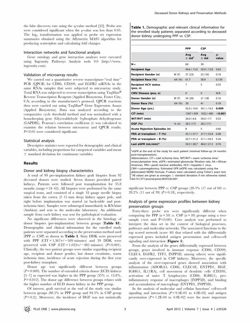
Table 1. Demographic and relevant clinical information forthe enrolled study patients separated according to deceaseddonor kidney undergoing PPP vs. CSP.
PPP CSP
Avg± std1
Avg± std
p-value
N = 60 39
Recipient Age 49.6613.0 50.9613.8 0.63
Recipient Gender (n) M (F) 37 (23) 23 (16) 0.16
Recipient Race (%) AA (%) 61.7 58.9 0.130
Recipient HCV status(pos, n)
7 5 0.55
CMV Disease (pos, n) 0 0 N/A
Donor Gender (n) M (F) 34 (26) 21 (18) 0.16
Donor Race (%) AA (%) 30 41 0.18
Donor Age (yrs.) 42.0614.6 34.1614.2 0.009
CIT (min) 13676509 10226485 ,0.001
WIT/RVT (min) 26.066.6 30.067.7 0.53
DGF (%) % (n) 28.3 (17) 28.2 (11)
Acute Rejection Episodes (n) 8 5 0.60
PRA at transplant – T (%) 43.1637.7 37.7635.8 0.48
PRA at transplant – B (%) 20.7631.2 27.1634.3 0.34
Last eGFR (mL/min)* 50.3628.7 48.6621.5 0.76
*eGFR at the end of the study for each patient (minimal follow-up 24 monthspost-transplantation).Abbreviations: CIT = cold ischemia time, WIT/RVT = warm ischemia time/revascularization time, eGFR = estimated glomerular filtration rate, AA = AfricanAmerican, PRA = panel reactive antibodies, HCV = hepatitis C virus,CMV = cytomegalovirus, Estimated GFR (eGFR) was calculated using theabbreviated MDRD formula. P-values were calculated using Fisher’s exact test.1All values are given as averages 6 standard deviation if not otherwise stated.doi:10.1371/journal.pone.0035526.t001
Deceased Donor Kidneys and Preservation Methods
PLoS ONE | www.plosone.org 3 April 2012 | Volume 7 | Issue 4 | e35526
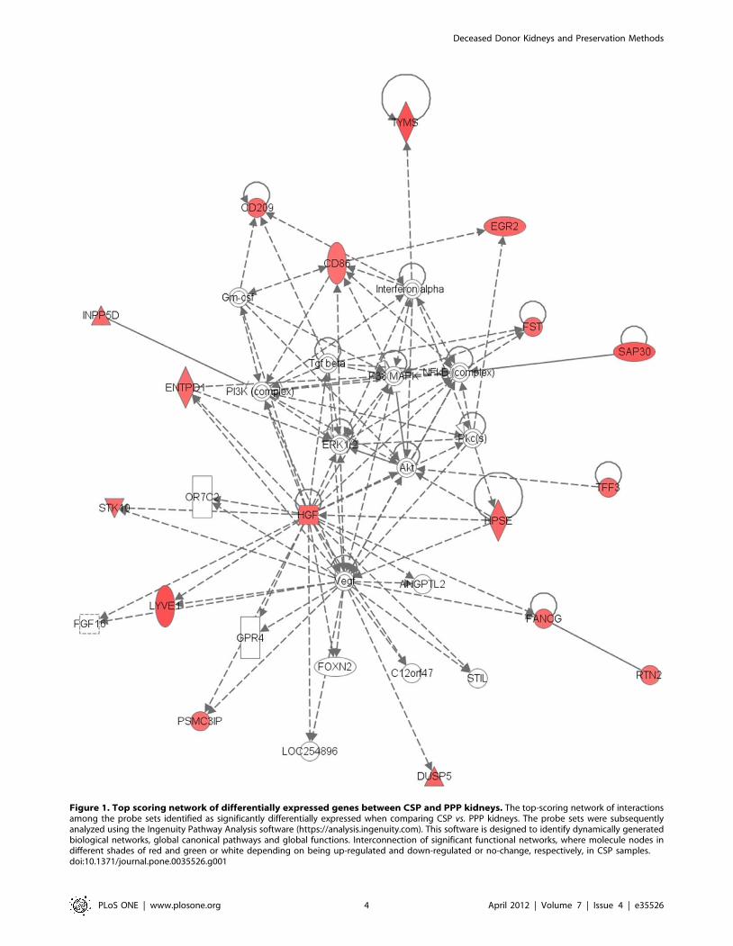
Figure 1. Top scoring network of differentially expressed genes between CSP and PPP kidneys. The top-scoring network of interactionsamong the probe sets identified as significantly differentially expressed when comparing CSP vs. PPP kidneys. The probe sets were subsequentlyanalyzed using the Ingenuity Pathway Analysis software (https://analysis.ingenuity.com). This software is designed to identify dynamically generatedbiological networks, global canonical pathways and global functions. Interconnection of significant functional networks, where molecule nodes indifferent shades of red and green or white depending on being up-regulated and down-regulated or no-change, respectively, in CSP samples.doi:10.1371/journal.pone.0035526.g001
Deceased Donor Kidneys and Preservation Methods
PLoS ONE | www.plosone.org 4 April 2012 | Volume 7 | Issue 4 | e35526
functions of genes significantly over-expressed in CSP kidney
samples. Seventeen probe sets were significantly different when
adjusting the analysis for CIT using probe set level linear models
(Table 2).
When the dataset was restricted to patients with a CIT,1200 -
minutes, there were 24 patients in PPP and 22 patients in CSP.
There were 5 significant probe sets when comparing PPP vs. CSP
groups using a two-sample t-test and P,0.001. There was still a
significant difference with respect to CIT between these two
groups (P = 0.004). Six probe sets were significant when adjusting
the analysis for CIT using probe set level linear models
Importantly, when the dataset was restricted to patients with
CIT.1200 minutes, there were 15 CSP and 37 PPP kidneys.
Eighteen probe sets were significantly different when comparing
the two groups (P,0.001) (Table 3) despite the fact that there was
no significant CIT between groups (P = 0.08). Importantly, all the
over expressed genes identified from this analysis, belong to the
same network (score 23) (Figure 2). The biological functions
associated with these genes were inflammatory response, cellular
movement, and immune cell trafficking.
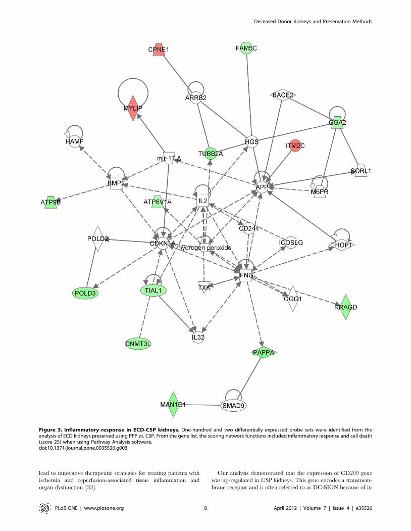
Gene expression profiles of expanded criteria donorkidneys (PPP vs. CSP)
ECD kidneys represented an important sub-group in our study
population (27.7%) and in line with the reported use of these grafts
in the USA.
Characteristics of the ECD kidneys by preservation group are
shown in the Table 4. As expected, there were more ECD
preserved with PPP. When gene expression profiles of ECD grafts
(PPP vs. CSP) were compared, 102 probe sets were statistically
differentially expressed (P,0.01). From the analysis of these genes,
we observed that genes involved in inflammation were down-
expressed in the ECD kidneys preserved with PPP as it is shown in
the Figure 3 (second top scored network, score 25). The incidence
of DGF in ECD kidneys was higher but not statistically different in
the PPP group (47.8% vs. 20%, P = 0.27 (one tailed Fisher Exact
Probability Test). Moreover, there was not statistical significantly
difference in graft function between ECD kidneys undergoing PPP
or CSP at 24 months post-kidney transplantation.
Evaluation of gene expression in paired kidneysIn the present study, seven donors provided paired kidneys (14
kidneys). We also evaluated how the gene expression patterns
differ among paired kidneys. Figure 4 shows the gene expression
profiles when two set of kidneys were compared at pre-
implantation time using the S-Score method to test for differential
expression between two different paired kidney sets. We
specifically evaluated three paired kidneys including one set of
SCD kidneys preserved using PPP and two sets of ECD kidneys
preserved using PPP or CSP. The first set of SCD kidneys
preserved using PPP performed well post-transplantation and the
gene expression profiles were similar between samples at pre-
implantation (Figure 4A). The second set of ECD undergoing
PPP had one kidney developing DGF post-transplantation (even
when CIT was not different between them) (Figure 4B). A similar
situation was observed with the set of ECD kidneys preserved with
CSP with only one kidney developing DGF post-transplantation
(Figure 4C). Also, not differences in CIT were observed. As
shown in Figure 4, a higher difference in probe sets was observed
in the last set (ECD kidneys undergoing CSP, with one kidney
developing DGF). For this last set of kidneys when applying the S-
Score method to test for differential expression between samples
and using the q-value method with q,0.05 to identify differen-
tially expressed probe sets, 137 were identified. From the analysis
of these genes, canonical pathway analysis showed that stress and
tissue injury were down regulated in the paired kidneys that did
not developed DGF post-kidney transplantation.
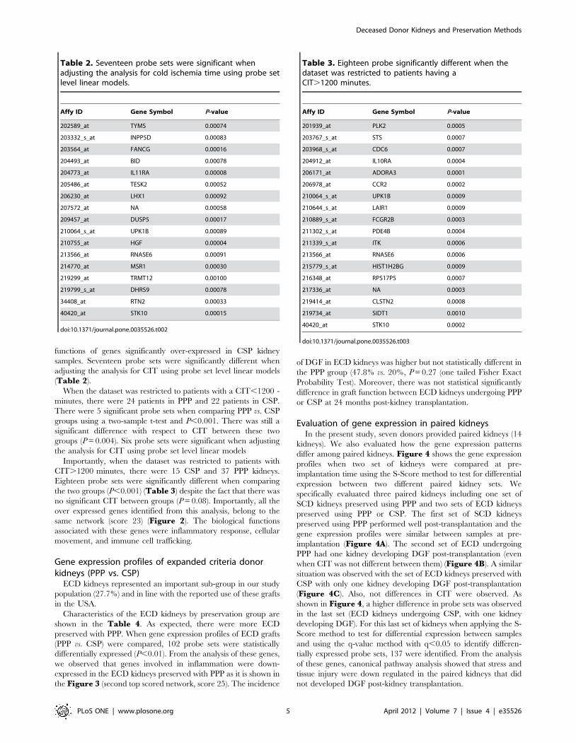
Table 2. Seventeen probe sets were significant whenadjusting the analysis for cold ischemia time using probe setlevel linear models.
Affy ID Gene Symbol P-value
202589_at TYMS 0.00074
203332_s_at INPP5D 0.00083
203564_at FANCG 0.00016
204493_at BID 0.00078
204773_at IL11RA 0.00008
205486_at TESK2 0.00052
206230_at LHX1 0.00092
207572_at NA 0.00058
209457_at DUSP5 0.00017
210064_s_at UPK1B 0.00089
210755_at HGF 0.00004
213566_at RNASE6 0.00091
214770_at MSR1 0.00030
219299_at TRMT12 0.00100
219799_s_at DHRS9 0.00078
34408_at RTN2 0.00033
40420_at STK10 0.00015
doi:10.1371/journal.pone.0035526.t002
Table 3. Eighteen probe significantly different when thedataset was restricted to patients having aCIT.1200 minutes.
Affy ID Gene Symbol P-value
201939_at PLK2 0.0005
203767_s_at STS 0.0007
203968_s_at CDC6 0.0007
204912_at IL10RA 0.0004
206171_at ADORA3 0.0001
206978_at CCR2 0.0002
210064_s_at UPK1B 0.0009
210644_s_at LAIR1 0.0009
210889_s_at FCGR2B 0.0003
211302_s_at PDE4B 0.0004
211339_s_at ITK 0.0006
213566_at RNASE6 0.0006
215779_s_at HIST1H2BG 0.0009
216348_at RPS17P5 0.0007
217336_at NA 0.0003
219414_at CLSTN2 0.0008
219734_at SIDT1 0.0010
40420_at STK10 0.0002
doi:10.1371/journal.pone.0035526.t003
Deceased Donor Kidneys and Preservation Methods
PLoS ONE | www.plosone.org 5 April 2012 | Volume 7 | Issue 4 | e35526

Discussion
From the time a patient is identified as a potential organ donor
it is critical to maintain adequate organ perfusion and avoid
hypoxemia. In the 1970s, most of the donated kidneys were
preserved by pulsatile perfusion preservation [23]. The situation
had reversed in the 1980s with the majority of kidneys being
preserved by cold storage preservation. The principal cause for the
change in the organ preservation method was that large-scale
studies of transplantation outcome [16–17,24] failed to find any
survival advantage for kidneys preserved by PPP. Consequently,
the disadvantages of PPP, including the need for a usually large
machine, consumables, technician and the risk of equipment
failure, as compared with the simplicity and low cost of CSP, were
not justified.
However, PPP has resurged in the last decade based upon the
belief that this preservation method leads to a reduced rate of graft
ischemia, DGF and eventually, improved long term graft function.
Figure 2. Differentially expressed genes between CSP and PPP kidneys with prolonged CIT. Eighteen probe sets were differentiallyexpressed when CIT was higher than 1,200 minutes and not significant between groups (CSP vs. PPP). Pathway Analysis showed that almost all thesegenes belong to the same network (score 23) and associate with the inflammatory response and immune cell trafficking.doi:10.1371/journal.pone.0035526.g002
Deceased Donor Kidneys and Preservation Methods
PLoS ONE | www.plosone.org 6 April 2012 | Volume 7 | Issue 4 | e35526
At the same time, comparative studies between PPP and CSP
started to show benefit of using PPP [24].
A report by Moers et al. [25] tends to support this practice. The
authors reported a prospective multicenter analysis of 336
consecutive grafts from deceased donors. Paired kidneys were
randomly divided and preserved by PPP or CSP and a total of 672
graft recipients were followed for 1 year. The authors reported
lower DGF incidence in PPP grafts (P = 0.01) as well as a reduced
serum creatinine level at 2 weeks post-kidney transplant and
reduced risk of graft failure. Also, graft survival at 1 year was better
in kidneys preserved by PPP (P = 0.04). As reported by the
editorial in the same journal [26], the limitations of this study are
that most of the kidney grafts had a short CIT (CIT mean 15 hr)
and the lack of an advantage of the use of PPP in the subgroup
analysis of ECD and donation after cardiac death kidneys.
Up-to- date, the published reports about the advantages and/or
disadvantages of the use PPP over CSP [22–26] are observational
and do not further explore the molecular and patho-physiological
mechanisms leading to decreased incidence of DGF and better
graft survival.
It is now well described that molecular profiling detects changes
not seen by histological evaluation and clinical markers. However,
the identification of predictive gene sets and the application to an
individualized patient management needs the integration of
clinical and pathology-based variables, as well as objective
reference markers of graft function, post-transplant complications,
and long-term outcomes [27].
At present, PPP is widely used in most areas of USA in renal
transplantation, allowing a greater use of ECD kidneys and
increasing the number of organs available for transplantation. In
addition, perfusion parameters, such as flow rate and resistance
are factors frequently used to assess and estimate the downstream
functionality of renal grafts [28]. However, a better understanding
of the molecular mechanisms involved in this complex process
associated with ischemia reperfusion injury and graft function may
provide biological insights supporting the recently described [25]
benefit of the use of PPP.
To the best of our knowledge, this is the first report showing
gene expression profiles of kidney donor biopsies in association
with clinical/demographic donor and recipient characteristics to
compare CSP vs. PPP in a prospective study.
Gene expression profile data of 99 consecutive deceased donor
kidneys preserved with different methods (CSP vs. PPP) were
evaluated. As expected, kidneys preserved by PPP had a tendency
to have longer CIT; the analysis was therefore controlled for CIT.
Our study included consecutive cadaveric donor kidney trans-
plants that were performed following clinical/surgical team
decisions based on organ quality despite the preservation method.
The present study represents a realistic situation about the
decisions and clinical practice that most regions in the USA
challenge daily.
In the present cohort of patients, histological evaluation at pre-
implantation time was performed for all the donor kidneys (TableS1). There were not statistical significant differences among
groups. We did not identify relationships between glomeruloscle-
rosis, tubular atrophy, and/or interstitial fibrosis and graft function
with gene expression profiles. Moreover, these results are in
concordance with the publication from Edwards et al. [29]. The
authors showed, using cadaveric kidneys (n = 3,444) with reported
biopsy results between October 25, 1999 and December 31, 2001,
that glomerulosclerosis on donor kidney biopsies does not correlate
well with 1-year graft survival and function, and percentage
glomerulosclerosis should not be used as the sole criterion for
discarding recovered cadaveric kidneys.
The principal finding from the molecular analysis was the
presence of over-expression of inflammatory genes in CSP
kidneys when compared with PPP kidneys. This is a point of
critical interest as the PPP group demonstrated similar DGF
incidence rates when compared with the CSP group despite of
having significant higher number of ECD donors with an
associated significant difference in donor age (older donors). In
the last decade, the proportion of deceased donors older than 50
years of age has increased from 21% to 30% in USA [30,31]
making this issue critical for further evaluation in the kidney
transplant field. With the knowledge that age alone is a
determinant of DGF it is expected that ECD organs are at
higher risk for complications than SCD organs [32]. A decreased
level of inflammation associated with the use of PPP may explain
these findings.
An imbalance in metabolic supply and demand within the
ischemic organ results in profound tissue hypoxia and microvas-
cular dysfunction. Subsequent reperfusion further enhances the
activation of innate and adaptive immune responses and cell death
programs. Recent advances in understanding the molecular and
immunological consequences of ischemia and reperfusion may
Table 4. Characteristics of ECD* donor kidneys preserved using PPP and CSP.
ECD-PPP n = 23 ECD-CSP N = 5 P-value
Recipient age (years) 50.2612.6 56.864.5 0.27
Recipient race (%AA) 73.9 80.0 0.43
Recipient gender (%M) 65.2 60.0 0.29
Donor age (years) 46.5616.3 49.8614.6 0.69
Donor race (%AA) 26.0 40.0 0.48
Donor gender (%M) 73.9 80.0 0.39
CIT (minutes) 1,4166570 1,3416395 0.80
Last donor serum creatinine (mg/dL) 1.2260.51 0.8760.5 0.26
DGF (%) (11/23) 47.8 (1/5) 20.0 0.22
eGFR at 24 months post-transplantation (mL/min) 57.2630.3 51.267.6 0.67
*ECD defined as previously described [7].AA: African-Americans, M: Male, DGF: Delayed Graft Function.doi:10.1371/journal.pone.0035526.t004
Deceased Donor Kidneys and Preservation Methods
PLoS ONE | www.plosone.org 7 April 2012 | Volume 7 | Issue 4 | e35526
lead to innovative therapeutic strategies for treating patients with
ischemia and reperfusion-associated tissue inflammation and
organ dysfunction [33].
Our analysis demonstrated that the expression of CD209 gene
was up-regulated in CSP kidneys. This gene encodes a transmem-
brane receptor and is often referred to as DC-SIGN because of its
Figure 3. Inflammatory response in ECD-CSP kidneys. One-hundred and two differentially expressed probe sets were identified from theanalysis of ECD kidneys preserved using PPP vs. CSP. From the gene list, the scoring network functions included inflammatory response and cell death(score 25) when using Pathway Analysis software.doi:10.1371/journal.pone.0035526.g003
Deceased Donor Kidneys and Preservation Methods
PLoS ONE | www.plosone.org 8 April 2012 | Volume 7 | Issue 4 | e35526

expression on the surface of dendritic cells and macrophages.
Dendritic cells and macrophages play an important role in the
innate and adaptive immune response of acute ischemia-reperfusion
injury (IRI). In the kidney they reside in the interstitial extracellular
compartment and are poised to interact with substances transported
from the tubule lumen into peritubular capillaries, endogenous
molecules released from parenchymal cells, exogenous invading
organisms, or with resident or infiltrating immune cells including
lymphocytes, nature killer T (NKT) cells, epithelial cells and
fibroblasts. Dendritic cells and macrophages are key initiators,
potentiators and effectors of innate immunity in kidney IRI and
induce injury either through inflammatory signals to other effector
cells or directly through the release of soluble mediators [34].
Activation of the innate immune system by ischemically damaged
tissue may increase the production of chemokines and adhesion
molecules by the endothelium and tubular epithelial cells to
facilitate the entry of leukocytes into the kidney.
The expression of CD86 was also up-regulated in CSP kidneys.
This protein is expressed by antigen-presenting cells, and it is the
ligand for two proteins at the cell surface of T cells, CD28 antigen
and cytotoxic T-lymphocyte-associated protein 4. Binding of this
protein with CD28 antigen is a costimulatory signal for activation
of the T-cell. Targeting the activation or effector function of
lymphocytes is a potentially effective approach to treat or modify
the immune response [34].
When the groups were sub-stratified according with CIT,
kidneys with longer CIT (when no significance was observed in
CIT between groups) presented an inflammatory signature when
preserved with CSP. This finding demonstrates the effect of PPP in
decreasing inflammation even on those kidneys with longer CIT.
From the evaluation of paired kidneys undergoing the same
preservation method, we observed that two sets of paired ECD
kidneys (one undergoing PPP (Figure 4B) and a second one CSP
(Figure 4C) one kidney in each set developed DGF. However,
when S-Score method was applied to evaluate the molecular
differences between samples, a higher number of genes were
differentially expressed between the ECD kidneys undergoing CSP
with down regulation of stress and tissue injury in the kidney that
did not develop DGF.
Biological modulation of ischemic acute kidney injury aims to
reduce the incidence of delayed graft function and to safely
increase the number of kidney transplantations using organs that
have suffered prolonged warm and cold ischemia [35]. In the
present article, we observed that the more important differential
pattern associated with pre-implantation biopsies when CSP and
PPP were compared, was inflammation. This pathway was up-
regulated in CSP kidneys even when the PPP group has a large
number of ECD and donor after cardiac death kidneys. Moreover,
no significant difference was observed in DGF incidence and graft
function post-transplantation (minimal follow-up two years). The
importance of this study relies not only in the identification of the
molecular signatures that characterize the use of PPP vs. CSP and
the sub-analysis per donor type (ECD vs. SCD) but also in the
functional validation of the results. All the studied biopsies were
from kidney grafts that were transplanted in recipients with end
stage kidney disease and followed for at least 24 months post-
transplantation. The present findings, based in the study of an
important number of samples and patient characterization,
support the importance of the use of PPP in ECD donor kidneys.
Supporting Information
Table S1 Histological evaluation of pre-implantation biopsies
classified by sub-groups.
(DOC)
Author Contributions
Conceived and designed the experiments: VRM DGM KJA. Performed
the experiments: VRM MJS JLS ALK KLB DGM CID MEW JAS.
Analyzed the data: KJA MJS VRM CID MEW JAS DGM. Contributed
reagents/materials/analysis tools: MJS JLS VRM CID KJA MEW JAS.
Wrote the paper: VRM CID DGM KJA MPP KLB MJS.
References
1. Wolfe RA, Ashby VB, Milford EL, Ojo AO, Ettenger RE, et al. (1999)
Comparison of mortality in all patients on dialysis, patients on dialysis awaiting
transplantation, and recipients of a first cadaveric transplant, N Engl J Med
341(23): 1725–1730.
2. McFarlane PA (2010) Should patients remain on intensive hemodialysis rather
than choosing to receive a kidney transplant? Semin Dial 23: 516–519.
3. United Network for Organ Sharing () National data website. Available: http://
www.optn.org/data. Accessed 2011 Nov 10.
Figure 4. Paired kidney analyses. The S-Score method was applied to assess whether there were any differences in gene expression betweenpaired kidneys. S-Score is a method that uses the probe level measurements in performing a test of hypothesis of differential probe set expressionwhen only two GeneChips are available for each comparison. A) Set of paired SCD-PPP that did not developed DGF, B) Set of paired ECD-PPP withone kidney developing DGF post-transplantation (CIT not significant), C) Set of paired ECD-SCP kidney with one kidney developing DGF post-transplantation (CIT not significant).doi:10.1371/journal.pone.0035526.g004
Deceased Donor Kidneys and Preservation Methods
PLoS ONE | www.plosone.org 9 April 2012 | Volume 7 | Issue 4 | e35526
4. Sung RS, Guidinger MK, Christensen LL, Ashby VB, Merion RM, et al. (2005)
Development and current status of ECD kidney transplantation. Clinical
Transplantation. pp 37–55.
5. Pascual J, Zamora J, Pirsch JD (2008) A systematic review of kidney
transplantation from expanded criteria donors. Am J Kidney Dis 52: 553–86.
6. Metzger RA, Delmonico FL, Feng S, Port FK, Wynn JJ, et al. (2003) Expanded
criteria donors for kidney transplantation. Am J Transplant 3(Suppl 4): 114–125.
7. UNOS Policy 3.5.1: Expanded criteria donor definition and point system,
United Network for Organ Sharing, Richmond, VA.
8. Port FK, Bragg-Gresham JL, Metzger RA, Dykstra DM, Gillespie BW, et al.
(2002) Donor characteristics associated with reduced graft survival: an approach
to expanding the pool of kidney donors. Transplantation 74: 1281–1286.
9. Schnitzler MA, Whiting JF, Brennan DC, Lin G, Chapman W, et al. (2003) The
expanded criteria donor dilemma in cadaveric renal transplantation. Trans-
plantation 75: 1940–1945.
10. Remuzzi G, Cravedi P, Perna A, Dimitrov BD, Turturro M, et al. (2006) Dual
Kidney Transplant Group. Long-term outcome of renal transplantation from
older donors. N Engl J Med 354: 343–352.
11. Schold JD, Kaplan B, Howard RJ, Reed AI, Foley DP, et al. (2005) Are we
frozen in time?: Analysis of the utilization and efficacy of pulsatile perfusion in
renal transplantation. Am J Transplant 5: 1681–1688.
12. Matsuoka L, Shah T, Aswad S, Bunnapradist S, Cho Y, et al. (2006) Pulsatile
perfusion reduces the incidence of delayed graft function in expanded criteria
donor kidney transplantation. Am J Transplant 6: 1473–1478.
13. Treckmann J, Moers C, Smits JM, Gallinat A, Maathuis MH, et al. (2011)
Machine perfusion versus cold storage for preservation of kidneys from
expanded criteria donors after brain death. Transpl Int 24: 548–554.
14. Perico N, Cattaneo D, Sayegh MH, Remuzzi G (2004) Delayed graft function in
kidney transplantation. Lancet 364: 1814–1827.
15. Yarlagadda SG, Coca SG, Formica RN, Jr., Poggio ED, Parikh CR (2009)
Association between delayed graft function and allograft and patient survival: a
systematic review and meta-analysis. Nephrol Dial Transplant 24: 1039–1047.
16. Wight JP, Chilcott JB, Holmes MW, Brewer N (2003) Pulsatile machine
perfusion vs. cold storage of kidneys for transplantation: a rapid and systematic
review. Clin Transplant 17: 293–307.
17. Sellers MT, Gallichio MH, Hudson SL, Young CJ, Bynon JS, et al. (2000)
Improved outcomes in cadaveric renal allograft with pulsatile perfusion. Clin
Transplant 14: 543–549.
18. Levey AS, Bosch JP, Lewis JB, Greene T, Rogers N, et al. (1999) A more
accurate method to estimate glomerular filtration rate from serum creatinine: a
new prediction equation. Modification of Diet in Renal Disease Study Group.
Ann Intern Med 130: 461–470.
19. Zhang L, Wang L, Ravindranathan A, Miles MF (2002) A new algorithm for
analysis of oligonucleotide arrays: Application to expression profiling in mousebrain regions. Journal of Molecular Biology 317: 225–235.
20. Kennedy RE, Archer KJ, Miles MF (2006) Empirical validation of the S-Score
algorithm in the analysis of gene expression data. BMC Bioinformatics 7: 154.21. Kennedy RE, Kerns RT, Kong X, Archer KJ, Miles MF (2006) S-Score: An R
package for detecting differential gene expression without gene expressionsummaries. Bioinformatics 22: 1272–1274.
22. Storey JD, Tibshirani R (2003) Statistical significance for genomewide studies.
Proc Natl Acad Sci U S A 100: 9440–9445.23. van der Vliet JA, Vroemen JP, Cohen B, Lansbergen Q, Kootstra G (1983)
Preservation of cadaveric kidneys. Cold storage or machine perfusion? ArchSurg 118: 1166–1168.
24. Shah AP, Milgrom DP, Mangus RS, Powelson JA, Goggins WC, et al. (2008)Comparison of pulsatile perfusion and cold storage for paired kidney allografts.
Transplantation 86: 1006–1009.
25. Moers C, Smits JM, Maathuis M-HJ, Treckmann J, van Gelder F, et al. (2009)Machine perfusion or cold storage in deceased-donor kidney transplantation.
N Engl J Med 360: 7–19.26. Tullius SG, Garcıa-Cardena G (2009) Organ procurement and perfusion before
transplantation. N Engl J Med 360: 78–80.
27. Mueller TF, Solez K, Mas V (2011) Assessment of kidney organ quality andprediction of outcome at time of transplantation. Semin Immunopathol 33:
185–199.28. Matsuno N, Konno YN, Jyojima Y, Akashi I, Iwamoto H, et al. (2010) Machine
perfusion preservation for kidney grafts with a high creatinine from uncontrolleddonation after cardiac death. Transplant Proc 42: 155–158.
29. Edwards EB, Posner MP, Maluf DG, Kauffman HM (2004) Reasons for non-use
of recovered kidneys: the effect of donor glomerulosclerosis and creatinineclearance on graft survival. Transplantation 77: 1411–1415.
30. Port FK (2003) Organ donation and transplantation trends in the United States,2001. Am J Transplant 3(suppl 4): 7–12.
31. Nathan HM, Conrad SL, Held PJ, McCullough KP, Pietroski RE, et al. (2003)
Organ donation in the United States. Am J Transplant 3(suppl 4): 29–40.32. Irish WD, Ilsley JN, Schnitzler MA, Feng S, Brennan DC (2010) A risk
prediction model for delayed graft function in the current era of deceased donorrenal transplantation. Am J Transplant 10: 2279–2286.
33. Eltzschig HK, Eckle T (2011) Ischemia and reperfusion-from mechanism totranslation. Nat Med 17: 1391–1401.
34. Nace G, Evankovich J, Eid R, Tsung A (2012) Dendritic cells and damage-
associated molecular patterns: endogenous danger signals linking innate andadaptive immunity. J Innate Immun 4: 6–15.
35. Siedlecki A, Irish W, Brennan DC (2011) Delayed graft function in the kidneytransplant. Am J Transplant 1: 2279–2296.
Deceased Donor Kidneys and Preservation Methods
PLoS ONE | www.plosone.org 10 April 2012 | Volume 7 | Issue 4 | e35526
Copyright © 2022 FDOKUMEN


![[Ovulation induction by pulsatile GnRH therapy in 2014: literature review and synthesis of current practice]](https://static.fdokumen.com/doc/165x107/6333c99c28cb31ef600d6b7b/ovulation-induction-by-pulsatile-gnrh-therapy-in-2014-literature-review-and-synthesis.jpg)