guidance document surgical technique for deceased donor ...
13
12 February 2015 Vs 1.0 1 GUIDANCE DOCUMENT SURGICAL TECHNIQUE FOR DECEASED DONOR ABDOMINAL ORGAN PROCUREMENT ATCA-TSANZ Guidelines G003/2015 Version 1.0 12 February 2015
-
Upload
khangminh22 -
Category
Documents
-
view
2 -
download
0
Transcript of guidance document surgical technique for deceased donor ...

12 February 2015 Vs 1.0
1
GUIDANCE DOCUMENT
SURGICAL TECHNIQUE FOR DECEASED DONOR
ABDOMINAL ORGAN PROCUREMENT
ATCA-TSANZ Guidelines G003/2015
Version 1.0 12 February 2015
12 February 2015 Vs 1.0
2
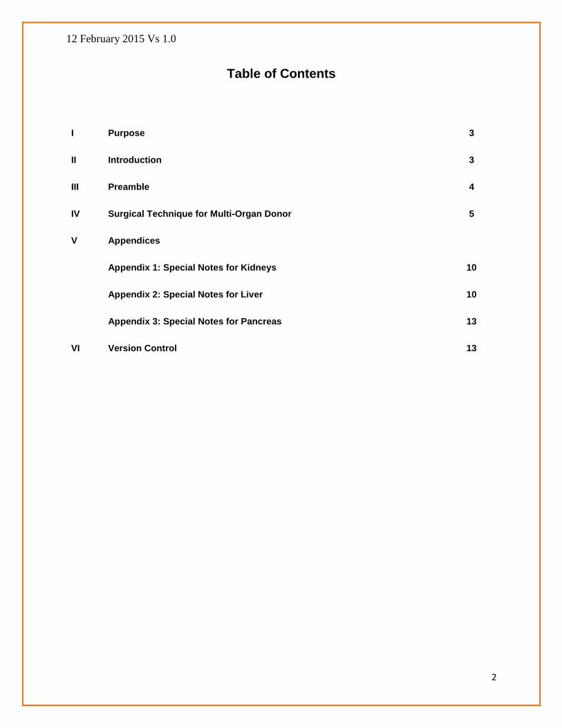
Table of Contents
I Purpose 3
II Introduction 3
III Preamble 4
IV Surgical Technique for Multi-Organ Donor 5
V Appendices
Appendix 1: Special Notes for Kidneys 10
Appendix 2: Special Notes for Liver 10
Appendix 3: Special Notes for Pancreas 13
VI Version Control 13

12 February 2015 Vs 1.0
3
I Purpose
The purpose of this reference document is to provide guidance on surgical techniques for
procurement of abdominal organs for transplantation. Health regulations and delivery of health
services vary between jurisdictions in Australia and New Zealand and therefore the guidelines
should be treated as reference material rather than a prescribed surgical procedure.
II Introduction
A minimum or basic surgical standard for retrieval of donor organs has been under discussions
in the Donor Surgeons and Donor Coordinators Advisory Committee (DSDC AC) for several
years. A consensus among surgeons has been achieved on a reference document on surgical
techniques.
The guidance document is intended to be used by surgeons and trainee surgeons. The guidelines
should be viewed only as recommendations; they do not establish legally enforceable
responsibilities. The word ‘should’ in this document means that something is suggested or
recommended, but not required. The guidelines do not represent the sole approach and
jurisdictions may use alternative methods.
Mention of specific products or equipment in this document does not represent an endorsement
of such products or equipment by the Transplantation Society of Australia and New Zealand
(TSANZ) nor does it necessarily represent preference for those products or equipment over
similar competitive products or equipment. It is incumbent on the reader who intends to use any
information, forms or procedures contained in this document to evaluate such materials for use in
the light of operational requirements associated with his or her facility.
TSANZ recognizes the efforts of the following surgeons who generously donated their time and
expertise in creating this document:
John Chen Director South Australian Liver Transplant Unit (South Australia)
Michael Fink Liver Transplant Surgeon, Liver Transplant Unit Victoria (Victoria)
Henry Pleass Liver Transplant Surgeon, Australian National Liver Transplant Unit (New South Wales)
12 February 2015 Vs 1.0
4
Deborah Verran Liver Transplant Surgeon, Australian National Liver
Transplant Unit (New South Wales)
Michael Crawford
Surgical Director, Australian National Liver Transplant Unit (New South Wales)
Richard Allen Director of Transplantation Services, RPA Transplant Institute (New South Wales)
III Preamble
1. The procedure will be performed by donor surgical teams accredited by a transplant
centre/hospital.
2. Liaison with donor coordinators, operating room staff and members of other procurement
teams and recipient surgeons, where necessary is primarily the responsibility of the senior
donor surgeon of that team performing the procedure.
3. The lead procurement surgeon is responsible for:
a. careful checking of the donor documentation, including confirmation of death, consent of
next of kin, Designated Officer and Coroner (if required), blood group, serology and the
Electronic Donor Record; Serology will be a laboratory report.
b. “time out” check of patient identification (name, UR number and date of birth) against the
documentation, particularly the declaration of death and consent forms,
c. ensuring that relevant medications (muscle relaxation, methylprednisolone 1 g, broad
spectrum antibiotics) have been given,
d. ensuring that labelling of the organ bags is correct,
e. ensuring that adequate descriptions of the anatomical, pathological and perfusion
characteristics are recorded for recipient surgeons,
f. communicating with recipient surgeons if there are particular issues that require
discussion,
12 February 2015 Vs 1.0
5
g. supporting the operating room personnel. This is particularly relevant for staff who have
had little or no experience of organ procurement, and
h. Ensuring that a brief operation report is completed for inclusion in the donor hospital
record.
The surgical technique description that follows is that of donation after brain death. The
principles of the surgical technique for donation after cardiac death (DCD) are similar, but the
processes occur more rapidly. DCD will occur in accordance with the National Protocol for
Donation after Cardiac Death (http://www.donatelife.gov.au/national-protocol-donation-and-
cardiac-death ) and relevant hospital or jurisdictional protocols. The thoracic and abdominal
surgical teams participate in the pre-withdrawal meeting and check the relevant documentation
prior to withdrawal of cardiorespiratory support. When the donor is brought to the operating
theatre, the lead surgeon checks the declaration of death form and ensures that the “time out”
check is performed. The warm phase dissection described below will not be carried out. The
key is rapid access to the retroperitoneum, cannulation of the aorta just above the bifurcation,
decompression by drainage of the inferior vena cava (IVC) and clamping of either the
supracoeliac aorta or distal thoracic aorta. Precedence should be given to removal of the
abdominal organs over the lungs, since the ischaemic tolerance of the lungs is greater than that
of the abdominal organs in the setting of DCD.
IV Surgical Technique for the Multi-organ Donor
1. The incision for the multi-organ donor is a long midline incision from the sternal notch to the
symphysis pubis. Midline sternotomy is standard, but might be avoided in the setting of kidney
only retrieval when there has been a previous sternotomy or at the request of the family. On
entering the abdomen a thorough examination of the intra abdominal and thoracic viscera is
performed to exclude conditions that would make organ donation contra-indicated
2. Initial operative exposure for procurement of the liver/pancreas will provide access to the
kidneys after cross clamping and flushing with preservation solutions. These steps include:
a. mobilisation of the right colon, descending and sigmoid colon and small bowel
mesentery to expose the kidneys, IVC, aorta and its bifurcation, duodenum and
pancreas
b. mobilisation of the duodenum and pancreas off the IVC and left renal vein
c. identification of both distal ureters below the pelvic brim
12 February 2015 Vs 1.0
6
d. identification of the junctions of the right and left renal veins with the IVC
e. identification of origin of superior mesenteric artery
f. slinging of the aorta just above the bifurcation with two heavy ties (e.g. no. 2 silk). Care
should be taken to ensure that there are no inferior renal arteries at or below the
intended level of aortic cannulation.
g. mobilisation of segments 2/3 of the liver by division of the left triangular ligament and
gastro-hepatic omentum (with care to avoid damage to an accessory or replaced left
hepatic artery arising from the left gastric artery, if present. The artery is confirmed to
be absent by both inspection and palpation)
h. identification of the common bile duct (CBD) just above the pancreas and ligation
distally. It is transected just above the tie (care to avoid vessels). The gallbladder is
incised, aspirated and flushed with normal saline until the effluent at the CBD is clear.
The bile duct is flushed with 100 ml normal saline.
i. identification of the supracoeliac aorta by division of the right crus of the diaphragm to
facilitate cross clamping. Note that attempting to sling the supracoeliac aorta can result
in posterior perforation, and if it is intended that the supracoeliac aorta be slung, this is
best left to the end of the warm phase.
j. If whole pancreas is to be procured, the lesser sac is entered by ligating and dividing
gastro-epiploic and short gastric vessels.
k. If pancreas (whole or for islets) is to be procured, the splenic flexure, spleen and body
and tail of pancreas are mobilised to allow adequate subsequent cooling of the
pancreas.
3. The cardiothoracic team is invited to perform preliminary dissection of thoracic viscera. 300
IU/kg of heparin are given intravenously to the donor (approximately 25,000 IU for an adult
donor). The appropriate perfusion cannula is inserted and tied into the infrarenal aorta. Care
should be taken to ensure that the cannula is intraluminal in the presence of severe
atherosclerosis. If it is intended that the IVC be cannulated, this occurs at this stage. Ensure
that the tubing attached to the IVC cannula is clamped.

12 February 2015 Vs 1.0
7
4. Cross clamping of the supracoeliac aorta is performed simultaneously with or immediately
following venting of the IVC or atrium and cold perfusion is commenced. The details of
perfusion may vary between units. If a high viscosity solution, such as University of Wisconsin
(UW) solution is used, perfusion should be first performed using a low viscosity solution, such
as Ross solution or Hartmanns (for example 4 litres) followed by UW solution. The exact
amount of UW solution used for in situ perfusion is usually about 1.5 to 2 L. An alternative
approach is to use a low viscosity solution, such as histidine-tryptophan-ketoglutarate (HTK)
solution. Perfusion may be performed either by gravity feed, at a height of approximately 1 m,
or by pressurised perfusion at approximately systemic arterial pressure. Aortic only perfusion
is the commonest approach, but a combination of aortic plus portal venous perfusion may be
used by some units or under some circumstances. Cold saline ice slush is placed in the
abdominal cavity, surrounding the liver, in front of both kidneys, behind and in front of the
pancreas.
5. The sequence of organ removal is the thoracic viscera first, followed by liver and pancreas en
bloc, kidneys (either en bloc or individually), iliac vessels. In some jurisdictions, mesenteric
lymph nodes and spleen are sampled. The right kidney must include the whole circumference
of the adjacent IVC. Both kidneys must include half circumference of adjacent aorta from
level of superior mesenteric artery to aortic bifurcation.
6. Organs removed en bloc are separated on the back table where the organs are checked for
adequacy of perfusion. When indicated, core biopsies of the kidneys are taken.
7. All organs apart from the pancreas are flushed with further preservation solution on the back
table. See following special notes for kidney, liver and pancreas.
8. All organs are bagged according to the National Standard Operation Procedures Electronic
Donor Record Utilisation for organ offer process organ transfer documentation, version 1
TSANZ SOP 002/2014; the organ is triple bagged with at least 500 ml of preservation
solution surrounding the organ in the first bag, slush is placed in the second bag and the third
bag is dry.
9. Vessels will be packaged according to the Standard Operating Procedures for packaging,
labeling, storage documentation of deceased donor vessels [ATCA-TSANZ SOP 003/2013]
currently being considered by TSANZ. The liver and pancreas (for whole organ
transplantation) should each be accompanied by a set of vessels (for example, and iliac

12 February 2015 Vs 1.0
8
artery and vein) and if the liver is split, each lobe should be accompanied by a set of vessels.
10. Any organs or tissues that have been removed and are not required for transplantation are
returned to the body. Standard closure (e.g. 1 Nylon and skin staples) is performed and a
dressing applied.
11. The principles with respect to organ biopsy are as follows:
a. Any suspicious lesion in either the chest or abdomen must be biopsied and
arrangements made for urgent frozen section and/or any other tissue sample
processing that may be required. Consideration may need to be given to the
availability of pathology resources (including the requisite expertise).
b. Biopsy of the liver can be performed either in-situ or ex-situ.
c. Biopsy of the kidney(s) may also be indicated in donors with risk factors for
chronic kidney disease and is best performed on the back table after a
comprehensive examination of the cortex has been undertaken.
d. All biopsies must be appropriately packaged and labelled and the relevant
information must be conveyed to the pathology laboratory.
12 February 2015 Vs 1.0
9
V. APPENDICES

12 February 2015 Vs 1.0
10
Appendix 1: Special Notes for Kidneys
1. The IVC should remain intact as a tube with the right kidney allowing transplanting surgeon
option to extend right renal vein prior to transplantation.
2. Removal of the peri-nephric fat is required to examine the cortex for evidence of adequate
perfusion and presence of obvious pathology. (The incidence of renal tumours discovered at
the time of cadaveric nephrectomy is said to be approximately 1%.) Additional back table
perfusion with 100ml of perfusion fluid is undertaken before bagging for transport for DCD and
retrograde flush.
Appendix 2: Special Notes for Liver
1. Assessment of the suitability of the liver for transplantation is a subjective skill which relies to
a large extent on experience. Features which may indicate an increased risk of primary non-
function include steatosis (yellow appearance, which is often most obvious after perfusion),
rounded edges and excessive firmness on palpation. Any concern about the macroscopic
appearance of the liver necessitates communication with the recipient surgeon and may
require biopsy (two core biopsies and one wedge biopsy are recommended for adequate
microscopic assessment of the donor liver).
2. The gallbladder is opened and distal CBD ligated just above the pancreas and transected
during the early phase of abdominal dissection and the gallbladder is lavaged with normal
saline. This step may be performed later in the process prior to aortic cross clamp.
3. The accessory right hepatic artery if present is not always readily palpable behind the common
bile duct. Assume the presence of this vessel until proven otherwise by dissection.
4. Hilar dissection is deferred to the back table by ensuring part/whole of the pancreas is
removed en bloc with the liver after cold perfusion.
5. The bile duct is flushed on the back table with 20-200ml of cold UW solution.
6. If aortic cool perfusion only is performed, a further 0.5 - 1 litre of cold UW solution is flushed
through the liver via the portal vein and Hepatic artery is also flushed with UW (in some units,
this is performed under pressure set at 100mmHg). The final bag will contain 900 - 1000 ml
of UW
12 February 2015 Vs 1.0
11
7. The lead abdominal surgeon must ensure that the cardiothoracic surgeon transects the
junction of the supradiaphragmatic IVC and the right atrium, so that sufficient IVC is available
for the recipient operation.
8. The diaphragm is divided to the left of the aorta, the thoracic aorta is transected, and the
diaphragm is divided around the bare area of the liver, with care to avoid traction injuries to
the liver, which tend to occur where the peritoneum overlying the right kidney attaches to the
capsule of the liver. The left renal vein is mobilised, dividing a small rim of IVC around the
insertion of the left renal vein, and dividing the loose tissue between the aorta and left renal
vein. Care should be taken during this manoeuvre to avoid injury to the right renal artery,
which may lie immediately posteriorly. The IVC is transected just above the top edge of the
right renal vein and just above the confluence of common iliac veins. The lymphatics lying
anterior to the aorta are divided with care to avoid injury to the left renal vein and the aorta is
then transected at the level of the cannulation site and then split longitudinally along the
anterior midline, again taking care not to injure the left renal vein. The renal artery orifices are
identified from within the lumen of the aorta. It is essential to identify and preserve any
accessory renal arteries, which may be present superior to the main renal artery orifices or
occasionally coming from low down, or even from the iliac vessels. The aorta is transected
above the renal arteries, with care to avoid injury to the superior mesenteric artery in the case
of procurement for whole pancreas transplantation. With the pancreas and spleen safely lifted
anteriorly, the tissues to the left of the aorta are divided up to the level of the transection of
the thoracic aorta. With the liver, pancreas and aortic tube held anteriorly, the tissues behind
the aorta and IVC are divided and the liver and pancreas block can then be removed to the
slush bath on the back table.
9. Separation of the liver and pancreas (whole graft) on the back table involves:
a. Division of the aortic patch with the celiac axis staying with the liver and the
superior mesenteric artery with the pancreas.
b. Mobilisation of the coeliac axis and common hepatic artery.
c. Transection of the splenic artery with a small rim (2 mm approximately) at the origin
staying with the liver and the majority staying with the pancreas. The splenic artery
stump on the pancreas side should be marked with a fine (e.g. 6/0 Prolene) suture
to assist subsequent identification.
12 February 2015 Vs 1.0
12
d. Ligation of the gastroduodenal artery on the pancreas side and transection leaving
a good length on the liver side.
e. Transection of the portal vein. A level that balances the needs of the liver and
pancreas recipient surgeons is selected. One cm above the superior level of the
pancreas is reasonable. The portal vein on the pancreas side should be marked
with a fine (e.g. 6/0 Prolene) suture to assist subsequent identification.
f. Transection of tissues (nerves, lymphatics etc.) posterior to the portal vein. Care
should be taken to preserve the maximum length of a replaced or accessory right
hepatic artery arising from the superior mesenteric artery. This can subsequently
be reconstructed onto the gastroduodenal artery at the recipient back table, and
thus this common anomaly does not preclude liver and whole pancreas donation.
10. Separation of the liver and pancreas is best performed with one of the abdominal surgeons
assisting the other and with each surgeon “protecting” the vessels of each organ, to minimise
the risk of accidental injury.
11. In separating the liver from the pancreas when the pancreas is being procured for islets,
vessels can be transacted in a way that maximises the length on the liver side, and without
ligating vessels on the pancreas side. However it is essential that the capsule of the pancreas
is not breeched, as this would result in leakage of the collagenase during islet preparation,
resulting in a reduced islet yield.

12 February 2015 Vs 1.0
13
Appendix 3: Special Notes for Pancreas
1. The same surgical requirements exist for removal of the donor pancreas if it is to be used for
vacularised whole pancreas transplantation or isolated islet transplantation, other than the
need to preserve the vessels to the pancreas.
2. A naso-gastric tube is inserted during the initial dissection phase to permit gastric and
duodenal suction and instillation of 50 -100ml of half strength povo-iodine. This solution
should be aspirated prior to withdrawal of NG tube.
3. The duodenum is divided above and below the pancreas with a linear cutting stapler (blue
insert). The SMA and SMV can similarly be divided as close to the inferior border of the
uncinate process as possible, ideally using a vascular cartridge of the linear cutting stapler.
Care must be taken not to injure the pancreas at this point, but this technique reduces blood
loss in the recipient operation significantly.
4. One of the iliac arteries and veins accompany the pancreas if it is being procured for whole
pancreas transplantation. It is important to include a good length of common, external and
internal iliac artery, because the splenic and superior mesenteric arteries are routinely
reconstructed using a Y graft of iliac artery at the recipient back table. The bifurcation of the
CIA must not be injured and must be included to enable back table reconstruction.
5. If the pancreas is being retrieved as a whole graft with duodenum, the spleen is often left
attached to the pancreas. However the spleen can be removed at the donor procedure,
provided there is sufficient gap between pancreatic tail and splenic hilum. A vascular stapler
can be used for this manoeuvre. If the pancreas is being retrieved for islets, the spleen and
duodenum are removed prior to bagging the pancreas.
VI. Version Control
Version #: Changes Approved by
Date Approved