Mastoid Trepanation in a Deceased from Medieval Croatia: A Case Report
6
209 or cutting the intervening bone 1. In terms of small cranial trepanne and/or »drill« holes, they are rarely reported in bioarchaeological studies and it is usually the question either of multiple cranial openings 1,14 or openings accom- panying various types of primary trepanations 7,12 . There are also a few examples of the so-called »double-« or »tri- ple-like«, i.e. 2–3 connected and relatively small cranial »drill« holes, like those identified in Celtic crania, report- ed from Austria 15 . However, one has to point out that usu- ally ancient trepanations described all over the world are supratentorial, i.e. occurring above the tentorium. The tentorium cerebelli is an expansion of the dura matter separating the cerebellum from the inferior portion of the occipital lobe 16 . Aufderheide and Rodriguez-Martín 17 de- fine the (supratentorial) trepanation technique as a pro- cedure which involves creating a defect in the skull vault to open a communication between the cranial cavity and the environment, whose success depends on avoiding in- jury to the meninges, brain and blood vessels. On the The trepanation – artificial removal of the bone from the cranial vault, has been practiced since ancient times 1,2 and probably traces back to Mesolithic Age 3 . There is a substantial number of various examples of trepanation reported from all over the world: Europe, Near East, Af- rica, Melanesia 2–9 , Asia 10 , the Americas 4,11,12 . An ancient trepanation was usually performed for three different pur- poses: medicine-related (surgical), the so-called culticul or magic-related and symbolic purposes 1,2 . In terms of med- icine-related purposes, the ancient trepanation was gener- ally practiced for relief of cranial fractures/wounds effects, such as an increased intracranial pressure, headaches, or other conditions 1,2 . However, there are also a few examples of medicine-related trepanation associated with possible ear pathologies. So far, such trepanations were described by Brothwell 13 , Mann 14 , Kato et al. 12 . With regard to the types of trepanation, it could be performed by scraping through the bone with a sharp implement or drilling a circle of small holes and breaking Coll. Antropol. 39 (2015) 1: 209–214 Case report Mastoid Trepanation in a Deceased from Mastoid Trepanation in a Deceased from Medieval Croatia: A Case Report Medieval Croatia: A Case Report Jadranka Boljunčić Jadranka Boljunčić 1 and Josip Hat and Josip Hat 2 1 Institute of Archaeology, Zagreb, Croatia 2 »Medikol« Polyclinic, Radiology Department, Zagreb, Croatia ABSTRACT ABSTRACT We present a rare case of infratentorial mastoid trepanation, by drilling, from medieval Croatia. An artificial ante- mortal opening was found in a male skeleton from the 11 th century cemetery Zvonimirovo. It was placed roughly at the intersection of the Frankfurt’s plane and the midline of the right mastoid. The right posterior parietal of the deceased also exhibited a callus-like formation consistent with the linear cranial fracture. Our aim was to investigate by computed tomography (CT) a possible presence of otopathology – a chronic middle ear infection – MEI/mastoiditis or cholesteatoma. On the other hand, both standard radiography and CT were employed in a cranial fracture diagnostic agreement. The generated CT scans confirmed the presence of an artificial hole running into a well defined trepanne canal connected with the antrum. The presence of otopathology was not established. The radiography and CT substantiated the presence of a linear posterior parietal discontinuity – without displacement, in front of the right lambdoid suture. From the medical point of view, it would be unusual to perform infratentorial – mastoid trepanation for reasons of treating supratentorial trauma, i.e. possible posttraumatic acute subdural hematoma (PTASDH). However, since there was a lack of CT evidence of osteolysis in ME, there is a possibility of medieval trepanation procedure performed for reasons of posttraumatic treat- ment. To our best knowledge, usually, ancient trepanations described in Croatian bioarchaeology and all over the world are supratentorial and do not always reveal such sophisticated surgical techniques. Key words: medieval surgery, mastoid drilling, otopathology, supratentorial trauma, computed tomography, Croatia Introduction Introduction Received for publication April 2, 2013
Transcript of Mastoid Trepanation in a Deceased from Medieval Croatia: A Case Report

209
J. Boljunčić and J. Hat: Medieval Mastoid Trepanation from Croatia, Coll. Antropol. 39 (2015) 1: 209–214
or cutting the intervening bone1. In terms of small cranial trepanne and/or »drill« holes, they are rarely reported in bioarchaeological studies and it is usually the question either of multiple cranial openings1,14 or openings accom-panying various types of primary trepanations7,12. There are also a few examples of the so-called »double-« or »tri-ple-like«, i.e. 2–3 connected and relatively small cranial »drill« holes, like those identifi ed in Celtic crania, report-ed from Austria15. However, one has to point out that usu-ally ancient trepanations described all over the world are supratentorial, i.e. occurring above the tentorium. The tentorium cerebelli is an expansion of the dura matter separating the cerebellum from the inferior portion of the occipital lobe16. Aufderheide and Rodriguez-Martín17 de-fi ne the (supratentorial) trepanation technique as a pro-cedure which involves creating a defect in the skull vault to open a communication between the cranial cavity and the environment, whose success depends on avoiding in-jury to the meninges, brain and blood vessels. On the
The trepanation – artifi cial removal of the bone from the cranial vault, has been practiced since ancient times1,2 and probably traces back to Mesolithic Age3. There is a substantial number of various examples of trepanation reported from all over the world: Europe, Near East, Af-rica, Melanesia2–9, Asia10, the Americas4,11,12. An ancient trepanation was usually performed for three different pur-poses: medicine-related (surgical), the so-called culticul or magic-related and symbolic purposes1,2. In terms of med-icine-related purposes, the ancient trepanation was gener-ally practiced for relief of cranial fractures/wounds effects, such as an increased intracranial pressure, headaches, or other conditions1,2. However, there are also a few examples of medicine-related trepanation associated with possible ear pathologies. So far, such trepanations were described by Brothwell13, Mann14, Kato et al.12.
With regard to the types of trepanation, it could be performed by scraping through the bone with a sharp implement or drilling a circle of small holes and breaking
Coll. Antropol. 39 (2015) 1: 209–214Case report
Mastoid Trepanation in a Deceased from Mastoid Trepanation in a Deceased from Medieval Croatia: A Case ReportMedieval Croatia: A Case Report
Jadranka BoljunčićJadranka Boljunčić1 and Josip Hat and Josip Hat2
1 Institute of Archaeology, Zagreb, Croatia2 »Medikol« Polyclinic, Radiology Department, Zagreb, Croatia
A B S T R A C TA B S T R A C T
We present a rare case of infratentorial mastoid trepanation, by drilling, from medieval Croatia. An artifi cial ante-mortal opening was found in a male skeleton from the 11th century cemetery Zvonimirovo. It was placed roughly at the intersection of the Frankfurt’s plane and the midline of the right mastoid. The right posterior parietal of the deceased also exhibited a callus-like formation consistent with the linear cranial fracture. Our aim was to investigate by computed tomography (CT) a possible presence of otopathology – a chronic middle ear infection – MEI/mastoiditis or cholesteatoma. On the other hand, both standard radiography and CT were employed in a cranial fracture diagnostic agreement. The generated CT scans confi rmed the presence of an artifi cial hole running into a well defi ned trepanne canal connected with the antrum. The presence of otopathology was not established. The radiography and CT substantiated the presence of a linear posterior parietal discontinuity – without displacement, in front of the right lambdoid suture. From the medical point of view, it would be unusual to perform infratentorial – mastoid trepanation for reasons of treating supratentorial trauma, i.e. possible posttraumatic acute subdural hematoma (PTASDH). However, since there was a lack of CT evidence of osteolysis in ME, there is a possibility of medieval trepanation procedure performed for reasons of posttraumatic treat-ment. To our best knowledge, usually, ancient trepanations described in Croatian bioarchaeology and all over the world are supratentorial and do not always reveal such sophisticated surgical techniques.
Key words: medieval surgery, mastoid drilling, otopathology, supratentorial trauma, computed tomography, Croatia
IntroductionIntroduction
Received for publication April 2, 2013
210
J. Boljunčić and J. Hat: Medieval Mastoid Trepanation from Croatia, Coll. Antropol. 39 (2015) 1: 209–214
other hand, an infratentorial trepanation technique, such as a mastoid trepanation, by drilling, in the present case (so far not described for ancient crania), occurs below the tentorium cerebelli.
There are some important criteria for identifying if a cranial opening has been made intentionally (by mechan-ical intervention)1, i.e. to determine whether a hole stems from possible accidental causes, such as excavation or whether it was the action of animals or plant roots. The same is the case with a possible presence of congenital alterations and/or anomalies which could cause some per-forations as well as infectious processes and ectocranial neoplasias8. The next step is to clarify antemortal/post-mortal status of the opening and the presence of accompa-nying fracture1.
As for the ancient/historical practice of trepanation from the Croatian territory, there is a general lack of sci-entifi c articles referring to such topic. So far, there were only two trepanation-related cases reported – one prehis-toric18, the other from a 5th century AD19. However, they are both supratentorial. This paper presents a rare case of unusual infratentorial mastoid trepanation, by drilling, in an individual from the 11th century Croatian burial complex Zvonimirovo. Given the anatomic position of an artifi cial mastoid »defect«, made for reasons of possible otopathology, we aimed to establish by computed tomog-raphy (CT) whether the surgery was performed due to possible chronic middle ear infection (MEI)/mastoiditis, or cholesteatoma. To the best of our knowledge, this is a unique case of an ancient infratentorial mastoid trepana-tion described so far.
Case ReportCase Report
The skeleton of an individual studied for a possible medicine-related therapeutic mastoid trepanation was
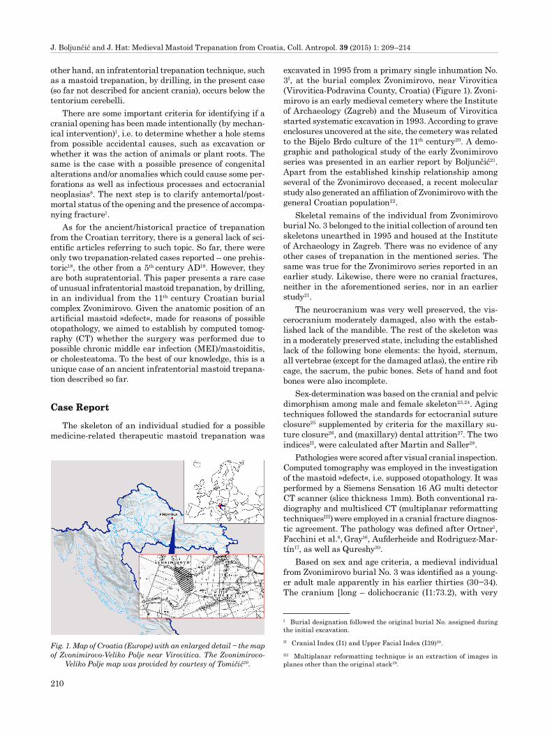
excavated in 1995 from a primary single inhumation No. 3I, at the burial complex Zvonimirovo, near Virovitica (Virovitica-Podravina County, Croatia) (Figure 1). Zvoni-mirovo is an early medieval cemetery where the Institute of Archaeology (Zagreb) and the Museum of Virovitica started systematic excavation in 1993. According to grave enclosures uncovered at the site, the cemetery was related to the Bijelo Brdo culture of the 11th century20. A demo-graphic and pathological study of the early Zvonimirovo series was presented in an earlier report by Boljunčić21. Apart from the established kinship relationship among several of the Zvonimirovo deceased, a recent molecular study also generated an affi liation of Zvonimirovo with the general Croatian population22.
Skeletal remains of the individual from Zvonimirovo burial No. 3 belonged to the initial collection of around ten skeletons unearthed in 1995 and housed at the Institute of Archaeology in Zagreb. There was no evidence of any other cases of trepanation in the mentioned series. The same was true for the Zvonimirovo series reported in an earlier study. Likewise, there were no cranial fractures, neither in the aforementioned series, nor in an earlier study21.
The neurocranium was very well preserved, the vis-cerocranium moderately damaged, also with the estab-lished lack of the mandible. The rest of the skeleton was in a moderately preserved state, including the established lack of the following bone elements: the hyoid, sternum, all vertebrae (except for the damaged atlas), the entire rib cage, the sacrum, the pubic bones. Sets of hand and foot bones were also incomplete.
Sex-determination was based on the cranial and pelvic dimorphism among male and female skeleton23,24. Aging techniques followed the standards for ectocranial suture closure25 supplemented by criteria for the maxillary su-ture closure26, and (maxillary) dental attrition27. The two indicesII, were calculated after Martin and Saller28.
Pathologies were scored after visual cranial inspection. Computed tomography was employed in the investigation of the mastoid »defect«, i.e. supposed otopathology. It was performed by a Siemens Sensation 16 AG multi detector CT scanner (slice thickness 1mm). Both conventional ra-diography and multisliced CT (multiplanar reformatting techniquesIII) were employed in a cranial fracture diagnos-tic agreement. The pathology was defi ned after Ortner1, Facchini et al.8, Gray16, Aufderheide and Rodriguez-Mar-tín17, as well as Qureshy30.
Based on sex and age criteria, a medieval individual from Zvonimirovo burial No. 3 was identifi ed as a young-er adult male apparently in his earlier thirties (30−34). The cranium [long – dolichocranic (I1:73.2), with very
I Burial designation followed the original burial No. assigned during the initial excavation.II Cranial Index (I1) and Upper Facial Index (I39)28.III Multiplanar reformatting technique is an extraction of images in planes other than the original stack29.
Fig. 1. Map of Croatia (Europe) with an enlarged detail − the map of Zvonimirovo-Veliko Polje near Virovitica. The Zvonimirovo-
Veliko Polje map was provided by courtesy of Tomičić20.

211
J. Boljunčić and J. Hat: Medieval Mastoid Trepanation from Croatia, Coll. Antropol. 39 (2015) 1: 209–214
slender – hyperlepten upper face (I39:60)] exhibited the presence of a small artifi cial circular bone defect to the right mastoid with additional trauma to the right poste-rior parietal (Figure 2–3). The defect was located roughly at the intersection of the Frankfurt’s plane and the mid-line of the right mastoid process. Gross morphology of »perforated« mastoid was consistent with an antemortal trepanne hole made by drilling. Bone structure around the hole was compact, bone margins smooth with a deli-cate marginal osteosclerozation. There were no outer signs of bone infection (Figure 3). The external maximum horizontal diameter of the opening measured with the caliper was 6.6mm. The axial CT scans at the level of both ME (tympanic cavities) and antra showed the presence of an artifi cial hole running into the trepanne canal con-nected with the posterior inferior antrum (Figure 4). The margins of the canal were regular and clearly osteoscle-rotic (Figures 4 and 5). The maximum transversal diam-eter of the opening obtained from CT was 7.6mm. The maximum transversal diameter of the canal at the turn of its mid-section into the medial third was 9.4mm. The maximum transversal diameter of the canal beside the
sigmoid sinus was 12mm. The maximum length of the trepanne canal was 18.8 mm at the anterior margin and 16mm at the posterior margin. The status of the right tympanic cavity (tympanum), epitimpanum, horizontal semicircular canal and inner auditory meatus was normal although with the presence of the sediment matrix. The facial nerve canal and right otic capsule – bone around the labyrinth were intact. The same applies to the left-hand side (Figure 5). The presence of two intact (dislocated) auditory ossicles – the maleus and incus (the stapes could not have been identifi ed) was established in the right pos-terior antrum (Figures 4 and 5). The same was true for the left posterior antrum. The axial CT scan of the right mastoid exhibited completely preserved periantral cellules and intercellular septa of the mastoid apex. The same was
Fig. 2. Right lateral aspect of the male cranium from the Zvoni-mirovo burial No. 3 with an antemortal infratentorial mastoid trepanne hole. Additional callus-like formation is placed in the
right posterior parietal (marked with white arrows).
Fig. 4. Axial CT scan of both tympanic – middle ear (ME) cavities and antra at the level of connection of the right mastoid trepanne canal with the posterior inferior antrum. Displaced antral posi-tion of incus (a) and maleus (b), posterior semicircular canal (c),
otic capsule (d).
Fig. 5. Axial CT scan of both ME cavities and antra at the level of visible inner auditory meatus and facial nerve canal. Incus (a) and caput malei (b) in the right antrum, horizontal semicircular canal (c), otic capsule (d) labyrinthal part of the facial nerve canal
(e), inner auditory meatus fi lled with the sediment matrix (f).
Fig. 3. Enlarged comparative lateral gross aspect of the right (trepanned) mastoid (on the left) and the left normal mastoid (on the right). Mild exostosis is present at the lower margin of the
right acoustic porus.
212
J. Boljunčić and J. Hat: Medieval Mastoid Trepanation from Croatia, Coll. Antropol. 39 (2015) 1: 209–214
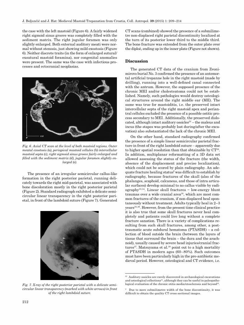
the case with the left mastoid (Figure 6). A fairly widened right sigmoid sinus groove was completely fi lled with the sediment matrix. The right jugular foramen was only slightly enlarged. Both external auditory meati were nor-mal without stenosis, just showing mild exostosis (Figure 6). Neither discrete traits (in the form of enlarged sutural/exsutural mastoid foramina), nor congenital anomalies were present. The same was the case with infectious pro-cesses and ectocranial neoplasias.
The presence of an irregular semicircular callus-like formation in the right posterior parietal, running deli-cately towards the right mid-parietal, was associated with bone discoloration mostly in the right posterior parietal (Figure 2). Standard radiograph exhibited a delicate semi-circular linear transparency in the right posterior pari-etal, in front of the lambdoid suture (Figure 7). Generated
CT scans (combined) showed the presence of a submilime-tre non-displaced right parietal discontinuity localized at the turn of its posterior lower third to the middle third. The bone fracture was extended from the outer plate over the diploë, ending up in the inner plate (Figure not shown).
DiscussionDiscussion
The generated CT data of the cranium from Zvoni-mirovo burial No. 3 confi rmed the presence of an antemor-tal artifi cial trepanne hole in the right mastoid (made by drilling), running into a well-defi ned canal connected with the antrum. However, the supposed presence of the chronic MEI and/or cholesteatoma could not be estab-lished. Namely, such pathologies would destroy osteologi-cal structures around the right middle ear (ME). The same was true for mastoiditis, i.e. the preserved intact intercellular septa of the right mastoid apex and perian-tral cellules excluded the presence of a possible ostitic pro-cess secondary to MEI. Additionally, the preserved dislo-cated, although intact auditory ossiclesIV – the maleus and incus (the stapes was probably lost during/after the exca-vation) also substantiated the lack of the chronic MEI.
On the other hand, standard radiography confi rmed the presence of a simple linear semicircular parietal frac-ture in front of the right lambdoid suture – apparently due to higher spatial resolution than that obtainable by CT29. In addition, multiplanar reformatting of a 3D data set allowed assessing the status of the fracture (the width, absence of the displacement and precise localization), which could not be scored by plain radiography. An ade-quate fracture healing statusV was diffi cult to establish by radiography, because fractures of the skull (also of the phalanges, scaphoid, calcaneus, and those of intra articu-lar surfaces) develop minimal to no callus visible by radi-ography33,34. Linear skull fractures − low-energy blunt traumas over a wide cranial area30, which are most com-mon fractures of the cranium, if non-displaced heal spon-taneously without treatment. Adults typically heal in 2–3 years35,36. However, from the present time clinical practice it is also true that some skull fractures never heal com-pletely and patients could live long without a complete fracture sanation. There is a variety of complications re-sulting from such skull fractures, among other, a post-traumatic acute subdural hematoma (PTASDH) – a col-lection of blood outside the brain (between the layers of tissue that surround the brain – the dura and the arach-noid), usually caused by severe head injuries/cranial frac-tures37. Matsuyama et al.38 point out to a high mortality of PTASDH in modern ages (60–80%). Such outcomes must have been particularly high in the pre-antibiotic me-dieval period. However, osteological and CT evidence, i.e.
IV Auditory ossicles are rarely discovered in archaeological excavations and osteological collections31, although they can be useful in paleopatho-logical evaluation of the chronic otitis media/otosclerosis and beyond32.V Due to mere submilimetre width of the bone discontinuity, it was diffi cult to obtain the quality CT cross-sectional images.
Fig. 6. Axial CT scan at the level of both mastoid regions. Outer meatal exostosis (a), periapical mastoid cellules (b) intercellular mastoid septa (c), right sigmoid sinus groove fairly enlarged and fi lled with the sediment matrix (d), jugular foramen slightly en-
larged (e).
Fig. 7. X-ray of the right posterior parietal with a delicate semi-circular linear transparency (marked with white arrows) in front
of the right lambdoid suture.

213
J. Boljunčić and J. Hat: Medieval Mastoid Trepanation from Croatia, Coll. Antropol. 39 (2015) 1: 209–214
bone reparation of both the trepanne hole and canal strongly substantiate surving of the individual (quite) some time after the performed procedure.
There is a spectrum of instruments (drill trepanons?) with which mastoid trepanation by drilling could be per-formed. The anatomical position of the trepanne canal – connected with the posterior inferior antrum, points out to a rather sophisticated technique performed by a skilled medieval surgeon. This is substantiated by the lack of bone damage around the facial nerve canal. Generally, fair knowledge about the internal bone structures in the human body and bone regeneration has been known ever since the early prehistory7. Apparently the same is the case with a medieval period from the territory of Croatia. The presence of a fairly widened right sigmoid sinus groove in the Zvonimirovo medieval individual is just a result of an anatomical variation. The terminal part of the sigmoid groove is usually larger on the right with no sig-nifi cant sex-related difference. The same is true with jugular fossa and venous portion of jugular foramen39.
Described mastoid trepanation, by drilling, is an in-fratentorial trepanation technique, i.e. occurs below the tentorium cerebelli. From the medical point of view, it would be unusual to perform an infratentorial trepana-
tion, i.e. by intervening into the posterior cranial fossa for reasons of treating supratentorial trauma, e. g. possible posttraumatic acute subdural haematoma (PTASDH). However, since there was a lack of CT evidence of osteoly-sis in ME, there is a possibility of medieval trepanation procedure performed for reasons of posttraumatic treat-ment.
In conclusion, the present paper reports an unusual antemortal infratentorial mastoid trepanation, by drill-ing, revealing a sophisticated operating procedure. Two ancient cases of trepanations reported from the Croatian territory and numerous from places all over the world are supratentorial and mostly do not reveal such sophisticated surgical techniques.
AcknowledgementsAcknowledgements
Computed tomography scanning was performed by courtesy of the staff of the Institute for Diagnostic and Interventional Radiology of the University Hospital Cen-ter »Sestre milosrdnice« (Zagreb). We also thank Prof. Dr. emeritus J. Stojanović for allowing standard radiography performing at the University Hospital Center Zagreb, Clinic for Oncology.
R E F E R E N C E SR E F E R E N C E S
1. ORTNER DJ, Identifi cation of Pathological Conditions in Human Skeletal Remains (Academic Press, Amsterdam, 2003). — 2. BERECZKI Z, MARCSIK A, Acta Med Lituan, 12 (2005) 65. — 3. LILLIE MC, Na-ture, 391 (1998) 854. — 4. TELLO JC, Prehistoric trephining among the Jauyos of Peru. In: Proceedings (International Congress of Americanists 1912, London, 1913). — 5. ENGLAND IA, Radiography, 28 (1962) 301. — 6. LISOWSKY FP, Prehistoric and early historic trepanation. In: BROTHWELL DR, SANDISON AT (Eds) Diseases in antiquity (Charles C Thomas, Springfi eld, 1967). — 7. MIKIĆ Ž, Glasnik antropol društva Jugosl, 17 (1980) 163. — 8. FACCHINI F, RASTELLI E, FERRERO L, FULCHERI E, Homo, 53 (2003) 247. — 9. ANDRUSHKO VA,VERANO JW, Am J Phys Anthropol, 137 (2008) 4. — 10. HAN K, CHEN X, Indo Pac Prehist Assoc Bull, 27 (2007) 22. — 11. PIGGOT S, Proc Prehist Soc, 6 (1940) 112. — 12. KATO K, SHINODA K, KITAGAWA Y, MANABE Y, OYAMADA J, IGAWA K, VIDAL H, ROKUTANDA A, Anthropol Sci, 115 (2007) 227. — 13. BROTHWELL DR, Man, 59 (1959) 95. — 14. MANN G, Int J Osteoarchaeol, 1 (1991) 165. — 15. URBAN OH, TESCH-LER-NICOLA M, SCHULTZ M, Archaeol Austriaca, 69 (1985) 13. — 16. GRAY H, Gray’s Anatomy (Barnes & Noble, New York, 2010, 15th ed.). — 17. AUFDERHEIDE AC, RODRÍGUEZ MARTÍN C, The Cambridge Encyclopedia of Human Paleopathology (Cambridge University Press, Cambridge, 1998). — 18. MALEZ M, NIKOLIĆ V, Rad Jugosl akad znan umjetn, 371 (1975) 171. — 19. NOVAK M, NAĐ M, PLEŠE T, ČAVKA M, Acta Medico-Historica Adriatica, 11 (2013) 197. — 20. TOMIČIĆ Ž, Ear-ly mediaeval graveyard Zvonimirovo-Veliko Polje, the municipality of Suhopolje. In: TOMIČIĆ Ž (Ed) Zvonimirovo and Josipovo graveyards from the Croatian early mediaeval period in the Virovitica and Podravina County (Institut za arheologiju, Zagreb-Virovitica, 1997). — 21.
BOLJUNČIĆ J, Archaeological populations from northern Croatia: a paleopathological survey. In: Book of abstracts (4th EAA Annual meeting, Göteborg, 1998). — 22. BOLJUNČIĆ J, Croat Med J, 8 (2007) 536. — 23. KROGMAN WM, IŞCAN MY, The Human Skeleton in Forensic Medicine (Charles C Tomas, Springfi eld IL, 1986). — 24. BASS WM, Human oste-ology. A Laboratory and Field Manual of the Human Skeleton (Missouri Archaeological Society, Columbia MO, 1987). — 25. MEINDL RS, LOVE-JOY CO, Am J Phys Anthropol, 68 (1985) 57. — 26. MANN RW, JANTZ RL, J Forensic Sci, 32 (1988) 148. — 27. LOVEJOY CO, Am J Phys An-thropol, 68 (1985) 47. — 28. MARTIN R, SALLER K, Lehrbuch der An-thropologie (Gustav Fisher, Stuttgart, 1957). — 29. SPOOR F, JEFFERY N, ZONNEVELD F, J Anat, 197 (2000) 61. — 30. QURESHY NH, Skull fracture. Online overview,accessed 25.10.2012. Available from: URL: http://emedicine.medscape.com/article/1-7/248108. — 31. MASALI M, CREMASCO MM, Hum evol, 21 (2006) 1. — 32. QVIST M, Acta Otola-ryngol, 120 (2000) 82. — 33. HENDREX RW, Fracture healing. In: ROG-ERS LF (Ed) Radiology of Skeletal Trauma (Churchill Livingstone, New York NY, 1992). — 34. JUHL JH, CRUMMY AB, Traumatic lesions of bones and joints. In: Essentials of Radiologic imaging (JB Lippincot, Philadelphia, 1993). — 35. LATCHAW RE, MR and CT imaging of the head, neck and spine (Mosby Year Book, St. Louis, 1991). — 36. BAR-KOVICH AJ, Pediatric neuroimaging (Raven Press, New York, 1995). — 37. Z GUIDES, Subdural hematoma, accessed 13.7.2014. Available from: URL: www.m.webmd.com/a-to-z-guides/subdural-hematoma-symp-toms-causes-treatments/1-4. — 38. MATSUYAMA T, SHIMOMURA T, OKUMURA Y, SAKAKI T, Surg Neurol, 48 (1997) 193. — 39. SOLTER M, PALJAN D, Z Anat Entwicklungsgesch, 140 (1973) 319.
J. Boljunčić
Institute of Archaeology, Ljudevita Gaja 32, Zagreb, Croatia.e-mail: [email protected]

214
J. Boljunčić and J. Hat: Medieval Mastoid Trepanation from Croatia, Coll. Antropol. 39 (2015) 1: 209–214
TREPANACIJA MASTOIDA U POKOJNIKA IZ SREDNJOVJEKOVNE HRVATSKETREPANACIJA MASTOIDA U POKOJNIKA IZ SREDNJOVJEKOVNE HRVATSKE
S A Ž E T A KS A Ž E T A K
Predstavili smo rijedak slučaj infratentorijalne trepanacije mastoida, bušenjem kosti, iz srednjovjekovne Hrvatske. Umjetni zaživotno načinjen otvor pronađen je na kosturu iz groblja Zvonimirovo koje potječe iz 11. stoljeća. Otvor je smješten približno na sjecištu frankfurtske ravnine i središnje linije desnog mastoida. Desni stražnji dio tjemene kosti pokojnika također prikazuje tvorbu nalik ožiljnoj koja odgovara linearnom prijelomu lubanje. Cilj je bio ispitati moguću nazočnost patologije uha – kronične upale srednjeg uha/mastoiditisa ili holesteatoma pomoću računalne tomografi je (CT). S druge strane, rabili smo standardnu radiografi ju i CT kako bismo uskladili tumačenje prijeloma lubanje. Dobive-ni CT presjeci potvrdili su nazočnost umjetnog mastoidnog otvora koji se nastavlja u dobro defi nirani trepanacijski kanal povezan s antrumom. Međutim, nismo utvrdili nazočnost patologije uha. Pomoću radiografi je i CT-a potvrdili smo nazočnost linearnog diskontinuiteta tjemene kosti – bez pomaka, ispred desnog lambdoidnog šava. S medicinskog gledišta bilo bi neobično izvoditi infratentorijalnu trepanaciju mastoida u cilju tretiranja supratentorijalne traume, tj. mogućeg posttraumatskog akutnog subduralnog hematoma (PTASDH). Međutim, budući da nema CT dokaza osteolize u srednjem uhu, moglo bi biti riječi o srednjovjekovnom trepanacijskom postupku u cilju posttraumatskog liječenja. Prema našim najboljim saznanjima trepanacije iz starijih razdoblja opisane u hrvatskoj bioarheologiji i diljem svijeta obično su supra-tentorijalne i ne podastiru uvijek tako sofi sticirane kirurške tehnike.