Anatomically Preserved Williamsonia (Williamsoniaceae): Evidence for Bennettitalean Reproduction in...
13
Anatomically Preserved Williamsonia (Williamsoniaceae): Evidence for Bennettitalean Reproduction in the Late Cretaceous of Western North America Author(s): Ruth A. Stockey and Gar W. Rothwell Source: International Journal of Plant Sciences, Vol. 164, No. 2 (March 2003), pp. 251-262 Published by: The University of Chicago Press Stable URL: http://www.jstor.org/stable/10.1086/346166 . Accessed: 16/07/2015 12:08 Your use of the JSTOR archive indicates your acceptance of the Terms & Conditions of Use, available at . http://www.jstor.org/page/info/about/policies/terms.jsp . JSTOR is a not-for-profit service that helps scholars, researchers, and students discover, use, and build upon a wide range of content in a trusted digital archive. We use information technology and tools to increase productivity and facilitate new forms of scholarship. For more information about JSTOR, please contact [email protected]. . The University of Chicago Press is collaborating with JSTOR to digitize, preserve and extend access to International Journal of Plant Sciences. http://www.jstor.org This content downloaded from 132.235.61.22 on Thu, 16 Jul 2015 12:08:52 PM All use subject to JSTOR Terms and Conditions
-
Upload
oregonstate -
Category
Documents
-
view
0 -
download
0
Transcript of Anatomically Preserved Williamsonia (Williamsoniaceae): Evidence for Bennettitalean Reproduction in...

Anatomically Preserved Williamsonia (Williamsoniaceae): Evidence for BennettitaleanReproduction in the Late Cretaceous of Western North AmericaAuthor(s): Ruth A. Stockey and Gar W. RothwellSource: International Journal of Plant Sciences, Vol. 164, No. 2 (March 2003), pp. 251-262Published by: The University of Chicago PressStable URL: http://www.jstor.org/stable/10.1086/346166 .
Accessed: 16/07/2015 12:08
Your use of the JSTOR archive indicates your acceptance of the Terms & Conditions of Use, available at .http://www.jstor.org/page/info/about/policies/terms.jsp
.JSTOR is a not-for-profit service that helps scholars, researchers, and students discover, use, and build upon a wide range ofcontent in a trusted digital archive. We use information technology and tools to increase productivity and facilitate new formsof scholarship. For more information about JSTOR, please contact [email protected].
.
The University of Chicago Press is collaborating with JSTOR to digitize, preserve and extend access toInternational Journal of Plant Sciences.
http://www.jstor.org
This content downloaded from 132.235.61.22 on Thu, 16 Jul 2015 12:08:52 PMAll use subject to JSTOR Terms and Conditions

251
Int. J. Plant Sci. 164(2):251–262. 2003.� 2003 by The University of Chicago. All rights reserved.1058-5893/2003/16402-0005$15.00
ANATOMICALLY PRESERVED WILLIAMSONIA (WILLIAMSONIACEAE):EVIDENCE FOR BENNETTITALEAN REPRODUCTION IN THE
LATE CRETACEOUS OF WESTERN NORTH AMERICA
Ruth A. Stockey1 and Gar W. Rothwell
Department of Biological Sciences, University of Alberta, Edmonton, Alberta T6G 2E9, Canada; andDepartment of Environmental and Plant Biology, Ohio University, Athens, Ohio 45701, U.S.A.
An anatomically preserved ovulate cycadeoid cone has been discovered in Upper Cretaceous (Campanian)sediments of Vancouver Island, British Columbia, Canada. The specimen is preserved by calcareous cellularpermineralization and displays diagnostic features of the genus Williamsonia Carruthers. The cone consistsof a receptacle from which tightly packed interseminal scales and ovulate sporophylls with terminal ovulesdiverge over an arc of ca. 300�. Adjacent interseminal scales interdigitate and form a continuous tissue. Seedsare erect and more or less round in cross sections at all levels, and a cupule is not produced. The sarcotestaconsists of multicellular peglike projections. The nucellus is attached to the integument only at the chalazaand is vascularized by a shallow cup of tracheids. Apically, the nucellus narrows to a solid finger-like projectionthat fits tightly into the base of the micropylar canal. A pollen chamber is not produced. Nucellar cells areoften separated from each other and are associated with large, hollow structures that represent pollen tubessimilar to those in living conifers. Cellular megagametophytes and immature embryos are also preserved insome seeds. Williamsonia bockii sp. nov. represents the most recent seed cone of the Williamsoniaceae and isthe first anatomically preserved reproductive structure of this family to be discovered in western North America.It reveals new features for the family Williamsoniaceae and allows for the interpretation of several additionalfacets of reproductive biology in the Bennettitales, particularly pollen tube production, pollination biology,and mode of fertilization.
Keywords: Bennettitales, Cretaceous, Cycadeoidea, pollen tubes, seed cones, Williamsonia.
Introduction
Bennettitalean seed plants formed a major component ofMesozoic vegetation worldwide and have been implicated inthe origin of flowering plants for nearly a century (Arber andParkin 1907; Crane 1985). Plants bore large frondlike leavessimilar to those of cycads but produced seeds and pollen inflower-like cones and had syndetocheilic stomata like GnetumL. and many flowering plants. Most bennettitalean genera areassigned either to the Cycadeoidaceae or Williamsoniaceae (Al-vin et al. 1967) by characters of vegetative organs, cone po-sition, and cone structure (Rothwell and Stockey 2002). Sev-eral species of the Cycadeoidaceae occur in both Jurassic andCretaceous sediments, but representatives of the Williamson-iaceae are most common in the Jurassic (Sahni 1932; Harris1932, 1969; Watson and Sincock 1992).
The Williamsoniaceae is well known from both compressionand permineralized specimens of vegetative and fertile organs(Sharma 1977). Some genera of the Williamsoniaceae havebisporangiate cones (e.g., Williamsoniella Thomas/Wielan-diella Nathorst; Watson and Sincock 1992), but others bearmonosporangiate fructifications. The latter includes the plantWilliamsonia Carruthers in which the seed cones are assigned
1 E-mail [email protected].
Manuscript received June 2002; revised manuscript received September 2002.
to Williamsonia, and isolated pollen cones are described asspecies of Weltrichia Braun emend. Harris (Watson and Sin-cock 1992). Previously described permineralized Williamsoniaseed cones have provided data about anatomy of the sub-tending bracts, sporophylls, and interseminal scales, featuresof the seeds and embryos, as well as information about overallcone structure (Delevoryas and Gould 1973; Sharma 1977).
Recent studies of permineralized fossil plants from Vancou-ver Island, British Columbia, Canada, have shown a diverseassemblage of Late Cretaceous vascular plants (Ludvigsen andBeard 1997; Rothwell and Stockey 2002; Smith and Stockey2002). Among these remains are permineralized bennettitaleancones that were probably transported some distance beforeburial in marine sediments (Rothwell and Stockey 2002). De-spite transport, permineralization with marine carbonates pro-duces excellent anatomical preservation and facilitates studyusing closely spaced cellulose acetate peels. Therefore, this ma-terial displays several anatomical features that are not knownfrom the previously studied silicified specimens.
The current investigation focuses on an isolated seed coneassignable to Williamsonia from the Brannen Lake locality onVancouver Island that shows features of the cone receptacle,interseminal scales, seeds, megagametophytes, pollen tubes,and immature embryos. By comparison to diagnostic anatom-ical structures of living seed plants, this and other well-preserved cycadeoid cone specimens from Vancouver Island(Rothwell and Stockey 2002) help to clarify several facets of
This content downloaded from 132.235.61.22 on Thu, 16 Jul 2015 12:08:52 PMAll use subject to JSTOR Terms and Conditions
This content downloaded from 132.235.61.22 on Thu, 16 Jul 2015 12:08:52 PMAll use subject to JSTOR Terms and Conditions
STOCKEY & ROTHWELL—WILLIAMSONIA BOCKII SP. NOV. 253
Fig. 1 Williamsonia bockii sp. nov. Holotype. VIPM 123. A, Oblique longitudinal section of cone showing divergence of sporophylls andinterseminal scales. Side 46, #1.8. B, External surface of concretion showing somewhat eroded surface of cone with exposed interseminal scalesand seeds, #1.6. C, Paradermal section near surface of cone showing interseminal scales and seeds (arrows) in transverse section. Top 32, #17.D, Tangential section of cone showing seeds in midlevel sections at center and more apical sections near periphery. Top 30, #4.3. E, Longitudinalsection of seeds and interseminal scales showing herbivore gallery partially filled with coprolites. Side 81, #6.5. F, Transverse section of coneaxis showing small vascular strands (v) and sclerotic nests (sn) embedded in incompletely preserved ground parenchyma. Side 61, #40.
integument structure, seed vascularization, and nucellus thatappear to be common to species of the Bennettitales. The newWilliamsonia cone reveals several previously unknown facetsof pollination and postpollination biology for the Bennetti-tales, and this provides an opportunity to recognize commonmodes of pollination, postpollination sealing of the ovules,pollen tube growth, and embryogeny as potential synapo-morphies for this order of Mesozoic seed plants. The Vancou-ver Island seed cone also differs from all previously describedspecies of Williamsonia and therefore is described as William-sonia bockii sp. nov.
Material and Methods
This study is based on an anatomically preserved, Late Cre-taceous seed cone collected at the Brannen Lake fossil locality3 km west of Nanaimo (49�12�16�N, 124�06�01�W; UTM co-ordinates 10U DK 198495) on Vancouver Island, British Co-lumbia, Canada (Ludvigsen and Beard 1997; Rothwell andStockey 2002). The single cone comes from the Moto-X Pitwest of Brannen Lake. Sediments exposed at this locality arefrom the Haslam Formation of the Nanaimo Basin, now datedas Upper Cretaceous (early Campanian; Ludvigsen and Beard1997) on the basis of the occurrence of the ammonite Spheno-ceramus schmidti (J. Haggert, personal communication).
The cone was photographed and then serial sectioned bythe well-known cellulose acetate peel technique (Joy et al.1956). Peels for microscopic examination and image capturewere mounted on standard microscope slides with Eukittmounting medium (O. Kindler, Freiburg, Germany). Imageswere captured with digital scanning cameras (i.e., Microlu-mina, Leaf Systems, Bedford, Mass.; PhotoPhase, Phase OneA/S, Frederiksberg, Denmark) and processed with Adobe Pho-toshop. The three-dimensional architecture of the pollen tubeswas reconstructed by tracing digital images of serial sectionsand then stacking the tracings. The specimen and peel prep-arations are housed in the Vancouver Island Paleontology Mu-seum, Qualicum Beach, British Columbia, Canada.
Systematics
Order—Bennettitales Engler
Family—Williamsoniaceae Carruthers
Genus—Williamsonia Carruthers
Species—Williamsonia bockii Stockey etRothwell sp. nov.
Specific diagnosis. Seed cone subspheroidal, at least 4.4cm long, 4.3 cm wide without subtending bracts. Receptacle
pyriform, 1.8 cm in diameter, with sclerotic nests and smallvascular bundles in ground tissue; bearing sporophylls andinterseminal scales around arc of 300�. Interseminal scales12–15 mm long, 2.2 mm in diameter; polygonal outlines ofscales forming interdigitating continuous tissue near cone sur-face; with terete vascular strand, lacking bundle sheath; fourto five surrounding each ovule. Sporophylls 6 mm long, 1 mmin diameter, with terete vascular trace and single erect terminalovule. Seeds ellipsoidal, 8 mm long, 1.7–2.0 mm wide; taperingto elongated micropylar canal; micropyle slightly protrudingbetween scales; circular in cross section at all levels. Sclerotestaof several layers of randomly oriented cells, 32–42 mm in di-ameter, with simple pits. Sarcotesta formed by multicellularpeglike projections. Nucellus attached to integument only atchalaza; forms micropylar plug of tissue; cup-shaped area ofvascular tissue at chalaza. Pollen tubes occur between integ-ument and nucellus, concentrated near micropyle, branched,52–131 mm in diameter.
Holotype hic designatus. Cone specimen BL 123 fromBrannen Lake, showing receptacle, interseminal scales, spo-rophylls, and seeds with megagametophytes, pollen tubes, andimmature embryos, is here designated the holotype.
Stratigraphic position and age. Haslam Formation of theNanaimo Basin (Brannen Lake), lower Campanian Stage ofthe Late Cretaceous.
Etymology. The epithet bockii is proposed for Peter Bock,Nanaimo, British Columbia, who collected and provided thespecimen for study.
Description
The specimen of Williamsonia bockii consists of approxi-mately one-half of an isolated cone partially embedded in acalcium carbonate nodule (fig. 1A, 1B). The surface that showsthe inside of the cone reveals an oblique longitudinal orien-tation, with the base of the receptacle just out of view (fig.1A). The external surface of the cone shows a pattern of po-lygonal interseminal scales (fig. 1B, 1C). Narrow, round tipsof the seed micropyles are not immediately evident among theinterseminal scales except in areas where the surface is erodeddown to the level of the seed cavities (fig. 1B, 1C). The conewas apparently nearly spherical in life, and the preserved halfis ca. 4.5 cm in diameter.
Interseminal scales and interspersed sporophylls radiatefrom a central receptacle around an arc of more than 300�(fig. 1A). Except at the base of the receptacle, where onlyinterseminal scales are produced, sporophylls are dispersedover the entire surface of the receptacle (fig. 1A). There is asmall number of galleries through the interseminal scale andseed tissues. These galleries contain coprolites of digested planttissue (fig. 1A, 1E). The galleries measure ca. 2–3 mm in di-
This content downloaded from 132.235.61.22 on Thu, 16 Jul 2015 12:08:52 PMAll use subject to JSTOR Terms and Conditions

254 INTERNATIONAL JOURNAL OF PLANT SCIENCES
Fig. 2 Williamsonia bockii sp. nov. Holotype. VIPM 123. A, Trans-verse section of interseminal scales and seed at midlevel showing radialsymmetry of seed, and interseminal scales that form continuous tissue.Arrows indicate vascular bundles. Top 25, #20. B, Enlargement ofinterseminal scale tissue showing interfingering radially elongate pe-ripheral cells that form a continuous tissue. Top 32, #55. C, Obliquesection near base of sporophylls (s) and interseminal scales (i). Notethat dark tissue between interseminal scales is thin; tissue betweeninterseminal scale and sporophyll is thick. Top 6, #30.
ameter, with coprolites 0.5 mm that probably represent thoseof orbatid mites (C. Labandiera, personal communication).
The receptacle is circular in cross section, measuring 1.8 cmin diameter. Tissue is preserved in only a part of the recepta-cular region, where it consists of small vascular bundles im-mersed in loosely arranged ground tissue (fig. 1F). Most of thecells of the ground tissue have prominent walls and emptylumens and are separated by spaces that may be taphonomicin origin (fig. 1F). Numerous clusters of cells with dark con-tents occur scattered in the receptacle and appear to be scleroticnests (fig. 1F). The vascular bundles are represented by a smallnumber of radial files of tracheids (fig. 1F). The tracheids areangular and measure 15–31 mm in diameter.
The interseminal scales are up to 1.5 cm long and 2.2 mmin diameter at the widest point (at the periphery of the cone).In tangential sections near the surface of the cone, the inter-seminal scales are polygonal in outline and separated fromeach other by the carbonate sediment (fig. 1C, 1D). At moreproximal levels, the peripheral cells of adjacent sporophyllsinterdigitate, forming a continuous tissue (fig. 2A, 2B). Eachscale has a centrally located terete vascular bundle consistingof several tracheids surrounded by a hollow that probablyrepresents unpreserved phloem (fig. 2C). Ground tissues of thescale are light colored and show prominent walls except at theperiphery, where they form a dark band (figs. 2A–2C, 3A).The dark band is one to three cell layers thick between adjacentinterseminal scales and three to five layers thick where theinterseminal scale abuts a sporophyll (fig. 2C). Near the baseof the sporophylls, the dark cells are relatively isodiametric incross section, but distally they become radially elongated (fig.2A, 2B). Except at the periphery of the cone, where the in-terseminal scales are separated from one another, there is nodistinct margin between adjacent scales and no evidence of acuticle between scales (fig. 2A–2C). Instead, the interdigitatingcells of adjacent interseminal scales form a continuous tissuethat completely surrounds the sporophylls with their terminalseeds (figs. 1D, 2A, 3B–3D). Cortical cells of the interseminalscales are longitudinally elongated up to the level where theseeds begin to taper toward the micropylar canal (fig. 3A).Distally, the cortical cells intergrade with isodiametric, thick-walled sclereids (fig. 3A–3C, 4A).
Sporophylls are more or less straight with a single erectterminal seed (figs. 1A, 3A). They are 6 mm long from thesurface of the receptacle to the base of the seeds. In crosssection, sporophylls are up to 1 mm in diameter and can bedistinguished from interseminal scales primarily by the thickerzone of dark cells that surrounds each sporophyll (fig. 2C).Like the interseminal scales, these sporophylls have a small
This content downloaded from 132.235.61.22 on Thu, 16 Jul 2015 12:08:52 PMAll use subject to JSTOR Terms and Conditions

Fig. 3 Williamsonia bockii sp. nov. Holotype. VIPM 123. A, Longitudinal section of sporophyll with terminal seed, surrounded by interseminalscales. Side 68, #14.4. B, Transverse section near tip of micropylar tube showing small micropylar opening. Top 12, #117. C, Transversesection near base of micropylar tube showing solid nucellar tip (nucellar plug). Top 5, #121. D, Transverse section of seed at midlevel showingsarcotestal pegs and wavy nucellus. Top 30, #54.
This content downloaded from 132.235.61.22 on Thu, 16 Jul 2015 12:08:52 PMAll use subject to JSTOR Terms and Conditions

This content downloaded from 132.235.61.22 on Thu, 16 Jul 2015 12:08:52 PMAll use subject to JSTOR Terms and Conditions

STOCKEY & ROTHWELL—WILLIAMSONIA BOCKII SP. NOV. 257
Fig. 4 Williamsonia bockii sp. nov. Holotype. VIPM 123. A, Longitudinal section of seed apex showing sclerotic tissue of surroundinginterseminal scales, nucellar plug, and pollen tubes. Side 60, #62. B, Tangential section of seed showing elongated sclerotestal cells (center) andmulticellular sarcotestal pegs. Side 60, #45. C, Transverse section of seed near micropylar end showing abundant pollen tubes between nucellusand integument. Top 32, #58. D, Longitudinal section of seed showing immature embryo (e) within megagametophyte (m). Side 39, #35. E,Longitudinal section of chalazal end of seed showing vascularized nucellus that is attached to integument only at base. Side 37, #62. F, Enlargementof nucellar vascular tissue in E showing scalariform/reticulate pitting of tracheids (left) and simple pitting of ground tissue (right). Side 37, #290.Abbreviations: is p interseminal scale, , tube, tissue.n p nucellus p p pollen v p vascular
centrally located bundle of tracheids surrounded by a narrowhollow zone (fig. 2C). Ground tissue of the sporophyll is sim-ilar to the light-colored cortical cells at proximal levels of theinterseminal scales (figs. 2C, 3A).
Seeds are ellipsoidal, up to 8 mm long and 2 mm wide, witha rounded base. Distally, they taper to an elongated micropylarcanal (figs. 1A, 3A, 4A; fig. 5A, 5B). In cross section, the seedsare nearly circular from the base of the seed to the tip of themicropylar canal (fig. 1C, 1D; figs. 2A, 3B–3D). There are noradiating ribs near the micropyle like those that characterizethe genus Cycadeoidea Buckland (Rothwell and Stockey2002). The sclerotesta consists of several layers of thick-walledcells that are 32–42 mm in diameter and randomly arrangedin cross sections (figs. 2A, 3D). In longitudinal sections, thesecells are elongated (fig. 4A, 4B) and display prominent irreg-ularly arranged simple pits (fig. 4B, 4D). To the inside of thesclerotesta, in some areas, there is a thin line that representspoorly preserved cells of the endotesta (fig. 3D). Cells of thesarcotesta have slightly thinner walls than those of the sclero-testa. They form multicellular peglike projections that radiateinto the space between the seed and the surrounding intersem-inal scales (fig. 3D; fig. 4A, 4B, 4D).
The nucellus is attached to the integument at the chalazalend of the seed (figs. 3A, 4D) and is free distally (fig. 3A, 3C,3D; figs. 4D, 5A). At the micropylar end, the nucellus narrowsand forms a solid plug of tissue that fills the micropylar canal(fig. 3A, 3C; figs. 4A, 5A). No pollen chamber is produced.In many seeds, cells of the nucellus cannot be identified to theoutside of the megaspore membrane at the level of the seedcavity (fig. 3D), but in others, one to four layers of looselyarranged thin-walled cells are preserved. In longitudinal sec-tions at this level, cells of the nucellus are rectangular andlongitudinally disposed (figs. 3A, 4B), measuring 13–39 mm( mm). The apical plug consists of tightly packedmean p 28.9cells with prominent walls and dark internal contents (fig. 3C).In cross sections, the plug shows a distinct epidermis of cellsthat lack internal contents (fig. 3C). Cells toward the base ofthe plug are larger and often less completely preserved thanthose within the micropylar canal (fig. 4A).
A terete strand of tracheids enters the base of the seed fromthe sporophyll and expands into a shallow cup at the base ofthe nucellus (fig. 4E). Wall-thickening patterns of the tracheidsare scalariform/reticulate (fig. 4F, at left). As in Cycadeoidea(Rothwell and Stockey 2002), this is the only vascular tissuein the seed, the integument being unvascularized.
The megagametophyte of most seeds is represented only bya megaspore membrane and the hollow that it surrounds (fig.2A; fig. 3A, 3D), but in some specimens remnants of goldenmaterial, including evidence of large thin-walled megagame-tophyte cells, are preserved (fig. 4D, at m). A few specimens
show two types of cells in this region. In addition to the mega-gametophyte tissue, there is an ellipsoidal area near one sidein the distal third of the seed cavity with smaller cells thatrepresent undifferentiated embryo tissue (fig. 4D, at e).
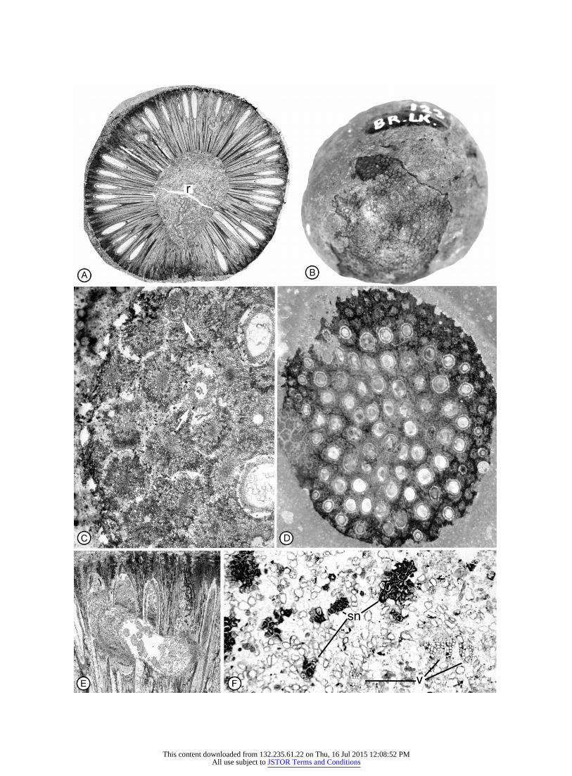
Both longitudinal and transverse sections of several speci-mens show a large number of prominent round or oval struc-tures that occur in the space between the integument and nu-cellus and within the nucellus (fig. 4A, 4C; fig. 5A). These ovalstructures measure 52–131 mm in diameter. They are mostcommon in the apical region where they are often crowded(fig. 4B), but some extend to the base of the seed cavity (figs.4E, 5A). Serial sections reveal that the structures are contin-uous over some distance and are therefore tubelike. A seedsectioned in longitudinal view shows this continuity. Tracingswere made of peels 32–75, and these were superimposed onan outline of the integument and nucellus from peel 43 of thissequence (fig. 5A). Figure 5B consists of tracings from nineconsecutive sections showing continuity of these structuresfrom level to level. When tracings of more sections are added,the continuity of the tubes is further demonstrated, but mor-phology of the tubes is obscured by the overlapping ovals.Tubes are most common from the base of the micropylar canal(figs. 4A, 5A) through the midregion (fig. 5C), and a few extendto the base of the seed cavity (fig. 5E).
Discussion
Nearly all dispersed specimens of anatomically preservedbennettitalean seed cones are thought to represent species ofplants assignable to either Cycadeoidaceae (i.e., species of Cy-cadeoidea and Monanthesia; Wieland 1906, 1916; Delevoryas1959) or Williamsoniaceae (i.e., species of Williamsonia; Sew-ard 1912; Sahni 1932; Delevoryas and Gould 1973; Sharma1977). However, specific criteria used in making generic de-terminations have not been specified in most previous studies.One permineralized cone that could not be correlated with agenus and family of plants has been assigned the morphogenusBennetticarpus Harris (Ohana et al. 1998), along with com-pression remains (Harris 1969), and the compressed Triassicseed cone Vardekloeftia Harris has not been assigned to afamily (Pederson et al. 1989). In an attempt to further clarifythe relationships of isolated bennettitalean seed cones, a con-trasting set of diagnostic characters has recently been devel-oped for relatively complete permineralized specimens, andthese provide criteria for distinguishing Williamsonia (Wil-liamsoniaceae) from Cycadeoidea or Monanthesia (Cycadeo-idaceae; Rothwell and Stockey 2002).
By virtue of having interseminal scales and sporophylls thatdiverge around an arc of ca. 300�, seeds that are circular incross sections at all levels, and sarcotestal cells that form small,
This content downloaded from 132.235.61.22 on Thu, 16 Jul 2015 12:08:52 PMAll use subject to JSTOR Terms and Conditions
258 INTERNATIONAL JOURNAL OF PLANT SCIENCES
Fig. 5 Williamsonia bockii sp. nov. Holotype. VIPM 123. Longi-tudinal sections of one seed reconstructing morphology of pollen tubesfrom serial sections. A, Midlongitudinal section of seed showing rel-ative positions of integument, nucellus, and pollen tubes in one section.Side 43, #19. B, Tracing of integument and nucellus from section infigure 5A (in black), with tracings of pollen tubes from nine consecutivesections (in red), #19.
horizontally elongated pegs, the cone described in this studyis clearly assignable to Williamsonia of the Williamsoniaceae.Contrasting characters of Cycadeoidea and Monanthesia (Cy-cadeoidaceae) include interseminal scales and sporophylls thatdiverge parallel to each other, seeds that are distinctly stellateat the base of the micropylar canal, and sarcotesta made upof axially elongated tubelike cells (table 1 of Rothwell andStockey 2002). Late Cretaceous Williamsonia bockii is by farthe geologically youngest species of permineralized William-soniaceae thus far described (table 1); the other species of seedcones are all from Jurassic strata (table 1). Williamsonia bockii
is also the only species of the genus to display interdigitatingcells of adjacent interseminal scales that form a continuoustissue (table 1). This latter feature results in a unique fusionof adjacent interseminal scales that has not been reported pre-viously for bennettitalean cones.
Like Williamsonia bucklandii (Unger) Saporta and William-sonia diquiyui Delevoryas and Gould, W. bockii is an isolatedspecimen, so its position on the plant is unknown. However,the other species of Williamsonia seed cones either have elon-gated receptacles or are known to terminate branches (Sahni1932; table 1). Bracts are absent from the W. bockii cone (fig.1A, 1B; table 1). This is probably because the bracts abscisedas interpreted by Sharma (1970b) for Williamsonia cones. Al-ternatively, the bracts could have been lost while W. bockiiwas transported into the marine environment before deposi-tion. Like Williamsonia gigas Carruthers, W. bockii has a pyr-iform receptacle, and this contrasts with the conical, hemi-spherical, or dome-shaped receptacles of the other knownspecies (table 1). Whereas W. gigas and W. diquiyui produceonly interseminal scales at the cone apex, W. bockii, W. buck-landii, Williamsonia scotica Seward, Williamsonia sewardianaSahni, and Williamsonia harrisiana Bose have sporophylls inthis region (table 1). Interseminal scales in W. bockii are longerthan those of most Williamsonia species, and they lack a bun-dle sheath like that preserved in W. diquiyui (table 1). Thenumber of interseminal scales surrounding the seeds of W.bockii (i.e., four to five) is the smallest known for Williamsoniaspecies; either the other species have ranges that overlap withW. bockii or the numbers are consistently larger (table 1).
The relative lengths of interseminal scales and sporophyllsvary from species to species in the genus Williamsonia, suchthat the seed micropyles protrude from the cones of some spe-cies, are flush with the tips of the interseminal scales in otherspecies, and are sunken below the tips of the interseminal scalesin still others (table 1). Like W. bucklandii, W. bockii hasslightly protruding seed micropyles. This character may reflectstructural differences among the species, but it could also berelated to pollination biology or to the developmental stage atwhich some cones were preserved (e.g., W. scotica appears tobe either an immature or abortive cone; Seward 1912).
Structure of the Nucellus
The nucellus of W. bockii and other bennettitalean speciesfor which such features are known is attached to the integu-ment only at the base of the ovule and is vascularized by ashallow cup of tracheids at the chalaza. In the micropylarregion, the nucellus tapers to a narrow plug of solid tissue thatextends into the micropylar canal. Such nucellar plugs havebeen figured and/or reported in virtually all species of ben-nettitalean cones for which such data are available. However,this feature has not been widely recognized as characterizingthe clade, and the nucellus has been interpreted differently forWilliamsonia, Bennetticarpus yezoites Ohana, Kimura andChitaley, and Vardekloeftia (Sharma 1976; Pedersen et al.1989; Ohana et al. 1998; see also Rothwell and Stockey 2002).Contrary to the interpretations by Sharma (1976) and Ohanaet al. (1998) for Williamsonia sp. and B. yezoites, respectively,there is no evidence for a pollen chamber in either taxon.Rather, photographs and drawings of these two previously
This content downloaded from 132.235.61.22 on Thu, 16 Jul 2015 12:08:52 PMAll use subject to JSTOR Terms and Conditions
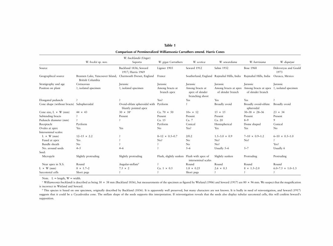
Table 1
Comparison of Permineralized Williamsonia Carruthers emend. Harris Cones
W. bockii sp. nov.W. bucklandii (Unger)
Saporta W. gigas Carruthers W. scotica W. sewardiana W. harrisiana W. diquiyui
Source Buckland 1836; Seward1917; Harris 1969
Lignier 1903 Seward 1912 Sahni 1932 Bose 1968 Delevoryas and Gould1973
Geographical source Brannen Lake, Vancouver Island,British Columbia
Charmouth Dorset, England France Southerland, England Rajmahal Hills, India Rajmahal Hills, India Oaxaca, Mexico
Stratigraphy and age Cretaceous Jurassic Jurassic Jurassic Jurassic Jurassic JurassicPosition on plant ?, isolated specimen ?, isolated specimen Among bracts at
branch apexAmong bracts at
apex of slenderbranching shoot
Among bracts at apexof slender branch
Among bracts at apexof slender branch
?, isolated specimen
Elongated peduncle ? ? Yes? Yes Yes Yes ?Cone shape (without bracts) Subspheroidal Ovoid-oblate spheroidal with
bluntly pointed apexPyriform ? Broadly ovoid Broadly ovoid-oblate
spheroidalBroadly ovoid
Cone size, L # W (mm) 44 # 43 30 # 38a Ca. 70 # 50 10+ # 12 15 # 15 30–38 # 28–36 23 # 34Subtending bracts ? Present Present Present Present Present PresentPeduncle diameter (mm) ? ? Ca. 15 Ca. 7 Ca. 20 8–9 9Receptacle Pyriform Pyriform Conical Hemispherical Dome shaped ConicalOvules at apex Yes Yes No Yes? Yes Yes NoInterseminal scales:
L # W (mm) 12–15 # 2.2 ? 8–12 # 0.5–0.7 2/0.2 1.5–3.0 # 0.9 7–10 # 0.9–1.2 6–10 # 0.5–1.0Fused at apex Yes ? No? No No? No? ?Bundle sheath No ? ? No No? Yes?No. around seeds 4–5 4–6 ? 5–6 Usually 5–6 5–7 Usually 6
Seed:Micropyle Slightly protruding Slightly protruding Flush, slightly sunken Flush with apex of
interseminal scalesSlightly sunken Protruding Protruding
Near apex in X.S. Round Angular-stellateb ? Round Round Round RoundL # W (mm) 8 # 1.7–2 7.5 # 2 Ca. 1 # 0.5 1.8 # 0.25 2.6 # 0.3 8 # 1.5–2.0 6.0–7.5 # 1.0–1.3Sarcotestal cells Short pegs ? ? Short pegs ? ? ?
Note. , .L p length W p widtha Williamsonia bucklandi is described as being 30 # 38 mm (Buckland 1836), but measurements of the specimen as figured by Wieland (1906) and Seward (1917) are 80 # 96 mm. We suspect that the magnification
is incorrect in Wieland and Seward.b This species is based on one specimen, originally described by Buckland (1836). It is apparently well preserved, but many characters are not known. It is badly in need of reinvestigation, and Seward (1917)
suggests that it could be a Cycadeoidea cone. The stellate shape of the seeds supports this interpretation. If reinvestigation reveals that the seeds also display tubular sarcotestal cells, this will confirm Seward’ssupposition.
This content downloaded from 132.235.61.22 on Thu, 16 Jul 2015 12:08:52 PMAll use subject to JSTOR Terms and Conditions

260 INTERNATIONAL JOURNAL OF PLANT SCIENCES
described taxa clearly show a solid nucellar plug in both Wil-liamsonia sp. (i.e., plate 2, fig. 4 of Sharma 1976) and B.yezoites (i.e., figs. 2.4, 2.5, 3.1 of Ohana et al. 1998) that iscomparable to that of W. bockii and other permineralized ben-nettitalean seeds (e.g., Cycadeoidea maccafferyi; Rothwell andStockey 2002).
The apical region of the nucellus is more difficult to interpretfrom the compressed cuticular remains of Vardekloeftia sulcataHarris. Pederson et al. (1989) illustrate carefully prepared mac-erations of V. sulcata that show several distinct layers of cu-ticular envelopes that are interpreted by those authors to rep-resent nucellus, integument, and cupule (fig. 2 of Pederson etal. 1989). By comparison to other gymnospermous seeds thattypically display a pollen chamber, this interpretation is quitereasonable. However, by comparison to permineralized ben-nettitalean seeds that lack a pollen chamber, an alternativeinterpretation now appears to be more probable (Rothwell andStockey 2002). The more recent interpretation regards the cu-ticular envelopes of V. sulcata as representing a nucellar plugsurrounded by integumentary cuticles (Rothwell and Stockey2002). Photographs of V. sulcata cuticles (i.e., plate III, 6; plateV, 3; plate V, 4 of Pederson et al. 1989) clearly show a centrallylocated, solid-appearing, elongated structure that is structur-ally indistinguishable from the nucellar plug of permineralizedbennettitalean seeds (Crepet and Delevoryas 1972; Rothwelland Stockey 2002; fig. 3A, 3C; fig. 4A), thus suggesting thatan apical nucellar plug may be a synapomorphy forBennettitales.
Pollen Tubes
The circular to oval structures that occur in peel sections ofseveral W. bockii seeds (figs. 4A–4C, 5A) are essentially iden-tical to anatomical sections of pollen tubes produced by bothliving and fossil araucarian conifers (Eames 1913; Stockey etal. 1994; Owens et al. 1995a, 1995b). In the araucarians,pollen tube growth is invasive, extending both through thetissues of the nucellus and between the nucellus and integument(Thomson 1907; Eames 1913; Haines 1981; Owens et al.1995a, 1995b). This corresponds precisely with the positionsand sizes of the structures in W. bockii (figs. 3A, 4A–4C, 5A).In addition, the tubelike nature of the structures in W. bockiihas been confirmed from serial sections of peels (e.g., fig. 5B),revealing an overall form and disposition within the seeds thatare comparable to the pollen tubes of the living araucarians(Eames 1913; Owens et al. 1995a, 1995b).
While the preservation of pollen tubes in fossil seeds orovules is rare, two previous examples have been documented.These include the Paleozoic seed fern Callistophyton (i.e., inan ovule of Callospermarion Eggert and Delevoryas; Rothwell1972) and the Jurassic conifer Araucaria nipponensis (Stockeyet al. 1994), thus demonstrating the potential for similar pres-ervation in W. bockii. While one might suspect that the struc-tures in W. bockii are either remnants of the nucellus or fungalhyphae, these alternatives are far less probable. The size of thetubes in W. bockii is similar to the comparable structures inthe living araucarians (i.e., 50–120 mm in W. bockii; Eames1913; Owens et al. 1995a, 1995b) and much larger thanknown for fungal hyphae. Also, there is no evidence of fungi
elsewhere in the cone. The possibility that the structures in W.bockii represent some form of nucellar proliferation as seenin extant Araucaria angustifolia (Burlingame 1914) and fossilAraucaria mirabilis (Stockey 1978) also has been considered.Such nucellar proliferation is known to occur in aborted ovulesand consists of a dense tissue of small cells rather than thelarge tubes found in W. bockii.
Pollination and Fertilization Biology
Both outcrossing via wind pollination and self-pollinationby an insect vector (possibly a beetle) have been proposed formembers of the Cycadeoidaceae (Delevoryas 1968; Crepet1972, 1974), but the position of the grains following polli-nation, postpollination microgametophyte development, andmode of fertilization have not been addressed for species ofthe Bennettitales. Pollination in Williamsonia may have beenby wind, since the cones that have been found attached occuron slender branches without tightly adhering microsporangiatestructures. Moreover, bracts that surround the cones at earlystages of development were apparently abscised during conedevelopment (Sharma 1970b). This contrasts with the axillaryreproductive structures of Monanthesia or Cycadeoidea, manyof which are known to be deeply embedded among the leafbases near the trunk of the plant (Wieland 1906). Some conesof the Cycadeoidaceae also have seeds that are tightly enclosedby both microsporangiate structures and bracts (Delevoryas1968), and this has led Crepet (1972, 1974) to hypothesizethat such species may have been pollinated by insects.
The absence of microsporangiate structures from William-sonia seed cones bolsters hypotheses of outcrossing and sup-ports suggestions of wind pollination for the genus. Seward(1912) describes a slightly funnel-shaped apex to the micro-pylar region of the seed in W. scotica, where the micropyle isflush with the interseminal scales (table 1). Sahni (1932) re-ports a similar seed apex in W. sewardiana where the micropyleis slightly sunken. Both of these morphologies are consistentwith outcrossing by wind pollination. We have been unableto see a funnel-shaped apex in W. bockii in which the micro-pylar ends of seeds are slightly protruding, but this cone mayhave undergone some abrasion before deposition. AlthoughW. bockii does have coprolite-filled galleries somewhat similarto those of cones in which insect pollination has been hy-pothesized for Cycadeoidea (Crepet 1972, 1974), the galleriesin W. bockii are located in positions that are more indicativeof destructive herbivory than pollination biology (i.e., throughthe sporophylls and the basal region of the seeds; fig. 1E).
Pollen grains are almost always absent from the interior ofBennettitalean seeds, even when embryos are present (Wieland1906; Rothwell and Stockey 2002). However, grains do occurin a Middle Jurassic specimen of Williamsonia sp. that wasprobably immature (Sharma 1977). This pollen is preservedin the most apical sections of the micropylar canal, distal tothe apex of the nucellar plug. At this level, elongated epidermalcells line the micropylar tube, suggesting to us that they hada secretory function. Grains also occur within seed cuticles ofV. sulcata (Pedersen et al. 1989). They are located betweenwhat we interpret to be the nucellar plug and the inner integ-umentary cuticle (i.e., plate V of Pedersen et al. 1989; Rothwell
This content downloaded from 132.235.61.22 on Thu, 16 Jul 2015 12:08:52 PMAll use subject to JSTOR Terms and Conditions

STOCKEY & ROTHWELL—WILLIAMSONIA BOCKII SP. NOV. 261
and Stockey 2002). However, whether these grains have fil-tered down between the integument and nucellar plug duringfossilization and/or preparation (as interpreted by us) or werelocated within a pollen chamber (as interpreted by Pedersonet al. 1989) cannot be determined from available specimens.New preparations of Vardekloeftia seed cuticles may help clar-ify this point.
As stressed above, pollen grains have not been reported inthe seed cavities of most bennettitaleans. This correlates wellwith the lack of a pollen chamber from bennettitalean ovulesbut does not explain the mode of fertilization biology in thisclade. However, the numerous pollen tubes that extend fromthe base of the micropylar plug to the chalaza of several W.bockii seeds may provide new insights into bennettitalean mi-crogametophyte development and fertilization biology. Thepollen tubes in W. bockii are virtually identical to those ofliving Agathis australis and other araucarian conifers in mor-phology, size, and distribution within the seed cavity (fig. 5;Eames 1913; Stockey et al. 1994; Owens et al. 1995a, 1995b).In both the Araucariaceae and W. bockii, pollen tubes occurbetween the integument and nucellus and within the nucellusfrom the base of the micropylar canal to the chalaza, but theyare most numerous at the micropylar end of the ovules (figs.5–9, 10 of Eames 1913; fig. 29 of Owens et al. 1995a; fig. 6of Owens et al. 1995b). In araucarian conifers, this distinctivepollen tube structure is correlated with a unique pollinationmechanism in which there is no pollen chamber, and the pollengrains do not enter the seed cavity (Berg 1950). Instead, thepollen grains land on the cone scale, and pollen tubes growinto the seed cavity through nucellar tissue that extends outof the micropyle.
Bennettitalean seeds are not borne on cone scales and donot have the tip of the nucellus that extends outside the mi-cropyle. However, the combination of pollination that placesgrains distal to the nucellar plug in the micropylar canal ofone species of Williamsonia (Sharma 1970a) and pollen tubeswith distinctly araucarian morphology in W. bockii suggeststhat pollination brought grains into only the distalmost partof the micropylar canal in most Bennettitalean ovules. If pol-lination of this type were followed by pollen tube growth pastthe nucellar plug and into the seed cavity, this would accountboth for the paucity of grains found within seed cavities ofBennettitalean seeds and for the distinctive pollen tube mor-phology that is identical for araucarian conifers and W. bockii.As in most living gymnospermous species, the pollen tube ofW. bockii probably developed within the seed cavity for sometime before fertilization took place. Growth of the megaga-metophyte and embryo probably pressed the nucellar plugtightly into the micropylar tube after the tube had grown intothe ovule, thus achieving postpollination sealing of the pollenchamber and explaining the absence of pollen grains in theseed cavity of most species.
Among living seed plants, pollen tube structure of araucar-ian conifers is extremely distinctive and is diagnostic of mi-crogametophytes that produce a haustorial pollen tube thatgrows extensively before facilitating siphonogamous fertiliza-tion (Eames 1913; Owens et al. 1995a, 1995b, 1997). If form/function relationships are comparable in the living araucarianconifers and W. bockii, then at least one species of Bennettitaleshad siphonogamous pollination from highly branched micro-
gametophytes with pollen tubes that also had a haustorial func-tion. The absence of a pollen chamber and the paucity of pollengrains from bennettitalean seeds (as interpreted by us) indicatesthat such a pollination/fertilization mechanism may charac-terize reproduction in Bennettitales as a whole. If, however,Vardekloeftia had a pollen chamber (as interpreted by Pedersenet al. 1989), then the distinctive pollination biology of Cy-cadeoidaceae and Williamsoniaceae may have evolved withinthe benettitalean clade.
The preservation of megagametophyte and embryo tissuesis relatively common in the seeds of Williamsonia and othergenera of permineralized bennettitaleans, where dicotyledon-ary embryos with fully developed and apparently quiescentembryos are commonly present (Lignier 1894; Seward 1917;Sharma 1977; Rothwell and Stockey 2002). By contrast, thesmall, undifferentiated embryos of W. bockii suggest that pres-ervation occurred closer to the time of pollination than to seeddispersal, and this accounts for the well-preserved pollen tubesin this cone.
Summary of Bennettitalean Reproduction
If the absence of a pollen chamber, the presence of a solidnucellar plug, highly branched pollen tubes, and siphonoga-mous fertilization are characteristic of Bennettitales as a whole,then these features can be added to the large body of dataabout seed development, pollen, pollination, megagameto-phytes, postpollination sealing of the seeds, and embryos thathas accumulated over the past 175 yr (e.g., Carruthers 1870;Lignier 1894; Seward 1917; Crepet and Delevoryas 1972; Tay-lor 1973; Crepet 1974; Rothwell and Stockey 2002) to providethe most complete understanding of reproductive biology yetachieved for an extinct clade of seed plants. As with livingplants, bennettitaleans underwent meiosis to produce a lineartetrad when the ovules were extremely small, and the tip ofthe nucellus was a solid mound of tissue (Crepet and Dele-voryas 1972).
Some bennettitalean species were apparently inbreeding,with bisporangiate cones and pollination effected by an insectvector. Others, including W. bockii, had monosporangiatecones and appear to have been outcrossing and wind polli-nated. In some species, pollination was apparently aided bymicropyles that protruded beyond the tips of the interseminalscales, and in one species, the pollinated ovules were retractedbetween the interseminal scales by a contractile mechanismsimilar to that of some roots (Rothwell and Stockey 2002).Pollination placed the microgametophytes into the tip of themicropylar canal, and the pollen tubes grew into the seed cav-ity. Postpollination sealing of the seeds was effected by a solidplug of tissue that occurs in the position where a pollen cham-ber is found in most other groups of nonflowering seed plants.
Megagametophyte tissue became cellular after filling theseed cavity, and archegonia formed toward the micropylar end.Embryogeny led to the production of dicotyledonous embryosthat are common to many species. The predominance of theseembryos, all with a comparable and apparently mature struc-ture, reveals the occurrence of postzygotic quiescence (Mapeset al. 1989) in the Bennettitales and suggests that seed dor-mancy mechanisms were well developed in the clade. This
This content downloaded from 132.235.61.22 on Thu, 16 Jul 2015 12:08:52 PMAll use subject to JSTOR Terms and Conditions

262 INTERNATIONAL JOURNAL OF PLANT SCIENCES
highly sophisticated mode of reproductive biology undoubt-edly contributed significantly to Bennettitales being among the
most widely distributed and successful clades of Mesozoic seedplants.
Literature Cited
Alvin KL, PDW Barnard, TM Harris, NF Hughes, RH Wagner, AWesley 1967 Gymnospermopsida. Pages 247–267 in WB Harland,et al., eds. The fossil record. Geological Society Special Publication,no. 2. Geological Society, London.
Arber EAN, J Parkin 1907 On the origin of angiosperms. J Linn SocLond Bot 38:29–80.
Berg DH 1950 The morphology of the gametophytes, fertilizationand proembryo of Araucaria bidwillii (Hook.). PhD diss. CornellUniversity, Ithaca, N.Y.
Bose MN 1968 A new species of Williamsonia from the RajmahalHills, India. J Linn Soc Lond Bot 61:121–127.
Buckland W 1836 Geology and mineralogy considered with referenceto natural theology. Bridgewater Treatises. No 2, 2 vols. London.
Burlingame LL 1914 The morphology of Araucaria brasiliensis. II.The ovulate cone and female gametophyte. Bot Gaz 57:490–508.
Carruthers W 1870 On fossil cycadean stems from the secondaryrocks of Britain. Trans Linn Soc Lond 26:675–708 (plates 54–63).
Crane PR 1985 Phylogenetic analysis of seed plants and the origin ofangiosperms. Ann Mo Bot Gard 72:716–793.
Crepet WL 1972 Investigations of North American cycadeoids: pol-lination mechanisms in Cycadeoidea. Am J Bot 59:1048–1056.
——— 1974 Investigations of North American cycadeoids: the re-productive biology of Cycadeoidea. Palaeontographica 148B:144–169.
Crepet WL, T Delevoryas 1972 Investigations of North American cy-cadeoids: early ovule ontogeny. Am J Bot 59:209–215.
Delevoryas T 1959 Investigations of North American cycadeoids:Monanthesia. Am J Bot 46:657–666.
——— 1968 Investigations of North American cycadeoids: structure,ontogeny and phylogenetic considerations of cones of Cycadeoidea.Palaeontographica Abteilung B Palaeophytologie 121B:122–133.
Delevoryas T, RE Gould 1973 Investigations of North American cy-cadeoids: Williamsonian cones from the Jurassic of Oaxaca, Mexico.Rev Palaeobot Palynol 15:27–42.
Eames AJ 1913 The morphology of Agathis australis. Ann Bot 27:1–38.
Haines RJ 1981 The embryology of Araucaria Juss. PhD thesis. Uni-versity of New England, Armidale, New South Wales.
Harris TM 1932 The fossil flora of Scoresby Sound, east Greenland.3. Caytoniales and Bennettitales. Medd Gronl 112:1–114.
——— 1969 The Yorkshire Jurassic Flora. III. Bennettitales. Br MusNat Hist Lond 675:1–186.
Joy KW, AJ Willis, WS Lacey 1956 A rapid cellulose peel techniquein paleobotany. Ann Bot, NS, 20:635–637.
Lignier O 1894 Vegetaux fossiles de Normandie, structure et affinitesdu Bennettites morierei Sap. & Mar. (Sp.). Imprimerie E. Lanier,Caen, France.
——— 1903 Le fruit du Williamsonia gigas Carr. et les Bennettitales.Documents nouveaux et notes critiques. Mem Soc Linn Normandie21:19–56.
Ludvigsen R, G Beard 1997 West coast fossils. Harbour Publishing,Madeira Park, British Columbia.
Mapes G, GW Rothwell, MT Howarth 1989 Evolution of seed dor-mancy. Nature 337:645–646.
Ohana T, T Kimura, S Chitaley 1998 Bennetticarpus yezoites sp. nov.
(Bennettitales) from the Upper Cretaceous of Hokkaido, Japan. Pa-leontol Res 2:108–119.
Owens JN, GL Catalano, SJ Morris, J Aitken-Christie 1995a Thereproductive biology of kauri (Agathis australis). I. Pollination andprefertilization development. Int J Plant Sci 156:257–269.
——— 1995b The reproductive biology of kauri (Agathis australis).II. Male gametes, fertilization, and cytoplasmic inheritance. Int JPlant Sci 156:404–416.
——— 1997 The reproductive biology of kauri (Agathis australis).IV. Late embryogeny, histochemistry, cone, and seed morphology.Int J Plant Sci 158:395–407.
Pedersen KR, PR Crane, EM Friis 1989 The morphology and phy-logenetic significance of Vardekloeftia Harris (Bennettitales). RevPalaeobot Palynol 60:7–24.
Rothwell GW 1972 Evidence of pollen tubes in Paleozoic pteridos-perms. Science 175:772–774.
Rothwell GW, RA Stockey 2002 Anatomically preserved Cycadeo-idea (Cycadeoidaceae), with a reevaluation of systematic charactersfor the seed cones of Bennettitales. Am J Bot 89:1447–1458.
Sahni B 1932 A petrified Williamsonia (W. sewardiana, sp. nov.) fromthe Rajmahal Hills, India. Mem Geol Surv India, PalaeontologiaIndica, NS, 20:1–19.
Seward AC 1912 A petrified Williamsonia from Scotland. PhilosTrans R Soc Lond B Biol Sci 203:101–126.
——— 1917 Fossil plants. Vol 3. Cambridge University Press,London.
Sharma BD 1970a On the structure of a Williamsonia cf. W. scoticafrom the Middle Jurassic rocks of the Rajmahal Hills, India. AnnBot, NS, 34:289–296.
——— 1970b On the vascular organization of the receptacles of seed-bearing Williamsonias from the Middle Jurassic rocks of Amarjolain the Rajmahal Hills, India. Ann Bot, NS, 34:1036–1070.
——— 1976 Fruit development in Williamsonia Carr. (Bennettitales).Geobios 9:503–507.
——— 1977 Indian Williamsonias: an illustrated review. Acta Pa-laeobot 18:19–29.
Smith SY, RA Stockey 2002 Permineralized pine cones from the Cre-taceous of Vancouver Island, British Columbia. Int J Plant Sci 163:185–196.
Stockey RA 1978 Reproductive biology of Cerro Cuadrado fossilconifers: ontogeny and reproductive strategies in Araucaria mirabilis(Spegazzini) Windhausen. Palaeontogr B 166:1–15.
Stockey RA, H Nishida, M Nishida 1994 Upper Cretaceous arau-carian cones from Hokkaido and Saghalien: Araucaria nipponensissp. nov. Int J Plant Sci 155:806–815.
Taylor TN 1973 A consideration of the morphology, ultrastructureand multicellular microgametophyte of Cycadeoidea dacotensis pol-len. Rev Palaeobot Palynol 16:157–164.
Thomson RB 1907 The Araucarieae: a “proto-siphongamic” methodof fertilization. Science, NS, 25:271–272.
Watson J, CA Sincock 1992 Bennettitales of the English Wealden.Monogr Palaeontogr Soc Lond 588:1–228.
Wieland GR 1906 American fossil cycads. Carnegie Inst WashingtonPubl 34:1–284.
——— 1916 American fossil cycads. 2. Taxonomy. Carnegie InstWashington Publ 34I:1–267.
This content downloaded from 132.235.61.22 on Thu, 16 Jul 2015 12:08:52 PMAll use subject to JSTOR Terms and Conditions