Attention orienting dysfunction with preserved automatic auditory change detection in migraine
12
Attention orienting dysfunction with preserved automatic auditory change detection in migraine Dominique Morlet a,b,c,⇑ , Geneviève Demarquay d , Frédérique Brudon e , Catherine Fischer a,b,c,f , Anne Caclin a,b,c a INSERM, U1028, Lyon Neuroscience Research Center, Brain Dynamics and Cognition Team, Lyon F-69000, France b CNRS, UMR5292, Lyon Neuroscience Research Center, Brain Dynamics and Cognition Team, Lyon F-69000, France c Université Lyon 1, Lyon F-69000, France d Hospices Civils de Lyon, Croix-Rousse Hospital, Neurology Department, Lyon F-69000, France e Tonkin Clinic, Villeurbanne F-69100, France f Hospices Civils de Lyon, Neurological Hospital, Functional Neurology and Epileptology Department, Lyon F-69000, France See Editorial, pages 433–434 article info Article history: Available online 8 November 2013 Keywords: Migraine Event-related potentials Attention orienting MMN N2b P3a highlights The mismatch negativity (MMN) is normal in migraine patients. The N1 orienting component and the N2b are increased in migraine patients. Auditory processes up to attention triggering are preserved in migraine patients. Attention orienting to sound income and sound deviance are exacerbated in migraine. These observations suggest abnormal activation of attention-related frontal networks. abstract Objective: To investigate automatic event-related potentials (ERPs) to an auditory change in migraine patients. Methods: Auditory ERPs were recorded in 22 female patients suffering from menstrually-related migraine and in 20 age-matched control subjects, in three sessions: in the middle of the menstrual cycle, before and during menses. In each session, 200 trains of tone-bursts each including two duration deviants were presented in a passive listening condition. Results: In all sessions, duration deviance elicited a mismatch negativity (MMN) showing no difference between the two groups. However, migraine patients showed an increased N1 orienting component to all incoming stimuli and a prolonged N2b to deviance. They also presented a different modulation of P3a amplitude along the menstrual cycle, which tended to normalise during migraine attacks. None of the studied ERP components showed a default of habituation. Conclusions: This passive paradigm highlighted increased automatic attention orienting to auditory changes but normal auditory sensory processing in migraineurs. Significance: Our observations suggest normal auditory processing up to attention triggering but enhanced activation of attention-related frontal networks in migraineurs. Ó 2013 International Federation of Clinical Neurophysiology. Published by Elsevier Ireland Ltd. All rights reserved. 1. Introduction Migraine is one of the most common headache disorders and affects 11% of the adult population (Stovner et al., 2007). Attacks are characterised by recurrent throbbing headaches accompanied by nausea, vomiting, photophobia, and/or phonophobia and are aggravated by movements (ICHD, 2004). Between attacks, 1388-2457/$36.00 Ó 2013 International Federation of Clinical Neurophysiology. Published by Elsevier Ireland Ltd. All rights reserved. http://dx.doi.org/10.1016/j.clinph.2013.05.032 ⇑ Corresponding author. Address: Lyon Neuroscience Research Center, Dycog Team, 69675 Bron Cedex, France. Tel.: +33 (0) 4 72 13 89 03; fax: +33 (0) 4 72 13 89 01. E-mail address: [email protected] (D. Morlet). Clinical Neurophysiology 125 (2014) 500–511 Contents lists available at ScienceDirect Clinical Neurophysiology journal homepage: www.elsevier.com/locate/clinph
-
Upload
independent -
Category
Documents
-
view
0 -
download
0
Transcript of Attention orienting dysfunction with preserved automatic auditory change detection in migraine

Clinical Neurophysiology 125 (2014) 500–511
Contents lists available at ScienceDirect
Clinical Neurophysiology
journal homepage: www.elsevier .com/locate /c l inph
Attention orienting dysfunction with preserved automatic auditorychange detection in migraine
1388-2457/$36.00 � 2013 International Federation of Clinical Neurophysiology. Published by Elsevier Ireland Ltd. All rights reserved.http://dx.doi.org/10.1016/j.clinph.2013.05.032
⇑ Corresponding author. Address: Lyon Neuroscience Research Center, DycogTeam, 69675 Bron Cedex, France. Tel.: +33 (0) 4 72 13 89 03; fax: +33 (0) 4 72 13 8901.
E-mail address: [email protected] (D. Morlet).
Dominique Morlet a,b,c,⇑, Geneviève Demarquay d, Frédérique Brudon e, Catherine Fischer a,b,c,f,Anne Caclin a,b,c
a INSERM, U1028, Lyon Neuroscience Research Center, Brain Dynamics and Cognition Team, Lyon F-69000, Franceb CNRS, UMR5292, Lyon Neuroscience Research Center, Brain Dynamics and Cognition Team, Lyon F-69000, Francec Université Lyon 1, Lyon F-69000, Franced Hospices Civils de Lyon, Croix-Rousse Hospital, Neurology Department, Lyon F-69000, Francee Tonkin Clinic, Villeurbanne F-69100, Francef Hospices Civils de Lyon, Neurological Hospital, Functional Neurology and Epileptology Department, Lyon F-69000, France
See Editorial, pages 433–434
a r t i c l e i n f o h i g h l i g h t s
Article history:Available online 8 November 2013
Keywords:MigraineEvent-related potentialsAttention orientingMMNN2bP3a
� The mismatch negativity (MMN) is normal in migraine patients.� The N1 orienting component and the N2b are increased in migraine patients.� Auditory processes up to attention triggering are preserved in migraine patients.� Attention orienting to sound income and sound deviance are exacerbated in migraine.� These observations suggest abnormal activation of attention-related frontal networks.
a b s t r a c t
Objective: To investigate automatic event-related potentials (ERPs) to an auditory change in migrainepatients.Methods: Auditory ERPs were recorded in 22 female patients suffering from menstrually-relatedmigraine and in 20 age-matched control subjects, in three sessions: in the middle of the menstrual cycle,before and during menses. In each session, 200 trains of tone-bursts each including two duration deviantswere presented in a passive listening condition.Results: In all sessions, duration deviance elicited a mismatch negativity (MMN) showing no differencebetween the two groups. However, migraine patients showed an increased N1 orienting component toall incoming stimuli and a prolonged N2b to deviance. They also presented a different modulation ofP3a amplitude along the menstrual cycle, which tended to normalise during migraine attacks. None ofthe studied ERP components showed a default of habituation.Conclusions: This passive paradigm highlighted increased automatic attention orienting to auditorychanges but normal auditory sensory processing in migraineurs.Significance: Our observations suggest normal auditory processing up to attention triggering butenhanced activation of attention-related frontal networks in migraineurs.� 2013 International Federation of Clinical Neurophysiology. Published by Elsevier Ireland Ltd. All rights
reserved.
1. Introduction
Migraine is one of the most common headache disorders andaffects 11% of the adult population (Stovner et al., 2007). Attacksare characterised by recurrent throbbing headaches accompaniedby nausea, vomiting, photophobia, and/or phonophobia and areaggravated by movements (ICHD, 2004). Between attacks,

D. Morlet et al. / Clinical Neurophysiology 125 (2014) 500–511 501
migraine patients may also show a hypersensitivity to visual, audi-tory, or olfactory stimuli (Main et al., 1997, 2000). Moreover,sound/light-induced headaches are frequent in migraine patients(Vingen et al., 1999; ICHD, 2004). The pathophysiological basis ofsuch hypersensitivity to external stimuli is currently not com-pletely elucidated.
In line with these symptoms, electrophysiological studies haverevealed changes in cortical excitability between migraine attacks.Increased attention orienting to new incoming stimuli had beenlong ago demonstrated in migraine patients. The initial (or early)contingent negative variation (iCNV), which reflects the orientingproperties of a warning stimulus in an active paradigm, has beenfound to be increased in migraine (Maertens de Noordhout et al.,1986), preferentially just before an attack (Kropp and Gerber,1998), but not systematically (Böcker et al., 1990; Mulder et al.,2001). A default in the habituation of several event-related poten-tials (ERPs) to visual and auditory stimuli has also been observed inmigraine patients in numerous studies (for a review, see Coppolaet al., 2009), although not systematically (Oelkers et al., 1999; Sandand Vanagaite, 2000; Omland et al., 2013). Using an auditory habit-uation paradigm made of small consecutive trains of tones (Woodsand Elmasian, 1986), we also found normal habituation pattern ofthe sensory N1 in migraineurs (Demarquay et al., 2011). Interest-ingly, this passive paradigm allowed to investigate the automaticresponse to the first stimuli of stimulation trains, and revealed thatmigraine patients exhibited a drastically augmented N1 orientingcomponent, a fronto-central negative component appearing afterthe obligatory sensory N1 when the inter-stimulus interval isgreater than 4 s (Näätänen and Picton, 1987; Alcaini et al., 1994).The response to standard stimuli inside the trains also exhibitedenhanced negative potentials in the descending slope of the N1wave. By comparison with the augmented N1 orienting componentobserved in response to the first stimuli of the trains, we called thisadditional component ‘‘residual orienting component’’ and weconcluded that migraine patients showed exacerbated attentionorienting not only to first stimuli after a silent gap but also to re-peated similar incoming stimuli.
The electrophysiological studies described above showedabnormal responses not only to attended warning stimuli but alsoto passively endured stimuli. They emphasized attention-orientingexacerbation and/or habituation deficit in migraine without clearlydisentangling basic auditory processing dysfunction and abnormalorienting processes. The use of passive oddball paradigms is of par-ticular interest here because it allows studying both automaticsensory responses to auditory changes and automatic attentiontriggering. Indeed, in those paradigms, a rare and random changein repetitive stimulation elicits a negative response (mismatchnegativity or MMN) disclosed when the response to the standardis subtracted from the response to the deviant. MMN is attributedto discriminative processes using memory traces developed fromthe previous stimulation (review in Näätänen et al., 2001, 2007,2011). It is noteworthy that the memory traces probed by theMMN are thought to reflect the outcome of perceptual processing(Näätänen and Winkler, 1999; Näätänen et al., 2011) and furtherthat MMN occurs automatically, even if subject’s attention is notdirected to the sounds (Näätänen et al., 1993). Nevertheless, ifdeviant stimuli are salient enough, attention-orienting processescan be triggered, and an N2b–P3a complex (brain orienting re-sponse) is further obtained following the MMN (Näätänen et al.,1982). In contrast, presenting a second consecutive deviant drasti-cally diminishes the MMN (and subsequent orienting responses),which has been called ‘‘short-term habituation of the MMN’’ (Samset al., 1984). This phenomenon was explained by the simultaneousoccurrence, for the repeated deviant, of mismatch processes withthe (declining) neuronal model of the standard and match pro-cesses with the neuronal model of the deviant. Altogether, the
study of ERPs triggered by a rare change in auditory stimulationthus offers the advantage to assess, in a passive listening situation,the integrity of pre-attentive stages of perceptual processing, as in-dexed by the MMN, as well as the automatic triggering of atten-tion, indexed by the N2b–P3a complex. For these reasons, passiveauditory oddball paradigms have been used in a very large numberof ERP studies in various pathologies (review in Näätänen, 2003;Näätänen et al., 2012), but surprisingly very rarely in migraine.
Indeed, most of the oddball paradigm studies in migraine haverequired an active detection of deviant tones and they mainlyfocused on the P300 in response to targets, which was found tobe altered in migraine patients (Drake et al., 1989; Mazzottaet al., 1995; Wang et al., 1995). The investigation of brain mecha-nisms to a stimulus change during passive listening has been rarelyreported in migraine patients. Only one study investigated theMMN in adult migraineurs and reported increased N1 and MMNlatencies, suggesting a hypo-activity of automatic cortical pro-cesses (de Tommaso et al., 2004). Two recent paediatric migrainestudies also pointed to subtle MMN alterations in these patients(Valeriani et al., 2009; Korostenskaja et al., 2011). One study (Wangand Schoenen, 1998) explored the amplitude of an N2–P3acomplex in response to deviant stimuli and reported for migrainepatients a potentiation of this response in successive blocks, con-trasting with a habituation in controls.
The aim of the present study was thus to investigate the specificresponse to deviant stimuli in migraine patients, from the pre-attentive processes (MMN) to the attention-orienting processes(N2b, P3a), and to assess the habituation of the different compo-nents. Abnormalities of the MMN would suggest dysfunction inbasic auditory processing of migraine patients. In light of our pre-vious results, we expected a pathological brain orienting responseto deviants.
2. Methods
The present analysis is the second part of a larger auditory ERPstudy conducted in migraineurs using small trains of repeatedstandard tones with occasional deviant stimuli. The first part ofthe analysis focused on the responses to standards and their habit-uation patterns (Demarquay et al., 2011). Here we explore the re-sponse to deviant stimuli.
2.1. Migraine patients and healthy subjects
Twenty-two female migraine patients suffering from menstrua-lly-related migraine without aura (A1.1.2 in ICHD 2004) were in-cluded in the study (mean age ± SD: 27 years ± 7; diseaseduration: 12 years ± 8; attack frequency: 2 ± 1 per month). Mi-graine patients and controls were recruited through advertisingin Lyon University and INSERM administration. A neurologist (CF,FB or GD) subsequently examined eligible subjects.
Twenty age-matched female controls participated in the study(28 years ± 9). Exclusion criteria for all subjects included chronicdaily headache, known morphological brain abnormality, currentsubstance abuse, and migraine preventive medication. 14 migrainepatients and 11 controls took oral contraceptives (v2 = 0.0649,p = 0.799).
All subjects gave their written informed consent (CCPPRB centreLéon-Bérard, Lyon, A 06-107, 04/20/06) and were remunerated fortheir contribution.
2.2. Recording procedure
The recording procedure has been described in detail previously(Demarquay et al., 2011).

502 D. Morlet et al. / Clinical Neurophysiology 125 (2014) 500–511
Patients and healthy subjects were recorded in 3 different ses-sions, in the middle of the menstrual cycle (session 0, S0, day15 ± 4 of the on-going cycle), before menses (session 1, S1, day�1 ± 1 of the cycle), and at the beginning of the menses (session2, S2, day 1 ± 1 of the cycle). As a general rule, S2 immediately fol-lowed S1 (mean delay = 2 days ± 1), except for 1 healthy subjectand for 3 patients, for whom S1 and S2 were in separate cycles.Session 0 randomly preceded or followed S1 and S2. S0 was beforeS1 in 12/22 patients and in 13/20 healthy subjects (v2 = 0.923,p = 0.337).
Among the 22 migraineurs, ERPs were recorded during amigraine attack in 12 patients (session 1 for 5 patients; session 2for 6 patients; session 0 for 1 patient). In all patients, the attack oc-curred before the onset of the ERP recording and lasted until theend; medication was given at the end of recording session.
During the recording sessions, the subject sat in a comfortablearmchair in a quiet room. She was instructed to watch a silent mo-vie of her choice and not to pay attention to the auditory stimuli.
EEG was recorded continuously from 7 scalp electrodes placedat frontal (Fz, F3, F4), central (Cz), and parietal (Pz) sites, and atthe two mastoids (M1, M2). The reference electrode was placedon the tip of the nose, the ground electrode on the forehead. Onebipolar EOG derivation was recorded from 2 electrodes placed onthe supra-orbital and infra-orbital ridges of the right eye. Using aMicromed System98 EEG recording system, the signal was ampli-fied (band-pass 0.3–100 Hz), digitised (sampling frequency1024 Hz) and stored for off-line analysis. One stimulation sessionlasted about 45 min without any interruption.
2.3. Auditory paradigm
The stimuli were delivered binaurally through insert earphonesat an intensity of 65 dB HL.
In each recording session, 200 trains of auditory stimuli werepresented with an average of 10 stimuli per train (random numberfrom 8 to 12). The inter-train interval ranged between 5.5 and 8.0 s(mean value 6.75 s). Within trains, stimulus onset asynchrony was610 ms.
The stimuli were spectrally rich tone bursts (fundamental fre-quency 800 Hz). The more frequent stimuli (standards) had aduration of 75 ms, including 5-ms rise and fall times. Durationdeviants (duration 30 ms) were used, because duration changes(with deviants shorter than standards) are known to providerobust and reproducible MMNs (Tervaniemi et al., 1999). Twodeviant tones were pseudo-randomly included in each train.The first deviant was randomly presented after 3–9 consecutivestandards. In each recording session, one hundred trains (50% ofthe trains) obeyed the rule routinely used in oddball paradigms,namely at least 2 standards were presented between the first
Fig. 1. Design and timing of the auditory stimulation paradigm. In each recording sessranging between 5.5 and 8 s. Within-train stimulus onset asynchrony (SOA) is 610 ms. Ttrain. In 50% of the trains, at least 2 standards are presented between the first and the setrains, the second deviant immediately follows the first one (repeated deviant, as in the sdeviants (colored in grey). ‘‘Generic standards’’ (colored in black) exclude the first 3 stim
and the second deviant. In the other half of the trains, the seconddeviant appeared immediately after the first one (repeated devi-ant). One recording session thus included 300 ‘‘generic’’ deviantsappearing after at least 2 standards (200 generic deviants beingfirst deviants and 100 generic deviants being second deviants)and 100 ‘‘repeated deviants’’ appearing immediately after an-other deviant. Fig. 1 illustrates the design and the timing of audi-tory stimulation.
Additionally, 20% of the trains ended with a salient stimulus(environmental sound). Evoked responses for these novel stimuliare beyond the scope of the present report.
2.4. Event-related potentials
The software package for electrophysiological analysis (ELAN)developed at the Lyon Neuroscience Research Center (Agueraet al., 2011) was used for ERP analysis. Responses to standardsand to deviants were considered for averaging for an epoch of700 ms including a pre-stimulus period of 100 ms. Epochs showingpeak-to-peak deflections larger than ±100 lV were rejected. Onepatient showed a large stimulation artifact at the mastoids 15 msafter stimulus onset in the 3 recording sessions. For this patient,an independent component analysis (ICA) was used in order to iso-late and remove this unwanted component.
The ERPs were baseline-corrected by subtracting the meanvalue of the signal during the 100 ms prior to the stimulus. A30-Hz low-pass digital filter (bidirectional Butterworth, 6th order)and a 2-Hz high-pass filter (bidirectional Butterworth, 2nd order)were applied to the averaged epochs.
To fulfill the different aims of the study, we performed the fol-lowing types of averages in each session for each participant: (1)averages of ‘‘generic standards’’ (in black Fig. 1), from which thefirst 3 standards in the trains and the standards that immediatelyfollowed a deviant were excluded; (2) averages of ‘‘generic devi-ants’’ (in black Fig. 1), from which the deviants that immediatelyfollowed another deviant were excluded; (3) averages of ‘‘first’’deviants, which included only the first deviant in each train; and(4) averages of ‘‘repeated deviants’’ (in grey Fig. 1), which includedonly the second deviants that immediately followed a first deviant.To assess long-term habituation averages 1 and 2 were performedseparately in the first half (first 100 trains) and in the second half(last 100 trains) of each session. Averages 3 and 4 were used toinvestigate the so-called ‘‘short-term habituation’’ to deviance.
2.5. Statistical analysis
In a first stage, we studied the response to generic deviants andto generic standards. In a second stage, the response to genericstandards was subtracted from the response to generic deviants
ion, 200 trains of 8–12 tone bursts are presented with an inter-train interval (ITI)wo deviants (tone-bursts shorter than the standard stimuli) are presented in eachcond deviant (as for example in the first train of the figure). In the other 50% of the
econd train of the figure). ‘‘Generic deviants’’ (colored in black) exclude the repeateduli of the trains and the standard stimuli following a deviant (colored in grey).

D. Morlet et al. / Clinical Neurophysiology 125 (2014) 500–511 503
in order to classically assess the response specific to deviance. Amismatch negativity (MMN) was expected around 100–150 msafter the point in time when the deviant stimulus turns out to beshorter than standards (in our paradigm, 25 ms after stimulus on-set). Based on previous studies using similar paradigms (Jung et al.,2006; Ruby et al., 2008), we also expected a central negative N2band a positive P3a following the MMN. At both stages, analyseswere performed along two streams. Firstly, we assessed ERP ampli-tudes around the maxima of the expected components (N1 for thestandard and deviant ERPs; MMN, N2b, and P3a for the subtractionERP). The time-windows and electrode sites of interest for theassessment of the expected components were derived from theobservation of the grand averages over sessions and subjects. Sec-ondly, in order to uncover possible abnormalities in the ERPs of mi-graine patients away from the peaks of the main expectedcomponents, we also performed a direct comparison of the ERPsof migraine patients and healthy subjects using Kruskal–Wallistests at each time sample (between 0 and 300 ms) and each elec-trode. For this analysis the data were pooled over the three ses-sions (see also Demarquay et al., 2011). To correct for multiplecomparisons, we only considered as significant the effects lastingmore than 15 ms (Guthrie and Buchwald, 1991). This procedure al-lowed defining two other time windows of interest (one in thedescending slope of N1 and one in the descending slope of N2b),where we further assessed session and habituation effects. Thetwo streams of analysis provide objective measures of the differentERP components, as they do not depend of visual selection of indi-vidual peaks.
In all cases, ANOVAs were applied to ERP mean amplitudes inthe time-windows of interest to investigate the effects of thebetween-subject factor Pathology (2 levels, 20 healthy subjectsversus 22 migraine patients) and the within-subject factors Session(3 levels, S0 = middle of the menstrual cycle, S1 = before menses,and S2 = during menses) and Long-Term Habituation (LTH, 2 levels,first half and second half of the recording sessions). When analyz-ing the N1, the within-subject factor Type of stimulus (2 levels,generic standard versus generic deviant) was also considered.The so-called ‘‘short-term habituation’’ to deviance was separatelyinvestigated in an ANOVA with pathology and session factors, com-paring the response to the first deviant and the response to theimmediately following deviant (within-subject factor Short-TermHabituation, STH, 2 levels, first deviant versus repeated deviant).
A saturated ANOVA model was used to test for all possible fac-tor interactions. When appropriate, Greenhouse–Geisser (G–G)correction of the degrees of freedom was applied. For post hoccomparisons we used Tukey’s HSD test. Statistical analyses wereperformed with the Statistica software (StatSoft Inc.).
3. Results
Across the three recording sessions, the mean number ofaccepted generic deviant trials was 266 ± 32 for the migraine pa-tients and 268 ± 27 for the healthy subjects (i.e. 11% of rejecteddeviant trials on average).
3.1. N1 sensory and orienting components to generic standards anddeviants
In both populations, N1 peaked around 85 ms (Fig. 2a) and dis-played a central topography with the typical polarity inversion atthe mastoids of the sensory N1 component (Fig. 2c). The sensoryN1 component was assessed in the 75–95 ms time-window atelectrode Cz. Sample-by-sample Kruskal–Wallis tests comparingthe responses to standard stimuli of patients and controls showedsignificant differences in the 100–120 ms time-window at F3, Fz,
F4, and Cz (see Fig. 3). Similar differences were obtained in thislatency range for the deviant ERPs. This time-window correspondsto the residual orienting component of the N1, hidden in thedescending slope of the sensory N1 and previously found enhancedin migraine patients in response to standard stimuli (Demarquayet al., 2011). We assessed the amplitude of the N1 orientingcomponent as the mean potential in the 100–120 ms time-windowon a cluster including the 4 frontal–central electrodes.
The results of four-way ANOVAs on the measures of N1 sub-components, with Pathology as between-subject factor and withSession, Type of stimulus, and Long-Term Habituation as within-subject factors are displayed in Table 1. Both sub-componentsshowed a significant long-term habituation that did not interactwith Pathology. The type of stimulus influenced the two compo-nents with opposite effects: deviants triggered a larger N1 orient-ing component than standards, but a smaller N1 sensorycomponent. For the sensory N1, the effect of the type of stimuluscan be attributed to the acoustical difference between the twosounds: the less energetic shorter deviant gives rise to smallerobligatory ERP components, as we already observed in a numberof other studies using the same MMN paradigm (Fischer et al.,1999; Ruby et al., 2008). In the N1 orienting component time-win-dow, the effect of the type of stimulus can be attributed to an over-lap with the onset of the MMN: the response to deviants showsenhanced negative potentials at frontal sites starting from200 ms, i.e. in the descending slope of N1. As expected from theKruskal–Wallis tests, a significant effect of Pathology was foundfor the N1 orienting component. Migraine patients thus presentan enhancement of negative potentials at the latency of the orient-ing component in the late part of the N1, as already observed forstandard stimuli (Demarquay et al., 2011). The present study sug-gests that this enhancement does not differ between deviant andstandard stimuli (see Fig. 3c and d).
3.2. Main components of the deviance-specific response: MMN, N2b,P3a
A negative difference wave was observed in both populationsafter 100 ms in the specific response to deviance (Fig. 2b). Visualinspection of the difference wave and its topography (Fig. 2b andc) made it possible to disentangle an MMN, maximal at frontal sitesand characterised by a pronounced polarity inversion at both mas-toids around 135 ms, followed in both populations by a centralnegativity without any accompanying polarity inversion (N2b)peaking around 165 ms and a central positivity (P3a) peakingaround 250 ms. We assessed MMN amplitude as the mean poten-tial between 115 and 145 ms measured at the 3 frontal electrodes(F3, Fz, and F4). N2b and P3a amplitudes were measured at Cz. N2bamplitude was measured as the mean potential between 145 and200 ms. The junction between N2b and P3a (i.e. the point wherethe response changes its polarity between the two components)occurs around 200 ms and seems different in the two groups. ForP3a we thus assessed the mean of positive potentials between200 and 300 ms, in order to avoid contaminating the estimate withthe latest part of the N2b. Fig. 4 displays the measures of MMN(Fig. 4a), N2b (Fig. 4b), and P3a (Fig. 4c) amplitudes for each groupand in each recording session.
3.2.1. Long-term habituation of the components of the deviance-specific response
Fig. 5 illustrates the effects of long-term habituation on thedifference response in the three recording sessions at frontal sites(average of F3, Fz, and F4) where MMN is measured and at Czwhere N2b and P3a are measured. The results of three-way ANO-VAs performed on the measures of the three components, withPathology as between-subject factor and Session and LTH as with-
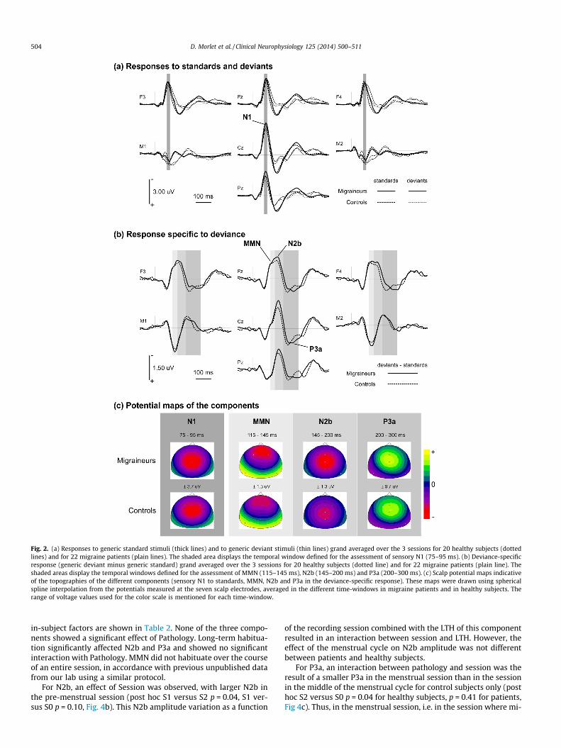
Fig. 2. (a) Responses to generic standard stimuli (thick lines) and to generic deviant stimuli (thin lines) grand averaged over the 3 sessions for 20 healthy subjects (dottedlines) and for 22 migraine patients (plain lines). The shaded area displays the temporal window defined for the assessment of sensory N1 (75–95 ms). (b) Deviance-specificresponse (generic deviant minus generic standard) grand averaged over the 3 sessions for 20 healthy subjects (dotted line) and for 22 migraine patients (plain line). Theshaded areas display the temporal windows defined for the assessment of MMN (115–145 ms), N2b (145–200 ms) and P3a (200–300 ms). (c) Scalp potential maps indicativeof the topographies of the different components (sensory N1 to standards, MMN, N2b and P3a in the deviance-specific response). These maps were drawn using sphericalspline interpolation from the potentials measured at the seven scalp electrodes, averaged in the different time-windows in migraine patients and in healthy subjects. Therange of voltage values used for the color scale is mentioned for each time-window.
504 D. Morlet et al. / Clinical Neurophysiology 125 (2014) 500–511
in-subject factors are shown in Table 2. None of the three compo-nents showed a significant effect of Pathology. Long-term habitua-tion significantly affected N2b and P3a and showed no significantinteraction with Pathology. MMN did not habituate over the courseof an entire session, in accordance with previous unpublished datafrom our lab using a similar protocol.
For N2b, an effect of Session was observed, with larger N2b inthe pre-menstrual session (post hoc S1 versus S2 p = 0.04, S1 ver-sus S0 p = 0.10, Fig. 4b). This N2b amplitude variation as a function
of the recording session combined with the LTH of this componentresulted in an interaction between session and LTH. However, theeffect of the menstrual cycle on N2b amplitude was not differentbetween patients and healthy subjects.
For P3a, an interaction between pathology and session was theresult of a smaller P3a in the menstrual session than in the sessionin the middle of the menstrual cycle for control subjects only (posthoc S2 versus S0 p = 0.04 for healthy subjects, p = 0.41 for patients,Fig 4c). Thus, in the menstrual session, i.e. in the session where mi-

Fig. 3. Investigation of the N1 orienting component. (a) Map of the difference between healthy subjects and migraine patients in the response to standards grand averagedover the first and the last 100 trains of the 3 sessions at 110 ms, i.e. around the latency of the difference maximum. (b) Map of the p values of the Kruskal–Wallis testcomparing the response to standards in healthy subjects and in migraine patients at 110 ms in the grand average of the first half and the last half of the trains of the 3sessions. (c) Response to standard stimuli grand averaged over the 3 sessions for 20 healthy subjects (thick dotted lines) and for 22 migraine patients (thick plain lines) at the4 frontal-central electrodes (F3, Fz, F4 and Cz). (d) Response to deviant stimuli grand averaged over the 3 sessions for 20 healthy subjects (thick dotted lines) and for 22migraine patients (thick plain lines) at the 4 frontal-central electrodes (F3, Fz, F4 and Cz). In (c) and (d), at each electrode, the difference curve between the two populations(Patients minus Controls) is plotted with a thin line and the thick line over the x-axis denotes the temporal window in which point-by-point Kruskal–Wallis tests shows asignificant difference between patients and controls.
Table 1Results of the four-way ANOVAs on the amplitudes of the N1 sub-components, with Pathology as between-subject factor and with Session (S0, S1, and S2), Long-Term Habituation(LTH: first half versus second half of the recording session) and Type of stimulus (standard versus deviant) as within-subject factors. Degrees of freedom for each factor areindicated in parenthesis. Sensory N1 was measured at Cz in the 75–95 ms time-window where the maximal amplitude is observed in the grand average response (see Fig. 2) andorienting component was measured at the 4 frontal–central electrodes in the 100–120 ms latency range, i.e., in the spatio-temporal window where a significant between-groupdifference was observed with Kruskal–Wallis tests (see Fig. 3). Results involving Pathology are printed in italics. Significant p values (p 6 0.05) are printed in bold.
Sensory N1 N1 orienting component
Cz (75–95 ms) F3 + Fz + F4 + Cz (100–120 ms)
F e–GG P F e–GG P
Pathology (1,40) 0.2609 0.612 11.317 0.002Session (2,80) 0.123 0.889 0.862 0.503 0.996 0.606Session � Pathology (2,80) 0.337 0.889 0.609 0.366 0.996 0.693LTH (1,40) 85.385 1.000 <0.001 27.125 1.000 <0.001LTH � Pathology (1,40) 0.939 1.000 0.338 0.031 1.000 0.861Type (1,40) 40.633 1.000 <0.001 21.350 1.000 <0.001Type � Pathology (1,40) 0.085 1.000 0.772 0.020 1.000 0.888Session � LTH (2,80) 1.013 0.948 0.364 0.335 0.958 0.707Session � LTH � Pathology (2,80) 1.261 0.948 0.288 0.106 0.958 0.891Session � type (2,80) 0.237 0.992 0.788 0.195 0.966 0.816Session � type � Pathology (2,80) 0.245 0.992 0.781 0.163 0.966 0.843LTH � type (1,40) 0.202 1.000 0.656 0.029 1.000 0.865LTH � type � Pathology (1,40) 1.683 1.000 0.202 1.264 1.000 0.268Session � LTH � type (2,80) 0.401 0.996 0.670 0.553 0.979 0.574Session � LTH � type � Pathology (2,80) 2.382 0.996 0.099 0.119 0.979 0.884
D. Morlet et al. / Clinical Neurophysiology 125 (2014) 500–511 505
graine attacks preferentially occur, the migraine patients did notshow the decrease in P3a component observed in healthy subjects.In order to highlight a possible effect of a migraine attack on P3a,
we performed an additional analysis differentiating the 11 patientswho actually presented a migraine attack during one of the record-ing sessions (S1 or S2) and the 10 patients who did not, excluding

Fig. 4. Mean amplitude for each session of the different components measured in the deviance-specific response (response to the generic deviant minus response to thegeneric standard). The black lines denote the standard error of the mean. In (a)–(c), the measures are displayed for each session in migraine patients and in controls, for: (a)MMN (mean value in the 115–145 ms time-window at the 3 frontal electrodes), (b) N2b (mean value in the 145–200 ms time-window at Cz) and (c) P3a (mean of positivevalues in the 200–300 ms time-window at Cz). S0 = session 0, S1 = session 1, S2 = session 2. In (d), P3a was measured in 3 groups of subjects: 10 patients who showed nomigraine attack during the recording sessions, 11 patients who suffered headache during one peri-menses session, and the 20 control subjects. The measures are displayed forthe session between menses (S0) and for a peri-menses session (the session with headache for the patients experiencing an attack during recording and a randomly chosenperi-menses session (S1 or S2) for the other two groups).
506 D. Morlet et al. / Clinical Neurophysiology 125 (2014) 500–511
the only subject who experienced headache during the session inthe middle of the cycle (S0). A 3-way ANOVA was applied to P3aamplitude, with the between subjects factor ‘‘migraine-attack’’ (3levels: control subjects, patients with and patients without mi-graine attack) and LTH and Session as within-subject factors. Inthis analysis, the Session factor had only 2 levels, the betweenmenses session (S0) and a peri-menses session, possibly with a mi-graine attack. For patients with a migraine attack, the peri-mensessession was S1 for 5 patients and S2 for 6 patients. For the patientswho did not report a migraine attack and for the controls, the peri-menses session was randomly chosen to be S1 (5 patients and 9controls) or S2 (5 patients and 11 controls, see Demarquay et al.(2011), for a similar analysis). This additional ANOVA resulted ina significant interaction between the intra-subject factor ‘‘Session’’and the between-subject factor ‘‘migraine attack’’ (F(2,38) = 4.653,p = 0.016). Post-hoc tests were not significant. However, asdisplayed in Fig. 4d, patients with a migraine attack showed a ten-dency for a decrease in P3a amplitude in the peri-menses sessionlike the healthy subjects, while patients without a migraine attackshowed a tendency for an increase in P3a amplitude in the peri-menses session. Another 3-way ANOVA using LTH and Session aswithin-subject factors, but restricted to the migraine patients (2levels: patients with and without migraine attack), confirmed thisdifferent modulation of P3a amplitude by migraine attacks, with amarginally significant interaction between the intra-subject factor
‘‘Session’’ and the between-subject factor ‘‘migraine attack’’(F(1,19) = 4.300, p = 0.052).
3.2.2. ‘‘Short-term habituation’’ of the components of the deviance-specific response
Fig. 6 shows the difference responses restricted to the first devi-ants in the trains (first deviant minus generic standard) and to thedeviants that immediately followed the first deviants (repeateddeviant minus generic standard), grand averaged over the 3 ses-sions. It illustrates the effects of the so-called ‘‘short-term habitu-ation’’ on the components of the response to deviance. Table 3displays the results of ANOVAs performed on MMN, N2b, andP3a components with Pathology as between-subject factor andwith Session and STH as within-subject factors. There was a signif-icant effect of STH on the three components (MMN, N2b, P3a), theresponse globally collapsing to repeated deviance in bothpopulations. For N2b, there was an interaction between STH andPathology due to a non-significant tendency (p = 0.26) for thiscomponent to be larger in migraine patients than in controls in re-sponse to the first deviant. For P3a an interaction between STH,Pathology and Session resulted from smaller amplitude of theP3a to the first deviant in S2 than in S0 for the healthy subjects(post hoc S2 versus S0 p = 0.07 for healthy subjects), as alsoobserved above for generic deviants.

Fig. 5. Long-term habituation of the response to deviance: difference response (generic deviant minus generic standard) averaged for 22 patients (plain lines) and for 20healthy subjects (dotted lines), in the first (thick lines) and in the last (thin lines) 100 trains in each of the 3 recording sessions (S0: session 0, S1: session 1, S2: session 2). Toprow: response averaged over the 3 frontal electrodes, bottom row: response at Cz.
Table 2Results of the three-way ANOVAs on the amplitudes of the different components (MMN, N2b, and P3a) of the difference response (deviant minus standard), with Pathology asbetween-subject factor and with Session (S0, S1, and S2) and Long-Term Habituation (LTH: first half versus second half of the session) as within-subject factors. Degrees offreedom for each factor are indicated in parenthesis. MMN was assessed as the mean amplitude at the 3 frontal electrodes between 115 and 145 ms, N2b as the mean amplitudeat Cz between 145 and 200 ms and P3a as the mean amplitude of positive potentials at Cz between 200 and 300 ms. Results involving Pathology are printed in italics. Significant pvalues (p 6 0.05) are printed in bold.
MMN N2b P3a
F3 + Fz + F4 (115–145 ms) Cz (145–200 ms) Cz (200–300 ms)
F e–GG P F e–GG P F e–GG P
Pathology (1,40) 0.139 0.712 2.219 0.144 0.078 0.781Session (2,80) 0.061 0.993 0.940 3.605 0.927 0.035 0.355 0.921 0.685Session � Pathology (2,80) 0.874 0.993 0.421 0.438 0.927 0.632 5.980 0.921 0.005LTH (1,40) 0.763 1.000 0.388 10.919 1.000 0.002 13.401 1.000 0.001LTH � Pathology (1,40) 0.164 1.000 0.688 2.192 1.000 0.147 1.920 1.000 0.174Session � LTH (2,80) 0.679 0.982 0.507 3.988 0.994 0.023 1.008 0.991 0.369Session � LTH � Pathology (2,80) 0.626 0.982 0.534 1.753 0.994 0.180 0.579 0.991 0.561
Fig. 6. ‘‘Short-term habituation’’ of the response to deviance: difference ERPs (deviant minus standard) restricted to the first deviants in the blocks (thick lines) and to thedeviants immediately following the first deviant (repeated deviants, thin lines) averaged over the 3 recording sessions, for 22 patients (plain lines) and for 20 healthy subjects(dotted lines).
D. Morlet et al. / Clinical Neurophysiology 125 (2014) 500–511 507
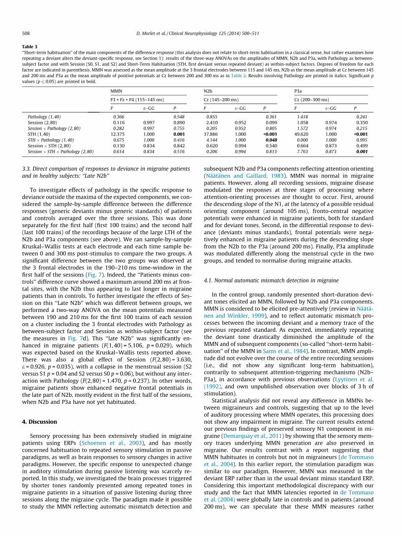
Table 3‘‘Short-term habituation’’ of the main components of the difference response (this analysis does not relate to short-term habituation in a classical sense, but rather examines howrepeating a deviant alters the deviant-specific response, see Section 1): results of the three-way ANOVAs on the amplitudes of MMN, N2b and P3a, with Pathology as between-subject factor and with Session (S0, S1, and S2) and Short-Term Habituation (STH, first deviant versus repeated deviant) as within-subject factors. Degrees of freedom for eachfactor are indicated in parenthesis. MMN was assessed as the mean amplitude at the 3 frontal electrodes between 115 and 145 ms, N2b as the mean amplitude at Cz between 145and 200 ms and P3a as the mean amplitude of positive potentials at Cz between 200 and 300 ms as in Table 2. Results involving Pathology are printed in italics. Significant pvalues (p 6 0.05) are printed in bold.
MMN N2b P3a
F3 + Fz + F4 (115–145 ms) Cz (145–200 ms) Cz (200–300 ms)
F e–GG P F e–GG P F e–GG P
Pathology (1,40) 0.366 0.548 0.855 0.361 1.418 0.241Session (2,80) 0.116 0.997 0.890 2.410 0.952 0.099 1.058 0.974 0.350Session � Pathology (2,80) 0.282 0.997 0.755 0.205 0.952 0.805 1.572 0.974 0.215STH (1,40) 12.375 1.000 0.001 37.886 1.000 <0.001 49.620 1.000 <0.001STH � Pathology (1,40) 0.675 1.000 0.416 4.144 1.000 0.048 0.000 1.000 0.995Session � STH (2,80) 0.130 0.834 0.842 0.620 0.994 0.540 0.664 0.873 0.499Session � STH � Pathology (2,80) 0.614 0.834 0.516 0.206 0.994 0.813 7.763 0.873 0.001
508 D. Morlet et al. / Clinical Neurophysiology 125 (2014) 500–511
3.3. Direct comparison of responses to deviance in migraine patientsand in healthy subjects: ‘‘Late N2b’’
To investigate effects of pathology in the specific response todeviance outside the maxima of the expected components, we con-sidered the sample-by-sample difference between the differenceresponses (generic deviants minus generic standards) of patientsand controls averaged over the three sessions. This was doneseparately for the first half (first 100 trains) and the second half(last 100 trains) of the recordings because of the large LTH of theN2b and P3a components (see above). We ran sample-by-sampleKruskal–Wallis tests at each electrode and each time sample be-tween 0 and 300 ms post-stimulus to compare the two groups. Asignificant difference between the two groups was observed atthe 3 frontal electrodes in the 190–210 ms time-window in thefirst half of the sessions (Fig. 7). Indeed, the ‘‘Patients minus con-trols’’ difference curve showed a maximum around 200 ms at fron-tal sites, with the N2b thus appearing to last longer in migrainepatients than in controls. To further investigate the effects of Ses-sion on this ‘‘Late N2b’’ which was different between groups, weperformed a two-way ANOVA on the mean potentials measuredbetween 190 and 210 ms for the first 100 trains of each sessionon a cluster including the 3 frontal electrodes with Pathology asbetween-subject factor and Session as within-subject factor (seethe measures in Fig. 7d). This ‘‘late N2b’’ was significantly en-hanced in migraine patients (F(1,40) = 5.106, p = 0.029), whichwas expected based on the Kruskal–Wallis tests reported above.There was also a global effect of Session (F(2,80) = 3.630,e = 0.926, p = 0.035), with a collapse in the menstrual session (S2versus S1 p = 0.04 and S2 versus S0 p = 0.06), but without any inter-action with Pathology (F(2,80) = 1.470, p = 0.237). In other words,migraine patients show enhanced negative frontal potentials inthe late part of N2b, mostly evident in the first half of the sessions,when N2b and P3a have not yet habituated.
4. Discussion
Sensory processing has been extensively studied in migrainepatients using ERPs (Schoenen et al., 2003), and has mostlyconcerned habituation to repeated sensory stimulation in passiveparadigms, as well as brain responses to sensory changes in activeparadigms. However, the specific response to unexpected changein auditory stimulation during passive listening was scarcely re-ported. In this study, we investigated the brain processes triggeredby shorter tones randomly presented among repeated tones inmigraine patients in a situation of passive listening during threesessions along the migraine cycle. The paradigm made it possibleto study the MMN reflecting automatic mismatch detection and
subsequent N2b and P3a components reflecting attention orienting(Näätänen and Gaillard, 1983). MMN was normal in migrainepatients. However, along all recording sessions, migraine diseasemodulated the responses at three stages of processing whereattention-orienting processes are thought to occur. First, aroundthe descending slope of the N1, at the latency of a possible residualorienting component (around 105 ms), fronto-central negativepotentials were enhanced in migraine patients, both for standardand for deviant tones. Second, in the differential response to devi-ance (deviants minus standards), frontal potentials were nega-tively enhanced in migraine patients during the descending slopefrom the N2b to the P3a (around 200 ms). Finally, P3a amplitudewas modulated differently along the menstrual cycle in the twogroups, and tended to normalise during migraine attacks.
4.1. Normal automatic mismatch detection in migraine
In the control group, randomly presented short-duration devi-ant tones elicited an MMN, followed by N2b and P3a components.MMN is considered to be elicited pre-attentively (review in Näätä-nen and Winkler, 1999), and to reflect automatic mismatch pro-cesses between the incoming deviant and a memory trace of theprevious repeated standard. As expected, immediately repeatingthe deviant tone drastically diminished the amplitude of theMMN and of subsequent components (so-called ‘‘short-term habit-uation’’ of the MMN in Sams et al., 1984). In contrast, MMN ampli-tude did not evolve over the course of the entire recording sessions(i.e., did not show any significant long-term habituation),contrarily to subsequent attention-triggering mechanisms (N2b–P3a), in accordance with previous observations (Lyytinen et al.(1992), and own unpublished observation over blocks of 3 h ofstimulation).
Statistical analysis did not reveal any difference in MMNs be-tween migraineurs and controls, suggesting that up to the levelof auditory processing where MMN operates, this processing doesnot show any impairment in migraine. The current results extendour previous findings of preserved sensory N1 component in mi-graine (Demarquay et al., 2011) by showing that the sensory mem-ory traces underlying MMN generation are also preserved inmigraine. Our results contrast with a report suggesting thatMMN habituates in controls but not in migraineurs (de Tommasoet al., 2004). In this earlier report, the stimulation paradigm wassimilar to our paradigm. However, MMN was measured in thedeviant ERP rather than in the usual deviant minus standard ERP.Considering this important methodological discrepancy with ourstudy and the fact that MMN latencies reported in de Tommasoet al. (2004) were globally late in controls and in patients (around200 ms), we can speculate that these MMN measures rather

p < 0.05
First 100 trains Last 100 trains
(b)
- 0.75 uV + 0.75 uV
Last 100 trainsFirst 100 trains
(a)
(d)
Ampl
itude
(mic
ro v
olts
)
- 1
- 0.8
- 0.6
- 0.4
- 0.2
0
0.2
0.4
S0 S1 S2
Migraine PatientsHealthy Subjects
“Late N2b” measure
Mean amplitude190 - 210 ms
F3-Fz-F4 averaged
First 100 trainsin each session
Patients minus Controls
Migraine PatientsHealthy Subjects
Response specific to devianceFirst 100 trains - 3 sessions averaged
0.75 uV
-
+
Fz
F3
“Late N2b”
F4
100 ms
(c)Response specific to deviance at 200 msPatients minus Controls
Scalp potentials
Response specific to deviance at 200 msPatients minus Controls
Kruskal-Wallis test
Fig. 7. Investigation of the ‘‘late N2b’’. (a) Map of the difference between healthy subjects and migraine patients in the deviance-specific responses (generic deviants minusgeneric standards) in the first and the last 100 trains, averaged over the 3 sessions at 200 ms, i.e. at the latency of the difference maximum. (b) Map of the p values of theKruskal–Wallis test comparing the deviance-specific responses in healthy subjects and in migraine patients at 200 ms in the first half and the second half of the trainsaveraged over the 3 sessions. (c) Deviance-specific response grand averaged over the first 100 trains of the 3 sessions for 20 healthy subjects (thick dotted lines) and for 22migraine patients (thick plain lines) at the 3 frontal electrodes (F3, Fz and F4). At each electrode, the difference curve between the two populations (patients minus controls) isplotted with a thin line and the thick line over the x-axis denotes the temporal window in which the point-by-point Kruskal–Wallis tests show a significant differencebetween patients and controls. (d) ‘‘Late N2b’’ amplitude (mean value and standard error of the mean in the 190–210 ms time-window averaged over the 3 frontal electrodes)for each population and for the 100 first trains of each session. S0 = session 0, S1 = session 1, S2 = session 2.
D. Morlet et al. / Clinical Neurophysiology 125 (2014) 500–511 509
concerned the N2b. Similarly, in their pediatric MMN study, Valeri-ani et al. (2009) reported an habituation deficit of the MMN in mi-graine, but did not attempt to disentangle MMN from N2b. In anattempt to dissociate the sensory mismatch processes from thesubsequent attention alerting processes, we assessed separatelythe mean amplitudes of the potentials at the respective latenciesof MMN and N2b. Other techniques such as trial-by-trial time–fre-quency analysis of the EEG, beyond the scope of the present report,could be helpful to definitely exclude differences in change detec-tion processes between migraine patients and controls.
Neither the present study, nor our previous migraine study(Demarquay et al., 2011) of auditory ERPs in a passive listening sit-uation evidenced any abnormality in basic sensory processing,including MMN generation. These observations are consistent withprevious studies showing no significant difference betweenmigraineurs and controls with regard to sensory N1 and P2 compo-nent (Drake et al., 1989; Sand and Vanagaite Vingen, 2000).
4.2. Abnormal attention orienting in migraine
Contrasting with the fairly normal MMN observed in migrainepatients, we observed at two different processing stages enhancedfrontal negative potentials in migraine patients, which suggest anincreased attention orienting (Halgren et al., 2011). Patientsshowed larger negative frontal potentials firstly in the descendingslope of the N1 for responses to both standard and deviant tones,
and secondly in the late part of the N2b in the response specificto deviance (deviants minus standards difference wave).
In a previous report (Demarquay et al., 2011), we already re-ported such increased fronto-central negative potentials for stan-dard tones in migraine. This effect was peculiarly pronouncedwhen tones appeared at the beginning of the trains of stimuli(i.e. after a long silence), when the orienting component (or com-ponent III, Näätänen and Picton, 1987) of the N1 is large (Alcainiet al., 1994). We interpreted these enhanced fronto-central nega-tive potentials in response to any standard tone as a residual N1orienting component. Here we show that a comparable effect isobtained with deviant tones, suggesting that augmented orientingprocesses might actually be elicited by any incoming auditorystimulus in migraine. Interestingly, the increase of the N1 orientingcomponent observed in migraine patients occurs earlier than theMMN (see Fig. 2). As MMN appears fairly normal in migraine, itcan be hypothesized that these abnormal early orienting processesdo not interfere with the sensory memory and comparison pro-cesses underlying MMN generation.
Migraine patients also showed augmented frontal negativepotentials in the deviance-specific response located in thedescending slope of N2b. This effect was mostly evident in the firsthalf of all sessions. N2b is a fronto-central subcomponent, whichtypically arises in active paradigms and has been associated withattention-triggering (Snyder and Hillyard, 1976; Näätänen andGaillard, 1983; Kiehl et al., 2001) and cognitive control
510 D. Morlet et al. / Clinical Neurophysiology 125 (2014) 500–511
encompassing response inhibition to a non-target deviant, re-sponse conflict, and error monitoring (Folstein and Van Petten,2008; Schwartze et al., 2011). N2b was shown to be associatedwith higher order processing than the MMN (Ritter et al., 1992).However N2b can also occur in passive oddball paradigms, in par-ticular when the difference between standards and deviants islarge enough (Näätänen et al. (1982); see also Ruby et al. (2008)for an example of N2b recorded in a passive oddball paradigmusing duration deviants). Although participants in our study wereinstructed to watch a silent movie and to ignore auditory stimuli,their attention might have been involuntarily oriented to the devi-ant stimuli and the prolonged negative potentials at the end of N2bobserved in migraineurs could reflect an abnormal activity at themoment of this involuntary shift of attention.
Moreover, P3a amplitude was modulated along the menstrualcycle in both groups. Controls showed a smaller P3a during mensesthan in the session between menses. As far as we know, this wasnever described in the literature. The overall migraine populationshowed an opposite pattern, with an enhanced P3a in the men-strual session. This abnormality tended to normalise duringmigraine attacks. In a previous study (Demarquay et al., 2011),we already observed P3a abnormalities depending on the record-ing session. However, whereas the present result concerned theP3a individualised in the response specific to deviance, the formerresult concerned the P3a in response to the first tones of the trains,a component partially overlapping the sensory P2. Thus, the tworesults can hardly be compared. Nevertheless, both results havein common a normalisation of P3a amplitudes during migraine at-tacks, in accordance with previous observations concerning severalother components (Kropp and Gerber, 1995, 1998; Evers et al.,1999; Judit et al., 2000).
As a whole, our results reveal ERP abnormalities arising in mi-graine patients at stages of processing where attention-orientingmechanisms are susceptible to operate. They are in keeping withfindings using active paradigms (and thus involving attention),showing abnormal cognitive responses like the P300 to targets(Drake et al., 1989; Wang et al., 1996; Evers et al., 1998) or theCNV to warning stimuli (Maertens de Noordhout et al., 1986;Kropp and Gerber, 1993). As the main generators of N1 orientingcomponent, N2b and P3a are thought to be located in frontal areas(see Alcaini et al. (1994) for the N1 orienting component, Halgrenet al. (2011) for the N2b and Polich and Criado (2006) for P3a), theabnormal automatic attention orienting towards acoustic stimuliobserved in migraineurs may reflect abnormal attention-relatedfrontal networks in migraine.
4.3. Sensory processing and attention orienting in migraine
To sum up, the present ERP findings allow setting the workinghypothesis of normal sensory stages of auditory processing in mi-graine, but exacerbated mechanisms of automatic attention orient-ing to the auditory stimulation, possibly relying on an abnormalinvolvement of frontal networks. As emphasized above, our resultsare in line with previous studies that showed normal N1 and P2sensory responses but altered cognitive responses like the iCNV,and P300. The apparent discrepancy with previous reports of a lackof habituation of auditory ERPs in migraine (Wang et al., 1996;Ambrosini et al., 2003) could be related to the paradigms used inthese studies. Indeed, lack of habituation was observed in para-digms using intensity dependence of auditory potentials (IDAP),where sound intensity varies randomly within blocks. No habitua-tion deficit was observed when stimulus intensity changes did notoccur within blocks (Sand and Vanagaite Vingen, 2000), or in par-adigms not involving intensity variations such as ours (Demarquayet al., 2011). We can speculate that random changes in the auditorystimulation might be more arousing than unvarying stimuli, and
thus more likely to automatically engage attentional networks.This is in keeping with the present findings of an increased atten-tion orienting to rare changes occurring in streams of otherwiserepetitive tones. Furthermore, a link between habituation deficitsin migraine and abnormal orienting activity has been already pro-posed by Siniatchkin et al. (2000).
In the visual modality, the habituation of ERPs has been widelytested in migraine patients, mainly using pattern-reversal stimuli,with contrasting results (Schoenen et al., 1995; Afra et al., 1998;Wang et al., 1999). One study suggested that the visual habituationdeficit in migraine depends on the stimulus characteristics(Oelkers et al., 1999), but a recent one using individual identifica-tion of visual evoked peaks in a blinded evaluation design foundnormal habituation in interictal migraineurs, with similar check-size effects in patients and controls (Omland et al., 2013). Besides,recent studies showed an increased automatic attentional responseto sudden-onset visual events in migraineurs (Mickleboroughet al., 2011a). Interestingly, in line with our observation of anenhancement of the N1 orienting component in auditory ERPs,these authors showed an early attention effect in the visual N1ERP component in migraine patients (Mickleborough et al.,2011b). Thus, in the visual modality also, further studies mightpossibly enlighten a link between observed habituation deficitsand attention orienting exacerbation.
4.4. Methodological issues
In the present study, in an attempt to follow a migraine cycle,we only recorded patients with menstrually-related migraine. Thisstrategy indeed allowed us to record 11 patients during a migraineattack. However, as patients were not requested to fulfill migrainediaries, it might be possible that, during some recordings, some pa-tients were in a pre-ictal state, a state where cortical excitabilityhas been shown to fluctuate. This would be more likely for sessionsS1 and S2, i.e., the sessions around menses. Indeed, before an at-tack, migraineurs show a normalisation of the interictal lack ofhabituation of CNV (Kropp and Gerber, 1995, 1998; Siniatchkinet al., 1999) and of IDAP potentials (Judit et al., 2000). It is howevernoteworthy that, except for the P3a, the between-group differencesobserved in the present study (enhanced N1 orienting componentand enhanced late N2b in migraine patients) did not depend on therecording sessions, suggesting that these effects are not modulatedalong the migraine cycle. We cannot exclude that a precise sortingof the recording sessions in ‘‘interictal’’ ‘‘pre-ictal’’ and ‘‘ictal’’ couldallow to reveal further differences between migraine patients andcontrols.
In conclusion, our study shows normal auditory sensory pro-cessing in migraine patients but increased automatic attention ori-enting processes to auditory changes (and more generally to anyincoming auditory stimulus). The pathophysiologic basis of suchheightened attentional response recorded in migraineurs is stillunknown, but could, at least partially, be linked to the close rela-tionship observed between environmental stimuli and migraine.Understanding the links between photo/phonophobia and an in-creased automatic attention orienting in migraine is a challengefor future studies.
Acknowledgements
We wish to thank the control subjects and the patients whoparticipated in this experiment. We are also grateful to the techni-cians of the Department of Functional Neurology and Epileptologyfor their help in recordings ERPs. The authors have no conflict ofinterest.
D. Morlet et al. / Clinical Neurophysiology 125 (2014) 500–511 511
References
Afra J, Cecchini AP, De Pasqua V, Albert A, Schoenen J. Visual evoked potentialsduring long periods of pattern-reversal stimulation in migraine. Brain1998;121:233–41.
Aguera PE, Jerbi K, Caclin A, Bertrand O. ELAN: a software package for analysis andvisualisation of MEG, EEG, and LFP signals. Comput Intell Neurosci2011;2011:158970.
Alcaini M, Giard MH, Thevenet M, Pernier J. Two separate frontal components in theN1 wave of the human auditory cortex. Psychophysiology 1994;31:611–5.
Ambrosini A, Rossi P, De Pasqua V, Pierelli F, Schoenen J. Lack of habituation causeshigh intensity dependence of auditory evoked cortical potentials in migraine.Brain 2003;126:2009–15.
Böcker KB, Timsit-Berthier M, Schoenen J, Brunia CH. Contingent negative variationin migraine. Headache 1990;30:604–9.
Coppola G, Pierelli F, Schoenen J. Habituation and migraine. Neurobiol Learn Mem2009;92:249–59.
de Tommaso M, Guido M, Libro G, Losito L, Difruscolo O, Sardaro M, et al. Interictallack of habituation of mismatch negativity in migraine. Cephalalgia2004;24:663–8.
Demarquay G, Caclin A, Brudon F, Fischer C, Morlet D. Exacerbated attentionorienting to auditory stimulation in migraine patients. Clin Neurophysiol2011;122:1755–63.
Drake Jr ME, Pakalnis A, Padamadan H. Long-latency auditory event relatedpotentials in migraine. Headache 1989;29:239–41.
Evers S, Bauer B, Grotemeyer KH, Kurlemann G, Husstedt IW. Event-relatedpotentials (P300) in primary headache in childhood and adolescence. J ChildNeurol 1998;13:322–6.
Evers S, Quibeldey F, Grotemeyer KH, Suhr B, Husstedt IW. Dynamic changes ofcognitive habituation and serotonin metabolism during the migraine interval.Cephalalgia 1999;19:485–91.
Fischer C, Morlet D, Bouchet P, Luaute J, Jourdan C, Salord F. Mismatch negativityand late auditory evoked potentials in comatose patients. Clin Neurophysiol1999;110:1601–10.
Folstein JR, Van Petten C. Influence of cognitive control and mismatch on the N2component of the ERP: a review. Psychophysiology 2008;45:152–70.
Guthrie D, Buchwald JS. Significance testing of difference potentials.Psychophysiology 1991;28:240–4.
Halgren E, Sherfey J, Irimia A, Dale AM, Marinkovic K. Sequential temporo-fronto-temporal activation during monitoring of the auditory environment fortemporal patterns. Hum Brain Mapp 2011;32:1260–76.
ICHD. The International Classification of Headache Disorders: 2nd edition.Cephalalgia 2004; 24 Suppl 1: 9–160.
Judit A, Sándor PS, Schoenen J. Habituation of visual and intensity dependence ofauditory evoked cortical potentials tends to normalise just before and duringthe migraine attack. Cephalalgia 2000;20:714–9.
Jung J, Morlet D, Mercier B, Confavreux C, Fischer C. Mismatch negativity (MMN) inmultiple sclerosis: an event-related potentials study in 46 patients. ClinNeurophysiol 2006;117:85.
Kiehl KA, Laurens KR, Duty TL, Forster BB, Liddle PF. Neural sources involved inauditory target detection and novelty processing: an event-related fMRI study.Psychophysiology 2001;38:133–42.
Korostenskaja M, Pardos M, Kujala T, Rose DF, Brown D, Horn P, et al. Impairedauditory information processing during acute migraine: amagnetoencephalography study. Int J Neurosci 2011;121:355–65.
Kropp P, Gerber W-D. Prediction of migraine attacks using a slow cortical potential,the contingent negative variation. Neurosci Lett 1998;257:73–6.
Kropp P, Gerber WD. Is increased amplitude of contingent negative variation inmigraine due to cortical hyperactivity or to reduced habituation? Cephalalgia1993;13:37–41.
Kropp P, Gerber WD. Contingent negative variation during migraine attack andinterval: evidence for normalisation of slow cortical potentials during theattack. Cephalalgia 1995;15:123–8. discussion 178–179.
Lyytinen H, Blomberg AP, Näätänen R. Event-related potentials and autonomicresponses to a change in unattended auditory stimuli. Psychophysiology1992;29:523–33.
Maertens de Noordhout A, Timsit-Berthier M, Timsit M, Schoenen J. Contingentnegative variation in headache. Ann Neurol 1986;19:78–80.
Main A, Dowson A, Gross M. Photophobia and phonophobia in migraineurs betweenattacks. Headache 1997;37:492–5.
Main A, Vlachonikolis I, Dowson A. The wavelength of light causing photophobia inmigraine and tension-type headache between attacks. Headache2000;40:194–9.
Mazzotta G, Alberti A, Santucci A, Gallai V. The event-related potential P300 duringheadache-free period and spontaneous attack in adult headache sufferers.Headache 1995;35:210–5.
Mickleborough MJ, Hayward J, Chapman C, Chung J, Handy TC. Reflexive attentionalorienting in migraineurs: the behavioral implications of hyperexcitable visualcortex. Cephalalgia 2011a;31:1642–51.
Mickleborough MJ, Truong G, Handy TC. Top-down control of visual cortex inmigraine populations. Neuropsychologia 2011b;49:1006–15.
Mulder EJCM, Linssen WHJP, Passchier J, De Geus EJC. Interictal and postictalcontingent negative variation in migraine without aura. Headache 2001;41:72–8.
Näätänen R. Mismatch negativity: clinical research and possible applications. Int JPsychophysiol 2003;48:179–88.
Näätänen R, Gaillard AWK. The orienting reflex and the N2 deflection of the event-related potential (ERP). In: Gaillard AWK, Ritter W, editors. Tutorials in ERPresearch: endogenous components. North Holland Publishing Company; 1983.p. 119–41.
Näätänen R, Kujala T, Escera C, Baldeweg T, Kreegipuu K, Carlson S, et al. Themismatch negativity (MMN)–a unique window to disturbed central auditoryprocessing in ageing and different clinical conditions. Clin Neurophysiol2012;123:424–58.
Näätänen R, Kujala T, Winkler I. Auditory processing that leads to consciousperception: a unique window to central auditory processing opened by themismatch negativity and related responses. Psychophysiology 2011;48:4–22.
Näätänen R, Paavilainen P, Rinne T, Alho K. The mismatch negativity (MMN) in basicresearch of central auditory processing: a review. Clin Neurophysiol2007;118:2544–90.
Näätänen R, Paavilainen P, Tiitinen H, Jiang D, Alho K. Attention and mismatchnegativity. Psychophysiology 1993;30:436–50.
Näätänen R, Picton T. The N1 wave of the human electric and magnetic response tosound: a review and an analysis of the component structure. Psychophysiology1987;24:375–425.
Näätänen R, Simpson M, Loveless NE. Stimulus deviance and evoked potentials. BiolPsychol 1982;14:53–98.
Näätänen R, Tervaniemi M, Sussman E, Paavilainen P, Winkler I. ‘‘Primitiveintelligence’’ in the auditory cortex. Trends Neurosci 2001;24:283–8.
Näätänen R, Winkler I. The concept of auditory stimulus representation in cognitiveneuroscience. Psychol Bull 1999;125:826–59.
Oelkers R, Grosser K, Lang E, Geisslinger G, Kobal G, Brune K, et al. Visual evokedpotentials in migraine patients: alterations depend on pattern spatialfrequency. Brain 1999;122:1147–55.
Omland PM, Nilsen KB, Uglem M, Gravdahl G, Linde M, Hagen K, et al. Visual evokedpotentials in interictal migraine: no confirmation of abnormal habituation.Headache 2013. http://dx.doi.org/10.1111/head.12006.
Polich J, Criado JR. Neuropsychology and neuropharmacology of P3a and P3b. Int JPsychophysiol 2006;60:172–85.
Ritter W, Paavilainen P, Lavikainen J, Reinikainen K, Alho K, Sams M, et al. Event-related potentials to repetition and change of auditory stimuli.Electroencephalogr Clin Neurophysiol 1992;83:306–21.
Ruby P, Caclin A, Boulet S, Delpuech C, Morlet D. Odd sound processing in thesleeping brain. J Cogn Neurosci 2008;20:296–311.
Sams M, Alho K, Näätänen R. Short-term habituation and dishabituation of themismatch negativity of the ERP. Psychophysiology 1984;21:434–41.
Sand T, Vanagaite Vingen J. Visual, long-latency auditory and brainstem auditoryevoked potentials in migraine: relation to pattern size, stimulus intensity,sound and light discomfort thresholds and pre-attack state. Cephalalgia2000;20:804–20.
Schoenen J, Ambrosini A, Sandor PS, Maertens de Noordhout A. Evoked potentialsand transcranial magnetic stimulation in migraine: published data andviewpoint on their pathophysiologic significance. Clin Neurophysiol2003;114:955–72.
Schoenen J, Wang W, Albert A, Delwaide PJ. Potentiation instead of habituationcharacterises visual evoked potentials in migraine patients between attacks. EurJ Neurol 1995;2:115–22.
Schwartze M, Rothermich K, Schmidt-Kassow M, Kotz SA. Temporal regularityeffects on pre-attentive and attentive processing of deviance. Biol Psychol2011;87:146–51.
Siniatchkin M, Gerber WD, Kropp P, Vein A. How the brain anticipates an attack: astudy of neurophysiological periodicity in migraine. Funct Neurol1999;14:69–77.
Siniatchkin M, Gerber WD, Kropp P, Voznesenskaya T, Vein AM. Are the periodicchanges of neurophysiological parameters during the pain-free interval inmigraine related to abnormal orienting activity? Cephalalgia 2000;20:20–9.
Snyder E, Hillyard SA. Long-latency evoked potentials to irrelevant, deviant stimuli.Behav Biol 1976;16:319–31.
Stovner L, Hagen K, Jensen R, Katsarava Z, Lipton R, Scher A, et al. The global burdenof headache: a documentation of headache prevalence and disabilityworldwide. Cephalalgia 2007;27:193–210.
Tervaniemi M, Lehtokoski A, Sinkkonen J, Virtanen J, Ilmoniemi RJ, Näätänen R.Test-retest reliability of mismatch negativity for duration, frequency andintensity changes. Clin Neurophysiol 1999;110:1388–93.
Valeriani M, Galli F, Tarantino S, Graceffa D, Pignata E, Miliucci R, et al. Correlationbetween abnormal brain excitability and emotional symptomatology inpaediatric migraine. Cephalalgia 2009;29:204–13.
Vingen JV, Sand T, Stovner LJ. Sensitivity to various stimuli in primary headaches: aquestionnaire study. Headache 1999;39:552–8.
Wang W, Schoenen J. Interictal potentiation of passive ‘‘oddball’’ auditory event-related potentials in migraine. Cephalalgia 1998;18:261–5. discussion 241.
Wang W, Schoenen J, Timsit-Berthier M. Cognitive functions in migraine withoutaura between attacks: a psychophysiological approach using the ‘‘oddball’’paradigm. Neurophysiol Clin 1995;25:3–11.
Wang W, Timsit-Berthier M, Schoenen J. Intensity dependence of auditory evokedpotentials is pronounced in migraine: an indication of cortical potentiation andlow serotonergic neurotransmission? Neurology 1996;46:1404–9.
Wang W, Wang GP, Ding XL, Wang YH. Personality and response to repeated visualstimulation in migraine and tension-type headaches. Cephalalgia1999;19:718–24. discussion 697–718.
Woods DL, Elmasian E. The habituation of event-related potentials to speech soundsand tones. Electroencephalogr Clin Neurophysiol 1986;65:446–59.