
Orienting attention to objects in visual short-term memory
10
Neuropsychologia 48 (2010) 419–428 Contents lists available at ScienceDirect Neuropsychologia journal homepage: www.elsevier.com/locate/neuropsychologia Orienting attention to objects in visual short-term memory Roberto Dell’Acqua a,e,* , Paola Sessa a , Paolo Toffanin b , Roy Luria c , Pierre Jolicœur d a Department of Psychology, University of Padova, Padova, Italy b BNC Neuroimaging Center, University of Groningen, Groningen, The Netherlands c Department of Psychology, University of Oregon, Eugene, OR, United States d Department of Psychology, University of Montreal, Montreal, QBC, Canada e Center for Cognitive Science, University of Padova, Padova, Italy article info Article history: Received 17 September 2009 Accepted 27 September 2009 Available online 3 October 2009 Keywords: Visual attention Perception Visual short-term memory N2pc abstract We measured electroencephalographic activity during visual search of a target object among objects available to perception or among objects held in visual short-term memory (VSTM). For perceptual search, a single shape was shown first (pre-cue) followed by a search-array, and the task was to decide whether the pre-cue was or was not in the search-array. For search of VSTM, a search-array was shown first followed by a single shape (post-cue), and the task was to decide whether the post-cue was or was not in the previously displayed search-array. We focused on early lateralized electrical brain activity over posterior and temporal areas time-locked to search-arrays in pre-cue trials and to post-cues in post-cue trials. In Experiment 1, search-arrays were composed of two lateralized shapes, displayed in the upper/lower two quadrants of the monitor. In Experiment 2, search-arrays were composed of four shapes, displayed at the corners of an imaginary square centered on fixation. In pre-cue trials, we observed an N2pc of about equal amplitude and latency for search-arrays composed of two or four shapes. In post- cue trials, we observed N2pc-like activity with search-arrays composed of two shapes, that was however substantially attenuated with search-arrays composed of four shapes. For many aspects, attending to a perceptual object was functionally and neurally analogous to attending to an object held in VSTM, suggesting that spatial selective attention biases search of objects during both ongoing perception and retention. © 2009 Elsevier Ltd. All rights reserved. 1. Introduction An important feature of the visual system is the retinotopic orga- nization of the primary visual cortex, in which nearby points on the retina activate nearby cells in the primary visual cortex. This retinotopic coding becomes coarser but is still generally preserved as visual processing continues in higher level, extra-striate visual areas (Aguirre, Zarahn, & D’Esposito, 1998; Jack et al., 2007; Zeki, 1993). It is possible that this early retinotopy has contributed to some degree of isomorphism between visual representations and objects in the world, such that distinct and adjacent parts of the physical world are represented as distinct and adjacent parts in isomorphic visual representations (Shepard, 1984). At the coarsest level, the left and right halves of visual space are represented in dis- tinct cerebral hemispheres (in a crossed fashion due to the crossing of the neural projections from the retina to the primary visual cor- * Corresponding author at: Department of Psychology, Via Venezia 8, 35131 Padova, Italy. Tel.: +39 049 8276545; fax: +39 049 8276511. E-mail address: [email protected] (R. Dell’Acqua). URL: http://colab.psy.unipd.it (R. Dell’Acqua). tex). The contralateral organization of the visual space seems to be generally retained in the lower portion of the dorsal visual stream up to and including the intra-parietal sulcus (IPS; Sereno, Pitzalis, & Martinez, 2001), and in inferior regions of the temporal cortex (Chelazzi, Miller, Duncan, & Desimone, 1993). Sensory representations are temporally fleeting and subject to decay or erasure by newly incoming visual input (e.g., Coltheart, 1980; Sperling, 1960), such that further processing is needed to consolidate them in a more durable representational format, usu- ally referred to as visual short-term memory (VSTM; Averbach & Coriel, 1961; Jolicœur & Dell’Acqua, 1998). VSTM representations are more resistant to visual masking, and temporally more stable, with a duration that outlasts the physical availability of visual input by several seconds (Phillips, 1974). The transition of visual input representation from sensory to VSTM status implies a substan- tial information reduction, since VSTM capacity is demonstrably a minuscule fraction of the information available at early stages of processing, with a limit in storage that is, on average, of only about 3–4 objects (Cowan, 2001; Stevanovski & Jolicœur, 2007; Vogel, Woodman, & Luck, 2001). Given the very high capacity of early visual processes and the very limited storage capacity of VSTM, most models of visual pro- 0028-3932/$ – see front matter © 2009 Elsevier Ltd. All rights reserved. doi:10.1016/j.neuropsychologia.2009.09.033
Transcript of Orienting attention to objects in visual short-term memory

Neuropsychologia 48 (2010) 419–428
Contents lists available at ScienceDirect
Neuropsychologia
journa l homepage: www.e lsev ier .com/ locate /neuropsychologia
Orienting attention to objects in visual short-term memory
Roberto Dell’Acquaa,e,!, Paola Sessaa, Paolo Toffaninb, Roy Luriac, Pierre Jolicœurd
a Department of Psychology, University of Padova, Padova, Italyb BNC Neuroimaging Center, University of Groningen, Groningen, The Netherlandsc Department of Psychology, University of Oregon, Eugene, OR, United Statesd Department of Psychology, University of Montreal, Montreal, QBC, Canadae Center for Cognitive Science, University of Padova, Padova, Italy
a r t i c l e i n f o
Article history:Received 17 September 2009Accepted 27 September 2009Available online 3 October 2009
Keywords:Visual attentionPerceptionVisual short-term memoryN2pc
a b s t r a c t
We measured electroencephalographic activity during visual search of a target object among objectsavailable to perception or among objects held in visual short-term memory (VSTM). For perceptual search,a single shape was shown first (pre-cue) followed by a search-array, and the task was to decide whetherthe pre-cue was or was not in the search-array. For search of VSTM, a search-array was shown firstfollowed by a single shape (post-cue), and the task was to decide whether the post-cue was or wasnot in the previously displayed search-array. We focused on early lateralized electrical brain activityover posterior and temporal areas time-locked to search-arrays in pre-cue trials and to post-cues inpost-cue trials. In Experiment 1, search-arrays were composed of two lateralized shapes, displayed in theupper/lower two quadrants of the monitor. In Experiment 2, search-arrays were composed of four shapes,displayed at the corners of an imaginary square centered on fixation. In pre-cue trials, we observed anN2pc of about equal amplitude and latency for search-arrays composed of two or four shapes. In post-cue trials, we observed N2pc-like activity with search-arrays composed of two shapes, that was howeversubstantially attenuated with search-arrays composed of four shapes. For many aspects, attending toa perceptual object was functionally and neurally analogous to attending to an object held in VSTM,suggesting that spatial selective attention biases search of objects during both ongoing perception andretention.
© 2009 Elsevier Ltd. All rights reserved.
1. Introduction
An important feature of the visual system is the retinotopic orga-nization of the primary visual cortex, in which nearby points onthe retina activate nearby cells in the primary visual cortex. Thisretinotopic coding becomes coarser but is still generally preservedas visual processing continues in higher level, extra-striate visualareas (Aguirre, Zarahn, & D’Esposito, 1998; Jack et al., 2007; Zeki,1993). It is possible that this early retinotopy has contributed tosome degree of isomorphism between visual representations andobjects in the world, such that distinct and adjacent parts of thephysical world are represented as distinct and adjacent parts inisomorphic visual representations (Shepard, 1984). At the coarsestlevel, the left and right halves of visual space are represented in dis-tinct cerebral hemispheres (in a crossed fashion due to the crossingof the neural projections from the retina to the primary visual cor-
! Corresponding author at: Department of Psychology, Via Venezia 8, 35131Padova, Italy. Tel.: +39 049 8276545; fax: +39 049 8276511.
E-mail address: [email protected] (R. Dell’Acqua).URL: http://colab.psy.unipd.it (R. Dell’Acqua).
tex). The contralateral organization of the visual space seems to begenerally retained in the lower portion of the dorsal visual streamup to and including the intra-parietal sulcus (IPS; Sereno, Pitzalis,& Martinez, 2001), and in inferior regions of the temporal cortex(Chelazzi, Miller, Duncan, & Desimone, 1993).
Sensory representations are temporally fleeting and subject todecay or erasure by newly incoming visual input (e.g., Coltheart,1980; Sperling, 1960), such that further processing is needed toconsolidate them in a more durable representational format, usu-ally referred to as visual short-term memory (VSTM; Averbach &Coriel, 1961; Jolicœur & Dell’Acqua, 1998). VSTM representationsare more resistant to visual masking, and temporally more stable,with a duration that outlasts the physical availability of visual inputby several seconds (Phillips, 1974). The transition of visual inputrepresentation from sensory to VSTM status implies a substan-tial information reduction, since VSTM capacity is demonstrablya minuscule fraction of the information available at early stages ofprocessing, with a limit in storage that is, on average, of only about3–4 objects (Cowan, 2001; Stevanovski & Jolicœur, 2007; Vogel,Woodman, & Luck, 2001).
Given the very high capacity of early visual processes and thevery limited storage capacity of VSTM, most models of visual pro-
0028-3932/$ – see front matter © 2009 Elsevier Ltd. All rights reserved.doi:10.1016/j.neuropsychologia.2009.09.033

420 R. Dell’Acqua et al. / Neuropsychologia 48 (2010) 419–428
cessing postulate a critical role for spatial selective attention (e.g.,Desimone & Duncan, 1995; Duncan & Humphreys, 1989), whichis hypothesized to bias information processing towards portionsof the visual world relevant for goal-directed behavior (e.g., Folk,Remington, & Johnston, 1992; Leblanc, Prime, & Jolicœur, 2008).Although much remains to be discovered about the neural realiza-tion of attention mechanisms and the algorithms for the selectionof objects and/or locations, whether by target activation, distractorsuppression, or both, recent work suggests a clear interdependencebetween spatial attention mechanisms and mechanisms involvedin the generation of both sensory and VSTM representations. Ingeneral, these results, which we review briefly in the forthcomingsections, converge to support the hypothesis proposed by Awh andJonides (2001) that attention mechanisms operating at the percep-tual level and attention mechanisms operating at the VSTM leveloverlap to a large extent, both functionally and neuroanatomically(e.g., Coull & Nobre, 1998; DeFockert, Rees, Frith, & Lavie, 2001;LaBar, Gitelman, Parrish, & Mesulam, 1999; Lepsien, Griffin, Devlin,& Nobre, 2005; Nobre et al., 2004; Nobre, Griffin, & Rao, 2008;Olivers, 2008; Yantis et al., 2002).
Two studies have tracked the event-related potential (ERP)response associated with attention deployment to sensory objectsand compared it with the ERP response associated with attentiondeployment to VSTM objects. Eimer and Kiss (in press) instructedsubjects to search a visual array for the presence of a target dia-mond, with the top or bottom corner cut off, among distractingcircles, and to indicate via button press the position of the cut corner(top or bottom). The shapes in the array were arranged circularlyaround fixation, with the shapes in one visual hemifield all coloredred and those in the opposite visual hemifield all colored green.In pre-cue trials, subjects were cued about the color of the targetbefore the appearance of the visual array, by having the visual arraypreceded by a centrally displayed colored square (pre-cue) match-ing either color in the visual array. In post-cue trials, subjects werecued about the color of the target after the visual array, by dis-playing the central colored square (post-cue) after the offset of thevisual array. In pre-cue trials, the ERP response was time-locked tothe onset of the visual array. In post-cue trials, the ERP response wastime-locked to the onset of the post-cue color cue. Not surprisingly,an N2pc response was found in pre-cue trials. The N2pc is a greaternegativity recorded between 200 and 300 ms at occipito-parietalsites contralateral to an attended stimulus (Brisson & Jolicœur,2007, 2008; Dell’Acqua, Pesciarelli, Jolicœur, Eimer, & Peressotti,2007; Dell’Acqua, Sessa, Jolicœur, & Robitaille, 2006; Eimer, 1996;Hickey, Di Lollo, & McDonald, 2008; Hopf et al., 2000; Jolicœur,Sessa, Dell’Acqua, & Robitaille, 2006a,b; Kiss, Jolicœur, Dell’Acqua, &Eimer, 2008; Luck & Hillyard, 1994; Luck, Woodman, & Vogel, 2002;Woodman & Luck, 1999, 2003a,b). The ERP response in post-cuetrials was, however, mainly in the form of sustained posterior-contralateral negativity (SPCN, e.g., Jolicœur, Brisson, & Robitaille,2008; Klaver, Talsa, Wijers, Heinze, & Mulder, 1999; McCollough,Machizawa, & Vogel, 2007; Vogel & Machizawa, 2004), namely, alater – with respect to N2pc – and more protracted enhanced neg-ativity recorded between 300 and 600 ms at occipito-parietal sitescontralateral to the target. Notably, the lateralized ERP response inpost-cue trials was elicited by the central post-cue colored square,and this suggested that the spatial arrangement of the colors inthe visual array was retained in VSTM (Gratton, 1998; Hommel,2002; Jiang, Olson, & Chun, 2000), and was effective in biasing theresponses of neurons in cortical areas surrounding IPS (e.g., Todd &Marois, 2004) during search of VSTM.
Using a similar logic, Kuo, Rao, Lepsien, and Nobre (2009)instructed subject to search a visual array of random shapes or col-ored squares for a target (shape in one condition, and color in adifferent condition) that was either shown before the visual array inpre-cue trials, or after the visual array in post-cue trials. The visual
array could be composed of either two or four eccentric shapesor colors, in order to evaluate the impact of varying the numberof items in a visual array on search efficiency in the sensory andVSTM domains. The ERP response in pre-cue trials was character-ized by an N2pc (i.e., a negativity contralateral to the target shapeor color that arose between 200 and 300 ms post-array onset). Asimilar response was observed in post-cue trials, where an N2pc-like response was time-locked to the post-cue shape or color. Inboth the shape and color conditions, variations in the visual arrayset-size produced no modulations of the N2pc response, either inpre-cue or post-cue trials.
2. The present study
An important goal of the present work was to determinewhether the scalp distribution of the lateralized ERP response dur-ing search of VSTM is identical to, or different from, the scalpdistribution observed during search in perception. Interestingly,the results of Kuo et al. (2009) suggest a possible difference on thebasis of their published isovoltage contour-maps comparing thedistribution of N2pc activity between pre-cue and post-cue trials.Although the perspective offered by their maps is from a posteriorview only, their figures suggest an anterior shift of the contralat-eral negativity in the post-cue condition relative to the pre-cuecondition. This issue deserves a particular consideration becauseof expected differences in neural circuitry involved in perceptualand mnemonic search. It seems indeed established that, in addi-tion to important similarities/overlap of cortical regions involvedin visual and VSTM search, there are also some notable differ-ences. For instance, signal increases in the insula, inferior frontalgyrus, and more in generally, ventro-lateral prefrontal regions, havebeen selectively described during VSTM search only (Courtney,Ungerleider, Keil, & Haxby, 1997; Nobre et al., 2004), and not invisual search. Perhaps, the slightly different topographical distri-butions of the N2pc-like responses in pre-cue vs. post-cue trialsof Kuo et al.’s experiments – although not statistically significant– reflect different contributions of memory-related activity in thetwo conditions. On the basis of this observation, we set out to deter-mine to what extent the scalp distributions of the lateralized ERPresponses, N2pc for visual search and N2pc for search of VSTM,would differ, and most particularly whether search of VSTM wouldbe associated with a more anterior scalp distribution than visualsearch.
A second motivation to carry out the present study relies onthe inconsistency between the results obtained by Eimer andKiss (in press) and Kuo et al. (2009). In both studies, a close-to-standard N2pc was observed in pre-cue trials. However, whereasan SPCN was observed in post-cues by Eimer and Kiss (in press),an N2pc similar to that observed in pre-cue trials was observedin post-cue trials by Kuo et al. (2009). One explanation may arisefrom differences in the task requirements in these two studies. InEimer and Kiss (in press) case, subjects first had to decode thepost-cue color, to orient attention towards the visual hemifieldcontaining the target, and then inspect the target within the color-matching hemifield to determine which corner was missing. Itmight be that this two-step process (find the target, identify theresponse-relevant feature) extended the duration of processing ofthe lateralized memory representation held in VSTM, leading to asustained response observed as an SPCN instead of an N2pc. Thetask in Kuo et al. (2009) study required a present/absent response,which may have required a shorter engagement on the target,and this may have yielded an N2pc without the later sustainedresponse (i.e., no SPCN). Given that our Experiment 1, whose designis schematically illustrated in Fig. 1, used a present/absent judg-ment, we expected to find an N2pc response in both the pre-cue

R. Dell’Acqua et al. / Neuropsychologia 48 (2010) 419–428 421
Fig. 1. Sequence of events on a pre-cue trial of Experiment 1. A white fixation dotwas presented at the center of the screen, flanked by two digits. Subjects wereinstructed to start rehearsing the names of the digits aloud before pressing thespacebar on the computer keyboard to start the trial, and continue to rehearse thedigits throughout the trial. Upon trial initiation, the digits disappeared and a fixedblank interval of 1000 ms preceded the onset of a centrally displayed shape (i.e.,pre-cue), which was exposed for 300 ms. Following the pre-cue offset, a variableinterval (1300–1500 ms) elapsed before the presentation of two lateralized shapes(i.e., search-array), that were displayed in the upper (as is the case in the figure)or lower (as indicated by the dotted contours, which were not displayed on screen)two quadrants of the screen. The search-array was exposed for 150 ms, and replacedwith a blank screen that lasted until subjects responded by pressing one of two keysof the numeric keypad of the computer keyboard (i.e., either “1” or “2,” counter-balanced across subjects) to indicate, without speed pressure, whether the pre-cuewas present or absent in the search-array. Following a response, a blank interval of1500 ms preceded the beginning of the next trial. During the inter-trial interval, aresponse feedback was provided to subjects by displaying centrally a “+” or a “"” incase of a correct or incorrect response, respectively. In the specific trial illustratedin the figure, the correct response is “absent.”.
and post-cue conditions (Fig. 2), as found in Kuo et al. (2009)study.
A final observation concerns the work of Kuo et al. (2009), andrefer specifically to the absence of N2pc modulations in responseto visual array set-size variations. An absence of N2pc amplitudevariations depending on visual array set-size is not unusual. Sincesimilar N2pc components have been found with visual search-arrays of 2 stimuli (e.g., Dell’Acqua et al., 2006, 2007; Eimer, 1996)up to arrays of 24 stimuli (e.g., Woodman & Luck, 1999; Woodman& Luck, 2003b),1 an absence of N2pc variations in pre-cue trials ofKuo’s et al. when the set-size varied between two and four randomshapes or colored squares were somewhat expected in pre-cue tri-als. What is more surprising was the absence of N2pc variationsin post-cue trials when the visual array was varied in set-size.In post-cue trials, increasing the size of the visual array from 2to 4 objects presumably implied an increase in VSTM load, andwe have recently shown that a memory load of 2 random poly-gons brings VSTM close to capacity (Luria, Sessa, Gotler, Jolicœur, &Dell’Acqua, in press). Interestingly, although much attention has
1 At first blush, this prediction appears based more on intuition than on a solidempirical ground. However, it must be noted that, to our knowledge, no studies haveever been published so far describing N2pc variations in visual search generatedthrough systematic manipulations of search-array size.
Fig. 2. Sequence of events on a post-cue trial of Experiment 1. A white fixationdot was presented at the center of the screen, flanked by two digits. Subjects wereinstructed to start rehearsing the names of the digits aloud before pressing the space-bar on the computer keyboard to start the trial, and continue to rehearse the digitsthroughout the trial. Upon trial initiation, the digits disappeared and a fixed blankinterval of 1000 ms preceded the onset of two lateralized shapes (i.e., memory-array), that were displayed for 300 ms in the lower (as is the case in the figure) orupper (as indicated by the dotted contours, which were not displayed on screen)two quadrants of the screen. Following the memory-array offset, a variable interval(1300–1500 ms) elapsed before the presentation of a centrally displayed shape (i.e.,post-cue). The post-cue was exposed for 150 ms and replaced with a blank screenthat lasted until subjects responded by pressing one of two keys of the numerickeypad of the computer keyboard (i.e., either “1” or “2,” counterbalanced acrosssubjects) to indicate, without speed pressure, whether the post-cue was presentor absent in the memory-array. Following a response, a blank interval of 1500 mspreceded the beginning of the next trial. During the inter-trial interval, a responsefeedback was provided to subjects by displaying centrally a “+” or a “"” in case ofa correct or incorrect response, respectively. In the specific trial illustrated in thefigure, the correct response is “present.” Note that, apart from the reverse order ofpre-/post-cues and search/memory-arrays, the sequence of events in pre-cue trials(Fig. 1) and post-cue trials (Fig. 2) was identical.
been devoted to the interaction between search efficiency andVSTM content (see Olivers, 2009, for a review), evidence from stud-ies focusing on the interaction between search efficiency and VSTMload is scant, and the emerging picture is somewhat contradictory.Whereas some researchers have proposed a dissociation betweenattention mechanisms underpinning visual search and VSTM main-tenance by showing no search slope variations in reaction time (RT)as a function of concurrent visual memory load (Woodman, Vogel,& Luck, 2001), more recent work revisiting this issue has providedevidence of diminished search efficiency with a concurrent VSTMload, when object features to be held in memory overlapped withobject features guiding search (Oh & Kim, 2004; Woodman & Luck,2004). This is precisely the condition implemented in post-cue tri-als of Kuo et al.’s design. It may be worth mentioning that Kuo etal. also recorded RT in both pre-cue and post-cue trials, observingresults that are consistent with the latter view. The effects of set-size on RT (slower RT with visual arrays composed of four shapesthan of two shapes) were more pronounced in post-cue trials thanpre-cue trials. So set-size effects that were not apparent at thelevel of N2pc were instead fully manifest behaviorally. It is hardto explain this inconsistency and this provided additional motiva-tion to examine effects of a set-size manipulation on the expected

422 R. Dell’Acqua et al. / Neuropsychologia 48 (2010) 419–428
N2pc in post-cue trials. Thus, in Experiment 2, we replaced the two-shape visual arrays used in Experiment 1 with four-shape arrays.The fact that the N2pc is reduced in amplitude when visual-spatialattention is taxed by an attentional blink (Dell’Acqua et al., 2006;see also Jolicœur et al., 2006a,b; Robitaille, Jolicœur, Dell’Acqua, &Sessa, 2007) suggested to us that search of VSTM under greater loadwould produce a reduction in N2pc amplitude (Brisson & Jolicœur,2007). This would be most evident in post-cue trials of Experiment2 because the requirement to maintain four items in VSTM wouldfill VSTM to capacity and thus impose the highest processing loadrelative to other conditions in the study (i.e., pre- and post-cue trialsof Experiment 1, pre-cue trials of Experiment 2).
3. Method
3.1. Participants
A total of 31 psychology students at the University of Padova took part in thepresent study, either as volunteers or in partial fulfillment of course requirements.There were 15 participants (8 females; mean age 22.0 ± 2.2) in Experiment 1, and 16participants (11 females; mean age 22.4 ± 1.8) in Experiment 2. All participants hadnormal or corrected-to-normal vision, and none reported a prior history of neu-rological disorders and/or was under medication at the time of testing. Most ofthe participants (83%) were right-handed. Each participant provided informed con-sent prior to the beginning of the experiment. Three subjects were discarded fromanalyses, one in Experiment 1 and two in Experiment 2, for an excessive rate ofocular artifacts, and so analyses were based on results from 14 participants in eachexperiment.
3.2. Material
The stimuli were 8 regular shapes (i.e., square, circle, triangle, pentagon, dia-mond, hexagon, moon, and cross) and the digits 1–9. They were displayed in black(4 cd/m2) on a light gray background (36 cd/m2) on a 19 in. cathode-ray tube mon-itor controlled by a Pentium IV CPU and Eprime® software (Psychology SoftwareTools Inc.). At a distance of 70 cm (set by a chin rest), each shape was scaled to fitinside a square subtending 1.6# of visual angle. The digits were .7# wide and 1# high.A white (50 cd/m2) disk with a black contour with a diameter of .3# was used asfixation point. When presented off-center, the center of each shape coincided withthe vertex of an imaginary square, centered on the monitor, with a side of 2.8# ofvisual angle.
3.3. Design
In both experiments, each participant performed two types of trials, pre-cue tri-als and post-cue trials. In pre-cue trials, one shape (i.e., pre-cue) was displayed for300 ms at the center of the monitor, followed, at an interval varying from 1300to 1500 ms (jittered randomly from trial to trial in steps of 20 ms), by an arraycomposed of a variable number of lateralized shapes (i.e., search-array) that was dis-played for 150 ms. In pre-cue trials, the task was to indicate, without speed pressure,whether the pre-cue shape was present or absent in the search-array. In post-cue tri-als, an array composed a variable number of lateralized shapes (memory-array) wasdisplayed for 300 ms, followed, at an interval varying from 1300 to 1500 ms (jitteredrandomly from trial to trial in steps of 20 ms), by a shape displayed for 150 ms atthe center of the monitor (i.e., post-cue). In post-cue trials, the task was to indicate,without speed pressure, whether the post-cue shape was present or absent in thememory-array. Participants responded by pressing one of two keys of the numerickeypad of the computer keyboard (either “1” or “2,” with the response mappingcounterbalanced across participants), and received visual feedback in the form of acentrally displayed “+” or “"” in case of a correct or incorrect response, respectively.In both pre-cue and post-cue trials, when the target was absent, all shapes used ona particular trial (i.e., cue shape and array shapes) were selected at random, withoutreplacement, from the set of available shapes. When the target was present, one ofthe shapes in the array (search-array in pre-cue trials or memory-array in post-cuetrials) matched the (pre- or post-) cue shape. In both experiments, there were 6blocks of 80 pre-cue trials and 6 blocks of 80 post-cue trials, for a total of 960 trials.In each experiment, half of the subjects started with 6 consecutive blocks of pre-cue trials, followed by 6 blocks of post-cue trials, and the other half of the subjectsperformed the trial types in the reverse order. In both experiments, two randomlyselected digits (always different) were presented prior to the beginning of the trial,flanking the central fixation point. Participants were instructed to repeat the digitsaloud throughout the trial,2 until a response was emitted. This articulatory suppres-
2 A microphone inside the recording chamber was used to detect and amplifythe digits’ rehearsal outside the chamber, in order to monitor whether participantswere repeating aloud the digits during each trial. When a participant did not repeat
sion task was adopted to minimize the possibility of phonological recoding of theshapes into their names during retention (e.g., Besner, Davies, & Daniels, 1981; seealso Vogel et al., 2001). All experiments were self-paced, with each trial startingwith a spacebar press.
3.4. ERP recording and signal processing
Using a head-cap with tin electrodes, EEG activity was recorded continuouslyfrom the Fp1, Fp2, Fz, F3, F4, F7, F8, C3, C4, Cz, P3, P4, Pz, O1, O2, T3, T4, T7, T8,P7, P8 sites, and the right earlobe, referenced to the left earlobe (Pivik et al., 1993).Horizontal EOG (HEOG) was recorded bipolarly from electrodes positioned on theouter canthi of both eyes. Vertical EOG (VEOG) was recorded bipolarly from twoelectrodes, positioned above and below the left eye. EEG, HEOG, and VEOG activitieswere amplified, filtered using a bandpass of 0.01–80 Hz, and digitized at a sam-pling rate of 250 Hz. Impedance at each electrode was maintained below 5 k!. TheEEG was re-referenced offline to the average of the left and right earlobes, and seg-mented into 700 ms epochs starting 100 ms prior to the onset of the search-array inpre-cue trials or to the onset of the post-cue in post-cue trials. Trials associated witha HEOG exceeding ±30 !V were discarded from analyses. Trials associated with eyeblinks, or any other artifact (electrode activity exceeding ±80 !V in a time interval of400 ms), were also discarded from analyses. In target-present trials, signal-averagedHEOG was used to control for possible eye movements away from fixation duringthe presentation of the search-array in pre-cue trials and the memory-array in post-cue trials (i.e., when the shapes were presented off-center). The difference betweenleft and right HEOG electrodes was averaged separately for trials in which the tar-get shape (i.e., the shape in the search-array matching the pre-cue or the shape inthe memory-array matching the post-cue) was displayed to the right or to the leftof the central fixation. A maximum deflection of less than 3 !V was observed forany given subject, ensuring that the average eye position did not deviate by morethan .2# towards the target shape included in search/memory-arrays during the ERPepoch. Lateralized activity was monitored at the P7/P8 and T3/T4 sites (i.e., wherethe lateralized activity was particularly pronounced), and only target-present trialswith a correct response were analyzed.
In both pre-cue and post-cue trials, for each electrode pair, the ERP contralateralto the target shape was calculated by averaging the ERP generated at the left-sidedelectrode when the target shape was displayed in the right visual hemifield and theERP generated at the right-sided electrode when the target shape was displayedin the left visual hemifield. The ERP ipsilateral to the target shape was calculatedby averaging the ERPs at the complementary sites. Isovoltage contour-maps repre-senting differences between brain activity over sites ipsilateral and contralateral tothe target shape were generated by spherical spline interpolation (Perrin, Pernier,Bertrand, & Echallier, 1989), after computing difference waves for each lateralizedelectrode pair and entering a voltage of zero at midline electrodes. To comparethe topographical distribution of activity over the scalp, normalization of the activ-ity values was carried out using the algorithm described by McCarthy and Wood(1985). Estimation of the latency of the ERP components of interest was carriedout using the jackknife approach (Ulrich & Miller, 2001), by finding the time atwhich a jackknife waveform reached 50% of peak amplitude. In the ERP analyses, theGreenhouse–Geisser correction for nonsphericity was applied when appropriate.
4. Results
4.1. Experiment 1
4.1.1. BehaviorThe mean proportion of hits was .97.3 We submitted to separate
analyses of variance (ANOVAs) standard indices of sensitivity (d$
and ˇ; Green & Swets, 1974) in which trial type (pre-cue vs. post-cue) and array position (above vs. below the horizontal meridian)were considered as within-subjects factors. Mean d$ and ˇ valueswere 4.1 and 1.7, respectively. Neither factor produced significanteffects in any of the analyses (all Fs < 1).
4.1.2. ERPAfter the exclusion of error trials and trials contaminated by eye
movements and/or any other artifact, 92% of trials were retainedfor analysis. The most important ERP results are shown in Fig. 3.The contralateral (dotted lines) and ipsilateral (solid lines) ERPs
the digits, the experimenter stopped the experiment, entered the chamber, andreminded the participant to do so.
3 The high proportion of correct responses did not allow us to compute stable ERPfunctions, in this experiment (as well as in Experiment 2), based on trials associatedwith misses.
R. Dell’Acqua et al. / Neuropsychologia 48 (2010) 419–428 423
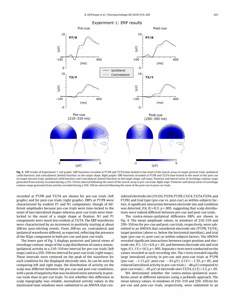
Fig. 3. ERP results of Experiment 1. Left graphs: ERP functions recorded at P7/P8 and T3/T4 time-locked to the onset of the search-array on target-present trials, ipsilateral(solid function) and contralateral (dotted function) to the target shape. Right graphs: ERP functions recorded at P7/P8 and T3/T4 time-locked to the onset of the post-cueon target-present trials, ipsilateral (solid function) and contralateral (dotted function) to the target shape. Left maps: Posterior and lateral views of isovoltage contour-mapsgenerated from activity recorded during a 210–310 ms interval following the onset of the search-array in pre-cue trials. Right maps: Posterior and lateral views of isovoltagecontour-maps generated from activity recorded during a 250–350 ms interval following the onset of the post-cue in post-cue trials.
recorded at P7/P8 and T3/T4 are shown for pre-cue trials (leftgraphs) and for post-cue trials (right graphs). ERPs at P7/P8 werecharacterized by evident P1 and N1 components, though of dif-ferent amplitudes because pre-cue trials were time-locked to theonset of two lateralized shapes whereas post-cue trials were time-locked to the onset of a single shape at fixation. N1 and P1components were much less evident at T3/T4. The ERP waveformswere characterized by an increment in positivity starting at about200 ms post-eliciting events. From 200 ms on, contralateral andipsilateral waveforms differed, as expected, reflecting the presenceof the N2pc component in both pre-cue and post-cue trials.
The lower part of Fig. 3 displays posterior and lateral views ofisovoltage contour-maps of the scalp distribution of contra-minus-ipsilateral activity in a 210–310 ms interval for pre-cue trials (leftmaps) and in a 250–350 ms interval for post-cue trials (right maps).These intervals were centered on the peak of the waveform foreach condition for the displayed electrode sites. As can be seen bycomparing left and right maps, the distribution of activity on thescalp was different between the pre-cue and post-cue conditions,with a peak of negativity that was localized more anteriorly in post-cue trials than in pre-cue trials. To test whether this difference inscalp topography was reliable, normalized activity values in thementioned time-windows were submitted to an ANOVA that con-
sidered electrode site (O1/O2, P3/P4, P7/P8, C3/C4, T3/T4, F3/F4, andF7/F8) and trial type (pre-cue vs. post-cue) as within-subjects fac-tors. A significant interaction between electrode site and conditionwas detected, F(6, 8) = 8.3; p < .005, suggesting that scalp distribu-tions were indeed different between pre-cue and post-cue trials.
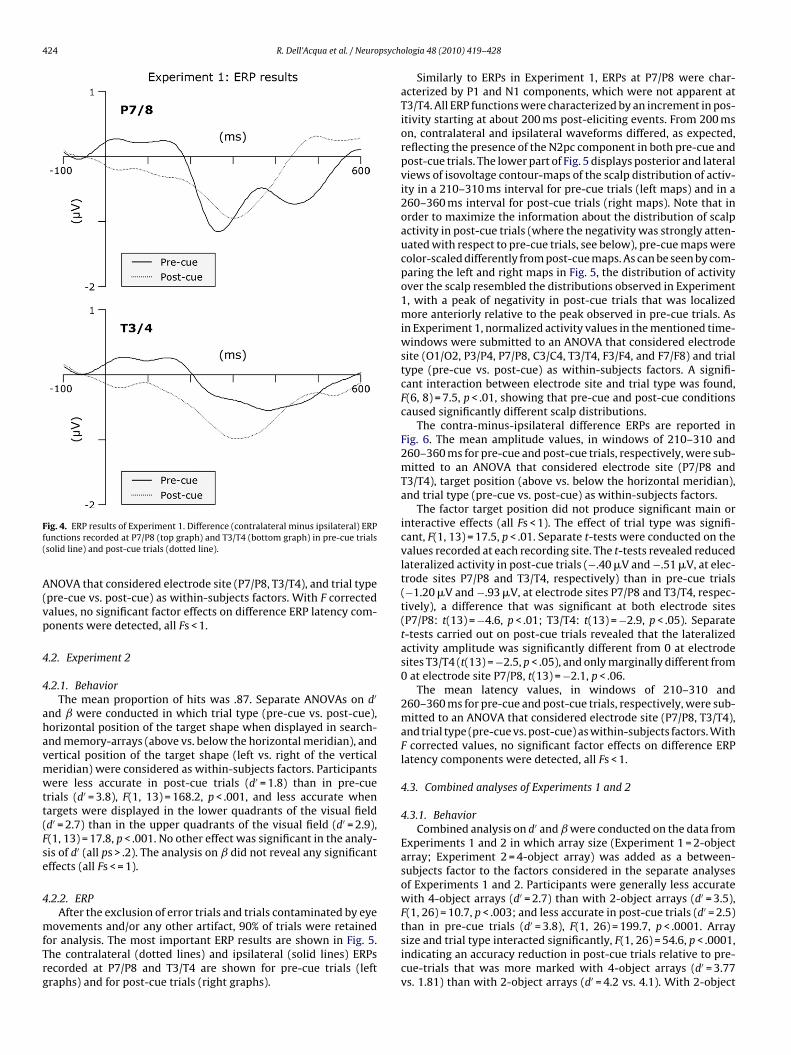
The contra-minus-ipsilateral difference ERPs are shown inFig. 4. The mean amplitude values, in windows of 210–310 and250–350 ms for pre-cue and post-cue trials, respectively, were sub-mitted to an ANOVA that considered electrode site (P7/P8, T3/T4),target position (above vs. below the horizontal meridian), and trialtype (pre-cue vs. post-cue) as within-subjects factors. The ANOVArevealed significant interactions between target position and elec-trode site, F(1, 13) = 6.9, p < .05, and between electrode site and trialtype, F(1, 13) = 16.5, p < .005. Separate t-tests were conducted on thevalues recorded at each recording site. The t-tests revealed equallylarge lateralized activity in pre-cue and post-cue trials at P7/P8(pre-cue: "1.11 !V; post-cue: ".91 !V), t(13) = "1.35, p > .05, andreduced lateralized activity in pre-cue trials (".48 !V) compared topost-cue trials (".95 !V) at electrode sites T3/T4, t(13) = 2.5, p < .05.
We determined whether the contra-minus-ipsilateral wave-forms onset at different latencies using a jackknife approach. Themean latency values, in windows of 210–310 and 250–350 ms forpre-cue and post-cue trials, respectively, were submitted to an
424 R. Dell’Acqua et al. / Neuropsychologia 48 (2010) 419–428
Fig. 4. ERP results of Experiment 1. Difference (contralateral minus ipsilateral) ERPfunctions recorded at P7/P8 (top graph) and T3/T4 (bottom graph) in pre-cue trials(solid line) and post-cue trials (dotted line).
ANOVA that considered electrode site (P7/P8, T3/T4), and trial type(pre-cue vs. post-cue) as within-subjects factors. With F correctedvalues, no significant factor effects on difference ERP latency com-ponents were detected, all Fs < 1.
4.2. Experiment 2
4.2.1. BehaviorThe mean proportion of hits was .87. Separate ANOVAs on d$
and ˇ were conducted in which trial type (pre-cue vs. post-cue),horizontal position of the target shape when displayed in search-and memory-arrays (above vs. below the horizontal meridian), andvertical position of the target shape (left vs. right of the verticalmeridian) were considered as within-subjects factors. Participantswere less accurate in post-cue trials (d$ = 1.8) than in pre-cuetrials (d$ = 3.8), F(1, 13) = 168.2, p < .001, and less accurate whentargets were displayed in the lower quadrants of the visual field(d$ = 2.7) than in the upper quadrants of the visual field (d$ = 2.9),F(1, 13) = 17.8, p < .001. No other effect was significant in the analy-sis of d$ (all ps > .2). The analysis on ˇ did not reveal any significanteffects (all Fs < = 1).
4.2.2. ERPAfter the exclusion of error trials and trials contaminated by eye
movements and/or any other artifact, 90% of trials were retainedfor analysis. The most important ERP results are shown in Fig. 5.The contralateral (dotted lines) and ipsilateral (solid lines) ERPsrecorded at P7/P8 and T3/T4 are shown for pre-cue trials (leftgraphs) and for post-cue trials (right graphs).
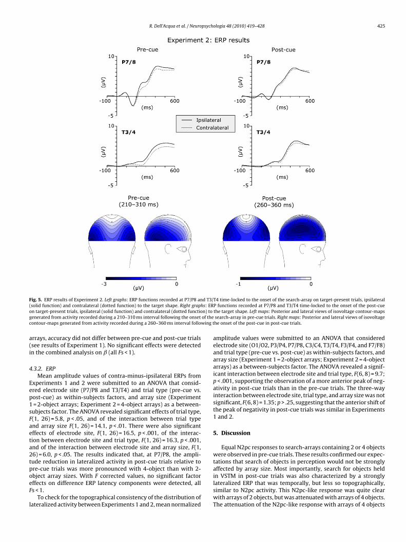
Similarly to ERPs in Experiment 1, ERPs at P7/P8 were char-acterized by P1 and N1 components, which were not apparent atT3/T4. All ERP functions were characterized by an increment in pos-itivity starting at about 200 ms post-eliciting events. From 200 mson, contralateral and ipsilateral waveforms differed, as expected,reflecting the presence of the N2pc component in both pre-cue andpost-cue trials. The lower part of Fig. 5 displays posterior and lateralviews of isovoltage contour-maps of the scalp distribution of activ-ity in a 210–310 ms interval for pre-cue trials (left maps) and in a260–360 ms interval for post-cue trials (right maps). Note that inorder to maximize the information about the distribution of scalpactivity in post-cue trials (where the negativity was strongly atten-uated with respect to pre-cue trials, see below), pre-cue maps werecolor-scaled differently from post-cue maps. As can be seen by com-paring the left and right maps in Fig. 5, the distribution of activityover the scalp resembled the distributions observed in Experiment1, with a peak of negativity in post-cue trials that was localizedmore anteriorly relative to the peak observed in pre-cue trials. Asin Experiment 1, normalized activity values in the mentioned time-windows were submitted to an ANOVA that considered electrodesite (O1/O2, P3/P4, P7/P8, C3/C4, T3/T4, F3/F4, and F7/F8) and trialtype (pre-cue vs. post-cue) as within-subjects factors. A signifi-cant interaction between electrode site and trial type was found,F(6, 8) = 7.5, p < .01, showing that pre-cue and post-cue conditionscaused significantly different scalp distributions.
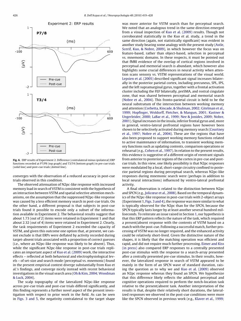
The contra-minus-ipsilateral difference ERPs are reported inFig. 6. The mean amplitude values, in windows of 210–310 and260–360 ms for pre-cue and post-cue trials, respectively, were sub-mitted to an ANOVA that considered electrode site (P7/P8 andT3/T4), target position (above vs. below the horizontal meridian),and trial type (pre-cue vs. post-cue) as within-subjects factors.
The factor target position did not produce significant main orinteractive effects (all Fs < 1). The effect of trial type was signifi-cant, F(1, 13) = 17.5, p < .01. Separate t-tests were conducted on thevalues recorded at each recording site. The t-tests revealed reducedlateralized activity in post-cue trials (".40 !V and ".51 !V, at elec-trode sites P7/P8 and T3/T4, respectively) than in pre-cue trials("1.20 !V and ".93 !V, at electrode sites P7/P8 and T3/T4, respec-tively), a difference that was significant at both electrode sites(P7/P8: t(13) = "4.6, p < .01; T3/T4: t(13) = "2.9, p < .05). Separatet-tests carried out on post-cue trials revealed that the lateralizedactivity amplitude was significantly different from 0 at electrodesites T3/T4 (t(13) = "2.5, p < .05), and only marginally different from0 at electrode site P7/P8, t(13) = "2.1, p < .06.
The mean latency values, in windows of 210–310 and260–360 ms for pre-cue and post-cue trials, respectively, were sub-mitted to an ANOVA that considered electrode site (P7/P8, T3/T4),and trial type (pre-cue vs. post-cue) as within-subjects factors. WithF corrected values, no significant factor effects on difference ERPlatency components were detected, all Fs < 1.
4.3. Combined analyses of Experiments 1 and 2
4.3.1. BehaviorCombined analysis on d$ and ˇ were conducted on the data from
Experiments 1 and 2 in which array size (Experiment 1 = 2-objectarray; Experiment 2 = 4-object array) was added as a between-subjects factor to the factors considered in the separate analysesof Experiments 1 and 2. Participants were generally less accuratewith 4-object arrays (d$ = 2.7) than with 2-object arrays (d$ = 3.5),F(1, 26) = 10.7, p < .003; and less accurate in post-cue trials (d$ = 2.5)than in pre-cue trials (d$ = 3.8), F(1, 26) = 199.7, p < .0001. Arraysize and trial type interacted significantly, F(1, 26) = 54.6, p < .0001,indicating an accuracy reduction in post-cue trials relative to pre-cue-trials that was more marked with 4-object arrays (d$ = 3.77vs. 1.81) than with 2-object arrays (d$ = 4.2 vs. 4.1). With 2-object
R. Dell’Acqua et al. / Neuropsychologia 48 (2010) 419–428 425
Fig. 5. ERP results of Experiment 2. Left graphs: ERP functions recorded at P7/P8 and T3/T4 time-locked to the onset of the search-array on target-present trials, ipsilateral(solid function) and contralateral (dotted function) to the target shape. Right graphs: ERP functions recorded at P7/P8 and T3/T4 time-locked to the onset of the post-cueon target-present trials, ipsilateral (solid function) and contralateral (dotted function) to the target shape. Left maps: Posterior and lateral views of isovoltage contour-mapsgenerated from activity recorded during a 210–310 ms interval following the onset of the search-array in pre-cue trials. Right maps: Posterior and lateral views of isovoltagecontour-maps generated from activity recorded during a 260–360 ms interval following the onset of the post-cue in post-cue trials.
arrays, accuracy did not differ between pre-cue and post-cue trials(see results of Experiment 1). No significant effects were detectedin the combined analysis on ˇ (all Fs < 1).
4.3.2. ERPMean amplitude values of contra-minus-ipsilateral ERPs from
Experiments 1 and 2 were submitted to an ANOVA that consid-ered electrode site (P7/P8 and T3/T4) and trial type (pre-cue vs.post-cue) as within-subjects factors, and array size (Experiment1 = 2-object arrays; Experiment 2 = 4-object arrays) as a between-subjects factor. The ANOVA revealed significant effects of trial type,F(1, 26) = 5.8, p < .05, and of the interaction between trial typeand array size F(1, 26) = 14.1, p < .01. There were also significanteffects of electrode site, F(1, 26) = 16.5, p < .001, of the interac-tion between electrode site and trial type, F(1, 26) = 16.3, p < .001,and of the interaction between electrode site and array size, F(1,26) = 6.0, p < .05. The results indicated that, at P7/P8, the ampli-tude reduction in lateralized activity in post-cue trials relative topre-cue trials was more pronounced with 4-object than with 2-object array sizes. With F corrected values, no significant factoreffects on difference ERP latency components were detected, allFs < 1.
To check for the topographical consistency of the distribution oflateralized activity between Experiments 1 and 2, mean normalized
amplitude values were submitted to an ANOVA that consideredelectrode site (O1/O2, P3/P4, P7/P8, C3/C4, T3/T4, F3/F4, and F7/F8)and trial type (pre-cue vs. post-cue) as within-subjects factors, andarray size (Experiment 1 = 2-object arrays; Experiment 2 = 4-objectarrays) as a between-subjects factor. The ANOVA revealed a signif-icant interaction between electrode site and trial type, F(6, 8) = 9.7;p < .001, supporting the observation of a more anterior peak of neg-ativity in post-cue trials than in the pre-cue trials. The three-wayinteraction between electrode site, trial type, and array size was notsignificant, F(6, 8) = 1.35; p > .25, suggesting that the anterior shift ofthe peak of negativity in post-cue trials was similar in Experiments1 and 2.
5. Discussion
Equal N2pc responses to search-arrays containing 2 or 4 objectswere observed in pre-cue trials. These results confirmed our expec-tations that search of objects in perception would not be stronglyaffected by array size. Most importantly, search for objects heldin VSTM in post-cue trials was also characterized by a stronglylateralized ERP that was temporally, but less so topographically,similar to N2pc activity. This N2pc-like response was quite clearwith arrays of 2 objects, but was attenuated with arrays of 4 objects.The attenuation of the N2pc-like response with arrays of 4 objects
426 R. Dell’Acqua et al. / Neuropsychologia 48 (2010) 419–428
Fig. 6. ERP results of Experiment 2. Difference (contralateral minus ipsilateral) ERPfunctions recorded at P7/P8 (top graph) and T3/T4 (bottom graph) in pre-cue trials(solid line) and post-cue trials (dotted line).
converges with the observation of a reduced accuracy in post-cuetrials observed in this condition.
The observed attenuation of N2pc-like response with increasedmemory load in search of VSTM is consistent with the hypothesis ofan interaction between VSTM and spatial selective attention mech-anisms, on the assumption that the suppressed N2pc-like responsewas caused by a less efficient memory search in post-cue trials. Onthe other hand, a different proposal is that subjects in post-cuetrials found it possible to encode only a subset of the informa-tion available in Experiment 2. The behavioral results suggest thatabout 1.73 (out of 2) items were retained in Experiment 1 and thatabout 2.32 (out of 4) items were retained in Experiment 2. Clearly,the task requirements of Experiment 2 exceeded the capacity ofVSTM, and given this outcome one option that, at present, we can-not exclude is that ERPs were deflated by activity recorded duringtarget-absent trials associated with a proportion of correct guesses(i.e., where an N2pc-like response was likely to be absent). Thus,while the significant N2pc-like response in post-cue trials repli-cates an important aspect of Kuo et al. (2009) work, the interactiveeffects – reflected at both behavioral and electrophysiological lev-els – of set-size and search mode (perceptual vs. mnemonic) foundin the present empirical context diverge substantially from Kuo etal.’s findings, and converge nicely instead with recent behavioralinvestigations in the visual search area (Oh & Kim, 2004; Woodman& Luck, 2004).
The scalp topography of the lateralized N2pc-like responseacross pre-cue trials and post-cue trials differed significantly, andthis finding represents a further novel aspect of the present inves-tigation with respect to prior work in the field. As can be seenin Figs. 3 and 5, the negativity contralateral to the target shape
was more anterior for VSTM search than for perceptual search.We noted that an analogous trend in the same direction emergedfrom a visual inspection of Kuo et al. (2009) results. Though notcorroborated statistically in the Kuo et al. study, a trend in thesame direction (again, not statistically significant) was evident inanother study bearing some analogy with the present study (Astle,Scerif, Kuo, & Nobre, 2009), in which however the focus was onfeature-based, rather than object-based, selection in perceptualvs. mnemonic domains. In these respects, it must be pointed outthat fMRI evidence of the overlap of cortical regions involved inperceptual and memorial search is abundant, which however alsohighlights some crucial differences in neural activity when atten-tion scans sensory vs. VSTM representations of the visual world.Lepsien et al. (2005) described significant signal increases bilater-ally in the posterior parietal cortex, including precuneus, SPL, IPS,and the left supramarginal gyrus, together with a frontal activationcluster including the FEF bilaterally, preSMA, and rostral cingulatezone, that was shared between perceptual and memorial search(Nobre et al., 2004). This fronto-parietal circuit is held to be theneural substratum of the intersection between working memoryand attention (Corbetta, Kincade, & Shulman, 2002; Gitelman et al.,1999; Hopfinger, Woldorff, Fletcher, & Mangun, 2001; Kastner &Ungerleider, 2000; LaBar et al., 1999; Nee & Jonides, 2009; Nobre,2001). Signal increases in the insula, inferior frontal gyrus and, morein general, ventro-lateral prefrontal regions have however beenshown to be selectively activated during memory search (Courtneyet al., 1997; Nobre et al., 2004). These are the regions that havealso been proposed to support working memory functions relatedto active maintenance of information, to transient working mem-ory functions such as updating contents, comparison operations orrehearsal (e.g., Cohen et al., 1997). In relation to the present results,this evidence is suggestive of a different origin of reentrant signalsfrom anterior to posterior regions of the cortex in pre-cue and post-cue trials. In this view, one likely possibility is that N2pc responseswere modulated by a local, short-range circuitry confined to poste-rior parietal regions during perceptual search, whereas N2pc-likeresponses during mnemonic search were (perhaps in addition tolocal neural interactions) influenced by ventro-lateral prefrontalactivity.
A final observation is related to the distinction between N2pcand SPCN (e.g., Jolicœur et al., 2008). Based on the temporal dynam-ics of the N2pc-like response in post-cue trials with 2-object arrays(Experiment 1, Figs. 3 and 4), the response was more similar to whatis typically observed for the N2pc than for the SPCN, because theSPCN typically lasts longer by at least another several hundred mil-liseconds. To reiterate an issue raised in Section 1, our hypothesis isthat this ERP pattern reflects the nature of the task, which requireda present/absent response with the contents of VSTM based on amatch with the post-cue. Following a successful match, further pro-cessing of VSTM was no longer required, and the enhanced activitycould be relatively short-lived. Given the distinctive nature of theshapes, it is likely that the matching operation was efficient andrapid, and did not require much further processing. Eimer and Kiss(in press) also compared ERP responses to a centrally presentedpost-cue stimulus with the response to a search-array presentedafter a centrally presented pre-cue stimulus. In their results, how-ever, the lateralized response in search of VSTM appeared to bemainly in the form of an SPCN wave of standard duration, rais-ing the question as to why we and Kuo et al. (2009) observedan N2pc response whereas they found an SPCN. We hypothesizethat this difference likely reflects the additional perceptual andcognitive operations required to perform the notch-location taskrelative to the present/absent task. Another interpretation of theresults is that, despite their relatively short durations, the lateral-ized responses we observed in the post-cue conditions were morelike the SPCN observed in previous work (e.g., Klaver et al., 1999;
R. Dell’Acqua et al. / Neuropsychologia 48 (2010) 419–428 427
McCollough et al., 2007) than N2pc. In the present work, prior tothe onset of the post-cue, an equal amount of information encodedfrom left and right visual hemifields was presumably held in VSTM,yielding a sustained bilateral response (e.g., Klaver et al., 1999).Upon onset of the post-cue shape, a match to the contents of VSTMmay have triggered a lateralized response. In this view, the presentresults from post-cue trials may therefore reflect a form of phasicSPCN resulting from a momentary imbalance between sustainedVSTM activity in left and right hemispheres. On the other hand,a problem for this line of argument may arise when consideringthe scalp distribution of the N2pc-like component we observedin post-cue trials, that was characterized by a shift of contra-minus-ipsilateral activity (relative to pre-cue trials) towards lateraltemporal regions rather than towards the dorso-parietal regionsdescribed by McCollough et al. (2007) for the SPCN. More work willbe required to pinpoint the neural generators of this somewhatatypical ERP component and underlying functional significance.Nonetheless, the fact that such ERP was strongly lateralized as afunction of location provides strong evidence for the maintenanceof spatial information in the representation of visual shape andfor a general similarity of mechanisms that search perceptual andmemory representations.
References
Aguirre, G. K., Zarahn, E., & D’Esposito, M. (1998). Neural components of topograph-ical representation. Proceedings of the National Academy of Sciences (USA), 95,839–846.
Astle, D. E., Scerif, G., Kuo, B.-C., & Nobre, A. C. (2009). Spatial selection of featureswithin perceived and remembered objects. Frontiers in Human Neuroscience, 3.Article 6.
Averbach, E., & Coriel, A. S. (1961). Short-term memory in vision. Bell System TechnicalJournal, 40, 309–328.
Awh, E., & Jonides, J. (2001). Overlapping mechanisms of attention and spatial work-ing memory. Trends in Cognitive Sciences, 5, 119–126.
Besner, D., Davies, J., & Daniels, S. (1981). Reading for meaning: The effects ofconcurrent articulation. Quarterly Journal of Experimental Psychology, 33A, 415–437.
Brisson, B., & Jolicœur, P. (2007). Electrophysiological evidence of central interfer-ence on the control of visual-spatial attention. Psychonomic Bulletin & Review,14, 126–132.
Brisson, B., & Jolicœur, P. (2008). Express attentional re-engagement but delayedentry into consciousness following invalid spatial cues in visual search. PLoSONE, 3, e3967.
Chelazzi, L., Miller, E. K., Duncan, J., & Desimone, R. (1993). A neural basis for visualsearch in inferior temporal cortex. Nature, 363, 345–347.
Cohen, J. D., Perlstein, W. M., Braver, T. S., Nystrom, L. E., Noil, D. C., Jonides, J., et al.(1997). Temporal dynamics of brain activation during a working memory task.Nature, 386, 604–608.
Coltheart, M. (1980). Iconic memory and visible persistence. Perception & Psy-chophysics, 27, 183–228.
Corbetta, M., Kincade, J. M., & Shulman, G. L. (2002). Neural systems for visual ori-enting and their relationships to spatial working memory. Journal of CognitiveNeuroscience, 14, 508–523.
Coull, J. T., & Nobre, A. C. (1998). Where and when to pay attention: The neural sys-tems for directing attention to spatial locations and to time intervals as revealedby both PET and fMRI. Journal of Neuroscience, 18, 7426–7435.
Courtney, S. M., Ungerleider, L. G., Keil, K., & Haxby, J. V. (1997). Transient andsustained activity in a distributed neural system for human working memory.Nature, 386, 608–611.
Cowan, N. (2001). The magical number 4 in short-term memory: A reconsiderationof mental storage capacity. Behavioral and Brain Sciences, 24, 87–185.
DeFockert, J. W., Rees, G., Frith, C. D., & Lavie, N. (2001). The role of working memoryin visual selective attention. Science, 291, 1803–1806.
Dell’Acqua, R., Pesciarelli, F., Jolicœur, P., Eimer, M., & Peressotti, F. (2007).The interdependence of spatial attention and lexical access as revealed byearly asymmetries in occipito-parietal ERP activity. Psychophysiology, 44, 436–443.
Dell’Acqua, R., Sessa, P., Jolicœur, P., & Robitaille, N. (2006). Spatial attention freezesduring the attentional blink. Psychophysiology, 43, 394–400.
Desimone, R., & Duncan, J. (1995). Neural mechanisms of selective visual attention.Annual Review of Neuroscience, 18, 193–222.
Duncan, J., & Humphreys, G. W. (1989). Visual search and stimulus similarity. Psy-chological Review, 96, 433–458.
Eimer, M. (1996). The N2pc component as an indicator of attentional selectivity.Electroencephalography and Clinical Neurophysiology, 99, 225–234.
Eimer, M., & Kiss, M. (in press). An electrophysiological measure of access to repre-sentations in visual working memory. Psychophysiology.
Folk, C. L., Remington, R. W., & Johnston, J. C. (1992). Involuntary covert orientingis contingent on attentional control settings. Journal of Experimental Psychology:Human Perception and Performance, 18, 1030–1044.
Gitelman, D. R., Nobre, A. C., Parrish, T. B., LaBar, K. S., Kim, Y. H., Meyer, J. R., et al.(1999). A large-scale distributed network for covert spatial attention: Furtheranatomical delineation based on stringent behavioral and cognitive controls.Brain, 122, 1093–1106.
Gratton, G. (1998). The contralateral organization of visual memory: A theoreticalconcept and a research tool. Psychophysiology, 35, 638–647.
Green, D. M., & Swets, J. A. (1974). Signal detection theory and psychophysics. Hunt-ington, NY: Krieger.
Hickey, C., Di Lollo, V., & McDonald, J. J. (2008). Electrophysiological indices of targetand distractor processing in visual search. Journal of Cognitive Neuroscience, 21,760–775.
Hommel, B. (2002). Responding to object files: Automatic integration of spatial infor-mation revealed by stimulus–response compatibility effects. Quarterly Journalof Experimental Psychology, 55A, 567–580.
Hopf, J.-M., Luck, S. J., Girelli, M., Hagner, T., Mangun, G. R., Scheich, H., et al.(2000). Neural sources of focused attention in visual search. Cerebral Cortex, 10,1233–1241.
Hopfinger, J. B., Woldorff, M. G., Fletcher, E. M., & Mangun, G. R. (2001). Dissociatingtop-down attentional control from selective perception and action. Neuropsy-chologia, 39, 1277–1291.
Jack, A. I., Patel, G. H., Astafiev, S. V., Snyder, A. Z., Akbudak, E., Shulman, G. L., etal. (2007). Changing human visual field organization from early visual to extra-occipital cortex. PLoS ONE, 5, e452.
Jiang, Y., Olson, I. R., & Chun, M. M. (2000). The organization of visual short-termmemory. Journal of Experimental Psychology: Learning, Memory, and Cognition,26, 683–702.
Jolicœur, P., Brisson, B., & Robitaille, N. (2008). Dissociation of the N2pc and sustainedposterior contralateral negativity in a choice response task. Brain Research, 1215,160–172.
Jolicœur, P., & Dell’Acqua, R. (1998). The demonstration of short-term consolidation.Cognitive Psychology, 36, 138–202.
Jolicœur, P., Sessa, P., Dell’Acqua, R., & Robitaille, N. (2006a). On the control ofvisual spatial attention: Evidence from human electrophysiology. PsychologicalResearch, 70, 414–424.
Jolicœur, P., Sessa, P., Dell’Acqua, R., & Robitaille, N. (2006b). Attentionalcontrol and capture in the attentional blink paradigm: Evidence fromhuman electrophysiology. European Journal of Cognitive Psychology, 18, 560–578.
Kastner, S., & Ungerleider, L. G. (2000). Mechanisms of visual attention in the humancortex. Annual Review of Neuroscience, 23, 315–341.
Kiss, M., Jolicœur, P., Dell’Acqua, R., & Eimer, M. (2008). Attentional capture by visualsingletons is mediated by top-down task-set: New evidence from the N2pccomponent. Psychophysiology, 45, 1013–1024.
Klaver, P., Talsa, D., Wijers, A. A., Heinze, H. J., & Mulder, G. (1999). An event-related brain potential correlate of visual short-term memory. NeuroReport, 10,2001–2005.
Kuo, B. C., Rao, A., Lepsien, J., & Nobre, A. C. (2009). Searching for targets withinthe spatial layout of visual short-term memory. Journal of Neuroscience, 29,8032–8038.
LaBar, K. S., Gitelman, D. R., Parrish, T. B., & Mesulam, M. (1999). Neuroanatomicoverlap of working memory and spatial attention networks: A functional MRIcomparison within subjects. NeuroImage, 10, 695–704.
Leblanc, É., Prime, D., & Jolicœur, P. (2008). Tracking the location of visuospatialattention in a contingent capture paradigm. Journal of Cognitive Neuroscience,20, 657–671.
Lepsien, J., Griffin, I. C., Devlin, J. T., & Nobre, A. C. (2005). Directing spatial atten-tion in mental representations: Interactions between attentional orienting andworking-memory load. NeuroImage, 26, 733–743.
Luck, S. J., & Hillyard, S. A. (1994). Spatial filtering during visual search: Evidence fromhuman electrophysiology. Journal of Experimental Psychology: Human Perceptionand Performance, 20, 1000–1014.
Luck, S., Woodman, G. F., & Vogel, E. K. (2002). Event-related potential studies ofattention. Trends in Cognitive Sciences, 4, 432–440.
Luria, R., Sessa, P., Gotler, A., Jolicœur, P., & Dell’Acqua, R. (in press). Visual short-term memory capacity for simple and complex objects. Journal of CognitiveNeuroscience.
McCarthy, G., & Wood, C. C. (1985). Scalp distributions of event-related potentials:An ambiguity associated with analysis of variance models. Electroencephalogra-phy and Clinical Neurophysiology, 62, 203–208.
McCollough, A. W., Machizawa, M. G., & Vogel, E. K. (2007). Electrophysiologicalmeasures of maintaining representations in visual working memory. Cortex, 43,77–94.
Nee, D. E., & Jonides, J. (2009). Common and distinct neural correlates of perceptualand memorial selection. NeuroImage, 45, 963–975.
Nobre, A. C. (2001). The attentive homunculus: Now you see it, now you don’t.Neuroscience and Biobehavioral Reviews, 25, 477–496.
Nobre, A. C., Coull, J. T., Maquet, P., Frith, C. D., Vandenberghe, R., & Mesulam, M. M.(2004). Orienting attention to locations in perceptual versus mental represen-tations. Journal of Cognitive Neuroscience, 16, 363–373.
Nobre, A. C., Griffin, I. C., & Rao, A. (2008). Spatial attention can bias search in visualshort-term memory. Frontiers in Human Neuroscience, 1. Article 4.
Oh, S.-H., & Kim, M.-S. (2004). The role of spatial working memory in visual searchefficiency. Psychonomic Bulletin & Review, 11, 275–281.
428 R. Dell’Acqua et al. / Neuropsychologia 48 (2010) 419–428
Olivers, C. N. L. (2008). Interactions between working memory and visual attention.Frontiers in Bioscience, 13, 1182–1191.
Olivers, C. N. L. (2009). What drives memory-driven attentional capture? The effectsof memory type, display type, and search type. Journal of Experimental Psychol-ogy: Human Perception and Performance, 35, 1275–1291.
Perrin, F., Pernier, J., Bertrand, O., & Echallier, J. F. (1989). Spherical splines for scalppotential and current density mapping. Electroencephalography and Clinical Neu-rophysiology, 72, 184–187.
Phillips, W. A. (1974). On the distinction between sensory storage and short-termvisual memory. Perception & Psychophysics, 16, 283–290.
Pivik, R. T., Broughton, R. J., Coppola, R., Davidson, R. J., Fox, N., & Nuwer, M. R.(1993). Guidelines for the recording and quantitative analysis of electroen-cephalographic activity in research contexts. Psychophysiology, 30, 547–558.
Robitaille, N., Jolicœur, P., Dell’Acqua, R., & Sessa, P. (2007). Short-term consolidationof visual patterns interferes with visuo-spatial attention: Converging evidencefrom human electrophysiology. Brain Research, 1185, 158–169.
Sereno, M. I., Pitzalis, S., & Martinez, A. (2001). Mapping of contralateral spacein retinotopic coordinates by a parietal cortical area in humans. Science, 294,1350–1354.
Shepard, R. N. (1984). Ecological constraints on internal representation: Reso-nant kinematics of perceiving, imagining, thinking, and dreaming. PsychologicalReview, 91, 417–447.
Sperling, G. (1960). The information available in brief visual presentations. Psycho-logical Monographs: General and Applied, 74, 1–29.
Stevanovski, B., & Jolicœur, P. (2007). Visual short-term memory: Central capacitylimitations in short-term consolidation. Visual Cognition, 15, 532–563.
Todd, J. J., & Marois, R. (2004). Capacity limit of visual short-term memory in humanposterior parietal cortex. Nature, 428, 751–754.
Ulrich, R., & Miller, J. (2001). Using the jackknife-based scoring method for measuringLRP onset effects in factorial designs. Psychophysiology, 38, 816–827.
Vogel, E. K., & Machizawa, M. G. (2004). Neural activity predicts individual differ-ences in visual working memory capacity. Nature, 428, 748–751.
Vogel, E. K., Woodman, G. F., & Luck, S. (2001). Storage of features, conjunctions, andobjects in visual working memory. Journal of Experimental Psychology: HumanPerception and Performance, 27, 92–114.
Woodman, G. F, & Luck, S. J. (1999). Electrophysiological measurement of rapid shiftsof attention during visual search. Nature, 400, 867–869.
Woodman, G. F., & Luck, S. J. (2003a). Dissociation among attention, perception,and awareness during object-substitution masking. Psychological Science, 14,605–611.
Woodman, G. F., & Luck, S. J. (2003b). Serial deployment of attention during visualsearch. Journal of Experimental Psychology: Human Perception and Performance,29, 121–138.
Woodman, G. F., & Luck, S. J. (2004). Visual search is slowed when visuospatialworking memory is occupied. Psychonomic Bulletin & Review, 11, 269–274.
Woodman, G. F., Vogel, E. K., & Luck, S. (2001). Visual search remains efficient whenvisual working memory is full. Psychological Science, 12, 219–224.
Yantis, S., Schwarzbach, J., Serences, J. T., Carlson, R. L., Steinmetz, M. A., Pekar, J. J.,et al. (2002). Transient neural activity in human parietal cortex during spatialattention shifts. Nature Neuroscience, 5, 995–1002.
Zeki, S. M. (1993). A vision of the brain. Oxford, UK: Blackwell Scientific Publications.