Human Interindividual Variability in Susceptibility to Airborne Particles
Upload
ucriversideCategory
view
3download
0
Focus on Parasitology– 2nd Edition –
Contribution of Bayer Animal Health at the19th International Conference of the WAAVP
August 10 – 14, 2003 · New Orleans, USA


Focus on Parasitology
Contribution of Bayer Animal Health
at the 19th International Conference
of the WAAVP
August 10 – 14, 2003 – New Orleans, USA
Bayer Symposium
Bayer Animal Health – Pets, Parasites and Product Solutions
Scientific Programme

PREFACE
The 19th International Conference of the World Association for the Advancement of
Veterinary Parasitology takes place in the city of New Orleans. As Chair of the
local organizing committee,
I am looking forward to a five-day conference with scientific stimulation and
social interaction.
Like most of biological science, the field of Veterinary Parasitology is undergoing
major advances because of the growth in molecular genetics and cell biology. At
the same time, many of our concerns and questions regarding our understanding of
parasites, their host interactions and their control remain unchanged. The theme
of the conference, “Old Dreams – New Visions:
Veterinary Parasitology in the 21st Century”, was designed to stimulate
discussion of the merging of new technologies with traditional studies to answer
persistent as well as new questions.
The program includes plenary lectures, symposia, round table discussions and oral
as well as poster presentations. Organizing a world congress like this would not be
possible without the support
of the sponsors. Bayer Animal Health, a parasitology-focused company, is a major
sponsor of this year’s WAAVP conference and will present papers of various
parasitological topics.
The presentations cover a wide variety of research fields in protozoology,
helminthology and on ectoparasites. All presentations, whether oral or poster
presentations, are combined in this supplement to ‘Parasitology Research’ titled
“Focus on Parasitology”. The proceedings, together
with this supplement, will remain as a reference document from this year’s
WAAVP congress for years to come.
I would like to welcome you all to the WAAVP conference and the city of New
Orleans, the city
of plenty – Creole and Cajun cuisine, music like blues, jazz, rock and roll to name a

INTRODUCTION
Bayer Animal Health
“Focus on Parasitology”– 2nd Edition –
Bayer Animal Health business group is proud to participate – as in previous years
– in the 19th International Conference of the World Association for the
Advancement of Veterinary Parasitology (WAAVP) 2003, in New Orleans,
Louisiana, USA. Parasites continue to have an important impact on the well being
of animals as well as human health and thus an effect on economic growth
throughout the world. Diseases caused by parasites remain a major threat to
farm, as well as pet animals and thus research and development of products in the
field of veterinary parasitology is an important segment within the Animal Health
industry. The total Animal Health market in the year 2002 was estimated at 14.0
billion Euro of which parasiticides were the largest market segment with 27% or
3.8 billion Euro. Bayer Animal Health business group is committed to supporting
parasitology research to ensure constant improvement in existing therapeutics
and the development of innovative remedies. In the ongoing battle against parasitic
infections the veterinary profession, we believe, plays an important role in both
the education and implementation of preventative care for producers as well as pet
owners. Pet owners especially rely upon the expertise and advice given by
veterinarians. Veterinarians are uniquely suited to the role of educating the public
about the hazards of zoonotic diseases. The theme of this year’s WAAVP
conference: “Old Dreams – New Visions: Veterinary Parasitology in the 21st
century” expresses clearly where we stand with our discipline.
We within Bayer Animal Health are proud to participate in this years meeting with
a total of 25 presentations from all fields of parasitology: ectoparasites,
helminths and protozoa, all combined in this brochure “Focus on Parasitology”, a
supplement to Parasitology Research one of the leading international parasitology
journals.
We hope you enjoy the conference, gain new scientific information, refresh old
friendships, make new friends and enjoy the city of New Orleans.
Norbert Mencke, DVM, PhD
Bayer AG

CONTENT
Bayer SymposiumBayer Animal Health – Pets, Parasites & Product Solutions
Evaluation of Permethrin and Imidacloprid for Prevention of Borrelia burgdorferi 105Transmission from Blacklegged Ticks (Ixodes scapularis) to Borrelia burgdorferi-free DogsSpencer J.A., Butler J.M., Stafford K.C., Pough M.B., Levy S.A., Bledsoe D.L. & Blagburn B.L.
Repellent Efficacy of a Combination Containing Imidacloprid and Permethrin 107against Sand Flies (Phlebotomus papatasi) on DogsMencke N., Volf P., Volfova V. & Stanneck D.
Evaluation of the Efficacy of an Imidacloprid 10% / Moxidectin 1% Spot-on 111against Otodectes cynotis in CatsFourie L.J., Kok D.J. & Heine J.
Larvicidal and Persistent Efficacy of an Imidacloprid and Moxidectin 113Topical Formulation against Endoparasites in Cats and DogsSamson-Himmelstjerna G. von, Epe C., Schimmel A. & Heine J.
Scientific ProgrammeEvaluation of K9 Advantix™ vs. Frontline Plus® Topical Treatments to Repel 115Brown Dog Ticks (Rhipicephalus sanguineus) on DogsYoung D.R., Arther R.G. & Davis W.L.
The Effects of an Imidacloprid and Permethrin Combination against Developmental Stages of Ixodesricinus Ticks 118Mehlhorn H., Schmahl G., Mencke N. & Bach T.
Efficacy of the Compound Preparation Imidacloprid 10% / Permethrin 50% Spot-on against Ticks 121(I. ricinus, R. sanguineus) and Fleas (Ct. felis) on DogsEpe C., Coati N. & Stanneck D.
European Multicenter Field Trial on the Efficacy and Safety of a Topical Formulation of Imidacloprid 124and Permethrin (Advantix™ ) in Dogs naturally infested with Ticks and/or FleasHellmann K., Knoppe T., Krieger K. & Stanneck D.
Progress of the International Work of the “Imidacloprid Flea Susceptibility Monitoring Team” 126Schroeder I., Blagburn B.L., Bledsoe D.L., Bond R., Denholm I., Dryden M.W., Jacobs D.E.,Mehlhorn H., Mencke N., Payne P., Rust M.K. & Vaughn M.B.
Flea Allergy Dermatitis in Cats: Establishment of a Functional In vitro Test 128Stuke K., Samson-Himmelstjerna G. von, Mencke N., Hansen O., Schnieder T. & Leibold W.
The Feline Leukemia Virus (FeLV) and the Cat Flea (Ctenocephalides felis) 131Vobis M., D’Haese J., Mehlhorn H. & Mencke N.
Evaluation of the Efficacy of an Imidacloprid 10% / Moxidectin 2.5% Spot-on against 134Sarcoptes scabiei var canis on Dogs

CONTENT
Scientific Programme
Fourie L.J., Du Rand C. & Heine J.

Parasitol Res (2003) 90: S105 – S106 DOI 10.1007/s00436-003-0904-8
Lyme borreliosis is a bacterial disease caused bythe spirochete Borrelia burgdorferi and
vectored by ticks of the Ixodes ricinus complex1. Inthe USA, the nymphal and adult stages of the deertick, Ixodes scapularis, transmit the spirochete todogs and humans. Larval ticks are infected viafeeding on small rodents, most notably the white-footed mouse. The bacteria are then transmittedtrans-stadially to nymphal and adult tick stages.Lyme borreliosis is generally confined to locationswhere the vector tick, the disease reservoir (thewhite-footed mouse) and the preferred host foradult I. scapularis ticks (the white tailed deer) areabundant. In such areas, prevalence of infection indogs may range locally as high as 85%2.
With high seroprevalence of canine Lyme borreliosisin certain areas, and the significant public healthaspects of this disease, tick control on dogs exposedto tick-infested habitats is now widely regarded asparamount. There are two aspects of tick control thatare the most important. As Lyme borreliosis, and awide variety of other tick-borne diseases, aretransmitted via the tick bite, prevention of tickattachment and feeding must be seen as the firstobligation of any tick control agent. Failing that (andno compound may be expected to be 100% effectivein this at any given time) a tick control product mustbe able to kill the tick before it has the opportunity totransmit any pathogens.
While various tick control compounds have demon-strated utility in the killing of ticks, little has beenwritten about the ability of these compounds toprevent transmission of tick-borne disease tosusceptible dogs. Elfassy, et al1, demonstrated thatamitraz-impregnated collars could successfullyprevent transmission of B. burgdorferi from adultIxodes scapularis (deer) ticks to dogs 7 days aftertreatment. Similar efficacy has been seen for fipronil
spray at days 7 and 33 post-treatment3. However,to date, no reports have emerged for any of thepopular topical “spot-on” tick control products. Asthese spot-on products occupy the overwhelmingmajority of the tick control market, this lack of datais a significant void in our understanding of the utilityof these products.
K9 Advantix is an effective tick control agent firstregistered in November 2002 in the USA. It is a spoton product containing 8.8% (w/w) imidacloprid and44% (w/w) permethrin. It is labeled to repel andkill four species of ticks, including Ixodesscapularis, for up to four weeks. It is also labeledto repel and kill mosquitoes and kill flea adults andlarvae. Methfessel and Turburg have demonstratedin vitro that permethrin and imidacloprid enhanceone another’s activity against the parasites’nervous system via separate and complementaryactivity along the axon and post-synapticmembrane, respectively4. Thus, this newcombination of two proven compounds provides a
S105
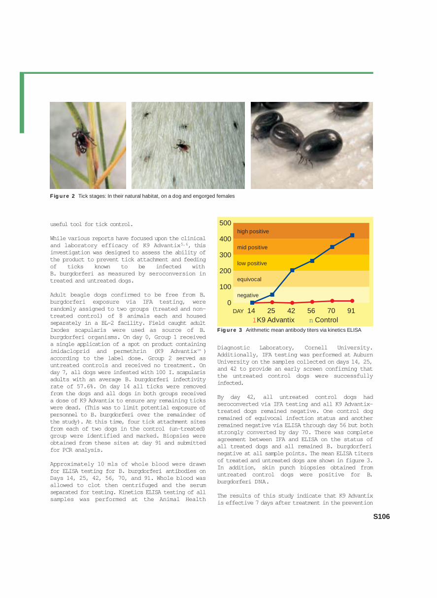
Figure 1 Two ticks attached with hair loss and redness
Evaluation of Permethrin and Imidacloprid forPrevention of Borrelia burgdorferi Transmissionfrom Blacklegged Ticks (Ixodes scapularis) toBorrelia burgdorferi-free Dogs
SPENCER J.A.1, BUTLER J.M.1, STAFFORD K.C.2, POUGH M.B.3, LEVY S.A.4,
BLEDSOE D.L.5 & BLAGBURN B.L.1
1Department of Pathobiology, College of Veterinary Medicine, Auburn University, Alabama, USA;2Department of Forestry and Horticulture, Connecticut Agricultural Experiment Station, New Haven, Connecticut, USA;3Animal Health Diagnostic Laboratory, Cornell University, Ithaca, New York, USA; 4Durham Veterinary Hospital, Durham,Connecticut, USA; 5Bayer HealthCare LLC, Animal Health Division, Shawnee Mission, Kansas, USA
useful tool for tick control.
While various reports have focused upon the clinicaland laboratory efficacy of K9 Advantix5,6, thisinvestigation was designed to assess the ability ofthe product to prevent tick attachment and feedingof ticks known to be infected with B. burgdorferi as measured by seroconversion intreated and untreated dogs.
Adult beagle dogs confirmed to be free from B.burgdorferi exposure via IFA testing, wererandomly assigned to two groups (treated and non-treated control) of 8 animals each and housedseparately in a BL-2 facility. Field caught adultIxodes scapularis were used as source of B.burgdorferi organisms. On day 0, Group 1 receiveda single application of a spot on product containingimidacloprid and permethrin (K9 Advantix™ )according to the label dose. Group 2 served asuntreated controls and received no treatment. Onday 7, all dogs were infested with 100 I. scapularisadults with an average B. burgdorferi infectivityrate of 57.6%. On day 14 all ticks were removedfrom the dogs and all dogs in both groups receiveda dose of K9 Advantix to ensure any remaining tickswere dead. (This was to limit potential exposure ofpersonnel to B. burgdorferi over the remainder ofthe study). At this time, four tick attachment sitesfrom each of two dogs in the control (un-treated)group were identified and marked. Biopsies wereobtained from these sites at day 91 and submittedfor PCR analysis.
Approximately 10 mls of whole blood were drawnfor ELISA testing for B. burgdorferi antibodies onDays 14, 25, 42, 56, 70, and 91. Whole blood wasallowed to clot then centrifuged and the serumseparated for testing. Kinetics ELISA testing of allsamples was performed at the Animal Health
Diagnostic Laboratory, Cornell University.Additionally, IFA testing was performed at AuburnUniversity on the samples collected on days 14, 25,and 42 to provide an early screen confirming thatthe untreated control dogs were successfullyinfected.
By day 42, all untreated control dogs hadseroconverted via IFA testing and all K9 Advantix-treated dogs remained negative. One control dogremained of equivocal infection status and anotherremained negative via ELISA through day 56 but bothstrongly converted by day 70. There was completeagreement between IFA and ELISA on the status ofall treated dogs and all remained B. burgdorferinegative at all sample points. The mean ELISA titersof treated and untreated dogs are shown in figure 3.In addition, skin punch biopsies obtained fromuntreated control dogs were positive for B.burgdorferi DNA.
The results of this study indicate that K9 Advantixis effective 7 days after treatment in the prevention
S106
Figure 2 Tick stages: In their natural habitat, on a dog and engorged females
500high positive
400mid positive
300low positive
200equivocal
100negative
0DAY 14 25 42 56 70 91
lK9 Advantix n ControlFigure 3 Arithmetic mean antibody titers via kinetics ELISA

Parasitol Res (2003) 90: S107 – S110 DOI 10.1007/s00436-003-0905-7
Infections in dogs with the protozoan parasiteLeishmania are widespread in Mediterraneancountries. Furthermore, canine leishmaniases havea worldwide distribution and can be found in Asia,Africa and America. First reports on canine leish-maniasis were recently published also from the US,it remains unclear until now whether the diseasewas imported from abroad, or has alreadyestablished within the country. Leishmaniases arevector-borne diseases: the promastigote stage ofthe parasite is transmitted to the host during theblood feeding of an insect vector, the sand fly. Thecausative agents of canine leishmaniasis areLeishmania infantum in the Mediterranean Basin andthe Middle East and L. chagasi in South and CentralAmerica. Initially, L. chagasi was considered to bedistinct from L. infantum, however, recentmolecular studies indicate that they areindistinguishable. Today we believe that theparasite reached the New World in dogs transportedfrom Europe to the Americans by the colonists. L.infantum/chagasi is also an important human healthproblem and dogs serve as the main reservoiranimal. Several studies showed that the prevalenceof human leishmaniasis could be significantlydecreased with control of leishmaniasis in dogs.
Repellent Efficacy of a Combination ContainingImidacloprid and Permethrin against Sand Flies(Phlebotomus papatasi) on Dogs
MENCKE N.1, VOLF P.2, VOLFOVA V.2 & STANNECK D.11Bayer AG, BHC-Business Group Animal Health, D-51368 Leverkusen, Germany2Charles University, Department of Parasitology, Prague, Czech Republic
Fig. 1 Life cycle of Leishmania
A Development in Man: 1 Flagellated L. donovani parasites carried byPhlebotomus species (promastigote stage) enter macrophages(Giemsa stain); 2–6 Intracellular development in macrophages and laterin endothelial cells; 7 Macrophages in perpheral blood containingamstigote stagesB Development in the sand fly vector: 8 Amastigote stages, within hostcells, in the fly’s midgut; 9 Growth and multiplication of the promasti-gote stage; 10 Flagellate stage (metacyclic promastigote form) fromfly’s proboscisC Development as in man A in animal reservoir (dogs, small rodents etc.)
Transmission by the sand fly can occur as follows:From Man to Man A‘B‘AFrom Animal to Animal C‘B‘CFrom Animal to Man C‘B‘Aand vice versa A‘B‘C
Taken from: PIEKARSKI G. (1989) Medical Parasitology; Springer – Berlin,
Heidelberg, New York
S107
10
1
2
3
4
5
6
7a b
ab
8
A
B
C
9
10
1
2
3
4
5
6
7
8
9

S108
Prevention of dogs from sand fly bites and thusreducing the risk of infection with L. infantum istherefore our veterinary obligation.
Biology of Leishmania and theirvectors
While there is no morphological differentiationbetween the Leishmania species, moleculartechniques implemented today distinguish thepreviously described species as the L. donovanicomplex (three species: L. donovani, L. infantum andL. chagasi), the L. mexicana complex (three mainspecies: L. mexicana,L. amazonensis and L. venezuelensis), further L.tropica, L. major,L. aethiopica and the subgenus Viannia with four mainspecies (L. braziliensis, L. guyanensis, L. panamensisand L. peruviana).
Within humans and dogs Leishmania multiply within aparasitophorous vacuole of macrophages asamastigotes (forms without flagella). Finally, thesemacrophages rupture and amastigotes enter otherphagocytic cells, mainly of the reticulo-endothelialsystems in liver, spleen, bone marrow and lymphnodes. Sand flies feeding on infected host ingest ablood meal with infected monocytic cells. In themidgut of the sand fly the amastigotes transform intoa flagellated promastigotes, multiply and then, duringthe second blood feeding, they are transmitted to thenext vertebrate host. Development to the infectiouspromastigote occurs under favorable tropicalconditions within 5 – 8 days. The incubation period indogs is several weeks to months.
>>
Fig. 2 Sand fly female (size 2.5 mm)

Sand flies belong to the insect order Diptera,suborder Nematocera. The Old World sand flies belongto genus Phlebotomus, the New World ones to thegenus Lutzomyia. Both these genera are importantvectors of Leishmania parasites. The biology of theadult sand fly is that of a typical bloodsuckingNematocera: both sexes feed on sugar solutions butfemales need a blood meal to produce eggs. Larvaehatching from the eggs develop through 4 larvalinstars. They are terrestrial and live in soil rich inorganic substrates where they also pupate. The adultfemales seek hosts for a blood meal in a clearcircadian activity. They are nocturnal blood feeders,resting over the hot day away from sunlight inrelatively cool and humid places, like cellars,stables, crevices or rodent holes. Once landed on thepotential host, the dog or other mammal, they hopover the coat aiming for less hairy place; in the headregion they like to bite around the muzzle, eye ormouth. Similarly to other bloodsucking insects, thesand fly saliva contains anticoagulants, vasodilatorypeptides and enzymes with antiinflammatory,antihemostatic and anaesthetic properties. Thesecomponents of saliva are important for transmissionand serve as enhancing factors of the parasiteinfection. Sand flies are fast feeders; once theycreated a small pool of blood in epidermis of the host they rapidly filltheir midgut with blood. Leishmania is well adaptedto this feeding habit, entering the host via theproboscis with the saliva injected into the host.Clinical Canine leishmaniasis
Clinical features of canine leishmaniasis varywidely, while skin lesions are the most usualmanifestation. The incubation period is 4 weeks toseveral years. The pathomechanism of the infectionis a combination of chronic inflammation of skin,liver, GI-tract, kidneys, eyes and bones and a immunemediated polyarthritis, glomerulonephritis, arthritisand uveitis. In addition, dogs presented to the clinicoften have concomitant infections, due toimmunosuppression thus complicating the diagnosis.The skin lesions are alopecia with intense, drydesquamation, usually on the head. In most casesweight loss is common. Circulating immunocomplexescausing glomerulonephritis, proteinuria and sub-sequently renal failure is a common cause of death inaffected animals. Besides the dogs with visibleclinical signs of the disease, asymptomatic carriersare frequently reported. While in endemic areas mostdogs have contact with the parasite, the prevalenceof the disease is usually up to 20%. The reason whysome dogs develop the disease and others areresistant is not fully understood yet. Diagnosis ofcanine leishmaniasis is complicated as most tests
available are not sensitive or specific enough.Several treatment regimes are recommended but aonce dog got ill the parasites will never beencompletely eliminated. Prevention of sand fly bitesis therefore the most important way to stop thecirculation of canine leishmaniasis.
Human Leishmaniasis
In humans the protozoan parasite of the genusLeishmania causes cutaneous (CL), mucocutaneous(MCL) and visceral (VL) leishmaniases. The WHOreported that worldwide 350 million humans in 88countries are at risk and 12 million people areaffected by leishmaniases, with about 1.5 – 2.0Million new cases of CL and 0.5 Million new cases ofVL cases annually. More than 90% of the VL cases arereported from Bangladesh, Brazil, India, Nepal andSudan. Visceral leishmaniasis, also known as ‘Kala-azar’ is caused by L. infantum in the Old World, L.chagasi in the New World, and by L. donovani in Africaand Asia. Coinfection in humans with immuno-deficiency syndroms like AIDS is common. Afterincubation period (usually 2-8 months) patients
S109
Repellent Efficacy of Imidacloprid and Permethrin against Sand Flies
Fig. 3 Dogs with skin lesions

develop pyrexia, wasting and hepatosplenomegaly;especially children are at risk. There is a long list ofclinical findings, with fever, discomfort from anenlarged spleen, abdominal swelling, weight loss,cough and diarrhea being the most prominent once.While untreated the mortality is about 90%. Aftersuccessful recovery from VL due to L. donovaniinfection, patients may develop so-called ‘post kalaazar dermal leishmaniasis’.
The mucocutaneous form (Espundia) occurs in somecases ofL. braziliensis infection in South America. Disease isfully manifested months or years after the cutaneoussores have healed. Papules and ulcerative lesionsoccur on the nose, mouth and larynx and finally maydestroy the whole face.
In the Old World, L. major and L. tropica are thecausative pathogens of the cutaneous form (orientalsore), while in the New World this form is causedmostly by the parasites of L. mexicana and L.brasiliensis complexes. Primary skin lesions occurat the site of sand fly bite, often at unprotected bodyregions like arms, legs and in the face. Most commontype of lesion is a chronic ulcer with an induratedmargin. The majority of these lesions are self-healingin several months leaving a scar.
S110
Fig. 4 Phlebotomus papatasi male (left) and femaleTaken from Mühlens P., Nauck E. & Vogel H. (1942), Krankheiten und Hygiene
der warmen Länder. Georg Thieme, Leipzig

Parasitol Res (2003) 90: S111 – S112 DOI 10.1007/s00436-003-0906-6
OBJECTIVE
The aim of this study was to determine whether asingle or two treatments, four weeks apart, with anovel, topically applied, formulation of Imidacloprid10% plus Moxidectin 1%, applied at a dose of 0.1ml/kg body mass, would be effective in thetreatment of ear mite, Otodectes cynotis,infestations in cats.
STUDY DESIGN AND METHODS
This study was performed in compliance with VICHGL9 “Good Clinical Practice, June 2000” at ClinVetInternational (Pty) Ltd, Bloemfontein, Republic ofSouth Africa. Thirty cats naturally infested with O.cynotis were allocated to three groups of 10 catseach by randomisation through minimization with
Day –1 body weight as the primary criterion. Group1 served as a negative placebo treated (Days 0 and+28) control. Group 2 received a single treatmentwith the Imidacloprid/Moxidectin solution on Day 0and a placebo treatment on Day +28. Group 3 wastreated with the Imidacloprid/Moxidectin solutionon Days 0 and +28. Treatments were blinded. Thecats were kept individually in stainless steel cagesin environmentally controlled rooms. The threedifferent treatment groups were kept under similarconditions in adjacent rooms. Eight days after thefirst treatment and thereafter at fortnightlyintervals, the ears of the cats were examined forthe presence of ear mites by using an otoscope, earscrapes and dry cotton swabs. Assessments on Day+50 were performed on anaesthetized cats. Thestudy schedule is summarized in Table 1.Efficacy evaluation was based on the presence ofmites in the ears of cats. The success rate in each
S111
Evaluation of the Efficacy of an Imidacloprid 10% /Moxidectin 1% Spot-on against Otodectes cynotisin Cats
FOURIE L.J.1, KOK D.J.2 & HEINE J.3
1 University of the Free State, Bloemfontein, Republic of South Africa;2 ClinVet International (Pty) Ltd., Bloemfontein, Republic of South Africa;3 Bayer AG, BHC-Business Group Animal Health, D-51368 Leverkusen, Germany
Figure 1 Debri in the ear of a cat infested with Otodectes cynotis mites (left) and the healthy ear of a cat (right) after two treatments withImidacloprid 10% / Moxidectin 1%, applied 28 days apart at a dose of 0.1 ml/kg body mass
Acclimatization Allocation to groups Treatment Pre- and Post-treatment mite assessments
Day: –7 Day: –1 Day: 0; +28 Day: –3; +8; +22; +36; +50
Table 1 Synoptic overview of the study layout

treatment group for Days +22 and +50 wascalculated as follows:
Success rate (%) = x 100 , where___ ·___y 1
x = number of cats observed with no live mitesy = total number of cats in groupRESULTS
Live mites, varying in numbers from 1 to 66 werecounted in scrapings taken (Day –3) from the earsof all 30 cats included in the study. Live mites were
observed in the ears of all the cats in the placebotreated group on all the assessment days. In the caseof the group of cats which received a singletreatment either one or two cats still harboured livemites in their ears on the different assessmentdays. No mites were observed in the ears of cats,which received two treatments, on any of theassessment days. The treatment success (%) forGroups 2 and 3 are summarized in Table 2.
CONCLUSION
A single treatment with theImidacloprid/Moxidectin solution applied at adosage of 0.1 ml/kg body mass resulted in a treat-ment success rate of 80% as assessed 50 daysafter treatment. Two treatments with theImidacloprid/Moxidectin solution, four weeksapart, at a dosage of 0.1 ml/kg body mass, wasefficacious in curing all cats from Otodectes cynotisinfestations as assessed 22 days after the secondtreatment (Day +50). l
S112
Table 2 Summary of the treatment success rate (%)for Groups 2 and 3 as assessed on Days +22 and +50,respectively.
Day Group 2: Treated Group 3: Treated twiceonce (Day 0) (Day 0 and +28)
+22 90 100
+50 80 100
Figure 2 Female mite with a visible egg

Parasitol Res (2003) 90: S113 – S114 DOI 10.1007/s00436-003-0907-5
INTRODUCTION
Dogs and cats are often co-infected with endo- andectoparasites. Roundworms of the genus Toxocararepresent the most prevalent endoparasites in dogsand cats followed by hookworms of the generaAncylostoma and Uncinaria (Coati et al., 2003).Worldwide many surveys have shown that fleas arethe dominating group of ectoparasites in dogs andcats with infection rates of up to 100% undercertain circumstances (Krämer and Mencke, 2001).Recently, in European field studies up to 27% oftested cats and 17% of dogs were found to be co-infected with gastro-intestinal nematodes and fleas(Knoppe, personal communications). In cases likethese the concomitant treatment using anectoparsiticide together with an anthelmintic isappropriate. In the present study we have tested theanthelmintic efficacy of a new topical antipara-siticide which combines the insecticide imidaclopridand the macrocyclic lactone moxidectin for thesimultaneous treatment and prevention of ecto- andendoparasitic infections in cats and dogs. Here, theefficacy against larval and immature stages ofhook- and roundworms in cats was specificallyinvestigated. Furthermore, the potential use of thisproduct for the prevention of patent endoparasiticinfections was demonstrated by showing thepersistent efficacy against Uncinaria stenocephalainfections in dogs.
MATERIALS AND METHODS
The test products were clear solutions withimidacloprid 10% (w/v)/moxidectin 2.5% (w/v)for spot on treatment of dogs and imidacloprid 10%(w/v) /moxidectin 1.0% (w/v) for cats. Thesolution was applied once cutaneously atrecommended minimum dosages of 0.1 ml/kg bodyweight, i.e. 10 mg imidacloprid and 2.5 mgmoxidectin per kg body weight in dogs and 1 m gmoxidection per kg body weight in cats.
Four groups of eight less than five month old catseach were artificially infected with 300 infectivestages of Ancylostoma tubaeforme and four furthergroups with Toxocara cati, respectively. One of theA. tubaeforme infected groups was treated at 7 andtogether with a placebo treated control groupnecropsied 12 days post infection (d.p.i.). The tworemaining groups were treated at 11 and necropsyat 16 d.p.i.. The T. cati groups were treated was at14 and 24 d.p.i., while necropsy was at 19 and 29d.p.i., respectively. Parasitic stages were countedfor the complete intestinal contents. Following theintestinal washings the small intestines, suspendedby wire, were soaked at 37°C in 0.9% saline fortwo hours to allow migration of mucosal larvalstages. In addition to that the mucosa of the smallintestines of the A. tubaeforme infected cats werestripped of and processed with pepsin-digestion fordetection of mucosal stages.
To assess the persistence of efficacy eight ofsixteen dogs were treated with the test product at18 days prior to experimental infection with 300 U.stenocephala larvae. At 21 d.p.i. all animals werenecropsied and worms were counted.
RESULTS AND DISCUSSION
For the control groups the following A. tubaeformestages (geometric means) were found per testanimal at 12 d.p.i. 9.1 (SD ±15.1) third stagelarvae, 36.2 (SD ±23.3) fourth stage larvae and 2.8(SD ±3.8) immature adults (Fig.1). At 16 d.p.i. 5.0(SD ±4.2), 45.9 (SD ±26.4) and 4.6 (SD ±7.0) third,fourth and immature adult stages, respectively,were counted per cat. Adult stages were presentneither at 12 d.p.i. nor at day 16 d.p.i. in any cat.Earlier investigations proved that the prepatencyperiod of this strain is approximately 28 days. Themean cumulative worm counts in the control groupswere 58.5 and 65.3 at 12 and 16 d.p.i.,respectively. No parasite stages were found in anyof the moxidectin/imidacloprid treated animals.
S113
Larvicidal and Persistent Efficacy of anImidacloprid and Moxidectin Topical Formulationagainst Endoparasites in Cats and Dogs
SAMSON-HIMMELSTJERNA G. VON1, EPE C.1, SCHIMMEL A.2 & HEINE J.2
1Institute of Parasitology, Hannover School of Veterinary Medicine, Buenteweg 17, D-30559 Hannover, Germany;2Bayer AG, BHC-Business Group Animal Health, D-51368 Leverkusen, Germany

Within the T. cati infected cats a mean of 5.1 (SD±3.7) fourth stage larvae and no immature adultstages were found at 19 d.p.i., while 5.8 (SD ±7.3)and 3.5 (SD ±5.3) fourth and immature adults (Fig.2) were present at 29 d.p.i., respectively. For thisstrain the prepatency period was found to beapproximately 42 days. The average overall wormcounts were 7.3 and 15.4 at 24 and 29 d.p.i.,respectively. In the animals treated with the testproduct and necropsied at 19 d.p.i. no parasiteswere found, whereas in those necropsied at 29 d.p.i.0.2 (SD ±0.01) and 0.3 (SD ±0.02) fourth andimmature adult stages, respectively were found.All test group worm counts differed significantly(p<0.05) from all respective control group wormcounts. These data clearly demonstrate highefficacies of the imidacloprid/moxidectin solutionagainst early but also against later larval andimmature adult stages of the two parasite speciestested in this study (Table 1).
In contrast to our results Okoshi and Murata (1957)found far less A. tubaeforme L3 in cats necropsied12 and 16 d.p.i., whereas similarly to this study theL4 stages represented the most prominent group.The recovery rates of 19.5 and 21.7 for 12 and 16d.p.i., respectively, found in the present study aresimilar to those described in the earlier publication.The large standard deviations observed in all groupsindicate that both strains exhibit extensivedifferences concerning the progress of developmentwithin individual hosts.
The untreated U. stenocephala infected dogs showedgeometric mean immature adult and adult parasitecounts of 4.6 (SD ±3.9) and 8.1 (SD ±4.3),respectively, while in the test product treatedanimals no worms were found. Thus the productproved to have a persistent efficacy over at least18 days which completely prevented thedevelopment of intestinal stages for this species.
To our knowledge this is the first time that ananthelmintic product was shown to completelyremove early larval stages of hookworms andascarids in cats and furthermore to prove per-sistence of efficacy. Similar findings concerning thelarvicidal efficacy were observed also in additionalstudies employing the same approach in dogs (datanot shown). Furthermore, complete adulticidalefficacies against the respective adult stages of theparasite species used here were found in multipleinvestigations (data shown elsewhere). Mostrecently the potential of human infection with T.canis by direct contact with dogs was illustrated by
S114
Table 1 Efficacies of the test product against larval and immatureadult stages of A. tubaeforme and T. cati in cats.
Species A. tubaeforme T. cati
Stage L3 L4 Immature L4
Immatureadults adults
Efficacy 10 0 10 0 10 0 100 (19 d.p.i.)9 1( % ) 97 (27 d.p.i.)
Figure 1 (left) Female A. tubaeforme L4 with a length of 2.14 mmisolated from the small intestines at 12 d.p.i..
Figure 2 (top) Female T. cati immature adult with a length of 5.12mm isolated from the small intestines at 29 d.p.i..

Parasitol Res (2003) 90: S115 – S117 DOI 10.1007/s00436-003-0908-4
INTRODUCTION
The ability of acaricides to repel or kill ticks beforethey attach to a host and feed is important for theprevention of transmission of tick born pathogens.A study design to compare therepellency/acaricidal properties of topicallyapplied products against the castor bean tick (Ixodesricinus), the primary vector of Lyme disease inEurope, following exposure to treated dogs has beenpreviously reported.1,2 A similar study design wasreported to demonstrate repellency and acaricidalproperties of a topical 45% w/w permethrinsolution (Kiltix® , Bayer Corp. USA) usedconcurrently with a 9.1% w/w imidacloprid spot onsolution (Advantage® , Bayer) against Americandog ticks (Dermacentor variabilis) the primaryvector of Rickettsia rickettsii, the causativeorganism of Rocky Mountain spotted fever in dogs.3
The present study was conducted to compare thetick repellency/acaricidal properties of acombination of 8.8% w/w imidacloprid and 44%w/w permethrin (K9 Advantix™ , Bayer Corp. USA)with the combination of 9.8% w/w fipronil and8.8% w/w S-methoprene (Frontline Plus® , Merial)against the Brown dog tick (Rhipicephalussanguineus) the vector of Ehrlichia canis in dogs.
MATERIALS AND METHODS
A total of 18 healthy laboratory dogs of bothgenders with a body weight between 6.9 and 24.8kg and the ability to harbor tick infestations wereincluded in the study. The dogs had not been treatedwith any acaricide within 60 – 90 days prior to thestudy. On test day –12 all dogs were bathed with amild, non-medicated shampoo. The dogs wereexperimentally infested with unfed adult brown dogticks (Rhipicephalus sanguineus). This study design
evaluated the transfer and attachment of ticks fromthe environment to dogs. Individual heavy-gaugeplastic pet transport carriers were used for tickchallenges. Each carrier contained a light colorednylon carpet with a nap of 12 – 15 mm to cover 50– 60% of the floor area. For each tick challenge, 50adult R. sanguineus (approximate male/female sexratio of 1:1) were placed on the carpet. The carpetwas sprayed lightly with water prior to placementwith the ticks. After a 15 minute acclimationperiod, dogs were placed in the carriers for a 2 hourperiod to expose them to the live ticks. The dogswere then removed, examined visually and combedfor ticks. The number of live ticks (attached andunattached) and dead ticks on the dogs were countedand removed. Live and dead ticks remaining on thecarpet and in the carriers were counted. Carrierswere thoroughly washed and carpets werediscarded after each use.
On test day -3 each dog was exposed to ticks asdescribed above. Pretreatment live tick counts
S115
Evaluation of K9 Advantix™ vs. Frontline Plus®Topical Treatments to Repel Brown Dog Ticks(Rhipicephalus sanguineus) on Dogs
YOUNG D.R.1, ARTHER R.G.2 & DAVIS W.L.2
1Young Veterinary Research Services, Modesto, California, USA;2 Bayer HealthCare LLC, Animal Health Division, Shawnee Mission, Kansas, USA
Figure 1 Mouthpart of a brown dog tick (Rhipicephalus sanguineus)

(attached and unattached on each dog) wererecorded. The dogs were then ranked by the totalnumber of live ticks and were blocked into groups ofthree. Within each block the dogs were randomizedto one of the three treatment groups and treated onstudy day 0 with the recommended dosage:
Group 1: 8.8% w/w imidacloprid plus 44.0% w/wpermethrin (K9 Advantix™, Bayer Corp. USA).
Group 2: 9.8% w/w fipronil plus 8.8% w/wS-methoprene (Frontline® Plus, Merial).
Group 3: Control (untreated).
S116
Test Day 3 7 14 21 28 35G
eom
etric
Mea
n C
ount
Control
Fig. 2 Geometric mean number of repelled ticks per carrier
Table 1 Geometric mean number of live R. sanguineus per dogand percent efficacy
Test Group 1 Group 2 Group 3Day K9 Advantix Frontline Plus Control
__χ Ticks/Dog% Efficacy
__χ Ticks/Dog% Efficacy
__χ Ticks/Dog
-3 35.2 – 34.5 – 35.2
3 0.4* 98.5 20.5 25.9 27.6
7 1.3* 95.4 12.8* 56.8 29.5
14 1.8* 90.6 24.0 -28.1 18.8
21 3.6* 84.0 24.1 -7.8 22.4
28 2.7* 89.1 27.4 -11.4 24.6
35 1.7* 85.9 14.6 -21.9 12.0
*Statistically different versus control group (P < 0.05)
Table 2 Geometric mean number of repelled ticksa per dog carrier
Test K9 Advantix Frontline Plus ControlDay Geo Mean No. Ticks Geo Mean No. TicksGeo Mean No. Ticks
3 40.1* 17.1 12.3
7 37.0* 28.1* 11.7
14 38.5* 17.0 21.0
21 32.3* 16.0 17.3
28 35.2* 10.9* 19.1
35 40.4 19.2 27.2
aDead plus live ticks remaining in the carrier.
*Statistically different versus control group (P < 0.05)
45
40
35
30
25
20
15
10
5
Frontline Plus K9 Advantix
>>

Tick challenge was performed on days 3, 7, 14, 21,28 and 35 post treatment. On each of these days thedogs were placed in the pet carriers for 2 hours andexposed to 50 adult R. sanguineus as previouslydescribed. Following this time interval the dogswere removed. Live and dead ticks observed on thedog and ticks remaining in the carrier were counted.Percent efficacy was calculated for the twotreatment groups on each evaluation day with thefollowing formula using the geometric mean tickcounts:
Results of this study were evaluated by descriptiveand inferential statistical methods. Means,variability parameters, and range of values werecalculated for tick counts. In addition, repeatedmeasures mixed model analysis of variance wasused on log (count +1) transformed tick countstesting for treatment group differences, usingcontrast statements to control group comparisons.
RESULTS
The geometric mean number of live ticks per dog(attached and unattached) and percent tick efficacyis displayed in Table 1. Pre-treatment (test day –3)live tick counts ranged from 27 – 46 (Geo mean =35.0 ticks/ dog) following 2 hours of exposure to
ticks in the pet carriers. There were significantlyfewer numbers of live ticks on the K9 Advantixtreated dogs than on the control dogs for all 6 post-treatment evaluations (P <0.05). The control dogshad a range of 12.0 – 29.5 live ticks/dog during eachof the 6 post-treatment tick exposure intervals. Arange of 0.4 – 3.6 live ticks/dog were recorded forthe K9 Advantix treated dogs through day 35. TheFrontline Plus treated dogs had a range of 12.8 –27.4 live ticks/dog through day 35. There were nosignificant differences between the number of liveticks observed on the control vs. Frontline Plustreated dogs during the post-treatment tickexposure periods for days 3, 14, 21, 28 and 35. Ontest days 14, 21, 28 and 35 more live ticks wereobserved on the Frontline Plus treated dogs than onthe control dogs.
The geometric mean number of repelled ticks (deadplus live ticks remaining in the carrier) aredisplayed in Table 2. There were significantly moreticks repelled on the K9 Advantix treated group thanthe control group for all post-treatment evaluationsexcept day 35. There were no differences in therepelled ticks between the Frontline Plus treateddogs and control dogs for days 3, 14, 21 and 35.Significantly more ticks were repelled on thecontrol dogs than the Frontline Plus treated dogs onday 28. The repellency values represent (2.3, 1.3,2.3, 2.0, 3.2, 2.1) fold greater repellency of K9Advantix vs Frontline Plus for test days 3, 7, 14,21, 28 and 35, respectively.
% Efficacy =
Geo Mean No. Live Ticks/Dog(Control) – Geo Mean No. Live
Ticks/Dog(Treated)
S117
Figure 3 Brown dog tick(Rhipicephalus sanguineus)

Parasitol Res (2003) 90: S118 – S120 DOI 10.1007/s00436-003-0910-x
INTRODUCTION
Ticks and especially the worldwide distributed mem-bers of the species of the genus Ixodes threaten thehealth of man and his companion animals, since theyare able to transmit several agents of diseases (e.g.Borreliosis, Ehrlichiosis, Babesiosis) (Lane andCrosskey 1995, Mehlhorn 2001a, 2001b; Piesmanet al. 1987). Thus exposition prophylaxis is neededin order to avoid infestation with ticks and thesubsequent transmission of tick-borne diseases.
The combination of 10% imidacloprid and 50%permethrin (K9 Advantix™ , trade mark Bayer AG,Leverkusen, Germany) was recently registered bythe EPA for the United States market. The product isindicated for the prevention and treatment of ticks,fleas and mosquitoes and is for monthly use in dogs.The repellent effect of synthetic pyrethroids, knownfor acaricides, was recently published forpermethrin topically applied to dogs (Endris et al.
2000, 2002). In the present paper the effects of thisnew product were tested in-vitro and in-vivo asreported to some extent in a recent publication(Mehlhorn et al. 2003).
MATERIAL AND METHODS
Ticks Specimens of Ixodes ricinus – the so-calledcastor bean tick – were collected from nature, wherethey lurk at the tip of plants (Fig. 1) and werepropagated in the institute. Unfed (hungry) larvae,nymphs and adults were used for the experiments.Dogs Four dogs were treated topically with K9Advantix at the recommended dosage of 0.1 ml/kg
S118
The Effects of an Imidacloprid and PermethrinCombination against Developmental Stages ofIxodes ricinus Ticks
MEHLHORN H.1, SCHMAHL G.1, MENCKE N.2 & BACH T.2
1Institute for Zoomorphology, Cell Biology and Parasitology, Heinrich-Heine University, D-40225Düsseldorf, Germany; 2Bayer AG, BHC-Business Group Animal Health, D-51368 Leverkusen, Germany
Fig. 3 In the case of K9-Advantix-treated hair the ticks move back im-mediately, when approaching the hair and try to approach again (arrows)
Fig. 2 Ticks were placed into a petri dish with hair of treated anduntreated dogs arranged in a circular ring
Fig.1 Unfed female of Ixodes ricinus questing for a host at the tip ofa plant
>>

body weight. This corresponds with a dosage of 10mg imidacloprid/kg and 50 mg permethrin/kg. Fourdogs remained untreated as controls. At 7, 14, 21and 28 days after this treatment, both flanks of oneof these dogs were shaved, producing a circular,hairless region of about 20 cm in diameter. The hairwere collected separately and used for in-vitrostudies (Fig. 2).
RESULTS
In-vitro studies
Test for repellent activityThe hair taken from the treated dogs at 7, 14, 21, or28 days post-treatment and additionally from theuntreated dogs was arranged as a circular barrier ona filter paper in individual glass Petri dishes (Fig. 2).Into the centre of this circular barrier of hair, 50larvae, 8 – 10 nymphs or adults of Ixodes ricinuswere placed and their behaviour was followed for atleast 1 h. In all cases, it was noted that the ticksimmediately started to run in the direction of the hair.However, at a distance of 1 – 10 mm before reachingthe hair, they stopped, raised their pair of front legs,where the Haller’s organ (a chemosensitive organ) islocated, moved these two legs and went backwards(Fig. 3). Then they tried to approach the hair barrierfor 1-2 min at other places. Later, 2-3 ticks enteredthe hair, while the rest remained in the hairlesscentre of the Petri dish. This „hot-foot“ reactionindicates the existence of an initial repellenteffect. This effect is very valuable for theprotection against an infestation with ticks, since atick in the host-seeking position has only seconds todecide whether it will attach or not to a host passingby. Control ticks, however, immediately entered theuntreated hair barrier and thus found shelter there.
Acaricidal activity of the hair of treated dogsHair samples from dogs treated 7, 14, 21, or 28 dayspreviously with K9-Advantix were mixed withlarvae, nymphs and adults of I. ricinus ticks in Petridishes. While larvae and nymphs died within 48 h(very often within 24 h), the adult Ixodes tickssurvived for a longer period, although completelyparalysed. Ixodes adults (Fig. 4) became paralysedwithin 3 – 24 h, showing only slow movements of thelegs without true body-motion and never recovered.
In-vivo studies
Individual adult Ixodes ricinus ticks were put bymeans of tweezers onto the hair of treated anduntreated dogs. In the case of treated dogs, the ticksdropped from the hair immediately after being put inposition. On untreated control dogs, the ticks moved
over the hair to reach the skin for attachment withina few seconds.
On days 7, 14, 21 and 28 after treatment of the dogs,Petri dish-covers containing ten freshly caught adultfemale I. ricinus ticks were fixed on the shavedlateral thorax of the dogs. The behaviour of the tickswas observed for 15 min and the Petri dishes werethen covered by blackout/ bandaging material. Tickswere allowed to attach on the shaved skin area for 1h in the dark. After this period, the Petri dishes wereremoved and the position of the ticks was noted. Allticks were collected in another Petri dish,transferred into an incubator and observed after 3,24 and 48 h by means of a stereo microscope.
When considering the situation after placing the ticksonto the shaved skin, it was seen that the ticks triedto avoid contact with the skin and assembled along theborder or the inner lid of the plastic Petri dish.However, after 3 – 5 min, some ticks started tocrawl around on the shaved skin. After 1 h, whenremoving the blackout/ bandaging material, it wasnoted that in both groups (treated or non-treateddogs) only a few ticks (mainly two or three out of tenticks) had attached on the skin, while the rest wasstill unattached. The results were independent of thelength of the period after treatment with the testproduct (i.e. the number of attached ticks was similaron the dogs at 7, 14, 21, or 28 days after thecompound was applied to the hair). However, allIxodes ticks were alive and motile after this firstperiod.
After 3 h, all Ixodes ticks in the Petri dishes collectedfrom the treated dogs were found lying on their backsand showing only slight, paralytic movements of alleight legs. This finding was identical on the ticksderiving from dogs being treated 7, 14, 21, or 28days previously. These ticks never recovered from
S119
Fig. 4 Light micrographs of Ixodes ticks, which had contact tountreated hair (a) or treated hair (b). In the first case the ticks crawledaround, while in the second case ticks became paralysed and later died
a b
The Effects of an Imidacloprid and Permethrin Combination…

this paralysis and died mostly within 24– 48 h afterfirst exposure to the skin, even when deriving fromdog treated 28 days previously. Thus, it can beconcluded that a contact of only 1 h to the shaved skinof an imidacloprid/permethrin-treated dog issufficient to kill Ixodes ticks.
Electron microscopic studiesWhen examining (by means of electron microscopy)the tissues of Ixodes ticks having had contact withK9-Advantix® treated hair or not (controls), it wasseen that damages were found at the level of muscleand nerve cells. The grade of damages increased withthe time of exposure leading to a final completemorphological and functional destruction (Fig. 5).
CONCLUSIONS
Our in-vivo and in-vitro experiments offered somesignificant results:
1. The new combination of 10% imidacloprid and 50%permethrin, applied topically to dogs, resulted in asignificant repellent effect, lasting for at least 4weeks against Ixodes ricinus ticks.
2. For dogs resting at tick-contaminated places (thusoffering the ticks time for delayed entry into thecoat), the acaricidal activity of the new productconsiderably limits the attachment of ticks. Thisefficacy is independent from life cycle stages of theI. ricinus tick, the known vector of pathogens thatcause borreliosis, ehrlichiosis and babesiosis.
3. Transmission of e.g. Borellia burgdorferi by Ixodesspp. ticks is dramatically increased by the durationof tick-feeding on the host (Piesman et al. 1987). Therepellency and acaricidal effect after 1 h of exposurereported here is expected to be sufficient to preventtransmission of tick-borne diseases.
4. The electron microscopical analysis of nerve andmuscles of ticks having had contact to treated hairshows significant, irreversible damages.
Fig. 5 Transmission electron micrographs of tissues of ticks that had been exposed to K9-Advantix or remained untreated.a) Control. The muscle fibres and the mitochondria (yellow) are unchanged.b) Cross section through a nerve (=peripheral sensillum) after 4 h of contact with K9-Advantix. Note the initiation of a vacuolization.c) Longitudinal section through a nerve of a tick after 48 h of exposure to K9- Advantix. Note the intense degeneration.d) Section through a muscle fibre after an exposition of 48 h. Note the contraction of the fibre and the destruction of mitochondria.
S120
a b c
d
Parasitol Res (2003) 90: S121 – S123 DOI 10.1007/s00436-003-0911-9
Efficacy of the Compound Preparation Imidacloprid10% (w/v) / Permethrin 50% (w/v) Spot-on against Ticks(I. ricinus, R. sanguineus) and Fleas (C. felis) on Dogs
EPE C.1, COATI N.1 & STANNECK D.2
1Institute of Parasitology, Hannover School of Veterinary Medicine, Buenteweg 17, D-30559 Hannover, Germany2Bayer AG, BHC-Business Group Animal Health, D-51368 Leverkusen, Germany
S121
STUDY OBJECTIVES ANDRATIONALE
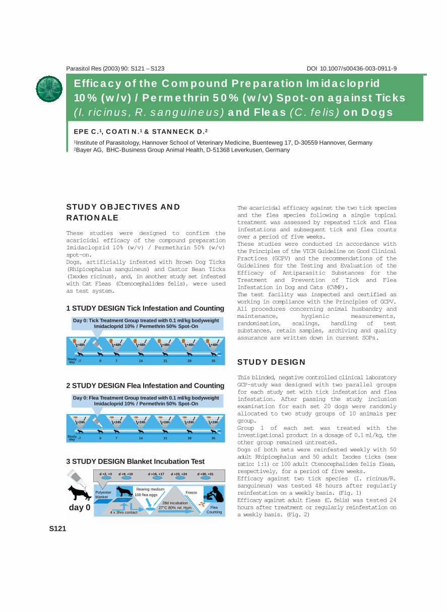
These studies were designed to confirm theacaricidal efficacy of the compound preparationImidacloprid 10% (w/v) / Permethrin 50% (w/v)spot-on.Dogs, artificially infested with Brown Dog Ticks(Rhipicephalus sanguineus) and Castor Bean Ticks(Ixodes ricinus), and, in another study set infestedwith Cat Fleas (Ctenocephalides felis), were usedas test system.
The acaricidal efficacy against the two tick speciesand the flea species following a single topicaltreatment was assessed by repeated tick and fleainfestations and subsequent tick and flea countsover a period of five weeks.These studies were conducted in accordance withthe Principles of the VICH Guideline on Good ClinicalPractices (GCPV) and the recommendations of theGuidelines for the Testing and Evaluation of theEfficacy of Antiparasitic Substances for theTreatment and Prevention of Tick and FleaInfestation in Dog and Cats (CVMP).The test facility was inspected and certified asworking in compliance with the Principles of GCPV.All procedures concerning animal husbandry andmaintenance, hygienic measurements,randomisation, scalings, handling of testsubstances, retain samples, archiving and qualityassurance are written down in current SOPs.
STUDY DESIGN
This blinded, negative controlled clinical laboratoryGCP-study was designed with two parallel groupsfor each study set with tick infestation and fleainfestation. After passing the study inclusionexamination for each set 20 dogs were randomlyallocated to two study groups of 10 animals pergroup.Group 1 of each set was treated with theinvestigational product in a dosage of 0.1 ml/kg, theother group remained untreated. Dogs of both sets were reinfested weekly with 50adult Rhipicephalus and 50 adult Ixodes ticks (sexratio: 1:1) or 100 adult Ctenocephalides felis fleas,respectively, for a period of five weeks.Efficacy against two tick species (I. ricinus/R.sanguineus) was tested 48 hours after regularlyreinfestation on a weekly basis. (Fig. 1)Efficacy against adult fleas (C. felis) was tested 24hours after treatment or regularly reinfestation ona weekly basis. (Fig. 2)
-7 0 7 14 21 28 35
+48h +48h +48h +48h +48h +48h
Studyday
-7 0 7 14 21 28 35
+24h +24h +24h +24h +24h +24h
Studyday
100 flea eggs
4 x 3hrs contact
Polyesterblanket
28d incubation27°C 80% rel. Hum.
Rearing mediumFreeze
FleaCounting
d +2, +3 d +9, +10 d +16, +17 d +23, +24 d +30, +31
day 0
1 STUDY DESIGN Tick Infestation and Counting
Day 0: Tick Treatment Group treated with 0.1 ml/kg bodyweightImidacloprid 10% / Permethrin 50% Spot-On
2 STUDY DESIGN Flea Infestation and Counting
Day 0: Flea Treatment Group treated with 0.1 ml/kg bodyweightImidacloprid 10% / Permethrin 50% Spot-On
3 STUDY DESIGN Blanket Incubation Test

S122
Efficacy against juvenile fleas (larvae of C. felis)was tested with a blanket incubation test on aweekly basis. (Fig. 3)Tolerance was tested as second criterion also on aweekly basis.
MATERIAL AND METHODS
Ticks1. Brown Dog Ticks (Rhipicephalus sanguineus)originated partly of the laboratory strain inMonheim, reared on rabbits and partly of thelaboratory strain of EL Labs Soquville inCalifornia/USA (ratio 50:50 Monheim: USA strain).The ticks for infestation were unengorged adultmales and females (ratio 1:1) that have moulted atleast 14 days before to the adult stage.
2. Castor Bean Ticks (Ixodes ricinus) originatedfrom the Charité, Berlin. The ticks for infestationwere unengorged males and females (ratio 1:1) inwhich the transmission of the spermatophore hadalready occurred.
FleasCat fleas (Ctenocephalides felis felis) of thelaboratory strain in Hanover, reared on cats, wereused as test parasites. The adult unfed fleas wereheld in polyvinylic vials at ≈27° C/ ≈80% relativehumidity until they were used for infestation(maximum for 12 days).
Viable flea eggs from the routine breeding of this fleastrain were used as test parasites for the blanketlarvicidal test. The flea eggs were at an age of max.48 hours.
Tick Infestation procedureAfter sedation dogs of study set 1 were placed inindividual transport boxes. The ticks were releasedonto the back of the dogs and were allowed todisperse and move into the hair without disturbance.Dogs were released from the transport boxes afterapprox. 30 minutes.
Flea Infestation procedureAll dogs of study set 2 were artificially infested withabout 100 unfed cat fleas by pouring the fleastogether with cocoon material out of the vials ontothe dog’s coat.
Tick Counting Procedures
The following examination procedure was followed:Protective clothing and disposable hand gloves wereworn during the clinical examinations; gloves werechanged between each dog. The dog was placed on asingle-use paper pad covered table and wasidentified by ear tattooing.
All ticks were counted. Total body surface of thedog was examined by thumb counting, parting thehair with the fingers and removing the ticks with aforceps (excepting day 0 counts where the tickswere left in situ). The following regions wereexamined: head, ears, neck, lateral areas, dorsalstrip from shoulder blades to base of tail, tail andanal area, fore legs and shoulders, hind legs andabdominal area from chest to inside hind legs.Removed ticks were placed into individually markedvials and differentiated and counted in thelaboratory. There was no time dictated for thecounting procedure.
Flea Counting:
Figure 1 Cat fleas (above); Castor bean ticks in all stages (below)
>>

S123
The following examination procedure was followed:Total body surface was combed with a flea comb inthe following sequence: Head, ears, lateral areas,dorsal strip from shoulder blades to base of tail, tailand anal area, fore legs and shoulders, hind legsabdominal area from chest to inside hind legs, neck(application site). Dogs were combed until fleaswere no longer found but for a minimum of 5minutes. Live fleas were collected, removed andcounted. Total counts of fleas were recorded.
Blanket Incubation TestFor assessment of the larvicidal properties of thetest formulation in the surroundings of the dogsduring the weeks after treatment, the dogs wereplaced on blankets for twelve hours a week dividedinto four contact intervals of 3 hours each. Blanketswere exchanged weekly. One circular sample wascut from the middle part of each blanket after eachstudy week, placed into individually marked plasticdishes and frozen at about –18° C for 24 hours tokill possible living fleas, larvae or eggs on thesamples. For the incubation test approximately 50flea eggs (originated from the same flea strainwhich was used for the adulticidal tests) wereplaced together with flea rearing medium on eachfleece sample and were incubated at 27°C and 80%rel. hum. for four weeks. The number of thedeveloping fleas was counted on day 28 after startof incubation.
RESULTS
Efficacy was calculated comparing the tick counts inthe treated group to the tick counts in the untreatedgroup based on the geometric means as recommendedin the guidelines.
The product’s curative efficacy againstRhipicephalus sanguineus was 74.0% (day 2); thepreventive efficacy was 94.0% (day 9), 97.6% (day16), 92.0% (day 23), 95.9% (day 30) and 91.5%(day 37) (Tab. 1).The product’s curative efficacy against Ixodesricinus was 67.0% (day 2); the preventive efficacywas 100.0% (day 9), 100.0% (day 16), 99.5% (day23), 98.7% (day 30) and 91.6% (day 37) (Tab. 2).The curative efficacy of the treatment against fleaswas 99.4% (d+1); the preventive efficacy was 99.8%(day 8), 99.9% (day 15), 98.8% (day 22), 95.7%(day 29) and 90.4% (day 36) (Tab. 3).The larvicidal efficacy on the blankets after 12 hoursdog contact was 99.2% (d+3); 98.2% (day 10),98.5% (day 18), 85.1% (day 24) and 50.2% (day31) (Tab. 4).The general and dermal tolerance of the product wasvery well and no adverse reactions were observedin any of the treated dogs during the study.
CONCLUSIONS
1) Efficacy against ticks
0
2
4
6
8
10
12
14
16
18
20
0 +2 +9 +16 +23 +30 +37
100.0% 100.0% 99.5% 98.7% 91.6%
Effi
cacy
in %
100
90
80
70
60
50
40
30
20
10
67.0%
Geo
Gro
up M
ean
Treated Group
Control Group
study days
Efficacy (Geo Mean):
Effi
cacy
in %
Geo
Gro
up M
ean
Treated GroupControl Group
study days
Efficacy (Geo Mean):
0
10
20
30
40
50
60
-5 0 +1* +8* +15* +22* +29* +36*
99.4% 99.8% 99.9% 98.9% 95.7% 90.4%100
90
80
70
60
50
0
2
4
6
8
10
12
14
16
18
20
-7 +3* +10* +17* +24* +31**
99.2% 98.2% 98.5% 85.1% 50.2%100
90
80
70
60
50
40
30
20
10
Effi
cacy
in %
Geo
Gro
up M
ean
Treated Group
Control Group
study days
Efficacy (Geo Mean):
0
2
4
6
8
10
12
14
16
18
20
-5 +1 +2 +9 +16 +23 +30 +37
74.0% 94.0% 97.6% 92.0% 95.9% 91.5%100
90
80
70
60
50
40
30
20
10
Effi
cacy
in %
Geo
Gro
up M
ean
Treated Group
Control Group
study days
Efficacy (Geo Mean):
Tab 2 Efficacy against Ticks (Ixodes ricinus)
Tab 3 Adulticidal Efficacy against Fleas (Ctenophalides felis) –Flea Counts
Tab 4 Larvicidal Efficacy against Fleas (Ctenophalides felis) –Blanket Incubation
Tab 1 Efficacy against Ticks (Rhipicephalus sanguineus)

Parasitol Res (2003) 90: S124 – S125 DOI 10.1007/s00436-003-0912-8
S124
INTRODUCTION
The efficacy and safety of the combination of Imida-cloprid and Permethrin spot-on was assessed indogs naturally infested with ticks and/or fleas incomparison to the marketed control productFrontline® spot-on containing Fipronil as a positivecontrol. The study was conducted as a multicenter,multiregional, controlled, randomised and blindednon-inferiority clinical field study according toVICH GL 9 (Good Clinical Practice) and Directive2001/82/EC.
MATERIAL AND METHODS
Twelve veterinary clinics in three different areas(North-East, East and South) ofGermany, nine clinics in threeareas (Central, West Coast andSouth) of France and two clinics intwo different areas (Central andSouth) of Italy enrolled patients tothe study (Fig. 1). A total of 363dogs showing tick and/or fleainfestations were randomlyallocated to one of the twotreatments in a ratio of 2:1 for theinvestigational veterinary productand the control product. 229 dogswere treated with 10% (w/v)Imidacloprid / 50% (w/v) Perme-thrin (group G1) and 134 dogs weretreated with 10% (w/v) Fipronil(Frontline® , group G2). Treat-ments were administered once onday 0 according to body weightusing prefilled applicator tubes.The randomisation was doneseparately for animals hostingticks on test day 0 and households
hosting fleas. Personnel were blinded regardingtreatments.
In case that additional dogs and/or cats weresharing the same household with flea infested studydogs, these supplementary animals were alsotreated with the same product as the primarypatient of the household (dogs with either Imidaclo-prid/Permethrin or Frontline® for dogs; cats withmarketed products Advantage® (10% (w/v)Imidacloprid) for cats or Frontline® for cats).
Clinical examinationfs and parasite counts wereperformed by the examining veterinarian. Theanimal owners returned the dogs for re-checkexamination on day 2, day 7 (±2), day 14 (±2), day21 (±2) and on day 28 (±2) for study completion.
European Multicenter Field Trial on the Efficacy andSafety of a Topical Formulation of Imidacloprid andPermethrin (Advantix™) in Dogs naturally infestedwith Ticks and/or Fleas
HELLMANN K.1, KNOPPE T.1, KRIEGER K.2 & STANNECK D.2
1Klifovet AG Munich, D-80689 München, Germany2Bayer AG, BHC-Business Group Animal Health, D-51368 Leverkusen, Germany
>>Figure 1 Locations of the investigational sites within Europe

S125
Parasites were counted on each visit and collectedfor species identification at the central laboratory.
Dogs with concurrent tick and flea infestation onday 0 were randomised as flea households when ≥5fleas were found on these dogs. All dogs showing ≥1viable tick on day 0 were included in the ‘tickefficacy population’ (n=170). From each fleahousehold, the dog with the highest flea count on day0 was defined the main patient and included into the‘flea efficacy population’ (n=108). Dogs withprotocol violations were excluded from bothpopulations.
For all tick and flea counts, geometric means werecalculated and used to determine the percentagereduction of tick and flea counts within eachtreatment group. Day 2, day 7, day 21 and day 28were compared to baseline, day 0. The hypothesisstated that the efficacy of Imidacloprid/Permethrinwas on an average more than 90 % over thetreatment period. The Mann-Whitney test was usedto show non-inferiority.
RESULTS AND DISCUSSION
For both, tick and flea efficacy population, greaterthan 90% efficacy was achieved forImidacloprid/Permethrin. Non-inferiority wasshown for the investigational product for bothefficacy populations (lower 97.5% confidence bound> 0.29).
Individual efficacy values for flea infestation areshown in Fig. 2. Imidacloprid/Permethrin valueswere significantly non inferior to the positivecontrol.
Individual efficacy values for tick infestations areshown in Fig. 3.
Ixodes efficacy was highly significantly (p =0.0062) different from positive control group atday 28 and the Rhipicephalus efficacy was near tosignificantly (p = 0.0508) different from thepositive control at day 28.
All 363 study dogs (229 dogs treated withImidacloprid/Permethrin, 134 dogs treated withFipronil (Frontline® ) represented the ‘safetypopulation’. No suspected adverse drug reactionwas reported for Frontline® (0%) whereas onesuspected adverse drug reaction was reported forImidacloprid/Permethrin (0.4%) (local reaction onone out of four application sites). Sixty-four catswere living in households with Imidacloprid/ Per-methrin treated study dogs. None of these catsshowed any adverse event as reported by theowners.
Sixty-five of the treated dogs were observed tohave concurrent tick and flea infestation on day 0.The prevalence of mixed infestations was calculatedbased on all study dogs (15.4%) and based on thehouseholds (14.2%). There was a permanentenvironmental tick and flea infestation pressureduring the study period based on the presentation oftick and flea infested dogs in 15 of the 23 veterinarycentres.
CONCLUSION
Flea efficacy
Visit
% R
educ
tio
n
100
90
80
70
602 7 14 21 28
Efficacy against Ixodes spp.
Visit
% R
educ
tio
n
100
90
80
70
602 7 14 21 28
Efficacy against Rhipicephalus spp.
Visit
% R
educ
tio
n
100
90
80
70
602 7 14 21 28
Fig. 2 Fig. 3

Parasitol Res (2003) 90: S126 – S127 DOI 10.1007/s00436-003-0913-7
S126
Progress of the International Work of the“Imidacloprid Flea Susceptibility Monitoring Team”
SCHROEDER I.1, BLAGBURN B.L.2, BLEDSOE D.L.8, BOND R.6, DENHOLM I.3,
DRYDEN M.W.4, JACOBS D.E.6, MEHLHORN H.7, MENCKE N.1, PAYNE P.4,
RUST M.K.5 & VAUGHN M.B.8
1Bayer AG, BHC-Business Group Animal Health, D-51368 Leverkusen, Germany; 2Department of Pathobiology, Collegeof Veterinary Medicine, Auburn University, Alabama, USA; 3BBSRC-Rothamstad, UK; 4Kansas State University (KSU), USA;5University of California, Riverside (UCR), USA;6Royal Vet College (RVC), London, UK; 7Institute for Zoomorphology,Cell Biology and Parasitology, Heinrich-Heine University, D-40225 Düsseldorf, Germany; 8Bayer Health Care LLC, AnimalHealth Division, Shawnee Mission, Kansas, USA
The cat flea, Ctenocephalides felis felis Bouché(Siphonaptera: Pulicidae) is the most important
ectoparasite of domestic cats and dogs worldwide, andis responsible for flea allergy dermatitis and fortransmission of infectious agents1,2. The introductionof highly effective insecticides for on-animaltreatment has eliminated established flea infestationsand safeguarded pets for weeks from newinfestations, even in the most difficult climaticconditions. One of those effective insecticides is theneonicotinoid imidacloprid (Advantage® ) which was
introduced to the Animal Health market in 1996 as thefirst neonicotinoid (chloronicotinyl)3 and has sincebecome one of the most successful and largest sellingveterinary products for flea control.
As a consequence of extensive exposure toinsecticides, C. felis has developed resistance to awide range of compounds, such as pyrethroids,organophosphates, carbamates and fipronil4,5.Resistance is defined by the WHO as ‘developmentof an ability in a strain of some organisms to
tolerate doses of a toxicant that wouldprove lethal to a majority ofindividuals in a normal population ofthe same species’6.Besides the abovementioned reports on resistance,different resistance ratios have beenpublished7. The prevalence ofresistant genes within a cat fleapopulation, and their clinicalrelevance, has been the subject ofmuch speculation and discussion innumerous publications with oftencontroversial conclusions. We mustbe ever conscious that the potentialexists for selection for reducedsusceptibility of any flea productagainst cat fleas8.
In 1999, an international team ofveterinarians, parasitologists, andentomologists, supported by BayerAnimal Health, was formed to acquirefundamental scientific knowledge bysurveying imidacloprid susceptibilityamong cat fleas collected from thefield. A larval bioassay was firstdeveloped to determine the baselinesusceptibility of established labora-tory flea strains to imidacloprid9,10.
Fig. 1 Development of a larval bioassay. Serial dilutions of imidacloprid in the flea media.Fig. 2 Development of a larval bioassay. Counts of flea pupae in the media.Fig. 3 Serial dilutions of imidacloprid for an adult in-vitro contact test.Fig. 4 Dead fleas exposure to the active imidacloprid on filter paper

S127
This basic research was necessary since laboratorymethods used previously to report resistance toflea isolates lacked comparability. This baselinework was followed by the evaluation of dose-response, over a range of imidacloprid concentra-tions (0.005 ppm – 3 mg litre-1), using 17 cat fleafield isolates selected and collected from a largegeographic area. The LD50-values of these fieldisolates (0.06 – 1.51 mg litre-1) differed little fromthose of the laboratory strains (0.07 – 0.77 m glitre-1).
Since the objective of the project is to monitor largenumbers of cat flea field isolates, the bioassay hadto be slightly modified. A single discriminating doseof 3 mg imidacloprid litre-1 in the flea media was chosen as the standardassay concentration. To determine the suscepti-bility of a field isolate, a minimum of 40 viable fleaeggs are necessary. Imidacloprid dissolved inacetone is accurately mixed with larval flea rearingmedia and the acetone allowed to evaporate totallybefore the media is transferred into glass petridishes. Twenty viable cat flea eggs are then exposedto the treated media, twenty to an acetone control.Petri dishes are covered and incubated at 26 –28°C, 75 – 80% RH for 28 days. Petri dishes areexamined after 5 days and again at 11 – 14 days forhatching of larvae and/or pupae development. After28 days, live adult fleas are counted in both controland treated plates and recorded. If survivorshipoccurs in the treated plates, the fleas reared in thecontrol media are placed on laboratory cats forbreeding. Rearing on laboratory cats is necessary toincrease numbers for follow up assays andcomparisons to existing laboratory strains. Ifsurvivorship in the 3 ppm assay is confirmed, theLD50 values of this isolate and the laboratory strainswill be determined by a dose-response study in the
range of 0.005 to 3 mg litre–1 imidacloprid. Bycomparison of these LD50 values it can be estimatedwhether a shift in susceptibility to imidacloprid hasoccurred. In either case the flea isolate will beresearched extensively.
Currently an in-vivo test which will evaluate the on-host efficacy of Advantage® is being established.Genetically established resistance mechanisms ininsects consist of enhanced degradation ofinsecticides through increased rate of metabolicdetoxification are well described.In cooperation withBayer Crop Science numerous experiments havebeen made to characterize metabolic resistance infleas. Further on molecular change in the target sitesof insecticides are intensively examined atBiotechnology and Biological Sciences ResearchCouncil (BBSRC), Rothamstad, UK to establish amolecular screening assay. The advantage of thisassay is the need of only a single flea to determine apossible mutation in the target site whereasenzymatic assays can only be conducted with a largenumber of individuals. A widespread occurrence ofmutations conferring resistance to pyrethroids andcyclodiene, and a significant increase of productionof enzymes responsible for resistance to organo-phosphates and carbamates are well known for fleas.However, no resistance could be detected against theneonicotinoid imidacloprid in cat fleas.
In 2001 and 2002, more than 190 separate eggcollections from individual flea isolates from USA,UK and Germany were tested in the laboratory witha discriminating dose of 3 mg litre-1. None of theisolates revealed reduced susceptibility to imidaclo-prid8,11.
By the establishment of a combination of theapproved bioassay and the currently evaluatedmetabolic and molecular assays the cause of anyapparently susceptibility reduction may becharacterized in due time. l
References1 Rust M K & Dryden M W (1997). The biology, ecology, and
management of the cat flea. Annu Rev Entomol 4 2: 451 – 4732 Mencke N & Jeschke P (2002). Therapy and prevention of
parasitic insects in veterinary medicine using imidacloprid.
Current Topics in Medicinal Chemistry 2 : 701 – 7153 Krämer F & Mencke N (2001). Flea Biology and Control: the
biology of the cat flea, control and prevention with imidacloprid
in small animals. Springer – Berlin, Heidelberg, New York.4 Bossard RL, Hinkle NC & Rust MK (1998). Review of insecticide
resistance in cat fleas (Siphonaptera: Pulicidae). J Med Entomol
35 (4): 415 – 422
Fig. 5 Head of a cat flea (Ctenocephalides felis felis)

Parasitol Res (2003) 90: S128 – S130 DOI 10.1007/s00436-003-0914-6
The cat flea, Ctenocephalides felis felis (Order:Siphonaptera) is an almost ubiquitous nuisance
to cats and dogs. Fleas feed on the blood of their hostby puncturing small blood vessels. Blood is suckedup into the food channel and periodically saliva isemitted through a small salivary canal. In someindividuals exposure to fleas leads to the conditionof flea allergy dermatitis (FAD). It is assumed thattype I hypersensitive reactions to antigeniccomponents contained in the saliva of fleas play amajor role in FAD (Arlian, 2002, Ribeiro, 1987).The diagnosis of feline FAD is frequently made bythe presence of the classical triad of pruritus, fleasand “typical distribution” of clinical signs.Unfortunately clinical signs can mimic almost anypruritic dermatosis and evidence of fleas is notalways present. Intracutaneous testing with anaqueous flea extract may be used to support a
presumptive diagnosis of FAD. However, generallyit can be seen that the cat appears to react tointracutaneous tests with poorly defined wheals andvery little erythema compared to dogs (Foster andO’Dair, 1993). Yet in vitro methods arecomparatively attractive, as there is no allergenboosting of patients. Moreover they can be testeddespite of severe skin disease, and sedation isusually not required. Furthermore, serologicaltesting by determination of allergen specific freeserum antibodies including IgE has very littleclinical relevance. Therefore, we are attempting todevelop a more reliable and for the patient lessinvasive allergy test in the cat: The functional invitro test (FIT) (Kaul, 1998) is monitoringexclusively those antibodies sensitizing basophilesand mastcells known as the prime initiators of typeI allergies. By means of their Fc-receptors theyaccumulate antibodies of selected isotypes on theirsurface. Depending on their specificity theseantibodies may bind the “fitting” antigens asbridging “allergens” causing the release of variousmediators and, thus, the induction of type I allergyreactions. Histamine is one of these mediators andthe only one being stored in considerable amounts inbasophils and mastcells exclusively (Fig. 1).
MATERIAL AND METHODS
Samples were taken from clinically healthylaboratory cats (Felis domestica) with no historyof skin disease and not infested with fleas for atleast 12 months and from individuals that have beencontinuously exposed to Ct. felis for the last 12months. The FIT was performed with 2,2 ml bloodcollected in EDTA tubes. Cats were bled bypuncturing the Vena cephalica antebrachii of theright or left foreleg without anesthesia. Histaminerelease of feline peripheral blood basophils was
S128
Flea Allergy Dermatitis in Cats:Establishment of a Functional In Vitro Test
STUKE K.1, SAMSON-HIMMELSTJERNA G. VON1, MENCKE N.2, HANSEN O.2,
SCHNIEDER T.1 & LEIBOLD W.3
1Institute of Parasitology and 3Immunology, Hannover School of Veterinary Medicine, Buenteweg 17,D-30559 Hannover, Germany; 2Bayer AG, BHC-Business Group Animal Health, D-51368 Leverkusen, Germany
basophilic granulocyteof mast cell
allergen
Fc Receptor
Granule
Antibody
Histamine
Fig. 1 Principle of triggering histamine release from sensi-tized basophilic granulocytes or mast cells by allergens

measured upon application of the followingtreatments: Spontaneous release of histamine wasobtained by incubating 200 µl washed, plasma freeblood cells 1:2,5 diluted in releasing buffer (PipesB) at 37°C for 60 minutes. Physical histaminerelease was obtained by boiling 200 µl washed,plasma free blood cells 1:5 diluted in Pipes B for 10minutes in a water bath. Antibody mediated releasewas obtained by cross linking membrane boundantibodies with a polyclonal rabbit antiserumagainst cat immunoglobulin G and light chains (RaCIgG [H+L]). 200 µl washed, plasma free blood cells1:2,5 diluted in Pipes B containing antiserum at final
S129
Fig. 2 Principle set up of the functional in vitro test (FIT) comprisingspontaneous, physical, antibody mediated and antigen induced releasefor each individual blood sample
Spontaneousrelease
Physicalrelease
Antibodyrelease
Testrelease
buffer buffer rabbit anticat IgG (H+L)
Fleaallergen
add
Methodology
Fig. 3 Allergen specific histamine release of washed peripheral blood cells from five cats (3 recently uninfested and 2 flea-infested individuals) following incubation with Ctenocephalides spp. extract at concentrations from 1 to 1x10⁻⁶ µg/ml.Spontaneous, physical and antibody release with RaC IgG [H+L] (100 and 30 µg/ml) serve as negative and positive controls,respectively. Histamine in the cell free supernatants is quantified by means of a radioimmunoassay in ng/ml blood (A) and aspercentage of the maximal histamine release (B) per cat.
His
tam
ine
[µ
g/m
l]
100 30 1 1 * 10-1 1 * 10-2 1 * 10-3 1 * 10-4 1 * 10-5 1 * 10-6
SpontaneousPhysical RaC IgG (H&L) Flea allergen [µg/ml]Release Release [µg/ml] Treatment
10
0
90
80
70
60
50
40
30
20
10
Cat 1 uninfested
Cat 2 uninfested
Cat 3 uninfested
Cat 4 infested
Cat 5 infested
100 33 1 1 * 10-1 1 * 10-2 1 * 10-3 1 * 10-4 1 * 10-5 1 * 10-6
SpontaneousPhysical RaC IgG (H&L) Flea allergen [µg/ml] (- ∆ Spontaneous release)Release Release [µg/ml] Treatment
10
0
90
80
70
60
50
40
30
20
10
Cat 1 uninfested
Cat 2 uninfested
Cat 3 uninfested
Cat 4 infested
Cat 5 infested
A
B
Flea Allergy Dermatitis in Cats: Establishment of a Functional In Vitro Test
was-hed
was-hed
was-hed
was-hed
His
tam
ine
[% o
f m
axi
ma
l re
lea
se]

concentrations of 100 and 30 µg/ml were incubatedat 37°C for 60 minutes. The actual testing forallergen specific reactivity was monitored bybridging of a sufficient amount of allergen bindingantibodies which were bound in vivo on the cellsurface of basophils. For this approach acommercially available aqueous whole flea extractof Ct. felis/canis (Greer Laboratories Inc., Lenoir,USA) was used. 200µl washed, plasma free bloodcells 1:2,5 diluted in Pipes B containing flea extractat final concentrations between 1 and 1x10⁻⁶ µg/mlwere incubated at 37°C for 60 minutes. Beforeapplication the aqueous flea extract was separatedfrom preservatives and reagents by gel filtrationtechnique using a Sephadex G PD 10 column. Aftercentrifugation of samples the histamine containingcell free supernatant was collected. The amount ofreleased histamine was quantified by a radio-immunoassay (RIA) (Fig. 2).
RESULTS AND DISCUSSION
As an example of variation in histamine releasebetween cats five individuals with different historyof flea-infestation were selected. Cat 1, 2 and 3were clinically healthy with no history of skindisease and free of Ct. felis for at least 12 months.However, it was known that cat 1 had served tomaintain the colony of Ct. felis in the Department ofParasitology, Hannover School of VeterinaryMedicine earlier. Cat 4 and 5 have been continuouslyexposed to cat fleas for the last 12 months. At thetime of sampling moderate dorsal lumbosacral hairloss was noticed in these cats.
All cats reacted dose-dependently with RaC IgG[H+L] antiserum. In cat 2, 3 and 4 the maximalrelease was achieved by antibody release. In cat 1and 5 the maximal release was obtained by boiling.Physical releases showed strong interindividualvariations (from 11 to 94 ng/ml blood). Theallergen preparation of Ctenocephalides spp. testedproved to be free of unspecific triggering of felinebasophils in concentration from 1 to 1x10⁻⁶ µg/ml.Applied in the same range it caused dose dependentand individually very different reactions. Forexample a dose-dependent histamine release could beobserved in cat 1, 4 and 5 whereas cat 2 and 3 didnot react to such preparations with a detectablehistamine release.
In order to assess the different forms of histaminerelease it has to be considered that physiologicalvariations of histamine contents among blood samplesrequire a relative evaluation of allergen releasedhistamine in relation to the maximal and spontaneousrelease from each blood specimen. The maximal
histamine release achieved by physical or antibodycontrols provides the 100% value. In relation to therelevant maximal release a threshold has still to bedetermined for the cat above which an allergeninduced histamine release can be consideredpositive. In the horse an allergen induced histaminerelease above 10% of the relevant maximal releaseis weighed as positive (Kaul, 1998). Extrapolatingthat to the animals no. 1, 4 and 5 would be consideredto be functionally sensitized against flea allergenswhile cat 2 and 3 are not sensitized. Initial resultsindicate that the FIT might provide a sensitivemonitoring system for flea allergy dermatitis in thecat. Further comparative studies with samples ofcats with and without signs of FAD will have to becarried out to determine cat related cut offs and toevaluate the clinical relevance of the results.Applicability, sensitivity and reliability have to beascertained. l
S130
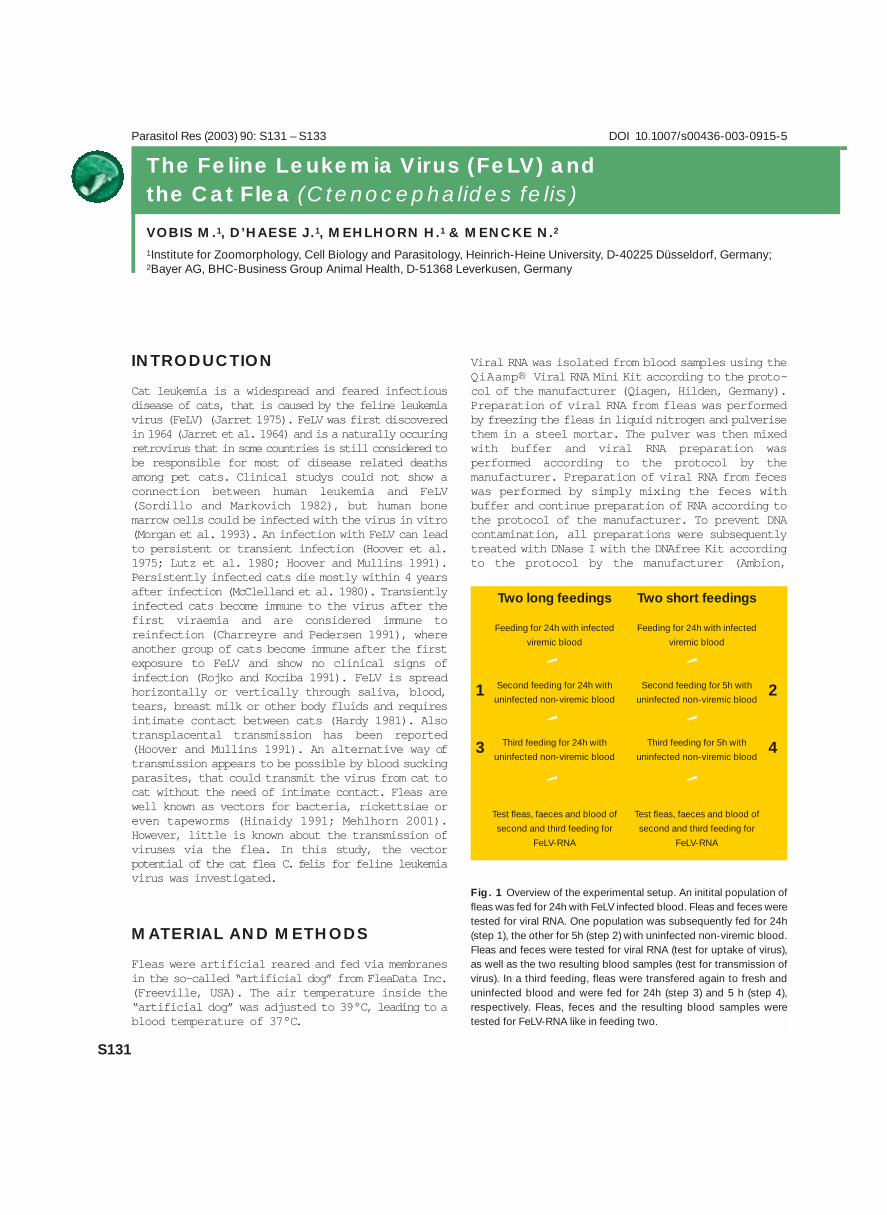
INTRODUCTION
Cat leukemia is a widespread and feared infectiousdisease of cats, that is caused by the feline leukemiavirus (FeLV) (Jarret 1975). FeLV was first discoveredin 1964 (Jarret et al. 1964) and is a naturally occuringretrovirus that in some countries is still considered tobe responsible for most of disease related deathsamong pet cats. Clinical studys could not show aconnection between human leukemia and FeLV(Sordillo and Markovich 1982), but human bonemarrow cells could be infected with the virus in vitro(Morgan et al. 1993). An infection with FeLV can leadto persistent or transient infection (Hoover et al.1975; Lutz et al. 1980; Hoover and Mullins 1991).Persistently infected cats die mostly within 4 yearsafter infection (McClelland et al. 1980). Transientlyinfected cats become immune to the virus after thefirst viraemia and are considered immune toreinfection (Charreyre and Pedersen 1991), whereanother group of cats become immune after the firstexposure to FeLV and show no clinical signs ofinfection (Rojko and Kociba 1991). FeLV is spreadhorizontally or vertically through saliva, blood,tears, breast milk or other body fluids and requiresintimate contact between cats (Hardy 1981). Alsotransplacental transmission has been reported(Hoover and Mullins 1991). An alternative way oftransmission appears to be possible by blood suckingparasites, that could transmit the virus from cat tocat without the need of intimate contact. Fleas arewell known as vectors for bacteria, rickettsiae oreven tapeworms (Hinaidy 1991; Mehlhorn 2001).However, little is known about the transmission ofviruses via the flea. In this study, the vectorpotential of the cat flea C. felis for feline leukemiavirus was investigated.
MATERIAL AND METHODS
Fleas were artificial reared and fed via membranesin the so-called “artificial dog” from FleaData Inc.(Freeville, USA). The air temperature inside the“artificial dog” was adjusted to 39°C, leading to ablood temperature of 37°C.
Viral RNA was isolated from blood samples using theQiAamp® Viral RNA Mini Kit according to the proto-col of the manufacturer (Qiagen, Hilden, Germany).Preparation of viral RNA from fleas was performedby freezing the fleas in liquid nitrogen and pulverisethem in a steel mortar. The pulver was then mixedwith buffer and viral RNA preparation wasperformed according to the protocol by themanufacturer. Preparation of viral RNA from feceswas performed by simply mixing the feces withbuffer and continue preparation of RNA according tothe protocol of the manufacturer. To prevent DNAcontamination, all preparations were subsequentlytreated with DNase I with the DNAfree Kit accordingto the protocol by the manufacturer (Ambion,
S131
The Feline Leukemia Virus (FeLV) andthe Cat Flea (Ctenocephalides felis)
VOBIS M.1, D’HAESE J.1, MEHLHORN H.1 & MENCKE N.2
1Institute for Zoomorphology, Cell Biology and Parasitology, Heinrich-Heine University, D-40225 Düsseldorf, Germany;2Bayer AG, BHC-Business Group Animal Health, D-51368 Leverkusen, Germany
Parasitol Res (2003) 90: S131 – S133 DOI 10.1007/s00436-003-0915-5
Fig. 1 Overview of the experimental setup. An initital population offleas was fed for 24h with FeLV infected blood. Fleas and feces weretested for viral RNA. One population was subsequently fed for 24h(step 1), the other for 5h (step 2) with uninfected non-viremic blood.Fleas and feces were tested for viral RNA (test for uptake of virus),as well as the two resulting blood samples (test for transmission ofvirus). In a third feeding, fleas were transfered again to fresh anduninfected blood and were fed for 24h (step 3) and 5 h (step 4),respectively. Fleas, feces and the resulting blood samples weretested for FeLV-RNA like in feeding two.
Two long feedings
Feeding for 24h with infected
viremic blood
Second feeding for 24h with
uninfected non-viremic blood
Third feeding for 24h with
uninfected non-viremic blood
Test fleas, faeces and blood of
second and third feeding for
FeLV-RNA
Two short feedings
Feeding for 24h with infected
viremic blood
Second feeding for 5h with
uninfected non-viremic blood
Third feeding for 5h with
uninfected non-viremic blood
Test fleas, faeces and blood of
second and third feeding for
FeLV-RNA
1 2
3 4
‘ ‘
‘ ‘
‘ ‘

Austin, USA). Reverse transcription of the viralRNA was performed using the 1st strand cDNASynthesis Kit for RT-PCR according to the protocolby the manufacturer (Roché, Indianapolis, USA).Amplification and detection of the FeLV cDNA wasperformed with a high sensitive nested PCR as pre-viously described (Miyazawa and Jarret 1997). TheDNA amplification was performed in 100 µl reactiontubes in a total volume of 50 µl using the Taq PCRCore Kit according to the protocol by themanufacturer (Qiagen, Hilden, Germany).
RESULTS
In a first feeding, an initial population of 110 fleaswere infected with FeLV through feeding for 24hwith 2 ml blood from an viremic cat (Fig. 1). Toverify successful uptake of viruses, 10 fleas andwhole feces of this initial population were tested forFeLV RNA. The viral RNA could be detected in bothsamples, fleas and feces (Fig. 2, lane 1 and 2). TheFeLV incorporated by the fleas via blood feeding andwas excreted with the feces. The remaining 100infected fleas were subsequently divided in twopopulations of 50 where population 1 was fed for 24hours (Fig. 1, step 1), and population 2 for 5 hours(Fig. 1, step 2) with 300 µl uninfected blood from ahealthy cat. After the defined time, 10 fleas andfeces of each population were tested for FeLV. ViralRNA could be detected in fleas of both populations,as well as in their feces (Fig. 2, lane 4,7,10,and11). FeLV RNA was detecable for 5 and 24 hoursrespectively after uptake by the flea and feeding
with uninfected blood. To investigate, wethertransmission of FeLV occurred, the remaining bloodfrom transfection 1 and 2 was examined. Viral RNAcould be detected in both samples of the previouslyuninfected blood from a clinically healthy cat (Fig.2, lane 3 and 9). This shows, that the cat flea C.felis can function as a vector for feline leukemiavirus RNA in vitro. Viral RNA was detectable in thefleas and its feces for up to 24 hours. In addition,the fleas successfully transmitted FeLV RNAdirectly from an infected blood sample to anuninfected one. In a third feeding study, the samefleas were fed again with fresh and uninfected blood(Fig. 1, step 3 and 4). After 5 and 24 hours,respectively, fleas, feces and the blood sampleswere examined for FeLV. After this third feeding,viral RNA could not be detected, neither in the fleas,their feces nor in the blood samples (Fig. 2, lane5,6,12 and 8,13,14). FeLV RNA was detectable inthe fleas up to 24 hours after feeding. During thistime, fleas also excreted viral RNA with theirfeces. In addition, the fleas were able to transmitFeLV RNA directly from one blood sample to anotherand functioned therefore as a vector for the virus.
DISCUSSION
Feline leukemia virus is known to be spread from catto cat via saliva (by licking), blood (scratching orfighting) and all other body fluids (Hardy et al.1975). A close and intimate contact between
S132
Fig. 2 PCR amplification of cDNA with inner primer pair targeting a 601bp fragment between U3 and gag region of FeLV-RNA. Prior to RT-PCR, all RNA-preparations where treated with DNase I to ensure, that no cellular proviral DNA was detected instead of viral RNA. 1: Feces offleas after feeding for 24h with FeLV infected blood. 2: Fleas after feeding for 24h with FeLV infected blood. 3: Uninfected blood after feedingfrom step 1. 4: Fleas after feeding from step 1. 5: Uninfected blood after feeding from step 3. 6: Fleas after feeding from step 3. 7: Feces offleas after feeding from step 1. 8: Feces of fleas after feeding from step 3. 9: Uninfected blood after feeding from step 2. 10: Fleas after feedingfrom step 2. 11: Feces of fleas after feeding from step 2. 12: Uninfected blood after feeding from step 4. 13: Fleas after feeding from step 4.14: Feces of fleas after feeding from step 4. All experiments were also performed with non-viremic blood as a negative control (not shown).No FeLV-RNA was detected. M: PeqGold 100bp ladder (Peqlab).
700 bp
601 bp
M 1 2 3 4 5 6 7 8 9 10 11 12 13 14
>>

S133
healthy and infected cats is sufficient to dispersethe virus within a population or household (Essex etal. 1977). The results of our work show, that theremay be another, from close cat to cat contactindependent transmission of feline leukemia virusby the cat flea C. felis. The flea is an importantvector for various pathogens, such as bacteria andrickettsiae. Due to the wide host range of somefleas, pathogens can even be spread across species,e.g. from rat to man (plague). Recently, even aninfection of human with Rickettsia felis through theflea has been discussed (Richter et al. 2002).However, not much is known about the flea as avector for viruses. It has been reported that some
viruses can outlast intact in the flea for at least 24hours (Smetana 1965). The possible transmissionthrough the flea of the feline leukemia virus, or theHIV related feline immunodeficiency virus, isunknown. One result of our study is, that if fleas arefed for 24 hours on FeLV positive blood, the fleaexcretes viral RNA with its feces. Under the viewof the various infection possibilities of FeLV, thisaspect may gain more importance. There may berisk that a healthy cat scratches flea feces into itsskin, as a result of itching flea bites or while fightingwith against other cats.
Another result of our studies might be the directtransmission of FeLV through the flea and the fleabite. The feline leukemia virus may be ingested bythe flea and excreted with the feces, but alsodirectly transmitted during the blood suckingactivity. Wether this route of FeLV infection is ofimportance in-vivo and has clinical significanceremains unanswered and further investigations onthis aspect are necessary. While in principle the riskof pathogen transmission via flea bites to both petanimals and human is known, it can be concluded thatprevention of flea infestation in pet animals isrecommended. Regular application of effectivetopical spot on’s using insecticides like imidaclopridwith its proven therapeutic and preventativeproperties eliminates flea infestation and thustransmission of diseases. l
Fig. 3 Frontal view of a cat flea (Ctenocephalides felis felis)

OBJECTIVES
The objectives of the study were firstly todetermine whether two treatments with a novelformulation of Imidacloprid 10% plus Moxidectin2.5%, administered four weeks apart, would beeffective against Sarcoptes scabiei var canis ondogs. Secondly to compare the results with that ofa positive control group of dogs treated withSelamectin.
STUDY DESIGN AND METHODS
This study was performed in compliance with VICHGL9 “Good Clinical Practice, June 2000” at ClinVetInternational (Pty) Ltd, situated in Bloemfontein,Republic of South Africa. Thirty dogs naturallyinfested with Sarcoptes scabiei var canis wereallocated to two groups of 15 dogs each, accordingto randomisation through minimization with Day –1body weight as primary criterion. One group wastreated with the Imidacloprid/ Moxidectin Spot-onat 0.1 ml/kg body weight and the second group withSelamectin (Stronghold® ) at 0.05 ml/kg (6 mg/kg)
body weight. Treatments were blinded and wereadministered as topical applications on Day 0 and +28. All the dogs were housed individually in pens,under strict quarantine conditions and no contactbetween the dogs was possible.
The presence or absence of mites was assessed bytaking skin scrapings from five body regionssuspected of being infested on each dog, and mitecounts were done on these. Clinical symptoms andthe extent of sarcoptic lesions and pruritus wereassessed and recorded. Comprehensivephotographic documentation was done (Fig. 1). Theschedule followed is summarized in Table 1.
The primary assessment variable used in this studywas the presence or absence of live mites or eggson the dogs on each assessment day followingtreatment. The success rate for each group wascalculated as follows:
Success rate (%) = x 100 , where___ ·___y 1
x = number of dogs observed with no live mites /eggsy = total number of dogs in the group
S134
Parasitol Res (2003) 90: S134 – S135 DOI 10.1007/s00436-003-0916-4
Evaluation of the Efficacy of an Imidacloprid 10% /Moxidectin 2.5% Spot-on against Sarcoptes scabieivar canis on Dogs
FOURIE L.J.1, DU RAND C.2 & HEINE J.3
1 University of the Free State, Bloemfontein, Republic of South Africa;2 ClinVet International (Pty) Ltd., Bloemfontein, Republic of South Africa;3 Bayer AG, BHC-Business Group Animal Health, D-51368 Leverkusen, Germany
Fig. 1 A dog naturally infested with Sarcoptes scabiei var canis before (Day –3) (left) and after (Day +50) (right) treatment (Imidacloprid 10% /Moxidectin 2.5% applied four weeks apart at a dose of 0.1 ml/kg body weight)
>>

Due to the nature and uncertainty factor of the mitecount assessment, false negatives could have beenrecorded, resulting in an overestimation of thesuccess rate. Consequently, clinical symptoms aswell as the frequency of pruritus were used toconfirm the success rate described above. Theabove-mentioned parameters were used tocategorize a dog as an “overall success” or“failure”. An overall success in terms of treatmentwas defined as a dog who complied with all of thefollowing conditions: No live mites or eggs; acomplete resolution of papules and crusts asassessed on Day +22 and +50 or +64; a marked (>80%) reduction in body areas showing alopecia; andthe frequency of pruritus not consistently high.
RESULTS
The treatment success rate and overall treatmentsuccess rate for the two study groups aresummarized in Table 2. On Day +8 S. scabiei wasfound in skin scrapings of two dogs treated with theImidacloprid / Moxidectin solution and of three dogstreated with Selamectin. From Day +22 andonwards, no S. scabiei mites were detected in theskin scrapings of any of the dogs, giving 100%treatment success rates for both groups. Based onthe criterion defined above, 7.1%, 92.9% and100% of the dogs treated with the Imidacloprid /Moxidectin solution were categorized as an overallsuccess in terms of treatment on Days +22, +50and +64, respectively. Five dogs in the Selamectintreated group displayed prominent alopecia and twoof these displayed localized areas with crusts onDay +64. Three of these dogs were infested withDemodex spp. mites, which may have been the causeof the alopecia.
S135
Table 1 Synoptic overview of the study layout * Only five dogs retained for extended periods of observation
Acclimatization
Day –7
Allocation toGroups
Day –1
Treatment
Day 0;Day + 28
PruritusIndex
Day –1; +7; +21;+35; +42; +57*;
+63*
Mite countsand clinical
assessments
Day –3; +8; +22;+36; +50; +64*
PhotographicDocumentation
Day –3; +22; +50;+64*
Pre- and Post-treatment assessments
Day +22
100
6.7
Day +50
100
66.7
Day +64*
100
66.7
Day +22
100
7.1
Day +50
100
92.9
Day +64*
100
100
Treatment Success rate (%)
Overall Treatment Success rate (%)
Group 1: Treated with Selamectin Group 2: Treated withImidacloprid/Moxidectin
Table 2 Summary of the treatmentand overall treatment success rates(%) for the study groups* Pooled data for Days +50 and +64

Parasitol Res (2003) 90: S136 – S138 DOI 10.1007/s00436-003-0917-3
S136
Feline Advantage Heart™ (Imidacloprid and Moxi-dectin) Topical Solution as Monthly Treatment forPrevention of Heartworm Infection (Dirofilaria immitis)and Control of Fleas (Ctenocephalides felis) on Cats
ARTHER R.G.1, BOWMAN D.D. 2, McCALL J.W.3, HANSEN O.4 & YOUNG D.R.5
1Bayer HealthCare LLC, Animal Health Division, Shawnee Mission, Kansas, USA; 2CHK, Stanwood, Michigan, USA;3TRS Athens, Georgia, USA; 4Bayer AG, BHC-Business Group Animal Health, D-51368 Leverkusen, Germany;5Young Veterinary Research Services, Turlock, California, USA
INTRODUCTION
Feline Advantage Heart™ is a combined formulationof 10% w/v imidacloprid plus 1% w/v moxidectinspot-on for dermal application to kittens and cats.The combination product has been developed byBayer, using the insecticide, imidacloprid, and themacrolide anthelmintic, moxidectin. It is intendedfor monthly application to provide treatment andcontrol of flea infestations, intestinal nematodes,and for prevention of feline heartworm disease.Imidacloprid is already marketed worldwide asAdvantage® (10 w/v imidacloprid = 9.1% w/w),a spot-on treatment for the control of fleas on catsand dogs. Imidacloprid applied monthly as a lowvolume topical application to cats or dogs provideshighly effective flea control.3,4 Fleas areimmobilized and killed after contact withimidacloprid on the skin or hair of cats. For smallanimal veterinary medicine, a moxidectin tabletformulation has been marketed for some time asmonthly prophylactic treatment for dogs,(Proheart® , Ft. Dodge) and more recently as aninjectable slow release formulation1. Moxidectin isalso available as an endectocide for ruminants andan anthelmintic for horses2. The new formulation of10% imidacloprid plus 1% moxidectin spot-on isproduced by replacing a proportion of the pharma-ceutically inactive component of the Advantage®formulation with moxidectin. With this additionalcomponent Bayer has extended the Advantage®spectrum of activity to include activity againstintestinal nematodes and heartworm disease pre-vention.
The studies presented here were conducted toevaluate flea control and heartworm prevention incats with topically applied imidacloprid andmoxidectin formulations.
MATERIAL AND METHODS
Flea Study No 1Thirty two short-haired cats were randomized into4 treatment groups balanced by sex andpretreatment flea infestations: (1) 10%imidacloprid + 1.0% moxidectin (2) 10%imidacloprid only (3) 1.0% moxidectin only (4)control (placebo without active ingredient). Each catwas infested with 100 recently emerged adult fleas(Ctenocephalides felis) on day –1. Each cat wastreated on test day 0 with the appropriate testarticle provided in pre-filled applicator tubesdesigned to deliver a minimum of 0.1 mL ofsolution/kg body weight. Treatments were appliedby dermal application of the product on the skin atthe base of the skull. Live fleas were combed fromthe cats, counted and removed on test day 1. Eachcat was reinfested with 100 live fleas on test days6, 13, 20, 27 and 34. Live fleas were combed fromthe cats, counted and removed on test days 7, 14,21, 28 and 35. Percent flea control efficacy wascalculated by comparing the geometric mean numberof live fleas on test groups 1 – 3 with the geometricmean number of live flea on the control cats withthe following formula:
% Efficacy =
Geo Mean No. Fleas (Controls) – Geo Mean No. Fleas
(Treated) x 100
Geo Mean No. Fleas (Controls)
Flea Study No 2Sixteen cats were randomized into 2 treatmentgroups balanced by sex and pretreatment fleainfestations: (1) 10% imidacloprid + 1.0%moxidectin or (2) control (placebo without activeingredient). Each cat was infested with 100
>>

recently emerged adult fleas on day –1. Each catwas treated on test day 0 with the appropriate testarticle provided in pre-filled applicator tubesdesigned to deliver a minimum of 0.1 mL ofsolution/kg body weight. Treatments were appliedby dermal application of the product on the skin atthe base of the skull. Live fleas were combed fromthe cats, counted and removed on test day 1. Eachcat was reinfested with 100 live fleas on test days6, 13, 20, 27 and 34. Live fleas were combed fromthe cats, counted and removed on test days 7, 14,21, 28 and 35. Percent flea control efficacy wascalculated by comparing the geometric mean numberof live fleas on test group 1 with the geometric meannumber of live fleas on the control cats (group 2)using the same formula indicated above.
Heartworm Study No 1 A controlled laboratory study was conducted toevaluate the efficacy and safety of topically appliedimidacloprid plus moxidectin againstexperimentally-induced infections with Dirofilariaimmitis larvae in cats. On test day –30 forty cats(20 males/20 females, 5 to 8 months of age) wereeach infected with 100 third stage D. immitislarvae. On test day –1 the cats were blocked bygender and body weight, and were randomlyassigned to the following 4 treatment groups: (1)10% imidacloprid +1.0% moxidectin, (2) 1.0%moxidectin only, (3) 10% imidacloprid only and (4)control (placebo without active ingredient). On testday 0 (30 days following experimental infectionswith D. immitis), cats were topically treated, at aminimum dose equal to 0.1 ml/kg body weight, withthe appropriate formulation. Study animals weremaintained until test day 140 (170 days post-infection). At that time, they were euthanized,necropsied, and examined for recovery of adult
heartworms within the abdominal and thoraciccavities, heart, lungs and connecting vascularsystem.
Heartworm Study No 2A second controlled laboratory study was conductedto further evaluate the safety and efficacy oftopically applied 10% imidacloprid + 1% moxidectinagainst experimentally-induced Dirofilaria immitisinfections in cats. On test day –30, twenty crossbreed cats (11 males/9 females, 5.5 to 6.5 monthsof age) were each infected with 100 third-stage D.immitis larvae (L3). On test day –1, cats wereblocked by gender and body weight and were thenrandomized to 2 treatment groups: (1) 10%imidacloprid +1.0% moxidectin or (2) control(placebo without active ingredient). On test day 0(30 days post-infection with D. immitis larvae),cats were topically treated, at a minimum doseequal to 0.1 ml/kg body weight, with either the testproduct or a placebo. Study animals weremaintained until test day 140 (170 days post-infection). At that time, they were euthanized,necropsied, and examined for the recovery of adultheartworms within the abdominal and thoraciccavities, heart, lungs and connecting vascularsystem.
RESULTS
Flea Study No 1Percent flea control efficacy is displayed in Figure1, group 1 (imidacloprid + moxidectin) and group 2(imidacloprid only) treatments provided 100 and98.4% flea efficacy on test day 1. No flea controlwas observed with group 3 treatment (moxidectinonly) on test day 1. Group1 treatments provided
S137
-4 1 7 14 21 28 35-50%-40%-30%-20%-10%
0%10%20%30%40%50%60%70%80%90%
100%
Feline Advantage Heart
Days post treatment
Per
cent
Fle
a E
ffica
cy
Moxidectin onlyImidacloprid only
-4 1 7 14 21 28 350%
10%
20%
30%
40%
50%
60%
70%
80%
90%
100%
Feline Advantage Heart
Days post treatment
Per
cent
Fle
a E
ffica
cy
Fig. 1 Flea Study No. 1: Comparative Flea Control Fig. 2 Flea Study No. 2: Percent Flea Control

S138
98.7, 100, 89.4, 98.0, and 88.4% flea efficacy ontest days 7, 14, 21, 28 and 35, respectively. Group2 treatments provided 100, 100, 100, 98.6 and96.5% flea efficacy for the same the fivecorresponding post-treatment periods. Percent fleacontrol for the moxidectin only treatment (group 3)ranged from 2.9 – 55.4% for test days 7 – 35. Thecontrol cats (group 4) retained a geometric meanrange of 11.9 to 31.7 live fleas/cat through testday 35.
Flea Study No 2Percent flea control efficacy is displayed in Figure2, group 1 (imidacloprid + moxidectin) provided100% flea efficacy on test day 1and then provided99, 99, 97, 98.0, and 90% efficacy on test days 7,14, 21, 28, and 35, respectively. The control cats(group 2) retained a geometric mean range of 27.7– 83.5 live fleas/cat through test day 35.
Heartworm Study No 1The recovery of adult heartworms and percentheartworm efficacy for heartworm study number 1is displayed in Figure 3. No adult heartworms wererecovered from the cats in group 1 treated withimidacloprid + moxidectin or from the cats in group2 treated with moxidectin only. Heartworm efficacyfor groups 1 and 2 was 100%. A total of 74 adultheartworms were recovered from 7 of 10 cats ingroup 3 and 61 adult heartworms were recoveredfrom 6 of 10 cats in group 4.
These results demonstrate that the 1.0%moxidectin plus 10% imidacloprid and 1.0%moxidectin formulations applied topically at aminimum dose of 0.1 ml/kg bodyweight were 100%efficacious against the development of adultheartworm infections in cats. The results alsodemonstrated that 10% imidacloprid, appliedtopically to cats, had no activity against larval
stages of D. immiti. Imidacloprid did not interferewith the efficacy of moxidectin against D. immitiswhen combined with moxidectin into oneformulation. No adverse or unusual reactions wereobserved following treatment with any of the testarticles.
Heartworm Study No 2The recovery of adult heartworms and percentheartworm efficacy for heartworm study number2 is displayed in Figure 4. No adult heartwormswere recovered from the cats in group 1 treatedwith imidacloprid + moxidectin only. Heartwormefficacy for group was 100%. A total of 31 adultheartworms were recovered from 8 of 10 cats ingroup 2. These results further demonstrate that the1.0% moxidectin plus 10% imidacloprid appliedtopically at a minimum dose of 0.1 ml/kgbodyweight is 100% efficacious against thedevelopment of adult heartworm infections in cats.
DISCUSSION
The results of these studies demonstrate thatmoxidectin solution applied alone topically to catsprovided little or no flea control. Imidacloprid aloneprovided no activity against the development ofheartworm larvae to the adult stage in cats but didnot interfere with the activity of moxidectin toprevent the development of heartworm larvae incats when the two active ingredients werecombined. These results are in agreement to similarresults observed during efficacy studies conductedwith 10% imidacloprid + 0.08% ivermectin fordogs.5 The results of these studies demonstratedthat ivermectin alone had little or no activityagainst fleas on dogs, imidacloprid alone did nothave activity against larval stages of canine
Fig. 3 Heartworm Study No. 1: Adult Heartworm Counts Fig. 4 Heartworm Study No. 2: Adult Heartworm Counts
FelineAdvantage
Heart
Moxidectin Placebo Imidaclo-
Heartworms (geometric mean) 5.
0
4.
5
4.
0
3.
5
3.
0
2.
4.6worms per 3.6
worms per
0.0worms per
cat
100%
0.0worms per
cat
100%
Feline Advantage Heart Placebo
Heartworms (geometric mean) 2.
5
2.
0
1.
5
1.
0
2.2 worms per cat
0.0 worms per cat
100% Efficacy

Parasitol Res (2003) 90: S139 – S140 DOI 10.1007/s00436-003-0918-2
S139
INTRODUCTION
From a veterinary medical perspective, optimumcontrol of preventable diseases has always been ahigher priority than disease therapy. Today,especially in small animal medicine, this objectivecan only be achieved by providing a product to thepet owner in an easy-to-administer formulation.The combination of 1% Moxidectin and 10%Imidacloprid in a spot-on formulation for dermalapplication fulfills these criteria. The combinationproduct has been developed by Bayer, using theneonicotinyl insecticide, Imidacloprid, and themacrocyclic lactone, Moxidectin. It is intended to
provide a treatment for, and prophylaxis against,fleas and a range of nematode infections of cats. Itis desirable to control and treat not only establishedadult gastrointestinal parasites, but also thedevelopmental stages including L4 and immatureadults of T. cati. The importance of controllingfeline parasitism is not only to relieve clinicalsymptoms in infected cats, but also to minimize thezoonotic potential of larval nematode infections inman (Sprent, 1956).
Moxidectin is related to the milbemycins and isbelieved to exert its effect in the same way as themilbemycins and avermectins. All appear to causeparalysis of susceptible parasite species by alteringchloride conductance into cells. It was originallybelieved that this was brought about by activity ofthe molecules at the GABA receptors (McKellar andBenchaoui, 1996). However, after furtherinvestigation, it is understood that ivermectin andmilbemycin D bind to glutamate-gated chloridechannels in nematodes, where they can potentiateglutamate or cause an increase of conductance in theabsence of glutamate (Martin, 1996).
STUDY DESIGN
A controlled laboratory study was conducted toevaluate the efficacy of a novel formulation ofImidacloprid + Moxidectin against fourth stagelarvae and immature adult Toxocara cati infectionin cats. Thirty-two animals experimentally infectedwith T. cati were treated topically with aformulation to provide at least 10 mg/kg ofImidacloprid and 1 mg/kg of Moxidectin one time ina controlled anthelmintic evaluation. The four treat-ment groups were as follows: 1. Imidacloprid + Moxi-dectin (treated on Day 14, necropsied on Day 19), 2.Placebo (treated on Day 14, necropsied on Day 19),3. Placebo (treated on Day 24, necropsied on Day 29),and 4. Imidacloprid + Moxidectin (treated on Day 24,necropsied on Day 29). During the five day post-
Evaluation of the Efficacy of a Combination ofImidacloprid and Moxidectin against ImmatureToxocara cati in Cats
REINEMEYER C.R.1 & CHARLES S.2
1East Tennessee Clinical Research, Inc., Knoxville, Tennessee, USA;2Bayer HealthCare LLC, Animal Health Division, Shawnee Mission, Kansas, USA
Figure 1 Toxocara cati in the duodenum of a cat

S140
treatment period, cats were observed daily forclinical changes to eyes, feces, respiration,behavioral attitude, locomotion/musculature, andskin conditions. At the end of the five-dayobservation period, the animals were necropsied andthe remaining worms were recovered and counted.At necropsy, alimentary contents and salineincubates of the small intestine were collected,preserved, and examined microscopically. Nema-todes were recovered, counted, and identified togenus, species, and stage of development.
RESULTS
Topical administration of Imidacloprid + Moxidectinproved safe in cats as evidenced by no adverseclinical signs post-treatment. The geometric meannumber of Toxocara cati larvae for the Day 19necropsy was 36.9 for Placebo and 1.0 for thecombination of Imidacloprid + Moxidectin. Thegeometric mean number of Toxocara cati adults forthe Day 29 necropsy was 59.2 for the placebo and1.0 for the combination of Imidacloprid +Moxidectin. Efficacy for the treatment group thatwas necropsied on Day 19 (larvae) was 97.2%,while the efficacy for the treatment group that wasnecropsied on Day 29 was 98.3%.
DISCUSSION
Infections with gastrointestinal nematodes, esp.Toxocara cati are significant and have beenrecorded not only in stray cats (Coman et al. 1981,Yamaguchi et al, 1996) but also in cats kept as petanimals (Hellmann et al. 2002). Fleas comprise themajor ectoparasitc infestation of both cats and dogsworldwide (Rust and Dryden 1997). Thecombination of these two active parasiticidesprovides a product to control and prevent infectionwith both fleas and Toxocara cati in cats. l
ReferencesComan BJ, Jones EH & Driesen MA (1981). Helminth parasites
and arthropods of feral cats. Australian Vet. Journal 5 7, 324 –
327
Hellmann K, Knoppe I, Radeloff I & Epe C (2002). Helminthen bei
Hund und Katze (Helminths in dogs and cats). Proceedings DVG,
Travemünde, p. 37
Martin RJ (1996). An electrophysiological preparation of Ascaris
suum pharyngeal muscle reveals a glutamate-gated chloride
channel sensitive to the avermectin analogue, milbemycin D.
Parasitology 1 1 2: 247 – 252
McKellar QA & Benchaoui HA (1996). Avermectins and
milbemycins. Journal of Pharmacology and Therapeutics 1 9:
331 – 351
Rust MK & Dryden MW (1997). The biology, ecology and
management of the cat flea. Annu Rev Entomol 4 2: 451 – 473
Sprent JFA (1956). The life history and development of
Toxocara cati (Schrank 1788) in the domestic cat. Parasitology
4 6: 54 – 79
Yamaguchi N et al (1996). Parasite prevalence in free-ranging
farm cats, Felis silvestris catus. Epidemiol. Infect. 1 1 6: 217 –
223

Parasitol Res (2003) 90: S141 – S142 DOI 10.1007/s00436-003-0919-1
S141
DOGS
I. MATERIAL AND METHODS
Dogs were allocated in twelve veterinary practiceslocated in various regions of Germany and France.A total of 1,355 faecal samples from dogs wereexamined for the presence of nematode eggs. 131worm-positive dogs were enrolled and randomlyallocated in a 2:1 ratio to one of the two treatments:
(a) A combination of 10 % (w/v) Imidaclopridand 2.5% (w/v) Moxidectin, administeredtopically at the skin surface between the shoulderblades. Pipettes containing a dose volume of 0.4ml were used for dogs of less than 4 kgbodyweight, 1.0 ml for dogs of 4 to 10 kgbodyweight, 2.5 ml for dogs between 10 and 25kg bodyweight and 4.0 ml for dogs above 25 kgbodyweight. Dogs weighing more than 40 kg weretreated with a combination of a 4 ml pipette anda smaller pipette size.
(b) A positive reference product (Drontal®Plus, Bayer) containing 50.0 mg praziquantel,144 mg pyrantelembonat and 150 mg febantel,administered orally at a dosage of 1/4 of a tabletfor 2.5 kg bodyweight.
On days 7 – 1 before treatment and 8 to 13 daysafter treatment faecal samples were taken forfaecal egg count examination. Efficacy was assessed
INTRODUCTION
Roundworms of the genus Toxocara and hookworms of the genera Ancylostoma and Uncinaria are the mostcommon intestinal worms in dogs and cats. Co-infections of intestinal worms and fleas occur in a highproportion of dogs and cats in Europe. For a simultaneous treatment against endo- and ectoparasites aspot on product was developed containing the neonicotinoide (chloronicotinyl) Imidacloprid and themacrocyclic lactone Moxidectin. The efficacy and safety have been proven in various experimental studiesin dogs and cats. Objective of this investigation was to assess the anthelmintic efficacy and safety of theproduct under European field conditions considering multiple complicating variables such as home
CATS
I. MATERIAL AND METHODS
Cats were allocated in twenty one veterinarypractices located in various regions of Germanyand France. A total of 1,292 faecal samples fromcats were examined for the presence of nematodeeggs. 141 worm-positive cats were enrolled andrandomly allocated in a 2:1 ratio to one of the twotreatments:
(c) a combination of 10.0% (w/v)Imidacloprid and 1% (w/v) Moxidectin,administered topically at the skin surface at thebase of the skull at a dose volume of 0.4 ml forcats less than 4 kg bodyweight and a dose volumeof 1.0 ml for cats of more than 4 kg bodyweight.
(d) a positive reference product (Drontal® ,Bayer) containing 20 mg praziquantel and 230mg pyrantelembonat, administered orally at adosage of 1/4 of a tablet for 1 kg bodyweight.
On days 7 – 1 before treatment and 7 to 13 daysafter treatment faecal samples were taken forfaecal egg count examination. Efficacy was assessedby analysing the reduction in faecal egg counts of
The Anthelmintic Efficacy and the Safety of aCombination of Imidacloprid and Moxidectin Spot-onin Cats and Dogs under Field Conditions in Europe
HELLMANN K.1, KNOPPE T.1, RADELOFF I.1 & HEINE J.2
1Klifovet AG Munich, D-80689 München, Germany;2Bayer AG, BHC-Business Group Animal Health, D-51368 Leverkusen, Germany

S142
by analysing the reduction in faecal egg counts ofthe first faecal sample (prior to treatment) incomparison to the second faecal sample taken posttreatment.
No adverse event was seen in any dog treated.
III. CONCLUSION
It can be concluded that the 10% Imidacloprid /2.5% Moxidectin spot-on combination is safe andhighly efficacious against Toxocara canis andAncylostomatidae in naturally infested dogs.
the first faecal sample (prior to treatment) incomparison to the second faecal sample taken posttreatment.
No adverse event was seen in any cat treated withthe Imidacloprid/Moxidectin combination.
III. CONCLUSION
It can be concluded that the 10% Imidacloprid / 1%Moxidectin spot-on combination is safe and highlyefficacious against Toxocara cati andAncylostomatidae in naturally infested cats. There
Geo. mean of faecal egg countTreatment Nematode Period 0 Period 1Reduction
species Day -7 to -1, NDay 8 to13,N(%)
Toxocara 462.9 46 5.5 46
98.81Imidacloprid/ canis
Moxidectin Ancylosto-462.4 28 0.4 28
99.92matidae
Toxocara 413.9 18 1.4 18
99.66Drontal® canis
Geo. mean of faecal egg countTreatment Nematode Period 0 Period 1Reduction
species Day -7 to -1, NDay 7 to 13,N(%)
Toxocara1054.8 72 0.1 72
99.99Imidacloprid/ cati
Moxidectin Ancylosto-259.9 6 0.94 6
99.64matidae
Toxocara1149.8 38 o.4 38
99.96Drontal® cati

Parasitol Res (2003) 90: S143 – S144 DOI 10.1007/s00436-003-0920-8
INTRODUCTION
There are numerous commercial cat anthelminticsavailable that contain pyrantel embonate or pamoateas the active ingredient which is effective againstascarids and hookworms. The dosage rate of thisactive ingredient when administered to dogs isconsiderably lower than that required in cats, but itremains a safe active. A new introduction in felineanthelmintics is Milbemax (a combination ofmilbemycin oxime plus praziquantel). Consideringthe private practitioner’s desire to see comparativeresults of treatment options, a trial was conductedto compare the efficacy against natural ascarids andhookworm infections in cats, of Milbemax andDrontal Cat tablets.
MATERIALS AND METHODS
The two commercially available formulations thatwere tested are: (i). Drontal Cat – a combinationcontaining 230 mg pyrantel embonate and 20 mgpraziquantel per tablet and used at a dosage rate of1 tablet per 4 kg live weight; (ii) Milbemax forCats containing 16 mg milbemycin oxime and 40mg praziquantel per tablet and used at a dosage rateof H tablet per 2 – 4 kg or 1 tablet per 4 – 8 kg; ORMilbemax for small cats and kittenscontaining 4 mg milbemycin oxime and 10 mgpraziquantel per tablet and used at a dosage rate ofH a tablet per 0,5 – 1 kg or 1 tablet per 1 – 2 kg. Anuntreated control group was included to confirm thepersistence of the infection throughout the trialperiod.
The cats used in the trial consisted of young cats ofabout 6 to 36 months of age which were obtainedfrom feral colonies. Faecal samples were collectedprior to treatment for worm egg counts using theMcMaster method with a saturated sugar solution.All the faeces excreted for 7 days after treatmentwere collected for the recovery of excretedhelminths. The actual treatment of individual catswas controlled by the authors, while all laboratory
examinations were conducted by otherinvestigators who were blinded to the treatmentgroups.
Cats were treated with the recommended dosagerate per kg except that with Drontal Cat, the dosewas tailored to the exact weight of each animal byfractioning the tablets while with Milbemax, therecommended size of tablet was applied accordingto the live weight scale. The treatment (piece/s oftablet) was included in a small amount of a purepilchard canned food, which was the only foodpresented to the cat on treatment day. (When thishad been consumed, additional food was supplied).
All cats were euthanased and autopsied 7 days aftertreatment. The small intestines were removed andprocessed for worm recovery. Worm counts wereconducted by dissecting microscope. At least 10%of all helminths recovered were identified at specieslevel.
RESULTS
The efficacy was determined for each animal byexpressing the number of worms excreted in thefaeces after treatment as a percentage of the sumof worms excreted and recovered at autopsy.Efficacy of a treatment was calculated as the
S143
Mouth of a Toxascaris
The Efficacy of two Anthelmintics against Ascaridsand Hookworms in naturally infected Cats
CATTON D.G. & VAN SCHALKWYK P.C.
Veterinary Consultants: P O Box 1247 Rivonia 2128, Republic of South Africa

S144
SOUTH AFRICAN PET ANTHELMINTICS
PRODUCT PRESENTATION DOSAGE SCALE (PP)Canex 4 Tablets C: 1/4 tablet = 1,5 kg – 2,5 kg 23,8 mg/kg (1,5 kg)143 mg PP + 543 mg OP 14,3 mg/kg (2,5 kg)+ 50 mg PZQ C: 1/2 tablet = 2,6 kg – 5 kg 27,5 mg/kg (2,6 kg)
14,3 mg/kg (5,0 kg)C: 1 tablet = 5,1 kg – 10 kg 28,0 mg/kg (5,1 kg)
14,3 mg/kg (10kg)
Antezole Dog Tablets Tablets C: 1 tablet / 10 kg (140 mg/10 kg PP) 140 mg/kg (1 kg)50 mg PZQ +140 mg PP 14,0 mg/kg (10 kg)+ 545 mg OP
Drontal Dog Tabs Tablets C: 1/4 tab / 0,75 kg 14,4 mg/kg (0,75 kg)Small/Medium/Large 1/2 tab / 1,5 kg 14,4 mg/kg (1,5 kg)Small 15 mg PZQ + 75 mg 1 tab / 3,0 kg 14,4 mg/kg (3 kg)Febantel + 43,2 mg PP
Antezole Liquid Liquid C: 1 ml / 10 kg 50 mg/kg (1 kg)50 mg PP/ml 5 mg/kg (10 kg)
F: 1 ml / 2,5kg 100 mg/kg (0,5 kg)20 mg/kg (2,5 kg)
Nemex Liquid Liquid C: 1 ml / 10 kg 50 mg/kg (1 kg)50 mg PP/ml 5 mg/kg (10 kg)
F: 1 ml /2,5 kg 100 mg/kg (0,5 kg)20 mg/kg (2,5 kg)
Antezole Cat Tablets Tablets F: 1 tablet / 4 kg (230 mg/4 kg PP) 460 mg/kg (0,5 kg)20 mg PZQ +230 mg PP 57,5 mg/kg (4 kg)
Drontal Cat Tablets F: 1/2 tablet / 2 kg 230 mg/kg (0,5 kg)20 mg PZQ + 230 mg PP 1 tablet / 4 kg 115 mg/kg (1 kg)
57,5 mg/kg (2 kg)57,5 mg/kg (4 kg)
Antezole Cat Paste Paste F: 1,5 ml / 2 kg (120 mg/2 kg PP) 240 mg/kg (0,5 kg)80 mg PP + 250 mg N/ml 60 mg/kg (2 kg)
PP = Pyrantel pamoate/embonate PZQ = Praziquantel OP = Oxantel pamoate N = NiclosamideC = Canine F = Feline
average of efficacy in all individual animals in agroup.
During the conduct of the trial, it was determinedthat a majority of cats were infected with hookwormalone and very few with both ascarids andhookworms. The trial thus had to be completedutilising some cats only infested with hookworms,others with both and some with ascarids alone. Thepercent efficacy calculated as a mean of eight catsper group, showed 100% efficacy of both productsagainst Toxocara spp. The efficacy againstAncylostoma spp. differs, with Drontal Cat showing99,1% efficacy, while Milbemax was lower at93,5%.
DISCUSSION
The results of this trial confirm the high efficacy(> 99%) for Drontal against ascarids andhookworms, as reported by other researchers. It iswell known that the pyrantel embonate andpraziquantel combinations are very safe and have ahigh therapeutic index. The dosage rate of pyrantelembonate as tested in this trial was 57,5 mg/kgwhich is the minimum recommended dosage.
When new products are developed and the minimumeffective dosage of the active ingredient isestablished, the next decision is in which dosageform to present the product. A dilemma is oftenencountered tailoring the dosage presentation to aparticular live weight range. In Table 1 the variancein dosage rates is illustrated with particularreference to pyrantel pamoate in several of theSouth African registered products.
In this trial 2 presentations of Milbemax were usedand are marketed commercially to cater for liveweight ranges of 500 g to 8 kg. Most of the catstreated during this trial weighed between 2 to 2,5

Parasitol Res (2003) 90: S145 – S146 DOI 10.1007/s00436-003-0921-7
S145
STUDY OBJECTIVES / INTRODUCTION
Between 1997 and 2001 four multi-centre-studiesevaluating the efficacy and safety of differentpharmaceuticals have been conducted in France andGermany according to the guidelines of Good ClinicalPractice. Collection of faecal samples in the courseof the multi-centre-studies was aimed at detectingcats positive for nematodes. Additionally, all catfaecal samples that were sent to the Institute ofParasitology between 1998 and 2002 in the courseof routine diagnostic examination were analysed andcompared to the study samples. Both sets of datawere not raised according to a preplannedepidemiological study with a representative samplecalculation and distribution but nevertheless theygive valuable information of the current prevalencesituation due to the high number of samples that wasexamined and to the fact that samples werecollected nearly from all over France and Germany.
MATERIAL AND METHODS
44 veterinary practices and two laboratories inFrance and Germany participated in the four multi-centre-studies during which approximatelyaltogether 3500 cat faecal samples were examined.For coproscopical examination a modifiedMcMaster-Technique was used. Only cats, thatwere tested positive for endoparasites wereincluded in the studies.
441 cat faecal samples sent to the Institute of Para-sitology between 1998 and 2002 were examined.Routinely, the Institute’s lab uses a combinedsedimentation-flotation method with ZnSO4 (seeRommel et al., 2000). Due to these differencesconcerning methods and motivation of these twodata sets, protozoa remained disregarded andtapeworm stages were not included in thecumulative interpretation of the data.
T. cati T. leonina A. tub Taeniids Dipylidium
Mescoces-
4,
5
4
3,
5
3
2,
5
2
1,
3,9 0,5 1,6 1,4 0,2 0,2
% Preva-
Tab. 2 Overall Prevalences of Cat Samples in
T. cati T. leonina A. tub Taeniids Dipylidium
Diphyllo-
12
10
8
6
4
2
0
11,3 0,3 0,4 0,4 0,3
% Mean Preva-
Tab. 1 Overall Prevalences of Multi-Centre
Recent Investigation on the Prevalence ofGastrointestinal Nematodes in Cats from Franceand Germany
COATI N.1, HELLMANN K.2, MENCKE N.3 & EPE C.1
1Institute of Parasitology, Hannover School of Veterinary Medicine, Buenteweg 17, D-30559 Hannover, Germany;2Klifovet AG, Munich, D-80689 München, Germany; 3 Bayer AG, BHC-Business Group Animal Health, D-51368Leverkusen, Germany

S146
For more detailed information about the cats testedpositive for the multi-centre-studies aquestionnaire was sent to the participatingveterinarians subsequently. Practicioners wereasked to answer questions about origin,environment, husbandry and frequency of anthel-mintic treatment of the endoparasite positiveanimals.
RESULTS
The samples of the multi-centre-studies containedin 11,3% of the positive samples stages ofToxocara cati, 0,4% of Ancylostoma tubaeformae,0,4% of taeniids, 0,3% of Toxascaris leonina,0,3% of Dipylidium spp. and 0,1% of Capillaria sp.(Tab.1).In 3,9% of the samples examined in the Institute ofParasitology T. cati eggs were found, 1,6% containedstages of A. tubaeformae, 1,4% of taeniids, 0,5% ofT. leonina and 0,2% of Dipylidium spp. (Tab.2).
The questionnaire evaluation revealed that 59% ofthe cats for which the questionnaire was answeredwere of private households, 21% were farmcats,11% belonged to boarding kennels and in 9% theorigin was stated unknown. About 60% had daily orpermanent access to outdoor environment, 17%went outdoors occasionally and approximately 20%of the helminth positive animals were housedindoors and did never have any access to the outdoorenvironment according to owners and veterinariansinformation (Tab. 3). About 80% of the cats, thatwere included in the multi-centre-studies were
treated anthelmintically twice a year or rarer,11% were dewormed 3 – 4 times per year and 2%more frequently (Tab. 4).
CONCLUSION AND DISCUSSION
Surprisingly, about 20% of the cats, that had beentested positive for endoparasites and therefore tookpart in the multi-centre-studie never had any accessto the outdoor environment. The etiology of theseinfections remains speculative – neither amechanical transmission (by owners) of infectivestages to the indoor cats as source of the infection
50
45
40
35
30
25
20
15
10
5
0
% Answers
Tab. 4 Anthelmintic Treatment of Cats
permanentlyfrequentlyoccasionallyno outdoorunknownaccess
20,61 40,35 16,67 18,42 3,95
4x 3 – 4x 1 – 2x <1x unknown
2,19 10,96 38,16 42,98 5,7
% Answers
Tab. 3 Outdoor Access of CatsDipylidium caninum

S147
SUMMARY
Infections with endoparasites in dogs and cats weredetermined by analysing the results of faecalexaminations (Flotation, MIFC, sedimentation,Baermann, smear, ProSpecT Giardia MicroplateAssay). Samples of 8438 dogs and 3167 cats fromthe years 1999 until 2002 have been included in theinvestigation. Evaluation of the samples indicatedthat 2717 dogs (32.2%) and 771 cats (24.3%) wereinfected with endoparasites.
In the infected dogs the following parasites have beenidentified:Class Nematodea: Toxocara canis: 22.4%;Toxascaris leonina: 1.8%; Ancylostomatidae: 8.6%;Trichuris vulpis: 4.0%; Capillaria spp.: 2.3%;Crenosoma vulpis: 0.9%; Angiostrongylus vasorum:0.3%;Class Cestodea: Taeniidae: 1.2%; Dipylidiumcaninum: 0.4%; Diplopylidium/Joyeuxiella: 0.1%;Mesocestoides: 0.2%; Diphyllobothrium latum:<0.1%;Class Sporozoea: Sarcocystis spp.: 9.0%;Cystoisospora spp.: 22.3%; C. canis: 8.0%; C.ohioensis: 17.0%; Hammondia/Neospora: 1.7%;Class Zoomastigophorea: Giardia spp.: 51.6%.
In the 771 infected cats the following prevalencesof parasites were found: Class Nematodea: Toxocara mystax: 26.2%;Ancylostoma tubaeforme: 0.3%; Capillaria spp.:7.0%; Aelurostrongylus abstrusus: 2.7%;Class Cestodea: Taeniidae: 2.6%; Dipylidiumcaninum: 0.1%;Class Sporozoea: Sarcocystis spp.: 2.2%;Cystoisospora spp.: 21.9%; C. felis: 15.3%, C.rivolta: 7.9%; Toxoplasma/Hammondia: 4.5%;Class Zoomastigophorea: Giardia spp.: 51.6%.
INTRODUCTION
Infections with endoparasites cause very differentclinical symptoms depending on the parasite speciesand abundance. This may vary from mildgastrointestinal disorders with anorexia and lowweight gain to weight loss, anaemia anddehydration. In severe cases developmentaldisorders or even mortality may occur. Thedevelopment of safe broad spectrum anthelminticshas led to a more regular deworming of especiallyyoung animals, commonly without previousdiagnosis. Whether or not this has led to a decreasein infection rates is not known. The target of thisstudy was to gain actual data on endoparasiteinfections (helminths and protozoa) in dogs and catsin Germany by analysing faecal checks from adiagnostic laboratory.
MATERIAL AND METHODS
The results of parasitological examinations fromthe Veterinary Laboratory Freiburg from 8438 dogsand 3167 cats have been included in the analysis(Tab. 1).
Reason for submission of the samples were: a) routine check for annual vaccination,b) general health check,c) gastrointestinal disorders,d) not specified.
All samples have been investigated with followingmethods:
a) Flotation (spec. grav. 1.3),b) MIFC (Merthiolate-Iodine-Formaldehyde-Concentration),
c) Sedimentation and Baermann (only if sufficientmaterial),
d) Smear (in case of fluid faeces),e) Giardia AG-ELISA (ProSpecT Giardia MicroplateAssay) (most of the samples).
Endoparasites in Dogs and Catsin Germany 1999 – 2002
BARUTZKI D.1 & SCHAPER R.2
1Veterinary Laboratory Freiburg, Postfach 100120, D-79120 Freiburg i.Br., Germany;2Bayer AG, BHC-Business Group Animal Health, D-51368 Leverkusen, Germany
Tab. 1 Endoparasite infections in dogs and cats
No. of No. / share No. of No. / shareexamined infected examined infected
dogs dogs cats cats
Year n n % n n %
1999 2129 649 30.5 764 152 19.92000 2014 702 34.9 758 197 26.02001 2063 663 32.1 819 218 26.62002 2232 703 31.5 826 204 24.7
1999-2002 8438 2717 32.2 3167 771 24.3
Parasitol Res (2003) 90: S147 – S149 DOI 10.1007/s00436-003-0922-6

S148
To examine the age dependence of the infection rateswith endoparasites the data were statisticallyproved by the chi-square-test.
RESULTS AND DISCUSSION
In dogs the most prevalent endoparasite (Fig. 1) wasGiardia spp. (16.6% of 8438 dogs), followed bynematodes (11.2%), coccidia (10.4%) and cestodes(0.6%). Infections in cats showed a similar pattern(Giardia spp. in 12.6%, nematodes in 8.0%, coccidiain 6.7% and cestodes in 0.7% of 3167 cats).
In the 2717 endoparasite positive (32.2%) of 8438examined dogs eggs from Toxocara canis,
up to 1 2933 389 13.3 52 1.8 111 3.8>1 – 5 2286 81 3.5 26 1.1 55 2.4>5 – 10 1365 34 2.5 8 0.6 20 1.5
>10 529 10 1.9 0 0 12 2.3
total 7113 514 7.2 86 1.2 198 2.8
Age(years)
Examineddogs with
known age(n)
Toxocarapostive dogs
No. (n) Share (%)
Trichurispositive dogsn %
Ancylostomatidaepositive dogsn %
Fig. 2 Prevalence (%) of helminths and coccidia in dogs(n=8438)
Prevalence % 0 1 2 3 4 5 6 7 8
Toxocara canisToxascaris leoninaAncylostomatidae
Trichuris vulpisCapillaria
Crenosoma vulpisAngiostrongylus
vasorumTaeniidae
Dipylidium caninumDiplopylidium/Joyeu-
xiellaMesocestoides
Diphyllobothriumlatum
Cystoisospora spp.C. ohioensis
Fig. 1 Infections with endoparasites in dogs (n=8438) andcats (n=3167)Prevalence % 0 2 4 6 8 10 12 14 16 18
DOGS Nematodes
Cestodes
Coccidia
Giardia
CATS Nematodes
Cestodes
Coccidia
Giardia
up to 1 2933 435 14.8 92 3.1>1 – 5 2286 43 1.9 68 3.0>5 – 10 1365 14 1.0 31 2.3
>10 529 7 1.3 13 2.5
total 7113 499 7.0 204 2.9
Age(years)
Examineddogs with
known age(n)
Cystoisosporapostive dogs
No. (n) Share(%)
Tab. 3 Prevalence of Toxocara canis, Trichuris vulpis and hookworms per age group of examined dogs
Tab. 2 Prevalence of Cystoisospora spp. and Sarcocystis spp. per age group of examined dogs
>>

S149
Toxascaris leonina, Ancylostomatidae, Trichurisvulpis, Capillaria spp., Taeniidae, Dipylidiumcaninum, Diplopylidium/Joyeuxiella,Mesocestoides spp. and Diphyllobothrium latum,larvae from Crenosoma vulpis and Angiostrongylusvasorum as well as oocysts from Cystoisosporaohioensis, C. canis, Hammondia heydorni/Neosporacaninum and Sarcocystis spp. have been identified(Fig. 2).
To analyse the age dependence of the determinedinfection rates, all dogs with known age (n = 7113)have been grouped into four age groups (Tab. 2, 3).Dogs up to one year of age showed significantlyhigher infections rates with Cystoisospora spp. (p< 0.0001), Toxocara canis (p < 0.0001), Trichurisvulpis (p < 0.001) and Ancylostomatidae(p<0.0001) compared to older dogs, whereasinfections with Sarcocystis spp. and cestodesseemed to be equally distributed amongst all agegroups.
Fig. 3 Prevalence (%) of helminths and coccidia in cats(n=3167)
Prevalence % 0 1 2 3 4 5 6 7
Toxocara mystaxAncylostoma tubae-
formeCapillaria
Aelurostrongylus abstru-sus
TaeniidaeDipylidium caninumCystoisospora spp.
C. felisC. rivolta
up to 1 1229 101 8.2 9 0.7 12 1.0 117 9.5
>1 – 5 495 20 4.0 5 1.0 5 1.0 30 6.1
>5 – 10 375 7 1.9 0 0 2 0.5 7 1.9
>10 374 6 1.6 1 0.3 3 0.8 4 1.1
total 2473 134 5.4 15 0.6 22 0.9 158 6.4
Age Examinedcats
Cystoisosporapostive cats
No. (n) Share (%)
Sarcocystispositive catsn %
Toxoplasmapositive catsn %
Toxocarapositive catsn %
Tab. 4 Prevalence of Cystoisospora spp., Sarcocystis spp., Toxoplasma gondii and Toxocara mystax per age group ofexamined cats
In the 771 endoparasite positive (24.3%) of 3167examined cats eggs of Toxocara mystax,Ancylostoma tubaeforme, Capillaria spp., Taeniidaeand Dipylidium caninum, larvae fromAelurostrongylus abstrusus as well as oocystsfrom Cystoisospora felis, C. rivolta, Toxoplasmagondii/Hammondia hammondi and Sarcocystis spp.have been found (Fig. 3).
In order to analyse the age dependence of theinfection rates, all cats with known age have beengrouped into four age groups (Tab. 4). Cats up to oneyear old showed significantly higher infection rateswith Cystoisospora spp. (p < 0.0001) and Toxocara
Taenia taeniaeformis

Parasitol Res (2003) 90: S150 – S152 DOI 10.1007/s00436-003-0923-5
SUMMARY
Pyrantel embonate and febantel are bothconstituents of Drontal® Plus and Drontal® Puppybroad spectrum anthelmintics for dogs. The effectsof pyrantel and the febantel metabolite fenbendazolewere investigated against Toxocara canis in-vitroby studying changes in worm motility and tissuedamage. Pyrantel and fenbendazole were added toworms incubated in media for 8 h at the followingconcentrations: pyrantel: 12,2 µg, 25 µg, or 50 µg;fenbendazole: 50 µg, 100 µg or 200 µg; mixture ofpyrantel and fenbendazole: 12,2 µg p + 50 µg f, 25µg p + 100 µg f or 50 µg p + 200 µg f. Following this8 h incubation period, one group of the worms wasimmediately fixed and studied by light- and electron
microscopical examination. Other groups have beenobserved for further 8 h periods up to 56 hours andthen studied in the same way.
S150
Synergistic Effects of Pyrantel and the FebantelMetabolite Fenbendazole on Adult Toxocara canis
MEHLHORN H.1, HANSER E.1, HARDER A.2, HANSEN O.2, MENCKE N.2 & SCHAPER R.2
1Institute for Zoomorphology, Cell Biology and Parasitology, Heinrich-Heine University, D-40225 Düsseldorf, Germany;2Bayer AG, BHC-Business Group Animal Health, D-51368 Leverkusen, Germany
Scanning electron micrograph of the anterior end of a T. canis worm Intestine with T. canis worms (top); Bundle of T. canis worms (bottom)
>>

S151
Intestine
Hypodermis
Hypodermis
Muscle cells
Muscle cells
Intestine
Intestine
Nerve axon
Cuticule
Cuticule
DMSO-Control-Group
DMSO-Control-Group
DMSO-Control-Group
DMSO-Control-GroupTable 1 Damage level of control worms followinglight and electron microscopical examinationGroup/con- Time Hypo- Longitudi-
Intestine Nerve Sexualcentration (h) dermis nal musclesaxons system
A 1 8 0 0 0 0 00 µg/ml 32 0 0 0 0 0
56 0 0 0 0 0
A 2 8 0 0 0 0 0DMSO 56 0 0 0 0 0
Table 2 Damage level of worms after incubation inmedium containing pyrantelGroup/con- Time Hypo- Longitudi-
Intestine Nerve Sexualcentration (h) dermis nal musclesaxons system
B1 8 0 0 0 0 012,2 µg/ml 32 0–1 0–1 1 1–20
B2 8 0 1 0–1 1 025 µg/ml 56 0 1 1–2 1–2 0
B2 8 0 1 0–1 1 050 µg/ml 32 0–1 1 1 1–2 0
Table 3 Damage level of worms after incubation inmedium containing fenbendazoleGroup/con- Time Hypo- Longitudi-
Intestine Nerve Sexualcentration (h) dermis nal musclesaxons system
B4 8 0 0 0 0 050 µg/ml 32 1 0–2 1–2 1 0
B5 8 0 0 0–1 0–1 0100 µg/ml 56 1 1 1–2 10
B6 8 0 0 1–2 1 0200 µg/ml 32 1–2 1–2 2–3 2–31
Table 4 Damage level of worms after incubation inmedium containing a combination of pyrantel andfenbendazole Group/con- Time Hypo- Longitudi-
Intestine Nerve Sexualcentration (h) dermis nal musclesaxons system
B7 8 0 0 0 0–1 012,2 µg/mlPyrantel +32 1 1 1–2 1–2 0–150 µg/mlFenbendazole 56 0 1–2 2 2–3
Bold numbers: Synergistic effect by combining the two compounds
damage (irreversible), = severe damage3

S152
RESULTS
There were no significant differences observableregarding motility changes between pyrantel orfenbendazole given as individual compounds and thepyrantel/fenbendazole combination. By contrast if
Intestine
Hypodermis
Hypodermis
Hypodermis
Muscle cells
Muscle cells
Muscle cells
Nerve axon
Nerve axon
Nerve axon
Nerve axon
Hypodermis
Cuticule
200 µg/ml Fenbedazol 32h
12,2 µg/ml Pyrantel + 50 µg/ml Fenbedazol 8h
25 µg/ml Pyrantel + 100 µg/ml Fenbedazol 56h 25 µg/ml Pyrantel + 100 µg/ml Fenbedazol 32h
25 µg/ml Pyrantel + 100 µg/ml Fenbedazol 8h
25 µg/ml Pyrantel + 100 µg/ml Fenbedazol 8h
25 µg/ml Pyrantel + 100 µg/ml Fenbedazol 8h

Parasitol Res (2003) 90: S153 – S154 DOI 10.1007/s00436-003-0924-4
Giardia spp. is a cosmopolitan flagellate whichinfects the small intestine of several animals
and also humans. During the last decade, Giardia hasgained momentum both as a dog and a human pathogenand giardiosis is nowadays considered as a re-emerging infectious disease. The prevalence in dogsranges from 5 to 80% according to the age of dogs,management system and colony size (Traldi andCastiglione, 1993; Nolan and Smith, 1995;Mochizuki et al., 2001). In humans, children, HIV-positive subjects or persons living in poor hygienicconditions (Kotler and Orestein, 1998; Ballone et al.,
2001) are frequently infected. According to the mostrecent research, some Giardia genotypes may beshared by human and dogs, particularly thosebelonging to the Assemblage A, Cluster A-I,revealing the possibility of an interspeciemtransmission mainly in urban areas (Graczyk et al.,1999; Thompson et al., 2000; van Keulen et al.,2002). Despite the public health importance and thepossible zoonotic role of Giardia, epidemiologicalsurveys on the prevalence of giardiosis in Italy arelacking and fragmentary, compared to othercountries.
S153
Prevalence of Giardia spp. in Dogs and Humansin Northern and Central Italy
CAPELLI G.1, PAOLETTI B.2, IORIO R.2, FRANGIPANE DI REGALBONO A.1,
PIETROBELLI M.1, BIANCIARDI P.3 & GIANGASPERO A.2
1University of Padua, Italy; 2University of Teramo, Italy; 3Bayer HealthCare, Animal Health Division, Milano, Italy
Giardia cysts

S154
The objective of this study was to determine theprevalence of Giardia in dogs and humans in Abruzzoand Veneto regions, (central and northern Italy,respectively) and, when possible, to identify riskfactors (habitat, age, infection evolution, sex,breeding) associated with dog’s infection.
A total of 916 faecal samples were examined, 616from dogs (436 from central Italy, 180 fromnorthern Italy) and 300 from humans.Epidemiological data of dogs concerning life style(owned, kennel or stray dogs), sex, breed, age,presence of gastrointestinal symptoms, werecollected. Faecal samples of humans were kindlyobtained from the local Hospital of Abruzzo region.Two coprological tests were performed in order tomaximize the chance of finding Giardia cysts: acentrifugation and flotation technique, combinedwith a sedimentation technique in Abruzzo region,and with MIF procedure (Meridian Diagnostic Inc.)in the Veneto region. The prevalence differences inrelation to epidemiological data were tested by c2
test. The strength of the relation between giardiosisand potential risk factors was estimated by thePrevalence Ratio (PR), and its importance for thepopulation was expressed by the AttributableProportion for the exposed dogs (APe).
The overall prevalence was 21.3% in dogs and 2%in humans. No differences were noted between theprevalence in dogs of central and northern Italy(22.2% and 19.4%, respectively). The major riskfactors for giardiosis in dogs were represented bydensity of dogs and age, i.e. increased prevalencewas significantly associated with kennel dogs(PR=2.13, APe=53%) and young dogs (PR=1.99,APe=50%). Higher prevalence was noted also indogs with gastrointestinal symptoms (PR=1.86,APe=46%). The findings of this study reveals thatthe prevalence of Giardia in dogs in northern andcentral Italy is high, and probably underestimated.The prevalence in dogs overlaps the rates obtainedin other countries (Oliveira-Sequeira et al., 2002)and seems to be higher than those registered inUmbria (Piergili Fioretti e Moretti, 1989) and Lazioregions (Rosso et al., 1989).
The high prevalence of Giardia in kennel dogsconfirms that confinement in a limited area is animportant risk factor: contamination of suchenvironment readily occurs through infectedanimals, thus providing a continuous source ofinfection to susceptible animals. Other contributing
factors may be the stress and the unbalanced diet.The age is also confirmed as an important riskfactor: dogs less than 1 year old were statisticallymore infected than adults.
The relationship between symptoms and presenceof Giardia seems to be strict and the dogs may beconsidered, in analogy with humans, very sensitiveto its pathological effects.
Prevalence in humans overlaps the survey carriedout in other industrialized countries (Fontaine et al.,1984) including Italy (Ballone et al., 2001).
The high level of giardiosis in dogs in the Abruzzoand Veneto regions demonstrates that they may beconsidered a significant potential reservoir ofinfection for humans even though, in our conditions,such a role for dogs has hitherto been unknown.Molecular techniques will have a major impact forunderstanding the epidemiology of Giardiainfections, and the molecular characterization ofcanine and human genotypes of Giardia – which is inprogress in this study – may add more informationon the zoonotic risk of animal origin. Lookingforward to obtain more information in this respect,screening of dogs for giardiosis, particularlypuppies, and their treatment to prevent eventualhuman infection, is highly recommended. l
ReferencesBallone E, Fazii P, Riario Sforza G, Scassa E, Di Nicola M, Ippolito
N, Di Mascio C & Schioppa F (2001). Indagine sulla diffusione
della giardiosi nell’area pescarese. Ann. Ig. 1 3, 11 – 120
Fontaine JP, Delage A & Lauraire MC (1984). Sur l’épidémiologie
de la giardiase. Ann. Parasitol. Hum. Comp. 5 9, 541 – 554
Graczyk RC, Thompson RCA, Fayer R, Adams P, Morgan UM &
Lewis EJ (1999). Giardia duodenalis of genotype A recovered
from clams in the Chesapeake Bay subestuary, Rhode River. Am.
J. Trop. Med. Hyg. 6 1, 526 – 529
Kotler DP & Orenstein JM (1998). Clinical syndromes associated
with microsporidiosis. Adv. Parasitol. 4 0, 321 – 349
Mochizuki M, Hashimoto M & Ishida T (2001). Recent
epidemiological status of canine viral enteric infections and
Giardia infection in Japan. J. Vet. Med. Sci. 6 3, 573 – 575
Nolan TJ & Smith G (1995). Time series analysis of the

Parasitol Res (2003) 90: S155 – S156 DOI 10.1007/s00436-003-0925-3
S155
Humans, Dogs and Parasitic Zoonoses – Unravellingthe Relationships in a Remote Endemic Community inNortheast India using Molecular Tools
TRAUB R.J.1, ROBERTSON I.D.1, IRWIN P.1, MENCKE N.2, MONIS P.3 & THOMPSON R.C.A.1
1School of Veterinary and Biomedical Sciences, Murdoch University, Western Australia 6150, Australia2Bayer AG, BHC-Business Group Animal Health, D-51368 Leverkusen, Germany3Australian Water Quality Centre, Bolivar, South Australia 5110, Australia
ABSTRACT
Canine parasitic zoonosespose a continuing publichealth problem, especially indeveloping countries andcommunities that are socio-economically disadvantaged.Our study combined the useof conventional and
molecular epidemiological tools to determine therole of dogs in transmission of gastrointestinal (GI)parasites such as hookworms, Giardia and Ascarisin a parasite endemic teagrowing community innortheast India. A highly sensitive and specificmolecular tool was developed to detect anddifferentiate the zoonotic species of caninehookworm eggs directly from faeces. This allowedepidemiological screening of canine hookwormspecies in this community to be conducted with easeand accuracy. The zoonotic potential of canineGiardia was also investigated by characterisingGiardia duodenalis recovered from humans and dogsliving in the same locality and households at threedifferent loci. Phylogenetic and epidemiologicalanalysis provided compelling evidence to support thezoonotic transmission of canine Giardia. Moleculartools were also used to identify the species ofAscaris egg present in over 30% of dog faecalsamples. The results demonstrated the role of dogsas a significant disseminator and environmentalcontaminator of Ascaris lumbricoides in commu-nities where promiscuous defecation practicesexist. Our study demonstrated the usefulness ofcombining conventional and molecularparasitological and epidemiological tools to helpsolve unresolved relationships with regards toparasitic zoonoses.
BACKGROUND
Molecular epidemiological tools have formed anintegral part of most epidemiological studies. It adds
another dimension to studies of disease aetiologyand provides information that classicalepidemiology cannot (Thompson, 2000). In thisstudy we developed and applied molecular tools incombination with classical parasitological andepidemiological methods to detect, diagnose andgenetically characterise infectious agents, such asAscaris, Giardia and canine hookworms. Moleculartools were utilised to accurately determine parasiteprevalence, increase the understanding oftransmission dynamics and provided importantinformation on the zoonotic potential of theseparasites in this community.
Faecal samples were collected from a total of 101dogs and 328 humans from three separate tea estatesin Assam, India. The majority of the population livein poor, overcrowded conditions, are uneducated andpractice poor standards of hygiene. The closerelationship shared with dogs coupled with a lack ofveterinary attention places this community at a highrisk of acquiring zoonotic diseases. Humans werefound heavily infected with geohelminths (Ascaris,Trichuris and hookworms). Ninety-nine percent ofdogs were found positive for at least one zoonoticGI parasite, with hookworms being the mostcommon. Parasites presumed to be host specific forhumans such as Ascaris were also encountered inover 30% of dog faeces.
MOLECULAR RESULTS ANDDISCUSSION
96% of dogs were positive for hookworms usingconventional microscopy. A PCR technique based onnucleotide differences in the ITS regions of canineAncylostoma was developed and applied to detectand distinguish hookworm eggs of zoonoticsignificance, directly from canine faeces. Twoseparate PCRs were utilised to distinguish betweenA. caninum and A. braziliense from A. ceylanicumbased on the difference in size of amplified products

S156
(Fig 1). Since the PCR for A.caninum and A. braziliense cross-reacted and the amplified productswere identical in size, an RFLP wasutilised to distinguish between thetwo species using restrictionenzyme BStN1 (Fig 1). The PCRsshowed 97% sensitivity and provedhighly specific. Out of the 101 dogssampled 72% were found positivefor A. caninum, 60% positive for A.braziliense and 37% had mixedinfections. No A. ceylanicum wasfound in the dogs. The overallprevalence of canine hookwormsusing a combination of microscopyand PCR was 98%, proving dogs topose a high zoonotic risk withregards to hookworms in this community. This mo-lecular technique allowed epidemiological screeningof canine hookworm species to be conducted withease and accuracy (Traub et al., In preparation).
A nested PCR was utilised to amplify a 130 bp regionof the SSU-rDNA gene of Giardia (Read et al., 2002)and to screen canine faeces in this community.Twenty percent of dogs were found to be positivefor Giardia by PCR compared to 3% using con-ventional microscopy. In order to determine thezoonotic potential of canine Giardia in thiscommunity, Giardia isolates from positive dogs andhumans were further amplified at two additionalgenetic loci, the elongation factor 1-α (ef1-α) andtriose phophate isomerase (tpi) genes using a simpledirect PCR method. Phylogenetic analysis of the tpigene provided better genetic resolution over theSSU-rDNA and ef1-α genes and clearly placed canineGiardia isolates within the genetic groupings of humanisolates (Assemblages A and B). Further evidence forzoonotic transmission was supported by recovery ofgenetically similar genotypes of Giardia isolatesfrom dog and humans living in the same household.Humans belonging to a household in which at least onedog was infected with Giardia were also significantly
L1 L2 L3 L4 L5 L6 L7
more likely to be infected with Giardia (odds ratio3.01, 95% CI, 1.11, 8.39) than individuals that didnot reside with infected dogs. We therefore providestrong molecular and epidemiological evidence tosupport the zoonotic potential of canine Giardia in thisremote tea-growing community in Assam (Traub etal., Submitted March 2003).
Finally, a PCR-RFLP technique based on a four basepair nucleotide difference in the first and second ITSregion of A. lumbricoides and A. suum was utilisedto determine the species of Ascaris eggs in dogfaeces. Ascaris eggs in dog faeces were identifiedas human derived A. lumbricoides. The frequentfinding of these eggs in high intensities in dog faecessuggested that dogs were acting as significantmechanical transmitters of Ascaris for humans.Multifactorial analyses showed that Ascaris-positive dogs were more likely to belong to a house-hold where at least one member defecated outdoors.Moreover, most dogs were allowed to roam freely.This indicated that dogs were ingesting theirowners’ faeces and disseminating Ascaris infectionby increasing the net exposure of infective stagesin contact with the human population. (Traub et al.,2002)
Figure 1 Determination of the species of hookworm eggs in canine faeces usingPCR-RFLP analysisL2, L3 and L4 display undigested PCR amplified products of ITS regions of A. ceylanicum(670 bp), A. caninum (550 bp) and A. braziliense (550 bp) respectively. L5, L6 and L7 displaythe PCR products of A. caninum (uncut), A. braziliense (cut) and a mixed infection with A. caninum and A. braziliense after digestion with BStN1, respectively.

S157
Population Biology Studies on Isospora suisin Piglets
MUNDT H.-C.1, JOACHIM A.2, DAUGSCHIES A.3 & ZIMMERMANN M.3
1Bayer AG, BHC-Business Group Animal Health, D-51368 Leverkusen, Germany;2Institute for Parasitology and Zoology, University of Veterinary Medicine, Vienna, A-1210 Wien, Austria;3Institute for Parasitology, Faculty of Veterinary Medicine, University of Leipzig, D-04103 Leipzig, Germany
The protozoan Isospora suis is one of the mostcommon enteropathogens in intensive piglet
production worldwide (as reviewed by Mundt andDaugschies 2000). Around four to five days afterthe ingestion of infectious oocysts and thesubsequent invasion of epithelial cells in the smallintestines, affected animals frequently show pastyto watery diarrhoea for 3 – 7 days. Althoughanimals of all age groups are susceptible to infectiononly suckling piglets in the first three weeks of lifedevelop clinical signs of disease (for refs., seeRobinson et al., 1983, Martineau and de Castillo2000 and others). However, from fieldobservations it is obvious that not all animals in onelitter are equally affected, although it can beassumed that the infection pressure should be equalfor all piglets of the same litter (Meyer et al.; 1999,Martineau and de Castillo, 2000).
Under standardised, experimental conditions thequalitative infection model (faecalconsistency/oocyst excretion) is highlyreproducible. Starting from a standard model(10,000 oocysts/ piglet on the third day of life)
several modifications and their influence on clinicaland parasitological parameters were investigated.Individual excretion rates are highly variablebetween animals, although the course of infection inindividuals usually follows the course of diarrhoeastarting on day 5 – 7 followed by excretion ofoocysts 1 – 2 days later. Both curves are usuallydouble-peaked (Fig. 1).
1. INFLUENCE OF INFECTION DOSE
Although the manifestation of coccidial infections isusually dose-dependent, different trials conductedunder the same standardised conditions with thesame I. suis-strain revealed no clear correlationbetween infection dose and qualitative excretionrates or diarrhoea (Fig. 2). This is supported byChristensen and Henriksen (1994) whodemonstrated that the excretion rates of litterinfected with 100 – 10,000 oocysts did not differsignificantly.
2. INFLUENCE OF AGE
In contrast to that, age seems to be an importantfactor for the development of the disease; pigletsinfected on day three of life were less affected thanthose infected on day 11 (Fig. 3). Similarly, whenpiglets are infected on day 3 vs. day 7, thecumulative excretion and diarrhoea rates indicatea slower onset of infection for older animals (Fig.4). Although these data are limited they support thefindings of Stuart et al. (1982) and Koudela andKucerová (1999) that piglets develop an age-
excretiondiarrhoea
4 5 6 7 8 9 10 11 12 13 14 15
infectiondose: 100000
100001000100
0102030405060708090
100
5 6 7 8 9 10 11 12 13 14days p.i.
infectiondose: 100000
100001000100
1,0
1,5
2,0
2,5
3,0
3,5
4,0
5 6 7 8 9 10 11 12 13 14days p.i.
feca
l sco
res
(>2:
dia
rrho
ea)
Fig. 1 Example of the course of isosporosis in individual animals ina standard model
Fig. 2 Influence of the different infection doses on the percentage of piglets ecreting oocysts (A) and showing clinical signs (B).Data compiled from Wüstenberg 2003 (100 and 100,000 oocysts) and Joachim et al. 2003 (1000 and 10,000 oocysts).
A
days p.i.
B
Parasitol Res (2003) 90: S157 – S158 DOI 10.1007/s00436-003-0926-2

S158
lower excretion rates and less pronounceddiarrhoea in older animals.
3. INFLUENCE OF INITIAL INFECTION
Since under natural conditions litter mates appearto become infected at different time points, a lowinitial infection (2/11 piglets infected with 100oocysts each on day 3 of life) and a high initialinfection (10/20 piglets infected with 1000oocysts each on the third day of life) were appliedto mimic this situation and it could be demonstratedthat a higher initial infection results in a fast andcomplete distribution of the infection within thelitter, whereas a lower infection pressure initiatesa slower and incomplete spread of the disease (Fig.5).
These studies support the assumption thatminimising the initial infection pressure is a key
measure to control clinical manifestation ofisosporosis. This can be achieved by strictdisinfection regimen and/or metaphylactictreatment of the piglets with anticoccidials thateffectively suppress the excretion of oocysts.Utilising these measures the spread of the infectioncan be decelerated until the piglets have developedsufficient resistance to the infection and clinicalsigns are minimal.
The efficacy of disinfection or treatment or a com-bination of both under the aspect of the parasite’spopulation biology will require furtherinvestigations. l
ReferencesChristensen JPB & Henriksen SA (1994). Shedding of oocysts
in piglets experimentally infected with Isospora suis. Act. Vet.
Scand. 3 5: 165 – 172
Joachim A, Zimmermann M, Mundt HC, Daugschies A (2003).
5 6 7 8 9 10 11 12 13 14days p.i.: 4 15 16
100%
80%
60%
40%
20%
0%5 6 7 8 9 10 11 12 13days p.i.: 4
100%
80%
60%
40%
20%
0%3
Fig. 3 Influence of the age on the course of isosporosis: Piglets infected with 1000 oocysts on the third day of life (A) excreted moreoocysts than those infected on day 11 (B). Diarrhoea was less prevalent in group B. lines: oocyst excretion, bars: diarrhoea
A
5 6 7 8 9 10 11 12 13 14days of life: 4 15 16
100%
80%
60%
40%
20%
0%2 31
cum
ulat
ive
infe
ctio
n ra
te
A
B
5 6 7 8 9 10 11 12 13 14days of life: 4 15 16
100%
80%
60%
40%
20%
0%2 31
cum
ulat
ive
rate
of d
iarr
hoea
17
A
B
Fig. 4 Influence of age on the cumulative rates of infection and diarrhoea. Group A (infection on day 3 of life) developed complete patentinfections by day 6 post infection, while in group B (infection on day 7 of life) the complete patent infection established only on day 9 postinfection. Similarly, diarrhoea was present in all animals by day 13 post infection in group A, while in group B only 87.5% of the animals developed clinical signs during the time of examination.
infection rates diarrhoea
5 6 7 8 9 10 11 12 13days p.i.: 4
100%
80%
60%
40%
20%
0%3
cum
ulat
ive
rate
s
14 15 5 6 7 8 9 10 11 12 13days p.i.: 4
100%90%80%70%60%50%40%30%20%10%0%
3
cum
ulat
ive
rate
s
14
Fig. 5 Effect of high (A) and low (B) initial infection rates in different litter. In litter where half of the piglets were infected with 1000 oocysts allpiglets developed diarrhoea by day 12 post infection (p.i.) and started to shed oocysts by day 15 p.i. In contrast, when only 18% of the pigletswere infected with 100 oocysts each the infection did not fully spread through the litter. bars: excretion, line: diarrhoea
A B
B

Parasitol Res (2003) 90: S159 – S161 DOI 10.1007/s00436-003-0927-1
S159
Studies on the Efficacy of Toltrazuril, Diclazuriland Sulphadimidine against Artificial Infection withIsospora suis in Piglets
MUNDT H.-C.1, DAUGSCHIES A.2, WÜSTENBERG S.1 & ZIMMERMANN M.2
1Bayer AG, BHC-Business Group Animal Health, D-51368 Leverkusen, Germany;2Institute for Parasitology, Faculty of Veterinary Medicine, University of Leipzig, D-04103 Leipzig, Germany
INTRODUCTION
Isospora suis, the causal agent of piglet coccidiosis,is a prevalent and economically important parasite.This study was undertaken to examine thetherapeutic potential of various substances.
MATERIALS AND METHODS
The investigations were carried out with pigletsinfected artificially with I. suis in three blindedstudies. The sows were housed conventionally. Ineach study 8 to 12 piglets per group were allocatedto four treatment groups on a random basis. At the
age of 3 days all the piglets were infected orallywith 104 sporulated oocysts of a pathogenic fieldisolate of I. suis obtained from a piglet-rearing farmin northern Germany. The animals were treatedorally with either toltrazuril (Baycox® 5%),diclazuril (0.25% suspension and 3% suspension),sulphadimidine or remained as infected untreatedcontrols (table 1).
Faecal consistency (score: 1 firm and formed, 2pasty, 3 semi-liquid, 4 liquid) and oocyst excretion(opg: oocysts per gram of faeces) were determinedfrom individual rectal faecal samples taken oncedaily in the morning on the day of infection (day 3)and from days 7 to 14 of life. Oocyst determinationwas carried out by flotation in saturated sodium
Table 1 Experimental design
Study Group Treatment Dose Frequency / Parametersage (days of life)
1 A tap water 1 ml 5 B toltrazuril 20 mg / kg 5 C diclazuril 2 mg / kg 5 and 6 D sulphadimidine 200 mg / kg 5, 6 and 7
2 A tap water 1 ml 5 B toltrazuril 20 mg / kg 5 C diclazuril 2 mg / kg 5 and 6 D sulphadimidine 200 mg / kg 5, 6 and 7
3 A tap water 1 ml 5 B toltrazuril 20 mg / kg 5
C1 diclazuril 15 mg / kg 5 C2 diclazuril 15 mg / kg 5 and 12
Clinical picture, faecalconsistency, oocystexcretion, bodyweight.
Study 2 primarily: villilength, patho-morphologicalalterations

S160
>>
600
500
400
300
200
100
0A B C D E F G
aver
age
leng
th (µ
m)
localisation
n untreated
n toltrazuril
n diclazuril
n sulphadimidine
RESULTS
The untreated control animals developed the typicalpicture of severe isosporosis as a result of theinfection, namely diarrhoea. The clinical picture ofthe treated groups was different. Signs ofisosporosis were manifest in groups C (diclazuril)
NORMAL(score 1 and 2; n =
259)
7 3 %
toltrazuril 0/80
untreated 35/88
sulphadimidine
35/94DIARRHOEA
NORMAL(score 1 and 2; n =
279)
7 8 %
toltrazuril 5/94
untreated 36/93
diclazuril once
18/872 2 %
Fig. 3 Average body weight gain of piglets (study 1) Fig. 4 Average body weight gain of piglets (study 3)
12
10
8
6
4
2
00 7 14 21 28
aver
age
wei
ght i
n kg
days:
n untreated
n toltrazuril
n diclazuril
n sulfadimidin
12
10
8
6
4
2
00 7 14 21 28
aver
age
wei
ght i
n kg
days:
n untreated
n 20mg/kg toltrazuril once (day 5)
n 15mg/kg diclazuril once (day 5)
n 15mg/kg diclazuril twice (day 5 and 12)
Fig. 2 Faecal consistency days 7 to 14 (study 3; n = 356 samples)Fig. 1 Faecal consistency days 7 to 14 (study 1; n = 356 samples)
chloride solution with glucose and counting in aMcMaster chamber. Liveweight development wasmeasured on days 0, 7, 14, 21, 28. Measurementof villi length was performed histologically.
Fig. 6 Average villi length day 11 p.i. (average, µm)

S161
and D (sulphadimidine), while the animals of group B(toltrazuril) remained normal.
In studies 1, 2 and 3, there was a significantdifference (p ≤ 0.05) in the occurrence of diarrhoeabetween the toltrazuril-treated piglets (nodiarrhoea) and groups A, C and D. The diclazuril andsulphadimidine groups did not differ significantlyfrom the untreated control animals. The results ofthe individual daily assessment of faecal consistencyscores of studies 1 and 3 are summarised in figures1 and 2.
Similar results were obtained for oocyst excretion.The diclazuril and sulphadimidine groups did notdiffer significantly from the untreated controlanimals. In the toltrazuril groups, oocyst excretionwas substantially reduced and remained low.
The weight-gain profile (figure 3 and 4) from day 7to day 28 for the toltrazuril treatment groupsrevealed consistently higher values than for theother treatment groups.
In the second study, the gut villi were considerablylonger on average in the animals treated withtoltrazuril than in the untreated control animals andthe other treatment groups on days 10 and 14 (day 7
and 11 p.i.). Differences to the toltrazuril group couldstill be determined 14 days p.i. despite rapidregeneration of the villi in the affected groups(figures 5 and 6).
Fig. 5 Villous structure 11 days p. i.
untreated toltrazuril
sulphadimidine diclazuril

Parasitol Res (2003) 90: S162 – S164 DOI 10.1007/s00436-003-0928-0
S162
Isospora suis has been described as an importantcause of suckling piglet diarrhoea in a number ofEuropean countries (Denmark, Germany)(Henriksen et al., 1989; Niestrath et al., 2002), inCanada (Bergeland, 1977) and in the U.S.A. (Sanfordet al., 1981). In Italy, recent epidemiologicalinvestigations (Kramer et al., 2002; Vezzoli et al.,2002) confirmed the remarkable spread of Isosporasuis in our farms too (68.2% positive farms inNorthern-Central Italy and 85.2% in Sardinia). Inspite of its remarkable spread, the role of thiscoccidia as a primary pathogen has been oftenquestioned due to the absence of a specific clinicalpicture and mainly to the difficulties in getting alaboratory diagnostic confirmation. The animalsinfected by Isospora suis develop diarrhoea with avarying appearance (liquid to pasty) andaccompanied by sensory depression anddehydration, mostly in the second week of age.Notwithstanding the mortality is very low orcompletely absent, economic losses can beremarkable and attributable to the decrease in dailyweight gain and to the impaired performances ofinfected animals in the subsequent productionphases (Dreisen et al., 1993; Del Castillo et al.,1996; Gherpelli e Barbieri, 1997).
The objective of the present study was to evaluatethe real role of Isospora suis in the etiology ofsuckling piglet diarrhoea.
MATERIALS AND METHODS
Two problem farms were selected, positive forIsospora suis, with a good hygienic standard and noongoing treatment. Positivity was confirmedthrough parasitological examination of fecal poolscollected from litter with symptoms consistentwith coccidiosis. We selected the litter with ongoingcharacteristic symptoms and from differentdelivery rooms. Concomitantly, at least 1 litterwith no enteric symptoms from each delivery room(for a total of 18 litter), as negative controls. A
fecal pool was collected from each litter at the onsetof the characteristic diarrhoea (yellow to grayish-yellow feces with liquid to pasty appearance) bydirect sampling from the rectum (T 0). The samelitter were then sampled 2 days (T 1) and 4 days(T 2) after the onset of symptoms and a last timeabout 1 week after weaning (T 3). At each samplingtime, the number of involved piglets per litter, theappearance and consistency of feces, and thenumber of possibly dead animals were recorded.
Diagnostic investigations were carried out on allpools sampled at T0 (litter with characteristicsymptoms and control litter) as follows:
fecal microscopic examination, bacteriologicalexamination, test for Salmonella spp., test forCryptosporidium spp., virological examination.
The fecal pools sampled at T1, T2 and T3 wereinstead tested only to detect and count oocystsattributable to Isospora suis.
The Role of Isospora suis in the Ethiology ofDiarrhoea in Suckling Piglets
GUALDI V.1, VEZZOLI F.1, LUINI M.1 & NISOLI L.2
1Instituto Zooprofilattico Sperimentale della Lombardia e dell’ Emilia Romagna “B. Ubertini”, Lodi Section, I-26900 Lodi, Italy2Bayer HealthCare, Animal Health Division, I-20256 Milano, Italy
>>

Aimed at verifying the presence of lesions at theenteric level, 3 piglets (T0, T1 and T2) with ongoingcharacteristic symptoms and positive at thecoproexamination were sacrificed. Besides theabove examinations, a direct smear was performedfor each animal, along with the parasitologicalexamination of content of duodenum, jejunum,cecum and colon. Smears of each intestinal portionwere stained (Giemsa) and observed at the opticalmicroscope (51,000) to detect type I merozoitesinside the cells of intestinal epithelium. On sectionsof duodenum (descending portion), jejunum (mediumportion), ileum, cecum (caudal third) and colon(central flexure) were carried out histologicalexaminations.
RESULTS
Overall, 15 litter (68.2%) among those presentingcharacteristic diarrhoea were positive for Isosporasuis versus only 2 control litter out of 18 (11.1%)(Fig. 1). In the litter with diarrhoea, the exposureto Isospora suis was 17 times greater than in litterwithout symptoms (OR = 17.13; 95% CI = 2.56 to147.66). Within litter, the number of affectedanimals raged between 3 and 5 and the whole litterwas never involved.
The onset of diarrhoea occurred during the secondweek of life in all litter but one (95%); the percentageof litter positive for I. suis versus time (ageexpressed in days) is illustrated by Fig. 2. Maximumpositive percentage was observed at day 12(25.8%).
As far as appearance and consistency of feces areconcerned, 24 out of 31 litter with yellow liquidfeces (77.4%) were positive for the presence of theprotozoan, in comparison with just 7 out of 25 litterwith yellow pasty feces (21.9%).
Among the 15 positive litter, just 1 became positiveat T1, while the remaining 14 (93.3%) werepositive right from the first sampling (T0). Amongthese 14 litter, 6 (42.9%) kept positive at allsampling times, while 5 (35.7%) were positive onlyat T0.
Concerning the positive percentage of poolscollected during the 4 samplings, 14 out of 21 pools(66.67%) were positive at T0 (diarrhoea onset), 8out of 21 (38.1%) at T1 (2 days later) and 9 out of21 (42.86%) at T2 (4 days later). No litter showeddiarrhoea and no fecal pools were positive forIsospora suis at T3 (7 days after weaning) (Fig. 3).All fecal pools sampled at T0 (litter with symptomsand controls) were screened for enteric pathogenscharacteristic of the suckling period. No litter waspositive for Salmonella spp., Clostridium
S163
Fig. 1 Relation between symptomatic litter and positivityfor Isospora suis
Fig. 2 Trend of litter positivity (%) versus time (ageexpressed in days)
Fig. 3 Positivity for Isospora suis of fecal pools in the 4samplings
Rota/eae+ E. Coli
Enterovirus-like
No etiologic agent
I. suis
I. suis/Enterovirus-like
I. suis/Circovirus-like
I. suis/PED
38%29%
24% 5%
5%
14%
9%5%
Fig. 4 Co-infections in the 21 litter with characteristicdiarrhoea
With Symptoms
Isospora + Isospora –
% positive litter
Age (days)
Controls
100%
80%
60%
40%
20%
0%
30%
25%
20%
15%
10%
5%
0%0 5 10 15 20
T0 T1 T2 T3
Isospora + Isospora –100%
80%
60%
40%
20%
0%

perfringens and Cryptosporidium spp. Figure 4reports in detail the findings relevant tosymptomatic litter.
At the post-mortem examination, bacteriologicalexamination of individual tracts was negative forSalmonella spp. and Cl. perfringens; the isolatedstrains of E. coli did not exhibit any pathogenic factorof those searched for; virological examination wasnegative; I. suis was found out in the colon of all 3animals and in the cecum of the animal sacrificed atT0. Histological examination disclosed hyperemia,generalized in the first animal and mostly involvingthe small intestine (jejunum and ileum) in both otheranimals. At the level of jejunum, 3 animals showedde-epithelization and 1 animal showed fusion of villi,with the presence of eosinophilic material within thecrypt lumens of the animal sacrificed at T2.
CONCLUSIONS
The importance of Isospora suis as a primarypathogen in the onset of suckling piglet diarrhoea wasshown in several trials (Stuart et al., 1980;Robinson et al., 1983; Harleman et al., 1984; Vitovecet al., 1990) and it was confirmed by this fieldexperience of ours. The results of performed fecal-microscopic, bacteriological and virologicalexaminations gave a clear evidence of thecorrelation between diarrhea and presence of Iso-spora suis. Though a high prevalence of Isospora suiswas also demonstrated on our territory (Vezzoli etal., 2002), this condition does not yet draw theproper attention from both veterinarians andfarmers, because of both difficulties in diagnosticconfirmation and underestimate of the associatedeconomic damage. In spite of the low mortality,economic losses can be in fact noteworthy andattributable to prolonged and unsuccessful antibiotictherapies, but mostly to growth retardation (up to20% weight loss and decrease in daily weight gain)continuing in the post-weaning phase too (Dreisen etal., 1993; Del Castillo et al., 1996; Gherpelli andBarbieri, 1997). The microclimate of deliveryrooms offers optimal conditions of temperature andhumidity for the development of oocysts which arehighly resistant to physical and chemical agents.Therefore, once confirmed a suspected coccidiosis,even based only on the clinical picture and differen-tial diagnosis, an intervention with effectivepreventive strategies aimed at limiting the economicdamage is mandatory. l
S164

Parasitol Res (2003) 90: S165 – S166 DOI 10.1007/s00436-003-0929-z
INTRODUCTION
Eimeria bovis is a pathogen that causes clinicalcoccidiosis in calves and can lead to bloodydiarrhoea and, in some cases, death in affectedanimals.
The objective of the study was to establish a modelfor infection with Eimeria bovis in calves and todetermine the efficacy of toltrazuril at variousdoses and at various treatment times.
MATERIAL AND METHODS
Two infection trials were carried out in calves.
The animals in both trials were kept undercomparable conditions commensurate with their ageat Bayer AG’s Large Animal Centre in Monheim.
All the animals were observed clinically every day;the consistency of their faeces and oocyst excretionwere determined three times a week in theprepatent period and daily from day 16 postinfection (p.i.). Oocyst excretion was determinedusing flotation with saturated NaCl solution followedby counting in McMaster chambers.
In the first trial, 53 bull calves between about twoand four weeks of age were infected orally with 5x 10⁴ sporulated Eimeria bovis oocysts each. On day14 p.i. the calves were randomised and treatedonce. On the randomisation day, animals which didnot fulfil the inclusion criteria were excluded. Theremaining animals were divided into four treatmentgroups (Table 1).
Table 1 Treatment groups; Trial 1
GROUP DOSAGE(NUMBER OF ANIMALS)
A (8) 15 ml water
B (8) 5 mg toltrazuril/kg live weight
C (9) 15 mg toltrazuril/kg live weight
D (9) 25 mg toltrazuril/kg live weight
In the second trial, 24 calves between about two andfour weeks of age were infected with 105 sporulatedEimeria bovis oocysts each. On day 12 p.i. thecalves were randomised and treated on days 12 and18 p.i. (Table 2).
Table 2 Treatment groups; Trial 2
GROUP DOSAGE TREATMENT TIME(NUMBER OF ANIMALS)
A (8) 15 ml water Day 12 p.i.
E (8) 15 mg toltrazuril / Day 12 p.i.kg live weight
F (8) 15 mg toltrazuril / After clinicalkg live weight manifestation of
coccidiosis
The animals were slaughtered at various timesduring the study and examined pathologically. Theslaughtering times were during the prepatent period
S165
Efficacy of Toltrazuril against Artificial Infections withEimeria bovis in CalvesMUNDT H.-C.1, DAUGSCHIES A.2, UEBE F1. & RINKE M.3
1Bayer AG, BHC-Business Group Animal Health, D-51368 Leverkusen, Germany;2Institute for Parasitology, Faculty of Veterinary Medicine, University of Leipzig, D-04103 Leipzig, Germany;3Bayer AG, Bayer HealthCare, Toxicology, Wuppertal, Germany
Figure 1 bloody diarrhoea

(day 16 p.i., animals in groups Aand E), in the patent period (day20 p.i., animals in groups A, Eand F) and in the late patentperiod or after it (day 28 p.i.,animals in groups A, E and F; day35 p.i., animals in group F).
RESULTS
In both trials, artificial infectionled to the clinical picture ofcoccidiosis associated withdiarrhoea and excretion ofEimeria bovis oocysts.
In the first trial, considerablymore animals in group A haddiarrhoea and it wasconsiderably more severe thanin groups B, C and D. There wasno significant differencebetween the treatment groups. Severe, bloodydiarrhoea only occurred in untreated animals.
In the second trial, bloody diarrhoea in part withtissue occurred in the untreated animals (Figure 1)and in those that received treatment at a late stage;it was not observed in the group given earlytreatment.
In the first trial, all the animals in the untreatedgroup excreted oocysts for between four and 12days; the highest opg value was 147,700 opgrecorded on day 20 p.i. All the animals in group Balso excreted oocysts, although this lasted only forone to seven days and the highest opg value recordedwas 2,450 opg on day 29 p.i. In group C one calfexcreted oocysts on day 22 p.i. (100 opg); in groupD four animals excreted oocysts on one or two days(50 opg in each case). Overall there was a distinctdifference in oocyst excretion between theuntreated control and the treatment groups, with
untreated animals excreting considerably moreoocysts than treated animals. The results obtainedin group B also differed markedly from those ingroups C and D; the animals in group B excretedmore oocysts than those in groups C and D. Therewas no statistically significant difference betweengroups C and D (Figure 2).
In the second trial, all the animals in the untreatedgroup and the group that received treatment at a latestage excreted oocysts (group A up to 302,000 opg;group F up to 150,200 opg). In group E (earlytreatment) only two animals excreted oocysts; theyexcreted on one day each (day 22 and 24 p.i.respectively) and the number of oocysts was low(50 opg) (Figure 3).
DISCUSSION
A model of infection with Eimeria bovis in calveswas established. Infection intensities of 5 x 10⁴ and
S166
opg numbers < 50 arerepresented as 0
18 19 20 21 22 23 24 25 26 27 28 29 30 310
5000
10000
15000
20000
25000
30000
35000
day p. i.opg
untreated
5 mg/kg, Toltrazuril
15 mg/kg, Toltrazuril
25 mg/kg, Toltrazuril
90000
80000
70000
60000
50000
40000
30000
20000
10000
0
opg
day p. i.18 19 20 21 22 23 24 25 26 27 28
untreated15 mg/kg, 18 d.p.i.15 mg/kg, 12 d.p.i.
Figure 2 Average oocyst excretion following artificial infection with 5 x 10⁴ sporulated
Figure 3 Average oocyst excretion following artificial infection with 5 x 10⁵ sporula-ted oocysts of


I M P R E S S U M
Publisher Bayer AG
Bayer HealthCare, Animal Health Division
Global Strategic Marketing
Norbert Mencke
51368 Leverkusen, Germany
E-Mail: [email protected]
Internet: www.animalhealth.bayerhealthcare.com
Editor Maria Schulze
Public Relations / Öffentlichkeitsarbeit
Königsbergerstr. 31
25462 Rellingen, Germany
E-Mail: [email protected]
Internet: www.mariaschulze-pr.de

Bayer AG
Bayer HealthCare
Animal Health Division
51368 Leverkusen
Germany
www.bayer.com
www.bayer-ah.com
www.nofleas.com
www.noworm.com
www.k9advantix.com
www.animalhealth.bayerhealthcare.com
Copyright © 2022 FDOKUMEN