Effects of Sublethal Doses of Imidacloprid in Malpighian Tubules of Africanized Apis mellifera...
7
Effects of Sublethal Doses of Imidacloprid in Malpighian Tubules of Africanized Apis mellifera (Hymenoptera, Apidae) CAROLINE DE ALMEIDA ROSSI, THAISA CRISTINA ROAT,* DAIANA ANTONIATAVARES, PRISCILA CINTRA-SOCOLOWSKI, AND OSMAR MALASPINA Departamento de Biologia, UNESP—Univ. Estadual Paulista, Campus de Rio Claro, Centro de Estudos de Insetos Sociais, Avenida 24-A, n.1515, Bela Vista, Rio Claro, S~ ao Paulo 13506-900, Brazil KEY WORDS cytotoxicity; honeybees; neonicotinoid; excretory system; cell death ABSTRACT In Brazil, imidacloprid is a widely used insecticide on agriculture and can harm bees, which are important pollinators. The active ingredient imidacloprid has action on the nerv- ous system of the insects. However, little has been studied about the actions of the insecticide on nontarget organs of insects, such as the Malpighian tubules that make up the excretory and osmoregulatory system. Hence, in this study, we evaluated the effects of chronic exposure to sub- lethal doses of imidacloprid in Malpighian tubules of Africanized Apis mellifera. In the tubules of treated bees, we found an increase in the number of cells with picnotic nuclei, the lost of part of the cell into the lumen, and a homogenization of coloring cytoplasm. Furthermore, we observed the presence of cytoplasmic vacuolization. We confirmed the increased occurrence of picnotic nuclei by using the Feulgan reaction, which showed the chromatin compaction was more intense in the tubules of bees exposed to the insecticide. We observed an intensification of the staining of the nucleus with Xylidine Ponceau, further verifying the cytoplasmic negative regions that may indicate autophagic activity. Additionally, immunocytochemistry experiments showed TUNEL positive nuclei in exposed bees, implicating increased cell apoptosis after chronic imidacloprid exposure. In conclusion, our results indicate that very low concentrations of imidacloprid lead to cytotoxic activity in the Malpighian tubules of exposed bees at all tested times for exposure and imply that this insecticide can alter honey bee physiology. Microsc. Res. Tech. 76:552–558, 2013. V C 2013 Wiley Periodicals, Inc. INTRODUCTION The indiscriminate use of pesticides not only sub- mits pollinators to situations of severe stress but may also cause strong economic damage that is evidenced by a constant decrease of bee density proximal to agri- cultural fields in various parts of the world. One of the most widely used insecticides is imidacloprid, which acts by inhibiting the action of nicotinic acetylcholine receptors located in many regions of the brain of the bee (Bicker, 1999; Brazil, 2011). However, the insecti- cide comes in contact with other organs during the course of the metabolism of its compounds. One of these organs is the Malpighian tubules, which are re- sponsible for the excretion of substances in the body. The excretory system comprises, in most types of insects, a variable number of Malpighian tubules. These play an important role in the detoxification pro- cess by actively functioning to dispose of substances that are not metabolized and are in excess in the orga- nism (Cruz-Landim, 2009). Previous studies have used behavioral and biochem- ical analysis of bees as an additional tool for interpret- ing the level of toxicity of some insecticides (Bernadou et al., 2009, Decourtye et al., 2005). Several reports using these methods show the effects of insecticides on proboscis’ extension response (Bernadou et al., 2009, Decourtye et al., 2005, El Hassani et al., 2005), orien- tation in complex mazes (Decourtye et al., 2008), survival (Alioune et al., 2009) and enzymes as bio- markers (Badiou-B en eteau et al., 2012). However, morphological data of the organs from bees treated with imidacloprid has not been previously examined. Therefore, we evaluated the effects of a diet containing sublethal doses of imidacloprid in workers of African- ized Apis mellifera through analysis of the Malpighian tubules using morphological, histochemical, and immunocytochemical techniques. MATERIAL AND METHODS Chemicals The analytical standard imidacloprid (92.5% of pu- rity) was obtained from Bayer CropScience (Brazil). So- dium chloride (NaCl), sodium phosphate dibasic (Na 2 HPO 4 ), potassium phosphate monobasic (KH 2 PO 4 ), paraformaldehyde, ethanol, hematoxylin, eosin, xylidine ponceau, periodic-acid, Schiff ’s reagent and chloride acid (HCl), xylene and polylysine were *Correspondence to: Thaisa Cristina Roat, UNESP—Centro de Estudos de Insetos Sociais, Avenida 24-A, n.1515—Bela Vista, 13506-900 Rio Claro/SP, Brazil. E-mail: [email protected] Received 18 December 2012; accepted in revised form 6 February 2013 Contract grant sponsor: Fundac ¸~ ao de Amparo a Pesquisa do Estado de S~ ao Paulo/FAPESP; Contract grant numbers: 2008/05018-7, 2006/57122-7; Contract grant sponsor: Conselho Nacional de Desenvolvimento Cient ıfico e Tecnol ogico/ CNPq. DOI 10.1002/jemt.22199 Published online 9 March 2013 in Wiley Online Library (wileyonlinelibrary.com). V V C 2013 WILEY PERIODICALS, INC. MICROSCOPY RESEARCH AND TECHNIQUE 76:552–558 (2013)
Transcript of Effects of Sublethal Doses of Imidacloprid in Malpighian Tubules of Africanized Apis mellifera...
Effects of Sublethal Doses of Imidacloprid in MalpighianTubules of Africanized Apis mellifera (Hymenoptera, Apidae)CAROLINE DE ALMEIDA ROSSI, THAISA CRISTINA ROAT,* DAIANA ANTONIA TAVARES,PRISCILA CINTRA-SOCOLOWSKI, AND OSMAR MALASPINADepartamento de Biologia, UNESP—Univ. Estadual Paulista, Campus de Rio Claro, Centro de Estudos de Insetos Sociais, Avenida24-A, n.1515, Bela Vista, Rio Claro, S~ao Paulo 13506-900, Brazil
KEY WORDS cytotoxicity; honeybees; neonicotinoid; excretory system; cell death
ABSTRACT In Brazil, imidacloprid is a widely used insecticide on agriculture and can harmbees, which are important pollinators. The active ingredient imidacloprid has action on the nerv-ous system of the insects. However, little has been studied about the actions of the insecticide onnontarget organs of insects, such as the Malpighian tubules that make up the excretory andosmoregulatory system. Hence, in this study, we evaluated the effects of chronic exposure to sub-lethal doses of imidacloprid in Malpighian tubules of Africanized Apis mellifera. In the tubulesof treated bees, we found an increase in the number of cells with picnotic nuclei, the lost of partof the cell into the lumen, and a homogenization of coloring cytoplasm. Furthermore, weobserved the presence of cytoplasmic vacuolization. We confirmed the increased occurrence ofpicnotic nuclei by using the Feulgan reaction, which showed the chromatin compaction wasmore intense in the tubules of bees exposed to the insecticide. We observed an intensification ofthe staining of the nucleus with Xylidine Ponceau, further verifying the cytoplasmic negativeregions that may indicate autophagic activity. Additionally, immunocytochemistry experimentsshowed TUNEL positive nuclei in exposed bees, implicating increased cell apoptosis afterchronic imidacloprid exposure. In conclusion, our results indicate that very low concentrations ofimidacloprid lead to cytotoxic activity in the Malpighian tubules of exposed bees at all testedtimes for exposure and imply that this insecticide can alter honey bee physiology. Microsc. Res.Tech. 76:552–558, 2013. VC 2013 Wiley Periodicals, Inc.
INTRODUCTION
The indiscriminate use of pesticides not only sub-mits pollinators to situations of severe stress but mayalso cause strong economic damage that is evidencedby a constant decrease of bee density proximal to agri-cultural fields in various parts of the world. One of themost widely used insecticides is imidacloprid, whichacts by inhibiting the action of nicotinic acetylcholinereceptors located in many regions of the brain of thebee (Bicker, 1999; Brazil, 2011). However, the insecti-cide comes in contact with other organs during thecourse of the metabolism of its compounds. One ofthese organs is the Malpighian tubules, which are re-sponsible for the excretion of substances in the body.
The excretory system comprises, in most types ofinsects, a variable number of Malpighian tubules.These play an important role in the detoxification pro-cess by actively functioning to dispose of substancesthat are not metabolized and are in excess in the orga-nism (Cruz-Landim, 2009).
Previous studies have used behavioral and biochem-ical analysis of bees as an additional tool for interpret-ing the level of toxicity of some insecticides (Bernadouet al., 2009, Decourtye et al., 2005). Several reportsusing these methods show the effects of insecticides onproboscis’ extension response (Bernadou et al., 2009,Decourtye et al., 2005, El Hassani et al., 2005), orien-tation in complex mazes (Decourtye et al., 2008),
survival (Alioune et al., 2009) and enzymes as bio-markers (Badiou-B�en�eteau et al., 2012). However,morphological data of the organs from bees treatedwith imidacloprid has not been previously examined.Therefore, we evaluated the effects of a diet containingsublethal doses of imidacloprid in workers of African-ized Apis mellifera through analysis of the Malpighiantubules using morphological, histochemical, andimmunocytochemical techniques.
MATERIAL AND METHODSChemicals
The analytical standard imidacloprid (92.5% of pu-rity) was obtained from Bayer CropScience (Brazil). So-dium chloride (NaCl), sodium phosphate dibasic(Na2HPO4), potassium phosphate monobasic(KH2PO4), paraformaldehyde, ethanol, hematoxylin,eosin, xylidine ponceau, periodic-acid, Schiff ’s reagentand chloride acid (HCl), xylene and polylysine were
*Correspondence to: Thaisa Cristina Roat, UNESP—Centro de Estudos deInsetos Sociais, Avenida 24-A, n.1515—Bela Vista, 13506-900 Rio Claro/SP,Brazil. E-mail: [email protected]
Received 18 December 2012; accepted in revised form 6 February 2013
Contract grant sponsor: Fundac~ao de Amparo a Pesquisa do Estado de S~aoPaulo/FAPESP; Contract grant numbers: 2008/05018-7, 2006/57122-7; Contractgrant sponsor: Conselho Nacional de Desenvolvimento Cient�ıfico e Tecnol�ogico/CNPq.
DOI 10.1002/jemt.22199Published online 9 March 2013 in Wiley Online Library (wileyonlinelibrary.com).
VVC 2013 WILEY PERIODICALS, INC.
MICROSCOPY RESEARCH AND TECHNIQUE 76:552–558 (2013)

obtained from Sigma Aldrich (Brazil). TUNEL reactionKit (ISCDDK in situ Cell Death Detection Kit) wasobtained from Roche Molecular Biochemicals (USA).The historesin embedding kit was purchased fromLeica Microsystems (Germany) and the paraffin—His-tosec from Merck (Brazil).
Honeybee Collection
Newly emerged adult bees of Africanized A. melli-fera were collected directly from the combs in the Api-ary of the Institute of Biosciences of Rio ClaroCampus. We verified the health and physiological sta-tus of the colony, according to the guidelines for testingchemicals in bees of OECD (Organization for EconomicCooperation and Development, 1998).
Honeybee Intoxication Assay
Bees were placed in disposable cages (10 bees percage) previously lined with filter paper. Three repli-cates were made for each dose and control groups.The experiments were conducted in an BOD chamberwith temperature of 32�C 6 1�C and a relative humid-ity of 70%. The experimental and control groupsreceived the same water supply, consisting of cottonsoaked in water and food. There were two controlgroups: one without solvent, fed only with the diet(sucrose solution in water—50% w/v), and the secondcontrol group with solvent, fed with the diet contain-ing 1% acetone (solvent used to dissolve thepesticide).
The sublethal doses used in this study were cal-culated using the LD50 of 80.9 hg of imidacloprid/bee obtained by Rossi et al., 2012 (personal
communication). For the experimental groups, weprepared contaminated diet from a stock solution inwhich the imidacloprid was dissolved using acetoneand mixed with sucrose solution. From this solu-tion, we made three dilutions yielding concentra-tions LD50/100 (0.809 hg/bee), LD50/50 (40.4 hg/bee)and LD50/10 (8.09 hg/bee). The periods of exposureto the insecticide were 1, 3, 5, 7, and 10 days foreach group, and for the assay, six bees were col-lected per group.
Morphology and Histochemistry Analysis
To obtain the Malpighian tubules, dissection wascarried out on bees in Petri dishes containing salinesolution (20 mM of Na2HPO4/KH2PO4, pH 7.4, 130mM of NaCl) under a Zeiss stereomicroscope with theaid of dissecting forceps and microscissors. Malpigh-ian tubules of honeybees present three distinctregions known as distal, medial and proximal. Afterthe dissection of bees the medial region was isolatedand fixed in 4% paraformaldehyde in phosphate buf-fered saline (0.1 M, pH 7.4). Subsequently, the mate-rial was dehydrated in a series of ethanol solutions inconcentrations of: 15, 30, 50, 70, 85, 90, 95, and 100%on ice (each lasting 2 h) (Silva-Zacarim et al., 2012).After dehydration, the material was incubated inembedding resin for 3 days, embedded in historesin.Sections of 7 mm were made using Leica Microtome(Germany).
Some sections of Malpighian tubules were stainedwith Hematoxylin-Eosin (HE) for morphological analy-sis and others were subjected to Xylidine Ponceau todetect total protein (Junqueira and Junqueira, 1983)
TABLE 1. Results of morphology and immunocytochemical analysis performed on the Malpighian tubules of A. mellifera exposed or notexposed to imidacloprid
Morphological analysis Tunnel reaction
Cytoplasmicvacuolization
Pyknoticnuclei
Loss of partof the cell
Homogenizationof staining ofthe cytoplasm Nuclei positive
Treatment DaysControl without solvent 1 2 2 2 2 2
3 2 2 2 2 25 2 2 2 2 27 2 2 2 2 210 2 2 2 2 2
Control with solvent 1 2 2 2 2 23 2 2 2 2 25 2 2 2 2 27 2 2 2 2 210 2 2 2 2 2
LD50/100 1 2 1 111 2 13 1 1 1 1 25 11 11 11 11 27 2 11 11 11 110 2 111 11 11 1
LD50/50 1 111 11 1 2 13 1 11 11 2 15 11 1 111 11 17 2 1 11 11 210 2 11 11 11 1
LD50/10 1 1 1 11 2 23 1 11 1 2 25 1 11 1 1 17 2 111 11 11 110 2 11 11 11 1
(1) Presence of alteration; (11) Alteration moderately present; (111) Alteration extremely present; (2) alteration absence.
EFFECTS OF IMIDACLOPRID IN AFRICANIZED HONEYBEES 553
Microscopy Research and Technique

and to the Feulgen reaction (counter-stained with FastGreen) to determine the level of chromatin compaction(Feulgen and Rossenbeck, 1924).
Finally, the sections were examined under a lightmicroscopy (Olympus BX51—Olympus America) andthe photomicrographs obtained by a digital camera
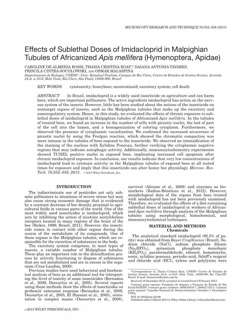
Fig. 1. Histological sections of the Malpighian tubules stained withHE of honeybees exposed or not exposed to imidacloprid. A: Malpigh-ian tubule of a bee from the control group without solvent after 3days, showing typical morphology of this organ. B: Malpighian tubuleof a bee from LD50/100 group after 3 days. Note homogenization inthe staining of the cytoplasm and the presence of pyknotic nuclei(arrow). C: Malpighian tubule of a bee exposed LD50/10 for 5 days.Verification of the presence of a part of cell disposed in the lumen
(Ce). D: Malpighian tubule of a bee exposed to LD50/50 for 1 day, notethe vacuolation (*). E–G: Malpighian tubules of bees exposed to LD50/100 for 1, and 7 days and from group treated with LD50/50 for 10days. Note the lost of part of the cell into the lumen (Ce) and in Epresence of pyknotic nuclei (arrow). H: Malpighian tubule of beesexposed to LD50/10 for 7 days. Note the presence of pyknotic nuclei(arrow). L 5 lumen, N 5 nuclei. [Color figure can be viewed in theonline issue, which is available at wileyonlinelibrary.com.]
554 C. DE ALMEIDA ROSSI ET AL.
Microscopy Research and Technique

(Olympus DP-71) and a Dell computer, linked to themicroscopy. For image acquisition was used the DPController software.
Immunocytochemical Analysis for Detection ofDNA Fragmentation by Endonucleases (TUNEL
Reaction—Terminal DeoxynucleotidylTransferase—Mediated Biotinylated UTP Nick
End Labeling)
Malpighian tubules were dissected and fixed asdescribed above. After dehydration in an ethanol se-ries, organs were diaphanized in xylene and embeddedin paraffin (Histosec, Merck).
Sections of 8 mm thickness were transferred to his-tological slides previously cleaned and treated withpolylysine. The Histosec wax was removed in severalxylene baths before the sections were rehydrated andthen submitted to a TUNEL reaction (ISCDDKin situ Cell Death Detection Kit) according to themanufacturer’s instructions. Negative and positivecontrols were made for the TUNEL reaction. Afterincubation, the slides were examined for detection ofthe fluorescein-labeled 30-OH ends of fragmentedDNA and photographed on a fluorescence microscopeOlympus BX-51 (Olympus America), using a wave-length of 450–500 nm.
RESULTSH–E Stain
The results obtained for the imidacloprid-exposedand controls groups are summarized in Table 1. TheMalpighian tubules of the control group without sol-vent showed typical morphology. The basal portions of
the cytoplasm were slightly stained, indicating thepresence of basal labyrinth. The cells presented largenuclei with many nucleoli. These basic characteristicsappear in all groups to a greater or lesser extent (Fig.1A). The Malpighian tubules of the bees in the solventcontrol group showed morphology similar to the con-trol group without solvent.
In Malpighian tubules of bees exposed to LD50/100,LD50/50 and LD50/10, we observed a homogeneousstaining of the apical and basal cytoplasm, suggestinga reduction of the basal labyrinth (Fig. 1B). In theLD50/100 group, these characteristics were observed inbees exposed for 3, 5, 7, and 10 days. In the othertreated groups, the homogenization of apical and basalcytoplasmic staining was observed in individuals col-lected after 5 days of exposure to concentrations ofLD50/50 e and LD50/10.
The vacuolization of the cytoplasm (Fig. 1D) waspresent in the tubules of the LD50/100 group in bees af-ter exposure for 3 and 5 days. In the LD50/50 and LD50/10 groups, the vacuolization was observed earlier,occurring after only 1 day and persisting until 5 daysof exposure.
The most evident features of the exposed groupswere the increased frequency and intensity of pyknoticnuclei (Fig. 1H) and the lost of part of the cell into thelumen (Figs. 1C and 1E–1G), which was present in allexposed groups at all ages analyzed.
Feulgen Reaction
The results obtained with histochemical analysisare summarized in Table 2. In the Feulgen reactiontest, the Malpighian tubules of both the control groupswith and without solvent showed cells with weakly
TABLE 2. Results of Fuelgen reaction and Xylidine Ponceau technique performed in the Malpighian tubules of A. mellifera exposed or notexposed to imidacloprid
Fuelgen reaction Xylidine Ponceau
Chromatincompaction
Region of the cytoplasmwith negative reaction
Staining ofthe nuclei
Uniform staining of basaland apical cytoplasm
Treatment DaysControl without solvent 1 2 2 1 2
3 2 2 1 25 2 2 1 27 2 2 1 110 2 2 1 1
Control with solvent 1 2 2 1 23 2 2 1 25 2 2 1 27 2 2 11 110 2 2 11 1
LD50/100 1 1 1 1 13 1 1 1 15 1 1 1 17 1 1 11 110 11 1 11 1
LD50/50 1 1 2 11 13 11 1 1 15 1 1 1 17 11 1 11 110 1 1 11 1
LD50/10 1 1 1 11 13 11 1 1 15 1 1 1 17 111 1 11 110 11 1 11 1
(1) Presence of alteration; (11) Alteration moderately present; (111) Alteration extremely present; (2) alteration absence.
EFFECTS OF IMIDACLOPRID IN AFRICANIZED HONEYBEES 555
Microscopy Research and Technique

stained nuclei at all timepoints indicating the presenceof decondensed chromatin (Fig. 2A). Tubules of thebees in the solvent control group showed the
characteristics. Bees from all groups exposed to insec-ticide at all timepoints, presented more stronglystained nuclei, indicating a higher level of chromatincompaction (Figs. 2B and 2C). These results furthersupport our previous findings of increased pyknoticnuclei revealed by HE staining.
Xylidine Ponceau Technique
The Malpighian tubules stained with Xylidine Pon-ceau of both control groups showed a distinct stainingin the basal and apical portions of cells until 5 days.The basal region was negative to this technique, mostlikely due to the basal labyrinth, while the apical por-tion was positive, indicating the presence of cytoplas-mic proteins (Fig. 3A). The nuclei and nucleoli of thecontrol groups were positive due to the marking of nu-clear proteins (histones and others) and nucleolar. Thetubules of bees exposed to insecticide at all timepointshad cytoplasm that was stained homogeneously withXylidine (Fig. 3B), indicating the loss of basal laby-rinth. Some cells presented a cytoplasm with negativeregions to Xylidine staining (Figs. 3C and 3D), whichmay correspond to vacuolated regions stained by HE.The increase in the intensity of nuclei staining some-times occurs under volume reducing conditions (Fig.3C).
Immunocytochemical Analysis
Nuclei positive for TUNEL reactions were observedonly in cells from the Malpighian tubules of beesexposed to insecticide (Fig. 4). For the group exposedto LD50/100, positive results were observed in beesfrom the 1, 7, and 10 days exposure groups. For theLD50/50 group, positive results were observed in beesat 1, 3, 5, and 10 days of exposure. For the LD50/10group, we observed positive nuclei reactions in allgroups after the 5th day of exposure.
DISCUSSION
Morphological, histochemical, and immunocyto-chemical alterations were observed in Malpighiantubules of bees from exposed groups compared to con-trol groups. Moreover, comparing the groups exposedto each other, we could not observe significant differ-ence in cytotoxicity, showing that even at very lowdoses the imidacloprid can alter the physiology of hon-eybees. The differences found between the treatmentsand exposure times mainly revealed alterations of in-tensity in the observed results.
Comparing the tubules of cells from bees exposed toLD50/100, LD50/50, or LD50/10, we found an increase inthe amount of pyknotic nuclei, the lost of parts of thecell into the lumen, the homogenization of the cyto-plasmic staining and vacuolated cells.
Increased homogeneous staining of the cytoplasm,as observed by analysis with Xylidine Ponceau, sug-gests the loss of basal membrane invagination of thesecells when compared to the controls. These structurespromote greater contact with the hemolymph and thusenhance the uptake of substances from it (Cruz-Landim, 1998). Therefore, this reduction or loss of thebasal labyrinth may indicate a tissue degeneration,which can compromise the reabsorption of water andions and excretion. These functions are essential to
Fig. 2. Histological sections of Apis mellifera bee Malpighiantubules subjected to the Feulgen reaction from both exposed and con-trol groups. A: Malpighian tubules of a bee from solvent control groupafter 1 day with slightly stained nuclei showing decondensed chroma-tin uniformly distributed. B and C: Malpighian tubules of beesexposed to LD50/10 on days 3 and 7, respectively, presenting histo-chemical alterations. Note the presence of strongly stained nuclei withpyknosis (arrow). L 5 lumen, N 5 nuclei. [Color figure can be viewedin the online issue, which is available at wileyonlinelibrary.com.]
556 C. DE ALMEIDA ROSSI ET AL.
Microscopy Research and Technique

Fig. 3. Hitological sections of the Malpighian tubules of Apis melli-fera bees subjected to reaction with Xylidine Ponceau from controland insecticide exposed groups. A: Malpighian tubule of bees fromthe control group without solvent after 3 days showing the negativebasal Xylidine staining and the weakly positive staining of the apicalregion of the cytoplasm. B–D: Malpighian tubules of bees from the
LD50/50 group from 10 and 1 day, and LD50/10 group after 1 day,respectively. Note the histochemical changes, the uniform cytoplas-mic staining, the presence of strongly stained nuclei (arrow), and thenegative region of cytoplasm to Xylidine Ponceau reaction (*).L 5 lumen, N 5 nuclei. [Color figure can be viewed in the online issue,which is available at wileyonlinelibrary.com.]
Fig. 4. Histological sections of the Malpighian tubules of honeybeessubjected to a Tunel reaction in control or imidacloprid exposedgroups. A–D: The Malpighian tubule of a bee from the control groups,featuring no positive nuclei to the reaction. A: control group withoutsolvent after 3 days. B: control group with solvent after 5 days. C:control group without solvent after 7 days. D: control group without
solvent after 10 days. E–H: Malpighian tubules of exposed bees show-ing the presence of positive nuclei (arrow). E: LD50/100 group after 7days. F: LD50/50 group after 1 day. F: LD50/100 group after 10 days.G: LD50/10 group after 7 days. [Color figure can be viewed in theonline issue, which is available at wileyonlinelibrary.com.]

normal body metabolism (Cruz-Landim, 1998) andshow the cytotoxic effect of the insecticide.
The loss of part of the cell to the lumen of the Mal-phigian tubules was observed in the groups exposed tothe insecticide. This characteristic was not observed incontrol groups and can be considered an indication ofthe cytotoxicity of imidacloprid for the organ.
The vacuolization of these cells from bees exposed toinsecticide, as evidenced by the negative region of thecytoplasm to Xylidine Ponceau. This alteration wasobserved mainly in the early days of the exposure ofbees to insecticide, and in the final period of exposurethis characteristic was not observed. It may indicatethe presence of autophagic vacuoles suggesting auto-phagic activity (Clarke, 1990). This indicates that thecell initially underwent an autophagic phase, which isa normal physiological process to have a turnover(recycling) of many proteins and organelles cells (Yosh-imori, 2004).
The presence of pyknotic nuclei observed by HEstaining, confirmed by the Feulgen reaction, was moreintense in bees exposed to the insecticide. Chromatincompaction in the nucleus can result in low transcrip-tional activity, suggesting the final phase of the apo-ptotic process of cells undergoing cell death (H€acker,2000; Silva-Zacarim et al., 2008; Wyllie, 1981).
The autophagic phase may not necessarily culmi-nate in cell death (Lockshin and Zakeri, 2004) but ifthe stress situation prevails as in the present study,the cell can be completely degenerated by this processor trigger the apoptotic process of cell death (Lockshinand Zakeri, 2004).
The obtained results suggest that exposure to suble-thal doses of imidacloprid induces cell death in theMalpighian tubules, which was confirmed by TUNELassay. Although programmed cell death is related tothe reorganization of tissue during metamorphosis(Gregorc et al., 2004), we found that this type of celldeath may also occur in organs of insects exposed totoxic substances.
In conclusion, sublethal doses of imidacloprid cancause morphological, histochemical, and immunocyto-chemical alterations on the Malpighian tubules ofbees. Such changes could undermine the process ofexcretion, the primary function of this structure, andcould affect the entire physiology of bees. Toxic sub-stances, when they are not completely eliminated, cancause intoxication of the insect and lead to impairedbehavior or even death of the bee.
REFERENCES
Aliouane Y, El Hassani Ak, Gary V, Armengaud C, Lambin M, Gauth-ier M. 2009. Subchronic exposure of honeybees to sublethal dosesof pesticides: Effects on behavior. Environ Toxicol Chem 28:113–122.
Badiou-B�en�eteau A, Carvalho SM, Brunet J-L, Carvalho GA, Bulet�eA, Giroud B, Belzunces LP. 2012. Development of biomarkers of
exposure to xenobiotics in the honey bee Apis mellifera: Applicationto the systemic insecticide Thiamethoxam. Ecotoxicol Environ Saf,(in press, EES-11-721R3).
Bernadou A, D�emares F, Couret-Fauvel T, Sandoz JC, Gauthier M.2009. Effect of fipronil on side specific antennal tactile learning inthe honeybee. J Insect Physiol 55:1099–1106.
Bicker G. 1999. Histtochemistry of classical neurotransmitters inantennal lobes and mushroom bodies of the honeybee. Microsc ResTech 45:174–183.
Brazil. 2011. Minist�erio da Agricultura, Pecu�aria e Abastecimento.Sistema de Agrot�oxico Fitossanit�arios (Agrofit). Bras�ılia. Dis-pon�ıvel em: Available at: <http://www.agricultura.gov.br/>. Acessoem: 7 jan. 2011.
Clarke PGH. 1990. Developmental cell death; morphological diversityand multiple mechanisms. Anat Embryol 181:195–206.
Cruz-Landim C. 1998. Specializations of the Malpighi Tubules cellsin a stingless bee, Melipona quadrifasciata anthidioides Lep. (Hy-menoptera, Apidae). Acta Microscopica 7:26–33.
Cruz-Landim C. 2009. Abelhas: Morfologia e func~ao de sistemas. S~aoPaulo: UNESP. 408 p.
Decourtye A, Devillers J, Genecque E, Le Menach K, Budzinski H,Cluzeaus S, Pham-Deleghe MH. 2005. Comparative sublethal tox-icity of nine pesticides on olfatory learning performances of thehoneybee Apis mellifera. Arch Environ Contam Toxicol 48:242–250.
Decourtye A, Lefort S, Devillers J, Gauthier M, Aupinel P, Tisseur M.2008. Sublethal effects of fipronil on the ability of honeybee (Apismellifera) to orientate in a complex maze. In: Oomen PA, ThompsonHM, editors. Hazard of pesticides to bees. 10th International Sym-posium of the ICP-BR Bee Protection Group. Bucharest: Julius-K€uhn-Archiv. pp. 75–83.
El Hassani AK, Dacher M, Gauthier M, Armengaud C. 2005. Effectsof sublethal doses of fipronil on the behavior of the honeybee (Apismellifera). Pharmacol Biochem Behav 82:30–39.
Feulgen R, Rossenbeck HM. 1924. Chemischer nachweis einer nucle-insaure vom typus der thymonucleinsaure und die darauf beru-hend elective farbung von zillkernen in mikroskopischenpraparaten hoppe-seylers. Z Physiol Chem 135:203–248.
Gregorc A, Pogacnik A, Bowen ID. 2004. Cell death in honeybee (Apismellifera) larvae treated with oxalic or formic acid. Apidologie35:453–460.
H€acker G. 2000. The morphology of apoptosis. Cell Tissue Res 301:5–17.
Junqueira LCU, Junqueira LMMS. 1983. T�ecnicas b�asicas de citolo-gia e histologia. S~ao Paulo: Editora Santos. pp. 48–81.
Lockshin RA, Zakeri Z. 2004. Apoptosis, autophay, and more. J Bio-chem Cell Biol 36:2405–2419.
OECD. 1998. Organization for Economic Co-Operation and Develop-ment. Guideline 214: Guidelines for the testing of chemicals: Hon-eybees, Acute contact toxicity test. http://puck.sourceoecd.org/vl=1619398/cl=20/nw=1/rpsv/ij/oecdjournals/1607310x/v1n2/s 15/p1. Accessed on February 15, 2012.
Silva-Zacarin ECM, Taboga SR, Silva De Moraes RLM. 2008. Nuclearalterations associated to programmed cell death in larval salivaryglands. Micron 39:117–127.
Silva-Zacarin ECM, Chauzat MP, Zeggane S, Drajnudel P, Schurr F,Faucon JP, Malaspina O, Engler JA. 2012. Protocol for optimizationof histological, histochemical and immunohistochemical analysesof larval tissues: application in histopathology of honey bee. p. 696-703. In: M�endez-Vilas A. (Ed.) Current microscopy contributions toadvances in science and technology, Vol. 1, 788 p., ISBN (13): 978-84-939843-5-9. Badajoz, Spain: Formatex Research Center, Decem-ber 2012.
Wyllie AH. 1981. Cell death: A new classification separating apoptosisfrom necrosis. In: Bowen ID, Lockshin RA, editors. Cell death inbiology and pathology. New York: Chapman & Hall, pp. 9–34.
Yoshimori T. 2004. Autophagy: A regulated bulk degradation processinside cells. Biochem Biophys Res Commun 313:453–458.
558 C. DE ALMEIDA ROSSI ET AL.
Microscopy Research and Technique