Antifungal Susceptibility Testing: A Primer for Clinicians
13
REVIEW ARTICLE INVITED Antifungal Susceptibility Testing • OFID • 1 Open Forum Infectious Diseases Received 24 June 2021; editorial decision 23 August 2021; accepted 26 August 2021. Correspondence: Nathan P. Wiederhold, PharmD, UT Health San Antonio, 7703 Floyd Curl Drive, San Antonio, TX 78229, USA ([email protected]). Open Forum Infectious Diseases ® 2021 © The Author(s) 2021. Published by Oxford University Press on behalf of Infectious Diseases Society of America. This is an Open Access article distributed under the terms of the Creative Commons Attribution-NonCommercial-NoDerivs licence (https://creativecommons.org/ licenses/by-nc-nd/4.0/), which permits non-commercial reproduction and distribution of the work, in any medium, provided the original work is not altered or transformed in any way, and that the work is properly cited. For commercial re-use, please contact [email protected] https://doi.org/10.1093/ofid/ofab444 Antifungal Susceptibility Testing: A Primer for Clinicians Nathan P. Wiederhold Fungus Testing Laboratory, Department of Pathology and Laboratory Medicine, University of Texas Health Science Center at San Antonio, San Antonio, Texas, USA Clinicians treating patients with fungal infections may turn to susceptibility testing to obtain information regarding the activity of different antifungals against a specific fungus that has been cultured. ese results may then be used to make decisions regarding a patient’s therapy. However, for many fungal species that are capable of causing invasive infections, clinical breakpoints have not been established. us, interpretations of susceptible or resistant cannot be provided by clinical laboratories, and this is especially true for many molds capable of causing severe mycoses. e purpose of this review is to provide an overview of susceptibility testing for clinicians, including the methods used to perform these assays, their limitations, how clinical breakpoints are established, and how the results may be put into context in the absence of interpretive criteria. Examples of when susceptibility testing is not warranted are also provided. Keywords. antifungal; clinical breakpoint; susceptibility testing. Over the last several decades, the number of individuals at risk for invasive fungal infections has markedly increased. This has been attributed to the increased number of patients who are immunocompromised, either due to disease or use of immu- nosuppressive agents, and those in critical care settings. As the number of individuals at risk for fungal infections has grown, so has our recognition of the number of different fungi that are capable of causing disease in humans. It is estimated that there may be between 1.5 and 5 million different fungal species, and more than 300 are capable of causing infections in humans [1–4]. These numbers will only continue to increase as molec- ular tools become more widely used for the detection and di- agnosis of new fungi and fungal diseases and as the number of at-risk individuals continues to rise. e discovery of new fungi capable of causing disease in both humans and the development of resistance to currently avail- able drugs have far outpaced the availability of new antifungals. One contemporary example of a newly described fungus is that of Candida auris. First described in 2009 [5], this emerging pathogen has quickly spread to multiple continents and has been associated with numerous outbreaks in different in- stitutions [6–8]. Isolates of this species are oſten found to be resistant to multiple antifungals [6–8], and resistance to all cur- rently available antifungals has also been described in some isolates [7, 9, 10]. Another example is Aspergillus fumigatus, a well known mold pathogen for which resistance outside of clin- ical antifungal exposure is now of increasing concern. Azole- resistant A fumigatus was first described in the 1990s in patients with chronic exposure to itraconazole [11, 12]. However, more than a decade ago azole-resistant strains were recovered from patients with invasive aspergillosis but without previous azole exposure, and this was subsequently linked to the use of azole- like compounds in the environment [13–15]. Azole resistance linked to environmental exposure has now been reported in countries around the world [3, 16], as have cases of clinical fail- ures in patients treated with azoles for Aspergillus infections caused by resistant isolates [3, 17–19]. Because of the discovery of new pathogenic fungi and the increased threat of antifungal resistance, clinicians oſten use susceptibility testing to help guide therapy in patients with invasive mycoses. e objective of this review is to provide an overview of antifungal suscepti- bility testing against yeasts and molds. is will include discus- sions of how antifungal susceptibility testing is performed, how the results may be interpreted and used to guide therapy, as well as their limitations. METHODS FOR ANTIFUNGAL SUSCEPTIBILITY TESTING In clinical microbiology laboratories, susceptibility testing is still primarily performed by in vitro phenotypic methods, which measure the ability of a particular drug to inhibit the growth of an organism over a range of concentrations. The readout is the minimum inhibitory concentration (MIC), which corresponds to the lowest concentration of the antifungal that inhibits the growth of the organism. Although significant ad- vances have been made in the arena of clinical microbiology, the antifungal susceptibility assays still used share similarities with those methods described by Sir Alexander Fleming [20] in Downloaded from https://academic.oup.com/ofid/article/8/11/ofab444/6367646 by guest on 27 March 2022
-
Upload
khangminh22 -
Category
Documents
-
view
4 -
download
0
Transcript of Antifungal Susceptibility Testing: A Primer for Clinicians
R E V I E W A R T I C L E I N V I T E D
Antifungal Susceptibility Testing • ofid • 1
Open Forum Infectious Diseases
Received 24 June 2021; editorial decision 23 August 2021; accepted 26 August 2021.Correspondence: Nathan P. Wiederhold, PharmD, UT Health San Antonio, 7703 Floyd Curl
Drive, San Antonio, TX 78229, USA ([email protected]).
Open Forum Infectious Diseases®2021© The Author(s) 2021. Published by Oxford University Press on behalf of Infectious Diseases Society of America. This is an Open Access article distributed under the terms of the Creative Commons Attribution-NonCommercial-NoDerivs licence (https://creativecommons.org/licenses/by-nc-nd/4.0/), which permits non-commercial reproduction and distribution of the work, in any medium, provided the original work is not altered or transformed in any way, and that the work is properly cited. For commercial re-use, please contact [email protected]://doi.org/10.1093/ofid/ofab444
Antifungal Susceptibility Testing: A Primer for CliniciansNathan P. Wiederhold
Fungus Testing Laboratory, Department of Pathology and Laboratory Medicine, University of Texas Health Science Center at San Antonio, San Antonio, Texas, USA
Clinicians treating patients with fungal infections may turn to susceptibility testing to obtain information regarding the activity of different antifungals against a specific fungus that has been cultured. These results may then be used to make decisions regarding a patient’s therapy. However, for many fungal species that are capable of causing invasive infections, clinical breakpoints have not been established. Thus, interpretations of susceptible or resistant cannot be provided by clinical laboratories, and this is especially true for many molds capable of causing severe mycoses. The purpose of this review is to provide an overview of susceptibility testing for clinicians, including the methods used to perform these assays, their limitations, how clinical breakpoints are established, and how the results may be put into context in the absence of interpretive criteria. Examples of when susceptibility testing is not warranted are also provided.
Keywords. antifungal; clinical breakpoint; susceptibility testing.
Over the last several decades, the number of individuals at risk for invasive fungal infections has markedly increased. This has been attributed to the increased number of patients who are immunocompromised, either due to disease or use of immu-nosuppressive agents, and those in critical care settings. As the number of individuals at risk for fungal infections has grown, so has our recognition of the number of different fungi that are capable of causing disease in humans. It is estimated that there may be between 1.5 and 5 million different fungal species, and more than 300 are capable of causing infections in humans [1–4]. These numbers will only continue to increase as molec-ular tools become more widely used for the detection and di-agnosis of new fungi and fungal diseases and as the number of at-risk individuals continues to rise.
The discovery of new fungi capable of causing disease in both humans and the development of resistance to currently avail-able drugs have far outpaced the availability of new antifungals. One contemporary example of a newly described fungus is that of Candida auris. First described in 2009 [5], this emerging pathogen has quickly spread to multiple continents and has been associated with numerous outbreaks in different in-stitutions [6–8]. Isolates of this species are often found to be resistant to multiple antifungals [6–8], and resistance to all cur-rently available antifungals has also been described in some
isolates [7, 9, 10]. Another example is Aspergillus fumigatus, a well known mold pathogen for which resistance outside of clin-ical antifungal exposure is now of increasing concern. Azole-resistant A fumigatus was first described in the 1990s in patients with chronic exposure to itraconazole [11, 12]. However, more than a decade ago azole-resistant strains were recovered from patients with invasive aspergillosis but without previous azole exposure, and this was subsequently linked to the use of azole-like compounds in the environment [13–15]. Azole resistance linked to environmental exposure has now been reported in countries around the world [3, 16], as have cases of clinical fail-ures in patients treated with azoles for Aspergillus infections caused by resistant isolates [3, 17–19]. Because of the discovery of new pathogenic fungi and the increased threat of antifungal resistance, clinicians often use susceptibility testing to help guide therapy in patients with invasive mycoses. The objective of this review is to provide an overview of antifungal suscepti-bility testing against yeasts and molds. This will include discus-sions of how antifungal susceptibility testing is performed, how the results may be interpreted and used to guide therapy, as well as their limitations.
METHODS FOR ANTIFUNGAL SUSCEPTIBILITY TESTING
In clinical microbiology laboratories, susceptibility testing is still primarily performed by in vitro phenotypic methods, which measure the ability of a particular drug to inhibit the growth of an organism over a range of concentrations. The readout is the minimum inhibitory concentration (MIC), which corresponds to the lowest concentration of the antifungal that inhibits the growth of the organism. Although significant ad-vances have been made in the arena of clinical microbiology, the antifungal susceptibility assays still used share similarities with those methods described by Sir Alexander Fleming [20] in
Dow
nloaded from https://academ
ic.oup.com/ofid/article/8/11/ofab444/6367646 by guest on 27 M
arch 2022
2 • ofid • Wiederhold
1929 to describe the activity of penicillin against Staphylococcus and other bacteria, which included agar diffusion and broth di-lution testing.
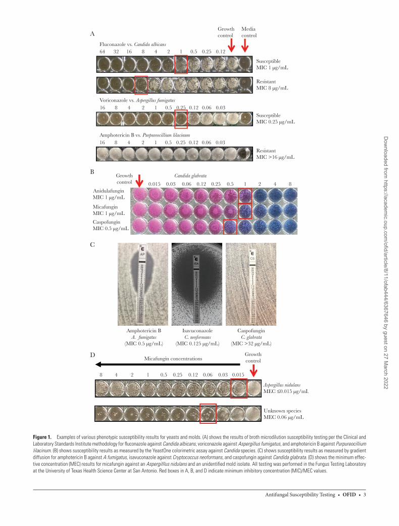
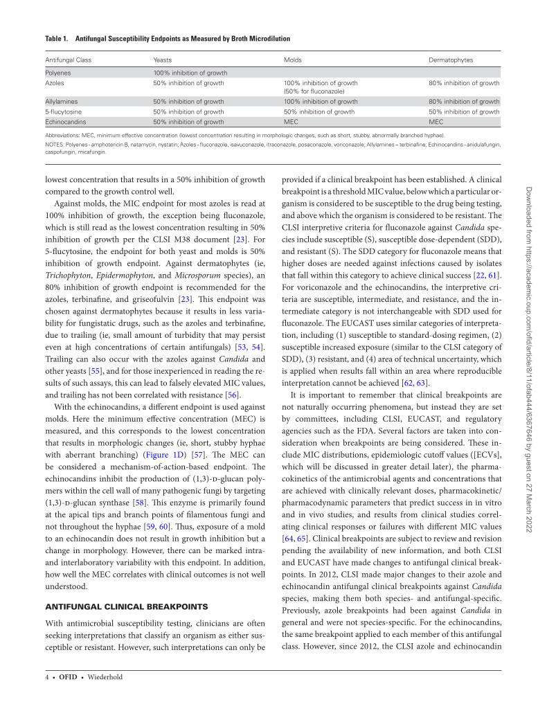
Several different formats are available for antifungal sus-ceptibility testing. Two organizations that establish and stand-ardize such methods are the Clinical and Laboratory Standards Institute (CLSI) and the European Committee on Antimicrobial Susceptibility Testing (EUCAST), and these are often used as the gold standards by which other methods are evaluated. The CLSI and EUCAST assays most often used are broth microdilution methods that utilize 96-well cell culture trays (Figure 1A) [21–23]. Several commercial assays are also now available for antifungal susceptibility testing. One that is widely used in clinical microbiology laboratories due to the ease of endpoint reading is the YeastOne Sensititre assay (Thermo Scientific, TREK Diagnostics). This is a broth microdilution-based assay that includes a colorimetric dye, resazurin (alamarBlue), which is converted to resorufin (dark pink to red in color) by metabol-ically active fungal cells. When metabolic activity is inhibited, the wells appear blue, and the change from pink/red to blue is the endpoint used to read the MIC in this assay (Figure 1B). Studies have reported excellent agreement between the YeastOne assay and the broth microdilution assays for Candida species, with es-sential agreement (MICs within 2 dilutions) ranging from 95% to 100% and categorical agreement (same classification of re-sults as susceptible or resistant) between 70% to 100% for both the azoles and the echinocandins [24–31]. Others have also noted good agreement for amphotericin B, different azoles, and the echinocandins with broth microdilution methods against Aspergillus species, although some variability has also been reported [32–37]. Good agreement (83% to 100%) has also been reported between the YeastOne and CLSI M38 assays for amphotericin B and posaconazole against Mucorales isolates and Fusarium species [32, 38], and similarly for voriconazole against Fusarium species [39], although a small number of iso-lates were generally included in these studies. However, others have reported poor agreement between the YeastOne and the CLSI broth microdilution assays for amphotericin B and dif-ferent azoles against Mucorales isolates [40].
Another format that is available for antifungal suscepti-bility testing is that of gradient diffusion (Etest, bioMerieux; MTS, Liofilchem). These assays use plastic strips that contain a concentration gradient of a particular agent, which is placed onto the surface of an agar growth plate that has been inocu-lated with a fungus. The antifungal diffuses from the strip into the agar, and after a period of incubation, the MIC is read as the concentration where the elliptical zone of inhibition intersects with the strip (Figure 1C). Good essential agree-ment (92% to 96.8%) has been reported between the CLSI and EUCAST broth microdilution methods and gradient dif-fusion assays against Candida [24, 25], with categorical agree-ment ranging from 80% to 97% between the Etest and CLSI
broth microdilution methods in one study for several azoles and 5-flucytosine [24]. These assays have also been used for mold susceptibility testing, where good agreement has been demonstrated for some, but not all, antifungals against dif-ferent filamentous fungi, primarily Aspergillus and Fusarium species [41]. However, marked variability has been reported for members of the order Mucorales and Scedosporium spe-cies [41–43].
An automated format is also available for use in clinical mi-crobiology laboratories that can perform both yeast antifungal susceptibility testing and yeast species identification (Vitek 2; bioMerieux), with good essential agreement (89.3% to 100%) and categorical agreement (92% to 99.5%) between this method and the CLSI broth microdilution assay [25, 44–49]. It is in-teresting to note that lower categorical agreement has been reported between both the Vitek and YeastOne methods and the CLSI broth microdilution method for fluconazole against Candida glabrata isolates [24, 45, 47]. In addition, this method cannot be used for mold susceptibility testing, and the number of antifungals that can currently be tested by this US Food and Drug Administration (FDA) cleared assay is limited (eg, caspofungin, micafungin, fluconazole, voriconazole, and 5-flucytosine) [50]. It is interesting to note that there have been several reports of falsely elevated antifungal MIC results, espe-cially with amphotericin B, against C auris when measured by Vitek 2 [9, 51, 52].
Because antifungal susceptibility testing is still performed by phenotypic assays, the turnaround time for results can be delayed for certain fungi due to incubation periods that are required. For Candida species, results are read after 24 hours of incubation. For other yeasts, including Cryptococcus and Rhodotorula species, a 72-hour incubation period is required per CLSI. For most clinically relevant molds the incubation period is 48 hours, but it can range from 24 hours for the Mucorales (eg, Rhizopus, Mucor, Cunninghamella, Lichtheimia, among others) up to 96 hours for dermatophytes. In addition, a pure subculture with adequate growth is needed even before the susceptibility assays can be run. Many clinical microbiology la-boratories do not perform their own mold susceptibility testing, so these are sent out to references laboratories, which can fur-ther delay results being made available to clinicians.
ENDPOINTS FOR ANTIFUNGAL SUSCEPTIBILITY TESTING
For antifungal susceptibility testing, the endpoints (eg, MIC) used to measure in vitro activity is dependent upon both the organism and the antifungal against which the fungus is tested (Table 1). For amphotericin B, MICs against both yeasts and molds are read as the lowest concentration that results in com-plete inhibition of growth, and this applies for both broth microdilution and gradient diffusion testing. For the azoles and echinocandins against yeasts, the MIC endpoint is read at the
Dow
nloaded from https://academ
ic.oup.com/ofid/article/8/11/ofab444/6367646 by guest on 27 M
arch 2022
Antifungal Susceptibility Testing • ofid • 3
Growthcontrol
64
A
B
C
D
32 16 8 4 2 1 0.5 0.25 0.12Fluconazole vs. Candida albicans
16 8 4 2 1 0.5 0.25 0.12 0.06 0.03Voriconazole vs. Aspergillus fumigatus
16 8 4
Growthcontrol 0.015
AnidulafunginMIC 1 µg/mL
MicafunginMIC 1 µg/mL
CaspofunginMIC 0.5 µg/mL
Amphotericin BA. fumigatus
(MIC 0.5 µg/mL)
Micafungin concentrationsGrowthcontrol
8 4 2 1 0.5 0.25 0.12 0.06 0.03 0.015
Aspergillus nidulansMEC ≤0.015 µg/mL
Unknown speciesMEC 0.06 µg/mL
IsavuconazoleC. neoformans
(MIC 0.125 µg/mL)
CaspofunginC. glabrata
(MIC >32 µg/mL)
0.03 0.06 0.12 0.25 0.5 1 2 4 8
2 1 0.5 0.25 0.12 0.06 0.03Amphotericin B vs. Purpureocillium lilacinum
Candida glabrata
Mediacontrol
SusceptibleMIC 1 µg/mL
SusceptibleMIC 0.25 µg/mL
ResistantMIC 8 µg/mL
ResistantMIC >16 µg/mL
Figure 1. Examples of various phenotypic susceptibility results for yeasts and molds. (A) shows the results of broth microdilution susceptibility testing per the Clinical and Laboratory Standards Institute methodology for fluconazole against Candida albicans, voriconazole against Aspergillus fumigatus, and amphotericin B against Purpureocillium lilacinum. (B) shows susceptibility results as measured by the YeastOne colorimetric assay against Candida species. (C) shows susceptibility results as measured by gradient diffusion for amphotericin B against A fumigatus, isavuconazole against Cryptococcus neoformans, and caspofungin against Candida glabrata. (D) shows the minimum effec-tive concentration (MEC) results for micafungin against an Aspergilllus nidulans and an unidentified mold isolate. All testing was performed in the Fungus Testing Laboratory at the University of Texas Health Science Center at San Antonio. Red boxes in A, B, and D indicate minimum inhibitory concentration (MIC)/MEC values.
Dow
nloaded from https://academ
ic.oup.com/ofid/article/8/11/ofab444/6367646 by guest on 27 M
arch 2022
4 • ofid • Wiederhold
lowest concentration that results in a 50% inhibition of growth compared to the growth control well.
Against molds, the MIC endpoint for most azoles is read at 100% inhibition of growth, the exception being fluconazole, which is still read as the lowest concentration resulting in 50% inhibition of growth per the CLSI M38 document [23]. For 5-flucytosine, the endpoint for both yeast and molds is 50% inhibition of growth endpoint. Against dermatophytes (ie, Trichophyton, Epidermophyton, and Microsporum species), an 80% inhibition of growth endpoint is recommended for the azoles, terbinafine, and griseofulvin [23]. This endpoint was chosen against dermatophytes because it results in less varia-bility for fungistatic drugs, such as the azoles and terbinafine, due to trailing (ie, small amount of turbidity that may persist even at high concentrations of certain antifungals) [53, 54]. Trailing can also occur with the azoles against Candida and other yeasts [55], and for those inexperienced in reading the re-sults of such assays, this can lead to falsely elevated MIC values, and trailing has not been correlated with resistance [56].
With the echinocandins, a different endpoint is used against molds. Here the minimum effective concentration (MEC) is measured, and this corresponds to the lowest concentration that results in morphologic changes (ie, short, stubby hyphae with aberrant branching) (Figure 1D) [57]. The MEC can be considered a mechanism-of-action-based endpoint. The echinocandins inhibit the production of (1,3)-d-glucan poly-mers within the cell wall of many pathogenic fungi by targeting (1,3)-d-glucan synthase [58]. This enzyme is primarily found at the apical tips and branch points of filamentous fungi and not throughout the hyphae [59, 60]. Thus, exposure of a mold to an echinocandin does not result in growth inhibition but a change in morphology. However, there can be marked intra- and interlaboratory variability with this endpoint. In addition, how well the MEC correlates with clinical outcomes is not well understood.
ANTIFUNGAL CLINICAL BREAKPOINTS
With antimicrobial susceptibility testing, clinicians are often seeking interpretations that classify an organism as either sus-ceptible or resistant. However, such interpretations can only be
provided if a clinical breakpoint has been established. A clinical breakpoint is a threshold MIC value, below which a particular or-ganism is considered to be susceptible to the drug being testing, and above which the organism is considered to be resistant. The CLSI interpretive criteria for fluconazole against Candida spe-cies include susceptible (S), susceptible dose-dependent (SDD), and resistant (S). The SDD category for fluconazole means that higher doses are needed against infections caused by isolates that fall within this category to achieve clinical success [22, 61]. For voriconazole and the echinocandins, the interpretive cri-teria are susceptible, intermediate, and resistance, and the in-termediate category is not interchangeable with SDD used for fluconazole. The EUCAST uses similar categories of interpreta-tion, including (1) susceptible to standard-dosing regimen, (2) susceptible increased exposure (similar to the CLSI category of SDD), (3) resistant, and (4) area of technical uncertainty, which is applied when results fall within an area where reproducible interpretation cannot be achieved [62, 63].
It is important to remember that clinical breakpoints are not naturally occurring phenomena, but instead they are set by committees, including CLSI, EUCAST, and regulatory agencies such as the FDA. Several factors are taken into con-sideration when breakpoints are being considered. These in-clude MIC distributions, epidemiologic cutoff values ([ECVs], which will be discussed in greater detail later), the pharma-cokinetics of the antimicrobial agents and concentrations that are achieved with clinically relevant doses, pharmacokinetic/ pharmacodynamic parameters that predict success in in vitro and in vivo studies, and results from clinical studies correl-ating clinical responses or failures with different MIC values [64, 65]. Clinical breakpoints are subject to review and revision pending the availability of new information, and both CLSI and EUCAST have made changes to antifungal clinical break-points. In 2012, CLSI made major changes to their azole and echinocandin antifungal clinical breakpoints against Candida species, making them both species- and antifungal-specific. Previously, azole breakpoints had been against Candida in general and were not species-specific. For the echinocandins, the same breakpoint applied to each member of this antifungal class. However, since 2012, the CLSI azole and echinocandin
Table 1. Antifungal Susceptibility Endpoints as Measured by Broth Microdilution
Antifungal Class Yeasts Molds Dermatophytes
Polyenes 100% inhibition of growth
Azoles 50% inhibition of growth 100% inhibition of growth (50% for fluconazole)
80% inhibition of growth
Allylamines 50% inhibition of growth 100% inhibition of growth 80% inhibition of growth
5-flucytosine 50% inhibition of growth 50% inhibition of growth 50% inhibition of growth
Echinocandins 50% inhibition of growth MEC MEC
Abbreviations: MEC, minimum effective concentration (lowest concentration resulting in morphologic changes, such as short, stubby, abnormally branched hyphae).
NOTES: Polyenes - amphotericin B, natamycin, nystatin; Azoles - fluconazole, isavuconazole, itraconazole, posaconazole, voriconazole; Allylamines – terbinafine; Echinocandins - anidulafungin, caspofungin, micafungin.
Dow
nloaded from https://academ
ic.oup.com/ofid/article/8/11/ofab444/6367646 by guest on 27 M
arch 2022
Antifungal Susceptibility Testing • ofid • 5
breakpoints are dependent upon both the antifungal tested and the specific fungal species [66].
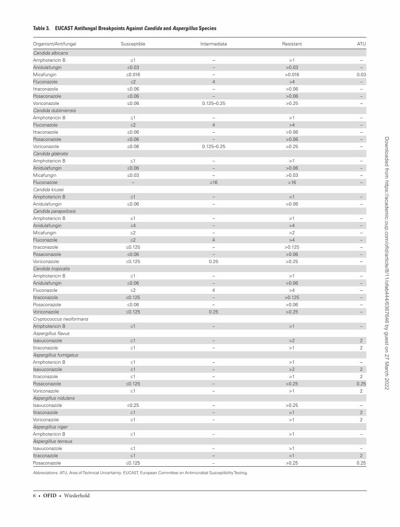
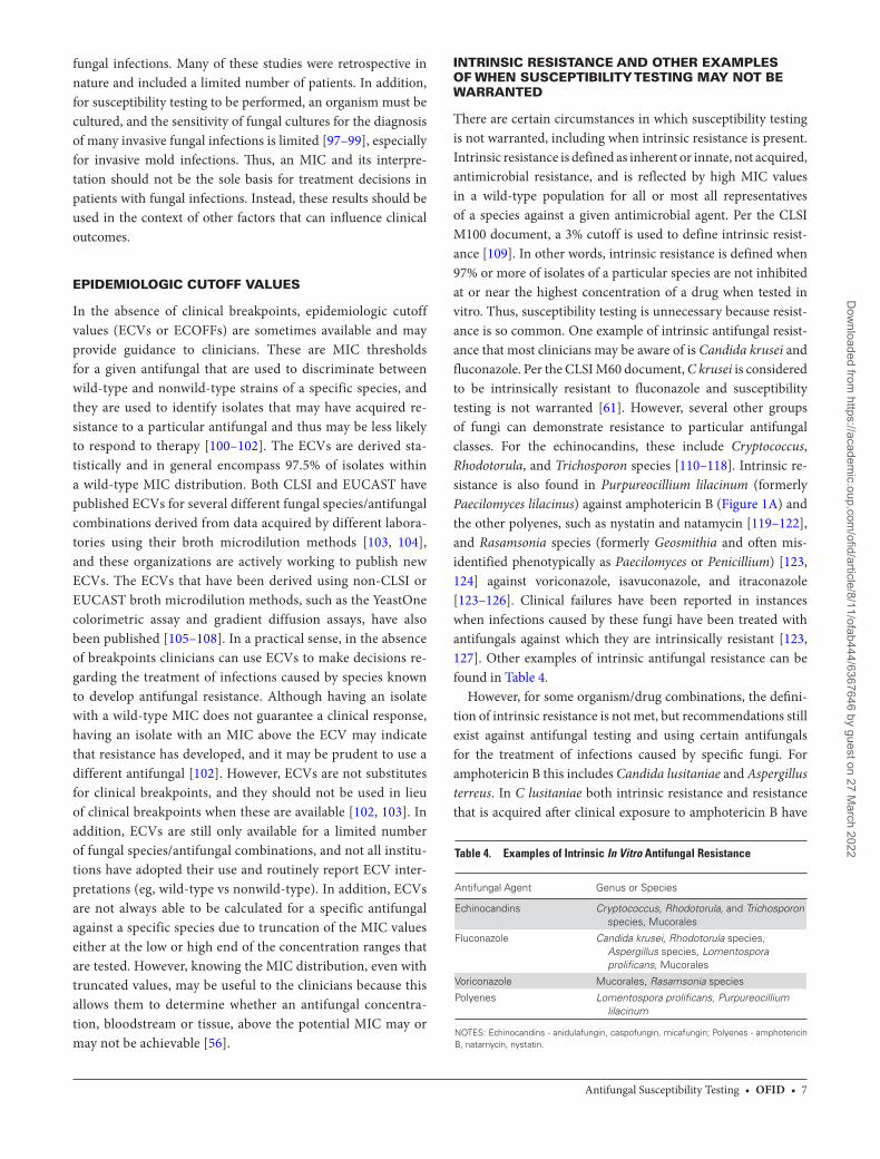
The number of species/antifungal combinations for which CLSI and EUCAST clinical breakpoints have been established is limited. Current CLSI and EUCAST breakpoints against Candida species are shown in Tables 2 and 3. The EUCAST has also set breakpoints for several antifungals against dif-ferent Aspergillus species and for amphotericin B against Cryptococcus neoformans (Table 3) [61, 67]. In June 2020, CLSI published a breakpoint for voriconazole against A fumigatus (Figure 1A) [68]. Because there are differences between the CLSI and EUCAST testing methods, it is not recommended to apply the breakpoints from one group if the MICs were deter-mined using the methods of the other group. For many patho-genic fungi, especially molds, interpretations of susceptibility
results cannot be provided because breakpoints have not yet been set.
The results of antifungal susceptibility testing are most useful when there is a correlation between in vitro results and clin-ical outcomes. However, the results of studies linking the 2 have been mixed. Numerous studies have been conducted, some reporting positive correlations between in vitro susceptibility results for antifungals against Candida species, C neoformans, and Aspergillus species and clinical outcomes in patients with infections caused by these fungi [69–82], whereas others have not [83–91]. A full review of studies that have attempted to find such correlations is beyond the scope of this review, and these have been reviewed in great detail elsewhere [92–96]. It is not surprising that the results of such studies are mixed given the numerous factors that can influence outcomes in patients with
Table 2. CLSI Antifungal Breakpoints against Candida species and Aspergillus fumigatus
Organism/Antifungal
Breakpoint
Susceptible SDD Intermediate Resistant
Candida albicans
Anidulafungin ≤0.25 – 0.5 ≥1
Caspofungin ≤0.25 – 0.5 ≥1
Micafungin ≤0.25 – 0.5 ≥1
Fluconazole ≤2 4 – ≥8
Voriconazole ≤0.12 – 0.25–0.5 ≥1
Candida glabrata
Anidulafungin ≤0.12 – 0.25 ≥0.5
Caspofungin ≤0.12 – 0.25 ≥0.5
Micafungin ≤0.06 – 0.125 ≥0.25
Fluconazole – ≤32 – ≥64
Candida guilliermondii
Anidulafungin ≤2 – 4 ≥8
Caspofungin ≤2 – 4 ≥8
Micafungin ≤2 – 4 ≥8
Candida krusei
Anidulafungin ≤0.25 – 0.5 ≥1
Caspofungin ≤0.25 – 0.5 ≥1
Micafungin ≤0.25 – 0.5 ≥1
Fluconazole – – – –
Voriconazole ≤0.5 – 1 ≥2
Candida parapsilosis
Anidulafungin ≤2 – 4 ≥8
Caspofungin ≤2 – 4 ≥8
Micafungin ≤2 – 4 ≥8
Fluconazole ≤2 4 – ≥8
Voriconazole ≤0.12 – 0.025–0.5 ≥1
Candida tropicalis
Anidulafungin ≤0.25 – 0.5 ≥1
Caspofungin ≤0.25 – 0.5 ≥1
Micafungin ≤0.25 – 0.5 ≥1
Fluconazole ≤2 4 – ≥8
Voriconazole ≤0.12 – 0.25–0.5 ≥1
Aspergillus fumigatus
Voriconazole ≤0.5 – 1 ≥2
Abbreviations: CLSI, Clinical and Laboratory Standards Institute; SDD, susceptible dose-dependent.
Dow
nloaded from https://academ
ic.oup.com/ofid/article/8/11/ofab444/6367646 by guest on 27 M
arch 2022
6 • ofid • Wiederhold
Table 3. EUCAST Antifungal Breakpoints Against Candida and Aspergillus Species
Organism/Antifungal Susceptible Intermediate Resistant ATU
Candida albicansAmphotericin B ≤1 – >1 –
Anidulafungin ≤0.03 – >0.03 –
Micafungin ≤0.016 – >0.016 0.03
Fluconazole ≤2 4 >4 –
Itraconazole ≤0.06 – >0.06 –
Posaconazole ≤0.06 – >0.06 –
Voriconazole ≤0.06 0.125–0.25 >0.25 –
Candida dubliniensisAmphotericin B ≤1 – >1 –
Fluconazole ≤2 4 >4 –
Itraconazole ≤0.06 – >0.06 –
Posaconazole ≤0.06 – >0.06 –
Voriconazole ≤0.06 0.125–0.25 >0.25 –
Candida glabrataAmphotericin B ≤1 – >1 –
Anidulafungin ≤0.06 – >0.06 –
Micafungin ≤0.03 – >0.03 –
Fluconazole – ≤16 >16 –
Candida kruseiAmphotericin B ≤1 – >1 –
Anidulafungin ≤0.06 – >0.06 –
Candida parapsilosisAmphotericin B ≤1 – >1 –
Anidulafungin ≤4 – >4 –
Micafungin ≤2 – >2 –
Fluconazole ≤2 4 >4 –
Itraconazole ≤0.125 – >0.125 –
Posaconazole ≤0.06 – >0.06 –
Voriconazole ≤0.125 0.25 >0.25 –
Candida tropicalisAmphotericin B ≤1 – >1 –
Anidulafungin ≤0.06 – >0.06 –
Fluconazole ≤2 4 >4 –
Itraconazole ≤0.125 – >0.125 –
Posaconazole ≤0.06 – >0.06 –
Voriconazole ≤0.125 0.25 >0.25 –
Cryptococcus neoformansAmphotericin B ≤1 – >1 –
Aspergillus flavusIsavuconazole ≤1 – >2 2
Itraconazole ≤1 – >1 2
Aspergillus fumigatusAmphotericin B ≤1 – >1 –
Isavuconazole ≤1 – >2 2
Itraconazole ≤1 – >1 2
Posaconazole ≤0.125 – >0.25 0.25
Voriconazole ≤1 – >1 2
Aspergillus nidulansIsavuconazole ≤0.25 – >0.25 –
Itraconazole ≤1 – >1 2
Voriconazole ≤1 – >1 2
Aspergillus nigerAmphotericin B ≤1 – >1 –
Aspergillus terreusIsavuconazole ≤1 – >1 –
Itraconazole ≤1 – >1 2
Posaconazole ≤0.125 – >0.25 0.25
Abbreviations: ATU, Area of Technical Uncertainty; EUCAST, European Committee on Antimicrobial Susceptibility Testing.
Dow
nloaded from https://academ
ic.oup.com/ofid/article/8/11/ofab444/6367646 by guest on 27 M
arch 2022
Antifungal Susceptibility Testing • ofid • 7
fungal infections. Many of these studies were retrospective in nature and included a limited number of patients. In addition, for susceptibility testing to be performed, an organism must be cultured, and the sensitivity of fungal cultures for the diagnosis of many invasive fungal infections is limited [97–99], especially for invasive mold infections. Thus, an MIC and its interpre-tation should not be the sole basis for treatment decisions in patients with fungal infections. Instead, these results should be used in the context of other factors that can influence clinical outcomes.
EPIDEMIOLOGIC CUTOFF VALUES
In the absence of clinical breakpoints, epidemiologic cutoff values (ECVs or ECOFFs) are sometimes available and may provide guidance to clinicians. These are MIC thresholds for a given antifungal that are used to discriminate between wild-type and nonwild-type strains of a specific species, and they are used to identify isolates that may have acquired re-sistance to a particular antifungal and thus may be less likely to respond to therapy [100–102]. The ECVs are derived sta-tistically and in general encompass 97.5% of isolates within a wild-type MIC distribution. Both CLSI and EUCAST have published ECVs for several different fungal species/antifungal combinations derived from data acquired by different labora-tories using their broth microdilution methods [103, 104], and these organizations are actively working to publish new ECVs. The ECVs that have been derived using non-CLSI or EUCAST broth microdilution methods, such as the YeastOne colorimetric assay and gradient diffusion assays, have also been published [105–108]. In a practical sense, in the absence of breakpoints clinicians can use ECVs to make decisions re-garding the treatment of infections caused by species known to develop antifungal resistance. Although having an isolate with a wild-type MIC does not guarantee a clinical response, having an isolate with an MIC above the ECV may indicate that resistance has developed, and it may be prudent to use a different antifungal [102]. However, ECVs are not substitutes for clinical breakpoints, and they should not be used in lieu of clinical breakpoints when these are available [102, 103]. In addition, ECVs are still only available for a limited number of fungal species/antifungal combinations, and not all institu-tions have adopted their use and routinely report ECV inter-pretations (eg, wild-type vs nonwild-type). In addition, ECVs are not always able to be calculated for a specific antifungal against a specific species due to truncation of the MIC values either at the low or high end of the concentration ranges that are tested. However, knowing the MIC distribution, even with truncated values, may be useful to the clinicians because this allows them to determine whether an antifungal concentra-tion, bloodstream or tissue, above the potential MIC may or may not be achievable [56].
INTRINSIC RESISTANCE AND OTHER EXAMPLES OF WHEN SUSCEPTIBILITY TESTING MAY NOT BE WARRANTED
There are certain circumstances in which susceptibility testing is not warranted, including when intrinsic resistance is present. Intrinsic resistance is defined as inherent or innate, not acquired, antimicrobial resistance, and is reflected by high MIC values in a wild-type population for all or most all representatives of a species against a given antimicrobial agent. Per the CLSI M100 document, a 3% cutoff is used to define intrinsic resist-ance [109]. In other words, intrinsic resistance is defined when 97% or more of isolates of a particular species are not inhibited at or near the highest concentration of a drug when tested in vitro. Thus, susceptibility testing is unnecessary because resist-ance is so common. One example of intrinsic antifungal resist-ance that most clinicians may be aware of is Candida krusei and fluconazole. Per the CLSI M60 document, C krusei is considered to be intrinsically resistant to fluconazole and susceptibility testing is not warranted [61]. However, several other groups of fungi can demonstrate resistance to particular antifungal classes. For the echinocandins, these include Cryptococcus, Rhodotorula, and Trichosporon species [110–118]. Intrinsic re-sistance is also found in Purpureocillium lilacinum (formerly Paecilomyces lilacinus) against amphotericin B (Figure 1A) and the other polyenes, such as nystatin and natamycin [119–122], and Rasamsonia species (formerly Geosmithia and often mis-identified phenotypically as Paecilomyces or Penicillium) [123, 124] against voriconazole, isavuconazole, and itraconazole [123–126]. Clinical failures have been reported in instances when infections caused by these fungi have been treated with antifungals against which they are intrinsically resistant [123, 127]. Other examples of intrinsic antifungal resistance can be found in Table 4.
However, for some organism/drug combinations, the defini-tion of intrinsic resistance is not met, but recommendations still exist against antifungal testing and using certain antifungals for the treatment of infections caused by specific fungi. For amphotericin B this includes Candida lusitaniae and Aspergillus terreus. In C lusitaniae both intrinsic resistance and resistance that is acquired after clinical exposure to amphotericin B have
Table 4. Examples of Intrinsic In Vitro Antifungal Resistance
Antifungal Agent Genus or Species
Echinocandins Cryptococcus, Rhodotorula, and Trichosporon species, Mucorales
Fluconazole Candida krusei, Rhodotorula species, Aspergillus species, Lomentospora prolificans, Mucorales
Voriconazole Mucorales, Rasamsonia species
Polyenes Lomentospora prolificans, Purpureocillium lilacinum
NOTES: Echinocandins - anidulafungin, caspofungin, micafungin; Polyenes - amphotericin B, natamycin, nystatin.
Dow
nloaded from https://academ
ic.oup.com/ofid/article/8/11/ofab444/6367646 by guest on 27 M
arch 2022
8 • ofid • Wiederhold
been reported [128–131]. This in vitro resistance has been ob-served by different methods of susceptibility testing, although it may be easier to detect by gradient diffusion testing com-pared with broth microdilution methods [132]. In one study, mutational frequencies for C lusitaniae at clinically relevant amphotericin B concentrations were higher than those ob-served for reference strains of Candida albicans and C glabrata (8 × 10⁵ vs <1 × 10⁹, respectively) [133], while others have reported that C lusitaniae is capable of switching between amphotericin B-susceptible and -resistant phenotypes, with some lineages demonstrating stable amphotericin resistance, whereas others were capable of switching between these pheno-types [134]. Thus, amphotericin B susceptibility results against C lusitaniae may not be meaningful in helping to guide therapy. Against A terreus, amphotericin B MICs are typically elevated (eg, >1 g/mL) [135–140]. However, several studies have also re-ported lower values against some strains [136, 137, 139, 140], with MICs as low as 0.125 g/mL in one large surveillance study [140]. In surveillance studies, the frequencies of amphotericin B MIC values of <1 g/mL have ranged between 6.8% and 32% [136, 140]. Thus, A terreus does not meet the definition of being intrinsically resistant to amphotericin B. However, amphotericin B is not recommended for the treatment of inva-sive aspergillosis caused by A terreus due to poor outcomes that have been reported in clinical studies [141, 142]. It is interesting to note that morphologic heterogeneity has been observed in amphotericin B-resistant A terreus cultures [143]. This phe-nomenon presents as the formation of different sectors that can be detected visually when an amphotericin B-resistant isolate is plated onto drug-free medium. Cultures taken from sectors that appear to be less pigmented, with reduced sporulation, and more cotton-like growth on the agar surface have been reported to have markedly lower amphotericin B MICs compared with resistant isolates (2 g/mL vs 32 g/mL per 1 report) and were also more virulent in a Galleria mellonella infection model [143]. In addition, the recently published European Confederation of Medical Mycology/International Society for Human and Animal Mycology/American Society for Microbiology guide-lines for the diagnosis and management of rare mold infections does not recommend susceptibility testing for the purpose of decision making for individual patients with currently available antifungals against Fusarium species [144], because there has been a lack of correlation between high MIC results and clinical failures [145].
ISSUES ASSOCIATED WITH SUSCEPTIBILITY TESTING OF SPECIFIC ANTIFUNGALS
There are several known methodological issues with cur-rently used antifungal susceptibility assays. For example, interlaboratory variability is an issue with caspofungin testing against Candida species when using either the CLSI or EUCAST broth microdilution methods, and this can result in some
laboratories reporting caspofungin MIC values that are elevated compared with those for anidulafungin and micafungin [146]. This variability may not be limited to the CLSI and EUCAST methods, because it was also observed in one multilaboratory study in which the YeastOne colorimetric assay was used [147]. Because of this issue, and the chance of a major error occurring (ie, reporting an isolate as resistant when it is sus-ceptible), EUCAST recommends against performing suscep-tibility testing with caspofungin against yeast, and instead the MIC results of anidulafungin and micafungin serve as surro-gate markers for susceptibility or resistance to caspofungin [148, 149]. Although CLSI does not recommend against the use of caspofungin susceptibility testing, it does state that la-boratories should confirm caspofungin intermediate or re-sistant results by either performing testing with anidulafungin or micafungin or by performing deoxyribonucleic acid se-quence analysis of the FKS genes to identify mutations associ-ated with echinocandin resistance in hotspot regions [61]. Both anidulafungin and micafungin susceptibility testing by the CLSI broth microdilution method have been shown to correlate well with predicting the FKS status of isolates, and MIC values for these echinocandins can be used as surrogates for caspofungin susceptibility or resistance [150–152].
Another issue that can occur with in vitro testing of the echinocandins is the paradoxical effect, also known as the Eagle-like effect. This is an attenuation of activity that occurs at higher echinocandin concentrations (growth observed) despite activity being observed at lower levels (no growth observed). First reported by Hall et al [153] with the investigational agent echinocandin cilofungin, it is analogous to the Eagle effect observed with cell wall active antibacterial agents, including penicillin, by Eagle and Musselman in 1948 [154, 155]. The par-adoxical effect has been reported for each of the echinocandins against different Candida species, including C auris most re-cently [156–159]. However, it is primarily an in vitro phenom-enon and is not correlated with in vivo resistance [56, 157, 160].
With amphotericin B, a clustering of MIC values between 0.25 and 1 g/mL is known to occur in broth microdilution for-mats that use Roswell Park Memorial Institute (RPMI) medium as the growth medium [56]. Because of this, it may be difficult to detect elevated amphotericin B MICs. To overcome this, gra-dient diffusion assays may be used [161–163]. However, frank amphotericin B resistance is easily observed against some fungi, such as P lilacinum, for which MIC values are often at or above the highest concentration of this polyene tested (eg, >16 g/mL) [119–122]. It is also known that the pH of the growth medium may influence in vitro susceptibility results. Several studies have recently reported increases in azole MIC values against Candida species when the pH of the growth medium in broth microdilution assays is dropped from a neutral (pH 7) to an acidic value (<5) [164, 165], and the authors have pos-tulated that this may be a reason for azole clinical failures in
Dow
nloaded from https://academ
ic.oup.com/ofid/article/8/11/ofab444/6367646 by guest on 27 M
arch 2022
Antifungal Susceptibility Testing • ofid • 9
the treatment of vulvovaginal candidiasis despite susceptibility testing, which is routinely performed at pH 7 per CLSI and EUCAST methods, that suggest these antifungals should have activity. It is interesting to note that this change in pH does not seem to affect the activity of ibrexafungerp, [166] a new orally available antifungal (triterpenoid class) recently approved for the treatment of vulvovaginal candidiasis in the United States. In contrast, a drop in pH may have the opposite effect on the activity of 5-flucytosine, because studies have demonstrated im-proved in vitro activity for this agent against Aspergillus species in an acidic environment [167, 168], and this may be due to derepression of the fcyB gene, which encodes purine-cytosine permease orthologous to known flucytosine importers, at a lower pH [169].
FUTURE DIRECTIONS OF RESISTANCE DETECTION AND CONCLUSIONS
As previously noted, antifungal susceptibility testing for clin-ical diagnostics is still primarily performed by phenotypic means. Efforts are now being devoted to the development of molecular assays that can be used to detect mechanisms of antifungal resistance, and several different technologies have been evaluated, including those that are culture independent [92, 170]. However, such assays are only useful when the mech-anisms of resistance are known and clinically validated. For the echinocandins, point mutations within highly conserved regions (hot spots) of the genes (ie, FKS1 and FKS2) that en-code the (1,3)-d-glucan synthase enzyme can cause resistance [171, 172]. Several molecular assays have been described for the detection of point mutations within these hot spot regions, including those that involved real-time multiplexed molecular beacon probes, melt-curve analysis, and microsphere-based assays with asymmetric polymerase chain reaction [173–176]. However, these assays are not widely available in clinical mi-crobiology laboratories. Molecular-based assays have also been developed for the detection of mutations in CYP51A, the gene that encodes lanosterol 14-demethylase in Aspergillus, the target of the azoles, which confer azole resistance in A fumigatus, including a commercial test that detects TR34/L98H and TR46/Y121F/T289A associated with environmental expo-sure to azoles in direct specimens [177, 178]. Unfortunately, this assay is not FDA cleared for clinical diagnostic use in the United States, and in many phenotypically resistant A fumigatus isolates the mechanisms of azole resistance are un-known, because CYP51A mutations have not always been de-tected [3, 18, 179]. In Candida species, resistance can develop with azole exposure due to a multitude of mechanisms, in-cluding alterations or overexpression of ERG11, the gene that encodes lanosterol 14-demethylase in Candida, efflux pumps, cellular response factors, as well as genomic plasticity, such as loss of heterozygosity, aneuploidy, and isochromosome formation [180, 181]. The number of mechanisms associated
with azole resistance has limited the ability to use molecular assays for the detection of resistance in clinical microbiology [170]. Finally, although our understanding of the mechan-isms of antifungal resistance is increasing, major knowledge gaps still exist for many pathogenic fungi. Thus, despite their limitations, phenotypic methods for antifungal susceptibility testing most likely will be used for clinical diagnostics for the foreseeable future.
AcknowledgmentsPotential conflicts of interest. N. P. W. has received funding from
Astellas, Cepheid, Covance, F2G, and Sfunga and has been an advisory board member for Mayne Pharma. He has also been a member of the CLSI Antifungal Susceptibility Testing Subcommittee as a reviewer, advisor, and voting member. The author has submitted the ICMJE Form for Disclosure of Potential Conflicts of Interest.
References1. Taylor LH, Latham SM, Woolhouse ME. Risk factors for human disease emer-
gence. Philos Trans R Soc Lond B Biol Sci 2001; 356:983–9.2. O’Brien HE, Parrent JL, Jackson JA, et al. Fungal community analysis by large-
scale sequencing of environmental samples. Appl Environ Microbiol 2005; 71:5544–50.
3. Meis JF, Chowdhary A, Rhodes JL, et al. Clinical implications of globally emerging azole resistance in Aspergillus fumigatus. Philos Trans R Soc Lond B Biol Sci 2016; 371:20150460.
4. Fisher MC, Henk DA, Briggs CJ, et al. Emerging fungal threats to animal, plant and ecosystem health. Nature 2012; 484:186–94.
5. Satoh K, Makimura K, Hasumi Y, et al. Candida auris sp. nov., a novel ascomycetous yeast isolated from the external ear canal of an inpatient in a Japanese hospital. Microbiol Immunol 2009; 53:41–4.
6. Chow NA, Gade L, Tsay SV, et al; US Candida auris Investigation Team. Multiple introductions and subsequent transmission of multidrug-resistant Candida auris in the USA: a molecular epidemiological survey. Lancet Infect Dis 2018; 18:1377–84.
7. Lockhart SR, Etienne KA, Vallabhaneni S, et al. Simultaneous emergence of multidrug-resistant Candida auris on 3 continents confirmed by whole-genome sequencing and epidemiological analyses. Clin Infect Dis 2017; 64:134–40.
8. Chowdhary A, Sharma C, Meis JF. Candida auris: a rapidly emerging cause of hospital-acquired multidrug-resistant fungal infections globally. PLoS Pathog 2017; 13:e1006290.
9. Kathuria S, Singh PK, Sharma C, et al. Multidrug-resistant Candida auris mis-identified as Candida haemulonii: characterization by matrix-assisted laser de-sorption ionization-time of flight mass spectrometry and DNA sequencing and its antifungal susceptibility profile variability by vitek 2, CLSI broth microdilution, and Etest method. J Clin Microbiol 2015; 53:1823–30.
10. Ostrowsky B, Greenko J, Adams E, et al; C. auris Investigation Work Group. Candida auris isolates resistant to three classes of antifungal medications - New York, 2019. MMWR Morb Mortal Wkly Rep 2020; 69:6–9.
11. Chryssanthou E. In vitro susceptibility of respiratory isolates of Aspergillus spe-cies to itraconazole and amphotericin B. acquired resistance to itraconazole. Scand J Infect Dis 1997; 29:509–12.
12. Denning DW, Venkateswarlu K, Oakley KL, et al. Itraconazole resistance in Aspergillus fumigatus. Antimicrob Agents Chemother 1997; 41:1364–8.
13. Verweij PE, Mellado E, Melchers WJ. Multiple-triazole-resistant aspergillosis. N Engl J Med 2007; 356:1481–3.
14. Verweij PE, Snelders E, Kema GH, et al. Azole resistance in Aspergillus fumigatus: a side-effect of environmental fungicide use? Lancet Infect Dis 2009; 9:789–95.
15. Snelders E, Huis In ‘t Veld RA, Rijs AJ, et al. Possible environmental origin of resistance of Aspergillus fumigatus to medical triazoles. Appl Environ Microbiol 2009; 75:4053–7.
16. Resendiz Sharpe A, Lagrou K, Meis JF, et al. Triazole resistance surveillance in Aspergillus fumigatus. Med Mycol 2018; 56(Suppl_1):83–92.
17. Steinmann J, Hamprecht A, Vehreschild MJ, et al. Emergence of azole-resistant invasive aspergillosis in HSCT recipients in Germany. J Antimicrob Chemother 2015; 70:1522–6.
18. Howard SJ, Cerar D, Anderson MJ, et al. Frequency and evolution of azole resist-ance in Aspergillus fumigatus associated with treatment failure. Emerg Infect Dis 2009; 15:1068–76.
Dow
nloaded from https://academ
ic.oup.com/ofid/article/8/11/ofab444/6367646 by guest on 27 M
arch 2022
10 • ofid • Wiederhold
19. Wiederhold NP, Gil VG, Gutierrez F, et al. First detection of TR34 L98H and TR46 Y121F T289A Cyp51 mutations in Aspergillus fumigatus isolates in the United States. J Clin Microbiol 2016; 54:168–71.
20. Fleming A. On the antibacterial action of cultures of a penicillium with spe-cial reference to their use in the isolation of B. influenzae. Br J Exp Pathol 1929; 10:226–36.
21. Arendrup MC, Cuenca-Estrella M, Lass-Flörl C, Hope W; EUCAST-AFST. EUCAST technical note on the EUCAST definitive document EDef 7.2: method for the determination of broth dilution minimum inhibitory concentrations of antifungal agents for yeasts EDef 7.2 (EUCAST-AFST). Clin Microbiol Infect 2012; 18:E246–7.
22. Clinical and Laboratory Standards Institute. Reference Method for Broth Dilution Antifungal Susceptibility Testing of Yeasts; Approved standard. 4th ed. Wayne, PA: Clinical and Laboratory Standards Institute; 2017.
23. Clinical and Laboratory Standards Institute. Reference Method for Broth Dilution Antifungal Susceptibility Testing of Filamentous Fungi. 3rd ed. Wayne, PA: Clinical and Laboratory Standards Institute; 2017.
24. Alexander BD, Byrne TC, Smith KL, et al. Comparative evaluation of Etest and Sensititre YeastOne panels against the Clinical and Laboratory Standards Institute M27-A2 reference broth microdilution method for testing Candida susceptibility to seven antifungal agents. J Clin Microbiol 2007; 45:698–706.
25. Cuenca-Estrella M, Gomez-Lopez A, Alastruey-Izquierdo A, et al. Comparison of the Vitek 2 antifungal susceptibility system with the clinical and laboratory stand-ards institute (CLSI) and European Committee on Antimicrobial Susceptibility Testing (EUCAST) broth microdilution reference methods and with the Sensititre YeastOne and Etest techniques for in vitro detection of antifungal resistance in yeast isolates. J Clin Microbiol 2010; 48:1782–6.
26. Espinel-Ingroff A, Pfaller M, Messer SA, et al. Multicenter comparison of the Sensititre YeastOne colorimetric antifungal panel with the NCCLS M27-A2 refer-ence method for testing new antifungal agents against clinical isolates of Candida spp. J Clin Microbiol 2004; 42:718–21.
27. Espinel-Ingroff A, Pfaller M, Messer SA, et al. Multicenter comparison of the Sensititre YeastOne colorimetric antifungal panel with the national committee for clinical laboratory standards M27-A reference method for testing clinical isolates of common and emerging Candida spp., Cryptococcus spp., and other yeasts and yeast-like organisms. J Clin Microbiol 1999; 37:591–5.
28. Pfaller MA, Chaturvedi V, Diekema DJ, et al. Comparison of the Sensititre YeastOne colorimetric antifungal panel with CLSI microdilution for antifungal susceptibility testing of the echinocandins against Candida spp., using new clin-ical breakpoints and epidemiological cutoff values. Diagn Microbiol Infect Dis 2012; 73:365–8.
29. Pfaller MA, Chaturvedi V, Diekema DJ, et al. Clinical evaluation of the Sensititre YeastOne colorimetric antifungal panel for antifungal susceptibility testing of the echinocandins anidulafungin, caspofungin, and micafungin. J Clin Microbiol 2008; 46:2155–9.
30. Pfaller MA, Espinel-Ingroff A, Jones RN. Clinical evaluation of the Sensititre YeastOne colorimetric antifungal plate for antifungal susceptibility testing of the new triazoles voriconazole, posaconazole, and ravuconazole. J Clin Microbiol 2004; 42:4577–80.
31. Pfaller MA, Jones RN; Microbiology Resource Committee, College of American Pathologists. Performance accuracy of antibacterial and antifungal susceptibility test methods: report from the College of American Pathologists Microbiology Surveys Program (2001-2003). Arch Pathol Lab Med 2006; 130:767–78.
32. Patel R, Mendrick C, Knapp CC, et al. Clinical evaluation of the Sensititre YeastOne plate for testing susceptibility of filamentous fungi to posaconazole. J Clin Microbiol 2007; 45:2000–1.
33. Guinea J, Peláez T, Alcalá L, Bouza E. Comparison of Sensititre YeastOne with the NCCLS M38-A microdilution method to determine the activity of amphotericin B, voriconazole, and itraconazole against clinical isolates of Aspergillus fumigatus. Diagn Microbiol Infect Dis 2006; 56:53–5.
34. Castro C, Serrano MC, Flores B, et al. Comparison of the Sensititre YeastOne colorimetric antifungal panel with a modified NCCLS M38-A method to deter-mine the activity of voriconazole against clinical isolates of Aspergillus spp. J Clin Microbiol 2004; 42:4358–60.
35. Martín-Mazuelos E, Pemán J, Valverde A, et al. Comparison of the Sensititre YeastOne colorimetric antifungal panel and Etest with the NCCLS M38-A method to determine the activity of amphotericin B and itraconazole against clin-ical isolates of Aspergillus spp. J Antimicrob Chemother 2003; 52:365–70.
36. Meletiadis J, Mouton JW, Meis JF, et al. Comparison of the Etest and the Sensititre colorimetric methods with the NCCLS proposed standard for antifungal susceptibility testing of Aspergillus species. J Clin Microbiol 2002; 40:2876–85.
37. Siopi M, Pournaras S, Meletiadis J. Comparative evaluation of Sensititre YeastOne and CLSI M38-A2 reference method for antifungal susceptibility testing of Aspergillus spp. against echinocandins. J Clin Microbiol 2017; 55:1714–9.
38. Espinel-Ingroff A. Comparison of three commercial assays and a modified disk diffusion assay with two broth microdilution reference assays for testing zygomycetes, Aspergillus spp., Candida spp., and Cryptococcus neoformans with posaconazole and amphotericin B. J Clin Microbiol 2006; 44:3616–22.
39. Linares MJ, Charriel G, Solís F, et al. Susceptibility of filamentous fungi to voriconazole tested by two microdilution methods. J Clin Microbiol 2005; 43:250–3.
40. Torres-Narbona M, Guinea J, Martínez-Alarcón J, et al. In vitro activities of amphotericin B, caspofungin, itraconazole, posaconazole, and voriconazole against 45 clinical isolates of zygomycetes: comparison of CLSI M38-A, Sensititre YeastOne, and the Etest. Antimicrob Agents Chemother 2007; 51:1126–9.
41. Dannaoui E, Espinel-Ingroff A. Antifungal susceptibly testing by concentration gradient strip etest method for fungal isolates: a review. J Fungi 2019; 5:108.
42. Lamoth F, Alexander BD. Comparing Etest and broth microdilution for antifungal susceptibility testing of the most-relevant pathogenic molds. J Clin Microbiol 2015; 53:3176–81.
43. Guinea J, Peláez T, Recio S, et al. In vitro antifungal activities of isavuconazole (BAL4815), voriconazole, and fluconazole against 1,007 isolates of zygomycete, Candida, Aspergillus, Fusarium, and Scedosporium species. Antimicrob Agents Chemother 2008; 52:1396–400.
44. Borghi E, Iatta R, Sciota R, et al. Comparative evaluation of the Vitek 2 yeast susceptibility test and CLSI broth microdilution reference method for testing antifungal susceptibility of invasive fungal isolates in Italy: the GISIA3 study. J Clin Microbiol 2010; 48:3153–7.
45. Bourgeois N, Dehandschoewercker L, Bertout S, et al. Antifungal susceptibility of 205 Candida spp. isolated primarily during invasive Candidiasis and comparison of the Vitek 2 system with the CLSI broth microdilution and Etest methods. J Clin Microbiol 2010; 48:154–61.
46. Pfaller MA, Diekema DJ, Procop GW, Rinaldi MG. Multicenter comparison of the VITEK 2 antifungal susceptibility test with the CLSI broth microdilution ref-erence method for testing amphotericin B, flucytosine, and voriconazole against Candida spp. J Clin Microbiol 2007; 45:3522–8.
47. Pfaller MA, Diekema DJ, Procop GW, Rinaldi MG. Multicenter comparison of the VITEK 2 yeast susceptibility test with the CLSI broth microdilution refer-ence method for testing fluconazole against Candida spp. J Clin Microbiol 2007; 45:796–802.
48. Pfaller MA, Diekema DJ, Procop GW, Rinaldi MG. Comparison of the Vitek 2 yeast susceptibility system with CLSI microdilution for antifungal susceptibility testing of fluconazole and voriconazole against Candida spp., using new clinical breakpoints and epidemiological cutoff values. Diagn Microbiol Infect Dis 2013; 77:37–40.
49. Posteraro B, Martucci R, La Sorda M, et al. Reliability of the Vitek 2 yeast suscep-tibility test for detection of in vitro resistance to fluconazole and voriconazole in clinical isolates of Candida albicans and Candida glabrata. J Clin Microbiol 2009; 47:1927–30.
50. bioMerieux. Perform timely antifungal susceptibility testing with VITEK 2 AST-YS09 Ref 423331. Available at: go.biomerieux.com/vitek-yeastast. Accessed 7 June 2021.
51. Morales-López SE, Parra-Giraldo CM, Ceballos-Garzón A, et al. Invasive infec-tions with multidrug-resistant yeast Candida auris, Colombia. Emerg Infect Dis 2017; 23:162–4.
52. Mathur P, Hasan F, Singh PK, et al. Five-year profile of candidaemia at an Indian trauma centre: high rates of Candida auris blood stream infections. Mycoses 2018; 61:674–80.
53. Norris HA, Elewski BE, Ghannoum MA. Optimal growth conditions for the de-termination of the antifungal susceptibility of three species of dermatophytes with the use of a microdilution method. J Am Acad Dermatol 1999; 40:S9–13.
54. Thatai P, Sapra B. Critical review on retrospective and prospective changes in antifungal susceptibility testing for dermatophytes. Mycoses 2016; 59:615–27.
55. Rex JH, Pfaller MA, Walsh TJ, et al. Antifungal susceptibility testing: practical aspects and current challenges. Clin Microbiol Rev 2001; 14:643–58, table of contents.
56. Berkow EL, Lockhart SR, Ostrosky-Zeichner L. Antifungal susceptibility testing: current approaches. Clin Microbiol Rev 2020; 33:e00069–19.
57. Arikan S, Lozano-Chiu M, Paetznick V, Rex JH. In vitro susceptibility testing methods for caspofungin against Aspergillus and Fusarium isolates. Antimicrob Agents Chemother 2001; 45:327–30.
58. Douglas CM, D’Ippolito JA, Shei GJ, et al. Identification of the FKS1 gene of Candida albicans as the essential target of 1,3-beta-D-glucan synthase inhibitors. Antimicrob Agents Chemother 1997; 41:2471–9.
59. Kurtz MB, Heath IB, Marrinan J, et al. Morphological effects of lipopeptides against Aspergillus fumigatus correlate with activities against (1,3)-beta-D-glucan synthase. Antimicrob Agents Chemother 1994; 38:1480–9.
60. Bowman JC, Hicks PS, Kurtz MB, et al. The antifungal echinocandin caspofungin acetate kills growing cells of Aspergillus fumigatus in vitro. Antimicrob Agents Chemother 2002; 46:3001–12.
Dow
nloaded from https://academ
ic.oup.com/ofid/article/8/11/ofab444/6367646 by guest on 27 M
arch 2022
Antifungal Susceptibility Testing • ofid • 11
61. Clinical and Laboratory Standards Institute. Performance Standards for Antifungal Susceptibility Testing of Yeasts. 2nd ed. CLSI supplement M60. Wayne, PA: Clinical and Laboratory Standards Institute; 2020.
62. EUCAST. Area of Technical Uncertainty (ATU) in antimicrobial susceptibility testing (1 June, 2020). Available at: https://www.eucast.org/fileadmin/src/media/PDFs/EUCAST_files/Breakpoint_tables/Area_of_Technical_Uncertainty_-_guidance_v2_2020.pdf. Accessed 19 June 2021.
63. Arendrup MC, Friberg N, Mares M, et al; Subcommittee on Antifungal Susceptibility Testing (AFST) of the ESCMID European Committee for Antimicrobial Susceptibility Testing (EUCAST). How to interpret MICs of antifungal compounds according to the revised clinical breakpoints v. 10.0 European committee on antimicrobial susceptibility testing (EUCAST). Clin Microbiol Infect 2020; 26:1464–72.
64. Rex JH, Pfaller MA. Has antifungal susceptibility testing come of age? Clin Infect Dis 2002; 35:982–9.
65. Pfaller MA, Rex JH, Rinaldi MG. Antifungal susceptibility testing: technical ad-vances and potential clinical applications. Clin Infect Dis 1997; 24:776–84.
66. Clinical and Laboratory Standards Institute. Reference Method for Broth Dilution Antifungal Susceptibility Testing of Yeasts; Fourth Informational Supplement. CLSI document M27-S4. Wayne, PA: Clinical and Laboratory Standards Institute; 2012.
67. EUCAST. The European Committee on Antimicrobial Susceptibility Testing. Breakpoint tables for interpretation of MICs for antifungal agents, version 10.0.: Copenhagen, Denmark: EUCAST; 2020.
68. Clinical and Laboratory Standards Institute. Performance Standards for Antifungal Susceptibility Testing of Filamentous Fungi. 2nd ed. CLSI supplement M61. Wayne, PA: Clinical and Laboratory Standards Institute; 2020.
69. Rex JH, Pfaller MA, Galgiani JN, et al. Development of interpretive break-points for antifungal susceptibility testing: conceptual framework and anal-ysis of in vitro-in vivo correlation data for fluconazole, itraconazole, and candida infections. Subcommittee on antifungal susceptibility testing of the national committee for clinical laboratory standards. Clin Infect Dis 1997; 24:235–47.
70. Baddley JW, Patel M, Bhavnani SM, et al. Association of fluconazole pharma-codynamics with mortality in patients with candidemia. Antimicrob Agents Chemother 2008; 52:3022–8.
71. Clancy CJ, Yu VL, Morris AJ, et al. Fluconazole MIC and the fluconazole dose/MIC ratio correlate with therapeutic response among patients with candidemia. Antimicrob Agents Chemother 2005; 49:3171–7.
72. Pfaller MA, Diekema DJ, Rex JH, et al. Correlation of MIC with outcome for Candida species tested against voriconazole: analysis and proposal for interpre-tive breakpoints. J Clin Microbiol 2006; 44:819–26.
73. EUCAST. Voriconazole: rationale for the clinical breakpoints, version 2.0. 20 March 2010. Available at: http://www.eucast.org/fileadmin/src/media/PDFs/EUCAST_files/Rationale_documents/Voriconazole_Rationale_Document_ver-sion_2.0_March_2010.pdf. Accessed 10 August 2019.
74. Lee CH, Chang TY, Liu JW, et al. Correlation of anti-fungal susceptibility with clinical outcomes in patients with cryptococcal meningitis. BMC Infect Dis 2012; 12:361.
75. Laverdière M, Lalonde RG, Baril JG, et al. Progressive loss of echinocandin ac-tivity following prolonged use for treatment of Candida albicans oesophagitis. J Antimicrob Chemother 2006; 57:705–8.
76. Thompson GR 3rd, Wiederhold NP, Vallor AC, et al. Development of caspofungin resistance following prolonged therapy for invasive candidiasis secondary to Candida glabrata infection. Antimicrob Agents Chemother 2008; 52:3783–5.
77. Lewis JS 2nd, Wiederhold NP, Wickes BL, et al. Rapid emergence of echinocandin resistance in Candida glabrata resulting in clinical and microbiologic failure. Antimicrob Agents Chemother 2013; 57:4559–61.
78. Hernandez S, López-Ribot JL, Najvar LK, et al. Caspofungin resistance in Candida albicans: correlating clinical outcome with laboratory susceptibility testing of three isogenic isolates serially obtained from a patient with progressive Candida esophagitis. Antimicrob Agents Chemother 2004; 48:1382–3.
79. Miller CD, Lomaestro BW, Park S, Perlin DS. Progressive esophagitis caused by Candida albicans with reduced susceptibility to caspofungin. Pharmacotherapy 2006; 26:877–80.
80. Cleary JD, Garcia-Effron G, Chapman SW, Perlin DS. Reduced Candida glabrata susceptibility secondary to an FKS1 mutation developed during candidemia treatment. Antimicrob Agents Chemother 2008; 52:2263–5.
81. Hakki M, Staab JF, Marr KA. Emergence of a Candida krusei isolate with reduced susceptibility to caspofungin during therapy. Antimicrob Agents Chemother 2006; 50:2522–4.
82. Krogh-Madsen M, Arendrup MC, Heslet L, Knudsen JD. Amphotericin B and caspofungin resistance in Candida glabrata isolates recovered from a critically ill patient. Clin Infect Dis 2006; 42:938–44.
83. Brosh-Nissimov T, Ben-Ami R. Differential association of fluconazole dose and dose/MIC ratio with mortality in patients with Candida albicans and non-albicans bloodstream infection. Clin Microbiol Infect 2015; 21:1011–7.
84. Fernández-Ruiz M, Guinea J, Lora-Pablos D, et al; CANDIPOP Project; GEIH-GEMICOMED (SEIMC) and REIPI. Impact of fluconazole susceptibility on the outcome of patients with candidaemia: data from a population-based surveil-lance. Clin Microbiol Infect 2017; 23:672.e1–e11.
85. Eschenauer GA, Carver PL, Patel TS, et al. Survival in patients with Candida glabrata bloodstream infection is associated with fluconazole dose. Antimicrob Agents Chemother 2018; 62:e01072–18.
86. Dannaoui E, Abdul M, Arpin M, et al; French Cryptococcosis Study Group. Results obtained with various antifungal susceptibility testing methods do not predict early clinical outcome in patients with cryptococcosis. Antimicrob Agents Chemother 2006; 50:2464–70.
87. Vena A, Muñoz P, Guinea J, et al. Fluconazole resistance is not a predictor of poor outcome in patients with cryptococcosis. Mycoses 2019; 62:441–9.
88. Aller AI, Martin-Mazuelos E, Lozano F, et al. Correlation of fluconazole MICs with clinical outcome in cryptococcal infection. Antimicrob Agents Chemother 2000; 44:1544–8.
89. Kartsonis N, Killar J, Mixson L, et al. Caspofungin susceptibility testing of isolates from patients with esophageal candidiasis or invasive candidiasis: relationship of MIC to treatment outcome. Antimicrob Agents Chemother 2005; 49:3616–23.
90. Andes DR, Ghannoum MA, Mukherjee PK, et al. Outcomes by MIC values for patients treated with isavuconazole or voriconazole for invasive aspergillosis in the phase 3 SECURE and VITAL trials. Antimicrob Agents Chemother 2019; 63:e01634–18.
91. Heo ST, Tatara AM, Jiménez-Ortigosa C, et al. Changes in in vitro susceptibility patterns of aspergillus to triazoles and correlation with aspergillosis outcome in a tertiary care cancer center, 1999-2015. Clin Infect Dis 2017; 65:216–25.
92. Badali H, Wiederhold N. Antifungal resistance testing and implications for man-agement. Curr Fungal Infect Rep 2019; 13:274–283.
93. Patel TS, Carver PL, Eschenauer GA. Are in vitro susceptibilities to azole antifungals predictive of clinical outcome in the treatment of candidemia? J Clin Microbiol 2018; 56:e01072–18.
94. Lamoth F, Lewis RE, Kontoyiannis DP. Role and interpretation of antifungal sus-ceptibility testing for the management of invasive fungal infections. J Fungi 2020; 7:17.
95. Bassetti M, Vena A, Bouza E, et al. Antifungal susceptibility testing in Candida, Aspergillus and Cryptococcus infections: are the MICs useful for clinicians? Clin Microbiol Infect 2020; 26:1024–33.
96. de Sousa ESO, Cortez ACA, de Souza Carvalho Melhem M, et al. Factors influencing susceptibility testing of antifungal drugs: a critical review of docu-ment M27-A4 from the Clinical and Laboratory Standards Institute (CLSI). Braz J Microbiol 2020; 51:1791–800.
97. Pfeiffer CD, Samsa GP, Schell WA, et al. Quantitation of Candida CFU in initial positive blood cultures. J Clin Microbiol 2011; 49:2879–83.
98. Clancy CJ, Nguyen MH. Finding the “missing 50%” of invasive candidiasis: how nonculture diagnostics will improve understanding of disease spectrum and transform patient care. Clin Infect Dis 2013; 56:1284–92.
99. Reichenberger F, Habicht J, Matt P, et al. Diagnostic yield of bronchoscopy in histologically proven invasive pulmonary aspergillosis. Bone Marrow Transplant 1999; 24:1195–9.
100. Pfaller MA, Diekema DJ. Progress in antifungal susceptibility testing of Candida spp. by use of Clinical and Laboratory Standards Institute broth microdilution methods, 2010 to 2012. J Clin Microbiol 2012; 50:2846–56.
101. Turnidge J, Paterson DL. Setting and revising antibacterial susceptibility break-points. Clin Microbiol Rev 2007; 20:391–408, table of contents.
102. Lockhart SR, Ghannoum MA, Alexander BD. Establishment and use of epidemi-ological cutoff values for molds and yeasts by use of the clinical and laboratory standards institute M57 standard. J Clin Microbiol 2017; 55:1262–8.
103. Clinical and Laboratory Standards Institute. Epidemiological Cutoff Values for Antifungal Susceptibility Testing. 2nd ed. Wayne, PA: Clinical and Laboratory Standards Institute; 2018.
104. EUCAST. Antimicrobial wild type distributions of microorganisms. Available at: https://mic.eucast.org/search/. Accessed 5 June 2021.
105. Espinel-Ingroff A, Arendrup M, Canton E, et al. Multicenter study of method-dependent epidemiological cutoff values for detection of resistance in Candida spp. and Aspergillus spp. to amphotericin b and echinocandins for the Etest agar diffusion method. Antimicrob Agents Chemother 2017; 61:e01792–16.
106. Espinel-Ingroff A, Colombo AL, Cordoba S, et al. International evaluation of MIC distributions and epidemiological cutoff value (ECV) definitions for Fusarium species identified by molecular methods for the CLSI broth microdilution method. Antimicrob Agents Chemother 2016; 60:1079–84.
107. Espinel-Ingroff A, Turnidge J, Alastruey-Izquierdo A, et al. Method-dependent epidemiological cutoff values for detection of triazole resistance in Candida and
Dow
nloaded from https://academ
ic.oup.com/ofid/article/8/11/ofab444/6367646 by guest on 27 M
arch 2022
12 • ofid • Wiederhold
Aspergillus species for the Sensititre YeastOne colorimetric broth and Etest agar diffusion methods. Antimicrob Agents Chemother 2019; 63:e01651–18.
108. Espinel-Ingroff A, Turnidge J, Alastruey-Izquierdo A, et al. Posaconazole MIC distributions for Aspergillus fumigatus species complex by four methods: impact of cyp51A mutations on estimation of epidemiological cutoff values. Antimicrob Agents Chemother 2018; 62:e01916–17.
109. Clinical and Laboratory Standards Institute. Performance Standards for Antimicrobial Susceptibility Testing. 30th ed. CLSI document M100. Wayne, PA: Clinical and Laboratory Standards Institute; 2020.
110. Bartizal K, Gill CJ, Abruzzo GK, et al. In vitro preclinical evaluation studies with the echinocandin antifungal MK-0991 (L-743,872). Antimicrob Agents Chemother 1997; 41:2326–32.
111. Espinel-Ingroff A. Comparison of in vitro activities of the new triazole SCH56592 and the echinocandins MK-0991 (L-743,872) and LY303366 against opportunistic filamentous and dimorphic fungi and yeasts. J Clin Microbiol 1998; 36:2950–6.
112. Espinel-Ingroff A. In vitro antifungal activities of anidulafungin and micafungin, licensed agents and the investigational triazole posaconazole as determined by NCCLS methods for 12,052 fungal isolates: review of the literature. Rev Iberoam Micol 2003; 20:121–36.
113. Krishnarao TV, Galgiani JN. Comparison of the in vitro activities of the echinocandin LY303366, the pneumocandin MK-0991, and fluconazole against Candida species and Cryptococcus neoformans. Antimicrob Agents Chemother 1997; 41:1957–60.
114. Tawara S, Ikeda F, Maki K, et al. In vitro activities of a new lipopeptide antifungal agent, FK463, against a variety of clinically important fungi. Antimicrob Agents Chemother 2000; 44:57–62.
115. Uchida K, Nishiyama Y, Yokota N, Yamaguchi H. In vitro antifungal activity of a novel lipopeptide antifungal agent, FK463, against various fungal pathogens. J Antibiot 2000; 53:1175–81.
116. Zhanel GG, Karlowsky JA, Harding GA, et al. In vitro activity of a new semi-synthetic echinocandin, LY-303366, against systemic isolates of Candida spe-cies, Cryptococcus neoformans, Blastomyces dermatitidis, and Aspergillus species. Antimicrob Agents Chemother 1997; 41:863–5.
117. Diekema DJ, Petroelje B, Messer SA, et al. Activities of available and investiga-tional antifungal agents against Rhodotorula species. J Clin Microbiol 2005; 43:476–8.
118. Serena C, Mariné M, Pastor FJ, et al. In vitro interaction of micafungin with conventional and new antifungals against clinical isolates of Trichosporon, Sporobolomyces and Rhodotorula. J Antimicrob Chemother 2005; 55:1020–3.
119. Aguilar C, Pujol I, Sala J, Guarro J. Antifungal susceptibilities of Paecilomyces spe-cies. Antimicrob Agents Chemother 1998; 42:1601–4.
120. Castelli MV, Alastruey-Izquierdo A, Cuesta I, et al. Susceptibility testing and mo-lecular classification of Paecilomyces spp. Antimicrob Agents Chemother 2008; 52:2926–8.
121. Espinel-Ingroff A, Chaturvedi V, Fothergill A, Rinaldi MG. Optimal testing con-ditions for determining MICs and minimum fungicidal concentrations of new and established antifungal agents for uncommon molds: NCCLS collaborative study. J Clin Microbiol 2002; 40:3776–81.
122. Saghrouni F, Saidi W, Ben Said Z, et al. Cutaneous hyalohyphomycosis caused by Purpureocillium lilacinum in an immunocompetent patient: case report and review. Med Mycol 2013; 51:664–8.
123. Hong G, White M, Lechtzin N, et al. Fatal disseminated Rasamsonia infection in cystic fibrosis post-lung transplantation. J Cyst Fibros 2017; 16:e3–7.
124. Babiker A, Gupta N, Gibas CFC, et al. Rasamsonia sp: an emerging infection amongst chronic granulomatous disease patients. A case of disseminated infec-tion by a putatively novel Rasamsonia argillacea species complex involving the heart. Med Mycol Case Rep 2019; 24:54–7.
125. Houbraken J, Giraud S, Meijer M, et al. Taxonomy and antifungal suscep-tibility of clinically important Rasamsonia species. J Clin Microbiol 2013; 51:22–30.
126. Steinmann J, Dittmer S, Houbraken J, et al. In vitro activity of isavuconazole against Rasamsonia species. Antimicrob Agents Chemother 2016; 60:6890–1.
127. Pastor FJ, Guarro J. Clinical manifestations, treatment and outcome of Paecilomyces lilacinus infections. Clin Microbiol Infect 2006; 12:948–60.
128. Pappagianis D, Collins MS, Hector R, Remington J. Development of resistance to amphotericin B in Candida lusitaniae infecting a human. Antimicrob Agents Chemother 1979; 16:123–6.
129. Merz WG. Candida lusitaniae: frequency of recovery, colonization, infection, and amphotericin B resistance. J Clin Microbiol 1984; 20:1194–5.
130. Powderly WG, Kobayashi GS, Herzig GP, Medoff G. Amphotericin B-resistant yeast infection in severely immunocompromised patients. Am J Med 1988; 84:826–32.
131. Guinet R, Chanas J, Goullier A, et al. Fatal septicemia due to amphotericin B-resistant Candida lusitaniae. J Clin Microbiol 1983; 18:443–4.
132. Peyron F, Favel A, Michel-Nguyen A, et al. Improved detection of amphotericin B-resistant isolates of Candida lusitaniae by Etest. J Clin Microbiol 2001; 39:339–42.
133. Atkinson BJ, Lewis RE, Kontoyiannis DP. Candida lusitaniae fungemia in cancer patients: risk factors for amphotericin B failure and outcome. Med Mycol 2008; 46:541–6.
134. Yoon SA, Vazquez JA, Steffan PE, et al. High-frequency, in vitro reversible switching of Candida lusitaniae clinical isolates from amphotericin B suscepti-bility to resistance. Antimicrob Agents Chemother 1999; 43:836–45.
135. Pfaller MA, Messer SA, Hollis RJ, Jones RN; SENTRY Participants Group. Antifungal activities of posaconazole, ravuconazole, and voriconazole com-pared to those of itraconazole and amphotericin B against 239 clinical iso-lates of Aspergillus spp. and other filamentous fungi: report from SENTRY Antimicrobial Surveillance Program, 2000. Antimicrob Agents Chemother 2002; 46:1032–7.
136. Baddley JW, Marr KA, Andes DR, et al. Patterns of susceptibility of Aspergillus isolates recovered from patients enrolled in the transplant-associated infection surveillance network. J Clin Microbiol 2009; 47:3271–5.
137. Espinel-Ingroff A, Johnson E, Hockey H, Troke P. Activities of voriconazole, itraconazole and amphotericin B in vitro against 590 moulds from 323 patients in the voriconazole Phase III clinical studies. J Antimicrob Chemother 2008; 61:616–20.
138. Escribano P, Peláez T, Recio S, et al. Characterization of clinical strains of Aspergillus terreus complex: molecular identification and antifungal susceptibility to azoles and amphotericin B. Clin Microbiol Infect 2012; 18:E24–6.
139. Vaezi A, Fakhim H, Arastehfar A, et al. In vitro antifungal activity of amphotericin B and 11 comparators against Aspergillus terreus species complex. Mycoses 2018; 61:134–42.
140. Risslegger B, Zoran T, Lackner M, et al. A prospective international Aspergillus terreus survey: an EFISG, ISHAM and ECMM joint study. Clin Microbiol Infect 2017; 23:776.e1–5.
141. Steinbach WJ, Benjamin DK Jr, Kontoyiannis DP, et al. Infections due to Aspergillus terreus: a multicenter retrospective analysis of 83 cases. Clin Infect Dis 2004; 39:192–8.
142. Hachem RY, Kontoyiannis DP, Boktour MR, et al. Aspergillus terreus: an emerging amphotericin B-resistant opportunistic mold in patients with hematologic malig-nancies. Cancer 2004; 101:1594–600.
143. Jukic E, Blatzer M, Binder U, et al. Impact of morphological sectors on antifungal susceptibility testing and virulence studies. Antimicrob Agents Chemother 2017; 61:e00755–17.
144. Hoenigl M, Salmanton-García J, Walsh TJ, et al. Global guideline for the diag-nosis and management of rare mould infections: an initiative of the European confederation of medical mycology in cooperation with the International Society for Human and Animal Mycology and the American Society for Microbiology. Lancet Infect Dis 2021; 21:e246–57.
145. Nucci M, Jenks J, Thompson GR, et al. Do high MICs predict the outcome in in-vasive fusariosis? J Antimicrob Chemother 2021; 76:1063–9.
146. Espinel-Ingroff A, Arendrup MC, Pfaller MA, et al. Interlaboratory variability of caspofungin MICs for Candida spp. Using CLSI and EUCAST methods: should the clinical laboratory be testing this agent? Antimicrob Agents Chemother 2013; 57:5836–42.
147. Eschenauer GA, Nguyen MH, Shoham S, et al. Real-world experience with echinocandin MICs against Candida species in a multicenter study of hospitals that routinely perform susceptibility testing of bloodstream isolates. Antimicrob Agents Chemother 2014; 58:1897–906.
148. Arendrup MC, Perlin DS, Jensen RH, et al. Differential in vivo activities of anidulafungin, caspofungin, and micafungin against Candida glabrata isolates with and without FKS resistance mutations. Antimicrob Agents Chemother 2012; 56:2435–42.
149. Arendrup MC, Rodriguez-Tudela JL, Lass-Florl C, et al. EUCAST technical note on anidulafungin. Clin Microbiol Infect 2011; 17:E18–20.
150. Pfaller MA, Diekema DJ, Jones RN, Castanheira M. Use of anidulafungin as a sur-rogate marker to predict susceptibility and resistance to caspofungin among 4,290 clinical isolates of Candida by using CLSI methods and interpretive criteria. J Clin Microbiol 2014; 52:3223–9.
151. Pfaller MA, Messer SA, Diekema DJ, et al. Use of micafungin as a surrogate marker to predict susceptibility and resistance to caspofungin among 3,764 clin-ical isolates of Candida by use of CLSI methods and interpretive criteria. J Clin Microbiol 2014; 52:108–14.
152. Clinical and Laboratory Standards Institute. Method for Antifungal Disk Diffusion Susceptibility Testing of Yeasts. 3rd ed. Wayne, PA: Clinical and Laboratory Standards Institute; 2018.
153. Hall GS, Myles C, Pratt KJ, Washington JA. Cilofungin (LY121019), an antifungal agent with specific activity against Candida albicans and Candida tropicalis. Antimicrob Agents Chemother 1988; 32:1331–5.
Dow
nloaded from https://academ
ic.oup.com/ofid/article/8/11/ofab444/6367646 by guest on 27 M
arch 2022
Antifungal Susceptibility Testing • ofid • 13
154. Eagle H. A Paradoxical zone phenomenon in the bactericidal action of penicillin in vitro. Science 1948; 107:44–5.
155. Eagle H, Musselman AD. The rate of bactericidal action of penicillin in vitro as a function of its concentration, and its paradoxically reduced activity at high con-centrations against certain organisms. J Exp Med 1948; 88:99–131.
156. Toth Z, Forgacs L, Kardos T, et al. Relative frequency of paradoxical growth and trailing effect with caspofungin, micafungin, anidulafungin, and the novel echinocandin rezafungin against Candida species. J Fungi 2020; 6:136.
157. Vanstraelen K, Lagrou K, Maertens J, et al. The Eagle-like effect of echinocandins: what’s in a name? Expert Rev Anti Infect Ther 2013; 11:1179–91.
158. Wiederhold NP. Paradoxical echinocandin activity: a limited in vitro phenom-enon? Med Mycol 2009; 47 (Suppl 1):S369–75.
159. Wiederhold NP. Attenuation of echinocandin activity at elevated concentrations: a review of the paradoxical effect. Curr Opin Infect Dis 2007; 20:574–8.
160. Clemons KV, Espiritu M, Parmar R, Stevens DA. Assessment of the paradoxical effect of caspofungin in therapy of candidiasis. Antimicrob Agents Chemother 2006; 50:1293–7.
161. Law D, Moore CB, Denning DW. Amphotericin B resistance testing of Candida spp.: a comparison of methods. J Antimicrob Chemother 1997; 40:109–12.
162. Pfaller MA, Messer SA, Bolmström A. Evaluation of Etest for determining in vitro susceptibility of yeast isolates to amphotericin B. Diagn Microbiol Infect Dis 1998; 32:223–7.
163. Wanger A, Mills K, Nelson PW, Rex JH. Comparison of Etest and National Committee for Clinical Laboratory Standards broth macrodilution method for antifungal susceptibility testing: enhanced ability to detect amphotericin B-resistant Candida isolates. Antimicrob Agents Chemother 1995; 39:2520–2.
164. Danby CS, Boikov D, Rautemaa-Richardson R, Sobel JD. Effect of pH on in vitro susceptibility of Candida glabrata and Candida albicans to 11 antifungal agents and implications for clinical use. Antimicrob Agents Chemother 2012; 56:1403–6.
165. Spitzer M, Wiederhold NP. Reduced antifungal susceptibility of vulvovaginal Candida species at normal vaginal pH levels: clinical implications. J Low Genit Tract Dis 2018; 22:152–8.
166. Sobel JD, Borroto-Esoda K, Azie N, Angulo D. In vitro pH activity of ibrexafungerp against fluconazole-susceptible and -resistant Candida isolates from women with vulvovaginal candidiasis. Antimicrob Agents Chemother 2021; 65:e0056221.
167. Te Dorsthorst DT, Mouton JW, van den Beukel CJ, et al. Effect of pH on the in vitro activities of amphotericin B, itraconazole, and flucytosine against Aspergillus isolates. Antimicrob Agents Chemother 2004; 48:3147–50.
168. Myoken Y, Sugata T, Myoken Y, et al. Antifungal susceptibility of Aspergillus species isolated from invasive oral infection in neutropenic patients with
hematologic malignancies. Oral Surg Oral Med Oral Pathol Oral Radiol Endod 1999; 87:174–9.
169. Gsaller F, Furukawa T, Carr PD, et al. Mechanistic basis of pH-dependent 5-flucytosine resistance in Aspergillus fumigatus. Antimicrob Agents Chemother 2018; 62:e02593–17.
170. Perlin DS, Wiederhold NP. Culture-independent molecular methods for detec-tion of antifungal resistance mechanisms and fungal identification. J Infect Dis 2017; 216:458–65.
171. Garcia-Effron G, Lee S, Park S, et al. Effect of Candida glabrata FKS1 and FKS2 mutations on echinocandin sensitivity and kinetics of 1,3-beta-D-glucan synthase: implication for the existing susceptibility breakpoint. Antimicrob Agents Chemother 2009; 53:3690–9.
172. Garcia-Effron G, Park S, Perlin DS. Correlating echinocandin MIC and kinetic inhibition of fks1 mutant glucan synthases for Candida albicans: implications for interpretive breakpoints. Antimicrob Agents Chemother 2009; 53:112–22.
173. Dudiuk C, Gamarra S, Leonardeli F, et al. Set of classical PCRs for detection of mutations in Candida glabrata FKS genes linked with echinocandin resistance. J Clin Microbiol 2014; 52:2609–14.
174. Balashov SV, Park S, Perlin DS. Assessing resistance to the echinocandin antifungal drug caspofungin in Candida albicans by profiling mutations in FKS1. Antimicrob Agents Chemother 2006; 50:2058–63.
175. Zhao Y, Nagasaki Y, Kordalewska M, et al. Rapid detection of FKS-associated echinocandin resistance in Candida glabrata. Antimicrob Agents Chemother 2016; 60:6573–7.
176. Pham CD, Bolden CB, Kuykendall RJ, Lockhart SR. Development of a Luminex-based multiplex assay for detection of mutations conferring resistance to echinocandins in Candida glabrata. J Clin Microbiol 2014; 52:790–5.
177. Pelzer BW, Seufert R, Koldehoff M, et al. Performance of the AsperGenius® PCR assay for detecting azole resistant Aspergillus fumigatus in BAL fluids from allo-geneic HSCT recipients: a prospective cohort study from Essen, West Germany. Med Mycol 2020; 58:268–71.
178. Mikulska M, Furfaro E, De Carolis E, et al. Use of Aspergillus fumigatus real-time PCR in bronchoalveolar lavage samples (BAL) for diagnosis of invasive aspergil-losis, including azole-resistant cases, in high risk haematology patients: the need for a combined use with galactomannan. Med Mycol 2019; 57:987–96.
179. Bueid A, Howard SJ, Moore CB, et al. Azole antifungal resistance in Aspergillus fumigatus: 2008 and 2009. J Antimicrob Chemother 2010; 65:2116–8.
180. Cowen LE, Sanglard D, Howard SJ, et al. Mechanisms of antifungal drug resist-ance. Cold Spring Harb Perspect Med 2014; 5:a019752.
181. Selmecki A, Forche A, Berman J. Aneuploidy and isochromosome formation in drug-resistant Candida albicans. Science 2006; 313:367–70.
Dow
nloaded from https://academ
ic.oup.com/ofid/article/8/11/ofab444/6367646 by guest on 27 M
arch 2022