KNOWLEDGE, ATTITUDE, AND PRACTICE OF CLINICIANS ...
24
KNOWLEDGE, ATTITUDE, AND PRACTICE OF CLINICIANS PRACTISING AT THE KENYATTA NATIONAL HOSPITAL ON IONIZING RADIATION 1 DR. WENDY GECAGA MBChB, Mmed (Radiology) University of Nairobi
-
Upload
khangminh22 -
Category
Documents
-
view
0 -
download
0
Transcript of KNOWLEDGE, ATTITUDE, AND PRACTICE OF CLINICIANS ...

KNOWLEDGE, ATTITUDE, AND PRACTICE OF
CLINICIANS PRACTISING AT THE KENYATTA
NATIONAL HOSPITAL ON IONIZING
RADIATION
1
DR. WENDY GECAGA MBChB, Mmed (Radiology) University of Nairobi

2
STUDY OBJECTIVES
To determine the clinician’s
• level of knowledge on ionizing radiation (IR)
• their attitudes and practice

3
STUDY SETTING AND DESIGN
Setting : A tertiary referral and teaching
hospital in Kenya
(Kenyatta National Hospital )
Study design : Descriptive cross-sectional
Sample size : 170
Sampling : Random method

4
METHODOLOGY
Questionnaire-based study :
- Doctor demographics
- Level of education/ competencies
- Actual knowledge on IR doses used in commonly requested examinations
- Cancer risk from IR
- Radio sensitivity of different organs.
- Most and least important consideration in the use of IR
- Patient education on IR
- Preferred methods of filling the knowledge gap (IF ANY)
Mostly close-ended questions and covered three broad categories of knowledge, attitude and practice.

5
Results
Number of participants :170
25 Consultants
66 Residents
21 Medical Officers
58 Clinical Officers
Sex distribution 84M : 86 F

Bar chart showing the age distribution of clinicians
The mean age of participating health workers was 35.8 years (SD 6.9), and 58 (34.1%)
were aged between 35 and 39 years.
0 10 20 30 40 50 60 70
24-29
30-34
35-39
40-44
45-49
>50
24-29 30-34 35-39 40-44 45-49 >50
% 15.9 27.7 34.1 6.5 10.6 5.3
n 27 47 58 11 18 9
6

Table showing years of clinical experience
Years of experience n %
< 2yrs
3 1.8
2-5yrs
60 35.3
>5yrs
107 62.9
Total
170 100
7

- ONLY 34.1% (58) of health workers were able to correctly identify all the imaging
modalities
- Significantly more consultants were able to correctly identify imaging modalities
compared to clinical officers p=< 0.001.
8
Table showing correct identification of radiological imaging techniques that use
ionizing radiation and techniques that do not use ionizing radiation
n %
Techniques using IR
Conventional radiography (X Ray) 159 93.5
Computed tomography (CT) 148 87.1
Fluoroscopic studies (Barium) 151 88.8
Radionuclide Imaging (RNI) 143 84.1
Techniques not using IR
Ultrasound 125 73.5
Magnetic resonance imaging 72 42.4
Correct classification of all 6 imaging techniques 58 34.1
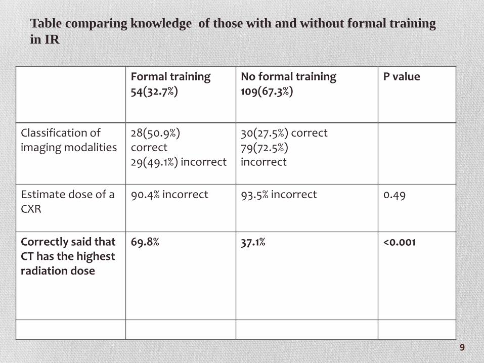
Formal training 54(32.7%)
No formal training 109(67.3%)
P value
Classification of imaging modalities
28(50.9%) correct 29(49.1%) incorrect
30(27.5%) correct 79(72.5%) incorrect
Estimate dose of a CXR
90.4% incorrect 93.5% incorrect 0.49
Correctly said that CT has the highest radiation dose
69.8% 37.1% <0.001
Table comparing knowledge of those with and without formal training
in IR
9

Classification of imaging modalities using IR:
More consultants, residents and medical officers correctly classified the
imaging modalities in all categories compared to the clinical officers p=<
0.004.
Significantly more consultants, residents and medical officers were able to
classify MRI and US as not using IR compared to the CO p=< 0.001
Highest radiation dose:
Consultants (76%) and residents (68%) were more likely to correctly respond
that an abdominal CT scan has the highest radiation compared to abdominal x-
ray,US or MRI, than the CO (8.9%) and MO (43.5%). p value of < 0.001
Estimation of radiation dose:
All the cadres of clinicians faired poorly when it came to estimating the
radiation dose when imaging different body parts

Table showing number of clinicians who classified the different organs as very
sensitive to ionizing radiation
Organ
n
Gonads
164
Breast
154
Lungs
131
Thyroid
118
Brain
114
Skin
108
Bone
marrow
72
Only 72 clinicians (42.3%) correctly rated bone marrow as a very sensitive
organ.
11

12

Table showing KNH health workers’ knowledge on risk of inducing cancer from
abdominal CT scan
n %
Risk of inducing cancer from
abdominal CT
Below correct risk 7 4.1
Correct risk (1 in 2000) 7 4.1
Above correct risk 58 34.1
Did not know 98 57.7
Total 170 100
58% DID NOT KNOW about the lifetime risk of inducing cancer from abdominal
CT scan
34.% of clinicians overestimated the lifetime risk of inducing cancer
Only 7% gave a correct response
13

Table showing distribution of clinician responses on the harmful effects of ionizing
radiation
Harmful effects n Percentage
Infertility 50 29.4
Breast cancer 43 25.3
Leukemia/lymphoma 18 10.6
Congenital malformations 15 8.8
Skin burns 11 6.5
Cervical/uterine/ovarian cancer 10 5.8
Cataracts/blindness 8 4.7
Psychological distress 5 2.9
Other cancers 5 2.9
Reduced libido 5 2.9
Skin cancer 4 2.3
Brain tumors 3 2.9
Lung cancer 3 1.7
These results correlate with the responses on organ sensitivity
14

Table showing importance of various considerations for health workers while
requesting imaging examinations for patients at KNH
Item
Very
important Important
Moderately
important
Least
importance
Not
important
Impact on the diagnosis 162(97.0) 5(3.0) 0(0.0) 0(0.0) 0(0.0)
Impact on the treatment 152(91.0) 14(8.4) 0(0.0) 1(0.6) 0(0.0)
Patient’s wishes 35(21.0) 44(26.3) 60(35.9) 25(15.0) 3(1.8)
Radiation dose to the
patient 7(4.2) 6(3.6) 29(17.4) 109(65.3) 16(9.6)
Cost 76(45.8) 80(48.2) 8(4.8) 2(1.2) 0(0.0)
15

Pie chart showing health worker practices related to patient referral for
ionizing radiation investigations
16
There were no statistically significant differences in unnecessary referrals
between health workers who reported having trained in IR 33/53 (62.3%)
compared those who had not trained in IR 61/109 (56%), chi = 0.58 (df= 1), p =
0.45.

17

Table showing health worker practices in KNH related to knowledge of ionizing
radiation
Very
important Important
Moderately
important
Least
importance
Not
important
Impact of knowledge of ionizing
radiation in different radiological
exams on practice 55(94.8) 3(5.2) 0(0.0) 0(0.0) 0(0.0)
Very
frequently Frequently Occasionally Rarely Never
Inform referrals for imaging studies
that use ionizing radiation on the
risks 0(0.0) 4(2.4) 18(10.9) 106(64.2) 37(22.4)
Patients ask you to explain to them
what examination they are going for,
what it entails and its risks 0(0.0) 1(0.6) 14(8.5) 77(46.7) 73(44.2)
18

Table showing percentage of clinicians who have attended continuous medical
education (CME) on ionizing radiation protection
Attended continuous medical education
On ionizing radiation protection
n % Clinicians
Yes 1 0.6
No 169 99.4
Total 170 100
19

Pie chart showing preferred mode of learning
RegularCME,n=9455.3%
Handbookwithreferral
guidelines,n=6739.4%
Noresponse,n=9
5.3%
20

21
CONCLUSIONS
1. Clinicians lack knowledge on ionizing radiation
2. There is a significant knowledge gap between the senior clinicians and
junior clinicians when it comes to some aspects of ionizing radiation.
3. Health workers with no IR training are less likely to correctly identify
all the techniques that use ionizing radiation compared to those with IR
training (50.9% versus 27.5%; OR = 0.37, 95% CI 0.18-0.72),
4. The clinicians with formal training had a statistically insignificant
advantage over those with no formal training as regards IR doses.
5. Deficiency in knowledge on IR and its potential risks leads to wrong
attitudes and poor practices.

1. Revision of the medical curricula to include more hours and subject content in
radiation protection.
2. Information on radiation protection to be disseminated through continuous
medical education (CME), as preferred by majority of the clinicians.
3. Senior clinician to countersign all requests that require the use of high dose
ionizing radiation ( Australian study1) .
4. Encourage the use of imaging referral guidelines 2.
1.Gerben B, et al. Doctor’s knowledge of patients radiation exposure from diagnostic imaging requested in the emergency department. Medical journal of Australia. (2010) .193(8): 450-3
2.Arslanoglu A, et al. Doctors’ and intern doctors’ knowledge about patients’ ionizing radiation exposure doses during common radiological examinations. DiagnIntervRadiol. ( 2007). 13: 53-55
RECOMMENDATIONS
22

5. Radiation doses and risks should be included in the imaging request form so
that the requesting physician can easily see the information and review if the
examination is still necessary.
5. A basic simplified information sheet to be given to patients to read through, to
enable them give informed consent prior to some of the imaging procedures.
It is hoped that the implementation of the above measures outlined may enable us
meet the triple A initiatives by IAEA on Radiation Protection of Patients
23

24
THANK YOU