Refining Susceptibility Loci of Chronic Obstructive ... - PLOS
10
Refining Susceptibility Loci of Chronic Obstructive Pulmonary Disease with Lung eqtls Maxime Lamontagne 1 , Christian Couture 1 , Dirkje S. Postma 2 , Wim Timens 3 , Don D. Sin 4,5 , Peter D. Pare ´ 4,5 , James C. Hogg 4,6 , David Nickle 7 , Michel Laviolette 1 , Yohan Bosse ´ 1,8 * 1 Centre de recherche de l’Institut universitaire de cardiologie et de pneumologie de Que ´bec, Que ´bec, Canada, 2 University of Groningen, University Medical Center Groningen, Department of Pulmonology, GRIAC Research Institute, Groningen, The Netherlands, 3 University of Groningen, University Medical Center Groningen, Department of Pathology and Medical Biology, GRIAC Research Institute, Groningen, The Netherlands, 4 University of British Columbia James Hogg Research Center, Center for Heart and Lung Health, St. Paul’s Hospital, Vancouver, British Columbia, Canada, 5 Respiratory Division, Department of Medicine, The University of British Columbia, Vancouver, British Columbia, Canada, 6 Department of Pathology and Laboratory Medicine, The University of British Columbia, Vancouver, British Columbia, Canada, 7 Merck & Co. Inc., Rahway, New Jersey, United States of America, 8 Department of Molecular Medicine, Laval University, Que ´bec, Canada Abstract Chronic obstructive pulmonary disease (COPD) is the fourth leading cause of mortality worldwide. Recent genome-wide association studies (GWAS) have identified robust susceptibility loci associated with COPD. However, the mechanisms mediating the risk conferred by these loci remain to be found. The goal of this study was to identify causal genes/variants within susceptibility loci associated with COPD. In the discovery cohort, genome-wide gene expression profiles of 500 non- tumor lung specimens were obtained from patients undergoing lung surgery. Blood-DNA from the same patients were genotyped for 1,2 million SNPs. Following genotyping and gene expression quality control filters, 409 samples were analyzed. Lung expression quantitative trait loci (eQTLs) were identified and overlaid onto three COPD susceptibility loci derived from GWAS; 4q31 (HHIP), 4q22 (FAM13A), and 19q13 (RAB4B, EGLN2, MIA, CYP2A6). Significant eQTLs were replicated in two independent datasets (n = 363 and 339). SNPs previously associated with COPD and lung function on 4q31 (rs1828591, rs13118928) were associated with the mRNA expression of HHIP. An association between mRNA expression level of FAM13A and SNP rs2045517 was detected at 4q22, but did not reach statistical significance. At 19q13, significant eQTLs were detected with EGLN2. In summary, this study supports HHIP, FAM13A, and EGLN2 as the most likely causal COPD genes on 4q31, 4q22, and 19q13, respectively. Strong lung eQTL SNPs identified in this study will need to be tested for association with COPD in case-control studies. Further functional studies will also be needed to understand the role of genes regulated by disease-related variants in COPD. Citation: Lamontagne M, Couture C, Postma DS, Timens W, Sin DD, et al. (2013) Refining Susceptibility Loci of Chronic Obstructive Pulmonary Disease with Lung eqtls. PLoS ONE 8(7): e70220. doi:10.1371/journal.pone.0070220 Editor: Xiao-Ping Miao, MOE Key Laboratory of Environment and Health, School of Public Health, Tongji Medical College, Huazhong University of Science and Technology, China Received May 6, 2013; Accepted June 14, 2013; Published July 30, 2013 Copyright: ß 2013 Lamontagne et al. This is an open-access article distributed under the terms of the Creative Commons Attribution License, which permits unrestricted use, distribution, and reproduction in any medium, provided the original author and source are credited. Funding: YB is a research scholar from the Heart and Stroke Foundation of Canada. This study was partly supported by the Chaire de pneumologie de la Fondation JD Be ´gin de l’Universite ´ Laval, the Fondation de l’Institut universitaire de cardiologie et de pneumologie de Que ´bec, and the Respiratory Health Network of the FRQS. The funders had no role in study design, data collection and analysis, decision to publish, or preparation of the manuscript. Competing Interests: DN is a full time employee of Merck. This does not alter the authors’ adherence to all the PLOS ONE policies on sharing data and materials. * E-mail: [email protected] Introduction Chronic obstructive pulmonary disease (COPD) is the fourth most common cause of death worldwide and is predicted to be the third leading cause of mortality in the world by the year 2030 [1]. COPD is a complex disease characterized by airflow obstruction that is not fully reversible [2]. Cigarette smoking is the most important cause of the rapid decline in pulmonary function that leads to COPD, but not all smokers develop the disease [3]. Moreover, there is familial aggregation of COPD suggesting a genetic contribution [4]. The only well-established genetic risk factors are inherited mutations causing a1-antitrypsin deficiency [5]. However, these mutations occur in only 1–5% of COPD patients [6]. The number of susceptibility genes for COPD is expanding rapidly with lists tabulated at 57 genes in 2009 [7] and at 144 genes in 2012 [8]. Recent genome-wide association studies (GWAS) have identified four susceptibility loci associated with COPD including 4q22 (FAM13A), 4q31 (HHIP), 15q25 (CHRNA3/ CHRNA5/IREB2) and 19q13 (RAB4B, EGLN2, MIA, CYP2A6) [9– 11]. The causal genes and genetic mechanisms mediating the risk within these loci remain to be found. The goal of this study is to identify lung expression quantitative trait loci (eQTL) within COPD susceptibility loci identified by GWAS. As part of the lung eQTL consortium, we have recently performed a genome-wide search for eQTLs in 1,111 human lung samples [12]. A predefined hypothesis of the consortium was that human lung eQTLs will identify the most informative markers and improve the localization of causal variants/genes in GWAS- nominated COPD loci [13]. Methods Ethics Statement All lung tissue samples were obtained in accordance with Institutional Review Board guidelines at the three sites: Laval PLOS ONE | www.plosone.org 1 July 2013 | Volume 8 | Issue 7 | e70220
-
Upload
khangminh22 -
Category
Documents
-
view
0 -
download
0
Transcript of Refining Susceptibility Loci of Chronic Obstructive ... - PLOS
Refining Susceptibility Loci of Chronic ObstructivePulmonary Disease with Lung eqtlsMaxime Lamontagne1, Christian Couture1, Dirkje S. Postma2, Wim Timens3, Don D. Sin4,5,
Peter D. Pare4,5, James C. Hogg4,6, David Nickle7, Michel Laviolette1, Yohan Bosse1,8*
1 Centre de recherche de l’Institut universitaire de cardiologie et de pneumologie de Quebec, Quebec, Canada, 2 University of Groningen, University Medical Center
Groningen, Department of Pulmonology, GRIAC Research Institute, Groningen, The Netherlands, 3 University of Groningen, University Medical Center Groningen,
Department of Pathology and Medical Biology, GRIAC Research Institute, Groningen, The Netherlands, 4 University of British Columbia James Hogg Research Center,
Center for Heart and Lung Health, St. Paul’s Hospital, Vancouver, British Columbia, Canada, 5 Respiratory Division, Department of Medicine, The University of British
Columbia, Vancouver, British Columbia, Canada, 6 Department of Pathology and Laboratory Medicine, The University of British Columbia, Vancouver, British Columbia,
Canada, 7 Merck & Co. Inc., Rahway, New Jersey, United States of America, 8 Department of Molecular Medicine, Laval University, Quebec, Canada
Abstract
Chronic obstructive pulmonary disease (COPD) is the fourth leading cause of mortality worldwide. Recent genome-wideassociation studies (GWAS) have identified robust susceptibility loci associated with COPD. However, the mechanismsmediating the risk conferred by these loci remain to be found. The goal of this study was to identify causal genes/variantswithin susceptibility loci associated with COPD. In the discovery cohort, genome-wide gene expression profiles of 500 non-tumor lung specimens were obtained from patients undergoing lung surgery. Blood-DNA from the same patients weregenotyped for 1,2 million SNPs. Following genotyping and gene expression quality control filters, 409 samples wereanalyzed. Lung expression quantitative trait loci (eQTLs) were identified and overlaid onto three COPD susceptibility lociderived from GWAS; 4q31 (HHIP), 4q22 (FAM13A), and 19q13 (RAB4B, EGLN2, MIA, CYP2A6). Significant eQTLs were replicatedin two independent datasets (n = 363 and 339). SNPs previously associated with COPD and lung function on 4q31(rs1828591, rs13118928) were associated with the mRNA expression of HHIP. An association between mRNA expression levelof FAM13A and SNP rs2045517 was detected at 4q22, but did not reach statistical significance. At 19q13, significant eQTLswere detected with EGLN2. In summary, this study supports HHIP, FAM13A, and EGLN2 as the most likely causal COPD geneson 4q31, 4q22, and 19q13, respectively. Strong lung eQTL SNPs identified in this study will need to be tested for associationwith COPD in case-control studies. Further functional studies will also be needed to understand the role of genes regulatedby disease-related variants in COPD.
Citation: Lamontagne M, Couture C, Postma DS, Timens W, Sin DD, et al. (2013) Refining Susceptibility Loci of Chronic Obstructive Pulmonary Disease with Lungeqtls. PLoS ONE 8(7): e70220. doi:10.1371/journal.pone.0070220
Editor: Xiao-Ping Miao, MOE Key Laboratory of Environment and Health, School of Public Health, Tongji Medical College, Huazhong University of Science andTechnology, China
Received May 6, 2013; Accepted June 14, 2013; Published July 30, 2013
Copyright: � 2013 Lamontagne et al. This is an open-access article distributed under the terms of the Creative Commons Attribution License, which permitsunrestricted use, distribution, and reproduction in any medium, provided the original author and source are credited.
Funding: YB is a research scholar from the Heart and Stroke Foundation of Canada. This study was partly supported by the Chaire de pneumologie de laFondation JD Begin de l’Universite Laval, the Fondation de l’Institut universitaire de cardiologie et de pneumologie de Quebec, and the Respiratory HealthNetwork of the FRQS. The funders had no role in study design, data collection and analysis, decision to publish, or preparation of the manuscript.
Competing Interests: DN is a full time employee of Merck. This does not alter the authors’ adherence to all the PLOS ONE policies on sharing data andmaterials.
* E-mail: [email protected]
Introduction
Chronic obstructive pulmonary disease (COPD) is the fourth most
common cause of death worldwide and is predicted to be the third
leading cause of mortality in the world by the year 2030 [1]. COPD
is a complex disease characterized by airflow obstruction that is not
fully reversible [2]. Cigarette smoking is the most important cause of
the rapid decline in pulmonary function that leads to COPD, but not
all smokers develop the disease [3]. Moreover, there is familial
aggregation of COPD suggesting a genetic contribution [4]. The
only well-established genetic risk factors are inherited mutations
causing a1-antitrypsin deficiency [5]. However, these mutations
occur in only 1–5% of COPD patients [6].
The number of susceptibility genes for COPD is expanding
rapidly with lists tabulated at 57 genes in 2009 [7] and at 144
genes in 2012 [8]. Recent genome-wide association studies
(GWAS) have identified four susceptibility loci associated with
COPD including 4q22 (FAM13A), 4q31 (HHIP), 15q25 (CHRNA3/
CHRNA5/IREB2) and 19q13 (RAB4B, EGLN2, MIA, CYP2A6) [9–
11]. The causal genes and genetic mechanisms mediating the risk
within these loci remain to be found.
The goal of this study is to identify lung expression quantitative
trait loci (eQTL) within COPD susceptibility loci identified by
GWAS. As part of the lung eQTL consortium, we have recently
performed a genome-wide search for eQTLs in 1,111 human lung
samples [12]. A predefined hypothesis of the consortium was that
human lung eQTLs will identify the most informative markers and
improve the localization of causal variants/genes in GWAS-
nominated COPD loci [13].
Methods
Ethics StatementAll lung tissue samples were obtained in accordance with
Institutional Review Board guidelines at the three sites: Laval
PLOS ONE | www.plosone.org 1 July 2013 | Volume 8 | Issue 7 | e70220
University (Quebec, Canada), University of British-Columbia
(Vancouver, Canada) and Groningen University (Groningen, The
Netherlands). All patients provided written informed consent and
the study was approved by the ethics committees of the Institut
universitaire de cardiologie et de pneumologie de Quebec
(IUCPQ) and the UBC-Providence Health Care Research
Institute Ethics Board for Laval and UBC, respectively. The study
protocol was consistent with the Research Code of the University
Medical Center Groningen and Dutch national ethical and
professional guidelines (‘‘Code of conduct; Dutch federation of
biomedical scientific societies’’; http://www.federa.org).
Study Subjects and Lung SpecimensStudy subjects and lung specimens were described previously
[12,14]. Briefly subjects were from three sites, Laval University,
University of British Columbia, and University of Groningen
(henceforth referred to as Laval, UBC, and Groningen, respec-
tively). At Laval, the lung specimens were provided by the IUCPQ
site of the Respiratory Health Network Tissue Bank of the Fonds
de recherche du Quebec – Sante (FRQS) (www.tissuebank.ca); at
Groningen, the lung specimens were provided by the local tissue
bank of the Department of Pathology, and at UBC, the lung
specimens were provided by the James Hogg Research Center
Biobank at St Paul’s Hospital. COPD diagnosis and severity were
determined according to the GOLD recommendations [2].
Clinical characteristics of subjects by site are shown in Table 1.
AssaysGenome-wide gene expression and genotyping profiles were
obtained using a custom Affymetrix array (see GEO platform
GPL10379) and the Illumina Human1M-Duo BeadChip array,
respectively. Gene expression data are available through the Gene
Expression Omnibus (GEO) repository with the accession number
GSE23546. Standard quality controls were performed as de-
scribed previously and only subjects that passed genotyping and
expression quality controls were included in this study with 409,
363, and 339 subjects from Laval, Groningen, and UBC,
respectively [12].
COPD Susceptibility LociLung eQTLs were overlaid onto COPD susceptibility loci
identified by previous GWAS except for the 15q25-CHRNA3/
CHRNA5/IREB2 locus that we have reported on previously [15].
Three COPD loci were considered; 4q22 (FAM13A), 4q31 (HHIP)
and 19q13 (RAB4B, EGLN2, MIA, CYP2A6). SNPs associated with
COPD from previous GWAS were tabulated for the three loci
(Table 2). SNPs genotyped in the lung eQTL consortium located
1 Mb up and downstream of the most distant associated SNPs in
both directions were evaluated. Chromosomes 4q22 (88,875,909-
90,886,297), 4q31 (144,480,780-146,506,456) and 19q13
(40,292,404-42,302,706) include 718, 412 and 739 SNPs, respec-
tively. Genes residing in the same regions were tested as cis-eQTLs
for probe sets for 14 genes on 4q22 (SPP1, PKD2, ABCG2, PPM1K,
HERC6, HERC5, PIGY, HERC3, NAP1L5, FAM13A, TIGD2,
GPRIN3, SNCA, MMRN1), 9 genes on 4q31 (FREM3, GYPE,
GYPB, GYPA, HHIP, ANAPC10, ABCE1, OTUD4, SMAD1) and 45
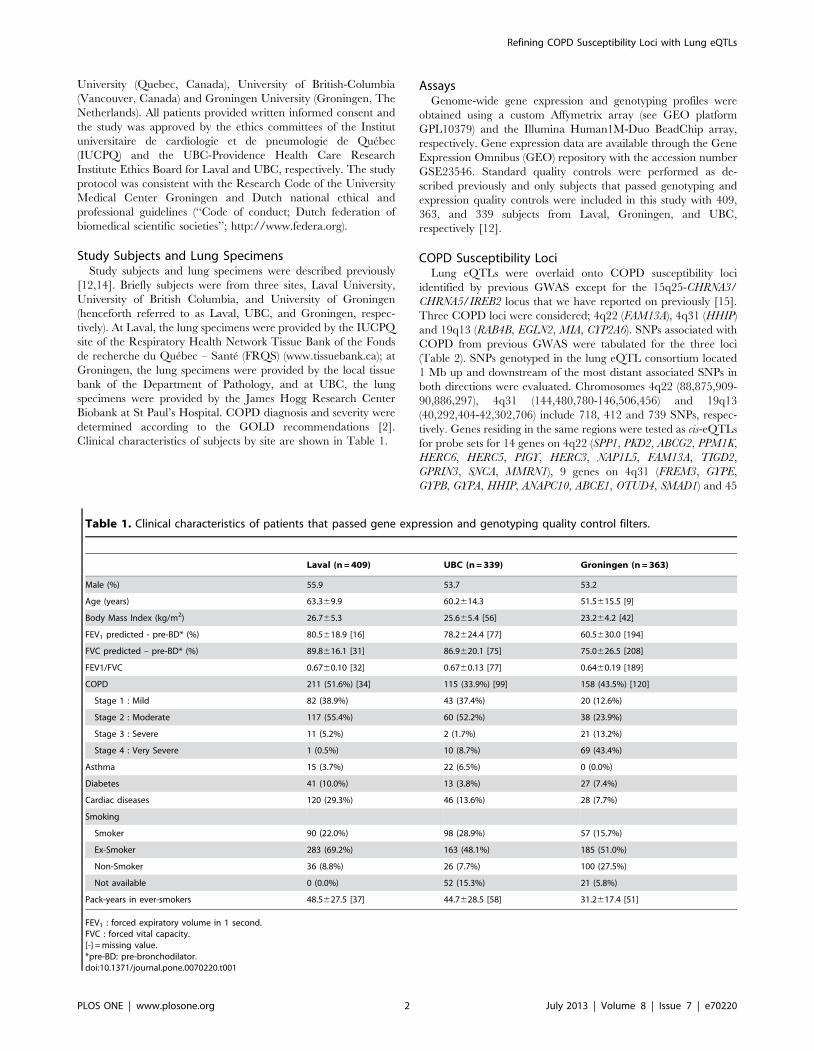
Table 1. Clinical characteristics of patients that passed gene expression and genotyping quality control filters.
Laval (n = 409) UBC (n = 339) Groningen (n = 363)
Male (%) 55.9 53.7 53.2
Age (years) 63.369.9 60.2614.3 51.5615.5 [9]
Body Mass Index (kg/m2) 26.765.3 25.665.4 [56] 23.264.2 [42]
FEV1 predicted - pre-BD* (%) 80.5618.9 [16] 78.2624.4 [77] 60.5630.0 [194]
FVC predicted – pre-BD* (%) 89.8616.1 [31] 86.9620.1 [75] 75.0626.5 [208]
FEV1/FVC 0.6760.10 [32] 0.6760.13 [77] 0.6460.19 [189]
COPD 211 (51.6%) [34] 115 (33.9%) [99] 158 (43.5%) [120]
Stage 1 : Mild 82 (38.9%) 43 (37.4%) 20 (12.6%)
Stage 2 : Moderate 117 (55.4%) 60 (52.2%) 38 (23.9%)
Stage 3 : Severe 11 (5.2%) 2 (1.7%) 21 (13.2%)
Stage 4 : Very Severe 1 (0.5%) 10 (8.7%) 69 (43.4%)
Asthma 15 (3.7%) 22 (6.5%) 0 (0.0%)
Diabetes 41 (10.0%) 13 (3.8%) 27 (7.4%)
Cardiac diseases 120 (29.3%) 46 (13.6%) 28 (7.7%)
Smoking
Smoker 90 (22.0%) 98 (28.9%) 57 (15.7%)
Ex-Smoker 283 (69.2%) 163 (48.1%) 185 (51.0%)
Non-Smoker 36 (8.8%) 26 (7.7%) 100 (27.5%)
Not available 0 (0.0%) 52 (15.3%) 21 (5.8%)
Pack-years in ever-smokers 48.5627.5 [37] 44.7628.5 [58] 31.2617.4 [51]
FEV1 : forced expiratory volume in 1 second.FVC : forced vital capacity.[-] = missing value.*pre-BD: pre-bronchodilator.doi:10.1371/journal.pone.0070220.t001
Refining COPD Susceptibility Loci with Lung eQTLs
PLOS ONE | www.plosone.org 2 July 2013 | Volume 8 | Issue 7 | e70220

genes on 19q13 (DYRK1B, FBL, FCGBP, PSMC4, ZNF546,
ZNF780B, ZNF780A, MAP3K10, TTC9B, CNTD2, AKT2,
C19orf47, PLD3, PRX, SERTAD1, SERTAD3, BLVRB, SPTBN4,
SHKBP1, LTBP4, NUMBL, ADCK4, ITPKC, C19orf54, SNRPA,
EGLN2, CYP2G1P, CYP2B6, CYP2A13, CYP2S1, AXL,
HNRNPUL1, TGFB1, B9D2, TMEM91, EXOSC5, BCKDHA,
B3GNT8, ATP5SL, LOC100505495, CEACAM21, CEACAM4,
CEACAM7, CEACAM5, CEACAM6).
Statistical AnalysisLung eQTLs were identified using a different model than in our
previous genome-wide lung eQTL mapping study [12]. Expres-
sion data were adjusted for age, sex, and smoking status using
robust residuals obtained with the robust fitting of linear models
function (rlm) in the R statistical package MASS. Residuals values
deviating from the median by more than three standard deviations
were filtered as outliers. Association tests between adjusted
expression traits and SNPs were performed using quantitative
association tests implemented in PLINK [16] (version 1.07).
Association tests were performed with the ‘‘assoc’’ command and
the Wald test asymptotic p-values were used. Each possible
combination of SNPs and genes were tested in the three COPD
susceptibility loci in the Laval dataset. Significant cis-eQTLs were
those passing Bonferroni correction considering the effective
number of independent SNPs and genes tested at each locus.
The number of independent variables was determined by using
the definition of Li and Ji [17], as implemented in SNPSpD [18]. P
value thresholds were set at 5.1061026 for 4q22 (0.05/(279.64
SNPs635 probesets), 1.5061025 for 4q31 (0.05/(128.26 SNPs626
probesets) and 3.4361026 for 19q13 (0.05/(246.73 SNPs659
probesets). Significant eQTLs in Laval dataset were then validated
in the UBC and Groningen datasets. P values lower than 0.05
were considered significant in the replication sets.
Results
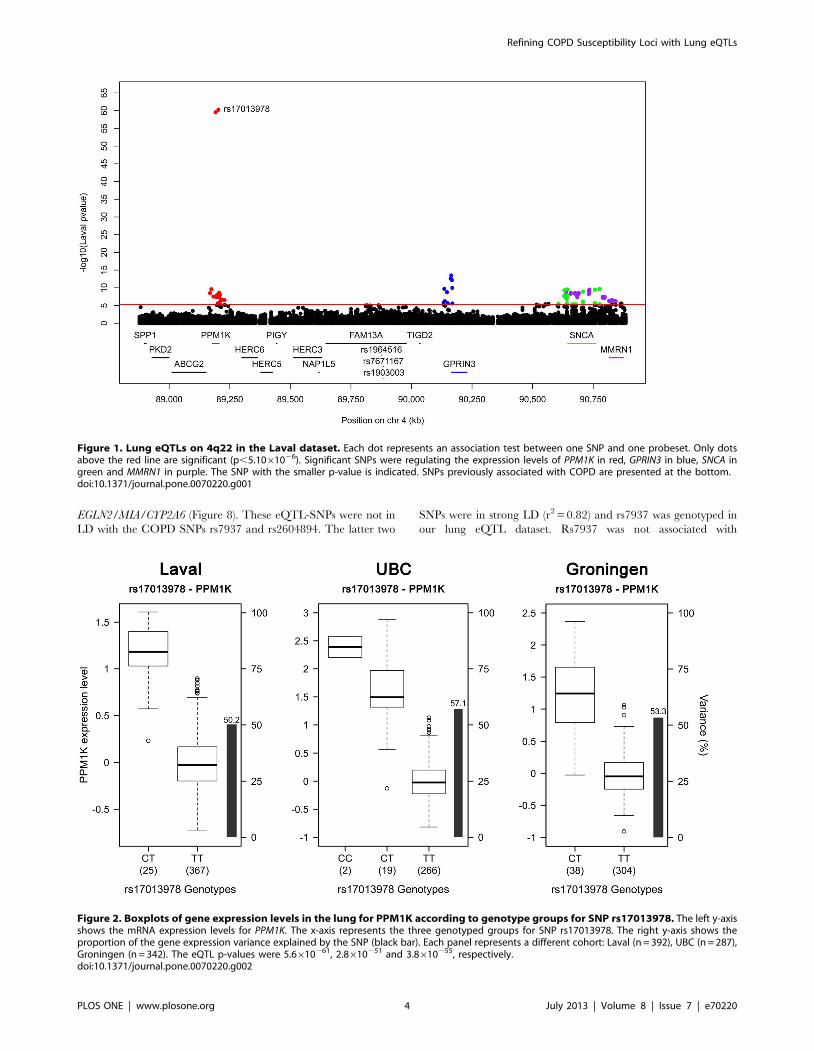
Lung eQTLs in the 4q22 Locus718 SNPs and 50 probesets covering 14 genes were located in
the defined region on chromosome 4q22. 91 eQTLs were detected
in the Laval set (Figure 1, Table S1). These 91 eQTLs consisted of
64 unique SNPs, 8 probesets and 4 genes (PPM1K, GPRIN3, SNCA,
MMRN1). Significant linkage disequilibrium (LD) was observed
among the 64 SNPs (Figure S1). 61 of the 91 eQTLs replicated in
both replication cohorts (P,0.05). The strongest association
detected in all cohorts was rs17013978 with PPM1K (Figure 1).
The expression level of this gene decreased with the number of T
alleles (Figure 2). In the three cohorts, this SNP explained 50.2 to
57.1% of the expression variance of PPM1K and the direction of
the effect was the same in the three cohorts. None of the SNPs
previously associated with COPD on 4q22 (Table 2) were
genotyped in our eQTL dataset, but five of them were found in
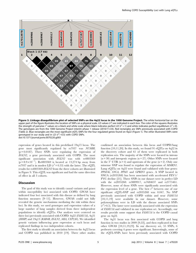
LD (r2.0.5) (Figure 3). These five SNPs did not significantly
associated with the expression of genes at that locus, but a trend
was observed with FAM13A. In fact, three of those five
polymorphisms, in complete LD with each other (rs2045517,
rs2869967, rs2869966) and in modest LD (r2 = 0.53–0.69) with
COPD SNPs were nominally associated with the expression levels
of FAM13A (p = 4.161025). The FAM13A-rs2045517 eQTL was
replicated in UBC, but not in Groningen (Figure 4).
Lung eQTLs in the 4q31 Locus412 SNPs and 34 probesets interrogating 9 unique genes were
tested around previously COPD associated SNPs on chromosome
4q31. Significant eQTLs found in the Laval dataset are shown in
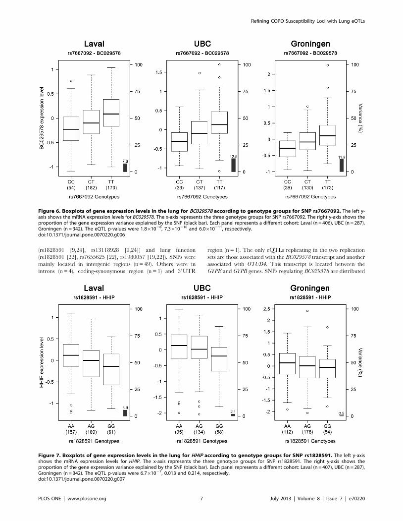
Figure 5 and Table S2. 55 unique SNPs, 6 probesets and 4 genes
(FREM3, BC029578, HHIP, OTUD4) were involved in the
significant eQTLs. Only eQTLs associated with BC029578 (35)
and OTUD4 (1) were replicated in the two replication sets. eQTL-
SNPs on chromosome 4q31 are subdivided in two strong LD
blocks (Figure S2). The strongest eQTL in Laval dataset, validated
in both replication sets, was rs7667092 with BC029578 (Figure 6).
The expression levels of the gene increased with the number of T
alleles in all cohorts. In the three cohorts, this SNP explained 7.6
to 12.5% of the gene expression variance of BC029578. However,
this polymorphism was not in LD with SNPs previously associated
with COPD (r2 = 0.016). Two SNPs (rs1828591, rs13118928)
previously associated with COPD were found to affect the
expression of HHIP. Rs1828591 was the most significant SNP
associated with HHIP in the Laval dataset. This eQTL was
replicated in UBC, but not in Groningen (Figure 7). The G allele
was associated with lower expression of HHIP in the Laval and
UBC datasets. The same pattern was observed in the Groningen
set, but the association was not significant.
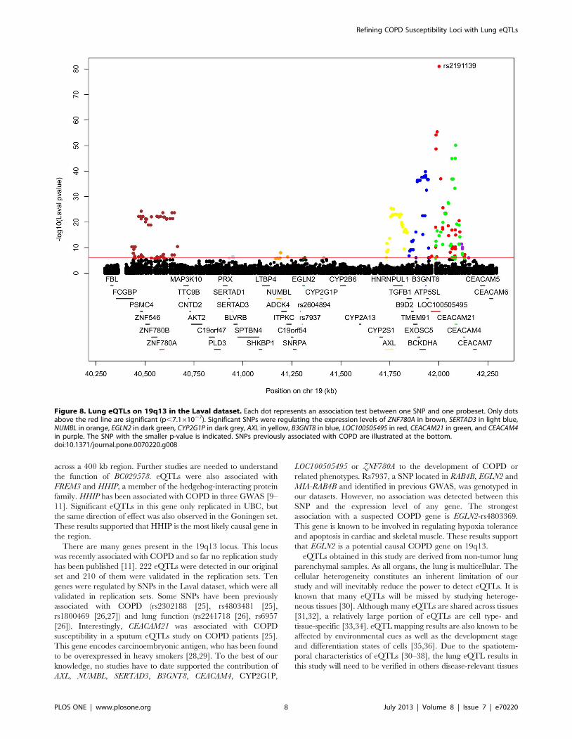
Lung eQTLs in the 19q13 LocusOn 19q13, 739 SNPs and 95 probesets covering 45 different
genes were tested. The expression levels of RAB4B, MIA and
CYP2A6 were not available in our datasets. 222 eQTLs were
detected (Figure 8 and Table S3). 174 SNPs were regulating 11
probesets located on 10 genes (ZNF780A, SERTAD3, NUMBL,
EGLN2, CYP2G1P, AXL, B3GNT8, LOC100505495, CEACAM21,
CEACAM4). 210 eQTLs were validated in both replication
cohorts. SNPs associated with gene expression were distributed
across four LD blocks (Figure S3). 26 SNPs were associated with
the expression levels of CEACAM21 and LOC100505495, and 3
others SNPs were associated with CEACAM21 and CEACAM4.
The eQTLs on 19q13 were mainly located in two discrete foci one
distal and one proximal to the COPD susceptibility locus RAB4B/
Table 2. SNPs associated with COPD in previous GWAS.
Locus SNPSNPposition Study
4q22 rs1964516 89,875,909 Cho et al. 2012. Human MolecularGenetics.11
rs7671167 89,883,979 Cho et al. 2010. Nature Genetics.10
Cho et al. 2012. Human MolecularGenetics.11
rs1903003 89,886,297 Cho et al. 2010. Nature Genetics.10
4q31 rs1828591 145,480,780 Cho et al. 2010. Nature Genetics.10
Pillai et al. 2009. PLoS Genetics.9
rs13118928 145,486,389 Cho et al. 2012. Human MolecularGenetics.11
Pillai et al. 2009. PLoS Genetics.9
rs13141641 145,506,456 Cho et al. 2012. Human MolecularGenetics.11
19q13 rs2604894 41,292,404 Cho et al. 2012. Human MolecularGenetics.11
rs7937 41,302,706 Cho et al. 2012. Human MolecularGenetics.11
doi:10.1371/journal.pone.0070220.t002
Refining COPD Susceptibility Loci with Lung eQTLs
PLOS ONE | www.plosone.org 3 July 2013 | Volume 8 | Issue 7 | e70220
EGLN2/MIA/CYP2A6 (Figure 8). These eQTL-SNPs were not in
LD with the COPD SNPs rs7937 and rs2604894. The latter two
SNPs were in strong LD (r2 = 0.82) and rs7937 was genotyped in
our lung eQTL dataset. Rs7937 was not associated with
Figure 1. Lung eQTLs on 4q22 in the Laval dataset. Each dot represents an association test between one SNP and one probeset. Only dotsabove the red line are significant (p,5.1061026). Significant SNPs were regulating the expression levels of PPM1K in red, GPRIN3 in blue, SNCA ingreen and MMRN1 in purple. The SNP with the smaller p-value is indicated. SNPs previously associated with COPD are presented at the bottom.doi:10.1371/journal.pone.0070220.g001
Figure 2. Boxplots of gene expression levels in the lung for PPM1K according to genotype groups for SNP rs17013978. The left y-axisshows the mRNA expression levels for PPM1K. The x-axis represents the three genotyped groups for SNP rs17013978. The right y-axis shows theproportion of the gene expression variance explained by the SNP (black bar). Each panel represents a different cohort: Laval (n = 392), UBC (n = 287),Groningen (n = 342). The eQTL p-values were 5.6610261, 2.8610251 and 3.8610255, respectively.doi:10.1371/journal.pone.0070220.g002
Refining COPD Susceptibility Loci with Lung eQTLs
PLOS ONE | www.plosone.org 4 July 2013 | Volume 8 | Issue 7 | e70220
expression of genes located in this predefined 19q13 locus. The
gene most significantly regulated by rs7937 was NUMBL
(p = 0.0187). Three SNPs were regulating the expression of
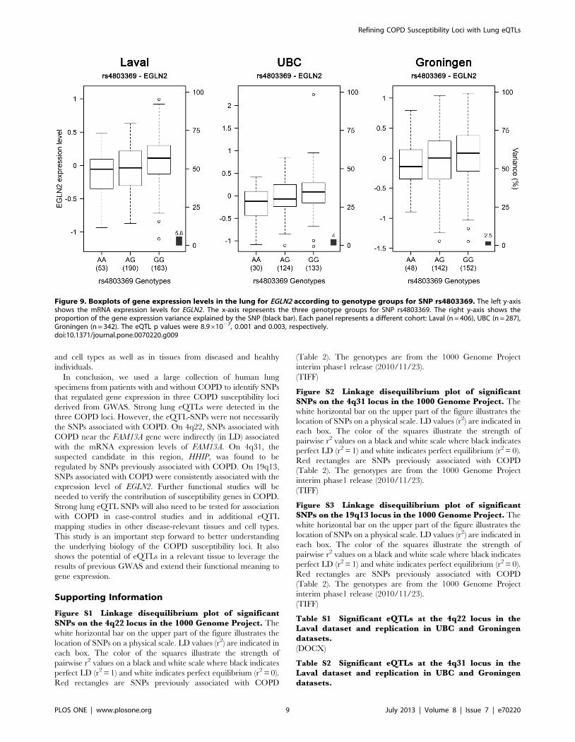
EGLN2, a gene previously associated with COPD. The most
significant association with EGLN2 was with rs4803369
(p = 8.961027). Rs4803369 is located at 13,274 bp away from
rs7937 and is in modest LD (r2 = 0.33) with the latter. The eQTL
results for rs4803369-EGLN2 from the three cohorts are illustrated
in Figure 9. This eQTL was significant and had the same direction
of effect in all 3 cohorts.
Discussion
The goal of this study was to identify causal variants and genes
within susceptibility loci associated with COPD. GWAS have
indicated four loci associated with this disease as defined by lung
function measures [9–11]. However, GWAS could not fully
revealed the genetic mechanisms mediating the risk within these
loci. In this study, we used genotypes and expression values of a
large number of lung samples derived from three independent
populations to identify eQTLs. Our analyses were centered on
three loci previously associated with COPD: 4q22 (FAM13A), 4q31
(HHIP) and 19q13 (RAB4B, EGLN2, MIA, CYP2A6). We identified
genetic variants influencing gene expression at each locus and
replicated findings in two independent cohorts.
The first study to identify an association between the 4q22 locus
and COPD was published in 2010 [19]. Three other studies
confirmed an association between this locus and COPD/lung
function [10,11,20]. In this study, we found 91 eQTLs on 4q22 in
the discovery cohort and 61 of them were replicated in both
replication sets. The majority of the SNPs were located in introns
(n = 30) and intergenic regions (n = 27). Other SNPs were located
in the 39 UTR (n = 4) and upstream of the gene (n = 2). Only one
missense SNP was found to regulate the expression of MMRN1.
Lung eQTLs on 4q22 were found and validated with four genes:
PPM1K, SNCA, PPM1 and GPRIN3 genes. A SNP located in
SNCA (rs2035268) has been associated with accelerated FEV1/
FVC decline [21]. Three SNPs in our dataset were in perfect LD
with the rs2035268: rs3889917, rs7684637 and rs3775461.
However, none of those SNPs were significantly associated with
the expression level of a gene. The best r2 between one of our
significant eQTL-SNP and rs2035268 was 0.047. No SNP
previously associated with COPD within and near FAM13A
[10,11,19] were available in our dataset. However, some
polymorphisms were in LD with the disease associated SNPs
(r2.0.5). The latter were nominally associated with the expression
of FAM13A and validated in one replication set. Accordingly, our
results provide some support that FAM13A is the COPD causal
gene on 4q22.
The 4q31 locus was first associated with COPD and lung
function in two studies in 2009 [9,22], and then replicated in four
other GWAS [10,11,19,23]. In our discovery set, 55 SNPs, 6
probesets covering 4 genes were significant. Interestingly, some of
the eQTL-SNPs have been previously associated with COPD
Figure 3. Linkage disequilibrium plot of selected SNPs on the 4q22 locus in the 1000 Genome Project. The white horizontal bar on theupper part of the figure illustrates the location of SNPs on a physical scale. LD values (r2) are indicated in each box. The color of the squares illustratesthe strength of pairwise r2 values on a black and white scale where black indicates perfect LD (r2 = 1) and white indicates perfect equilibrium (r2 = 0).The genotypes are from the 1000 Genome Project interim phase 1 release (2010/11/23). Red rectangles are SNPs previously associated with COPD(Table 2). Blue rectangles are the most significant eQTL-SNPs for the four regulated genes found on 4q22 (Figure 1). The other illustrated SNPs weregenotyped in our study and in LD (r2.0.5) with COPD SNPs.doi:10.1371/journal.pone.0070220.g003
Refining COPD Susceptibility Loci with Lung eQTLs
PLOS ONE | www.plosone.org 5 July 2013 | Volume 8 | Issue 7 | e70220
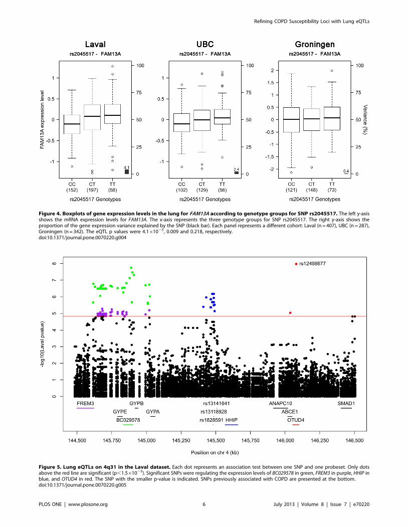
Figure 4. Boxplots of gene expression levels in the lung for FAM13A according to genotype groups for SNP rs2045517. The left y-axisshows the mRNA expression levels for FAM13A. The x-axis represents the three genotype groups for SNP rs2045517. The right y-axis shows theproportion of the gene expression variance explained by the SNP (black bar). Each panel represents a different cohort: Laval (n = 407), UBC (n = 287),Groningen (n = 342). The eQTL p values were 4.161025, 0.009 and 0.218, respectively.doi:10.1371/journal.pone.0070220.g004
Figure 5. Lung eQTLs on 4q31 in the Laval dataset. Each dot represents an association test between one SNP and one probeset. Only dotsabove the red line are significant (p,1.561025). Significant SNPs were regulating the expression levels of BC029578 in green, FREM3 in purple, HHIP inblue, and OTUD4 in red. The SNP with the smaller p-value is indicated. SNPs previously associated with COPD are presented at the bottom.doi:10.1371/journal.pone.0070220.g005
Refining COPD Susceptibility Loci with Lung eQTLs
PLOS ONE | www.plosone.org 6 July 2013 | Volume 8 | Issue 7 | e70220
(rs1828591 [9,24], rs13118928 [9,24]) and lung function
(rs1828591 [22], rs7655625 [22], rs1980057 [19,22]). SNPs were
mainly located in intergenic regions (n = 49). Others were in
introns (n = 4), coding-synonymous region (n = 1) and 39UTR
region (n = 1). The only eQTLs replicating in the two replication
sets are those associated with the BC029578 transcript and another
associated with OTUD4. This transcript is located between the
GYPE and GYPB genes. SNPs regulating BC029578 are distributed
Figure 6. Boxplots of gene expression levels in the lung for BC029578 according to genotype groups for SNP rs7667092. The left y-axis shows the mRNA expression levels for BC029578. The x-axis represents the three genotype groups for SNP rs7667092. The right y-axis shows theproportion of the gene expression variance explained by the SNP (black bar). Each panel represents a different cohort: Laval (n = 406), UBC (n = 287),Groningen (n = 342). The eQTL p-values were 1.861028, 7.3610210 and 6.0610211, respectively.doi:10.1371/journal.pone.0070220.g006
Figure 7. Boxplots of gene expression levels in the lung for HHIP according to genotype groups for SNP rs1828591. The left y-axisshows the mRNA expression levels for HHIP. The x-axis represents the three genotype groups for SNP rs1828591. The right y-axis shows theproportion of the gene expression variance explained by the SNP (black bar). Each panel represents a different cohort: Laval (n = 407), UBC (n = 287),Groningen (n = 342). The eQTL p-values were 6.761027, 0.013 and 0.214, respectively.doi:10.1371/journal.pone.0070220.g007
Refining COPD Susceptibility Loci with Lung eQTLs
PLOS ONE | www.plosone.org 7 July 2013 | Volume 8 | Issue 7 | e70220
across a 400 kb region. Further studies are needed to understand
the function of BC029578. eQTLs were also associated with
FREM3 and HHIP, a member of the hedgehog-interacting protein
family. HHIP has been associated with COPD in three GWAS [9–
11]. Significant eQTLs in this gene only replicated in UBC, but
the same direction of effect was also observed in the Goningen set.
These results supported that HHIP is the most likely causal gene in
the region.
There are many genes present in the 19q13 locus. This locus
was recently associated with COPD and so far no replication study
has been published [11]. 222 eQTLs were detected in our original
set and 210 of them were validated in the replication sets. Ten
genes were regulated by SNPs in the Laval dataset, which were all
validated in replication sets. Some SNPs have been previously
associated with COPD (rs2302188 [25], rs4803481 [25],
rs1800469 [26,27]) and lung function (rs2241718 [26], rs6957
[26]). Interestingly, CEACAM21 was associated with COPD
susceptibility in a sputum eQTLs study on COPD patients [25].
This gene encodes carcinoembryonic antigen, who has been found
to be overexpressed in heavy smokers [28,29]. To the best of our
knowledge, no studies have to date supported the contribution of
AXL, NUMBL, SERTAD3, B3GNT8, CEACAM4, CYP2G1P,
LOC100505495 or ZNF780A to the development of COPD or
related phenotypes. Rs7937, a SNP located in RAB4B, EGLN2 and
MIA-RAB4B and identified in previous GWAS, was genotyped in
our datasets. However, no association was detected between this
SNP and the expression level of any gene. The strongest
association with a suspected COPD gene is EGLN2-rs4803369.
This gene is known to be involved in regulating hypoxia tolerance
and apoptosis in cardiac and skeletal muscle. These results support
that EGLN2 is a potential causal COPD gene on 19q13.
eQTLs obtained in this study are derived from non-tumor lung
parenchymal samples. As all organs, the lung is multicellular. The
cellular heterogeneity constitutes an inherent limitation of our
study and will inevitably reduce the power to detect eQTLs. It is
known that many eQTLs will be missed by studying heteroge-
neous tissues [30]. Although many eQTLs are shared across tissues
[31,32], a relatively large portion of eQTLs are cell type- and
tissue-specific [33,34]. eQTL mapping results are also known to be
affected by environmental cues as well as the development stage
and differentiation states of cells [35,36]. Due to the spatiotem-
poral characteristics of eQTLs [30–38], the lung eQTL results in
this study will need to be verified in others disease-relevant tissues
Figure 8. Lung eQTLs on 19q13 in the Laval dataset. Each dot represents an association test between one SNP and one probeset. Only dotsabove the red line are significant (p,7.161027). Significant SNPs were regulating the expression levels of ZNF780A in brown, SERTAD3 in light blue,NUMBL in orange, EGLN2 in dark green, CYP2G1P in dark grey, AXL in yellow, B3GNT8 in blue, LOC100505495 in red, CEACAM21 in green, and CEACAM4in purple. The SNP with the smaller p-value is indicated. SNPs previously associated with COPD are illustrated at the bottom.doi:10.1371/journal.pone.0070220.g008
Refining COPD Susceptibility Loci with Lung eQTLs
PLOS ONE | www.plosone.org 8 July 2013 | Volume 8 | Issue 7 | e70220
and cell types as well as in tissues from diseased and healthy
individuals.
In conclusion, we used a large collection of human lung
specimens from patients with and without COPD to identify SNPs
that regulated gene expression in three COPD susceptibility loci
derived from GWAS. Strong lung eQTLs were detected in the
three COPD loci. However, the eQTL-SNPs were not necessarily
the SNPs associated with COPD. On 4q22, SNPs associated with
COPD near the FAM13A gene were indirectly (in LD) associated
with the mRNA expression levels of FAM13A. On 4q31, the
suspected candidate in this region, HHIP, was found to be
regulated by SNPs previously associated with COPD. On 19q13,
SNPs associated with COPD were consistently associated with the
expression level of EGLN2. Further functional studies will be
needed to verify the contribution of susceptibility genes in COPD.
Strong lung eQTL SNPs will also need to be tested for association
with COPD in case-control studies and in additional eQTL
mapping studies in other disease-relevant tissues and cell types.
This study is an important step forward to better understanding
the underlying biology of the COPD susceptibility loci. It also
shows the potential of eQTLs in a relevant tissue to leverage the
results of previous GWAS and extend their functional meaning to
gene expression.
Supporting Information
Figure S1 Linkage disequilibrium plot of significantSNPs on the 4q22 locus in the 1000 Genome Project. The
white horizontal bar on the upper part of the figure illustrates the
location of SNPs on a physical scale. LD values (r2) are indicated in
each box. The color of the squares illustrate the strength of
pairwise r2 values on a black and white scale where black indicates
perfect LD (r2 = 1) and white indicates perfect equilibrium (r2 = 0).
Red rectangles are SNPs previously associated with COPD
(Table 2). The genotypes are from the 1000 Genome Project
interim phase1 release (2010/11/23).
(TIFF)
Figure S2 Linkage disequilibrium plot of significantSNPs on the 4q31 locus in the 1000 Genome Project. The
white horizontal bar on the upper part of the figure illustrates the
location of SNPs on a physical scale. LD values (r2) are indicated in
each box. The color of the squares illustrate the strength of
pairwise r2 values on a black and white scale where black indicates
perfect LD (r2 = 1) and white indicates perfect equilibrium (r2 = 0).
Red rectangles are SNPs previously associated with COPD
(Table 2). The genotypes are from the 1000 Genome Project
interim phase1 release (2010/11/23).
(TIFF)
Figure S3 Linkage disequilibrium plot of significantSNPs on the 19q13 locus in the 1000 Genome Project. The
white horizontal bar on the upper part of the figure illustrates the
location of SNPs on a physical scale. LD values (r2) are indicated in
each box. The color of the squares illustrate the strength of
pairwise r2 values on a black and white scale where black indicates
perfect LD (r2 = 1) and white indicates perfect equilibrium (r2 = 0).
Red rectangles are SNPs previously associated with COPD
(Table 2). The genotypes are from the 1000 Genome Project
interim phase1 release (2010/11/23).
(TIFF)
Table S1 Significant eQTLs at the 4q22 locus in theLaval dataset and replication in UBC and Groningendatasets.
(DOCX)
Table S2 Significant eQTLs at the 4q31 locus in theLaval dataset and replication in UBC and Groningendatasets.
Figure 9. Boxplots of gene expression levels in the lung for EGLN2 according to genotype groups for SNP rs4803369. The left y-axisshows the mRNA expression levels for EGLN2. The x-axis represents the three genotype groups for SNP rs4803369. The right y-axis shows theproportion of the gene expression variance explained by the SNP (black bar). Each panel represents a different cohort: Laval (n = 406), UBC (n = 287),Groningen (n = 342). The eQTL p values were 8.961027, 0.001 and 0.003, respectively.doi:10.1371/journal.pone.0070220.g009
Refining COPD Susceptibility Loci with Lung eQTLs
PLOS ONE | www.plosone.org 9 July 2013 | Volume 8 | Issue 7 | e70220
(DOCX)
Table S3 Significant eQTLs at the 19q13 locus in theLaval dataset and replication in UBC and Groningendatasets.(DOCX)
Acknowledgments
The authors would like to thank Christine Racine and Sabrina Biardel at
the Respiratory Health Network Tissue Bank of the FRQ for their valuable
assistance. We also acknowledge the staff of Calcul Quebec for IT support
with the high performance computer clusters.
Author Contributions
Conceived and designed the experiments: M. Lamontagne YB. Performed
the experiments: DN. Analyzed the data: M. Lamontagne. Contributed
reagents/materials/analysis tools: M. Laviolette CC PP DS JH DP WT.
Wrote the paper: M. Lamontagne YB.
References
1. World Health Organization (2008) World Health Statistics 2008. Available:
http://www.who.int/whosis/whostat/2008/en/. Accessed 2012 Nov 12.
2. Rabe KF, Hurd S, Anzueto A, Barnes PJ, Buist SA, et al. (2007) Global strategy
for the diagnosis, management, and prevention of chronic obstructive
pulmonary disease: GOLD executive summary. Am J Respir Crit Care Med
176: 532–555.
3. Mannino DM, Homa DM, Akinbami LJ, Ford ES, Redd SC (2002) Chronic
obstructive pulmonary disease surveillance–United States, 1971–2000. Respir
Care 47: 1184–1199.
4. Kueppers F, Miller RD, Gordon H, Hepper NG, Offord K (1977) Familial
prevalence of chronic obstructive pulmonary disease in a matched pair study.
Am J Med 63: 336–342.
5. Ganrot PO, Laurell CB, Eriksson S (1967) Obstructive lung disease and trypsin
inhibitors in alpha-1-antitrypsin deficiency. Scand J Clin Lab Invest 19: 205–
208.
6. Marciniuk DD, Hernandez P, Balter M, Bourbeau J, Chapman KR, et al. (2012)Alpha-1 antitrypsin deficiency targeted testing and augmentation therapy: A
Canadian Thoracic Society clinical practice guideline. Can Respir J 19: 109–
116.
7. Bosse Y (2009) Genetics of chronic obstructive pulmonary disease: a succinct
review, future avenues and prospective clinical applications. Pharmacogenomics
10: 655–667.
8. Bosse Y (2012) Updates on the COPD gene list. Int J Chron Obstruct Pulmon
Dis 7: 607–663.
9. Pillai SG, Ge D, Zhu G, Kong X, Shianna KV, et al. (2009) A Genome-WideAssociation Study in Chronic Obstructive Pulmonary Disease (COPD):
Identification of Two Major Susceptibility Loci. PLoS Genet 5: e1000421.
10. Cho MH, Boutaoui N, Klanderman BJ, Sylvia JS, Ziniti JP, et al. (2010)
Variants in FAM13A are associated with chronic obstructive pulmonary disease.
Nat Genet 42: 200–202.
11. Cho MH, Castaldi PJ, Wan ES, Siedlinski M, Hersh CP, et al. (2012) A genome-
wide association study of COPD identifies a susceptibility locus on chromosome
19q13. Hum Mol Genet 21: 947–957.
12. Hao K, Bosse Y, Nickle DC, Pare PD, Postma DS, et al. (2012) Lung eQTLs to
Help Reveal the Molecular Underpinnings of Asthma. PLoS Genet 8: e1003029.
13. Bosse Y, Sin DD, Laviolette M, Sandford A, Hogg J, et al. (2010) Hypothesis-
driven research on genomic data derived from a large scale lung eQTL mapping
study. WebmedCentral 1: WMC00724.
14. Bosse Y, Postma DS, Sin DD, Lamontagne M, Couture C, et al. (2012)Molecular Signature of Smoking in Human Lung Tissues. Cancer Res 72:
3753–3763.
15. Nguyen JDU, Lamontagne M, Couture C, Pare PD, Sin DD, et al.
Susceptibility loci for lung cancer on chromosome 15q25 are associated with
mRNAs levels of CHRNA5 in the lung. Submit.
16. Purcell S, Neale B, Todd-Brown K, Thomas L, Ferreira MA, et al. (2007)
PLINK: a toolset for whole genome association and population-based linkage
analyses. Genome Res 81: 559–575.
17. Li J, Ji L (2005) Adjusting multiple testing in multilocus analyses using the
eigenvalues of a correlation matrix. Heredity (Edinb) 95: 221–227.
18. Nyholt DR (2004) A Simple Correction for Multiple Testing for Single-
Nucleotide Polymorphisms in Linkage Disequilibrium with Each Other.
Am J Hum Genet 74: 765–769.
19. Hancock DB, Eijgelsheim M, Wilk JB, Gharib SA, Loehr LR, et al. (2010) Meta-analyses of genome-wide association studies identify multiple loci associated with
pulmonary function. Nat Genet 42: 45–52.
20. Pillai SG, Kong X, Edwards LD, Cho MH, Anderson WH, et al. (2010) Loci
identified by genome-wide association studies influence different disease-relatedphenotypes in chronic obstructive pulmonary disease. Am J Respir Crit Care
Med 182: 1498–1505.
21. Curjuric I, Imboden M, Nadif R, Kumar A, Schindler C, et al. (2012) Differentgenes interact with particulate matter and tobacco smoke exposure in affecting
lung function decline in the general population. PloS One 7: e40175.22. Wilk JB, Chen TH, Gottlieb DJ, Walter RE, Nagle MW, et al. (2009) A
Genome-Wide Association Study of Pulmonary Function Measures in theFramingham Heart Study. PLoS Genet 5: e1000429.
23. Repapi E, Sayers I, Wain LV, Burton PR, Johnson T, et al. (2010) Genome-wide
association study identifies five loci associated with lung function. Nat Genet 42:36–44.
24. Van Durme YM, Eijgelsheim M, Joos GF, Hofman A, Uitterlinden AG, et al.(2010) Hedgehog-interacting protein is a COPD susceptibility gene: the
Rotterdam Study. Eur Respir J 36: 89–95.
25. Qiu W, Cho MH, Riley JH, Anderson WH, Singh D, et al. (2011) Genetics ofsputum gene expression in chronic obstructive pulmonary disease. PLoS One 6:
e24395.26. Celedon JC, Lange C, Raby BA, Litonjua AA, Palmer LJ, et al. (2004) The
transforming growth factor-beta1 (TGFB1) gene is associated with chronicobstructive pulmonary disease (COPD). Hum Mol Genet 13: 1649–1656.
27. Ito M, Hanaoka M, Droma Y, Hatayama O, Sato E, et al. (2008) The
Association of Transforming Growth Factor Beta 1 Gene Polymorphisms withthe Emphysema Phenotype of COPD in Japanese. Intern Med 47: 1387–1394.
28. Fukuda I, Yamakado M, Kiyose H (1998) Influence of smoking on serumcarcinoembryonic antigen levels in subjects who underwent multiphasic health
testing and services. J Med Syst 22: 89–93.
29. Alexander JC, Silverman NA, Chretien PB (1976) Effect of age and cigarettesmoking on carcinoembryonic antigen levels. JAMA 235: 1975–1979.
30. Fairfax BP, Makino S, Radhakrishnan J, Plant K, Leslie S, et al. (2012) Geneticsof gene expression in primary immune cells identifies cell type-specific master
regulators and roles of HLA alleles. Nat Genet 44: 502–510.31. Ding J, Gudjonsson JE, Liang L, Stuart PE, Li Y, et al. (2010) Gene Expression
in Skin and Lymphoblastoid Cells: Refined Statistical Method Reveals Extensive
Overlap in cis-eQTL Signals. Am J Hum Genet 87: 779–789.32. Grunberg E, Smal KS, Hedman AK, Nica AC, Buil A, et al. (2012) Mapping
cis- and trans-regulatory effects across multiple tissues in twins. Nat Genet 44:1084–1089.
33. Dimas AS, Deutsch S, Stranger BE, Montgomery SB, Borel C, et al. (2009)
Common regulatory variation impacts gene expression in a cell type-dependentmanner. Science 325: 1246–1250.
34. Fu J, Wolfs MG, Deelen P, Westra HJ, Fehrmann RS, et al. (2012) Unravelingthe Regulatory Mechanisms Underlying Tissue-Dependent Genetic Variation of
Gene Expression. PLoS Genet 8: e1002431.
35. Gerrits A, Li Y, Tesson BM, Bystrykh LV, Weersing E, et al. (2009) Expressionquantitative trait loci are highly sensitive to cellular differentiation state. PLoS
Genet 5: e1000692.36. Smirnov DA, Morley M, Shin E, Spielman RS, Cheung VG (2009) Genetic
analysis of radiation-induced changes in human gene expression. Nature 459:587–591.
37. Miao X, Leon TY, Ngan ES, So MT, Yuan ZW, et al. (2010) Reduced RET
expression in gut tissue of individuals carrying risk alleles of Hirschsprung’sdisease. Hum Mol Genet 19: 1461–1467.
38. Lonsdale J, Thomas J, Salvatore M, Phillips R, Lo E, et al. (2013) TheGenotype-Tissue Expression (GTEx) project. Nat Genet 45: 580–585.
Refining COPD Susceptibility Loci with Lung eQTLs
PLOS ONE | www.plosone.org 10 July 2013 | Volume 8 | Issue 7 | e70220