Overexpression of OSBP-related protein 2 (ORP2) induces changes in cellular cholesterol metabolism...
12
See discussions, stats, and author profiles for this publication at: https://www.researchgate.net/publication/7878062 Overexpression of OSBP-related protein 2 (ORP2) induces changes in cellular cholesterol metabolism and enhances endocytosis Article in Biochemical Journal · September 2005 DOI: 10.1042/BJ20042082 · Source: PubMed CITATIONS 65 READS 48 9 authors, including: Some of the authors of this publication are also working on these related projects: Metabolism, Incorporation and Distribution of n-6 and n-3 polyunsaturated fatty acids in human bone marrow-derived mesenchymal stromal cells View project The role of Angiopoietinlike proteins in cardiovascular disases View project Saara Laitinen Finnish Red Cross Blood Service 57 PUBLICATIONS 1,109 CITATIONS SEE PROFILE Kimmo Tanhuanpää University of Helsinki 23 PUBLICATIONS 1,028 CITATIONS SEE PROFILE Pentti Somerharju University of Helsinki 158 PUBLICATIONS 5,192 CITATIONS SEE PROFILE Vesa M Olkkonen Minerva Foundation Institute for Medical Res… 260 PUBLICATIONS 6,955 CITATIONS SEE PROFILE All content following this page was uploaded by Kimmo Tanhuanpää on 02 December 2016. The user has requested enhancement of the downloaded file. All in-text references underlined in blue are linked to publications on ResearchGate, letting you access and read them immediately.
Transcript of Overexpression of OSBP-related protein 2 (ORP2) induces changes in cellular cholesterol metabolism...
Seediscussions,stats,andauthorprofilesforthispublicationat:https://www.researchgate.net/publication/7878062
OverexpressionofOSBP-relatedprotein2(ORP2)induceschangesincellularcholesterolmetabolismandenhancesendocytosis
ArticleinBiochemicalJournal·September2005
DOI:10.1042/BJ20042082·Source:PubMed
CITATIONS
65
READS
48
9authors,including:
Someoftheauthorsofthispublicationarealsoworkingontheserelatedprojects:
Metabolism,IncorporationandDistributionofn-6andn-3polyunsaturatedfattyacidsinhumanbone
marrow-derivedmesenchymalstromalcellsViewproject
TheroleofAngiopoietinlikeproteinsincardiovasculardisasesViewproject
SaaraLaitinen
FinnishRedCrossBloodService
57PUBLICATIONS1,109CITATIONS
SEEPROFILE
KimmoTanhuanpää
UniversityofHelsinki
23PUBLICATIONS1,028CITATIONS
SEEPROFILE
PenttiSomerharju
UniversityofHelsinki
158PUBLICATIONS5,192CITATIONS
SEEPROFILE
VesaMOlkkonen
MinervaFoundationInstituteforMedicalRes…
260PUBLICATIONS6,955CITATIONS
SEEPROFILE
AllcontentfollowingthispagewasuploadedbyKimmoTanhuanpääon02December2016.
Theuserhasrequestedenhancementofthedownloadedfile.Allin-textreferencesunderlinedinblue
arelinkedtopublicationsonResearchGate,lettingyouaccessandreadthemimmediately.
Biochem. J. (2005) 390, 273–283 (Printed in Great Britain) doi:10.1042/BJ20042082 273
Overexpression of OSBP-related protein 2 (ORP2) induces changes incellular cholesterol metabolism and enhances endocytosisRiikka HYNYNEN*, Saara LAITINEN*1, Reijo KAKELA†, Kimmo TANHUANPAA‡, Sari LUSA‡, Christian EHNHOLM*,Pentti SOMERHARJU§, Elina IKONEN‡ and Vesa M. OLKKONEN*2
*Department of Molecular Medicine, National Public Health Institute, Biomedicum, P.O. Box 104, Helsinki FI-00251, Finland, †Department of Biology, University of Joensuu,P.O. Box 111, Joensuu FI-80101, Finland, ‡Institute of Biotechnology, University of Helsinki, Viikinkaari 9, P.O. Box 56, Helsinki FI-00014, Finland, and §Department of Biochemistry,Institute of Biomedicine, University of Helsinki, Biomedicum, P.O. Box 63, Helsinki FI-00014, Finland
ORP2 [OSBP (oxysterol-binding protein)-related protein 2]belongs to the 12-member mammalian ORP gene/protein family.We characterize in the present study the effects of inducibleORP2 overexpression on cellular cholesterol metabolism inHeLa cells and compare the results with those obtained for CHOcells (Chinese-hamster ovary cells) that express ORP2 consti-tutively. In both cell systems, the prominent phenotype is en-hancement of [14C]cholesterol efflux to all extracellular acceptors,which results in a reduction of cellular free cholesterol. Nochange was observed in the plasma membrane cholesterol contentor distribution between raft and non-raft domains upon ORP2expression. However, elevated HMG-CoA (3-hydroxy-3-methyl-glutaryl-CoA) reductase activity and LDL (low-density lipopro-tein) receptor expression, as well as enhanced transport of newlysynthesized cholesterol to a cyclodextrin-accessible pool, suggest
that the ORP2 expression stimulates transport of cholesterol outof the endoplasmic reticulum. In contrast with ORP2/CHO cells,the inducible ORP2/HeLa cells do not show down-regulation ofcholesterol esterification, suggesting that this effect representsan adaptive response to long-term cholesterol depletion in theCHO cell model. Finally, we provide evidence that ORP2 bindsPtdIns(3,4,5)P3 and enhances endocytosis, phenomena that areprobably interconnected. Our results suggest a function of ORP2in both cholesterol trafficking and control of endocytic membranetransport.
Key words: cholesterol efflux, cholesterol homoeostasis,cholesterol metabolism, endocytosis, oxysterol-binding protein(OSBP), transferrin.
INTRODUCTION
In mammalian cells cholesterol homoeostasis is achieved throughcomplex regulatory circuits that control, on the one hand, the bio-synthesis and esterification of cholesterol and, on the other, theuptake of cholesterol from lipoprotein carriers and the effluxof cholesterol to extracellular acceptors. The major regulators ofcholesterol biosynthesis and uptake, as well as fatty acid biosyn-thesis, are transcription factors called SREBPs (sterol-regulatory-element-binding proteins) and their sterol-sensing accessory fac-tor, the SCAP (SREBP cleavage activating protein) [1,2]. Anotherroute for transcriptional regulation of sterol metabolism involvesthe liver X receptors, nuclear receptors that bind oxidized chol-esterolderivatives,oxysterols,andcontrol theexpressionof severalgenes involved in intestinal cholesterol adsorption, cellular chol-esterol efflux, plasma lipid transport, and conversion of cholesterolinto bile acids (reviewed in [3]).
The subcellular distribution of cholesterol is markedly asym-metric. A majority of cell cholesterol resides in the PM (plasmamembrane), where it constitutes 35–45% of the lipid molecules.Also early and recycling endosomes and the trans-part of theGolgi complex contain significant amounts of cholesterol, while
the ER (endoplasmic reticulum) is cholesterol-poor (reviewed in[4]). The mechanisms responsible for this asymmetry are not wellunderstood. The ER contains the major sensors and enzymaticactivities responsible for the maintenance of cellular cholesterolhomoeostasis, the SREBP–SCAP system, the ACATs (acyl-CoA:-cholesterol acyltransferases) enzymes that generate the storageform of cholesterol, CEs (cholesteryl esters), and the rate-limitingenzymes of the cholesterol biosynthetic pathway. Accordingly,the homoeostatic apparatus is responsive to changes in the ERcholesterol content (reviewed in [5,6]).
The efflux of cholesterol from cells occurs through four mech-anisms, the combination of which determines the rate of chol-esterol efflux from a given cell type in vivo. First, cholesterol cantransfer to acceptors in the circulation via a diffusion-mediatedprocess that is bidirectional. The net transfer here is driven byextracellular cholesterol esterification that maintains a concen-tration gradient between lipoprotein surfaces and cell membrane.Secondly, a similar bidirectional transport process can be facilit-ated by SR-B1 (scavenger receptor B-1). Thirdly, cholesterol andphospholipids are removed from cells by an active unidirectionalprotein-dependent mechanism involving ABCA1 (ATP-binding-cassette transporter A1) and lipid-poor apolipoprotein acceptors
Abbreviations used: ABCA1, ATP-binding-cassette transporter A1; ACAT, acyl-CoA:cholesterol acyltransferase; CE, cholesteryl ester; CHO cells,Chinese hamster ovary cells; LDL, low-density lipoprotein; DiI-LDL, 1,1′-dioctadecyl-3,3,3′,3′-tetramethyl-indocarbocyanineperchlorate-labelled LDL;DRM, detergent-resistant membrane; ER, endoplasmic reticulum; FBS, fetal bovine serum; FC, free cholesterol; GST, glutathione S-transferase; HDL,high-density lipoprotein; HMG-CoA, 3-hydroxy-3-methylglutaryl-CoA; HPTLC, high-performance TLC; LC–ESI, liquid chromatography–electrosprayionization; LPDS, lipoprotein-deficient serum; OSBP, oxysterol-binding protein; ORP2, OSBP-related protein 2; PC, phosphatidylcholine; PE,phosphatidylethanolamine; PH, pleckstrin homology; PI, phosphatidylinositol; PLCδ, phospholipase Cδ; PM, plasma membrane; PS, phosphatidylserine;SCAP, sterol-regulatory-element-binding protein cleavage activating protein; SM, sphingomyelin; SR-B1, scavenger receptor B-1; SREBP, sterol-regulatory-element-binding protein; TC, total cholesterol; Tfn, transferrin.
1 Present address: Departement d’Atherosclerose, UR545 INSERM, Institut Pasteur de Lille, 1 rue du Pr Calmette, BP 245, 59019 Lille, France.2 To whom correspondence should be addressed (email [email protected]).
c© 2005 Biochemical Society
274 R. Hynynen and others
[7]. The recently discovered fourth mechanism involves ABCG1and ABCG4 and spherical HDL (high-density lipoprotein)acceptors [8].
OSBP (oxysterol-binding protein) is a cytoplasmic protein thatshows affinity for a number of oxysterols, 27-carbon oxygenatedderivatives of cholesterol [9]. Overexpression of OSBP in CHOcells (Chinese-hamster ovary cells) increases the synthesis ofcholesterol and SM (sphingomyelin) [10,11] and OSBP has beensuggested to play a role in the trafficking of ceramide from theER to the Golgi apparatus [12]. A family of human ORPs (OSBP-related proteins) was recently identified that, in addition toOSBP, consists of 11 members. Related protein families are pre-sent throughout the eukaryotic kingdom, suggesting a funda-mental role of the ORPs in lipid metabolism [13]. We recentlycharacterized one of the human OSBP-related proteins, ORP2,and presented evidence for its role in the control of cellularcholesterol efflux and esterification in stably transfected CHOcells [14]. Furthermore, analysis of Saccharomyces cerevisiaestrains with disruptions of the yeast ORP (OSH) genes revealedmajor phenotypic effects involving sterol metabolism [15,16]. Toinvestigate the mechanisms underlying the effects of ORP2 oncellular cholesterol metabolism, we have now established stableT-REx HeLa cell lines that can be induced with doxycycline tooverexpress ORP2. Inducible expression allows the analysis ofthe acute effects of ORP2, in contrast with the constitutivelyexpressing ORP2/CHO cells, the phenotype of which may be dueto adaptive processes during the long selection and single-cellcloning periods in the presence of excess ORP2. We characterizein the present study the effects of inducible ORP2 overexpressionon cellular cholesterol metabolism and compare the results withthose obtained with the constitutive ORP2/CHO cells. Further-more, in search for the mechanisms responsible for the observedalterations in cholesterol metabolism, we identify a phospho-inositide ligand of ORP2 and assess the effects of ORP2 over-expression on fluid-phase and receptor-mediated endocytosis.
EXPERIMENTAL
Antibodies and other reagents
Rabbit polyclonal anti-LDL receptor antibody (where LDLstands for low-density lipoprotein) was purchased from PROGEN(Heidelberg, Germany), rabbit polyclonal antibodies anti-SR-B1 and anti-ABCA1 from Novus Biologicals (Littleton, CO,U.S.A.), and rabbit polyclonal anti-GRP-94 from Santa Cruz Bio-technology (Heidelberg, Germany). Rabbit antibodies raisedagainst the cytosolic domain of mouse syntaxin 2 were producedin the laboratory using a standard immunization method. Pro-duction of rabbit polyclonal anti-ORP2 antibody is described in[14]. Tetramethylrhodamine-conjugated dextran (10 kDa), DiI-LDL (1,1′-dioctadecyl-3,3,3′,3′ -tetramethyl-indocarbocyanine-perchlorate-labelled LDL) and FITC-conjugated Tfn (transferrin)were from Molecular Probes (Leiden, The Netherlands).
Selection of stably transfected inducible HeLa cells and cell culture
Human ORP2 cDNA [17] was cloned into a BamHI site ofthe pcDNA4/TO expression vector (Invitrogen, Leek, TheNetherlands). T-REx HeLa cells (Invitrogen) were transfectedwith the expression plasmid using LipofectamineTM 2000(Invitrogen). Cells containing expression plasmid were selectedwith 1.0 mg/ml zeocin in growth medium and identified byimmunofluorescence microscopy with rabbit anti-ORP2 antibody[14] after a 20 h induction with 1 µg/ml doxycycline. Single-cellcloning was carried out by limiting dilution. Cell lines 21f3, 38b10and 40d5 were used in the present study.
T-REx HeLa cells were cultured in Eagle MEM (Sigma) supple-mented with 10% (v/v) Tet System-approved FBS (fetal bovineserum; BD Biosciences, Heidelberg, Germany and ClonTechLaboratories, Heidelberg, Germany), 10 mM Hepes, 100 units/mlpenicillin, 100 µg/ml streptomycin and 5 µg/ml blasticidin. Thetransfected cells were cultured in the same medium supplementedwith 600 µg/ml zeocin. ORP2 overexpression in T-REx HeLacells was induced with 1 µg/ml doxycycline for 42 h.
Stably transfected CHO-K1 cells were cultured in Iscove’smodified Dulbecco’s medium (Sigma), supplemented with 10 %FBS (Life Technologies, Basel, Switzerland), 100 units/ml peni-cillin, 100 µg/ml streptomycin and 400 µg/ml Geneticin (G-418sulphate; Life Technologies). The stably transfected ORP2/CHOcell lines (clone A5a9 described in [14] and clone A6d6), andCHO cells transfected with the pcDNA3.1 (Invitrogen) vectorplasmid (denoted as control cells) were used.
Assay for [14C]cholesterol efflux and esterification
Cells were seeded on day 1 on 3 cm dishes and cultured in a me-dium containing 10% serum. On day 2, the medium was changedand 54 µCi/mmol [14C]cholesterol (0.2 µCi/dish in a volume of2 ml; Amersham Biosciences, Little Chalfont, Bucks., U.K.),and in some experiments 2 µg/ml ACAT inhibitor PKF 058-035(Novartis, Basel, Switzerland) were added to fresh serum contain-ing medium. ORP2 expression was induced in T-REx HeLa cellswith 1 µg/ml doxycycline in the labelling medium. After 42 h oflabelling in the presence or absence of doxycycline the growthmedium was discarded. The cells were washed twice with PBS,and 1 ml of serum-free medium or medium containing 20 % (v/v)human serum was added. After 2 h at 37 ◦C, the radioactivityin the medium and in the cells was measured. In some experimentsthe efflux was carried out using human HDL2 (15 µg/ml of HDL;efflux time 4 h), BSA (2 mg/ml) or BSA plus human apoA-I(20 µg/ml; efflux time 4 h) as acceptors. After washing, the cellswere scraped into 1 ml of ice-cold 2% (w/v) NaCl. Extractionand HPTLC (high-performance TLC) analysis of the lipids wereperformed as previously described in [14,18]. The amounts of14C-labelled FC (free cholesterol) and CEs were measured byliquid-scintillation counting. The total amount of [14C]cholesterolradioactivity incorporated into the cells during the labelling perioddid not differ significantly between the induced and the non-induced ORP2/HeLa cells.
Quantification of cyclodextrin-accessible cell-surface cholesterol
Tet-inducible ORP2/HeLa cells were incubated for 42 h with[14C]cholesterol in a medium supplemented with 10% FBS orwith 5% LPDS (lipoprotein-deficient serum), chased for 2 h, andsubjected to a 5 min incubation with 5 mM methyl-β-cyclodextrin(Sigma) in Hepes-buffered serum-free medium on a shaking waterbath at 37 ◦C. In the case of ORP2/HeLa cells, incubation in thepresence or absence of doxycycline (1 µg/ml) was carried outconcomitantly with the 42 h labelling. The cyclodextrin mediumwas collected and the cells were harvested as described above.The [14C]cholesterol content of the medium and of the cells wasdetermined by liquid-scintillation counting.
Enrichment of PMs by the colloidal silica method
The colloidal silica method for the enrichment of PMS was firstdescribed in [19]. Chlorhydrol-coated Levasil 50 beads {Ober-meier; nominal diameter 500 Å (1 Å = 0.1 nm), 30% dilutionprepared as in [19]} were diluted to 1% with 20 mM MES(pH 6.7), 150 mM NaCl and centrifuged at 800 g for 5 min at 4 ◦Cto remove aggregates. Two 10 cm dishes of T-REx HeLa cells
c© 2005 Biochemical Society

ORP2 and cellular cholesterol metabolism 275
were incubated for 40 h in the absence or presence of 1 µg/mldoxycycline. The cells were washed twice with 20 mM Mes and150 mM NaCl (pH 6.7), and overlaid with 5 ml of 1 % silica sol-ution. After incubating the cells on ice for 1 min, they were washedand overlaid with 5 ml of 1 mg/ml polyacrylic acid in the abovebuffer. After 1 min on ice the cells were washed and scraped in25 mM Hepes/KOH (pH 7.4), 4 mM MgCl2 and 150 mM NaCl.The cells were then disrupted with a Dounce homogenizer and thelysate was added on 1 ml of 70 % (w/v) Nycodenz (Nyegaard,Oslo, Norway), followed by centrifugation at 63000 g for 45 minat 4 ◦C in a SW60 Beckman rotor. Total membranes were isolatedin sucrose step gradients as described in [14].
LC–ESI (liquid chromatography–electrospray ionization)–MSanalysis of phospholipids
The lipids of PM fractions of the HeLa cell lines enriched using thesilica method were extracted [20], spiked with internal standards,evaporated to dryness under nitrogen, and dissolved in chloro-form/methanol (1:2). Several internal standards were needed tocorrect for the effects of polar head group and acyl chain lengthon the instrument response according to previously reportedprocedures [21,22]. The synthetic di-16:1, di-20:1 and di-22:1 PC(phosphatidylcholine) species were purchased from Avanti PolarLipids (Alabastor, AL, U.S.A.). The di-16:1, di-20:1 and di-22:1PE (phosphatidylethanolamine) and PS (phosphatidylserine),34:2 and 36:2 PI (phosphatidylinositol), and 15:0, 21:0 and 25:0SM species were synthesized and purified in our laboratory as de-scribed previously [22]. Just before the MS analysis, 1% NH4OHwas added and the lipid extracts with the internal standards wereinfused to the electrospray source of a Quattro Micro triple quad-rupole mass spectrometer (Micromass, Altrincham, Cheshire,U.K.). The PC and SM (precursor of 184), PE (neutral loss of 141),PS (neutral loss of 87) and PI (precursor of 241) species wereselectively detected using head group-specific MS/MS scanningmodes. The quantifications and calibrations, applying the re-sponses of the internal standards, were performed using VisualBasic-based macros in Excel (Microsoft).
Analysis of cholesterol distribution into detergent-insolublemembranes
T-REx HeLa cells were labelled with [14C]cholesterol in the pre-sence or absence of 1 µg/ml doxycycline for 42 h and TritonX-100 insoluble membranes were extracted as described pre-viously [18]. Briefly, after labelling, the cells were washed withice-cold PBS and harvested by centrifugation. The cell pelletswere resuspended in TNE (25 mM Tris/HCl, pH 7.5, 150 mMNaCl and 5 mM EDTA), 10% (w/v) sucrose, 1 mM dithiothreitol,protease inhibitor mixture (25 µg/ml each of chymostatin, leu-peptin, antipain and pepstatin A) and 1% Triton X-100 at 4 ◦C.After 10 min on ice, the cell homogenate was mixed with 60%OptiPrepTM and overlaid with steps of 35, 30, 25, 20 and0% OptiPrepTM in TNE/10% sucrose/1% Triton X-100. Thegradient was centrifuged at 164000 g for 4 h at 4 ◦C in a BeckmanSW60 rotor. Six fractions were collected from the top and theradioactivity was measured by liquid-scintillation counting.
Analysis of the arrival of newly synthesized cholesterol at the PM
For measuring the appearance of newly synthesized [3H]chol-esterol at the PM, a method described in [18] was used. Briefly,ORP2/CHO and CHO control cells prelabelled for 36 h with[14C]cholesterol were labelled for 15 min with [3H]acetate andchased for 2 h. During the last 5 min of the chase, the cells wereincubated with 5 mM methyl-β-cyclodextrin (Sigma) in Hepes-
buffered serum-free medium on a shaking water bath at 37 ◦C.The medium was collected and the cells were harvested as de-scribed above. From the cell suspension and the medium 100 µlwas used to determine the [14C]cholesterol content by liquid-scintillation counting. Another 100 µl of the cell sample was usedfor protein analysis. Lipids were extracted from the remainingmedium and cell samples, and [3H]cholesterol was analysed byHPTLC and Ag+-HPLC [18].
Assay of HMG-CoA (3-hydroxy-3-methylglutaryl-CoA)reductase activity
Cells were incubated in a medium supplemented with LPDSfor 42 h. In the case of tet-inducible cells, 42 h induction wascarried out concomitantly with the LPDS treatment. HMG-CoAreductase activity in total membranes of control and ORP2expressing cells was assayed as described in [23]. For each assay,100 µg of membrane protein and 0.20 µCi of [3-14C]HMG-CoA(57 mCi/mmol; Amersham Biosciences) were used. The reactionswere linear between 5 and 135 min, and a reaction time of 90 minwas chosen for the experiments.
Analysis of cholesterol concentrations
In order to analyse the total, free and esterified cholesterol inT-REx HeLa cell lines, cells were grown on 3 cm dishes in a me-dium containing 10% serum or 5% LPDS in the presence orabsence of doxycycline for 42 h. Cells were homogenized in 1 mlof 2% NaCl and aliquots of 200 µl were taken for protein ana-lysis. Lipids were extracted from the remaining 800 µl with 1:2chloroform/methanol [24]. The lower phase containing the lipidswas evaporated under nitrogen, freeze-dried for 1 h and dissolvedin 120 µl of methanol. Cholesterol was measured enzymaticallyusing either Cholesterol CHOD-PAP (kit 1489232, Roche,Mannheim, Germany) for TC (total cholesterol) or Free Chol-esterol C (kit 274-47109E, Wako, Tokyo, Japan) for FC. Esterifiedcholesterol was calculated by subtracting the FC from the TC.The cholesterol content of PMs enriched with the silica methodwas determined using a more sensitive assay described in [25].
Assays for fluid-phase and receptor-mediated endocytosis
Inducible T-REx HeLa cells were grown on coverslips in the pre-sence or absence of doxycycline for 42 h and then, in the case ofDiI-LDL or FITC–Tfn uptake, incubated in serum-free mediumfor 2 h at 37 ◦C. Rhodamine–dextran (5 mg/ml in serum-contain-ing growth medium) or DiI-LDL (10 µg/ml in serum-free me-dium) was internalized at 37 ◦C. After different times the cellswere washed with PBS and fixed with 4 % (w/v) paraformal-dehyde for 20 min. FITC–Tfn (50 µg/ml in serum-free medium)was similarly internalized for 5, 15 or 60 min, and the cells werefixed after 30 s treatment in acetate buffer (pH 4.5) and 100 mMNaCl, to remove surface-bound Tfn conjugates. For recycling,FITC–Tfn was internalized at 16 ◦C for 60 min, and the cells werewashed and treated with acid buffer, followed by a 5 or 15 minchase in serum-free medium containing 500 µg/ml of unlabelledhuman holo-Tfn. The coverslips were mounted in Mowiol con-taining 50 mg/ml of 1,4-diazabicyclo(2.2.2)octane and analysedusing a Leica TCS SP1 laser scanning confocal microscopesystem. The cellular fluorescence was quantified from imagesrecorded at identical microscope settings, with the pinhole open(Airy 5), using the Quantify program set of the Leica LCS softwarepackage.
Liposome preparation and binding assay
Liposome pull-down assays were carried out as described in[26]. Liposomes were prepared from L-α-phosphatidylcholine
c© 2005 Biochemical Society

276 R. Hynynen and others
(Sigma, 100 nmol/assay), [14C]dipalmitoyl phosphatidylcholine(50 nCi/assay, Amersham Biosciences), butylated hydroxy-toluene (1 nmol/assay) and 1 nmol of the different dipalmitoyl-phosphoinositides [PtdIns(3)P, PtdIns(4)P, PtdIns(5)P, PtdIns-(3,5)P2, PtdIns(4,5)P2 (Matreya, Pleasant Gap, PA, U.S.A.),PtdIns(3,4)P2 and PtdIns(3,4,5)P3 (Sigma)]. The reagents weredried under nitrogen, freeze-dried for 1 h and stored in −20 ◦Cuntil use. Liposomes were prepared by resuspending the lipidsin 20 mM Hepes/KOH (pH 7.6) and 100 mM KCl, vortex mixingand sonicating. Aggregates and large vesicles were removed bycentrifugation at 16000 g for 15 min. Purified GST (glutathioneS-transferase)–ORP2 fusion protein (100 µg), GST fusion of thePH (pleckstrin homology) domain of PLCδ (phospholipase Cδ)(30 µg) or GST alone (50 µg) were immobilized on 30 µl ofglutathione–Sepharose 4B beads (Amersham Biosciences) in theabove buffer. The amounts of protein were designed to obtainequimolar amounts of ORP2 and PLCδ PH domain on the beads,taking into account that the GST–ORP2 preparation contained65% intact protein and 35% proteolytically released GST.The amount of control GST equalled that of the GST moiety on theORP2 beads and slightly exceeded that on the PLCδ PH domainbeads. The beads were then washed twice with the above buffer,mixed with the liposomes and incubated for 30 min at room tem-perature (22 ◦C) on a roller. The beads were collected by centri-fugation at 500 g for 5 min and washed three times with thesame buffer. The radioactivity associated with the pellets andthe supernatants was determined by liquid-scintillation counting.
Other methods
Protein concentrations were determined with the Bio-Rad DC Pro-tein assay (catalogue no. 500-0116) using BSA as a standard.
For Western blotting, 15 µg of total protein was separated inSDS/12.5% (ORP2, SR-B1, syntaxin 2 or GRP-94), 8 % (LDLreceptor) or 5% (ABCA1) polyacrylamide gels and transferredto Hybond-C nitrocellulose membrane (Amersham Biosciences)according to the manufacturer’s instructions. Unspecific bindingof antibodies was blocked with 5% (w/v) fat-free cow’s milk in10 mM Tris/HCl, pH 7.4, 150 mM NaCl and 0.1% Tween-20.Primary antibodies diluted in the same buffer were incubatedwith the filters overnight at 4 ◦C, and the bound antibodieswere visualized using horseradish peroxidase-conjugated goatanti-rabbit IgG (Bio-Rad Laboratories, Glattbrugg, Switzerland)and the enhanced chemiluminescence system (ECL®, AmershamBiosciences).
GST–ORP2 and GST–PH(PLCδ) were produced in Es-cherichia coli BL21 from the expression vector pGEX-1λT(Pharmacia, Orsay, France) and purified using glutathione–Sepharose 4B using a standard method.
RESULTS
Characterization of inducible ORP2/HeLa cell lines
To characterize the acute effects of ORP2 overexpression we usedthree independent inducible T-REx ORP2/HeLa cell lines (21f3,38b10 and 40d5). Two constitutive ORP2/CHO cell lines (A6d6and A5a9) were included in some of the analyses for a comparison.In mock-transfected T-REx HeLa cells, no endogenous ORP2 wasdetectable. In the non-induced situation, two of the transfected celllines (21f3 and 38b10) showed some leakage ORP2 expressionwhile the third one (40d5) did not. After 42 h induction, all threelines displayed strong ORP2 expression comparable with thatseen in the constitutive ORP2/CHO lines (Figure 1). In contrastwith the T-REx HeLa cells, endogenous ORP2 was detectable incontrol CHO cells transfected with the empty vector (see also [14]).
Figure 1 Western-blot analysis of ORP2/HeLa and ORP2/CHO cells
Lysates (15 µg of total protein) of ORP2/HeLa cells (top panel) or ORP2/CHO cells (bottompanel) were resolved by SDS/PAGE, transferred on to nitrocellulose membrane and stainedwith rabbit polyclonal anti-ORP2 antibody followed by visualization using HRP-conjugatedanti-rabbit IgG and enhanced chemiluminescence. The cell lines are specified below the panels,and the absence or presence of a 42 h induction of the HeLa cell lines with 1 µg/ml doxycyclineis indicated on the top. ‘Ctrl’ indicates cell lines transfected with the corresponding empty vectorplasmid.
Effect of inducible ORP2 overexpression on cholesterol efflux
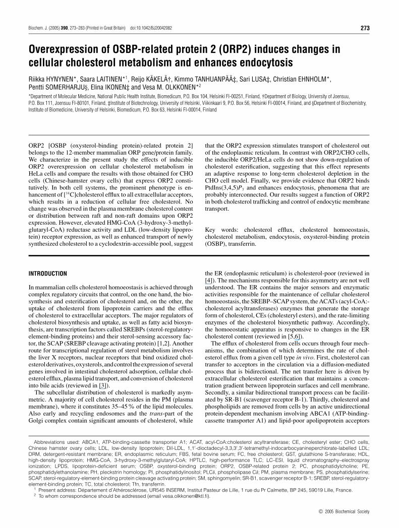
To test if the inducible ORP2/HeLa cells display increased chol-esterol efflux similar to that observed in ORP2/CHO cells [14],the amount of [14C]cholesterol effluxed to 20 % (v/v) humanserum was measured after 42 h of [14C]cholesterol labelling andinduction of ORP2 expression. After induction, all three ORP2/HeLa cell lines displayed an approx. 15% increase in cholesterolefflux as compared with non-induced cells (Figure 2A). Doxy-cycline treatment did not affect [14C]cholesterol efflux fromcells that were stably transfected with the empty vector plasmid(results not shown). Similar increment in cholesterol efflux upondoxycycline induction of ORP2 expression was also observedwhen BSA (Figure 2B) or human HDL2 (results not shown)were used as acceptors. Furthermore, addition of human apoA-I(20 µg/ml) to the BSA efflux medium caused no further incrementin cholesterol efflux (Figure 2B). To study the possibility that theincreased amount of radioactive cholesterol in the medium couldbe due to shedding of PM, we also measured cholesterol efflux tothe medium with no acceptors (Figure 2C). The relative amountof [14C]cholesterol released into the acceptor-free medium wasnegligibly low (0.4–1.4%). Even though the values for the in-duced, ORP2-expressing cells were higher than for the non-induced cells, the extent of putative membrane shedding was sosmall that it cannot explain the observed changes in cholesterolefflux to serum acceptors.
We also analysed by Western blotting the expression levelsof two proteins known to be involved in cholesterol efflux,ABCA1 and SR-B1, in induced and non-induced ORP2/HeLacells. Using a commercial antibody that detected a strong signalin macrophages, we did not observe ABCA1 expression in thesecells. SR-B1 protein was detectable, but there was no differencein the expression level between the induced and non-induced cellspecimens (results not shown).
PM lipid composition and cholesterol organizationin ORP2/HeLa cells
The above results suggested that the ORP2-mediated efflux en-hancement may result from a change in the cellular or PM
c© 2005 Biochemical Society
ORP2 and cellular cholesterol metabolism 277
Figure 2 Effects of ORP2 overexpression on cholesterol efflux to differentacceptors
(A) The inducible ORP2/HeLa cells (identified below the panel) were labelled with [14C]choles-terol for 42 h in the absence (white bars) or presence (black bars) of 1 µg/ml doxycycline,incubated for 2 h in a medium containing 20 % human serum, and the radioactivity in theefflux medium and the cells was determined. (B) Similar experiments as in (A) carried outwith ORP2/HeLa line 21f3 using 0.2 % BSA or 0.2 % BSA with 20 µg/ml human apoA-I, inserum-free medium as acceptors (efflux time 4 h). (C) In a setting identical with that in (A), theORP2/HeLa cell lines were labelled with [14C]cholesterol and incubated for 2 h in serum-freemedium that contained no cholesterol acceptor. The radioactivity in the cells and the media wasthen determined by liquid-scintillation counting. The results are shown as the percentage ofradioactivity found in the medium (mean +− S.E.M.; n = 3; ***P < 0.001, *P < 0.05, t test).
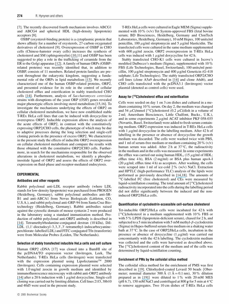
cholesterol distribution that makes it more readily available forefflux. We next probed the PM [14C]cholesterol in ORP2/HeLa21f3 cells cultured in a medium supplemented either with 10%FBS or with 5% LPDS, under inducing or non-inducing condi-tions, with a brief treatment with methyl-β-cyclodextrin that doesnot permeabilize the PMs of the cells [18]. Under both conditions,the expression of ORP2 was associated with a significant increasein the cyclodextrin extractability of the cholesterol (Figure 3A).
To determine the lipid composition of the ORP2/HeLa cell PMsin the induced and non-induced states, we enriched a PM fractionof these cells, cultured in the presence of 10% FBS, using apreviously described colloidal silica method [19]. As judged fromWestern blotting using antibodies raised against PM (syntaxin 2)and ER (GRP-94) markers and densitometric scanning, the enrich-ment of PMs was 6-fold. The cholesterol content of these mem-branes was determined enzymatically and phospholipids were
Figure 3 Organization of cholesterol in the PM of ORP2/HeLa cells
(A) The ORP2/HeLa cell line 21f3 was labelled with [14C]cholesterol for 42 h in the absence(white bars) or presence (black bars) of 1 µg/ml doxycycline, either in a medium containing 5 %LPDS or one supplemented with 10 % complete FBS. The cell-surface cholesterol was extractedfor 5 min with 5 mM methyl-β-cyclodextrin, and radioactivity in the medium and in the cells wasdetermined. The bars represent the portion recovered in the medium (mean +− S.E.M.; n = 3).(B) PMs of ORP2/HeLa cells incubated in 10 % FBS containing medium in the absence of pre-sence of induction, were enriched using the colloidal silica technique. Cholesterol was quantifiedby an enzymatic method and phospholipids by LC–ESI–MS, as described in the Experimentalsection. The results are shown as a mol% distribution, and represent the means +− S.E.M. for thethree cell lines. (C) The ORP2/HeLa cell lines were labelled with [14C]cholesterol for 42 h inthe absence or presence of induction in a medium supplemented with 10 % FBS, and distributionof the radioactivity between Triton X-100 insoluble and soluble fractions was determined asspecified in the Experimental section. The bars represent the portion of [14C]cholesterol intwo top fractions containing the DRM, and represent a mean +− S.E.M. for three experiments.**P < 0.01, *P < 0.05, t test.
analysed by LC–ESI–MS. Cholesterol represented 27–31 mol%of the total lipids, and there was no difference between theinduced and non-induced ORP2/HeLa membrane preparations(Figure 3B). Furthermore, there was no significant differencebetween the induced and non-induced cells in the relative amountsof the SM, PC, diacyl-PE, PS or PI classes (Figure 3B), nor in themolecular species composition within these classes (results notshown).
To investigate the possibility that the distribution of cholesterolbetween DRM (detergent-resistant membrane) and sensitivemembrane domains might have changed in the induced ORP2/HeLa cells, we isolated Triton X-100-resistant membrane do-mains of the cells and analysed the proportion of the cellular[14C]cholesterol found in the DRM (Figure 3C). The analysisrevealed no difference between the induced and non-inducedORP2/HeLa cells. A similar result was obtained when a milderdetergent, Brij96, was used for raft isolation.
c© 2005 Biochemical Society
278 R. Hynynen and others
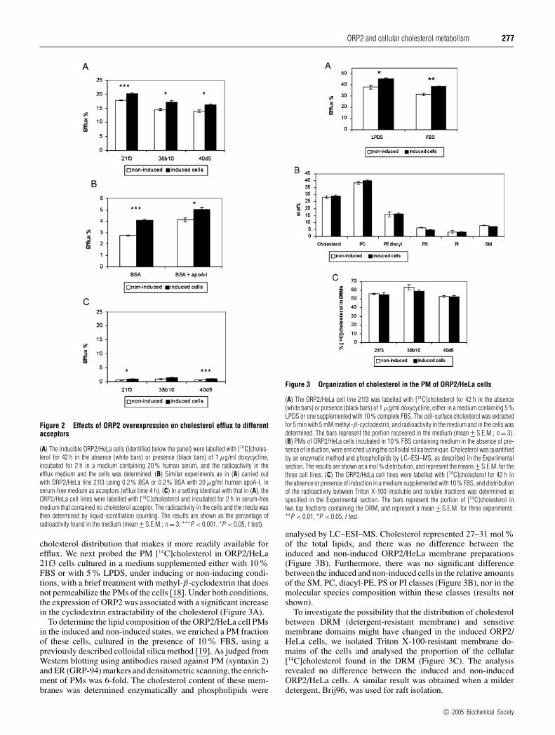
Figure 4 Effect of ORP2 overexpression on the efflux of newly synthesizedcholesterol to methyl-β-cyclodextrin
Control (Ctrl) CHO and ORP2 expressing A5a9 (ORP2/CHO) cells were cultured inlipoprotein-deficient medium containing [14C]cholesterol as described in the Experimentalsection. To measure the appearance of newly synthesized [3H]cholesterol at the PM, cells werelabelled with [3H]acetate for 15 min and chased for 2 h. During the last 5 min of the chase thecells were incubated with 5 mM methyl-β-cyclodextrin. The synthesized [3H]cholesterol andthe pre-existing [14C]cholesterol in the cells and the media were isolated by HPTLCand HPLC and quantified by liquid-scintillation counting. The results are shown as the percentageof the total [3H] (black bars) or [14C] (white bars) cholesterol found in the efflux medium(mean +− S.E.M.; n = 7; ***P < 0.001, t test).
To address the question whether association of the ORP2 pro-tein directly with the cellular PMs might provide an explanationfor the increased cholesterol efflux, we Western blotted totalmembrane and PM fractions isolated from the inducible HeLacells using the colloidal silica method, with ORP2 antibodies.Even though, as shown previously by Laitinen et al. [14], a minorportion of the ORP2 was membrane associated, the protein wasnot detected in the PM fraction (results not shown).
Intracellular transport of newly synthesized cholesterol in cellsoverexpressing ORP2
To investigate whether ORP2 affects the transport of newlysynthesized cholesterol from the ER to the PM, we measuredthe appearance of newly synthesized [3H]cholesterol in a cell-surface pool accessible for extraction with methyl-β-cyclodextrin[18]. These experiments were carried out in the stably transfectedORP2/CHO cell line A5a9, the efflux phenotype of which ismarkedly more pronounced than that of the inducible HeLa cells[14]. To be able to compare the results with those obtained forprelabelled cholesterol which mainly resides in the PM, the cellswere first incubated for 36 h with [14C]cholesterol. Labelling ofnewly synthesized cholesterol was then carried out as described inthe Experimental section. Cyclodextrin was added to the mediumin the last 5 min of the 2 h chase. The medium and the cellswere collected, and both [3H]cholesterol and [14C]cholesterol werequantified. In control cells, 21% of the newly synthesized[3H]cholesterol was accessible to cyclodextrin after the 2 h chase,while in the ORP2 cells the proportion of newly synthesized chol-esterol accessible to cyclodextrin was significantly higher, 30%(Figure 4). Also the extractability of prelabelled [14C]cholesterolwas significantly higher in the ORP2 cells as compared with thecontrols (33% versus 28%). However, the difference was lesspronounced than for [3H]cholesterol. The results indicate that thetransport of newly synthesized cholesterol to the cell surface ingeneral or to cell-surface domains preferentially accessible to cy-clodextrin extraction is enhanced in the cells that overexpressORP2.
Cholesterol esterification in ORP2/HeLa cells
To investigate how the homoeostatic control of cholesterol meta-bolism functions in the ORP2/HeLa cells apparently losing FC
Figure 5 Cholesterol esterification in cells expressing ORP2
(A) Effect of ORP2 overexpression on cholesterol esterification. The three inducible HeLa celllines were labelled with [14C]cholesterol for 42 h in the absence (white bars) or presence (blackbars) of 1 µg/ml doxycycline. Labelled FC and CEs were separated from cell extracts usingHPTLC and quantified by liquid-scintillation counting. The results are shown as a percentage of14C-labelled CEs of total labelled cholesterol (mean +− S.E.M.; n = 9, ***P < 0.001, *P < 0.05,t test). (B) Effect of ACAT inhibition on the efflux of [14C]cholesterol from control CHO orORP2/CHO cells. Control (white bars) and ORP2/CHO A5a9 cells (black bars) were labelled with[14C]cholesterol for 42 h in the absence (no PKF) or presence (PKF) of an ACAT inhibitor (PKF058-035). Cells were then incubated for 2 h in a medium containing 20 % human serum. Theradioactivity of the cells and the media was determined by liquid-scintillation counting. The re-sults are shown as the percentage of radioactivity found in the medium (mean +− S.E.M.; n = 6,***P < 0.001, t test).
from the PM due to ORP2 expression, we determined the pro-portion of the [14C]cholesterol that was esterified after 42 hof ORP2 induction and labelling (Figure 5A). In contrast withORP2/CHO cells that display a reduction of cholesterol esteri-fication [14], the induced ORP2 overexpression led to enhancedcholesterol esterification in two HeLa cell lines out of the three.In the third line (38b10), the esterification was high already in thenon-induced state and there was no change upon induction.The fact that the ORP2/HeLa and the ORP2/CHO cells both showenhanced cholesterol efflux but the cholesterol esterificationphenotypes are different suggested that the observed increase inefflux is not a secondary phenomenon due to reduced cholesterolesterification. To clarify the issue further, we employed the ORP2/CHO A5a9 cell line that shows a marked (approx. 50%) enhance-ment of [14C]cholesterol efflux as compared with control CHOcells. The cells were labelled with [14C]cholesterol in the presenceor absence of the ACAT inhibitor PKF 058-035. In the pre-sence of PKF, esterification of [14C]cholesterol was abolished(99% inhibition) in both cell lines. The efflux of labelled chol-esterol to human serum was measured and the results in the pre-sence and absence of the inhibitor were compared. The ORP2/CHO cells displayed a significant increase in cholesterol effluxas compared with controls both in the absence and in thepresence of PKF (Figure 5B). We therefore conclude that the in-crease in [14C]cholesterol efflux in ORP2 overexpressing cells isnot due to decreased ACAT activity and a resulting increase in FCavailable for efflux, but due to other mechanisms that are involved.
c© 2005 Biochemical Society
ORP2 and cellular cholesterol metabolism 279
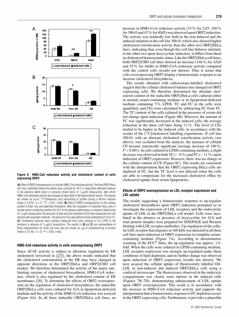
Figure 6 HMG-CoA reductase activity and cholesterol content of cellsexpressing ORP2
(A) Effect of ORP2 overexpression on cellular HMG-CoA reductase activity. The three ORP2/HeLacell lines (identified below the panel) were cultured for 42 h in lipoprotein-deficient mediumin the absence (white bars) or presence (black bars) of 1 µg/ml doxycycline, after whichHMG-CoA reductase activity was assayed as described in the Experimental section. The resultsare shown as d.p.m. [14C]mevalonic acid lactone/mg of protein during a 90 min reaction(mean +− S.E.M.; n = 3, **P < 0.01, t test). (B) Effect of ORP2 overexpression on the cellularcontent of total, free and esterified cholesterol. After the incubation of inducible HeLa cells innormal serum-containing medium for 42 h in the absence (white bars) or presence (black bars)of 1 µg/ml doxycycline, the amounts of total and free cholesterol (CH) were measured from cellextracts with enzymatic methods. The amount of CEs was determined by subtracting FC from TC.(C) Cholesterol amounts were similarly measured from cells cultured in 5 % LPDS in thepresence or absence of 1 µg/ml doxycycline. The results in (B and C) are representative ofthree measurements for three cell lines and are shown as µg of cholesterol/mg of protein(mean +− S.E.M.; n = 9, *P < 0.05, t test).
HMG-CoA reductase activity in cells overexpressing ORP2
Since ACAT activity is subject to allosteric regulation by ERcholesterol (reviewed in [27]), the above results indicated thatthe cholesterol concentration in the ER may have changed inopposite directions in the ORP2/HeLa and ORP2/CHO cellmodels. We therefore determined the activity of the major rate-limiting enzyme of cholesterol biosynthesis, HMG-CoA reduc-tase, which is also regulated by the cholesterol content of ERmembranes [28]. To determine the effects of ORP2 overexpres-sion on the regulation of cholesterol biosynthesis, the inducibleORP2/HeLa cells were cultured for 42 h in lipoprotein-deficientmedium and the activity of the HMG-CoA reductase was assayed(Figure 6A). In all three inducible ORP2/HeLa cell lines, an
increase in HMG-CoA reductase activity (33% for 21f3, 168%for 38b10 and 43 % for 40d5) was observed upon ORP2 induction.The activity was markedly low both in the non-induced and theinduced situation in the cell line 38b10, which also showed highercholesterol esterification activity than the other two ORP2/HeLalines, indicating that, even though this cell line behaves similarlyto the other two upon doxycycline induction, it differs from themin cholesterol homoeostatic status. Like the ORP2/HeLa cell lines,both ORP2/CHO cell lines showed an increase (146% for A5a9and 55% for A6d6) in HMG-CoA reductase activity comparedwith the control cells (results not shown). Thus it seems thatcells overexpressing ORP2 display a homoeostatic response to anincrease cholesterol biosynthesis.
The results obtained with radioisotope-labelled cholesterolsuggest that the cellular cholesterol balance has changed in ORP2expressing cells. We therefore determined the absolute chol-esterol content of the inducible ORP2/HeLa cells cultured eitherin normal serum-containing medium or in lipoprotein-deficientmedium containing 5% LPDS. TC and FC in the cells werequantified, and CEs were calculated by subtracting FC from TC.The TC content of the cells cultured in the presence of serum didnot change upon induction (Figure 6B). However, the amount ofFC was significantly decreased in the induced cells, the averagereduction in the three cell lines being 11%. The level of CEstended to be higher in the induced cells, in accordance with theresults of the [14C]cholesterol labelling experiments. If cell line38b10, with an aberrant cholesterol esterification activity (seeabove), was excluded from the analysis, the increase of cellularCE became statistically significant (average increase of 100%,P < 0.001). In cells cultured in LPDS-containing medium, a slightdecrease was observed in both TC (−8%) and FC (−11%) uponinduction of ORP2 expression. However, there was no change inthe cellular content of CE (Figure 6C). The results are consistentwith the interpretation that the ORP2 expressing HeLa cells aredepleted of FC, but the TC level is not affected when the cellsare able to compensate for the increased cholesterol efflux bycholesterol uptake from serum lipoproteins.
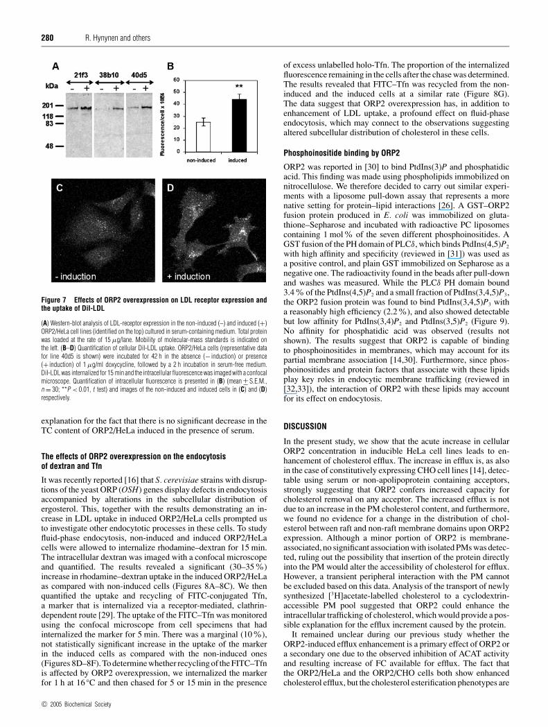
Effects of ORP2 overexpression on LDL receptor expression andLDL uptake
The results suggesting a homoeostatic response to up-regulatecholesterol biosynthesis upon ORP2 induction prompted us toinvestigate the expression of LDL receptors and the endocytoticuptake of LDL in the ORP2/HeLa cell model. Cells were incu-bated in the absence or presence of doxycycline for 42 h andtotal protein samples were prepared for SDS/PAGE and Westernblotting with LDL receptor antibodies. Up-regulation of the cellu-lar LDL receptor that migrates at 160 kDa was detected in all threecell lines upon induction of ORP2 expression in complete serum-containing medium (Figure 7A). According to densitometricscanning of the ECL® films, the up-regulation was approx. 1.6-fold. When the cells were cultured in LPDS-containing medium,LDL receptor expression was strongly up-regulated under theseconditions of lipid depletion, and no further change was observedupon induction of ORP2 expression (results not shown). Wenext assayed the cellular uptake of fluorescently labelled DiI-LDL in non-induced and induced ORP2/HeLa cells using aconfocal microscope. The fluorescence observed in the endocyticcompartments was clearly more intense in the induced cells(Figures 7B–7D), demonstrating enhancement of LDL uptakeupon ORP2 overexpression. This result is in accordance withthe increase in HMG-CoA reductase activity and supports theinterpretation that a homoeostatic response to FC depletion occursin the ORP2 expressing cells. Furthermore, it provides a plausible
c© 2005 Biochemical Society
280 R. Hynynen and others
Figure 7 Effects of ORP2 overexpression on LDL receptor expression andthe uptake of DiI-LDL
(A) Western-blot analysis of LDL-receptor expression in the non-induced (–) and induced (+)ORP2/HeLa cell lines (identified on the top) cultured in serum-containing medium. Total proteinwas loaded at the rate of 15 µg/lane. Mobility of molecular-mass standards is indicated onthe left. (B–D) Quantification of cellular DiI-LDL uptake. ORP2/HeLa cells (representative datafor line 40d5 is shown) were incubated for 42 h in the absence (− induction) or presence(+ induction) of 1 µg/ml doxycycline, followed by a 2 h incubation in serum-free medium.DiI-LDL was internalized for 15 min and the intracellular fluorescence was imaged with a confocalmicroscope. Quantification of intracellular fluorescence is presented in (B) (mean +− S.E.M.,n = 30; **P < 0.01, t test) and images of the non-induced and induced cells in (C) and (D)respectively.
explanation for the fact that there is no significant decrease in theTC content of ORP2/HeLa induced in the presence of serum.
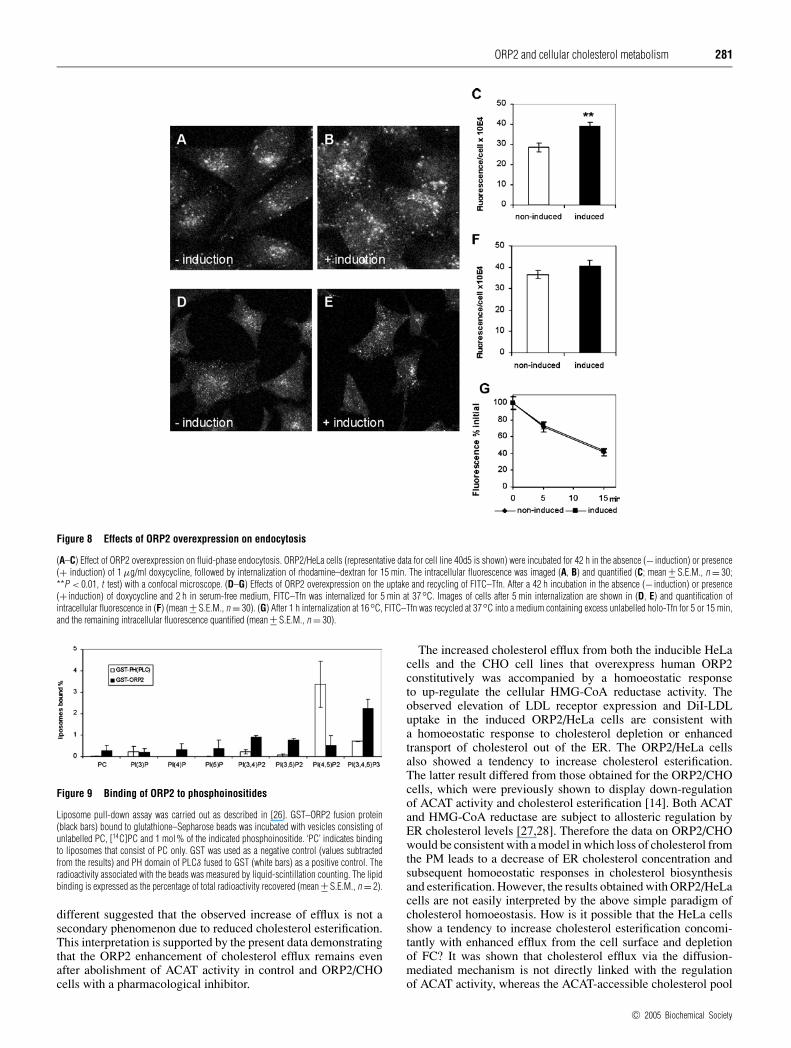
The effects of ORP2 overexpression on the endocytosisof dextran and Tfn
It was recently reported [16] that S. cerevisiae strains with disrup-tions of the yeast ORP (OSH) genes display defects in endocytosisaccompanied by alterations in the subcellular distribution ofergosterol. This, together with the results demonstrating an in-crease in LDL uptake in induced ORP2/HeLa cells prompted usto investigate other endocytotic processes in these cells. To studyfluid-phase endocytosis, non-induced and induced ORP2/HeLacells were allowed to internalize rhodamine–dextran for 15 min.The intracellular dextran was imaged with a confocal microscopeand quantified. The results revealed a significant (30–35 %)increase in rhodamine–dextran uptake in the induced ORP2/HeLaas compared with non-induced cells (Figures 8A–8C). We thenquantified the uptake and recycling of FITC-conjugated Tfn,a marker that is internalized via a receptor-mediated, clathrin-dependent route [29]. The uptake of the FITC–Tfn was monitoredusing the confocal microscope from cell specimens that hadinternalized the marker for 5 min. There was a marginal (10%),not statistically significant increase in the uptake of the markerin the induced cells as compared with the non-induced ones(Figures 8D–8F). To determine whether recycling of the FITC–Tfnis affected by ORP2 overexpression, we internalized the markerfor 1 h at 16 ◦C and then chased for 5 or 15 min in the presence
of excess unlabelled holo-Tfn. The proportion of the internalizedfluorescence remaining in the cells after the chase was determined.The results revealed that FITC–Tfn was recycled from the non-induced and the induced cells at a similar rate (Figure 8G).The data suggest that ORP2 overexpression has, in addition toenhancement of LDL uptake, a profound effect on fluid-phaseendocytosis, which may connect to the observations suggestingaltered subcellular distribution of cholesterol in these cells.
Phosphoinositide binding by ORP2
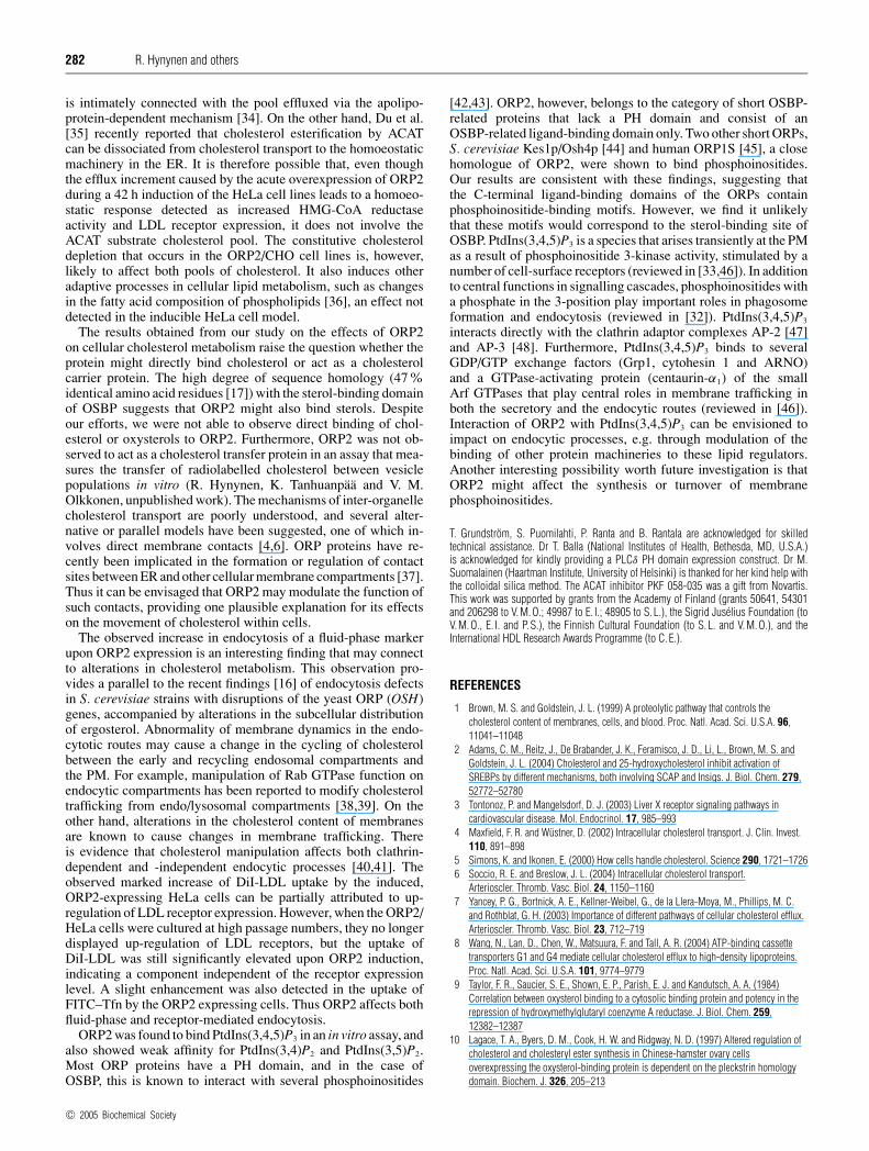
ORP2 was reported in [30] to bind PtdIns(3)P and phosphatidicacid. This finding was made using phospholipids immobilized onnitrocellulose. We therefore decided to carry out similar experi-ments with a liposome pull-down assay that represents a morenative setting for protein–lipid interactions [26]. A GST–ORP2fusion protein produced in E. coli was immobilized on gluta-thione–Sepharose and incubated with radioactive PC liposomescontaining 1 mol% of the seven different phosphoinositides. AGST fusion of the PH domain of PLCδ, which binds PtdIns(4,5)P2
with high affinity and specificity (reviewed in [31]) was used asa positive control, and plain GST immobilized on Sepharose as anegative one. The radioactivity found in the beads after pull-downand washes was measured. While the PLCδ PH domain bound3.4% of the PtdIns(4,5)P2 and a small fraction of PtdIns(3,4,5)P3,the ORP2 fusion protein was found to bind PtdIns(3,4,5)P3 witha reasonably high efficiency (2.2 %), and also showed detectablebut low affinity for PtdIns(3,4)P2 and PtdIns(3,5)P2 (Figure 9).No affinity for phosphatidic acid was observed (results notshown). The results suggest that ORP2 is capable of bindingto phosphoinositides in membranes, which may account for itspartial membrane association [14,30]. Furthermore, since phos-phoinositides and protein factors that associate with these lipidsplay key roles in endocytic membrane trafficking (reviewed in[32,33]), the interaction of ORP2 with these lipids may accountfor its effect on endocytosis.
DISCUSSION
In the present study, we show that the acute increase in cellularORP2 concentration in inducible HeLa cell lines leads to en-hancement of cholesterol efflux. The increase in efflux is, as alsoin the case of constitutively expressing CHO cell lines [14], detec-table using serum or non-apolipoprotein containing acceptors,strongly suggesting that ORP2 confers increased capacity forcholesterol removal on any acceptor. The increased efflux is notdue to an increase in the PM cholesterol content, and furthermore,we found no evidence for a change in the distribution of chol-esterol between raft and non-raft membrane domains upon ORP2expression. Although a minor portion of ORP2 is membrane-associated, no significant association with isolated PMs was detec-ted, ruling out the possibility that insertion of the protein directlyinto the PM would alter the accessibility of cholesterol for efflux.However, a transient peripheral interaction with the PM cannotbe excluded based on this data. Analysis of the transport of newlysynthesized [3H]acetate-labelled cholesterol to a cyclodextrin-accessible PM pool suggested that ORP2 could enhance theintracellular trafficking of cholesterol, which would provide a pos-sible explanation for the efflux increment caused by the protein.
It remained unclear during our previous study whether theORP2-induced efflux enhancement is a primary effect of ORP2 ora secondary one due to the observed inhibition of ACAT activityand resulting increase of FC available for efflux. The fact thatthe ORP2/HeLa and the ORP2/CHO cells both show enhancedcholesterol efflux, but the cholesterol esterification phenotypes are
c© 2005 Biochemical Society
ORP2 and cellular cholesterol metabolism 281
Figure 8 Effects of ORP2 overexpression on endocytosis
(A–C) Effect of ORP2 overexpression on fluid-phase endocytosis. ORP2/HeLa cells (representative data for cell line 40d5 is shown) were incubated for 42 h in the absence (− induction) or presence(+ induction) of 1 µg/ml doxycycline, followed by internalization of rhodamine–dextran for 15 min. The intracellular fluorescence was imaged (A, B) and quantified (C; mean +− S.E.M., n = 30;**P < 0.01, t test) with a confocal microscope. (D–G) Effects of ORP2 overexpression on the uptake and recycling of FITC–Tfn. After a 42 h incubation in the absence (− induction) or presence(+ induction) of doxycycline and 2 h in serum-free medium, FITC–Tfn was internalized for 5 min at 37◦C. Images of cells after 5 min internalization are shown in (D, E) and quantification ofintracellular fluorescence in (F) (mean +− S.E.M., n = 30). (G) After 1 h internalization at 16◦C, FITC–Tfn was recycled at 37◦C into a medium containing excess unlabelled holo-Tfn for 5 or 15 min,and the remaining intracellular fluorescence quantified (mean +− S.E.M., n = 30).
Figure 9 Binding of ORP2 to phosphoinositides
Liposome pull-down assay was carried out as described in [26]. GST–ORP2 fusion protein(black bars) bound to glutathione–Sepharose beads was incubated with vesicles consisting ofunlabelled PC, [14C]PC and 1 mol% of the indicated phosphoinositide. ‘PC’ indicates bindingto liposomes that consist of PC only. GST was used as a negative control (values subtractedfrom the results) and PH domain of PLCδ fused to GST (white bars) as a positive control. Theradioactivity associated with the beads was measured by liquid-scintillation counting. The lipidbinding is expressed as the percentage of total radioactivity recovered (mean +− S.E.M., n = 2).
different suggested that the observed increase of efflux is not asecondary phenomenon due to reduced cholesterol esterification.This interpretation is supported by the present data demonstratingthat the ORP2 enhancement of cholesterol efflux remains evenafter abolishment of ACAT activity in control and ORP2/CHOcells with a pharmacological inhibitor.
The increased cholesterol efflux from both the inducible HeLacells and the CHO cell lines that overexpress human ORP2constitutively was accompanied by a homoeostatic responseto up-regulate the cellular HMG-CoA reductase activity. Theobserved elevation of LDL receptor expression and DiI-LDLuptake in the induced ORP2/HeLa cells are consistent witha homoeostatic response to cholesterol depletion or enhancedtransport of cholesterol out of the ER. The ORP2/HeLa cellsalso showed a tendency to increase cholesterol esterification.The latter result differed from those obtained for the ORP2/CHOcells, which were previously shown to display down-regulationof ACAT activity and cholesterol esterification [14]. Both ACATand HMG-CoA reductase are subject to allosteric regulation byER cholesterol levels [27,28]. Therefore the data on ORP2/CHOwould be consistent with a model in which loss of cholesterol fromthe PM leads to a decrease of ER cholesterol concentration andsubsequent homoeostatic responses in cholesterol biosynthesisand esterification. However, the results obtained with ORP2/HeLacells are not easily interpreted by the above simple paradigm ofcholesterol homoeostasis. How is it possible that the HeLa cellsshow a tendency to increase cholesterol esterification concomi-tantly with enhanced efflux from the cell surface and depletionof FC? It was shown that cholesterol efflux via the diffusion-mediated mechanism is not directly linked with the regulationof ACAT activity, whereas the ACAT-accessible cholesterol pool
c© 2005 Biochemical Society
282 R. Hynynen and others
is intimately connected with the pool effluxed via the apolipo-protein-dependent mechanism [34]. On the other hand, Du et al.[35] recently reported that cholesterol esterification by ACATcan be dissociated from cholesterol transport to the homoeostaticmachinery in the ER. It is therefore possible that, even thoughthe efflux increment caused by the acute overexpression of ORP2during a 42 h induction of the HeLa cell lines leads to a homoeo-static response detected as increased HMG-CoA reductaseactivity and LDL receptor expression, it does not involve theACAT substrate cholesterol pool. The constitutive cholesteroldepletion that occurs in the ORP2/CHO cell lines is, however,likely to affect both pools of cholesterol. It also induces otheradaptive processes in cellular lipid metabolism, such as changesin the fatty acid composition of phospholipids [36], an effect notdetected in the inducible HeLa cell model.
The results obtained from our study on the effects of ORP2on cellular cholesterol metabolism raise the question whether theprotein might directly bind cholesterol or act as a cholesterolcarrier protein. The high degree of sequence homology (47%identical amino acid residues [17]) with the sterol-binding domainof OSBP suggests that ORP2 might also bind sterols. Despiteour efforts, we were not able to observe direct binding of chol-esterol or oxysterols to ORP2. Furthermore, ORP2 was not ob-served to act as a cholesterol transfer protein in an assay that mea-sures the transfer of radiolabelled cholesterol between vesiclepopulations in vitro (R. Hynynen, K. Tanhuanpaa and V. M.Olkkonen, unpublished work). The mechanisms of inter-organellecholesterol transport are poorly understood, and several alter-native or parallel models have been suggested, one of which in-volves direct membrane contacts [4,6]. ORP proteins have re-cently been implicated in the formation or regulation of contactsites between ER and other cellular membrane compartments [37].Thus it can be envisaged that ORP2 may modulate the function ofsuch contacts, providing one plausible explanation for its effectson the movement of cholesterol within cells.
The observed increase in endocytosis of a fluid-phase markerupon ORP2 expression is an interesting finding that may connectto alterations in cholesterol metabolism. This observation pro-vides a parallel to the recent findings [16] of endocytosis defectsin S. cerevisiae strains with disruptions of the yeast ORP (OSH)genes, accompanied by alterations in the subcellular distributionof ergosterol. Abnormality of membrane dynamics in the endo-cytotic routes may cause a change in the cycling of cholesterolbetween the early and recycling endosomal compartments andthe PM. For example, manipulation of Rab GTPase function onendocytic compartments has been reported to modify cholesteroltrafficking from endo/lysosomal compartments [38,39]. On theother hand, alterations in the cholesterol content of membranesare known to cause changes in membrane trafficking. Thereis evidence that cholesterol manipulation affects both clathrin-dependent and -independent endocytic processes [40,41]. Theobserved marked increase of DiI-LDL uptake by the induced,ORP2-expressing HeLa cells can be partially attributed to up-regulation of LDL receptor expression. However, when the ORP2/HeLa cells were cultured at high passage numbers, they no longerdisplayed up-regulation of LDL receptors, but the uptake ofDiI-LDL was still significantly elevated upon ORP2 induction,indicating a component independent of the receptor expressionlevel. A slight enhancement was also detected in the uptake ofFITC–Tfn by the ORP2 expressing cells. Thus ORP2 affects bothfluid-phase and receptor-mediated endocytosis.
ORP2 was found to bind PtdIns(3,4,5)P3 in an in vitro assay, andalso showed weak affinity for PtdIns(3,4)P2 and PtdIns(3,5)P2.Most ORP proteins have a PH domain, and in the case ofOSBP, this is known to interact with several phosphoinositides
[42,43]. ORP2, however, belongs to the category of short OSBP-related proteins that lack a PH domain and consist of anOSBP-related ligand-binding domain only. Two other short ORPs,S. cerevisiae Kes1p/Osh4p [44] and human ORP1S [45], a closehomologue of ORP2, were shown to bind phosphoinositides.Our results are consistent with these findings, suggesting thatthe C-terminal ligand-binding domains of the ORPs containphosphoinositide-binding motifs. However, we find it unlikelythat these motifs would correspond to the sterol-binding site ofOSBP. PtdIns(3,4,5)P3 is a species that arises transiently at the PMas a result of phosphoinositide 3-kinase activity, stimulated by anumber of cell-surface receptors (reviewed in [33,46]). In additionto central functions in signalling cascades, phosphoinositides witha phosphate in the 3-position play important roles in phagosomeformation and endocytosis (reviewed in [32]). PtdIns(3,4,5)P3
interacts directly with the clathrin adaptor complexes AP-2 [47]and AP-3 [48]. Furthermore, PtdIns(3,4,5)P3 binds to severalGDP/GTP exchange factors (Grp1, cytohesin 1 and ARNO)and a GTPase-activating protein (centaurin-α1) of the smallArf GTPases that play central roles in membrane trafficking inboth the secretory and the endocytic routes (reviewed in [46]).Interaction of ORP2 with PtdIns(3,4,5)P3 can be envisioned toimpact on endocytic processes, e.g. through modulation of thebinding of other protein machineries to these lipid regulators.Another interesting possibility worth future investigation is thatORP2 might affect the synthesis or turnover of membranephosphoinositides.
T. Grundstrom, S. Puomilahti, P. Ranta and B. Rantala are acknowledged for skilledtechnical assistance. Dr T. Balla (National Institutes of Health, Bethesda, MD, U.S.A.)is acknowledged for kindly providing a PLCδ PH domain expression construct. Dr M.Suomalainen (Haartman Institute, University of Helsinki) is thanked for her kind help withthe colloidal silica method. The ACAT inhibitor PKF 058-035 was a gift from Novartis.This work was supported by grants from the Academy of Finland (grants 50641, 54301and 206298 to V. M. O.; 49987 to E. I.; 48905 to S. L.), the Sigrid Juselius Foundation (toV. M. O., E. I. and P. S.), the Finnish Cultural Foundation (to S. L. and V. M. O.), and theInternational HDL Research Awards Programme (to C. E.).
REFERENCES
1 Brown, M. S. and Goldstein, J. L. (1999) A proteolytic pathway that controls thecholesterol content of membranes, cells, and blood. Proc. Natl. Acad. Sci. U.S.A. 96,11041–11048
2 Adams, C. M., Reitz, J., De Brabander, J. K., Feramisco, J. D., Li, L., Brown, M. S. andGoldstein, J. L. (2004) Cholesterol and 25-hydroxycholesterol inhibit activation ofSREBPs by different mechanisms, both involving SCAP and Insigs. J. Biol. Chem. 279,52772–52780
3 Tontonoz, P. and Mangelsdorf, D. J. (2003) Liver X receptor signaling pathways incardiovascular disease. Mol. Endocrinol. 17, 985–993
4 Maxfield, F. R. and Wustner, D. (2002) Intracellular cholesterol transport. J. Clin. Invest.110, 891–898
5 Simons, K. and Ikonen, E. (2000) How cells handle cholesterol. Science 290, 1721–17266 Soccio, R. E. and Breslow, J. L. (2004) Intracellular cholesterol transport.
Arterioscler. Thromb. Vasc. Biol. 24, 1150–11607 Yancey, P. G., Bortnick, A. E., Kellner-Weibel, G., de la Llera-Moya, M., Phillips, M. C.
and Rothblat, G. H. (2003) Importance of different pathways of cellular cholesterol efflux.Arterioscler. Thromb. Vasc. Biol. 23, 712–719
8 Wang, N., Lan, D., Chen, W., Matsuura, F. and Tall, A. R. (2004) ATP-binding cassettetransporters G1 and G4 mediate cellular cholesterol efflux to high-density lipoproteins.Proc. Natl. Acad. Sci. U.S.A. 101, 9774–9779
9 Taylor, F. R., Saucier, S. E., Shown, E. P., Parish, E. J. and Kandutsch, A. A. (1984)Correlation between oxysterol binding to a cytosolic binding protein and potency in therepression of hydroxymethylglutaryl coenzyme A reductase. J. Biol. Chem. 259,12382–12387
10 Lagace, T. A., Byers, D. M., Cook, H. W. and Ridgway, N. D. (1997) Altered regulation ofcholesterol and cholesteryl ester synthesis in Chinese-hamster ovary cellsoverexpressing the oxysterol-binding protein is dependent on the pleckstrin homologydomain. Biochem. J. 326, 205–213
c© 2005 Biochemical Society
ORP2 and cellular cholesterol metabolism 283
11 Lagace, T. A., Byers, D. M., Cook, H. W. and Ridgway, N. D. (1999) Chinese hamsterovary cells overexpressing the oxysterol binding protein (OSBP) display enhancedsynthesis of sphingomyelin in response to 25-hydroxycholesterol. J. Lipid Res. 40,109–116
12 Wyles, J. P., McMaster, C. R. and Ridgway, N. D. (2002) Vesicle-associated membraneprotein-associated protein-A (VAP-A) interacts with the oxysterol-binding protein tomodify export from the endoplasmic reticulum. J. Biol. Chem. 277, 29908–29918
13 Lehto, M. and Olkkonen, V. M. (2003) The OSBP-related proteins: a novel protein familyinvolved in vesicle transport, cellular lipid metabolism, and cell signalling.Biochim. Biophys. Acta 1631, 1–11
14 Laitinen, S., Lehto, M., Lehtonen, S., Hyvarinen, K., Heino, S., Lehtonen, E., Ehnholm, C.,Ikonen, E. and Olkkonen, V. M. (2002) ORP2, a homolog of oxysterol binding protein,regulates cellular cholesterol metabolism. J. Lipid Res. 43, 245–255
15 Beh, C. T., Cool, L., Phillips, J. and Rine, J. (2001) Overlapping functions of the yeastoxysterol-binding protein homologues. Genetics 157, 1117–1140
16 Beh, C. T. and Rine, J. (2004) A role for yeast oxysterol-binding protein homologs inendocytosis and in the maintenance of intracellular sterol-lipid distribution. J. Cell Sci.117, 2983–2996
17 Lehto, M., Laitinen, S., Chinetti, G., Johansson, M., Ehnholm, C., Staels, B., Ikonen, E.and Olkkonen, V. M. (2001) The OSBP-related protein family in humans. J. Lipid Res. 42,1203–1213
18 Heino, S., Lusa, S., Somerharju, P., Ehnholm, C., Olkkonen, V. M. and Ikonen, E.(2000) Dissecting the role of the golgi complex and lipid rafts in biosynthetictransport of cholesterol to the cell surface. Proc. Natl. Acad. Sci. U.S.A. 97, 8375–8380
19 Chaney, L. K. and Jacobson, B. S. (1983) Coating cells with colloidal silica for high yieldisolation of plasma membrane sheets and identification of transmembrane proteins.J. Biol. Chem. 258, 10062–10072
20 Folch, J. M., Lees, M. and Sloane-Stanley, G. H. (1957) A simple method for theisolation and purification of total lipids from animal tissue. J. Biol. Chem. 226,497–509
21 Koivusalo, M., Haimi, P., Heikinheimo, L., Kostiainen, R. and Somerharju, P. (2001)Quantitative determination of phospholipid compositions by ESI-MS: effects of acyl chainlength, unsaturation, and lipid concentration on instrument response. J. Lipid Res. 42,663–672
22 Kakela, R., Somerharju, P. and Tyynela, J. (2003) Analysis of phospholipid molecularspecies in brains from patients with infantile and juvenile neuronal-ceroid lipofuscinosisusing liquid chromatography-electrospray ionization mass spectrometry. J. Neurochem.84, 1051–1065
23 Wilce, P. A. and Kroon, P. A. (1992) Assay of 3-hydroxy-3-methylglutaryl coenzyme A(HMG-CoA) reductase. In Lipoprotein Analysis, A Practical Approach (Converse, C. A.and Skinner, E. R., eds.), pp. 203–214, Oxford University Press, Oxford
24 Bligh, E. G. and Dryer, W. J. (1959) A rapid method of total lipid extraction andpurification. Can. J. Biochem. Physiol. 37, 911–917
25 Gamble, W., Vaughan, M., Kruth, H. S. and Avigan, J. (1978) Procedure for determinationof free and total cholesterol in micro- or nanogram amounts suitable for studies withcultured cells. J. Lipid Res. 19, 1068–1070
26 Schiavo, G., Gu, Q. M., Prestwich, G. D., Sollner, T. H. and Rothman, J. E. (1996)Calcium-dependent switching of the specificity of phosphoinositide binding tosynaptotagmin. Proc. Natl. Acad. Sci. U.S.A. 93, 13327–13332
27 Chang, T. Y., Chang, C. C. and Cheng, D. (1997) Acyl-coenzyme A:cholesterolacyltransferase. Annu. Rev. Biochem. 66, 613–638
28 Sever, N., Yang, T., Brown, M. S., Goldstein, J. L. and DeBose-Boyd, R. A. (2003)Accelerated degradation of HMG CoA reductase mediated by binding of insig-1 to itssterol-sensing domain. Mol. Cell 11, 25–33
29 Maxfield, F. R. and McGraw, T. E. (2004) Endocytic recycling. Nat. Rev. Mol. Cell. Biol. 5,121–132
30 Xu, Y., Liu, Y., Ridgway, N. D. and McMaster, C. R. (2001) Novel members of the humanoxysterol-binding protein family bind phospholipids and regulate vesicle transport.J. Biol. Chem. 276, 18407–18414
31 Lemmon, M. A. (2003) Phosphoinositide recognition domains. Traffic 4, 201–21332 Owen, D. J. (2004) Linking endocytic cargo to clathrin: structural and functional insights
into coated vesicle formation. Biochem. Soc. Trans. 32, 1–1433 Wenk, M. R. and De Camilli, P. (2004) Protein-lipid interactions and phosphoinositide
metabolism in membrane traffic: insights from vesicle recycling in nerve terminals.Proc. Natl. Acad. Sci. U.S.A. 101, 8262–8269
34 Li, Q., Tsujita, M. and Yokoyama, S. (1997) Selective down-regulation by protein kinase Cinhibitors of apolipoprotein-mediated cellular cholesterol efflux in macrophages.Biochemistry (Moscow) 36, 12045–12052
35 Du, X., Pham, Y. H. and Brown, A. J. (2004) Effects of 25-hydroxycholesterol oncholesterol esterification and sterol regulatory element-binding protein processing aredissociable: implications for cholesterol movement to the regulatory pool in theendoplasmic reticulum. J. Biol. Chem. 279, 47010–47016
36 Kakela, R., Tanhuanpaa, K., Laitinen, S., Somerharju, P. and Olkkonen, V. M. (2005)Over-expression of OSBP-related protein 2 (ORP2) in CHO cells induces alterations ofphospholipid species composition. Biochem. Cell Biol., in the press
37 Olkkonen, V. M. and Levine, T. P. (2004) Oxysterol binding proteins: in more than oneplace at one time? Biochem. Cell Biol. 82, 87–98
38 Holtta-Vuori, M., Tanhuanpaa, K., Mobius, W., Somerharju, P. and Ikonen, E. (2002)Modulation of cellular cholesterol transport and homeostasis by Rab11. Mol. Biol. Cell13, 3107–3122
39 Choudhury, A., Dominguez, M., Puri, V., Sharma, D. K., Narita, K., Wheatley, C. L.,Marks, D. L. and Pagano, R. E. (2002) Rab proteins mediate Golgi transport of caveola-internalized glycosphingolipids and correct lipid trafficking in Niemann-Pick C cells.J. Clin. Invest. 109, 1541–1550
40 Naslavsky, N., Weigert, R. and Donaldson, J. G. (2004) Characterization of a nonclathrinendocytic pathway: membrane cargo and lipid requirements. Mol. Biol. Cell 15,3542–3552
41 Pichler, H. and Riezman, H. (2004) Where sterols are required for endocytosis.Biochim. Biophys. Acta 1666, 51–61
42 Levine, T. P. and Munro, S. (1998) The pleckstrin homology domain of oxysterol-bindingprotein recognises a determinant specific to Golgi membranes. Curr. Biol. 8, 729–739
43 Levine, T. P. and Munro, S. (2002) Targeting of Golgi-specific pleckstrin homologydomains involves both PtdIns 4-kinase-dependent and -independent components.Curr. Biol. 12, 695–704
44 Li, X., Rivas, M. P., Fang, M., Marchena, J., Mehrotra, B., Chaudhary, A., Feng, L.,Prestwich, G. D. and Bankaitis, V. A. (2002) Analysis of oxysterol binding proteinhomologue Kes1p function in regulation of Sec14p-dependent protein transport from theyeast Golgi complex. J. Cell Biol. 157, 63–77
45 Fairn, G. D. and McMaster, C. R. (2004) Identification and assessment the role of anominal phospholipid binding region of oxysterol binding protein related protein ORP1Sin the regulation of vesicular transport. Biochem. J. 387, 889–896
46 Payrastre, B., Missy, K., Giuriato, S., Bodin, S., Plantavid, M. and Gratacap, M. (2001)Phosphoinositides: key players in cell signalling, in time and space. Cell. Signalling 13,377–387
47 Gaidarov, I., Chen, Q., Falck, J. R., Reddy, K. K. and Keen, J. H. (1996) A functionalphosphatidylinositol 3,4,5-trisphosphate/phosphoinositide binding domain in theclathrin adaptor AP-2 alpha subunit. Implications for the endocytic pathway.J. Biol. Chem. 271, 20922–20929
48 Hao, W., Tan, Z., Prasad, K., Reddy, K. K., Chen, J., Prestwich, G. D., Falck, J. R., Shears,S. B. and Lafer, E. M. (1997) Regulation of AP-3 function by inositides. Identification ofphosphatidylinositol 3,4,5-trisphosphate as a potent ligand. J. Biol. Chem. 272,6393–6398
Received 15 December 2004/11 April 2005; accepted 28 April 2005Published as BJ Immediate Publication 28 April 2005, doi:10.1042/BJ20042082
c© 2005 Biochemical Society