Nienke Westerink - WUR eDepot
170
The role of AVR4 and AVR4E proteins in virulence and avirulence of the tomato pathogen Cladosporium fulvum Molecular aspects of disease susceptibility and resistance Nienke Westerink
-
Upload
khangminh22 -
Category
Documents
-
view
2 -
download
0
Transcript of Nienke Westerink - WUR eDepot

The role of AVR4 and AVR4E proteins in virulence and
avirulence of the tomato pathogen Cladosporium fulvum
Molecular aspects of disease susceptibility and resistance
Nienke Westerink

Promotor: Prof. dr. ir. P.J.G.M. de Wit
Hoogleraar in de Fytopathologie
Wageningen Universiteit
Co-promotor: Dr. ir. M.H.A.J. Joosten
Universitair docent bij het Laboratorium voor Fytopathologie
Wageningen Universiteit
Promotiecommissie: Dr. M. Stuiver, BASF Plant Science, Limburgerhof, Duitsland
Dr. A.F.J.M. van den Ackerveken, Universiteit van Utrecht
Prof. dr. B.J.C. Cornelissen, Universiteit van Amsterdam
Prof. dr. ir. J. Bakker, Universiteit van Wageningen

Nienke Westerink
The role of AVR4 and AVR4E proteins in virulence and
avirulence of the tomato pathogen Cladosporium fulvum
Molecular aspects of disease susceptibility and resistance
Proefschrift
ter verkrijging van de graad van doctor
op gezag van de rector magnificus
van Wageningen Universiteit,
Prof. dr. ir. L. Speelman
in het openbaar te verdedigen
op woensdag 5 maart 2003
des namiddags te vier uur in de Aula.

ISBN 90-5808-782-4

Table of contents
Chapter 1 General introduction and outline of the thesis 7
Chapter 2 Cladosporium fulvum evades Hcr9-4E-mediated resistance by
abolishing Avr4E expression or by modifying the AVR4E elicitor
protein
41
Chapter 3 The AVR4 elicitor protein of Cladosporium fulvum binds to fungal
components with high affinity
67
Chapter 4 The tomato pathogen Cladosporium fulvum evades Cf-4-mediated
resistance by secreting unstable disulfide bond AVR4 mutants that
are still capable of binding to chitin
87
Chapter 5 Structural separation of necrosis-inducing activity and chitin-binding
ability within the AVR4 protein
107
Chapter 6 Summarizing discussion 125
Summary 137
Samenvatting 143
References 147
Nawoord 163
CV 167
List of publications 169


Chapter 1
General introduction and outline of the thesis
The general introduction will be published by Nienke Westerink, Matthieu H. A. J. Joosten
and Pierre J. G. M. de Wit (2002) in “Fungal Disease Resistance in Plant-Biochemistry,
Molecular Biology and Genetic Engineering”, edited by Z.K. Punja.

Chapter 1
8

General introduction
9
General introduction and outline of the thesis
SUMMARY
A large variety of fungal avirulence (Avr) genes has been identified to encode proteins that
trigger defense responses in plants carrying the complementary resistance (R) gene. It
appeared that several pathogens circumvent this R gene-mediated resistance by mutation of
the active Avr gene into an inactive allele. Loss of the avirulence determinant, however,
might be associated with a reduced virulence of the pathogen, as some Avr genes
contribute to virulence either by suppressing (basal) defense responses or by interacting
with host-derived virulence targets. Although the primary function of Avr genes involves
virulence rather than avirulence, the actual contribution to virulence has not been
substantiated for the majority of fungal Avr genes. Detailed analysis of gene-for-gene pairs
has provided further insight as to how and where AVR proteins are recognized by resistant
plant genotypes. It appeared that a direct interaction between AVR and R proteins is an
exception rather than a rule. Several models have been proposed supporting an indirect
interaction between AVR and R proteins and the involvement of at least a third component
in the recognition complex.
INTRODUCTION
Pathogenic fungi use diverse strategies to ingress their host plants. Some pathogens enter
plants through wounds or natural openings, while others use specialized structures, such as
appressoria, to penetrate intact plant surfaces or enter the host using cuticle- and cell wall-
degrading enzymes. Most fungal pathogens colonize all plant organs, like leaves, stems,
and roots, either by growing between the cells as intercellular mycelium or by penetrating
cells and subsequently growing as intracellular mycelium. Some fungi kill their host and feed
on dead tissue (necrotrophs), while others colonize the living host (biotrophs) or even
require living tissue to complete their life cycle (obligates). During the biotrophic phase,
signal and nutrient exchange between pathogen and host is often mediated by specialized
infection structures, such as haustoria.
Most plants are resistant toward the majority of pathogenic fungi. A common and
effective durable type of resistance is non-host resistance that prevents plants from
becoming infected by potential pathogens. Non-host resistance often involves a protection
provided by physical barriers or by early signaling events and highly localized responses
Chapter 1
10
within the cell wall (Heath, 2000). Host resistance, however, is usually restricted to a
particular pathogen species and is commonly expressed against specific pathogen
genotypes. In this case, the plant specifically recognizes the invading pathogen and active
defense responses are induced that lead to resistance. Elicitation of defense responses is
mediated by the perception of pathogen signal molecules encoded by avirulence (Avr)
genes, only when the matching plant resistance (R) gene is present, which results in an
incompatible interaction between host (resistant) and pathogen (avirulent). If the R and/or
Avr gene are absent or non-functional, the interaction between host (susceptible) and
pathogen (virulent) is compatible. Opposed to the basal defense responses that often
partially inhibit pathogens during colonization of the host plant, R gene-mediated resistance
involves a rapid and effective defense mechanism that is often associated with a localized
death of plant cells, called the hypersensitive response (HR).
Opposed to race-specific elicitors encoded by Avr genes, race-nonspecific (or general)
elicitors stimulate defense responses in all genotypes of at least one plant species. These
general elicitors are not direct products of Avr genes, but rather structural fungal cell wall
components (like chitin- or glucan oligosaccharides) released by plant hydrolytic enzymes
(Nürnberger, 1999). In this chapter, we will focus on fungal Avr gene products (race-specific
elicitors) that confer species- or genotype-specific resistance. The function of Avr genes as
avirulence determinants, i.e. how do Avr gene products induce R-gene mediated resistance,
as well as virulence determinants, i.e. how do Avr gene products contribute to virulence of
the pathogen, will be discussed in detail. Four models are presented that illustrate different
mechanisms underlying perception of Avr gene products by plants, leading either to disease
susceptibility or resistance.
BACKGROUND
The first report that describes resistance of plants to fungal pathogens goes back to the end
of the nineteenth century where Farrer showed that certain wheat cultivars are resistant to
the rust fungus Puccinia graminis f. sp. tritici (Farrer, 1898). A few years later, in 1905, Biffen
reported that wheat varieties and their progeny inherited resistance toward Puccinia
striiformis in a Mendelian fashion (Biffen, 1905). In subsequent years, studies revealed that
the resistance character is often a dominant monogenic trait, which provided the possibility
to breed for resistance against pathogens. Soon after introduction of resistant plants in
agriculture, however, varieties that were initially resistant to a given pathogen now became
infected. In all cases, the changes were due to the appearance of new physiological races of
the pathogen that were able to overcome resistance. The genetic basis of variability within a

General introduction
11
pathogen species was first described by Johnson, who crossed two races of P. graminis f.
sp. tritici and showed that inheritance of (a)virulence also followed Mendel’s law (Johnson et
al., 1934). Flor, working on the Melampsora lini-flax interaction, and Oort, working on the
Ustilago triticiwheat interaction, were the first to present the genetic basis of specific gene-
for-gene interactions between a host plant and a pathogen (Flor, 1942; Oort, 1944). These
authors demonstrated that (a)virulence of physiologic races of M. lini and U. tritici is
conditioned by a single pair of genes specific for each host-pathogen interaction. This gene-
for-gene relationship refers to an interaction, whereby for each dominant resistance (R)
gene in the host there is a corresponding avirulence (Avr) gene in the pathogen. By crossing
different races of M. lini that are virulent on a particular flax variety to races that are
avirulent, Flor showed that avirulence and virulence of pathogens is inherited as a dominant
and as a recessive trait, respectively (Flor, 1958). At that time, the nature of the “mutations”
leading to virulence in the flax rust fungus was unknown. Day (1957) postulated that
“changing the parasite substance taking part in the primary interaction between host and
pathogen would abolish defense responses leading to plant disease resistance”. Indeed,
recent genetic and biochemical data, obtained from various host-pathogen interactions for
which a gene-for-gene relationship has been described, and which involve either viruses,
bacteria, fungi, or nematodes, reveal that elicitation of defense responses is circumvented
by mutations or deletions in an Avr gene (Nürnberger, 1999).
To explain the molecular basis of the gene-for-gene concept, various models have been
proposed, which will be discussed in detail. Consistent with all models is that the product of
the Avr gene is recognized, either directly or indirectly, by the product of the corresponding
R gene present in the resistant plant. This recognition is often associated with a rapid local
necrosis of host cells at the site of penetration, the so-called hypersensitive response (HR),
which is the hallmark of gene-for-gene-based resistance and resembles programmed cell
death in animals. The HR is associated with the induction of defense-related responses,
including lignification, cell wall enforcement, callose deposition, accumulation of
phytoalexins, and transcription of genes encoding pathogenesis-related (PR) proteins that
prevent further spread of the invading pathogen.
To date, a variety of Avr genes has been identified to encode proteins that trigger
defense responses in plants carrying the complementary R gene. Flor (1942) has
demonstrated that Avr-R gene interactions are phenotypically epistatic over “virulence-
susceptibility” gene interactions. This implies that in the presence of the complementary R
gene, the Avr gene product does not provide any advantage to the pathogen, as it restricts
the host range of the pathogen. Yet, although Avr genes have been identified as avirulence
determinants, their primary function is expected to be associated with virulence rather than

Chapter 1
12
with avirulence. Indeed, evidence is accumulating that Avr genes encode effector proteins
that contribute to the establishment of a compatible interaction between pathogen and host,
either by suppressing (basal) defense responses or by interacting with host-derived
virulence targets. Thus, loss of the avirulence determinant, in order to overcome R gene-
mediated resistance, might decrease the virulence of the pathogen. This implies that the
most effective defense strategy for plants is to target R gene specificity toward Avr genes of
which the products function to condition virulence (see this chapter).
FUNGAL (A)VIRULENCE GENES WITH GENOTYPE- AND SPECIES SPECIFICITY
Avirulence (Avr) genes have been discovered by virtue of the capacity of their encoded
products to induce defense responses in plants carrying the corresponding resistance (R)
gene. Avr genes are important determinants in the interaction between pathogen and host,
as they govern host specificity. In fungus-plant interactions, fifteen Avr genes have thus far
been cloned and demonstrated to govern either genotype- or species specificity (Table 1).
The Avr and Ecp genes of Cladosporium fulvum
Cladosporium fulvum is a biotrophic fungus that causes leaf mould of tomato plants. C.
fulvum penetrates tomato leaves through stomata and obtains nutrients via enlarged
intercellular hyphae that are in close contact with the host cells. During infection no
specialized feeding structures, such as haustoria, are formed. A few weeks after
penetration, when intercellular spaces are fully colonized, conidiophores emerge through
stomata and numerous conidia are produced that can repeat infection of healthy tomato
plants. During colonization, different proteins are secreted by C. fulvum into the intercellular
space between the tomato mesophyll cells. Analysis of the proteins present in the apoplast
of colonized tomato leaves led to the cloning of seven genes of C. fulvum, all of which
encode elicitor proteins. Moreover, the gene encoding elicitor protein AVR2 was cloned by a
functional screening of a cDNA library of C. fulvum that was grown in vitro under starvation
conditions. Four elicitor proteins, AVR2, AVR4, AVR4E, and AVR9 are race-specific and
trigger HR-associated defense responses in tomato plants that carry the matching Cf
resistance gene (Joosten and De Wit, 1999). The other four elicitors, extracellular proteins
ECP1, ECP2, ECP4, and ECP5, as well as ECP3, for which the encoding gene has not yet
been identified, are secreted by all strains of C. fulvum that have been analyzed up till now
(Joosten and De Wit, 1999). Individual accessions within the Lycopersicon genus have been
identified in which these ECP proteins trigger a specific HR (Laugé et al., 2000). The
General introduction
13
matching R genes, designated Cf-ECPs, present in these resistant individuals have not yet
been introduced into commercial cultivars .
Race-specific avirulence gene Avr2 of C. fulvum
Avirulence gene Avr2 confers avirulence of C. fulvum on tomato plants carrying the Cf-2
resistance gene. The Avr2 gene was cloned based on HR induction of the encoded AVR2
protein in Cf-2 tomato by functional screening of a cDNA library of C. fulvum grown in vitro
under starvation conditions that was constructed in a binary potato virus X (PVX)-based
expression vector (Takken et al., 2000a). Avr2 encodes a cysteine-rich protein of 78 amino
acids that contains a predicted signal peptide of 20 amino acids for extracellular targeting
(Luderer et al., 2002b). Strains of C. fulvum that are virulent on Cf-2 tomato plants carry
different modifications in the open reading frame (ORF) of Avr2 (Table 2). In addition to a
variety of different single base pair deletions or insertions, all of which result in the
production of truncated AVR2 proteins, one of the modifications involves a retrotransposon
insertion in the Avr2 ORF (Luderer et al., 2002b). Cf-2-mediated resistance has been
reported to require the Rcr3 gene (Dixon et al., 2000). Rcr3 was isolated by positional
cloning and encodes a cysteine protease that is secreted into the apoplastic space of tomato
(Krüger et al., 2002). Rcr3 was originally identified in EMS-mutagenized Cf-2 plants that
either showed a partial loss (rcr3-1) or a complete loss (rcr3-3) of Cf-2-mediated resistance
(Dixon et al., 2000). PVX-mediated expression of Avr2 in rcr3-1 and rcr3-3 mutant Cf-2
plants resulted in impaired and abolished systemic HR symptoms, respectively, suggesting
a role of the extracellular Rcr3 protein in perception of AVR2 by Cf-2 plants (Luderer et al.,
2002b). Thus far, no differences have been observed between the virulence of C. fulvum
strains lacking a functional copy of Avr2 and similar strains that are complemented with a
functional genomic clone of Avr2 (Luderer et al., 2002b).
Race-specific avirulence gene Avr4 of C. fulvum
The AVR4 elicitor protein is secreted into the apoplastic space of tomato as a proprotein
of 135 amino acids (Joosten et al., 1994). N- and C-terminal processing by fungal and plant
proteases results in a mature protein of 86 amino acids (Joosten et al., 1997). The AVR4
protein contains 8 cysteine residues, all of which are involved in intramolecular disulfide
bonds (Chapter 4). Opposed to Avr9 (see below), the Avr4 promoter sequence does not
contain nitrogen-responsive elements, indicating that Avr4 is regulated in a different way.
During pathogenesis, however, the expression profiles of both Avr4 and Avr9 are similar in
time and space (Van den Ackerveken et al., 1994; Joosten et al., 1997). Strains of C. fulvum
Chapter 1
14
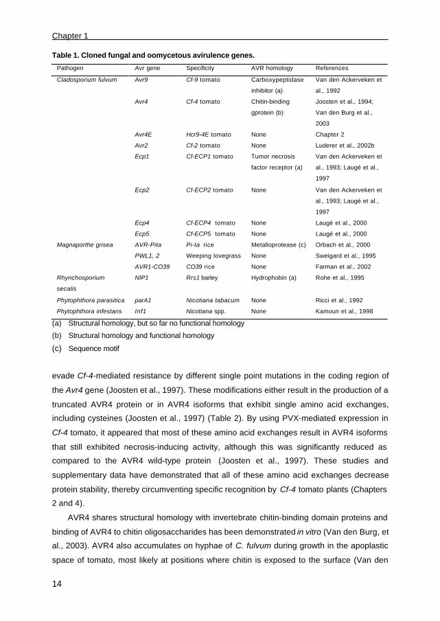
Table 1. Cloned fungal and oomycetous avirulence genes.
Pathogen Avr gene Specificity AVR homology References
Cladosporium fulvum Avr9 Cf-9 tomato Carboxypeptidase
inhibitor (a)
Van den Ackerveken et
al., 1992
Avr4 Cf-4 tomato Chitin-binding
gprotein (b)
Joosten et al., 1994;
Van den Burg et al.,
2003
Avr4E Hcr9-4E tomato None Chapter 2
Avr2 Cf-2 tomato None Luderer et al., 2002b
Ecp1 Cf-ECP1 tomato Tumor necrosis
factor receptor (a)
Van den Ackerveken et
al., 1993; Laugé et al.,
1997
Ecp2 Cf-ECP2 tomato None Van den Ackerveken et
al., 1993; Laugé et al.,
1997
Ecp4
Ecp5
Cf-ECP4 tomato
Cf-ECP5 tomato
None
None
Laugé et al., 2000
Laugé et al., 2000
Magnaporthe grisea AVR-Pita
PWL1, 2
AVR1-CO39
Pi-ta rice
Weeping lovegrass
CO39 rice
Metalloprotease (c)
None
None
Orbach et al., 2000
Sweigard et al., 1995
Farman et al., 2002
Rhynchosporium
secalis
NIP1 Rrs1 barley Hydrophobin (a) Rohe et al., 1995
Phytophthora parasitica parA1 Nicotiana tabacum None Ricci et al., 1992
Phytophthora infestans Inf1 Nicotiana spp. None Kamoun et al., 1998
(a) Structural homology, but so far no functional homology
(b) Structural homology and functional homology
(c) Sequence motif
evade Cf-4-mediated resistance by different single point mutations in the coding region of
the Avr4 gene (Joosten et al., 1997). These modifications either result in the production of a
truncated AVR4 protein or in AVR4 isoforms that exhibit single amino acid exchanges,
including cysteines (Joosten et al., 1997) (Table 2). By using PVX-mediated expression in
Cf-4 tomato, it appeared that most of these amino acid exchanges result in AVR4 isoforms
that still exhibited necrosis-inducing activity, although this was significantly reduced as
compared to the AVR4 wild-type protein (Joosten et al., 1997). These studies and
supplementary data have demonstrated that all of these amino acid exchanges decrease
protein stability, thereby circumventing specific recognition by Cf-4 tomato plants (Chapters
2 and 4).
AVR4 shares structural homology with invertebrate chitin-binding domain proteins and
binding of AVR4 to chitin oligosaccharides has been demonstrated in vitro (Van den Burg, et
al., 2003). AVR4 also accumulates on hyphae of C. fulvum during growth in the apoplastic
space of tomato, most likely at positions where chitin is exposed to the surface (Van den
General introduction
15
Burg et al., 2003). Furthermore, an AVR4-specific high-affinity binding site (HABS) of fungal
origin has been identified, which appeared to be heat- and proteinase K-resistant (Chapter
3). Although the latter suggests a non-proteinaceous character, AVR4 also crosslinks to a
fungus-derived molecule with a molecular mass of approximately 75 kDa (Chapter 3),
implying that AVR4 binds either with high affinity to a heat- and proteinase K-resistant
protein or with high affinity to polysaccharides and possibly with low affinity to another fungal
protein.
It appeared that, only in the presence of AVR4, the highly sensitive fungus Trichoderma
viride is protected against the antifungal activities of plant chitinases (Van den Burg et al.,
2003). The insensitivity of C. fulvum to plant chitinases as well as endoglucanases in vitro
(Joosten et al., 1995), however, does not depend on the production of AVR4 by the fungus,
suggesting that in this case other components protect the fungus against these hydrolases.
Although not measurable in vitro, AVR4 might still contribute to protect C. fulvum against cell
wall degradation during growth in planta.
Race-specific avirulence gene Avr4E of C. fulvum
Strains of C. fulvum that carry the Avr4E gene are avirulent on tomato plants carrying
Hcr9-4E (a homologue of Cladosporium resistance gene Cf-9), which is, in addition to Cf-4
(Hcr9-4D), the other functional Cf resistance gene present at the Cf-4 locus (Takken et al.,
1999). The Avr4E gene encodes a cysteine-rich protein of 101 amino acids that is secreted
into the extracellular space of tomato leaves (Chapter 2). Although the Cf-4 and Hcr9-4E
resistance genes share a high degree of overall sequence similarity (Parniske et al., 1997),
their matching Avr gene products do not share any sequence homology. Various strains of
C. fulvum have been identified that evade both Cf-4- and Hcr9-4E-mediated resistance. For
these strains, loss of the Avr4 avirulence function was caused by a variety of different single
point mutations in the Avr4 allele, as mentioned earlier (Joosten et al., 1997). Loss of the
Avr4E avirulence function appeared to be based on two different molecular mechanisms.
First, strains of C. fulvum were identified that carry an Avr4E allele with two point mutations,
resulting in amino acid changes Phe62Leu and Met73Thr (AVR4ELT) (Table 2) (Chapter 2). In
contrast to the AVR4 isoforms, this elicitor-inactive AVR4ELT protein is as stable as the wild-
type AVR4E protein. It appeared that single amino acid substitution Phe62Leu rather than
Met73Thr reduced the elicitor activity of AVR4E, suggesting that this single amino acid
replacement Phe62Leu can mediate circumvention of AVR4E recognition by Hcr9-4E plants
(Chapter 2). Single point mutations in Avr4E, however, which render elicitor-inactive AVR4EL
and elicitor-active AVR4ET, have not been identified in natural virulent and avirulent strains
Chapter 1
16
of C. fulvum , respectively. Although we cannot exclude a possible simultaneous evolutionary
event underlying the double amino acid substitution, strains of C. fulvum carrying Avr4ELT
most likely derived from yet unidentified avirulent strains carrying Avr4ET.
Surprisingly, all other strains virulent on Hcr9-4E-containing plants carry an Avr4E allele
that is identical to the Avr4E allele in avirulent strains. It appeared, however, that these
strains do not secrete the AVR4E protein upon colonization of the apoplastic space of
tomato (Chapter 2). Complementation of these virulent strains with a genomic Avr4E
sequence of an avirulent strain of C. fulvum conferred avirulence on Hcr9-4E plants,
suggesting that, in this case, abolished AVR4E expression results in circumvention of Hcr9-
4E-mediated resistance. Indeed, no Avr4E transcripts could be detected when northern blot
analysis was performed on RNA isolated from a compatible interaction between these
strains and tomato. Whether recombination events or (transposon) insertions within the
promoter sequence of Avr4E cause abolished AVR4E expression still needs to be
elucidated.
Race-specific avirulence gene Avr9 of C. fulvum
Avr9, which is the first fungal Avr gene that has been cloned and characterized,
encodes a precursor protein of 63 amino acids that contains a 23-amino acid signal
sequence (Van Kan et al., 1991). Upon secretion into the apoplast, AVR9 is further
processed at the N-terminus by fungal and plant proteases into a mature protein of 28 amino
acids, six of which are cysteines. The three-dimensional structure of AVR9, elucidated by1H-NMR, revealed that the protein contains three antiparallel â-strands that are
interconnected by three disulfide bridges (Van den Hooven et al., 2001). The AVR9 protein,
which contains a cystine knot, is structurally most related to potato carboxypeptidase
inhibitor (CPI) (Van den Hooven et al., 2001). AVR9, however, does not have amino acid
residues identical to those located at known CPI-inhibitory sites and thus far no protease-
inhibiting activity could be detected. Structural analysis revealed that all six cysteine
residues present in AVR9 are essential for its structure and necrosis-inducing activity
(Kooman-Gersmann et al., 1997; Van den Hooven et al., 2001). In addition, residue Phe21,
present in the solvent-exposed hydrophobic â-loop region, is also essential for the necrosis-
inducing activity of AVR9 (Kooman-Gersmann et al., 1997). Moreover, when applied to
transgenic Cf-9 tobacco cell suspensions, AVR9 mutant peptide carrying Phe21Ala is
incapable of inducing medium alkalization, whereas its capacity to induce an oxidative burst
was reduced (De Jong et al., 2000). Virulence of C. fulvum strains on Cf-9 plants appeared
to be the result of a deletion of the entire Avr9 gene (Table 2). Moreover, disruption of Avr9

General introduction
17
by homologous recombination in C. fulvum strains that are normally avirulent on Cf-9 plants
did not affect in vitro growth or virulence of the fungus on susceptible tomato plants,
suggesting that Avr9 is dispensable for full virulence (Marmeisse et al., 1993). Although
dispensable, the expression of Avr9 is induced under nitrogen-limiting conditions in vitro
(Van den Ackerveken et al., 1994; Pérez-García et al., 2001), which suggests that AVR9
might be involved in the nitrogen metabolism of the fungus. Pérez-García et al. (2001)
identified a gene in C. fulvum , designated Nrf1, which has a strong similarity to nitrogen
regulatory proteins of Aspergillus nidulans. Although Nrf1-deficient strains do not express
Avr9 under nitrogen starvation conditions in vitro, these strains are still avirulent on Cf-9
tomato plants, suggesting that NRF1 is a major, yet not the only, positive regulator of Avr9
expression (Pérez-García et al., 2001).
Non-race-specific extracellular protein (Ecp) genes of C. fulvum
Four genes encoding extracellular proteins ECP1, ECP2, ECP4, and ECP5 have been
cloned (Van den Ackerveken et al., 1993; Laugé et al., 2000). These Ecp genes all encode
cysteine-rich proteins that are abundantly secreted by all strains of C. fulvum during
colonization of tomato leaves. These proteins do neither share sequence homology with
each other nor with any sequences present in the database. Although the even number of
cysteine residues present in the ECPs suggests that these residues contribute to protein
stability, some (as demonstrated for ECP1 and ECP2) appear not to be involved in
intramolecular disulfide bonds (Luderer et al., 2002a). As found for Avr4 and Avr9,
transcription of both Ecp1 and Ecp2 is strongly induced in planta (Wubben et al., 1994),
indicating that plant-derived signals are required for the induction of both Avr and Ecp gene
expression. Tomato accessions that develop a HR upon inoculation with recombinant PVX
expressing Ecp2 have been identified (Laugé et al., 1998b). The responding accessions all
carry a single dominant gene, designated Cf-ECP2 gene, and show HR-associated
resistance toward ECP2-producing strains of C. fulvum (Laugé et al., 1998b). ECP1, ECP3,
ECP4, and ECP5 have also been shown to act as elicitors of HR on tomato accessions and
wild Lycopersicon plants that are resistant toward C. fulvum , most likely through recognition
of the corresponding secreted ECP (Laugé et al., 2000). Opposed to the Avr genes, no
modifications have thus far been found in the Ecp genes of naturally occurring strains of C.
fulvum . This might be due to a lack of selection pressure on the pathogen to overcome Cf-
ECP-mediated resistance, as the Cf-ECPs have not yet been introduced in commercial
cultivars. On the other hand, as all strains of C. fulvum analyzed so far secrete the ECPs,
disruption or modification of the encoding genes is thought to cause reduced virulence of the
Chapter 1
18
fungus on tomato. Indeed, the Ecp2 gene appears to be required for colonization and
sporulation of C. fulvum on mature tomato plants (Laugé et al., 1997). Moreover, Ecp1-
deficient strains fail to sporulate as abundantly as the wild-type strain on mature tomato
plants (Laugé et al., 1997). This implies that ECP1 and ECP2 are both required for full
virulence of C. fulvum on tomato. In addition, both Ecp1- and Ecp2-deficient strains induce
plant defense-associated responses more quickly and to higher levels than wild-type strains,
suggesting that both ECPs are involved in suppression of host defense-associated
responses during colonization (Laugé et al., 1997). Based on this observation, an interesting
parallel can be drawn with mammalian systems, in which viruses have been reported to
produce extracellular suppressors of host defense responses (Laugé et al., 1997).
Interestingly, ECP1 shares structural homology (based on the spacing of the cysteine
residues) with a viral T2 suppressor protein as well as with the family of tumor-necrosis
factor receptors (TNFRs) (Laugé et al., 1997). The T2 suppressor protein compromises the
establishment of host defense responses by interacting with mediators of the immune
system (tumor necrosis factors), thereby preventing its binding to endogenous TNFRs.
Furthermore, a putative receptor-like kinase has been identified in plants that shares
structural homology with the TNFR family. One possibility could be that ECP1 competitively
inhibits the binding of defense signaling molecules to this plant receptor protein, thereby
suppressing the induction of host defense responses.
The Avr genes of Magnaporthe grisea
The filamentous ascomycete Magnaporthe grisea is the causal agent of blast disease
on many species of the grass family, such as rice. M. grisea initiates infection by a
germinating conidium that quickly differentiates into a specialized cell, the appressorium.
Once mature, the melanized appressorium generates enormous hydrostatic pressure that
forces a narrow penetration peg through the plant cuticle and epidermal cell wall. After
penetration, the fungus grows intracellularly and produces sporulating lesions within five to
seven days.
Genotype-specific Avr genes of M. grisea
Strains of M. grisea that carry the Avr-Pita (AVR2-YAMO) gene are avirulent on rice
cultivars that carry the corresponding R gene Pi-ta (Orbach et al., 2000). The Avr-Pita gene
is located very close to the telomere of chromosome 3 and encodes a predictedpolypeptide
of 223 amino acids. AVR-Pita exhibits substantial similarity to NPII, a neutral zinc

General introduction
19
Table 2. Overview of mutations identified in the open reading frames of Avr2, Avr4, Avr4E, and
Avr9 of Cladosporium fulvum strains that are virulent on tomato plants that carry Cf-2, Cf-4,
Hcr9-4E or Cf-9, respectively.
Mutations in Avr2 Codon position in
ORF
Mutation in AVR2 Predicted protein*
Wild-type - No mutation
C to T (stop) 66 Frame shift/stop
�T 72 Frame shift
�C 24 Frame shift/stop
�A 23 Frame shift/stop
+A 40 Frame shift/stop
+A 23 Frame shift/stop
+ Transposon 19 Insertion of 5 kB
Mutations in Avr4 Codon position in
ORF
Mutation in AVR4 Predicted protein
Wild-type - No mutation
�C 42 Frameshift
G to T 64 Cys-64-Tyr
C to T 66 Thr-66-Ile
T to C 67 Tyr-67-His
G to T 70 Cys-70-Tyr
G to T 109 Cys-109-Tyr
Mutations in Avr4E Codon position in
ORF
Mutation in
AVR4E
Predicted protein
Wild-type - No mutation
T to C; T to C 82 and 93 Phe-82-Leu; Met-
93-Thr
Mutation in Avr9 Predicted Protein
Wild-type - No mutation
Deletion of ORF No protein No protein
*) The speckled areas in the horizontal bars represent the signal peptide of the AVR proteins; open
areas represent the mature part of the AVR protein; hatched areas represent the amino acid sequence
encoded that follows the frameshift mutation in Avr2 and Avr4. The cysteine residues are indicated as
vertical lines and amino acid substitutions as dotted vertical lines. The black areas represent the amino
acid sequence that is removed by N- and C-terminal processing.
Chapter 1
20
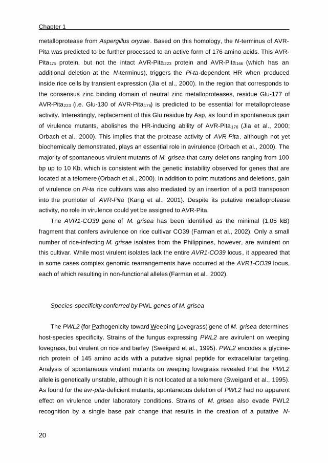
metalloprotease from Aspergillus oryzae. Based on this homology, the N-terminus of AVR-
Pita was predicted to be further processed to an active form of 176 amino acids. This AVR-
Pita176 protein, but not the intact AVR-Pita223 protein and AVR-Pita166 (which has an
additional deletion at the N-terminus), triggers the Pi-ta-dependent HR when produced
inside rice cells by transient expression (Jia et al., 2000). In the region that corresponds to
the consensus zinc binding domain of neutral zinc metalloproteases, residue Glu-177 of
AVR-Pita223 (i.e. Glu-130 of AVR-Pita176) is predicted to be essential for metalloprotease
activity. Interestingly, replacement of this Glu residue by Asp, as found in spontaneous gain
of virulence mutants, abolishes the HR-inducing ability of AVR-Pita176 (Jia et al., 2000;
Orbach et al., 2000). This implies that the protease activity of AVR-Pita, although not yet
biochemically demonstrated, plays an essential role in avirulence (Orbach et al., 2000). The
majority of spontaneous virulent mutants of M. grisea that carry deletions ranging from 100
bp up to 10 Kb, which is consistent with the genetic instability observed for genes that are
located at a telomere (Orbach et al., 2000). In addition to point mutations and deletions, gain
of virulence on Pi-ta rice cultivars was also mediated by an insertion of a pot3 transposon
into the promoter of AVR-Pita (Kang et al., 2001). Despite its putative metalloprotease
activity, no role in virulence could yet be assigned to AVR-Pita.
The AVR1-CO39 gene of M. grisea has been identified as the minimal (1.05 kB)
fragment that confers avirulence on rice cultivar CO39 (Farman et al., 2002). Only a small
number of rice-infecting M. grisae isolates from the Philippines, however, are avirulent on
this cultivar. While most virulent isolates lack the entire AVR1-CO39 locus , it appeared that
in some cases complex genomic rearrangements have occurred at the AVR1-CO39 locus,
each of which resulting in non-functional alleles (Farman et al., 2002).
Species-specificity conferred by PWL genes of M. grisea
The PWL2 (for Pathogenicity toward Weeping Lovegrass) gene of M. grisea determines
host-species specificity. Strains of the fungus expressing PWL2 are avirulent on weeping
lovegrass, but virulent on rice and barley (Sweigard et al., 1995). PWL2 encodes a glycine-
rich protein of 145 amino acids with a putative signal peptide for extracellular targeting.
Analysis of spontaneous virulent mutants on weeping lovegrass revealed that the PWL2
allele is genetically unstable, although it is not located at a telomere (Sweigard et al., 1995).
As found for the avr-pita-deficient mutants, spontaneous deletion of PWL2 had no apparent
effect on virulence under laboratory conditions. Strains of M. grisea also evade PWL2
recognition by a single base pair change that results in the creation of a putative N-

General introduction
21
glycosylation site. This PWL2 mutant protein exhibits reduced elicitor-activity either due to
glycosylation or due to the amino acid change itself.
The PWL2 gene is a member of a rapidly evolving gene family of which the homologue
PWL1 and the allelic PWL3/PWL4 genes map at different chromosomal locations (Kang et
al., 1995). The PWL2 protein is 75 percent identical to the PWL1 protein, and 51- and 57
percent identical to the PWL3 and PWL4 proteins, respectively. Opposed to PWL1 and
PWL2, the PWL3 and PWL4 genes are non-functional Avr genes, as they do not confer
avirulence on weeping lovegrass. In contrast to PWL3, PWL4 becomes functional in
preventing infection of weeping lovegrass when its expression is driven by either the PWL1
or the PWL2 promoter (Kang et al., 1995). This indicates that PWL4 encodes a functional
AVR protein, which is not recognized by weeping lovegrass due to lack of expression of the
gene.
The Avr genes of Rhynchosporium secalis
The fungus Rhynchosporium secalis is known as the causal agent of leaf scald on
barley, rye and other grasses. R. secalis initiates infection by penetrating the cuticle,
followed by extracellular growth of hyphae between the cuticle and the outer epidermal cell
walls. The fungus develops an extensive subcuticular stroma, causes an early collapse of a
few epidermal cells and the underlying mesophyll cells, and finally starts to sporulate.
Amongst the secreted proteins in culture filtrates of R. secalis, a class of necrosis-inducing
proteins, NIPs, has been identified that induce necrosis in certain barley cultivars and other
cereals (Wevelsiep et al., 1993). The phytotoxicity of these NIPs, which is associated with
lesion-development, appeared to be based on their stimulatory effect on the plant
plasmalemma H+-ATPase, probably in order to release plant nutrients (Wevelsiep et al.,
1993).
Strains of R. secalis that secrete NIP1 are unable to grow on barley cultivars that carry
the Rrs1 gene. Rrs1-mediated resistance is not associated with a rapid HR, but with the
accumulation of mRNAs encoding peroxidase and PR proteins of the PR-5 class (Rohe et
al., 1995). The NIP1 gene encodes a secreted elicitor-active protein of 60 amino acids, 10 of
which are cysteine residues (Rohe et al., 1995). The three-dimensional structure of NIP1
revealed that all 10 cysteines form intramolecular disulfide bonds, providing stability to the
protein (Van ‘t Slot, unpublished data). The spacing pattern of the first eight cysteine
residues in NIP1 (-C-CC-C-C-CC-C-) has also been found in another class of fungal
proteins, the hydrophobins. The partially resolved disulfide bond pattern of the Ophiostoma
ulmi hydrophobin, however, differs from that of NIP1, suggesting that NIP1 is not functionally
Chapter 1
22
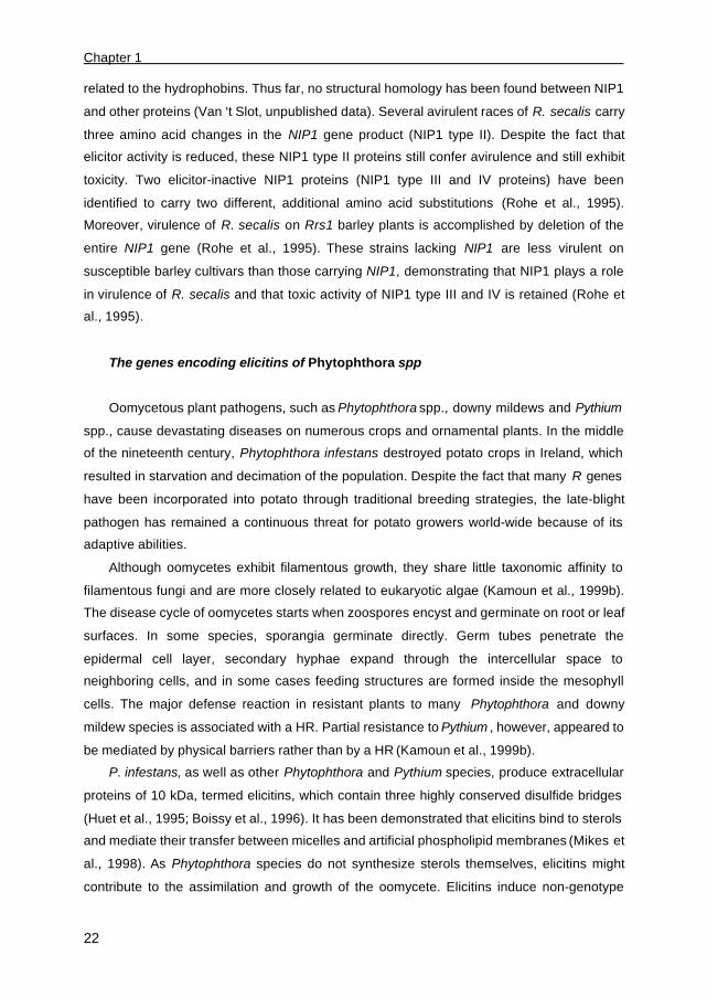
related to the hydrophobins. Thus far, no structural homology has been found between NIP1
and other proteins (Van ‘t Slot, unpublished data). Several avirulent races of R. secalis carry
three amino acid changes in the NIP1 gene product (NIP1 type II). Despite the fact that
elicitor activity is reduced, these NIP1 type II proteins still confer avirulence and still exhibit
toxicity. Two elicitor-inactive NIP1 proteins (NIP1 type III and IV proteins) have been
identified to carry two different, additional amino acid substitutions (Rohe et al., 1995).
Moreover, virulence of R. secalis on Rrs1 barley plants is accomplished by deletion of the
entire NIP1 gene (Rohe et al., 1995). These strains lacking NIP1 are less virulent on
susceptible barley cultivars than those carrying NIP1, demonstrating that NIP1 plays a role
in virulence of R. secalis and that toxic activity of NIP1 type III and IV is retained (Rohe et
al., 1995).
The genes encoding elicitins of Phytophthora spp
Oomycetous plant pathogens, such as Phytophthora spp., downy mildews and Pythium
spp., cause devastating diseases on numerous crops and ornamental plants. In the middle
of the nineteenth century, Phytophthora infestans destroyed potato crops in Ireland, which
resulted in starvation and decimation of the population. Despite the fact that many R genes
have been incorporated into potato through traditional breeding strategies, the late-blight
pathogen has remained a continuous threat for potato growers world-wide because of its
adaptive abilities.
Although oomycetes exhibit filamentous growth, they share little taxonomic affinity to
filamentous fungi and are more closely related to eukaryotic algae (Kamoun et al., 1999b).
The disease cycle of oomycetes starts when zoospores encyst and germinate on root or leaf
surfaces. In some species, sporangia germinate directly. Germ tubes penetrate the
epidermal cell layer, secondary hyphae expand through the intercellular space to
neighboring cells, and in some cases feeding structures are formed inside the mesophyll
cells. The major defense reaction in resistant plants to many Phytophthora and downy
mildew species is associated with a HR. Partial resistance to Pythium , however, appeared to
be mediated by physical barriers rather than by a HR (Kamoun et al., 1999b).
P. infestans, as well as other Phytophthora and Pythium species, produce extracellular
proteins of 10 kDa, termed elicitins, which contain three highly conserved disulfide bridges
(Huet et al., 1995; Boissy et al., 1996). It has been demonstrated that elicitins bind to sterols
and mediate their transfer between micelles and artificial phospholipid membranes (Mikes et
al., 1998). As Phytophthora species do not synthesize sterols themselves, elicitins might
contribute to the assimilation and growth of the oomycete. Elicitins induce non-genotype

General introduction
23
specific defense-associated responses, including a HR, in plants of the genus Nicotiana (i.e.
Solanaceae) and in some cultivars of radish, turnip, and rape (i.e. Cruciferae).
Species-specific gene parA1 of Phytophthora parasitica
In Phytophthora parasitica , the absence of elicitin production correlated with high
virulence on tobacco. Although elicitins are encoded by a multigene family, it appeared that
parA1 is the main elicitin-encoding gene expressed in vitro and in planta by P. parasitica.
The parA1 gene was cloned from Phytophthora parasitica and encodes a secreted protein
parasiticein of 98 amino acids, of which 6 are cysteine residues (Ricci et al., 1992). The
parA1 gene has been proposed to act as a species-specific Avr gene, as it triggers HR-
mediated resistance toward P. parasitica in Nicotiana tabacum (tobacco). Elicitin-producing
P. parasitica isolates have been demonstrated to cause disease on tobacco, but not on
tomato, upon down-regulation of parA1 expression (Colas et al., 2001). It appeared that this
down-regulation event relies on a mechanism that is dependent on the P. parasitica
genotype rather than the host plant (Colas et al., 2001).
Genotype-specific gene inf1 of Phytophthora infestans
The inf1 gene of P. infestans encoding infestin was cloned by screening a cDNA library
of a compatible interaction between P. infestans and potato with a parA1 gene fragment
(Kamoun et al., 1997). While high levels of inf1 transcripts are observed in mycelium grown
in vitro, the expression of inf1 is down-regulated in planta (Kamoun et al., 1997). Opposed to
parA1, down-regulation of inf1 expression is not required to evade plant defense responses,
as the host plant potato does not respond to INF1 (Kamoun et al., 1997). In leaves of non-
host Nicotiana plants, however, injection of INF1 induces a specific HR. To determine
whether INF1 plays a role in non-host resistance, members of the Nicotiana family were
inoculated with inf1-deficient P. infestans strains. These inf1-deficient strains are able to
cause disease when inoculated on leaves of N. benthamiana, whereas on other Nicotiana
species, such as N. tabacum (tobacco), however, these inf1-deficient strains are still
avirulent (Kamoun et al., 1998). This demonstrates that INF1 confers non-host resistance
toward P. infestans in N. benthamiana, but is not the main determinant of non-host
resistance to P. infestans in other Nicotiana species. Putative additional candidates that
confer avirulence on tobacco are the products of the inf2A and inf2B genes of P. infestans,
which also induce a HR when injected into N. tabacum leaves (Kamoun et al., 1998).

Chapter 1
24
PATHOGENS HAVE EVOLVED MECHANISMS TO COUNTERACT PLANT DEFENSE
As describe above, plant pathogens evade R gene-mediated resistance by modification
of the elicitor proteins either by mutations in, or deletion of, the Avr genes or by (down)-
regulation of Avr gene expression. Yet, when the circumvention of elicitor detection fails, or
when the elicitor component is essential for virulence, pathogens require mechanisms to
subvert the induced plant defense responses. Indeed, plant pathogens counteract plant
defenses by secreting enzymes that detoxify defense compounds, including phytoalexins, or
use ATP-binding cassette (ABC)-transporters to mediate the efflux of toxic compounds (as
reviewed by Idnurm and Howlett, 2001). Moreover, some bacterial pathogens interfere with
R gene-mediated resistance by secreting proteins that “mask” the presence of a particular
AVR effector protein (Ritter and Dangl, 1996).
Recent studies revealed that some pathogenic fungi have evolved counter-defense
mechanisms that enable the suppression of plant defense responses. This mechanism
involves a class of proteins, termed glucanase inhibitor proteins (GIPs), which are secreted
by Phytophthora sojae f. sp. glycines and which inhibit the endoglucanase (EnGL) activity of
its host soybean (Rose et al., 2002). Sequences homologous to GIPs have been identified in
genomic DNA of other Phytophthora species, while several other plant pathogenic fungi do
not exhibit related sequences (Rose et al., 2002). GIPs are homologous to the trypsin class
of serine proteases, but do not exhibit proteolytic activity. The basis of endoglucanase
inhibition by GIPs involves the formation of a stable complex. This association appeared to
be specific, as GIP1 inhibits soybean endoglucanase-A (EnGL-A) but not EnGL-B. GIPs
also suppress the release of elicitor-active oligoglucosides from P. sojae mycelial walls in
vitro and during pathogenesis in vivo (Rose et al., 2002). Thus, GIPs suppress cell wall
disassembly by inhibiting endoglucanases, and suppress the activation of defense-
associated responses by inhibiting the release of oligoglucoside elicitors (Rose et al., 2002).
The CgDN3 gene of the hemi-biotrophic pathogen Colletotrichum gloeosporioides,
which encodes an extracellular protein of 54 amino acids, is induced under nitrogen
starvation conditions at the early stage of infection on tropical pasture legume (Stephenson
et al., 2000). On intact leaves of a normally compatible host, CgDN3-disrupted mutants were
nonpathogenic and elicited a hypersensitive-like response, the latter of which may be part of
a basal defense reaction toward C. gloeosporioides (Stephenson et al., 2000). When applied
to wound sites, however, these mutants were able to grow necrotropically on host leaves
and form necrotic spreading lesions. It was therefore suggested that at the early stages of
the primary infection process, CgDN3 is required for pathogenicity and functions to suppress
the hypersensitive-like response (Stephenson et al., 2000). Opposed to the C. fulvum ECP1
General introduction
25
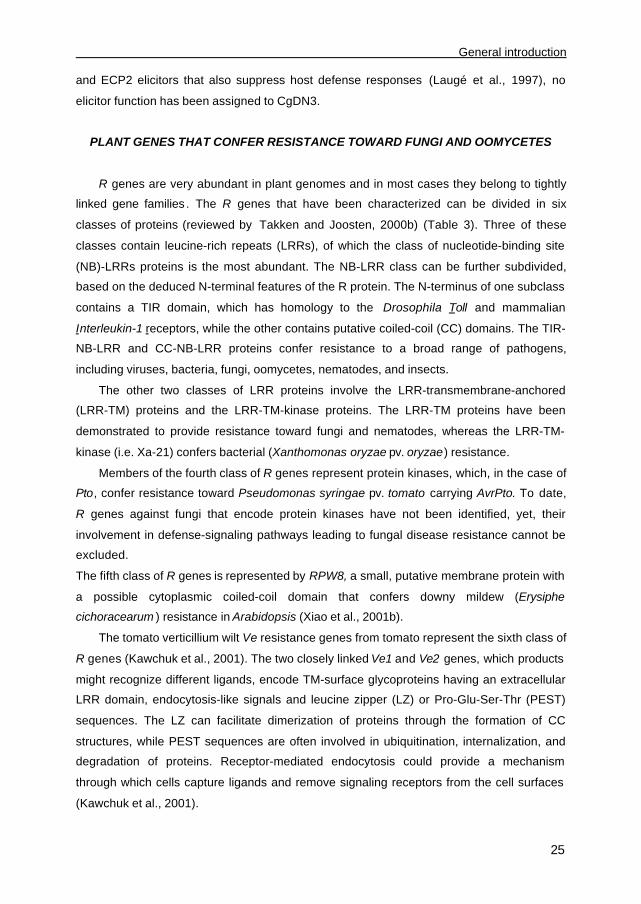
and ECP2 elicitors that also suppress host defense responses (Laugé et al., 1997), no
elicitor function has been assigned to CgDN3.
PLANT GENES THAT CONFER RESISTANCE TOWARD FUNGI AND OOMYCETES
R genes are very abundant in plant genomes and in most cases they belong to tightly
linked gene families . The R genes that have been characterized can be divided in six
classes of proteins (reviewed by Takken and Joosten, 2000b) (Table 3). Three of these
classes contain leucine-rich repeats (LRRs), of which the class of nucleotide-binding site
(NB)-LRRs proteins is the most abundant. The NB-LRR class can be further subdivided,
based on the deduced N-terminal features of the R protein. The N-terminus of one subclass
contains a TIR domain, which has homology to the Drosophila Toll and mammalian
Interleukin-1 receptors, while the other contains putative coiled-coil (CC) domains. The TIR-
NB-LRR and CC-NB-LRR proteins confer resistance to a broad range of pathogens,
including viruses, bacteria, fungi, oomycetes, nematodes, and insects.
The other two classes of LRR proteins involve the LRR-transmembrane-anchored
(LRR-TM) proteins and the LRR-TM-kinase proteins. The LRR-TM proteins have been
demonstrated to provide resistance toward fungi and nematodes, whereas the LRR-TM-
kinase (i.e. Xa-21) confers bacterial (Xanthomonas oryzae pv. oryzae) resistance.
Members of the fourth class of R genes represent protein kinases, which, in the case of
Pto, confer resistance toward Pseudomonas syringae pv. tomato carrying AvrPto. To date,
R genes against fungi that encode protein kinases have not been identified, yet, their
involvement in defense-signaling pathways leading to fungal disease resistance cannot be
excluded.
The fifth class of R genes is represented by RPW8, a small, putative membrane protein with
a possible cytoplasmic coiled-coil domain that confers downy mildew (Erysiphe
cichoracearum ) resistance in Arabidopsis (Xiao et al., 2001b).
The tomato verticillium wilt Ve resistance genes from tomato represent the sixth class of
R genes (Kawchuk et al., 2001). The two closely linked Ve1 and Ve2 genes, which products
might recognize different ligands, encode TM-surface glycoproteins having an extracellular
LRR domain, endocytosis-like signals and leucine zipper (LZ) or Pro-Glu-Ser-Thr (PEST)
sequences. The LZ can facilitate dimerization of proteins through the formation of CC
structures, while PEST sequences are often involved in ubiquitination, internalization, and
degradation of proteins. Receptor-mediated endocytosis could provide a mechanism
through which cells capture ligands and remove signaling receptors from the cell surfaces
(Kawchuk et al., 2001).

Chapter 1
26
Table 3. R proteins identified in gene-for-gene interactions conferring resistance toward fungi
and oomycetes, classified based on homologous structural domains. TIR, Toll/interleukin 1
receptor-like domain; NB, nucleotide binding site; LRR/LRD, leucine-rich repeat/domain; CC, coiled-coil;
TM, transmembrane domain; PEST, Pro-Glu-Ser-Thr sequences.
R gene Pathosystem Structure Matching
Avr gene
References
L6, M, N, P Flax/ Melampsora lini TIR-NB-LRR Unknown Islam and Mayo, 1990
RPP1, 10,
14
Arabidopsis/ Peronospora parasitica TIR-NB-LRR Unknown Botella et al., 1998
RPP4, 5 Arabidopsis/ Peronospora parasitica TIR-NB-LRR Unknown Van der Biezen et al.,
2002
RPP8 Arabidopsis/ Peronospora parasitica CC-NB-LRR Unknown McDowell et al., 1998
RPP13 Arabidopsis/ Peronospora parasitica CC-NB-LRR Unknown Bittner-Eddy et al., 2000
Mla Barley/ Erysiphe graminis f.sp. hordei CC-NB-LRR Unknown Halterman et al., 2001
R1 Potato/ Phytophthora infestans CC-NB-LRR Unknown Ballvora et al., 2002
Dm3 Lettuce/ Bremia lactucae NB-LRR Unknown Anderson et al., 1996
I2 Tomato/ Fusarium oxysporum f.sp.
lycopersici
NB-LRR Unknown Ori et al., 1997
Rp1 Maize/ Puccinia sorghi NB-LRR Unknown Richter et al., 1995
Pi-ta Rice/ Magnaporthe grisea NB-LRD Avr-Pita Bryan et al., 2000
Cf-2 Tomato/ Cladosporium fulvum LRR-TM Avr2 Dixon et al., 1996
Cf-4 Tomato/ Cladosporium fulvum LRR-TM Avr4 Thomas et al., 1997
Hcr9-4E Tomato/ Cladosporium fulvum LRR-TM Avr4E Takken et al., 1999
Cf-5 Tomato/ Cladosporium fulvum LRR-TM Unknown Dixon et al., 1998
Cf-9 Tomato/ Cladosporium fulvum LRR-TM Avr9 Jones et al., 1994
Rpw8 Arabidopsis/ Erysiphe cichoracearum CC-TM Unknown Xiao et al., 2001b
Ve1, Ve2 Tomato/ Verticillium dahliae CC-LRR-TM-
PEST
Unknown Kawchuk et al., 2001
The NB-LRR class of R proteins
In flax, NB-LRR proteins have been identified that mediate recognition of 31 different
rust (Melampsora lini) strains. These different resistance specificities are distributed among
five polymorphic loci, K, L, M, N, and P (Islam and Mayo, 1990). Comparative sequence
analysis of the R proteins encoded by the flax L alleles has demonstrated that both LRR and
TIR domains play a role in determining resistance specificities (Luck et al., 2000). Flax R
genes confer only resistance to those strains of M. lini that carry the corresponding Avr
gene, however, none of the 31 genetically defined Avr genes has been cloned yet.
In Arabidopsis, several R gene loci have been identified that confer resistance toward
strains of the oomycetous pathogen Peronospora parasitica. These RPP loci comprise
genes that encode TIR-NB-LRR proteins (RPP1, 4, 5, 10, and 14) and CC-NB-LRR proteins

General introduction
27
(RPP8 and RPP13) (Botella et al., 1998; Van der Biezen et al., 2002; Bittner-Eddy et al.,
2000; McDowell et al. 1998). The RPP4 gene of the Arabidopsis landrace Columbia (Col) is
an orthologue of the RPP5 gene of Landsberg erecta (Ler). RPP4 confers resistance to P.
parasitica races Emoy2 and Emwa1, while RPP5 confers resistance toward these races as
well as toward P. parasitica race Noco2 (Van der Biezen et al., 2002). These strains of P.
parasitica might carry two distinct, yet not identified, Avr determinants, i.e. AvrRPP4 and
AvrRPP5 that are recognized by RPP4 and RPP5, respectively. RPP4 and RPP5, on the
other hand, could also have overlapping specificities, whereby AvrRPP4, but not AvrRPP5,
is recognized by both RPP4 and RPP5.
Other R genes that belong to the NB-LRR class, which are also members of complex
resistance loci, confer multiple resistance specificities toward fungal pathogens as well. In
barley, the Mla locus confers resistance against various races of powdery mildew (Erysiphe
graminis f. sp. hordei) on a gene-for-gene basis (Halterman et al., 2001). Moreover, the Rp1
rust (Puccinia sorghi) locus in maize and the Dm3 downy mildew (Bremia lactucae) locus in
lettuce all contain multiple genetically linked resistance specificities (Richter et al., 1995;
Anderson et al., 1996). For all of these gene-for-gene interactions no matching Avr genes
have thus far been characterized.
The rice blast gene Pi-ta confers gene-for-gene-based resistance against strains of M.
grisea that express AVR-Pita. Pi-ta encodes a putative cytoplasmic receptor and is a
member of the NB-receptor class of R genes. Pi-ta lacks an N-terminal TIR or CC domain
and the C-terminal leucine-rich domain (LRD) lacks the characteristic LRR motif found in
other proteins of the NB-LRR class. The predicted protein encoded by Pi-ta present in
resistant rice varieties differs by only one amino acid, located at position 918, from the
protein encoded by susceptible rice varieties (Bryan et al., 2000). Moreover, it appeared that
Pi-ta protein with alanine-918, but not Pi-ta with serine-918, interacts with AVR-Pita in the
cytoplasm of rice cells to induce resistance responses.
The TM-LRR class of R proteins
Another class of R genes includes the Cf genes of tomato, which confer resistance to
strains of C. fulvum carrying the corresponding Avr gene (Table 3). The Cf genes encode
proteins with a predicted signal peptide for extracellular targeting, a LRR region, a TM
domain and a short cytoplasmic tail (Jones and Jones, 1996; Joosten and De Wit, 1999).
The Cf genes are members of multigene families and have been designated Hcr2 or Hcr9
(for homologues of Cladosporium resistance genes Cf-2 and Cf-9, respectively). Two nearly
identical Cf-2 genes (Cf-2.1 and Cf-2.2) and Cf-5 (Hcr2-5C) confer resistance toward C.

Chapter 1
28
fulvum strains through recognition of the Avr2 and Avr5 gene products, respectively (Dixon
et al., 1996 and 1998). The Cf-4 and Cf-9 gene clusters each consist of five Hcr9
homologues, of which Hcr9-4D (i.e. Cf-4), Hcr9-4E and Hcr9-9C (i.e. Cf-9) gene products
specifically recognize AVR4, AVR4E and AVR9, respectively (Thomas et al., 1997; Jones et
al., 1994; Takken et al., 1999).
PERCEPTION OF AVR GENE PRODUCTS BY RESISTANT PLANT GENOTYPES
In the absence of the corresponding R gene, many Avr genes have been demonstrated
to provide a selective advantage to the pathogen (Laugé and De Wit, 1998c; White et al.,
2000; Staskawicz et al., 2001; Bonas and Lahaye, 2002). This, together with the
maintenance of Avr genes within pathogen populations, implies that the primary function of
Avr genes is to confer virulence to the pathogen. Although for most Avr gene products no
biological function has yet been defined, it appears that in colonized plant tissue AVR
proteins co-localize with various host virulence targets. Together with the fact that proper
subcellular targeting is essential for the avirulence activity of AVRs, this implies that AVR
proteins, virulence targets and R proteins are possibly part of one complex (Van der Hoorn
et al., 2002). Four models have been proposed addressing the question how and where
AVR proteins are recognized by resistant plant genotypes (Fig. 1). These models will be
discussed in detail in the following section.
Direct perception of AVR proteins
This model reflects the most simple interpretation of Flor’s gene-for-gene hypothesis; a
classical receptor-ligand model that predicts a direct interaction between Avr and R gene
products (Fig. 1). Thus far, direct physical interaction has only been demonstrated for Pto
from tomato and AvrPto from Pseudomonas syringae (Tang et al., 1996), and for Pi-ta from
rice and AVR-Pita from M. grisae (Jia et al., 2000). The cytoplasmic localization of both Pto
and Pi-ta is consistent with the observation that AvrPto and Avr-Pita induce a HR when
expressed inside plant cells (Scofield et al., 1996; Jia et al., 2000). Pto is a Ser/Thr kinase
that interacts with and phosphorylates a second Ser/Thr kinase, Pti1, and several defense-
related transcription factors, such as Pti4, Pti5, and Pti6 (Martin et al., 1993; Xiao et al.,
2001a). The most straightforward explanation for induction of plant defense responses
would be that binding of AvrPto to Pto leads to a conformational change of the kinase
protein that in turn triggers downstream defense signaling pathways. Pto function, however,
requires Prf, a NB-LRR-encoding gene (Salmeron et al., 1996). Therefore, the “co-receptor”

General introduction
29
and/or the “guard” model (see below) was put forward to rationalize the mechanism of
AvrPto-induced defense activation (Fig. 1). AVR-Pita recognition, on the other hand, is
mediated by direct interaction with the LRD of Pi-ta (Bryan et al., 2000; Jia et al., 2000). It
appeared that the putative metalloprotease activity of AVR-Pita is required for its direct
interaction with Pi-ta (Jia et al., 2000), suggesting a protease-dependent defense elicitation
model (Fig. 1). Possibly, Pi-ta contains protease cleavage sites, which upon proteolytic
processing renders an active form either by a conformational change or by the release of
elicitor peptide(s) that trigger(s) defense responses.
Indirect perception of AVR proteins
In addition to direct perception of AVR proteins by R proteins, three models have been
proposed that postulate an indirect interaction between Avr and R gene products to take
place. These models include enzymatic- or protease-dependent defense elicitation, or the
involvement of a “co-receptor” or a “guard” protein in triggering defense responses.
Protease-dependent defense elicitation model
Rcr3, a gene required for Cf-2-mediated resistance toward C. fulvum strains carrying
Avr2, has recently been cloned and encodes a tomato cysteine endoprotease (Krüger et al.,
2002). The fact that Rcr3 is secreted into the apoplastic space is consistent with the
extracellular localization of AVR2 and Cf-2. Several protease-dependent mechanisms
underlying the Cf-2-mediated resistance can be envisaged, whereby Rcr3 most likely
functions upstream of Cf-2. Rcr3 might process AVR2 to generate a mature ligand, or Rcr3
might degrade AVR2, thereby releasing active elicitor peptides that interact with the
extracellular LRR of Cf-2. This would imply that not AVR2 itself but rather a protease-
dependent signal triggers Cf-2-mediated resistance. Another possibility is that AVR2 forms a
complex with Rcr3, which subsequently triggers Cf-2-dependent downstream signaling
pathways that lead to disease resistance. Rcr3, on the other hand, might also be part of a
basal defense mechanism of the plant that upon inhibition by AVR2 turns on Cf-2 mediated
defense responses (see below; “guard hypothesis”).
The “co-receptor” model
Most R genes encode proteins that carry LRR domains. These LRRs, located either
intracellular (in the case of most NB-LRR proteins) or extracellular (in the case of LRR-TM
Chapter 1
30
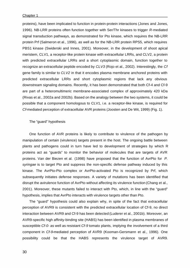
proteins), have been implicated to function in protein-protein interactions (Jones and Jones,
1996). NB-LRR proteins often function together with Ser/Thr kinases to trigger R-mediated
signal transduction pathways, as demonstrated for Pto kinase, which requires the NB-LRR
protein Prf (Salmeron et al., 1996), as well as for the NB-LRR protein RPS5, which requires
PBS1 kinase (Swiderski and Innes, 2001). Moreover, in the development of shoot apical
meristem, CLV1, a receptor-like protein kinase with extracellular LRRs, and CLV2, a protein
with predicted extracellular LRRs and a short cytoplasmic domain, function together to
recognize an extracellular peptide encoded by CLV3 (Rojo et al., 2002). Interestingly, the Cf
gene family is similar to CLV2 in that it encodes plasma membrane anchored proteins with
predicted extracellular LRRs and short cytoplasmic regions that lack any obvious
downstream signaling domains. Recently, it has been demonstrated that both Cf-4 and Cf-9
are part of a heteromultimeric membrane-associated complex of approximately 420 kDa
(Rivas et al., 2002a and 2002b). Based on the analogy between the two systems, it could be
possible that a component homologous to CLV1, i.e. a receptor-like kinase, is required for
Cf-mediated perception of extracellular AVR proteins (Joosten and De Wit, 1999) (Fig. 1).
The “guard” hypothesis
One function of AVR proteins is likely to contribute to virulence of the pathogen by
manipulation of certain (virulence) targets present in the host. The ongoing battle between
plants and pathogens could in turn have led to development of strategies by which R
proteins act as “guards” to monitor the behavior of molecules that are targets of AVR
proteins. Van der Biezen et al. (1998) have proposed that the function of AvrPto for P.
syringae is to target Pto and suppress the non-specific defense pathway induced by this
kinase. The AvrPto-Pto complex or AvrPto-activated Pto is recognized by Prf, which
subsequently initiates defense responses. A variety of mutations has been identified that
disrupt the avirulence function of AvrPto without affecting its virulence function (Chang et al.,
2001). Moreover, these mutants failed to interact with Pto, which, in line with the “guard”
hypothesis, implies that AvrPto interacts with virulence targets other than Pto.
The “guard” hypothesis could also explain why, in spite of the fact that extracellular
perception of AVR9 is consistent with the predicted extracellular location of Cf-9, no direct
interaction between AVR9 and Cf-9 has been detected (Luderer et al., 2001b). Moreover, an
AVR9-specific high affinity-binding site (HABS) has been identified in plasma membranes of
susceptible Cf-0- as well as resistant Cf-9 tomato plants, implying the involvement of a third
component in Cf-9-mediated perception of AVR9 (Kooman-Gersmann et al., 1996). One
possibility could be that the HABS represents the virulence target of AVR9.

General introduction
31
Fig. 1. Schematic representation of gene-for-gene interactions at the protein level. Four models
have been proposed that describe either direct (I) or indirect (II-IV) perception of avirulence (AVR)
proteins by plant resistance (R) proteins. (I) This classical model predicts a direct interaction between
an AVR protein (A) and a matching R protein. Defense responses are induced independently or
dependently, as illustrated by scissors, of the proteolytic activity of the AVR protein. (II) Binding of AVR
protein to host-encoded proteins might involve the generation of protease-dependent elicitor peptide(s)
or complex(es). The AVR protein, on the other hand, might also trigger R-gene mediated resistance by
suppressing the generation of proteolytically processed negative regulators of defense responses. (III)
The AVR protein binds to, at least, an additional component (C), which subsequently interacts with the
R protein to trigger defense responses. The interaction between R protein and “co-receptor” might either
be required for receptor activation after AVR binding or for recruitment of a functional receptor complex
that mediates AVR recognition. (IV) According to the “guard” hypothesis, the R protein safeguards the
virulence target (V) of the AVR protein. For further details, see text. (This figure has been modified from
Bonas et al. 2002).

Chapter 1
32
Cf-9 may “guard” this virulence target, sense its modification by AVR9 and trigger
downstream defense responses leading to resistance (Van der Hoorn et al., 2002). Recent
studies with tobacco cell suspensions revealed that Cf-9-mediated defense responses are
attenuated at temperatures higher than 20°C and are completely suppressed at 33°C (De
Jong et al., 2002). Interestingly, a correlation was found between the temperature-sensitivity
of this response and the amount of AVR9-HABSs, suggesting that the HABS is unstable at
elevated temperatures (De Jong et al., 2002).
The Cf-4-mediated defense responses are also temperature-sensitive, yet, opposed to
AVR9, thus far no plant-derived AVR4-specific HABS has been detected (Chapter 3). As
AVR4 exhibits chitin-binding activity, an interaction with chitin oligosaccharides might be
required for AVR4 recognition by Cf-4 plants. However, Cf-4-mediated defense responses
are also triggered by AVR4 in the absence of chitin (De Jong et al., 2002), suggesting a
perception mechanism independent of chitin. Thus, in order to be consistent with the “guard”
hypothesis, AVR4 may function to bind to chitin, as well as to a host-encoded virulence
target that is “guarded” by Cf-4.
Virulence targets can also include regulatory proteins that are involved in signal
transduction pathways that activate basal or specific plant defense responses. Mutational
analysis of Arabidopsis has identified several genes required for NB-LRR gene-mediated
resistance toward the oomycete P. parasitica as well as the bacterium P. syringae
(McDowell et al., 2000; Dangl and Jones, 2001). One of these genes, RIN4, has been
proposed to be a target of the virulence activities of two elicitor proteins of P. syringae,
AvrRpm1 and AvrB (Mackey et al., 2002). In the absence of RIN4, plants exhibit enhanced
resistance toward P. syringae and P. parasitica, indicating that RIN4 negatively regulates
the basal defense-signaling pathway. It has been suggested that interaction with and/or
phosphorylation of RIN4 by AVR proteins enhances the activity of RIN4 as a negative
regulator of basal plant defense. Thus, in the absence of RPM1, the AVR proteins function
as virulence factors by manipulating RIN4 and suppressing basal defense mechanisms,
whereas in resistant plants manipulation of RIN4 by AVR proteins is sensed by RPM1,
which subsequently mounts a HR (Mackey et al., 2002).
The RPP4 and RPP5 resistance genes of Arabidopsis thaliana confer resistance to P.
parasitica. The encoding proteins both interact with AtRSH1, a predicted cytoplasmic
molecule with significant homology to bacterial RelA and SpoT protein (Van der Biezen et
al., 2000). These RelA/SpoT proteins function as rapidly activated transcription cofactors in
bacteria. AtRSH1 is proposed to mediate transcriptional activation of stress- and defense-
related genes and compounds (Van der Biezen et al., 2000). In line with the “guard”
hypothesis, pathogen-derived AVR proteins might interfere with the function of AtRSH1, and
General introduction
33
RPP4 and RPP5 might have evolved to specifically recognize this physical association and
subsequently activate defense responses (Van der Biezen et al., 2000).
The elicitin proteins of Phytophthora spp. behave like sterol carrier proteins. They can
bind and pick up sterols from plasma membranes (Mikes et al., 1998). The ability of elicitins
to load and transfer sterols correlates with their HR-inducing elicitor activity (Osman et al.,
2001). Moreover, mutations that affect the affinity of different elicitins for sterol also seemed
to affect the affinity for the HABS identified on plasmamembranes of tobacco cells (Osman
et al., 2001). A model has been proposed in which binding of elicitins to the HABS requires
the formation of a sterol-elicitin complex (Osman et al., 2001). Thus the HABS, which is
composed of two plasmamembrane N-glycoproteins of 50 kDa and 162 kDa (Bourque et al.,
1999), most likely evolved to recognize the sterol-elicitin complex. This heterodimeric HABS
is also detected in plasma membranes of Arabidopsis, which is non-responsive toward
elicitins (Bourque et al., 1999), indicating that additional components are required to trigger
defense responses in Arabidopsis. One possibility could be that a functional, yet
unidentified, R protein is present in tobacco, but not in Arabidopsis, which “guards” the
interaction of the sterol-elicitin complex with the heterodimeric HABS and thereby confers
disease resistance.
DEVELOPMENT OF TRANSGENIC PLANTS THAT DISPLAY BROAD RESISTANCE
AGAINST PATHOGENS
Genetic analysis of plant disease resistance has demonstrated that single R gene
products control resistance by mediating specific recognition, either directly or indirectly, of
complementary AVR proteins produced by pathogens. Breeders have often used R genes to
introduce resistance in their crops. However, the introgressed R genes, with only a few
exceptions, have been shown to lack durability in the field (Stuiver and Custers, 2001). Early
evidence arose from studies on the rust fungi P. graminis f. sp. tritici and M. lini that
suggested a possible relationship between the durability of R genes and pathogen variation;
Avr genes with low mutation rates corresponded to more durable resistance (Flor, 1958).
Flor (1958) proposed that easily mutated Avr genes in pathogen populations are likely less
critical to pathogen fitness than those that mutate rarely. Thus, if loss of avirulence function
is associated with a reduced virulence of the pathogen, the complementary R gene is
durable. Therefore, in order to confer durable resistance in crop plants, there is a great
interest for cloning R genes, such as Cf-ECP2 (Laugé et al., 1998b), which target Avr genes
that are important for virulence.
Chapter 1
34
De Wit (1992) proposed a strategy, which has been referred to as the two-component
sensor system, to apply Avr genes in molecular resistance breeding. Thereby, an Avr gene
is transferred together with its complementary R gene to a given crop plant. To specifically
trigger defense responses upon pathogen challenge, one of the two components is placed
under the control of a pathogen-inducible plant promoter, whereas the other is constitutively
expressed. Activation of the gene cassette will lead to localized HR that will prevent further
spread of the invading pathogen. The effectiveness of this HR-mediated resistance,
however, relies on the ability of the pathogen to induce the promoter, as well as on the
timing of, and sensitivity toward, the HR response. Yet, one of the major advantages of this
strategy is to confer broad-spectrum disease resistance independent of AVR recognition and
thus irrespective of the ability of pathogens to overcome R gene-mediated resistance. This
strategy has been used to create transgenic plants that show broad-spectrum and high-level
fungal control (Stuiver and Custers, 2001). As this strategy uses the endogenous defense
components of the plant to engineer resistance, knowledge is required about how well
downstream signaling components, including host-encoded virulence targets, are
functionally conserved amongst plant species. For example, R genes frequently fail to
function when transferred between plant species, especially when the species are not
closely related. This suggests that the possibility to transfer resistance to commercially
relevant crops by genetic engineering is often limited.
Transgenic tobacco plants expressing the elicitor protein cryptogein under the control of
a pathogen-inducible promoter develop a HR in a normally compatible interaction with P.
parasitica var nicotiana (Keller et al., 1999). These transgenic plants also display enhanced
resistance to fungal pathogens that are unrelated to Phytophthora species (Keller et al.,
1999). Furthermore, the oomycete inf1 and fungal Avr9 genes confer avirulence to PVX on
tobacco and Cf-9 tomato, respectively (Kamoun et al., 1999a). These results demonstrate
that Avr genes can induce resistance to unrelated pathogens by the induction of a HR.
CONCLUSIONS AND FUTURE DIRECTIONS
To date, fifteen Avr genes of fungal origin have been cloned and demonstrated to
govern either host genotype- or species-specificity. For some of these Avr genes sequence-
or structural homology was found to known gene (products), thereby suggesting a putative
intrinsic function for the pathogen. In the case of AVR9 and AVR-Pita, which exhibit
homology to a carboxypeptidase inhibitor and a metalloprotease, respectively, no
biochemical evidence is yet available confirming these putative functions. AVR4, on the
other hand, exhibits structural homology to invertebrate chitin-binding domain proteins, binds

General introduction
35
to chitin in vitro, and protects T. viride, and possibly C. fulvum, against cell wall disassembly
by plant chitinases . In spite of the proposed contribution of several Avr genes to virulence of
the fungus, a virulence role has only been assigned for the proteins encoded by Ecp1, Ecp2
and NIP1. It is conceivable that Avr genes exhibit a role in virulence that is either difficult to
quantify or is functionally compensated for by other effector proteins. One interesting field for
future research would therefore be to create near-isogenic strains by gene-replacement or
gene-silencing that differ only at the Avr loci, allowing subsequent detailed comparison on
susceptible hosts to assess their contribution to virulence.
Recognition of AVR proteins is mediated by structurally different classes of R proteins.
Detailed analysis of gene-for-gene pairs has provided further insight in how and where AVR
proteins are recognized by resistant plant genotypes. Mutations in motifs that target AVR or
R proteins to specific cellular compartments usually abolish AVR recognition by the
corresponding R protein, suggesting that components required for triggering of defense
responses are part of the same complex. This implies that recognition of extracytoplasmic
proteins would occur at the extracellular side of plant plasmamembranes. Indeed, export of
the CLV3 protein to the extracellular space of tomato is required for its interaction with and
activation of the membrane-bound CLV1/CLV2 receptor complex. Yet, in the case of the
extracellular AVR and ECP proteins of C. fulvum , for which the location allows an interaction
with the extracellular LRRs of the Cf proteins, thus far no such interaction has been
demonstrated. Moreover, evidence for direct interaction between AVR and R proteins is very
limited and has only been demonstrated between AvrPto and Pto, and between AVR-Pita
and Pi-ta.
For most other gene-for-gene pairs, models have been proposed that describe an
indirect rather than a direct perception of AVR proteins by R proteins. Consistent with these
models is the involvement of, at least, a third component in the perception complex. Such a
component might represent a putative virulence target (like the AVR9-HABS) encoded by
the host, which is guarded by the R protein. AVR proteins, however, might have more than
one virulence target. This has been proposed for AvrPto, as mutants have been identified
that retained their virulence function while their interaction with Pto was abolished, as well as
for AVR4, for which binding to chitin is not required for induction of Cf-4-mediated
resistance. Thus, in order to elucidate the intrinsic function of AVR proteins, it is important to
identify their putative virulence targets. Indirect perception, on the other hand, might also
involve co-receptors (like protein kinases) that form a complex with R proteins. Ligand-
induced conformational changes or phosphorylation of receptors could facilitate
transmission from the extracytoplasmic to the cytoplasmic domains and subsequently trigger
signal transduction. Moreover, indirect perception could also involve protease-dependent

Chapter 1
36
elicitation of defense responses, which has been proposed for AVR-Pita, which encodes a
putative metalloprotease, as well as for AVR2, which requires a cysteine endoprotease Rcr3
to trigger Cf-2-mediated defense responses. Ongoing research will identify additional
players that are involved in the current models of AVR perception by resistant plants.
Several R genes of the NB-LRR family have been cloned that confer resistance toward
M. lini (L6, M, N, and P), P. parasitica (RPP1, 4, 5, 8, 10, 13, and 14), B. lactucae (Dm3), V.
dahliae (Ve1 and Ve2), F. oxysporum (I2), and P. infestans (R1). Most of these fungal and
oomycetous pathogens, however, are not very amenable to molecular manipulation, which
hampers cloning of the matching Avr genes. Although the NB-LRR proteins lack any obvious
subcellular targeting signatures, for at least one NB-LRR protein, RPM1, which recognizes
intracellular targeted AvrRpm1 and AvrB, association with the plasmamembrane as a result
of myristylation has been demonstrated (Boyes et al., 1998). The putative cytoplasmic
localization of the NB-LRR proteins suggests intracellular perception of the Avr gene
products. Most fungal pathogens that are recognized by these NB-LRR proteins, however,
produce haustoria and grow extracellularly. This implies that these pathogens might use
specialized mechanisms for delivery of AVR proteins into plant cells. Whether such
mechanisms show parallels with the bacterial Hrp-mediated type III secretion system (Bonas
and Van den Ackerveken, 1997) needs to be elucidated.
The maintenance of Avr genes in pathogen populations suggests that the primary
function of these genes is to contribute to virulence of the pathogen. A focus for future
research should involve breeding strategies aimed at plant resistance directed against
essential effector proteins of pathogens, which will lead to development of crop species that
harbor durable resistance. However, to achieve such durability, more insight is required into
the molecular mechanisms underlying the adaptation of pathogens to R gene-mediated
resistance.
ACKNOWLEDGEMENTS
We thank H. A. Van den Burg, C. F. De Jong, and R. Luderer for making available
unpublished work and R. A. L. Van der Hoorn for drawing the cartoons. N. Westerink is
supported by research grants from Wageningen University. The research team of M. H. A. J.
Joosten and P. J. G. M. de Wit, working on the interaction between C. fulvum and tomato at
the Laboratory of Phytopathology at Wageningen University, is supported by NWO-ALW
(Netherlands Organization for Scientific Research, Council Earth and Life Science) and
NWO-STW (Netherlands Organization for Scientific Research, Technology Foundation).
Outline of the thesis
37
Outline of the thesis
Cladosporium fulvum is a biotrophic fungus that causes leaf mould of tomato. During
colonization of the extracellular space of tomato leaves, various proteins are secreted,
amongst which are the small and stable, cysteine-rich avirulence (AVR) proteins. These
AVR proteins elicit defense responses, including a hypersensitive response (HR), in plants
expressing the matching Cf resistance genes. The Cf-4 and Cf-9 resistance genes belong to
a large family of homologues of C. fulvum resistance gene Cf-9 (Hcr9s) that confer
resistance to C. fulvum through (in)direct recognition of the Avr4 and Avr9 gene products,
respectively.
At the Cf-4 locus, homologue Hcr9-4E was found to confer resistance, independent of
the Cf-4 gene, towards strains of C. fulvum carrying a novel avirulence determinant
designated Avr4E. To examine whether Hcr9-4E is as effective as Cf-4 in conferring
resistance, apoplastic fluid isolated from C. fulvum -infected tomato plants that express Hcr9-
4E was assayed for the presence of ECP2, a virulence protein that is secreted by all strains
of C. fulvum during growth in tomato leaves (Chapter 2). It appeared that Hcr9-4E is indeed
a functional resistance gene that recognizes a yet unknown avirulence determinant,
designated AVR4E. Therefore, we set out to purify the AVR4E protein from apoplastic fluid
of C. fulvum-infected tomato leaves and to clone and characterize the Avr4E gene (Chapter
2). Furthermore, we studied the molecular mechanism underlying the evasion of Cf-
mediated resistance. We found that strains of C. fulvum evade Hcr9-4E-mediated resistance
either by lack of Avr4E expression or by production of a stable, elicitor-inactive AVR4E
mutant protein that carries two amino acid substitutions, Phe62-to-Leu and Met73-to-Thr.
The Cf genes confer resistance to strains of C. fulvum through recognition of different
avirulence determinants by a mechanism that still needs to be elucidated. There are
indications that Cf-9 plays an indirect role in the perception of AVR9 and that at least a third
component is involved in the perception of AVR9 by plants carrying the Cf-9 gene. To
examine whether a similar situation holds for perception of AVR4, we performed binding
studies with 125I-AVR4 on microsomal membranes fractions (MFs) of MM-Cf0 (susceptible)
and MM-Cf4 (resistant) tomato plants and non-host plant species (Chapter 3). We identified
an AVR4 high-affinity binding site (HABS) in MFs that appeared to originate from fungi
present on partially infected tomato leaves rather than from the tomato plant itself. Detailed
analyses showed that this fungus-derived AVR4-specific HABS is proteinase K- and heat-
resistant. In addition, affinity crosslinking experiments demonstrated that AVR4 specifically
interacts with a component of approximately 75 kDa that is of fungal origin. When binding
was performed on MFs isolated from plants that were grown under contained conditions, no
Chapter 1
38
AVR4-specific HABS could be detected. This implies that the mechanism of perception of
AVR4 by plants carrying Cf-4 is different from that described for several other fungus-
derived elicitor proteins, including AVR9, for which a HABS has been found in MFs of
several plant species.
Several natural strains of C. fulvum have been identified to evade Cf-4-mediated
resistance by production of AVR4 mutant proteins carrying a Cys-to-Tyr substitution. As
demonstrated by recombinant potato virus-X (PVX)-mediated expression, it appeared that
some of these AVR4 mutant proteins still exhibit residual elicitor activity, while others are no
longer recognized by plants carrying the Cf-4 gene. To determine which of the disulfide
bonds present in AVR4 are essential for conformational stability and elicitor activity of AVR4,
all eight cysteine residues were individually replaced by alanine and, subsequently the
mutant proteins were expressed in PVX and recombinant PVX constructs were inoculated
onto MM-Cf4 plants to assay for necrosis-inducing activity (NIA) (Chapter 4). Partial
chemical reduction of the disulfide bonds was also performed, followed by mass
spectrometry, which resulted in confirmation of disulfide bonds identified by PVX
experiments and in assignment of the remaining disulfide bonds in AVR4. The disulfide bond
pattern and the spacing of the cysteine residues were subsequently used to carry out a motif
search, which revealed that AVR4 contains an invertebrate chitin-binding domain (inv
ChBD). It appeared that three disulfide bonds in AVR4, which are conserved amongst
members of the inv ChBD family, are required for conformational stability of AVR4.
Moreover, strains of C. fulvum were found to evade Cf-4-mediated resistance by secretion of
unstable disulfide bond mutants of AVR4 that retained their ability to bind to chitin.
In Chapter 5, we analyzed whether domains responsible for NIA and chitin-binding
ability within the AVR4 protein can be distinguished. An antibody-affinity analysis of
overlapping, synthetic peptides (PEPSCAN) was performed to identify antigenic domains in
AVR4 containing putatively solvent-exposed residues that might condition NIA of AVR4.
Alanine substitutions were subsequently introduced for residues that are present in the
major antigenic domain of AVR4. Moreover, all aromatic residues and two additional
residues that are present in conserved domains of hypothetical chitin-binding structures of
AVR4 were also replaced independently by alanine. The results described in Chapter 5
suggest that multiple, rather than single, amino acid substitutions in AVR4 are required to
obtain a reduction in the NIA and the chitin-binding ability of AVR4.
In the summarizing discussion (Chapter 6), the different resistance specificities
conferred by Hcr9s are discussed. Moreover, we discuss a potential phylogenetic
relationship between the different C. fulvum isolates, we speculate about the role of AVR4

Outline of the thesis
39
and the Cf-4 gene in co-evolution between C. fulvum and tomato and we elaborate on the
role of other AVRs in virulence and avirulence of C. fulvum .

40

Chapter 2
Cladosporium fulvum evades Hcr9-4E-mediated resistance
by abolishing Avr4E expression or by modifying the AVR4E
elicitor protein
Part of this chapter has previously been published (Takken et al. 1999). A modified version
of part of this chapter will be submitted for publication by Nienke Westerink, Bas F.
Brandwagt, Pierre J. G. M. de Wit and Matthieu H. A. J. Joosten.

Chapter 2
42

Circumvention of Hcr9-4E-mediated resistance
43
Cladosporium fulvum evades Hcr9-4E-mediated resistance by
abolishing Avr4E expression or by modifying the AVR4E elicitor
protein
Abstract
Introgression of resistance trait Cf-4 from wild tomato species into tomato cultivar
MoneyMaker has resulted in near-isogenic line MM-Cf4 that confers resistance to
Cladosporium fulvum through recognition of AVR4. Within the Cf-4 introgressed region, five
homologues of Cladosporium resistance gene Cf-9 (Hcr9s) are present. While Hcr9-4D
represents the functional Cf-4 resistance gene mediating recognition of AVR4, Hcr9-4E
confers resistance towards C. fulvum upon recognition of a novel avirulence determinant,
designated AVR4E. Here, we demonstrate that Hcr9-4E is a functional resistance gene and
we cloned the Avr4E gene, which encodes a cysteine-rich protein of 101 amino acids that is
secreted into the apoplastic space during growth of C. fulvum in tomato leaves.
Complementation studies showed that AVR4E confers avirulence to strains of C. fulvum that
are normally virulent on Hcr9-4E-transgenic plants, indicating that AVR4E is a genuine
avirulence determinant. It appeared that strains of C. fulvum evade Hcr9-4E-mediated
resistance either by lack of Avr4E gene expression or by production of a stable AVR4E
mutant protein that carries two amino acid substitutions, Phe62-to-Leu and Met73-to-Thr.
Moreover, we demonstrated by site-directed mutagenesis that single amino acid substitution
Phe62Leu reduced the elicitor activity of AVR4E, indicating that this substitution in AVR4E
might be sufficient to evade Hcr9-4E-mediated resistance.
Introduction
The biotrophic fungal pathogen Cladosporium fulvum causes leaf mould on its only host
tomato. During colonization, C. fulvum secretes several proteins into the apoplastic space of
tomato leaves, among which are race-specific elicitor proteins encoded by avirulence (Avr)
genes. These AVR proteins induce defense-related responses, including a hypersensitive
response (HR), in tomato plants that carry the corresponding Cf resistance (R) genes
(Joosten and De Wit, 1999). The Cf genes encode proteins with a predicted signal peptide,
an extracellular leucine-rich repeat (LRR) region, a transmembrane domain and a short
cytoplasmic tail. The highest degree of amino acid variability is found in the N-terminal,

Chapter 2
44
solvent-exposed residues of the LRR parallel â-sheet structure, which is implicated to
determine resistance specificity.
The Cf genes are organized in clusters of homologues, which have been designated
Hcr2s and Hcr9s for homologues of Cladosporium resistance gene Cf-2 and Cf-9,
respectively. Whilst the Cf-2 and Cf-5 loci map at identical locations on the short arm of
chromosome 6 of tomato (Balint-Kurti et al., 1994), the Cf-4 and Cf-9 loci map at the short
arm of chromosome 1 of tomato (Jones et al., 1993). At the Cf-2 locus, the nearly identical
Cf-2.1 and Cf-2.2 genes confer resistance towards strains of C. fulvum carrying the
matching Avr2 gene, whereas a third homologue (Hcr2-2A) is nonfunctional (Dixon et al.,
1996). Four other Hcr2 homologues are found at the Cf-5 locus, of which Hcr2-5C is the
functional Cf-5 gene that was thought to mediate recognition of the not yet characterized
Avr5 gene product (Dixon et al., 1998). The Cf-4 and Cf-9 gene clusters, which originate
from wild species of the genus Lycopersicon, have been introgressed in tomato cultivar
MoneyMaker (MM), resulting in near-isogenic lines MM-Cf4 and MM-Cf9, respectively
(Stevens and Rick, 1988; Boukema, 1981). Each locus comprises five Hcr9s, of which Hcr9-
4D and Hcr9-9C are the functional Cf-4 and Cf-9 genes, respectively (Parniske et al., 1997;
Thomas et al., 1997 and 1998), as these homologues confer resistance to C. fulvum through
recognition of AVR4 and AVR9, respectively (Van Kan et al., 1991; Joosten et al., 1994).
Within the Hcr9 gene family, resistance specificities conferred by genes other than Cf-4
and Cf-9 have been identified. At the Cf-9 locus, Hcr9-9B and Hcr9-9E were found to
provide weak resistance towards C. fulvum infection, in particular towards C. fulvum races 4,
5, and 5.9, suggesting that these homologues mediate recognition of avirulence
determinants other than AVR4 and AVR9 (Parniske et al., 1997; Panter et al., 2002).
Moreover, this weak resistance was only observed in adult plants and is clearly different
from Cf-9-mediated resistance induced by recognition of AVR9, as some disease symptoms
are still present on these plants (Parniske et al., 1997; Laugé et al., 1998a; Panter et al.,
2002).
Takken et al. (1998) identified two Cf-4 mutant plants carrying a Ds transposon insertion
in the open reading frame (ORF) of Hcr9-4E. As this insertion also affected the function of
Cf-4, these mutant Cf-4 plants were found to be susceptible towards strains of C. fulvum that
produce the AVR4 elicitor protein (race 5) (Takken et al., 1998). Moreover, a Ds transposon
insertion within homologue Hcr9-4D or a deletion of the ORF of Hcr9-4D resulted in mutants
that still conferred resistance towards C. fulvum (Takken et al., 1999), which suggests that
Hcr9-4E also exhibits the ability to mediate disease resistance. Indeed, by performing
complementation experiments, it appeared that homologue Hcr9-4E conferred resistance
towards C. fulvum , independent of Hcr9-4D, through recognition of a novel AVR

Circumvention of Hcr9-4E-mediated resistance
45
determinant, which has been designated AVR4E. Opposed to Hcr9-4E, homologues Hcr9-
4A, Hcr9-4B and Hcr9-4C are non-functional in conferring resistance towards C. fulvum race
5, as these homologues are present in five Cf-4/Cf-9 recombinant plants that are susceptible
to this race (Parniske et al., 1997; Thomas et al., 1997).
Here, we demonstrate that Hcr9-4E confers the same level of resistance towards C.
fulvum as Hcr9-4D. We also purified AVR4E from apoplastic washing fluids of C. fulvum-
infected tomato leaves and subsequently cloned the encoding Avr4E gene. Avr4E encodes
a cysteine-rich protein, of which the mature form contains 101 amino acids, which is
secreted into the apoplastic space of tomato leaves. Complementation studies showed that
AVR4E confers avirulence to strains of C. fulvum that are normally virulent on Hcr9-4E-
transgenic plants, demonstrating that AVR4E is a genuine avirulence determinant. It
appeared that C. fulvum circumvents Hcr9-4E-mediated resistance either by lack of Avr4E
gene expression or by production of a stable AVR4E mutant protein that carries two amino
acid substitutions Phe62Leu and Met73Thr. Moreover, we showed by site-directed
mutagenesis that single amino acid substitution Phe62Leu reduced the elicitor activity of
AVR4E, indicating that this substitution in AVR4E might be sufficient to evade Hcr9-4E-
mediated resistance.
Results
Transgenic tomato plants expressing Hcr9-4E are resistant to C. fulvum race 5
Cf-4 mutant plants have been identified that lack a functional Cf-4 resistance gene, as a
result of a deletion of Cf-4 or a transposon insertion in the open reading frame (ORF) of Cf-4
(Takken et al., 1999). These Cf-4 mutant plants still confer resistance towards C. fulvum
race 5, a strain that produces the AVR4 elicitor protein. To examine whether this resistance
phenotype was mediated by homologue Hcr9-4E, adult Hcr9-4E-transgenic plants were
inoculated with C. fulvum race 5. Although some fungal growth was observed on primary
leaves of both 3-week-old Hcr9-4D- and Hcr9-4E-transgenic plants (data not shown), adult
Hcr9-4E-transgenics appeared to be as resistant as Hcr9-4D transgenic or MM-Cf4 plants
that carry the complete Cf-4 cluster (Fig. 1). Transgenic plants expressing Hcr9-4C,
however, are susceptible to C. fulvum race 5 (Fig. 1), which is consistent with previously
obtained results (Thomas et al., 1997).
The level of resistance conferred by Hcr9-4D and Hcr9-4E is the same
To further analyze whether Hcr9-4E confers the same level of resistance towards C. fulvum
as Hcr9-4D, Ecp2 expression analysis was performed on (transgenic) tomato plants that
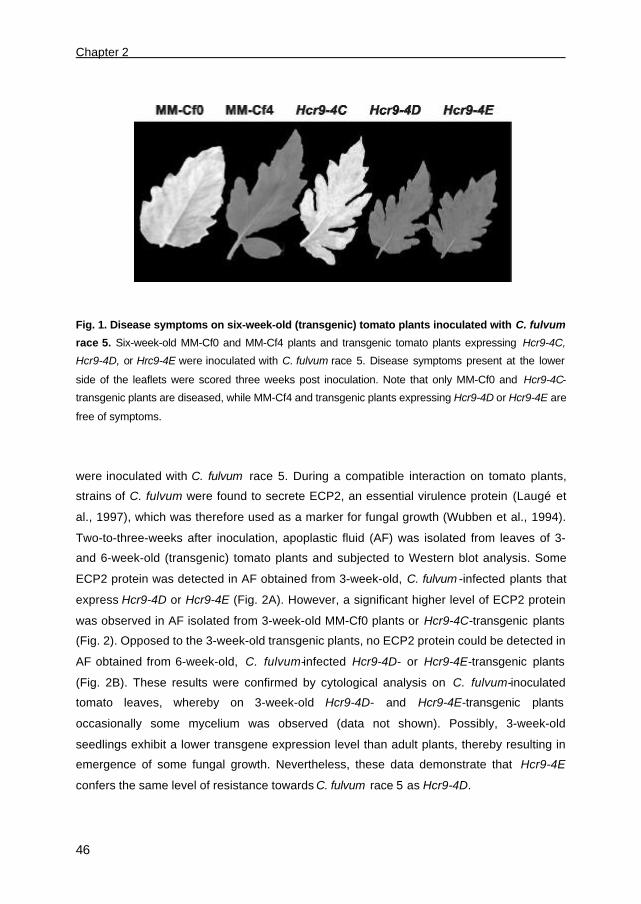
Chapter 2
46
Fig. 1. Disease symptoms on six-week-old (transgenic) tomato plants inoculated with C. fulvum
race 5. Six-week-old MM-Cf0 and MM-Cf4 plants and transgenic tomato plants expressing Hcr9-4C,
Hcr9-4D, or Hrc9-4E were inoculated with C. fulvum race 5. Disease symptoms present at the lower
side of the leaflets were scored three weeks post inoculation. Note that only MM-Cf0 and Hcr9-4C-
transgenic plants are diseased, while MM-Cf4 and transgenic plants expressing Hcr9-4D or Hcr9-4E are
free of symptoms.
were inoculated with C. fulvum race 5. During a compatible interaction on tomato plants,
strains of C. fulvum were found to secrete ECP2, an essential virulence protein (Laugé et
al., 1997), which was therefore used as a marker for fungal growth (Wubben et al., 1994).
Two-to-three-weeks after inoculation, apoplastic fluid (AF) was isolated from leaves of 3-
and 6-week-old (transgenic) tomato plants and subjected to Western blot analysis. Some
ECP2 protein was detected in AF obtained from 3-week-old, C. fulvum -infected plants that
express Hcr9-4D or Hcr9-4E (Fig. 2A). However, a significant higher level of ECP2 protein
was observed in AF isolated from 3-week-old MM-Cf0 plants or Hcr9-4C-transgenic plants
(Fig. 2). Opposed to the 3-week-old transgenic plants, no ECP2 protein could be detected in
AF obtained from 6-week-old, C. fulvum-infected Hcr9-4D- or Hcr9-4E-transgenic plants
(Fig. 2B). These results were confirmed by cytological analysis on C. fulvum-inoculated
tomato leaves, whereby on 3-week-old Hcr9-4D- and Hcr9-4E-transgenic plants
occasionally some mycelium was observed (data not shown). Possibly, 3-week-old
seedlings exhibit a lower transgene expression level than adult plants, thereby resulting in
emergence of some fungal growth. Nevertheless, these data demonstrate that Hcr9-4E
confers the same level of resistance towards C. fulvum race 5 as Hcr9-4D.
Circumvention of Hcr9-4E-mediated resistance
47
Hcr9-4E recognizes a novel AVR determinant that is designated AVR4E
When Hcr9-4E-transgenic plants were inoculated with potato virus X (PVX)::Avr4, no
necrotic symptoms developed (Thomas et al., 1997). Moreover, Hcr9-4E transgenics as well
as plants that lack a functional Cf-4, but carry a functional Hcr9-4E, exhibited no seedling
lethality when crossed to transgenic plants expressing Avr4 (Thomas et al., 1997; Takken et
al., 1999). This implies that Hcr9-4E confers resistance towards C. fulvum race 5 through
recognition of an avirulence determinant that is different from AVR4, which we designated
AVR4E. To show that, in addition to AVR4, AVR4E is secreted by C. fulvum race 5, AF was
isolated from C. fulvum race 5-infected MM-Cf0 leaves. When this AF was injected into
leaves of Hcr9-4D transgenics and MM-Cf4 plants necrotic lesions developed, whereas a
more chlorotic response developed in leaves of Hcr9-4E-transgenic plants (Fig. 3). This AF
did not induce necrosis or chlorosis upon injection into leaves of Hcr9-4C transgenics or
MM-Cf0 plants (Fig. 3). Moreover, necrotic lesions were induced when AF was injected into
leaves of transgenic Nicotiana tabacum plants expressing Hcr9-4D or Hcr9-4E, especially
when these genes were under control of the 35S promoter of cauliflower mosaic virus (data
not shown). Together, these data demonstrate that novel avirulence determinant AVR4E
can be detected in AF through its necrosis-inducing activity (NIA) on plants that carry Hcr9-
4E.
Fig. 2. Immunodetection of the ECP2 protein in the
apoplastic space of C. fulvum-infected (transgenic)
tomato leaves. A, Three-week-old and B, six-week-old
MM-Cf0 and MM-Cf4 plants, and transgenic tomato plants
expressing Hcr9-4C, Hcr9-4D, or Hcr9-4E, under control of
their native promoter, were inoculated with C. fulvum race 5.
Apoplastic-washing fluid (AF) was isolated from these C.
fulvum-infected leaves, three weeks post inoculation.
Proteins present in 5 µl of AF were separated by SDS-
PAGE and Western blot analysis was performed, using
antibodies raised against ECP2, a protein that is used as a
marker for successful infection. Note that ECP2 protein is only detected in AF of susceptible MM-Cf0
and Hcr9-4C-transgenic plants.
Isolation of the gene encoding avirulence determinant AVR4E
To isolate AVR4E, proteins present in AF of C. fulvum race 5-infected tomato leaves were
fractionated by gel filtration and the fractions were assayed for NIA by injection into leaves of
Hcr9-4E transgenic plants. Proteins present in fractions that exhibited NIA were subjected to

Chapter 2
48
Fig. 3. Necrosis-inducing activity of apoplastic fluid from a compatible C. fulvum-MM-Cf0
interaction after injection into (transgenic) tomato leaves (Takken et al., 1999; Fig. 4). Apoplastic
fluid (AF) obtained from leaves of C. fulvum race 5-infected MM-Cf0 was injected into leaves of MM-Cf0
and MM-Cf4 plants and transgenic tomato plants expressing Hcr9-4C, Hcr9-4D or Hcr9-4E. Note that
only resistant plants show necrosis or chlorosis (see also Figs. 1 and 2).
anion exchange chromatography and fractions that exhibited biological activity in Hcr9-4E
transgenics were subsequently purified by reverse-phase chromatography (Fig. 4).
Following these purification steps, we identified a protein that exhibited specific NIA in Hcr9-
4E-transgenic plants (Fig. 4; fraction III).
From this protein, which we tentatively designated AVR4E, an N-terminal and an
internal amino acid sequence was determined. To isolate the gene encoding AVR4E,
anchored PCR on genomic DNA of C. fulvum race 5 was performed, using degenerate
oligonucleotides that were designed based on the determined amino acid sequences. In this
way, we obtained the 5’-end of Avr4E, which was subsequently used to screen a cDNA
library obtained from leaves of C. fulvum race 5-infected MM-Cf0 plants, which resulted in
isolation of the full-length cDNA of Avr4E (Fig. 5). To isolate the genomic sequence of
Avr4E, we screened a genomic library from C. fulvum race 4, as a genomic library of C.
fulvum race 5 was not available at the time. This screening resulted in the identification of
the promoter and terminator region of the Avr4E sequence of race 4, which subsequently
allowed isolation of the genomic Avr4E sequence of race 5 by PCR.
Comparison of the Avr4E cDNA sequence with the genomic Avr4E sequence of C.
fulvum race 5 revealed that the Avr4E gene does not contain introns (Fig. 5). Moreover,
Southern blot analysis of C. fulvum race 4 and race 5 showed that Avr4E is present as a
single-copy gene in the genome of these strains (data not shown). The promoter region,
which is nearly identical in race 4 and race 5, contains the consensus sequences (TA)GATA

Circumvention of Hcr9-4E-mediated resistance
49
and CAAT (Fig. 5). The (TA)GATA sequences are involved in regulation of Avr9 gene
expression under nitrogen-limiting conditions in vitro and in planta (Snoeijers et al., 1999;
Pérez-García et al., 2001), while the CAAT sequences play a role in conidiophore
development (Adrianopoulos and Timberlake, 1994).
AVR4E behaves as a genuine avirulence determinant
To determine whether the isolated protein represents the product encoded by avirulence
gene Avr4E, two strains of C. fulvum virulent on Hcr9-4E-transgenic plants, i.e. strain South
America (SA) and race 4, were transformed with a genomic Avr4E clone of race 5. For
strain SA, virulence on Hcr9-4E transgenics is not associated with virulence on Hcr9-4D
transgenics, indicating that strain SA behaves like a race 4E (Fig. 6A). Unlike strain SA, C.
fulvum race 4 is virulent on Hcr9-4D-transgenic plants and MM-Cf4 plants that carry the
complete Cf-4 locus, indicating that race 4 does not produce a functional AVR4 elicitor
protein (Fig. 6C) (Joosten et al., 1994 and 1997).
Insertion of the genomic Avr4E sequence of race 5 into the genome of strain SA and C.
fulvum race 4 resulted in transformants that are avirulent on Hcr9-4E transgenics as well as
on MM-Cf4 plants (Fig. 6B and 6D). To show that these transformants indeed secrete a
functional AVR4E protein, AF was isolated from susceptible MM-Cf0 plants that were
infected with either these transformants or the parent strains. Only AF obtained from
transformant-infected tomato showed NIA when injected into leaves of Hcr9-4E-transgenic
plants (data not shown). These results demonstrate that the isolated Avr4E gene indeed
encodes the race-specific avirulence determinant AVR4E, which confers avirulence to
strains that are normally virulent on plants that carry Hcr9-4E.
Fig. 4. Fractionation and isolation of the AVR4E
protein from apoplastic fluid of C. fulvum race
5-infected MM-Cf0 plants. Proteins present in
apoplastic-washing fluid (AF) of a compatible
interaction between C. fulvum race 5 and MM-Cf0
were separated by gel filtration. The fractions that
exhibited necrosis-inducing activity (NIA) when
injected into leaves of Hcr9-4E- transgenic plants
were subjected to anion exchange chromatography.
Subsequent reverse-phase chromatography of the
fractions showing NIA resulted in fractions (II and
III) that still exhibited NIA in leaves of Hcr9-4E transgenics, as indicated (+). From the protein present in
fraction III (indicated by an arrow), a partial amino acid sequence was obtained.
Chapter 2
50
CCCTAACTCTAGGGTCTACCGTTAGGGTAACGGAACCTTAGTTTTAGGGTTCTTTATACTACGATCCCGCCGCCGCTAGCGTAGGCGGGTCGCTACTACTTAAGTAGATTGCTCGTCTTAATAAGAGCTATTAAATAGATAGTCCTTGTCGATAGTAGCAATAGATCTAAGGGTGTTAACTTCAATACTATATAAATAAGCGTATATATAGGTAATATCCTATTATTAAATAAGGCTACGTACGCTAAGAATTAGGTGACTAATTGGTTAGCTATAGCCTCTTTAAGAGCAATACTTATATATTCTTAATAGATATAAACAATATATAACCTGCTTTTAAAATACCCTATATAAGCCCGTATAATAACTTCCTATAACTACTTCTAACGTAATAAGATTAATAAGGTAACTATAGCTCTTAAGGTAATTAGTAAGGAAGTTAGAGTCCTAAAGTACCTTACTTTAAAACTCTAAGGAGGTGTATGAAAATAAAGTATAACCTTATAAGCGTAAGATAAGATTAAAAGGTTAAAATAAGCGTTATAGGGTATAAGAATAGAATGGGTATTAGCCGAGGAGATTGCGATAACTAAGTTAGGGCAATAAATTGCCCTATATAAGAGCTAATAAGGCACTACTTTCAAATGACATAGTTTTTCACACCTTATAAACCAATGCCCTGTACTTTCCAATCGAGATCCCCAGTTTCGTAGGTAACTTAAGCCTTTGTTTTGAGGCTGAAACTTGCAAGTAACTTTTAAGGACCAAATTGCAAAGGCTTCTAGCGAAACACCGCGCCTGTGCCCTGGTCGTCGAACACCCGCTACTCCACGGAAACCATTCAAAGGGATGCGGACAGGCGCTGTAGAGCACGCATTTCTTCCACCAATCTAGGAATGATACCAAGCCTGTCCGTCGAAAGTGAGTGAATGACAAATCAAGAAAGATATGTCTTTAGCTATGGTCCTCCTCCACAAGCTTAGCAACAGAACATTAAATCACACTGTAGTCGGCCGAGCCAAACCTGCGCTCTGCGTGAAGAGCTCATCTCGGCCATACCCCGGTCAAAGCCATATCCTTCTCGATATTGCTCACTCATGCCACGGTTCTGCCTATTGGCTTTACGGCTTACGGGAGGGCATATTCCATCGCCTTCTCAGTATCAGTCTAGAACTAAACATCCTTCATCTCCTACGCCACTAGCCCATTGCACCACTTTTGGTCGGTGCGGCCGCCCCTCTTGACTCCCTAAACGTTGTTCTACTACATAAAAGGGTTCGAGTGGCCCGGTATATCGCTGTGCGACCTGCACAGGCAGTTTCACGAAGGCGAACCCGATCAAAGGCCTGCAAACCGCGGTCCGCAGCGAAGTAAATTTTGCGGACGAGTCTTCGAAGGAATTATGGCCTAATCCGCGACCTAGAGCCTGACCTACATTCCACAACCTCACCCCACTATACCGAGGACGATCCAGCATGCAGTTTCCAACCCCTCACCTCCTGCTCACCACCCTCCTCGCT
M Q F P T P H L L L T T L L A 1 10
ACTATCACAACCGCCGATTTCTCGCGCGATTGCCCGCCAGGCAGCGGCGTTGGCAATCAAGCCGAATGGAGCGCACGCGGCGTAGATGGC T I T T A D F S R D C P P G S G V G N Q A E W S A R G V D G 20 30 40
ACTGCGATCCCTAGAGAACTTGACGCTCACAGCCTCTGTGACTGCTTCAAGCCGTTCTTGAACGTGCTGGGATGTTCGGTCACATCGGTT T A I P R E L D A H S L C D C F K P F L N V L G C S V T S V
50 60 70
GTGACGGAGAGAGCTGTTTTTGTGAAGGGACATCTCAACTATTGTGCGCGGATGTCGGAGTCGGTGGCAGGGATCTCGCCGTGTAAGGAG V T E R A V F V K G H L N Y C A R M S E S V A G I S P C K E
80 90 100
TGGGAGATTGAGGTTGGGGGAGCGCATCCTGAGAGGATGGCAAACAGATAGATCAGCGGTAGACACCTAAATTCCATAGTCGACTGTTTG W E I E V G G A H P E R M A N R
110 120
CCTCGCGAAAGACTAACTACCTTCTAATCTCAGCCAGGGGTTCCGGACTGGACTGACAGACAGCAAGCTAAGGAAGATGGCCTTGCCAATATCGCTCTGGAACAATCAGCTTCGTCTCGTCAAGACACACAAGTTATGCATAGGTGGCCCGGTCTTAGCGTTCTGTTATTTAAGTAAAATGTAGTGCCGGAGAGCGACGGCAGGAATTAACTCCCACTTGGTTTTTTCGGCTTTTTTGAGCGTTAGTGTTTCCTGATATCGTTAGTAGCTCGCTTCTTACTTCTACTACTTAGTCGCGTCGCTTACTAACGCGTTTCTAGTCAGTACTACTACTAATAAATATAGGCTTCCTGAGTTAGTATAATAAATAGTATAGAATAAAGGTAGTACTATAGGCTTTCTTATATTCTTATAGAAGTATATTAGAAAGTACGCTTTTCCTAAAATGATTAGAAACACACTTAGAAACACGCTTATATACTCTCTTTTAGTAGTTATTCTTTATAACGTACTTTACTACCGAGCGGGCTATACTACCGAGCGGGCTACTTTTACTAAGAACTATACTACTTCTACTTTAATTGCGACGGTATCTTAACACGACGACATCCTACAGAATCGTTACTAGGAGGACAATTCCTCTTTAGATTCTTCGCTATCTAGCGAGTCTACTTAATAGGTACGTACGTTATACCCCGACTCGATATATTGCTTATAGCGTCGTAGCCTTAGTACTAGCCGAATACTATCTAGCAACTCTCTACT
Fig. 5. DNA and protein sequence of the AVR4E elicitor protein. The genomic sequence of the
Avr4E gene of C. fulvum is shown. The Avr4E gene encodes a pre-protein that contains a 20-amino
acid N-terminal signal peptide (underlined) for extracellular targeting. The mature form of the AVR4E
protein consists of 101 amino acids (bold), including six cysteines. The consensus sequences
(TA)GATA and CAAT present in the promoter are underlined. The N-terminal- and internal amino acid
sequences derived from the purified AVR4E protein are indicated (italics).
AVR4E is a small, secreted cysteine-rich protein
The Avr4E ORF encodes a predicted protein of 121 amino acids, which consists of six
cysteine residues. The signal peptide cleavage site of AVR4E was predicted using the
SignalP program (Nielsen et al., 1997), which identified a 20-amino acid N-terminal signal
sequence for extracellular targeting (Fig. 5). The predicted cleavage site is consistent with
the sequence information obtained from N-terminal sequencing of AVR4E purified from AF
(Fig. 4), demonstrating that the N-terminus of AVR4E is not further processed by fungal- or

Circumvention of Hcr9-4E-mediated resistance
51
plant proteases in the apoplastic space of tomato. Assuming that C-terminal processing
does not occur, the mature AVR4E protein consists of 101 amino acids. No significant
sequence homology could yet be found between the Avr4E gene, or the AVR4E protein, and
sequences present in the database.
Fig. 6. AVR4E behaves as a genuine avirulence determinant on tomato plants carrying Hcr9-4E.
Leaves of MM-Cf0 and MM-Cf4 plants, and transgenic plants expressing Hcr9-4D or Hcr9-4E were
inoculated with strain SA (A) and race 4 (C), or with four independent transformants of strain SA (B) and
race 4 (D), carrying the Avr4E genomic sequence of race 5. Photographs of the lower side of the
leaflets were taken three weeks post inoculation. Note that no disease symptoms are present on leaves
of Hcr9-4E-transgenic plants and MM-Cf4 plants that were inoculated with transformants that carry
Avr4E (indicated by arrows in B and D).
C. fulvum employs two different mechanisms to evade AVR4E recognition
As demonstrated by C. fulvum race 4, strains of C. fulvum have developed the ability to
evade both Hcr9-4D- and Hcr9-4E-mediated resistance (Fig. 6C). Previously, it has been
demonstrated that, in most cases, strains circumvent Hcr9-4D-mediated resistance by
production of AVR4 isoforms that carry different single amino acid substitutions (Joosten et
al., 1994 and 1997).
To investigate whether loss of AVR4E recognition was also accomplished by production
of mutated AVR4E isoforms, we PCR-amplified the Avr4E ORF from the genome of 25
strains of C. fulvum that were virulent and 10 strains that were avirulent on Hcr9-4E-

Chapter 2
52
transgenic plants. Sequence analysis of these PCR products revealed that all avirulent
strains carry an Avr4E sequence with an ORF identical to that of the Avr4E gene present in
C. fulvum race 5 (Fig. 5). However, for 17 strains of C. fulvum virulent on Hcr9-4E-
transgenic plants, among which are race 4 and race SA, a similar modification was detected
in the ORF of the Avr4E gene. This modification involves a double point mutation, resulting
in amino acid substitutions, Phe62-to-Leu and Met73-to-Thr (avr4ELT). To analyze whether
avr4ELT still exhibits NIA, we employed the PVX-based expression system (Chapman et al.,
1992). In spite of the fact that the native signal peptide of AVR2, AVR4 and AVR9 has been
reported to mediate proper extracellular targeting of these proteins when expressed in
tomato using PVX (Takken et al., 2000a), the endogenous signal peptide of AVR4E did not
(data not shown). Therefore, PVX::Avr4E and PVX::avr4ELT were obtained by cloning the
sequence encoding mature AVR4E and avr4ELT protein, respectively, downstream of the
sequence encoding the PR-1a signal peptide of Nicotiana tabacum (Hammond-Kosack et
al., 1995). Tomato plants were subsequently inoculated with the recombinant PVX vectors
and subsequently examined for the development of systemic HR (Fig. 7). It appeared that
Hcr9-4E-transgenic plants and MM-Cf4 plants inoculated with PVX::Avr4E developed a clear
systemic HR, which eventually resulted in plant death. When Hcr9-4E transgenics were
inoculated with PVX::avr4ELT, systemic mosaic symptoms were detected that were caused
by the PVX infection (Fig. 7). Thus, unlike AVR4E, avr4ELT does not exhibit NIA,
demonstrating that strains of C. fulvum evade Hcr9-4E-mediated resistance by modifying
AVR4E in such way that plants carrying Hcr9-4E do no longer recognize this isoform.
Fig. 7. PVX-mediated expression
of Avr4E and avr4ELT
in(transgenic) tomato plants.
Three-week-old MM-Cf0 and MM-
Cf4 plants and transgenic plants
expressing Hcr9-4D or Hcr9-4E
were inoculated with PVX::Avr4E
(upper panel) or PVX::avr4ELT (lower
panel). Plants were photographed
14 days post inoculation with PVX.
Note that, opposed to PVX::Avr4E,
PVX::avr4ELT does not cause
systemic necrosis in MM-Cf4 and
Hcr9-4E-transgenic plants.

Circumvention of Hcr9-4E-mediated resistance
53
Interestingly, all other strains virulent on Hcr9-4E-transgenic plants (i.e. 8 out of 25) carry an
Avr4E ORF that is identical to that of strains avirulent on Hcr9-4E-transgenic plants. To
analyze whether these 8 virulent strains produced the AVR4E protein, AF was isolated from
MM-Cf0 plants that were infected by these virulent strains and injected into leaves of Hcr9-
4E transgenic plants. No NIA was observed (data not shown), indicating that these virulent
strains do not produce a functional AVR4E elicitor protein. Moreover, transformation of these
virulent strains with a genomic Avr4E clone of C. fulvum race 5 resulted in avirulence on
Hcr9-4E transgenics, as confirmed by 8 independent transformants of two parent strains
(data not shown).
To further investigate the mechanism conditioning virulence of these strains on plants
carrying Hcr9-4E, the expression of Avr4E during growth on susceptible tomato plants was
investigated. For strains of C. fulvum that are avirulent (race 5) as well as strains that are
virulent on these plants and carry avr4ELT (strain SA and race 4), Avr4E transcripts
accumulated during growth on tomato (Group I and II; Fig. 8). However, for strains that are
virulent on Hcr9-4E-transgenic plants and carry a wild-type Avr4E ORF, no Avr4E transcripts
could be detected (Group III; Fig. 8). However, transformation of these strains with the
genomic Avr4E clone of race 5 resulted in accumulation of Avr4E transcripts in planta
(Group IV; Fig. 8). Altogether, these data demonstrate that strains of C. fulvum evade Hcr9-
4E-mediated resistance either by lack of Avr4E gene expression or by production of an
AVR4E mutant protein that carries two amino acid substitutions, Phe62Leu and Met73Thr.
Fig. 8. Expression of Avr4E during
growth of C. fulvum on susceptible
tomato plants. Northern blot
analysis was performed on total RNA
isolated from leaves of MM-Cf0
plants that were infected with strains
of C. fulvum that are either avirulent
(I and IV) or virulent (II and III) on
Hcr9-4E-transgenic plants. Lane 1;
avirulent race 5 carrying Avr4E; lanes 2 and 3: virulent race 4 and strain SA carrying avr4ELT,
respectively; lanes 4-to-7: naturally virulent race 2.4, race 2.4.5.9, race 2.4.5.9.11 and race 2.4.8.11,
respectively, carrying a wild-type Avr4E ORF; and lane 8: avirulent race 2.4.8.11 transformed with the
Avr4E genomic sequence of race 5. The expression of Avr4E (upper panel) and actin (lower panel) was
assayed using DNA fragments of either the Avr4E ORF or the C. fulvum actin gene, respectively, of
which the latter served as a control probe.
Chapter 2
54
The single amino acid substitution Phe62Leu is sufficient for loss of AVR4E avirulence
function
As Agrobacterium-mediated transient expression in tobacco has previously been
demonstrated to facilitate successful expression of extracellular elicitors and membrane-
anchored Cf proteins (Van der Hoorn et al., 2000), we used this method to study AVR
elicitor activity. To this aim, Agrobacterium cultures carrying AVR- and Hcr9-4-encoding
genes were mixed in a 1:1 ratio and infiltrated into tobacco leaves (Fig. 9A). In the presence
of Hcr9-4E, but not Hcr9-4D, AVR4E induced a necrotic response, whereas AVR4 induced
necrosis only in the presence of Hcr9-4D (Fig. 9A).
To investigate whether the amino acid substitutions, Phe62Leu and Met73Thr, are both
required for loss of AVR4E avirulence function, the NIA of avr4ELT and AVRE mutant
proteins carrying the single amino acid substitution Phe62Leu (avr4EL) or Met73Thr (avr4ET)
was examined by agroinfiltration. Therefore, a quantitative analysis (Van der Hoorn et al.,
2000) was performed by infiltration of a dilution series of Agrobacterium cultures carrying
AVR4E (mutant)- and Hcr9-4E-encoding genes. The NIA of AVR4 isoforms that are
produced by strains of C. fulvum virulent on Hcr9-4D transgenics was also analyzed by
infiltration of dilution series of Agrobacterium cultures carrying AVR4 (mutant)- and Hcr9-4D-
encoding genes. The percentage of infiltrated leaf area that had become necrotic was
measured 7 days post inoculation (dpi) and plotted against the percentage of Agrobacterium
cultures carrying AVR (mutant)-encoding genes. For all AVR4 mutant proteins tested, a
significant reduction in NIA was observed as compared to AVR4 (data not shown). These
observations confirmed results obtained previously, using PVX-mediated expression
(Joosten et al., 1997). Agrobacterium cultures carrying avr4ELT induced necrosis of 50% of
the infiltrated leaf area at a significantly lower dilution than Agrobacterium cultures carrying
wild-type Avr4E (Fig. 9B). Thus, opposed to PVX-mediated expression in tomato (Fig. 7),
avr4ELT still exhibited some level of NIA when agroinfiltrated in tobacco leaves. Interestingly,
no difference in NIA was observed between avr4EL and avr4ELT, or between avr4ET and
AVR4E (Fig. 9B), suggesting that the single amino acid substitution Phe62Leu in AVR4E is
sufficient to evade Hcr9-4E-mediated resistance.
AVR4E mutant proteins carrying single- or double amino acid substitutions are stable
We also analyzed whether the single- and double amino acid substitutions affected the
protein stability of AVR4E. To test this hypothesis, we transiently expressed Avr4E, avr4ELT,
as well as avr4EL and avr4ET by agroinfiltration into tobacco leaves and analyzed the
stability of the produced proteins. Moreover, to determine whether instability of AVR4
Circumvention of Hcr9-4E-mediated resistance
55
isoforms enables circumvention of Hcr9-4D-mediated resistance, as suggested by Joosten
et al. (1997), we also tested the protein stability of these AVR4 mutant proteins.
To facilitate detection of the various mutant proteins by Western blotting, c-Myc-tagged
AVR4 and AVR4E (mutant) proteins were made. Hereto, the sequence encoding AVR
(mutant) proteins was cloned downstream of the sequence encoding a c-Myc-tag, which in
turn is downstream of the PR-1a signal sequence. Co-infiltration of Agrobacterium cultures
carrying c-Myc-tagged AVR-encoding genes with cultures carrying the matching Hcr9-4
resistance gene demonstrated that the c-Myc-tag did not affect the NIA of AVR4 and AVR4E
(data not shown).
Western blot analysis was performed on AF isolated from leaves that were
agroinfiltrated, at opposite leaf-halves, with cultures carrying wild-type Avr and cultures
carrying AVR (mutant)-encoding genes. Comparison of protein levels within one leaf showed
that the same amount of protein was present in AF of leaves agroinfiltrated with cultures
carrying AVR4E mutant-encoding genes as compared to cultures carrying wild-type Avr4E
(Fig. 9C). However, a significantly lower amount of protein was detected for the AVR4
mutant proteins as compared to wild-type AVR4 (Fig. 9C). These data demonstrate that
evasion of Hcr9-4D-mediated resistance is accomplished by production of AVR4 isoforms
that are unstable, whereas evasion of AVR4E recognition by plants that carry Hcr9-4E is
mediated by production of an elicitor-inactive AVR4E mutant protein that is very stable.
Discussion
The Hcr9-4E resistance protein confers resistance upon recognition of the AVR4E
elicitor
Resistance trait Cf-4 has been introgressed from wild tomato species into tomato cultivar
MoneyMaker (MM) (Stevens and Rick, 1988; Boukema, 1981), resulting in the near-isogenic
line MM-Cf4 that confers resistance to Cladosporium fulvum through recognition of the Avr4
gene product (Joosten et al., 1994). Within the introgressed Cf-4 region, five homologues of
Cladosporium resistance gene Cf-9 (Hcr9s) are present, of which homologue Hcr9-4D
represents the functional Cf-4 resistance gene. It has previously been reported that
homologue Hcr9-4E is also a functional Cf gene (Takken et al., 1998) and confers
resistance towards C. fulvum race 5 through recognition of a novel avirulence determinant,
designated AVR4E (Takken et al., 1999). Here, we demonstrate that homologue Hcr9-4E
confers the same level of resistance towards C. fulvum race 5 as Hcr9-4D. We found that
the AVR4E protein is a cysteine-rich, secreted protein of 101 amino acids that is stable in

Chapter 2
56
Fig. 9. Necrosis-inducing activity and stability of AVR4E and AVR4 (mutant) proteins in
apoplastic fluids of agroinfiltrated tobacco leaves. A, Agrobacterium cultures carrying Avr4E or
Avr4 were co-infiltrated into leaves of 6-week-old tobacco plants in a 1:1 ratio with cultures carrying
Hcr9-4E or Hcr9-4D. Necrosis-inducing activity (NIA) was scored at three days post infiltration (dpi) and
photographs were taken 7 dpi. B, Agrobacterium cultures carrying Avr4E (x), avr4ET (open triangle),
avr4EL (black triangle) or avr4ELT (black dot) were diluted in Agrobacterium cultures carrying Hcr9-4E
and infiltrated into tobacco leaves. The relative necrotic leaf area (percentage) was measured and
plotted against the percentage of culture carrying AVR4E (mutant)-encoding genes. C, Western blot
analysis of apoplastic fluids isolated from tobacco leaves that were infiltrated, at opposite leaf halves,
with cultures carrying AVR4E/AVR4 wild-type and mutant-encoding genes. A Myc-tag fused at the N-
terminus of both AVR4E- and AVR4 (mutant) proteins allowed detection of these proteins by antibodies
raised against c-Myc. The NIA of the AVR4E- and AVR4 wild-type (+++++) and mutant proteins (+ and
++), as determined by agroinfiltration, is indicated between brackets.
0
50
100
0,1 1 10 100
% Agrobacterium carrying Avr
% n
ecro
sis
C
A B

Circumvention of Hcr9-4E-mediated resistance
57
the apoplastic space of tomato. As found for other small and stable, secreted cysteine-rich
elicitor proteins of C. fulvum (Van den Burg et al., 2001b; Van den Hooven et al., 2001;
Luderer et al., 2002b), the six cysteine residues present in AVR4E are thought to be
involved in disulfide bonding. The genomic Avr4E sequence of race 5 was found to confer
avirulence to strains that are normally virulent on Hcr9-4E-transgenic plants, indicating that
AVR4E functions as a genuine avirulence determinant.
Circumvention of Hcr9-4D- and Hcr9-4E-mediated resistance
In order to enable growth on MM-Cf4 plants , C. fulvum has to evade both Hcr9-4D- and
Hcr9-4E-mediated resistance. For strains of C. fulvum virulent on Hcr9-4D-transgenic plants,
a variety of single point mutations in the open reading frame (ORF) of the Avr4 gene has
been found (Joosten et al., 1997). It appeared that these modifications resulted in unstable
AVR4 isoforms, as previously has been suggested by the authors, indicating that protein
stability plays a crucial role in evasion of Hcr9-4D-mediated resistance. Unlike AVR4,
circumvention of AVR4E recognition by plants that carry Hcr9-4E is mediated by production
of a stable AVR4E mutant protein. This AVR4E mutant protein carries two amino acid
substitutions, Phe62-to-Leu and Met73-to-Thr (avr4ELT), as a result of a double point mutation
in the Avr4E ORF. No other modifications were identified in the Avr4E ORF of strains that
are virulent on Hcr9-4E transgenics. PVX-mediated expression of avr4ELT in tomato plants
that express Hcr9-4E induced no systemic necrosis, whereas Agrobacterium-mediated
expression of avr4ELT in tobacco leaves showed some level of necrosis-inducing activity
(NIA). The higher level of sensitivity towards elicitor proteins observed in tobacco is thought
to be due to a more efficient Cf-mediated defense-signaling pathway in tobacco as
compared to tomato (Hammond-Kosack et al., 1998; Kamoun et al., 1999a). Nevertheless, it
appeared that production of avr4ELT mutant protein does not trigger Hcr9-4E-mediated
defense responses that restrict growth of C. fulvum .
We further analyzed whether amino acid substitutions Phe62Leu and Met73Thr are both
required for loss of AVR4E avirulence function. Interestingly, the AVR4E mutant protein
carrying Met73Thr (avr4ET) appeared to be as active as AVR4E, whereas mutant protein
carrying Phe62Leu (avr4EL) is as active as avr4ELT, suggesting that the single amino acid
substitution Phe62Leu is sufficient to evade Hcr9-4E-mediated resistance. Moreover, as
demonstrated for avr4ELT, the avr4EL and avr4ET mutant proteins are as stable as the wild-
type AVR4E protein. Thus, Hcr9-4E-mediated resistance is evaded by secretion of an
AVR4E mutant protein that exhibits an essential conformational- or qualitative change, as a
result of amino acid substitution Phe62Leu.

Chapter 2
58
For eight other strains of C. fulvum virulent on Hcr9-4E-transgenic plants that we analyzed,
no modifications were found in the Avr4E ORF. Nevertheless, these strains do not secrete a
functional AVR4E elicitor protein. Strikingly, it appeared that no AVR4E protein was
produced during growth of these strains in tomato leaves due to the lack of Avr4E gene
expression. Transformation with the genomic Avr4E sequence of C. fulvum race 5 resulted
in accumulation of Avr4E transcripts in planta, thereby conferring avirulence to the fungus on
plants that carry Hcr9-4E. Thus, it seems that all elements required for proper Avr4E
expression, including transcription factors and regulatory proteins, are present in parent
strains that lack Avr4E expression. Possibly, the lack of Avr4E gene expression in these
virulent strains is caused by a defect in (cis-regulatory elements of) the promoter of Avr4E.
Also, for the rice blast fungus, Magnaporthe grisea, it has been reported that PWL4 is a non-
functional Avr gene due to a defective promoter, as expression of the PWL4 ORF under
control of the PWL1 or the PWL2 promoter conferred avirulence to the fungus on weeping
lovegrass (Kang et al., 1995). In addition, insertion of a Pot3 transposon into the promoter of
the AVR-Pita gene of M. grisea resulted in gain of virulence on rice cultivars carrying the Pi-
ta resistance gene (Kang et al., 2001). Moreover, in the promoter sequence of the PDA1
gene of Nectria heamatococca positively acting elements are present that are necessary for
high level of transcription in vitro (RuanandStraney, 1996). In the promoter of Avr4E,
(TA)GATA boxes and CAAT-like boxes are present that might regulate nitrogen metabolism
or conidiophore development, respectively, upon binding of transcription factors like AreA
(AdrianopoulosandTimberlake, 1994; Snoeijers et al., 1999; Pérez-García et al., 2001).
Strains that lack Avr4E expression might carry modifications in the promoter, possibly in
elements that are essential for Avr4E expression. On the other hand, lack of Avr4E
expression might be caused by a transposon insertion in the promoter of Avr4E, as found in
the Avr2 ORF of strains of C. fulvum virulent on Cf-2 plants (Luderer et al., 2002b).
Alternatively, the terminator region of Avr4E might carry mutations, resulting in Avr4E
transcripts that are unstable.
Sequence polymorphism in the ORF of Avr genes
As avr4EL exhibits the same elicitor activity as avr4ELT, one would expect that the single
amino acid substitution Phe62Leu would be sufficient for loss of AVR4E avirulence function.
However, thus far, no strains of C. fulvum virulent on Hcr9-4E-transgenic plants have been
identified that carry avr4EL. Moreover, although avr4ET is as active as AVR4E, no strains
avirulent on Hcr9-4E transgenics were found that carry this mutation. This implies that
strains carrying avr4ELT originated from strains carrying wild-type Avr4E as a result of one
single evolutionary event. However, another, more favorable, hypothesis would be that two

Circumvention of Hcr9-4E-mediated resistance
59
independent evolutionary events occurred, where initially strains evolved that carried avr4ET,
which are still avirulent on plants carrying Hcr9-4E, followed by emergence of strains that
carry avr4ELT, which are virulent on these plants.
Within the C. fulvum population, several strains of C. fulvum have been identified that
evade more than one Cf-mediated resistance trait, including Cf-2, Cf-4 (“Hcr9-4D”), Cf-5, Cf-
9, and Hcr9-4E (see also Chapter 6). Strains virulent on plants that express Cf-9 lack the
entire Avr9 ORF (Van Kan et al., 1991). The ORF of the Avr2 and Avr4 genes, however,
contain more sequence variation than the Avr4E gene (Joosten et al., 1997; Luderer et al.,
2002b), suggesting that C. fulvum has endured more extensive selection pressure to evade
Cf-2- and Hcr9-4D-mediated resistance than to evade Hcr9-4E-mediated resistance.
However, a total of 17 breeding lines that carry Cf-4 were also found to carry Hcr9-4E
(Haanstra et al., 2000), implying that selection pressure to evade AVR4 recognition is
identical to that of AVR4E. Moreover, a low genetic variability was found within the C. fulvum
population, as determined by AFLP analysis, implying that the C. fulvum population consists
of a single clonal lineage (Joosten and De Wit, 1999). Thus, strains of C. fulvum virulent on
Hcr9-4D-transgenic plants might have evolved from a common ancestor strain that already
enabled the ability to grow on plants carrying Hcr9-4E.
The intrinsic function of AVR4E for C. fulvum
Strains of C. fulvum that carry avr4ELT or strains that lack Avr4E expression do not show a
measurable reduction in virulence on susceptible tomato plants when compared to strains
that carry wild-type Avr4E, suggesting that AVR4E does not clearly contribute to virulence of
C. fulvum . On the other hand, both AVR4E and avr4ELT might play a role in virulence of C.
fulvum. As only one type of modification was found in the Avr4E ORF, mutations other than
Phe62Leu and Met73Thr might affect the stability of AVR4E and thereby the virulence of C.
fulvum. Moreover, although for various virulent strains no Avr4E transcripts could be
detected on Northern blots, there could still be some level of Avr4E expression present that
is below detection level. This might result in the presence of minute amounts of potentially
stable AVR4E protein, which would be sufficient for full virulence of C. fulvum on susceptible
tomato plants. Thus, as previously demonstrated for Phythophtora parasitica isolates (Colas
et al., 2001), downregulation rather than a complete loss of Avr4E expression might
constitute a suitable mechanism for C. fulvum to evade Hcr9-4E-mediated resistance,
without deleterious consequences for the pathogen.

Chapter 2
60
Materials and Methods
Plants and strains of Cladosporium fulvum
Tomato (Lycopersicon esculentum) cultivar Moneymaker (MM), near isogenic line MM-Cf4
(containing an introgression segment that carries the Cf-4 locus), transgenic tomato plants
that carry Hcr9-4D or Hcr9-4E (Thomas et al., 1997), Nicotiana clevelandii and (transgenic)
tobacco (Nicotiana tabacum cv. Petite Havana SR1) plants, were grown under standard
greenhouse conditions. The various strains of Cladosporium fulvum were cultured according
to De Wit and Flach (1979).
C. fulvum inoculation procedure and isolation of apoplastic washing fluids
Strains of C. fulvum were inoculated onto tomato plants as described by De Wit (1977).
Briefly, suspensions of conidia of C. fulvum (approximately 5 x 10⁶ conidia per ml) were
used to inoculate the lower leaf surface of 3-to-6-week-old tomato plants. Apoplastic-
washing fluids (AFs) were isolated from C. fulvum-infected tomato leaves, three weeks after
inoculation, as described by De Wit and Spikman (1982).
DNA manipulations
All plasmid manipulations were carried out essentially as described by Sambrook et al.
(1989). Polymerase chain reactions (PCRs) were performed with Pfu (Stratagene, La Jolla,
CA) or with the Expand High Fidelity PCR system (Roche Diagnostics, Mannheim,
Germany), according to the manufacturer’s instructions. Restriction enzymes, T4 ligase, and
Escherichia coli DH5á cells were obtained from Invitrogen (Breda, The Netherlands).
Oligonucleotides were synthesized by Amersham-Pharmacia (Buckinghamshire, UK) and
probes were labeled with the Random Primers DNA labeling System (Promega, Breda, The
Netherlands), using α32P-dATP (Amersham-Pharmacia, Buckinghamshire, UK).
Purification of the AVR4E elicitor protein
Proteins present in AF isolated from a compatible interaction between C. fulvum race 5 and
MM-Cf0 were separated by gel filtration on a Sephadex G-50 column (Amersham-
Pharmacia, Roosendaal, The Netherlands), as described by Joosten et al. (1990). The
fractions that showed specific necrosis-inducing activity (NIA) when injected into leaves of
Hcr9-4E-transgenic plants were subjected to anion exchange chromatography on a
Resource Q column (Amersham-Pharmacia, Roosendaal, The Netherlands), according to
the protocol described by Laugé et al. (2000). Fractions that are biologically active were
subsequently loaded on a ProRPC HR 5/10 reverse-phase column (Amersham-Pharmacia,
Circumvention of Hcr9-4E-mediated resistance
61
Roosendaal, The Netherlands), as described by Joosten et al. (1994). A highly purified
protein that exhibits NIA in leaves of Hcr9-4E plants was obtained and used to determine an
N-terminal and an internal amino acid sequence of the AVR4E protein.
Isolation of a cDNA and a genomic clone of Avr4E
To facilitate isolation of the Avr4E gene by reverse genetics, degenerated oligonucleotides
were designed. Two forward primers N1 (5’-gtnggnaaycargcngartgg-3’) and N2 (5’-
gtngayggnacngcnathcc-3’) were constructed, based on the N-terminal amino acid sequence
(VGNQAEW and VDGTAI), respectively. Reverse primer X-3b (5’-gcnccnacytcdatytccca-3’)
was designed, based on the internal protein sequence (WEIEVGG). Anchored PCR was
performed essentially according to Stuurman et al. (1996), using BstYI and ApoI to digest
100 ng of genomic DNA of C. fulvum race 5, followed by ligation of the corresponding
double-stranded oligonucleotides (“anchors”). A series of nested PCR reactions were
performed with N1 and N2 together with primers VECT24 (5’-agcactctccagcctctcaccgcc-3’,
which anneals to the BstYI-digested anchor site) and RH24 (5’-agcactctccagcctctctcaccgca-
3’, which anneals to the ApoI-digested anchor site), and the PCR products were sequenced.
Sequence analysis revealed that the putative translation products of the PCR fragments
correspond to the AVR4E peptide sequences. Additional 5’-sequence information was
obtained by PCR using primer RH24 together with reverse primers X-3b, as well as by PCR
using RH24 together with two homologous reverse primers N3 (5’-cccagcacgttcaagaac-3’)
and N4 (5’-gaagcagtcacagaggctg), which anneal to the 5’-end of Avr4E.
In order to isolate the full-length Avr4E sequence, a cDNA library derived from a
compatible interaction between C. fulvum race 5 and tomato (Van Kan et al., 1991) was
screened using the Avr4E PCR fragments as a probe, according to the method described by
Van den Ackerveken et al. (1992). This screening resulted in the isolation of a clone carrying
the full-length Avr4E open reading frame (ORF). By screening a genomic library derived
from C. fulvum race 4, we obtained promoter- and terminator sequence information, which
allowed subsequent isolation of the genomic Avr4E sequence of C. fulvum race 5 by PCR,
using the following primers; forward primer PROM-B (5’-cgcggatccctaactctagggtctacc-3’,
BamHI-site underlined) and reverse primer TER-X (5’-gatcctcgaggccacctatgcataacttg-3’,
XhoI-site underlined). PCR resulted in a fragment that contains a promoter region of 1475
bp and a terminator region of 190 bp, flanking the Avr4E ORF of C. fulvum race 5. The PCR
product was digested with BamHI and XhoI and ligated into BglII/XhoI-digested site of
pAN7-1, a plasmid carrying a hygromycin B selection marker (Punt et al., 1987). The
obtained plasmid was used to transform strains of C. fulvum that are virulent on Hcr9-4E-
transgenic plants.
Chapter 2
62
Transformation of C. fulvum
The procedure for preparation of C. fulvum protoplasts was adapted from the method
described by Harling et al. (1988) and Van den Ackerveken et al. (1992). Mycelium of C.
fulvum strain SA, race 4, race 2.4.8.11, and race 2.4.5.9 was harvested from 3-day-old
cultures that were grown in vitro in liquid B5 medium. Protoplast were obtained upon
digestion of the mycelium in MM (20 mM 2-[N-morpholino] ethanesulfonic acid, pH 5.8, 1 M
MgS04), containing 25 mg of Glucanex (Novo Nordisk, Denmark) and 1 mg of kitalase
(Wako Pure Chemical Inductries, Ltd.) per ml. Transformation of the protoplasts was
achieved with polyethylene glycol 6000 (Merck), according to Oliver et al. (1987).
Transformants that were resistant to hygromycin B (Sigma, Bornem, Belgium), obtained 3-4
weeks after culture on potato dextrose agar medium were subcultured to obtain mono-spore
isolates.
Sequencing the open reading frame of Avr4E in virulent and avirulent strains of C.
fulvum
For standard PCR analysis, genomic DNA of C. fulvum was isolated according to the
procedure described by Cenis (1992), after growth of the fungus for 5 days in liquid B5
medium. Following DNA isolation, the Avr4E ORF was PCR-amplified using forward primer
4E-F (5’- ggacgagtcttcgaagga-3’), which anneals 76 nucleotides upstream from the ATG
start codon, and reverse primer 4E-R3 (5’- ctgagattagaaggtagttag-3’), annealing 51 bases
downstream of the TAG stop codon. Sequence analysis of the Avr4E ORF was also
performed using the 4E-F and 4E-R3 primers.
Northern blot analysis
Total RNA from C. fulvum-infected tomato leaves was isolated 21 days after inoculation
using a hot phenol-extraction procedure, according to the manufacturer’s instructions
(CLONTECH Laboratories, Palo Alto, USA). Fifteen µg of glyoxal-denatured RNA was
separated on 0.01 M sodium phosphate pH 7.0 agarose gels and transferred to Hybond N+
membrane (Amersham-Pharmacia, Buckinghamshire, UK) by capillary blotting with 0.025 M
sodium phosphate buffer pH 7.0 (Sambrook et al., 1989). The RNA was immobilized by UV-
crosslinking and the RNA gel blots were prehybridized at 65°C for 60 min in modified Church
and Gilbert buffer (0.5 M phosphate buffer, pH 7.2, 7% SDS and 1 mM EDTA) (Church and
Gilbert, 1984). Subsequently, the blots were hybridized over-night at 65°C in the same buffer
containing radio-labeled DNA fragments, representing the Avr4E gene and the actin gene of
C. fulvum . The blots were washed at 65°C with 0.5 x SSC (75 mM NaCl, 7.5 mM sodium

Circumvention of Hcr9-4E-mediated resistance
63
citrate), containing 0.1 % SDS. Kodak X-OMAT films were subsequently exposed to the
blots.
Construction of PVX derivatives and PVX inoculation procedure
The recombinant constructs PVX::Avr4E and PVX::avr4ELT were obtained by PCR using
genomic DNA isolated from C. fulvum race 5 and race 4, respectively, as templates.
Constructs that carry the endogenous signal sequence of Avr4E for extracellular targeting
were obtained by PCR with forward primer PVX4E-N (5’-ccatcgatgcagttttccaacccctca-3’) and
reverse primer PVX4E-R (5’-ccatcgatctatctgtttgccatcctctc-3’) (ClaI site underlined). PVX
constructs carrying the PR-1a signal sequence (Hammond-Kosack et al., 1995), upstream of
the sequence encoding mature AVR4E (mutant) protein, were obtained by PCR-mediated
overlap extension. First, to obtain the PR-1a signal sequence with an Avr4E overhang at the
3’-end, PCR was performed using forward primer OX10 (5’-caatcacagtgttggcttgc-3’) and
reverse primer PR4E-R (5’-gcgcgagaaatcggcacggcaagagtggg-3’), together with PVX::Avr4
(Joosten et al., 1997) as a template. The sequence encoding mature AVR4E and avr4ELT
were obtained by PCR using forward primer 4E-F (5’-tcttgccgtgccgatttctcgcgcgattgc-3’) and
PVX4E-R, together with genomic DNA of race 5 and race 4, respectively, as a template. The
two sets of PCR products were subsequently used as templates in an overlap extension
PCR with primers OX10 and PVX4E-R. The PCR fragments were digested with ClaI and
cloned into the ClaI-digested site of PVX-expression vector pTX∆GC3A (Hammond-Kosack
et al., 1995), downstream of the PVX coat protein promoter. In vitro transcription,
amplification of the virus particles on Nicotiana clevelandii and PVX inoculations on
(transgenic) tomato plants were performed as described by Hammond-Kosack et al. (1995).
Construction of binary plasmids and transformation of Agrobacterium
Binary plasmids carrying Avr4E and Avr4 and their derivatives were constructed as follows:
PCR fragments containing the ORF of Avr4E and avr4ELT were generated using AT4E-N (5’-
tagctcgagcgatttctcgcgcgattgcc-3’, XhoI site underlined) and AT4E-B (5’-
cgcggatccctatctgtttgccatcctctc-3’, BamHI site underlined) with PVX::Avr4E and PVX::avr4ELT
vectors as a template, respectively. Fragments containing the sequences that encode
mature avr4EL and avr4ET were obtained by overlap extension PCR with PVX::Avr4E as a
template. For avr4EL, the following sets of primers were used: AT4E-N with F62L-R (5’-
cacaagaacagctctctc-3’) and F62L-F (5’-gagagagctgttcttgtg-3’) with AT4E-B. For avr4ET,
AT4E-N with M73T-R (5’-cgactccgacgtccgcgc-3’) and M73T-F (5’-gcgcggacgtcggagtcg-3’)
with AT4E-B were employed. PCR fragments containing the ORF of Avr4, avr4 (C35Y), avr4
(Y38H), avr4 (C41Y) and avr4 (C80Y) (Joosten et al., 1997) were generated using primer A4-

Chapter 2
64
NX (5’-tagctcgagcaaggccccaaaactcaacc-3’, XhoI site underlined) and A4-BC (5’-
cgcggatccctattgcggcgtctttaccg-3’, BamHI site underlined) with genomic DNA of C. fulvum
race 5, race 2.4.8.11 (C35Y), race 4 (Y38H), race 4 (C41Y) and race 2.4.5.9.11 (C80Y) as
templates. The amplified fragments were digested with XhoI/BamHI, cloned into the
XhoI/BamHI-digested site of pBluescript SK+ (Amersham-Pharmacia, Buckinghamshire, UK)
and sequenced. This plasmid was digested with XhoI and BamHI, and the fragments that
contain the Avr ORFs were subsequently cloned into the XhoI/BamHI-digested site of binary
plasmid pNW30, a derivative of pRH271 (Van der Hoorn, unpublished). This binary plasmid
pNW30 carries the Cauliflower Mosaic Virus (CaMV) 35S-promoter and the PI-II terminator
(An et al., 1989), both of which flank the PR-1a signal sequence (Hammond-Kosack et al.,
1995) and the sequence encoding a double c-Myc-tag.
Binary plasmid carrying Hcr9-4E was constructed by PCR with primers Cf 4E-F (5’-
agctccatgggttgtgtaaaacttatatttttcatgc-3’) and Cf 4E-R (5’-agctctgcagctaatatatcttttcttgtgctttttt-
cattctcg-3’), using a genomic clone containing Hcr9-4E (Takken et al., 1998) as a template,
following the procedure as described for binary plasmid carrying Hcr9-4D (pCf-4)
constructed by Van der Hoorn et al. (2000).
The binary plasmids were transferred to Agrobacterium tumefaciens strain MOG101
(Hood et al., 1993) by electroporation.
Agroinfiltration and analysis of apoplastic washing fluids
Agrobacterium-mediated transient expression was performed essentially as described by
Van der Hoorn et al. (2000). Cultures containing recombinant Agrobacterium carrying the
different binary plasmids were resuspended to a final OD600 of 2 and infiltrated into tobacco
leaves in the presence of 200 µM acetosyringone. As tobacco leaves at different
developmental stages might exhibit different transient expression levels, the stability of the
AVR (mutant) proteins was determined within one tobacco leaf. Three-to-four days after co-
infiltration with Agrobacterium carrying Hcr9-4D or Hcr9-4E, the necrosis-inducing activity of
the AVR4 and AVR4E (mutant) proteins was scored. AF was isolated three days after
infiltration with cultures that carry AVR-encoding genes, according to the method described
by De Wit and Spikman (1982). Ten times concentrated AF (7 µl) was supplied with 2 µl 5x
SDS loading buffer (50 mM Tris-HCl pH 6.8, 5% (w/v) SDS, 10% (v/v) glycerol, 5% 2-
mercaptoethanol and 0.0025% bromophenol blue) and incubated at 95°C for 5 min. Proteins
were separated by SDS-PAGE on gels containing 15% (w/v) acrylamide (Laemmli, 1970)
and transferred to nitrocellulose membranes (Schleider and Schuell, Dassel, Germany) by
blotting for 2 hrs at 250 mA. Detection of c-Myc-tagged proteins was performed by
incubation of the filters in blocking buffer (BB) (1x phosphate-buffered saline (PBS) (Oxoid)
Circumvention of Hcr9-4E-mediated resistance
65
and 5% skimmed milk powder [ELK, Campina]), followed by over-night incubation in BB
supplied with 1:1000 diluted antibodies raised against c-Myc (rabbit polyclonal IgG [Santa
Cruz Biotechnology]). After 3 x 15 min washes in wash buffer (WB) (1x PBS, 0.05% Tween-
20), filters were incubated for 2 hrs in BB supplied with a 1:1000 diluted secondary antibody
(Anti-Rabbit Ig-horseradish peroxidase [Amersham-Pharmacia, Buckinghamshire, UK]).
Following 3 x 15 min washes in WB, c-Myc-tagged proteins were detected using Super
Signal Chemiluminescent Substrate (Pierce, Rockford, USA) and Kodak X-OMAT films.
Immunodetection of ECP2 proteins was performed according to the method described by
Wubben et al. (1994).
Acknowledgements
We thank Guy Bauw (Department of Genetics, University of Gent, Belgium) for
determination of amino acid sequences of the AVR4E protein and Richard Oliver (School of
Biology, University of Perth, Australia) for providing the genomic library of C. fulvum race 4.
We also thank Renier Van der Hoorn (Laboratory of Phytopathology, Wageningen
University) for constructing vector RH271 that was used for agroinfiltration and for critically
reading the manuscript. N. Westerink and B. F. Brandwagt were supported by research
grants from Wageningen University and NWO-STW (Netherlands Organization for Science
Research, Technology Foundation), respectively.

66
Chapter 3
The AVR4 elicitor protein of Cladosporium fulvum binds to
fungal components with high affinity
This chapter has been published by Nienke Westerink,* Ronelle Roth,* Harrold A. Van den
Burg, Pierre J. G. M. De Wit, and Matthieu H. A. J. Joosten (2002) in Molecular Plant-
Microbe Interactions 15 (12), 1219-1227.
* Equal author contribution

Chapter 3
68
AVR4 binds to fungal components
69
The AVR4 elicitor protein of Cladosporium fulvum binds to
fungal components with high affinity
Abstract
The interaction between tomato and the fungal pathogen Cladosporium fulvum complies
with the gene-for-gene system. Strains of C. fulvum that produce race-specific elicitor AVR4
induce a hypersensitive response, leading to resistance, in tomato plants that carry the Cf-4
resistance gene. The mechanism of AVR4 perception was examined by performing binding
studies with 125I-AVR4 on microsomal membranes of tomato plants. We identified an AVR4-
high affinity-binding site (KD = 0.05 nM), which exhibited all the characteristics expected for
ligand-receptor interactions, such as saturability, reversibility and specificity. Surprisingly,
the AVR4-high affinity-binding site appeared to originate from fungi present on infected
tomato plants rather than from the tomato plants themselves. Detailed analysis showed that
this fungus-derived, AVR4-specific binding site is heat and proteinase K resistant. Affinity
crosslinking demonstrated that AVR4 specifically binds to a component of about 75 kDa that
is of fungal origin. Our data suggest that binding of AVR4 to fungal component(s) is related
to the intrinsic virulence function of AVR4 for C. fulvum .
Introduction
Active disease resistance in plants relies on a highly sensitive and specific defense system
that enables recognition and evasion of invading pathogens. Pathogens that produce
nonspecific elicitors are able to initiate a disease response in most genotypes within a plant
species (Knogge 1996). Race-specific elicitors are considered to induce resistance only in
genotypes of host plants that carry the corresponding resistance (R) gene (Flor 1971; De
Wit 1997). Addition of elicitor proteins to suspension-cultured plant cells results in a rapid
induction of plant defense-associated responses (Ham et al. 1991; Basse et al. 1993;
Nürnberger et al. 1994; Boller 1995; Hanania and Avni 1997; Meindl et al. 1998; Felix et al.
1999; Lee et al. 2001). The high sensitivity of suspension-cultured cells and their extremely
quick responses to low amounts of elicitor, suggest that the induction of defense responses
is mediated by receptors that exhibit high affinities for elicitor proteins. Indeed, specific high
affinity-binding sites (HABSs) have been identified for elicitor proteins, peptides and
oligosaccharide signals (Cheong and Hahn 1991; Nürnberger et al. 1995; Mithöfer et al.

Chapter 3
70
1996; Ito et al. 1997; Matsubayashi et al. 1997; Bourque et al. 1999; Matsubayashi and
Sakagami 1999; Scheer and Ryan 1999; Meindl et al. 2000). Since the majority of HABSs
identified thus far resides in the plasma membrane of plant cells, a role for HABS as putative
receptors for elicitor molecules has been proposed. A close correlation has also been found
between the biological activity of elicitor molecules and the affinity for their binding sites,
which further implies that the HABSs for these elicitors act as functional receptors (Hanania
and Avni 1997; Meindl et al. 1998 and 2000; Thevissen et al. 2000).
The interaction between tomato and the extracellular, biotrophic pathogen
Cladosporium fulvum is a well-established model system that complies with the gene-for-
gene model. Strains of C. fulvum that carry avirulence (Avr) genes induce an active defense
response, including the hypersensitive response (HR), in tomato plants that carry the
corresponding Cf resistance genes (Joosten and De Wit 1999). Several Cf genes and their
corresponding Avr genes have been cloned (Joosten and De Wit 1999). The Cf gene family
encodes putative plasma membrane-anchored proteins with a predicted extracellular
leucine-rich repeat domain, a transmembrane region and a short cytoplasmic tail (Thomas et
al. 1998). For Cf-4 and Cf-9, the corresponding avirulence genes of C. fulvum , Avr4 and
Avr9, respectively, have been cloned (Van den Ackerveken et al. 1992; Joosten et al. 1994).
AVR4 and AVR9 elicitor proteins are both encoded as pre-proproteins that are processed by
fungal and plant proteinases within the leaf apoplast into mature proteins of 86 and 28
amino acids, respectively. To date, very little is known about the intrinsic function of AVR4
and AVR9 for the fungus. No clear deleterious effects on virulence have been found in
strains of C. fulvum in which either the Avr4 or the Avr9 gene is truncated or deleted
(Marmeisse et al. 1993; Joosten et al. 1997). Nevertheless, both Avr4 and Avr9 gene
products may have a role in virulence, development, reproduction, or general fitness of C.
fulvum.
The predicted extra-cytoplasmic localization of Cf-4 and Cf-9 would fit with a model in
which the secreted AVR4 and AVR9 proteins are perceived in the leaf apoplast.
Suspension-cultured tobacco cells expressing Cf-9 rapidly produce defense response-
associated active oxygen species in response to AVR9 (Piedras et al. 1998). However, no
direct interaction between AVR9 and Cf-9 has yet been demonstrated (Luderer et al.
2001b). Furthermore, binding studies with AVR9 have shown that both MM-Cf9 (resistant)
and MM-Cf0 (susceptible) plants contain an AVR9-specific HABS in the plasma membrane
(Kooman-Gersmann et al. 1996), suggesting an indirect role for Cf-9 in perception of AVR9.
Such indirect perception has been referred to as the ‘Guard hypothesis’ (Van der Biezen
and Jones 1998), whereby it is proposed that the HABS represents a virulence target for
AVR9. The Cf-9 protein is thought to act as a ‘guard’ to monitor the behavior of a host-
AVR4 binds to fungal components
71
encoded virulence protein or proteins that are targets for AVR9 (Luderer and Joosten 2001a;
Van der Hoorn et al. 2002).
In this study, we have investigated the binding of AVR4 to microsomal membranes of
MM-Cf4 (resistant) and MM-Cf0 (susceptible) tomato plants, and non-host plant species. We
have identified an AVR4 HABS that appeared to originate from fungi present on infected
tomato plants rather than from tomato itself. Interestingly, this fungus-derived AVR4 HABS is
heat and proteinase K resistant. In addition, affinity crosslinking demonstrated that AVR4
specifically interacts with a component of approximately 75 kDa that is of fungal origin.
Binding of AVR4 to fungal components in relation to its intrinsic virulence function for C.
fulvum will be discussed.
Results
Production and biological activity of iodinated AVR4
In order to examine whether a high affinity-binding site (HABS) is present for AVR4 on
tomato membranes, it was essential to obtain large quantities of pure, elicitor-active AVR4
protein. The methylotrophic yeast Pichia pastoris was used as an eukaryotic, heterologous
expression system (Romanos 1995) to obtain high levels of correctly folded mature AVR4
protein. AVR4 protein isolated from intercellular fluids (IF) obtained from C. fulvum -infected
tomato leaves is unglycosylated (Joosten et al. 1997). To prevent yeast-specific
glycosylation of AVR4; the potential N-linked glycosylation site (NLS) in AVR4 was removed
by changing the codon for serine into a codon for alanine in the open reading frame (ORF)
of Avr4. By using Potato virus X (PVX)-mediated expression in MM-Cf4 plants, as described
by Joosten and associates (1997), this mutation was found not to affect the necrosis-
inducing activity of AVR4 (data not shown). Heterologously expressed AVR4 protein was
purified from the culture filtrate by ion exchange and reverse phase high-performance liquid
chromatography (HPLC) (Van den Burg et al. 2001). Injection of purified AVR4 into MM-Cf4
leaves resulted in a clear hypersensitive response (HR), demonstrating that the protein had
retained its elicitor activity (Van den Burg et al. 2001).
To test whether iodination affects the biological activity of AVR4, iodination with non-
radioactive iodine (127I) was performed using a lactoperoxidase labeling protocol, resulting in
mono 127I-labeled AVR4. Unlabeled AVR4 induces a rapid oxidative burst in suspension-
cultured tobacco cells expressing Cf-4 in a concentration-dependent manner (De Jong et al.
2002). Addition of 127I-labeled AVR4 protein to these cell suspensions resulted in a similar,
concentration-dependent response (Fig. 1). This demonstrates that iodination does not
affect the biological activity of the AVR4 elicitor. Identical labeling conditions were applied

Chapter 3
72
300
400
500
600
700
800
900
0 5 10 15 20 25 30
Time (min)
Flu
ore
scen
ce (
A.U
.)
0
0.3 nM
1.5 nM
Fig. 1. Oxidative burst of suspension-
cultured tobacco cells expressing Cf-4 after
treatment with AVR4 or 127I-AVR4.
Suspension-cultured tobacco cells expressing
Cf-4 were supplied with the fluorescent probe
pyranin (10µg/ml) and challenged with buffer
(open square) and with two concentrations (0.3
and 1.5 nM) of unlabeled AVR4 (black dot) or127I-labeled AVR4 (open triangle). Quenching of
the pyranin fluorescence, due to production of
H2O2 by the cells, was measured in time. A.U. =
arbitrary units.
with radioactive iodine (125I) to obtain mono 125I-labeled AVR4 that was used in subsequent
binding experiments.
Binding of 125I-AVR4 to microsomal membranes of MM-Cf4 tomato plants
To examine the presence of AVR4-specific binding sites on microsomal membrane fractions
(MFs) isolated from MM-Cf4 tomato leaves, binding of 125I-AVR4 to MFs was followed over
time and at various temperatures. Optimal binding was achieved at 37°C (data not shown).
An AVR4-specific binding site was found and binding reached equilibrium 4 h after
incubation with AVR4 at 37°C (Fig. 2A). Specific binding of 125I-AVR4 remained stable up to
8 h, followed by a slow, partial decrease after 22 h of incubation (data not shown). At lower
temperatures, i.e. 4°C and 24°C, equilibrium of 125I-AVR4-specific binding was reached after
22 h and 10 h of incubation, respectively (data not shown).
Addition of a 500-fold molar excess of unlabeled AVR4 at equilibrium resulted in a
decrease of specific binding of 125I-AVR4, demonstrating that AVR4-specific binding is
reversible (Fig. 2B). From these data, a dissociation constant (KD) of 0.09 nM was calculated
for AVR4 (Fig. 2C), suggesting that AVR4 binds to microsomal membranes of MM-Cf4 with
high affinity.
Saturation of 125I-AVR4 binding to microsomal membranes of MM-Cf4
Incubation of MFs isolated from MM-Cf4 leaves with increasing concentrations of 125I-AVR4
showed that AVR4 binding is saturable (Fig. 3A). Saturation of AVR4-specific binding was
reached at an 125I-AVR4 concentration of approximately 0.25 nM (Fig. 3A). Scatchard
analysis performed on these data resulted in a plot with a linear slope, which implies a
uniform population of AVR4-specific binding sites. The calculated KD value of 0.05 nM (KD =
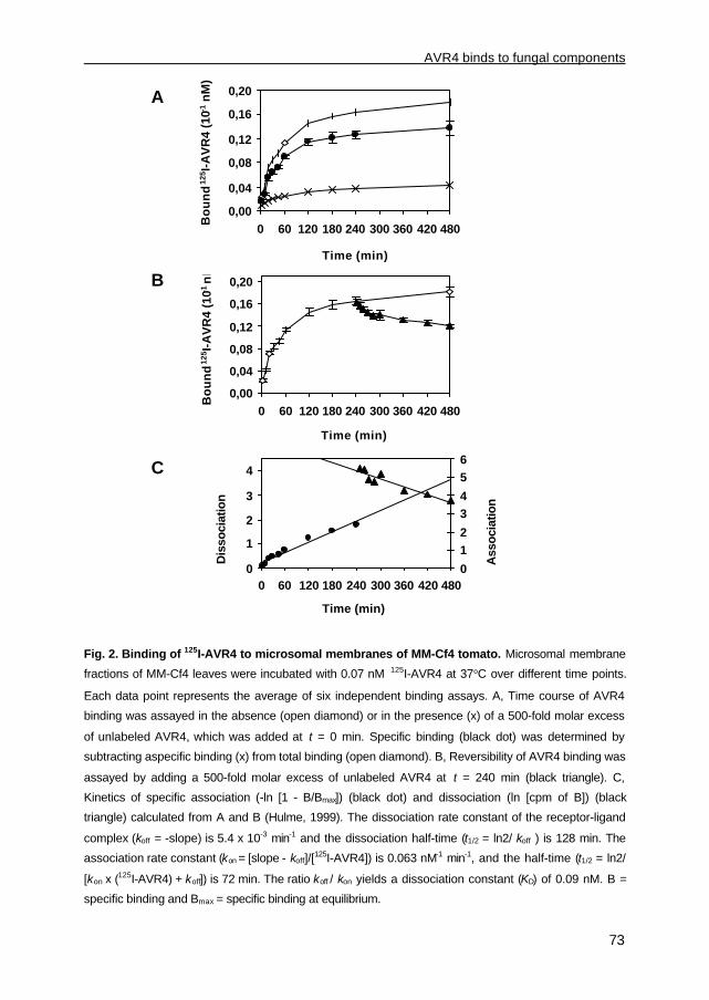
AVR4 binds to fungal components
73
Fig. 2. Binding of 125I-AVR4 to microsomal membranes of MM-Cf4 tomato. Microsomal membrane
fractions of MM-Cf4 leaves were incubated with 0.07 nM 125I-AVR4 at 37°C over different time points.
Each data point represents the average of six independent binding assays. A, Time course of AVR4
binding was assayed in the absence (open diamond) or in the presence (x) of a 500-fold molar excess
of unlabeled AVR4, which was added at t = 0 min. Specific binding (black dot) was determined by
subtracting aspecific binding (x) from total binding (open diamond). B, Reversibility of AVR4 binding was
assayed by adding a 500-fold molar excess of unlabeled AVR4 at t = 240 min (black triangle). C,
Kinetics of specific association (-ln [1 - B/Bmax]) (black dot) and dissociation (ln [cpm of B]) (black
triangle) calculated from A and B (Hulme, 1999). The dissociation rate constant of the receptor-ligand
complex (koff = -slope) is 5.4 x 10⁻³ min-1 and the dissociation half-time (t1/2 = ln2/ koff ) is 128 min. The
association rate constant (kon = [slope - koff]/[125I-AVR4]) is 0.063 nM-1 min-1, and the half-time (t1/2 = ln2/
[kon x (125I-AVR4) + koff]) is 72 min. The ratio koff / kon yields a dissociation constant (KD) of 0.09 nM. B =
specific binding and Bmax = specific binding at equilibrium.
0,00
0,04
0,08
0,12
0,16
0,20
0 60 120 180 240 300 360 420 480
Time (min)
Bo
un
d 12
5 I-A
VR
4 (1
0-1 n
M)
0
1
2
3
4
0 60 120 180 240 300 360 420 480
Time (min)
0
12
34
56
Dis
soci
atio
n
Ass
ocia
tion
0,00
0,04
0,08
0,12
0,16
0,20
0 60 120 180 240 300 360 420 480
Time (min)
Bo
un
d 12
5 I-A
VR
4 (1
0-1 n
M)
C
B
A

Chapter 3
74
Fig. 3. Saturation of 125I-AVR4 binding to microsomal membranes of MM-Cf4 tomato. A,
Microsomal membranes isolated from MM-Cf4 tomato leaves were incubated for 4 h at 37°C with
increasing concentrations of 125I-AVR4. Aspecific binding was determined by addition of a 500-fold
molar excess of unlabeled AVR4 at t = 0 min (x). Specific binding (black dot), which was determined by
subtracting aspecific binding (x) from total binding (open diamond), was saturated at a free ligand
concentration of about 0.25 nM. Free ligand concentration was calculated by subtracting the initially
added 125I-AVR4 from total bound 125I-AVR4. Each data point represents the average of six independent
binding assays. B, Scatchard plot analysis of data obtained for specific binding of 125I-AVR4 as
presented in A. From the slope of the Scatchard plot a dissociation constant (KD = -1/slope) of 0.05 nM
was calculated, while extrapolation of the slope to the x-axis allowed a calculation of the total receptor
concentration (Rt = 0.02 nM = 0.02 pmol ml-1 per 48 µg of protein per 0.1 ml of assay) of 0.04 pmol/mg
of protein. C, Hill plot of data obtained for specific binding (B) of 125I-AVR4 as presented in A. Bmax
represents the amount of specific AVR4 binding at saturation level. From the slope of the Hill plot the
Hill coefficient (nH = 0.91) was calculated.
-1/slope) is in the same order of magnitude as the KD value calculated from the rate
constants (Fig. 2C). The total concentration of binding sites (Rt = 0.04 pmol/mg of protein)
was determined by extrapolation of the slope to the x-axis (Fig. 3B). The calculated Hill
coefficient (nH) of 0.91 is close to 1, suggesting that AVR4 binding to microsomal
membranes of MM-Cf4 is non-cooperative (Fig. 3C).
0,0
0,1
0,2
0,3
0,4
0,5
0 0,1 0,2 0,3 0,4 0,5 0,6
Bo
un
d 12
5 I-A
VR
4 (1
0-1 n
M)
A
0
0,1
0,2
0,3
0,00 0,01 0,02
Bound 125I-AVR4 (nM)
Bo
un
d/F
ree
125 I
-AV
R4
B
-0,5
-0,2
0,1
0,4
0,7
1 2 3
Free 125I-AVR4 (log [pM])
log
(B
/Bm
ax-B
)
C
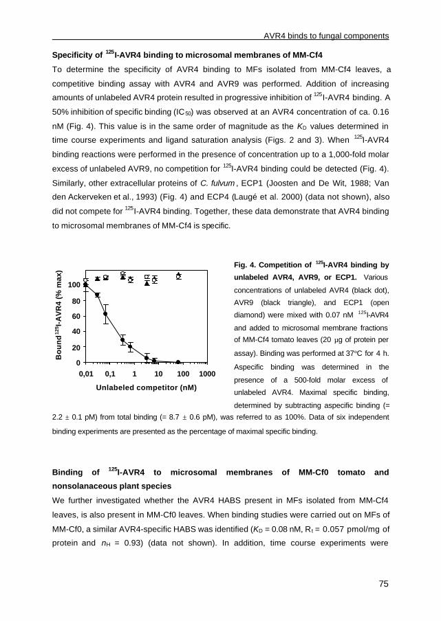
AVR4 binds to fungal components
75
0
20
40
60
80
100
0,01 0,1 1 10 100 1000
Unlabeled competitor (nM)
Bo
un
d 12
5 I-A
VR
4 (%
max
)Specificity of 125I-AVR4 binding to microsomal membranes of MM-Cf4
To determine the specificity of AVR4 binding to MFs isolated from MM-Cf4 leaves, a
competitive binding assay with AVR4 and AVR9 was performed. Addition of increasing
amounts of unlabeled AVR4 protein resulted in progressive inhibition of 125I-AVR4 binding. A
50% inhibition of specific binding (IC50) was observed at an AVR4 concentration of ca. 0.16
nM (Fig. 4). This value is in the same order of magnitude as the KD values determined in
time course experiments and ligand saturation analysis (Figs. 2 and 3). When 125I-AVR4
binding reactions were performed in the presence of concentration up to a 1,000-fold molar
excess of unlabeled AVR9, no competition for 125I-AVR4 binding could be detected (Fig. 4).
Similarly, other extracellular proteins of C. fulvum , ECP1 (Joosten and De Wit, 1988; Van
den Ackerveken et al., 1993) (Fig. 4) and ECP4 (Laugé et al. 2000) (data not shown), also
did not compete for 125I-AVR4 binding. Together, these data demonstrate that AVR4 binding
to microsomal membranes of MM-Cf4 is specific.
Fig. 4. Competition of 125I-AVR4 binding by
unlabeled AVR4, AVR9, or ECP1. Various
concentrations of unlabeled AVR4 (black dot),
AVR9 (black triangle), and ECP1 (open
diamond) were mixed with 0.07 nM 125I-AVR4
and added to microsomal membrane fractions
of MM-Cf4 tomato leaves (20 µg of protein per
assay). Binding was performed at 37°C for 4 h.
Aspecific binding was determined in the
presence of a 500-fold molar excess of
unlabeled AVR4. Maximal specific binding,
determined by subtracting aspecific binding (=
2.2 ± 0.1 pM) from total binding (= 8.7 ± 0.6 pM), was referred to as 100%. Data of six independent
binding experiments are presented as the percentage of maximal specific binding.
Binding of 125I-AVR4 to microsomal membranes of MM-Cf0 tomato and
nonsolanaceous plant species
We further investigated whether the AVR4 HABS present in MFs isolated from MM-Cf4
leaves, is also present in MM-Cf0 leaves. When binding studies were carried out on MFs of
MM-Cf0, a similar AVR4-specific HABS was identified (KD = 0.08 nM, R t = 0.057 pmol/mg of
protein and nH = 0.93) (data not shown). In addition, time course experiments were

Chapter 3
76
performed with MFs of MM-Cf0 (Fig. 2), which resulted in sim ilar AVR4-specific binding
characteristics as found in MFs of MM-Cf4 plants (data not shown).
To determine whether nonsolanaceous plant species also contain AVR4-specific
binding sites, MFs isolated from leaves of Arabidopsis spp. and barley grown under regular
greenhouse conditions were incubated with 125I-AVR4. Interestingly, these nonsolanaceous
plant species showed the presence of an AVR4-specific HABS with KD values of a similar
order of magnitude as found in MM-Cf0 and MM-Cf4 plants (KD = 0.07 nM and KD = 0.12 nM
for Arabidopsis spp. and barley, respectively).
Further biochemical characterization of the AVR4 HABS
To further analyze the nature of the AVR4 HABS, MFs isolated from MM-Cf4 leaves were
subjected to a number of different treatments, prior to binding assays with 125I-AVR4. In
addition, binding experiments with 125I-AVR9, which served as a control, were carried out
(Kooman-Gersmann et al. 1996).
To test for heat-stability of the AVR4 HABS, MFs were incubated at 95°C for 15 min,
prior to incubation with 125I-AVR4 or 125I-AVR9. Surprisingly, while heat-treatment completely
abolished AVR9 binding, AVR4-specific binding to MFs was reduced by only 15% (Fig. 5A).
In order to assay for proteinase-sensitivity of the AVR4 HABS, MFs were treated with
proteinase K. The presence of low concentrations of proteinase K during incubation with 125I-
AVR4, however, resulted in breakdown of the AVR4 protein (data not shown). Therefore,
proteinase K-treated MFs were heated prior to incubation with 125I-AVR4 in order to
inactivate the enzyme. As proteinase K did not affect the stability of AVR9 (Kooman-
Gersmann et al. 1996), no heat-treatment was required to inactivate proteinase K prior to125I-AVR9 binding. While proteinase K-treatment of MFs completely abolished 125I-AVR9-
specific binding, no significant effect was found for AVR4 binding (Fig. 5B). Incubation of
MFs with heat-treated proteinase K only slightly affected 125I-AVR9-specific binding,
whereas the 125I-AVR4-specific binding remained unaffected.
Finally, fractions were isolated from MFs of MM-Cf4 leaves using the two-phase
partitioning protocol to enrich for plasma membranes (Larsson et al. 1987). When binding
was performed on membranes present in the upper phase, a significant increase in AVR9-
specific binding sites, which are localized predominantly in the plasma membrane (Kooman-
Gersmann et al. 1996), was found, indicating that plasma membrane-enrichment had
occurred (Fig. 5C). In contrast to the increase in the amount of binding sites for AVR9, a
significant decrease in AVR4-specific binding sites was found in these plasma membrane-
enriched fractions (Fig. 5C). Together, our data indicate that the AVR4 HABS is heat and
proteinase K resistant and that it is not associated with the plasma membrane.
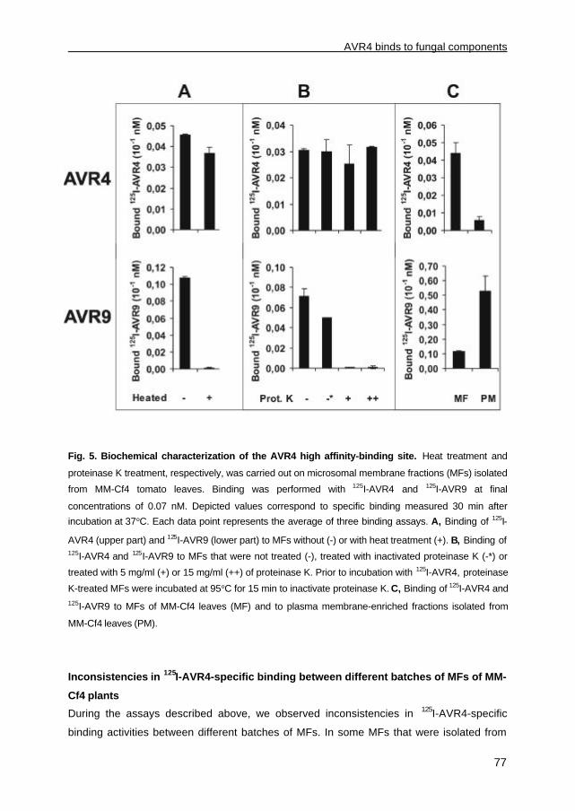
AVR4 binds to fungal components
77
Fig. 5. Biochemical characterization of the AVR4 high affinity-binding site. Heat treatment and
proteinase K treatment, respectively, was carried out on microsomal membrane fractions (MFs) isolated
from MM-Cf4 tomato leaves. Binding was performed with 125I-AVR4 and 125I-AVR9 at final
concentrations of 0.07 nM. Depicted values correspond to specific binding measured 30 min after
incubation at 37°C. Each data point represents the average of three binding assays. A, Binding of 125I-
AVR4 (upper part) and 125I-AVR9 (lower part) to MFs without (-) or with heat treatment (+). B, Binding of125I-AVR4 and 125I-AVR9 to MFs that were not treated (-), treated with inactivated proteinase K (-*) or
treated with 5 mg/ml (+) or 15 mg/ml (++) of proteinase K. Prior to incubation with 125I-AVR4, proteinase
K-treated MFs were incubated at 95°C for 15 min to inactivate proteinase K. C, Binding of 125I-AVR4 and125I-AVR9 to MFs of MM-Cf4 leaves (MF) and to plasma membrane-enriched fractions isolated from
MM-Cf4 leaves (PM).
Inconsistencies in 125I-AVR4-specific binding between different batches of MFs of MM-
Cf4 plants
During the assays described above, we observed inconsistencies in 125I-AVR4-specific
binding activities between different batches of MFs. In some MFs that were isolated from

Chapter 3
78
leaves of MM-Cf4 plants, as well as from Arabidopsis spp., no 125I-AVR4-specific binding
sites could be detected. Interestingly, all of these MFs were isolated from plants that were
grown in confined compartments. The MFs that exhibited 125I-AVR4-specific binding sites,
however, were isolated from MM-Cf4 and Arabidopsis plants grown under regular
greenhouse conditions.
This observation, together with the predicted nonproteinaceous character of the AVR4
HABS, prompted us to investigate the origin of the AVR4 HABS further. When tomato plants
are grown under regular greenhouse conditions, it appears that these plants are often
infected with Oidium lycopersicum , thereby showing some sporulating colonies on the
leaflets. To investigate whether a correlation exists between the presence of fungal
contaminants and binding of AVR4 to its HABS, binding assays were performed on MFs
isolated from MM-Cf4 leaves that were severely infected with O. lycopersicum (i.e., leaves of
which 50 to 75% of the surface was infected). Interestingly, the total concentration of binding
sites (Rt = 0.09 pmol/mg of protein), which exhibit a KD value of 0.11 nM, was higher in these
MFs (data not shown) compared with the concentration in the MFs isolated from MM-Cf4
plants grown under regular greenhouse conditions (Rt= 0.04 pmol/mg of protein) (Fig. 3).
To discriminate between fungus- and plant-specific binding, as well as fungus-induced,
plant-specific binding, we intended to test O. lycopersicum for the presence of an AVR4-
specific binding site. However, the requirement of plant material for the obligate pathogen O.
lycopersicum to grow and reproduce hampered the isolation of sufficient fungal
Fig. 6. Saturation of 125I-AVR4 binding to Cladosporium fulvum mycelium extracts. C. fulvum
extracts were incubated with increasing amounts of 125I-AVR4 alone (open diamond) or in the presence
of a 500-fold molar excess of unlabeled AVR4 (x). Specific binding (black dot) was determined by
subtracting aspecific binding from total binding. Data points are fitted to a hyperbolic curve and each
data point represents an average of three independent binding assays.
0,0
0,2
0,4
0,6
0,8
1,0
0 0,1 0,2 0,3 0,4 0,5 0,6
Free 125I-AVR4 (nM)
Bo
un
d 12
5 I-A
VR
4 (1
0-1 n
M)

AVR4 binds to fungal components
79
biomass. Therefore, we performed binding assays on C. fulvum mycelium extracts instead.
Significantly, an AVR4-specific HABS was detected, which exhibited a KD value of 0.26 nM,
in the absence of plant material (Fig. 6). The total concentration of binding sites in these
fractions (Rt = 9.2 pmol/mg of protein) was significantly higher than the Rt in MFs isolated
from MM-Cf4 plants (Fig. 3). Moreover, the AVR4 HABS present in the mycelium extracts
also appeared to be heat-resistant (data not shown), as demonstrated for the AVR4 HABS
present in MFs isolated from MM-Cf4 plants (Fig. 5A). Together, these data demonstrate
that the AVR4 HABS is of fungal rather than of plant origin.
Chemical crosslinking of 125I-AVR4 to a distinct compound is dependent on the
presence of a fungal infection of tomato
To identify the fungus-derived AVR4 HABS, 125I-AVR4 was chemically crosslinked to C.
fulvum mycelium extracts and MFs of O. lycopersicum-infected MM-Cf4 leaves. 125I-AVR4
was crosslinked to a single band that migrates as a molecule of approximately 85 kDa on
sodium dodecyl sulfate-polyacrylamide gel electrophoresis (SDS-PAGE) in MFs isolated
from O. lycopersicum-infected MM-Cf4 leaves (Fig. 7A). No AVR4-specific crosslinking was
detected in MFs isolated from MM-Cf4 plants grown in confined compartments (Fig. 7B).
Significantly, 125I-AVR4 was crosslinked to a band of the same size when binding was
performed with C. fulvum mycelium extracts (Fig. 7C). In the presence of a 500-fold molar
excess of unlabeled AVR4, no crosslinked band could be detected, suggesting that
crosslinking to this molecule is specific for AVR4 (Fig. 7A and C). These data demonstrate
that the AVR4-specific crosslinked component is of fungal origin rather than of plant origin.
Use of glutaraldehyde, m-maleimidobenzoyl-N-hydrozysuccinimide ester (MBS) and photo-
reactive N-hydrozysulfosuccinimidyl-4-azidobenzoate (sulfo-HSAB) as crosslinkers, which
all react through different crosslinking mechanisms, gave similar results (data not shown).
Presuming that one 125I-AVR4 molecule (molecular mass of 10 kDa) is crosslinked to a
single molecule, the predicted molecular weight of the fungus-derived AVR4-specific
crosslinked band is approximately 75 kDa.
Discussion
An AVR4-specific high affinity-binding site that is of fungal origin
To characterize the mechanism of specific perception of AVR4 by MM-Cf4 plants,
microsomal fractions (MFs) from leaves of such plants were analyzed for the presence of an
AVR4-high affinity-binding site (HABS). Although an AVR9-specific HABS was detected in
the plasma membranes of MM-Cf9 plants (Kooman-Gersmann et al. 1996), no such binding
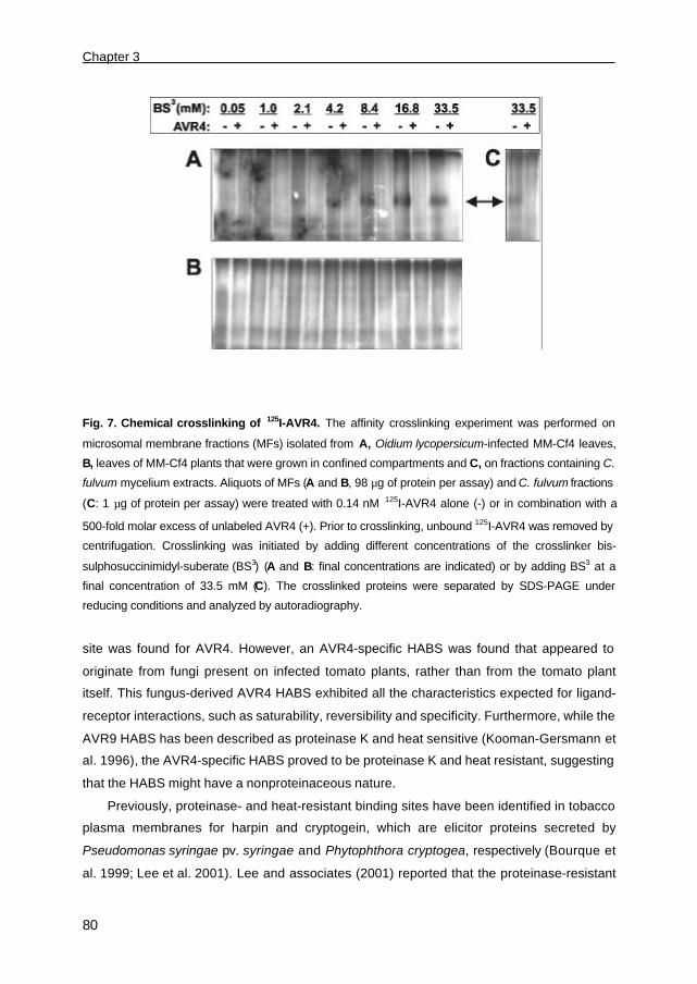
Chapter 3
80
Fig. 7. Chemical crosslinking of 125I-AVR4. The affinity crosslinking experiment was performed on
microsomal membrane fractions (MFs) isolated from A, Oidium lycopersicum-infected MM-Cf4 leaves,
B, leaves of MM-Cf4 plants that were grown in confined compartments and C, on fractions containing C.
fulvum mycelium extracts. Aliquots of MFs (A and B, 98 µg of protein per assay) and C. fulvum fractions
(C: 1 µg of protein per assay) were treated with 0.14 nM 125I-AVR4 alone (-) or in combination with a
500-fold molar excess of unlabeled AVR4 (+). Prior to crosslinking, unbound 125I-AVR4 was removed by
centrifugation. Crosslinking was initiated by adding different concentrations of the crosslinker bis-
sulphosuccinimidyl-suberate (BS3) (A and B: final concentrations are indicated) or by adding BS3 at a
final concentration of 33.5 mM (C). The crosslinked proteins were separated by SDS-PAGE under
reducing conditions and analyzed by autoradiography.
site was found for AVR4. However, an AVR4-specific HABS was found that appeared to
originate from fungi present on infected tomato plants, rather than from the tomato plant
itself. This fungus-derived AVR4 HABS exhibited all the characteristics expected for ligand-
receptor interactions, such as saturability, reversibility and specificity. Furthermore, while the
AVR9 HABS has been described as proteinase K and heat sensitive (Kooman-Gersmann et
al. 1996), the AVR4-specific HABS proved to be proteinase K and heat resistant, suggesting
that the HABS might have a nonproteinaceous nature.
Previously, proteinase- and heat-resistant binding sites have been identified in tobacco
plasma membranes for harpin and cryptogein, which are elicitor proteins secreted by
Pseudomonas syringae pv. syringae and Phytophthora cryptogea, respectively (Bourque et
al. 1999; Lee et al. 2001). Lee and associates (2001) reported that the proteinase-resistant

AVR4 binds to fungal components
81
binding site for harpin is nonproteinaceous. The heat-resistant binding site for cryptogein,
however, was shown to be a glycoprotein (Bourque et al. 1999). Indeed, glycosylated
proteins repeatedly have been demonstrated to be more resistant against thermal or
chemical denaturation and proteolytic degradation compared with their nonglycosylated
counterparts (Tanner and Lehle, 1987). Thus, the heat- and proteinase K-resistant fungal
AVR4 HABS might represent either a stable glycoprotein or a nonproteinaceous
polysaccharide, like glucan or chitin fragments (Bartnicki-Garcia 1968; Kollar et al. 1997).
The latter is consistent with high-affinity binding interactions between (plant) proteins and
oligosaccharide elicitors that have been identified before (Cosio et al., 1988; Cheong and
Hahn, 1991; Shibuya et al., 1993; Baureithel et al., 1994; Asensio et al., 2000; Brandhorst
and Klein, 2000; Day et al., 2001; Ito et al., 1997). Interestingly, we identified an AVR4-
specific crosslinked molecule of approximately 75 kDa that migrates as a distinct band on
SDS-PAGE instead of as a smear, suggesting that it represents a proteinaceous compound
rather than a polysaccharide. Significantly, this AVR4-specific molecule of ca. 75 kDa
proved to be of fungal rather than of plant origin. Thus, the fungal heat- and proteinase K-
resistant AVR4 HABS might represent a molecule of ca. 75 kDa. Alternatively, the fungal
AVR4 HABS might represent a polysaccharide. In that case, binding of AVR4 to this
polysaccharide might be required for subsequent crosslinking of AVR4 to the molecule of 75
kDa, which in turn exhibits polysaccharide-binding properties.
Our data demonstrate that the AVR4 HABS is present in at least two different classes of
fungi (i.e., Fungi Imperfecti and Ascomycetes). Although the exact nature of the HABS is
unknown, binding of AVR4 to C. fulvum mycelium extracts might indicate that AVR4 in some
way contributes to virulence of C. fulvum on tomato. It could be possible that AVR4 binds to
the surface of C. fulvum and protects the fungus against attack by extracellular cell wall-
degrading enzymes of tomato. Natural strains that lack a functional AVR4 protein, however,
do not exhibit a reduced virulence under laboratory conditions (Joosten et al., 1997). These
strains might have compensated for the lack of AVR4 by the production of another secreted
protein that protects the fungus, or might indeed exhibit a reduced virulence that is not
detected under laboratory conditions. To further elucidate the intrinsic function of AVR4, we
are currently identifying the fungal component and components to which AVR4 binds.
The mechanism underlying AVR4 perception by MM-Cf4 plants
Although AVR4 binds to fungal components with high affinity, this interaction is not required
for elicitor activity of AVR4 in MM-Cf4 plants, because AVR4 exhibits necrosis-inducing
activity when injected into MM-Cf4 leaves in the absence of fungal components. Since
growth of C. fulvum is restricted to the apoplastic space of tomato, AVR4 perception by MM-

Chapter 3
82
Cf4 plants most likely occurs extracellularly. However, an AVR4-specific binding site in
microsomal membranes of MM-Cf4 plants has not yet been detected. This implies that the
mechanism of perception of AVR4 by MM-Cf4 plants is different from the perception of
several other fungus-derived elicitors, including AVR9, for which a HABS has been found in
the plasma membrane of solanaceous plants. The mechanism of AVR4 perception by MM-
Cf4 plants remains to be unraveled.
Materials and Methods
Plant materials
Plants were either grown under regular greenhouse conditions or, to obtain pathogen-free
plants, in confined compartments. Leaves were harvested from 4-week-old tomato
(Lycopersicon esculentum ) cultivar Moneymaker (MM-Cf0), a near-isogenic line of MM-Cf0
carrying the Cf-4 resistance gene (MM-Cf4), Arabidopsis sp. (Arabidopsis thaliana ecotype
Columbia) and barley (Hordeum vulgare cv. Triumph).
Isolation of microsomal membrane fractions and plasma membranes
Microsomal membrane fractions (MFs) were obtained as described by Sandstrom and
associates (1987). Briefly, leaves were homogenized in a blender (Snijders Scientific,
Tilburg, The Netherlands) in MB1 buffer (25 mM Tris-HCl, pH 7.5, 250 mM sucrose, 3 mM
EDTA, fatty-acid free bovine serum albumin (BSA) at 10 µg/ml, and 1 mM
phenylmethylsulfonyl fluoride [PMSF]), following filtration through one layer of cheese cloth
and two layers of Miracloth (Calbiochem, La Jolla, CA, U.S.A.) and differential centrifugation
steps. Plasma membranes were purified from these microsomal membranes by aqueous
two-phase partitioning (Larsson et al. 1987). In brief, three successive extraction steps were
performed in a 6.5% polyethylene glycol-6.5% dextran separating system containing 4 mM
KCl, 250 mM sucrose, and 5 mM potassium phosphate buffer, pH 7.5. After isolation, the
MFs and the plasma membrane-enriched fractions were resuspended in MB3 buffer (10 mM
Tris-HCl, pH 7.5, 250 mM sucrose and 1 mM PMSF) and stored at -80°C. The protein
concentration of these fractions was determined using a BCA protein assay reagent with
BSA as a standard (Pierce Chemical, Rockford, IL, U.S.A.).
Isolation of Cladosporium fulvum mycelium extracts
Cladosporium fulvum race 4 was grown on potato dextrose agar (PDA) or in liquid B5
medium in shake cultures at 22°C and 90 rpm (De Wit and Flach, 1979). C. fulvum
mycelium extracts were obtained, based on the method as described by Sandstrom and

AVR4 binds to fungal components
83
associates (1987), as follows: liquid B5 medium was inoculated with conidia from ten-day-
old PDA cultures of C. fulvum (1 x 10⁷ conidia ml-1) and the culture was allowed to grow for
72 h in a rotary shaker at 22°C. The mycelium was harvested and 400 mg of fresh weight
was transferred to a 1.5-ml Eppendorf tube. To remove remaining liquid B5 medium,
mycelium was pelleted by centrifugation at 9,000 x g, resuspended in 500 µl of MB1 buffer,
and pelleted again. Subsequently, the mycelium was crushed with a potter. Following
centrifugation at 3,000 x g for 3 min, the supernatant was transferred to a new Eppendorf
tube and centrifuged at 9,000 x g for 20 min. The pellet was resuspended in 50 µl of MB3
buffer. These fractions, which are referred to as mycelium extracts, were used in binding
assays (0.5 µg of protein per assay) and were used for crosslinking with 125I-AVR4 (1 µg of
protein per assay).
DNA manipulation
Standard molecular biological techniques were carried out as described by Sambrook and
associates (1989). Polymerase chain reactions (PCRs) were performed with pfu polymerase
(Stratagene, La Jolla, CA, U.S.A.), according to the manufacturer’s instructions. PCR-based
cloning was performed with Avr4 cDNA (Joosten et al., 1997) as a template, to obtain
construct pPIC-A4 that allowed production of mature AVR4 by Pichia pastoris strain GS115.
To introduce a single amino acid change (S78A) at the position of the putative N-
glycosylation site in the ORF of Avr4, a PCR was performed with forward primer
cgctcgagaaaagagaggctgaagctaaggccccaa (underlined: XhoI site) and reverse primer
cgcgaattctacgtatcattgcggcgtctttaccggacacgtggccaggtttgg (underlined: EcoRI site, bold:
mutation that introduces the Ser-78 to Ala-78 substitution). The amplified PCR product was
digested with XhoI/EcoRI and ligated into the XhoI/EcoRI-digested expression vector pPIC9
(Invitrogen Corp., Groningen, The Netherlands), resulting in construct pPIC-A4. In this way,
the ORF encoding AVR4 was cloned in frame downstream of the á-factor secretion signal,
which is fused to the methanol-inducible alcohol oxidase (AOX1) promoter (Ellis et al. 1985).
To increase transformation efficiency and to facilitate targeted integration at the His - locus,
resulting in P. pastoris His+ transformants (Cregg and Russell, 1998), pPIC-A4 was
linearized with SalI and transformed to P. pastoris spheroplasts.
Purification and iodination of AVR4
P. pastoris GS115 His +/mut+ transformants transgenic for Avr4 were selected for production
of the AVR4 protein in BMMY medium as described in the Invitrogen manual (version L).
Large-scale AVR4 production and purification was performed essentially according to Van
den Burg and associates (2001). Briefly, a fermentation protocol was used employing FM22
Chapter 3
84
medium (Stratton et al. 1998), which resulted in high cell densities of P. pastoris and high
levels of heterologous protein production. Cell-free culture filtrate containing the AVR4
protein was applied to an equilibrated Phenyl Sepharose Fast Flow column (Amersham-
Pharmacia, Roosendaal, The Netherlands). Following extensive washing with buffer B (10
mM Tris-HCl, pH 8.6, 1 mM EDTA and 45% saturated (NH4)2SO4), the AVR4 protein was
eluted with a linear gradient of 300 ml from 100% buffer B to 100% buffer A (10 mM Tris-
HCl, pH 8.6 and 1 mM EDTA). The AVR4-containing fractions were pooled, dialyzed against
buffer A, and loaded on a Q-Sepharose Fast Flow column (Amersham-Pharmacia). The flow
through, which contained the AVR4 protein, was acidified with trifluoroacetic acid (TFA) and
injected on a C4 reverse phase-HPLC column (Waters Chromatography Div., Etten-Leur,
The Netherlands). Following elution at 30% acetonitril and 0.1% TFA in water, fractions
containing AVR4 were collected and lyophilized prior to storage.
The purified AVR4 protein was nonradio-iodinated with 127I and radio-iodinated with 125I
following a lactoperoxidase labeling protocol (ANAWA Trading SA, Zürich, Switzerland).
AVR9 was iodinated as described by Kooman-Gersmann and associates (1996). Iodination
of AVR4 resulted in monolabeled 127I-AVR4 and 125I-AVR4 proteins, as determined by HPLC
and MALDI-TOF mass spectrometry (H. A. Van den Burg, unpublished data). These
proteins were lyophilized from a solution containing 5% D-mannitol, 0.5% BSA, 1 mM
methionine, 21 mM butanesulfonic acid and 80 KIU aprotinin in 225 mM sodium phosphate
buffer (pH 5.0). Stock solutions of the proteins were prepared in water and stored at -20°C.
The specific radioactivity of mono-iodinated 125I-AVR4 and 125I-AVR9 was 2,130 and 2,200
Ci/mmol, respectively.
Oxidative burst assay
Oxidative burst assays on suspension-cultured tobacco cells expressing Cf-4 were
performed according to De Jong and associates (2000). Briefly, cell suspensions were
grown for 4 days after subculturing, at 25°C on a gyratory shaker at 120 rpm. Subsequently,
cells were harvested and washed with assay buffer (50 mM MES/NaOH, pH 5.7, 175 mM
mannitol, 0.5 mM K2SO4, and 0.5 mM CaCl2). Aliquots of the cells (250 mg of cells per 250
µl of assay buffer) were supplied with the fluorescent probe pyranin (10 µg/ml) (Apostol et al.
1989) and cells were challenged with unlabeled AVR4 or 127I-AVR4 at final concentrations of
0.3 nM and 1.5 nM. Directly after addition of the elicitor, the quenching of pyranin
fluorescence was recorded at an excitation wavelength of 405 nm and an emission
wavelength of 512 nm.

AVR4 binds to fungal components
85
Binding assay
A number of experiments have been carried out to minimize aspecific binding and optimize
specific binding. The effects of pH (pH 5.0-pH 9.0), ionic strength (5 mM up to 1 M of MgCl2
or NaCl) and the concentration of MFs (up to 100 µg of protein per assay) were analyzed.
MFs were incubated with an appropriate concentration of 125I-AVR4 (up to 0.2 nM final
concentration) and unlabeled AVR4 (up to 2,000-fold molar excess) at different
temperatures. Eventually, the following experimental conditions were established as optimal.
MFs were resuspended in binding buffer (Bb) (10 mM sodium phosphate buffer [NaPi], pH
6.0, 5 mM MgCl2, and 0.1% BSA). Subsequently, aliquots of MFs (48 µg of protein in a final
volume of 100 µl) were incubated with 125I-AVR4 or 125I-AVR9 (0.07 nM final concentration)
alone (total binding) or in combination with a 500-fold molar excess of unlabeled AVR4 or
AVR9 (aspecific binding), respectively. The same conditions were used when binding was
performed with 125I-AVR4 or 125I-AVR9 on plasma membrane-enriched fractions (48 µg of
protein per reaction). In competition experiments, aliquots of MFs (20 µg of protein per
assay) were incubated with 125I-AVR4 and various concentrations of different competitor
proteins. Specific binding was determined by subtracting aspecific binding from total binding.
Binding assays were terminated by filtration through glass fibre filters (Multiscreen-FB, 1.0
µm glass fibre, Millipore Corp., Etten-Leur, The Netherlands) that were impregnated with
0.5% (wt/vol) polyethylenimine for 30 min and subsequently washed with 100 µl of wash
buffer (Bb supplied with 1 M NaCl, pH 6.0) (Bruns et al. 1983). Following filtration, filters
were washed three times with 100 µl of wash buffer. The radioactivity retained on the filters
was measured by liquid scintillation counting (model LS-6000 TA; Beckman Instruments,
Irvine, CA, U.S.A.)
Proteinase treatment
MFs (48 µg of protein/reaction) were incubated in MB3 buffer for 2 h at 25°C with proteinase
K from Tritirachium album (Merck) at 5 or 15 mg/ml. In control assays, MFs were incubated
with 15 mg/ml of heat-treated proteinase K (incubated for 15 min at 100°C) at similar
conditions. Prior to incubation with 125I-AVR4, the mixture containing the microsomal
membranes and proteinase K was heated for 15 min at 95°C.
Chemical crosslinking of 125I-AVR4
MFs (48 µg per reaction) were incubated with 125I-AVR4 at a final concentration of 0.07 nM,
either alone or in combination with a 500-fold molar excess of unlabeled AVR4. Following
incubation with 125I-AVR4 for 4 h at 37°C, the incubation mixture was centrifuged for 30 min
at 100,000 x g to remove unbound 125I-AVR4. The pellet was resuspended in 1 mM NaPi

Chapter 3
86
buffer, pH 6.0 and crosslinking was initiated by adding increasing amounts (0.05 mM up to
33.5 mM final concentration) of the crosslinking reagents bis-sulphosuccinimidyl-suberate
(BS3), MBS, sulfo-HSAB (Pierce Chemical, Rockford, IL, U.S.A.), or glutaraldehyde (Sigma,
Zwijndrecht, The Netherlands). Crosslinking reactions were performed at 25°C for 30 min
and reaction mixtures containing sulfo-HSAB were subsequently irradiated for 5 min with UV
light (254 nm). All crosslinking reactions were terminated by adding SDS loading buffer (50
mM Tris-HCl, pH 6.8, 5% (wt/vol) SDS, 10% (vol/vol) glycerol, 5% 2-mercaptoethanol and
0.0025% bromophenol blue). Samples were incubated at 95°C for 5 min and separated by
SDS-PAGE on gels containing 7.5% (wt/vol) acrylamide (Laemmli 1970). Gels were dried
and analyzed by autoradiography.
Acknowledgments
We thank C. F. De Jong for performing the oxidative burst assay and R. A. L. Van der Hoorn
for helpful suggestions and critically reading the manuscript. N. Westerink was supported by
research grants from Wageningen Agricultural University (VF 94.01); R. Roth was supported
by grants from the European Molecular Biology Organization and the European Community
(ER4001GT97-2132).

Chapter 4
The tomato pathogen Cladosporium fulvum evades
Cf-4-mediated resistance by secreting unstable disulfide
bond AVR4 mutants that are still capable of binding to
chitin
This chapter will be submitted for publication with minor modifications by Harrold A. van den
Burg, Nienke Westerink, Kees-Jan Francoijs, Ronelle Roth, Esmeralda Woestenenk, Sjef
Boeren, Pierre J.G.M. de Wit, Matthieu H.A.J. Joosten, and Jacques Vervoort.

Disulfide bond mutants of AVR4
88

Chapter 4
89
The tomato pathogen Cladosporium fulvum evades Cf-4-
mediated resistance by secreting unstable disulfide bond AVR4
mutants that are still capable of binding to chitin
Abstract
Disease resistance in plants commonly requires two complementary genes, an avirulence
gene in the pathogen and a matching resistance gene in the host. The AVR4 elicitor protein
of the tomato pathogen Cladosporium fulvum induces defense responses in tomato carrying
the Cf-4 resistance gene. AVR4 contains 8 cysteine residues, all of which are involved in
disulfide bonds. Here, we elucidated the disulfide pattern of AVR4 and its role in
conformational stability and elicitor activity. Using a method known as partial reduction/mass
mapping, the disulfide bonds present in AVR4 were identified as Cys 11-Cys 41, Cys 21-Cys 27,
Cys 35-Cys 80, and Cys 57-Cys 72. A motif-based search based on the disulfide bond pattern and
the spacing of the cysteine residues of AVR4 revealed that AVR4 contains an invertebrate
chitin-binding domain (inv ChBD). Three of the four disulfide bonds present in AVR4, i.e.
[11-41], [35-80], and [57-72], are conserved in inv ChBDs and moreover, these three bonds
appear to be required for conformational stability of AVR4. Independent disruption of each of
these three disulfide bonds in AVR4 resulted in a protein that is sensitive to proteolysis. The
Cys-to-Tyr mutations identified in natural strains of C. fulvum involve two of these three
conserved disulfide bonds, i.e. [11-41] and [35-80]. Moreover, AVR4 mutant proteins with a
disruption in one of these disulfide bonds are still capable of binding to chitin. When bound
to chitin, these disulfide bond AVR4 mutants are less sensitive to proteolytic breakdown.
Thus, while evasion of Cf-4-mediated resistance by C. fulvum appears to result from
instability and protease sensitivity of AVR4 mutant proteins present in the apoplastic space
of tomato, these unstable AVR4 mutants are still capable of binding to chitin in vitro,
implying that they preserve their putative intrinsic function in protecting the cell wall of C.
fulvum against plant chitinases is retained.
Introduction
Gene-for-gene-based disease resistance in plants commonly requires two complementary
genes, an avirulence (Avr) gene in the pathogen and a matching resistance (R) gene in the
host (Flor, 1971; Dangl and Jones, 2001). The Cf resistance genes of tomato mediate

Disulfide bond mutants of AVR4
90
specific recognition of extracellular elicitor proteins encoded by Avr genes of the pathogenic
fungus Cladosporium fulvum (Joosten and De Wit, 1999). The Avr genes of C. fulvum and
their matching Cf genes have become valuable instruments to investigate signal
transduction pathways leading to plant disease resistance (Romeis et al., 1999 and 2001b;
Durrant et al., 2000; De Jong et al., 2000 and 2002a; Rivas et al., 2002a). To obtain
sustainable resistance, the Cf resistance genes were introgressed from wild Lycopersicon
species into commercial tomato cultivars. However, due to selection pressure, new strains of
C. fulvum emerged that had developed the ability to overcome these introgressed resistance
traits by modification of the Avr gene products (Day, 1957). While some Avr genes in these
virulent C. fulvum strains were found to be absent (Van Kan et al., 1991), others were found
to contain point mutations or transposon insertions (Luderer et al., 2002b). It appeared,
however, that natural strains of C. fulvum carrying these mutated Avr genes do not exhibit
reduced virulence under laboratory conditions (Van Kan et al., 1991; Joosten et al., 1994;
Joosten et al., 1997; Luderer et al., 2002b), suggesting that AVR proteins are not essential
for virulence or that the modified AVR isoforms still contribute to virulence of C. fulvum . The
genetic variation observed so far, is strictly limited to race-specific Avr genes and is absent
in genes that encode other extracellular elicitor proteins (ECPs) of C. fulvum (Luderer et al.,
2002a).
Although the intrinsic function of AVR proteins of C. fulvum during infection remains to
be elucidated, AVRs of other pathogens are reported to contribute to virulence by
suppressing plant defense responses (Kjemtrup et al., 2000; White et al., 2000; Bonas and
Lahaye, 2002). This implies that modification of the AVRs, in order to evade induction of
plant defense responses, might carry a virulence penalty for the pathogen.
For AVR4 of C. fulvum , a role in virulence has been proposed, as it binds specifically to
chitin in vitro as well as to fungal cell walls in planta (Van den Burg et al., 2003). Moreover,
AVR4 has been demonstrated to protect fungi against plant chitinases (Van den Burg et al.,
2003). Previously, it has been demonstrated that all strains of C. fulvum , except one, evade
Cf-4-mediated resistance by secreting AVR4 mutant proteins that carry single amino acid
substitutions (Joosten et al., 1994 and 1997). The point mutations involve (i) a deletion of a
single nucleotide, resulting in a truncated AVR4 protein, (ii) mutations that replace Thr66 by
Ile and Tyr67 by His, and (iii) mutations that result in single Cys-to-Tyr substitutions at
positions 64, 70, and 109. The latter residues correspond to Thr36, Tyr 37, Cys 35, Cys 41, and
Cys 80 of the mature AVR4 protein, respectively (Joosten et al., 1997). Interestingly, some of
these AVR4 mutant proteins still exhibited necrosis-inducing activity (NIA), when transiently
expressed in tomato using potato virus X (PVX) (Joosten et al., 1997). However, none of
these AVR4 isoforms could be detected in apoplastic fluid isolated from tomato leaves that
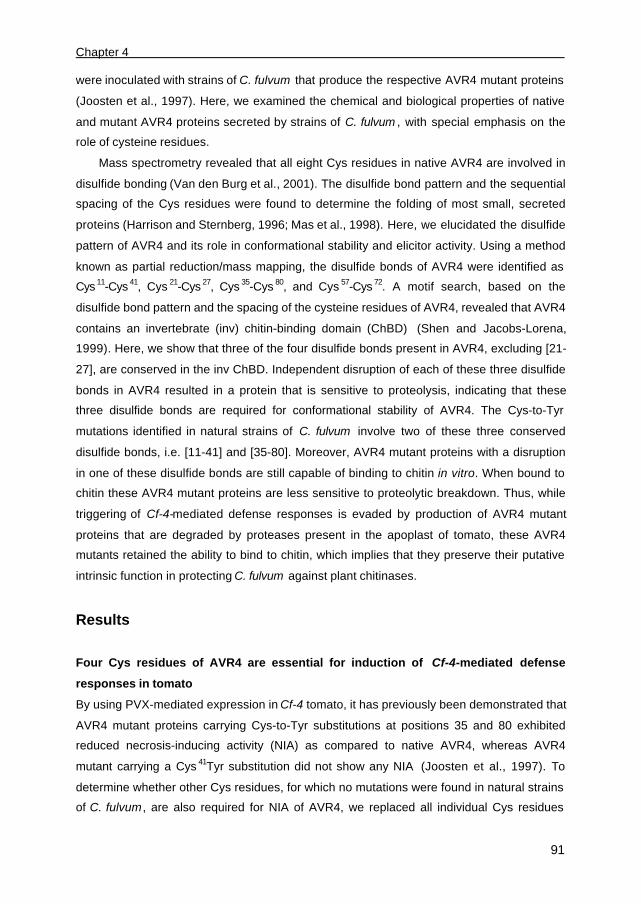
Chapter 4
91
were inoculated with strains of C. fulvum that produce the respective AVR4 mutant proteins
(Joosten et al., 1997). Here, we examined the chemical and biological properties of native
and mutant AVR4 proteins secreted by strains of C. fulvum , with special emphasis on the
role of cysteine residues.
Mass spectrometry revealed that all eight Cys residues in native AVR4 are involved in
disulfide bonding (Van den Burg et al., 2001). The disulfide bond pattern and the sequential
spacing of the Cys residues were found to determine the folding of most small, secreted
proteins (Harrison and Sternberg, 1996; Mas et al., 1998). Here, we elucidated the disulfide
pattern of AVR4 and its role in conformational stability and elicitor activity. Using a method
known as partial reduction/mass mapping, the disulfide bonds of AVR4 were identified as
Cys 11-Cys 41, Cys 21-Cys 27, Cys 35-Cys 80, and Cys 57-Cys 72. A motif search, based on the
disulfide bond pattern and the spacing of the cysteine residues of AVR4, revealed that AVR4
contains an invertebrate (inv) chitin-binding domain (ChBD) (Shen and Jacobs-Lorena,
1999). Here, we show that three of the four disulfide bonds present in AVR4, excluding [21-
27], are conserved in the inv ChBD. Independent disruption of each of these three disulfide
bonds in AVR4 resulted in a protein that is sensitive to proteolysis, indicating that these
three disulfide bonds are required for conformational stability of AVR4. The Cys-to-Tyr
mutations identified in natural strains of C. fulvum involve two of these three conserved
disulfide bonds, i.e. [11-41] and [35-80]. Moreover, AVR4 mutant proteins with a disruption
in one of these disulfide bonds are still capable of binding to chitin in vitro. When bound to
chitin these AVR4 mutant proteins are less sensitive to proteolytic breakdown. Thus, while
triggering of Cf-4-mediated defense responses is evaded by production of AVR4 mutant
proteins that are degraded by proteases present in the apoplast of tomato, these AVR4
mutants retained the ability to bind to chitin, which implies that they preserve their putative
intrinsic function in protecting C. fulvum against plant chitinases.
Results
Four Cys residues of AVR4 are essential for induction of Cf-4-mediated defense
responses in tomato
By using PVX-mediated expression in Cf-4 tomato, it has previously been demonstrated that
AVR4 mutant proteins carrying Cys-to-Tyr substitutions at positions 35 and 80 exhibited
reduced necrosis-inducing activity (NIA) as compared to native AVR4, whereas AVR4
mutant carrying a Cys 41Tyr substitution did not show any NIA (Joosten et al., 1997). To
determine whether other Cys residues, for which no mutations were found in natural strains
of C. fulvum , are also required for NIA of AVR4, we replaced all individual Cys residues

Disulfide bond mutants of AVR4
92
independently by Ala. The mutant Avr4 constructs were subsequently introduced into the
PVX vector and inoculated onto four-week-old Cf-4 tomato plants. As found for AVR4
mutant carrying Cys 41Tyr substitution, it appeared that mutant proteins carrying an Ala
substitution at positions Cys 11 or Cys 41 did not exhibit any NIA (Fig. 1). Moreover, inoculation
of PVX::Avr4 constructs encoding AVR4 mutant proteins carrying a Cys 21Ala or Cys 27Ala
substitution resulted in the same NIA as demonstrated for constructs that encode AVR4
mutant proteins carrying Cys 35Ala and Cys 80Ala substitutions. The latter two mutants are as
active as the Cys 35Tyr and Cys 80Tyr substitution mutants found in natural strains of C.
fulvum. Thus, the finding that AVR4 mutant proteins carrying Cys-to-Ala substitutions at
positions 11, 41, 57, and 72 all show no NIA suggests that these residues are involved in the
formation of disulfide bonds in AVR4. As these mutants already lack NIA, subsequent
analysis using double Cys-to-Ala substitution mutants would not result in further assignment
of disulfide bond pattern of AVR4 involving these Cys residues. For residues Cys 21, Cys ,27,
Cys 35 and Cys 80 of which the single Cys-to-Ala substitution mutants showed reduced NIA,
however, six additional PVX::Avr4 constructs encoding AVR4 mutant proteins with double
Cys-to-Ala substitutions were obtained. Four of the six double mutants did no longer exhibit
NIA, whereas mutants carrying Cys 21,27Ala and Cys 35,80Ala substitutions were as active as
mutants carrying one of the corresponding single Cys-to-Ala substitution (Table 1). These
data suggest that Cys 21 is connected with Cys 27 and Cys 35 with Cys 80.
Fig. 1. Necrosis-inducing activity of PVX::Avr4 derivatives in MM-Cf4 tomato plants. The first
group of four-week-old MM-Cf4 tomato plants were inoculated with PVX::Avr4 derivatives containing a
mutation in either Cys11 (1), Cys41 (5), Cys57 (6), or Cys72 (7). Note that these plants exhibited no
systemic necrosis, but rather systemic mosaic symptoms induced by the viral infection. The second
group of MM-Cf4 plants was inoculated with PVX::Avr4 derivatives containing a mutation in Cys21 (2),
Cys27 (3), Cys35 (4), or Cys80 (8). Note that these plants developed severe necrotic symptoms. The
photograph was taken 10 days post inoculation. The Cys residues of AVR4, for which modifications
were found in strains of C. fulvum virulent on plants carrying Cf-4, are underlined.

Chapter 4
93
Table 1. Necrosis-inducing activity (NIA) of PVX::Avr4
derivatives on MM-Cf4 tomato plants.
Single Cys-to-Ala
substitution
NIA Double Cys-to-Ala
substitutions
NIA
-Cys11AlaCys21AlaCys27AlaCys35AlaCys41AlaCys57AlaCys72AlaCys80Ala
+++++-
++++++---
++
Cys21, 27AlaCys21, 35AlaCys21, 80AlaCys27, 35AlaCys27, 80AlaCys35, 80Ala
++----
++
Chemical reduction and cyanylation of the disulfide bonds in AVR4
In order to determine the disulfide bond pattern of AVR4, we followed a chemical approach.
We partially reduced the disulfide bonds by treating AVR4 with tris-(2-carboxyethyl)-
phosphine hydrochloride (TCEP) at pH 3.0. This treatment minimized intramolecular
rearrangements of disulfide bonds (Gray, 1993; Van den Hooven et al., 2001). The free
cysteine thiol groups were directly modified by alkylation with 1-cyano-4-diethylamino-
pyridinium (CDAP) under acidic conditions and the resulting peptides were separated by
reverse-phase HPLC (Fig. 2). In the presence of a 6-fold molar excess of TCEP,
approximately 50% of native AVR4 was reduced as observed by an increased HPLC
retention time of the formed species (Fig. 2). Subsequent MALDI-TOF mass spectrometry
(MS) identified four peaks that represent AVR4 species with one reduced disulfide bond
(indicated by *, Fig. 2). Peaks that elute at higher acetonitrile concentrations contain AVR4
species with more than one disulfide bond reduced, which is consistent with the finding that
higher retention times reflect an increase in hydrophobicity and therefore an increase in the
unfolded state of the AVR4 species (data not shown). The peaks eluting at 30.8 min and
30.9 min could not be separated in one run on HPLC, but after an additional run both
species appeared more than 85% pure.
Assignment of the disulfide bonds in AVR4 using mass spectrometry
To determine which of disulfide bond was reduced in the AVR4 des-species, the collected
protein fractions were lyophilized and dissolved in 1 M NH4OH to induce base-catalyzed
cleavage of the peptide bond that precedes the two modified half-cystines. This cleavage
leads to peptide iminothiazolidine derivatives (itz) (Wu and Watson, 1998). The double
cleavage reaction is not always complete and as a side-reaction β-elimination might occur at
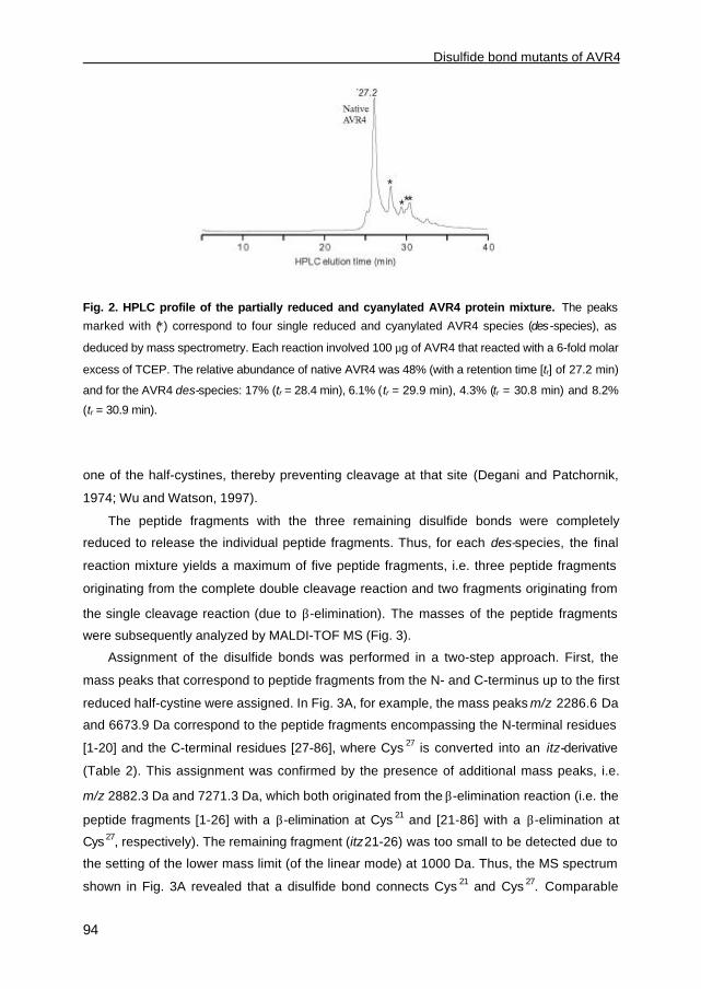
Disulfide bond mutants of AVR4
94
Fig. 2. HPLC profile of the partially reduced and cyanylated AVR4 protein mixture. The peaks
marked with (∗) correspond to four single reduced and cyanylated AVR4 species (des-species), as
deduced by mass spectrometry. Each reaction involved 100 µg of AVR4 that reacted with a 6-fold molar
excess of TCEP. The relative abundance of native AVR4 was 48% (with a retention time [tr] of 27.2 min)
and for the AVR4 des-species: 17% (tr = 28.4 min), 6.1% (tr = 29.9 min), 4.3% (tr = 30.8 min) and 8.2%
(tr = 30.9 min).
one of the half-cystines, thereby preventing cleavage at that site (Degani and Patchornik,
1974; Wu and Watson, 1997).
The peptide fragments with the three remaining disulfide bonds were completely
reduced to release the individual peptide fragments. Thus, for each des-species, the final
reaction mixture yields a maximum of five peptide fragments, i.e. three peptide fragments
originating from the complete double cleavage reaction and two fragments originating from
the single cleavage reaction (due to β-elimination). The masses of the peptide fragments
were subsequently analyzed by MALDI-TOF MS (Fig. 3).
Assignment of the disulfide bonds was performed in a two-step approach. First, the
mass peaks that correspond to peptide fragments from the N- and C-terminus up to the first
reduced half-cystine were assigned. In Fig. 3A, for example, the mass peaks m/z 2286.6 Da
and 6673.9 Da correspond to the peptide fragments encompassing the N-terminal residues
[1-20] and the C-terminal residues [27-86], where Cys 27 is converted into an itz-derivative
(Table 2). This assignment was confirmed by the presence of additional mass peaks, i.e.
m/z 2882.3 Da and 7271.3 Da, which both originated from the β-elimination reaction (i.e. the
peptide fragments [1-26] with a β-elimination at Cys 21 and [21-86] with a β-elimination at
Cys 27, respectively). The remaining fragment (itz21-26) was too small to be detected due to
the setting of the lower mass limit (of the linear mode) at 1000 Da. Thus, the MS spectrum
shown in Fig. 3A revealed that a disulfide bond connects Cys 21 and Cys 27. Comparable

Chapter 4
95
mass peak analyses of the peptide mixtures generated from the other three AVR4 des-
species resulted in the assignment of disulfide bonds [11-41], [57-72] and [35-80] (Fig. 3B,
3C and 3D, respectively). Together, these data demonstrate that a disulfide bond connects
Cys 21 and Cys 27, as well as Cys 35 and Cys 80, which is consistent with the data obtained in
the PVX bioassays (Fig. 1; Table 1). Moreover, the mass mapping allowed us to assign two
additional disulfide bonds, [11-41] and [57-72], in AVR4.
Fig. 3. MALDI-TOF mass spectra of the four peptide mixtures obtained after base-induced
cleavage of the peptide bond and full reduction of the AVR4 des-species. The mass per charge
(m/z) is given for those peaks that are used to assign what Cys residues are involved in formation of
intramolecular disulfide bonds. The panels A-D represent (in order of elution from the HPLC) peptide
mixtures containing AVR4 des-species A, [21-27], B, [11-41], C, [57-72] and D, [35-80], respectively.
The mass peaks that correspond to double charged mass peaks ([M + 2H]2+) (∗) are indicated. Re-
occurring mass peaks (#) reflect cross-contamination with other AVR4 des-species.
Three disulfide bonds in AVR4 are required for its conformational stability
Previously, no AVR4 mutant proteins could be detected in apoplastic fluid (AF) isolated from
tomato plants infected by strains of C. fulvum producing these isoforms. Therefore, we
compared the stability of the four mutant AVR4 des-species with native AVR4. It appeared
that, in the absence of AF, native AVR4 as well as the AVR4 des-species are stable over the
time span of one hr (Fig. 4). However, when mutant AVR4 des-species were incubated with

Disulfide bond mutants of AVR4
96
Table 2. Theoretical masses (m/z) of peptide fragments obtained after base-catalyzed cleavage
of peptide bonds in AVR4 des-species*.
Des-[11-41] Des-[21-27] Des-[35-80] Des-[57-72]
Fragment m/z Fragment m/z Fragment m/z Fragment m/z
1-10
itz11-40
itz 41-86
β-(1-40)
β-(11-86)
1144.3
3415.9
5075.9
4482.2
8413.8
1-20
itz 21-26
itz 27-86
β-(1-26)
β-(21-86)
2285.6
676.8
6673.7
2884.4
7272.4
1-34
itz 35-79
itz 80-86
β-(1-79)
β-(35-86)
3805.4
5031.8
798.9
8759.2
5752.7
1-56
itz 57-71
itz 72-86
β-(1-71)
β-(57-86)
6160.2
1800.0
1675.9
7882.2
3397.9
* ; AVR4 des-species represent cyanylated AVR4 proteins that carry one reduced disulfide bond
(Fig. 2). Base-catalyzed cleavage of the peptide bond that precedes the two modified half-cystines
results in peptide iminothiazolidine derivatives (itz). A side-reaction occurs when the double cleavage
reaction is not complete, resulting in β-elimination at one of the half-cystines (β). For the majority of
the peptide fragments, the relative deviation between measured and calculated masses is less than
0.05%.
AF, three of the four species could no longer be detected, whereas native AVR4 and [21-
27]-AVR4 des-species were not affected. Moreover, in the presence of protease inhibitors
(Fig. 4, +AF, + Prot. Inh.), treatment with AF did no longer affect the stability of these AVR4
des-species, suggesting that these AVR4 species are degraded by proteases present in AF.
Thus, unlike disulfide bond [21-27], disulfide bonds [11-41], [35-80] and [57-72] are required
for conformational stability of AVR4.
AVR4 contains an invertebrate chitin-binding domain
The identified disulfide bond pattern of AVR4 was further used to perform a motif-based
search (URL:http://motif.genome.ad.jp). This search identified a sequence stretch in AVR4
that was homologous to a stretch present in proteins of the invertebrates Manduca sexta,
Brugia malayi, and Penaeus japonicus, all of which exhibit chitinase activity (Shen and
Jacobs-Lorena, 1999). The homology, however, is restricted to the C-terminal domain,
which contains six conserved cysteine residues. Moreover, this domain has recently been
identified as the invertebrate chitin-binding domain (inv ChBD) (Shen and Jacobs-Lorena,
1999). For one member of the inv ChBD family, tachycitin of Japanese Horseshoe crab
(Tachypleus tridentatus), the disulfide bond pattern has been solved (Kawabata et al.,
1996). Unlike AVR4, tachycitin contains five disulfide bonds of which three are conserved
within the inv ChBD. Subsequent sequence alignment revealed that the disulfide bonds [11-
41], [35-80], and [57-72] present in AVR4 are conserved within the ChBD family (Fig. 5). The
additional disulfide bond [21-27] within AVR4 does not share homology with disulfide bonds
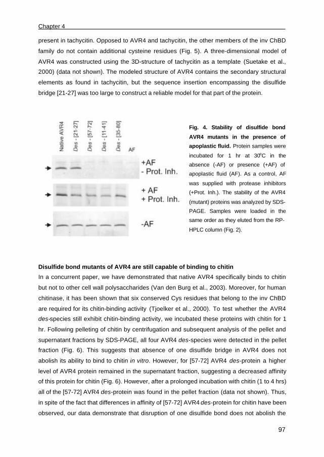
Chapter 4
97
present in tachycitin. Opposed to AVR4 and tachycitin, the other members of the inv ChBD
family do not contain additional cysteine residues (Fig. 5). A three-dimensional model of
AVR4 was constructed using the 3D-structure of tachycitin as a template (Suetake et al.,
2000) (data not shown). The modeled structure of AVR4 contains the secondary structural
elements as found in tachycitin, but the sequence insertion encompassing the disulfide
bridge [21-27] was too large to construct a reliable model for that part of the protein.
Fig. 4. Stability of disulfide bond
AVR4 mutants in the presence of
apoplastic fluid. Protein samples were
incubated for 1 hr at 30oC in the
absence (-AF) or presence (+AF) of
apoplastic fluid (AF). As a control, AF
was supplied with protease inhibitors
(+Prot. Inh.). The stability of the AVR4
(mutant) proteins was analyzed by SDS-
PAGE. Samples were loaded in the
same order as they eluted from the RP-
HPLC column (Fig. 2).
Disulfide bond mutants of AVR4 are still capable of binding to chitin
In a concurrent paper, we have demonstrated that native AVR4 specifically binds to chitin
but not to other cell wall polysaccharides (Van den Burg et al., 2003). Moreover, for human
chitinase, it has been shown that six conserved Cys residues that belong to the inv ChBD
are required for its chitin-binding activity (Tjoelker et al., 2000). To test whether the AVR4
des-species still exhibit chitin-binding activity, we incubated these proteins with chitin for 1
hr. Following pelleting of chitin by centrifugation and subsequent analysis of the pellet and
supernatant fractions by SDS-PAGE, all four AVR4 des-species were detected in the pellet
fraction (Fig. 6). This suggests that absence of one disulfide bridge in AVR4 does not
abolish its ability to bind to chitin in vitro. However, for [57-72] AVR4 des-protein a higher
level of AVR4 protein remained in the supernatant fraction, suggesting a decreased affinity
of this protein for chitin (Fig. 6). However, after a prolonged incubation with chitin (1 to 4 hrs)
all of the [57-72] AVR4 des-protein was found in the pellet fraction (data not shown). Thus,
in spite of the fact that differences in affinity of [57-72] AVR4 des-protein for chitin have been
observed, our data demonstrate that disruption of one disulfide bond does not abolish the

Disulfide bond mutants of AVR4
98
chitin-binding activity of AVR4. It should be mentioned here that [57-72] AVR4 des-protein
might have been contaminated with [35-80] AVR4 des-protein, which could in turn have
interfered with the chitin-binding assay. This implies that, as the [35-80] AVR4 des-protein
exhibits the same affinity for chitin as native AVR4, the [57-72] AVR4 des-protein might even
exhibit a lower affinity for chitin than observed in our assay.
Fig. 5. Schematic diagram of the
disulfide bond pattern of tachycitin and
AVR4. The connected open bars represent
three conserved disulfide bonds of the
invertebrate chitin-binding domain (inv
ChBD). The connected filled bars
represent additional disulfide bonds. Gaps
in the alignment are indicated by a thin-line
in tachycitin. The residue numbers
(bottom) and the successive order (top) of the Cys residues of AVR4 are depicted. The diagram is
based on the disulfide bond pattern of tachycitin and the spacing of cysteine residues of other inv
ChBDs, using the consensus found in SMART and PFAM protein domain databases (ID codes
SM00494 / PF01607) (Schultz et al., 1998; Bateman et al., 2002; Van den Burg et al., 2003).
Binding to chitin protects disulfide bond mutants of AVR4 against proteolytic
breakdown
During growth in tomato, C. fulvum remains confined to the intercellular spaces of tomato
leaves (De Wit, 1977). It has been proposed that after secretion AVR4 binds directly to
regions of the cell wall of hyphae of C. fulvum, where chitin is accessible (Van den Burg et
al., 2003). When the exposed chitin is saturated with AVR4, free AVR4 is thought to be
released into the apoplast. We investigated whether the AVR4 des-species were less
sensitive to proteolytic degradation after binding to chitin. Therefore, we pre-incubated these
AVR4 des-species with chitin for four hrs, rather than one hr, to ensure a complete
association between these proteins and chitin. After incubation with chitin, the solutions
were supplied with AF and stability of AVR4 mutant proteins was followed in time. Over the
period of four hrs, native AVR4 and the [21-27] AVR4 des-protein that were incubated with
chitin, remained fully stable in the presence of AF (Fig. 7; Table 3). Moreover, when bound
to chitin, normally unstable AVR4 des-species [11-41], [35-80] and [57-72] are less sensitive
to degradation by proteases present in AF.
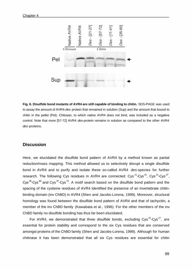
Chapter 4
99
Fig. 6. Disulfide bond mutants of AVR4 are still capable of binding to chitin. SDS-PAGE was used
to assay the amount of AVR4-des protein that remained in solution (Sup) and the amount that bound to
chitin in the pellet (Pel). Chitosan, to which native AVR4 does not bind, was included as a negative
control. Note that more [57-72] AVR4 des-protein remains in solution as compared to the other AVR4
des-proteins.
Discussion
Here, we elucidated the disulfide bond pattern of AVR4 by a method known as partial
reduction/mass mapping. This method allowed us to selectively disrupt a single disulfide
bond in AVR4 and to purify and isolate these so-called AVR4 des-species for further
research. The following Cys residues in AVR4 are connected: Cys 11-Cys 41, Cys 21-Cys 27,
Cys 35-Cys 80 and Cys 57-Cys 72. A motif search based on the disulfide bond pattern and the
spacing of the cysteine residues of AVR4 identified the presence of an invertebrate chitin-
binding domain (inv ChBD) in AVR4 (Shen and Jacobs-Lorena, 1999). Moreover, structural
homology was found between the disulfide bond pattern of AVR4 and that of tachycitin, a
member of the inv ChBD family (Kawabata et al., 1996). For the other members of the inv
ChBD family no disulfide bonding has thus far been elucidated.
For AVR4, we demonstrated that three disulfide bonds, excluding Cys 21-Cys 27, are
essential for protein stability and correspond to the six Cys residues that are conserved
amongst proteins of the ChBD family (Shen and Jacobs-Lorena, 1999). Although for human
chitinase it has been demonstrated that all six Cys residues are essential for chitin

Disulfide bond mutants of AVR4
100
Fig. 7. Binding to chitin protects disulfide bond mutants of AVR4 against proteolytic breakdown.
To test whether binding to chitin protects normally unstable disulfide bond mutants of AVR4 against
proteases present in apoplastic fluid (AF) of tomato, AVR4 des-species were pre-incubated with chitin,
followed by an incubation at 30°C for 4 hrs in the absence (-) or the presence (+) of AF. The stability of
the AVR4 des-species was analyzed by SDS-PAGE.
recognition (Tjoelker et al., 2000), for AVR4 disruption of one of the three disulfide bonds
does not abolish its ability to bind to chitin in vitro. It has been demonstrated that strains of
C. fulvum evade Cf-4-mediated resistance by modification of the Avr4 gene product. Most
modifications involved substitution of Cys residues at positions 35, 41 and 80 by Tyr
(Joosten et al., 1997), indicating that disruption of only two of the three conserved disulfide
bonds have, so far, contributed to evasion of AVR4 recognition. When AVR4 mutant
proteins carrying a disruption in one of these conserved disulfide bonds were exposed to
apoplastic fluid (AF) isolated from healthy or C. fulvum-infected tomato leaves, the proteins
are rapidly degraded as a result of protease activity present in AF. Thus, as previously
suggested by Joosten et al. (1997), C. fulvum evades Cf-4-mediated resistance by
production of AVR4 isoforms that are degraded by proteases present in the apoplast of
tomato leaves. Interestingly, these unstable AVR4 isoforms still exhibit chitin-binding activity.
However, disruption of disulfide bond [57-72] was found to reduce the affinity of AVR4 for
chitin. Moreover, the 3D-structure of tachycitin shows that disulfide bond [57-72] is present
in a functional chitin-binding moiety (Suetake et al., 2000), which supports a decreased
affinity for chitin when this bond is disrupted. Assuming that binding of AVR4 to chitin
protects C. fulvum against plant chitinases, as proposed by Van den Burg et al. (2003),
strains of C. fulvum that produce AVR4 mutant proteins lacking disulfide bond [57-72] to
evade Cf-4-mediated resistance would probably be more sensitive towards plant chitinases.

Chapter 4
101
Table 3. Overview of necrosis-inducing activity, stability and chitin-binding ability of AVR4
disulfide bond mutants.
Disrupted
Disulfide
bond1
Cys residue
pair2
NIA 3 Stability in
AF4
Binding to
chitin5
Stability of
chitin-bound
AVR46
Identified
number of
strains 7
- - +++++ + + + ND
[21-27] 2-3 ++ + + + 0
[57-72] 6-7 - - +/- +/- 0
[11-41] 1-5 - - + +/- 2
[35-80] 4-8 ++ - + + 6
1) Disruption of one disulfide bond either by a cysteine substitution in PVX::Avr4 or by partial
chemical reduction
2) Successive numbering of the cysteine residues in AVR4 (Fig. 5)
3) Necrosis-inducing activity (NIA) was assayed using PVX-mediated expression of Avr4 derivatives
in MM-Cf4 tomato plants (Fig. 1)
4) AVR4 des-species are present (+) or absent (-) after incubation with apoplastic fluid (AF) (Fig. 4)
5) Affinity of AVR4 des-species for chitin; +: binds to chitin; +/- :decreased affinity for chitin (Fig. 6)
6) Stability of the AVR4 des-species in AF after binding to chitin ; +: stable in the presence of chitin;
+/-: partially stable in the presence of chitin (Fig. 7)
7) Number of strains of Cladosporium fulvum identified so far carrying a single disulfide bond mutant
of AVR4 (Joosten et al., 1994 and 1997).
ND: Not determined
Interestingly, strains of C. fulvum producing such a disulfide bond mutant of AVR4 have not
been found in nature, possibly due to the fact that modification of [57-72] in AVR4 carries a
virulence penalty for C. fulvum .
The disulfide bond [21-27] in AVR4 does not seem to contribute to conformational
stability of the protein, as AVR4 mutants lacking this bond are not degraded by proteases
present in AF (Fig. 4). Moreover, this disulfide bond is absent in proteins of the inv ChBD
family, suggesting that it might form a solvent-exposed loop (Fig. 5). Preliminary NMR
analysis of AVR4 revealed that the region surrounding Cys 21 adopts an α-helix (C. Spronk
and H. van den Burg, unpublished data). As demonstrated previously for a cysteine-knot
protein (Darling et al., 2000), the α-helix might increase the stability of this part of AVR4,
implying that disulfide bond [21-27] itself is not essential for the conformational stability of
AVR4.
When expressed in Cf-4 tomato plants using the expression vector PVX, the necrosis-
inducing activity (NIA) of AVR4 lacking disulfide bond [21-27] was found to be similar to the
NIA of AVR4 mutant proteins lacking [35-80]. The NIA of both mutants, however, is less than

Disulfide bond mutants of AVR4
102
that of native AVR4. However, in contrast to disulfide bond [35-80], disulfide bond [21-27]
does not contribute to conformational stability of AVR4, suggesting that the mechanisms
underlying the reduced NIA of both AVR4 des-species are different. Possibly, when AVR4
mutant carrying a disrupted disulfide bond [35-80] is expressed by PVX in tomato, a fraction
of the protein is degraded by proteases present in the intercellular space, while the
remaining fraction of the protein triggers Cf-4-mediated defense responses. The reduced
NIA of AVR4 mutant carrying a disrupted disulfide bond [21-27] cannot be explained by
sensitivity to proteases, but is most likely due to the amino acid substitution itself or due to a
conformational change of the protein. Possibly, the remaining NIA of disulfide bond mutant
[21-27] of AVR4 still triggers Cf-4-mediated defense responses that restrict growth of C.
fulvum . This could be a reason why, in spite of the fact that disruption of disulfide bond [21-
27] does not affect chitin-binding activity of AVR4, no strains of C. fulvum virulent on plant
carrying Cf-4 have thus far been found that carry such a mutation.
In addition to the three Cys substitutions present in the majority of mutants found in
nature and discussed above, two other natural amino acid substitutions have been found in
AVR4 (Thr37Ile and Tyr 38His) (Joosten et al., 1997). Both modifications are thought to affect
the conformational stability of AVR4, based on a report by Zhu and Braun (1999). The
substituted residues are located in strand β2 of the first anti-parallel β-sheet, which is in the
core of the protein structure (Suetake et al., 2000). In AVR4, Thr37 and Tyr38 are putatively
paired with Pro30 and Tyr29 in strand β1, respectively, and these four residues also appear to
be conserved in the inv ChBD (Van den Burg et al., 2003). Moreover, Zhu and Braun (1999)
showed that in β-strands Pro-Ile cross-strand contact pairs are virtually absent, whereas a
Pro-Thr cross-strand contact pair is allowed (Zhu and Braun, 1999). Similarly, aromatic
residues are exclusively found at position 38 (Tyr/ Phe/ Trp > 98%) of the inv ChBD. They
also showed that hydrophobic residues are the favorite cross-strand contact partners,
whereas His residues are not favored as a cross-strand contact partner for these
hydrophobic residues. Therefore, we propose that Thr37Ile and Tyr38His substitutions will
both lead to a partially destabilization of the anti-parallel β-sheet, which in turn could
decrease conformational stability of AVR (See also Chapters 2 and 5).
Altogether, the disulfide bond AVR4 mutants produced by natural strains of C. fulvum
show increased sensitivity towards proteases present in the apoplast as compared to native
AVR4. However, these AVR4 mutant proteins retained the ability to bind to chitin, and
moreover, when bound to chitin they are less sensitive to proteolytic breakdown. This
suggests that strains virulent on plants carrying Cf-4 produce disulfide bond mutants of
AVR4 that escape degradation by proteases present in the apoplast upon binding to the cell
wall of C. fulvum at site where chitin is accessible. Thus, while Cf-4-mediated resistance is
Chapter 4
103
evaded, the putative intrinsic function of the disulfide bond mutants of AVR4, in protecting
the cell wall of C. fulvum against plant chitinases, is retained.
Materials and Methods
Construction of PVX derivatives and PVX inoculation procedure
Avr4 mutants encoding various Cys-to-Ala substitutions were generated by PCR-based site-
directed mutagenesis on the plasmid pTX∆GC3a, containing the native Avr4 sequence
(Joosten et al., 1997). PCR amplification was carried out using mutagenic primers (Table 4)
designed to generate two overlapping PCR fragments that carry the introduced mutation.
These PCR fragments were subsequently used in a PCR-mediated overlap extension, using
forward primer OX10 and reverse primer N31 (Table 4). The PCR product was digested with
ClaI, and cloned into the ClaI-digested site of the expression vector pTX∆GC3a (Chapman
et al., 1992) and sequenced. In vitro transcription of the plasmids and subsequent
inoculation on Nicotiana clevelandii and tomato was performed as described (Joosten et al.,
1997). N. clevelandii and the tomato (Lycopersicon esculentum ) cultivars Moneymaker (MM)
and the near-isogenic line MM-Cf4 were grown as described (De Wit and Flach, 1979).
Table 4. Mutagenic primers of Avr4 used to introduce Cys-to-Ala
substitutions in PVX::Avr4.
Primer 5’ 3’C11AaC11AbC21AaC21AbC27AaC27AbC35AaC35AbC41AaC41AbC57AaC57AbC72AaC72AbC80AaC80Ab
OX10N31
CTT GGG GCT TGG CTG GGT TGT ATG GCCA TAC AAC CCA GCC AAG CCC CAA GCCT TGG GAC CCA TGG CCT TGG TGT CGCGA CAC CAA GGC CAT GGG TCC CAA GGGGG TTC GGG TAG AGG GCA TCC TTG GGA CCCGGG TCC CAA GGA TGC CAC TGT CGG GGGTA TGT AGG TTG TGG CAC TGT CGG GGCCC CGA CAG TGC CAC AAC CTA CAT ACCGT CGA GCG GTA CGG CCT GTA TGT AGGCCT ACA TAC AGG CCG TAC CGC TCG ACGGCA GTC CTT TTG GGG CTG GCT TAA CCA CGTG GTT AAG CCA GCC CCA AAA GGA CTG CGGT TTG GAT AGT CGG CCC ACT TCT TGC CGGC AAG AAG TGG GCC GAC TAT CCA AAC CGTC TTT ACC GGG GCC GTA CTC AGGCCT GAG TAC GGC CCC GGT AAA GAC
CAA TCA CAG TGT TGG CTT GCGAC CCT ATG GGC TGT GTT G
Partial reduction and cyanylation of AVR4
Expression of heterologous AVR4 was achieved in the methylotrophic yeast Pichia pastoris,
and AVR4 was purified from culture fluid (Van den Burg et al., 2001). The disulfide bonds of

Disulfide bond mutants of AVR4
104
AVR4 were partially reduced with TCEP (Sigma) (Wu and Watson, 1997 and 1998). An 0.1
M TCEP stock solution was prepared in 6 M Guanidine-HCl in 0.1 M citrate buffer (pH 3)
and stored at –20oC (up to six months without deterioration). For each reduction reaction,
100 µg of native AVR4 was dissolved in 10 µL of 6 M guanidine-HCl in 0.1 M citrate buffer
(pH 3). The reaction was initiated by adding a 6-fold molar excess of TCEP to AVR4,
following an incubation at 20oC for 15 min. Subsequently, a 80-fold molar excess of CDAP
(Sigma) was added to cyanylate the freed thiol groups (15 min, 20 oC, in the dark). The 0.1
M CDAP stock solution in 6M guanidine-HCl in 0.1 M citrate buffer (pH 3) was freshly
prepared prior to each reaction.
Reverse-phase high-performance liquid chromatography of the partially reduced
AVR4 protein mixture
The TCEP/CDAP reaction mixtures were separated by analytical reverse-phase high
performance liquid chromatography (RP-HPLC), using a 150 × 3.9 mm Delta-Pak C18
column (300 Å, 5 µm; Waters). The separation was monitored at 215 nm and predominant
peaks were manually collected. HPLC elution solvents consisted of 0.1% (v/v) trifluoracetic
acid (TFA) in water (solvent A), and 0.1% (v/v) TFA in acetonitrile (solvent B). HPLC was
operated at a flow rate of 1 mL/min. The applied gradient was from 5 to 20% (percentage B
in solvent A) in 2 min, from 20 to 30% in 40 min, and from 30% to 60% in 3 min. AVR4
eluted at approximately 25% of solvent B. Integration of the HPLC profile was achieved
using the supplied Waters software. Appropriate fractions (containing the AVR4 des-
species) were lyophilized before storage. All solvents used were HPLC grade.
Base-catalyzed cleavage and identification of the fully reduced peptide fragments by
mass spectrometry
Lyophilized HPLC fractions containing the AVR4 des-species were dissolved, first, in 2 µL of
1 M NH4OH, 6 M guanidine-HCl, followed by 5 µL of 1 M NH4OH, and incubated at 20oC for
1 hr. The excess of NH4OH was evaporated in a Speed-Vac system for approximately 30
min to obtain an almost dry sample. Subsequently, the remaining disulfide bonds were
reduced by adding an excess of TCEP (10 µL of 0.1 M TCEP stock) and the mixture was
incubated at 37oC for 30 min. The peptide mixtures were analyzed by MALDI-TOF mass
spectrometry (MS) (Perceptive Biosystems Voyager DE-RP). Thereby, small aliquots of the
peptide samples were applied to a saturated matrix solution that was freshly prepared (α-
cyano-4-hydroxycinnamic acid; Sigma; 10 mg/mL in acetonitrile/water/TFA [50/50/1, v/v/v])
and 1 µL was deposited on a sample plate (Karas and Hillenkamp, 1988; Kussmann et al.,
1997). Depicted spectra represent averages of 100 to 256 consecutive laser pulses. The

Chapter 4
105
instrument was generally operated in the positive mode at an acceleration voltage of 23 kV,
combined with a delayed extraction. Spectra were externally calibrated with bovine
Cytochrome C (12,230.9 Da), bovine Insulin (5,734.6 Da) (both Sigma) and Microperoxidase
8 (1,506.5 Da) (Primus et al., 1999).
Incubation of the AVR4 des-species with apoplastic fluid
Apoplastic fluid (AF) was isolated from leaves of MM-Cf4 plants (De Wit and Spikman, 1982)
that were infected with strain #38 of C. fulvum (race 4) (Joosten et al., 1997). Native AVR4
and AVR4 des-species (4 µg) were incubated at 30oC for 1 hr in the presence of 0.1 µL AF
(~0.1 µg total protein). Protease inhibitors consist of a standard protein inhibitor cocktail with
EDTA (Roche; 1 tablet/ml of AF). Protein samples were separated by tricine SDS-PAGE
(Schägger et al., 1987).
The polysaccharide-binding assay
Native AVR4 and AVR4 des-species (4 µg) were incubated at room temperature for 1 hr
(unless stated otherwise) with an excess of 5 mg of insoluble chitin beads (New England
Biolabs) or chitosan (Sigma) in 50 mM Tris ⋅HCl (pH 8) and 150 mM NaCl (500 µL final
volume) as described (Van den Burg et al., 2003). The insoluble fraction was precipitated by
centrifugation (13,000 x g for 3 min.). The supernatant was recovered and lyophilized. The
pellet fraction was boiled in 200 µL 1% SDS to release bound protein and subsequently
centrifuged. The retrieved supernatant (containing bound AVR4) fraction as well as the
lyophilized supernatant fraction (containing unbound AVR4) was examined for its protein
content by tricine SDS-PAGE.
Molecular modeling of AVR4 using the NMR structure of tachycitin
The NMR structure of tachycitin (ID code PDB databank: 1DQC) was used as template
structure to model the structure of AVR4 using Modeler 6.1 (Sali and Blundell, 1993; Sali et
al., 1995; Fiser et al., 2000). Additional loop refinement was used to model the two relatively
large gaps, i.e. 6- and 12 amino acids. The disulfide bonds were fixed during the
calculations. A thousand models were constructed, of which the ten lowest scoring
structures were further examined. The models were found reliable using standard algorithms
(Sippl, 1993; Sanchez and Sali, 1998).

106
Chapter 5
Structural separation of necrosis-inducing activity and
chitin-binding ability within the AVR4 protein
Nienke Westerink, Iris J.E. Stulemeijer, Harrold A. Van den Burg, Pierre J.G.M. De Wit and
Matthieu H.A.J. Joosten.

Chapter 5
108
Necrosis-inducing activity and chitin-binding ability within AVR4
109
Structural separation of necrosis-inducing activity and chitin-
binding ability within the AVR4 protein
Abstract
The product of avirulence gene Avr4 of the tomato pathogen Cladosporium fulvum has been
identified by its ability to elicit Cf-4-mediated defense responses, including the
hypersensitive response (HR), in tomato. The AVR4 elicitor has also been demonstrated to
share structural homology with the chitin-binding domain of chitin-binding proteins of
invertebrates (inv ChBD) and to bind to chitin in vitro. AVR4 does not require binding to
chitin in order to elicit a HR. Here, we analyzed whether domains responsible for necrosis-
inducing activity (NIA) and chitin-binding ability within AVR4 can be distinguished. Therefore,
we performed a peptide scan (PEPSCAN) analysis using polyclonal antibodies raised
against native AVR4 to identify antigenic domains in AVR4 containing putatively solvent-
exposed residues that condition NIA. This PEPSCAN identified one major antigenic domain
(Cys 41-Cys 57) and two minor antigenic domains (Ile17-Asn31; Gln62-Asn76) in the AVR4
protein. All single residues, except for Cys, Pro, and Gly, which are present in the major
antigenic domain were replaced by Ala, as this domain exhibits the highest affinity for AVR4
antibodies. Moreover, to determine which residues are important for the chitin-binding ability
of AVR4, all aromatic residues and two additional residues (conserved in inv ChBDs) were
replaced by Ala. We show that substitution of single amino acid residues in the major
antigenic domain of AVR4 does not affect its NIA. Substitution of the aromatic residues Tyr 38
and Trp63 by Ala, however, reduced the NIA of AVR4. These AVR4 mutant proteins appear
to be unstable, suggesting that Tyr 38 and Trp63 contribute to conformational stability rather
than to NIA of AVR4. In this study, no individual amino acid residues could be identified that
are essential for binding of AVR4 to chitin, suggesting that multiple rather than single amino
acid residues contribute to chitin-binding ability.
Introduction
The product of the avirulence gene Avr4 of the tomato pathogen Cladosporium fulvum has
been identified by its ability to confer avirulence to strains of the fungus on tomato plants
that carry the matching Cf-4 resistance gene (Joosten et al., 1994). Avr4 encodes a pre-pro-
protein of 135 amino acids that is targeted to the apoplastic space during growth in tomato
leaves and processed by plant and/or fungal proteases into a mature protein of 86 amino
Chapter 5
110
acids (Joosten et al., 1997). No sequence homology was found between AVR4 and other
sequences available in databases. A motif search, based on the spacing of the Cys residues
and the elucidated disulfide bond pattern of AVR4, revealed that AVR4 shares structural
homology with proteins of the invertebrate chitin-binding domain (inv ChBD) family (Van den
Burg et al., 2003), including tachycitin of the horseshoe crab Tachypleus tridentatus
(Kawabata et al., 1996). Although there is no significant amino acid sequence similarity
between inv ChBDs and plant ChBDs, a local structural similarity was found between the inv
ChBD of tachycitin and the ChBD of the plant chitin-binding protein hevein (Shen and
Jacobs-Lorena, 1999; Suetake et al., 2000). For hevein, the aromatic amino acid residues
Trp21 and Trp23 have been identified to establish specific binding to chitin-derived
oligosaccharides through hydrophobic interactions (Asensio et al., 1995 and 1998). This
binding is further strengthened by hydrogen bonding with residue Ser19 of hevein (Asensio et
al., 1998). These chitin-binding residues of hevein share a high degree of conservation with
hydrophobic and polar residues of ChBDs of invertebrate and plant proteins (Suetake et al.,
2000).
Van den Burg et al. (2003) have demonstrated that AVR4 binds to chitin in vitro and that
it protects both Trichoderma viride and Fusarium solani f.sp. phaseoli against cell wall lysis
and growth inhibition by plant chitinases in vitro. However, C. fulvum is insensitive toward
chitinases in vitro (Joosten et al., 1995), possibly due to the fact that chitin is not accessible
in cell walls of C. fulvum in vitro (Van den Burg et al., 2003). As the Avr4 gene is not
expressed during in vitro-growth of C. fulvum (Joosten et al., 1997), protection against plant
chitinases in vitro is independent of AVR4. On the other hand, the Avr4 gene is induced
instantly upon ingress of the stomata of tomato leaves and AVR4 was found to accumulate
on the cell wall of C. fulvum in planta, most likely at sites where chitin is accessible (Joosten
et al., 1997; Van den Burg et al., 2003). This implies that during growth of C. fulvum in
susceptible tomato leaves, AVR4 protects the fungus against plant chitinases. In addition,
most AVR4 mutant proteins secreted by strains of C. fulvum virulent on plants expressing
Cf-4 are modified in such way that they are degraded by proteases in the apoplastic space
of tomato, yet, still exhibit the ability to bind to chitin (Chapter 4). This indicates that AVR4
recognition is evaded without losing the putative intrinsic function of AVR4 in protecting the
cell wall of C. fulvum against hydrolytic activity of plant chitinases.
In AvrPto, an AVR protein that triggers Pto-mediated resistance towards Pseudomonas
syringae pv. tomato , a cluster of mutations has been identified that disrupt its avirulence but
not its virulence function (Shan et al., 2000). This structural separation of avirulence and
virulence function raised the question whether similar mutations can be identified in AVR4
that abolish its necrosis-inducing activity (NIA) on plants that express Cf-4, without affecting

Necrosis-inducing activity and chitin-binding ability within AVR4
111
its chitin-binding ability. For AVR9 mutant proteins that exhibit reduced NIA, it has been
demonstrated that these mutants also display a reduced affinity for polyclonal antibodies
raised against native AVR9, indicating that there is a positive correlation between
antigenicity and NIA (Mahé et al., 1998). To determine which amino acid residues in AVR4
are required for its NIA, we performed a peptide scan (PEPSCAN) analysis (Meloen et al.,
1995) using polyclonal antibodies raised against native AVR4. This epitope mapping
technique identified one major antigenic domain (Cys 41-Cys 57) and two minor antigenic
domains (Ile17-Asn31; Gln62-Asn76). Here, we replaced all single amino acid residues, except
for Cys, Pro, and Gly, in the major antigenic domain of AVR4 by Ala, as this domain exhibits
the highest affinity for AVR4 antibodies and was therefore thought to contain putatively
solvent-exposed amino acid residues that condition NIA of AVR4. Moreover, to determine
which residues in AVR4 are important for its chitin-binding ability, all aromatic residues and
two additional residues (conserved in inv ChBDs) were replaced by Ala. We show that
substitution of single amino acid residues in the major antigenic domain of AVR4 does not
affect its NIA. Substitution of the aromatic residues Tyr38 and Trp63 by Ala, however, reduced
the NIA of AVR4. These AVR4 mutant proteins appear to be unstable, suggesting that Tyr 38
and Trp63 contribute to conformational stability rather than to NIA of AVR4. In this study, no
AVR4 mutant protein could be identified that exhibited a decreased affinity for chitin,
implying that multiple rather than single amino acid residues contribute to chitin-binding
ability of AVR4.
Results
The identification of antigenic domains in AVR4
For AVR9 mutant proteins, a positive correlation was found between affinity for polyclonal
antibodies raised against native AVR9 and the necrosis-inducing activity (NIA) (Mahé et al.,
1998), suggesting that putatively solvent-exposed amino acid residues present at antigenic
sites might condition NIA. To identify antigenic domains in AVR4 that might contain residues
that are required for its NIA, a peptide scan (PEPSCAN) analysis (Meloen et al., 1995) was
performed using polyclonal antibodies raised against native AVR4. Therefore, a series of 77
overlapping peptides of 12 amino acid residues (12-mers), spanning the overall mature
AVR4 protein of 86 amino acids, were synthesized on polyethylene pins and used for
epitope-mapping by conventional ELISA. This PEPSCAN identified one major antigenic
domain (Cys 41-Cys 57) and two minor antigenic domains (Ile17-Asn31 and Gln62-Asn76) in the
mature AVR4 protein (Fig. 1).
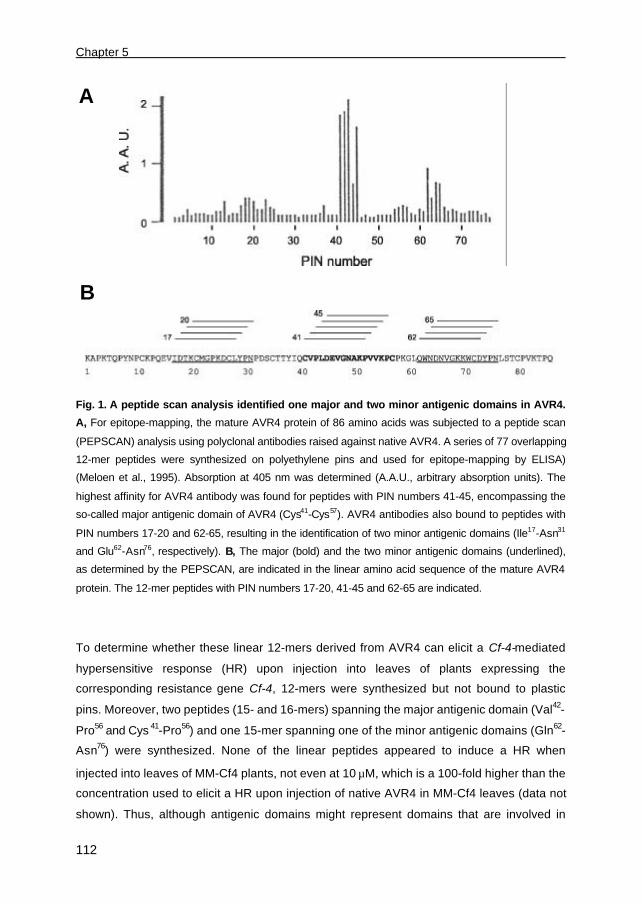
Chapter 5
112
Fig. 1. A peptide scan analysis identified one major and two minor antigenic domains in AVR4.
A, For epitope-mapping, the mature AVR4 protein of 86 amino acids was subjected to a peptide scan
(PEPSCAN) analysis using polyclonal antibodies raised against native AVR4. A series of 77 overlapping
12-mer peptides were synthesized on polyethylene pins and used for epitope-mapping by ELISA)
(Meloen et al., 1995). Absorption at 405 nm was determined (A.A.U., arbitrary absorption units). The
highest affinity for AVR4 antibody was found for peptides with PIN numbers 41-45, encompassing the
so-called major antigenic domain of AVR4 (Cys41-Cys57). AVR4 antibodies also bound to peptides with
PIN numbers 17-20 and 62-65, resulting in the identification of two minor antigenic domains (Ile17-Asn31
and Glu62-Asn76, respectively). B, The major (bold) and the two minor antigenic domains (underlined),
as determined by the PEPSCAN, are indicated in the linear amino acid sequence of the mature AVR4
protein. The 12-mer peptides with PIN numbers 17-20, 41-45 and 62-65 are indicated.
To determine whether these linear 12-mers derived from AVR4 can elicit a Cf-4-mediated
hypersensitive response (HR) upon injection into leaves of plants expressing the
corresponding resistance gene Cf-4, 12-mers were synthesized but not bound to plastic
pins. Moreover, two peptides (15- and 16-mers) spanning the major antigenic domain (Val42-
Pro56 and Cys 41-Pro56) and one 15-mer spanning one of the minor antigenic domains (Gln62-
Asn76) were synthesized. None of the linear peptides appeared to induce a HR when
injected into leaves of MM-Cf4 plants, not even at 10 µM, which is a 100-fold higher than the
concentration used to elicit a HR upon injection of native AVR4 in MM-Cf4 leaves (data not
shown). Thus, although antigenic domains might represent domains that are involved in
A
B

Necrosis-inducing activity and chitin-binding ability within AVR4
113
AVR4 recognition by MM-Cf4 plants, linear synthetic peptides derived from AVR4 do not
elicit Cf-4-mediated HR.
The necrosis-inducing activity (NIA) of AVR4 mutant proteins
The major antigenic domain of AVR4, as determined by PEPSCAN (Fig. 1), exhibits the
highest affinity for AVR4 antibodies and was therefore thought to contain putatively solvent-
exposed residues that condition NIA of AVR4. To identify which of these residues are
required for NIA of AVR4, amino acid residues present in the major antigenic domain of
AVR4 (Asp45, Glu46, Asn49, and Lys 51) were replaced, one by one, by Ala. For all other
residues present in this domain (Val42, Leu44, Val47, Val53, Val54, and Lys 55), it has previously
been demonstrated, by PVX-mediated expression of AVR4 mutant protein-encoding genes
in plants carrying Cf-4, that independent replacement of these residues by alanine did not
affect the NIA of AVR4 (Table 1). Moreover, as Cys, Pro and Gly residues are known to
affect the overall structure of proteins significantly, these residues were not replaced.
To determine the NIA of the AVR4 mutant proteins, Agrobacterium cultures carrying
AVR4 (mutant)-encoding genes were diluted into an Agrobacterium culture carrying the Cf-4
resistance gene and infiltrated into tobacco leaf sectors. No significant differences in
necrosis-inducing activities were observed between the various AVR4 mutant proteins and
AVR4 wild-type, demonstrating that residues Asp45, Glu46, Asn49, and Lys 51 are not
essential, at least not individually, for NIA of AVR4 (Table 1).
Interestingly, amongst the AVR4 mutant proteins carrying substitution of amino acid
residues that might be required for chitin-binding ability of AVR4 (see below), two mutants,
AVR4Y38A and AVR4W63A, were identified that showed reduced NIA (Fig. 2). A similar
reduced activity was observed for the AVR4 mutant protein carrying a histidine rather than
an alanine substitution at position Tyr38 (Chapter 2), which is a naturally occurring AVR4
mutant protein secreted by a strain of C. fulvum that is virulent on MM-Cf4 plants (Joosten et
al., 1997). When apoplastic fluid (AF) isolated from agroinfiltrated tobacco leaves was
analyzed by western blotting, it appeared that AVR4Y38A and AVR4W63A are unstable (Fig. 3;
Table 1). These data suggest that residues Tyr38 and Trp63 both contribute to conformational
stability rather than to NIA of AVR4.
The chitin-binding ability of AVR4 mutant proteins
To determine whether aromatic residues are required for chitin-binding ability of AVR4, as
demonstrated for hevein (Asensio et al. 1995 and 1998), we replaced all individual aromatic
residues present in AVR4 (Tyr8, Tyr 29, Tyr 38, Trp63, Trp71 and Tyr 74) by Ala. Moreover, we
replaced two additional residues in AVR4 (Ser34 and Asp73) by Ala, as these are conserved
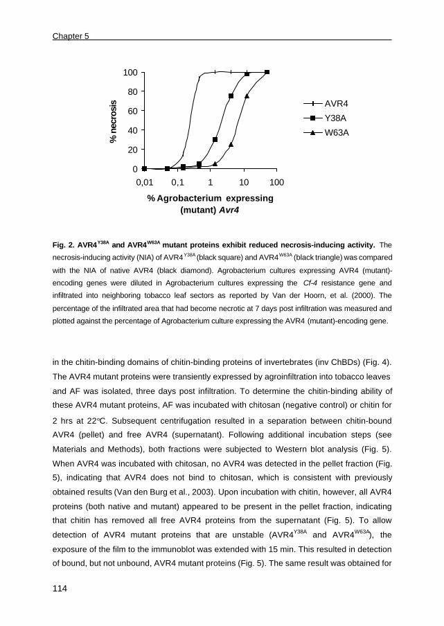
Chapter 5
114
Fig. 2. AVR4Y38A and AVR4W63A mutant proteins exhibit reduced necrosis-inducing activity. The
necrosis-inducing activity (NIA) of AVR4Y38A (black square) and AVR4W63A (black triangle) was compared
with the NIA of native AVR4 (black diamond). Agrobacterium cultures expressing AVR4 (mutant)-
encoding genes were diluted in Agrobacterium cultures expressing the Cf-4 resistance gene and
infiltrated into neighboring tobacco leaf sectors as reported by Van der Hoorn, et al. (2000). The
percentage of the infiltrated area that had become necrotic at 7 days post infiltration was measured and
plotted against the percentage of Agrobacterium culture expressing the AVR4 (mutant)-encoding gene.
in the chitin-binding domains of chitin-binding proteins of invertebrates (inv ChBDs) (Fig. 4).
The AVR4 mutant proteins were transiently expressed by agroinfiltration into tobacco leaves
and AF was isolated, three days post infiltration. To determine the chitin-binding ability of
these AVR4 mutant proteins, AF was incubated with chitosan (negative control) or chitin for
2 hrs at 22°C. Subsequent centrifugation resulted in a separation between chitin-bound
AVR4 (pellet) and free AVR4 (supernatant). Following additional incubation steps (see
Materials and Methods), both fractions were subjected to Western blot analysis (Fig. 5).
When AVR4 was incubated with chitosan, no AVR4 was detected in the pellet fraction (Fig.
5), indicating that AVR4 does not bind to chitosan, which is consistent with previously
obtained results (Van den Burg et al., 2003). Upon incubation with chitin, however, all AVR4
proteins (both native and mutant) appeared to be present in the pellet fraction, indicating
that chitin has removed all free AVR4 proteins from the supernatant (Fig. 5). To allow
detection of AVR4 mutant proteins that are unstable (AVR4Y38A and AVR4W63A), the
exposure of the film to the immunoblot was extended with 15 min. This resulted in detection
of bound, but not unbound, AVR4 mutant proteins (Fig. 5). The same result was obtained for
0
20
40
60
80
100
0,01 0,1 1 10 100
% Agrobacterium expressing (mutant) Avr4
% n
ecro
sis AVR4
Y38A
W63A
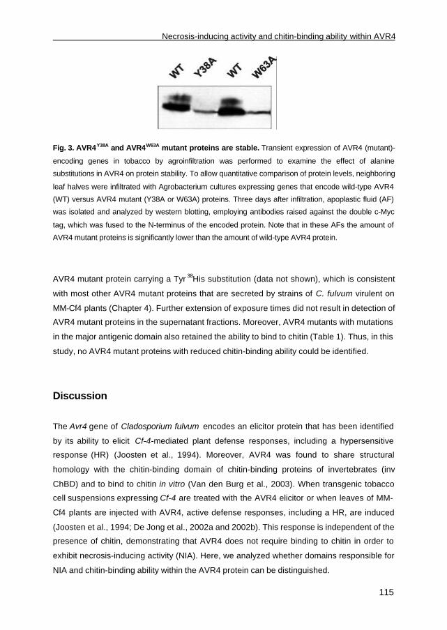
Necrosis-inducing activity and chitin-binding ability within AVR4
115
Fig. 3. AVR4Y38A and AVR4W63A mutant proteins are stable. Transient expression of AVR4 (mutant)-
encoding genes in tobacco by agroinfiltration was performed to examine the effect of alanine
substitutions in AVR4 on protein stability. To allow quantitative comparison of protein levels, neighboring
leaf halves were infiltrated with Agrobacterium cultures expressing genes that encode wild-type AVR4
(WT) versus AVR4 mutant (Y38A or W63A) proteins. Three days after infiltration, apoplastic fluid (AF)
was isolated and analyzed by western blotting, employing antibodies raised against the double c-Myc
tag, which was fused to the N-terminus of the encoded protein. Note that in these AFs the amount of
AVR4 mutant proteins is significantly lower than the amount of wild-type AVR4 protein.
AVR4 mutant protein carrying a Tyr 38His substitution (data not shown), which is consistent
with most other AVR4 mutant proteins that are secreted by strains of C. fulvum virulent on
MM-Cf4 plants (Chapter 4). Further extension of exposure times did not result in detection of
AVR4 mutant proteins in the supernatant fractions. Moreover, AVR4 mutants with mutations
in the major antigenic domain also retained the ability to bind to chitin (Table 1). Thus, in this
study, no AVR4 mutant proteins with reduced chitin-binding ability could be identified.
Discussion
The Avr4 gene of Cladosporium fulvum encodes an elicitor protein that has been identified
by its ability to elicit Cf-4-mediated plant defense responses, including a hypersensitive
response (HR) (Joosten et al., 1994). Moreover, AVR4 was found to share structural
homology with the chitin-binding domain of chitin-binding proteins of invertebrates (inv
ChBD) and to bind to chitin in vitro (Van den Burg et al., 2003). When transgenic tobacco
cell suspensions expressing Cf-4 are treated with the AVR4 elicitor or when leaves of MM-
Cf4 plants are injected with AVR4, active defense responses, including a HR, are induced
(Joosten et al., 1994; De Jong et al., 2002a and 2002b). This response is independent of the
presence of chitin, demonstrating that AVR4 does not require binding to chitin in order to
exhibit necrosis-inducing activity (NIA). Here, we analyzed whether domains responsible for
NIA and chitin-binding ability within the AVR4 protein can be distinguished.

Chapter 5
116
Fig. 4. Sequence alignment between AVR4 and various chitin-binding proteins of invertebrates.
The identified disulfide bond pattern and the cysteine spacing of AVR4 was used to perform a motif-
based search (URL:http://motif.genome.ad.jp) (Van den Burg, et al. 2003). AVR4 was found to
contain a chitin-binding domain as found in chitin-binding proteins of invertebrates (inv ChBD). The
alignment made in Clustal X shows the following sequences (depicted from top-to-bottom): Mature
AVR4 protein of Cladosporium fulvum, tachycitin of Tachypleus tridentatus (Japanese horseshoe crab),
Chitinase of Manduca sexta (tobacco hawkmoth), Brugia malayi (nematode), Penaeus japonicus
(prawn), and Peritrophin 44 of Lucilia cuprina (fly). The latter sequence contains four inv ChBDs, which
represent Per44_1 to Per44_4. The inv ChBD contains six Cys residues that are conserved (black
boxed). The consensus is indicated at the bottom (C, cysteine residue; p, polar; s, small residues; h,
hydrophobic; a, aromatic). The major antigenic domain (Cys41-Cys57) and the two minor antigenic
domains (Ile17-Asn31 and Glu62-Asn76), as determined by PEPSCAN (Fig.1), are shown by bars on top of
the alignment. Asterisks at the top indicate all residues, except for Tyr8, which have been selected for
mutational analysis of native AVR4. ID, ID-code used in the trEMBL database; ORF, total number of
residues encoded by the open reading frame (for AVR4 the mature protein is depicted); start/end,
represents the first and last amino acid residue of the ChBD, respectively. (This figure has been
modified from Van den Burg et al., 2003).
The effect of alanine substitutions in AVR4 on its necrosis-inducing activity (NIA)
Several strains of C. fulvum virulent on tomato plants expressing Cf-4 have been found to
secrete AVR4 mutant proteins that carry single amino acid substitutions, including
substitutions of cysteine residues that are involved in disulfide bonding in AVR4 (Joosten et
al., 1997; Chapter 4). It appeared that these AVR4 mutant proteins are sensitive to
proteolytic breakdown in the apoplastic space of tomato, implying that these amino acid
substitutions affect the conformational stability of AVR4 (Joosten et al. 1997; Chapter 4).
Here, we examined whether AVR4 mutant proteins could be identified that are stable and
exhibit reduced NIA. Further, we tested the mutants for their chitin-binding ability (see
below). For AVR9 mutant proteins, a positive correlation was found between NIA and
antigenicity (Mahé et al., 1998), suggesting that antigenic sites contain putatively solvent-
exposed residues that condition its NIA activity. Here, we examined which antigenic
domains in AVR4 are recognized by (or bind to) polyclonal antibodies raised against native

Necrosis-inducing activity and chitin-binding ability within AVR4
117
Fig. 5. The introduced alanine substitutions do not affect the chitin-binding ability of AVR4.
AVR4 (mutant) proteins carrying alanine substitutions of residues at positions Tyr8, Tyr29, Ser34, Tyr38,
Trp63, Trp71, Asp73 or Tyr74 were tested for their ability to bind to chitin. Apoplastic fluids (AFs), isolated
from agroinfiltrated tobacco leaves, containing these AVR4 (mutant) proteins were incubated with 5 mg
of chitin at 22°C for 2 hrs. As a negative control, AF containing wild-type AVR4 (WT) was incubated with
chitosan instead of chitin. Following centrifugation, free AVR4 (supernatant; s) was separated from
chitin-bound AVR4 (pellet; p). Western blot analysis was performed using c-Myc-antibodies. Note that
different amounts of AVR4 (mutant) protein are present in these AFs and that, except for the negative
control, no AVR4 (mutant) protein is detected in the supernatant fraction. B, AFs containing unstable
AVR4Y38A and AVR4W63A were analyzed on separate blots, as protein detection required longer exposure
of film to the blot.
AVR4. Epitope-mapping employing a peptide scan (PEPSCAN) methodology identified one
major antigenic domain (Cys 41-Cys 57) and two minor antigenic domains (Ile17-Asn31 and
Gln62-Asn76) in the mature AVR4 protein. It appeared that peptides spanning the individual
antigenic domains did not elicit a HR when injected into leaves of MM-Cf4 plants. As
demonstrated for most AVR4 mutant proteins produced by C. fulvum , cysteine residues are
essential for proper conformation and stability of AVR4 (Joosten et al., 1997; Chapter 4).
Possibly, linear 12-15-mer peptides derived from AVR4 do not induce a Cf-4-mediated HR
due to the fact that they lack secondary structures that are required for conformational
stability and NIA, thereby rendering the peptides sensitive to proteolytic breakdown in the
apoplastic space of tomato leaves.
The major antigenic domain includes a sequence (Val42-Pro56) that is absent in ChBDs
of invertebrates (Fig. 4), suggesting that this domain might represents a loop-like structure
that is recognized by AVR4 antibodies and possibly also by plants that express Cf-4.
However, replacement of individual residues in this major antigenic domain of AVR4 by
alanine did not affect the NIA of AVR4 mutant proteins. Possibly, a multimeric peptide is
responsible for NIA of AVR4, as previously demonstrated for ethylene-inducing xylanase
(EIX) elicitor (Rotblat et al., 2002). This implies that multiple, rather than single amino acid
replacements are required in the antigenic domain to cause a reduction in NIA of AVR4.

Chapter 5
118
Table 1. The effect of alanine substitutions on necrosis-inducing activity, chitin-binding ability,
and stability of AVR4.
Necrosis-inducing activity (NIA) of AVR4 (mutant) proteins was analyzed by transient expression using
agroinfiltration into tobacco leaves. Apoplastic fluid isolated from these leaves, containing AVR4
(mutant) proteins, was used in chitin-binding assays and also used to determine protein stability. ND:
not determined. ChBD: Chitin-binding domain.
AVR4 mutant proteins carrying a Tyr 38Ala or Trp63Ala substitution were found to exhibit
reduced NIA. These two AVR4 mutants appear to be unstable, most likely due to proteolytic
breakdown, suggesting that, in addition to the Cys residues (Chapter 4), residues Tyr 38 and
Trp63 contribute to conformational stability rather than to NIA of AVR4.
The effect of alanine substitutions on chitin-binding ability of AVR4
For hevein, a plant chitin-binding protein, residue Ser19 and aromatic residues Trp21 and
Trp23 have been identified to represent an essential part of the ChBD (Cys 12-Ser32) (Asensio
et al., 1995 and 1998) (Fig. 6). Three-dimensional (3D) structure-based sequence alignment
between hevein and inv chitin-binding protein tachycitin revealed that residues Ser19, Trp21
and Trp23 of hevein correspond to Asn47, Tyr49 and Val52 of tachycitin, respectively (Suetake
et al., 2000). Although AVR4 does not show structural sequence similarity with plant ChBDs,
subsequent sequence alignment between tachycitin and AVR4 identified residues Asn64,
Gly68, and Trp71 to be part of a putative chitin-binding domain of AVR4 (Fig. 6).
Mutation NIA Chitin-bindingability
Stability
AVR4 wild-type - +++++ + +
Major antigenic domain V42A +++++ ND NDL44A +++++ ND NDD45A +++++ + +E46A +++++ + +V47A +++++ ND NDN49A +++++ + +K51A +++++ + +V53A +++++ ND NDV54A +++++ ND NDK55A +++++ ND ND
Aromatic residues Y8A +++++ + +Y29A +++++ + +Y38A +++ + -W63A + + -W71A +++++ + +Y74A +++++ + +
Conserved in ChBD S34A +++++ + +D73A +++++ + +

Necrosis-inducing activity and chitin-binding ability within AVR4
119
Hevein (12-32) CPNNLCCS--QW-GWCGST-DEYCSTachycitin (40-60) CPKGLHYN--AYLKVCDW-PSK-AGAVR4 (57-79) CPKGLQWNDNVGKKWCDY-PNL-ST
Fig. 6. Structure-based sequence alignment of tachycitin and AVR4 with the C12-S32 chitin-
binding domain of hevein. For hevein, segment C12-S32 was identified as an essential chitin-binding
domain. Three-dimensional structure comparison of hevein and tachycitin identified the putative chitin-
binding domain of tachycitin. Subsequent amino acid sequence alignment between tachycitin and AVR4
revealed that residues N64, G68, and W71, are part of a putative chitin-binding domain of AVR4. Amino
acid residues conserved between the plant, invertebrate and fungal chitin-binding proteins are indicated
in bold. Asterisks at the top indicate residues of hevein (S19, W21, and W23) that are essential for its
chitin binding.
However, in this study, we have not examined whether residues Asn64 and Gly68 in AVR4
are essential for its chitin-binding ability. As in AVR4 residue Asn64 is in close proximity of
residues Asp65 and Asn66, which may also contribute to chitin binding through hydrogen
bonding, multiple rather than single amino acid substitutions might be required to abolish
chitin-binding ability of AVR4. The involvement of residue Gly68 was not examined as this
residue is thought to be required for structural integrity of AVR4 and, moreover, is not
conserved in inv ChBDs.
Here, we examined whether the aromatic residues and two additional residues (Ser34
and Asp73), which are conserved in inv ChBDs, are involved in binding of AVR4 to chitin.
Although most selected residues (Tyr29, Ser34, Tyr 38, Trp63, Asp73, and Tyr 74) are conserved
in inv ChBDs, all of the AVR4 mutant proteins retained their ability to bind to chitin.
Moreover, AVR4 mutant proteins with an alanine substitution at position Asp45, Glu46, Asn49,
or Lys 51 in the major antigenic domain of AVR4, also retained chitin-binding ability. In
addition, a histidine substitution of residue Tyr38, as found in an AVR4 mutant protein
secreted by a strain of C. fulvum virulent on MM-Cf4 plants, did not affect the chitin-binding
ability of AVR4. Thus, similar to strains of C. fulvum that produce AVR4 mutant proteins
carrying a disrupted disulfide bridge (Chapter 4), this strain also circumvents Cf-4-mediated
resistance by secretion of an unstable AVR4 mutant protein that is still capable of binding to
chitin.
Strikingly, although we performed a selective alanine substitution analysis, no AVR4
mutant proteins were identified with decreased affinity for chitin. Possibly, residues other
than those selected by us are required for binding of AVR4 to chitin in vitro. On the other
* * *
Chapter 5
120
hand, to preserve its chitin-binding ability, AVR4 might respond to evolutionary changes in
the cell wall composition of C. fulvum by adaptive substitution of amino acid residues in the
putative ChBD of AVR4, suggesting that AVR4 has the ability to encounter functional
modifications. Nevertheless, disruption of disulfide bond Cys 57-Cys 72 in AVR4 reduced its
affinity for chitin, as a significant amount of this AVR4 mutant protein was found to be free of
chitin in the supernatant fraction (Chapter 4). The major difference between the two reported
assays concerns the ratio between AVR4 and chitin used in the chitin-binding assays. Here,
we used 75-100 ng of AVR4 in our chitin-binding assay, whereas in Chapter 4 a 40-fold
higher concentration of AVR4 (4µg) was used. Moreover, when prolonged incubation times
were used, AVR4 mutant protein could no longer be detected in the supernatant fraction.
Thus, in our study, shorter incubation times together with an excess of AVR4 mutant protein
in relation to chitin might identify amino acid residues that are essential for chitin-binding
ability of AVR4.
Although single amino acid mutations were identified that abolished the conformational
stability of AVR4, no modifications were found that resulted in a stable AVR4 mutant
proteins that exhibit reduced NIA. Possibly, to affect the AVR4 biologically active site that is
recognized by Cf-4 plants, multiple rather than single amino acids need to be substituted, in
particular the residues present in the major and minor antigenic domains. As multiple
residues are conserved in the chitin-binding domain, more than one residue is thought to
condition the ability of AVR4 to bind to chitin. It could be possible that binding of AVR4 to
chitin inhibits the NIA of AVR4, which would imply that residues required for chitin binding of
AVR4 are also required for its NIA. On the other hand, when bound to chitin, the biologically
active site of AVR4 might not be accessible to elicit a Cf-4-mediated HR. Altogether,
subsequent multiple amino acid substitutions in AVR4 might elucidate whether domains
responsible for NIA and chitin-binding ability within the AVR4 protein can be distinguished.
Materials and Methods
Plant materials
Tomato (Lycopersicon esculentum) cultivar Moneymaker (MM), near-isogenic line MM-Cf4
(containing an introgession segment that carries the Cf-4 locus) and tobacco (Nicotiana
tabacum cv. Petite Havana SR1) plants were grown under standard greenhouse conditions.
PEPSCAN of the mature AVR4 protein
To identify linear epitopes derived from AVR4 recognized by polyclonal antibodies raised
against native AVR4, the mature AVR4 protein of 86 amino acids was subjected to a peptide

Necrosis-inducing activity and chitin-binding ability within AVR4
121
scan (PEPSCAN) analysis. A set of 77 peptides corresponding to 12-mers spanning the
mature AVR4 protein, each peptide overlapping the previous peptide with 11 amino acid
residues, was synthesized on solid (polyethylene) support pins (at the COOH-terminus of
each amino acid sequence) (Meloen et al., 1995; Williams et al., 1998). The pins with bound
peptide were placed into individual wells of a 96-well microtitre plate and an ELISA
procedure was carried out, according to Geysen et al. (1987). Briefly, nonspecific binding
sites on the pins were blocked by incubation in blocking buffer (PBS containing 5%
ovalbumin, 5% horse serum and 1% (v/v) Tween-80) for 30 min at room temperature.
Polyclonal antibodies raised against native AVR4 (mouse) was diluted in blocking buffer
(1:1000) and incubated overnight at 4°C. After washing with PBS containing 1% (v/v)
Tween-80, the pins were transferred to wells containing goat-anti-mouse immunoglobulin
horse radish peroxidase conjugate. After incubation for 1 hr at 37°C, unbound antibody was
removed from the supports and antibodies were detected by incubation in fresh substrate
solution (50 mg ABTS and 20 µl of 30% (w/w) H2O2 solution in 100 ml of 0.1 M sodium
phosphate/0.08 M citrate buffer, pH 4). Plates were read at 405 nM.
Construction of plasmids for agroinfiltration
To facilitate transient expression of AVR4 mutant proteins by agroinfiltration, binary plasmids
were constructed. For site-directed mutagenesis, two PCR fragments were obtained, making
use of primers to introduce mutations and specific restriction sites (Table 2). These PCR
fragments were subsequently used in overlap extension PCR to amplify a PCR product that
contained the open reading frame (ORF) of the Avr4 (mutant) gene. Two methods were
carried out to introduce an alanine substitution at position Ser78, which abolished the
putative N-glycosylation site of AVR4. Following method (1), overlap extension PCR was
performed with forward primer XF7 (introduction of a XhoI site) and reverse primer B20-C2
(introduction of a PstI site and the Ser78-to-Ala mutation) (Table 2), using PVX::Avr4 as a
template (Joosten et al., 1997). Following method (2), overlap extension PCR was
performed with forward primer A4-XN (introduction of a XhoI site) and reverse primer A4-BC
(introduction of a BamHI site), using pPIC-A4 (Chapter 3) as a template. The PCR products
were subsequently digested with XhoI/ PstI (1) or with XhoI/ BamHI (2), cloned into either
XhoI/ PstI-digested pRH80 (Van der Hoorn et al., 2000) or XhoI/ BamHI-digested
pBluescript SK+ (Amersham-Pharmacia, Buckinghamshire, UK), respectively, and
sequenced. Following subsequent digestion, the ORFs were cloned into XhoI/ EcoRI-
digested pRH298 (Van der Hoorn, unpublished data) or XhoI/ BamHI-digested pNW30,
respectively (Chapter 2). The binary plasmids pRH298 and pNW30 both carry the
Cauliflower Mosaic Virus (CaMV) 35S-promoter and the Pi-II terminator (An et al., 1989).

Chapter 5
122
The ORFs of (mutant) Avr4 were cloned downstream of a double c-Myc-tag, which itself is
located downstream of the PR-1a signal peptide for extracellular targeting (Hammond-
Kosack et al., 1995).
Table 2. Primers used to introduce mutations and to generate PCR fragments encoding AVR4
(mutant) proteins. Forward (f) and reverse (r) primers and mutations (bold) are indicated and
restriction sites are underlined.
Primer Sequence (5’ to 3’)
XF7 (XhoI site) (f) TAGCTCGAGCAAGGCCCCCAAAACTCAACCB20-C2 (PstI site) (r) AAAACTGCAGTCATTGCGGCGTCTTTACCGGACACGTGGCCAGGTTTGGATAA4-XN (XhoI site) (f) TAGCTCGAGCAAGGCCCCCAAAACTCAACCA4-BC (BamHI site) (r) CGCGGATCCCTATTGCGGCGTCTTTACCGY8A (f) CTCAACCAGCCAACCCATG
(r) CATGGGTTGGC TGGTTGAGY29 (f) GATTGTCTCGCCCCGAACCCCG
(r) CGGGGTTCGGGGCGAGACAATCS34A (f) GAACCCCGACGCCTGTACAACC
(r) GGTTGTACAGGCGTCGGGGTTCY38A (f) GTACAACCGCCATACAGTGTG
(r) CACACTGTATGGCGGTTGTACD45A (f) CCGCTCGCCGAAGTTGGCAAT
(r) ATTGCCAACTTCGGCGAGCGGE46A (f) CCGCTCGACGCCGTTGGCAAT
(r) ATTGCCAACGGCGTCGAGCGGN49A (f) GTTGGCGCCGCGAAGCCTGTG
(r) CACAGGCTTCGCGGCGCCAACK51A (f) GTTGGCAATGCGGCCCCTGTG
(r) CACAGGGGCCGCATTGCCAACW63A (f) GGACTGCAGGCCAACGATAACG
(r) CGTTATCGTTGGCCTGCAGTCCD73A (f) GAAGTGGTGCGCCTATCCAAACC
(r) GGTTTGGATAGGCGCACCACTTCY74A (f) GTGGTGCGACGCCCCAAACCTG
(r) CAGGTTTGGGGCGTCGCACCAC
Agroinfiltration and analysis of apoplastic fluids
Agroinfiltration of transgenic cultures of Agrobacterium tumefaciens carrying various AVR4
(mutant)-encoding constructs into Nicotiana tabacum cv. Petite Havana SR1 was performed
as described in Chapter 2. Apoplastic fluids (AFs) were isolated from agroinfiltrated tobacco
leaves, 3 days post infiltration (dpi) and freeze-dried (Chapter 2). Western blot analysis of
AFs containing the AVR4 mutant proteins was performed using c-Myc-antibodies (Chapter
2), as mutations in the major antigenic domain of AVR4 might interfere with the affinity for
polyclonal antibodies raised against native AVR4.
To test the necrosis-inducing activity (NIA) of AVR4 (mutant) proteins, Agrobacterium
cultures carrying AVR4 (mutant)-encoding genes were diluted in an Agrobacterium culture
carrying the Cf-4 resistance gene and infiltrated into leaf sectors of tobacco.

Necrosis-inducing activity and chitin-binding ability within AVR4
123
Chitin-binding assay
Aliquots of AFs (100 µl), containing approximately 75-100 ng of AVR4 (mutant) protein, were
freeze-dried and resuspended in 500 µl of chitin-binding buffer (ChBB) (50mM Tris/Hcl
pH8.0 and 100 mM NaCl). Samples were supplied with approximately 5 mg of chitin (purified
chitin powder from crab shells [Sigma-Aldrich, Zwijndrecht, The Netherlands]) and incubated
at 22°C for 2 hrs under continuous rotation. As a negative control, AFs were incubated with
chitosan (from crab shells [Sigma-Aldrich, Zwijndrecht, The Netherlands]) (Van den Burg et
al., 2003). After incubation, samples were centrifuged for 10 min at 13,000 x rpm in a
microfuge to separate chitin-bound AVR4 (pellet) from free AVR4 (supernatant). The
supernatant was subsequently freeze-dried and the pellet fraction was washed 3 times with
ChBB (500 µl) to remove all free AVR4 (mutant) protein. To dissociate bound AVR4 (mutant)
protein from chitin or chitosan, the pellet fractions were resuspended into 150 µl of 1% (w/v)
SDS and incubated at 95°C for 5 min. Following incubation, samples were centrifuged and
supernatant, containing dissociated “bound” AVR4 (mutant) protein, was freeze-dried.
Freeze-dried fractions containing unbound AVR4 and bound AVR4 (mutant) proteins were
resuspended in 1x SDS-loading buffer (10 mM Tris/HCl pH6.8, 1% (w/v) SDS, 2% (v/v)
glycerol, 1% 2-mercaptoethanol and 0.0005 % bromophenol blue) (50 µl) and 10 µl was run
on SDS-PAGE. Proteins were blotted on nitrocellulose membranes and the c-Myc-tagged
AVR4 (mutant) proteins were detected using Super Signal Chemiluminescent Substrate
(pico ECL kit) (Pierce, Rockford, USA), as described in Chapter 2. Kodak X-OMAT films
were exposed to the blots for 2-5 min or, occasionally, for 20 min.
Acknowledgements
We thank Mevr. Posthuma, W.M.M. Schaaper and R.H. Meloen (ID-DLO, Lelystad, The
Netherlands) for carrying out the AVR4 PEPSCAN analysis.

124

Chapter 6
Summarizing discussion

Chapter 6
126

Summarizing discussion
127
Summarizing discussion
The majority of proteins produced by pathogens during growth on susceptible plants are
thought to play a role in pathogenesis. Some of these proteins, the products of avirulence
(Avr) genes, have been demonstrated to trigger defense responses in resistant plants that
carry the matching resistance (R) genes, thereby causing avirulence of the pathogenic
isolates producing these AVR proteins. As a result of selection pressure, pathogens have
developed a variety of different strategies to modify these AVRs and thereby evade the
induction of plant defense responses. Plants, however, have developed various surveillance
and defense mechanisms to withstand the persistent attacks by continuously changing
pathogen populations. Most R genes are members of complex loci and recombination
between alleles and sometimes between different homologues are thought to mediate
generation of new resistance specificities (Hammond-Kosack and Jones, 1997; Parniske et
al., 1997; Richter and Ronald, 2000). Once activated by AVRs directly or indirectly, R
proteins are extremely efficient in inducing plant defense responses that restrict further
growth of invading pathogens (Hammond-Kosack and Jones, 1996).
Resistance specificities conferred by homologues of Cladosporium fulvum resistance
gene Cf-9
Within the Hcr9 (homologues of Cladosporium R gene Cf-9) gene family of tomato, three
homologues have thus far been identified that confer resistance towards C. fulvum. The
homologues Hcr9-4D, Hcr9-4E and Hcr9-9C, which represent the Cf-4, Cf-4E and Cf-9
genes, respectively, mediate the induction of defense responses upon recognition of race-
specific elicitors of C. fulvum , AVR4, AVRE, and AVR9, respectively (Thomas et al., 1998;
Joosten and De Wit, 1999) (Chapters 1 and 2). Although the various Hcr9 homologues
share a high degree of sequence homology, all other Hcr9 homologues identified so far, are
non-functional in conferring full resistance towards C. fulvum (Parniske et al., 1997; Thomas
et al., 1997). The sequence variation in the Hcr9s, which mainly occurs in the N-terminal
domain and particularly in the solvent-exposed residues of the â-sheets in the leucine-rich
repeat (LRR) domains of the encoded proteins, is thought to determine the distinct
resistance specificities of the functional Hcr9s. Indeed, gene shuffling and domain swap
experiments between Cf-4 and Cf-9 revealed that these variant residues are important for
Cf-4 and Cf-9 function (Van der Hoorn et al., 2001; Wulff et al., 2001). Van der Hoorn et al.
(2001) also demonstrated that it is possible to construct a chimeric Cf-4 protein that carries
most of the specificity determinants of Cf-9, with the exception of three solvent-exposed
amino acid residues. However, the number of LRRs, which distinguishes Cf-4 and Cf-9 (25

Chapter 6
128
and 27 LRRs, respectively), is essential for Cf-4 and Cf-9 function and can therefore not be
changed (Van der Hoorn et al., 2001). As Hcr9-4E and Cf-9 carry the same number of
LRRs, this implies that by gene shuffling and domain swaps, a chimeric Cf protein might be
generated that exhibits dual recognition specificity towards both AVR4E and AVR9.
A striking feature observed for AVR-induced defense responses is that plants carrying
the functional Hcr9 gene respond in a highly sensitive manner to low amounts of the
corresponding AVR protein (Van den Ackerveken et al., 1992; Takken et al., 1999). This
implicates that the induction of plant defense responses is mediated by receptors that exhibit
high affinity for these AVR elicitor proteins (see below). Previously, early defense responses
have been studied in transgenic tobacco cell suspensions that express Cf-4 or Cf-9 (Piedras
et al., 1998; De Jong et al., 2002b). These studies revealed that both in Cf-4- and Cf-9-cell
suspensions medium alkalization and an oxidative burst are induced upon treatment with
AVR4 and AVR9, respectively. The induction of these responses is suppressed at 33°C (De
Jong et al., 2002b). Moreover, when Cf-4- and Cf-9-mediated defense responses are
compared, no significant differences in expression of genes that are involved in downstream
signaling could be detected (Cai et al., 2001). Nevertheless, distinct necrotic patterns
developed in tomato seedlings that express both Cf-4 and Avr4 as compared to seedlings
that express both Cf-9 and Avr9 (Cai et al., 2001). As AVR4E was found to exhibit necrosis-
inducing activity (NIA) in tobacco in the presence of Hcr9-4E (Chapter 2), it would be
interesting to employ tobacco cell suspensions that carry Hcr9-4E, in addition to Cf-4 and
Cf-9, to identify differences and similarities in activation of Hcr9-mediated signaling
pathways. The recent introduction of novel approaches including transcriptomics and
proteomics combined with transient silencing assays and mass-spectrometry (Romeis,
2001a) will be instrumental to elucidate novel components in downstream signaling
pathways that lead to resistance.
Phylogenic relationships between isolates of C. fulvum
During growth in the apoplastic space of tomato, C. fulvum secretes a variety of different
race-specific AVR proteins and non-race-specific extracellular proteins (ECPs), all of which
are small, cysteine-rich, and stable proteins that do not share sequence homology (Joosten
and De Wit, 1999). The AVRs and ECPs have been shown to act as elicitor proteins in
tomato plants that carry the matching R gene (Joosten and De Wit, 1999). Natural strains of
C. fulvum were found to evade triggering of plant defense responses by modifying Avr gene
products. Unlike the modifications found in Avr genes, thus far only silent mutations have
been identified in the open reading frame (ORF) of the virulence gene Ecp2 in natural
strains of C. fulvum (M.J.D. de Kock, personal communication). On plants carrying Cf-ECP2,

Summarizing discussion
129
the virulence factor ECP2 behaves as an avirulence determinant (Laugé et al., 1998b).
Possibly, there might have been a lack of selection pressure on C. fulvum to overcome
ECP2 recognition, as, opposed to the Cf resistance traits, Cf-ECP2 has not yet been
introduced in commercially grown tomato cultivars (Chapter 1).
Within the C. fulvum population, several isolates have been identified that evade more
than one Cf resistance trait. Subsequent sequence analysis of the corresponding Avr ORFs
revealed that C. fulvum employs a variety of different mechanisms to evade Cf-mediated
resistance. Evasion of Cf-2-mediated resistance by C. fulvum is enabled by a
retrotransposon insertion, or by several different single base pair deletions or insertions in
the Avr2 ORF, resulting in the production of truncated AVR2 proteins (Luderer et al., 2002b).
Strains of C. fulvum evade Cf-4-mediated resistance by production of a truncated AVR4
protein, or by secretion of unstable AVR4 mutant proteins that carry different single amino
acid substitutions. In the ORF of the Avr4E gene present in strains virulent on plants
carrying Hcr9-4E, a double point mutation was found, resulting in a two amino acid
substitutions, Phe62Leu and Met73Thr (avr4ELT). This elicitor-inactive mutant protein is stable
and the single amino acid substitution Phe62Leu in AVR4E appears to be sufficient to evade
Hcr9-4E- mediated resistance (Chapter 2). Moreover, some strains virulent on Hcr9-4E
plants have been identified that carry a wild-type Avr4E ORF, yet, produce no functional
AVR4E elicitor protein due to lack of Avr4E gene expression (Chapter 2). Finally, all strains
of C. fulvum that are virulent on tomato plants carrying the Cf-9 gene lack the entire Avr9
ORF (Van den Ackerveken et al., 1992).
AFLP analysis on DNA of C. fulvum revealed that lack of sufficient polymorphism
amongst isolates within the world-wide C. fulvum population did not allow construction of a
reliable phylogenetic tree (Joosten and De Wit, 1999). In spite of this limitation, mutations
identified in the ORF of Avr2, Avr4, Avr4E and Avr9 were used to obtain a simplified
phylogenetic relationship between C. fulvum isolates carrying these mutations (See also
Chapter 1; Table 2). As we excluded isolates for which not all four Avr ORFs were
sequenced, this analysis represents only a small fraction (fourteen) of the isolates present in
our worldwide C. fulvum collection. Clustering resulted in a so-called dendrogram, which
grouped fourteen strains into four main clusters (phenons) (Fig. 1). Phenon 1 includes
strains that are virulent on Cf-2 plants (i.e. races 2) and avirulent on plants that carry Cf-4,
Hcr9-4E or Cf-9. This suggests that these strains have thus far only endured selection
pressure on AVR2 to overcome Cf-2-mediated resistance. Phenon 2 represents strains that
have endured selection pressure on AVR2, AVR4 and AVR4E to overcome Cf-2, Cf-4 and
Hcr9-4E-mediated resistance, respectively (i.e. races 2.4) (Fig. 1). Strains virulent on plants
carrying the complete Cf-4 locus are referred to as races 4, while strains virulent on plants

Chapter 6
130
carrying Hcr9-4D or Hcr9-4E are referred to as races 4D and races 4E, respectively. A total
of 17 breeding lines that carry Cf-4 were also found to carry Hcr9-4E (Haanstra et al., 2000),
suggesting that the selection pressure to overcome Cf-4 is comparable to the selection
pressure to overcome Hcr9-4E. Thus, while the presence of Cf-4 is associated with Hcr9-4E,
plants might exist that carry Hcr9-4E but not Cf-4. This would explain why, opposed to races
4 and races 4E, thus far no races 4D have been identified. In phenon 2, all strains of C.
fulvum are clustered that lack Avr4E gene expression (NE), indicating that these strains
might have a common ancestor carrying a mutation in the promoter sequence of Avr4E
(Chapter 2). Possibly, following evasion of Hcr9-4E-mediated AVR4E recognition, two
strains emerged, one with a Cys 35-to-Tyr and the other with a Cys 80-to-Tyr substitution.
These two strains might have endured consecutive selection pressure to overcome Cf-2-
mediated AVR2 recognition (race 2.4, race 2.4.8.11 and race 2.4.5), as well as Cf-9-
mediated AVR9 recognition (race 2.4.5.9.11 and 2.4.9.11). There is prove that Cf-8 is allelic
to Cf-4 and that plants carrying Cf-11 also carry Cf-4 (Haanstra et al., 2000), suggesting that
races 8 are identical to races 4 and that races 11 require additional modification of the Avr4
gene product to enable growth on plants carrying Cf-11. The strains clustered in phenon 3
all carry the same modification in the Avr4E gene product (resulting in avr4ELT) and are
virulent on plants carrying Cf-5 (Fig. 1). Race 2.5.9 might have evolved from race 2.5, which
in turn might have evolved from race 5. To be able to grow on plants that carry the complete
Cf-4 locus, strains of C. fulvum require modifications of both the Avr4 and Avr4E gene
products. Phenon 4 includes strains in which both these gene products are modified and as
a result, these strains are virulent on plants that carry both Cf-4 and Hcr9-4E. The low
genetic variability observed in Avr4E suggests that these strains might have diverged from a
common ancestor carrying avr4ELT. However, as single amino acid substitution Phe62Leu is
sufficient to lose elicitor activity of AVR4E (Chapter 2), strains carrying avr4ELT might have
evolved from strains carrying the single amino acid substitution Met73Thr, but within the C.
fulvum population, no such strains have thus far been identified.
The intrinsic function of AVR4
The AVR4 protein was found to share structural homology with the chitin-binding domain of
chitin-binding proteins of invertebrates (inv ChBD) (Van den Burg et al., 2003). AVR4 has
been demonstrated to bind to chitin in vitro, but not to other polysaccharides . Moreover, an
AVR4-specific high-affinity binding site (HABS) was found in fungal extracts derived from C.
fulvum mycelium grown in vitro (Chapter 3). This AVR4-HABS appeared to be heat and
proteinase K resistant and was therefore thought to represent a nonproteinaceous, possibly
chitin-like, component. However, dissociation of AVR4 from its HABS was accomplished

Summarizing discussion
131
Fig. 1. Phylogenetic relationships between C. fulvum isolates, based on variation observed in
sequences of wild-type and mutant Avr ORFs. The open reading frames (ORFs) of Avr2, Avr4,
Avr4E and Avr9 genes present in several natural strains of C. fulvum avirulent or virulent on plants
carrying Cf-2, Hcr9-4D, Hcr9-4E or Cf-9 (wild-type versus races 2, 4D, 4E or 9, respectively) have been
sequenced (See also Chapter 1). Strains indicated by Gattieres Furon 90 (GF 90), Alénya B, Pons 89
and La Maxe originate from France. For most other strains the origin is unknown. Mutations in the Avr2
ORF involve a transposon insertion (Tr), single base pair deletions (-) or insertions (+) in the codon of
Lys23 or a nucleotide exchange (C to T). The mature AVR4 mutant proteins carry a substitution Tyr-to-
Cys at position 35, 41 or 80 (TGT to TAT), or a substitution Tyr37-to-His (TAC to CAC) or Thr36-to-Ile
(ACC to ATC). Strains have been identified that lack Avr4E gene expression (NE). A double point
mutation in the Avr4E ORF results in amino acid substitution Phe62-to-Leu (TTT to CTT) and Met73-to-
Thr (ATG to ACG) in mature AVR4E. Strains virulent on plants carrying Cf-9 do not produce the AVR9
protein (-) due to the absence of the entire Avr9 ORF. Wt: wild-type.
under the same conditions as association, whereas elution of AVR4 from chitin required
extreme conditions, such as boiling in 1% SDS. Moreover, affinity crosslinking experiments
revealed that AVR4 binds to a fungal component of about 75 kDa, suggesting that the
AVR4-HABS represents a proteinaceous compound rather than a polysaccharide such as
chitin. Previously, receptor-like proteins of similar sizes (MW between 75 and 85 kDa) have

Chapter 6
132
been identified in plasma membranes of plant cells that exhibit chitin-binding properties
(Shibuya et al., 1993; Baureithel et al., 1994; Ito et al., 1997; Kim et al., 2000; Day et al.,
2001). As chitin- and glycan polysaccharides are the major components present in fungal
cell walls, it could be possible that fungi also carry proteins that exhibit chitin-binding
properties. In that case, AVR4 might bind to chitin and this complex might in turn be
crosslinked to a fungal chitin-binding protein of about 75 kDa.
AVR4 protects the cell wall of Trichoderma viride and Fusarium solani f. sp. phaseoli
against lysis by plant chitinases in vitro (Van den Burg et al., 2003). However, in vitro-grown
C. fulvum is insensitive towards the hydrolytic action of plant endoglucanases and chitinases
(Joosten et al., 1995), possibly due to the fact that chitin is not accessible in the cell walls of
the fungus grown in vitro (Van den Burg et al., 2003). However, during growth of C. fulvum
in the apoplastic space of tomato, AVR4 was found to accumulate on the cell wall of the
fungus, most likely at sites where chitin is exposed and accessible (Van den Burg et al.,
2003). This is consistent with the observation that no Avr4 gene expression could be
detected during growth of C. fulvum in vitro, whereas Avr4 expression is induced instantly
after penetration of tomato leaves (Joosten et al., 1997). Thus, the situation in vitro seems
quite different from that in planta, suggesting that binding of AVR4 to the cell wall of C.
fulvum protects the fungus against plant chitinases during colonization of tomato (Van den
Burg et al., 2003).
A similar counter-defensive strategy has also been reported for the oomycete
Phytophthora sojae, which secretes glucanase inhibitor proteins (GIPs) that also accumulate
on the surface of walls of Phytophthora species (Ham et al., 1997; Shiraishi et al., 1997;
Rose et al., 2002). These GIPs were found to inhibit the endoglucanases activity of
soybean, thereby providing protection against mycelial cell lysis by soybean
endoglucanases. Moreover, GIPs suppress the release of glucan oligosaccharide elicitors
from oomycetous cell walls and, as a consequence, the activation of plant defense
responses normally induced by these oligosaccharides (Côté and Hahn, 1994; Ebel and
Cosio, 1994). In the case of AVR4, interaction with chitin could potentially reduce the
release of chitin oligosaccharide elicitors, thereby suppressing induction of plant defense
responses mediated by putative plasma membrane-localized high-affinity binding sites
(HABS) (Baureithel et al., 1994; Ito et al., 1997; Day et al., 2001). On the other hand,
unbound AVR4 could potentially inhibit the activation of plant defense responses by
interacting with these released chito-oligomers. However, as these plasma membrane-
associated HABSs exhibit significantly higher affinities for chito-oligomers as AVR4, AVR4 is
not expected to play an active role in suppressing plant defense responses but is rather
Summarizing discussion
133
expected to have a passive role in protecting C. fulvum against cell wall degradation by plant
chitinases (Van den Burg et al., 2003).
Perception of AVR4 by resistant plants carrying Cf-4
When transgenic tobacco cell suspensions expressing Cf-4 are treated with AVR4 or when
leaves of tomato plants carrying Cf-4 are injected with AVR4, active defense responses are
induced, including a hypersensitive response (HR) (Joosten et al., 1994; De Jong et al.,
2002b). This response appeared to be independent of the presence of chitin, demonstrating
that AVR4 does not require binding to chitin in order to exhibit (NIA).
Various AVR proteins have been demonstrated to suppress (basal) plant defense
responses by interacting with host-encoded proteins, the so-called putative virulence targets
(Chapter 1). In resistant plants, however, this interaction is thought to be sensed by R
proteins, which subsequently induce downstream defense pathways (Chapter 1). To
examine the mechanism(s) of AVR4 perception by resistant plants, binding studies were
performed on microsomal membrane fractions (MFs) of plants carrying Cf-4 that were grown
under contained conditions, in order to prevent contamination by saprophytes or pathogens
(Chapter 3). It appeared that optimized binding conditions that allowed detection of an
AVR9-HABS (Kooman-Gersmann et al., 1996) did not allow detection of an AVR4-HABS.
Moreover, various changes in binding conditions (pH, ionic strength, temperature- and
incubation time, concentration of radiolabeled AVR4 and MFs) did not accomplish detection
of an AVR4-HABS. This could possibly be due to instability or proteinase-sensitivity of the
putative AVR4-HABS. However, when proteinase inhibitors were added to MF during
isolation or during binding experiments, no AVR4-HABS could be detected. Alternatively,
kinase activity and phosphorylation might be required for proper binding of AVR4 to its
HABS, as previously demonstrated for binding of flagellin to its receptor complex (Gomez-
Gomez et al., 2001). Kinase activity is also necessary for assembly of the CLAVATA
complex, which consists of receptor-like protein kinase CLV1 and receptor-like protein CLV2
that regulate meristem and organ development in Arabidopsis, and for binding of this
complex to CLV3 (Trotochaud et al., 2000). However, when MFs isolated from Cf-4 tomato
plants grown under contained conditions were supplied with phosphatase inhibitors or when
ATP was added (with or without proteinase inhibitors) during binding assays, still no AVR4-
HABS could be detected. All together, these data indicate that the AVR4-HABS is extremely
unstable or that the HABS is low abundant in MFs of Cf-4 tomato . Alternatively, binding of
AVR4 to its HABS might be transient and therefore difficult to detect.
These data suggest that the mechanism of perception of AVR4 by plants carrying Cf-4 is
different from the perception of several other fungal elicitor peptides and proteins, including
Chapter 6
134
AVR9, for which a HABS has been found in the plasma membrane of plants. Nevertheless,
as AVR4 triggers defense responses as quickly as AVR9 (De Jong et al., 2002b), it is not
conceivable that perception of AVR4 involves a low affinity-binding site. Binding of AVR4 to
its binding site most likely requires additional components that were absent in our binding
assays, such as soluble components present in the extracellular space of tomato leaves.
This has also been proposed for recognition of AVR2 by Cf-2 tomato, whereby AVR2 is
thought to interact with Rcr3, an extracellular tomato cysteine endoprotease that is required
for Cf-2-mediated resistance (Krüger et al., 2002). However, from our data, we cannot
exclude the possibility that a direct interaction takes place between AVR4 and Cf-4.
Additional experiments are required to elucidate whether AVR4 binds to Cf-4 or whether an
additional component is involved, as suggested for the perception of AVR9 by plants
carrying Cf-9.
Does circumvention of AVR recognition result in a virulence penalty for C. fulvum?
As production of AVRs and ECPs restricts the range of tomato genotypes that can be
colonized by C. fulvum, the primary function of these elicitors is expected to be associated
with virulence rather than with avirulence. Indeed, ECP1 and ECP2 have been
demonstrated to play a role in virulence of C. fulvum (Laugé et al., 1997). It would therefore
be beneficial for C. fulvum to evade induction of plant defense responses by modification of
Avr and Ecp gene products in such way that their avirulence but not their virulence functions
are lost.
Strains of C. fulvum evade Cf-4-mediated resistance by production of AVR4 mutant
proteins carrying single amino acid substitutions that are sensitive to proteolytic breakdown,
suggesting that loss of the AVR4 avirulence function is mediated by mutations that affect the
conformational stability of AVR4 (Chapters 2 and 4). These AVR4 mutant proteins, however,
retained the ability to bind to chitin in vitro, indicating that the avirulence function of AVR4 is
abolished but not its putative intrinsic function in protecting the cell wall of C. fulvum against
lysis by plant chitinases (Chapter 4). Nevertheless, a strain of C. fulvum virulent on plants
carrying Cf-4 was found to secrete a truncated AVR4 protein due to a frameshift mutation in
the Avr4 ORF. This strain appeared to be as virulent on susceptible tomato plants as strains
that produce wild-type AVR4 (Joosten et al., 1997). Possibly, the lack of a functional AVR4
is compensated for by another protein that carries a function similar to AVR4, i.e. providing a
passive protection against plant chitinases. Alternatively, as demonstrated for P. sojae that
produces GIPs (Rose et al., 2002), this strain might have acquired more aggressive
mechanisms to protect itself against plant chitinases by producing carbohydrate or protein
inhibitors that bind to the active site cleft of chitinases. Indirect evidence for occurrence of

Summarizing discussion
135
such competitive inhibitors comes from Bishop et al. (2000) who suggest that evolutionary
changes in the active site cleft of plant chitinases are a direct consequence of production of
inhibitors produced by fungal pathogens.
Opposed to the AVR4 mutants, the AVR4E mutant protein carrying substitutions
Phe62Leu and Met73Thr (avr4ELT) has lost its elicitor activity but is as stable as wild-type
AVR4E, suggesting that Hcr9-4E-mediated resistance is evaded as a result of a
conformational- or qualitative change in AVR4E (Chapter 2). Strains that secrete this AVR4E
mutant are as virulent on susceptible tomato plants as strains that secrete wild-type AVR4E.
This suggests that either AVR4E does not contribute to virulence of C. fulvum or both
AVR4E and avr4ELT play a role in virulence of the fungus. In addition, the identification of
only one type of modification in the Avr4E ORF of strains virulent on plants carrying Hcr9-4E
suggests that mutations other than Phe62Leu and Met73Thr affect the stability of AVR4E and
at the same time the virulence of C. fulvum . This would imply that strains identified to
circumvent AVR4E recognition by lack of Avr4E gene expression still exhibit some level of
Avr4E gene expression, which is below detection level, yet, high enough to contribute to
virulence of C. fulvum.
As natural strains of C. fulvum that lack a functional copy of Avr2 are as virulent as
similar strains that were transformed with a functional Avr2 gene, AVR2 appears not to be
crucial for virulence of C. fulvum on susceptible tomato plants (Luderer et al., 2002b). AVR2,
however, could potentially play a role in virulence of C. fulvum by suppressing the proteolytic
activity of Rcr3 (Krüger et al., 2002) during growth of C. fulvum in the apoplastic space of
tomato. Currently, efforts are being made to elucidate the role of AVR2 and Rcr3 in Cf-2-
mediated defense responses. Also, the role of Rcr3 as a putative virulence target will be
addressed.
Although AVR9 exhibits homology to a carboxypeptidase inhibitor (Van den Hooven et
al., 2001), no biochemical evidence was found to prove that AVR9 is a genuine protease
inhibitor. Strains of C. fulvum that lack the Avr9 ORF are as virulent on susceptible tomato
plants as strains that express Avr9, suggesting that absence of AVR9 does not have a
measurable effect on virulence of the fungus. Expression of Avr9, however, is under control
of the major nitrogen regulator NRF1 and appears to be repressed when levels of nitrate are
artificially increased in the apoplast of tomato (Van den Ackerveken et al., 1992; Pérez-
García et al., 2001). This suggests that AVR9 might be involved in nitrogen metabolism of C.
fulvum and its presence might contribute to growth of C. fulvum under nitrogen-limiting
conditions on susceptible tomato plants.
It is conceivable that no pronounced role in virulence can be assigned to single AVRs of C.
fulvum as other proteins that exhibit a function similar to the AVRs might compensate for the
Chapter 6
136
function of single AVRs, suggesting that the AVRs are redundant. In that case, AVRs might
exhibit a subtle virulence function but their actual contribution to virulence might be difficult
to detect under laboratory conditions. Currently, near-isogenic strains of C. fulvum that lack
multiple Avr genes are created by gene silencing. It will be interesting to see whether these
strains are significantly compromised in virulence on susceptible tomato plants.

Summary

Summary
138
Summary
139
Summary
Active disease resistance in plants relies on highly sensitive and specific surveillance
systems that enable recognition and restriction of growth of invading pathogens. Numerous
resistance (R) genes have been characterized from various plant species that provide
typical gene-for-gene-based resistance to a variety of different pathogens that carry the
matching avirulence (Avr) genes. Because an Avr gene product is not beneficial for the
pathogen in the presence of the matching R gene, the primary function of Avr gene products
is expected to be associated with virulence rather than with avirulence (Chapter 1). Indeed,
evidence is accumulating that Avr genes encode effector proteins that condition
establishment of a compatible interaction between a pathogen and a susceptible host, either
by interacting with host-encoded virulence targets to suppress (basal) defence responses or
by interfering with activities of pathogenesis-related (PR) proteins or other induced defence
responses. Thus, losing an avirulence determinant, in order to overcome R gene-mediated
resistance, might carry a virulence penalty for the pathogen.
On the introgressed region in tomato plants carrying the Cf-4 locus (MM-Cf4 plants), five
homologues of Cladosporium fulvum resistance gene Cf-9 (Hcr9-4s) are present. In Chapter
2, we demonstrated that homologue Hcr9-4E confers a similar level of resistance to C.
fulvum as homologue Hcr9-4D, which represents the Cf-4 gene, through recognition of a
novel avirulence determinant, designated AVR4E. The Avr4E gene encodes a cysteine-rich
protein of 121 amino acids that is secreted into the apoplastic space of tomato as a mature
protein of 101 amino acids (Chapter 2). The genomic Avr4E sequence of C. fulvum race 5
confers avirulence (after transformation) to strains of C. fulvum that are normally virulent on
plants carrying Hcr9-4E, indicating that AVR4E is a genuine a race-specific avirulence
determinant. Strains of C. fulvum evade Hcr9-4E-mediated resistance either by lack of
Avr4E gene expression or by production of a stable AVR4E mutant protein that is elicitor-
inactive and carries two amino acid substitutions, Phe62Leu and Met73Thr. Moreover, we
demonstrated by site-directed mutagenesis that the single amino acid substitution Phe62Leu
in AVR4E is sufficient to evade Hcr9-4E-mediated resistance (Chapter 2).
Strains of C. fulvum that produce race-specific elicitor AVR4 induce a hypersensitive
response (HR) leading to resistance in tomato plants carrying the Cf-4 resistance gene. In
Chapter 3, the mechanism of AVR4 perception by Cf-4 plants is examined by performing
binding studies with 125I-AVR4 on microsomal membranes fractions (MFs) of MM-Cf0
(susceptible) and MM-Cf4 (resistant) tomato plants and non-host plant species. We
identified an AVR4-high affinity-binding site (HABS) (KD = 0.05 nM) that exhibited all the
characteristics expected for ligand-receptor interactions, such as saturability, reversibility

Summary
140
and specificity. Surprisingly, the AVR4-HABS present in MFs appeared to originate from
fungi present on partially infected tomato plants rather than from the tomato plant itself. This
fungus-derived, AVR4-specific HABS is heat- and proteinase K-resistant, suggesting that it
might be a non- proteinaceous component. Affinity cross-linking demonstrated that AVR4
specifically binds to a component of about 75 kDa of fungal origin, a phenomenon that is
possibly related to the intrinsic function of AVR4 for C. fulvum . As no AVR4-specific HABS
could be detected in MFs of tomato plants that were grown under contained conditions, the
mechanism of perception of AVR4 by MM-Cf4 plants appears to be different from that found
for several other fungus-derived peptides and elicitors, including AVR9, for which a HABS
has been identified in MFs of several plant species.
The AVR4 protein of C. fulvum contains 8 cysteine residues, all of which are involved in
disulfide bonds. Assignment of the disulfide bond pattern of AVR4 was partly achieved by a
biological analysis using potato virus-X (PVX)-mediated expression of AVR4 disulfide bond
mutants in plants carrying Cf-4, and subsequently confirmed and completed by partial
chemical reduction of AVR4, followed by mass mapping (Chapter 4). The four disulfide
bonds present in AVR4 were identified as Cys 11-Cys 41, Cys 21-Cys 27, Cys 35-Cys 80 and Cys 57-
Cys 72. The disulfide bond pattern and the spacing of the cysteine residues were
subsequently used to carry out a motif-based search, which revealed that AVR4 contains a
chitin-binding domain that is also present in chitin-binding proteins of invertebrates (inv
ChBD). Three disulfide bonds in AVR4 are conserved amongst members of the inv ChBD
family and these bonds are required for conformational stability of AVR4 (Chapter 4). In
natural strains of C. fulvum disruption of two of these three conserved disulfide bonds, i.e.
between Cys 11-Cys 41 and Cys 35-Cys 80, results in lack of AVR4 recognition by Cf-4 plants.
However, all four disulfide bond mutants of AVR4 retained the ability to bind to chitin in vitro.
Moreover, when bound to chitin, these disulfide bond mutants of AVR4 are less sensitive to
proteolytic breakdown. Thus, while evasion of Cf-4-mediated resistance by C. fulvum
appears to result from instability and protease sensitivity of AVR4 mutant proteins present in
the apoplast of tomato, AVR4 mutants retained their putative intrinsic role in protecting the
cell wall of C. fulvum against plant chitinases.
AVR4 does not require binding to chitin to induce Cf-4-mediated plant defence
responses. In Chapter 5, we analyzed whether domains responsible for necrosis-inducing
activity (NIA) and chitin-binding ability within AVR4 can be distinguished. Therefore, we
performed a peptide scan (PEPSCAN) analysis using polyclonal antibodies raised against
native AVR4 to identify antigenic domains containing putatively solvent-exposed residues
that might condition NIA of AVR4. This antibody-affinity analysis identified one major (Cys 41-
Cys 57) and two minor antigenic domains in AVR4 (Ile17-Asn31 and Gln62-Asn76). Selective

Summary
141
alanine substitutions were performed on all, except for Cys, Pro and Gly, residues present in
the major antigenic domain of AVR4, as this domain exhibits the highest affinity for AVR4
polyclonal antibodies. Moreover, to determine which residues in AVR4 are important for
chitin binding, all aromatic residues and two additional residues present in conserved
domains of the anticipated chitin-binding domain(s) were individually substituted by alanine.
We showed that replacement of single amino acids in the major antigenic domain of AVR4
does not affect its NIA (Chapter 5). Substitution of aromatic residues Tyr38 and Trp63 by Ala,
however, reduced the NIA of AVR4. However, these AVR4 mutant proteins appeared to be
unstable, suggesting that Tyr 38 and Trp63 contribute to conformational stability rather than to
NIA of AVR4. In this study, no individual amino acid residues could be identified that are
essential for binding of AVR4 to chitin, suggesting that multiple rather than single amino acid
residues contribute to chitin-binding ability.
It would be beneficial for C. fulvum to evade induction of plant defense responses by
modifying the Avr gene products in such way that their avirulence but not their virulence
functions are lost (Chapter 6). For most single AVRs, however, no pronounced role in
virulence of C. fulvum could be assigned, possibly due to the fact that other proteins
compensated for their intrinsic functions, suggesting that AVRs are redundant.

142
Samenvatting
143
Samenvatting
Actieve ziekteresistentie in planten is afhankelijk van buitengewoon gevoelige en specifieke
controlesystemen. Deze systemen maken het mogelijk binnendringende ziekteverwekkers
te herkennen, waarna de plant diverse afweermechanismen activeert, welke leiden tot een
groeiremming van deze ziekteverwekkers. Een aantal resistentie (R) genen, afkomstig van
verscheidene plantensoorten, zijn gekarakteriseerd. Deze R genen verschaffen de plant een
typische, op het gen-om-gen principe gebaseerde, resistentie tegen ziekteverwekkers die de
corresponderende avirulentie (Avr) genen bevatten. Gezien het feit dat in de aanwezigheid
van het corresponderende R gen, een Avr gen product niet bijdraagt aan de virulentie van
een ziekteverwekker, bestaat het vermoeden dat de primaire functie van Avr gen producten
geassocieerd is met virulentie en niet met avirulentie (Hoofdstuk 1). Inderdaad, een aantal
avirulentie-eiwitten blijken ziekteverwekkers in staat te stellen planten te infecteren, door
een interactie aan te gaan met virulentiedoelwitten die aanwezig zijn in de gastheer en
zodoende het (basale) verdedigingsmechanisme van de plant onderdrukken, of door de
activiteit van zogenoemde pathogenese-gerelateerde eiwitten of andere geïnduceerde
afweermechanismen te remmen. Dit houdt in dat het verlies van avirulentie-eiwitten,
hetgeen mogelijkerwijs gepaard gaat met het omzeilen van R gen-gebaseerde resistentie,
een vermindering van virulentie van de ziekteverwekker tot gevolg kan hebben.
Op het Cf-4 locus zijn vijf Hcr9-4s genen aanwezig, welke vernoemd zijn naar
homologen van resistentiegen Cf-9 tegen Cladosporium fulvum (‘Hcr9s’). In Hoofdstuk 2 is
onderzocht of homoloog Hcr9-4E, door middel van herkenning van het avirulentie-eiwit
AVR4E, resistentie kon geven tegen C. fulvum die vergelijkbaar was met die van homoloog
Hcr9-4D, ook wel het Cf-4 resistentiegen genoemd. Het Avr4E gen codeert voor een
cysteïne-rijk eiwit van 121 aminozuren, dat door C. fulvum uitgescheiden wordt in de
intercellulaire ruimtes van tomaat als een eiwit van 101 aminozuren (Hoofdstuk 2).
Transformatie van virulente isolaten van C. fulvum met de genomische sequentie van Avr4E
leidt tot avirulentie op planten met het Hcr9-4E gen. Dit houdt in dat AVR4E functioneert als
een fysio-specifiek, avirulentie-eiwit. Gebleken is dat bepaalde isolaten van C. fulvum die
virulent zijn op Hcr9-4E tomatenplanten het Avr4E gen niet tot expressie brengen, terwijl
andere virulente isolaten een stabiele AVR4E mutant eiwit produceren met twee
aminozuursubstituties, Phe62Leu en Met73Thr. In Hoofdstuk 2 laten we ook zien dat de
elicitoractiviteit van AVR4E te niet wordt gedaan door de enkele aminozuursubstitutie
Phe62Leu, hetgeen suggereert dat deze enkele substitutie in AVR4E kan leiden tot virulentie
van C. fulvum op Hcr9-4E tomatenplanten.
Samenvatting
144
Isolaten van C. fulvum die het fysio-specifieke avirulentie-eiwit AVR4 produceren, induceren
een overgevoeligheidsreactie in tomatenplanten met het Cf-4 gen. In Hoofdstuk 3 is het
mechanisme van de herkenning van AVR4 door Cf-4 planten bestudeerd met behulp van
bindingstudies met radioactief gelabeld AVR4 en microsomale membraanfracties (MFs),
afkomstig van MM-Cf0 (vatbare) en MM-Cf4 (resistente) tomatenplanten en plantensoorten
die geen gastheer zijn voor C. fulvum. Een hoge affiniteits-bindingsplaats (‘HABS’) voor
AVR4 (KD = 0.05 nM) werd gevonden in MFs. Deze AVR4-HABS voldeed aan alle
kenmerken die verwacht kunnen worden voor ligand-receptor interacties, zoals
verzadigbaarheid, reversibiliteit en specificiteit. De geïdentificeerde AVR4-HABS bleek
echter niet afkomstig te zijn van de tomatenplant, maar van schimmels die aanwezig waren
op gedeeltelijk geïnfecteerde tomatenplanten. Het bleek dat deze AVR4-HABS bestand was
tegen hittebehandeling en tegen een behandeling met proteinase K. Dit suggereert dat de
AVR4-HABS mogelijk geen eiwitachtige component is. ‘Crosslink’-experimenten lieten zien
dat AVR4 specifiek bindt aan een component van ongeveer 75 kDa, welke ook afkomstig is
van de schimmel, hetgeen mogelijk duidt op een intrinsieke functie van AVR4 voor C.
fulvum. In MFs, afkomstig van tomatenplanten die zijn opgegroeid in een pathogeen-vrije,
geconditioneerde omgeving, is door ons geen AVR4-specifieke HABS gedetecteerd. Dit
houdt mogelijkerwijs in dat het mechanisme van herkenning van AVR4 door Cf-4
tomatenplanten verschilt van het mechanisme gevonden voor menig ander avirulentie-eiwit
of peptide, inclusief AVR9, waarvoor een HABS is geïdentificeerd in MFs van verscheidene
plantensoorten.
Het AVR4 avirulentie-eiwit van C. fulvum bezit 8 cysteine residuen, welke allen
betrokken zijn bij het vormen van zwavelbruggen. Het patroon van de zwavelbruggen werd
deels opgehelderd door een elicitor activiteitsanalyse uit te voeren aan AVR4 mutanten,
welke door het aardappelvirus X (‘PVX’) tot expressie werden gebracht in Cf-4
tomatenplanten (Hoofdstuk 4). Ook werd er gebruik gemaakt van een chemische methode,
waarbij de massa van verschillende AVR4 fragmenten, welke werden verkregen na digestie
van gedeeltelijk gereduceerd AVR4 eiwit, werd bepaald. De vier zwavelbruggen aanwezig in
AVR4 werden geïdentificeerd als zijnde Cys 11-Cys 41, Cys 21-Cys 27, Cys 35-Cys 80 en Cys 57-
Cys 72 (Hoofdstuk 4). Vervolgens werd er met behulp van het patroon van de zwavelbruggen
en de onderlinge afstand van de cysteine residuen gezocht naar bekende
sequentiemotieven in AVR4. Een chitine-bindend domein werd gevonden in AVR4 dat sterk
lijkt op het chitine-bindend domein van invertebraten (‘inv ChBD’). Gebleken is dat de drie
geconserveerde zwavelbruggen in AVR4, met uitzondering van Cys 21-Cys 27, ook essentieel
zijn voor de stabiliteit van AVR4 (Hoofdstuk 4). In natuurlijke isolaten van C. fulvum wordt de
herkenning door Cf-4 planten omzeild door verbreking van de Cys 11-Cys 41 of Cys 35-Cys 80

Samenvatting
145
zwavelbrug. Gebleken is dat AVR4 nog steeds chitine kan binden wanneer één zwavelbrug
verbroken is. Ook zijn deze zwavelbrugmutanten minder gevoelig zijn voor proteolytische
afbraak wanneer ze gebonden zijn aan chitine. Dus, hoewel de productie van instabiele
AVR4 mutanten tot gevolg heeft dat isolaten van C. fulvum virulent worden op Cf-4
tomatenplanten, hebben deze mutanten hun vermeende intrinsieke functie, namelijk het
beschermen van de celwand van C. fulvum tegen chitinases, behouden.
Binding van AVR4 aan chitine is niet vereist om een afweer te induceren in Cf-4
tomatenplanten. In Hoofdstuk 5 is onderzocht of er onderscheid gemaakt kan worden tussen
domeinen die verantwoordelijk zijn voor de necrose-inducerende activiteit (‘NIA’) van AVR4
en domeinen die betrokken zijn bij de binding van AVR4 aan chitine. Om in AVR4 antigene
domeinen te identificeren die mogelijk betrokken zijn bij de NIA van AVR4, is er een
affiniteitsanalyse uitgevoerd met synthetische, overlappende peptiden van AVR4
(‘PEPSCAN’) en polyklonaal serum gemaakt tegen natief AVR4. Deze analyse resulteerde
in de identificatie van één groot (Cys 41-Cys 57) en twee kleinere (Ile17-Asn31 and Gln62-Asn76)
antigene domeinen in AVR4 (Hoofdstuk 5). Daar het grote antigene domein in AVR4 de
hoogste affiniteit heeft voor polyklonale AVR4 antilichamen, zijn alle residuen, behalve Cys,
Pro, and Gly, aanwezig in dit domein, vervangen door alanine en is de NIA van de mutanten
bepaald. Ook is gekeken naar residuen in AVR4 die mogelijk belangrijk zijn voor chitine
binding door alle aromatische residuen en twee additionele residuen, die geconserveerd zijn
in mogelijke chitine-bindende domeinen, afzonderlijk te vervangen door Ala. Substitutie van
de afzonderlijke aminozuren in het grote antigene domein van AVR4 leidde niet tot een
significante verandering van de NIA van AVR4 (Hoofdstuk 5). Echter, substitutie van de
aromatische residuen Tyr38 and Trp63 door Ala reduceerde de NIA van AVR4 aanzienlijk. Het
bleek echter dat deze AVR4 mutanten niet stabiel waren, hetgeen betekent dat Tyr38 and
Trp63 bijdragen aan de stabiliteit maar niet aan de NIA van AVR4. De afzonderlijke
aminozuursubstituties in deze studie hadden in geen van de gevallen effect op het chitine-
bindende vermogen van AVR4. Mogelijk zijn hier meerdere residuen bij betrokken en is er
een veelvoud aan substituties nodig om een reductie teweeg te brengen in het chitine-
bindende vermogen van AVR4.
Het zal voor C. fulvum waarschijnlijk heilzaam zijn om avirulentie-eiwitten zodanig te
modificeren dat deze niet langer herkend worden, maar nog wel kunnen bijdragen aan
virulentie (Hoofdstuk 6). Aan de meeste avirulentie-eiwitten van C. fulvum kon tot dusver
echter nog geen duidelijke rol in virulentie worden toegeschreven. Waarschijnlijk kan de
functie van een groot deel van deze avirulentie-eiwitten overgenomen worden door andere
eiwitten, hetgeen betekent dat de functie van deze avirulentie-eiwitten uitwisselbaar is.

146

References
147
References
Adrianopoulos, A. and Timberlake, W. E. (1994). The Aspergillus nidulans abaA gene encodes a
transcriptional activator that acts as a genetic switch to control development. Mol. Cell. Biol. 14:
2503-2515.
An, G., Mitra, A., Choi, H. K., Costa, M. A., An, K., Thornburg, R. W. and Ryan, C. A. (1989). Functional
analysis of the 3' control region of the potato wound- inducible proteinase inhibitor II gene. Plant
Cell 1: 115-122.
Anderson, P. A., Okubara, P. A., Arreyogarcia, R., Meyers, B. C. and Michelmore, R. W. (1996).
Molecular analysis of irradiation-induced and spontaneous deletion mutants at a disease resistance
locus in Lactuca sativa. Mol. Gen. Genet. 251: 316-325.
Apostol, I., Heinstein, P. F. and Low, P. S. (1989). Rapid stimulation of an oxidative burst during
elicitation of cultured plant cells: role in defense and signal transduction. Plant Physiol. 90: 109-
116.
Asensio, J. L., Canada, F. J., Bruix, M., Gonzalez, C., Khiar, N., Rodriquez-Romero, A. and Jimenez-
Barbero, J. (1998). NMR investigations of protein-carbohydrate interactions: refined three-
dimensional structure of the complex between hevein and methyl â-chitobioside. Glycobiology 8:
569-577.
Asensio, J. L., Canada, F. J., Bruix, M., Rodriquez-Romero, A. and Jimenez-Barbero, J. (1995). The
interaction of hevein with N-acetylglucosamine-containing oligosaccharides. Solution structure of
hevein complexed to chitobiose. Eur. J. Biochem. 230: 621-633.
Asensio, J. L., Canada, F. J., Siebert, H. C., Laynez, J., Poveda, A., Nieto, P. M., Soedjanaamadja, U.
M., Gabius, H. J. and Jimenez-Barbero, J. (2000). Structural basis for chitin recognition by defense
proteins: GlcNAc residues are bound in a multivalent fashion by extended binding sites in hevein
domains. Chem. Biol. 7: 529-543.
Balint-Kurti, P. J., Dixon, M. S., Jones, D. A., Norcott, K. A. and Jones, J. D. G. (1994). RFLP linkage
analysis of the Cf-4 and Cf-9 genes for resistance to Cladosporium fulvum in tomato. Theor. Appl.
Genet. 88: 691-700.
Ballvora, A., Ercolano, M. R., Weiss, J., Meksem, K., Bormann, C. A., Oberhagemann, P., Salamini, F.
and Gebhardt, C. (2002). The R1 gene for potato resistance to late blight (Phytophthora infestans )
belongs to the leucine zipper/NBS/LRR class of plant resistance genes. Plant J. 30: 361-371.
Bartnicki-Garcia, S. (1968). Cell wall chemistry, morphogenesis, and taxonomy of fungi. Annu. Rev.
Microbiol. 22: 87-108.
Basse, C. W., Fath, A. and Boller, T. (1993). High affinity binding of a glycopeptide elicitor to tomato
cells and microsomal membranes and displacement by specific glycan suppressors. J. Biol. Chem.
268: 14724-14731.
Bateman, A., Birney, E., Cerruti, L., Durbin, R., Etwiller, L., Eddy, S. R., Griffiths-Jones, S., Howe, K. L.,
Marshall, M., and Sonnhammer, E. L. (2002). The Pfam protein families database. Nucleic Acids
Res. 30: 276-280.

References
148
Baureithel, K., Felix, G. and Boller, T. (1994). Specific, high affinity binding of chitin fragments to tomato
cells and membranes. Competitive inhibition of binding by derivatives of chitooligosaccharides and
a Nod factor of Rhizobium. J. Biol. Chem. 269: 17931-17938.
Biffen, R. H. (1905). Mendel's law of inheritance and wheat breeding. J. of Agricultural Science 1: 4-48.
Bishop, J. G., Dean, A. M. and Mitchell-Olds, T. (2000). Rapid evolution in plant chitinases: Molecular
targets of selection in plant-pathogen coevolution. Proc. Natl. Acad. Sci. U.S.A. 97: 5322-5327.
Bittner-Eddy, P. D., Crute, I. R., Holub, E. B. and Beynon, J. L. (2000). RPP13 is a simple locus in
Arabidopsis thaliana for alleles that specify downy mildew resistance to different avirulence
determinants in Peronospora parasitica. Plant J. 21: 177-188.
Boissy, G., De La Fortelle, E., Kahn, R., Huet, J. C., Bricogne, G., Pernollet, J. C. and Brunie, S. (1996).
Crystal structure of a fungal elicitor secreted by Phytophthora cryptogea, a member of a novel
class of plant necrotic proteins. Structure 4: 1429-1439.
Boller, T. (1995). Chemoperception of microbial signals in plant cells. Annu. Rev. Plant Physiol. Plant
Mol. Biol. 46: 189-214.
Bonas, U. and Van den Ackerveken, G. F. (1997). Recognition of bacterial avirulence proteins occurs
inside the plant cell: a general phenomenon in resistance to bacterial diseases? Plant J. 12: 1-7.
Bonas, U. and Lahaye, T. (2002). Plant disease resistance triggered by pathogen-derived molecules:
refined models of specific recognition. Curr. Opin. Microbiol. 5: 44-50.
Botella, M. A., Parker, J. E., Frost, L. N., Bittner-Eddy, P. D., Beynon, J. L., Daniels, M. J., Holub, E. B.
and Jones, J. D. G. (1998). Three genes of the Arabidopsis RPP1 complex resistance locus
recognize distinct Peronospora parasitica avirulence determinants. Plant Cell 10: 1847-1860.
Boukema, I. W. (1980). Races of Cladosporium fulvum Cke (Fulvia fulva) and genes for resistance in
tomato (Lycopersicon Mill.). Eucarpia Tomato Meeting, Avignon: 287-292
Bourque, S., Binet, M. N., Ponchet, M., Pugin, A. and Lebrun-Garcia, A. (1999). Characterization of the
cryptogein binding sites on plant plasma membranes. J. Biol. Chem. 274: 34699-34705.
Boyes, D. C., Nam, J. and Dangl, J. L. (1998). The Arabidopsis thaliana RPM1 disease resistance gene
product is a peripheral plasma membrane protein that is degraded coincident with the
hypersensitive response. Proc. Natl. Acad. Sci. USA 95: 15849-15854.
Brandhorst, T. and Klein, B. (2000). Cell wall biogenesis of Blastomyces dermatitidis. Evidence for a
novel mechanism of cell surface localization of a virulence-associated adhesin via extracellular
release and reassociation with cell wall chitin. J. Biol. Chem. 275: 7925-7934.
Bruns, R. F., Lawson-Wendling, K. and Pugsley, T. A. (1983). A rapid filtration assay for soluble
receptors using polyethylenimine-treated filters. Anal. Biochem. 132: 74-81.
Bryan, G. T., Wu, K., Farrall, L., Jia, Y., Hershey, H. P., McAdams, S. A., Faulk, K. N., Donaldson, G.
K., Tarchini, R. and Valent, B. (2000). A single amino acid difference distinguishes resistant and
susceptible alleles of the rice blast resistance gene Pi-ta. Plant Cell 12: 2033-2046.
Cai, X., Takken, F. L. W., Joosten, M. H. A. J. and De Wit, P. J. G. M. (2001). Specific recognition of
AVR4 and AVR9 results in distinct patterns of hypersensitive cell death in tomato, but similar
patterns of defence-related gene expression. Mol. Plant Pathol. 2: 77-86.
Cenis, J. L. (1992). Rapid extraction of fungal DNA for PCR amplification. Nucleic Acids Res. 20: 2380.
References
149
Chang, J. H., Tobias, C. M., Staskawicz, B. J. and Michelmore, R. W. (2001). Functional studies of the
bacterial avirulence protein AvrPto by mutational analysis. Mol. Plant-Microbe Interact. 14: 451-
459.
Chapman, S., Kavanagh, T. and Baulcombe, D. C. (1992). Potato virus X as a vector for gene
expression in plants. Plant J. 2: 549-557.
Cheong, J. J. and Hahn, M. G. (1991). A specific, high-affinity binding site for the hepta-â-glucoside
elicitor exists in soybean membranes. Plant Cell 3: 137-147.
Church, G. M. and Gilbert, W. (1984). Genomic sequencing. Proc. Natl. Acad. Sci. USA 81: 1991-1995.
Colas, V., Conrod, S., Venard, P., Keller, H., Ricci, P. and Panabières, F. (2001). Elicitin genes
expressed in vitro by certain tobacco isolates of Phytophthora parasitica are down regulated during
compatible interactions. Mol. Plant-Microbe Interact. 14: 326-335.
Cosio, E. G., Pöpperl, H., Schmidt, W. E. and Ebel, J. 1988. High-affinity binding of fungal β-glucan
fragments to soybean (Glycine max L.) microsomal fractions and protoplasts. Eur. J. Biochem.
175:309-315.
Côté, F. and Hahn, M. G. (1994). Oligosaccharines: structures and signal transduction. Plant Mol. Biol.
26: 1379-1411.
Cregg, J. M. and Russell, K. A. (1998). Transformation. Methods Mol. Biol. 103: 27-39.
Dangl, J. L. and Jones, J. D. G. (2001). Plant pathogens and integrated defence responses to infection.
Nature 411: 826-833.
Darling, R. J., Ruddon, R. W., Perini, F. and Bedows, E. (2000). Cystine knot mutations affect the
folding of the glycoprotein hormone á-subunit. Differential secretion and assembly of partially
folded intermediates. J. Biol. Chem. 275: 15413-15421.
Day, P. R. (1957). Mutation to virulence in Cladosporium fulvum. Nature 179: 1141-1142.
Day, R. B., Okada, M., Ito, Y., Tsukada, K., Zaghouani, H., Shibuya, N. and Stacey, G. (2001). Binding
site for chitin oligosaccharides in the soybean plasma membrane. Plant Physiol. 126: 1162-1173.
Degani, Y. and Patchornik, A. (1974). Cyanylation of sulfhydryl groups by 2-nitro-5-thiocyanobenzoic
acid. High-yield modification and cleavage of peptides at cysteine residues. Biochemistry 13: 1-11.
De Jong, C. F., Honée, G., Joosten, M. H. A. J. and De Wit, P. J. G. M. (2000). Early defence
responses induced by AVR9 and mutant analogues in tobacco cell suspensions expressing the Cf-
9 resistance gene. Physiol. Mol. Plant Pathol. 56: 169-177.
De Jong, C. F., Laxalt, A., Ligterink, W., De Wit, P. J. G. M., Joosten, M. H. A. J. and Munnik, T.
(2002a). Evidence for the involvement of the novel lipid second messenger PA in Cf-4/Avr4-
mediated signalling. Submitted for publication.
De Jong, C. F., Takken, F. L., Cai, X., De Wit, P. J. G. M. and Joosten, M. H. A. J. (2002b). Attenuation
of Cf-mediated defense responses at elevated temperatures correlates with a decrease in elicitor-
binding sites. Mol. Plant-Microbe Interact. 15: 1040-1049.
De Wit, P. J. G. M. (1977). A light and scanning-electron microscopic study of infection of tomato plants
by virulent and avirulent races of Cladosporium fulvum. Neth. J. Plant Path. 83: 109-122.
De Wit, P. J. G. M. and Flach, W. (1979). Differential accumulation of phytoalexins in tomato leaves, but
not in fruits after inoculation with virulent and avirulent races of Cladosporium fulvum. Physiol. Plant
Pathol. 15: 257-267.

References
150
De Wit, P. J. G. M. and Spikman, G. (1982). Evidence for the occurence of race and cultivar-specific
elicitors of necrosis in intercellular fluids of compatible interactions of Cladosporium fulvum and
tomato. Physiol. Plant Pathol. 21: 1-11.
De Wit, P. J. G. M. (1992). Molecular characterisation of gene-for-gene systems in plant-fungus
interactions and the application of avirulence genes in control of plant pathogens. Annu. Rev.
Phytopathol. 30: 391-418.
De Wit, P. J. G. M. (1997). Pathogen avirulence and plant resistance: a key role for recognition. Trends
Plant Sci. 2: 452-458.
Dixon, M. S., Jones, D. A., Keddie, J. S., Thomas, C. M., Harrison, K. and Jones, J. D. G. (1996). The
tomato Cf-2 disease resistance locus comprises two functional genes encoding leucine-rich repeat
proteins. Cell 84: 451-459.
Dixon, M. S., Hatzixanthis, K., Jones, D. A., Harrison, K. and Jones, J. D. G. (1998). The tomato Cf-5
disease resistance gene and six homologs show pronounced allelic variation in leucine rich repeat
copy number. Plant Cell 10: 1915-1925.
Dixon, M. S., Golstein, C., Thomas, C. M., Van der Biezen, E. A. and Jones, J. D. G. (2000). Genetic
complexity of pathogen perception by plants: the example of Rcr3, a tomato gene required
specifically by Cf-2. Proc. Natl. Acad. Sci. USA 97: 8807-8814.
Durrant, W. E., Rowland, O., Piedras, P., Hammond-Kosack, K. E. and Jones, J. D. G. (2000). cDNA-
AFLP reveals a striking overlap in race-specific resistance and wound response gene expression
profiles. Plant Cell 12: 963-977.
Ebel, J. and Cosio, E. G. (1994). Elicitors of plant defense responses. Int. Rev. Cytol. 148: 1-36.
Ellis, S. B., Brust, P. F., Koutz, P. J., Waters, A. F., Harpold, M. M. and Gingeras, T. R. (1985). Isolation
of alcohol oxidase and two other methanol regulatable genes from the yeast Pichia pastoris. Mol.
Cell Biol. 5: 1111-1121.
Ellis, J., Dodds, P. and Pryor, T. (2000). The generation of plant disease resistance gene specificities.
Trends Plant Sci. 5: 373-379.
Farman, M. L., Eto, Y., Nakao, T., Tosa, Y., Nakayashiki, H., Mayama, S. and Leong, S. A. (2002).
Analysis of the structure of the AVR1-CO39 avirulence locus in virulent rice-infecting isolates of
Magnaporthe grisea. Mol. Plant-Microbe Interact. 15: 6-16.
Farrer, W. (1898). The making and improvement of wheats for Australian conditions. Agricultural Gaz.
NSW 9: 131-168.
Felix, G., Duran, J. D., Volko, S. and Boller, T. (1999). Plants have a sensitive perception system for the
most conserved domain of bacterial flagellin. Plant J. 18: 265-276.
Fiser, A., Do, R. K. and Sali, A. (2000). Modeling of loops in protein structures. Protein Sci. 9: 1753-
1773.
Flor, H. H. (1942). Inheritance of pathogenicity in Melampsora lini. Phytopathol. 32: 653-669.
Flor, H. H. (1958). Mutation to wider virulence in Melampsora lini . Phytopathol. 48: 297-301.
Flor, H. H. (1971). Current status of the gene-for-gene concept. Annu. Rev. Phytopathol. 9: 275-296.
Geysen, H. M., Rodda, S. J. and Mason, T. J. (1987). Strategies for epitope analysis using peptide
analysis. J. Immunol. Methods 102: 259-265.
References
151
Gomez-Gomez, L., Bauer, Z. and Boller, T. (2001). Both the extracellular leucine-rich repeat domain
and the kinase activity of FSL2 are required for flagellin binding and signaling in Arabidopsis. Plant
Cell 13: 1155-1163.
Gray, W. R. (1993). Disulfide structures of highly bridged peptides: A new strategy for analysis. Protein
Sci. 2: 1732-1748.
Haanstra, J. P., Thomas, C. M., Jones, J. D. and Lindhout, P. (2000). Dispersion of the Cf-4 disease
resistance gene in Lycopersicon germplasm. Heredity 85: 266-270.
Hahn, M., Jüngling, S. and Knogge, W. (1993). Cultivar-specific elicitation of barley defense reactions
by the phytotoxic peptide NIP1 from Rhynchosporium secalis. Mol. Plant-Microbe Interact. 6: 745-
754.
Halterman, D., Zhou, F., Wei, F., Wise, R. P. and Schulze-Lefert, P. (2001). The MLA6 coiled-coil, NBS-
LRR protein confers AvrMla6-dependent resistance specificity to Blumeria graminis f. sp. hordei in
barley and wheat. Plant J. 25: 335-348.
Ham, K.-S., Kauffmann, S., Albersheim, P. and Darvill, A. G. (1991). A soybean pathogenesis-related
protein with â-1,3-glucanase activity releases phytoalexin elicitor-active heat stable fragments from
fungal cell walls. Mol. Plant-Microbe Interact. 4: 545-552.
Ham, K.-S., Wu, S.-C., Darvill, A. G. and Albersheim, P. (1997). Fungal pathogens secrete an inhibitor
protein that distinguishes isoforms of plant pathogenesis-related endo-â-1,3-glucanases. Plant J.
12: 169-179.
Hammond-Kosack, K. E., Staskawicz, B. J., Jones, J. D. G. and Baulcombe, D. C. (1995). Functional
expression of a fungal avirulence gene from a modified potato virus X genome. Mol. Plant-Microbe
Interact. 8: 181-185.
Hammond-Kosack, K. E. and Jones, J. D. G. (1996). Resistance gene-dependent plant defense
responses. Plant Cell 8: 1773-1791.
Hammond-Kosack, K. E. and Jones, J. D. G. (1997). Plant disease resistance genes. Annu. Rev. Plant
Physiol. Plant Mol. Biol. 48: 575-607.
Hammond-Kosack, K. E., Tang, S., Harrison, K. and Jones, J. D. G. (1998). The tomato Cf-9 disease
resistance gene functions in tobacco and potato to confer responsiveness to the fungal avirulence
gene product AVR9. Plant Cell 10: 1251-1266.
Hanania, U. and Avni, A. (1997). High-affinity binding site for ethylene-inducing xylanase elicitor on
Nicotiana tabacum membranes. Plant J. 12: 113-120.
Harling, R., Kenyon, L., Lewis, B. G., Oliver, R. P., Turner, J. G. and Coddington, A. (1988). Conditions
for efficient isolation and regeneration of protoplasts from Fulvia fulva. J. Phytopathol. 122: 143-
146.
Harrison, P. M. and Sternberg, M. J. E. (1996). The disulphide â-cross: from cystine geometry and
clustering to classification of small disulphide-rich protein folds. J. Mol. Biol. 264: 603-623.
Heath, M. C. (2000). Nonhost resistance and nonspecific plant defenses. Curr. Opin. Plant Biol. 3: 315-
319.
Hood, E. E., Gelvin, S. B., Melchers, L. S. and Hoekema, A. (1993). New Agrobacterium helper
plasmids for gene transfer to plants. Transgenic Res. 2: 208-218.

References
152
Huet, J. C., Le Caer, J. P., Nespoulous, C. and Pernollet, J. C. (1995). The relationship between the
toxicity and the primary and secondary structures of elicitin-like protein elicitors secreted by the
phytopathogenic fungus Pythium vexans. Mol. Plant-Microbe Interact. 8: 302-310.
Hulme, E. C. (1999). Analysis of binding data. Methods Mol. Biol. 106: 139-185.
Idnurm, A. and Howlett, B. J. (2001). Pathogenicity genes of phytopathogenic fungi. Mol. Plant Path. 2:
241-255.
Islam, M. R. and Mayo, G. M. E. (1990). A compendium on host genes in flax conferring resistance to
flax rust. Plant Breed. 104: 89-100.
Ito, Y., Kaku, H. and Shibuya, N. (1997). Identification of a high-affinity binding protein for N-
acetylchitooligosaccharide elicitor in the plasma membrane of suspension-cultured rice cells by
affinity labeling. Plant J. 12: 347-356.
Jia, Y., McAdams, S. A., Bryan, G. T., Hershey, H. P. and Valent, B. (2000). Direct interaction of
resistance gene and avirulence gene products confers rice blast resistance. EMBO J. 19: 4004-
4014.
Johnson, T., Newton, M. and Brown, A. M. (1934). Further studies of the inheritance of spore colour and
pathogenicity in crosses between physiologic forms of Puccinia graminis tritici. Scient. Agr. 14:
360-373.
Jones, D. A., Dickinson, M. J., Balint-Kurti, P. J., Dixon, M. S. and Jones, J. D. G. (1993). The complex
resistance loci revealed in tomato by classical and RFLP mapping of the Cf-2, Cf-4, Cf-5 and Cf-9
genes for resistance to Cladosporium fulvum. Mol. Plant-Microbe Interact. 6: 348-357.
Jones, D. A., Thomas, C. M., Hammond-Kosack, K. E., Balint-Kurti, P. J. and Jones, J. D. G. (1994).
Isolation of the tomato Cf-9 gene for resistance to Cladosporium fulvum by transposon tagging.
Science 266: 789-793.
Jones, D. A. and Jones, J. D. G. (1996). The roles of leucine-rich repeats in plant defences. Adv. Bot.
Res. Adv. Plant Pathol. 24: 90-167.
Joosten, M. H. A. J. and De Wit, P. J. G. M. (1988). Isolation, purification and preliminary
characterization of a protein specific for compatible Cladosporium fulvum (syn. Fulvia fulva)-tomato
interactions. Physiol. Mol. Plant Pathol. 33: 241-253.
Joosten, M. H. A. J., Bergmans, C. J. B., Meulenhoff, J. S., Cornelissen, B. J. C. and De Wit, P. J. G. M.
(1990). Purification and serological characterisation of three basic 15-kilodalton pathogenesis-
related proteins from tomato. Plant Physiol. 94: 585-591.
Joosten, M. H. A. J., Cozijnsen, T. J. and De Wit, P. J. G. M. (1994). Host resistance to a fungal tomato
pathogen lost by a single base-pair change in an avirulence gene. Nature 367: 384-386.
Joosten, M. H. A. J., Verbakel, H. M., Nettekoven, M. E., Van Leeuwen, J., Van der Vossen, R. T. M.
and De Wit, P. J. G. M. (1995). The phytopathogenic fungus Cladosporium fulvum is not sensitive
to the chitinase and â-1,3-glucanase defense proteins of its host tomato. Physiol. Mol. Plant Pathol.
46: 45-59.
Joosten, M. H. A. J., Vogelsang, R., Cozijnsen, T. J., Verberne, M. C. and De Wit, P. J. G. M. (1997).
The biotrophic fungus Cladosporium fulvum circumvents Cf-4-mediated resistance by producing
unstable AVR4 elicitors. Plant Cell 9: 367-379.

References
153
Joosten, M. H. A. J. and De Wit, P. J. G. M. (1999). The tomato-Cladosporium fulvum interaction: A
versatile experimental system to study plant-pathogen interactions. Annu. Rev. Phytopathol. 37:
335-367.
Kamoun, S., Lindqvist, H. and Govers, F. (1997a). A novel class of elicitin-like genes from Phytophthora
infestans. Mol. Plant-Microbe Interact. 10: 1028-1030.
Kamoun, S., van West, P., de Jong, A. J., de Groot, K. E., Vleeshouwers, V. G. and Govers, F. (1997b).
A gene encoding a protein elicitor of Phytophthora infestans is down-regulated during infection of
potato. Mol. Plant-Microbe Interact. 10: 13-20.
Kamoun, S., Van West, P., Vleeshouwers, V. G., De Groot, K. E. and Govers, F. (1998). Resistance of
Nicotiana benthamiana to Phytophthora infestans is mediated by the recognition of the elicitor
protein INF1. Plant Cell 10: 1413-1426.
Kamoun, S., Honée, G., Weide, R., Laugé, R., Kooman-Gersmann, M., De Groot, K., Govers, F. and De
Wit, P. J. G. M. (1999a). The fungal gene Avr9 and the oomycete gene inf1 confer avirulence to
potato virus X on tobacco. Mol. Plant-Microbe Interact. 12: 459-462.
Kamoun, S., Huitema, E. and Vleeshouwers, V. G. (1999b). Resistance to oomycetes: a general role for
the hypersensitive response? Trends Plant Sci. 4: 196-200.
Kang, S., Lebrun, M. H., Farrall, L. and Valent, B. (2001). Gain of virulence caused by insertion of a
Pot3 transposon in a Magnaporthe grisea avirulence gene. Mol. Plant-Microbe Interact. 14: 671-
674.
Kang, S., Sweigard, J. A. and Valent, B. (1995). The PWL host specificity gene family in the blast
fungus Magnaporthe grisea. Mol. Plant-Microbe Interact. 8: 939-948.
Karas, M. and Hillenkamp, F. (1988). Laser desorption ionization of proteins with molecular masses
exceeding 10,000 daltons. Anal. Chem. 60: 2299-2301.
Kawabata, S., Nagayama, R., Hirata, M., Shigenaga, T., Agarwala, K. L., Saito, T., Cho, J., Nakajima,
H., Takagi, T. and Iwagana, S. (1996). Tachycitin, a small granular component in horseshoe crab
hemocytes, is an antimicrobial protein with chitin-binding activity. J. Biochem. 120: 1253-1260.
Kawchuk, L. M., Hachey, J., Lynch, D. R., Kulcsar, F., Van Rooijen, G., Waterer, D. R., Robertson, A.,
Kokko, E., Byers, R., Howard, R. J., Fischer, R., and D. Prufer. (2001). Tomato Ve disease
resistance genes encode cell surface-like receptors. Proc. Natl. Acad. Sci. U.S.A. 98: 6511-6515.
Keller, H., Pamboukdjian, N., Ponchet, M., Poupet, A., Delon, R., Verrier, J. L., Roby, D. and Ricci, P.
(1999). Pathogen-induced elicitin production in transgenic tobacco generates a hypersensitive
response and nonspecific disease resistance. Plant Cell 11: 223-235.
Kim, Y. S., Lee, J. H., Yoon, G. M., Cho, H. S., Park, S. W., Suh, M. C., Choi, D., Ha, H. J., Liu, J. R.
and Pai, H. S. (2000). CHRK1, a chitinase-related receptor-like kinase in tobacco. Plant Physiol.
123: 905-915.
Kjemtrup, S., Nimchuk, Z. and Dangl, J. L. (2000). Effector proteins of phytopathogenic bacteria:
bifunctional signals in virulence and host recognition. Curr. Opin. Microbiol. 3: 73-78.
Knogge, W. (1996). Fungal infection of plants. Plant Cell 8: 1711-1722.
Kollar, R., Reinhold, B. B., Petrakova, E., Yeh, H. J., Ashwell, G., Drgonova, J., Kapteyn, J. C., Klis, F.
M., and Cabib, E. (1997). Architecture of the yeast cell wall. â(1-->6)-glucan interconnects
mannoprotein, â(1-->)3-glucan, and chitin. J. Biol. Chem. 272: 17762-17775.
References
154
Kooman-Gersmann, M., Honée, G., Bonnema, G. and De Wit, P. J. G. M. (1996). A high-affinity binding
site for the AVR9 peptide elicitor of Cladosporium fulvum is present on plasma membranes of
tomato and other solanaceous plants. Plant Cell 8: 929-938.
Kooman-Gersmann, M., Vogelsang, R., Hoogendijk, E. C. and De Wit, P. J. G. M. (1997). Assignment
of amino acid residues of the AVR9 peptide of Cladosporium fulvum that determine elicitor activity.
Mol. Plant-Microbe Interact. 10: 821-829.
Krüger, J., Thomas, C. L., Golstein, C., Dixon, M. S., Smoker, M., Tang, S., Mulder, L. and Jones, J. D.
G. (2002). A tomato cysteine protease required for Cf-2-dependent disease resistance and
suppression of autonecrosis. Science 296: 744-747.
Kussmann, M., Nordhoff, E., Rahbek-Nielsen, H., Haebel, S., Rossel-Larsen, M., Jakobsen, L., Gobom,
J., Mirgorodskaya, E., Kroll-Kristensen, A., Palm, L., and Roepstorff, P. (1997). Matrix-assisted
laser desorption/ionization mass spectrometry sample preparation techniques designed for various
peptide and protein analytes. J. Mass Spectrom. 32: 593-601.
Laemmli, U. K. (1970). Cleavage of structural proteins during assembly of the head of bacteriophage
T4. Nature 227: 680-685.
Larsson, C., Widell, S. and Kjellbom, P. 1987. Preparation of high-purity plasma membranes. Methods
Enzymol. 148: 558-568.
Laugé, R., Joosten, M. H. A. J., Van den Ackerveken, G. F. J. M., Van den Broek, H. W. and De Wit, P.
J. G. M. (1997). The in planta-produced extracellular proteins ECP1 and ECP2 of Cladosporium
fulvum are virulence factors. Mol. Plant-Microbe Interact. 10: 725-734.
Laugé, R., Dmitriev, A. P., Joosten, M. H. A. J. and De Wit, P. J. G. M. (1998a). Additional resistance
gene(s) against Cladosporium fulvum present on the Cf-9 introgressed segment are associated
with strong PR protein accumulation. Mol. Plant-Microbe Interact. 11: 301-308.
Laugé, R., Joosten, M. H. A. J., Haanstra, J. P., Goodwin, P. H., Lindhout, P. and De Wit, P. J. G. M.
(1998b). Successful search for a resistance gene in tomato targeted against a virulence factor of a
fungal pathogen. Proc. Natl. Acad. Sci. USA 95: 9014-9018.
Laugé, R. and De Wit, P. J. G. M. (1998c). Fungal avirulence genes: structure and possible functions.
Fungal Genet. Biol. 24: 285-297.
Laugé, R., Goodwin, P. H., De Wit, P. J. G. M. and Joosten, M. H. A. J. (2000). Specific HR-associated
recognition of secreted proteins from Cladosporium fulvum occurs in both host and non-host plants.
Plant J. 23: 735-745.
Leckie, F., Mattei, B., Capodicasa, C., Hemmings, A., Nuss, L., Aracri, B., De Lorenzo, G. and Cervone,
F. (1999). The specificity of polygalacturonase-inhibiting protein (PGIP): a single amino acid
substitution in the solvent-exposed b-strand/b-turn region of the leucine-rich repeats (LRRs)
confers a new recognition capability. EMBO J. 18: 2352-2363.
Lee, J., Klessig, D. F. and Nürnberger, T. (2001). A harpin binding site in tobacco plasma membranes
mediates activation of the pathogenesis-related gene HIN1 independent of extracellular calcium but
dependent on mitogen-activated protein kinase activity. Plant Cell 13: 1079-1093.
Luck, J. E., Lawrence, G. J., Dodds, P. N., Shepherd, K. W. and Ellis, J. G. (2000). Regions outside of
the leucine-rich repeats of flax rust resistance proteins play a role in specificity determination. Plant
Cell 12: 1367-1377.
References
155
Luderer, R. and Joosten, M. H. A. J. (2001a). Avirulence proteins of plant pathogens: determinants of
victory and defeat. Mol. Plant Path. 2: 355-364.
Luderer, R., Rivas, S., Nürnberger, T., Mattei, B., Van den Hooven, H. W., Van der Hoorn, R. A. L.,
Romeis, T., Wehrfritz, J. M., Blume, B., Nennstiel, D., Zuidema, D., Vervoort, J., De Lorenzo, G.,
Jones, J. D. G., De Wit, P. J. G. M., and Joosten, M. H. A. J. (2001b). No evidence for binding
between resistance gene product Cf-9 of tomato and avirulence gene product AVR9 of
Cladosporium fulvum. Mol. Plant-Microbe Interact. 14: 867-876.
Luderer, R., De Kock, M. J. D., Dees, R. H. L., De Wit, P. J. G. M. and Joosten, M. H. A. J. (2002a).
Functional analysis of cyteine residues of ECP elicitor proteins of the fungal tomato pathogen
Cladosporium fulvum. Mol. Plant Path. 3: 91-95.
Luderer, R., Takken, F. L., De Wit, P. J. G. M. and Joosten, M. H. A. J. (2002b). Cladosporium fulvum
overcomes Cf-2-mediated resistance by producing truncated AVR2 elicitor proteins. Mol. Microbiol.
45: 875-884.
Mackey, D., Holt, B. F., 3rd, Wiig, A. and Dangl, J. L. (2002). RIN4 interacts with Pseudomonas
syringae type III effector molecules and is required for RPM1-mediated resistance in Arabidopsis.
Cell 108: 743-754.
Mahé, E., Vossen, P., Van den Hooven, H. W., Le-Nguyen, D., Vervoort, J. and De Wit, P. J. G. M.
(1998). Solid-phase synthesis, conformational analysis, and biological activity of AVR9 elicitor
peptides of the fungal tomato pathogen Cladosporium fulvum. J. Peptide Res. 52: 482-494.
Marmeisse, R., Van den Ackerveken, G. F. J. M., Goosen, T., De Wit, P. J. G. M. and Van den Broek,
H. W. J. (1993). Disruption of the avirulence gene Avr9 in the two races of the tomato pathogen
Cladosporium fulvum causes virulence on the tomato genotypes with the complementary
resistance gene Cf-9. Mol. Plant-Microbe Interact. 6: 412-417.
Martin, G. B., Brommonschenkel, S. H., Chunwongse, J., Frary, A., Ganal, M. W., Spivey, R., Wu, T.,
Earle, E. D. and Tanksley, S. D. (1993). Map-based cloning of a protein kinase gene conferring
disease resistance in tomato. Science 262: 1432-1436.
Mas, J. M., Aloy, P., Marti-Renom, M. A., Oliva, B., Blanco-Aparicio, C., Molina, M. A., de Llorens, R.,
Querol, E. and Aviles, F. X. (1998). Protein similarities beyond disulphide bridge topology. J. Mol.
Biol. 284: 541-548.
Matsubayashi, Y., Takagi, L. and Sakagami, Y. (1997). Phytosulfokine-á, a sulfated pentapeptide,
stimulates the proliferation of rice cells by means of specific high- and low-affinity binding sites.
Proc. Natl. Acad. Sci. USA 94: 13357-13362.
Matsubayashi, Y. and Sakagami, Y. (1999). Characterization of specific binding sites for a mitogenic
sulfated peptide, phytosulfokine-á, in the plasma-membrane fraction derived from Oryza sativa L.
Eur. J. Biochem. 262: 666-671.
McDowell, J. M., Dhandaydham, M., Long, T. A., Aarts, M. G., Goff, S., Holub, E. B. and Dangl, J. L.
(1998). Intragenic recombination and diversifying selection contribute to the evolution of downy
mildew resistance at the RPP8 locus of Arabidopsis. Plant Cell 10: 1861-1874.
McDowell, J. M., Cuzick, A., Can, C., Beynon, J., Dangl, J. L. and Holub, E. B. (2000). Downy mildew
(Peronospora parasitica) resistance genes in Arabidopsis vary in functional requirements for
NDR1, EDS1, NPR1 and salicylic acid accumulation. Plant J. 22: 523-529.

References
156
Meindl, T., Boller, T. and Felix, G. (1998). The plant wound hormone systemin binds with the N-terminal
part to its receptor but needs the C-terminal part to activate it. Plant Cell 10: 1561-1570.
Meindl, T., Boller, T. and Felix, G. (2000). The bacterial elicitor flagellin activates its receptor in tomato
cells according to the address-message concept. Plant Cell 12: 1783-1794.
Meloen, R. H., Puijk, W. C., Langedijk, J. P. M., Van Amerongen, A. and Schaaper, W. M. M. (1995). In:
Immunological recognition of peptides in medicine and biology (Eds: Zegers, N. D., Boersma, W. J.
A. and Claassen, CRC Press (E. Boca Raton), pp. 15-31.
Mikes, V., Milat, M. L., Ponchet, M., Panabières, F., Ricci, P. and Blein, J.-P. (1998). Elicitins secreted
by Phytophthora are a new class of sterol carrier proteins. Biochem. Biophys. Res. Commun. 245:
133-139.
Mithöfer, A., Lottspeich, F. and Ebel, J. (1996). One-step purification of the b-glucan elicitor-binding
protein from soybean (Glycine max L.) roots and characterization of an anti-peptide antiserum.
FEBS Lett. 381: 203-207.
Nielsen, H., Engelbrecht, J., Briunak, S. and Von Heijne, G. (1997). Identification of prokaryotic and
eukaryotic signal peptides and prediction of their cleavage sites. Protein Eng. 10: 1-6.
Nürnberger, T., Nennstiel, D., Jabs, T., Sacks, W. R., Hahlbrock, K. and Scheel, D. (1994). High affinity
binding of a fungal oligopeptide elicitor to parsley plasma membranes triggers multiple defense
responses. Cell 78: 449-460.
Nürnberger, T., Nennstiel, D., Hahlbrock, K. and Scheel, D. (1995). Covalent cross-linking of the
Phytophthora megasperma oligopeptide elicitor to its receptor in parsley membranes. Proc. Natl.
Acad. Sci. USA 92: 2338-2342.
Nürnberger, T. (1999). Signal perception in plant pathogen defense. Cell. Mol. Life Sci. 55: 167-182.
Oliver, R. P., Roberts, I. N., Harling, R., Kenyon, L., Punt, P. J., Dingemans, M. A. and Van den Hondel,
C. A. M. J. J. (1987). Transformation of Fulvia fulva, a fungal pathogen of tomato, to hygromycin B
resistance. Curr. Genet. 12: 231-233.
Oort, A. J. P. (1944). Onderzoekingen over stuifbrand. II. Overgevoeligheid voor stuifbrand (Ustilago
tritici). In: Planteziekten 50: 73-106.
Orbach, M. J., Farrall, L., Sweigard, J. A., Chumley, F. G. and Valent, B. (2000). A telomeric avirulence
gene determines efficacy for the rice blast resistance gene Pi-ta. Plant Cell 12: 2019-32.
Ori, N., Eshed, Y., Paran, I., Presting, G., Aviv, D. H., Tanksley, S. D., Zamir, D. and Fluhr, R. (1997).
The I2C family from the wilt disease resistance locus I2 belongs to the nucleotide binding, leucine-
rich repeat superfamily of plant resistance genes. Plant Cell 9: 521-532.
Osman, H., Vauthrin, S., Mikes, V., Milat, M. L., Panabieres, F., Marais, A., Brunie, S., Maume, B.,
Ponchet, M. and Blein, J. P. (2001). Mediation of elicitin activity on tobacco is assumed by elicitin-
sterol complexes. Mol. Biol. Cell 12: 2825-34.
Panter, S. N., Hammond-Kosack, K. E., Harrison, K., Jones, J. D. G., Jones, D. A. (2002).
Developmental control of promoter activity is not responsible for mature onset of Cf-9B-mediated
resistance to leaf mold in tomato. Mol. Plant-Microbe interact. 11: 1099-1107
Parniske, M., Hammond-Kosack, K. E., Golstein, C., Thomas, C. M., Jones, D. A., Harrison, K., Wulff,
B. B. and Jones, J. D. G. (1997). Novel disease resistance specificities result from sequence
exchange between tandemly repeated genes at the Cf-4/9 locus of tomato. Cell 91: 821-832.
References
157
Pérez-García, A., Snoeijers, S. S., Joosten, M. H. A. J., Goosen, T. and De Wit, P. J. G. M. (2001).
Expression of the avirulence gene Avr9 of the fungal tomato pathogen Cladosporium fulvum is
regulated by the global nitrogen response factor NRF1. Mol. Plant-Microbe Interact. 14: 316-325.
Pernollet, J. C., Sallantin, M., Salle-Tourne, M. and Huet, J. C. (1993). Elicitin isoforms from seven
Phytophthora species: Comparison of their physio-chemical properties and toxicity to tobacco and
other plant species. Physiol. Mol. Plant Pathol. 42: 53-67.
Piedras, P., Hammond-Kosack, K. E., Harrison, K. and Jones, J. D. G. (1998). Rapid, Cf-9- and Avr9-
dependent production of active oxygen species to tobacco suspension cultures. Mol. Plant-Microbe
Interact. 11: 1155-1166.
Primus, J. L., Boersma, M. G., Mandon, D., Boeren, S., Veeger, C., Weiss, R. and Rietjens, I. M.
(1999). The effect of iron to manganese substitution on microperoxidase 8 catalysed peroxidase
and cytochrome P450 type of catalysis. J. Biol. Inorg. Chem. 4: 274-283.
Punt, P. J., Oliver, R. P., Dingemanse, M. A., Pouwels, P. H. and Van den Hondel, C. A. (1987).
Transformation of Aspergillus based on the hygromycin B resistance marker from Escherichia coli.
Gene 56: 117-124.
Ricci, P., Trentin, F., Bonnet, P., Venard, P., Mouton-Perronet, F. and Bruneteau, M. (1992). Differential
production of parasiticein an elicitor of necrosis and resistance in tobacco, by isolates of
Phytophthora parasitica. Plant Pathol. 41: 298-307.
Richter, T. E., Pryor, T. J., Bennetzen, J. L. and Hulbert, S. H. (1995). New rust resistance specificities
associated with recombination in the Rp1 complex in maize. Genetics 141: 373-381.
Richter, T. E. and Ronald, P. C. (2000). The evolution of disease resistance genes. Plant Mol. Biol. 42:
195-204.
Ritter, C. and Dangl, J. L. (1996). Interference between two specific pathogen recognition events
mediated by distinct plant disease resistance genes. Plant Cell 8: 251-257.
Rivas, S., Mucyn, T., Van den Burg, H. A., Vervoort, J. and Jones, J. D. G. (2002a). An approximately
400 kDa membrane-associated complex that contains one molecule of the resistance protein Cf-4.
Plant J. 29: 1-16.
Rivas, S., Romeis, T. and Jones, J. D. G. (2002b). The Cf-9 disease resistance protein is present in an
~420-kilodalton heterodimeric membrane-associated complex at one molecule per complex. Plant
Cell 14: 1-15.
Rohe, M., Gierlich, A., Hermann, H., Hahn, M., Schmidt, B., Rosahl, S. and Knogge, W. (1995). The
race-specific elicitor, NIP1, from the barley pathogen, Rhynchosporium secalis, determines
avirulence on host plants of the Rrs1 resistance genotype. EMBO J. 14: 4168-4177.
Rojo, E., Sharma, V. K., Kovaleva, V., Raikhel, N. V. and Fletcher, J. C. (2002). CLV3 is localized to the
extracellular space, where it activates the Arabidopsis CLAVATA stem cell signaling pathway. Plant
Cell 14: 969-977.
Romanos, M. (1995). Advances in the use of Pichia pastoris for high-level expression. Curr. Opin.
Biotech. 6: 527-533.
Romeis, T., Piedras, P., Zhang, S., Klessig, D. F., Hirt, H. and Jones, J. D. G. (1999). Rapid Avr9- and
Cf-9-dependent activation of MAP kinases in tobacco cell cultures and leaves: convergence of
resistance gene, elicitor, wound, and salicylate responses. Plant Cell 11: 273-287.

References
158
Romeis, T. (2001a). Protein kinases in the plant defence response. Curr. Opin. Plant Biol. 4: 407-414.
Romeis, T., Ludwig, A. A., Martin, R. and Jones, J. D. G. (2001b). Calcium-dependent protein kinases
play an essential role in a plant defence response. EMBO J. 20: 5556-5567.
Rose, J. K. C., Ham, K.-S., Darvill, A. G. and Albersheim, P. (2002). Molecular cloning and
characterization of glucanase inhibitor proteins (GIPs): Co-evolution of a counter-defense
mechanism by plant pathogens. Plant Cell 14: 1329-1345.
Rotblat, B., Enshell-Seijffers, D., Gershoni, J. M., Schuster, S. and Avni, A. (2002). Identification of an
essential component of the elicitation active site of the EIX protein elicitor. Plant J. 32: 1049-55.
Ruan, Y. and Straney, D. C. (1996). Identification of elements in the PDA1 promoter of Nectria
heamatococca necessary for a high level of transcription in vitro. Mol. Gen. Genet. 250: 29-38.
Sali, A. and Blundell, T. L. (1993). Comparative protein modelling by satisfaction of spatial restraints. J.
Mol. Biol. 234: 779-815.
Sali, A., Potterton, L., Yuan, F., van Vlijmen, H. and Karplus, M. (1995). Evaluation of comparative
protein modeling by MODELLER. Proteins 23: 318-326.
Salmeron, J. M., Oldroyd, G. E. D., Rommens, C. M. T., Scofield, S. R., Kim, H. S., Lavelle, D. T.,
Dahlbeck, D. and Staskawicz, B. J. (1996). Tomato Prf is a member of the leucine-rich repeat class
of plant disease resitance genes and lies embedded within the Pto kinase gene cluster. Cell 86:
123-133.
Sambrook, J., Fritsch, E. F. and Maniatis, T. (1989). In: Molecular cloning. A laboratory manual. Cold
Spring Harbour, New York, Cold Spring Harbour Laboratory Press.
Sanchez, R. and Sali, A. (1998). Large-scale protein structure modeling of the Saccharomyces
cerevisiae genome. Proc. Natl. Acad. Sci. U.S.A. 95: 13597-13602.
Sandstrom, R. P., De Boer, A. H., Lomax, T. L. and Cleland, R. E. (1987). Latency of plasma membrane
H+-ATPase in vesicles isolated by aqueous phase partitioning. Plant Physiol. 85: 693-698.
Schägger, H., and Von Jagow, G. (1987). Coomassie blue-sodium dodecyl sulfate-polyacrylamide gel
electrophoresis for direct visualization of polypeptides during electrophoresis. Anal. Biochem. 166:
368-379.
Schultz, J., Milpetz, F., Bork, P., and Ponting, C. P. (1998). SMART, a simple modular architecture
research tool: identification of signaling domains. Proc. Natl. Acad. Sci. U.S.A. 95: 5857-5864.
Scheer, J. M. and Ryan, C. A. (1999). A 160-kD systemin receptor on the surface of Lycopersicon
peruvianum suspension-cultured cells. Plant Cell 11: 1525-1536.
Scofield, S. R., Tobias, C. M., Rathjen, J. P., Chang, J. H., Lavelle, D. T., Michelmore, R. W. and
Staskawicz, B. J. (1996). Molecular basis of gene-for-gene specificity in bacterial speck disease of
tomato. Science 274: 2063-2065.
Shan, L., He, P., Zhou, J. M. and Tang, X. (2000). A cluster of mutations disrupt the avirulence but not
the virulence function of AvrPto. Mol. Plant-Microbe Interact. 13: 592-598.
Shen, Z. and Jacobs-Lorena, M. (1999). Evolution of chitin-binding proteins in invertebrates. J. Mol.
Evol. 48: 341-347.
Shibuya, N., Kaku, H., Kuchitsu, K. and Maliarik, M. J. (1993). Identification of a novel high-affinity
binding site for N-acetylchitooligosaccharide elicitor in the membrane fraction from suspension-
cultured rice cells. FEBS Lett. 329: 75-78.

References
159
Shiraishi, T., Yamada, T., Ichinose, Y., Kiba, A. and Toyoda, K. (1997). The role of suppressors in
determining host-parasite specificities in plant cells. Int. Rev. Cytol. 172: 55-93.
Sippl, M. J. (1993). Recognition of errors in three-dimensional structures of proteins. Proteins 17: 355-
362.
Snoeijers, S. S., Vossen, P., Goosen, T., Van den Broek, H. W. and De Wit, P. J. G. M. (1999).
Transcription of the avirulence gene Avr9 of the fungal tomato pathogen Cladosporium fulvum is
regulated by a GATA-type transcription factor in Aspergillus nidulans . Mol. Gen. Genet. 261: 653-
659.
Staskawicz, B. J., Mudgett, M. B., Dangl, J. L. and Galan, J. E. (2001). Common and contrasting
themes of plant and animal diseases. Science 292: 2285-2289.
Stephenson, S. A., Hatfield, J., Rusu, A. G., Maclean, D. J. and Manners, J. M. (2000). CgDN3: an
essential pathogenicity gene of Colletotrichum gloeosporioides necessary to avert a
hypersensitive-like response in the host Stylosanthes guianensis. Mol. Plant-Microbe Interact. 13:
929-941.
Stevens, M. A. and Rick, C.M. (1988). Genetics of breeding. In: The tomato crop. (Eds. Atherton, J.G.
and Rudich, J.). pp. 35-109.
Stratton, J., Chiruvolu, V. and Meagher, M. (1998). High cell-density fermentation. Methods Mol. Biol.
103: 107-120.
Stuiver, M. H. and Custers, J. H. H. V. (2001). Engineering disease resistance in plants. Nature 411:
865-868.
Stuurman, J., De Vroomen, M. J., Nijkamp, H. J. and Van Haaren, M. J. (1996). Single-site manipulation
of tomato chromosomes in vitro and in vivo using Cre-lox site-specific recombination. Plant Mol.
Biol. 32: 901-913.
Suetake, T., Tsuda, S., Kawabata, S., Miura, K., Iwanaga, S., Hikichi, K., Nitta, K. and Kawano, K.
(2000). Chitin-binding proteins in invertebrates and plants comprise a common chitin-binding
structural motif. J. Biol. Chem. 275: 17929-17932.
Sweigard, J. A., Carroll, A. M., Kang, S., Farrall, L., Chumley, F. G. and Valent, B. (1995). Identification,
cloning, and characterization of PWL2, a gene for host species specificity in the rice blast fungus.
Plant Cell 7: 1221-1233.
Swiderski, M. R. and Innes, R. W. (2001). The Arabidopsis PBS1 resistance gene encodes a member of
a novel protein kinase subfamily. Plant J. 26: 101-112.
Takken, F. L., Schipper, D., Nijkamp, H. J. and Hille, J. (1998). Identification and Ds-tagged isolation of
a new gene at the Cf-4 locus of tomato involved in disease resistance to Cladosporium fulvum race
5. Plant J. 14: 401-411.
Takken, F. L., Thomas, C. M., Joosten, M. H. A. J., Golstein, C., Westerink, N., Hille, J., Nijkamp, H. J.,
De Wit, P. J. G. M. and Jones, J. D. G. (1999). A second gene at the tomato Cf-4 locus confers
resistance to Cladosporium fulvum through recognition of a novel avirulence determinant. Plant J.
20: 279-288.
Takken, F. L., Luderer, R., Gabriels, S. H., Westerink, N., Lu, R., De Wit, P. J. G. M. and Joosten, M. H.
A. J. (2000a). A functional cloning strategy, based on a binary PVX-expression vector, to isolate
HR-inducing cDNAs of plant pathogens. Plant J. 24: 275-283.
References
160
Takken, F. L. W. and Joosten, M. H. A. J. (2000b). Plant resistance genes: their structure, function and
evolution. Eur. J. of Plant Pathol. 106: 699-713.
Tang, X., Frederick, R. D., Zhou, J., Halterman, D. A., Jia, Y. and Martin, G. B. (1996). Initiation of plant
disease resistance by physical interaction of AvrPto and Pto kinase. Science 274: 2060-2063.
Tanner, W. and Lehle, L. (1987). Protein glycosylation in yeast. Biochim. Biophys. Acta. 906: 81-99.
Thevissen, K., Osborn, R. W., Acland, D. P. and Broekaert, W. F. (2000). Specific binding sites for an
antifungal plant defensin from Dahlia (Dahlia merckii) on fungal cells are required for antifungal
activity. Mol. Plant-Microbe Interact. 13: 54-61.
Thomas, C. M., Dixon, M. S., Parniske, M., Golstein, C. and Jones, J. D. G. (1998). Genetic and
molecular analysis of tomato Cf genes for resistance to Cladosporium fulvum. Phil. Trans. R. Soc.
Lond. 353: 1413-1424.
Thomas, C. M., Jones, D. A., Parniske, M., Harrison, K., Balint-Kurti, P. J., Hatzixanthis, K. and Jones,
J. D. G. (1997). Characterization of the tomato Cf-4 gene for resistance to Cladosporium fulvum
identifies sequences that determine recognitional specificity in Cf-4 and Cf-9. Plant Cell 9: 2209-
2224.
Tjoelker, L. W., Gosting, L., Frey, S., Hunter, C. L., Trong, H. L., Steiner, B., Brammer, H. and Gray, P.
W. (2000). Structural and functional definition of the human chitinase chitin-binding domain. J. Biol.
Chem. 275: 514-520.
Trotochaud, A. E., Jeong, S. and Clark, S. E. (2000). CLAVATA3, a multimeric ligand for the
CLAVATA1 receptor-kinase. Science 289: 613-617.
Van den Ackerveken, G. F. J. M., Van Kan, J. A. L. and De Wit, P. J. G. M. (1992). Molecular analysis
of the avirulence gene Avr9 of the fungal tomato pathogen Cladosporium fulvum fully supports the
gene-for-gene hypothesis. Plant J. 2: 359-366.
Van den Ackerveken, G. F. J. M., Van Kan, J. A. L., Joosten, M. H. A. J., Muisers, J. M., Verbakel, H. M.
and De Wit, P. J. G. M. (1993). Characterization of two putative pathogenicity genes of the fungal
tomato pathogen Cladosporium fulvum. Mol. Plant-Microbe Interact. 6: 210-215.
Van den Ackerveken, G. F. J. M., Dunn, R. M., Cozijnsen, T. J., Vossen, J. P. M. J., Van den Broek, H.
W. J. and De Wit, P. J. G. M. (1994). Nitrogen limitation induces expression of the avirulence gene
Avr9 in the tomato pathogen Cladosporium fulvum. Mol. Gen. Genet. 243: 277-285.
Van der Biezen, E. A. and Jones, J. D. G. (1998). Plant disease-resistance proteins and the gene-for-
gene concept. Trends Biochem. Sci. 23: 454-456.
Van der Biezen, E. A., Sun, J., Coleman, M. J., Bibb, M. J. and Jones, J. D. G. (2000). Arabidopsis
RelA/SpoT homologs implicate (p)ppGpp in plant signaling. Proc. Natl. Acad. Sci. USA 97: 3747-
3752.
Van der Biezen, E. A., Freddie, C. T., Kahn, K., Parker, J. E. and Jones, J. D. G. (2002). Arabidopsis
RPP4 is a member of the RPP5 multigene family of TIR-NB-LRR genes and confers downy mildew
resistance through multiple signalling components. Plant J. 29: 439-451.
Van den Burg, H. A., De Wit, P. J. G. M. and Vervoort, J. (2001). Efficient 13C/15N double labeling of
the avirulence protein AVR4 in a methanol-utilizing strain (Mut+) of Pichia pastoris. J. Biomol. NMR
20: 251-261.

References
161
Van den Burg, H. A., Harrison, S. J., Joosten, M. H. A. J., Vervoort, J., and De Wit, P. J. G. M. (2003).
Avirulence factor AVR4 of the tomato pathogen Cladosporium fulvum is a chitin-binding lectin that
protects fungi against plant chitinases. Submitted for publication.
Van der Hoorn, R. A. L., Laurent, F., Roth, R. and De Wit, P. J. G. M. (2000). Agroinfiltration is a
versatile tool that facilitates comparative analyses of Avr9/Cf-9-induced and Avr4/Cf-4-induced
necrosis. Mol. Plant-Microbe Interact. 13: 439-446.
Van der Hoorn, R. A. L., Roth, R. and De Wit, P. J. G. M. (2001). Identification of distinct specificity
determinants in resistance protein Cf-4 allows construction of a Cf-9 mutant that confers
recognition of avirulence protein AVR4. Plant Cell 13: 273-285.
Van der Hoorn, R. A. L., De Wit, P. J. G. M. and Joosten, M. H. A. J. (2002). Balancing selection favors
guarding resistance proteins. Trends Plant Sci. 7: 67-71.
Van den Hooven, H. W., Van den Burg, H. A., Vossen, P., Boeren, S., De Wit, P. J. G. M. and Vervoort,
J. (2001). Disulfide bond structure of the AVR9 elicitor of the fungal tomato pathogen Cladosporium
fulvum: Evidence for a cysteine knot. Biochemistry 40: 3458-3466.
Van Kan, J. A. L., Van den Ackerveken, G. F. J. M. and De Wit, P. J. G. M. (1991). Cloning and
characterization of complementary DNA of avirulence gene Avr9 of the fungal pathogen
Cladosporium fulvum, causal agent of tomato leaf mold. Mol. Plant-Microbe Interact. 4: 52-59.
Wevelsiep, L., Rüpping, E. and Knogge, W. (1993). Stimulation of barley plasmalemma H+-ATPase by
phytotoxic peptides from the fungal pathogen Rhynchosporium secalis. Plant Physiol. 101: 297-
301.
White, F. F., Yang, B. and Johnson, L. B. (2000). Prospects for understanding avirulence gene function.
Curr. Opin. in Plant Biol. 3: 291-298.
Williams, S. C., Badley, R. A., Davis, P. J., Puijk, W. C. and Meloen, R. H. (1998). Identification of
epitopes within â-lactoglobulin recognised by polyclonal antibodies using phage display and
PEPSCAN. J. Immunol. Methods 213: 1-17.
Wu, J. and Watson, J. T. (1997). A novel methodology for assignment of disulfide bond pairings in
proteins. Protein Sci. 6: 391-398.
Wu, J. and Watson, J. T. (1998). Optimization of the cleavage reaction for cyanylated cysteinyl proteins
for efficient and simplified mass mapping. Anal. Biochem. 258: 268-276.
Wubben, J. P., Joosten, M. H. A. J. and De Wit, P. J. G. M. (1994). Expression and localization of two in
planta induced extracellular proteins of the fungal tomato pathogen Cladosporium fulvum. Mol.
Plant-Microbe Interact. 7: 516-524.
Wulff, B. B. H., Thomas, C. M., Smoker, M., Grant, M. and Jones, J. D. G. (2001). Domain swapping
and gene shuffling identify sequences required for induction of an Avr-dependent hypersensitive
response by the tomato Cf-4 and Cf-9 proteins. Plant Cell 13: 255-272.
Xiao, F., Tang, X. and Zhou, J. M. (2001a). Expression of 35S::Pto globally activates defense-related
genes in tomato plants. Plant Physiol. 126: 1637-1645.
Xiao, S., Ellwood, S., Calis, O., Patrick, E., Li, T., Coleman, M. and Turner, J. G. (2001b). Broad-
spectrum mildew resistance in Arabidopsis thaliana mediated by RPW8. Science 291: 118-120.

References
162
Zhu, H. and Braun, W. (1999). Sequence specificity, statistical potentials, and three-dimensional
structure prediction with self-correcting distance geometry calculations of â-sheet formation in
proteins. Protein Sci. 8: 326-342.

Nawoord
163
Nawoord
Nu alleen nog de verdediging, en dan…KLAAR! Na 5 jaar zwoegen en zweten is er dan toch
een einde gekomen aan dit project, met als trots en tevreden resultaat: Dr. Nienk! De
afgelopen periode is niet alleen een hele gezellige maar ook een erg leerzame periode
geweest. Zonder de hulp van mijn familie, vrienden, en collega’s zou dit boekje niet tot stand
zijn gekomen! Ik wil dan ook als eerste trouwe Jopie, bolle Gijs (die altijd over mijn
toetsenbord liep) en wijlen Heinzje (mooiste mannetje van Utrecht) bedanken voor hun
afleiding tijdens het vele thuiswerken.
Papa en mama, hier is die dan: mijn boekje! Mama, op cruciale momenten zorgde jij voor
het juiste perspectief. Jij hebt mij geleerd wat echt belangrijk is! Papa, jou heb ik vele malen
om advies gevraagd en altijd gaf jouw inzicht een verheldering van de situatie. Fijn dat je mij
ter zijde staat (zit) als een van mijn paranimfen tijdens de verdediging van mijn proefschrift.
Bedankt lieve ouders, dat jullie altijd voor mij klaar hebben gestaan.
Pierre, altijd druk, druk. Toch wist je tijd vrij te maken voor je AIO’s! Vaak hebben we bij
elkaar gezeten om het een en ander te bespreken en altijd zorgde jij ervoor dat er spijkers
met koppen geslagen werden. Ik wil je bedanken voor je onbegrensde vertrouwen en je
input tijdens werkbesprekingen.
Matthieu, de verhuizing van alle Clado’s naar het eerste Clado-lab gaf jou de mogelijkheid
om je te bemoeien met mij en mijn onderzoek. Ik moet zeggen: dit heeft mij goed gedaan!
Jouw betrokkenheid (o.a. “proefjes doen in het lab”) heeft een belangrijke bijdrage geleverd
aan het tot stand komen van mijn proefschrift. Ook wil ik je bedanken voor je geduld en tijd
die je gestoken hebt in het (nauwkeurige!) nakijken van mijn hoofdstukken. Veel geluk
gewenst met de uitbreiding van je gezin!
En dan hebben we natuurlijk de Clado’s! Bench-maatje John, met jou heb ik veel lol gehad.
Misschien iets teveel, want in het tweede Clado-Lab wilde je niet meer naast me zitten (te
onrustig tijdens het inzetten van een “PCR reactie” ??). Boterhammen met
nutella/pindakaas, wenkbrauwen die BD spreken, sandalen: Je kan niet vroeg genoeg
beginnen met afbouwen…..Wie weet, kom ik nog eens langs (…). Kamergenootje Marco,
zingen kun je als de beste hoor. Ik heb het altijd erg gezellig gevonden op onze kamer. Bas,
zonder jou geen Hoofdstuk 2. Bedankt voor je input! Misschien wil John wel een keer
gebruik maken van het logeerbed (weet jij nog hoe groot de voordeur was bij “Onder de

Nawoord
164
Linden”?). Renier, als er één iemand in de wetenschap thuis hoort, ben jij dat wel. Vele
discussies hebben we gehad en vaak heb je me geholpen bij nieuwe experimenten.
Bedankt! Paranimf Camiel (Camilio), vaak heb je het (achter de schermen) voor mij
opgenomen en meerdere keren had ik dat niet eens in de gaten...Dabberen kan je als de
beste en altijd oog voor (mooie) meisjes. Ik hoop dat je snel een uitdaging vindt waarin je al
je wilde ideeën kwijt kan. Rianne, jij voelde goed aan wanneer het mij teveel werd en zorgde
voor leuke afleiding. Bedankt! Maarten, bij plantenveredeling helemaal je draai gevonden
(de “overkant” houdt je in de gaten hoor!). Suzanne, altijd in de weer. Ik hoop dat deze
ijverigheid beloond gaat worden met leuke resultaten. Harrold, hoewel onze samenwerking
enigszins onverwacht tot stand kwam, heb ik plezierig met je samengewerkt. Rob, ik heb
altijd erg kunnen genieten van je absurde en wilde verhalen. Ik ken niemand die zoveel
(gekke) dingen heeft mee gemaakt als jij! Ik hoop dat we elkaar in de toekomst blijven zien.
Iris, ik heb jou met veel plezier begeleid toen je nog student was en ik wil je bedanken voor
je bijdrage aan mijn proefschrift (het is toch een hoofdstukje geworden!). Paul, ik heb nu
eenmaal nog nooit iemand gezien met zo veel borsthaar. Frank, ik wil je bedanken voor het
brainstormen en je wetenschappelijke input tijdens werkbesprekingen. Jouw enthousiasme
in het Lab, en in de trein (...), hebben we allemaal denk ik wel een beetje gemist. Richard, I
enjoyed the many fruitful discussions we had (especially in Loburg), and I look forward to
see you on the 5 th of March.
En niet te vergeten: alle niet-Clado’s. Wilco, dansen tot je erbij neervalt. Jan, jammer dat je
er niet bij kan zijn. Arjen, (bijna?) overal verstand van. Anna, falling on the floor while
dancing doesn’t hurt. Peter, eet niet te veel patat. Maurice, leukste student bij Fyto. Martijn,
lekker hard lachen is goed! Ali en Ria, voor al uw administratieve problemen. Wim, jij had
alles in de kast. Ioannis, hope things work out in future (no army for you?). Lute-Harm, Jos
en Sander; altijd gezellig tijdens koffiepauzes. Marcel, bedankt voor je steun! Maïta, succes
met de afronding en veel geluk gewenst in Engeland. Ook wil ik alle andere collega’s die ik
hier niet genoemd heb bedanken voor de gezelligheid tijdens koffiepauzes, labuitjes, borrels
(!) en (cacao) kerstdiners.
Mijn stattie Utrecht! Hoewel de ‘NS’ en ‘het weer’ lang niet altijd hebben meegewerkt, heb ik
nooit spijt gehad van het op-en-neer reizen tussen Utrecht en Wageningen (het is mij
trouwens een raadsel waarom het station Ede-Wageningen niet gewoon Ede heet). Ik heb in
Utrecht een erg leuke tijd gehad; niet alleen op de tennisbaan (Willemien, Marjolein en
Désirée: misschien dit jaar weer kampioen?), maar ook in de kroeg (Elssie, lekkere
kroegtijgers waren we!).

Nawoord
165
Dave, drinking without eating: will we ever learn. I enjoyed spending time with you in
Wageningen (especially in Loburg, Vlaamsche Reus and “De Zaaier”), on Xanthos, in
France (camping) and on skiing holidays.... I think you made an excellent decision by
moving to Utrecht and I hope we keep in touch in future.
En dan zijn er natuurlijk mijn lieve broertjes, Peter en Jeroen. Ook jullie hebben alles van
dichtbij mee mogen maken. Het is inderdaad eindelijk af. Peet, jou (nuchtere) opvattingen
hebben mij de afgelopen jaren enorm gesteund. Ook hebben wij vele gezellige avonden
gehad (waarvan meerdere in ’t Hart) en ik hoop dat er nog meer zullen volgen. Jeroenie
(ook wel Sabaronni), bij jou heb ik vaak mijn hart kunnen luchten en ik wil je bedanken voor
je inlevingsgevoel. Ik kijk met erg veel plezier terug op onze vakanties samen en hoop dat
we nog vaak samen zullen fietsen.
Ik geloof dat mijn beste investering inderdaad maar een paar eurootjes gekost heeft.
Allerliefste Ben, lang heb ik uitgekeken naar iemand zoals jij (met krulletjes). Gevonden! Je
bent een enorme steun voor me geweest tijdens de afronding van dit boekje (Bad timing,
haha!). Door jouw aanwezigheid zijn de laatste loodjes minder zwaar uitgevallen dan
verwacht. Het zal voor jouw zeker niet altijd makkelijk geweest zijn (misschien word ik wel
nooit Doornroosje?). Zullen we het contract verlengen?

166

Curriculum vitae
167
Curriculum vitae
Nienke Westerink werd op 22 mei 1974 geboren in Groningen. In juni 1992 behaalde zij aan
de Menso Alting College in Hoogeveen het V.W.O. diploma. In datzelfde jaar begon zij aan
haar studie biologie aan de Universiteit van Utrecht, met als specialisatierichting Moleculaire
Microbiologie. De specialisatiefase omvatte twee stageperiodes, beide van ongeveer 9
maanden. Bij de vakgroep Moleculaire Microbiologie aan de Universiteit van Utrecht heeft zij
onderzoek verricht aan de vouwing van het buitenmembraan eiwit PhoE van Escherichia
coli onder begeleiding van Dr. E. F. Eppens, Dr. J. P. M. Tommassen en Prof. dr. W. P. M.
Hoekstra. Bij de sectie Fytopathologie van het voormalige biotechnologische bedrijf MOGEN
in Leiden heeft zij onderzoek verricht aan de complementaire eigenschappen van
verschillende chitinases die aanwezig zijn in planten. Zij vervolgde deze stage met een
tijdelijke aanstelling als onderzoeker bij de sectie Nematologie van MOGEN. In mei 1997
behaalde zij haar doctoraal diploma en in datzelfde jaar begon zij haar promotieonderzoek
bij het laboratorium voor Fytopathologie aan de Universiteit van Wageningen onder
begeleiding van Dr. ir. M.H.A.J. Joosten en Prof. dr. ir. P.J.G.M. de Wit. Het
promotieonderzoek werd ondersteund door een onderzoeksbeurs van de Universiteit van
Wageningen en de resultaten van dit onderzoek staan beschreven in dit proefschrift.

168
List of publications
169
List of publications
Takken, F. L., Thomas, C. M., Joosten, M. H. A. J., Golstein, C., Westerink, N., Hille, J.,
Nijkamp, H. J., De Wit, P. J. G. M., and Jones, J. D. G. (1999). A second gene at the tomato
Cf-4 locus confers resistance to Cladosporium fulvum through recognition of a novel
avirulence determinant. Plant J. 20: 279-288.
Takken, F. L., Luderer, R., Gabriels, S. H., Westerink, N., Lu, R., De Wit, P. J. G. M., and
Joosten, M. H. A. J. (2000). A functional cloning strategy, based on a binary PVX-expression
vector, to isolate HR-inducing cDNAs of plant pathogens. Plant J. 24: 275-83.
Joosten, M. H. A. J., Cai, X., Van der Hoorn, R. A. L., De Jong, C. F., De Kock, M., Laugé,
R., Luderer, R., Roth, R., Takken, F. L., Vossen, P., Weide, R., Westerink, N., De Wit, P. J.
G. M. (2000). Fungal (A)virulence and host resistance in the Cladosporium fulvum -tomato
interaction. In: Biol. of Plant-Microbe Interact. Vol. 2 (Eds. P. J. G. M. de Wit, T. Bisseling
and W. J. Stiekema). St. Paul Minnesota, USA: Internat. Soc. For Mol. Plant-Microbe
Interact., 2000. pp. 29-34.
Van den Burg, H. A., Westerink, N ., Francoijs, K. -J., Roth, R., Woestenenk, E., Boeren, S.,
De Wit, P. J. G. M., Joosten, M. H. A. J., and Vervoort, J. (2003). The tomato pathogen
Cladosporium fulvum evades Cf-4-mediated resistance by secreting unstable disulfide bond
AVR4 mutants that are still capable of binding to chitin. Submitted for publication.
Westerink, N., Roth, R., Van den Burg, H. A., De Wit, P. J. G. M., and Joosten, M. H. A. J.
(2002). The AVR4 elicitor protein of Cladosporium fulvum binds to fungal components with
high affinity. Mol Plant-Microbe Interact 15: 1219-1227.
Westerink, N., Joosten, M. H. A. J., and De Wit, P. J. G. M. (2002). Fungal (a)virulence
factors at the crossroad of disease susceptibility and resistance. In: Fungal Disease
Resistance in Plants-Biochemistry, Molecular Biology, and Genetic Engineering (Eds. Z. K.
Punja and W. Y. Luck). Haworth press, Burnaby, Canada.
170
The research presented in this thesis was carried out at the Laboratory of Phytopathology of
Wageningen University, The Netherlands and was financially supported by a research grant
(VF 94.01) from the Wageningen University.
Westerink, Nienke
The role of AVR4 and AVR4E proteins in virulence and avirulence of the tomato pathogen
Cladosporium fulvum ; molecular aspects of disease susceptibility and resistance
Thesis Wageningen University, The Netherlands
With references-With summary in Dutch
Cover by Winneke Hazewinkel