Unravelling desiccation tolerance - WUR eDepot
211
Unravelling desiccation tolerance in germinated Arabidopsis seeds Julio Maia
-
Upload
khangminh22 -
Category
Documents
-
view
0 -
download
0
Transcript of Unravelling desiccation tolerance - WUR eDepot
Unravelling desiccation tolerance in germinated Arabidopsis seeds
Julio Maia
Thesis committee
PromotorProf. dr. Harro J. Bouwmeester, Professor of Plant PhysiologyWageningen University
Co-promotorDr. H.W.M. HilhorstAssociate Professor, Laboratory of Plant PhysiologyWageningen University
Dr. W. LigterinkAssistant Professor, Laboratory of Plant PhysiologyWageningen University
Other membersProf. dr. G.C. Angenent, Wageningen University, The Netherlands Prof. dr. S.C.M. Smeekens, Utrecht University, The Netherlands Prof. dr. J. Buitink, Research Institute of Horticulture and Seeds, Angers, FranceProf. dr. Jill Farrant, University of Cape Town, Cape Town, South Africa
This research was conducted under the auspices of the Graduate School of Experimental Plant Sciences (EPS)

Thesissubmitted in fulfillment of the requirements for the degree of doctor
at Wageningen Universityby the authority of the Rector Magnificus
Prof. dr. M.J. Kropff,in the presence of the
Thesis Committee appointed by the Academic Boardto be defended in publicon Friday 16 May 2014at 1.30 p.m. in the Aula.
Unravelling desiccation tolerance in germinated Arabidopsis seeds
Julio Maia
Julio MaiaUnravelling desiccation tolerance in germinated Arabidopsis seeds211 pages.
PhD thesis, Wageningen University, Wageningen, NL (2014)With references, with summaries in Dutch, English and Portuguese
ISBN 978-94-6257-022-1
Chapter 1Genral introduction
Chapter 2The re-establishment of desiccation tolerance in germinated Arabidopsis thaliana seeds and its associated transcriptome
Chapter 3Abscisic acid (ABA) sensitivity regulates desiccation tolerance in germinated Arabidopsis seeds
Chapter 4All roads lead to Rome: Metabolite profiling of the re-establishment of desiccation tolerance in germinated Arabidopsis seeds
Chapter 5Proteomic changes associated with the re-establishment of desiccation tolerance in germinated Arabidopsis seeds
Chapter 6General discussion
SummarySamenvattingSumárioAcknowledgementsCurriculum vitaeEducation certificate
7
27
55
95
121
161
183187191195201203
CONTENTS

On doing a PhD abroad...
Basta amar para escolher bem; o diabo que fosse era sempre boa escolha.
Machado de Assis

Chapter 1General Introduction

88
Chapter 1
Life without water
Water is the most limiting resource in living systems. Water molecules constitute most of the cellular volume of plants as well as of most other organisms. Due to their properties, water molecules are critical components of chemical reactions; they contribute to the stability of proteins, DNA, lipids and membranes. How different organisms survive in the absence or under very limited amounts of water is still an open question. The first observations of such a phenomenon were done by Antonie van Leeuwenhoek, a Dutch tradesman and scientist, who recorded them in his letter ‘On certain Animalcules found in the sediment in gutters of the roofs of houses’. In this letter he describes how certain ‘animalcules’ (today’s microorganisms) would behave when dehydrated and rehydrated (adapted from Keilin (1959):
“I have often placed the Animalcules I have before described out of the water, not leaving the quantity of a grain of sand adjoining to them, in order to see whether, when all the water about them was evaporated
and they were exposed to the air, their bodies would burst, as I had often seen in other Animalcules. But now I found that when almost
all the water was evaporated, so that the creature could no longer be covered with water, nor move itself as usual, it then contracted itself into an oval figure, and in that state it remained, nor could I perceive that the moisture evaporated from its body, for it preserved its oval
and round shape unhurt.”
Later van Leeuwenhoek also observed that after pouring water in this dry sediment many microorganisms would unfold their bodies and come to life again. He repeated these experiments many times with the same success and even ‘animalcules’ that were in a dry sediment that was kept in his study for months were competent to regain life, again, after rehydrated as described below:
“As soon as I had poured on the water, I stirred the whole about, that the sediment which, by means of the hairs in it, seemed to adhere like

99
General introduction
a solid body, might be the sooner mixed with the water: and when it had settled to the bottom of the glass, I examined it, and perceived
some of the Animalcules lying closely heaped together. In a short time afterwards they began to extend their bodies, and in half an hour at
least a hundred of them were swimming about the glass...”
Insightfully, van Leeuwenhoek also hypothesized that if such organisms could stay so long in a dry state and regain life, this should be the way of survival in places where water bodies dry up during summer time or the dry season (e.g. in deserts). He also suggested that these ‘animalcules’ where likely transported from one place to another in the dried mud adhered to the feet or feathers of aquatic birds; which already illustrates some of many ecological functions and life histories that can be attributed to the possibility of surviving in a dry state. Later, this view of the distribution of freshwater organisms by birds was further developed by Charles Darwin (1859) in The Origin of Species. What van Leeuwenhoek did not mention, however, was that after the ‘animalcules’ where contracted in a ball shape they also did not have water inside of them and were living in an anhydrobiotic quiescent state which we now know depends on the activation of a series of protective mechanisms such as the accumulation of certain proteins and sugars (Angelovici et al., 2010; Farrant & Moore, 2011). To date a vast body of knowledge has been built around the understanding of anhydrobiosis (life without water). Many fields of biology are linked to this topic and problems of fundamental and practical importance are being addressed. For example, we still struggle to answer the fundamental question whether all processes of life can enter reversible stasis; we wonder how certain organisms can switch off their metabolism when under unfavourable conditions; we worry about how pathogenic microorganisms, their spores and cysts tolerate dehydration stress and use this dry quiescent period to disseminate. Furthermore, we dream of ways to preserve biological materials for the long-term, such as blood cells for transfusion, tissues for transplantation, useful bacterial, fungi and yeast strains for their multiple uses in the biotechnological industry. We strive to preserve vaccines, as their shelf life is often too limited to allow fair prices and their distribution to places where energy supply and proper conditions for storage are precarious, and to keep seeds for conservation
1010
Chapter 1
and cultivation. In other words, we depend on the existence and maintenance of life without water and consequently on the existence of the wide spread mechanism called ‘desiccation tolerance’. Desiccation tolerance
Desiccation tolerance (DT) is the ability of certain organisms to deal with extreme water loss to levels below 0.1g H2O per gram dry weight and subsequent re-hydration without accumulation of lethal damage. DT is different from drought tolerance. DT refers to tolerance to removal of almost all cellular water and its replacement by molecules that also form hydrogen bonds and which will substitute for missing water interactions (Hoekstra et al., 2001). To be considered desiccation tolerant an organism has also to withstand the dry state for a longer period which can range from days to centuries. Such desiccation tolerant organisms usually do not avoid water losses; instead they deal with water removal by equipping themselves with protective molecules and by entering into a quiescent metabolically inactive state (Alpert, 2005). Drought tolerance, however, denotes the capacity to tolerate moderate dehydration down to ~0.3g H2O per gram dry weight. Usually drought refers to a temporary type of stress which will be dealt with via the continuation of most of the physiological functions of the organism while preventing water loss, for example via stomata closure or the accumulation of solutes. If the drought period is too long, drought tolerant organisms will perish for they still need to compensate for the energy demanded to survive under drought stress. For example, plants cope with water limitation by using strategies that can be thought of as escaping, avoiding or tolerating the stress (Verslues & Juenger, 2011) (Figure 1). It has been postulated that DT has first appeared when, most probably, unicellular algae adventurously started to colonize intertidal zones in their first attempts to conquer the land (Oliver et al., 2000; Gaff & Oliver, 2013). Such algae were probably the precursors to the Bryophyte and Tracheophyte lineages of terrestrial plants, which further evolved the ability to tolerate desiccation in their vegetative tissues and reproductive structures (Oliver et al., 2000; Farrant & Moore, 2011). Later on, concomitantly with the appearance of vascularization and other structures such as stomata and more waxy leaves, DT was released

1111
General introduction
from vegetative tissues but maintained in spores, pollen and later in seeds (Oliver et al., 2000; Gaff & Oliver, 2013). Because of its importance, DT is a common trait found in a broad range of organisms. DT can be found in bacteria, algae, mosses, ferns, higher plants, fungi, tardigrades, nematodes and in their spores, pollen and cystic forms (Potts, 1994; Challabathula & Bartels, 2013). To express DT these organisms have to successfully employ a series of complex responses, which comprises the perception and transduction of stress or developmental signals, the alteration of the composition of cell walls, organs and organelles, the accumulation of protective macromolecules, the induction of a repair system, and the removal of reactive oxygen species (ROS) (Figure 1) (Moore et al., 2009; Bewley et al., 2013). DT also demands a co-ordinate deactivation of metabolism and the presence of protection and repair mechanisms to endure, also, the damages imposed by re-hydration (Moore et al., 2009). DT is especially common in seeds of angiosperms. Such seeds that can tolerate desiccation and long-term dry storage are termed ‘orthodox’ (Roberts, 1973). Orthodox seeds acquire DT during their development. Acquisition of DT is a multigenic event that is tightly linked to genetic programs expressed during embryo development (Le et al., 2010; Verdier et al., 2013). Acquisition of DT is commonly initiated together with the accumulation of reserves and acquisition of dormancy and is usually fully established just before the drying phase at the end of seed maturation (Bewley et al., 2013). Our most cultivated crops, such as rice, wheat, corn, barley, soybean and beans produce desiccation tolerant seeds. However, a vast number of wild species, particularly from wet climate areas, produce desiccation sensitive (DS) seeds. DS or ‘recalcitrant’ seeds do not tolerate drying and, thus, are hardly storable (Roberts, 1973). Consequently, the use and conservation of recalcitrant-seeded species remains a challenge. Some economically important plants that produce recalcitrant seeds are avocado (Persea americana), cocoa (Theobroma cacao), mango (Mangifera indica), lychee (Litchi chinensis) and the rubber tree (Hevea brasiliensis).

1212
Chapter 1
Figure 1. Different dehydration adaptation strategies ranging from drought escape to survival of severe dehydration. Adapted from Verslues & Juenger (2011).
The molecular basis of DT
DT is based on a spectrum of relatively complex protection mechanisms that accompany dehydration. At the molecular level, a strong correlation between protective mechanisms activated during dehydration, such as the accumulation of late embryogenesis abundant (LEA) proteins, heat shock proteins (HSPs), non-reducing sugars and antioxidants have so far been proposed as playing a central role in desiccation tolerance (Berjak, 2006; Farrant & Moore, 2011; Hundertmark et al., 2011).
1313
General introduction
The accumulation of soluble sugars and LEA proteins relate to mechanisms of structural and macromolecular protection. Besides their putative role in the dry state protecting membranes and stabilizing macromolecules by replacing water interactions (Battaglia et al., 2008), LEA proteins are also involved in intracellular glass formation and stabilization (Wolkers et al., 2001). Together with non-reducing soluble sugars, LEA proteins seem to play an important role in controlling the viscosity and mobility properties of these biological glasses in the dried state (Wolkers et al., 2001; Buitink & Leprince, 2008). LEA proteins may also play a role protecting DNA, stabilizing cytoskeleton filaments and acting as molecular chaperones (Wise, 2004). Apart from their interactions with LEA proteins and participation in the formation of bioglasses, sugars, such as the raffinose family of oligosaccharides (RFOs) and sucrose can also act alone as osmolytes to maintain the osmotic balance under stress (Crowe et al., 1998; Yancey, 2001), and/or as osmoprotectants with chaperone or reactive oxygen species (ROS) scavenging activities (Yancey, 2005). Detoxification of ROS is a critical adaptive mechanism in desiccation tolerance. Many molecular antioxidants, such as ascorbate, glutathione, polyols, tocopherols, quinones, flavonoids and phenolics are believed to operate during drying and re-hydration to alleviate the oxidative stress imposed by desiccation (Kranner & Birtić, 2005). Antioxidant enzymes that scavenge ROS such as glutathione S-transferase and Mn superoxide dismutase (SOD) are involved in alleviating desiccation stress immediately upon rehydration (Kranner & Birtić, 2005). For example, transgenic tobacco plants overexpressing a glutathione S-transferase with glutathione peroxidase activity displayed enhanced seedling growth under a variety of stressful c onditions (Roxas, 2000). In two other overexpression studies a superoxide dismutase and an aldehyde dehydrogenase have been shown to increase stress tolerance via reducing oxidative damage in alfalfa and Arabidopsis, respectively (McKersie et al., 2000; Sunkar et al., 2003). Interestingly, both in vegetative tissues and seeds, DT shares the expression of a 1-cys-peroxiredoxin (Illing et al., 2005), which is suggestive of its participation in the DT scavenging system. 1-cys-peroxiredoxin has been shown to scavenge peroxides and to reduce peroxidised membrane phospholipids (Manevich et al., 2002). DT also demands structural stabilization processes and, apparently,
1414
Chapter 1
the displacement of the plasmalemma caused by retraction of the cytoplasm during dehydration is linked to the ability to tolerate drying by ensuring membrane integrity maintenance during desiccation (Farrant, 2000). Folding walls and membranes during drying may be linked to the prevention of tension between the plasmalemma and the cell wall (Moore et al., 2008). Also, intracellular space vacuolization may be connected to maintenance of cellular structure by preventing cytoplasm retraction, plasmalemma detachment and cell wall collapse. These mechanisms were found in the mesophyll cells of Xerophyta humilis and Craterostigma wilmsii in response to drying. In both species vacuolization occurred, which was suggested to prevent plasmalemma detachment and cell wall collapse during drying (Farrant, 2000). Recently, the development of high-throughput technologies and the sequencing of the Arabidopsis and Medicago genomes (AGI, 2000; Young et al., 2011) have lifted the study of desiccation tolerance in seeds to a next level in which its signalling components and downstream targets can be evaluated in much more detail. In seeds, DT is a complex trait that demands a cascade of events involving abscisic acid (ABA) signalling, including the recently discovered PYR/PYL/RCAR receptor family consisting of pyrabactin resistance 1 (PYR1)-like regulatory components of ABA receptors, the type 2c protein phosphatases (PP2Cs) and the sucrose-non-fermenting kinase 1-related protein kinase 2 (SnRK2s) family (Umezawa et al., 2010; Komatsu et al., 2013). DT also requires the activation of certain transcription factors (TFs) such as the B3 transcription factor ABA insensitive 3 (ABI3) and the bZIP TF ABI5 (Ooms et al., 1993; Terrasson et al., 2013). These signals activate downstream genes and pathways responsible for the accumulation of LEA proteins, sugars, osmolytes and amino acids, among other molecules that are necessary for DT (Chandra Babu et al., 2004; Tunnacliffe & Wise, 2007; Moore et al., 2009). An additional important strategy involved in DT is metabolic arrest. It has been argued that desiccation tolerant organisms reduce or adapt their metabolic activities to reduce the chance of producing reactive oxygen species (ROS) (Pammenter & Berjak, 1999). For example, plant metabolic processes such as photosynthesis and carbohydrate metabolism are sensitive to water deficit and can be a source of ROS production under stressful conditions. Therefore, to avoid the oxidative damage imposed by these ROS, plants use different

1515
General introduction
strategies such as metabolic arrest, dismantlement of the photosynthetic apparatus or production of protective molecules (Moore et al., 2009). A strategy to induce metabolic arrest during drying is the reduction of monosaccharide content to block respiration (Farrant et al., 2007). However, the mechanisms by which metabolism is down-regulated for desiccation to be acquired is still largely unknown. The cell cycle has been considered as a good marker in the loss of desiccation tolerance (Osborne & Boubriak, 1994; Boubriak et al., 2000). During the cell cycle, a nuclear DNA content of 2C is found in cells at the pre-synthetic phase (G1) and 4C in cells in which DNA replication has occurred (G2) but cell division has not. The unit C denotes the DNA content for the haploid condition and it has been suggested that cells in the G1 phase of the cell cycle (2C DNA content) are more resistant to stress and have greater longevity when compared to cells with double the amount of (vulnerable) DNA in the G2 phase (4C DNA content) (Saracco et al., 1995). The switch from a desiccation tolerant to a desiccation sensitive state in seeds has not been fully understood, but generally coincides with the cells entering the G2 phase (Faria et al., 2005). Another important cell cycle component that may be involved in DT in seeds and vegetative tissues of plants is the microtubular cytoskeleton. Since microtubular dynamics and integrity can be affected by dehydration (Sargent et al., 1981) they have been suggested to play a crucial role in DT. Its integrity is very sensitive to water losses and it was hypothesized recently that they can interact with LEA proteins to form part of the cytoskeleton (Wise, 2004). To summarize, the mechanisms involved in DT may be roughly divided in three groups: 1) signalling mechanisms, gene regulation and functional proteomics; 2) metabolic adjustment and antioxidant systems; and 3) macromolecular and mechanical stability (Moore et al., 2009). To assure the expression of DT, a multitude of mechanisms is involved and changes in gene expression, protein and metabolite abundance are necessary. Ample scientific evidence in combination with the fact that DT has evolved quite early in evolution indicates that the mechanisms required for DT are conserved in the various life forms. Consequently, studying DT in as many organisms and systems as possible holds the potential to uncover the conserved set of fundamental properties governing this trait.

1616
Chapter 1
Loss and re-establishment of DT in germinated seeds
Because seeds progressively acquire DT during development and progressively lose it during germination they have been proposed as a well-defined and very convenient system to explore DT (Figure 2a). Consequently, a number of studies has been undertaken to investigate the molecular basis of acquisition, loss and re-establishment of DT in seeds (Buitink et al., 2003; Gallardo et al., 2003; Boudet et al., 2006; Buitink et al., 2006; Maia et al., 2011; Verdier et al., 2013). It has been demonstrated that many of the components employed during the acquisition of DT are useful to improve tolerance to drought, heat, cold and salt in vegetative tissues of crop species. For example, overexpression of certain enzymes, proteins and antioxidants, which were shown to accumulate during the acquisition of DT in seeds, promoted increased tolerance to high salinity-, dehydration-, osmotic-, and heavy metal stresses (Maqbool et al., 2002; Sunkar et al., 2003; Sun et al., 2013). Thus, understanding the mechanisms of DT is an important step towards the preservation of recalcitrant seeds and other desiccation sensitive tissues and the improvement of crops for various stresses. In seeds, the experimental models adopted until now are almost all based on the acquisition of DT during seed development (Blackman et al., 1992; Xu & Bewley, 1995; Black et al., 1999; Sreedhar, 2002; Illing et al., 2005; Verdier et al., 2013). This approach makes it difficult to assess whether the observed events are directly related to the acquisition of DT or to other development related features, such as the accumulation of reserves and induction of dormancy. Therefore, models capable of discriminating overlapping developmental programs from the acquisition of DT, such as germinated seeds in which DT is “rescued” are very promising to understand the genetic and molecular mechanisms associated with this intriguing feature. As described by Bruggink & van der Toorn (1995) DT can be re-induced after it is lost in germinated seeds by the application of a mild osmotic stress. This approach has been validated in germinated seeds of Medicago truncatula (Buitink et al., 2003), Tabebuia impetiginosa (Vieira et al., 2010) and, recently, Arabidopsis thaliana (Maia et al., 2011), Chapter 2 of this thesis). The use of the model of loss and re-establishment of DT in Arabidopsis, in combination with all the genetic and molecular tools so far developed for
1717
General introduction
(a)
Histodiffe-rentiation
desiccation Germination and seedling growth
Maturation storage
Des
icca
tion
tole
ranc
e
Start DT loss
Re-establishment of DT
Dispersion/harvestDispersion/harvest Imbibition
(b)
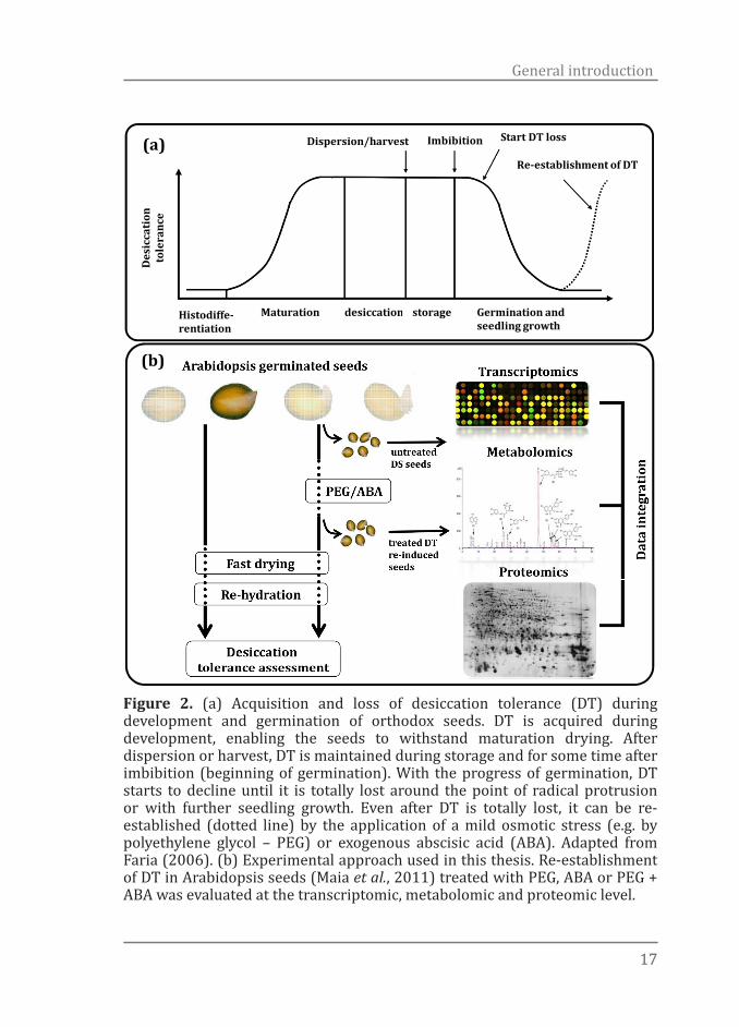
Figure 2. (a) Acquisition and loss of desiccation tolerance (DT) during development and germination of orthodox seeds. DT is acquired during development, enabling the seeds to withstand maturation drying. After dispersion or harvest, DT is maintained during storage and for some time after imbibition (beginning of germination). With the progress of germination, DT starts to decline until it is totally lost around the point of radical protrusion or with further seedling growth. Even after DT is totally lost, it can be re-established (dotted line) by the application of a mild osmotic stress (e.g. by polyethylene glycol – PEG) or exogenous abscisic acid (ABA). Adapted from Faria (2006). (b) Experimental approach used in this thesis. Re-establishment of DT in Arabidopsis seeds (Maia et al., 2011) treated with PEG, ABA or PEG + ABA was evaluated at the transcriptomic, metabolomic and proteomic level.

1818
Chapter 1
this species, introduces a powerful approach to unravel the regulation and mechanism of DT in higher plants. Since the nineties, Arabidopsis has been used as a model species in many different research fields. Its genome was the first one of a higher plant that was sequenced and it has been systematically annotated and curated. The Arabidopsis gene knockout collections, a unique resource for plant biology, are readily available and the site of T-DNA insertions has been determined for over 300,000 independent transgenic lines. Through these collections, insertional mutants are available for most genes in Arabidopsis. Additionally, a myriad of information about gene expression, metabolites and protein profiles is available from public databases. The use of “omics” technologies such as metabolomics, proteomics and transcriptomics associated with model species can now be combined with genomic and expression profiling of non-model species, e.g. by RNAseq, and can aid in the discovery of relevant genes and bring new insights to identify new genes and strategies for crop improvement. We have designed a model system in Arabidopsis to distinguish between genes, cellular events and structures related to DT and other developmental processes. We have used a dehydration/rehydration model to create the relevant physiological states and have studied these states using several –omics approaches (Figure 2b).
Outline of this thesis
The aim of the research presented in this thesis is to explore the molecular basis of desiccation tolerance in seeds. We explore the possibilities of using germinated desiccation sensitive Arabidopsis seeds which are rescued to become tolerant again as a system to study DT. Using this experimental approach in combination with “omics” technologies such as metabolomics, proteomics and transcriptomics and specific available knock-out mutants we aim to discover DT relevant genes, metabolites and metabolic pathways. The insights obtained in this study can also aid to the design of crop improvement strategies. Chapter 1 introduces the definition of the term desiccation tolerance (DT), the relevance of this trait in nature and its societal impact. It introduces how desiccation tolerant seeds can be used as a convenient system in the study of DT and describes the model of loss and re-establishment of DT in germinated
1919
General introduction
seeds and the advantages of this strategy. Chapter 1 also describes briefly what is known about the molecular basis of DT and the potential use of Arabidopsis for studying this trait. Chapter 2 presents the implementation of the system of loss and re-establishment of DT in germinated Arabidopsis seeds and its potential uses to study DT. We show that an osmotic stress can re-induce DT in desiccation sensitive germinated Arabidopsis seeds. With the aid of microarrays we compare the transcriptomes of DT re-induced versus desiccation sensitive seeds. We show that DT demands a re-setting of developmentally related processes which might also be involved in the acquisition of DT during seed development. Further analysis of this dataset reveals that abscisic acid (ABA) is the main player regulating re-establishment of DT in Arabidopsis seeds. In Chapter 3 we extend the system of loss and re-establishment of DT in Arabidopsis by testing it in an ABA-deficient mutant, aba2-1, and demonstrate that ABA can substitute for the osmotic signal in the re-induction of DT. Using ABA biosynthesis and signalling mutants we confirm that ABA plays a key role in re-establishment of DT. We also present strong evidence that re-establishment of DT depends on modulation of ABA sensitivity rather than enhanced ABA levels. Evaluation of several ABA-insensitive mutants, which can still produce normal desiccation tolerant seeds but are impaired in re-establishing DT, demonstrates that acquisition of DT during seed development is genetically different from its re-establishment during germination. Chapter 4 presents the characterization of the metabolic phenotype of Arabidopsis Col-0 and aba2-1 germinated seeds subjected to a set of treatments which combine the application of osmotic stress and ABA to re-establish DT. The metabolic signatures of PEG-induced DT was remarkably different from the one found in seeds treated with ABA. PEG-treated seeds displayed a nitrogen-rich metabolome while the metabolome of ABA-treated seeds was enriched with carbon-rich compounds. We also discuss a core set of metabolites found to be strongly correlated to the re-establishment of DT. Chapter 5 focuses on describing the proteome related to re-establishment of DT under the same conditions and treatments described in Chapter 4 and reveals a set of proteins that might play pivotal roles in DT. A marked enrichment of hydrophilic and charged late embryogenesis
2020
Chapter 1
abundant (LEA) proteins emerges as the most prominent protein type in the re-establishment of DT in Arabidopsis seeds. We discuss probable roles and mechanisms of action of those proteins during drying and in a dry cytoplasm. Finally, we speculate that accumulation of negative charges on the surface of proteins may represent a basal mechanism of adaptation to environments where proteins are more prone to misfolding and interaction with each other and membranes, as in the case of desiccation stress. Chapter 6 discusses and integrates the various topics addressed in this thesis and identifies new challenges and possibilities for further research. Links between the three datasets (transcripts, proteins and metabolites) generated in our study, some important genes, proteins, metabolites, and some of the mechanisms underlying DT are further discussed.
References
Alpert P. 2005. The Limits and Frontiers of Desiccation-Tolerant Life. Integrative and Comparative Biology 45(5): 685-695.
AGI. 2000. Analysis of the genome sequence of the flowering plant Arabidopsis thaliana. Nature 408(6814): 796-815.
Angelovici R, Galili G, Fernie AR, Fait A. 2010. Seed desiccation: a bridge between maturation and germination. Trends in Plant Science 15(4): 211-218.
Battaglia M, Olvera-Carrillo Y, Garciarrubio A, Campos F, Covarrubias AA. 2008. The Enigmatic LEA Proteins and Other Hydrophilins. Plant physiology 148(1): 6-24.
Berjak P. 2006. Unifying perspectives of some mechanisms basic to desiccation tolerance across life forms. Seed Science Research 16(1): 1-15.
Bewley JD, Bradford K, Hilhorst H, Nonogaki H. 2013. Seeds: Physiology of Development, Germination and Dormancy: New York, US: Springer.
Black M, Corbineau F, Gee H, Come D. 1999. Water content, raffinose, and dehydrins in the induction of desiccation tolerance in immature wheat embryos. Plant physiology 120(2): 463-472.
Blackman SA, Obendorf RL, Leopold AC. 1992. Maturation proteins and sugars in desiccation tolerance of developing soybean seeds. Plant physiology 100(1): 225-230.
Boubriak I, Dini M, Berjak P, Osborne DJ. 2000. Desiccation and survival in the recalcitrant seeds of Avicennia marina : DNA replication , DNA repair and protein synthesis. Seed Science Research: 307-315.
Boudet J, Buitink J, Hoekstra FA, Rogniaux H, Larre C, Satour P, Leprince O. 2006. Comparative analysis of the heat stable proteome of radicles of Medicago truncatula seeds during germination identifies late embryogenesis abundant proteins associated with desiccation tolerance. Plant physiology 140(4): 1418-
2121
General introduction
1436.Bruggink T, van der Toorn P. 1995. Induction of desiccation tolerance in germinated
seeds. Seed Science Research 5(1): 1-4.Buitink J, Leger JJ, Guisle I, Vu BL, Wuillème S, Lamirault G, Bars AL, Meur NL, Becker
A, Küster H, Leprince O. 2006. Transcriptome profiling uncovers metabolic and regulatory processes occurring during the transition from desiccation-sensitive to desiccation-tolerant stages in Medicago truncatula seeds. The Plant Journal 47(5): 735-750.
Buitink J, Leprince O. 2008. Intracellular glasses and seed survival in the dry state. Comptes Rendus Biologies 331(10): 788-795.
Buitink J, Vu BL, Satour P, Leprince O. 2003. The re-establishment of desiccation tolerance in germinated radicles of Medicago truncatula Gaertn. seeds. Seed Science Research 13(4): 273-286.
Challabathula D, Bartels D. 2013. Desiccation tolerance in resurrection plants: new insights from transcriptome, proteome and metabolome analysis. Frontiers in Plant Science 4: 482.
Chandra Babu R, Zhang J, Blum A, David Ho TH, Wu R, Nguyen HT. 2004. HVA1, a LEA gene from barley confers dehydration tolerance in transgenic rice (Oryza sativa L.) via cell membrane protection. Plant Science 166(4): 855-862.
Crowe JH, Carpenter JF, Crowe LM. 1998. The role of vitrification in anhydrobiosis. Annual Review of Physiology 60(1): 73-103.
Faria JM. 2006. Desiccation tolerance and sensitivity in Medicago truncatula and Inga vera seeds. PhD thesis thesis, Wageningen UR Wageningen.
Faria JMR, Buitink J, van Lammeren AaM, Hilhorst HWM. 2005. Changes in DNA and microtubules during loss and re-establishment of desiccation tolerance in germinating Medicago truncatula seeds. Journal of Experimental Botany 56(418): 2119-2130.
Farrant JM. 2000. A comparison of mechanisms of desiccation tolerance among three angiosperm resurrection plant species. Plant Ecology: 29-39.
Farrant JM, Brandt W, Lindsey GG. 2007. An Overview of Mechanisms of Desiccation Tolerance in Selected Angiosperm Resurrection Plants. Plant Stress 1: 72–84.
Farrant JM, Moore JP. 2011. Programming desiccation-tolerance: From plants to seeds to resurrection plants. Current Opinion in Plant Biology 14(3): 340-345.
Gaff DF, Oliver M. 2013. The evolution of desiccation tolerance in angiosperm plants: a rare yet common phenomenon. Functional Plant Biology 40(4): 315-328.
Gallardo K, Le Signor C, Vandekerckhove J, Thompson RD, Burstin J. 2003. Proteomics of Medicago truncatula Seed Development Establishes the Time Frame of Diverse Metabolic Processes Related to Reserve Accumulation. Plant physiology 133(2): 664-682.
Hoekstra Fa, Golovina Ea, Buitink J. 2001. Mechanisms of plant desiccation tolerance. Trends in Plant Science 6(9): 431-438.
Hundertmark M, Buitink J, Leprince O, Hincha DK. 2011. The reduction of seed-specific dehydrins reduces seed longevity in Arabidopsis thaliana. Seed Science Research 21(03): 165-173.
Illing N, Denby KJ, Collett H, Shen A, M FJ. 2005. The signature of seeds in resurrection
2222
Chapter 1
plants: a molecular and physiological comparison of desiccation tolerance in seeds and vegetative tissues. Integrative and Comparative Biology 45(5): 771-787.
Keilin D. 1959. The Leeuwenhoek Lecture: The Problem of Anabiosis or Latent Life: History and Current Concept. Proceedings of the Royal Society of London. Series B, Biological Sciences 150(939): 149-191.
Komatsu K, Suzuki N, Kuwamura M, Nishikawa Y, Nakatani M, Ohtawa H, Takezawa D, Seki M, Tanaka M, Taji T, Hayashi T, Sakata Y. 2013. Group A PP2Cs evolved in land plants as key regulators of intrinsic desiccation tolerance. Nature Communications 4.
Kranner I, Birtić S. 2005. A modulating role for antioxidants in desiccation tolerance. Integrative and Comparative Biology 45(5): 734-740.
Le BH, Cheng C, Bui AQ, Wagmaister JA, Henry KF, Pelletier J, Kwong L, Belmonte M, Kirkbride R, Horvath S, Drews GN, Fischer RL, Okamuro JK, Harada JJ, Goldberg RB. 2010. Global analysis of gene activity during Arabidopsis seed development and identification of seed-specific transcription factors. Proceedings of the National Academy of Sciences 107(18): 8063-8070.
Maia J, Dekkers BJW, Provart NJ, Ligterink W, Hilhorst HWM. 2011. The re-establishment of desiccation tolerance in germinated Arabidopsis thaliana seeds and its associated transcriptome. PLoS ONE 6(12).
Manevich Y, Sweitzer T, Pak JH, Feinstein SI, Muzykantov V, Fisher AB. 2002. 1-Cys peroxiredoxin overexpression protects cells against phospholipid peroxidation-mediated membrane damage. Proceedings of the National Academy of Sciences 99(18): 11599-11604.
Maqbool S, Zhong H, El-Maghraby Y, Ahmad A, Chai B, Wang W, Sabzikar R, Sticklen M. 2002. Competence of oat (Avena sativa L.) shoot apical meristems for integrative transformation, inherited expression, and osmotic tolerance of transgenic lines containing hva1. 105(2-3): 201-208.
McKersie BD, Murnaghan J, Jones KS, Bowley SR. 2000. Iron-Superoxide Dismutase Expression in Transgenic Alfalfa Increases Winter Survival without a Detectable Increase in Photosynthetic Oxidative Stress Tolerance. Plant physiology 122(4): 1427-1438.
Moore JP, Le NT, Brandt WF, Driouich A, Farrant JM. 2009. Towards a systems-based understanding of plant desiccation tolerance. Trends Plant Sciences 14(2): 110-117.
Moore JP, Vicré-Gibouin M, Farrant JM, Driouich A. 2008. Adaptations of higher plant cell walls to water loss: drought vs desiccation. Physiologia Plantarum 134(2): 237-245.
Oliver M, Tuba Z, Mishler B. 2000. The evolution of vegetative desiccation tolerance in land plants. 151(1): 85-100.
Ooms JJJ, Léon-Kloosterziel KM, Bartels D, Koornneef M, Karssen CM. 1993. Acquisition of desiccation tolerance and longevity in seeds of Arabidopsis thaliana: A comparative study using abscisic acid-insensitive abi3 mutants. Plant physiology 102(4): 1185-1191.
Osborne DJ, Boubriak II. 1994. DNA and desiccation tolerance. Seed Science Research 4(02): 175-185.

2323
General introduction
Pammenter NW, Berjak P. 1999. A review of recalcitrant seed physiology in relation to desiccation-tolerance mechanisms. Plant Cell: 13-37.
Potts M. 1994. Desiccation tolerance of prokaryotes. Microbiological Reviews 58(4): 755-805.
Roberts EH. 1973. Predicting the storage life of seeds. Seed Science and Technology 1: 499-514.
Roxas VP. 2000. Stress Tolerance in Transgenic Tobacco Seedlings that Overexpress Glutathione S-Transferase/Glutathione Peroxidase. Plant and Cell Physiology 41(11): 1229-1234.
Saracco F, Bino RJ, Bergervoet JH, Lanteri S. 1995. Influence of priming-induced nuclear replication activity on storability of pepper (Capsicum annuum L.) seed. Seed Science Research 5(1): 25-29.
Sargent JA, Mandi SS, Osborne D. 1981. The loss of desiccation tolerance during germination: An ultrastructural and biochemical approach. 105(3-4): 225-239.
Sreedhar L. 2002. In vivo Characterization of the Effects of Abscisic Acid and Drying Protocols Associated with the Acquisition of Desiccation Tolerance in Alfalfa (Medicago sativa L.) Somatic Embryos. Annals of Botany 89(4): 391-400.
Sun Z, Qi X, Wang Z, Li P, Wu C, Zhang H, Zhao Y. 2013. Overexpression of TsGOLS2, a galactinol synthase, in Arabidopsis thaliana enhances tolerance to high salinity and osmotic stresses. Plant Physiology and Biochemistry 69(0): 82-89.
Sunkar R, Bartels D, Kirch H-H. 2003. Overexpression of a stress-inducible aldehyde dehydrogenase gene from Arabidopsis thaliana in transgenic plants improves stress tolerance. The Plant Journal 35(4): 452-464.
Terrasson E, Buitink J, Righetti K, Ly Vu B, Pelletier S, Lalanne D, Zinsmeister J, Leprince O. 2013. An emerging picture of the seed desiccome: confirmed regulators and newcomers identified using transcriptome comparison. Frontiers in Plant Science 4.
Tunnacliffe A, Wise MJ. 2007. The continuing conundrum of the LEA proteins. Die Naturwissenschaften 94(10): 791-812.
Umezawa T, Nakashima K, Miyakawa T, Kuromori T, Tanokura M, Shinozaki K, Yamaguchi-Shinozaki K. 2010. Molecular basis of the core regulatory network in ABA responses: sensing, signaling and transport. Plant cell physiology 51(11): 1821-1839.
Verdier J, Lalanne D, Pelletier S, Torres-Jerez I, Righetti K, Bandyopadhyay K, Leprince O, Chatelain E, Vu BL, Gouzy J, Gamas P, Udvardi MK, Buitink J. 2013. A Regulatory Network-Based Approach Dissects Late Maturation Processes Related to the Acquisition of Desiccation Tolerance and Longevity of Medicago truncatula Seeds. Plant physiology 163(2): 757-774.
Verslues PE, Juenger TE. 2011. Drought, metabolites, and Arabidopsis natural variation: a promising combination for understanding adaptation to water-limited environments. Current Opinion in Plant Biology 14(3): 240-245.
Vieira CV, da Silva EAA, Alvarenga AA, Castro EM, Toorop PE. 2010. Stress-associated factors increase after desiccation of germinated seeds of Tabebuia impetiginosa Mart. Plant Growth Regulation 62(3) 257-263.
Wise M. 2004. POPP the question: what do LEA proteins do? Trends in Plant Science
2424
Chapter 1
9(1): 13-17.Wolkers WF, McCready S, Brandt WF, Lindsey GG, Hoekstra FA. 2001. Isolation and
characterization of a D-7 LEA protein from pollen that stabilizes glasses in vitro. Biochimica et Biophysica Acta (BBA) - Protein Structure and Molecular Enzymology 1544(1–2): 196-206.
Xu N, Bewley JD. 1995. The role of abscisic acid in germination, storage protein synthesis and desiccation tolerance in alfalfa (Medicago sativa L.) seeds, as shown by inhibition of its synthesis by fluridone during development. Journal of Experimental Botany 46(287): 687-694.
Yancey PH. 2001. Water Stress, Osmolytes and Proteins. American Zoologist 41(4): 699-709.
Yancey PH. 2005. Organic osmolytes as compatible, metabolic and counteracting cytoprotectants in high osmolarity and other stresses. Journal of Experimental Biology 208(15): 2819-2830.
Young ND, Debellé F, Oldroyd GED, et al., 2011. The Medicago genome provides insight into the evolution of rhizobial symbioses. Nature 480, 520-4.

2525
General introduction

Não quero muitas e nempoucas palavras,Não quero definições e nemquero sentenças,Quero apenas caminhar comsede e ouvir-me silenciosamenteenquanto atravesso essa vidaem tumulto, esse alarde, essainsana busca de tudo, para onada que preciso
Aline Binns

Chapter 2The re-establishment of desiccation toler-ance in germinated Arabidopsis thaliana seeds and its associated transcriptome
Authors:Julio Maia1*, Bas J.W. Dekkers1,2, Nicholas J. Provart3, Wilco Ligterink1*, Henk W. M. Hilhorst1
1 Wageningen Seed Lab, Laboratory of Plant Physiology, Wageningen University, Wageningen, The Netherlands2Department of Molecular Plant Physiology, Utrecht University, Utrecht, The Netherlands3Department of Cell and Systems Biology/Centre for the Analysis of Ge-nome Evolution and Function, University of Toronto, Toronto, Canada* Author for correspondence: [email protected]
PLoS ONE (2011) 6(12), pp e29123DOI: 10.1371/journal.pone.0029123

2828
Chapter 2
Abstract
The combination of robust physiological models with “omics” studies holds promise for the discovery of genes and pathways linked to how organisms deal with drying. Here we used a transcriptomics approach in combination with an in vivo physiological model of re-establishment of desiccation tolerance (DT) in Arabidopsis thaliana seeds. We show that the incubation of desiccation sensitive (DS) germinated Arabidopsis seeds in a polyethylene glycol (PEG) solution re-induces the mechanisms necessary for expression of DT. Based on a SNP-tile array gene expression profile, our data indicates that the re-establishment of DT, in this system, is related to a programmed reversion from a metabolic active to a quiescent state similar to prior to germination. Our findings show that transcripts of germinated seeds after the PEG-treatment are dominated by those encoding late embryogenesis abundant (LEA), seed storage and dormancy related proteins. On the other hand, a massive repression of genes belonging to many other classes such as photosynthesis, cell wall modification and energy metabolism occurs in parallel. Furthermore, comparison with a similar system for Medicago truncatula reveals a significant overlap between the two transcriptomes. Such overlap may highlight core mechanisms and key regulators of the trait DT. Taking into account the availability of the many genetic and molecular resources for Arabidopsis, the described system may prove useful for unraveling DT in higher plants.
Introduction
Desiccation tolerance (DT), or anhydrobiosis, can be conceptually defined as the ability to survive, by reversible cessation of metabolism, the removal of almost all cellular free water when in equilibrium with moderately dry air and resume normal function when re-hydrated (Phillips et al., 2002 ). By definition, desiccation tolerance is the ability of living organisms to deal with water losses below 0.1 g H2O g-1 dry weight and survive the re-hydration process without permanent damage (Oliver et al., 2000). Orthodox seeds acquire DT during development, concomitantly with a myriad of other processes like cell proliferation, reserve deposition,
29
Desiccation tolerance transcriptome
developmental arrest and maturation drying (Bewley & Black, 1994). DT in (orthodox) seeds is based on a range of relatively complex protection mechanisms that accompany dehydration (Illing et al., 2005). A strong correlation between protective mechanisms activated during dehydration, such as the accumulation of late embryogenesis abundant (LEA) proteins and dehydrins, non-reducing sugars, sucrose, reactive oxygen species (ROS) scavenging, as well as switching off of metabolism, have been so far postulated as playing major roles in this phenomenon (Berjak, 2006). It has also been suggested that DT demands structural stabilizing processes, such as plasmalemma displacement and intracellular space vacuolization (Farrant, 2000; Moore et al., 2008). In short, the mechanisms involved in DT may be roughly divided in three groups: 1) signalling mechanisms, gene regulation and functional proteomics; 2) metabolic adjustment and antioxidant systems; and 3) macromolecular and mechanical stability (Moore et al., 2009). Several studies of the acquisition of DT during seed development (Sreedhar et al., 2002; Illing et al., 2005) and on its loss upon germination (Buitink et al., 2003; Faria et al., 2005; Buitink et al., 2006; Daws et al., 2007) have been reported. Interestingly, DT can be rescued in germinated seeds by the application of a mild osmotic stress. The re-induction of DT in germinated seeds by incubation in PEG was first reported by Bruggink and van der Toorn (1995). They showed that DT could be fully restored in germinated seeds of Cucumis sativus and Impatiens walleriana. These authors suggested that this approach could serve as a convenient model system in studies of DT and may have important implications for the agricultural industry (Bruggink & van der Toorn, 1995). This strategy to re-induce DT in germinated seeds has been confirmed in other species like Medicago (Medicago truncatula) and Tabebuia impetiginosa. The re-establishment of DT in primary roots of Medicago germinated seeds by a mild osmotic stress (-1.5 MPa) treatment has been so far used to identify the transcriptome and (heat stable) proteome associated with DT (Boudet et al., 2006; Buitink et al., 2006). Furthermore, the application of a cold or heat shock prior to osmotic treatment improved desiccation tolerance in protruded radicles of T. impetiginosa (Vieira et al., 2010). These findings suggest the existence of overlapping mechanisms acting in parallel, or synergistically, in different stress types. Thus, understanding DT would not only enhance insights
3030
Chapter 2
related to tolerance mechanisms of water deficit but also to other stresses, such as cold, salt and heat. Despite numerous studies on the acquisition of DT during seed development and the re-induction of DT in germinated seeds, none of these have been able to ‘filter out’ the mechanisms that are not directly associated with DT but are coupled with other concomitant developmental pathways. Therefore, models capable of discriminating between overlapping developmental programs and the acquisition of DT are extremely promising to understand the genetic and molecular mechanisms controlling desiccation tolerance and sensitivity in seeds. Arabidopsis (Arabidopsis thaliana) is a well-known model system in plant biology. Evidently, the use of this system together with all the genetic and molecular tools so far developed would generate a powerful model to further unravel the regulation and mechanisms of DT in higher plants. However it is not known whether germinated seeds of Arabidopsis are desiccation sensitive and if DT can be re-established after it has been lost upon completion of germination. Here we show that Arabidopsis seeds lost DT upon germination and that DT can be re-induced in germinated seeds. Further we present the associated transcriptome of desiccation sensitive (DS) and DT germinated Arabidopsis seeds. The discovery of relevant genes may bring new insights to identify new strategies for crop production under abiotic stresses and highlight putative key hubs involved in the regulation of seed survival in the dry state. Furthermore, the use of Arabidopsis for studying loss and re-establishment of DT in germinated seeds in combination with the genetic and molecular tools developed for this model species engenders a powerful model to further unravel DT in higher plants.
Materials and methods
Assessment of desiccation tolerance
Seeds of Arabidopsis, accession Columbia (Col-0), were cold stratified for 72h at 4oC in 9 cm Petri dishes on two layers of blue filter paper (Anchor paper Co.) and 10 ml of distilled water. After stratification to break residual dormancy, germination was performed at 22oC under constant white light and determined

31
Desiccation tolerance transcriptome
from three independent replicates of 100 seeds, by counting the number of individual seeds that had a protruded radicle. To determine the percentage of desiccation-tolerant germinated seeds, four developmental stages were defined. For that a stereomicroscope was used and the seeds were grouped as follows: (stage I) testa rupture; (stage II) seeds at radical protrusion; (stage III) germinated seeds showing a primary root of 0.3-0.5mm length; and (stage IV) at the appearance of the first root hairs (Figure 1). These developmental stages were achieved approximately 24, 28, 32 and 36 hours after the seeds were transferred from 4oC to the optimum germination conditions at 22oC. Four replicates of 25 seeds for each stage were fast-dried for three days at 20°C under a forced air flow at 32% relative humidity (RH), which was achieved by a saturated calcium chloride solution in a closed chamber. Water contents were assessed gravimetrically for triplicate samples of 70 germinated seeds, by determination of the fresh weight and subsequent dry weight after 17h at 105°C (ISTA, 2009). Water contents were expressed on a dry weight basis. After dehydration, germinated seeds were pre-humidified in humid air (100% RH) for 24h at 22oC in the dark, in order to avoid imbibitional damage (Leopold, 1986), and then rehydrated in H2O at 22°C on a Copenhagen Table under a 12/12h dark/light regime. Germinated seeds that continued their development and transformed into viable seedlings were considered desiccation-tolerant.
Dehydration curves
Cold-stratified seeds were placed to germinate at 22oC under constant white light. Three replicates of 70 germinated seeds of the four developmental stages (Figure 1) were selected, placed in small
Figure 1. Arabidopsis seeds at different developmental stages during and after visible germination. I - testa rupture; II - at radical protrusion; III - primary root of approximately 0.3mm length; and IV – at appearance of the first root hairs.

3232
Chapter 2
aluminium pans and dried under a saturated CaCl2 atmosphere inside a drying chamber with a forced air flow (32% RH at 20oC). Concomitantly, three replicates of 70 germinated seeds of each developmental stage were picked-up and incubated in 6-cm petri dishes containing 1.2 ml of a polyethylene glycol (PEG 8000) solution with an osmotic potential of -2.5 MPa on one layer of filter paper at 22oC. After 3d of PEG incubation and a quick wash in distilled water to remove residual PEG, the seeds were transferred to small aluminium pans and dried under a saturated CaCl2 atmosphere inside a drying chamber with a forced air flow. During the drying step and PEG incubation, samples were taken at intervals to measure water content by gravimetry.
Assessment of the loss and re-establishment of DT
To assess the re-establishment of DT in germinated seeds, they were selected by their developmental stage (I, II, III and IV – Figure 1) using a stereomicroscope and either (fast) dried directly or after 3d of incubation in PEG solution. Incubation was done in the dark at 22oC, in 6-cm Petri dishes containing 1.2 ml of PEG solution (-2.5 MPa) on one sheet of filter paper. After incubation, germinated seeds were rinsed thoroughly in distilled water with the aid of a set of sieves, transferred to new Petri dishes with one dry sheet of germination paper and then dehydrated, pre-humidified and rehydrated as described before. Germinated seeds that resumed growth and generated a viable seedling after rehydration were considered DT. Four independent experiments of 25 germinated seeds each were carried out for each treatment.
RNA extraction, target synthesis and microarray hybridization
Germinated seeds of stage II after PEG incubation (DT) and non-treated germinated seeds at the same developmental stage (DS) were used for the RNA extractions. Total RNA was extracted according to the hot borate protocol modified from Wan and Wilkins (1994). Three replicates of approximately 1000 germinated seeds for each treatment were homogenized and mixed with 800 µL of extraction buffer (0.2M Na borate decahydrate (Borax), 30mM EGTA, 1% SDS, 1% Na deoxycholate (Na-DOC)) containing 1.6 mg DTT and 48 mg PVP40 which had been heated to 80°C. 1 mg proteinase K was added to this suspension and incubated for 15 min at 42oC. After adding 64 μl of 2M KCL the

33
Desiccation tolerance transcriptome
samples were incubated on ice for 30 min and subsequently centrifuged for 20 min at 12,000g. Ice-cold 8M LiCl was added to the supernatant in a final concentration of 2M and the tubes were incubated overnight on ice. After centrifugation for 20 min at 12,000g at 4ºC, the pellets were washed with 750µl ice-cold 2M LiCl. The samples were centrifuged for 10 min at 10,000g at 4ºC and the pellets were re-suspended in 100 µl DEPC treated water. The samples were phenol chloroform extracted, DNAse treated (RQ1 DNase, Promega) and further purified with RNEasy spin columns (Qiagen) following the manufacturer’s instructions. RNA quality and concentration were assessed by agarose gel electrophoresis and UV spectrophotometry. RNA was processed for use on Affymetrix® Arabidopsis SNPtile array (atSNPtilx520433) as described by the manufacturer. Briefly, 1μg of total RNA was reverse transcribed using a T7-Oligo(dT) Promoter Primer in the first-strand cDNA synthesis reaction. Following RNase H-mediated second-strand cDNA synthesis, the double-stranded cDNA was purified and served as template in the subsequent in vitro transcription (IVT) reaction. The IVT reaction was carried out in the presence of T7 RNA Polymerase and a biotinylated nucleotide analog/ribonucleotide mix for complementary RNA (cRNA) amplification and biotin labeling. The biotinylated cRNA targets were then cleaned up, fragmented, and hybridized to the SNPtile array. The hybridization data was extracted using an R-script with the help of an annotation-file based on TAIR9 annotation (http://aquilegia.uchicago.edu/naturalvariation-/cisTrans/ArrayAnnotation.html). Data were normalized in R using quantile normalization and average results for the 3 arrays per sample were used for further analysis. All data are MIAME compliant as detailed on the MGED Society website http://www.mged.org-/Workgroups/MIAME/miame.html and the data discussed in this publication have been deposited in NCBI's Gene Expression Omnibus (Edgar et al., 2002) and are accessible through GEO Series accession number GSE30853 (http://www.ncbi.nlm.nih.gov/geo/query-/acc.cgi?acc=GSE30853).
Microarray analysis
For analysis of the DT/DS gene set, germinated seeds after PEG treatment versus non-treated germinated seeds at the same developmental stage, we used the over-representation analysis (ORA) tool of GeneTrailExpress (Keller

3434
Chapter 2
et al., 2008). This analysis was employed to identify significantly enriched gene ontologies (GO) categories. ORA was performed with the following parameters: significance level: 0.05, p-value adjustment for multiple testing: Bonferroni adjustment, minimum class size: 3, maximum class size: 40. To identify cis-acting promoter elements potentially involved in regulating the co-expression of genes involved in DT, the Arabidopsis expression network analysis (Athena) tool was used (http://www.bioinformatics2.wsu.edu/cgi-bin/Athena/cgi/home.pl) (O'connor et al., 2005). Specifically, the promoter regions of all differentially up-regulated genes (Fold change ≥ 2 and P-value ≤ 0.05) were searched for any common motifs located within a 1kb region upstream of the translational start site.
Results
Assessment of the re-establishment of desiccation tolerance
Previous reports have shown that DT can be fully rescued in germinated seeds (Bruggink & van der Toorn, 1995; Buitink et al., 2003; Vieira et al., 2010). Here it was tested whether DT could be re-induced in Arabidopsis seeds by treating them in a PEG (-2.5 MPa) solution for three days at 22oC. DT can be re-induced in imbibed seeds only in a limited time frame and its loss usually coincides, depending on the species, with the protrusion of the radicle tip and/or radicle length (Bruggink & van der Toorn, 1995; Buitink et al., 2003; Vieira et al., 2010). Therefore we defined four clearly distinct developmental stages to assess for re-induction of DT. The stages are: stage I (testa rupture), stage II (seeds at radical protrusion), stage III (germinated seeds showing a primary root of 0.3-0.5mm length) and stage IV (at the appearance of the first root hairs) (Figure 1). First we determined the best developmental stage suitable for the re-establishment of DT. For all stages DT could be re-established to a certain extent however seeds at earlier stages (i.e. stages I and II) performed better as compared to the two later stages. Seeds completing germination at stage I and germinated at stage II, showed 100% of re-establishment of DT regarding primary roots, cotyledons and seedling formation while for those at stage III a slight reduction in all parameters was observed (Figure 2). Germinated seeds at stage IV showed low competence in re-establishing DT with a final survival rate of 38% (Figure 2).

35
Desiccation tolerance transcriptome
This treatment pointed to a developmental stage-dependent re-establishment of DT and, in addition, indicates that different seed parts have differential sensitivity to drying. Interestingly, more seedlings survived due to lateral root formation. This phenomenon was also observed by several other authors. They frequently noted the appearance of lateral roots after the primary root had been lethally damaged by desiccation (Koster & Leopold, 1988; Bruggink & van der Toorn, 1995; Vieira et al., 2010). Seedlings that did not resume radicle growth, frequently showed growth of the cotyledons and, to a lesser extent, also of the hypocotyl. However, the longer the radicle before dehydration, the less frequent the growth of cotyledons and hypocotyl. Next we investigated the effect of the osmotic potential and time of incubation on the ability to re-establish DT in germinated Arabidopsis seeds. Therefore, first, seeds at stage II were submitted to different PEG concentrations (Figure 3a). DT could be substantially re-induced (approximately 100%) for all parameters in seeds treated with PEG -2.5 MPa. A lower PEG osmotic potential (-1.7MPa) re-induced DT as well and resulted in nearly 100% survival of the cotyledons after drying. However, this concentration resulted in a huge drop of primary root survival to approximately 10%. A larger percentage of seedlings survived (60%) although this depended on lateral root formation. From -2.5 MPa downwards there was an abrupt drop in DT re-establishment, decreasing to 34% for cotyledons, 1% for primary roots and 7% for
Figure 2. Re-establishment of desiccation tolerance in Arabidopsis seeds. Desiccation tolerance was determined after drying the germinated seeds with or without previous PEG treatment, followed by pre-humidification and rehydration. Survival of cotyledons (circles) and primary roots (squares) was scored 5d after rehydration and of seedlings (triangles) 10d after rehydration. Each data point is the average of four independent experiments of 25 seed/seedlings. Bars represent standard error.

3636
Chapter 2
seedling formation at -3.5 MPa, to 0% for all parameters at -5.0 and -7.0 MPa (Figure 3A). Thus a concentration of -2.5MPa PEG solution is the optimal concentration to re-establish DT of stage II Arabidopsis seeds. Lastly we varied the incubation time from several hours up to three days using stage II seeds in a -2.5 MPa PEG solution. As the acquisition of DT is an active process (Angelovici et al., 2010), the incubation time also appeared to be crucial to the recovery of DT. A minimum of three days in -2.5 MPa PEG was necessary for full re-establishment of this attribute in germinated Arabidopsis seeds (Figure 3B).
Drying responses
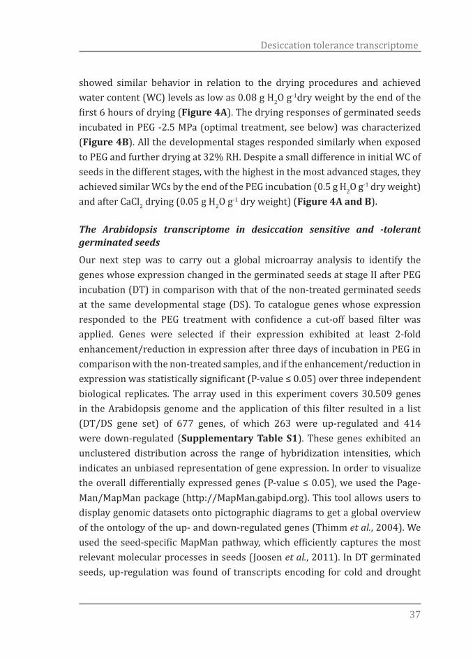
Different dehydration conditions, especially drying rates, can significantly influence the response of desiccation-sensitive plant tissues (Tobias & Norman, 2009). Therefore we first studied the de-hydration behaviour of the Arabidopsis seeds at all four defined stages. All developmental stages
Figure 3. Osmotic potential and incubation time effects on re-establishment of desiccation tolerance. (A) PEG osmotic potential effect on DT; (B) -2.5 MPa PEG solution incubation time effect on DT. Survival of cotyledons and primary roots was scored 5d after rehydration and of seedlings 10d after rehydration. Each data point is the average of four independent experiments of 25 seeds at developmental stage II. Bars represent standard error. Control = germinated seeds dried directly at 32% RH without previous PEG treatment.
37
Desiccation tolerance transcriptome
showed similar behavior in relation to the drying procedures and achieved water content (WC) levels as low as 0.08 g H2O g-1dry weight by the end of the first 6 hours of drying (Figure 4A). The drying responses of germinated seeds incubated in PEG -2.5 MPa (optimal treatment, see below) was characterized (Figure 4B). All the developmental stages responded similarly when exposed to PEG and further drying at 32% RH. Despite a small difference in initial WC of seeds in the different stages, with the highest in the most advanced stages, they achieved similar WCs by the end of the PEG incubation (0.5 g H2O g-1 dry weight) and after CaCl2 drying (0.05 g H2O g-1 dry weight) (Figure 4A and B).
The Arabidopsis transcriptome in desiccation sensitive and -tolerant germinated seeds
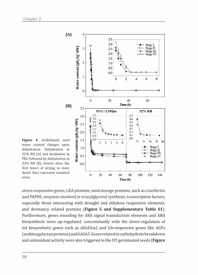
Our next step was to carry out a global microarray analysis to identify the genes whose expression changed in the germinated seeds at stage II after PEG incubation (DT) in comparison with that of the non-treated germinated seeds at the same developmental stage (DS). To catalogue genes whose expression responded to the PEG treatment with confidence a cut-off based filter was applied. Genes were selected if their expression exhibited at least 2-fold enhancement/reduction in expression after three days of incubation in PEG in comparison with the non-treated samples, and if the enhancement/reduction in expression was statistically significant (P-value ≤ 0.05) over three independent biological replicates. The array used in this experiment covers 30.509 genes in the Arabidopsis genome and the application of this filter resulted in a list (DT/DS gene set) of 677 genes, of which 263 were up-regulated and 414 were down-regulated (Supplementary Table S1). These genes exhibited an unclustered distribution across the range of hybridization intensities, which indicates an unbiased representation of gene expression. In order to visualize the overall differentially expressed genes (P-value ≤ 0.05), we used the Page-Man/MapMan package (http://MapMan.gabipd.org). This tool allows users to display genomic datasets onto pictographic diagrams to get a global overview of the ontology of the up- and down-regulated genes (Thimm et al., 2004). We used the seed-specific MapMan pathway, which efficiently captures the most relevant molecular processes in seeds (Joosen et al., 2011). In DT germinated seeds, up-regulation was found of transcripts encoding for cold and drought
3838
Chapter 2
stress responsive genes, LEA proteins, seed storage proteins, such as cruciferins and PAP85, enzymes involved in triacylglycerol synthesis, transcription factors, especially those interacting with drought and ethylene responsive elements, and dormancy related proteins (Figure 5 and Supplementary Table S1). Furthermore, genes encoding for ABA signal transduction elements and ABA biosynthesis were up-regulated, concomitantly with the down-regulation of GA biosynthetic genes such as AtGA3ox2 and GA-responsive genes like AGPs (arabinogalactan proteins) and GASA3. Genes related to carbohydrate breakdown and antioxidant activity were also triggered in the DT germinated seeds (Figure
Figure 4. Arabidopsis seed water content changes upon dehydration. Dehydration at 32% RH (A) and incubation in PEG followed by dehydration at 32% RH (B). Inserts show the first hours of drying in more detail. Bars represent standard error.

39
Desiccation tolerance transcriptome
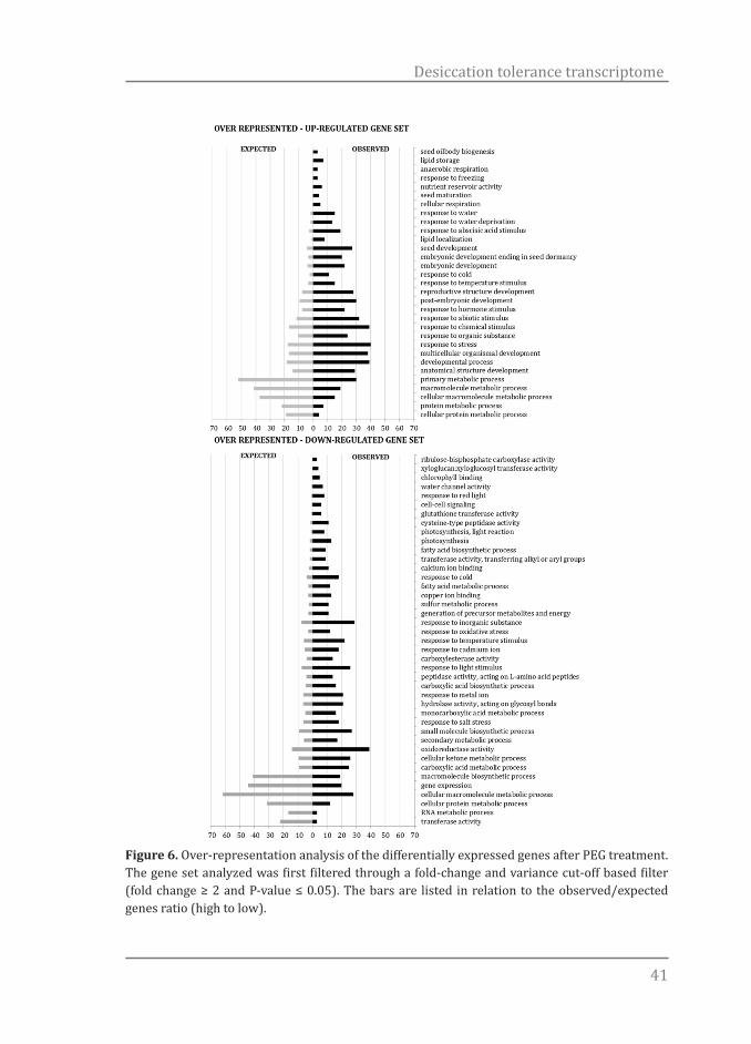
5). In contrast, a massive repression of genes related to DNA biosynthesis and chromatin structure, energy metabolism and cell wall modification was seen in the DT germinated seeds (Figure 5). Regarding the ‘Energy’ class, down-regulation occurred particularly for genes related to photosynthesis and the Calvin cycle (Figure 5). To verify whether our strategy to visualize and filter the gene set was valid, a more detailed analysis was undertaken. We used the over-representation analysis (ORA) tool of GeneTrailExpress (Keller et al., 2008). This analysis compares a gene set of interest to the reference set and when considering a certain functional category as a gene ontology (GO) term, it attempts to detect if this category is over-represented or under-represented in the respective gene set. It also estimates how likely this is due to chance (Keller et al., 2008). The filtered gene set was split into two subsets, up- and down-regulated, and the program allowed us to determine which GO categories were significantly enriched (P-value ≤ 0.05) in the DT/DS up- and down-regulated gene subsets (Figure 6). GO terms describing developmentally related processes such as ‘lipid storage’, ‘nutrient reservoir activity’ and ‘seed maturation’ together with GO terms describing responses to various abiotic stresses responses such as freezing, water deprivation and cold were among the top-ranked enriched processes in the DT/DS up-regulated gene subset. It is important to stress that the genes were ranked in relation to the observed/expected ratio and GO terms such as the ones related to seed development, embryonic development ending in seed dormancy, post-embryonic development, response to hormone and abiotic stimulus as well as response to stress were represented by a large number of genes in the DT/DS up-regulated gene subset (Figure 6). Furthermore, response to water deprivation and abscisic acid (ABA) stimulus categories were, at the same time, represented by a high number of genes and high-ranked, reinforcing the importance of the hormone ABA to the acquisition of DT. Drought responsive genes such as DREB2A, XERO1, LEA genes and ABA-responsive genes, such as EM1, GEA6, RAB18, LTI65, RD29B, among others, appeared in the DT/DS gene set (Supplementary Table S1). Interestingly, the RD29B gene, which was highly up-regulated in the DT/DS gene set, was not listed in the GO category ‘response to ABA stimulus’ (Supplementary Table S2), showing that the power of this
4040
Chapter 2
analysis can be limited by the GO annotation’s accuracy. LTI65 contains two ABA-responsive elements (ABREs) that are required for the dehydration-responsive expression of RD29B as cis-acting elements (Nakashima et al., 2006).
On the other hand, all the drought responsive genes that were highly up-regulated in the DT/DS set such as PER1, RAB18, ATDI21 and DREB2A, were retrieved in the GO category ‘response to water deprivation’. Analyses of DREB2A has shown that it is possible to increase drought stress tolerance of the transgenic plants overexpressing this gene and revealed that DREB2A regulates the expression of many water stress inducible genes (Sakuma et al., 2006; Maruyama et al., 2009). As expected, GO categories denoting processes related to energy such as ‘response to red light’, ‘chlorophyll binding’ and ‘photosynthesis’, as well as GO categories grouping genes related to cell wall
Figure 5. Seed MapMan molecular network map. Log2 ratios are used to express relative transcript levels in germinated Arabidopsis seeds at stage II treated for 3d in -2.5 MPa PEG versus non-treated seeds in the same developmental stage. Red squares, higher levels in PEG treated seeds; blue squares, higher levels in non-treated seeds. Only ratios with P values lower or equal to 0.05 are displayed.
41
Desiccation tolerance transcriptome
Figure 6. Over-representation analysis of the differentially expressed genes after PEG treatment. The gene set analyzed was first filtered through a fold-change and variance cut-off based filter (fold change ≥ 2 and P-value ≤ 0.05). The bars are listed in relation to the observed/expected genes ratio (high to low).

4242
Chapter 2
breakdown and loosening, metabolism of fatty acids, among other post-germination related processes, were over-represented in the down-regulated DT/DS gene subset. For example, xyloglucan endotransglycosylases/hydrolase (XET) genes, such as MERI5B, XTH9 and TCH4 were down-regulated in the DT/DS gene set (Supplementary Table S1). This gene family encodes for enzymes that modify a major structural component of the plant cell wall, xyloglucans, and therefore may influence plant growth and development (Campbell & Bram, 1999). Furthermore, we compared the gene list after the cut-off filter (DT/DS gene set - Supplementary Table S1) against a data set obtained in a similar system for Medicago (Buitink et al., 2006) and a significant overlap was found. Among 111 genes that were present in both Arabidopsis and Medicago gene sets, 49 were down- and 48 were up-regulated in both species and 14 displayed opposite response (Figure 7 and Supplementary Table S3).As in Arabidopsis, genes encoding for antioxidant activity, ABA signalling, seed storage proteins, LEA proteins, drought and ethylene responsive elements and dormancy related traits were up-regulated in the rescued Medicago germinated seeds (Supplementary Tables S3 and S4 and Figure S1). The most significant overlap in the down-regulated genes occurred for genes related to cell wall and energy metabolism. The genes showing opposite response belonged to a varied range of classes. Based on these results we hypothesized that a controlled reversion from the germination program towards the seed developmental program is taking place during the incubation in PEG and that this transition is necessary for the re-establishment of DT. To verify this, we examined the expression during seed imbibition of the top 50 ranked of both up- and down-regulated genes in the DT/DS set and created a heat map in the Expression Browser of the BAR website (Toufighi et al., 2005). The heat maps clearly corroborated our hypothesis and it appears indeed that the germinating seeds are reverted to a developmental, desiccation tolerant, stage (Figure 8). According to this in silico analysis, genes that were up-regulated in the PEG treated seeds and may be related to the acquisition of DT were down-regulated upon imbibition while genes that were down-regulated in the treated seeds were up-regulated upon imbibition (Figure 8). More details about this analysis can be seen in the supplementary material (Supplementary Table

43
Desiccation tolerance transcriptome
S5).
Identification of promoter motifs within up-regulated enriched genes
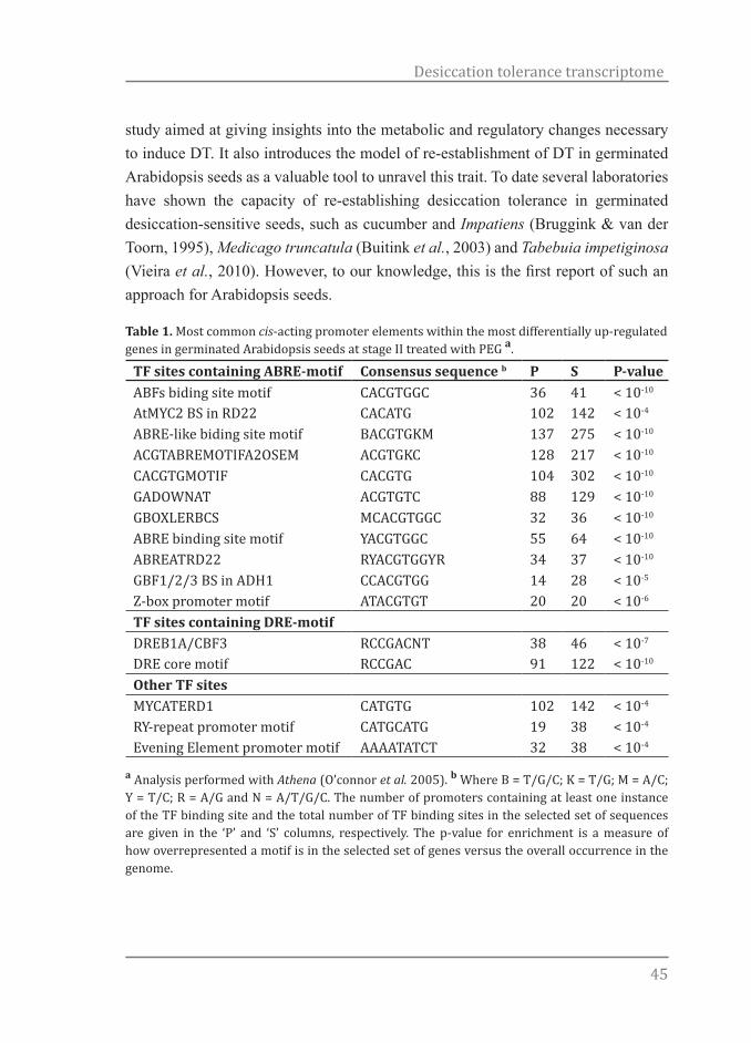
Information about the promoter region of genes can provide valuable information on how these genes are regulated. Besides that, the presence of similar elements in different genes can suggests common regulatory mechanisms of their expression. An analysis of the differentially up-regulated genes in the DT/DS gene set was performed to identify potential cis-acting promoter elements. In particular, this analysis identified two main transcription factor (TF) site groups overrepresented within the promoters of germinated seeds treated with PEG. The first group consists of 11 enriched motifs containing the core sequence (ACGTG), named ABA-responsive element (ABRE) and the second of 2 enriched motifs containing the drought responsive elements (DRE), core motif (A/GCCGACA). Next to these two groups also a MYC-related, RY element and an Evening element were overrepresented. Table 1 displays the promoter element consensus sequence, number of promoters with TF sites and the number of predicted TF sites.
Discussion
The ability to study desiccation tolerance/sensitivity in combination with ‘omics’ techniques and in vivo physiology creates new opportunities for examining how
Figure 7. Overlapping homologous genes in Arabidopsis and Medicago germinated seeds after re-establishment of DT. (A) total number of overlapping genes; (B) down-regulated in both systems and; (C) up-regulated in both systems. The gene lists are presented in Supplementary Table S3.
(A) (B)
(C)
Arabidopsis Medicago
566 111 882
365 50549
48215 389

4444
Chapter 2
organisms deal with desiccation stress. Here we explore the possibility to rescue desiccation tolerance in desiccation sensitive, germinated, Arabidopsis seeds by PEG-treatment and its associated transcriptome. By observing changes in gene expression in germinated seeds of Arabidopsis in response to the PEG treatment, this
Figure 8. Heat maps displaying the gene expression profile during seed imbibition according to the Bio-Array Resource database. The top 50 up- and down-regulated genes in the DT/DS set were tested, in silico, for their expression during seed imbibition (0 to 24h). The DT/DS up-regulated genes showed up-regulation (red) in dry seeds and down-regulation (blue) during seed imbibition. The DT/DS down-regulated genes showed down regulation (blue) in the dry seed and in early phases of seed imbibition and up-regulation along germination. The values in the legend are log2-transformed ratios.
45
Desiccation tolerance transcriptome
study aimed at giving insights into the metabolic and regulatory changes necessary to induce DT. It also introduces the model of re-establishment of DT in germinated Arabidopsis seeds as a valuable tool to unravel this trait. To date several laboratories have shown the capacity of re-establishing desiccation tolerance in germinated desiccation-sensitive seeds, such as cucumber and Impatiens (Bruggink & van der Toorn, 1995), Medicago truncatula (Buitink et al., 2003) and Tabebuia impetiginosa (Vieira et al., 2010). However, to our knowledge, this is the first report of such an approach for Arabidopsis seeds.
Table 1. Most common cis-acting promoter elements within the most differentially up-regulated genes in germinated Arabidopsis seeds at stage II treated with PEG a.
TF sites containing ABRE-motif Consensus sequence b P S P-valueABFs biding site motif CACGTGGC 36 41 < 10-10
AtMYC2 BS in RD22 CACATG 102 142 < 10-4
ABRE-like biding site motif BACGTGKM 137 275 < 10-10
ACGTABREMOTIFA2OSEM ACGTGKC 128 217 < 10-10
CACGTGMOTIF CACGTG 104 302 < 10-10
GADOWNAT ACGTGTC 88 129 < 10-10
GBOXLERBCS MCACGTGGC 32 36 < 10-10
ABRE binding site motif YACGTGGC 55 64 < 10-10
ABREATRD22 RYACGTGGYR 34 37 < 10-10
GBF1/2/3 BS in ADH1 CCACGTGG 14 28 < 10-5
Z-box promoter motif ATACGTGT 20 20 < 10-6
TF sites containing DRE-motif DREB1A/CBF3 RCCGACNT 38 46 < 10-7
DRE core motif RCCGAC 91 122 < 10-10
Other TF sitesMYCATERD1 CATGTG 102 142 < 10-4
RY-repeat promoter motif CATGCATG 19 38 < 10-4
Evening Element promoter motif AAAATATCT 32 38 < 10-4
a Analysis performed with Athena (O’connor et al. 2005). b Where B = T/G/C; K = T/G; M = A/C; Y = T/C; R = A/G and N = A/T/G/C. The number of promoters containing at least one instance of the TF binding site and the total number of TF binding sites in the selected set of sequences are given in the ‘P’ and ‘S’ columns, respectively. The p-value for enrichment is a measure of how overrepresented a motif is in the selected set of genes versus the overall occurrence in the genome.
4646
Chapter 2
In our experimental conditions (drying at 32% RH at 20oC), DT was completely lost before radicle protrusion (stage I) and could be completely rescued at stages I and II. DT is commonly lost before visible germination. However, for some species it can be rescued until certain developmental stages after visible germination (radicle protrusion). Evidently, even for an apparently homogeneous batch of seeds, germination does not occur uniformly. Consequently, at any given point of the germination time course, the population of seeds is comprised of germinated and non-germinated seeds of different developmental stages. Thus, to characterize the loss and re-establishment of DT in a more precise way, we defined four developmental stages to assess this trait. The precise definition of developmental stages reduced heterogeneity within and between biological replicates and increased experimental reproducibility. The use of precisely defined developmental stages (Figure 1) also brings up the possibility to investigate why DT can be rescued, after it is lost, only in a short developmental frame between germination and seedling establishment. As observed for other species, (Koster & Leopold, 1988; Bewley & Black, 1994; Black et al., 1999; Buitink et al., 2003), the different Arabidopsis embryonic tissues displayed distinct sensitivities to drying and progressively lost tolerance to desiccation upon radicle protrusion. The fact that we were able to rescue DT in seeds that were incubated in PEG at 22oC, makes this system in Arabidopsis more robust in relation to the ones so far reported. For example, it avoids possibly overlapping responses associated with cold, since in all other reports the combination of an osmotic stress and low temperature was used to rescue DT in sensitive germinated seeds (Bruggink & van der Toorn, 1995; Buitink et al., 2003; Vieira et al., 2010). Our genome-wide analysis of the genes that responded to the PEG treatment identified a total of 263 genes as being up-regulated and 414 genes as being down-regulated. The functional analysis of this data set suggests that mechanisms related to proteins, DNA and membrane stabilization, such as accumulation of soluble sugars and LEA proteins (Buitink et al., 2003; Buitink & Leprince, 2008) aided by activation of antioxidant and ROS scavenging systems (Berjak, 2006; Buitink et al., 2006; Moore et al., 2009) were required for the re-establishment of DT (Figures 5 and 6). Our data supports the notion that to re-establish DT, germinated Arabidopsis seeds partially return to a quiescent stage prior to germination which resembles the dry seed (Figure 8). In agreement with this, several developmental and dormancy related genes were re-activated (Figures 5 and 6) and promoter motifs related

47
Desiccation tolerance transcriptome
to abiotic stresses, but also to the dry seed stage, such as DRE, ABRE as well as MYCATERD1 and the RY-repeat containing motifs, were enriched within the promoters of up-regulated genes after PEG treatment (Table 1). One remarkable feature of the DT/DS gene set expression profile was that transcripts related to energy metabolism such as the ones encoding for components of the photosynthetic apparatus were among the most strongly down-regulated. One explanation is that metabolic processes such as photosynthesis and carbohydrate metabolism are sensitive to water deficit and plants use different strategies such as reduction of the photosynthetic rate and/or accumulation of protective molecules to avoid damage by ROS generated by photosynthesis (Moore et al., 2009). One noteworthy example of such a strategy is employed by poikilochlorophyllous resurrection plants. These organisms break down chlorophyll and dismantle thylakoid membranes during dehydration in order to avoid ROS formation and it has been argued that this mechanism may be conserved in both vegetative tissues of resurrection plants and orthodox seeds (Illing et al., 2005; Rodriguez et al., 2010). According to our data, the re-establishment of DT in germinated Arabidopsis seeds is comparable to the acquisition of DT in a range of different organisms. For instance, it is possible to find resemblances between the transcriptomes associated with DT in seeds, resurrection plants, mosses, fungi as well as with the acquisition of DT in developing seeds (Oliver, 2005; Oliver et al., 2009; Leprince & Buitink, 2010; Rodriguez et al., 2010). As in those organisms, germinated Arabidopsis seeds experienced a complex cascade of molecular events including a combination of activation/deactivation of genes followed by biochemical alterations that lead to the acquisition of DT. Consistent with this idea, we found a considerable overlap, both in the up- and down-regulated genes when comparing the A. thaliana DT/DS gene set with the one from a similar experiment in Medicago (Buitink et al., 2006). Interestingly, the deactivation of photosynthesis seems to be less relevant to Medicago than to Arabidopsis. While a massive repression of photosynthesis related genes occurred in Arabidopsis, this was not observed in Medicago (Figure 5, Supplementary Figure S1 and Table S4). Furthermore, besides being present in both systems, accumulation of transcripts related to sucrose and triacylglycerol (TAG) biosynthesis appears to have distinct relevance depending on the species. While in Arabidopsis the synthesis of TAG appears to be more relevant, the synthesis of sucrose seems to
4848
Chapter 2
be more important for Medicago. The accumulation of sucrose and TAG has been described as playing protective roles in seeds and embryos (Attree et al., 1992; Bruggink & van der Toorn, 1995; Buitink et al., 2003). This suggests that the observed differences could be due to intrinsic properties of those seeds. For example, Arabidopsis produces relatively more oleaginous seeds while Medicago produces starchy seeds. Furthermore, the fact that there were more differences than similarities when comparing the gene sets of Medicago and Arabidopsis will partly be the result of differences in experimental set-up and used material. In our experiment, RNA was extracted from whole Arabidopsis seeds while only the radical tips were used in the case of Medicago (Buitink et al., 2006). Here we describe a robust physiological model in Arabidopsis seeds that can mimic other DT systems and may contribute to a comprehensive understanding of stresses associated with desiccation tolerance and sensitivity by utilizing the evident advantages of this species, such as the extensive mutant collections. Comparing the available data sets related to DT in different organisms could pinpoint the core mechanisms that are orchestrating DT and are conserved among multiple organisms. In contrast, the differences in the expression of genes between these two species point that during DT establishment, the fundamental processes required are likely to be universal, although the causal individual genes are not necessarily conserved, and more species-specific.
Acknowledgments
This work was supported by the “Coordenação de Aperfeiçoamento de Pessoal de Nível Superior” (CAPES, Brazil)(JM) and by the Netherlands Organization for Scientific Research (NWO)(BJWD). We thank José Marcio Rocha Faria (Federal University of Lavras, Brazil) for external supervision and assistance.
References
Angelovici R, Galili G, Fernie AR, Fait A. 2010. Seed desiccation: a bridge between maturation and germination. Trends in Plant Science 15(4): 211-218.
Attree SM, Pomeroy MK, Fowke LC. 1992. Manipulation of conditions for the culture
49
Desiccation tolerance transcriptome
of somatic embryos of white spruce for improved triacylglycerol biosynthesis and desiccation tolerance. Planta 187: 395-404.
Berjak P. 2006. Unifying perspectives of some mechanisms basic to desiccation tolerance across life forms. Seed Science Research 16(1): 1-15.
Bewley JD, Black M. 1994. Seeds: physiology of development and germination. New York: Plenum Press.
Black M, Corbineau F, Gee H, Come D. 1999. Water content, raffinose, and dehydrins in the induction of desiccation tolerance in immature wheat embryos. Plant physiology 120(2): 463-472.
Boudet J, Buitink J, Hoekstra FA, Rogniaux H, Larré C, Satour P, Leprice O. 2006. Comparative Analysis of the Heat Stable Proteome of Radicles of Medicago truncatula Seeds during Germination Identifies Late Embryogenesis Abundant Proteins Associated with Desiccation Tolerance. Plant Physiology 140(4): 1418-1436.
Bruggink T, van der Toorn P. 1995. Induction of desiccation tolerance in germinated seeds. Seed Science Research 5: 1-4.
Buitink J, Leger JJ, Guisle I, Vu BL, Wuillème S, Lamirault G, Le Bars A, Le Meur N, Becker A, Küster H, Leprince O. 2006. Transcriptome profiling uncovers metabolic and regulatory processes occurring during the transition from desiccation-sensitive to desiccation-tolerant stages in Medicago truncatula seeds. The Plant Journal 47(5): 735-750.
Buitink J, Leprince O. 2008. Intracellular glasses and seed survival in the dry state. Comptes Rendus Biologies 331(10): 788-795.
Buitink J, Vu BL, Satour P, Leprince O. 2003. The re-establishment of desiccation tolerance in germinated radicles of Medicago truncatula Gaertn. seeds. Seed Science Research 13(4): 273-286.
Campbell P, Bram J. 1999. Xyloglucan endotransglycosylases: diversity of genes, enzymes and potential wall-modifying functions. Trends in Plant Science 4(9): 361-366.
Daws MI, Bolton S, Burslem DFRP, Garwood NC, Mullins CE. 2007. Loss of desiccation tolerance during germination in neo-tropical pioneer seeds: implications for seed mortality and germination characteristics. Seed Science Research 17(04): 273-281.
Edgar R, Domrachev M, Lash AE. 2002. Gene Expression Omnibus: NCBI gene expression and hybridization array data repository. Nucleic acids research 30(1): 207-210.
Faria JMR, Buitink J, van Lammeren AAM, Hilhorst HWM. 2005. Changes in DNA and microtubules during loss and re-establishment of desiccation tolerance in germinating Medicago truncatula seeds. Journal of Experimental Botany 56(418): 2119-2130.
Farrant JM. 2000. A comparison of mechanisms of desiccation tolerance among three angiosperm resurrection plant species. Plant Ecology 151(1): 29-39.
Illing N, Denby KJ, Collett H, Shen A, Farrant JM. 2005. The signature of seeds in resurrection plants: A molecular and physiological comparison of desiccation tolerance in seeds and vegetative tissues. Integrative and Comparative Biology 45(5): 771-787.

5050
Chapter 2
ISTA. 2009. International rules for seed testing. Bassersdorf: International Seed Testing Association.
Joosen RVL, Ligterink W, Dekkers BJW, Hilhorst HWM. 2011. Visualization of molecular processes associated with seed dormancy and germination using MapMan. Seed Science Research 21(2): 143-152.
Keller A, Backes C, Al-Awadhi M, Gerasch A, Kuentzer J, Kohlbacher O, Kaufmann M, Lenhof HP. 2008. GeneTrailExpress: a web-based pipeline for the statistical evaluation of microarray experiments. BMC bioinformatics 9: 552.
Koster KL, Leopold AC. 1988. Sugars and desiccation tolerance in seeds. Plant Physiology 88(3): 829-832.
Leopold ACaV, C.W. 1986. Physical attributes of desiccated seeds In: Leopold AC ed. Membranes, metabolism and dry organisms. London: Comstock Publishing, 23-34.
Leprince O, Buitink J. 2010. Desiccation tolerance: From genomics to the field. Plant Science 179(6): 554-564.
Maruyama K, Takeda M, Kidokoro S, Yamada K, Sakuma Y, Urano K, Fujita M, Yoshiwara K, Matsukura S, Morishita Y, Sasaki R, Suzuki H, Saito K, Shibata D, Shinozaki K, Yamaguchi-Shinozaki K. 2009. Metabolic pathways involved in cold acclimation identified by integrated analysis of metabolites and transcripts regulated by DREB1A and DREB2A. Plant physiology 150(4): 1972-1980.
Moore JP, Le NT, Brandt WF, Driouich A, Farrant JM. 2009. Towards a systems-based understanding of plant desiccation tolerance. Trends in Plant Science 14(2): 110-117.
Moore JP, Vicré-Gibouin M, Farrant JM, Driouich A. 2008. Adaptations of higher plant cell walls to water loss: drought vs desiccation. Physiologia Plantarum 134(2): 237-245.
Nakashima K, Fujita Y, Katsura K, Maruyama K, Narusaka Y, Seki M, Shinozaki K, Yamaguchi-Shinozaki K. 2006. Transcriptional regulation of ABI3- and ABA-responsive genes including RD29B and RD29A in seeds, germinating embryos, and seedlings of Arabidopsis. Plant molecular biology 60(1): 51-68.
O’connor TR, Dyreson C, Wyrick JJ. 2005. Athena: a resource for rapid visualization and systematic analysis of Arabidopsis promoter sequences. Bioinformatics 21: 4411-4413.
Oliver MJ. 2005. Desiccation tolerance in bryophytes: a reflection of the primitive strategy for plant survival in dehydrating habitats? Integrative and Comparative Biology 45(5): 788-799.
Oliver MJ, Hudgeons J, Dowd SE, Payton PR. 2009. A combined subtractive suppression hybridization and expression profiling strategy to identify novel desiccation response transcripts from Tortula ruralis gametophytes. Physiologia Plantarum 136(4): 437-460.
Oliver MJ, Tuba Z, Mishler BD. 2000. The evolution of vegetative desiccation tolerance in land plants. Plant Ecology 151(1982): 85-100.
Phillips JR, Oliver MJ, Bartels D 2002 Molecular genetics of desiccation and tolerant systems. In: Black M, Pritchard H eds. Desiccation and survivalin plants: drying without dying. Wallingford, UK: CABI Publishing, 319–341.
Rodriguez MCS, Edsgärd D, Sarfraz HS, Alquezar D, Rasmussen M, Gilbert T,

51
Desiccation tolerance transcriptome
Nielsen BH, Bartels D, Mundy J. 2010. Transcriptomes of the desiccation tolerant resurrection plant Craterostigma plantagineum. The Plant journal : for cell and molecular biology: 212-228.
Sakuma Y, Maruyama K, Osakabe Y, Qin F, Seki M, Shinozaki K, Yamaguchi-Shinozaki K. 2006. Functional analysis of an Arabidopsis transcription factor, DREB2A, involved in drought-responsive gene expression. Gene Expression 18(5): 1292-1309.
Sreedhar L, Wolkers WF, Hoekstra FA, Bewley JD. 2002. In vivo characterization of the effects of abscisic acid and drying protocols associated with the acquisition of desiccation tolerance in Alfalfa (Medicago sativa L.) somatic embryos. Annals of Botany 89(4): 391-400.
Thimm O, Bläsing O, Gibon Y, Nagel A, Meyer S, Krüger P, Selbig J, Müller La, Rhee SY, Stitt M. 2004. Mapman: a user-driven tool to display genomics data sets onto diagrams of metabolic pathways and other biological processes. The Plant Journal 37: 914–939.
Tobias MN, Norman WP. 2009. Dehydration kinetics of embryonic axes from desiccation-sensitive seeds: an assessment of descriptive Models. Journal of integrative plant biology 51(11): 1002-1007.
Toufighi K, Bradyk SM, Austin R, Ly E, Provart NJ. 2005. The Botany Array Resource: e-Northerns, Expression Angling, and promoter analyses. The Plant Journal 43(1): 153-163.
Ulker B, Peiter E, Dixon DP, Moffat C, Capper R, Bouche N, Edwards R, Sanders D, Knight H, Knight MR. 2008. Getting the most out of publicly available T-DNA insertion lines. Plant Journal 56(4): 665-677.
Vieira CV, da Silva EAA, Alvarenga AA, Castro EM, Toorop PE. 2010. Stress-associated factors increase after desiccation of germinated seeds of Tabebuia impetiginosa Mart. Plant Growth Regulation.
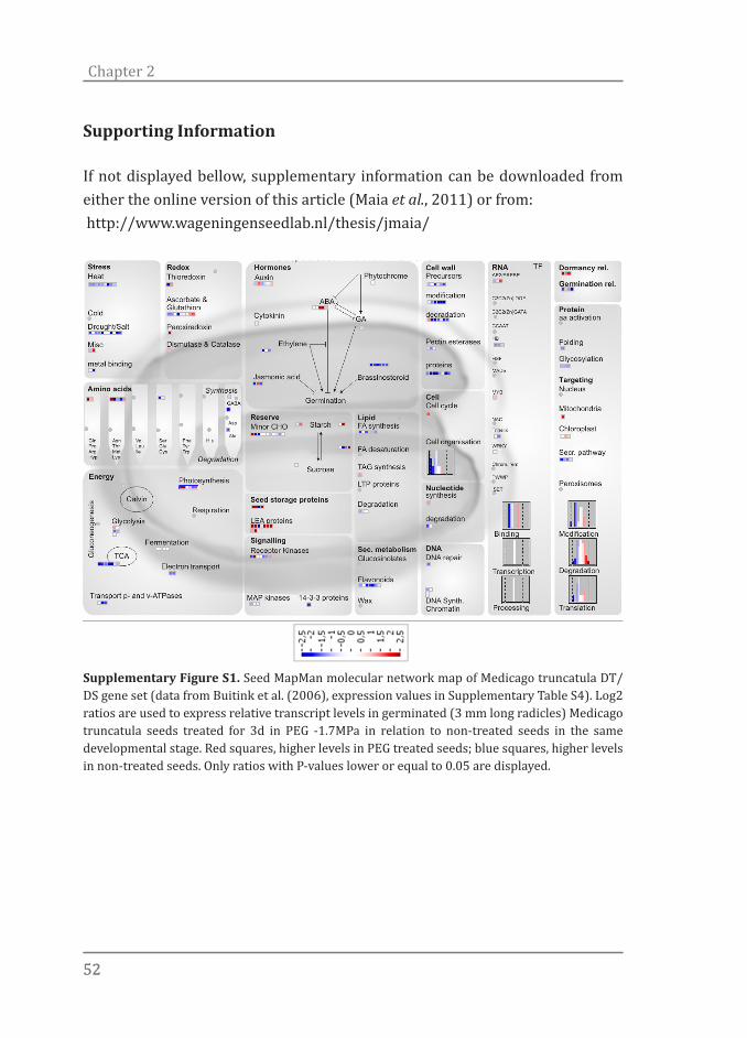
5252
Chapter 2
Supporting Information
If not displayed bellow, supplementary information can be downloaded from either the online version of this article (Maia et al., 2011) or from: http://www.wageningenseedlab.nl/thesis/jmaia/
Supplementary Figure S1. Seed MapMan molecular network map of Medicago truncatula DT/DS gene set (data from Buitink et al. (2006), expression values in Supplementary Table S4). Log2 ratios are used to express relative transcript levels in germinated (3 mm long radicles) Medicago truncatula seeds treated for 3d in PEG -1.7MPa in relation to non-treated seeds in the same developmental stage. Red squares, higher levels in PEG treated seeds; blue squares, higher levels in non-treated seeds. Only ratios with P-values lower or equal to 0.05 are displayed.

53
Desiccation tolerance transcriptome
Supplementary Table S1. Differentially up- and down-regulated genes (DT/DS gene set) after the re-establishment of desiccation tolerance (DT) in germinated Arabidopsis seeds at stage II by PEG treatment.
Supplementary Table S2. Over-representation analysis of the differentially expressed genes after PEG treatment. The analyzed gene set was first filtered through a fold-change and variance cut-off based filter (fold change ≥ 2 and P-value ≤ 0.05). (available online)
Supplementary Table S3. Comparison between the Arabidopsis gene list for expression after re-establishment of desiccation tolerance (DT/DS gene set - Supplementary Table S1) against a data set obtained in a similar system for Medicago (Buitink et al., 2006). (available online)
Supplementary Table S4. Log2 ratios of relative transcript levels in germinated (3-mm long radicles) Medicago truncatula seeds treated for 3d in PEG -1.7MPa in relation to non-treated seeds in the same developmental stage with the respective AGI code of the closest Arabidopsis homologue. Only ratios with P-values lower or equal to 0.05 are shown (Buitink et al., 2006). (available online)
Supplementary Table S5. Details of the heat maps displaying the gene expression profile during seed imbibition according to the Bio-Array Resource database. The top 50 up- and down-regulated genes in the DT/DS set were tested, in silico, for their expression during seed imbibition. (available online)

The worst illiterate is the political illiterate, he doesn’t hear, doesn’t speak, nor participates in the political events. He doesn’t know the cost of life, the price of the bean, of the fish, of the flour, of the rent, of the shoes and of the medicine, all depends on political decisions. The political illiterate is so stupid that he is proud and swells his chest saying that he hates politics. The imbecile doesn’t know that, from his political ignorance is born the prostitute, the abandoned child, and the worst thieves of all, the bad politician, corrupted and flunky of the national and multinational companies.
Bertolt Brecht
Chapter 3Abscisic acid (ABA) sensitivity regulates des-iccation tolerance in germinated Arabidopsis seeds
Authors:Julio Maia1, Bas J.W. Dekkers1,2, Miranda J. Dolle1, Kerstin Gühl1, Wilco Ligterink1, Henk W. M. Hilhorst1
1 Wageningen Seed Lab, Laboratory of Plant Physiology, Wageningen University, Wageningen, The Netherlands 2Department of Molecular Plant Physiology, Utrecht University, Utrecht, The Netherlands
New Phytologist (2014) DOI: 10.1111/nph.12785

5656
Chapter 3
Summary
During germination orthodox seeds lose their desiccation tolerance (DT) and become sensitive to extreme drying. Yet, DT can be rescued, in a well-defined developmental window, by the application of a mild osmotic stress before dehydration. A role for abscisic acid (ABA) has been implicated in this stress response and in DT re-establishment. However, the path from sensing an osmotic cue and its signalling to DT re-establishment is still largely unknown. Analysis of DT, ABA sensitivity, ABA content and gene expression was performed in desiccation sensitive (DS) and tolerant Arabidopsis thaliana seeds. Furthermore, loss and re-establishment of DT in germinated Arabidopsis seeds was studied in ABA-deficient and -insensitive mutants. We demonstrate that the developmental window in which DT can be re-established strongly correlates with the window in which ABA sensitivity is still present. Using ABA biosynthesis and -signalling mutants we show that this hormone plays a key role in DT re-establishment. Surprisingly, re-establishment of DT depends on modulation of ABA sensitivity rather than enhanced ABA levels. Also, evaluation of several ABA-insensitive mutants, which can still produce normal desiccation tolerant seeds but are impaired in re-establishing DT, shows that acquisition of DT during seed development is genetically different from its re-establishment during germination.
Introduction
The seed is a key structure in the plant life cycle which aids to dispersal and survival of the species. Traits such as seed dormancy and desiccation tolerance are important in this respect. Desiccation tolerance (DT) can be regarded as the ability of an organism to tolerate extreme water loss while keeping structural integrity and viability. DT is common in most seeds but not in vegetative tissues of angiosperms, except for a few so-called resurrection plants. Seeds which are able to withstand extreme drying to water levels below 0.1 gram H2O per gram dry weight, are termed ‘orthodox’ (Bewley & Black, 1994; Bewley et al., 2013). In orthodox seeds DT is acquired during the maturation phase which involves a complex regulatory network (Jia et al., 2013; Verdier et al., 2013).

5757
ABA sensitivity and Desiccation tolerance
In Arabidopsis, seed maturation is controlled by master regulators which interact in a complex manner and include the CCAAT-box binding factor LEAFY COTYLEDON (LEC1), and the three B3 domain-containing proteins ABSCISIC ACID INSENSITIVE (ABI3), FUSCA (FUS3) and LEC2. Collectively these are also known as the LAFL network. This network exerts tight control of developmental processes to assure normal seed development and maturation, including the acquisition of DT, by affecting the expression of downstream targets which includes other transcription factors, hormonal pathways and the expression of seed storage proteins (SSP) and LATE EMBRYOGENESIS ABUNDANT (LEA) genes (To et al., 2006); (Gutierrez et al., 2007; Santos-Mendoza et al., 2008; Jia et al., 2013). As soon as dry seeds are rehydrated, they quickly lose their DT and thus become desiccation sensitive (DS) again (Bewley & Black, 1994; Bewley et al., 2013). However, in a well-defined developmental window, it is possible to rescue DT in germinating seeds by applying a mild osmotic stress before drying (Bruggink & van der Toorn, 1995; Buitink et al., 2003; Maia et al., 2011). This model of re-establishment of DT in germinated seeds has already been used as a model to investigate DT in several species (Bruggink & van der Toorn, 1995; Buitink et al., 2003; Vieira et al., 2010; Maia et al., 2011). In these studies, germinated seeds were exposed to various concentrations of polyethylene glycol (PEG) or PEG in combination with exogenous abscisic acid (ABA) or ABA-biosynthesis inhibitors. This model of re-establishment of DT in germinated seeds has also been used to study the transcriptome related to DT in the model plants Medicago truncatula and Arabidopsis thaliana (Buitink et al., 2006; Maia et al., 2011). In these studies a marked enrichment of transcription factor (TF) sites containing ABA-responsive elements among the promoters of the most highly up-regulated DT-associated genes was reported, implying that ABA plays a crucial role in their regulation (Buitink et al., 2006; Maia et al., 2011). ABA is a phytohormone known as a central regulator of plant development and responses to environmental stresses. To date, over 100 loci have been identified as being involved in ABA signalling (Cutler et al., 2010) including the ABA insensitive loci abi1, abi2, abi3 (Koornneef et al., 1984), abi4 and abi5 (Finkelstein, 1994). ABI1 and ABI2 encode PP2C phosphatases and ABI3, ABI4 and ABI5 are TFs of the B3, AP2 and bZIP classes, respectively
5858
Chapter 3
(Giraudat et al., 1992; Leung et al., 1997; Finkelstein et al., 1998; Finkelstein & Lynch, 2000b; Lopez-Molina & Chua, 2000). ABI5, a transcription factor that plays an important role in the response to exogenous ABA during germination, defines a narrow developmental window, following germination, during which plants monitor the environmental osmotic status before initiating vegetative growth (Lopez-Molina et al., 2001). In Arabidopsis seeds, ABA regulates ABI5 accumulation and its activity during a limited window between 12 and 48 h of germination. Dry seeds of abi5 mutants show reduced transcript levels of ABA-responsive genes, and it has been hypothesized that ABI5 is necessary to bring germinated embryos into a quiescent state upon drought, thereby protecting young seedlings from the loss of water (Finkelstein & Lynch, 2000a; Lopez-Molina et al., 2001). The ABI proteins are part of a recently discovered cascade of events involving ABA receptors, protein phosphatases and protein kinases (Ma et al., 2009; Park et al., 2009). The core of this pathway consists of three protein families: the PYR/PYL/RCAR receptor family consisting of PYRABACTIN RESISTANCE1 (PYR1) like regulatory components of ABA receptors, the TYPE 2C PROTEIN PHOSPHATASES (PP2Cs) and the SUCROSE-NON-FERMENTING KINASE1-RELATED PROTEIN KINASE2 (SnRK2s) family (Umezawa et al., 2010). Together, these three protein families form a double negative regulatory pathway. In the absence of ABA the PP2Cs inactivate SnRK2s by dephosphorylation (Umezawa et al., 2009). Conversely, when ABA is present it binds to the PYL/PYR/RCAR receptors, thus creating a complex which interacts with the PP2Cs. Via this interaction, the dephosphorylation of the SnRK2s by the PP2Cs is inhibited (Ma et al., 2009; Park et al., 2009). The active kinases subsequently phosphorylate different proteins including membrane proteins and TFs (e.g. ABI5) eventually leading to an ABA response. In this study we investigated the role of ABA in the loss and re-establishment of DT in the model plant Arabidopsis, using physiological assays, gene expression analysis and hormone measurements. We found that ABA is essential to re-establish DT by an osmotic treatment in germinated Arabidopsis seeds. Surprisingly, the re-establishment of DT seemed not to depend on enhanced ABA levels but is more likely driven by modulation of ABA sensitivity. Finally, several ABA-deficient and -insensitive mutants which produce normal
5959
ABA sensitivity and Desiccation tolerance
desiccation tolerant seeds were impaired in their ability to re-establish DT during germination, suggesting that acquisition of DT during seed development is genetically distinct from re-establishment of DT during germination.
Materials and methods
Plant materials and growth conditions
The following Arabidopsis thaliana (L.) Heynh. lines were used: ecotype Columbia (Col-0); abi3-8, abi3-9, abi4-3 and abi5-7 (Nambara et al., 2002); aba2-1 (Léon-Kloosterziel et al., 1996); and an Arabidopsis ecotype C24 line expressing the RD29A::LUC (RESPONSIVE TO DESICCATION 29A) transgene (Ishitani et al., 1997). In all experiments Arabidopsis seeds were cold stratified for 72h at 4°C in 9-cm Petri dishes to eliminate residual dormancy. Germination assays were performed under constant white light at 22ºC. Seeds of four developmental stages were used, i.e. at testa rupture (stage I), at radicle protrusion (stage II), showing a primary root of 0.3-0.5 mm in length (stage III) and at the appearance of the first root hairs before cotyledon greening (stage IV) (Figure 1a) (adapted from Maia et al., 2011).
Re-establishment of desiccation tolerance and assessment of ABA sensitivity
Re-establishment of DT and ABA sensitivity were measured in parallel in germinated wild type and mutant seeds. To determine the re-establishment of DT, seeds at stages I, II, III and IV were either dried directly (untreated controls) or after three days of incubation in PEG at -2.5 MPa (treated samples). The PEG concentration used was such that it inhibited further growth of the treated germinated seeds, thus keeping them in the same developmental stage as they were collected in. After incubation, treated seeds that were still in stages I, II, III and IV were rinsed thoroughly in distilled water with the aid of a sieve, transferred to a new Petri dish with one dry sheet of germination paper and then dehydrated for three days at 20°C at 32% relative humidity (RH), which was achieved over a saturated calcium chloride solution in a closed chamber. After dehydration, treated and untreated seeds were pre-humidified (100% RH) for 24h at 22°C in the dark to avoid imbibitional damage (Leopold & Vertucci, 1986). Survival of cotyledons and primary roots, five days after rehydration,

6060
Chapter 3
and viable seedlings, ten days after rehydration, were scored. Cotyledons that continued their development became green and opened and primary roots that were further elongated were considered alive. To determine ABA sensitivity, germinated seeds from each germination stage were transferred to 6-cm Petri dishes, containing two filter papers moistened with 1 ml 5µM ABA. The samples were incubated at 22oC under constant light for ten days. Germinated seeds that continued their development and possessed expanded green cotyledons were considered insensitive while the ones arrested were assessed as ABA sensitive.
Luciferase imaging and analysis
The Arabidopsis (ecotype C24) line expressing the firefly luciferase reporter gene under control of the Arabidopsis RD29A promoter (RD29A::LUC) was used (Ishitani et al., 1997). The LUC reporter gene under the control of the RD29A promoter was developed to screen mutants with defects in their abiotic-stress
Figure 1. (a) Developmental stages of germinated Arabidopsis seeds. Developmental stages I to IV represent seeds at testa rupture (stage I), at radicle protrusion (stage II), showing a primary root of 0.3-0.5mm in length (stage III) and at the appearance of the first root hairs before greening of cotyledons (stage IV). (b) Re-establishment of desiccation tolerance and ABA sensitivity in germinated Arabidopsis seeds. ABA sensitivity (dashed red line) and survival of cotyledons (black bars), primary roots (grey bars) and seedlings (white bars). Untreated seeds at all developmental stages were completely desiccation sensitive. Values are expressed as the average of four replicates ± SE of 20 to 30 germinated seeds. (c) RD29A::LUC activity of germinated Arabidopsis seeds over time. Seeds at stages I (red line), II (black line) and IV (green line) were collected and treated with 5 µM ABA solution. The ‘blue line’ represents untreated samples incubated in water. RD29 promoter activity was measured using a chemiluminescent assay in the dark at 20oC. Values are expressed as the average of three replicates ± SE of 100 germinated seeds. Error bars are depicted as shading of the curves.
6161
ABA sensitivity and Desiccation tolerance
signal transduction pathways (Ishitani et al., 1997). This promoter is responsive to cold, osmotic stress, and ABA (Yamaguchi-Shinozaki & Shinozaki, 1993). Luciferase imaging and analysis were performed as previously described (Van Leeuwen et al., 2000) with some modifications. RD29A::LUC seeds were cold stratified for 72 h at 4°C in 9-cm Petri dishes on two layers of blue filter paper and 10 ml of 1 mM firefly D-luciferin, sodium-salt (Duchefa). Germination was performed under constant white light at 22ºC. One hundred seeds at each developmental stage were used for further analysis. RD29A promoter activity was measured by acquiring images in the dark at 17-20oC every 7 min with exposure times of 5 min using a Pixis 1024B camera system (Princeton Instruments) equipped with a 35 mm, 1:1.4 Nikkor SLR camera lens (Nikon). Relative luminescence was analysed for each group of 100 seeds in each image as mean grey value using ROI manager of ImageJ 1.44n software (http://rsbweb.nih.gov/ij/). Background subtraction was performed for all luciferase-imaging experiments.
RNA extraction
Total RNA extraction was performed according to the hot borate protocol modified from Wan and Wilkins (1994) as previously described by Maia et al. (2011). At least 100 seeds were used in each extraction. RNA integrity was assessed by analysis on a 1.2% agarose gel and RNA sample quality and concentration were additionally assessed using a Nanodrop ND-1000 (Nanodrop Technologies Inc.).
Real-time quantitative PCR (RT-qPCR) conditions
cDNA was synthesized using the iScript™ cDNA synthesis kit (Bio-rad) according to the manufacturers protocol. An iQ-SYBR-Green-Supermix (Bio-Rad) was used for gene expression analysis on a MyIQ RT-qPCR machine (Bio-Rad). The RT-qPCR program run consisted of a first step at 95oC for 3 min followed by 40 cycles of 15s at 95oC and 1 min at 60oC. Primers used (Supplementary Table S1) were designed preferably in the 3’ part of the transcript. When possible, primer pairs were designed spanning an intron-exon border. Melting curve analysis was performed after the RT-qPCR run (55 to 95oC with 0.5oC increments every 10s). For all primers, a single peak was observed, confirming the synthesis of

6262
Chapter 3
a single product. Primer efficiencies were assessed with LinReg PCR software (Ramakers et al., 2003). All primers showed efficiencies of at least 90%. Ct values were obtained from iQ5 software (Bio-rad) (base line subtracted, threshold value of 91 RFU) and analyzed with qbase+ (Biogazelle). Seven reference genes (Dekkers et al., 2012) were tested. Their expression stability was calculated by geNORM (Vandesompele et al., 2002) (Supplementary Figure S1) and the three most stable (AT3g59990, AT2g28390 and AT3g33520 – Supplementary Table S1) were used for further studies. RT-qPCR data of each gene of interest was normalized against the three selected reference genes. Finally, calibrated normalized relative quantity (CNRQ) values were exported from qbase+ and statistically analyzed with SISVAR software (Furtado, 2011).
Extraction of ABA from germinated Arabidopsis seeds
For ABA analysis, 20-50 mg of fresh untreated or treated (incubation for 3 and 7h in -2.5 MPa PEG) seeds (stages I, II, III and IV) were frozen in liquid nitrogen and ground in a dismembrator (Mikro-dismembrator U, B. Braun Biotech International) at 2000 RPM for 1.5 min with the help of 1/4’’ stainless steel beads. Samples were extracted with 1.5 ml of isopropanol/acetic acid (99:1) containing 2.5 mM dithiothreitol and 0.025 nmol of [2H6]-ABA as internal standard in a 2-ml centrifuge tube. The tubes were vortexed and sonicated for 10 min in a Branson 3510 ultrasonic bath. Samples were incubated for 1 h at 4°C while shaking. After the first extraction, samples were centrifuged for 10 min (2500 g). The liquid phase was carefully transferred to a 4 ml glass vial. The pellets were re-extracted overnight with another 1.5 ml of isopropanol/acetic acid (99:1). The combined isopropanol/acetic acid fractions were dried in a SpeedVac centrifuge (SPD121P, Thermo Scientific) and the residue was dissolved in 1 ml UPLC grade water. Samples were transferred to MAX columns (Oasis® 30mg 1cc, Waters) previously equilibrated with 1 ml 100% methanol (HPLC supra gradient) followed by 1 ml of water. The columns/samples were washed with 1 ml UPLC grade water followed by 1 ml 100% methanol. After washing, 1 ml methanol/2% formic acid was added to the columns in two steps of 500 μl and the flow through was collected. Samples were dried in a SpeedVac centrifuge and re-suspended in 100 μl UPLC grade water. The samples were stored at -20°C until measurement.
6363
ABA sensitivity and Desiccation tolerance
ABA detection and quantification by liquid chromatography-tandem mass spectrometry (LC-MS/MS)
ABA analysis was performed with a Waters Xevo tandem quadropole mass spectrometer equipped with an electrospray ionization source and coupled to an Acquity UPLC system (Waters, USA). Chromatographic separation was achieved using an Acquity UPLC BEH C18 column (100 x 2.1 mm, 1.7 μm) (Waters, USA), applying a water/acetonitrile gradient, starting at 5% acetonitrile for 1.0 min, raised to 50% (v/v) acetonitrile in 5.67 min, followed by a 1.33 min gradient to 98% (v/v) acetonitrile which was then maintained for 1 min before returning to 5% acetonitrile in water using a 0.13 min gradient. The column was equilibrated at this solvent composition for 1.87 min prior to the next injection. Total run time was 11 min. The column was operated at 50oC with a flow-rate of 0.5 ml min-1 and sample injection volume was 10 μl. The mass spectrometer was operated in negative electrospray ionization (ESI) mode. The cone and desolvation gas flows were 50 and 1000 L h-1, respectively. Argon was used for fragmentation by collision-induced dissociation in the ScanWave collision cell. The capillary voltage was set at 2.3 kV, the source temperature at 150oC and the desolvation gas temperature at 650oC. The cone voltage (CV), collision energy (CE) and parent daughter transitions were optimized by injecting pure ABA and [2H6]-ABA in the Waters IntelliStart MS console. Transitions were selected based on the most abundant and specific fragment ions. For ABA, the Multiple reaction monitoring (MRM) transitions m/z 263.074>219.127 at a CE of 14 eV and 263.074>153.007 at 10 eV both with CV of 18V; and for [2H6]-ABA, the transitions m/z 269.16>225.14 at CE of 18 eV, 269.16>206.99 at CE of 22 eV and 269.16>158.95 at CE of 13 eV with CV of 16V were selected. ABA was quantified using a calibration curve with known amount of standards and based on the ratio of the summed area of the MRM transitions for ABA to those for [2H6]-ABA. Data acquisition and analysis were performed using MassLynx 4.1 software (Waters, USA).

6464
Chapter 3
Results
DT re-establishment in germinated Arabidopsis seeds strongly correlates with the presence of and sensitivity to ABA
We have previously shown that DT can be re-induced in germinated Arabidopsis seeds by treating them in a PEG (-2.5 MPa) solution (Maia et al., 2011). Here we evaluated whether the window of ABA sensitivity during and after germination (Lopez-Molina et al., 2001) correlates with the time frame in which DT can be re-established. For this, sensitivity to 5 μM ABA was correlated with the ability to re-establish DT in cotyledons, primary roots and seedlings in seeds at four developmental stages during and after germination (Figure 1a,b). The ability of Col-0 seeds to re-establish DT during germination decreased together with their ABA-sensitivity (Figure 1b). DT could be re-established in Col-0 seeds at developmental stages I, II and III when ABA sensitivity was still high. In agreement with our previous results, Col-0 seeds at stages I and II displayed 100% of re-establishment of DT of primary roots, cotyledons and seedlings, while for those at stage III a slight reduction of DT was observed in all three structures. Low ABA sensitivity levels of germinated Col-0 seeds at stage IV correlated with a reduced ability to re-establish DT (Figure 1b). To further investigate the correlation between ABA-sensitivity and re-establishment of DT, we checked the promoter activity of the ABA-responsive gene RD29A during the four developmental stages (Fig 1c). An increase in RD29A::LUC activity was measured in all seeds as quickly as 30 min after addition of ABA. This activity reached its maximum around 150 min after ABA-incubation. Confirming our previous results on ABA-sensitivity, RD29A::LUC activation also seemed to be dependent on the seed developmental stage. In relation to earlier developmental stages, seeds at stage IV showed less RD29A::LUC activity in the presence of exogenous ABA, indicating a reduction in ABA signal transduction. Because we observed a strong correlation between ABA-sensitivity and the ability of seeds to re-induce DT we investigated whether wild type ABA levels are needed for DT re-establishment. Therefore, we assessed the ability of ABA-deficient Arabidopsis aba2 mutant seeds to re-establish DT. Emphasizing the role of ABA in DT re-establishment, germinated seeds (stage II) of the
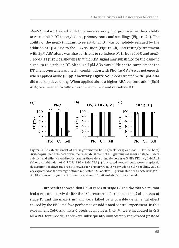
6565
ABA sensitivity and Desiccation tolerance
aba2-1 mutant treated with PEG were severely compromised in their ability to re-establish DT in cotyledons, primary roots and seedlings (Figure 2a). The ability of the aba2-1 mutant to re-establish DT was completely rescued by the addition of 1µM ABA to the PEG solution (Figure 2b). Interestingly, treatment with 5µM ABA alone was also sufficient to re-induce DT in both Col-0 and aba2-1 seeds (Figure 2c), showing that the ABA signal may substitute for the osmotic signal to re-establish DT. Although 1µM ABA was sufficient to complement the DT phenotype when applied in combination with PEG, 1µM ABA was not enough when applied alone (Supplementary Figure S2). Seeds treated with 1µM ABA did not stop developing. When applied alone a higher ABA concentration (5µM ABA) was needed to fully arrest development and re-induce DT.
Our results showed that Col-0 seeds at stage IV and the aba2-1 mutant had a reduced survival after the DT treatment. To rule out that Col-0 seeds at stage IV and the aba2-1 mutant were killed by a possible detrimental effect caused by the PEG itself we performed an additional control experiment. In this experiment Col-0 and aba2-1 seeds at all stages (I to IV) were incubated in -2.5 MPa PEG for three days and were subsequently immediately rehydrated (instead
Figure 2. Re-establishment of DT in germinated Col-0 (black bars) and aba2-1 (white bars) Arabidopsis seeds. To determine the re-establishment of DT, germinated seeds at stage II were selected and either dried directly or after three days of incubation in -2.5 MPa PEG (a), 5µM ABA (b) or a combination of -2.5 MPa PEG + 1µM ABA (c). Untreated control seeds were completely desiccation sensitive and are not shown. PR = primary root, Ct = cotyledons, Sdl = seedling. Values are expressed as the average of three replicates ± SE of 20 to 30 germinated seeds. Asterisks (** P ≤ 0.01) represent significant differences between Col-0 and aba2-1 treated seeds.

6666
Chapter 3
of dehydrated). All seedlings developed normally after direct re-hydration following the PEG treatment. This shows that the seeds were not killed by PEG but were incapable to regain DT by the PEG treatment (Supplementary Figure S2). Together, these results show a fundamental role for ABA in the re-establishment of DT. Therefore, we further investigated the importance of ABA-biosynthesis and -signalling in this response.
Enhanced ABA content is not required for re-establishment of DT
We investigated whether increased ABA content is necessary for DT re-establishment. Therefore, we measured the expression of ABA-biosynthetic and -catabolic genes and ABA content in response to the PEG treatment. The expression of seven genes involved in ABA-biosynthesis was investigated (NINE-CIS-EPOXYCAROTENOID DIOXYGENASE - NCED3, NCED5, NCED6, NCED9, and ABSCISIC ACID-DEFICIENT - ABA1, ABA2 and ABA3) and one gene involved in ABA degradation (CYTOCHROME P450 - CYP707A2) (Figure 3a,b). All genes showed significant changes in their expression levels during development and after treatment (Figure 3b). For example, NCED6 was significantly down-regulated in stages I to IV in untreated seeds. This gene was also down-regulated by PEG at stages I and II. In contrast, the expression of ABA1 and ABA2 significantly increased with seedling development in untreated seeds and, except for the expression of ABA2 in seeds at stage IV, was highly induced in PEG-treated seeds. Overall, the expression of all analyzed ABA-biosynthesis genes was up-regulated in PEG-treated seeds, with the exception of NCED6. In contrast, the ABA catabolic gene CYP707A2 was down-regulated with development and in PEG-treated seeds (Figure 3b). These expression patterns suggest that ABA content increases upon PEG treatment. To confirm our gene expression data, ABA content was determined in seeds (stages I to IV) in the presence and absence of PEG. In contrast to what the expression of ABA-biosynthesis genes suggested, ABA content did not change significantly after PEG treatment, as compared to untreated seeds (Figure 4). This data suggests that either ABA is not being synthesized or it is being further modified into different metabolic products. To confirm and extend our analysis we performed a second independent experiment in a different laboratory
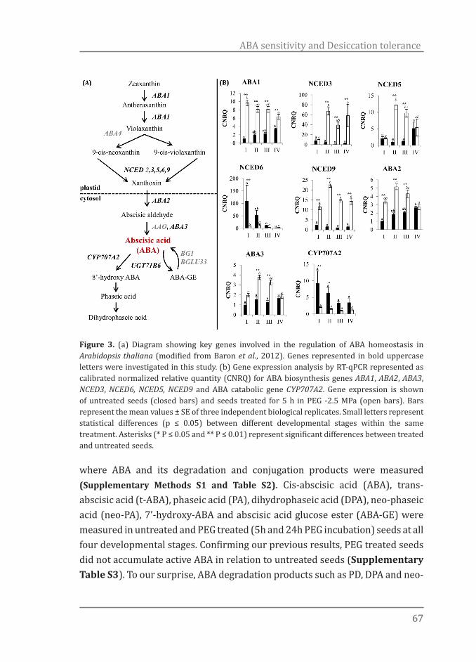
6767
ABA sensitivity and Desiccation tolerance
where ABA and its degradation and conjugation products were measured (Supplementary Methods S1 and Table S2). Cis-abscisic acid (ABA), trans-abscisic acid (t-ABA), phaseic acid (PA), dihydrophaseic acid (DPA), neo-phaseic acid (neo-PA), 7’-hydroxy-ABA and abscisic acid glucose ester (ABA-GE) were measured in untreated and PEG treated (5h and 24h PEG incubation) seeds at all four developmental stages. Confirming our previous results, PEG treated seeds did not accumulate active ABA in relation to untreated seeds (Supplementary Table S3). To our surprise, ABA degradation products such as PD, DPA and neo-
Figure 3. (a) Diagram showing key genes involved in the regulation of ABA homeostasis in Arabidopsis thaliana (modified from Baron et al., 2012). Genes represented in bold uppercase letters were investigated in this study. (b) Gene expression analysis by RT-qPCR represented as calibrated normalized relative quantity (CNRQ) for ABA biosynthesis genes ABA1, ABA2, ABA3, NCED3, NCED6, NCED5, NCED9 and ABA catabolic gene CYP707A2. Gene expression is shown of untreated seeds (closed bars) and seeds treated for 5 h in PEG -2.5 MPa (open bars). Bars represent the mean values ± SE of three independent biological replicates. Small letters represent statistical differences (p ≤ 0.05) between different developmental stages within the same treatment. Asterisks (* P ≤ 0.05 and ** P ≤ 0.01) represent significant differences between treated and untreated seeds.
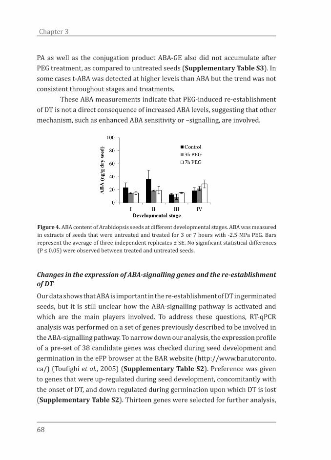
6868
Chapter 3
PA as well as the conjugation product ABA-GE also did not accumulate after PEG treatment, as compared to untreated seeds (Supplementary Table S3). In some cases t-ABA was detected at higher levels than ABA but the trend was not consistent throughout stages and treatments. These ABA measurements indicate that PEG-induced re-establishment of DT is not a direct consequence of increased ABA levels, suggesting that other mechanism, such as enhanced ABA sensitivity or –signalling, are involved.
Changes in the expression of ABA-signalling genes and the re-establishment of DT
Our data shows that ABA is important in the re-establishment of DT in germinated seeds, but it is still unclear how the ABA-signalling pathway is activated and which are the main players involved. To address these questions, RT-qPCR analysis was performed on a set of genes previously described to be involved in the ABA-signalling pathway. To narrow down our analysis, the expression profile of a pre-set of 38 candidate genes was checked during seed development and germination in the eFP browser at the BAR website (http://www.bar.utoronto.ca/) (Toufighi et al., 2005) (Supplementary Table S2). Preference was given to genes that were up-regulated during seed development, concomitantly with the onset of DT, and down regulated during germination upon which DT is lost (Supplementary Table S2). Thirteen genes were selected for further analysis,
Figure 4. ABA content of Arabidopsis seeds at different developmental stages. ABA was measured in extracts of seeds that were untreated and treated for 3 or 7 hours with -2.5 MPa PEG. Bars represent the average of three independent replicates ± SE. No significant statistical differences (P ≤ 0.05) were observed between treated and untreated seeds.
6969
ABA sensitivity and Desiccation tolerance
among which 3 ABA-receptors, 7 phosphatases and 3 transcription factors (Supplementary Table S1). To identify the best time points to monitor ABA signalling during the re-establishment of DT, an expression time series during PEG treatment was performed. The expression of the ABA-responsive RD29A gene was used as a reference. To obtain a first overview of the RD29A promoter activity, we measured its activity using a luciferin imaging assay (Supplementary Figure S3a). Based on the luciferin activity assay results, material from germinated seeds (stage II), both untreated and incubated in -2.5 MPa PEG for 3, 7, 20, 48 and 72 hours was prepared and used for RT-qPCR analysis (Supplementary Figure S3b). Both RT-qPCR and luciferin assays demonstrated that RD29A expression was quickly up-regulated upon PEG treatment (Supplementary Figure S3a,b). RD29A expression started around 3 h and reached its highest level 7h after PEG incubation after which it quickly declined after 20h (Supplementary Figure S3a,b). Based on the expression patterns observed for RD29A under osmotic treatment we decided to analyze the expression of the 13 selected ABA signalling genes after 5h of PEG incubation when ABA responsive genes are most likely to be expressed (Figure 5). The expression levels of PYL (PYRABACTIN RESISTANCE 1-LIKE) 7 and PYL9 were markedly increased after 5h of PEG-treatment (Figure 5a). The increased expression of PYL7 and PYL9 in response to PEG indicates that these genes are possibly involved in the regulation of re-establishment of DT in germinated seeds. No significant changes in transcript level were observed for PYL5, which suggests that either different receptor proteins are involved in the signalling for distinct environmental cues, or different tissues, with e.g. PYL7 and PYL9 as important genes to respond to osmotic stress in germinating seeds. Apart from this, when under osmotic stress, all PYL genes showed higher gene expression after radicle protrusion (stages II, III and IV) when compared to seeds at stage I (Figure 5a). Taken together, the enhanced expression of PYL7 and PYL9 in response to PEG suggests that increased ABA sensitivity is involved in the re-establishment of DT. All PP2C genes evaluated were consistently up-regulated under osmotic stress at all developmental stages but showed very diverse expression patterns over development (Figure 5b). For instance, while HIGHLY ABA INDUCED
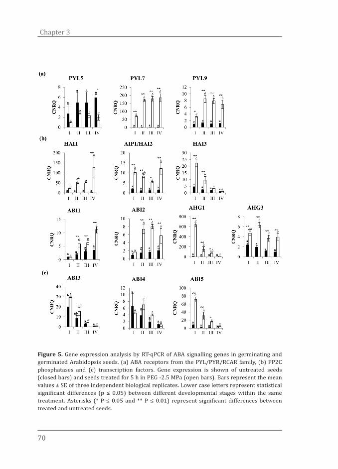
7070
Chapter 3
Figure 5. Gene expression analysis by RT-qPCR of ABA signalling genes in germinating and germinated Arabidopsis seeds. (a) ABA receptors from the PYL/PYR/RCAR family, (b) PP2C phosphatases and (c) transcription factors. Gene expression is shown of untreated seeds (closed bars) and seeds treated for 5 h in PEG -2.5 MPa (open bars). Bars represent the mean values ± SE of three independent biological replicates. Lower case letters represent statistical significant differences (p ≤ 0.05) between different developmental stages within the same treatment. Asterisks (* P ≤ 0.05 and ** P ≤ 0.01) represent significant differences between treated and untreated seeds.
7171
ABA sensitivity and Desiccation tolerance
PP2C GENE (HAI)1 and ABA INSENSITIVE (ABI)1 were up-regulated over time in response to osmotic stress, HAI3 and ABA HYPERSENSITIVE GERMINATION (AHG)1 showed the exact opposite (Figure 5b). The expression profiles of AKT1 INTERACTING PROTEIN PHOSPHATASE (AIP)1, ABI2 and AHG3 did not show any clear correlation with the aptitude of DS seeds to re-establish DT. Interestingly, HAI1, ABI1 and ABI2 expression were only triggered by PEG from stage II onwards, indicating the possible existence of a link with the expression of PYL genes and a developmental switch between testa rupture and radicle protrusion. The trends of expression found for ABI3, ABI4 and ABI5 were very similar. Their transcript abundance decreased along germination in both the absence and presence of PEG (Figure 5c). ABI3 and ABI4 did not show significant up-regulation in PEG-treated seeds. ABI5, however, was strongly up-regulated under osmotic stress. Yet, the PEG-induced up-regulation of ABI5 decreased during development and was comparable to the ABI5 transcript levels found in untreated seeds when at stage IV (Figure 5c). The expression pattern found for ABI5 correlated well with ABA-sensitivity and the developmental window were DT can still be re-established which is suggestive for a function of ABI5 in the re-establishment of DT in germinated Arabidopsis seeds. By using the expression of ABA-signalling genes as an indicator of ABA-sensitivity we conclude that developmental phase plays a crucial role in this attribute. In general, most of the changes in expression occurred with development and under PEG-treatment. Remarkably, in spite of being negative regulators of ABA-signalling (Gosti et al., 1999; Ma et al., 2009), all PP2Cs evaluated in this study where up-regulated in PEG-treated seeds. Our findings are in agreement with microarray expression data of young Arabidopsis seedlings exposed to osmotic, salt, drought and cold stress that also showed up-regulation of most PP2C genes (Figure S4) (Toufighi et al., 2005).
ABA-insensitive and -deficient mutants show reduced re-establishment of DT
We tested whether DT can be re-induced in ABA-deficient and -insensitive mutants. We evaluated whether four ABA-insensitive mutants (abi3-9, abi3-8, abi4-3 and abi5-7) and one ABA-deficient mutant (aba2-1) were able to

7272
Chapter 3
re-establish DT during and after visible germination (i.e. four developmental stages, see Figure 1a) when treated with a -2.5 MPa PEG solution (Figure 6). In spite of being fully desiccation tolerant by the end of seed maturation, all germinated mutant seeds evaluated were less competent in re-establishing DT when compared to Col-0 germinated seeds (Figure 6). Although all the mutants were still able to recover DT when at stage I, this response was quickly lost after stage II and completely disappeared, in most cases, at stage III (i.e. abi3-9, abi3-8, abi4-3 and aba2-1 - Figure 6). The ability of Col-0 seeds to re-establish DT during germination decreased together with their ABA-sensitivity. In agreement with this, all ABA insensitive (abi) mutants tested were less tolerant to desiccation and displayed a similar pattern of re-establishment of DT and ABA-sensitivity decrease during germination (Figure 6). The less ABA-sensitive the mutant was, the quicker the decrease in its ability to recover DT with seed development. Although still sensitive to ABA in stages II and III, aba2-1 seeds were no longer able to re-establish DT (Figure 6). This negative correlation between ABA sensitivity and DT found for this mutant indicates that besides a functional ABA-signalling system, the presence of certain levels of ABA is also necessary for DT re-establishment. Although some abi-mutants had similar ABA-sensitivity scores, they still differed in their ability to re-establish DT (Figures 6 and 7). When measuring ABA-sensitivity, only expansion, greening and opening of the cotyledons were taken as parameters. Consequently, other differences in post-germination events such as root and hypocotyl elongation were largely neglected. Yet, these minor differences can still be seen when considering seedling appearance and fresh weight after 10 days of ABA treatment (Figure 7). Different abi-mutants that were transferred to ABA (5 µM) at the same developmental stage displayed different fresh-weights and clearly showed differences in development by the end of the treatment (Figure 7). For instance, when transferred to ABA at stages III and IV, ABA-sensitivity of abi3-8 and abi3-9 was scored as 0%. However, clear differences in fresh weight and development were observed among these mutants after 10 days in ABA (Figure 7). The abi3-8 seeds (stage III and IV) had developed further and had visibly more root growth when compared to abi3-9 seeds transferred to ABA at the same developmental stage. These differences indicate that abi3-8 is less sensitive to ABA than abi3-

7373
ABA sensitivity and Desiccation tolerance
9 and could explain why, in spite of having received the same score for ABA sensitivity, abi3-8 was less competent to re-establish DT in stage III. Taken together, this data confirms that ABA sensitivity plays a pivotal role in the re-establishment of DT in germinated Arabidopsis seeds. In addition, the phenotype found for the abi5-7 mutant (Figures 6 and 7), in combination with the expression pattern found for this gene (Figure 5c) highlights the importance of ABI5 in the re-establishment of DT in germinated Arabidopsis seeds.
Figure 6. Re-establishment of DT and ABA-sensitivity in germinated Arabidopsis seeds as expressed by ABA sensitivity (dashed red line), survival of cotyledons (black bars), primary roots (grey bars) and seedlings (white bars). Values are expressed as the average of four replicates ± SE of 20 to 30 germinated seeds. Untreated seeds of all accessions at all developmental stages were completely desiccation sensitive.
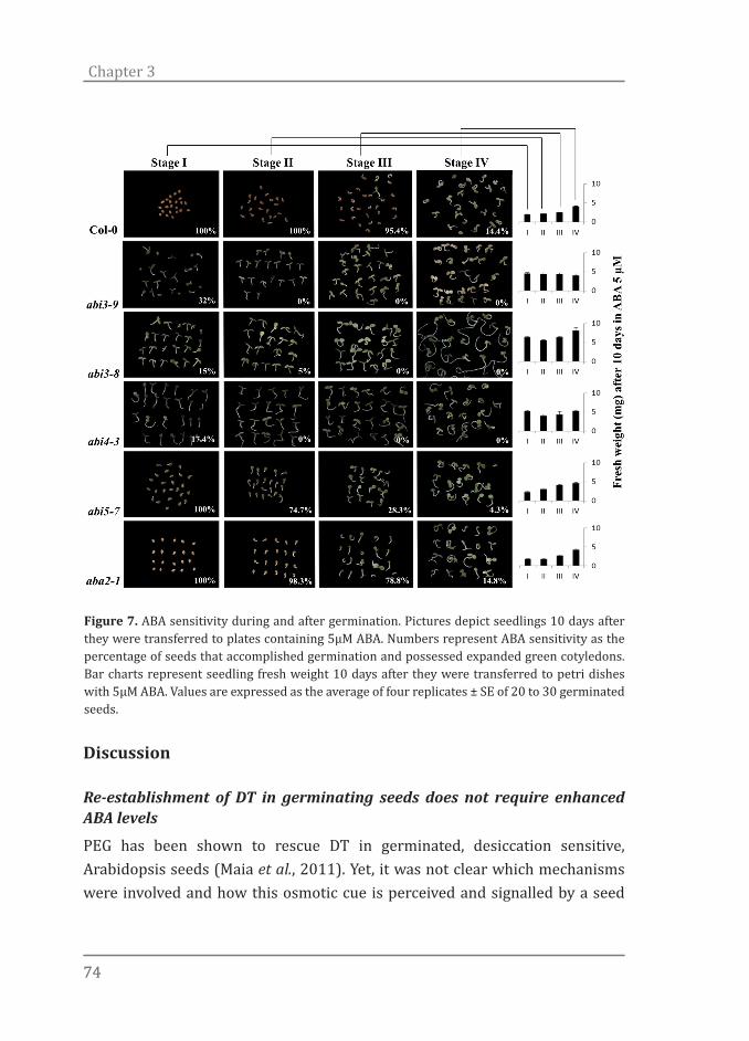
7474
Chapter 3
Discussion
Re-establishment of DT in germinating seeds does not require enhanced ABA levels
PEG has been shown to rescue DT in germinated, desiccation sensitive, Arabidopsis seeds (Maia et al., 2011). Yet, it was not clear which mechanisms were involved and how this osmotic cue is perceived and signalled by a seed
Figure 7. ABA sensitivity during and after germination. Pictures depict seedlings 10 days after they were transferred to plates containing 5µM ABA. Numbers represent ABA sensitivity as the percentage of seeds that accomplished germination and possessed expanded green cotyledons. Bar charts represent seedling fresh weight 10 days after they were transferred to petri dishes with 5µM ABA. Values are expressed as the average of four replicates ± SE of 20 to 30 germinated seeds.
7575
ABA sensitivity and Desiccation tolerance
or seedling. So far, two pathways by which a plant may respond to an osmotic cue have been suggested, namely an ABA-dependent and an ABA-independent pathway (Yamaguchi-Shinozaki & Shinozaki, 2006). However, most frequently, experiments point towards a model dependent on ABA level-directed responses (Raghavendra et al., 2010; Fujita et al., 2011; Frey et al., 2012; Miyakawa et al., 2013). According to this view, elevated levels of ABA are required to activate signalling cascades which will ultimately lead to a stress response (e.g. re-establishment of DT). It is not clear, however, if ABA levels, synthesis, degradation and sensitivity, are all important factors in the recovery of DT. Our working hypothesis was that germinated DS Arabidopsis seeds respond to an osmotic stress (PEG) by engaging ABA biosynthesis, leading to increased ABA levels. This increase in ABA content would then trigger an ABA-directed stress response. However, our data suggests that increased levels of ABA are not required for the re-establishment of DT in germinated Arabidopsis seeds since we were not able to detect changes in ABA content upon PEG treatment. This observation indicates that DT can be fully re-established by PEG treatment and that this is not a direct consequence of increased ABA levels. In accordance with our findings, it has been suggested that ABA accumulation is not always necessary to elicit stress responses. For instance, studies with seedlings of Salix spp. have indicated that, instead of affecting ABA levels, changes in day length are regulating cessation of growth, probably by affecting ABA sensitivity (Barros & Neill, 1986). Furthermore, plants expressing a cowpea mosaic virus factor (CMV) exhibited increased drought tolerance without over-accumulation of ABA (Westwood et al., 2013). According to these authors, CMV infection probably affected ABA signalling and/or perception instead. Altogether, these findings show that next to enhancing ABA levels, modulation of ABA perception and signalling can be sufficient to induce a proper stress response. The level of ABA in any particular tissue in a plant is determined by the rate of biosynthesis and catabolism of the hormone (Nambara & Marion-Poll, 2005). In addition, transport, beside conjugation or hydrolysis of ABA and ABA-GE, is increasingly gaining importance due to new discoveries in this field of research (Lee et al., 2006; Priest et al., 2006; Umezawa et al., 2010). Thus, accumulation of ABA degradation and/or conjugation products could, in part, explain why ABA levels were not increased in PEG treated seeds. Conversely,

7676
Chapter 3
although genes involved in ABA-biosynthesis and -catabolism were respectively up- and down-regulated suggesting that ABA levels should be increased, enhanced levels of ABA and its degradation and conjugation products were not detected in PEG treated seeds (Supplemental table S3). In a few cases higher t-ABA levels were detected in PEG treated seeds indicating an alternative route by which active ABA could be recycled. However, this trend was not consistent throughout stages and treatments. ABA could very well be synthesized at the same ratio it is degraded leading to a constant ABA balance. Furthermore, it is also possible that instead of being synthesized as a general response, ABA is locally synthesized in very small amounts in essential tissues or translocated from one tissue to another. In the first scenario, very small changes in ABA levels would be expected and detecting those changes would be the bottleneck while in the case of translocation, changes in ABA levels would not be expected.
The re-establishment of DT is limited to the developmental time window of ABA sensitivity and requires ABI3, ABI4 and ABI5
During germination Arabidopsis seeds rapidly lose their DT and become sensitive to extreme drying (Maia et al., 2011 ). Yet, in a well-defined developmental window between the stages of testa rupture and root hair formation, seedlings are still able to fully re-establish DT when submitted to a mild osmotic stress before drying (Bruggink & van der Toorn, 1995; Buitink et al., 2003; Maia et al., 2011). Also for other species the re-establishment of DT is limited to a certain developmental stage after the completion of germination (Bruggink & van der Toorn, 1995; Buitink et al., 2003; Maia et al., 2011). We show here that beyond this developmental window the ability to re-establish DT decreases and quickly disappears concomitantly with the loss of the ability of ABA to arrest development. The developmental time window in which seeds are able to respond to ABA has been described by Lopez-Molina et al. (2001). It was suggested that ABA, in addition to delaying germination, can also reversibly block growth during a narrow developmental time interval following germination and before the onset of vegetative growth. Within this window, ABA induces ABI5 expression and protein accumulation early in development and acts as a repressor of
7777
ABA sensitivity and Desiccation tolerance
germination and growth. Beyond this time window, ABA is unable to induce ABI5 protein accumulation and to block growth. This window is proposed to act as a developmental checkpoint during which Arabidopsis germinating seeds can still “decide”, based on environmental cues, to arrest development or continue germination towards seedling establishment (Lopez-Molina et al., 2001). In our model system ABI5 was significantly up-regulated in germinated seeds under osmotic stress at stage I, II and III (Figure 5c). However, at stage IV, when DT cannot be induced, ABI5 expression is no longer induced by the osmotic treatment. This expression pattern matched perfectly with the developmental window where ABA can still be perceived and DT still rescued in Arabidopsis seeds. This overlap between induction of ABI5 expression and re-establishment of DT, suggests that this TF plays an important role in defining the developmental checkpoint that determines whether seeds can still recruit DT-related mechanisms. This is supported by the fact that mutations in this locus (i.e. abi5-7) compromised the re-establishment of DT, which was most clearly visible in stage III (Figure 6). Also genes encoding for two other transcription factors that are important for ABA signalling, ABI3 and ABI4, are relevant in this response. These genes were not significantly up-regulated by osmotic stress as was ABI5, but seeds mutated in these two genes clearly showed a reduced capacity to re-establish DT. The phenotypes of abi3-8, abi3-9 and abi4-3 were even stronger as compared to the abi5-7 mutant and the phenotypes were already visible at stage II. At this stage abi5-7 was still sensitive to the PEG treatment, which implies that other genes are also involved in the modulation of this temporal checkpoint. ABI5 encodes a member of the basic leucine zipper (bZIP) TF family (Finkelstein & Lynch, 2000b; Lopez-Molina & Chua, 2000) which comprises more than 75 members (Jakoby et al., 2002). ABI5 is part of clade A of this family which contains 13 members. Several members within this clade bind conserved cis-elements known as ABA responsive elements (Kim et al., 1997; Choi et al., 2000; Uno et al., 2000) and some of them are known to function redundantly in response to ABA and stress (Finkelstein et al., 2005; Yoshida et al., 2010). This redundancy provides an explanation why the abi5-7 single mutant was not completely depleted in its capacity to re-establish DT.

7878
Chapter 3
The acquisition of DT during seed development is genetically distinguishable from DT re-establishment during germination
Many experimental models used to study DT were based on assessing acquisition of DT during seed development (Blackman et al., 1992; Xu & Bewley, 1995; Nedeva & Nikolova, 1997; Black et al., 1999; Sreedhar, 2002; Illing et al., 2005). Acquisition of DT is developmentally controlled and is acquired during the seed maturation phase (Bewley et al., 2013). Seed developmental mutants that have a disrupted seed maturation program, such as lec1, lec2, fus3 and severe abi3 mutants indeed fail to produce desiccation tolerant seeds. Although ABA is required for normal seed development, the vast majority of ABA-insensitive and –deficient mutants described to date are still able to produce completely desiccation tolerant seeds by the end of seed maturation (Cutler et al., 2010). The only exceptions are the strong abi3 alleles or the genetic combination of the weak abi3-1 allele with the ABA-deficient aba1-1 mutant which have an impaired capacity to acquire DT during seed maturation (Nambara et al., 1992; Ooms et al., 1993; Ooms et al., 1994). We used several mutants (aba2-1, abi3-8, abi3-9, abi4-3 and abi5-7) in this study. In spite of being compromised for ABA sensitivity or synthesis, all of these mutants produce desiccation tolerant seeds at the end of seed maturation. Interestingly, at stage I the picture seems similar to what is observed during seed development: despite the different mutated genes, DT is fully re-established. However, at stages II and III all five mutants showed a reduced capacity to re-establish DT. The ABA deficient aba2-1 mutant was incapable of fully recovering its DT when submitted to PEG from stage II (radicle protrusion) onwards. The inability of aba2-1 to re-establish DT implies that WT levels of ABA are necessary to re-establish DT in seeds at later developmental stages. Also in Medicago seeds, the re-establishment of DT is dependent on ABA. When an ABA biosynthesis inhibitor was applied, DT could no longer be re-established (Buitink et al., 2003). In the same report, it was also shown that the application of exogenous ABA fully rescued the DT phenotype similarly as observed in Arabidopsis seeds. The observation that the window in which DT can be re-established is much narrower in abi mutants as compared to WT, suggests that ABI TFs are very relevant to the re-establishment of DT in Arabidopsis seeds and that they modulate the window in which DT can be re-induced.

7979
ABA sensitivity and Desiccation tolerance
These observations show that the acquisition of DT during seed development is different from the re-establishment of DT in geminating seeds. From our results two hypotheses can be formulated. Firstly, there are (largely) distinct pathways involved in inducing DT during seed development and during germination. Considering the fact that seeds of a strong abi3 allele or the abi3-1 aba1-1 double mutant indeed lack DT, it is evident that ABI3 acts in both pathways. Secondly, the pathways that induce DT do involve ABA, ABI4 and ABI5, as well, but their function remains hidden (when testing the mutant alleles) due to additional redundant factors present during seed development. Contrary to the seed developmental DT network, the network in the germinating seed needs to be rapidly induced de novo upon osmotic stress, and may therefore lack sufficient redundancy to counteract mutations in its components. During the transitions from an embryonic to a post-germinative transcriptional program, dramatic changes occur and, thus, such redundant factors may not be able to be expressed anymore, thereby revealing a role for ABA, ABI4 and ABI5 in the acquisition of DT.
Acknowledgements
We thank the technical and intellectual input from our colleagues of the Wageningen Seed Lab and the Laboratory of Plant Physiology. Special thanks go to Kerstin Guhl for help with the ABA measurements, and to Ralph Bours and Dr Sander van der Krol for help with the luciferin assays. This work was supported by the ‘Coordenação de Aperfeicoamento de Pessoal de Nıvel Superior’ (CAPES, Brazil) (J.M.) and by the Netherlands Organization for Scientific Research (NWO) (B.J.W.D.).
References
Abrams, S. R., Nelson, K. and Ambrose, S. J. 2003. Deuterated abscisic acid analogs for mass spectrometry and metabolism studies. Journal of Labelled Compounds and Radiopharmaceuticals, 46: 273–283.
Andrew R.S Ross, Stephen J Ambrose, Adrian J Cutler, J Allan Feurtado, Allison R Kermode, Ken Nelson, Rong Zhou, Suzanne R Abrams. 2004. Determination of endogenous and supplied deuterated abscisic acid in plant tissues by high-performance liquid chromatography-electrospray ionization tandem mass

8080
Chapter 3
spectrometry with multiple reaction monitoring. Analytical Biochemistry, 329: 324-333
Baron KN, Schroeder DF, Stasolla C. 2012. Transcriptional response of abscisic acid (ABA) metabolism and transport to cold and heat stress applied at the reproductive stage of development in Arabidopsis thaliana. Plant Science 188-189: 48-59.
Barros RS, Neill SJ. 1986. Periodicity of response to abscisic acid in lateral buds of willow (Salix viminalis L.). Planta 168: 530-535.
Bewley JD, Black M. 1994. Seeds: physiology of development and germination. New York, US: Springer.
Bewley JD, Bradford K, Hilhorst H, Nonogaki H. 2013. Seeds: Physiology of Development, Germination and Dormancy. New York, US: Springer.
Black M, Corbineau F, Gee H, Come D. 1999. Water content, raffinose, and dehydrins in the induction of desiccation tolerance in immature wheat embryos. Plant Physiology 120: 463-472.
Blackman SA, Obendorf RL, Leopold AC. 1992. Maturation proteins and sugars in desiccation tolerance of developing soybean seeds. Plant Physiology 100: 225-230.
Bruggink T, van der Toorn P. 1995. Induction of desiccation tolerance in germinated seeds. Seed Science Research 5: 1-4.
Buitink J, Leger JJ, Guisle I, Vu BL, Wuillème S, Lamirault G, Alice, Nolwenn, Becker A, Küster H, Leprince O. 2006. Transcriptome profiling uncovers metabolic and regulatory processes occurring during the transition from desiccation-sensitive to desiccation-tolerant stages in Medicago truncatula seeds. Plant Journal 47: 735-750.
Buitink J, Vu BL, Satour P, Leprince O. 2003. The re-establishment of desiccation tolerance in germinated radicles of Medicago truncatula Gaertn. seeds. Seed Science Research 13: 273-286.
Cutler SR, Rodriguez PL, Finkelstein RR, Abrams SR. 2010. Abscisic acid: Emergence of a core signaling network. Annual Review of Plant Biology. 651-679.
Dekkers BJW, Willems L, Bassel GW, van Bolderen-Veldkamp RP, Ligterink W, Hilhorst HWM, Bentsink L. 2012. Identification of Reference Genes for RT–qPCR Expression Analysis in Arabidopsis and Tomato Seeds. Plant and Cell Physiology 53: 28-37.
Finkelstein R, Gampala SSL, Lynch TJ, Thomas TL, Rock CD. 2005. Redundant and distinct functions of the ABA response loci ABA-insensitive (ABI)5 and ABRE-binding factor (ABF)3. Plant Molecular Biology 59: 253-267.
Finkelstein RR. 1994. Mutations at two new Arabidopsis ABA response loci are similar to the abi3 mutations. Plant Journal 5: 765-771.
Finkelstein RR, Li Wang M, Lynch TJ, Rao S, Goodman HM. 1998. The arabidopsis abscisic acid response locus ABI4 encodes an APETALA2 domain protein. Plant Cell 10: 1043-1054.
Finkelstein RR, Lynch TJ. 2000a. Abscisic acid inhibition of radicle emergence but not seedling growth is suppressed by sugars. Plant Physiology 122: 1179-1186.
Finkelstein RR, Lynch TJ. 2000b. The arabidopsis abscisic acid response gene ABI5 encodes a basic leucine zipper transcription factor. Plant Cell 12: 599-609.
Frey A, Effroy D, Lefebvre V, Seo M, Perreau F, Berger A, Sechet J, To A, North
8181
ABA sensitivity and Desiccation tolerance
HM, Marion-Poll A. 2012. Epoxycarotenoid cleavage by NCED5 fine-tunes ABA accumulation and affects seed dormancy and drought tolerance with other NCED family members. Plant Journal 70: 501-512.
Fujita Y, Fujita M, Shinozaki K, Yamaguchi-Shinozaki K. 2011. ABA-mediated transcriptional regulation in response to osmotic stress in plants. Journal of Plant Research 124: 509-525.
Furtado DF. 2011. Sisvar: a computer statistical analysis system. Ciência e Agrotecnologia 35: 1039-1042.
Giraudat J, Hauge BM, Valon C, Smalle J, Parcy F, Goodman HM. 1992. Isolation of the Arabidopsis ABI3 gene by positional cloning. Plant Cell 4: 1251-1261.
Gosti F, Beaudoin N, Serizet C, Webb AAR, Vartanian N, Giraudat J. 1999. ABI1 protein phosphatase 2C is a negative regulator of abscisic acid signaling. Plant Cell 11: 1897-1909.
Gutierrez L, Van Wuytswinkel O, Castelain M, Bellini C. 2007. Combined networks regulating seed maturation. Trends in Plant Science 12: 294-300.
Illing N, Denby KJ, Collett H, Shen A, M FJ. 2005. The signature of seeds in resurrection plants: a molecular and physiological comparison of desiccation tolerance in seeds and vegetative tissues. Integrative and Comparative Biology 45: 771-787.
Ishitani M, Xiong L, Stevenson B, Zhu JK. 1997. Genetic analysis of osmotic and cold stress signal transduction in Arabidopsis: Interactions and convergence of abscisic acid-dependent and abscisic acid-independent pathways. Plant Cell 9: 1935-1949.
Jakoby M, Weisshaar B, Dröge-Laser W, Vicente-Carbajosa J, Tiedemann J, Kroj T, Parcy F. 2002. bZIP transcription factors in Arabidopsis. Trends in Plant Science 7: 106-111.
Jia H, Suzuki M, McCarty DR. 2013. Regulation of the seed to seedling developmental phase transition by the LAFL and VAL transcription factor networks. Wiley Interdisciplinary Reviews: Developmental Biology 3: 135-145.
Koornneef M, Reuling G, Karssen C. 1984. The isolation and characterization of abscisic acid-insensitive mutants of Arabidopsis thaliana. Physiologia Plantarum 61: 377-383.
Lee KH, Piao HL, Kim HY, Choi SM, Jiang F, Hartung W, Hwang I, Kwak JM, Lee IJ. 2006. Activation of glucosidase via stress-induced polymerization rapidly increases active pools of abscisic acid. Cell 126: 1109-1120.
Léon-Kloosterziel KM, Gil MA, Ruijs GJ, Jacobsen SE, Olszewski NE, Schwartz SH, Zeevaart JAD, Koornneef M. 1996. Isolation and characterization of abscisic acid-deficient Arabidopsis mutants at two new loci. Plant Journal 10: 655-661.
Leopold AC, Vertucci CW 1986. Membranes, metabolism and dry organisms. In: Leopold AC ed. Physical attributes of desiccated seeds. London, UK: Comstock Publishing, 23-34.
Leung J, Merlot S, Giraudat J. 1997. The Arabidopsis ABSCISIC ACID-INSENSITIVE2 (ABI2) and ABI1 genes encode homologous protein phosphatases 2C involved in abscisic acid signal transduction. Plant Cell 9: 759-771.
Lopez-Molina L, Chua N-H. 2000. A null mutation in a bZIP factor confers ABA-insensitivity in Arabidopsis thaliana. Plant and Cell Physiology 41: 541-547.
Lopez-Molina L, Mongrand S, Chua N-H. 2001. A postgermination developmental

8282
Chapter 3
arrest checkpoint is mediated by abscisic acid and requires the ABI5 transcription factor in Arabidopsis. Proceedings of the National Academy of Sciences of the United States of America 98: 4782-4787.
Ma Y, Szostkiewicz I, Korte A, Moes D, Yang Y, Christmann A, Grill E. 2009. Regulators of PP2C phosphatase activity function as abscisic acid sensors. Science 324: 1064-1068.
Maia J, Dekkers BJW, Provart NJ, Ligterink W, Hilhorst HWM. 2011. The re-establishment of desiccation tolerance in germinated Arabidopsis thaliana seeds and its associated transcriptome. PLoS ONE 6: e29123.
Miyakawa T, Fujita Y, Yamaguchi-Shinozaki K, Tanokura M. 2013. Structure and function of abscisic acid receptors. Trends in Plant Science 18: 259-266.
Nambara E, Marion-Poll A. 2005. Abscisic acid biosynthesis and catabolism. Annual Review of Plant Biology 56: 165-185.
Nambara E, Naito S, McCourt P. 1992. A mutant of Arabidopsis which is defective in seed development and storage protein accumulation is a new abi3 allele. Plant Journal 2: 435-441.
Nambara E, Suzuki M, Abranis S, McCartjy DR, Kaniiya Y, McCourt P. 2002. A screen for genes that function in abscisic acid signaling in Arabidopsis thaliana. Genetics 161: 1247-1255.
Nedeva D, Nikolova A. 1997. Desiccation tolerance in developing seeds. Bulgarian Journal of Plant Physiology 23: 100–113.
Ooms JJJ, Léon-Kloosterziel KM, Bartels D, Koornneef M, Karssen CM. 1993. Acquisition of desiccation tolerance and longevity in seeds of Arabidopsis thaliana: A comparative study using abscisic acid-insensitive abi3 mutants. Plant Physiology 102: 1185-1191.
Ooms JJJ, Veen R, Karssen CM. 1994. Abscisic acid and osmotic stress or slow drying independently induce desiccation tolerance in mutant seeds of Arabidopsis thaliana. Physiologia Plantarum 92: 506-510.
Park S-Y, Fung P, Nishimura N, Jensen DR, Fujii H, Zhao Y, Lumba S, Santiago J, Rodrigues A, Chow T-fF, Alfred SE, Bonetta D, Finkelstein R, Provart NJ, Desveaux D, Rodriguez PL, McCourt P, Zhu J-K, Schroeder JI, Volkman BF, Cutler SR. 2009. Abscisic acid inhibits Type 2C Protein Phosphatases via the PYR/PYL Family of START Proteins. Science 324: 1068-1071.
Priest DM, Ambrose SJ, Vaistij FE, Elias L, Higgins GS, Ross ARS, Abrams SR, Bowles DJ. 2006. Use of the glucosyltransferase UGT71B6 to disturb abscisic acid homeostasis in Arabidopsis thaliana. Plant Journal 46: 492-502.
Raghavendra AS, Gonugunta VK, Christmann A, Grill E. 2010. ABA perception and signaling . Trends in Plant Science 15: 395-401.
Ramakers C, Ruijter JM, Lekanne Deprez RH, Moorman AFM. 2003. Assumption-free analysis of quantitative real-time polymerase chain reaction (PCR) data. Neuroscience Letters 339: 62-66.
Saika H, Okamoto M, Miyoshi K, Kushiro T, Shinoda S, Jikumaru Y, Fujimoto M, Arikawa T, Takahashi H, Ando M, Arimura SI, Miyao A, Hirochika H, Kamiya Y, Tsutsumi N, Nambara E, Nakazono M. 2007. Ethylene promotes submergence-induced expression of OsABA8ox1, a gene that encodes ABA 8′-hydroxylase in rice.

8383
ABA sensitivity and Desiccation tolerance
Plant and Cell Physiology 48: 287-298.Santos-Mendoza M, Dubreucq B, Baud S, Parcy F, Caboche M, Lepiniec L. 2008.
Deciphering gene regulatory networks that control seed development and maturation in Arabidopsis. Plant Journal 54: 608-620.
Sreedhar L. 2002. In vivo characterization of the effects of abscisic acid and drying protocols associated with the acquisition of desiccation tolerance in alfalfa (Medicago sativa L.) somatic embryos. Annals of Botany 89: 391-400.
To A, Valon C, Savino G, Guilleminot J, Devic M, Giraudat J, Parcy F. 2006. A network of local and redundant gene regulation governs Arabidopsis seed maturation. Plant Cell 18: 1642-1651.
Toufighi K, Brady SM, Austin R, Ly E, Provart NJ. 2005. The botany array resource: e-Northerns, expression angling, and promoter analyses. Plant Journal 43: 153-163.
Umezawa T, Nakashima K, Miyakawa T, Kuromori T, Tanokura M, Shinozaki K, Yamaguchi-Shinozaki K. 2010. Molecular basis of the core regulatory network in ABA responses: sensing, signaling and transport. Plant Cell Physiology 51: 1821-1839.
Umezawa T, Sugiyama N, Mizoguchi M, Hayashi S, Myouga F, Yamaguchi-Shinozaki K, Ishihama Y, Hirayama T, Shinozaki K. 2009. Type 2C protein phosphatases directly regulate abscisic acid-activated protein kinases in Arabidopsis. Proceedings of the National Academy of Sciences of the United States of America 106: 17588-17593.
Van Leeuwen W, Hagendoorn MJM, Ruttink T, Van Poecke R, Van Der Plas LHW, Van Der Krol AR. 2000. The use of the luciferase reporter system for in planta gene expression studies. Plant Molecular Biology Reporter 18: 143a-a143t.
Vandesompele J, De Preter K, Pattyn F, Poppe B, Van Roy N, De Paepe A, Speleman F. 2002. Accurate normalization of real-time quantitative RT-PCR data by geometric averaging of multiple internal control genes. Genome Biology 3:RESEARCH0034
Verdier J, Lalanne D, Pelletier S, Torres-Jerez I, Righetti K, Bandyopadhyay K, Leprince O, Chatelain E, Ly Vu B, Gouzy J, Gamas P, Udvardi M, Buitink J. 2013. A regulatory network-based approach dissects late maturation processes related to the acquisition of desiccation tolerance and longevity of Medicago truncatula seeds. Plant Physiology 163: 757-74.
Vieira CV, da Silva EAA, Alvarenga AA, Castro EM, Toorop PE. 2010. Stress-associated factors increase after desiccation of germinated seeds of Tabebuia impetiginosa Mart. Plant Growth Regulation. 62(3): 257-263.
Wan CY, Wilkins TA. 1994. A modified hot borate method significantly enhances the yield of high-quality RNA from cotton (Gossypium hirsutum L.). Analytical Biochemistry 223: 7-12.
Westwood JH, McCann L, Naish M, Dixon H, Murphy AM, Stancombe MA, Bennett MH, Powell G, Webb AAR, Carr JP. 2013. A viral RNA silencing suppressor interferes with abscisic acid-mediated signaling and induces drought tolerance in Arabidopsis thaliana. Molecular Plant Pathology 14: 158-170.
Xu N, Bewley JD. 1995. The role of abscisic acid in germination, storage protein synthesis and desiccation tolerance in alfalfa (Medicago sativa L.) seeds, as

8484
Chapter 3
shown by inhibition of its synthesis by fluridone during development. Journal of Experimental Botany 46: 687-694.
Yamaguchi-Shinozaki K, Shinozaki K. 1993. Characterization of the expression of a desiccation-responsive rd29 gene of Arabidopsis thaliana and analysis of its promoter in transgenic plants. Molecular and General Genetics 236: 331-340.
Yamaguchi-Shinozaki K, Shinozaki K. 2006. Transcriptional regulatory networks in cellular responses and tolerance to dehydration and cold stresses. Annual Review of Plant Biology 57:781–803.
Yoshida T, Fujita Y, Sayama H, Kidokoro S, Maruyama K, Mizoi J, Shinozaki K, Yamaguchi-Shinozaki K. 2010. AREB1, AREB2, and ABF3 are master transcription factors that cooperatively regulate ABRE-dependent ABA signaling involved in drought stress tolerance and require ABA for full activation. Plant Journal 61: 672-685.
Zaharia, L. I., Galka, M. M., Ambrose, S. J. and Abrams, S. R. 2005. Preparation of deuterated abscisic acid metabolites for use in mass spectrometry and feeding studies. Journal of Labelled Compounds and Radiopharmaceuticals, 48: 435–445.
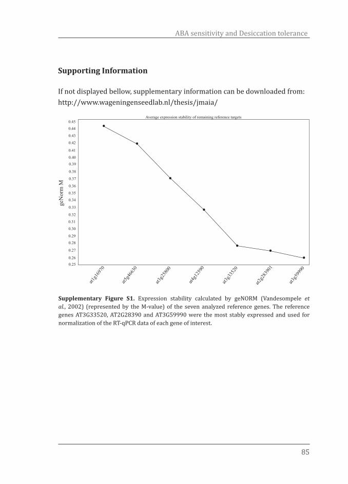
8585
ABA sensitivity and Desiccation tolerance
Supporting Information
If not displayed bellow, supplementary information can be downloaded from: http://www.wageningenseedlab.nl/thesis/jmaia/
0.45
0.44
0.43
0.42
0.41
0.400.39
0.38
0.37
0.36
0.35
0.34
0.33
0.32
0.31
0.30
0.29
0.28
0.27
0.260.25
at1g1
6970
at5g4
6630
at3g2
5800
at4g1
2590
at3g3
3520
at2g2
8390
1
at3g5
9990
geN
orm
M
Average expression stability of remaining reference targets
Supplementary Figure S1. Expression stability calculated by geNORM (Vandesompele et al., 2002) (represented by the M-value) of the seven analyzed reference genes. The reference genes AT3G33520, AT2G28390 and AT3G59990 were the most stably expressed and used for normalization of the RT-qPCR data of each gene of interest.
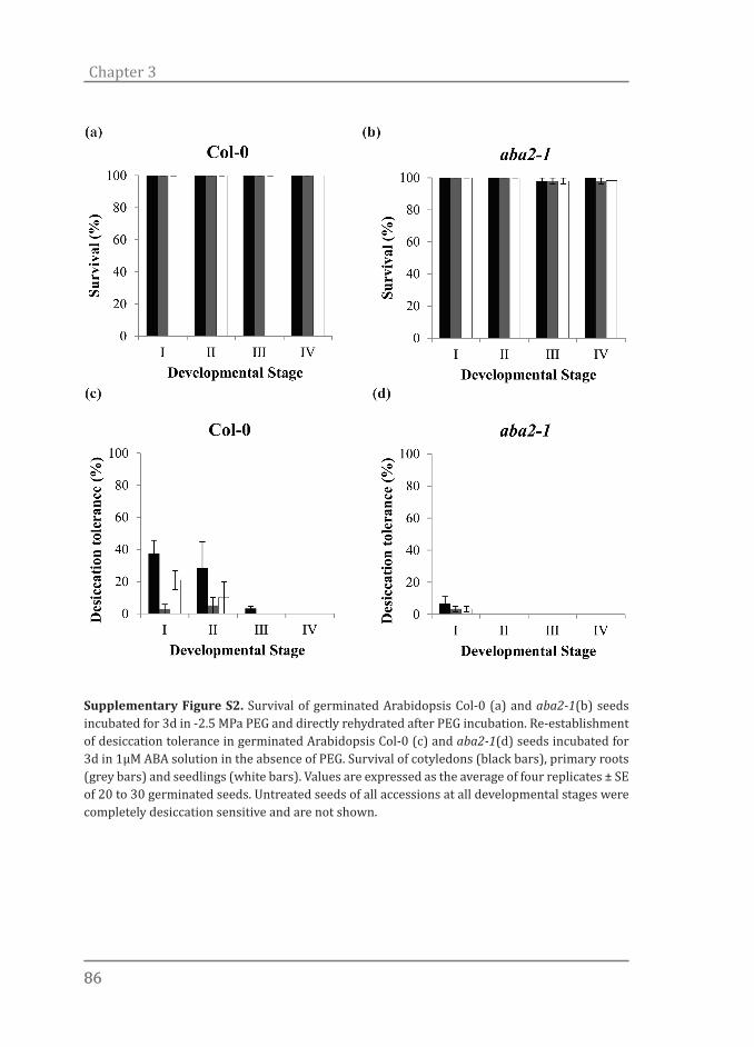
8686
Chapter 3
Supplementary Figure S2. Survival of germinated Arabidopsis Col-0 (a) and aba2-1(b) seeds incubated for 3d in -2.5 MPa PEG and directly rehydrated after PEG incubation. Re-establishment of desiccation tolerance in germinated Arabidopsis Col-0 (c) and aba2-1(d) seeds incubated for 3d in 1µM ABA solution in the absence of PEG. Survival of cotyledons (black bars), primary roots (grey bars) and seedlings (white bars). Values are expressed as the average of four replicates ± SE of 20 to 30 germinated seeds. Untreated seeds of all accessions at all developmental stages were completely desiccation sensitive and are not shown.
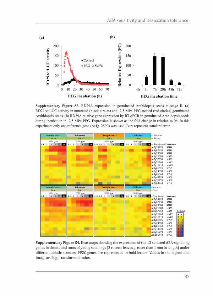
8787
ABA sensitivity and Desiccation tolerance
0
50
100
150
200
0h 3h 7h 20h 48h 72h
Rel
ativ
e E
xpre
ssio
n (F
C)
PEG incubation time
0
50
100
150
200
0 10 20 30 40 50 60 70
RD
29A
::LU
C a
ctiv
ity
PEG incubation (h)
ControlPEG -2.5MPa
(a) (b)
Supplementary Figure S3. RD29A expression in germinated Arabidopsis seeds at stage II. (a) RD29A::LUC activity in untreated (black circles) and -2.5 MPa PEG treated (red circles) germinated Arabidopsis seeds. (b) RD29A relative gene expression by RT-qPCR in germinated Arabidopsis seeds during incubation in -2.5 MPa PEG. Expression is shown as the fold change in relation to 0h. In this experiment only one reference gene (At4g12590) was used. Bars represent standard error.
Supplementary Figure S4. Heat maps showing the expression of the 13 selected ABA-signalling genes in shoots and roots of young seedlings (2 rosette leaves greater than 1 mm in length) under different abiotic stresses. PP2C genes are represented in bold letters. Values in the legend and image are log2-transformed ratios.
← Res. Area← Tissue
0.5 1 3 6 12 24 0.5 1 3 6 12 24 ### 0.5 1 3 6 12 24 0.5 1 3 6 12 24 ← Time (hours) Gene name
At5g59220 HAI1At1g07430 HAI2At2g29380 HAI3At4g26080 ABI1At5g57050 ABI2At5g51760 AHG1At3g11410 AHG3At4g01026 PYL7At3g24650 ABI3At1g01360 PYL9At2g40220 ABI4At2g36270 ABI5At5g05440 PYL5
← Res. Area← Tissue
0.5 1 3 6 12 24 0.5 1 3 6 12 24 ### 0.5 1 3 6 12 24 0.5 1 3 6 12 24 ← Time (hours) Gene name
At5g59220 HAI1At1g07430 HAI2At2g29380 HAI3At4g26080 ABI1At5g57050 ABI2 LegendAt5g51760 AHG1 + -At3g11410 AHG3 0 0At4g01026 PYL7 1 -1At3g24650 ABI3 2 -2At1g01360 PYL9 3 -3At2g40220 ABI4 4 -4At2g36270 ABI5 5 -5At5g05440 PYL5 6 -6
Osmotic stress Salt stress Drought stress Cold stressShoot Shoot Shoot
Osmotic stress Salt stress Drought stress Cold stress
ShootWild typeWild typeWild typeWild type
Root Root Root RootWild type Wild type Wild type Wild type
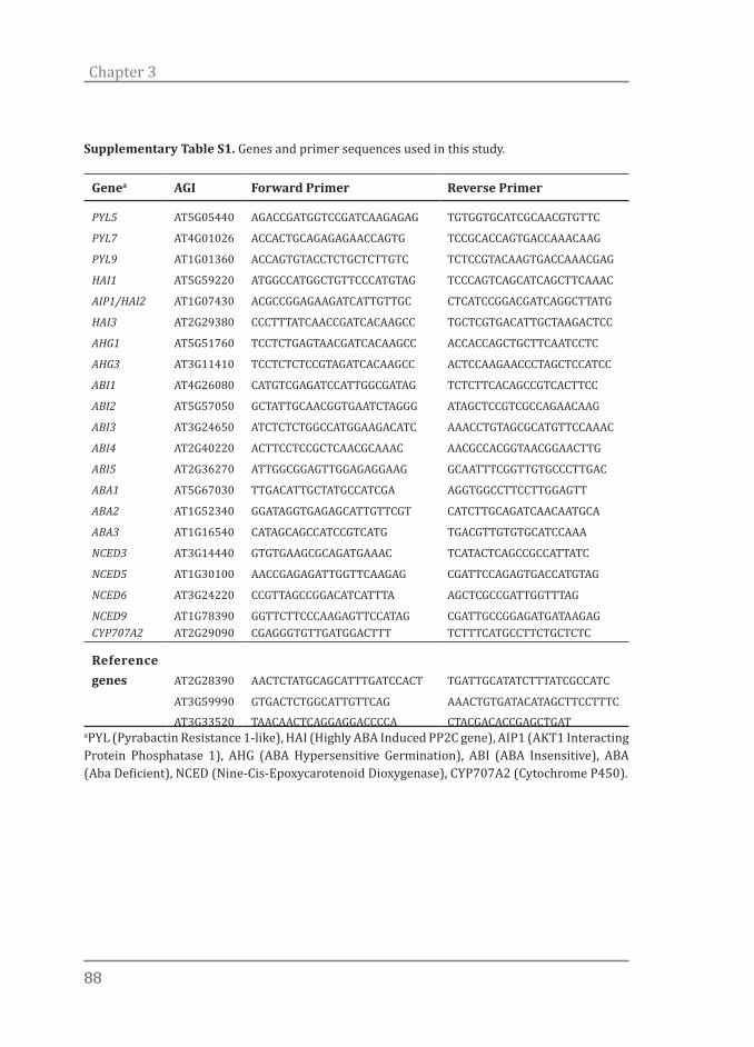
8888
Chapter 3
Supplementary Table S1. Genes and primer sequences used in this study.
Genea AGI Forward Primer Reverse Primer
PYL5 AT5G05440 AGACCGATGGTCCGATCAAGAGAG TGTGGTGCATCGCAACGTGTTC
PYL7 AT4G01026 ACCACTGCAGAGAGAACCAGTG TCCGCACCAGTGACCAAACAAG
PYL9 AT1G01360 ACCAGTGTACCTCTGCTCTTGTC TCTCCGTACAAGTGACCAAACGAG
HAI1 AT5G59220 ATGGCCATGGCTGTTCCCATGTAG TCCCAGTCAGCATCAGCTTCAAAC
AIP1/HAI2 AT1G07430 ACGCCGGAGAAGATCATTGTTGC CTCATCCGGACGATCAGGCTTATG
HAI3 AT2G29380 CCCTTTATCAACCGATCACAAGCC TGCTCGTGACATTGCTAAGACTCC
AHG1 AT5G51760 TCCTCTGAGTAACGATCACAAGCC ACCACCAGCTGCTTCAATCCTC
AHG3 AT3G11410 TCCTCTCTCCGTAGATCACAAGCC ACTCCAAGAACCCTAGCTCCATCC
ABI1 AT4G26080 CATGTCGAGATCCATTGGCGATAG TCTCTTCACAGCCGTCACTTCC
ABI2 AT5G57050 GCTATTGCAACGGTGAATCTAGGG ATAGCTCCGTCGCCAGAACAAG
ABI3 AT3G24650 ATCTCTCTGGCCATGGAAGACATC AAACCTGTAGCGCATGTTCCAAAC
ABI4 AT2G40220 ACTTCCTCCGCTCAACGCAAAC AACGCCACGGTAACGGAACTTG
ABI5 AT2G36270 ATTGGCGGAGTTGGAGAGGAAG GCAATTTCGGTTGTGCCCTTGAC
ABA1 AT5G67030 TTGACATTGCTATGCCATCGA AGGTGGCCTTCCTTGGAGTT
ABA2 AT1G52340 GGATAGGTGAGAGCATTGTTCGT CATCTTGCAGATCAACAATGCA
ABA3 AT1G16540 CATAGCAGCCATCCGTCATG TGACGTTGTGTGCATCCAAA
NCED3 AT3G14440 GTGTGAAGCGCAGATGAAAC TCATACTCAGCCGCCATTATC
NCED5 AT1G30100 AACCGAGAGATTGGTTCAAGAG CGATTCCAGAGTGACCATGTAG
NCED6 AT3G24220 CCGTTAGCCGGACATCATTTA AGCTCGCCGATTGGTTTAG
NCED9 AT1G78390 GGTTCTTCCCAAGAGTTCCATAG CGATTGCCGGAGATGATAAGAGCYP707A2 AT2G29090 CGAGGGTGTTGATGGACTTT TCTTTCATGCCTTCTGCTCTC
Reference genes AT2G28390 AACTCTATGCAGCATTTGATCCACT TGATTGCATATCTTTATCGCCATC
AT3G59990 GTGACTCTGGCATTGTTCAG AAACTGTGATACATAGCTTCCTTTC
AT3G33520 TAACAACTCAGGAGGACCCCA CTACGACACCGAGCTGATaPYL (Pyrabactin Resistance 1-like), HAI (Highly ABA Induced PP2C gene), AIP1 (AKT1 Interacting Protein Phosphatase 1), AHG (ABA Hypersensitive Germination), ABI (ABA Insensitive), ABA (Aba Deficient), NCED (Nine-Cis-Epoxycarotenoid Dioxygenase), CYP707A2 (Cytochrome P450).

8989
ABA sensitivity and Desiccation tolerance
Sd+Sq Sd+Sq Sd+Sq Sd+Sq Sd+Sq Sd+Sq Seed Seed Seed Seed Seed Seed ← Tissue
9.7 9.7 9.7 9.7 9.7 9.7 0.3 0.3 0.3 0.3 0.3 0.3← Growth Stage
Seed5+Sil
Seed6-Sil
Seed7-Sil
Seed8-Sil
Seed9-Sil
Seed10-Sil
wt-Col-0
wt-Col-0
wt-Col-0
wt-Col-0
wt-Col-0
wt-Col-0 ← Mutant
0 1 3 6 12 24 ← Time
Wt Wt Wt Wt Wt Wt
Dry seed
(germ)
Seed 1 HAI
(germ)
Seed 3 HAI
(germ)
Seed 6 HAI
(germ)
Seed 12 HAI
(germ)
Seed 24 HAI
(germ)
← Description
_ _ _ _ _ _ Col-0 Col-0 Col-0 Col-0 Col-0 Col-0 ← AliasGerm.Time.I
Germ.Time.I
Germ.Time.I
Germ.Time.I
Germ.Time.I
Germ.Time.I
← Res. Area
Gene Name
-1 -0.5 -0.2 0.1 0 0.1 0.9 1 0 -0.5 -1.3 -1 At1g01360 PYL90 0 1.6 3.9 4.3 4.8 1.5 1.1 -1.8 -0.7 -1 -2.5 At1g07430 HAI2
0.1 -0.3 0.1 -2.5 -2.1 -1.9 -1.1 -1.4 -2 0.2 0.6 1.8 At1g15080 ATLPP2-0.4 -1 -0.9 -0.3 -0.4 -0.1 1.5 1.4 1.4 0.4 -0.1 -0.4 At1g22280 PAPP2C-0.2 0 0.1 -0.7 -0.9 -0.8 -0.2 -0.2 0.9 0.3 0.1 0.9 At1g35670 ATCDPK2
0 0 0 -0.4 -0.3 -0.7 -0.4 -1 -0.7 -0.7 0.2 0.6 At1g48270 GCR1-0.6 -1.3 0.7 -0.3 -0.1 -0.5 -0.9 -0.9 -2.8 -1.6 -1.3 -1.3 At1g49720 ABF1
0 0.5 0.3 1 0.9 1 0.8 0.9 0.9 0.1 -0.1 -0.3 At1g61040 VIP5-0.1 0.4 0.9 2.2 2.6 2.3 1 0.8 -0.2 -0.7 -1 -1.2 At1g72770 HAB11.5 1.2 3.2 3.8 4.2 4.3 0.7 1 -2.7 0.1 0 0.4 At1g73000 PYL3-2.5 -2.8 -3 0.1 -0.8 0 -0.6 -0.5 -0.2 -0.7 -0.5 -0.7 At1g80840 ATWRKY40-0.7 -0.9 -1.1 -2 -1.9 -2 -0.5 -0.4 -0.4 -0.1 -0.2 -0.3 At2g26300 ATGPA10.1 0 0.8 3.9 5.2 4.7 0.6 0 0.9 0.7 -0.8 -2.4 At2g29380 HAI30 0.1 2.2 5.1 5.5 5.8 2.5 2.3 0.5 -0.4 -0.8 -1.3 At2g36270 ABI5-1 -1.5 -1.4 -0.9 -1.4 -1 -5.6 -4.3 0.8 1 1.2 1.3 At2g38310 PYL4
-0.4 -1 -0.6 0.2 0.4 1.2 0.3 -0.7 -1 -1 -0.5 -0.8 At2g40180 ATHPP2C52.9 3.2 2.9 2.7 2 2.4 -2.5 -1.5 2.1 2.1 2.2 2.2 At2g40220 ABI41 0.9 1.2 1 0.6 0.7 -2.7 -0.4 2 2 3 2.7 At2g40330 PYL6
1.2 0.8 1.4 1.3 1.8 1.5 1.5 1 -0.7 -0.2 -0.2 -0.6 At3g11410 AHG3-1.3 -0.6 0.3 0.1 0.7 0.5 2.5 2.1 0 -0.9 -0.5 -2.3 At3g14440 ATNCED3-0.8 -0.2 0.1 1.1 1.1 0.8 1.4 0.9 -1.8 -1.3 -1.3 -1.5 At3g19290 ABF44.4 5.7 6 6.5 6.9 6.8 0 -0.3 -0.5 0.4 0 -0.2 At3g24650 ABI3-0.3 -0.2 0.2 0.3 0.6 0.2 1.1 1.2 -0.8 -1.7 -2.1 -3.3 At4g01026 PYL7-0.4 0.2 0 -0.8 -1 -1.1 -0.6 -0.5 0 -0.5 -0.6 -0.2 At4g09570 ATCPK40.1 -0.2 0.2 0 0.3 0 -0.2 -0.4 -0.4 0.7 0.3 0 At4g26080 ABI1-0.2 1.4 2.7 3 3.6 3 3.5 3.2 -1.2 -1.5 -1.9 -2.1 At4g27410 RD26-0.8 -1 -1.1 -1.2 -1.2 -1.1 -2.7 -1.2 -0.4 -2 -1.5 0.1 At4g34000 ABF31.2 0 -0.1 1.7 1.3 1.6 0.7 0.6 1.1 0.5 0.4 0.1 At5g05440 PYL50.3 -0.1 -0.6 -3.1 -4.6 -5 -1.9 -1.4 -2.1 -1.5 -1 1.5 At5g13630 ABAR0.9 -0.4 -1.5 -3.4 -5 -5 -1 -1.8 -2.5 -2.3 -1.6 -1.6 At5g25610 RD22-0.4 0 -0.1 0.1 -0.1 0 0.1 -0.5 -0.1 0 -0.1 0 At5g42400 ATXR70.3 0.1 -0.1 -0.3 -0.6 -0.8 -1 -0.8 -0.8 -0.2 -0.1 -0.3 At5g44200 ATCBP20-0.7 -3.1 -4.6 -4.6 -4.6 -3 -2.9 -2.4 0.2 0.9 1.4 0.7 At5g46790 PYL11.3 3.1 4.5 7.9 8 8.4 1.3 1.3 -0.1 -1.3 -2.4 -3.3 At5g51760 AHG11.5 0.3 2.2 2.7 2.9 3.1 2.7 2.3 -0.4 -0.4 -1 -1.5 At5g57050 ABI22.8 3.9 4.2 3.3 3.6 2.9 0.9 0.9 -1.8 -1.8 -1.9 -2.2 At5g59220 HAI10 -0.2 -0.8 -0.7 -1.1 -0.9 -0.4 -0.6 -0.3 -0.1 -0.1 -0.1 At5g61150 VIP4
-0.4 -1.2 -1.2 -0.2 -0.7 0.2 0.8 1 1.4 1.3 0.9 0.2 At5g66880 SNRK2-3
Supplementary Table S2. Details of the heat maps displaying the gene expression profile of a pre-set of 38 candidate genes thought to be involved in ABA signalling during seed development and germination according to the eFP browser at the BAR website (http://www.bar.utoronto.ca/) (Toufighi et al., 2005). Values in the image are log2-transformed ratios.

9090
Chapter 3
Sample Information ABA and ABA metabolites (ng/g DW)# Develop.
StageTreatment Sample
weight (mg)
ABA DPA ABA-GE
PA 7'OH-ABA
neo-PA
t-ABA
1 I untreat. 49.3 6 n.d. n.d. n.d. n.d. n.d. n.d.2 I untreat. 50.4 5 n.d. <8 n.d. n.d. n.d. <43 I untreat. 51.9 7 n.d. n.d. n.d. n.d. n.d. n.d.4 I 5h PEG 52.8 <3.8 n.d. n.d. n.d. n.d. n.d. <4.95 I 5h PEG 49.3 <4.1 n.d. n.d. n.d. n.d. n.d. n.d.6 I 5h PEG 52.6 <3.8 n.d. n.d. n.d. n.d. n.d. n.d.7 I 24h PEG 52.3 <3.8 n.d. n.d. n.d. n.d. n.d. <3.88 I 24h PEG 53.1 <3.8 n.d. n.d. n.d. n.d. n.d. <3.89 I 24h PEG 48.9 5 n.d. n.d. n.d. n.d. n.d. <4.110 II untreated 50.4 5 n.d. n.d. n.d. n.d. <4 n.d.11 II untreated 52.5 7 n.d. n.d. n.d. n.d. n.d. <3.812 II untreated 50.8 7 n.d. n.d. n.d. n.d. n.d. n.d.13 II 5h PEG 48.9 <4.1 n.d. n.d. n.d. n.d. n.d. n.d.14 II 5h PEG 50.0 5 n.d. n.d. n.d. n.d. n.d. <415 II 5h PEG 52.1 <3.8 n.d. n.d. n.d. n.d. n.d. <3.816 II 24h PEG 49.6 <4 n.d. n.d. n.d. n.d. n.d. <417 II 24h PEG 49.5 4 n.d. n.d. n.d. n.d. n.d. n.d.18 II 24h PEG 52.6 7 n.d. n.d. n.d. n.d. n.d. <3.819 III untreated 51.2 4 n.d. <9.5 n.d. n.d. n.d. n.d.20 III untreated 51.2 6 n.d. n.d. n.d. n.d. n.d. <4.821 III untreated 51.8 12 n.d. n.d. n.d. n.d. n.d. <822 III 5h PEG 49.0 5 n.d. n.d. n.d. n.d. n.d. <4.423 III 5h PEG 47.4 5 n.d. n.d. n.d. n.d. n.d. <4.224 III 5h PEG 50.5 8 n.d. n.d. n.d. n.d. n.d. 725 III 24h PEG 50.6 <4 n.d. n.d. n.d. n.d. n.d. <426 III 24h PEG 49.0 5 n.d. n.d. n.d. n.d. n.d. <4.1
Supplementary Table S3. Quantification of ABA and ABA metabolites in Arabidopsis seeds*. Fresh untreated and treated (incubation for 5 and 24h in -2.5 MPa PEG) seeds were used. Developmental stage represent seeds at testa rupture (I), at radicle protrusion (II), showing a primary root of 0.3-0.5 mm in length (III) and at the appearance of the first root hairs (IV). Abscisic acid (ABA) and its metabolites dihydrophaseic acid (DPA), phaseic acid (PA), neo-PA (neo-PA), ABA-glucose ester (ABA-GE), 7’-hydroxy-ABA (7’OH-ABA) were measured. Values preceded by a < sing were below limit of reliable quantification. Samples in which values were below limit of detection are indicated as n.d. Supplementary Table S3. Quantification of ABA and ABA metabolites in Arabidopsis seeds*. Fresh untreated and treated (incubation for 5 and 24h in -2.5 MPa PEG) seeds were used. Developmental stage represent seeds at testa rupture (I), at radicle protrusion (II), showing a primary root of 0.3-0.5 mm in length (III) and at the appearance of the first root hairs (IV). Abscisic acid (ABA) and its metabolites dihydrophaseic acid (DPA), phaseic acid (PA), neo-PA (neo-PA), ABA-glucose ester (ABA-GE), 7’-hydroxy-ABA (7’OH-ABA) were measured. Values preceded by a < sing were below limit of reliable quantification. Samples in which values were below limit of detection are indicated as n.d.

9191
ABA sensitivity and Desiccation tolerance
27 III 24h PEG 12.7 <22.6 n.d. n.d. n.d. n.d. n.d. <15.728 IV untreated 50.4 7 n.d. <9.3 n.d. n.d. n.d. 529 IV untreated 52.2 10 n.d. <6.2 n.d. n.d. n.d. <3.830 IV untreated 49.6 11 n.d. <7.9 n.d. n.d. n.d. 1731 IV 5h PEG 49.3 7 n.d. n.d. n.d. n.d. n.d. <4.132 IV 5h PEG 51.3 <3.9 n.d. n.d. n.d. n.d. n.d. <3.933 IV 5h PEG 51.0 7 n.d. n.d. n.d. n.d. n.d. 834 IV 24h PEG 51.6 6 n.d. n.d. n.d. n.d. n.d. n.d.35 IV 24h PEG 50.5 7 n.d. n.d. n.d. n.d. n.d. <436 IV 24h PEG 51.0 7 n.d. n.d. n.d. n.d. n.d. 5
*Supplementary Methods S1
Plant Material
Fresh untreated or treated (incubation for 3 and 7h in -2.5 MPa PEG) Arabidopsis seeds (stages I, II, III and IV) were frozen in liquid nitrogen, freeze dried and ground in a dismembrator (Mikro-dismembrator U, B. Braun Biotech International) at 2000 RPM for 1.5 min with the help of 1/4’’ stainless steel beads.
Chemicals and Calibration Curves
A number of compounds namely DPA, ABA-GE, PA, 7’-OH-ABA, neoPA, and trans-ABA were synthesized and prepared at the National Research Council of Canada, Saskatoon, SK, Canada. The deuterated forms of the hormones which were used as internal standards that include d3-DPA, d5-ABAGE, d3-PA, d4-7’-OH-ABA, d3-neoPA, d4-ABA and d4-trans-ABA were synthesized and prepared at NRCC SK according to Abrams et al. (2003) and Zaharia et al. (2005). The deuterated forms of selected hormones used as recovery (external) standards, d6-ABA and d2-ABA-GE, were prepared and synthesized at NRCC SK. Calibration curves were created for all compounds of interest. Quality control samples (QCs) were run along with the tissue samples.
Instrumentation
Analysis was performed on a UPLC/ESI-MS/MS utilizing a Waters ACQUITY UPLC system, equipped with a binary solvent delivery manager and a sample manager coupled to a Waters Micromass Quattro Premier XE quadrupole
9292
Chapter 3
tandem mass spectrometer via a Z-spray interface. MassLynx™ and QuanLynx™ (Micromass, Manchester, UK) were used for data acquisition and data analysis.
Extraction and purification
An aliquot (100 μL) containing the deuterated internal standards, each at a concentration of 0.2 ng μL-1, was added to homogenized plant tissue (approximately 50 mg); 3 mL of isopropanol:water:glacial acetic acid (80:19:1, v/v/v) was then added, and the samples were agitated in the dark for 24 h at 4 ºC. Samples were then centrifuged and the supernatant was isolated and dried on a Büchi Syncore Polyvap (Büchi, Switzerland). Samples were reconstituted in 100 μL acidified methanol, adjusted to 1 mL with acidified water, and then partitioned against 2 mL hexane. After 30 min, the aqueous layer was isolated and dried as above. Dry samples were reconstituted in 100 μL acidified methanol and adjusted to 1 mL with acidified water. The reconstituted samples were loaded onto equilibrated Oasis HLB cartridges (Waters, Mississauga, ON, Canada), washed with acidified water, and eluted with acetonitrile:water:glacial acetic acid (30:69:1, v/v/v). The eluate was then dried on a LABCONCO centrivap concentrator (Labconco Corporation, Kansas City, MO, USA). An internal standard blank was prepared with 100 μL of the deuterated internal standards mixture. Quality control standards (QC) were prepared by adding 100 μL and 30 μL (separately) of a mixture containing the analytes of interest, each at a concentration of 0.2 ng μL-1, to 100 μL of the internal standard mix. Finally, samples, blanks, and QCs were reconstituted in an aqueous solution of 40% methanol (v/v), containing 0.5% acetic acid and 0.1 ng μL-1 of each of the recovery standards.
Hormone quantification by HPLC-ESI-MS/MS
The samples were subjected to UPLC-ES-MS/MS analysis and quantification, similar to that described in detail in Ross et al. (2004). Samples were injected onto an ACQUITY UPLC® HSS C18 column (2.1x100 mm, 1.8 μm) with an ACQUITY HSS C18 VanGuard Pre-column (2.1x5 mm, 1.8 μm) and separated by a gradient elution of water containing 0.025% acetic acid against an increasing percentage of acetonitrile containing 0.025% acetic acid. Briefly, the analysis utilizes the Multiple Reaction Monitoring (MRM) function of the MassLynx
9393
ABA sensitivity and Desiccation tolerance
v4.1 (Waters Inc) control software. The resulting chromatographic traces are quantified off-line by the QuanLynx v4.1 software (Waters Inc) wherein each trace is integrated and the resulting ratio of signals (non-deuterated/internal standard) is compared with a previously constructed calibration curve to yield the amount of analyte present (ng per sample). Calibration curves were generated from the MRM signals obtained from standard solutions based on the ratio of the chromatographic peak area for each analyte to that of the corresponding internal standard, as described by Ross et al. (2004). The QC samples, internal standard blanks and solvent blanks were also prepared and analysed along each batch of tissue samples.
References
Abrams SR, Nelson K, Ambrose SJ. 2003. Deuterated abscisic acid analogs for mass spectrometry and metabolism studies. Journal of Labelled Compounds and Radiopharmaceuticals 46: 273–283.
Ross ARS, Ambrose SJ, Cutler AJ, Feurtado JA, Kermode AR, Nelson K, Zhou R, Abrams SR. 2004. Determination of endogenous and supplied deuterated abscisic acid in plant tissues by high-performance liquid chromatography-electrospray ionization tandem mass spectrometry with multiple reaction monitoring. Analytical Biochemistry 329: 324-333
Zaharia LI, Galka MM, Ambrose SJ, Abrams SR. 2005. Preparation of deuterated abscisic acid metabolites for use in mass spectrometry and feeding studies. Journal of Labelled Compounds and Radiopharmaceuticals 48: 435–445.

O sorriso enriquece os recebedores sem empobrecer os doadores.
Mário Quintana
Chapter 4All roads lead to Rome: Metabolite profiling of the re-establishment of desiccation toler-ance in germinated Arabidopsis seeds
Authors:Julio Maia1, Harry Thiewes1, Leo Willems1, Wilco Ligterink1, Henk W. M. Hilhorst1
1 Wageningen Seed Lab, Laboratory of Plant Physiology, Wageningen University, Wageningen, The Netherlands
(in preparation for submission)
9696
Chapter 4
Abstract
Desiccation tolerance (DT) is a widespread trait found in all kingdoms of life. DT is especially common in seeds of land plants. Such seeds acquire DT during development and become sensitive again to extreme dehydration around the point of visible germination. Yet, if confronted with suboptimal conditions, such as osmotic stress, germinated desiccation sensitive seeds are able to activate global changes in gene expression and metabolite composition to re-establish DT. Here we characterized the metabolic phenotype of Arabidopsis Col-0 and aba2-1 germinated seeds subjected to a set of treatments which combine the application of osmotic stress by polyethylene glycol (PEG) and abscisic acid (ABA) to re-establish DT. The metabolic signature of PEG-induced DT was remarkably different from the one found in seeds treated with ABA. PEG-treated seeds displayed a nitrogen-rich metabolome while ABA-treated seeds displayed more carbon-rich compounds such as sugars and sugar-alcohols. Independently of the applied treatment, seeds with re-established DT consistently accumulated raffinose, galactinol, and sucrose and displayed reduced levels of methionine, glycine, alanine and leucine. Our data implies the existence of two different metabolic signatures that both can result in the expression of DT and support the general consent that mechanisms such as ROS scavenging, cytoplasm vitrification and sugar signalling, in concert with other protection mechanisms, are crucial for the acquisition of DT.
Introduction
Tolerance to complete desiccation is a common feature of many life forms such as spores, pollen and resurrection plants, but especially of angiosperm seeds. Such seeds acquire desiccation tolerance (DT) during their development. Among other developmental related events, DT is usually initiated together with the accumulation of reserves and acquisition of dormancy, and is usually fully established just before the drying phase at the end of seed maturation (Bewley et al., 2013). DT is a multigenic complex trait that demands a cascade of events involving abscisic acid (ABA) signalling, including the recently discovered PYR/PYL/RCAR receptor family consisting of PYRABACTIN RESISTANCE 1 (PYR1)-
9797
Desiccation tolerance metabolome
LIKE regulatory components of ABA receptors, the type 2c protein phosphatases (PP2Cs) and the Sucrose-Non-Fermenting Kinase 1-Related Protein Kinase 2 (SnRK2s) family (Umezawa et al., 2010; Komatsu et al., 2013). DT also requires the activation of certain transcription factors (TF) such as the B3 transcription factor ABA insensitive 3 (ABI3) and the bZip TF ABI5 (Ooms et al., 1993; Terrasson et al., 2013). These signals activate downstream genes and pathways responsible for the accumulation of late embryogenesis abundant (LEA) proteins, sugars, osmolytes and amino acids, among other molecules that are necessary for DT. These molecules will act by protecting other macromolecules such as DNA, RNA, proteins and enzymes but also membranes from the damage imposed by desiccation stress (Chandra Babu et al., 2004; Tunnacliffe & Wise, 2007; Moore et al., 2009). Although DT is a widespread trait that has been thoroughly investigated for more than a century, understanding the mechanisms underlying it is still a challenge and probably many important molecules involved in DT remain to be discovered and functionally characterized. A versatile system to study DT consists of the loss and re-establishment of DT in germinated seeds (Bruggink & van der Toorn, 1995). This system utilizes the observation that germinating seeds usually lose their DT around the point of radical protrusion and that loss of DT can be reversed at this point by the application of an osmotic stress. The elements engaged in re-establishing DT in germinated seeds are, thus, likely to be free of overlapping seed developmental processes that may be confused with specific features of DT. “Omics” data obtained using this approach unveiled putative new genes and proteins correlated with DT and reinforced the link between ABA and DT (Buitink et al., 2006; Maia et al., 2011). More recently, this system has been employed, using an ABA-deficient Arabidopsis mutant, aba2-1, and a set of different treatments consisting of the application of the osmoticum polyethylene glycol (PEG), ABA or PEG + ABA to re-establish DT to significantly divergent levels (Chapter 3). Both PEG and ABA could fully rescue DT in germinated Col-0 Arabidopsis seeds, showing that ABA can completely substitute for the osmotic signal in order to re-establish DT. Although both treatments lead to the same phenotype, it remains to be investigated whether the metabolic output induced by PEG and ABA is the same. To date re-induction of desiccation tolerance in seeds has not been

9898
Chapter 4
investigated for its associated metabolome. The metabolome is the final expression of the genotype and variations of it, under varying conditions, and can be treated as the organism’s chemical phenotype. Thus, untargeted metabolite profiling appears to be a good method to explore DT related molecules. Although large-scale metabolomic studies have been reported for Arabidopsis plants undergoing dehydration or ABA treatment (Kempa et al., 2008; Urano et al., 2009), no reports have addressed it in the context of desiccation stress. Metabolomic studies have so far only been reported for whole plants of two desiccation tolerant species, the grass species Sporobolus stapfianus and the spikemoss Selaginella lepidophylla (Oliver et al., 2011; Yobi et al., 2012; Yobi et al., 2013). The desiccation induced metabolic changes found in these species included mainly amino acids, followed by carbohydrates, lipids and antioxidants. In this study we set out to unravel important metabolites and response mechanisms related to the re-establishment of DT in germinated Arabidopsis seeds. This is the first study reporting untargeted metabolic profiling of the re-establishment of DT in germinated seeds of Arabidopsis in response to osmotic (PEG) and hormonal (ABA) stimuli. With the aid of an ABA-deficient mutant, aba2-1, that cannot recover DT when treated with PEG (Chapter 3), we explored the metabolic differences in PEG- and ABA-induced DT.
Material and Methods
Plant material and growth conditions
Arabidopsis thaliana (L.) Heynh. lines Col-0 and aba2-1 (Léon-Kloosterziel et al., 1996) were used in this study. In all experiments Arabidopsis seeds were cold-stratified for 72h at 4°C in 9 cm Petri dishes on two layers of blue filter paper (Anchor Paper) and 10 ml of distilled water, to remove residual dormancy. Germination assays were performed in a germination cabinet under constant white light at 22ºC. Seeds at radicle protrusion (stage II) (Maia et al., 2011) were used in all experiments.
Design of the experimental system
It has been demonstrated that DT can be fully rescued in germinated

9999
Desiccation tolerance metabolome
Arabidopsis seeds by the application of an osmotic treatment (PEG) before drying (Chapter 2) (Maia et al., 2011). Furthermore, with the aid of some ABA-insensitive and ABA-deficient mutants we have shown that ABA is necessary for the re-establishment of DT (Chapter 3). We also revealed that ABA alone is sufficient to rescue DT in Col-0 and aba2-1 seeds and that aba2-1 seeds treated only with PEG failed to fully re-establish DT. Based on these results we defined an experimental design to further explore DT related mechanisms and to investigate the differences between PEG- and ABA-induced DT (Figure 1). Desiccation tolerance was assessed as follows: Germinated seeds at stage II (radicle protrusion) were selected and either dried directly or after 3d of incubation in -2.5 MPa polyethylene glycol (PEG), 5µM ABA (ABA) or a combination of -2.5MPa PEG + 1µM ABA (PEG + ABA). After incubation, the seeds were rinsed and dehydrated for 3d at 20°C at 32% relative humidity (RH). After dehydration, the seeds were pre-humidified in humid air (100% RH) for 24h at 22°C in the dark to avoid imbibitional damage (Leopold & Vertucci, 1986). Germinated seeds that generated a viable seedling were considered desiccation tolerant (DT) and those that did not were considered desiccation sensitive (DS).
Figure 1. Flow diagram depicting the re-establishment of DT in Arabidopsis Col-0 and aba2-1 seeds under different treatments. Samples were collected before (‘untreated controls’) and after each treatment (‘treated samples’). Samples are named after their genotype (wt for Col-0 and aba for aba2-1 seeds), followed by the treatment (fresh untreated germinated Arabidopsis seeds at the point of radical protrusion (F) and treated with PEG (P), ABA (A) or PEG + ABA (PA)).
PEG ABAPEG
+ ABA
Drying
Re-hydration
DS DT DT DT
Col-0
wt-F(control )
wt-P wt-A wt-PA
PEG ABAPEG
+ ABA
Drying
Re-hydration
DS DS DT DT
aba2-1
aba-F(control )
aba-P aba-A aba-PA
Germinated seeds at stage II
Sample collection (untreated controls)
Treatments
Sample collection (treated samples)
32% RH at 20oC
Re-hydration at 22oC
Phenotypes after DT evaluation
Pre-humidification Pre-humidification100% RH at 22oC
100100
Chapter 4
Metabolite profiling
Primary metabolite extraction was performed according to Roessner et al. (2000) with modifications. Treated and untreated germinated seeds (20 mg) were homogenized using a microdismembrator (Sartorius) in 2-mL tubes with one 1/4’’ stainless steel bead precooled in liquid nitrogen; 700µL of methanol:chloroform (4:3) was added, together with the standard (final concentration of 0.2 mg mL–1 ribitol) and mixed thoroughly. After 10 min of sonication, 200 mL Milli-Q water was added to the mixture, followed by vortexing and centrifugation (5 min, 12,000g). The methanol phase was collected in a glass vial. 500 µL of methanol/chloroform was added to the remaining organic phase and kept on ice for 10 min. 200µL of Milli-Q water was added followed by vortexing and centrifugation (5 min, 12,000g). The methanol phase was collected and added to the previously collected polar phase. 100µL were transferred to a glass vial and dried overnight using a SpeedVac (35°C, Savant SPD121). A GC-TOF-MS method with some minor modifications (Carreno-Quintero et al., 2012) was used. Detector voltage was set at 1,600 V. Raw data were processed using the ChromaTOF software 2.0 (Leco Instruments) and further processed using the MetAlign software (Lommen, 2009) to extract and align the mass signals (signal-to-noise ratio ≥ 2). The output was further processed by the MetAlign Output Transformer (Plant Research International), and mass signals that were present in less than three samples were discarded. Centrotypes were created using the MSClust program (Tikunov et al., 2012). This resulted in 195 unique centrotypes or representative masses.
Compound identification
The mass spectra of the representative masses were used for the identification by matching to an in-house-constructed library, the Golm Metabolome Database (http://gmd.mpimp-golm.mpg.de/) and the National Institute of Standards and Technology NIST05 (http://www.nist.gov/srd/mslist.cfm) libraries. This identification is based on spectra similarity and comparison of retention indices calculated by using a third-order polynomial function (Strehmel et al., 2008). Library hits were manually curated.

101101
Desiccation tolerance metabolome
Data normalization and multivariate analysis
All mass intensity values were normalized by an internal standard (ribitol). Relative amounts of the metabolites were obtained by normalizing the intensity of individual masses to the response of the internal standard. The ratio between the mass intensity value of the putative compound and the ribitol internal standard was then scaled by multiplying the resulting value by the average of the ribitol mass intensity across all samples. All values were then finally corrected by their respective sample dry weight. After normalization, all the data sets were Log10 transformed and mean-centred to place all the measured metabolites on an equal level so as to enable metabolites with both low concentrations and high concentrations to be more comparable. After pre-processing the data, a multivariate principal component analysis (PCA) was used for dimensional reduction and identification of important variables. After PCA analysis, pairwise comparisons between groups were performed. Pairwise comparisons were done with Log10 transformed data and Student t-tests were applied.
Hierarchical clustering
In hierarchical cluster analysis, each sample begins as a separate cluster and the algorithm proceeds to combine them until all samples belong to one cluster. Two parameters need to be considered when performing hierarchical clustering; similarity measure and clustering algorithm. Normalized, Log10 transformed and mean-centred data was further processed by the MetaboAnalyst 2.0 web tool (Xia et al., 2009; Xia et al., 2012). We used Pearson’s correlation as similarity measure and Ward’s linkage (clustering to minimize the sum of squares of any two clusters) as clustering algorithm. Changes in metabolite relative quantities are displayed as a dendrogram added to a heatmap for visual aid.
Results
PEG- and ABA-induced metabolic changes
After defining the treatments to re-establish DT, we further explored the differences and similarities between the responses of germinated Col-0
102102
Chapter 4
and aba2-1 seeds to PEG, ABA and PEG + ABA. We profiled the two different genotypes for primary metabolites before and after the different treatments (Figure 1). From all representative masses obtained we were able to identify and annotate 54 metabolites (Supplementary Table S1). The PCA score plot of the GC-TOF-MS analysed metabolite data showed tight grouping of biological replicates, attesting for good data quality (Figure 2a). The grouping observed in the PCA score plot was largely genotype-independent and it was noticeable that untreated samples and samples submitted to similar DT re-establishment treatments (ABA or PEG + ABA) clustered together. Overall, three main clusters (named DS, DT-ABA and DT-PEG) were evident (Figure 2a). DS comprises the untreated desiccation sensitive Col-0 and aba2-1 seeds, DT-ABA represents desiccation tolerant Col-0 and aba2-1 ABA-treated seeds, whereas cluster DT-PEG covers Col-0 PEG-treated and Col-0 and aba2-1 PEG + ABA-treated samples. As expected, clusters derived from Col-0 and aba2-1 samples treated with PEG did not overlap, probably due to the inability of aba2-1 PEG-treated seeds to fully re-establish DT. The cluster only formed by aba2-1 PEG treated seeds is situated in between the desiccation sensitive DS and the desiccation tolerant DT-PEG clusters. After simplification of the metabolite data by PCA analysis, PC1, 2 and 3 explained 47.3, 25.8 and 12.6% of the total variance, respectively and, together, the first three components accounted for 85.6% of the total explained variance (Figure 2b). Hierarchical cluster analysis resulted in a heat map, which was used to identify relevant patterns in metabolite changes among samples and clusters (Figure 3). Overall, the clustering (clusters DS, DT-ABA and DT-PEG) created based on the PCA analysis was robust and just a few metabolites changed in opposite directions between different treatments within a cluster (arrows in Figure 3). Interestingly, four of the five amino acids present in cluster DT-PEG, leucine, glycine, threonine and tyrosine, were also consistently decreased when ABA was present in the treatment (wt-PA and aba-PA – Figure 3). Although possessing distinct genetic backgrounds, the untreated DS controls of Col-0 and aba2-1 samples (cluster DS) displayed very similar metabolic signatures with very few differences in metabolite abundance. Remarkably, although clusters DT-ABA and DT-PEG both represent seeds with re-established DT, a shift in the groups of metabolites between these two

103103
Desiccation tolerance metabolome
clusters, was observed (Figure 3). Abundance of most of the identified amino acids was decreased in cluster DT-ABA whereas they were over accumulated in cluster DT-PEG (Figure 3). Contrary to what was observed for amino acids, most of the sugars, sugar alcohols and organic acids were induced by ABA (cluster DT-ABA) and inhibited when PEG was present in the DT re-establishment treatment (cluster DT-PEG). These results suggest the occurrence of a shift in the carbon/nitrogen balance where PEG- and ABA-treated seeds tend to accumulate nitrogen and carbon-rich compounds, respectively. To further explore this we estimated the number of carbon and nitrogen atoms in each sample (Figure 4). In agreement with our predictions, PEG-treated seeds accumulated relatively more nitrogen atoms than ABA-treated seeds while the latter displayed a relatively higher number of carbon atoms (Figure 4). Thus, our analysis suggests either the existence of different strategies to re-establish DT, or that DT signalling proceeds independently of the metabolic signature.
DS
DT-PEG
DT-ABA
(a) (b)
Figure 2. (a) Score plot between the first two PCs. The explained variance is shown in brackets. Samples are named after their genotype (wt for Col-0 and aba for aba2-1 seeds followed by the treatment (germinated Arabidopsis seeds at the point of radical protrusion untreated (F) and treated with PEG (P), ABA (A) and PEG + ABA (PA)). Three clusters were evident and are depicted by black dotted circles. (b) Scree plot showing the variance explained by principal components (PCs). The green and blue lines show the accumulated explained variance and the variance explained by individual PCs, respectively.

104104
Chapter 4
Figure 3. Clustering result shown as heatmap (distance measure using Pearson, and clustering algorithm using ward). The heatmap provides intuitive visualization of the data table. Each coloured cell on the map corresponds to a concentration value, with samples in rows and metabolites in columns. Arrows indicate metabolites that changed in opposite directions within a cluster. Samples are named after their genotype (wt for Col-0 and aba for aba2-1 seeds) followed by the treatment (germinated Arabidopsis seeds at the point of radical protrusion untreated (F) and treated with PEG (P), ABA (A) and PEG + ABA (PA)). Numbers between brackets indicate biological replicates.
Clus
ter D
SCl
uste
r DT-
ABA
Clus
ter D
T-PR
G
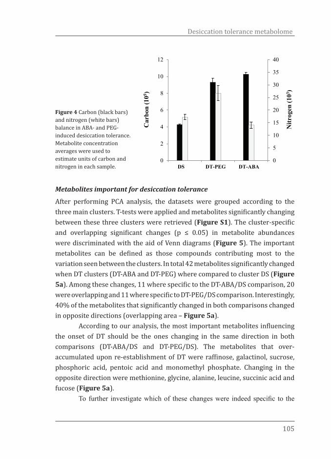
105105
Desiccation tolerance metabolome
Metabolites important for desiccation tolerance
After performing PCA analysis, the datasets were grouped according to the three main clusters. T-tests were applied and metabolites significantly changing between these three clusters were retrieved (Figure S1). The cluster-specific and overlapping significant changes (p ≤ 0.05) in metabolite abundances were discriminated with the aid of Venn diagrams (Figure 5). The important metabolites can be defined as those compounds contributing most to the variation seen between the clusters. In total 42 metabolites significantly changed when DT clusters (DT-ABA and DT-PEG) where compared to cluster DS (Figure 5a). Among these changes, 11 where specific to the DT-ABA/DS comparison, 20 were overlapping and 11 where specific to DT-PEG/DS comparison. Interestingly, 40% of the metabolites that significantly changed in both comparisons changed in opposite directions (overlapping area – Figure 5a). According to our analysis, the most important metabolites influencing the onset of DT should be the ones changing in the same direction in both comparisons (DT-ABA/DS and DT-PEG/DS). The metabolites that over-accumulated upon re-establishment of DT were raffinose, galactinol, sucrose, phosphoric acid, pentoic acid and monomethyl phosphate. Changing in the opposite direction were methionine, glycine, alanine, leucine, succinic acid and fucose (Figure 5a). To further investigate which of these changes were indeed specific to the
Car
bon
(105 )
Nitr
ogen
(103 )
0
5
10
15
20
25
30
35
40
0
2
4
6
8
10
12
DS DT-PEG DT-ABA
Figure 4 Carbon (black bars) and nitrogen (white bars) balance in ABA- and PEG-induced desiccation tolerance. Metabolite concentration averages were used to estimate units of carbon and nitrogen in each sample.
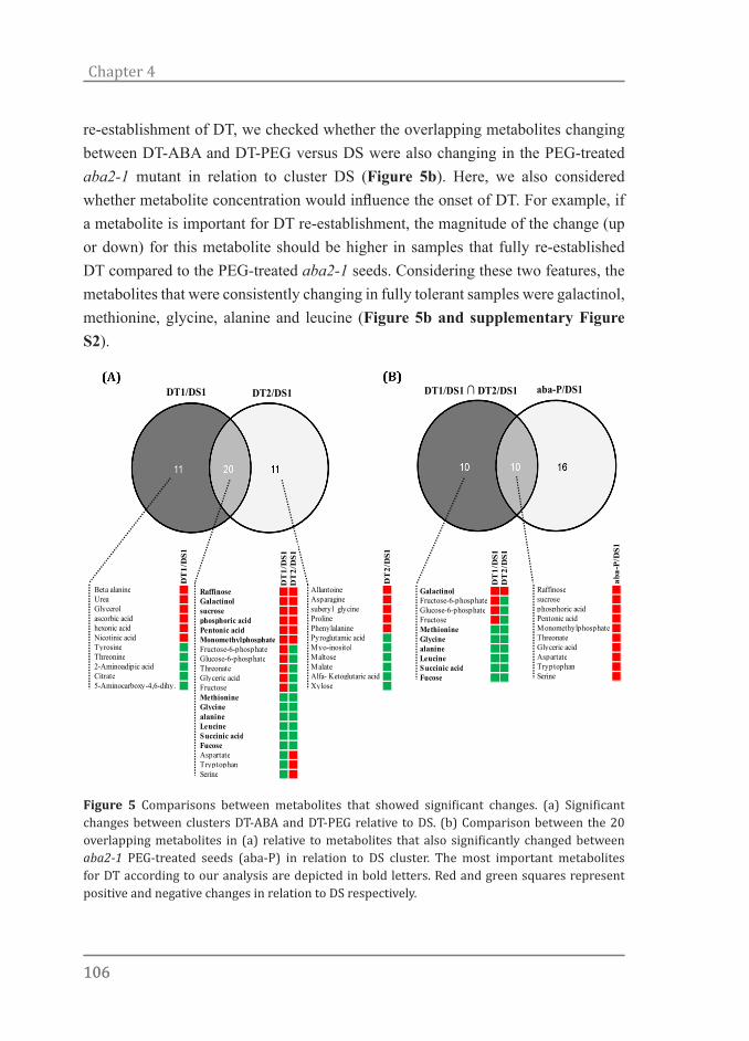
106106
Chapter 4
re-establishment of DT, we checked whether the overlapping metabolites changing between DT-ABA and DT-PEG versus DS were also changing in the PEG-treated aba2-1 mutant in relation to cluster DS (Figure 5b). Here, we also considered whether metabolite concentration would influence the onset of DT. For example, if a metabolite is important for DT re-establishment, the magnitude of the change (up or down) for this metabolite should be higher in samples that fully re-established DT compared to the PEG-treated aba2-1 seeds. Considering these two features, the metabolites that were consistently changing in fully tolerant samples were galactinol, methionine, glycine, alanine and leucine (Figure 5b and supplementary Figure S2).
DT1/DS1 DT2/DS1 DT1/DS1 ∩ DT2/DS1 aba-P/DS1
DT
1/D
S1D
T2/
DS1
RaffinoseGalactinolsucrosephosphoric acidPentonic acidMonomethylphosphateFructose-6-phosphateGlucose-6-phosphateThreonateGlyceric acidFructoseMethionineGlycinealanineLeucineSuccinic acidFucoseAspartateTryptophanSerine
DT
1/D
S1
Beta alanineUreaGlycerolascorbic acidhexonic acidNicotinic acidTyrosineThreonine2-Aminoadipic acidCitrate5-Aminocarboxy-4,6-dihy.
DT
2/D
S1
AllantoineAsparaginesuberyl glycineProlinePhenylalaninePyroglutamic acidMyo-inositolMaltoseMalateAlfa- Ketoglutaric acidXylose
DT
1/D
S1D
T2/
DS1
GalactinolFructose-6-phosphateGlucose-6-phosphateFructoseMethionineGlycinealanineLeucineSuccinic acidFucose
aba-
P/D
S1
Raffinosesucrosephosphoric acidPentonic acidMonomethylphosphateThreonateGlyceric acidAspartateTryptophanSerine
Figure 5 Comparisons between metabolites that showed significant changes. (a) Significant changes between clusters DT-ABA and DT-PEG relative to DS. (b) Comparison between the 20 overlapping metabolites in (a) relative to metabolites that also significantly changed between aba2-1 PEG-treated seeds (aba-P) in relation to DS cluster. The most important metabolites for DT according to our analysis are depicted in bold letters. Red and green squares represent positive and negative changes in relation to DS respectively.

107107
Desiccation tolerance metabolome
To further confirm our findings and to correlate them with other studies we plotted the most important DT-metabolites in combination with data obtained from a recently published study in which the transcriptome of Arabidopsis seeds, at the same developmental stage as used here, with re-established DT was compared to the transcriptome of desiccation sensitive seeds (Maia et al., 2011), Chapter 2). To visualize the overall differentially expressed genes and metabolites, we used the MapMan package (http://MapMan.gabipd.org). The results of this analysis are summarised in Figure 6. Galactinol and sucrose accumulated in DT re-established seeds and at the same time two galactinol synthase genes, GolS1 and GolS2, and a sucrose synthase gene, SUS3, were up-regulated (Figure 6). Although we detected raffinose accumulation in seeds with re-established DT, raffinose synthase genes were not differentially up-regulated in germinated Col-0 seeds after 3d in PEG.Raffinose metabolism
GolS1GolS2
SUS3
DIN10
ATBETAFRUCT4
PPCK1PFK2
IPGAM1ENO1
PK
Figure 6. MapMan molecular network map depicting the raffinose metabolic pathway. Log2 ratios are used to express relative transcript levels in germinated Arabidopsis seeds at stage II treated for 3 d in -2.5 MPa PEG versus non-treated seeds in the same developmental stage (Maia et al., 2011, Chapter 2) and metabolite levels in the DT-PEG/DS comparison. Differences in both transcript (coloured squares) and metabolite (coloured circles) levels are shown. Red and blue means higher and lower levels in DT seeds respectively. Only ratios with P values lower or equal to 0.05 are displayed.
108108
Chapter 4
Discussion
Our initial phenotyping demonstrated different magnitudes of DT recovery between the aba2-1 mutant and Col-0. It was also shown that without or with very low levels of ABA, PEG is not sufficient to fully re-establish DT (Chapter 3 and Figure 1). In line with other studies (Buitink et al., 2003), this finding suggests ABA-mediated signalling as an essential route to re-establish DT. Still, it is not clear which mechanisms are involved and how these cues are perceived and signalled by a seed or seedling. So far, two pathways have been suggested; an ABA-dependent and an ABA-independent pathway (Yamaguchi-Shinozaki & Shinozaki, 2006). Most frequently, however, experimental data points towards a model which is dependent on ABA level-directed responses (Raghavendra et al., 2010; Fujita et al., 2011; Frey et al., 2012; Miyakawa et al., 2013) where an osmotic cue (e.g. PEG) will lead to elevated levels of ABA. This increase in ABA content would then be required to activate signalling cascades which would ultimately lead to a stress response. With this in mind, a physiological system in which the effects of osmotic stress can be distinguished from what is activated by ABA was proposed. Essentially, the use of an ABA deficient mutant (aba2-1) which is not able to respond to PEG and re-establish DT (Figure 1), probably due to its very low ABA content(Léon-Kloosterziel et al., 1996), confirms our suggestion that ABA and not the osmotic environment rescued its desiccation tolerant phenotype (Figure 1). This enabled us to further discriminate between ABA-dependent and -independent mechanisms that might be mediating the re-establishment of DT. Given these facts, we focused on two main questions: (1) Is DT induced by ABA distinct from DT induced by PEG and (2) which are the metabolites that are essential for the re-establishment of DT?
Seeds with ABA- and PEG-induced DT display distinct metabolic signatures
It is clear that both PEG and ABA can re-induce DT in germinated wild type Arabidopsis seeds (Figure 1). However, once ABA biosynthesis is compromised, as in the aba2-1 mutant, the osmotic cue generated by PEG is no longer sufficient to trigger DT. Metabolite profiling was performed to separate the effects of PEG and ABA at the metabolite level. We thus generated a list of candidates important for DT, such as galactinol and raffinose. We observed a strong shift

109109
Desiccation tolerance metabolome
in the metabolites that changed among clusters DT-ABA and DT-PEG although both clusters represented seed samples with completely restored DT. Most of the changes in metabolite abundance induced by PEG or ABA were very distinct, suggesting the existence of alternative ways of expressing DT or independence of metabolic signature from signalling pathways to re-establishment of DT. PEG-treated samples (cluster DT-PEG) were always more different from the control (cluster DS) than ABA-treated samples (cluster DT-ABA). The first noticeable difference between DT-ABA and DT-PEG was a carbon and nitrogen (C/N) metabolic shift. PEG-treated seeds accumulated more N-compounds, mainly amino acids, than ABA-treated seeds but relatively less C-compounds, such as sugars and sugar alcohols. The osmotic stress imposed by PEG appears to influence the balance between the accumulation of amino acids and sugars, leading to a nitrogen rich metabolome (Figure 4). Both C-compounds, such as raffinose family oligosaccharides (RFOs) and sucrose, and N-compounds, such as amino acids, can act as osmolytes (Crowe et al., 1998; Yancey, 2001). Osmolytes are highly soluble organic compounds of low molecular weight that play a role in maintaining cell volume and water balance. Osmolyte accumulation has been reported as a general response of plants to salt, drought and osmotic stress (Yeo, 1998; Yancey, 2005). This response depends on the cellular accumulation of osmolytes, which help the plant to maintain osmotic balance under stress and/or act as osmoprotectants with chaperone or reactive oxygen species (ROS) scavenging activities (Yancey, 2005). However, it is not known why there is such an enormous variety of organic osmolytes, or why organisms employ complex osmolyte mixtures. A possible reason for this variability may be a result of unique properties of some osmolytes that may be helpful only with certain stresses under specific conditions (Yancey, 2001; Yancey, 2005). Thus, the reason why osmolytes vary among and within organisms is simply due to different metabolic shifts imposed by distinct stresses. For example, the widespread use of non-nitrogenous carbohydrates and sulfonium osmolytes in photosynthesizing organisms may arise from nitrogen limitation (Hockin et al., 2012). Coherent with this idea, in the case of germinated Arabidopsis seeds, is the nature of the signal to re-establish DT, namely PEG or ABA. This seems to be crucial in recruiting the relevant osmoprotectants. With our experimental approach we show

110110
Chapter 4
an emerging plasticity in the attainment of DT. Thus it appears that at least two different metabolic signatures can result in the expression of one single phenotype, namely desiccation tolerance, although it cannot be ruled out that a small number of specific metabolites are responsible for this phenotype.
Metabolites associated with DT
Metabolomic analysis revealed strong differences between germinated seeds after treatment with an osmoticum and ABA, as indicated by changes in the levels of primary metabolites, including amino acids, organic acids and sugars. Considering that germinated seeds treated with PEG, ABA and PEG + ABA were all able to re-establish DT, it was expected that comparison of the lists of metabolites that positively and negatively changed between clusters DT-ABA and DT-PEG relative to cluster DS would yield the core set of metabolic changes governing re-establishment of DT. Following this criterion, the metabolites that consistently over-accumulated upon re-establishment of DT are raffinose, galactinol, sucrose, phosphoric acid, and monomethyl phosphate. According to the same criterion, methionine, glycine, alanine, leucine, succinic acid and fucose were less abundant in seeds with re-established DT. When we included the aba2-1 PEG-treated seeds in our analysis we observed that, although those seeds were not able to fully re-establish DT, they still accumulated certain metabolites, such as raffinose, sucrose, phosphoric acid, and monomethyl phosphate, up to levels comparable to the ones observed for seeds in which DT was restored. This indicates that those metabolites might be important but not limiting to the process of re-establishment of DT. PEG and ABA displayed additive effects on galactinol accumulation in DT re-established seeds. On the other hand, raffinose accumulated equally in DT re-established and aba2-1 PEG-treated seeds. Consequently, if RFOs are indeed important for the re-establishment of DT, galactinol might be the limiting factor hampering DT re-establishment in the PEG-treated aba2-1 mutant seeds. Overlapping our results with the transcriptome data obtained from a similar experiment where Arabidopsis seeds at the same developmental stage as used here, were treated with PEG to re-establish DT (Maia et al., 2011), revealed interesting links between metabolites and transcripts(Figure 6). Genes such as SUS3, GolS1 and GolS2 were up-regulated concomitantly with the accumulation of sucrose, galactinol,

111111
Desiccation tolerance metabolome
and raffinose. SUS3 encodes a protein with sucrose synthase activity that has been shown to be important for sucrose metabolism in developing seeds, especially during the late maturation phase when DT is also acquired (Angeles-Núñez & Tiessen, 2012). Although we detected raffinose accumulation, raffinose synthase genes were not differentially up-regulated in DT re-established seeds after 3d in PEG. Only the DARK INDUCIBLE 10 (DIN10) gene, which encodes a glycosyl hydrolase, was found to be down-regulated in DT seeds. DIN10 has been shown to have its expression suppressed when sucrose is supplied (Fujiki et al., 2001). Thus, the high levels of sucrose found here might explain why this gene was down-regulated in DT re-established seeds. Together, these observations provide direct links between metabolite abundance and gene expression. They also reinforce the importance of certain sugars and sugar alcohols including sucrose and galactinol, respectively, in the re-establishment of DT. Galactinol, raffinose, and sucrose are involved in the biosynthesis of RFOs, such as stachyose and verbascose (Loewus & Murthy, 2000; Peterbauer et al., 2002). Functionally, RFOs are used for carbon transport and storage by the plant, although they have also been reported as protective agents during maturation drying of seeds (Peterbauer & Richter, 2001; Downie et al., 2003) and responding to a wide range of stresses (Gupta & Kaur, 2005). RFOs have been traditionally associated with DT in seeds (Angelovici et al., 2010), but also in resurrection plants (Peters et al., 2007; Lehner et al., 2008), mosses (Gechev et al., 2012) and ferns (Farrant et al., 2009). These sugars are suggested to protect cellular integrity during desiccation via several distinct mechanisms. RFOs have been suggested to act as osmoprotectants, to stabilize membranes and as substrates for the necessary generation of energy to support active processes during acquisition of DT (Angelovici et al., 2010). In line with our findings, RFOs have been shown to accumulate to relatively high levels during the acquisition of DT in maturing Arabidopsis seeds (Taji et al., 2002). Furthermore, increased intracellular levels of galactinol and raffinose in transgenic Arabidopsis plants correlated with increased tolerance to salt, chilling and drought stress (Taji et al., 2002; Nishizawa et al., 2008). In addition, galactinol and raffinose effectively protected salicylate from attack by hydroxyl radicals in vitro, suggesting the possibility that those sugars might also act as hydroxyl radical scavengers to protect plant cells from oxidative damage (Nishizawa et al., 2008). Finally, RFOs are also believed to contribute to the structural stability of organelles, membranes,

112112
Chapter 4
enzymes and proteins, other macromolecules, and the glassy state (Koster, 1991; Crowe et al., 1992; Horbowicz & Obendorf, 1994). The formation of a stable glassy cytoplasm is an important mechanism that allows organisms to withstand extreme drying via protecting macromolecules but also ensuring cell integrity during drying. Besides the accumulation of certain sugars we also observed consistently low concentrations of glycine, alanine and leucine in seeds with re-established DT. These amino acids are all aliphatic and share the property of being hydrophobic. Considering and knowing that important DT-associated proteins, such as LEA proteins are highly hydrophilic (Wise, 2004; Hundertmark & Hincha, 2008), we speculate that a decrease in the contents of hydrophobic amino acids is required to enhance cell hydrophilicity. Significant DT-specific accumulation of hydrophilic amino acid residues was not found, maybe due to their recruitment to hydrophilic proteins. Accordingly, genes encoding for highly hydrophilic LEA proteins were found amongst the most up-regulated in DT re-established Arabidopsis seeds after 3d of PEG incubation (Maia et al., 2011), Chapter 2). In summary, the accumulation of raffinose, galactinol, and sucrose indicates that those sugars have a role in the restoration of DT in germinated Arabidopsis seeds. Considering their attributed functions, our results support the general consent that mechanisms such as ROS scavenging, cytoplasm vitrification and sugar signalling in concert with other protection mechanisms are crucial for the expression of DT. The system we used to investigate DT entirely excludes possible developmentally controlled features that could be coincidental to acquisition of DT. Thus, our findings indicate that, to the contrary of many reports (Hoekstra et al., 1994; Ooms et al., 1994; Black et al., 1999), there is indeed a strong correlation between the accumulation of raffinose and acquisition of DT.
Acknowledgements
We thank our colleagues of the Wageningen Seed Lab and the Laboratory of Plant Physiology. This work was supported by the “Coordenação de Aperfeiçoamento de Pessoal de Nível Superior” (CAPES, Brazil)(JM).
113113
Desiccation tolerance metabolome
References
Angeles-Núñez J, Tiessen A. 2012. Regulation of AtSUS2 and AtSUS3 by glucose and the transcription factor LEC2 in different tissues and at different stages of Arabidopsis seed development. 78(4-5): 377-392.
Angelovici R, Galili G, Fernie AR, Fait A. 2010. Seed desiccation: a bridge between maturation and germination. Trends in Plant Science 15(4): 211-218.
Bewley JD, Bradford K, Hilhorst H, Nonogaki H. 2013. Seeds: Physiology of Development, Germination and Dormancy: Springer.
Black M, Corbineau F, Gee H, Come D. 1999. Water content, raffinose, and dehydrins in the induction of desiccation tolerance in immature wheat embryos. Plant physiology 120(2): 463-472.
Bruggink T, van der Toorn P. 1995. Induction of desiccation tolerance in germinated seeds. Seed Science Research 5(1): 1-4.
Buitink J, Leger JJ, Guisle I, Vu BL, Wuillème S, Lamirault G, Bars AL, Meur NL, Becker A, Küster H, Leprince O. 2006. Transcriptome profiling uncovers metabolic and regulatory processes occurring during the transition from desiccation-sensitive to desiccation-tolerant stages in Medicago truncatula seeds. The Plant Journal 47(5): 735-750.
Buitink J, Vu BL, Satour P, Leprince O. 2003. The re-establishment of desiccation tolerance in germinated radicles of Medicago truncatula Gaertn. seeds. Seed Science Research 13(4): 273-286.
Carreno-Quintero N, Acharjee A, Maliepaard C, Bachem CWB, Mumm R, Bouwmeester H, Visser RGF, Keurentjes JJB. 2012. Untargeted Metabolic Quantitative Trait Loci Analyses Reveal a Relationship between Primary Metabolism and Potato Tuber Quality. Plant physiology 158(3): 1306-1318.
Chandra Babu R, Zhang J, Blum A, David Ho TH, Wu R, Nguyen HT. 2004. HVA1, a LEA gene from barley confers dehydration tolerance in transgenic rice (Oryza sativa L.) via cell membrane protection. Plant Science 166(4): 855-862.
Crowe JH, Carpenter JF, Crowe LM. 1998. The role of vitrification in anhydrobiosis. Annual Review of Physiology 60(1): 73-103.
Crowe JH, Hoekstra FA, Crowe LM. 1992. Anhydrobiosis. Annual Review of Physiology 54(1): 579-599.
Downie B, Gurusinghe S, Dahal P, Thacker RR, Snyder JC, Nonogaki H, Yim K, Fukanaga K, Alvarado V, Bradford KJ. 2003. Expression of a GALACTINOL SYNTHASE gene in tomato seeds is up-regulated before maturation desiccation and again after imbibition whenever radicle protrusion is prevented 1. Plant Physiology 131(3): 1347-1359.
Farrant JM, Lehner A, Cooper K, Wiswedel S. 2009. Desiccation tolerance in the vegetative tissues of the fern Mohria caffrorum is seasonally regulated. The Plant Journal 57(1): 65-79.
Frey A, Effroy D, Lefebvre V, Seo M, Perreau F, Berger A, Sechet J, To A, North HM, Marion-Poll A. 2012. Epoxycarotenoid cleavage by NCED5 fine-tunes ABA accumulation and affects seed dormancy and drought tolerance with other NCED family members. The Plant Journal 70(3): 501-512.
114114
Chapter 4
Fujiki Y, Yoshikawa Y, Sato T, Inada N, Ito M, Nishida I, Watanabe A. 2001. Dark-inducible genes from Arabidopsis thaliana are associated with leaf senescence and repressed by sugars. Physiologia Plantarum 111(3): 345-352.
Fujita Y, Fujita M, Shinozaki K, Yamaguchi-Shinozaki K. 2011. ABA-mediated transcriptional regulation in response to osmotic stress in plants. Journal of Plant Research 124(4): 509-525.
Gechev TS, Dinakar C, Benina M, Toneva V, Bartels D. 2012. Molecular mechanisms of desiccation tolerance in resurrection plants. Cellular and Molecular Life Sciences 69(19): 3175-3186.
Gupta AK, Kaur N. 2005. Sugar signalling and gene expression in relation to carbohydrate metabolism under abiotic stresses in plants. Journal of biosciences 30(5): 761-776.
Hockin NL, Mock T, Mulholland F, Kopriva S, Malin G. 2012. The response of diatom central carbon metabolism to nitrogen starvation is different from that of green algae and higher plants. Plant physiology 158(1): 299-312.
Hoekstra FA, Haigh AM, Tetteroo FAA, van Roekel T. 1994. Changes in soluble sugars in relation to desiccation tolerance in cauliflower seeds. Seed Science Research 4: 143.
Horbowicz M, Obendorf RL. 1994. Seed desiccation tolerance and storability: Dependence on flatulence-producing oligosaccharides and cyclitols—review and survey. Seed Science Research 4(04): 385-405.
Hundertmark M, Hincha D. 2008. LEA (late embryogenesis abundant) proteins and their encoding genes in Arabidopsis thaliana. BMC Genomics 9(1): 118.
Kempa S, Krasensky J, Dal Santo S, Kopka J, Jonak C. 2008. A central role of abscisic acid in stress-regulated carbohydrate metabolism. PLoS ONE 3(12).
Komatsu K, Suzuki N, Kuwamura M, Nishikawa Y, Nakatani M, Ohtawa H, Takezawa D, Seki M, Tanaka M, Taji T, Hayashi T, Sakata Y. 2013. Group A PP2Cs evolved in land plants as key regulators of intrinsic desiccation tolerance. Nature Communications 4.
Koster KL. 1991. Glass formation and desiccation tolerance in seeds. Plant physiology 96(1): 302-304.
Lehner A, Chopera DR, Peters SW, Keller F, Mundree SG, Thomson JA, Farrant JM. 2008. Protection mechanisms in the resurrection plant Xerophyta viscosa: Cloning, expression, characterisation and role of XvINO1, a gene coding for a myo-inositol 1-phosphate synthase. Functional Plant Biology 35(1): 26-39.
Léon-Kloosterziel KM, Gil MA, Ruijs GJ, Jacobsen SE, Olszewski NE, Schwartz SH, Zeevaart JAD, Koornneef M. 1996. Isolation and characterization of abscisic acid-deficient Arabidopsis mutants at two new loci. Plant Journal 10(4): 655-661.
Leopold AC, Vertucci CW 1986. Membranes, metabolism and dry organisms. In: Leopold AC ed. Physical attributes of desiccated seeds. London: Comstock Publishing, 23-34.
Loewus FA, Murthy PPN. 2000. myo-Inositol metabolism in plants. Plant Science 150(1): 1-19.
Lommen A. 2009. MetAlign: interface-driven, versatile metabolomics tool for hyphenated full-scan mass spectrometry data preprocessing. Analytical Chemistry 81(8): 3079-3086.

115115
Desiccation tolerance metabolome
Maia J, Dekkers BJW, Provart NJ, Ligterink W, Hilhorst HWM. 2011. The re-establishment of desiccation tolerance in germinated Arabidopsis thaliana seeds and its associated transcriptome. PLoS ONE 6(12).
Miyakawa T, Fujita Y, Yamaguchi-Shinozaki K, Tanokura M. 2013. Structure and function of abscisic acid receptors. Trends in Plant Science 18(5): 259-266.
Moore JP, Le NT, Brandt WF, Driouich A, Farrant JM. 2009. Towards a systems-based understanding of plant desiccation tolerance. Trends Plant Sci 14(2): 110-117.
Nishizawa A, Yabuta Y, Shigeoka S. 2008. Galactinol and raffinose constitute a novel function to protect plants from oxidative damage. Plant physiology 147(3): 1251-1263.
Oliver MJ, Guo L, Alexander DC, Ryals JA, Wone BWM, Cushman JC. 2011. A sister group contrast using untargeted global metabolomic analysis delineates the biochemical regulation underlying desiccation tolerance in Sporobolus stapfianus. The Plant Cell Online 23(4): 1231-1248.
Ooms JJJ, Léon-Kloosterziel KM, Bartels D, Koornneef M, Karssen CM. 1993. Acquisition of desiccation tolerance and longevity in seeds of Arabidopsis thaliana: A comparative study using abscisic acid-insensitive abi3 mutants. Plant physiology 102(4): 1185-1191.
Ooms JJJ, Wilnier JA, Karssen CM. 1994. Carbohydrates are not the sole factor determining desiccation tolerance in seeds of Arabidopsis thaliana. Physiologia Plantarum 90(3): 431-436.
Peterbauer T, Mucha J, Mach L, Richter A. 2002. Chain elongation of raffinose in pea seeds. Isolation, characterization, and molecular cloning of a multifunctional enzyme catalyzing the synthesis of stachyose and verbascose. Journal of Biological Chemistry 277(1): 194-200.
Peterbauer T, Richter A. 2001. Biochemistry and physiology of raffinose family oligosaccharides and galactosyl cyclitols in seeds. Seed Science Research 11(3): 185-197.
Peters S, Mundree SG, Thomson Ja, Farrant JM, Keller F. 2007. Protection mechanisms in the resurrection plant Xerophyta viscosa (Baker): both sucrose and raffinose family oligosaccharides (RFOs) accumulate in leaves in response to water deficit. Journal of Experimental Botany 58(8): 1947-1956.
Raghavendra AS, Gonugunta VK, Christmann A, Grill E. 2010. ABA perception and signalling. Trends in Plant Science 15(7): 395-401.
Roessner U, Wagner C, Kopka J, Trethewey RN, Willmitzer L. 2000. Simultaneous analysis of metabolites in potato tuber by gas chromatography–mass spectrometry. The Plant Journal 23(1): 131-142.
Strehmel N, Hummel J, Erban A, Strassburg K, Kopka J. 2008. Retention index thresholds for compound matching in GC–MS metabolite profiling. Journal of Chromatography B 871(2): 182-190.
Taji T, Ohsumi C, Iuchi S, Seki M, Kasuga M, Kobayashi M, Yamaguchi-Shinozaki K, Shinozaki K. 2002. Important roles of drought- and cold-inducible genes for galactinol synthase in stress tolerance in Arabidopsis thaliana. The Plant journal 29(4): 417-426.
Terrasson E, Buitink J, Righetti K, Ly Vu B, Pelletier S, Lalanne D, Zinsmeister
116116
Chapter 4
J, Leprince O. 2013. An emerging picture of the seed desiccome: confirmed regulators and newcomers identified using transcriptome comparison. Frontiers in Plant Science 4.
Tikunov YM, Laptenok S, Hall RD, Bovy A, Vos RCH. 2012. MSClust: a tool for unsupervised mass spectra extraction of chromatography-mass spectrometry ion-wise aligned data. 8(4): 714-718.
Tunnacliffe A, Wise MJ. 2007. The continuing conundrum of the LEA proteins. Die Naturwissenschaften 94(10): 791-812.
Umezawa T, Nakashima K, Miyakawa T, Kuromori T, Tanokura M, Shinozaki K, Yamaguchi-Shinozaki K. 2010. Molecular basis of the core regulatory network in ABA responses: sensing, signaling and transport. Plant cell physiology 51(11): 1821-1839.
Urano K, Maruyama K, Ogata Y, Morishita Y, Takeda M, Sakurai N, Suzuki H, Saito K, Shibata D, Kobayashi M, Yamaguchi-Shinozaki K, Shinozaki K. 2009. Characterization of the ABA-regulated global responses to dehydration in Arabidopsis by metabolomics. Plant Journal 57(6): 1065-1078.
Wise M. 2004. POPP the question: what do LEA proteins do? Trends in Plant Science 9(1): 13-17.
Xia J, Mandal R, Sinelnikov IV, Broadhurst D, Wishart DS. 2012. MetaboAnalyst 2.0-a comprehensive server for metabolomic data analysis. Nucleic Acids Research 40: 1-7.
Xia J, Psychogios N, Young N, Wishart DS. 2009. MetaboAnalyst: A web server for metabolomic data analysis and interpretation. Nucleic Acids Research 37: W652-W660.
Yamaguchi-Shinozaki K, Shinozaki K 2006. Transcriptional regulatory networks in cellular responses and tolerance to dehydration and cold stresses. Annual Review of Plant Biology 57: 781-803.
Yancey PH. 2001. Water Stress, Osmolytes and Proteins. American Zoologist 41(4): 699-709.
Yancey PH. 2005. Organic osmolytes as compatible, metabolic and counteracting cytoprotectants in high osmolarity and other stresses. Journal of Experimental Biology 208(15): 2819-2830.
Yeo A. 1998. Molecular biology of salt tolerance in the context of whole-plant physiology. Journal of Experimental Botany 49(323): 915-929.
Yobi A, Wone BWM, Xu W, Alexander DC, Guo L, Ryals JA, Oliver MJ, Cushman JC. 2012. Comparative metabolic profiling between desiccation-sensitive and desiccation-tolerant species of Selaginella reveals insights into the resurrection trait. The Plant Journal 72(6): 983-999.
Yobi A, Wone BWM, Xu W, Alexander DC, Guo L, Ryals JA, Oliver MJ, Cushman JC. 2013. Metabolomic Profiling in Selaginella lepidophylla at Various Hydration States Provides New Insights into the Mechanistic Basis of Desiccation Tolerance. Molecular Plant 6(2): 369-385.
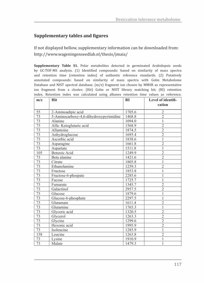
117117
Desiccation tolerance metabolome
Supplementary tables and figures
If not displayed bellow, supplementary information can be downloaded from: http://www.wageningenseedlab.nl/thesis/jmaia/
Supplementary Table S1. Polar metabolites detected in germinated Arabidopsis seeds by GC-TOF-MS analysis. (1) Identified compounds: based on similarity of mass spectra and retention time (retention index) of authentic reference standards. (2) Putatively annotated compounds: based on similarity of mass spectra with Golm Metabolome Database and NIST spectral database. (m/z) fragment ion chosen by MMSR as representative ion fragment from a cluster; (Hit) Golm or NIST library matching hit; (RI) retention index. Retention index was calculated using alkanes retention time values as reference.
m/z Hit RI Level of identifi-cation
55 2-Aminoadipic acid 1705.6 273 5-Aminocarboxy-4,6-dihydroxypyrimidine 1468.8 273 Alanine 1094.0 173 Alfa- Ketoglutaric acid 1568.9 273 Allantoine 1874.5 273 Anhydroglucose 1695.4 273 Ascorbic acid 1838.6 173 Asparagine 1661.8 273 Aspartate 1511.8 1105 Benzoic Acid 1249.9 273 Beta alanine 1421.6 273 Citrate 1805.8 173 Ethanolamine 1259.3 273 Fructose 1853.8 173 Fructose-6-phospate 2285.6 173 Fucose 1725.7 173 Fumarate 1345.7 273 Galactinol 2957.5 273 Glucose 1879.6 173 Glucose-6-phosphate 2297.5 173 Glutamate 1611.4 273 Glutamine 1765.3 173 Glyceric acid 1320.5 273 Glycerol 1263.3 273 Glycine 1299.6 273 Hexonic acid 1995.9 273 Isoleucine 1285.9 1158 Leucine 1263.8 273 Lysine 1910.9 173 Malate 1479.3 1

118118
Chapter 4
73 Maltose 2710.1 1128 Methionine 1514.3 1241 Monomethylphosphate 1169.1 273 Myo-inositol 2075.2 2180 Nicotinic acid 1295.7 273 Pentonic acid 1737.9 273 Phenylalanine 1622.9 2299 phosphoric acid 1262.4 273 Proline 1292.8 173 Pyroglutamic acid 1519.0 273 Raffinose 3447.1 273 Serine 1351.1 173 Sinapic acid 2241.0 273 Suberyl-glycine 1616.0 2147 Succinic acid 1308.7 273 Sucrose 2614.0 173 Threonate 1547.3 273 Threonine 1376.5 273 Tryptophan 2211.3 173 Tyrosine 1930.2 2147 Urea 1234.7 173 Valine 1207.8 273 Xylofuranose 1755.0 273 Xylose 1640.9 1
DT-ABA/DS DT-PEG/DS DT-ABA/DT-PEG(a) (b) (c)
Supplementary Figure S1. Important features selected by volcano plot with fold change threshold set to (x) 1 and t-tests threshold (y) 0.05 between different clusters (DT-ABA/DS, DT-PEG/DS and DT-ABA/DT-PEG – Figure 1). The black circles represent features above the threshold. P values are log transformed. The most significant metabolites are assigned in the charts.

119119
Desiccation tolerance metabolome
0
20
40
60
80
100
120
140Galactinol
0
500
1000
1500
2000Raffinose
01000020000300004000050000600007000080000
sucrose
0
200
400
600
800
1000phosphoric acid
020406080
100120140160
Pentonic acid
0
10
20
30
40
50
60Monomethylphosphate
0
20
40
60
80
100Fructose-6-phospate
0
50
100
150
200Glucose-6-phosphate
0
50
100
150
200Threonate
0
50
100
150
200
250
300
350Glyceric acid
0
2000
4000
6000
8000
10000
12000
14000Fructose
0
10
20
30
40
50
60Methionine
050
100150200250300350400
Glycine
0
500
1000
1500
2000alanine
050
100150200250300350400
Leucine
0
50
100
150
200
250
300Succinic acid
0
5
10
15
20
25Fucose
0500
1000150020002500300035004000
Aspartate
050
100150200250300350400
Tryptophan
0
500
1000
1500
2000
2500
3000
3500Serine
Supplementary Figure S2. Average relative metabolite contents in the DS (dark grey), DT-PEG (light grey), DT-ABA (white) and aba2-1 PEG-treated (black) samples. Error bars indicate standard error.
Memória
Amar o perdidodeixa confundidoeste coração.
Nada pode o olvidocontra o sem sentidoapelo do Não.
As coisas tangíveistornam-se insensíveisà palma da mão
Mas as coisas findasmuito mais que lindas,essas ficarão.
Carlos Drummond de Andrade

Chapter 5Proteomic changes associated with the re-establishment of desiccation tolerance in germinated Arabidopsis seeds
Authors: Julio Maia1, Loïc Rajjou, Gwendel Kueff, Wilco Ligterink1, Henk W. M. Hilhorst1
1 Wageningen Seed Lab, Laboratory of Plant Physiology, Wageningen University, Wageningen, The Netherlands 2Institut Jean-Pierre Bourgin, INRA AgroParisTech, Versailles, France
(in preparation for submission)

122122
Chapter 5
Abstract
Desiccation tolerant organisms are capable of surviving almost total loss of cellular water, the dry state and regain metabolic activity after re-hydration without accumulating lethal damage. To establish desiccation tolerance (DT) a myriad of molecules are needed among which proteins with protective roles. Here we used the capacity of germinating Arabidopsis seeds to lose and regain DT as a model system to explore the DT associated proteome. Arabidopsis Col-0 and aba2-1 germinated seeds submitted to three different treatments which induce DT to significantly different levels, allowed us to identify 97 protein spots strongly correlated to DT. The proteins identified from these spots are mainly participating in abiotic stress and abscisic acid (ABA) responses, embryo development and sugar metabolism. A promoter analysis revealed that all genes coding for these proteins contain ABA-responsive elements (ABREs) indicating a central role for ABA in their regulation. Late embryogenesis abundant (LEA) proteins, especially of the LEA_4 family, were markedly enriched in seeds with re-established DT, which is suggestive of a specific role of this LEA family in DT in Arabidopsis. Interestingly, the identified LEA_4 proteins are significantly enriched with charged and polar amino acids and have a relatively low hydropathy (GRAVY) index in comparison to other members of this family. We suggest that negatively charged LEA_4 proteins might be acting together with sugars to stabilize bioglasses and on their own via steric and electrostatic prevention of interactions between aggregation-prone and partially unfolded proteins.
Introduction
Orthodox seeds, as opposed to recalcitrant, acquire desiccation tolerance (DT) during their development on the mother plant after which they can cope with an anhydrobiotic lifestyle. Because of this ability, orthodox seeds can be stored under cold and dry conditions for a few to thousands of years, depending on the species, with minor viability losses (Farnsworth, 2000; Berjak and Pammenter, 2008; Li and Pritchard, 2009). As maturing orthodox seeds begin to lose water, embryos are subjected to a range of relatively complex biochemical

123123
Desiccation tolerance proteome
changes, varying from the accumulation of late embryogenesis abundant (LEA) proteins, dehydrins, sugars and scavengers of reactive oxygen species (ROS), to accumulation of food reserves and a programmed switching off of metabolism (Berjak, 2006). Most, if not all, of these events are regulated by abscisic acid (ABA). In fact, numerous genes that are coding for proteins involved in desiccation tolerance, such as those coding for LEA proteins, antioxidants, and heat shock proteins, are known to be triggered both by ABA and water stress, which is suggestive of a cross-talk between ABA signalling pathways and those induced by dehydration. In seeds DT is a multigenic trait that is tightly linked to genetic programs expressed during embryo development (e.g. reserve accumulation) (Le et al., 2007; Le et al., 2010; Verdier et al., 2013). The overlap between DT and embryo development makes it difficult to disentangle these two fundamental processes and poses a challenge to determine unambiguously the molecules involved in DT. In plants, DT has been investigated in developing and germinating seeds (Leprince et al., 1993; Ooms et al., 1993; Daws et al., 2007), resurrection plants (Illing et al., 2005; Farrant & Moore, 2011), mosses (Proctor et al., 2007; Wood, 2007), ferns (Farrant et al., 2009), and pollen (Franchi et al., 2011), but also in a more elaborate system in which DT, after being lost in germinating seeds, is fully re-established by the application of a mild osmotic stress (Bruggink & van der Toorn, 1995; Buitink et al., 2003). Studying the re-establishment of DT may enhance the chance of identifying true DT mechanisms, as parallel developmental components are eliminated. This system has been recently implemented in the model species Arabidopsis thaliana, which has provided new prospects to explore its benefits in combination with genetic tools available for this species, such as mutant collections and extensive sequence information (Maia et al., 2011). To date re-establishment of DT in germinated seeds has been used to study the transcriptome and heat stable proteome associated with desiccation stress (Boudet et al., 2006; Buitink et al., 2006; Maia et al., 2011) and it is now clear that this trait is controlled by a multitude of genes and proteins. Thus, approaches combining untargeted assessments of these molecules should be informative to better understand the regulation and mechanisms of DT. In this context, proteomic profiling holds promise for exploring such a complex trait. Proteins, due to their functional properties, might provide more

124124
Chapter 5
direct links between gene function and particular phenotypes. Different from transcriptomic studies, two dimensional (2D) proteomic profiling will not offer a genome wide view of the proteome; however, it will certainly provide solid links between perturbations (e.g. osmotic stress), distinct proteins and traits of interest. Many proteins have been associated with DT but so far only a few have been proven crucial. Among those, LEA proteins are the most frequently mentioned. LEA proteins accumulate concurrently with the acquisition of DT in seeds and other anhydrobiotes (Illing et al., 2005; Hand et al., 2011) and their expression has been linked to various stresses such as heat, cold and drought. ABA application has also been demonstrated to trigger the accumulation of these proteins (Galau et al., 1986; Espelund et al., 1992; Finkelstein & Lynch, 2000), which is complemented by the fact that many genes coding for LEA proteins display ABA-responsive elements in their promoter regions (Battaglia et al., 2008). LEA proteins are highly conserved among a wide variety of species. They are usually rich in glycine, and low in hydrophobic residues, which make them extremely hydrophilic. They are often intrinsically disordered; however, some LEA proteins partially fold, mainly into α-helices, during drying or in the presence of membranes (Goyal et al., 2003; Hincha & Thalhammer, 2012). Due to these physico-chemical characteristics, they were hypothesized to function specifically in the protection of membranes and proteins against desiccation damage by binding water tightly or providing hydrophilic interactions in the absence of free water and to prevent crystallization of cellular components, through their ability to act as stabilizing solvents (Tunnacliffe & Wise, 2007; Shih et al., 2008; Hincha & Thalhammer, 2012). LEA proteins are also believed to act together with sugars, particularly non-reducing disaccharides, on the formation and stabilization of bioglasses, which are presumably required in the dry state (Blackman et al., 1992; Buitink & Leprince, 2004). Furthermore, LEA proteins are thought to have other protective functions against desiccation, including ion binding and antioxidant activity (Tunnacliffe & Wise, 2007; Battaglia et al., 2008; Amara et al., 2012). To date, 51 LEA proteins have been identified in Arabidopsis (Bies-Ethève et al., 2008; Hundertmark & Hincha, 2008). They are divided in nine groups based on

125125
Desiccation tolerance proteome
their amino acid sequence and presence of conserved motifs (Bies-Ethève et al., 2008; Hundertmark & Hincha, 2008). Evidence of an in vivo role of some of these proteins in seed desiccation was demonstrated by Manfre et al. (2009) who showed that Arabidopsis seeds from the em6-1 mutant lost more water during air drying than wild-type seeds, which is consistent with the role of ATEM6 (LEA_5 family) in water binding/loss during embryo maturation. Although LEA proteins are normally associated with abiotic stress tolerance, especially dehydration and cold, their actual function and mechanisms of action remain largely unknown. LEA proteins are very diverse and there is more and more evidence indicating that LEA proteins from different families have protective roles under different stresses (Amara et al., 2012). However, this remains to be determined for the vast majority of them. Here, we show that the use of a strong physiological system in combination with the utilization of mutants and proteomics can be a powerful approach to pinpoint specific mechanisms related to a trait of interest, in our case DT. Overall, the proteome found to be related to the re-establishment of DT in germinated Arabidopsis seeds is mainly enriched in proteins participating in stress, embryo development, ABA responses and sugar metabolism. All genes coding for the proteins found in our study contain a core sequence (ACGTG) in their promoter known as the ABA-responsive element (ABRE) indicating this hormone as the most central signalling source regulating DT-related genes. LEA proteins, especially of the LEA_4 family, were markedly enriched in seeds with re-established DT, suggesting a specific relationship between this family of LEA proteins and DT in Arabidopsis. The LEA_4 proteins found here are significantly enriched with charged and polar amino acids and we speculate that they might be acting together with sugars in the stabilization of bioglasses, but also on their own via steric and electrostatic prevention of interactions between aggregation-prone and partially unfolded proteins.
Materials and methods
Plant materials and growth conditions
Arabidopsis thaliana (L.) Heynh. lines Col-0 and aba2-1 (Léon-Kloosterziel et al., 1996) were used. In all experiments seeds were cold stratified for 72h at

126126
Chapter 5
4°C in 9-cm Petri dishes on two layers of blue filter paper (Anchor Paper) and 10 ml of distilled water to remove residual dormancy. Germination assays were performed in a germination cabinet under constant white light at 22ºC.
Re-establishment of desiccation tolerance
The ability to re-establish DT under different treatments was tested in germinated Col-0 and aba2-1 seeds. Germinated seeds with radicles protruded by 1mm (stage II in (Maia et al., 2011) were selected with the aid of a stereomicroscope and either dried directly or after three days of incubation in PEG at -2.5 MPa, 5µM ABA or a combination of -2.5 MPa PEG + 1µM ABA. Treated seeds were rinsed, dehydrated for three days at 20°C and 32% relative humidity (RH), pre-humidified and scored for survival of cotyledons, primary roots and viable seedlings as described before (Maia et al., 2011).
Total soluble protein extraction
30 mg of dry seeds were ground to a powder with mortar and pestle in liquid nitrogen. Extraction buffer and protease inhibitor, as previously described by Rajjou et al. (2008), were added to the seed powder, followed by two-minutes of grinding. The extract was transferred to a 1.5 ml Eppendorf tube and incubated with DNase I, RNase A, and DTT at 4oC for 1h on a rotating disc. The total soluble protein extract was collected as supernatant after centrifugation at 14000 rpm at 4oC for 20 min.
2D gel electrophoresis
Protein separation was performed with 20 µl of protein extract, equivalent to about 150 µg of total protein. 2D gel electrophoresis was conducted as described before (Rajjou et al., 2008; Rajjou et al., 2011), adapted for gel strips forming an immobilized nonlinear pH gradient from 3 to 11 (Immobilized DryStrip pH 3-11 NL, 24 cm; GE Healthcare). For each analysed sample, 2D gels were made with at least three biological replicates. Protein staining and Gel analyses
2D gels were stained with silver nitrate according to Rajjou et al. (2008).

127127
Desiccation tolerance proteome
Silver-stained gels were placed between two layers of cellophane membrane stretched on cassette frames for drying. Images of dry gels were scanned with a Sharp JX-330 scanner (Arc et al., 2012). Quantitative image analysis was carried out with Progenesis Samespot software (v3.2, NonLinear Dynamics) to detect kinetics of protein accumulation and protein comparison of different samples. The SameSpot software was used to perform a one-way ANOVA for each spot. Spot abundance changes with fold change higher than 1.5 (up or down) and a p-value ≤ 0.05 in the comparison between all the established groups of biological replicates were retrieved. Protein identification
Protein spots of interest were isolated from the 2D gel, digested with trypsin and sequenced by LC-MS/MS as described before (Arc et al., 2012). Obtained sequences were compared to the TAIR10 protein database using XTandem and XTandem Pipeline (http://pappso.inra.fr/bioinfo/xtandempipeline/).
Results
Design of the experimental system
It has been demonstrated that DT can be fully rescued in germinated Arabidopsis seeds by the application of an osmotic treatment (PEG) before drying (Maia et al., 2011). Furthermore, with the aid of some ABA-insensitive and one ABA-deficient mutant we have shown that ABA is necessary for the re-establishment of DT (Chapters 2 and 3). We also revealed that ABA alone is sufficient to rescue DT in Col-0 and aba2-1 seeds and that aba2-1 seeds treated only with PEG fail to re-establish DT. Based on these results, we defined 4 protein expression profiles whereby we actively searched for protein spots significantly changing in their abundance (Figure 1a,b). In total we found 97 protein spots displaying significant changes (Figure 1a and Figure 2). Interestingly, most of the proteins up-regulated in the seeds with re-established DT were spotted in the upper left corner of the 2D SDS–PAGE gels while the down regulated proteins were just a few with smaller molecular weights (Figure 2). This observation provides clues for upcoming proteomic studies on DT where pH ranges that better resolve those regions in

128128
Chapter 5
the gel should be tested. The protein spots found following the profiles depicted in figure 1a were grouped into 2 main categories: DT up- (1) and DT down-regulated (2) protein spots (Figure 1). After spot selection, principal component analysis (PCA) was performed on 2 distinct groups of protein spots; on the 52 protein spots corresponding to type I (both over and under accumulated) (Figure 1a) and on the 45 protein spots corresponding to type II (Figure 1a).
Untreated PEG ABA PEG+ABA
DT DT DTDS
DS DS DTDT
Col-0
aba2-1
Pro�ile Type I
No. of spots (I)
No. of spots (II)
Total No. of spots
Pro�ile features
36
16
34
11
70
27
Desiccation tolerance related over accumulated proteins: High levels in DT re-established samples in relation to untreated DS samples and the aba2-1 PEG-treated sample
Desiccation tolerance related under accumulated proteins: low levels in DT re-established samples in relation to untreated DS samples and the aba2-1 PEG-treated sample
DT-up
DT-down
Pro�ile Type II
DS DT
DT
(a)
(b)
Spot 1124 - cupin family protein
Spot 0887 - late embryogenesis abundant domain-containing protein
DS
Fig 1. (a) Synopsis of the selected expression profiles of proteins of Col-0 and aba2-1 germinated Arabidopsis seeds after different treatments to re-induce desiccation tolerance. The bar diagrams schematize changes in spot abundance. For each feature of interest, two profiles were proposed a stringent (type I) and a general (type II). General profiles (type II) were generated via combining more gel images into the same category in order to enhance statistical power and increase the number of significant protein spot abundance differences between DS and DT samples. Samples are named after their genotype (Col for Col-0 and aba for aba2-1 seeds) followed by the treatment (germinated Arabidopsis seeds at the point of radical protrusion untreated (F) and treated with PEG (P), ABA (A) and PEG + ABA (PA)). In type II profiles all gel images originated from untreated and the aba2-1 PEG treated seeds (DS) were grouped as where the gel images of treated seeds that lead to a DT phenotype (DT). (b) Example of two protein spots belonging to DT-up type I (spot 0887) and II (spot 1124).

129129
Desiccation tolerance proteome
The PCA score plots of the analysed protein spot data showed tight grouping of biological replicates indicating good data quality (Figure 3). Overall, the grouping observed for the proteins selected based on profiles type I was genotype independent (Figure 3a). Samples submitted to similar DT re-establishing treatments (ABA or PEG + ABA), as well as untreated samples, clustered together (Figure 3a). Col-0 and aba2-1 samples treated with PEG, Col-P and aba-P respectively (Figure 3a), did not fall into the same cluster probably due to the inability of aba2-1 PEG-treated seeds to re-establish DT. This indicates that aba2-1 PEG-treated samples do not accumulate or have reduced
(a) (b)
(c) (d)
Figure 2. Reference maps of the proteome of germinated Arabidopsis seeds. Maps depict proteins selected according to the profiles 1a (a), 1b (b), 2a (c) and 2b (d). A 150 μg aliquot of the proteins was separated by 2D SDS–PAGE using 24 cm non-linear immobilized pH gradient strips (3–10).

130130
Chapter 5
levels of proteins essential for DT. When we grouped the samples according to their phenotype after re-establishment of DT, protein profiles type II, two big clusters were observed, one containing the DT- and another the DS samples (Figure 3b).
Functional classification of the identified protein sets
All selected spots (Figure 2) were isolated and their peptides were identified and proteins inferred. We identified 116 unique protein coding genes (Supplementary Tables S1 and S2). Overall, the DT-up protein set was enriched with many LEA proteins, some heat shock proteins (HSP) and proteins
aba-ACol-ACol-PAaba-PA
Col-Faba-Faba-P
(a)
Col-P
Col-P/Col-A/Col-PA/aba-A/aba-PACol-F/aba-F/aba-P
(b)
Figure 3 Principal component analysis (PCA) plots depicting different groupings according to the over and under accumulated set of proteins of type I (a) and type II (b) (Figure 1a). PCAs were generated with the aid of the Progenesis Samespot software (v3.2, NonLinear Dynamics). Samples are named according to their genotype (Col for Col-0 and aba for aba2-1 seeds followed by the treatment (germinated Arabidopsis seeds at the point of radical protrusion untreated (F) and treated with PEG (P), ABA (A) and PEG + ABA (PA)). Different symbols represent distinct treatments and genotypes.
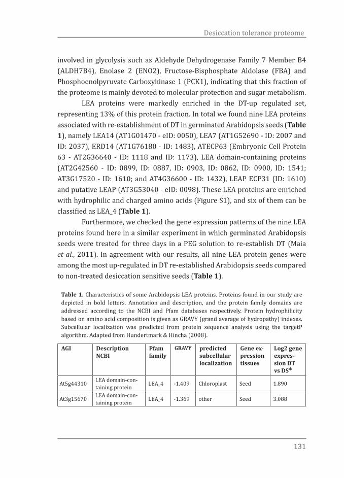
131131
Desiccation tolerance proteome
involved in glycolysis such as Aldehyde Dehydrogenase Family 7 Member B4 (ALDH7B4), Enolase 2 (ENO2), Fructose-Bisphosphate Aldolase (FBA) and Phosphoenolpyruvate Carboxykinase 1 (PCK1), indicating that this fraction of the proteome is mainly devoted to molecular protection and sugar metabolism. LEA proteins were markedly enriched in the DT-up regulated set, representing 13% of this protein fraction. In total we found nine LEA proteins associated with re-establishment of DT in germinated Arabidopsis seeds (Table 1), namely LEA14 (AT1G01470 - eID: 0050), LEA7 (AT1G52690 - ID: 2007 and ID: 2037), ERD14 (AT1G76180 - ID: 1483), ATECP63 (Embryonic Cell Protein 63 - AT2G36640 - ID: 1118 and ID: 1173), LEA domain-containing proteins (AT2G42560 - ID: 0899, ID: 0887, ID: 0903, ID: 0862, ID: 0900, ID: 1541; AT3G17520 - ID: 1610; and AT4G36600 - ID: 1432), LEAP ECP31 (ID: 1610) and putative LEAP (AT3G53040 - eID: 0098). These LEA proteins are enriched with hydrophilic and charged amino acids (Figure S1), and six of them can be classified as LEA_4 (Table 1). Furthermore, we checked the gene expression patterns of the nine LEA proteins found here in a similar experiment in which germinated Arabidopsis seeds were treated for three days in a PEG solution to re-establish DT (Maia et al., 2011). In agreement with our results, all nine LEA protein genes were among the most up-regulated in DT re-established Arabidopsis seeds compared to non-treated desiccation sensitive seeds (Table 1).
Table 1. Characteristics of some Arabidopsis LEA proteins. Proteins found in our study are depicted in bold letters. Annotation and description, and the protein family domains are addressed according to the NCBI and Pfam databases respectively. Protein hydrophilicity based on amino acid composition is given as GRAVY (grand average of hydropathy) indexes. Subcellular localization was predicted from protein sequence analysis using the targetP algorithm. Adapted from Hundertmark & Hincha (2008).
AGI Description NCBI
Pfam family
GRAVY predicted subcellular localization
Gene ex-pression tissues
Log2 gene expres-sion DT vs DS*
At5g44310 LEA domain-con-taining protein LEA_4 -1.409 Chloroplast Seed 1.890
At3g15670 LEA domain-con-taining protein LEA_4 -1.369 other Seed 3.088

132132
Chapter 5
At1g52690similar to LEA protein from Brassica napus
LEA_4 -1.317 other Bud, seed + stress 1.867
At4g21020 LEA domain-con-taining protein LEA_4 -1.291 Mitochon-
drion Seed 2.399
At1g76180 dehydrin ERD14 dehy-drin -1.265 other Non-seed 0.090
At3g02480 ABA-responsive protein-related LEA_4 -1.213 other
Reproduc-tiveseed + salt
1.578
At3g53040 LEA domain-con-taining protein LEA_4 -1.194 other Seed 2.351
At4g13560 LEA domain-con-taining protein LEA_4 -1.181 other Reproduc-
tive -0.023
At4g36600 LEA domain-con-taining protein LEA_4 -1.072 Mitochon-
drion Seed 2.153
At3g17520 LEA domain-con-taining protein LEA_4 -1.047 secreted Seed 3.014
At2g36640 LEA protein AtECP63 LEA_4 -1.023 other Seed 2.415
At2g42560 LEA domain-con-taining protein LEA_4 -0.978 other Seed +
salt 2.354
At2g18340 LEA domain-con-taining protein LEA_4 -0.93 secreted Seed 0.831
At4g13230 LEA domain-con-taining protein LEA_4 -0.831 Mitochon-
drion Bud 0.046
At2g03740 LEA domain-con-taining protein LEA_4 -0.703 Chloroplast Bud -0.031
At2g42540 cold-regulated protein COR15a - -0.554 Chloroplast Non-seed
+ stress 0.080
At2g42530 cold-regulated protein COR15b - -0.542 Chloroplast Non-seed
+ stress -0.147
At2g03850 LEA domain-con-taining protein LEA_4 -0.496 Chloroplast Bud -1.200
At1g72100 LEA domain-con-taining protein LEA_4 -0.46 secreted Seed 2.193
At3g22500 Seed maturation protein AtECP31 SMP -0.341 other Salt 2.985
At1g01470 LEA14 LEA_2 0.056 other all tissues 2.102
* Values from Maia et al. (2011)
After identification, the lists of proteins that were over- or under accumulated in each profile (Figure 1) were individually analysed to determine which GO categories and KEGG pathways were significantly enriched (P-value ≤0.01) in the DT-up and –down regulated protein sets. The over-representation
133133
Desiccation tolerance proteome
analysis (ORA) tool of GeneTrailExpress (Keller et al., 2008) was used for this analysis. To facilitate visualization and interpretation, redundant GO terms were filtered out with the help of the online tool REViGO (Supek et al., 2011). Only one KEGG pathway, glycolysis/gluconeogenesis, was found to be enriched (Supplementary Table S3 and Figure S2). The most significantly over-represented GO terms in the DT-up regulated inferred protein set (profiles 1a and 1b – Figure 1) can be divided into stress related responses, chemical responses, development related responses and inorganic substance related responses. Within those main categories, the most highly significant enriched GO terms were ‘response to cadmium ion’, ‘response to inorganic substance’, ‘response to chemical stimulus’ and ‘response to desiccation’ (Figure 4). Response to desiccation was highly ranked and among the proteins in this category were Responsive to Dehydration (RD) 2 and 19 (AT2G21620 - ID:1860 and AT4G39090 - ID:1610, respectively), ALDH7B4(AT1G54100 - ID:1053), LEA14 and ERD14 (Supplementary Table S3). Stress related processes such as ‘response to osmotic stress’, ‘response to salt’, as well as development related processes such as ‘embryo development ending in seed dormancy’, ‘embryo development’ and ‘fruit development’ were also among the processes with high protein number observed versus expected ratios but also with high absolute protein numbers. Response to abscisic acid stimulus’ has been frequently linked to DT and various abiotic stresses. Here, this category was, at the same time, represented by a large number of proteins and high-ranked in relation to its protein obs/exp ratio reinforcing the importance of ABA in the re-establishment of DT. Proteins encoded by ABA-responsive genes, such as ERD14, Cruciferin (CRU) 1, 2 and 3, ENO2, ALDH7B4 and the well-known Responsive to Desiccation 29B (RD29B), among others, appeared in this protein set (Supplementary Table S3). Finally, when we looked at the DT down-regulated protein set (profiles 2a and 2b – Figure 1) the most enriched GO terms were ‘response to stimulus’, ‘response to stress’, ‘response to biotic stimulus’, ‘response to temperature stimulus’ and ‘response to abiotic stimulus’ (Figure 4 and Supplementary Table S3). Response to heat and temperature had the highest observed to expected protein number ratios (Figure 4). Among the six proteins found in those two categories, three were Heat Shock Proteins (HSPs); namely Cognate

134134
Chapter 5
Protein 70-1 (HSC70-1), Heat Shock Protein 70-3 (HSP70-3), and Heat Shock Protein 17.4 (HSP17.4). HSPs are highly conserved among organisms and their synthesis is known to be induced in response to heat (Lindquist & Craig, 1988). HSPs have also been persistently linked to DT (Wehmeyer & Vierling, 2000) as they contribute to a coordinated folding of proteins under normal as well
-35-25-15-5515
response to desiccationresponse to cadmium ion
response to inorganic substanceresponse to abscisic acid stimulus
response to salt stressembryo develop. ending in seed dormancy
response to osmotic stressembryonic development
response to coldresponse to chemical stimulus
response to abiotic stimulusresponse to endogenous stimulus
reproductive developmental processpost-embryonic development
response to stressdevelopmental process
multicellular organismal process
response to heatresponse to temperature stimulus
response to cadmium ionresponse to metal ion
response to biotic stimulusdefense response
response to abiotic stimulusresponse to stress
response to stimulusresponse to chemical stimulus
expected observed
Protein number
DT-up protein set
DT-down protein set
Figure 4 Over-representation analysis (ORA) of the differentially over- and under-accumulated proteins. ORA was performed with the following parameters: significance level: 0.01, p-value adjustment for multiple testing: Bonferroni adjustment, minimum class size: 3, maximum class size: 40. GO terms and Bonferroni P-values obtained from GeneTrailExpress (Keller et al., 2008) were used as input to summarize the data in REViGO (Supek et al., 2011) under default settings (allowed similarity: 0.7, database: UniProt, semantic similarity measure: SimRel).

135135
Desiccation tolerance proteome
as under abiotic stress conditions (Waters, 2013). However, although acting in the “universal” process of protein folding, our data suggests that distinct HSPs might be more or less active upon different stress conditions with HSC70-1, HSP70-3 and HSP17.4 not required for DT. This fraction of the proteome was also heavily represented by Cruciferins that could be spotted in many different isoforms (Supplementary Table S2).
Table 2. Most common cis-acting promoter elements within the DT up- and down-regulated protein sets in Arabidopsis Col-0 and aba2-1 germinated seeds.
Enriched promoter element a Consensus sequence b P S p-value
DT-up protein set
ABFs binding site motif CACGTGGC 10 13 < 10-3
ABRE-like binding site motif BACGTGKM 37 74 < 10-6
ACGTABREMOTIFA2OSEM ACGTGKC 33 62 < 10-8
GADOWNAT ACGTGTC 24 34 < 10-8
ABRE binding site motif YACGTGGC 16 21 < 10-6
ABREATRD22 RYACGTGGYR 8 10 < 10-3
CACGTGMOTIF CACGTG 31 90 < 10-6
DT-down protein set
ABRE-like binding site motif BACGTGKM 15 24 < 10-4
a Analysis performed with the Arabidopsis expression network analysis (Athena) tool (http://www.bioinformatics2.wsu.edu/cgi-bin/Athena/cgi/home.pl) (O’Connor et al., 2005). b Where B = T/G/C; K = T/G; M = A/C; Y = T/C; R = A/G and N = A/T/G/C. The number of promoters containing at least one instance of the TF binding site and the total number of TF binding sites in the selected set of sequences are given in the ‘P’ and ‘S’ columns, respectively. The p-value for enrichment is a measure of how over-represented a motif is in the selected set of genes versus the overall occurrence in the genome.
Promoter analysis
Assessment of the promoter region of genes provides valuable insights in the regulation of gene expression. Also, the presence of similar elements in different genes is an indication of common regulatory mechanisms for their expression. Here, the promoter regions of all proteins in the DT-up and -down sets were searched for any common motifs located within a 1 kb region upstream of the translational start site. With this analysis we identified 7 different enriched
136136
Chapter 5
motifs (Table 2). Interestingly, all motifs identified as enriched, in both up- and down-protein sets, contained a core sequence (ACGTG) known as the ABA-responsive element (ABRE). This observation again indicates ABA as the most prominent signalling source regulating DT-related genes.
Discussion
Complex traits demand elaborate screening methods
Here, we used a system of loss and re-establishment of DT in Arabidopsis seeds (Maia et al., 2011) in combination with three treatments to re-induce DT (PEG, ABA and PEG + ABA) and two Arabidopsis genotypes (Col-0 and aba2-1). This strategy allowed us to explore a physiological system which contains several filters to enhance the identification of proteins more likely related to the re-establishment of DT than to other developmental events. Using this system we were able to select 70 up-regulated and 27 down-regulated protein spots in seeds with re-established DT (Figure 1 and Supplementary Tables S1 and S2). ‘Response to desiccation’ was the GO term with the highest observed to expected protein number ratio. The over-representation of this GO term was also highly significant (P-value = 6.1-07) which is a strong indication of the accuracy of our spot selection procedure to pinpoint DT-related proteins. Besides that, many LEA proteins were also retrieved and identified. The presence of LEA proteins, since they have been largely correlated to tolerance to various abiotic stresses and desiccation in particular (Tunnacliffe & Wise, 2007; Battaglia et al., 2008), is also a good indication that the other proteins identified here are likely to play a role in DT. Other proteins also previously associated with DT, such as ALDH7B4, RD2 and RD29B were also present in our data set.
ABA plays a major role in the regulation of DT up-regulated proteins
Proteins encoded by ABA-responsive genes, such as ERD14, ENO2, ALDH7B4, Aconitase 3 (ACO3) and RD29B were found to be significantly correlated with DT in our screen. The genes coding for these proteins all contain ABRE-motifs in their promoter regions and are all regulated by ABA. For example, ACO3 is phosphorylated in response to ABA (Ghelis et al., 2008) and RD29B contains two ABREs and has been widely used as a marker in studies of ABA-dependent

137137
Desiccation tolerance proteome
pathways of abiotic stress responses (Yamaguchi-Shinozaki & Shinozaki, 1993; Ishitani et al., 1997; Nakashima et al., 2006). Additionally, ERD14, a LEA_2 family protein whose gene expression is induced early in response to dehydration stress, is also induced in response to ABA (Kiyosue et al., 1994). This protein has chaperone activity and has been shown, in vitro, to prevent the aggregation and inactivation of a wide variety of enzymes (Kovacs et al., 2008), a functionality needed in desiccating organisms. Strengthening the importance of ABA in the regulation of DT, we found that the GO term ‘response to abscisic acid stimulus’ was represented by a relatively large number of the proteins identified as being up-regulated in DT re-established seeds. This GO term also had a very low p-value in the GO enrichment analysis, which is an indication of the significance of ABA-signalling in DT re-establishment. Additionally, all genes encoding for the proteins in the DT-up and -down sets contained at least one ABRE-motif upstream of their translational start sites (Table 2) suggesting a common ABA-dependent mechanism for the regulation of their expression. Similar results were found in Medicago and Arabidopsis gene expression studies where the most up-regulated genes in seeds with re-established DT also showed an enrichment of genes containing ABRE-motifs in their promoter regions (Buitink et al., 2006; Maia et al., 2011). Finally, it is also important to mention that ABA alone can re-induce DT in Col-0 and aba2-1 seeds, which shows that ABA can completely substitute for the osmotic signal previously thought to be essential to induce the proteome required to re-establish DT in seeds.
The possible role of charged LEA proteins in DT
In this study, we found nine LEA proteins associated with re-establishment of DT in germinated Arabidopsis seeds. This protein fraction represents 13% of all proteins identified as up-regulated in seeds with re-established DT. From those, six can be classified as LEA_4 (Table 1) (Hundertmark & Hincha, 2008). Furthermore, considering the number of LEA proteins in the Arabidopsis genome, it is clear that not only the total number of LEA proteins found was significantly high in our data set, but also the number of those belonging to the LEA_4 family was markedly over-represented; 33% of the proteins classified as LEA_4 in Arabidopsis were found to be strongly correlated with re-establishment
138138
Chapter 5
of DT, which suggests a specific relationship between this type of LEA proteins and DT. In agreement with this, LEA_4 proteins have been frequently linked to increased desiccation- and drought tolerance in plants. For instance, under severe dehydration, a LEA_4 protein was shown to accumulate in wheat shoots but not in roots. In this case, shoots resumed growth after dehydration whereas roots died and were replaced by newly developing roots, which provided a link between this protein and dehydration tolerance (Ried & Walker-Simmons, 1993). A direct connection between a LEA_4 protein and stress tolerance was shown for the first time in transgenic rice harbouring a barley LEA_4 gene (HAV1) which conferred resistance to drought and salinity stress (Xu et al., 1996). This resistance was later attributed to HAV1 activity in cell membrane protection from injury under drought stress (Chandra Babu et al., 2004). HAV1 overexpression was also shown to increase dehydration tolerance in wheat and oat (Sivamani et al., 2000; Maqbool et al., 2002). The rapeseed LEA_4 gene, MEleaN4, introduced into Chinese cabbage or lettuce also lead to improved drought tolerance (Park et al., 2005a; Park et al., 2005b). Additionally, LEA_4 proteins seem to be more frequently associated with increments in drought, osmotic and salt stress tolerance as compared to LEA proteins belonging to other families. For example, in a study were three dehydrin LEA proteins were knocked-down, DT was not compromised whereas longevity was significantly affected (Hundertmark et al., 2011). In another example, overexpression of Arabidopsis RAB18 (dehydrin LEA) in Arabidopsis plants failed to improve freezing or drought tolerance (Lang and Palva, 1992). In agreement with this, the comparison between the LEA profiles of a recalcitrant-seeded species, Castanospermum australe, and Medicago truncatula, which produces orthodox seeds, showed remarkable differences. For instance, dehydrins comprise 20% of the LEA proteome of mature M. truncatula seeds which is in stark contrast with an 83% representation in the C. australe LEA proteome (Delahaie et al., 2013). Taken together, these findings illustrate that different LEA groups might perform specific functions among different types of stress, with LEA_4 proteins being more important under dehydration pressure. As discussed above, LEA proteins have been widely assayed and shown to play a role in DT. However, the mechanisms by which LEA proteins act to

139139
Desiccation tolerance proteome
protect other molecules are still elusive. In the Arabidopsis genome, LEA_4 is the most dominant group containing 18 members (Hundertmark & Hincha, 2008). This group is very heterogeneous and the gene products differ greatly in size and in their grand average hydropathy index (GRAVY) (Hundertmark & Hincha, 2008). Except for LEA14 (LEA_2 group), which is slightly hydrophobic, all other LEA proteins up-regulated in seeds with re-established DT have a relatively low GRAVY index, which indicates high hydrophilicity. Interestingly, all LEA_4 proteins found here had lower GRAVY indexes in comparison to other members of the same family, suggesting that hydrophilicity is an essential property required in LEA proteins that act as protectants under desiccation stress. In agreement, LEA proteins, due to their high hydrophilicity, were suggested to preserve macromolecular structure and function by providing a hydrophilic surrounding which can substitute for water molecules removed under dehydration stress (Battaglia et al., 2008). During dehydration the chance of protein unfolding and aggregation increases due to changes in the cell physicochemical properties and increased probability of physical contact to other proteins and membranes. Thus, one hypothesis to explain the protein stabilisation function of LEA_4 proteins in seeds with re-established DT is that they might behave as “molecular shields”, which sterically or electrosterically prevent interactions between aggregation-prone and partially unfolded proteins (Chakrabortee et al., 2007). As molecular shields, LEA_4 proteins could also function by preventing membrane fusion, a common feature observed under desiccation stress (Farrant et al., 2007). When we compared the amino acid sequences of the nine LEA proteins found in the DT-up regulated set against a thousand random sets of nine Arabidopsis protein sequences we noticed that charged and polar amino acids are significantly enriched in our set of LEA proteins (Supplementary Figure S1). Interestingly, among the charged amino acids, our nine LEA proteins display an accumulation of aspartic acid, glutamic acid and lysine while the proteins from our random sets have relatively more arginine, which is an indication that the nine LEA proteins found here are more negatively charged. Amino acid charge-increasing substitutions at multiple sites and their accumulation corresponded with increases in muscle myoglobin concentrations and the adaption of both semi- and fully aquatic lifestyles in
140140
Chapter 5
mammals. Similar to a likely mode of action of LEA proteins, myoglobins with increased net surface charge allowed higher density of this protein per muscle unit while electrosterically preventing their aggregation and subsequent loss of function (Mirceta et al., 2013). Thus, increasing negative charges at the protein surface seems to be a basal mechanism of adaptation to conditions where aggregation-prone proteins and membranes are physically forced together, as under desiccation stress. Besides repelling each other, LEA proteins probably form a matrix to prevent other aggregation-prone proteins to interact. Another feature that cannot be neglected is the fact that LEA proteins are largely unfolded under hydrated conditions lacking a well-defined surface. However, drying or association with phospholipid bilayers can induce LEA proteins to fold (Chakrabortee et al., 2007), which will bring about alternative functions other than the ones they perform under hydrated conditions. More insight into surface charge of LEA proteins will be possible with the elucidation of their 3D structure under different hydration levels and presence of solutes and salts.
LEA proteins and sugars act together in the re-establishment of DT in Arabidopsis seeds
We found glycolysis/gluconeogenesis as the only enriched KEGG pathway, which contained several of the DT-up regulated proteins (Supplementary Figure S2a). Some of these proteins could also be mapped to the raffinose pathway (Supplementary Figure S2b). Together, this indicates that sugar metabolism might be more active in seeds with re-established DT. Based on the proteome, however, it is hard to infer which types of sugars are accumulating or being degraded. Nevertheless, the association of acquisition of DT with certain sugars such as sucrose, raffinose series of oligosaccharides and galactinol, have been repeatedly reported (Koster & Leopold, 1988; Obendorf, 1997; Berjak, 2006; Peters et al., 2007; Li et al., 2011). We found that raffinose, sucrose and galactinol differentially accumulate in Arabidopsis seeds with re-established DT (Chapter 3). Thus, we are tempted to speculate that these sugars are probably acting together with LEA proteins to counteract the damage produced by water limitation, by promoting stability of the bioglasses needed in the dry state (Buitink & Leprince, 2004). In Chapter 3 we also found a significant negative correlation between the sucrose/raffinose ratio and re-establishment
141141
Desiccation tolerance proteome
of DT. Similar results were found by Vandecasteele et al. (2011), who found a negative correlation between sucrose/raffinose oligosaccharide family and seed vigour. Therefore, we hypothesise that hydrophilic charged LEA_4 proteins are contributing to counteract the damage produced by water limitation by the formation of a tight hydrogen-bonding network in the dehydrating cytoplasm. Together with sugars, these LEA proteins might promote a long-term stability of sugar glasses during anhydrobiosis. Supporting it, a dehydrated mixture of sucrose and a pollen LEA_4 protein showed both a higher glass transition temperature and increased average strength of hydrogen bonding in comparison to dehydrated sucrose alone (Wolkers et al., 2001). Additionally, a similar impact on bioglass stability was documented for synthetic peptides composed of two or four 11-mer motifs of LEA_4 proteins from insects, nematodes, and plants (Shimizu et al., 2009). Further insights into in vivo sugar/LEA protein ratios will be certainly helpful to fire up new in vitro studies and to support the physiological relevance of those findings.
Acknowledgements
We thank the technical and intellectual input from our colleagues of the Wageningen Seed Lab and the Laboratory of Plant Physiology. This work was supported by the “Coordenação de Aperfeiçoamento de Pessoal de Nível Superior” (CAPES, Brazil)(JM).
References
Amara I, Odena A, Oliveira E, Moreno A, Masmoudi K, Pagès M, Goday A. 2012. Insights into Maize LEA Proteins: From Proteomics to Functional Approaches. Plant and Cell Physiology 53(2): 312-329.
Arc E, Chibani K, Grappin P, et al., 2012. Cold Stratification and Exogenous Nitrates Entail Similar Functional Proteome Adjustments during Arabidopsis Seed Dormancy Release. Journal of Proteome Research 11, 5418-32.
Berjak P, Pammenter NW, 2008. From Avicennia to Zizania: Seed Recalcitrance in Perspective. Annals of Botany 101, 213-28.
Battaglia M, Olvera-Carrillo Y, Garciarrubio A, Campos F, Covarrubias AA. 2008. The Enigmatic LEA Proteins and Other Hydrophilins. Plant physiology 148(1): 6-24.
Berjak P. 2006. Unifying perspectives of some mechanisms basic to desiccation
142142
Chapter 5
tolerance across life forms. Seed Science Research 16(1): 1-15.Bies-Ethève N, Gaubier-Comella P, Debures A, Lasserre E, Jobet E, Raynal M, Cooke
R, Delseny M. 2008. Inventory, evolution and expression profiling diversity of the LEA (late embryogenesis abundant) protein gene family in Arabidopsis thaliana. 67(1-2): 107-124.
Blackman SA, Obendorf RL, Leopold AC. 1992. Maturation proteins and sugars in desiccation tolerance of developing soybean seeds. Plant physiology 100(1): 225-230.
Boudet J, Buitink J, Hoekstra FA, Rogniaux H, Larre C, Satour P, Leprince O. 2006. Comparative analysis of the heat stable proteome of radicles of Medicago truncatula seeds during germination identifies late embryogenesis abundant proteins associated with desiccation tolerance. Plant physiology 140(4): 1418-1436.
Bruggink T, van der Toorn P. 1995. Induction of desiccation tolerance in germinated seeds. Seed Science Research 5(1): 1-4.
Buitink J, Leger JJ, Guisle I, Vu BL, Wuillème S, Lamirault G, Bars AL, Meur NL, Becker A, Küster H, Leprince O. 2006. Transcriptome profiling uncovers metabolic and regulatory processes occurring during the transition from desiccation-sensitive to desiccation-tolerant stages in Medicago truncatula seeds. The Plant Journal 47(5): 735-750.
Buitink J, Leprince O. 2004. Glass formation in plant anhydrobiotes: Survival in the dry state. Cryobiology 48(3): 215-228.
Buitink J, Vu BL, Satour P, Leprince O. 2003. The re-establishment of desiccation tolerance in germinated radicles of Medicago truncatula Gaertn. seeds. Seed Science Research 13(4): 273-286.
Chakrabortee S, Boschetti C, Walton LJ, Sarkar S, Rubinsztein DC, Tunnacliffe A. 2007. Hydrophilic protein associated with desiccation tolerance exhibits broad protein stabilization function. Proceedings of the National Academy of Sciences of the United States of America 104(46): 18073-18078.
Chandra Babu R, Zhang J, Blum A, David Ho TH, Wu R, Nguyen HT. 2004. HVA1, a LEA gene from barley confers dehydration tolerance in transgenic rice (Oryza sativa L.) via cell membrane protection. Plant Science 166(4): 855-862.
Daws MI, Bolton S, Burslem DFRP, Garwood NC, Mullins CE. 2007. Loss of desiccation tolerance during germination in neo-tropical pioneer seeds: implications for seed mortality and germination characteristics. Seed Science Research 17(04): 273-281.
Delahaie J, Hundertmark M, Bove J, Leprince O, Rogniaux H, Buitink J. 2013. LEA polypeptide profiling of recalcitrant and orthodox legume seeds reveals ABI3-regulated LEA protein abundance linked to desiccation tolerance. Journal of Experimental Botany 64(14):4559-4573.
Espelund M, Sæbøe-Larssen S, Hughes DW, Galau GA, Larsen F, Jakobsen KS. 1992. Late embryogenesis-abundant genes encoding proteins with different numbers of hydrophilic repeats are regulated differentially by abscisic acid and osmotic stress. The Plant Journal 2(2): 241-252.
Farnsworth, E. (2000) The ecology and physiology of viviparous and recalcitrant seeds. Annual Review of Ecology and Systematics 31: 107–138

143143
Desiccation tolerance proteome
Farrant JM, Brandt W, Lindsey GG. 2007. An overview of mechanisms of desiccation tolerance in selected angiosperm resurrection plants. Plant Stress 1: 72–84.
Farrant JM, Lehner A, Cooper K, Wiswedel S. 2009. Desiccation tolerance in the vegetative tissues of the fern Mohria caffrorum is seasonally regulated. The Plant Journal 57(1): 65-79.
Farrant JM, Moore JP. 2011. Programming desiccation-tolerance: from plants to seeds to resurrection plants. Current Opinion in Plant Biology 14(3): 340-345.
Finkelstein RR, Lynch TJ. 2000. The Arabidopsis abscisic acid response gene ABI5 encodes a basic leucine zipper transcription factor. The Plant Cell Online 12(4): 599-609.
Franchi GG, Piotto B, Nepi M, Baskin CC, Baskin JM, Pacini E. 2011. Pollen and seed desiccation tolerance in relation to degree of developmental arrest, dispersal, and survival. Journal of Experimental Botany 62(15): 5267-5281.
Galau G, Hughes DW, Dure L, III. 1986. Abscisic acid induction of cloned cotton late embryogenesis-abundant (LEA) mRNAs. 7(3): 155-170.
Ghelis T, Bolbach G, Clodic G, Habricot Y, Miginiac E, Sotta B, Jeannette E. 2008. Protein tyrosine kinases and protein tyrosine phosphatases are involved in abscisic acid-dependent processes in Arabidopsis seeds and suspension cells. Plant Physiology 148(3): 1668-1680.
Goyal K, Tisi L, Basran A, Browne J, Burnell A, Zurdo J, Tunnacliffe A. 2003. Transition from natively unfolded to folded state induced by desiccation in an anhydrobiotic nematode protein. Journal of Biological Chemistry 278(15): 12977-12984.
Hand SC, Menze MA, Toner M, Boswell L, Moore D. 2011. LEA proteins during water stress: Not just for plants anymore. Annual Review of Physiology 73(1): 115-134.
Hincha DK, Thalhammer A. 2012. LEA proteins: IDPs with versatile functions in cellular dehydration tolerance. Biochemical Society Transactions 40(5): 1000-1003.
Hundertmark M, Buitink J, Leprince O, Hincha DK. 2011. The reduction of seed-specific dehydrins reduces seed longevity in Arabidopsis thaliana. Seed Science Research 21(3): 165-173.
Hundertmark M, Hincha D. 2008. LEA (Late Embryogenesis Abundant) proteins and their encoding genes in Arabidopsis thaliana. BMC Genomics 9(1): 118.
Illing N, Denby KJ, Collett H, Shen A, M FJ. 2005. The signature of seeds in resurrection plants: A molecular and physiological comparison of desiccation tolerance in seeds and vegetative tissues. Integrative and Comparative Biology 45(5): 771-787.
Ishitani M, Xiong L, Stevenson B, Zhu JK. 1997. Genetic analysis of osmotic and cold stress signal transduction in Arabidopsis: Interactions and convergence of abscisic acid-dependent and abscisic acid-independent pathways. Plant Cell 9(11): 1935-1949.
Keller A, Backes C, Al-Awadhi M, Gerasch A, Kuntzer J, Kohlbacher O, Kaufmann M, Lenhof H-P. 2008. GeneTrailExpress: a web-based pipeline for the statistical evaluation of microarray experiments. BMC Bioinformatics 9(1): 552.
Kiyosue T, Yamaguchi-Shinozaki K, Shinozaki K. 1994. Characterization of Two cDNAs (ERD10 and ERD14) corresponding to genes that respond rapidly to

144144
Chapter 5
dehydration stress in Arabidopsis thaliana. Plant and Cell Physiology 35(2): 225-231.
Koster KL, Leopold aC. 1988. Sugars and desiccation tolerance in seeds. Plant physiology 88(3): 829-832.
Kovacs D, Kalmar E, Torok Z, Tompa P. 2008. Chaperone activity of ERD10 and ERD14, two disordered stress-related plant proteins. Plant physiology 147(1): 381-390.
Lång V, Palva ET, 1992. The expression of a rab-related gene, rab18, is induced by abscisic acid during the cold acclimation process of Arabidopsis thaliana (L.) Heynh. Plant Molecular Biology 20, 951-62.
Le BH, Cheng C, Bui AQ, Wagmaister JA, Henry KF, Pelletier J, Kwong L, Belmonte M, Kirkbride R, Horvath S, Drews GN, Fischer RL, Okamuro JK, Harada JJ, Goldberg RB. 2010. Global analysis of gene activity during Arabidopsis seed development and identification of seed-specific transcription factors. Proceedings of the National Academy of Sciences 107(18): 8063-8070.
Le BH, Wagmaister JA, Kawashima T, Bui AQ, Harada JJ, Goldberg RB. 2007. Using Genomics to Study Legume Seed Development. Plant physiology 144(2): 562-574.
Léon-Kloosterziel KM, Gil MA, Ruijs GJ, Jacobsen SE, Olszewski NE, Schwartz SH, Zeevaart JAD, Koornneef M. 1996. Isolation and characterization of abscisic acid-deficient Arabidopsis mutants at two new loci. Plant Journal 10(4): 655-661.
Leprince O, Hendry GAF, McKersie BD. 1993. The mechanisms of desiccation tolerance in developing seeds. Seed Science Research 3(04): 231-246.
Li X, Zhuo J, Jing Y, Liu X, Wang X. 2011. Expression of a GALACTINOL SYNTHASE gene is positively associated with desiccation tolerance of Brassica napus seeds during development. Journal of Plant Physiology 168(15): 1761-1770.
Lindquist S, Craig EA. 1988. The Heat-Shock Proteins. Annual Review of Genetics 22(1): 631-677.
Maia J, Dekkers BJW, Provart NJ, Ligterink W, Hilhorst HWM. 2011. The re-establishment of desiccation tolerance in germinated Arabidopsis thaliana seeds and its associated transcriptome. PLoS ONE 6(12).
Manfre AJ, LaHatte Ga, Climer CR, Marcotte WR. 2009. Seed dehydration and the establishment of desiccation tolerance during seed maturation is altered in the Arabidopsis thaliana mutant atem6-1. Plant & Cell Physiology 50(2): 243-253.
Maqbool S, Zhong H, El-Maghraby Y, Ahmad A, Chai B, Wang W, Sabzikar R, Sticklen M. 2002. Competence of oat (Avena sativa L.) shoot apical meristems for integrative transformation, inherited expression, and osmotic tolerance of transgenic lines containing hva1. 105(2-3): 201-208.
Mirceta S, Signore AV, Burns JM, Cossins AR, Campbell KL, Berenbrink M. 2013. Evolution of mammalian diving capacity traced by myoglobin net surface charge. Science 340(6138).
Nakashima K, Fujita Y, Katsura K, Maruyama K, Narusaka Y, Seki M, Shinozaki K, Yamaguchi-Shinozaki K. 2006. Transcriptional Regulation of ABI3- and ABA-responsive Genes Including RD29B and RD29A in Seeds, Germinating Embryos, and Seedlings of Arabidopsis. 60(1): 51-68.
O’Connor TR, Dyreson C, Wyrick JJ. 2005. Athena: a resource for rapid visualization and systematic analysis of Arabidopsis promoter sequences. Bioinformatics

145145
Desiccation tolerance proteome
21(24): 4411-4413.Obendorf RL. 1997. Oligosaccharides and galactosyl cyclitols in seed desiccation
tolerance. Seed Science Research 7(02): 63-74.Ooms JJJ, Léon-Kloosterziel KM, Bartels D, Koornneef M, Karssen CM. 1993.
Acquisition of desiccation tolerance and longevity in seeds of Arabidopsis thaliana: A comparative study using abscisic acid-insensitive abi3 mutants. Plant physiology 102(4): 1185-1191.
Park B-J, Liu Z, Kanno A, Kameya T. 2005a. Genetic improvement of Chinese cabbage for salt and drought tolerance by constitutive expression of a B. napus LEA gene. Plant Science 169(3): 553-558.
Park B-J, Liu Z, Kanno A, Kameya T. 2005b. Increased tolerance to salt- and water-deficit stress in transgenic lettuce (Lactuca sativa L.) by constitutive expression of LEA. 45(2): 165-171.
Peters S, Mundree SG, Thomson Ja, Farrant JM, Keller F. 2007. Protection mechanisms in the resurrection plant Xerophyta viscosa (Baker): both sucrose and raffinose family oligosaccharides (RFOs) accumulate in leaves in response to water deficit. Journal of Experimental Botany 58(8): 1947-1956.
Proctor MCF, Ligrone R, Duckett JG. 2007. Desiccation tolerance in the moss Polytrichum formosum: physiological and fine-structural changes during desiccation and recovery. Annals of Botany 99(6): 75-93.
Rajjou L, Belghazi M, Catusse J, Ogé L, Arc E, Godin B, Chibani K, Ali-Rachidi S, Collet B, Grappin P, Jullien M, Gallardo K, Job C, Job D, Kermode AR 2011. Proteomics and posttranslational proteomics of seed dormancy and germination. In: Walker JM ed.: Humana Press, 215-236.
Rajjou L, Lovigny Y, Groot SPC, Belghazi M, Job C, Job D. 2008. Proteome-wide characterization of seed aging in Arabidopsis: A comparison between artificial and natural aging protocols. Plant physiology 148(1): 620-641.
Ried JL, Walker-Simmons MK. 1993. Group 3 Late Embryogenesis Abundant Proteins in Desiccation-Tolerant Seedlings of Wheat (Triticum aestivum L.). Plant physiology 102(1): 125-131.
Shih Md, Hoekstra FA, Hsing YIC 2008. Late embryogenesis abundant proteins. Advances in Botanical Research. 211-255.
Shimizu T, Kanamori Y, Furuki T, Kikawada T, Okuda T, Takahashi T, Mihara H, Sakurai M. 2009. Desiccation-induced structuralization and glass formation of group 3 late embryogenesis abundant protein model peptides. Biochemistry 49(6): 1093-1104.
Sivamani E, Bahieldin A, Wraith JM, Al-Niemi T, Dyer WE, Ho T-HD, Qu R. 2000. Improved biomass productivity and water use efficiency under water deficit conditions in transgenic wheat constitutively expressing the barley HVA1 gene. Plant Science 155(1): 1-9.
Supek F, Bošnjak M, Škunca N, Šmuc T. 2011. REVIGO summarizes and visualizes long lists of gene ontology terms. PLoS ONE 6(7): e21800.
Tunnacliffe A, Wise MJ. 2007. The continuing conundrum of the LEA proteins. Die Naturwissenschaften 94(10): 791-812.
146146
Chapter 5
Vandecasteele C, Teulat-Merah B, Morere-Le Paven MC, Leprince O, Ly Vu B, Viau L, Ledroit L, Pelletier S, Payet N, Satour P, Lebras C, Gallardo K, Huguet T, Limami AM, Prosperi JM, Buitink J. 2011. Quantitative trait loci analysis reveals a correlation between the ratio of sucrose/raffinose family oligosaccharides and seed vigour in Medicago truncatula. Plant, Cell & Environment 34(9): 1473-1487.
Verdier J, Lalanne D, Pelletier S, Torres-Jerez I, Righetti K, Bandyopadhyay K, Leprince O, Chatelain E, Ly Vu B, Gouzy J, Gamas P, Udvardi M, Buitink J. 2013. A regulatory network-based approach dissects late maturation processes related to the acquisition of desiccation tolerance and longevity of Medicago truncatula seeds. Plant physiology 163(2):757-74.
Waters ER. 2013. The evolution, function, structure, and expression of the plant sHSPs. Journal of Experimental Botany 64(2): 391-403.
Wehmeyer N, Vierling E. 2000. The expression of small heat shock proteins in seeds responds to discrete developmental signals and suggests a general protective role in desiccation tolerance. Plant physiology 122(4): 1099-1108.
Wolkers WF, McCready S, Brandt WF, Lindsey GG, Hoekstra FA. 2001. Isolation and characterization of a D-7 LEA protein from pollen that stabilizes glasses in vitro. Biochimica et Biophysica Acta (BBA) - Protein Structure and Molecular Enzymology 1544(1–2): 196-206.
Wood AJ. 2007. Invited essay: New frontiers in bryology and lichenology: The nature and distribution of vegetative desiccation-tolerance in hornworts, liverworts and mosses. Bryologist 110(2): 163-177.
Xu D, Duan X, Wang B, Hong B, Ho Tuan-Hua D, Wu R. 1996. Expression of a late embryogenesis abundant protein gene, HVA1, from barley confers tolerance to water deficit and salt stress in transgenic rice. Plant physiology 110(1): 249-257.
Yamaguchi-Shinozaki K, Shinozaki K. 1993. Characterization of the expression of a desiccation-responsive rd29 gene of Arabidopsis thaliana and analysis of its promoter in transgenic plants. Molecular and General Genetics 236(2-3): 331-340.

147147
Desiccation tolerance proteome
Supporting information
If not displayed bellow, supplementary information can be downloaded from: http://www.wageningenseedlab.nl/thesis/jmaia/
0
2
4
6
8
10
12
14
16
A C D E F G H I K L M N P Q R S T V W Y
DT
Randomns
ns ns
ns
ns
ns
**
**
**
*
**
**
**
**
**
****
**
**
**
Amin
o ac
id re
sidu
e (%
)
Supplementary Figure S1. Amino acid residue enrichment in the nine LEA proteins found to be over-accumulating in Arabidopsis seeds with re-established DT. The average proportion of each amino acid found in the DT over-accumulated set of LEA proteins (black bars) was compared against the average proportions in one thousand random sets of 9 proteins taken from the Arabidopsis genome (grey bars). Asterisks (* P ≤ 0.05 and ** P ≤ 0.01) represent significant differences between our set of LEA proteins and the random set.
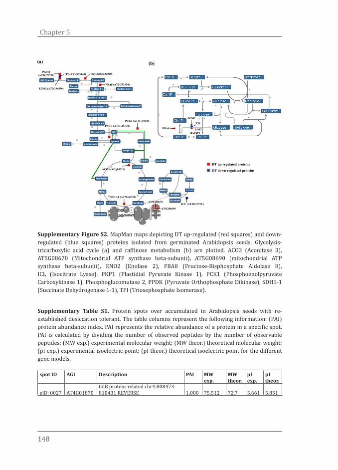
148148
Chapter 5
Supplementary Table S1. Protein spots over accumulated in Arabidopsis seeds with re-established desiccation tolerant. The table columns represent the following information: (PAI) protein abundance index. PAI represents the relative abundance of a protein in a specific spot. PAI is calculated by dividing the number of observed peptides by the number of observable peptides; (MW exp.) experimental molecular weight; (MW theor.) theoretical molecular weight; (pI exp.) experimental isoelectric point; (pI theor.) theoretical isoelectric point for the different gene models.
spot ID AGI Description PAI MW exp.
MW theor.
pI exp.
pI theor.
eID: 0027 AT4G01870tolB protein-related chr4:808473-810431 REVERSE 1.000 75.512 72.7 5.661 5.851
PGM2 (AT1G70730) TPI (AT3G55440)
ENO2 (AT2G36530)
PKP1 (AT3G22960)
FBA8 (AT3G52930)
PCK1 (AT4G37870)
PPDK (AT4G15530)
ACO3 (At2g05710)
SDH1-1 (AT5G66760)
AT5G08670
AT5G08690ICL (AT3G21720 )
FBA8
PKP1
PGM2
TPI
LOS2
DT up-regulated proteins
DT down-regulated proteins
(a) (b)
Supplementary Figure S2. MapMan maps depicting DT up-regulated (red squares) and down-regulated (blue squares) proteins isolated from germinated Arabidopsis seeds. Glycolysis-tricarboxylic acid cycle (a) and raffinose metabolism (b) are plotted. ACO3 (Aconitase 3), AT5G08670 (Mitochondrial ATP synthase beta-subunit), AT5G08690 (mitochondrial ATP synthase beta-subunit), ENO2 (Enolase 2), FBA8 (Fructose-Bisphosphate Aldolase 8), ICL (Isocitrate Lyase). PKP1 (Plastidial Pyruvate Kinase 1), PCK1 (Phosphoenolpyruvate Carboxykinase 1), Phosphoglucomutase 2, PPDK (Pyruvate Orthophosphate Dikinase), SDH1-1 (Succinate Dehydrogenase 1-1), TPI (Triosephosphate Isomerase).

149149
Desiccation tolerance proteome
eID: 0029 AT3G11930
universal stress protein (USP) family protein chr3:3776377-3777399 FORWARD 1.714 24.310 21.4 5.544 5.604
eID: 0029 AT4G28520
CRC, CRU3 CRU3 (CRUCIFERIN 3); nutrient reservoir chr4:14087602-14089623 FORWARD 0.174 24.31 58.1 5.544 6.998
eID: 0050 AT1G01470
LSR3, LEA14 LEA14 (LATE EM-BRYOGENESIS ABUNDANT 14) chr1:172295-172826 REVERSE 0.750 15.81 16.5 4.150 4.532
eID: 0094 AT5G19820
EMB2734 EMB2734 (EM-BRYO DEFECTIVE 2734); lyase chr5:6695733-6701249 REVERSE 0.381 110.0 123.6 4.900 4.502
eID: 0098 AT3G53040
late embryogenesis abundant pro-tein, putative / LEA protein, putative chr3:19675775-19677383 REVERSE 0.706 62.731 52 5.107 5.019
eID: 0098 ATCG00120
ATPA Encodes the ATPase alpha subunit, which is a subunit of ATP synthase 0.118 62.731 55.2 5.107 4.898
eID: 0099 AT5G26000
TGG1 TGG1 (THIOGLUCOSIDE GLUCOHYDROLASE 1); hydrolase, hydrolyzing O-glycosyl compounds chr5:9079681-9082350 REVERSE 0.391 71.190 61 5.450 5.703
eID: 0109 AT1G17020ATSRG1, SRG1 SRG1 (SENES-CENCE-RELATED GENE 1) 0.286 42.750 40.9 5.333 5.110
eID: 0109 AT3G09820ADK1 ADK1 (ADENOSINE KINASE 1) chr3:3012127-3014629 FORWARD 0.125 42.750 37.7 5.333 5.110
eID: 0109 AT4G13360catalytic chr4:7775129-7777697 FORWARD 0.476 42.750 46.1 5.333 6.639
eID: 0109 AT5G03300
ADK2 ADK2 (ADENOSINE KINASE 2); kinase chr5:796572-798996 FORWARD 0.214 42.750 37.7 5.333 4.907
eID: 0109 AT5G53400
nuclear movement family protein chr5:21678814-21680609 FOR-WARD 0.250 42.750 34.4 5.333 4.993
eID: 0133 AT5G28540BIP1 BIP1; ATP binding chr5:10540669-10543278 REVERSE 0.400 82.439 73.5 5.042 4.811
eID: 0133 AT5G36210serine-type peptidase chr5:14265432-14270502 REVERSE 0.455 82.439 81.1 5.042 5.507
eID: 0133 AT5G42020
BIP2, BIP BIP (LUMINAL BIND-ING PROTEIN); ATP binding chr5:16824925-16827708 REVERSE 0.400 82.439 73.4 5.042 4.843
ID: 0220 AT3G11910ubiquitin-specific protease, putative chr3:3761764-3770296 REVERSE 0.103 132.055 130.4 5.373 5.268
ID: 0227 AT2G30110
MOS5, ATUBA1 ATUBA1 (ARABIDOP-SIS THALIANA UBIQUITIN-ACTIVAT-ING ENZYME 1); ubiquitin activating enzyme chr2:12859709-12864446 REVERSE 0.426 130.000 120 5.100 4.900
ID: 0424 AT5G63840
RSW3 RSW3 (RADIAL SWELLING 3); hydrolase, hydrolyzing O-gly-cosyl compounds chr5:25562282-25566148 FORWARD 0.250 110.712 104.1 5.814 6.204
ID: 0432 AT3G53230
cell division cycle protein 48, putative / CDC48, putative chr3:19734394-19737467 FOR-WARD 0.743 99.317 90.2 5.081 4.784
ID: 0456 AT1G63770peptidase M1 family protein chr1:23661828-23667906 REVERSE 0.610 103.813 105.9 5.360 6.479

150150
Chapter 5
ID: 0456 AT4G15530
PPDK PPDK (PYRUVATE ORTHO-PHOSPHATE DIKINASE); kinase/ pyruvate, phosphate dikinase chr4:8864826-8870725 REVERSE 0.795 103.813 104 5.360 6.065
ID: 0461 AT1G63770peptidase M1 family protein chr1:23661454-23667906 REVERSE 0.595 103.813 109.6 5.391 6.479
ID: 0461 AT4G15530
PPDK PPDK (PYRUVATE ORTHO-PHOSPHATE DIKINASE); kinase/ pyruvate, phosphate dikinase chr4:8864826-8870725 REVERSE 0.795 103.813 104 5.391 6.065
ID: 0480 AT2G05710
aconitate hydratase, cytoplasmic, putative / citrate hydro-lyase/aconitase, putative chr2:2141588-2146347 FORWARD 0.897 96.470 108 5.810 7.173
ID: 0486 AT2G05710
aconitate hydratase, cytoplasmic, putative / citrate hydro-lyase/aconitase, putative chr2:2141588-2146347 FORWARD 0.897 106.563 108 5.703 7.173
ID: 0494 AT3G20630
TTN6, ATUBP14, UBP14 UBP14 (UBIQUITIN-SPECIFIC PROTEASE 14); ubiquitin-specific protease chr3:7203007-7208346 REVERSE 0.375 95.248 88.2 5.002 4.843
ID: 0494 AT5G52300
RD29B, LTI65 LTI65/RD29B (RESPONSIVE TO DESSICATION 29B) chr5:21254431-21256630 FORWARD 0.156 95.248 65.8 5.002 4.815
ID: 0498 AT1G64110AAA-type ATPase family protein chr1:23800550-23804903 REVERSE 0.064 97.403 91.8 5.748 6.823
ID: 0498 AT2G05710
aconitate hydratase, cytoplasmic, putative / citrate hydro-lyase/aconitase, putative chr2:2141588-2146347 FORWARD 0.897 97.403 108 5.748 7.173
ID: 0499 AT1G56070
AT1G56075.1, LOS1 LOS1 (Low expression of osmotically responsive genes 1); translation elongation fac-tor/ translation factor, nucleic acid binding chr1:20971910-20974742 REVERSE 0.625 96.248 93.7 5.835 6.163
ID: 0526 AT2G25970KH domain-containing protein chr2:11078922-11082682 REVERSE 0.125 92.036 64.5 4.845 5.118
ID: 0526 AT5G52300
RD29B, LTI65 LTI65/RD29B (RESPONSIVE TO DESSICATION 29B) chr5:21254431-21256630 FORWARD 0.156 92.036 65.8 4.845 4.815
ID: 0545 AT5G52300
RD29B, LTI65 LTI65/RD29B (RESPONSIVE TO DESSICATION 29B) chr5:21254431-21256630 FORWARD 0.156 91.369 65.8 5.022 4.815
ID: 0564 AT3G48870
ATHSP93-III, HSP93-III, ATCLPC ATCLPC (CASEINOLYTIC PRO-TEASE C); ATP binding / ATPase chr3:18133348-18136993 REVERSE 0.409 96.804 105.6 5.424 6.355
ID: 0564 AT5G50920
ATHSP93-V, HSP93-V, CLPC, DCA1, CLPC1 CLPC (HEAT SHOCK PRO-TEIN 93-V); ATP binding / ATPase chr5:20732936-20737026 REVERSE 0.475 96.804 103.3 5.424 6.733

151151
Desiccation tolerance proteome
ID: 0565 AT5G50920
ATHSP93-V, HSP93-V, CLPC, DCA1, CLPC1 CLPC (HEAT SHOCK PRO-TEIN 93-V); ATP binding / ATPase chr5:20732936-20737026 REVERSE 0.475 96.198 103.3 5.466 6.733
ID: 0573 AT1G79690
ATNUDT3 ATNUDT3 (Arabidopsis thaliana Nudix hydrolase homolog 3); hydrolase chr1:29990253-29995064 FORWARD 0.237 88.317 86.7 5.246 5.098
ID: 0573 AT3G15730
PLD, PLDALPHA1 PLDALPHA1 (PHOSPHOLIPASE D ALPHA 1); phospholipase D chr3:5330842-5333481 FORWARD 0.129 88.317 91.7 5.246 5.705
ID: 0578 AT3G18860
transducin family protein / WD-40 repeat family protein chr3:6501780-6508358 FORWARD 0.382 89.926 84.1 5.321 5.256
ID: 0578 AT5G52300
RD29B, LTI65 LTI65/RD29B (RESPONSIVE TO DESSICATION 29B) chr5:21254431-21256630 FORWARD 0.156 89.926 65.8 5.321 4.815
ID: 0579 AT3G18860
transducin family protein / WD-40 repeat family protein chr3:6501780-6508358 FORWARD 0.382 87.650 84.1 5.362 5.256
ID: 0640 AT1G50380
prolyl oligopeptidase family protein chr1:18666148-18669853 FOR-WARD 0.556 88.551 80.8 5.505 5.570
ID: 0640 AT4G03200catalytic chr4:1408296-1412566 FORWARD 0.242 88.551 91.7 5.505 6.290
ID: 0681 AT3G16460jacalin lectin family protein chr3:5593035-5595528 FORWARD 1.850 82.763 72.3 5.415 5.172
ID: 0681 AT3G16460jacalin lectin family protein chr3:5593035-5595528 FORWARD 1.850 82.763 72.3 5.415 5.172
ID: 0681 AT5G65620
peptidase M3 family protein / thimet oligopeptidase family protein chr5:26239177-26243010 FORWARD 0.179 82.763 88.6 5.415 6.202
ID: 0727 AT5G66420
similar to tm-1^GCR26 pro-tein [Solanum lycopersicum] (GB:BAF75725.1) 0.370 79.716 80.6 5.465 5.949
ID: 0793 AT4G25580
stress-responsive protein-related chr4:13056329-13058666 FOR-WARD 0.750 n.a 66.4 n.a 6.664
ID: 0793 AT4G37870
PCK1, PEPCK PCK1/PEPCK (PHOSPHOENOLPYRUVATE CAR-BOXYKINASE 1); ATP binding / phosphoenolpyruvate carboxykinase (ATP) chr4:17802968-17806326 REVERSE 0.077 n.a 73.3 n.a 7.082
ID: 0793 AT3G09260
PSR3.1, PYK10 PYK10 (phosphate starvation-response 3.1); hydrolase, hydrolyzing O-glycosyl compounds chr3:2840663-2843736 REVERSE 0.813 n.a 59.6 n.a 6.912
ID: 0797 AT4G25580
stress-responsive protein-related chr4:13056329-13058666 FOR-WARD 0.750 n.a 66.4 n.a 6.664
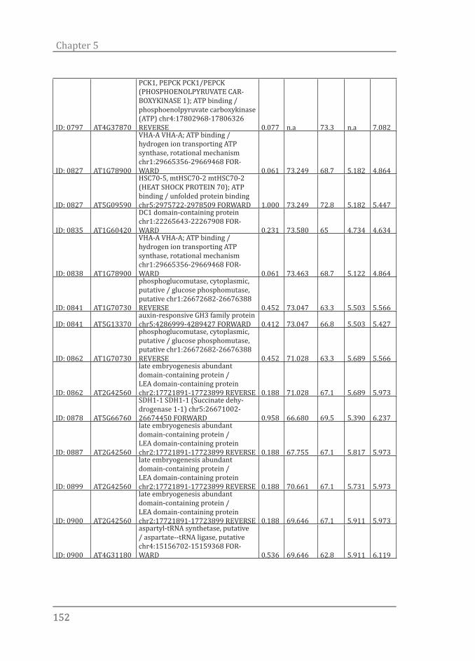
152152
Chapter 5
ID: 0797 AT4G37870
PCK1, PEPCK PCK1/PEPCK (PHOSPHOENOLPYRUVATE CAR-BOXYKINASE 1); ATP binding / phosphoenolpyruvate carboxykinase (ATP) chr4:17802968-17806326 REVERSE 0.077 n.a 73.3 n.a 7.082
ID: 0827 AT1G78900
VHA-A VHA-A; ATP binding / hydrogen ion transporting ATP synthase, rotational mechanism chr1:29665356-29669468 FOR-WARD 0.061 73.249 68.7 5.182 4.864
ID: 0827 AT5G09590
HSC70-5, mtHSC70-2 mtHSC70-2 (HEAT SHOCK PROTEIN 70); ATP binding / unfolded protein binding chr5:2975722-2978509 FORWARD 1.000 73.249 72.8 5.182 5.447
ID: 0835 AT1G60420
DC1 domain-containing protein chr1:22265643-22267908 FOR-WARD 0.231 73.580 65 4.734 4.634
ID: 0838 AT1G78900
VHA-A VHA-A; ATP binding / hydrogen ion transporting ATP synthase, rotational mechanism chr1:29665356-29669468 FOR-WARD 0.061 73.463 68.7 5.122 4.864
ID: 0841 AT1G70730
phosphoglucomutase, cytoplasmic, putative / glucose phosphomutase, putative chr1:26672682-26676388 REVERSE 0.452 73.047 63.3 5.503 5.566
ID: 0841 AT5G13370auxin-responsive GH3 family protein chr5:4286999-4289427 FORWARD 0.412 73.047 66.8 5.503 5.427
ID: 0862 AT1G70730
phosphoglucomutase, cytoplasmic, putative / glucose phosphomutase, putative chr1:26672682-26676388 REVERSE 0.452 71.028 63.3 5.689 5.566
ID: 0862 AT2G42560
late embryogenesis abundant domain-containing protein / LEA domain-containing protein chr2:17721891-17723899 REVERSE 0.188 71.028 67.1 5.689 5.973
ID: 0878 AT5G66760
SDH1-1 SDH1-1 (Succinate dehy-drogenase 1-1) chr5:26671002-26674450 FORWARD 0.958 66.680 69.5 5.390 6.237
ID: 0887 AT2G42560
late embryogenesis abundant domain-containing protein / LEA domain-containing protein chr2:17721891-17723899 REVERSE 0.188 67.755 67.1 5.817 5.973
ID: 0899 AT2G42560
late embryogenesis abundant domain-containing protein / LEA domain-containing protein chr2:17721891-17723899 REVERSE 0.188 70.661 67.1 5.731 5.973
ID: 0900 AT2G42560
late embryogenesis abundant domain-containing protein / LEA domain-containing protein chr2:17721891-17723899 REVERSE 0.188 69.646 67.1 5.911 5.973
ID: 0900 AT4G31180
aspartyl-tRNA synthetase, putative / aspartate--tRNA ligase, putative chr4:15156702-15159368 FOR-WARD 0.536 69.646 62.8 5.911 6.119

153153
Desiccation tolerance proteome
ID: 0903 AT2G42560
late embryogenesis abundant domain-containing protein / LEA domain-containing protein chr2:17721891-17723899 REVERSE 0.188 67.190 67.1 5.780 5.973
ID: 0903 AT5G64570
ATBXL4, XYL4 XYL4 (beta-xylosidase 4); hydrolase, hydrolyzing O-gly-cosyl compounds chr5:25827453-25830535 REVERSE 0.630 67.190 84.1 5.780 7.712
ID: 1000 AT3G03960chaperonin, putative chr3:1024439-1027611 FORWARD 0.760 63.755 58.8 5.251 5.014
ID: 1000 AT3G22960
PKP1, PKP-ALPHA PKP-ALPHA/PKP1 (PLASTIDIAL PYRUVATE KINASE 1); pyruvate kinase chr3:8139376-8141778 FORWARD 0.143 63.755 65 5.251 5.718
ID: 1032 AT5G21990
tetratricopeptide repeat (TPR)-con-taining protein chr5:7273398-7276321 FORWARD 0.061 63.483 60.6 5.503 5.628
ID: 1053 AT1G54100
ALDH7B4 ALDH7B4 (ALDEHYDE DEHYDROGENASE 7B4); 3-chlo-roallyl aldehyde dehydrogenase chr1:20199103-20202521 REVERSE 0.333 59.570 54.1 5.368 5.229
ID: 1058 AT5G08670
ATP synthase beta chain 1, mito-chondrial chr5:2818396-2821150 REVERSE 0.667 59.556 59.5 5.326 6.527
ID: 1058 AT5G08690
ATP synthase beta chain 2, mito-chondrial chr5:2825740-2828353 FORWARD 0.667 59.556 59.6 5.326 6.598
ID: 1068 AT1G02640
ATBXL2, BXL2 BXL2 (BETA-XYLO-SIDASE 2); hydrolase, hydrolyzing O-glycosyl compounds chr1:564293-567580 FORWARD 0.107 57.603 82.8 6.511 8.421
ID: 1068 AT3G22640cupin family protein chr3:8011909-8013890 REVERSE 0.722 57.603 54.9 6.511 7.145
ID: 1078 AT3G22640cupin family protein chr3:8011909-8013890 REVERSE 0.722 53.820 54.9 6.408 7.145
ID: 1105 AT1G51980
mitochondrial processing pep-tidase alpha subunit, putative chr1:19327361-19330440 REVERSE 0.381 57.103 54.3 5.327 6.264
ID: 1105 AT2G36530
LOS2 LOS2 (Low expression of osmotically responsive genes 1); phosphopyruvate hydratase chr2:15328160-15330865 REVERSE 0.286 57.103 47.6 5.327 5.515
ID: 1105 AT4G25580
stress-responsive protein-related chr4:13056329-13058666 FOR-WARD 0.750 57.103 66.4 5.327 6.664
ID: 1118 AT2G36640
ATECP63 ATECP63 (EMBRYONIC CELL PROTEIN 63) chr2:15364098-15365631 REVERSE 0.933 55.365 48.4 5.319 5.191
ID: 1124 AT3G22640cupin family protein chr3:8011909-8013890 REVERSE 0.722 53.902 54.9 6.497 7.145
ID: 1173 AT2G36640
ATECP63 ATECP63 (EMBRYONIC CELL PROTEIN 63) chr2:15364098-15365631 REVERSE 0.933 55.870 48.4 5.290 5.191
ID: 1173 AT5G10920
argininosuccinate lyase, puta-tive / arginosuccinase, putative chr5:3441806-3443893 FORWARD 0.148 55.870 57.4 5.290 5.681
ID: 1264 AT1G11650ATRBP45B ATRBP45B; RNA binding chr1:3914895-3917941 FORWARD 1.333 48.191 44 5.500 5.953
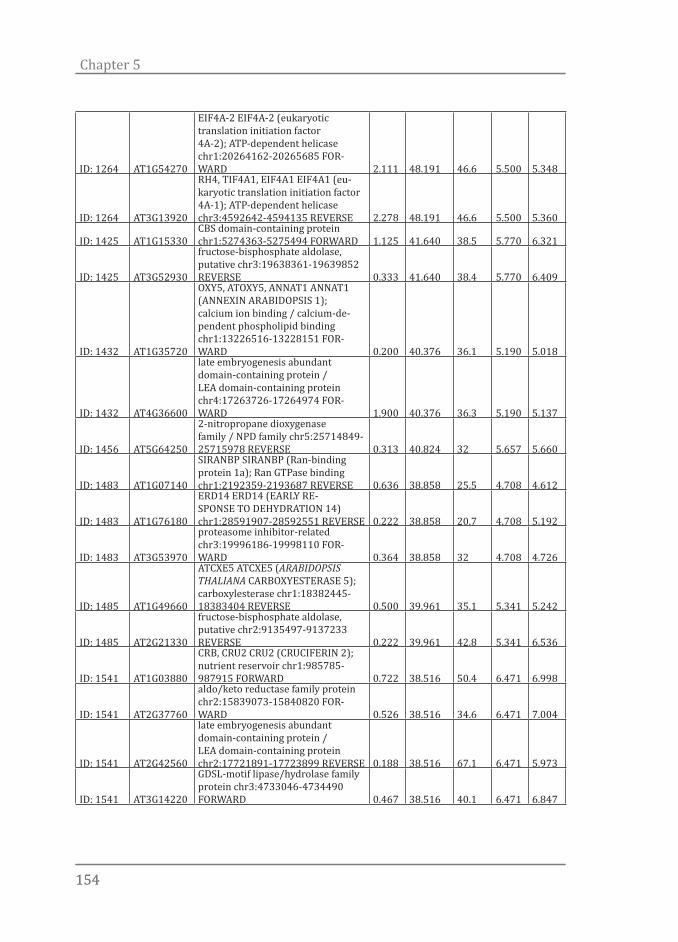
154154
Chapter 5
ID: 1264 AT1G54270
EIF4A-2 EIF4A-2 (eukaryotic translation initiation factor 4A-2); ATP-dependent helicase chr1:20264162-20265685 FOR-WARD 2.111 48.191 46.6 5.500 5.348
ID: 1264 AT3G13920
RH4, TIF4A1, EIF4A1 EIF4A1 (eu-karyotic translation initiation factor 4A-1); ATP-dependent helicase chr3:4592642-4594135 REVERSE 2.278 48.191 46.6 5.500 5.360
ID: 1425 AT1G15330CBS domain-containing protein chr1:5274363-5275494 FORWARD 1.125 41.640 38.5 5.770 6.321
ID: 1425 AT3G52930
fructose-bisphosphate aldolase, putative chr3:19638361-19639852 REVERSE 0.333 41.640 38.4 5.770 6.409
ID: 1432 AT1G35720
OXY5, ATOXY5, ANNAT1 ANNAT1 (ANNEXIN ARABIDOPSIS 1); calcium ion binding / calcium-de-pendent phospholipid binding chr1:13226516-13228151 FOR-WARD 0.200 40.376 36.1 5.190 5.018
ID: 1432 AT4G36600
late embryogenesis abundant domain-containing protein / LEA domain-containing protein chr4:17263726-17264974 FOR-WARD 1.900 40.376 36.3 5.190 5.137
ID: 1456 AT5G64250
2-nitropropane dioxygenase family / NPD family chr5:25714849-25715978 REVERSE 0.313 40.824 32 5.657 5.660
ID: 1483 AT1G07140
SIRANBP SIRANBP (Ran-binding protein 1a); Ran GTPase binding chr1:2192359-2193687 REVERSE 0.636 38.858 25.5 4.708 4.612
ID: 1483 AT1G76180
ERD14 ERD14 (EARLY RE-SPONSE TO DEHYDRATION 14) chr1:28591907-28592551 REVERSE 0.222 38.858 20.7 4.708 5.192
ID: 1483 AT3G53970
proteasome inhibitor-related chr3:19996186-19998110 FOR-WARD 0.364 38.858 32 4.708 4.726
ID: 1485 AT1G49660
ATCXE5 ATCXE5 (ARABIDOPSIS THALIANA CARBOXYESTERASE 5); carboxylesterase chr1:18382445-18383404 REVERSE 0.500 39.961 35.1 5.341 5.242
ID: 1485 AT2G21330
fructose-bisphosphate aldolase, putative chr2:9135497-9137233 REVERSE 0.222 39.961 42.8 5.341 6.536
ID: 1541 AT1G03880
CRB, CRU2 CRU2 (CRUCIFERIN 2); nutrient reservoir chr1:985785-987915 FORWARD 0.722 38.516 50.4 6.471 6.998
ID: 1541 AT2G37760
aldo/keto reductase family protein chr2:15839073-15840820 FOR-WARD 0.526 38.516 34.6 6.471 7.004
ID: 1541 AT2G42560
late embryogenesis abundant domain-containing protein / LEA domain-containing protein chr2:17721891-17723899 REVERSE 0.188 38.516 67.1 6.471 5.973
ID: 1541 AT3G14220
GDSL-motif lipase/hydrolase family protein chr3:4733046-4734490 FORWARD 0.467 38.516 40.1 6.471 6.847
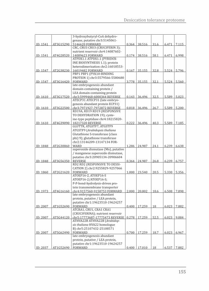
155155
Desiccation tolerance proteome
ID: 1541 AT3G15290
3-hydroxybutyryl-CoA dehydro-genase, putative chr3:5145061-5146620 FORWARD 0.364 38.516 31.6 6.471 7.115
ID: 1541 AT4G28520
CRC, CRU3 CRU3 (CRUCIFERIN 3); nutrient reservoir chr4:14087602-14089623 FORWARD 0.174 38.516 58.1 6.471 6.998
ID: 1547 AT2G38230
ATPDX1.1 ATPDX1.1 (PYRIDOX-INE BIOSYNTHESIS 1.1); protein heterodimerization chr2:16018553-16019482 FORWARD 0.167 35.155 32.8 5.524 5.796
ID: 1547 AT3G16420
PBP1 PBP1 (PYK10-BINDING PROTEIN 1) chr3:5579566-5580680 FORWARD 3.778 35.155 32.1 5.524 5.560
ID: 1610 AT3G17520
late embryogenesis abundant domain-containing protein / LEA domain-containing protein chr3:5999468-6000364 REVERSE 0.143 36.496 32.5 5.589 5.023
ID: 1610 AT3G22500
ATECP31 ATECP31 (late embryo-genesis abundant protein ECP31) chr3:7971927-7972872 REVERSE 0.818 36.496 26.7 5.589 5.200
ID: 1610 AT4G39090
RD19A, RD19 RD19 (RESPONSIVE TO DEHYDRATION 19); cyste-ine-type peptidase chr4:18215820-18217320 REVERSE 0.222 36.496 40.3 5.589 7.185
ID: 1848 AT2G30860
GLUTTR, ATGSTF7, ATGSTF9 ATGSTF9 (Arabidopsis thaliana Glutathione S-transferase (class phi) 9); glutathione transferase chr2:13146209-13147134 FOR-WARD 1.286 24.987 24.1 6.239 6.638
ID: 1848 AT3G56350
superoxide dismutase (Mn), putative / manganese superoxide dismutase, putative chr3:20905134-20906604 REVERSE 0.364 24.987 26.8 6.239 6.757
ID: 1860 AT2G21620
RD2 RD2 (RESPONSIVE TO DESSI-CATION 2) chr2:9255829-9257066 FORWARD 1.000 23.540 20.5 5.330 5.356
ID: 1973 AT4G16160
ATOEP16-2, ATOEP16-S ATOEP16-2/ATOEP16-S; P-P-bond-hydrolysis-driven pro-tein transmembrane transporter chr4:9157560-9158753 FORWARD 2.000 20.802 18.6 6.508 7.898
ID: 2007 AT1G52690
late embryogenesis abundant protein, putative / LEA protein, putative chr1:19623510-19624257 FORWARD 0.400 17.259 18 6.023 7.882
ID: 2007 AT5G44120
ATCRA1, CRU1, CRA1 CRA1 (CRUCIFERINA); nutrient reservoir chr5:17773687-17775473 REVERSE 0.278 17.259 52.5 6.023 9.084
ID: 2007 AT5G62490
ATHVA22B ATHVA22B (Arabidop-sis thaliana HVA22 homologue B) chr5:25107432-25108571 FORWARD 0.700 17.259 18.7 6.023 6.967
ID: 2037 AT1G52690
late embryogenesis abundant protein, putative / LEA protein, putative chr1:19623510-19624257 FORWARD 0.400 17.010 18 6.537 7.882
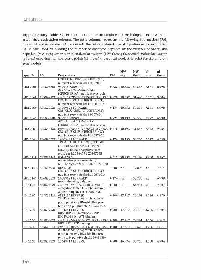
156156
Chapter 5
Supplementary Table S2. Protein spots under accumulated in Arabidopsis seeds with re-established desiccation tolerant. The table columns represent the following information: (PAI) protein abundance index. PAI represents the relative abundance of a protein in a specific spot. PAI is calculated by dividing the number of observed peptides by the number of observable peptides; (MW exp.) experimental molecular weight; (MW theor.) theoretical molecular weight; (pI exp.) experimental isoelectric point; (pI theor.) theoretical isoelectric point for the different gene models.
spot ID AGI Description PAIMW exp.
MW theor.
pI exp.
pI theor.
eID: 0060 AT1G03880
CRB, CRU2 CRU2 (CRUCIFERIN 2); nutrient reservoir chr1:985785-987915 FORWARD 0.722 10.652 50.558 7.861 6.998
eID: 0060 AT5G44120
ATCRA1, CRU1, CRA1 CRA1 (CRUCIFERINA); nutrient reservoir chr5:17773687-17775473 REVERSE 0.278 10.652 31.645 7.861 9.084
eID: 0060 AT4G28520
CRC, CRU3 CRU3 (CRUCIFERIN 3); nutrient reservoir chr4:14087602-14089623 FORWARD 0.174 10.652 58.235 7.861 6.998
eID: 0061 AT1G03880
CRB, CRU2 CRU2 (CRUCIFERIN 2); nutrient reservoir chr1:985785-987915 FORWARD 0.722 10.493 50.558 7.972 6.998
eID: 0061 AT5G44120
ATCRA1, CRU1, CRA1 CRA1 (CRUCIFERINA); nutrient reservoir chr5:17773687-17775473 REVERSE 0.278 10.493 31.645 7.972 9.084
eID: 0061 AT4G28520
CRC, CRU3 CRU3 (CRUCIFERIN 3); nutrient reservoir chr4:14087602-14089623 FORWARD 0.174 10.493 58.235 7.972 6.998
eID: 0119 AT3G55440
TPI, ATCTIMC ATCTIMC (CYTOSO-LIC TRIOSE PHOSPHATE ISOM-ERASE); triose-phosphate isom-erase chr3:20564771-20567055 FORWARD 0.615 29.993 27.169 5.600 5.167
eID: 0147 AT1G14930
major latex protein-related / MLP-related chr1:5152460-5153030 REVERSE 1.500 n.a 17.892 n.a 7.214
eID: 0147 AT4G28520
CRC, CRU3 CRU3 (CRUCIFERIN 3); nutrient reservoir chr4:14087602-14089623 FORWARD 0.174 n.a 58.235 n.a 6.998
ID: 1023 AT3G21720isocitrate lyase, putative chr3:7652796-7655880 REVERSE 0.080 n.a 64.244 n.a 7.204
ID: 1260 AT5G19510
elongation factor 1B alpha-subunit 2 (eEF1Balpha2) chr5:6581856-6583139 REVERSE 0.300 47.747 24.201 4.266 4.170
ID: 1260 AT2G37220
29 kDa ribonucleoprotein, chloro-plast, putative / RNA-binding pro-tein cp29, putative chr2:15642059-15643410 REVERSE 0.200 47.747 30.718 4.266 4.784
ID: 1260 AT5G42020
BIP2, BIP BIP (LUMINAL BIND-ING PROTEIN); ATP binding chr5:16824925-16827708 REVERSE 0.400 47.747 73.561 4.266 4.843
ID: 1260 AT5G28540BIP1 BIP1; ATP binding chr5:10540669-10543278 REVERSE 0.400 47.747 73.629 4.266 4.811
ID: 1268 AT2G37220
29 kDa ribonucleoprotein, chloro-plast, putative / RNA-binding pro-tein cp29, putative chr2:15642059-15643410 REVERSE 0.200 46.974 30.718 4.338 4.784
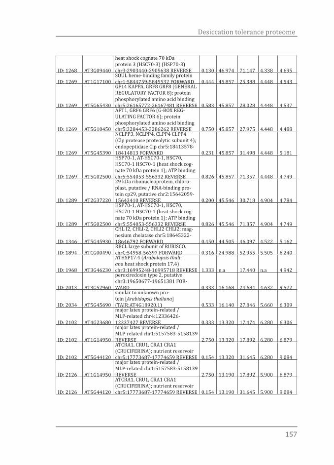
157157
Desiccation tolerance proteome
ID: 1268 AT3G09440
heat shock cognate 70 kDa protein 3 (HSC70-3) (HSP70-3) chr3:2903440-2905638 REVERSE 0.130 46.974 71.147 4.338 4.695
ID: 1269 AT1G17100SOUL heme-binding family protein chr1:5844759-5845532 FORWARD 0.444 45.857 25.388 4.448 4.543
ID: 1269 AT5G65430
GF14 KAPPA, GRF8 GRF8 (GENERAL REGULATORY FACTOR 8); protein phosphorylated amino acid binding chr5:26165772-26167481 REVERSE 0.583 45.857 28.028 4.448 4.537
ID: 1269 AT5G10450
AFT1, GRF6 GRF6 (G-BOX REG-ULATING FACTOR 6); protein phosphorylated amino acid binding chr5:3284453-3286262 REVERSE 0.750 45.857 27.975 4.448 4.488
ID: 1269 AT5G45390
NCLPP3, NCLPP4, CLPP4 CLPP4 (Clp protease proteolytic subunit 4); endopeptidase Clp chr5:18413578-18414813 FORWARD 0.231 45.857 31.498 4.448 5.181
ID: 1269 AT5G02500
HSP70-1, AT-HSC70-1, HSC70, HSC70-1 HSC70-1 (heat shock cog-nate 70 kDa protein 1); ATP binding chr5:554053-556332 REVERSE 0.826 45.857 71.357 4.448 4.749
ID: 1289 AT2G37220
29 kDa ribonucleoprotein, chloro-plast, putative / RNA-binding pro-tein cp29, putative chr2:15642059-15643410 REVERSE 0.200 45.546 30.718 4.904 4.784
ID: 1289 AT5G02500
HSP70-1, AT-HSC70-1, HSC70, HSC70-1 HSC70-1 (heat shock cog-nate 70 kDa protein 1); ATP binding chr5:554053-556332 REVERSE 0.826 45.546 71.357 4.904 4.749
ID: 1346 AT5G45930
CHL I2, CHLI-2, CHLI2 CHLI2; mag-nesium chelatase chr5:18645322-18646792 FORWARD 0.450 44.505 46.097 4.522 5.162
ID: 1894 ATCG00490RBCL large subunit of RUBISCO. chrC:54958-56397 FORWARD 0.316 24.988 52.955 5.505 6.240
ID: 1968 AT3G46230
ATHSP17.4 (Arabidopsis thali-ana heat shock protein 17.4) chr3:16995248-16995718 REVERSE 1.333 n.a 17.440 n.a 4.942
ID: 2013 AT3G52960
peroxiredoxin type 2, putative chr3:19650677-19651381 FOR-WARD 0.333 16.168 24.684 4.632 9.572
ID: 2034 AT5G45690
similar to unknown pro-tein [Arabidopsis thaliana] (TAIR:AT4G18920.1) 0.533 16.140 27.846 5.660 6.309
ID: 2102 AT4G23680
major latex protein-related / MLP-related chr4:12336426-12337427 REVERSE 0.333 13.320 17.474 6.280 6.306
ID: 2102 AT1G14950
major latex protein-related / MLP-related chr1:5157583-5158139 REVERSE 2.750 13.320 17.892 6.280 6.879
ID: 2102 AT5G44120
ATCRA1, CRU1, CRA1 CRA1 (CRUCIFERINA); nutrient reservoir chr5:17773687-17774659 REVERSE 0.154 13.320 31.645 6.280 9.084
ID: 2126 AT1G14950
major latex protein-related / MLP-related chr1:5157583-5158139 REVERSE 2.750 13.190 17.892 5.900 6.879
ID: 2126 AT5G44120
ATCRA1, CRU1, CRA1 CRA1 (CRUCIFERINA); nutrient reservoir chr5:17773687-17774659 REVERSE 0.154 13.190 31.645 5.900 9.084
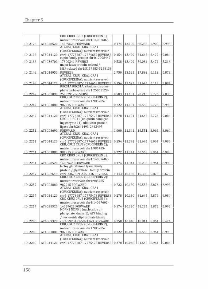
158158
Chapter 5
ID: 2126 AT4G28520
CRC, CRU3 CRU3 (CRUCIFERIN 3); nutrient reservoir chr4:14087602-14089623 FORWARD 0.174 13.190 58.235 5.900 6.998
ID: 2138 AT5G44120
ATCRA1, CRU1, CRA1 CRA1 (CRUCIFERINA); nutrient reservoir chr5:17773687-17774659 REVERSE 0.154 13.499 31.645 5.472 9.084
ID: 2138 AT4G36700cupin family protein chr4:17298447-17300341 REVERSE 0.538 13.499 59.084 5.472 5.216
ID: 2148 AT1G14950
major latex protein-related / MLP-related chr1:5157583-5158139 REVERSE 2.750 13.525 17.892 6.113 6.879
ID: 2148 AT5G44120
ATCRA1, CRU1, CRA1 CRA1 (CRUCIFERINA); nutrient reservoir chr5:17773687-17774659 REVERSE 0.154 13.525 31.645 6.113 9.084
ID: 2242 AT1G67090
RBCS1A RBCS1A; ribulose-bisphos-phate carboxylase chr1:25052128-25052912 REVERSE 0.583 11.101 20.216 5.726 7.835
ID: 2242 AT1G03880
CRB, CRU2 CRU2 (CRUCIFERIN 2); nutrient reservoir chr1:985785-987915 FORWARD 0.722 11.101 50.558 5.726 6.998
ID: 2242 AT5G44120
ATCRA1, CRU1, CRA1 CRA1 (CRUCIFERINA); nutrient reservoir chr5:17773687-17775473 REVERSE 0.278 11.101 31.645 5.726 9.084
ID: 2251 AT3G08690
UBC11 UBC11 (ubiquitin-conjugat-ing enzyme 11); ubiquitin-protein ligase chr3:2641493-2642495 FORWARD 1.000 11.341 16.551 8.964 8.064
ID: 2251 AT5G44120
ATCRA1, CRU1, CRA1 CRA1 (CRUCIFERINA); nutrient reservoir chr5:17773687-17774659 REVERSE 0.154 11.341 31.645 8.964 9.084
ID: 2251 AT1G03880
CRB, CRU2 CRU2 (CRUCIFERIN 2); nutrient reservoir chr1:985785-987915 FORWARD 0.722 11.341 50.558 8.964 6.998
ID: 2251 AT4G28520
CRC, CRU3 CRU3 (CRUCIFERIN 3); nutrient reservoir chr4:14087602-14089623 FORWARD 0.174 11.341 58.235 8.964 6.998
ID: 2257 AT1G07645
lactoylglutathione lyase family protein / glyoxalase I family protein chr1:2367609-2368346 REVERSE 1.143 10.130 15.388 5.876 6.676
ID: 2257 AT1G03880
CRB, CRU2 CRU2 (CRUCIFERIN 2); nutrient reservoir chr1:985785-987915 FORWARD 0.722 10.130 50.558 5.876 6.998
ID: 2257 AT5G44120
ATCRA1, CRU1, CRA1 CRA1 (CRUCIFERINA); nutrient reservoir chr5:17773687-17775473 REVERSE 0.278 10.130 31.645 5.876 9.084
ID: 2257 AT4G28520
CRC, CRU3 CRU3 (CRUCIFERIN 3); nutrient reservoir chr4:14087602-14089623 FORWARD 0.174 10.130 58.235 5.876 6.998
ID: 2280 AT4G09320
NDPK1 NDPK1 (nucleoside di-phosphate kinase 1); ATP binding / nucleoside diphosphate kinase chr4:5923421-5924363 FORWARD 0.750 10.048 18.814 8.964 8.474
ID: 2280 AT1G03880
CRB, CRU2 CRU2 (CRUCIFERIN 2); nutrient reservoir chr1:985785-987915 FORWARD 0.722 10.048 50.558 8.964 6.998
ID: 2280 AT5G44120
ATCRA1, CRU1, CRA1 CRA1 (CRUCIFERINA); nutrient reservoir chr5:17773687-17775473 REVERSE 0.278 10.048 31.645 8.964 9.084

159159
Desiccation tolerance proteome
ID: 2282 AT5G44120
ATCRA1, CRU1, CRA1 CRA1 (CRUCIFERINA); nutrient reservoir chr5:17773687-17775038 REVERSE 0.200 6.490 31.645 13.310 9.084
ID: 2282 AT1G03880
CRB, CRU2 CRU2 (CRUCIFERIN 2); nutrient reservoir chr1:985785-987915 FORWARD 0.722 6.490 50.558 13.310 6.998
ID: 2282 AT4G28520
CRC, CRU3 CRU3 (CRUCIFERIN 3); nutrient reservoir chr4:14087602-14089623 FORWARD 0.174 6.490 58.235 13.310 6.998
ID: 2288 AT5G44120
ATCRA1, CRU1, CRA1 CRA1 (CRUCIFERINA); nutrient reservoir chr5:17773687-17775038 REVERSE 0.200 10.325 31.645 8.458 9.084
ID: 2288 AT1G03880
CRB, CRU2 CRU2 (CRUCIFERIN 2); nutrient reservoir chr1:985785-987915 FORWARD 0.722 10.325 50.558 8.458 6.998
ID: 2288 AT4G28520
CRC, CRU3 CRU3 (CRUCIFERIN 3); nutrient reservoir chr4:14087602-14089623 FORWARD 0.174 10.325 58.235 8.458 6.998
ID: 2289 AT5G44120
ATCRA1, CRU1, CRA1 CRA1 (CRUCIFERINA); nutrient reservoir chr5:17773687-17775038 REVERSE 0.200 10.498 31.645 8.330 9.084
ID: 2289 AT1G03880
CRB, CRU2 CRU2 (CRUCIFERIN 2); nutrient reservoir chr1:985785-987915 FORWARD 0.722 10.498 50.558 8.330 6.998
ID: 2289 AT4G28520
CRC, CRU3 CRU3 (CRUCIFERIN 3); nutrient reservoir chr4:14087602-14089623 FORWARD 0.174 10.498 58.235 8.330 6.998
ID: 2317 AT4G27140
2S seed storage protein 1 / 2S albu-min storage protein / NWMU1-2S al-bumin 1 chr4:13607369-13607863 FORWARD 0.800 10.000 19.014 5.800 5.708
ID: 2317 AT5G07190
ATS3 ATS3 (ARABIDOPSIS THALI-ANA SEED GENE 3) chr5:2237611-2238489 FORWARD 0.333 10.000 23.083 5.800 6.742
ID: 2317 AT4G28520
CRC, CRU3 CRU3 (CRUCIFERIN 3); nutrient reservoir chr4:14087602-14089623 FORWARD 0.174 10.000 58.235 5.800 6.998
ID: 2345 AT5G44120
ATCRA1, CRU1, CRA1 CRA1 (CRUCIFERINA); nutrient reservoir chr5:17773687-17775473 REVERSE 0.278 n.a 31.645 n.a 9.084
ID: 2345 AT4G28520
CRC, CRU3 CRU3 (CRUCIFERIN 3); nutrient reservoir chr4:14087602-14089623 FORWARD 0.174 n.a 58.235 n.a 6.998
Supplementary Table S3. Over-representation analysis (ORA) of the over- and under-accumulated proteins followed by output simplification using REVIGO web tool. (available on line)

Quando nada acontece, há um milagre que não estamos vendo (When nothing happens there is a miracle taking place that we cannot see). Guimarães Rosa

Chapter 6General discussion

162162
Chapter 6
Unveiling desiccation tolerance
The ability of certain organisms to survive water content levels below 0.1g H2O per gram dry weight, and resume growth after re-hydration is known as desiccation tolerance (DT). DT is different from drought tolerance. Drought tolerance is the capacity to tolerate moderate dehydration, to ~0.3g H2O per gram dry weight, whereas DT refers to tolerance to the removal of almost all cellular water and replacing it by molecules that also form hydrogen bonds (Hoekstra et al., 2001). DT allowed life to move from sea to land and permitted the ascend of ‘inferior’ and ‘superior’ plants which lead to a complete re-shaping of terrestrial ecosystems up to what we are now used to (Farrant & Moore, 2011). Desiccation tolerance is such an important trait that no single group of organisms has monopolized it. DT can be found in all taxa and is the means by which a vast number of life forms (e.g. seeds, pollen, yeast and bacterial strains) can survive air-dry imposed conditions, be dispersed and long-term stored. In spite of its importance, tolerance to desiccation stress has largely been neglected in science. The ability of living systems to survive desiccation, for example the survival of orthodox seeds and unicellular organisms, was seen as an ordinary biological event, deserving little attention. However, since the late 70s, with the publication of Dry Biological Systems (Crowe & Clegg, 1978), studies of the physiology, physics, biophysics and, most recently, genetics of dry living systems have burst into bloom. Joint efforts from many researchers, who are attempting to understand DT, not only in plants but also in bacteria, yeast, fungi, roundworms, arthropods, and even in human cells has advanced our current understanding of how living systems deal with drying. Furthermore, many examples of societal uses of this knowledge, such as the stabilization of biomolecules and eukaryotic cells, have been demonstrated (Satpathy et al., 2004; Potts et al., 2005; Li et al., 2012; Loi et al., 2013). Empowered by this knowledge, by the flourishing of large-scale and high-throughput technologies, and by the decreasing cost of sequencing we experience a unique moment in which scrutinizing the mechanisms of DT to finally engineer it in desiccation sensitive (DS) organisms is no longer a dream. The work presented in this thesis describes the genetic and molecular basis of desiccation tolerance in germinating Arabidopsis seeds. With the aid
163163
General discussion
of a wide variety of techniques and approaches to search genes (Chapter 2), metabolites (Chapter 4), proteins (Chapter 5) and biological pathways (Chapters 2, 3, 4 and 5) that are important in DT, this thesis provides many leads for further research both at the fundamental and applied level. In the first part, focus lies on the methods to study DT in seeds and the advantages of using Arabidopsis and loss and re-establishment of DT as a biological system. Next, it is shown how the system of loss and re-establishment of DT in Arabidopsis seeds can be expanded and combined with the use of mutants to simplify otherwise too large datasets and to narrow down the search for candidate genes and molecules (Chapters 2, 4 and 5). In a more in depth study the role of the plant hormone abscisic acid (ABA) in the re-establishment of DT is explored (Chapter 3). Important genes, proteins and metabolites strongly correlated to DT and some of the mechanisms underlying this trait are discussed in the last section.
Arabidopsis and the re-establishment of desiccation tolerance
DT is the property of surviving the removal of all, or almost all cellular water without irreversible damage (Leprince & Buitink, 2010). The expression of DT demands cellular modifications involved in signalling mechanisms, gene regulation, functional proteomics, metabolic adjustments, antioxidant systems, macromolecular stability and physical stability (Moore et al., 2009). These responses are interlinked to many pathways, making it difficult to make a clear distinction among them. The molecular basis of DT is complex and it is not clear yet if its mechanism and regulation differs among organisms, species and tissues. Thus, to answer the intriguing question of what are the essential mechanisms necessary for DT, the responses to desiccation need to be assessed to the highest detail and, preferably, in a system that minimizes experimental noise. The vast majority of angiosperm species, including Arabidopsis, produce orthodox desiccation tolerant seeds. Such seeds acquire DT in the terminal phase of seed development and can be long term stored without significant losses in viability (Bewley et al., 2013). After development, during germination, orthodox seeds gradually lose DT and become sensitive to extreme drying. Due
164164
Chapter 6
to these changes in their desiccation tolerance behaviour, orthodox seeds are a very convenient system to study the mechanisms involved in DT (Bewley et al., 2013). Also, as mentioned, orthodox seeds are storable, which provides a continuous homogeneous source of plant material for research. Another advantage of using orthodox seeds is the possibility of re-inducing DT after it is lost during germination by the application of a mild osmotic stress before drying (Bruggink & van der Toorn, 1995). This approach has been confirmed in other species including Medicago truncatula (Buitink et al., 2003) and Tabebuia impetiginosa (Vieira et al., 2010) and is a powerful model system to study DT. Despite the advantages of using seeds with re-established DT, most of the studies on DT have been performed on the acquisition of DT during seed development (Blackman et al., 1992; Xu & Bewley, 1995; Black et al., 1999; Sreedhar, 2002; Illing et al., 2005) or its loss upon germination (Sargent et al., 1981; Daws et al., 2007; Wang et al., 2012). Acquisition of DT in developing seeds is part of the developmental program and is independent of environmental signals, such as drought, heat or cold (Bewley et al., 2013). Acquisition of DT in developing seeds seems to be largely affected by the LAFL developmental network. This network is controlled by master regulators which interact in a complex manner and include the CCAAT-box binding factor LEAFY COTYLEDON (LEC1), and the three B3 domain-containing proteins ABSCISIC ACID INSENSITIVE (ABI3), FUSCA (FUS3) and LEC2 (Jia et al., 2013). The LAFL network also regulates other diverse seed-specific process including the deposition of storage reserves, developmental arrest of the embryo, dormancy and longevity (Raz et al., 2001; Kagaya et al., 2005; To et al., 2006; Jia et al., 2013). Consequently, studying DT during development makes it difficult to assess whether the observed events are directly related to the acquisition of DT or to other development-related features (Farrant & Moore, 2011). When comparing seeds at different stages during development, a large proportion of the modifications, such as the differential expression of genes and accumulation of certain proteins, are likely not involved in promoting adaptation to dehydration per se but are related to other coincidental events. These unrelated differences are misleading when it comes to unravelling mechanisms of DT, and are undesirable. An additional drawback of studying DT during seed development is that this system is labor intensive. It requires

165165
General discussion
experiments in which flowers have to be manually pollinated and tagged, and fruit and seed development followed in time (Kermode & Bewley, 1985; Ooms et al., 1993; Verdier et al., 2013). Although simple to execute, the study of the loss of DT during germination suffers from similar problems as the ones mentioned above when different morpho-physiological states are compared. In the case of loss of DT during germination, germination related processes will obscure real DT related events. The system of loss and re-establishment of DT in germinated seeds does not suffer from the problems mentioned above and constitutes a “cleaner” method to investigate DT specific responses. To combine the advantages of a model plant species and the system of loss and re-establishment of DT we implemented it in Arabidopsis thaliana (Chapter 2, Maia et al., 2011). In this study we demonstrate that germinated Arabidopsis seeds are fully competent to regain DT if treated with an osmotic solution and that untreated seeds, in contrast, are totally sensitive to drying. Arabidopsis has been widely used as a model species in many different research fields. The Arabidopsis genome annotation is highly curated, vast gene knockout collections are available and the site of T-DNA insertions has been determined for over 300,000 independent transgenic lines. Through these collections, insertional mutants are available for most of its genes and the possibilities to use multiple knockouts are promising to eliminate redundancy. A myriad of information about gene expression, metabolites and protein profiles is available from public databases such as the ‘The Arabidopsis Information Resource’ (TAIR) database and can be readily used in comparative studies. Furthermore, the use of Arabidopsis seeds opens up the possibility to study DT in more detail and to explore the molecular basis of the signalling pathways involved in this trait. In Chapter 2 we compared the transcriptome of DS- against DT-induced seeds, which yielded a list of 677 differentially up- and down-regulated genes. In the same study we show that the capacity of Arabidopsis seeds to regain DT was lost after further seedling development and germinated seeds that had developed root hairs were no longer able to fully regain DT, which indicates that DT re-establishment is restricted to a well-defined developmental window. The list of candidate genes generated in Chapter 2, (Maia et al., 2011) is a good source of genes for investigation of their knockout lines for phenotypic

166166
Chapter 6
modifications in their ability to regain DT. The small seed size of Arabidopsis and the fact that seeds in each of the studied developmental stages have to be actively searched and manually collected, makes the use of this system too labour intensive to be applied in large genetic screens. However, a method to automate the scoring of re-establishment of DT in Arabidopsis seeds is currently being developed. Creating high-throughput methods to screen for DT will also bring this system into the phenomics era (Houle et al., 2010).
ABA signalling and desiccation tolerance
The signalling mechanisms that coordinate DT are not well understood. Some reports support the notion that changes in endogenous concentrations of ABA in response to dehydration are required in the acquisition of DT (Meurs et al., 1992; Kermode & Finch-Savage, 2002; Sreedhar, 2002; Fujita et al., 2011). Still, no conclusive evidence has been presented and it is not clear if ABA content, synthesis, degradation and sensitivity, are all important factors in the acquisition or recovery of DT. In this thesis, we advance the knowledge in this area and show that the transcriptome (Chapter 2) and the proteome (Chapter 5) involved in the re-establishment of DT, in germinated Arabidopsis seeds, depends on ABA to function and that ABA-signalling rather than increased ABA content modulates the osmotic cue which leads to the re-establishment of DT (Chapter 3). In support of these findings we show that germinated seeds from the ABA-deficient aba2-1 mutant are not competent to regain DT when treated with an osmoticum in the absence of exogenous ABA. Also, seeds from ABA-insensitive mutants (i.e. abi3-9, abi3-8, abi4-3 and abi5-7) are less competent to regain DT in comparison to Col-0 wild type seeds and decreased competence to re-establish DT during seedling development strongly correlates with decreased ABA sensitivity.
ABA levels and stress responses
ABA is known to control a wide range of plant growth and developmental processes including seed development and dormancy. ABA concentrations can
167167
General discussion
fluctuate dramatically in specific tissues during development or in response to stresses. It is commonly known that higher ABA levels, as a result of water stress, confer increased drought tolerance to whole plants. In developing seeds, ABA content may increase as much as 100-fold (Ried & Walker-Simmons, 1990). This increase in ABA content coincides with the accumulation of food reserves, such as proteins and sugars, and the so-called late embryogenesis abundant (LEA) proteins, as well as the acquisition of dormancy and desiccation tolerance (Kermode & Finch-Savage, 2002). Thus, increased amounts of ABA are believed to be necessary to induce DT. In our study, however, we show that re-establishment of DT depends on modulation of ABA sensitivity rather than enhanced ABA content. Although ABA-biosynthesis and -degradation genes were up- and down-regulated, respectively, in seeds with re-established DT, accumulation of endogenous ABA and/or its degradation products was not observed after the osmotic treatment (Chapter 3). Instead we observed a generalized increase in the expression of genes participating in ABA signalling pathways. Three genes, namely an ABA receptor (PYL7), a PP2C (AHG1) and a transcription factor (ABI5), were markedly up-regulated in the PEG-treated samples (Figure 5, Chapter 3). As mentioned in Chapter 3, ABA accumulation is not always necessary to elicit ABA related responses. For instance, studies with seedlings of Salix spp. have indicated that changes in day length regulate cessation of growth, probably by affecting ABA sensitivity (Barros & Neill, 1986). Also, plants expressing a cowpea mosaic virus (CMV) factor exhibited increased drought tolerance without over-accumulation of ABA (Westwood et al., 2013). According to these authors, CMV infection probably affected ABA signalling and/or perception instead. In the moss Tortula ruralis ABA is not detectable upon drying and exogenous application of ABA did not appear to modulate the activation of genes and synthesis of proteins (Oliver, 2005). T. ruralis seems to rely on jasmonic acid instead but the evidence for that is still elusive. Recently, Ji et al. (2011) showed a negative correlation between ABA content and drought tolerance in wheat. Drought-tolerant lines did not accumulate ABA and appeared to be more sensitive to the effect of this hormone. Altogether, these findings show that apart from enhancing ABA content, modulation of ABA perception and signalling can be sufficient to induce a proper stress response and may be part of

168168
Chapter 6
the mechanism by which DT is re-induced in germinated seeds. Still we cannot explain why ABA-biosynthesis and catabolism genes are being respectively up- and down-regulated in Arabidopsis seeds treated with osmoticum. Another feature that cannot be ignored is that different seed parts displayed variable levels of re-establishment of DT. Cotyledons were the most tolerant tissue followed by hypocotyls and roots. In this case, experiments in which more sensitive ABA detection methods are used or the study of ABA levels in isolated tissues would be of added value. Studying the re-establishment of DT in species that produce bigger seeds (more abundant biological material) would be also an option to further investigate if changes in ABA content are indeed necessary in the re-establishment of DT.
Gene networks involved in ABA-mediated DT signalling
Arabidopsis seeds acquire desiccation tolerance during embryo development. The regulation of these two processes is orchestrated by a network that includes the master regulators LEAFY COTYLEDON 1 (LEC1), LEC1-LIKE (L1L), and the B3 domain transcription factors, LEAFY COTYLEDON 2 (LEC2), FUSCA3 (FUS3), and ABSCISIC ACID INSENSITIVE 3 (ABI3) (Gutierrez et al., 2007; Santos-Mendoza et al., 2008). ABI3 can form homodimers but also heterodimers with ABI5 and they regulate the expression of several downstream genes which act upon both seed development and acquisition of DT (Nakamura et al., 2001). Interestingly, the vast majority of ABA-insensitive and –deficient mutants described to date are still able to produce completely desiccation tolerant seeds by the end of seed maturation (Cutler et al., 2010). Exceptions are the strong abi3 alleles and the genetic combination of a weak abi3-1 allele with the ABA-deficient aba1-1 mutant, which display severe developmental problems and impaired capacity to acquire DT during seed maturation (Nambara et al., 1992; Ooms et al., 1993; Ooms et al., 1994). In Chapter 3 we show that ABA deficiency or sensitivity hampers the re-establishment of DT in germinated Arabidopsis seeds but does not harm its acquisition during seed development. All mutants analysed (i.e. aba2-1, abi3-9, abi3-8, abi4-3 and abi5-7) in this study were desiccation tolerant by the end of seed development. In contrast, the same mutants were compromised in

169169
General discussion
their capacity to regain DT after radicle protrusion (Stage II). Before radical protrusion, however, both ABA-deficient and –insensitive mutant seeds were still able to regain DT, which implies that during seed development and during seedling development partially overlapping genetic networks are utilized to induce DT. Because strong alleles of the abi3 mutant suffer from developmental issues and do not acquire DT during development we are confident to say that at least ABI3 is participating in both networks. Other genes like ABI5 could also be involved in the acquisition of DT, but the high redundancy observed in this TF family (Finkelstein et al., 2005) might explain why single gene knockouts do not reveal any acquisition of DT phenotypes. Acquisition of DT during development seems to be more strongly controlled than its re-establishment after germination. Apparently, other signalling factors necessary to activate DT genes might be already missing after visible germination and characterize a point of no return where germinated seeds are fully engaged in germination and committed to enter vegetative growth. It has been hypothesized that ABI5 is necessary to bring germinated embryos into a quiescent state upon drought, thereby protecting young seedlings from the loss of water (Finkelstein & Lynch, 2000; Lopez-Molina et al., 2001). In agreement with this, we also observed up-regulation of the ABI5 gene expression in germinated seeds under osmotic stress. The inability of ABI5 to respond to osmotic stress in seeds beyond Stage II coincides with the developmental checkpoint were DT can no longer be re-induced (Chapter 3). Apparently there is a programmatic conflict where germinating seeds have to “choose” whether to return to a developmental-like program and become DT again or to become a (DS) seedling, and ABI5 is involved in it.
ABA signalling triggers shifts in the transcriptome, proteome and metabolome of germinated Arabidopsis seeds, resulting in the acquisition of DT: integration of omics data
Approaches to combining untargeted assessments of gene expression, protein- and metabolite profiles present a powerful tool to better understand the regulation and mechanisms of DT. Often, comparisons between different experiments and methods are hampered because different developmental

170170
Chapter 6
stages or plant growing conditions are used. With the system developed in this thesis we have generated a reliable system to study DT (Chapter 2). In our system germinated seeds are selected based on precise morphological traits, such as the rupture of the seed coat or the protrusion of the radicle tip. Once they have reached those stages, seeds are treated to regain DT. With the aid of this experimental system we have evaluated DT related mechanisms at the physiological (Chapters 2 and 3), transcriptomic (Chapter 2), metabolomic (Chapter 4) and proteomic level (Chapter 5). Our goal was to integrate the three datasets to get better insight in the mechanisms controlling DT and to narrow down the selection of candidate genes for further gene function studies. Bearing in mind the vast amount of data generated by the methods used in our study, and the high probability of noise (e.g. unrelated genes, false positives and negatives) that is inherent to these methods, it was crucial to use a more elaborate experimental design in which we also included the aba2-1 mutant (Chapters 4 and 5). This, in our view, simplified the output by reducing noise (i.e. unrelated genes) and facilitated data interpretation. Meaningful interpretation of the data, however, demands appropriate analysis methods and, preferably user-friendly, bioinformatics tools. One such tool is the MapMan package (http://MapMan.gabipd.org) (Thimm et al., 2004). This tool allowed us to display the three datasets onto pictographic diagrams to get a global overview of the ontology of the up- and down-regulated genes, proteins and metabolites. Other tools which were used were GeneTrailExpress for over-representation analysis (ORA) of significantly enriched gene ontologies (GO) (Keller et al., 2008), and Athena, which mines for enriched cis-acting promoter elements that can potentially be involved in regulating gene sets of interest (O’Connor et al., 2005). For example, in our transcriptome study, the ‘Response to Water Deprivation’ and ‘Abscisic Acid Stimulus’ categories were, at the same time, represented by a high number of genes and high-ranked (observed/expected number of genes), showing that water stress responsive genes were recruited and confirming that ABA is relevant in the re-establishment of DT. In our protein study (Chapter 5) we observed that ‘Response to Desiccation’ was the enriched GO term with the highest observed to expected protein number ratio and which was highly significant (P-value = 6.10-07). These insights are good indications of the accuracy and stringency of our gene and protein selection procedure to pinpoint DT-related mechanisms. Moreover,
171171
General discussion
many LEA proteins were retrieved in both datasets. Since LEA proteins have been largely correlated to tolerance to various abiotic stresses, particularly desiccation (Tunnacliffe & Wise, 2007; Battaglia et al., 2008), this is also an indication that the other proteins and genes retrieved in our study are likely to play a role in DT. Overlapping the results of our transcriptome, proteome and metabolome revealed interesting links between metabolites and transcripts. For example, genes such as SUCROSE SYNTHASE 3 (SUS3) and GALACTINOL SYNTHASE (GOLS-1 and GOLS-2) were up-regulated (Chapter 2) together with the accumulation of sucrose, galactinol, and raffinose (Chapter 4). SUS3 encodes a sucrose synthase which has been shown to be important for sucrose metabolism in developing seeds, especially during the late maturation phase when DT is acquired (Angeles-Núñez & Tiessen, 2012). Galactinol, raffinose, and sucrose are involved in the biosynthesis of raffinose family oligosaccharides (RFOs), such as stachyose and verbascose (Loewus & Murthy, 2000; Peterbauer et al., 2002). Although raffinose accumulated in seeds with re-established DT, raffinose synthase genes were not differentially up-regulated. Only the DARK INDUCIBLE 10 (DIN10) gene, which encodes a glycosyl hydrolase, was down-regulated in seeds with re-established DT. DIN10 expression has been shown to be suppressed when sucrose is supplied (Fujiki et al., 2001). Thus, the high levels of sucrose found provides a possible explanation why this gene was down-regulated. Together, these observations provide direct links between metabolite abundance and gene expression. They also reinforce the importance of certain sugars, especially sucrose and raffinose and sugar-alcohols, such as galactinol in the re-establishment of DT. In line with our findings, sucrose and RFOs have been associated with DT in seeds (Angelovici et al., 2010) and resurrection plants (Peters et al., 2007; Lehner et al., 2008). RFOs have been shown to accumulate to relatively high levels during the acquisition of DT in maturing Arabidopsis seeds (Taji et al., 2002). Increased intracellular levels of galactinol and raffinose in transgenic Arabidopsis plants overexpressing the HEAT SHOCK TRANSCRIPTION FACTOR A2 (HSFA2) or GOLS-2 correlated with increased tolerance to salt, chilling and drought stress (Taji et al., 2002; Nishizawa et al., 2008). Finally, RFOs are also believed to contribute to the structural stability of organelles, membranes, enzymes and proteins, other macromolecules, and the glassy state, which is enhanced in the presence of LEA proteins (Koster, 1991; Crowe et al., 1992; Horbowicz & Obendorf, 1994) which,

172172
Chapter 6
were markedly present in our transcriptome and proteome datasets. In the transcriptome of DT re-established seeds, LEA-, seed storage reserve- and sugar metabolism-related transcripts stood out (Chapter 2). A similar picture emerged from our protein analysis (Chapter 5). In this data set, proteins involved in sugar metabolism were overrepresented and the only enriched KEGG pathway was Glycolysis/Gluconeogenesis. Remarkably, from the 50 LEA proteins identified so far in Arabidopsis, 26 were retrieved in our transcriptome study as displaying increased expression in DT re-established seeds (FC ≥ 1.5 and p-value ≤ 0.01). Nine of those LEA proteins were also present in the over accumulated protein set. The LEA proteins found in the two datasets belonged to the highest up-regulated in seeds with re-established DT. Interestingly, six of those LEA proteins are classified as LEA_4 (Hundertmark & Hincha, 2008), which is suggestive of a specific role of this type of LEA proteins in re-establishment of DT. The LEA_4 family is very heterogeneous and its gene products differ greatly in size and in hydrophilicity (Hundertmark & Hincha, 2008). Interestingly, the LEA_4 family members identified in our proteome set are significantly enriched with charged and polar amino acids and are relatively more hydrophilic in comparison to other members of this family (Chapter 5). Also, the fraction of LEA proteins up-regulated in seeds with re-established DT was enriched with hydrophilic LEA proteins while the down regulated LEA proteins were mainly hydrophobic. Taken together, the consistent up-regulation of sugar metabolism-related genes and proteins and the actual accumulation of sugars such as sucrose and raffinose in seeds with re-established DT, we propose that negatively charged LEA proteins, especially LEA_4, are acting together with those sugars in order to stabilize bioglasses. Together they may form a tight hydrogen-bonding network in the dehydrating cytoplasm (Hoekstra et al., 2001). We also suggest that LEA proteins could be acting on their own via steric and electrostatic prevention of interactions between aggregation-prone and partially unfolded proteins (Tunnacliffe & Wise, 2007). During drying, the chance of protein unfolding and aggregation increases due to physicochemical changes in the cell cytoplasm. Thus, charged LEA proteins that accumulate in the drying cytoplasm could be acting via creating a matrix where, besides providing extra hydrogen-bonding, they repel each other, leaving open spaces which will be filled by DS proteins. In such a way proteins that are more likely to form aggregates or denaturate, such as enzymes, would be protected
173173
General discussion
and interaction with each other would be prevented. For example, LEA proteins have been proposed to act as “molecular shields”, which prevent the approach and interaction of aggregation-prone protein species by steric or electrostatic repulsion, analogous to polymer stabilization of colloidal suspensions (Liu & Luijten, 2004; Chakrabortee et al., 2007; Xing et al., 2012). Another interesting analogy to what we propose as a mechanism of action of LEA proteins in DT was described in mammalian myoglobin. In the case of mammalian myoglobin, amino acid charge-increasing substitutions at multiple sites, and their accumulation, correspond to increases in muscle myoglobin concentrations and the adoption of both semi- and fully aquatic lifestyles. Similar to LEA proteins, myoglobins with increased net surface charge allowed high density of this protein per muscle unit while electrosterically preventing their aggregation and loss of function (Mirceta et al., 2013). Thus, increasing negative charges on the protein surface seems to be a basal mechanism of adaptation to conditions where aggregation-prone proteins and membranes are physically forced together, as in the case of desiccation stress. Considering that, studies comparing the amino acid composition of homologous LEA proteins from DS and DT organisms have the potential to shed light on the evolution of this intriguing family of proteins and on how they became so important in desiccation tolerant life forms.
Summary: a model of the re-establishment of DT in germinated Arabidopsis seeds
The main events involved in the re-establishment of desiccation tolerance in germinated Arabidopsis seeds are summarized in Figure 1. In our efforts to help understanding of DT we generated a system of loss and re-establishment of DT in germinated Arabidopsis seeds (Chapters 2 and 3). Using this system, we found several genes (Chapter 2), proteins (Chapter 5) and metabolites (Chapter 4) that where correlated to DT. We also showed that osmotic stress can re-induce DT in Arabidopsis via re-setting developmentally related processes involved in DT and that developmental arrest is a fundamental response required in the re-establishment of DT. Surprisingly, ABA content did not increase in response to osmoticum and such developmental related processes are likely controlled by ABA via the modulation of its signal instead. Despite our efforts to analyse

174174
Chapter 6
the three datasets produced in our study in detail we were not able to exploit their full potential yet. The identification of differentially expressed genes and over-accumulated proteins and metabolites is only the beginning of the gene discovery process, especially in relation to understanding the underlying genetic components of DT. Our studies generated valuable insights about DT and lists of genes, proteins and metabolites that can be directly used in the generation of new hypothesis and planning of follow-up experiments.
Germination /developmental arrest23 - (ABI5)3
Synthesis and accumulation of LEA proteins25
- (specially hydrophilic LEA_4 family members)
Accumulation of soluble sugars and sugar alcohols245
- Up-regulation of GOLS1, GOLS2 and SUS3- Sucrose- Raffinose- Galactinol
Hydrophilic LEA-proteins and sugars aiding to bioglass formationH
Signalling perception and ABA signalling moulation23
Enhanced ABA signalling3
- (PYL7, PYL9; ABI5) (PP2Cs ?)
ABA content remains constant3
Activation of drought and ABA responsive genes245 - (many LEA and maturation protein genes)
Molecular shield formationH
hydrophilic charged LEA proteins electrosterically protect other macromolecules from interacting and aggregating (e.g. enzymes)
Water content
DT
DS
PEG / ABA
H2O
Re-establishment of DT cycle
Desiccation tolerance
Activation of the atioxidant system25
(ALDH7B4, SOD(Mn))
Water content
Figure 1 Theoretical model depicting the main events controlling the re-establishment of desiccation tolerance in germinated Arabidopsis seeds. Desiccation tolerance can be fully re-established in germinated desiccation sensitive (DS) Arabidopsis seeds by treating them with polyethylene glycol (PEG), abscisic acid (ABA) or both. Once desiccation tolerance is fully re-established, the desiccation tolerant (DT) germinated Arabidopsis seeds can be further dried to water contents below 0.1 g H2O g-1 dry weight. After re-hydration, the DT seeds will resume growth and continue their development. Superscript letters represent the following information: T transcriptomic analysis – Chapter 2; E expression studies – Chapter 3; M metabolite profiling – Chapter 4; P proteomic study – Chapter 5; H Hypothetical mechanisms. Processes followed by more than one superscript letter were confirmed in more than one study.

175175
General discussion
Further considerations
Significant progress has been achieved in the field of desiccation tolerance. To date, many important components of DT, such as the soluble sugars and oligosaccharides, antioxidants and proteins with protective functions, have been uncovered (Moore et al., 2008; Moore et al., 2009; Leprince & Buitink, 2010; Farrant & Moore, 2011). Different organisms, however, might still have their “preferences” and follow different strategies to achieve anhydrobiosis (life without water). Still, it seems that, independent of molecular identity, their mode of action, in many cases, can be universal. For example, different sugars can be applied in the formation of biological glasses with sucrose (Chapter 4) and trehalose as the most important sugars in DT in plants and animals, respectively (Buitink & Leprince, 2004; Berjak, 2006; Iturriaga et al., 2009). Different proteins that share certain properties such as charge and solubility can also display similar properties in the stabilization of biomolecules (Chapter 5) (Hand et al., 2011). Due to these properties, there is increasing interest in using such DT mechanisms that can be extrapolated from seeds, nematodes and yeast cells, among other DT organisms, to human cells, to the dry-storage of vaccines, and to crops (Crowe, 2005; Potts et al., 2005). This potential has directed increasing attention to this intriguing phenomenon and exploiting the diversity of molecules that might be involved in DT is a priceless source of information when considered for their biotechnological applications. From our study and from the literature it is clear that desiccation tolerance is influenced by a multitude of genes, proteins and metabolites. In this thesis, we used three high-throughput approaches; namely transcriptomics, metabolomics and proteomics to investigate DT in germinated Arabidopsis seeds. Our goal was to generate datasets in different layers and integrate them to get better insights in the mechanisms controlling DT and to narrow down the selection of candidate genes for further gene function studies. Although our microarray study (Chapter 2) generated the biggest dataset, transcript content is not always directly proportional to protein content. Protein content will be driven not only by the initial amount of a certain mRNA template but is also highly dependent on the control of ribosome recruitment and modulation of the elongation or termination of protein synthesis and protein degradation
176176
Chapter 6
(Gebauer & Hentze, 2004). However, only in the case of microarrays a full genome approach was taken (Chapter 2). In contrast, only the most abundant proteins were spotted in our 2D acrylamide gels (Chapter 5) and most of the existent metabolites were not identified or even isolated (Chapter 4). Thus, relying only on exact matches between the different datasets will certainly exclude many interesting features that can still be seen at the transcriptional level, such as the activation of certain transcription factors. In this respect, the use of strategies in which array experiments covering more physiological/developmental conditions, as in a recent study by Verdier et al. (2013), are valuable. In this study a conditional-dependent network of global transcription interactions together with metabolite profiling, revealed distinct co-expression modules related to the acquisition of DT, longevity, and pod abscission. With the aid of such networks more assertive guesses in the selection of candidate genes can be made. In our system, we focused on late DT mechanisms. Seeds before (DS) and after three days of osmotic- or ABA-treatment, when they were fully DT, were analysed. Thus, to pinpoint early mechanisms associated with DT and to reveal the pathways controlling it, time series experiments, monitoring gene expression, protein composition and metabolite abundance during the re-establishment of DT in PEG or ABA incubation will be essential. With the aid of such experiments metabolite fluxes can be investigated, which can reveal the most important metabolic pathways involved in DT. The combination of transcriptomics with non-gel based proteome profiling approaches also holds promise in the study of DT. Such methods can produce more extensive protein datasets and are, in most cases, less labour intensive than gel-based approaches (Abdallah et al., 2012). Despite the advantages of using germinated Arabidopsis seeds to study DT in plants, it is important to mention that other interesting systems are also available. The most common are desiccation tolerant mosses, ferns and resurrection plants (Challabathula & Bartels, 2013). With the recent developments in sequencing technology these non-model species became suitable for gene expression and in-depth protein profiling studies. These approaches, together with comparative studies with non-desiccation tolerant plants, will certainly provide novel insights in the molecular processes required

177177
General discussion
for the acquisition of DT and will elucidate genes with unknown functions. Comparing such datasets with the available information is also a challenge and will require new tools for data analysis, interpretation and visualization.
References
Abdallah C, Dumas-Gaudot E, Renaut J, Sergeant K. 2012. Gel-based and gel-free quantitative proteomics approaches at a glance. International Journal of Plant Genomics 2012: 17.
Angeles-Núñez J, Tiessen A. 2012. Regulation of AtSUS2 and AtSUS3 by glucose and the transcription factor LEC2 in different tissues and at different stages of Arabidopsis seed development. 78(4-5): 377-392.
Angelovici R, Galili G, Fernie AR, Fait A. 2010. Seed desiccation: a bridge between maturation and germination. Trends in Plant Science 15(4): 211-218.
Barros RS, Neill SJ, 1986. Periodicity of response to abscisic acid in lateral buds of willow (Salix viminalis L.). Planta 168, 530-5.
Battaglia M, Olvera-Carrillo Y, Garciarrubio A, Campos F, Covarrubias AA. 2008. The enigmatic LEA proteins and other hydrophilins. Plant physiology 148(1): 6-24.
Berjak P. 2006. Unifying perspectives of some mechanisms basic to desiccation tolerance across life forms. Seed Science Research 16(1): 1-15.
Bewley JD, Bradford K, Hilhorst H, Nonogaki H. 2013. Seeds: Physiology of Development, Germination and Dormancy: Springer.
Black M, Corbineau F, Gee H, Come D. 1999. Water content, raffinose, and dehydrins in the induction of desiccation tolerance in immature wheat embryos. Plant physiology 120(2): 463-472.
Blackman Sa, Obendorf RL, Leopold aC. 1992. Maturation proteins and sugars in desiccation tolerance of developing soybean seeds. Plant physiology 100(1): 225-230.
Bruggink T, van der Toorn P. 1995. Induction of desiccation tolerance in germinated seeds. Seed Science Research 5(1): 1-4.
Buitink J, Leprince O. 2004. Glass formation in plant anhydrobiotes: Survival in the dry state. Cryobiology 48(3): 215-228.
Buitink J, Vu BL, Satour P, Leprince O. 2003. The re-establishment of desiccation tolerance in germinated radicles of Medicago truncatula Gaertn. seeds. Seed Science Research 13(4): 273-286.
Chakrabortee S, Boschetti C, Walton LJ, Sarkar S, Rubinsztein DC, Tunnacliffe A. 2007. Hydrophilic protein associated with desiccation tolerance exhibits broad protein stabilization function. Proceedings of the National Academy of Sciences of the United States of America 104(46): 18073-18078.
Challabathula D, Bartels D. 2013. Desiccation tolerance in resurrection plants: new insights from transcriptome, proteome and metabolome analysis. Frontiers in Plant Science 4.
Crowe JH. 2005. Stabilization of Dry Mammalian Cells: Lessons from Nature. Integrative
178178
Chapter 6
and Comparative Biology 45(5): 810-820.Crowe JH, Clegg JS. 1978. Dry Biological Systems. New York: Academic Press.Crowe JH, Hoekstra FA, Crowe LM. 1992. Anhydrobiosis. Annual Review of Physiology
54(1): 579-599.Cutler SR, Rodriguez PL, Finkelstein RR, Abrams SR 2010. Abscisic acid: Emergence
of a core signaling network. Annual Review of Plant Biology. 651-679.Daws MI, Bolton S, Burslem DFRP, Garwood NC, Mullins CE. 2007. Loss of desiccation
tolerance during germination in neo-tropical pioneer seeds: implications for seed mortality and germination characteristics. Seed Science Research 17(04): 273-281.
Farrant JM, Moore JP. 2011. Programming desiccation-tolerance: From plants to seeds to resurrection plants. Current Opinion in Plant Biology 14(3): 340-345.
Finkelstein R, Gampala SSL, Lynch TJ, Thomas TL, Rock CD. 2005. Redundant and distinct functions of the ABA response loci ABA-INSENSITIVE(ABI)5 and ABRE-BINDING FACTOR (ABF)3. Plant Molecular Biology 59(2): 253-267.
Finkelstein RR, Lynch TJ. 2000. The Arabidopsis Abscisic Acid Response Gene ABI5 Encodes a Basic Leucine Zipper Transcription Factor. The Plant Cell Online 12(4): 599-609.
Fujiki Y, Yoshikawa Y, Sato T, Inada N, Ito M, Nishida I, Watanabe A. 2001. Dark-inducible genes from Arabidopsis thaliana are associated with leaf senescence and repressed by sugars. Physiologia Plantarum 111(3): 345-352.
Fujita Y, Fujita M, Shinozaki K, Yamaguchi-Shinozaki K. 2011. ABA-mediated transcriptional regulation in response to osmotic stress in plants. Journal of Plant Research 124(4): 509-525.
Gebauer F, Hentze MW. 2004. Molecular mechanisms of translational control. 5(10): 827-835.
Gutierrez L, Van Wuytswinkel O, Castelain M, Bellini C. 2007. Combined networks regulating seed maturation. Trends in Plant Science 12(7): 294-300.
Hand SC, Menze MA, Toner M, Boswell L, Moore D. 2011. LEA proteins during water stress: Not just for plants anymore. Annual Review of Physiology 73(1): 115-134.
Hoekstra Fa, Golovina Ea, Buitink J. 2001. Mechanisms of plant desiccation tolerance. Trends in Plant Science 6(9): 431-438.
Horbowicz M, Obendorf RL. 1994. Seed desiccation tolerance and storability: Dependence on flatulence-producing oligosaccharides and cyclitols—review and survey. Seed Science Research 4(4): 385-405.
Houle D, Govindaraju DR, Omholt S. 2010. Phenomics: The next challenge. Nature Reviews Genetics 11(12): 855-866.
Hundertmark M, Hincha D. 2008. LEA (late embryogenesis abundant) proteins and their encoding genes in Arabidopsis thaliana. BMC Genomics 9(1): 118.
Illing N, Denby KJ, Collett H, Shen A, M FJ. 2005. The signature of seeds in resurrection plants: A molecular and physiological comparison of desiccation tolerance in seeds and vegetative tissues. Integrative and Comparative Biology 45(5): 771-787.
Iturriaga G, Suárez R, Nova-Franco B. 2009. Trehalose metabolism: From osmoprotection to signaling. International Journal of Molecular Sciences 10(9): 3793-3810.
Ji X, Dong B, Shiran B, Talbot MJ, Edlington JE, Hughes T, White RG, Gubler
179179
General discussion
F, Dolferus R. 2011. Control of abscisic acid catabolism and abscisic acid homeostasis is important for reproductive stage stress tolerance in cereals. Plant physiology 156(2): 647-662.
Jia H, Suzuki M, McCarty DR. 2013. Regulation of the seed to seedling developmental phase transition by the LAFL and VAL transcription factor networks. Wiley Interdisciplinary Reviews: Developmental Biology 3: 135–145.
Kagaya Y, Toyoshima R, Okuda R, Usui H, Yamamoto A, Hattori T. 2005. LEAFY COTYLEDON1 controls seed storage protein genes through its regulation of FUSCA3 and ABSCISIC ACID INSENSITIVE3. Plant and Cell Physiology 46(3): 399-406.
Keller A, Backes C, Al-Awadhi M, Gerasch A, Kuntzer J, Kohlbacher O, Kaufmann M, Lenhof H-P. 2008. GeneTrailExpress: a web-based pipeline for the statistical evaluation of microarray experiments. BMC Bioinformatics 9(1): 552.
kermode AR, Bewley JD. 1985. The role of maturation drying in the transition from seed development to germination: I. ACQUISITION OF DESICCATION–TOLERANCE AND GERMINABILITY DURING DEVELOPMENT OF Ricinus communis L. SEEDS. Journal of Experimental Botany 36(12): 1906-1915.
Kermode AR, Finch-Savage BE 2002. Desiccation sensitivity in orthodox and recalcitrant seeds in relation to development. In: Black M, Pritchard HW eds. Desiccation and survival in plants: drying without dying. New York: CABI publishing, 149-184.
Koster KL. 1991. Glass Formation and Desiccation Tolerance in Seeds. Plant physiology 96(1): 302-304.
Lehner A, Chopera DR, Peters SW, Keller F, Mundree SG, Thomson JA, Farrant JM. 2008. Protection mechanisms in the resurrection plant Xerophyta viscosa: Cloning, expression, characterisation and role of XvINO1, a gene coding for a myo-inositol 1-phosphate synthase. Functional Plant Biology 35(1): 26-39.
Leprince O, Buitink J. 2010. Desiccation tolerance: From genomics to the field. Plant Science 179(6): 554-564.
Li S, Chakraborty N, Borcar A, Menze MA, Toner M, Hand SC. 2012. Late embryogenesis abundant proteins protect human hepatoma cells during acute desiccation. Proceedings of the National Academy of Sciences 109(51): 20859-20864.
Liu J, Luijten E. 2004. Stabilization of Colloidal Suspensions by Means of Highly Charged Nanoparticles. Physical Review Letters 93(24): 247802.
Loewus FA, Murthy PPN. 2000. myo-Inositol metabolism in plants. Plant Science 150(1): 1-19.
Loi P, Iuso D, Czernik M, Zacchini F, Ptak G. 2013. Towards storage of cells and gametes in dry form. Trends in Biotechnology 31(12): 688-695.
Lopez-Molina L, Mongrand S, Chua N-H. 2001. A postgermination developmental arrest checkpoint is mediated by abscisic acid and requires the ABI5 transcription factor in Arabidopsis. Proceedings of the National Academy of Sciences of the United States of America 98(8): 4782-4787.
Maia J, Dekkers BJW, Provart NJ, Ligterink W, Hilhorst HWM. 2011. The re-establishment of desiccation tolerance in germinated Arabidopsis thaliana seeds

180180
Chapter 6
and its associated transcriptome. PLoS ONE 6(12).Meurs C, Basra AS, Karssen CM, van Loon LC. 1992. Role of Abscisic Acid in the
Induction of Desiccation Tolerance in Developing Seeds of Arabidopsis thaliana. Plant physiology 98(4): 1484-1493.
Mirceta S, Signore AV, Burns JM, Cossins AR, Campbell KL, Berenbrink M. 2013. Evolution of Mammalian Diving Capacity Traced by Myoglobin Net Surface Charge. Science 340(6138).
Moore JP, Le NT, Brandt WF, Driouich A, Farrant JM. 2009. Towards a systems-based understanding of plant desiccation tolerance. Trends Plant Sci 14(2): 110-117.
Moore JP, Vicré-Gibouin M, Farrant JM, Driouich A. 2008. Adaptations of higher plant cell walls to water loss: drought vs desiccation. Physiologia Plantarum 134(2): 237-245.
Nakamura S, Lynch TJ, Finkelstein RR. 2001. Physical interactions between ABA response loci of Arabidopsis. The Plant journal : for cell and molecular biology 26(6): 627-635.
Nambara E, Naito S, McCourt P. 1992. A mutant of Arabidopsis which is defective in seed development and storage protein accumulation is a new abi3 allele. Plant Journal 2(4): 435-441.
Nishizawa A, Yabuta Y, Shigeoka S. 2008. Galactinol and raffinose constitute a novel function to protect plants from oxidative damage. Plant physiology 147(3): 1251-1263.
O’Connor TR, Dyreson C, Wyrick JJ. 2005. Athena: a resource for rapid visualization and systematic analysis of Arabidopsis promoter sequences. Bioinformatics 21(24): 4411-4413.
Oliver MJ. 2005. Desiccation Tolerance in Bryophytes: A Reflection of the Primitive Strategy for Plant Survival in Dehydrating Habitats? Integrative and Comparative Biology 45(5): 788-799.
Ooms JJJ, Léon-Kloosterziel KM, Bartels D, Koornneef M, Karssen CM. 1993. Acquisition of desiccation tolerance and longevity in seeds of Arabidopsis thaliana: A comparative study using abscisic acid-insensitive abi3 mutants. Plant physiology 102(4): 1185-1191.
Ooms JJJ, van der Veen R, Karssen CM. 1994. Abscisic acid and osmotic stress or slow drying independently induce desiccation tolerance in mutant seeds of Arabidopsis thaliana. Physiologia Plantarum 92(3): 506-510.
Peterbauer T, Mucha J, Mach L, Richter A. 2002. Chain elongation of raffinose in pea seeds. Isolation, characterization, and molecular cloning of a multifunctional enzyme catalyzing the synthesis of stachyose and verbascose. Journal of Biological Chemistry 277(1): 194-200.
Peters S, Mundree SG, Thomson Ja, Farrant JM, Keller F. 2007. Protection mechanisms in the resurrection plant Xerophyta viscosa (Baker): both sucrose and raffinose family oligosaccharides (RFOs) accumulate in leaves in response to water deficit. Journal of Experimental Botany 58(8): 1947-1956.
Potts M, Slaughter SM, Hunneke F, Garst JF, Helm RF. 2005. Desiccation Tolerance of Prokaryotes: Application of Principles to Human Cells. Integrative and Comparative Biology 45(5): 800-809.
181181
General discussion
Raz V, Bergervoet JH, Koornneef M. 2001. Sequential steps for developmental arrest in Arabidopsis seeds. Development 128(2): 243-252.
Ried JL, Walker-Simmons MK. 1990. Synthesis of Abscisic Acid-Responsive, Heat-Stable Proteins in Embryonic Axes of Dormant Wheat Grain. Plant physiology 93(2): 662-667.
Santos-Mendoza M, Dubreucq B, Baud S, Parcy F, Caboche M, Lepiniec L. 2008. Deciphering gene regulatory networks that control seed development and maturation in Arabidopsis. Plant Journal 54(4): 608-620.
Sargent JA, Mandi SS, Osborne D. 1981. The loss of desiccation tolerance during germination: An ultrastructural and biochemical approach. 105(3-4): 225-239.
Satpathy GR, Török Z, Bali R, Dwyre DM, Little E, Walker NJ, Tablin F, Crowe JH, Tsvetkova NM. 2004. Loading red blood cells with trehalose: a step towards biostabilization. Cryobiology 49(2): 123-136.
Sreedhar L. 2002. In vivo characterization of the effects of abscisic acid and drying protocols associated with the acquisition of desiccation tolerance in alfalfa (Medicago sativa L.) somatic embryos. Annals of Botany 89(4): 391-400.
Taji T, Ohsumi C, Iuchi S, Seki M, Kasuga M, Kobayashi M, Yamaguchi-Shinozaki K, Shinozaki K. 2002. Important roles of drought- and cold-inducible genes for galactinol synthase in stress tolerance in Arabidopsis thaliana. The Plant journal 29(4): 417-426.
Thimm O, Bläsing O, Gibon Y, Nagel A, Meyer S, Krüger P, Selbig J, Müller LA, Rhee SY, Stitt M. 2004. MAPMAN: a user-driven tool to display genomics data sets onto diagrams of metabolic pathways and other biological processes. The Plant Journal 37(6): 914-939.
To A, Valon C, Savino G, Guilleminot J, Devic M, Giraudat J, Parcy F. 2006. A network of local and redundant gene regulation governs Arabidopsis seed maturation. Plant Cell 18(7): 1642-1651.
Tunnacliffe A, Wise MJ. 2007. The continuing conundrum of the LEA proteins. Die Naturwissenschaften 94(10): 791-812.
Verdier J, Lalanne D, Pelletier S, Torres-Jerez I, Righetti K, Bandyopadhyay K, Leprince O, Chatelain E, Vu BL, Gouzy J, Gamas P, Udvardi MK, Buitink J. 2013. A regulatory network-based approach dissects late maturation processes related to the acquisition of desiccation tolerance and longevity of Medicago truncatula seeds. Plant physiology 163(2): 757-774.
Vieira CV, da Silva EAA, Alvarenga AA, Castro EM, Toorop PE. 2010. Stress-associated factors increase after desiccation of germinated seeds of Tabebuia impetiginosa Mart. Plant Growth Regulation 62(3) 257-263.
Wang W-Q, Møller IM, Song S-Q. 2012. Proteomic analysis of embryonic axis of Pisum sativum seeds during germination and identification of proteins associated with loss of desiccation tolerance. Journal of Proteomics 77: 68-86.
Westwood JH, McCann L, Naish M, Dixon H, Murphy AM, Stancombe MA, Bennett MH, Powell G, Webb AAR, Carr JP. 2013. A viral RNA silencing suppressor interferes with abscisic acid-mediated signaling and induces drought tolerance in Arabidopsis thaliana. Molecular Plant Pathology 14: 158–170.
Xing X, Sun G, Li Z, Ngai T. 2012. Stabilization of colloidal suspensions: Competing
182182
Chapter 6
effects of nanoparticle halos and depletion mechanism. Langmuir 28(46): 16022-16028.
Xu N, Bewley JD. 1995. The role of abscisic acid in germination, storage protein synthesis and desiccation tolerance in alfalfa (Medicago sativa L.) seeds, as shown by inhibition of its synthesis by fluridone during development. Journal of Experimental Botany 46(287): 687-694.

183183
Summary-Samenvatting-Resumo
Summary
Water is the most limiting resource in living systems. Consequently, desiccation stress was one of the first and most challenging obstacles to the successful adaptation of life to terrestrial environments. Desiccation tolerance (DT) thus became a crucial feature which is found across many life forms, such as spores, pollen and resurrection plants, but especially in angiosperm seeds. Such seeds acquire DT during their development among other developmental related events, such as the accumulation of food reserves and acquisition of dormancy. The expression of DT demands a complex range of mechanisms, such as the perception and transduction of stress or developmental signals, the alteration of the composition of cell walls, organs and organelles, the accumulation of protective macromolecules, the induction of a repair system, and the lessening of oxidative damage. DT also demands a co-ordinate deactivation of metabolism and the presence of protection and repair mechanisms to cope with the damages imposed by re-hydration. To date a vast body of knowledge has been built around the understanding of desiccation tolerance and many fields of biology are linked to this topic. However, little is known about the genetic regulation and regulatory processes involved in this trait and many genes, proteins and metabolites associated with it remain to be discovered. In our efforts to help understanding DT we generated a system of loss and re-establishment of DT in germinated seeds of Arabidopsis thaliana (Chapter 2). This approach combines the power of the model species Arabidopsis with a system to study DT without interference by concomitant developmental processes. Arabidopsis has a small (125 MBp) and well annotated genome, short generation time, high transformation efficiency, and a large panel of available mutants. These features allow its use in the study of the genetic basis of DT. Using this system, we show that osmotic stress can re-induce DT in Arabidopsis via re-setting developmentally related processes involved in DT (Chapter 2). Such processes are likely controlled by abscisic acid (ABA). Furthermore, we demonstrate that ABA can substitute for the osmotic signal in the re-induction of DT in germinating Arabidopsis seeds and that modulation of ABA-signalling rather than ABA synthesis seems to mediate the re-induction of DT in germinating Arabidopsis seeds (Chapter 3). In the same study we

184184
Summary-Samenvatting-Resumo
demonstrate that the developmental window in which DT can be re-established strongly correlates with the window in which ABA sensitivity is still present. Using ABA biosynthesis and -signalling mutants, which can still produce normal desiccation tolerant seeds but are impaired in re-establishing DT, we revealed that acquisition of DT during seed development is genetically different from its re-establishment during germination. In Chapter 4 an untargeted metabolic profiling investigation combined with re-establishment of DT by treatments with osmotic or ABA solutions revealed that the accumulation of certain metabolites, such as raffinose and galactinol, are positively correlated with the re-establishment of DT whereas certain amino acids, including methionine, glycine, alanine and leucine, displayed a negative correlation with this trait. The metabolic signature of osmotic-induced DT was remarkably different from the one found in seeds treated with ABA. Seeds with osmotic re-induced DT displayed a nitrogen-rich metabolome while ABA-treated seeds had more carbon-rich compounds such as sugars and sugar-alcohols. Our data implies the existence of two different metabolic signatures that both can result in the expression of DT, and support the general consent that mechanisms such as ROS scavenging, cytoplasm vitrification and sugar signalling are crucial for the acquisition of DT, in concert with other protection mechanisms. Studying the proteome associated with DT (Chapter 5) we found a set of proteins that may play pivotal roles in this trait. Particularly, late embryogenesis abundant (LEA) proteins from the LEA_4 family (PF02987) accumulated in seeds with re-established DT. An enrichment of proteins which are involved in desiccation and drought stress responses indicate that our selection method was effective to pinpoint DT-related proteins. We discuss probable roles and mechanisms of action of those proteins during drying and in a dry cytoplasm. Finally, we speculate that accumulation of negative charges on the surface of proteins might represent a basal mechanism of adaptation to environments in which proteins are more prone to misfolding and interaction with each other and membranes, as is the case in desiccation stress. In Chapter 6 the various topics addressed in this thesis are integrated. Links between the three datasets (transcripts, proteins and metabolites) generated in our study, some important genes, proteins, metabolites, and some
185185
Summary-Samenvatting-Resumo
of the mechanisms underlying DT are further discussed. Also the advantages of using Arabidopsis seeds as a system to study DT are discussed and the possibilities of using “omics” emerging technologies to investigate DT in non-model species. Finally, new challenges and possibilities for further research are identified, as well as possible uses for the datasets generated in this thesis for the study of DT. The identification of differentially expressed genes and over-accumulated proteins and metabolites is only the beginning of the gene discovery process, especially in relation to understanding the underlying genetic components of DT. Using strong physiological models, such as the re-establishment of DT in germinated seeds, together with “omics” tools and data integration in comparative studies with non-desiccation tolerant plants, will certainly provide novel insights in the molecular processes required for the acquisition of DT and will elucidate genes with currently unknown functions.

186186
Summary-Samenvatting-Resumo
187187
Summary-Samenvatting-Resumo
Samenvatting
Water is een primaire levensbehoefte van alle organismen. Dit betekent dat uitdrogingsstress één van de eerste en meest uitdagende obstakels was voor een succesvolle adaptatie aan een terrestrische omgeving. Uitdroogtolerantie werd zo een essentiële eigenschap die kan worden aangetroffen in vele levensvormen, zoals sporen, pollen en zogenaamde verrijzenisplanten, maar vooral in zaden van de Angiospermae. Dergelijke zaden ontwikkelen uitdroogtolerantie tegelijk met een aantal andere gebeurtenissen tijdens hun ontwikkeling, zoals de opslag van reservevoedsel en de aanleg van kiemrust. De expressie van uitdroogtolerantie vereist een complex samenspel van mechanismen, zoals de perceptie en transductie van stress- en ontwikkelingssignalen, de verandering van celwandsamenstelling, organen en organellen, de accumulatie van beschermende macromoleculen, de inductie van een reparatiesysteem en het verminderen van oxidatieve schade. Uitdroogtolerantie vereist ook een gecoördineerde deactivering van het metabolisme alsmede de beschikbaarheid van beschermings- en reparatiemechanismen om het hoofd te bieden aan schade die ontstaat tijdens rehydratatie. Tot nu toe is een grote hoeveelheid kennis opgebouwd rondom het begrijpen van uitdroogtolerantie en veel disciplines binnen de biologie zijn verbonden met dit fenomeen. Er is echter weinig bekend over de genetische regulatie en de regulerende processen die betrokken zijn bij deze eigenschap en vele genen, eiwitten en metabolieten die ermee zijn geassocieerd moeten waarschijnlijk nog ontdekt worden. Wij hebben getracht uitdroogtolerantie beter te begrijpen door een system van verlies en herintroductie van uitdroogtolerantie te ontwikkelen in gekiemde zaden van de zandraket (Arabidopsis thaliana, Hoofdstuk 2). Deze benadering combineert de kracht van de modelsoort Arabidopsis met een systeem om uitdroogtolerantie te bestuderen zonder interferentie van zich gelijktijdig afspelende ontwikkelingsprocessen. Arabidopsis bezit een klein genoom (125 Mbp) dat goed is geannoteerd, het heeft een korte generatietijd, een hoge transformatie efficiëntie en een lange lijst van beschikbare mutanten. Deze eigenschappen maken deze soort uitermate geschikt voor de bestudering van de genetische basis van uitdroogtolerantie. Met dit systeem laten we
188188
Summary-Samenvatting-Resumo
zien dat osmotische stress uitdroogtolerantie kan herintroduceren via het ‘resetten’ van ontwikkelings gerelateerde processen die betrokken zijn bij uitdroogtolerantie (Hoofdstuk 2). Deze processen worden waarschijnlijk gereguleerd door abscisinezuur (ABA). Bovendien laten we zien dat ABA het osmotische signaal kan vervangen in de herintroductie van uitdroogtolerantie in kiemende Arabidopsis zaden en dat het waarschijnlijker is dat de modulatie van ABA-signalering de herintroductie van uitdroogtolerantie medieert in plaats van de synthese van ABA (Hoofdstuk 3). In dezelfde studie laten wij zien dat het ‘ ontwikkelingsvenster’ waarin uitdroogtolerantie kan worden geherintroduceerd sterk correleert met het venster waarin nog steeds gevoeligheid voor ABA aanwezig is. Met behulp van mutanten van de ABA-biosynthese en ABA-signalering, die in staat zijn om uitdroogtolerante zaden te produceren, maar daarentegen niet in staat zijn tot herintroductie van uitdroogtolerantie, laten wij zien dat de aanleg van uitdroogtolerantie tijdens de zaadontwikkeling genetisch verschilt van herintroductie van uitdroogtolerantie tijdens kieming. In Hoofdstuk 4 laat ongerichte metabolietprofilering van herintroductie van uitdroogtolerantie door behandeling met osmoticum of ABA, zien dat de accumulatie van bepaalde metabolieten, waaronder raffinose en galactinol, positief zijn gecorreleerd met de herintroductie van uitdroogtolerantie, terwijl bepaalde aminozuren, zoals methionine, glycine, alanine en leucine juist een negatieve correlatie laten zien. De metabolische signatuur van osmotisch geїnduceerde uitdroogtolerantie was opmerkelijk verschillend van die geїnduceerd door ABA. Zaden met osmotisch geїnduceerde uitdroogtolerantie lieten een stikstofrijk metaboloom zien terwijl de met ABA behandelde zaden grotere hoeveelheden koolstofrijke metabolieten bezaten, zoals suikers en suiker-alcoholen. Onze resultaten impliceren het bestaan van twee verschillende metabolische signaturen die beide kunnen resulteren in de expressie van uitdroogtolerantie en ondersteunen de algemene consensus dat mechanismen zoals het wegvangen van reactieve zuurstofradicalen, vitrificatie van cytoplasma en suikersignalering, samen met andere beschermingsmechanismen, cruciaal zijn voor de acquisitie van uitdroogtolerantie,. Bestudering van het proteoom dat geassocieerd is met uitdroogtolerantie (Hoofdstuk 5) leverde een set van eiwitten op die een sleutelrol kunnen spelen
189189
Summary-Samenvatting-Resumo
bij deze eigenschap. Vooral de LEA eiwitten van de LEA_4 familie (PF02987) accumuleerden in zaden met geherintroduceerde uitdroogtolerantie. Een verrijking van eiwitten die over het algemeen betrokken zijn bij de reacties op drogings- en uitdrogingsstress bevestigt de effectiviteit van onze selectiemethode om eiwitten te identificeren die gerelateerd zijn met uitdroogtolerantie. Wij bespreken de mogelijke rollen en mechanismen van deze eiwitten gedurende droging en in een geheel droog cytoplasma. Tot besluit stellen wij voor dat de accumulatie van negatieve ladingen op eiwitoppervlaktes een basaal mechanisme reflecteren van adaptatie aan omgevingen waarin eiwitten meer neiging hebben tot misvouwing en interactie met elkaar en met membranen, zoals b.v. bij uitdrogingsstress. In Hoofdstuk 6 worden de diverse onderwerpen van dit proefschrift geїntegreerd. Verbanden tussen de drie data sets (transcripten, eiwitten en metabolieten) die in ons onderzoek zijn gegenereerd, enkele belangrijke genen, eiwitten en metabolieten alsmede enkele van de mechanismen die ten grondslag liggen aan uitdroogtolerantie worden verder beschouwd. Ook de voordelen van het gebruik van gekiemde Arabidopsis zaden als een systeem om uitdroogtolerantie te bestuderen worden besproken alsmede de mogelijkheden om ‘omics’ technologieën te gebruiken voor de studie van uitdroogtolerantie in niet-modelsoorten. Tenslotte worden nieuwe uitdagingen en mogelijkheden voor vervolgonderzoek aan uitdroogtolerantie geїdentificeerd, alsmede mogelijkheden voor het gebruik van de hier gegenereerde datasets. De identificatie van differentieel tot expressie gebrachte genen en over-geaccumuleerde eiwitten en metabolieten vormt slechts het begin van het ontdekkingsproces van genen in relatie tot het begrijpen van de onderliggende genetische componenten van uitdroogtolerantie. Door het gebruik van krachtige fysiologische modellen, zoals de herintroductie van uitdroogtolerantie in gekiemde zaden, samen met ‘omics’ gereedschappen en integratie van data in vergelijkende studies met niet uitdroogtolerante planten, zullen nieuwe inzichten worden verkregen in de moleculaire processen die vereist zijn voor de acquisitie van uitdroogtolerantie en zullen genen worden geїdentificeerd met tot dusver onbekende functies.

190190
Summary-Samenvatting-Resumo
191191
Summary-Samenvatting-Resumo
Sumário
A água é o recurso mais limitante em sistemas biológicos. Consequentemente, o estresse por dessecação foi um dos primeiros e mais difíceis obstáculos à adaptação bem sucedida de organismos vivos aos ambientes terrestres. Consequentemente, a tolerância à dessecação (TD) tornou-se uma propriedade crucial em muitas formas de vida. Tais como esporos, pólen, plantas revivescentes e, especialmente, em sementes de angiospermas. Tais sementes adquirem TD durante o seu desenvolvimento concomitantemente a outros eventos, também ligados ao desenvolvimento, como o acúmulo de reservas e a aquisição de dormência. A expressão da TD exige uma variedade de mecanismos, tais como a percepção e a transdução de sinais de estresse, a alteração da composição de paredes celulares e organelas, o acúmulo de macromoléculas de proteção, a indução de um sistema de reparo e a proteção contra danos oxidativos. TD também demanda a desativação coordenada do metabolismo e a presença de mecanismos de reparo necessários à reversão dos danos impostos pela reidratação. Até então, um vasto corpo de conhecimento foi desenvolvido em torno da compreensão da TD e muitos campos da biologia estão ligados a este tópico. Todavia, pouco se sabe sobre a regulação genética e outros processos regulatórios envolvidos na TD, com muitos genes, proteínas e metabólitos associados a este fenômeno permanecendo desconhecidos. Para compreender os mecanismos associados à TD, desenvolvemos um sistema fisiológico no qual sementes germinadas de Arabidopsis thaliana, sensíveis à dessecação (SD), são pré-condicionadas, antes da secagem, com o intuito de restabelecer a TD (Capítulo 2). Esta estratégia combina as vantagens inerentes à espécie modelo A. thaliana com um sistema para investigar a TD. A. thaliana possui um genoma pequeno (125 Mbp) e bem anotado, curto tempo de geração, podendo ser facilmente transformada, possuindo um vasto painel de mutantes insercionais prontamente disponível. Com o uso deste sistema, foi demonstrado que a aplicação de um estresse osmótico é suficiente para reinduzir TD em sementes germinadas de Arabidopsis. Essa reindução se dá através da reativação de processos ligados ao desenvolvimento que também controlam a aquisição ou restabelecimento da TD (Capítulo 2). A ativação

192192
Summary-Samenvatting-Resumo
destes mecanismos é, provavelmente, controlada pelo ácido abscísico (ABA) e foi possível demonstrar que o sinal osmótico necessário na reindução da TD em sementes de Arabidopsis pode ser substituído pela aplicação exógena de ABA. Além disso, demonstramos também que a reindução da TD nessas sementes parece ser mais dependente da modulação da sinalização do ABA do que da regulação da síntese e acúmulo desse hormônio (Capítulo 3). No mesmo estudo, observamos uma forte correlação entre a faixa do desenvolvimento em que a TD pode ser restabelecida e a faixa na qual a sensibilidade ao ABA ainda está presente. Usando mutantes deficientes na síntese do ABA e na sinalização a esse hormônio, que ainda assim produzem sementes tolerantes à dessecação, mas são deficientes no restabelecimento da DT, revelamos que a aquisição da TD durante o desenvolvimento da semente é geneticamente diferente do seu restabelecimento durante a germinação. No Capítulo 4, o perfil metabólico primário associado ao restabelecimento da TD foi avaliado. Para tal, a tolerância à dessecação em sementes germinadas de Arabidopsis foi reinduzida através da aplicação de soluções osmóticas ou ABA. Neste estudo foi observada uma correlação positiva entre o acúmulo de certos metabólitos, como rafinose e galactinol, e o restabelecimento da TD. Ao contrário, o conteúdo de certos aminoácidos, como metionina, glicina, alanina e leucina, foi consistentemente reduzido em sementes tolerantes à dessecação. Interessantemente, o perfil metabólico induzido por tratamentos contendo soluções osmóticas foi notavelmente diferente do perfil metabólico induzido em sementes tratadas com ABA. Sementes tratadas com soluções osmóticas acumularam relativamente mais compostos nitrogenados, enquanto que sementes tratadas com ABA acumularam mais compostos de carbono. Estes resultados indicam a existência de duas assinaturas metabólicas distintas que podem resultar na reindução de um mesmo fenótipo (tolerância à dessecação). Os dados obtidos nesse capítulo também apoiam o consenso de que mecanismos como a capacidade de neutralizar radicais livres de oxigênio, vitrificação do citoplasma e sinalização por açúcares são provavelmente cruciais para a aquisição e restabelecimento da TD. No Capítulo 5 o proteoma associado ao restabelecimento da TD em sementes de Arabidopsis também foi investigado. Nesse estudo, proteínas, particularmente proteínas LEA (late embryogenesis abundant) da família
193193
Summary-Samenvatting-Resumo
LEA_4 (PF02987) notavelmente acumularam em sementes nas quais a TD foi reinduzida. Além de proteínas LEA, outras previamente identificadas como sendo importantes em respostas a estresses hídricos também acumularam durante o restabelecimento da TD. A presença dessas proteínas é uma boa indicação de que os métodos utilizados em nosso estudo foram eficientes para seleção de proteínas essenciais à aquisição da TD. Neste capítulo, são discutidos os prováveis mecanismos de ação dessas proteínas durante a secagem e no citoplasma seco. No Capítulo 6, os diversos temas abordados nesta tese foram integrados. Conexões entre os três conjuntos de dados gerados em nosso estudo (transcriptoma, proteoma e metaboloma) e também a importância de alguns genes, proteínas e metabólitos são discutidos. Neste capítulo, as vantagens do uso de sementes de Arabidopsis como um sistema para o estudo da tolerância à dessecação são também discutidas, juntamente com as possibilidades da utilização de novas tecnologias na investigação da TD em espécies não modelo. Finalmente, novos desafios e possibilidades para futuras pesquisas são identificados, bem como as possíveis aplicações do conhecimento gerado nesta tese para o estudo da tolerância à dessecação. O uso de modelos fisiológicos tais como o da perda e reindução da TD em sementes germinadas em combinação com tecnologias “ômicas” certamente ajudará no processo de entendimento dos mecanismos associados à TD e na elucidação da função de genes de interesse.

194
195
Acknowledgements
The successful accomplishment of this thesis is a big step in my life and career. Working during the last four years in the Laboratory of Plant Physiology and belonging to the Seed Group was definitely a privilege. It is rare to find in one place both top quality science and a friendly relaxed working atmosphere surrounded by creative, smart, enthusiastic and happy people. I am very thankful for that and I will miss it a lot! Both envisioning my research project and finalizing my PhD thesis would not have been possible without the professional and personal help of many colleagues and friends. As many things happened during these last years I will initially try to be somewhat chronological in my acknowledgements but will allow chaos to take place towards the end. First, I would like to thank José Marcio Rocha Faria and Edvaldo Aparecido Amaral da Silva, my former Master degree supervisor and co-supervisor respectively. José Marcio and Amaral, without your enthusiasm about the Netherlands and copious amounts of stories about this flat country and how science works there I would not have come to Wageningen for a full PhD. Never mind all the stories about the delicious beers I would be able to taste and how organized things could be there. Many thanks for your support and help in conceiving the project ideas. Zé, I still remember the day when you introduced Henk Hilhorst, my current PhD supervisor, to me and we talked about the possibilities of doing a full PhD in The Netherlands under his supervision. Henk’s reply to my question whether he would supervise me yes or no was: “Give it a try and we will see what happens”. The answer itself doesn’t tell much, however; the tone was quite defying and I thank you, Henk, for the challenge you set my mind upon at that moment. Henk, it was a pleasure to work under your supervision for these years and I hope we can still do a lot of cool science together in the future. We had innumerable good moments together, from high calibre science to crap talking in the coffee corner, going to many international conferences, climbing mountains, whisky drinking and even sharing crappy hotel rooms. Thanks for providing an easy going atmosphere for me to work in and for allowing me a lot of freedom during my PhD studies. Oh no! Do I really have to talk about this guy? I think yes! His name is Wilco Ligterink. This is one of the most amazing persons I have ever met. Wilco,
196
you have got the most inspiring laugh ever. I just loved working with you and the joy of having you around as being my daily supervisor. You are the star of the party (I think people in Bahia would agree with me on that one). Thanks a lot for being always available for my inquiries, scientific and non-scientific, and being always in a great mood to help me at any time of the day. I forgive you about the chips you crushed in my face that day, just promise you won’t do it again. I also sincerely appreciated the efforts of Harro Bouwmeester, our group leader, for keeping the wheel turning and the other Plant Physiology educators Leónie Bentsink with who I spent a lot of time and got a lot of good advice from. My appreciation goes also to Sander van der Krol for always having good questions but also some questions just to break the ice in our Monday morning seminars, to Robert Hall for advising me on my metabolomics work and to Dick Vreugdenhil and Richard Immink for advising me during the progress of my research. Outside Wageningen UR, I would like to thank Loïc Rajjou and Gwendal Cueff, our collaborators at INRA, France, for all the hard work on the protein experiments. Back to the Seed Lab I would like to thank you guys again for our friendship and all the fun we had together. I enjoyed each one of you and your particularities. Anderson Tadeu (Mr. Anderson), Bas Dekkers (although you drank all my cachaça I still love you), Hanzi He (why did you get so…lalala), Bing Bai (craziest Chinese ever), Deborah (Debrinha), Phuong Nguyen, Paulo Ribeiro (Paulo paulada power), Maria Cecília (Cecis, thanks a lot for all the help in many many experiments), Alexandre Marques (Baiano), Farzaneh, Elise, Cimille (C-mil), Letícia Cattelan, José Contado, Cihan Tas, Manuela Oliveira, Gabriela Lajo, Somayeh, Ronny Joosen, Rashid Kazmi, Noorullah Khan (Mr. Khan), Cristiane Brito, Renake (Genever), Cristiane (C. Gomes), Wilson Vicente (Sr. Wilson), Gonda Buijs, Guillaume Lacavé (the good morning man), Harm Nijveen, Nafiseh. You all contributed to my work either directly or indirectly. Super special thanks goes to our technicians Leo Willems (technician, DJ, MC and shower singer), Kerstin Guhl (thanks for defeating the Triple Quad monster), Marieke and Juriaan Rienstra. You are key for the Lab well-being and were essential for the accomplishment of my thesis. Among those I should mention also Harry Thiewes and Miranda Dolle, my two master students. It was a pleasure to have you around as students and mentors at the same time. I
197
learned a lot from you and I hope I could teach you also something about good science practices, plant and seed physiology and molecular biology. The product of our work will certainly yield two scientific papers and most importantly many other questions to research on. Finally, I also thank the plant physiology technicians Diaan, Marielle and Francel. Dear Postdocs I did not forget you. As a member of the EPS council I fought to have you around. Your help is fundamental. Bas, you were central in some of my papers and it is no coincidence that you are co-author on several of them. Thanks a lot for your advice and help in the lab and with writing. I also thank Thierry Delatte (Cherry according to the Dutch) for always being helpful by providing good explanations on the most various topics and for being willing and enthusiastic to share ideas. Thanks also to Emilie van Dijk-Fradin, Carolien Ruyter-Spira, Lidiya Sergeeva and Tatsiana. Out of the lab special thanks goes to our goddess Rina Anthonijsz. Rina, you are just great! Without you, collapse is certain. I also love your enthusiasm in all plant physiology events and defence parties. Keep rocking! Now I would like to mention a couple of special friends: “Tell me good news” that was the first thing Felipe Leal (Filipim) would say in the morning. Felipe, thanks for all the sports, coffees, runs and scientific conversations we had. Keep strong and learning bioinformatics. That will be the bottleneck when quantum computers come about and we will need you even more. Charles Neris (Charlito), you are the man of the people and without you my weekends would be surely much more boring and repetitive than they were. You are a great friend and person. One day we will watch all the movies you recorded and laugh about all the silly moments we shared (Palavra de malamadre). By the way, since you left all the electronic devices in my place stopped working. Padraic Flood, you are a very special person. I learned a lot from you. The only problem is that I forgot most of the details of almost everything! Thanks for being an honest and trustworthy friend. I hope we can do some science together one day and maybe travel around the world by bike as we have discussed many times. Besides that, it is just impossible to mention a special moment with you since there were so many. One thing I must say is that I love the way you make every single thing no matter how small it is into something special and memorable. Erik Wijnker, besides being a great scientist and snow artist (reference to the snow sculpture

198
made by you, Padraic and me at the Torckpark), I also must say you are a very valuable friend. Thanks a lot for giving me advice in all fields of my life. I enjoyed very much sharing your company and passion for science and I hope I can find a way to work with you in the future. The doppies hunt is something I will never forget. Natalia Carreno (Tiquita), I wish to see you many more times and have our usual long conversation about very deep topics. Super power hugs for you. Manickam (J: How are you? M: As I am). I hope you find what you are looking for. Desalegn (Oh yeah baby! Be our sunshine!), I really appreciated your help when I was stuck with all the ‘omics’ data. Thiago Okubo (gaaaaaalo!), Maurício Dimitrov (Fala jovem!), Fernanda Paganelli, Fernanda Nunes (Thanks for the emotional support and much much more; you will be always special), Anabele and Wiebe (You are just great! Thanks for all the barbecues and Christmas evenings in your house. I really felt welcomed in your company), Ulisses (next time I promise I will give a better speech!), Benyamin, Neli, Mr. Song (Oh yeah!!!), Ralph Bours, Daniela Sueldo (Who is going to teach me phytopathology now? I will miss our coffee breaks!), Rik, Frank, Tina, Bart Nieuwenhuis, Catarina Cardoso, Aldana, Robertinha (I learned a lot from you, thanks), Camila Patreze, Nico, Dieguito (Big Diego), Andrea, Greice (Miuda), Julinha, Jennifer and Cezary (Thanks a lot for all the climbing evenings and weekends. They were essential to keep my body and mind healthy), Tobia Pollina, Sebastian Béguerie, Paola, Juracy, Lilian, Giovanni, Johanna, Natalia Moreno, Jimmy, Ya-Fen, Aaron, Agatinha, Aninha, Mohamed El Soda (rule number ten: forget all other previous rules), Florien, Ping Ping, Mina, Veronika, Iliana, Yasmina, Pavlos, Sophie, Nikos, and many others. I have stories about all of you and I will keep them in my heart. Thank you all for being part of my life! I also would like to mention some friends that visited me: Tio Juninho, Renato (Tião), Ilias Rotsias (thanks a lot for you friendship and great laughter. It is been a pleasure to spend time with you. You are (… up man!), Lucas Soliva, Ana Paula Catizane, Berbal, Johnny, Heggon, Tiririca (full Dutch experience in 36 hours, unforgettable!), Fabão, Patrick, Vitinho, Jackinho, Dodoi, Marquito and maybe others that I forgot. You were also essential and I appreciate the nice stories and fresh energy you brought to me from the most various places. You certainly helped me charging my batteries to continue working on my thesis.

199
Finalmente, eu gostaria de agradecer minha família. Obrigado por estarem sempre presentes mesmo que à distância. A sensação de que se algo desse errado eu poderia prontamente me apoiar em vocês foi fundamental para que eu me sentise seguro mesmo estando tão longe de todos. Obrigado aos meus Pais Cláudio e Loanda, minha querida avó (Dida) e aos meus irmãos Lucio e Lucas e todos os demais entes da minha família. Não vou citar mais nomes pra não dar briga. Vocês são um belo exemplo de diversidade e diferentes histórias de vida das quais quero tirar as melhores lições.
Thanks BedanktObrigadoGracias謝謝
धनयवाद
Terima kasihGo raibh maith agat
GrazieDzięki
TeşekkürlerSư cám ơn
…!

200
201
Curriculum Vitae
Julio Maia de Oliveira was born on 24 April, 1984 in Varginha, Brazil. In 2002 he joinned the undergraduate program in Biology at the Federal University of Minas Gerais. During His bachelor studies, he worked at the Department of Microbiology where he joined projects on biotrophic rusts, taxonomy of soil fungi and on the biological control of citrus fungi with predatory yeasts. Later, he also participated in a exchange program and spent six months studying at the Federal University of Santa Catarina, Brazil. During his bachelor studies, he also worked outside academia at the Minas Gerais Forest Institute (IEF-MG) carrying out projects on ecosystems conservation, environmental education and restoration of degraded forests. After leaving the IEF-MG, in 2006, he joined the Faculty of Botany, where he worked on plant-growth-promoting mycorrhizal fungi and Rhizobium bacteria, and seed physiology. He conducted experiments on seed germination and dormancy, and selected native strains of rhizobia that were able to substitute for the use of nitrogenous chemical fertilizers in the production of plantlets in reforestation programs. The data obtained during this period was reported in his bachelor monograph and later published. After graduating, he continued his work on seed physiology and forests restoration as a professional Biologist. Later in 2006, he engaged in a MSc in Forest Engineering. His MSc project was developed in collaboration with the Institute of Botany of São Paulo and had its focus on the study of the loss of desiccation tolerance in seeds of Brazilian tree species. Soon after accomplishing his MSc he worked as a researcher in two projects aiming at diagnosing dormancy, desiccation tolerance and sensitivity in seeds, and to search for bioproducts originated from native Brazilian plant species. In 2009, Julio started working as a PhD student at the Wageningen University, Laboratory of Plant Physiology under the supervision of Prof. Harro Bouwmeester, Dr. Henk W.M. Hilhorst and Dr. Wilco Ligterink. The research he developed used an integrated approach of transcriptomics, proteomics and metabolomics and focused on the understanding of desiccation tolerance and the discovery of genes, which could be useful to improve drought tolerance in crops. Meanwhile he supervised two master students and published two chapters of his thesis in peer reviewed journals. Currently he is still working at Wageningen UR on the integration of the datasets obtained during his PhD.
202
Publications
Maia J, Dekkers BJW, Dolle JM, Ligterink W, Hilhorst HWM. 2014. Abscisic acid (ABA) sensitivity regulates desiccation tolerance in germinated Arabidop-sis seeds. New Phytologist DOI: 10.1111/nph.12785.
Maia J, Dekkers BJW, Provart NJ, Ligterink W, Hilhorst HWM. 2011. The re-establishment of desiccation tolerance in germinated Arabidopsis thaliana seeds and its associated transcriptome. PLoS ONE 6(12): e29123.
Guimarães CC, Faria JMR, Maia J, da Silva EAA. 2011. Evaluation of the loss of desiccation tolerance and nuclear dna content in seeds of Peltophorum dubium (spreng.) taubert during and after germination. Revista Brasileira de Sementes 33(2): 207-215.
Maia J, Scotti MR. 2010. Growth of Inga vera willd. subsp. affinis under rizo-bia inoculation. Journal of Soil Science and Plant Nutrition 10(2): 139 -149.
Maia J, Thiewes H, Willems L, Ligterink W, Hilhorst HWM. Metabolite pro-filing of the re-establishment of desiccation tolerance in germinated Arabidop-sis seeds. (in preparation for submission).
Maia J, Rajjou L, Kueff G, Ligterink W, Hilhorst HWM. Proteomic changes associated with the re-establishment of desiccation tolerance in germinated Arabidopsis seeds. (in preparation for submission).

203
Education Statement of the Graduate SchoolExperimental Plant Sciences
Issued to: Julio Maia de Oliveira Date: 16 May 2014 Group: Plant Physiology, Wageningen University and Research Centre
1) Start-up phase date► First presentation of your project
Desiccation tolerance in ressurection plants and seeds Jun 28, 2010► Writing or rewriting a project proposal
Loss and reestablishment of desiccation tolerance in resurrection plants and seeds: a promising tool to discover new genes and their networks
Feb 2010
► Writing a review or book chapter► MSc courses► Laboratory use of isotopes
Subtotal Start-up Phase 7.5 credits*
2) Scientific Exposure date► EPS PhD student days
EPS PhD student day, Utrecht University, Utrecht, NL Jun 01, 2010EPS PhD student day, Wageningen University, Wageningen, NL
May 20, 2011
3rd European Retreat of PhD Students in Plant Sciences (Orsay, FR)
Jul 05-08, 2011
4th European Retreat of PhD Students in Plant Sciences (Norwich, UK)
Aug 15-17, 2012
EPS PhD student day, University of Amsterdam, Amsterdam, NL
Nov 30, 2012
► EPS theme symposiaEPS theme 3 'Metabolism and Adaptation', Leiden University, NL
Feb 19, 2010
EPS theme 3 'Metabolism and Adaptation', Wageningen University, NL
Feb 10, 2011
EPS theme 3 'Metabolism and Adaptation', Utrecht University, NL
Apr 26, 2012
204
EPS theme 3 'Metabolism and Adaptation', University of Amsterdam, NL
Mar 22, 2013
EPS theme 3 'Metabolism and Adaptation', University of Wageningen, NL
Mar 11, 2014
► NWO Lunteren days and other National PlatformsALW meeting 'Experimental Plant Sciences', Lunteren, NL Apr 19-20, 2010ALW meeting 'Experimental Plant Sciences', Lunteren, NL Apr 04-05, 2011ALW meeting 'Experimental Plant Sciences', Lunteren, NL Apr 22-23, 2013
► Seminars (series), workshops and symposiaInvited seminar Wim Soppe 'The molecular regulation of seed dormancy'
Oct 20, 2009
WEES seminar Justin Borevitz 'Genetics of Adaptation: From model organisms to model ecosystems'
Jan 12, 2010
Invited seminar Christiane Gebhardt 'The Molecular Basis of Quantitative Traits in Potato'
Feb 05, 2010
Invited seminar Eric Schranz 'Genome archeology in the Brassicales'
Mar 17, 2010
Invited seminar Bas Haring 'The value of Biodiversity' Sep 16, 2010WEES seminar Jennifer A. Marshall Graves 'Weird animal genomes, sex and the future of men'
Nov 16, 2010
Invited seminar Kirsten Bomblies 'Genetic incompatibility and the plant immune system'
Nov 18, 2010
Invited seminar Jose Jimenz-Gomez 'Next generation quantitative genetics'
Nov 29, 2010
Invited seminar Ales Pecinka 'Genome and epigenome stability under abiotic stress'
Nov 29, 2010
Invited seminar Christian Hermans 'Molecular basis of plant nutrition: Insights into the responses to magnesium and nitrate availability
Dec 01, 2010
Invited seminar Eric Visser 'Exploring roots - selectie root placement in nutrient-rich hotspots'
Dec 08, 2010
WEES seminar William Ratcliff ’Experimental Evolution of Multicellularity’
Aug 26, 2011
Flying seminar Robert Furbank 'Plant Phenomics, photosynthesis and the global food security challenge'
Sep 02, 2011
WEES seminar Angus Buckling ‘Bacteria-phage evolutionary ecology: lab, wild and applications’
Oct 20, 2011
Mini-symposim "Plant Breeding in the Genomics Era" Nov 25, 2011
205
Flying seminar Neil Baker 'Evaluation of the roleof the water-water cycle as a mechanism for protecting the photosynthetic apparatus from high light
Dec 06, 2011
WEES seminar Marc van Roosmalen ‘The Brazilian Amazon: Hotspot of Biodiversity - New Species and Threats’
Dec 07, 2011
WEES seminar Jennifer McElwain 'Impacts of global warming on plant biodiversity and ecosystem function: A 200 million year old case study form East Greenland'
Jan 20, 2012
Invited seminar Graham Seymour "Regulation of Ripening in Fleshy Fruits"
Jan 24, 2012
Flying seminar Cornelia Spetea Wiklund 'Lessons from photosynthetic analysis in three widely used Arabidopsis ecotypes'
Feb 21, 2012
WEES seminar Jaap de Roode (Emory University, Texas, USA): 'Host-parasite coevolution within ecological communities: evolution of parasite virulence and behavioral medication in monarch butterflies'
Mar 15, 2012
WEES seminar Remco Daalder (City Ecologist of Amsterdam, Amsterdam, The Netherlands): 'Can ecologists shape cities?'
May 24, 2012
Invited seminar Osé Crossa "Genomic Selection" Jun 14, 2012Invited seminar Steven Penfield (University of Exter, Exter, UK): 'Parenting in plants: maternal control of seed dormancy'
Jun 12, 2012
Invited seminar Jill M. Farrant (University of Cape-Town, South Africa): 'Use of resurrection plants as models to understand how plants tolerate extreme water loss: a systems biology approach with applications for making drought tolerant crops'
Jun 26, 2012
Invited seminar Inez Hortenze Slamet-Loedin (International Rice Research Institute (IRRI), The Philippines): 'Genetic modification for iron biofortification and drought tolerance in rice’
Jun 29, 2012
Invited seminar Lauren McIntyre 'Genotype–phenotype mapping in a post-GWAS world'
Sep 19, 2012
Invited seminar David C. Baulcombe (Cambridge University, UK): 'Plant versus virus: defence, counter defence and `counter counter defence'
Oct 10, 2012
206
Invited seminar Patrick Forterre (Pasteur Institute / University of Paris South, France): 'New concepts on the origin and nature of viruses: their major role in both ancient and recent biological evolution'
Oct 18, 2012
Invited seminar Ruth Finkelstein (University of California, Santa Barbara, USA): 'ABA signaling network in Arabidopsis'
Nov 14, 2012
Invited seminar Aaron Fait (Ben-Gurin University of Negev, Ben-Gurion, Israel): 'Tackling natural variance in seed metabolism integrating metabolite profiles via network analysis'
Dec 04, 2012
Invited seminar Hans de Kroon (RU Nijmegen, The Netherlands): 'Mechanisms and consequences of belowground interactions between grassland species'
Dec 11, 2012
Invited seminar Niels Anten (Wageningen UR, The Netherlands): 'Tragedies and cooperation in plant communities: the aboveground perspective'
Dec 11, 2012
WEES seminar Marten Scheffer (Wageningen University, The Netherlands): 'Anticipating critical transitions'
May 23, 2013
WEES seminar Johan van de Koppel (Royal Netherlands Institute for Sea Research - NIOZ-Yerseke, NL): ‘The ecology of animal movement: can we learn from physics?’
Sep 19, 2013
Plant Sciences Seminar on Plant Metabolomics - (Robert Hall, Plant Research International and Laboratory of Plant Physiology, Wageningen University): ‘Metabolomics in the lab: a myriad of applications’ / (Nicole van Dam, Institute of Water and Wetland Research, Radboud University, Nijmegen) ‘Metabolomics in the wild: Assessing the functional diversity of plant chemical defences in non-model systems’
Oct 08, 2013
Invited seminar Wim van den Ende (KU Leuven, Belgium) 'Multifunctional sugars'
Dec 13, 2013
Invites seminar Dani Zamir 'Geno-Pheno in plant breeding'
Feb 10, 2014
Invited seminar Emma Allen-Vercoe (University of Guelph, Canada): 'Microbial Ecosystems as Medicine'
Feb 20, 2014
► Seminar plus► International symposia and congresses
Molecular Seed Biology, University of York, UK 2010 (4 days)
Jul 18-21, 2010
207
10th Conference of the International Society for Seed Science, Brazil 2011 (5 days)
Apr 10-15, 2011
6th International Workshop on Desiccation Tolerance and Sensitivity of Seeds and Vegetative Plant Tissues, Kwazulu-Natal, South Africa. (5 days)
Jan 08-13, 2012
South African Association of Botanists (SAAB) - 39th Annual Conference, Drakensberg, South Africa (4 days)
Jan 20-24, 2013
4th Workshop on the Molecular Aspects of Seed Dormancy and Germination - Paris, France 2013 (3 days)
Jul 09-12, 2013
► PresentationsOral: The re-establishment of Desiccation tolerance in germinated Arabidopsis thaliana (Columbia) seeds and its associated transcriptome (Lunteren, NL)
Apr 04-05, 2011
Oral: The re-establishment of Desiccation tolerance in germinated Arabidopsis thaliana (Columbia) seeds and its associated transcriptome (Internationl seed science conference, Brazil)
Apr 10-15, 2011
Oral: The re-establishment of Desiccation tolerance in germinated Arabidopsis thaliana germinated seeds and its associated transcriptome (3dr Joint Retreat of PhD Students in Plant Sciences (Orsay, France)
Jul 05-08, 2011
Oral presentation: Re-establishment of desiccation tolerance in germinated Arabidopsis seeds: what can we do with it? (6th International Workshop on Desiccation Sensitivity and Tolerance in Seeds and Vegetative tissues, South Africa)
Jan 08-13, 2012
Invited slam talk: Loss and reestablishment of desiccation tolerance in resurrection plants and seeds: new genes and networks (Wageningen University, NL)
Nov 06, 2012
Oral: Arabidopsis seeds as a system to understand desiccation tolerance (South African Association of Botanists (SAAB) - 39th Annual Conference, Drakensberg, South-Africa)
Jan 20-24, 2013
Oral: ABA sensitivity regulates desiccation tolerance in germinated Arabidopsis seeds (Lunteren, NL)
Apr 22-23, 2013
Oral: French National Institute for Agricultural Research (INRA) (Versailles, France)
Feb 2012

208
Oral: ABA sensitivity regulates desiccation tolerance in germinated Arabidopsis seeds (4th Workshop on the Molecular Aspects of Seed Dormancy and Germination - Paris, France)
Jul 09-12, 2013
Oral: Arabidopsis seeds as a system to understand desiccation tolerance(Dutch seed symposium, Wageningen, NL)
Oct 01, 2013
Oral: Unravelling desiccation tolerance in Arabidopsis germinated seeds (EPS theme 3 symposium, Wageningen, NL)
Mar 11, 2014
Oral: Abscisic Acid (ABA) sensitivity regulates desiccation tolerance in germinated Arabidopsis seeds (New Frontiers in Anhydrobiosis, Pornichet, France)
Mar 23-26, 2014
Poster: EPS PhD day, Wageningen UR, NL May 20, 2011Poster: 4th European Retreat of PhD Students in Plant Sciences, UK, England (Award for Best Poster)
Aug 15-17, 2012
Poster: Abscisic Acid (ABA) sensitivity regulates desiccation tolerance in germinated Arabidopsis seeds (Lunteren, NL)
Apr 14-15, 2014
► IAB interviewMeeting with a member of the International Advisory Board
Nov 14, 2012
► ExcursionsMushroom identification day (1/2 day) 2011Seed Valley visit (ENZA Zaden and INCOTEC) 2011Mushroom identification day (1/2 day) 2013Rijk Zwaan visit (seed and breeding company) (1 day) Sep 27, 2013
Subtotal Scientific Exposure 33.4 credits*
3) In-Depth Studies date► EPS courses or other PhD courses
Spring School ‘RNAi & the World of Small RNA Molecules’, Wageningen, NL
Apr 14-16, 2010
Plant Genomics and Bioinformatics: Application of Omics and Bioinformatics in Plant Breeding - Sofia, Bulgaria - 2010
Sep 12-18, 2010
Master Class Seed Technology, Wageningen, NL May 23-26, 2011
► Journal club

209
Literature discussions in Plant Physiology 2009-2013► Individual research training
Colaboration work on proteomics under supervision of Dr. Loïc Rajjou- French National Institute for Agricultural Research (INRA), Versailles, France (8 weeks).
Feb-Mar 2012
Subtotal In-Depth Studies 10.2 credits*
4) Personal development date► Skill training courses
English Speaking and Listening III Mar 02-Jun 02, 2010
PhD Competence Assessment Nov 09, 2010ExPectationsS Career Day Nov 19, 2010TTI-GG Networking Event Sep 19, 2012Scientific Writing Oct 18, 2012ExPectationsS Career Day, Creativity in Science Feb 01, 2013How to write a world class paper Oct 17, 2013
► Organisation of PhD students day, course or conference
EPS Flying Seminars, 5 Seminars Sep 2011-Dec 2012
► Membership of Board, Committee or PhD councilMember of EPS PhD Student Council 2011-2013
Subtotal Personal Development 5.8 credits*
TOTAL NUMBER OF CREDIT POINTS* 56.9Herewith the Graduate School declares that the PhD candidate has complied with the educational requirements set by the Educational Committee of EPS which comprises of a minimum total of 30 ECTS credits
* A credit represents a normative study load of 28 hours of study.
210
This work was performed at the Laboratory of Plant Physiology, Wageningen University, and financially supported by the ‘Coordenação de Aperfeicoamento de Pessoal de Nıvel Superior’ (CAPES, Brazil).
Cover by Lucio Maia ([email protected])Germinating seed (Digital adaptation of a 120 x 140cm acrylic paint on canvas)
Thesis layout by Julio Maia
Printed by: Proefschriftmaken.nl || Uitgeverij BOXPressPublished by: Uitgeverij BOXPress, ‘s-Hertogenbosch

211