Parkinsonism and impaired axonal transport in a mouse model of frontotemporal dementia
Upload
independentCategory
view
6download
0
BioMed CentralMolecular Neurodegeneration
ss
Open AcceResearch articleNG2 cells response to axonal alteration in the spinal cord white matter in mice with genetic disruption of neurofilament light subunit expressionYa Jun Wu1,2, Ya Fang Tang2,4, Zhi Cheng Xiao2,3, Zhen Min Bao1 and Bei Ping He*2Address: 1Division of Life Science and Technology, Ocean University of China, Qingdao, Shandong, PR China, 2Department of Anatomy, Yong Loo Lin School of Medicine, National University of Singapore, Singapore, 3Department of Clinical Research, Singapore General Hospital, Singapore and 4Centre for Life Sciences, National University of Singapore, Singapore
Email: Ya Jun Wu - [email protected]; Ya Fang Tang - [email protected]; Zhi Cheng Xiao - [email protected]; Zhen Min Bao - [email protected]; Bei Ping He* - [email protected]
* Corresponding author
AbstractBackground: Chondroitin sulphate proteoglycan (NG2) expressing cells, morphologicallycharacterized by multi-branched processes and small cell bodies, are the 4th commonest cellpopulation of non-neuronal cell type in the central nervous system (CNS). They can interact withnodes of Ranvier, receive synaptic input, generate action potential and respond to somepathological stimuli, but the function of the cells is still unclear. We assumed the NG2 cells mayplay an active role in neuropathogenesis and aimed to determine if NG2 cells could sense andresponse to the alterations in the axonal contents caused by disruption of neurofilament lightsubunit (NFL) expression.
Results: In the early neuropathological development stage, our study showed that the diameter ofaxons of upper motor neurons of NFL-/- mice decreased significantly while the thickness of theirmyelin sheath increased remarkably. Although there was an obvious morphological distortion inaxons with occasionally partial demyelination, no obvious changes in expression of myelin proteinswas detected. Parallel to these changes in the axons and their myelination, the processes of NG2cells were disconnected from the nodes of Ranvier and extended further, suggesting that these cellsin the spinal cord white matter could sense the alteration in axonal contents caused by disruptionof NFL expression before astrocytic and microglial activation.
Conclusion: The structural configuration determined by the NFL gene may be important formaintenance of normal morphology of myelinated axons. The NG2 cells might serve as an earlysensor for the delivery of information from impaired neurons to the local environment.
BackgroundNeurodegenerative diseases are the main causes for disa-bility, dementia, and death in elderly people. Two com-
mon signs of neurodegenerative diseases observed fromclinically characterized autopsy tissues at the terminalstage of the diseases are neuronal cell death and glial cell
Published: 28 October 2008
Molecular Neurodegeneration 2008, 3:18 doi:10.1186/1750-1326-3-18
Received: 23 August 2008Accepted: 28 October 2008
This article is available from: http://www.molecularneurodegeneration.com/content/3/1/18
© 2008 Wu et al; licensee BioMed Central Ltd. This is an Open Access article distributed under the terms of the Creative Commons Attribution License (http://creativecommons.org/licenses/by/2.0), which permits unrestricted use, distribution, and reproduction in any medium, provided the original work is properly cited.
Page 1 of 15(page number not for citation purposes)

Molecular Neurodegeneration 2008, 3:18 http://www.molecularneurodegeneration.com/content/3/1/18
activation. However, the causative relations between thesetwo phenomena are still not fully understood, especiallyat the early stage of neuropathogenesis.
Previous studies have reported that abnormal neurofila-ment aggregates are often associated with decreases in thelevel of NFL mRNA, for instance, more than 70% down-regulation of NFL mRNA was detected in degeneratingneurons of amyotrophic lateral sclerosis (ALS) [1,2].Therefore, in our previous study, we adopted a mousemodel for ALS. In this model, neurodegeneration is initi-ated in neurons after disruption of NFL expression. TheNFL-/- mice will develop abnormal protein accumulationin neuronal perikarya and proximal axons, a commonphenomenon in neurodegenerative diseases, withoutobvious signs of motor dysfunction in the early stage.They thus serve as a model for investigation of the tempo-ral relationship between the neuronal aggregates and glialactivation (another common phenomenon in neurode-generation diseases) [3]. Three developmental stages ofneurodegeneration in the spinal cord of this animalmodel have been classified. In the first stage, neurofila-ment heavy subunit (NFH) is redistributed and accumu-lates in the neuronal perikarya and proximal axons of thespinal cord, together with inhibited expression of the β-subunit (CD11b) of complement receptor type 3 inmicroglial cells and retarded microglial transformation inresponse to axotomy [3]. In the second stage, at the age of6 months, the number of Iba-1 positive microgliaincreases [4] but not CD11b positive microglia [3] and thenumber of aggregate-bearing neurons begins to decline[4]. In the last stage, at about 10 months of age, thenumber of motor neurons decreases with a significantincrease in astrocyte numbers [4].
In this current study, we examined the first stage of path-ological development in NFL-/- mice, focusing on NG2cells (a glial cell type with unknown function) in thewhite matter of lumbar spinal cord segments where axonsof upper motor neurons can be found. Based on thehypothesis that neurofilament (NF) redistribution in theupper motor neurons may signal to glial cells in the spinalcord white matter and thereby contribute to the pathogen-esis of lower motor neurons in ALS, we explored if NG2cells in the spinal cord would sense and respond to thepathological alterations in upper motor neurons beforethe onset of significant motor neuron death.
In the CNS, the NG2 expressing cells are morphologicallycharacterized by multi-branched processes and small cellbodies. They are in contact with nodes of Ranvier, receivesynaptic input, and generate action potential [5-8]. It hasbeen proven that rapid signal exchanges exist between theneuronal axons and NG2 cells [9]. However, it is notknown if NG2 cells could respond to pathological infor-
mation from NFL-/- axons in the earlier stage of neu-ropathogenesis during which microglia are inhibited [3].
The role of NG2 cells in neuronal networks of adult brainis not yet known. Studies have shown that NG2 cells pro-liferate in response to loss of myelin or oligodendrocytes,as seen in the white matter of spinal cord with geneticmyelination defects or induced demyelination [10,11].Although there is no evidence showing that NG2 cellscould directly contribute to remyelination, some NG2cells have been found to positively express CNPase in thedemyelination model [11], suggesting an ability of NG2cells to differentiate into oligodendrocytes in response todemyelination. However, the similarity in cell numbersand ubiquitous distribution in the brain parenchymabetween NG2 cells and astrocytes, microglia or oli-godendrocytes in the adult CNS [12-15], have cast doubton the necessity for having such a large reservoir of pro-genitor cells in the brain. In various brain injury modelswithout obvious demyelization, the number of NG2 cellsadjacent to the damage sites increases with up-regulatedexpression of NG2 molecules and the cells become hyper-trophic [16,17], which suggest that NG2 cells are able tosense other stimulation besides demyelination and there-fore they may serve important functions in the brain.
In this study, we provide evidence of the ability of NG2cells to respond to alterations in the axons of NFL-/- neu-rons even before astrocytic and microglial activation,which suggest that NG2 cells may serve as an early sensorin neuropathological disorders.
Results1. NFH protein diminished from neuronal processes and NFH protein aggregates formed in the brain cortex of NFL-/- miceSimilar to our previous observation [3], in the deep lam-ina V layer of the sensorimotor cortex of the brain, redis-tribution of NFH was evident by a reduction of NFH inneuronal processes of neurons, resulting in fewer NFHpositive neuronal processes in NFL-/- mice (Fig. 1A) incomparison to the wild type mice (Fig. 1B). Protein aggre-gates were observed in the regions of the brain cortex ofthe NFL-/- mice.
2. CD11b downregulation in the spinal cord white matter of NFL-/- miceSignificant down regulation in CD11b expression in bothmRNA and protein levels in the grey matter of lumbar seg-ments of the spinal cord in NFL-/- mice have been demon-strated in our early experiments [3]. In the current study,an obviously lesser immunofluorescent intensity wasobserved in the white matter containing corticospinaltract (fig. 2, arrows in A and B; C and D higher magnifica-tion) of NFL-/- mice (A, C), in comparison against that in
Page 2 of 15(page number not for citation purposes)

Molecular Neurodegeneration 2008, 3:18 http://www.molecularneurodegeneration.com/content/3/1/18
the wild type mice (B, D), suggesting a downregulation ofCD11b expression in NFL-/- mice.
3. Evidence of axonal abnormalitiesOur results have indicated that there were some structuralabnormalities in axons in the white matter region con-taining corticospinal tract in the lumbar segments of thespinal cord of NFL-/- mice at the age of 2 and 6 monthswhen there is no obvious sign of activation of astrocytesbut inhibition of microglia is observed.
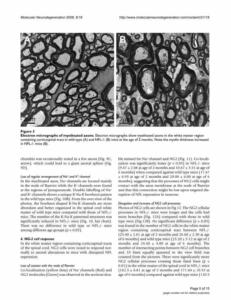
Reduction in diameters of myelinated axons and increase in thickness of myelin sheathIn transverse sections of the spinal cord, a significantdecrease in axonal diameter and increase in the thicknessof myelin sheath were observed in NFL-/- mice (Fig. 3B),in comparison with those from wild type mice (Fig. 3A).Examination of the frequency distribution of axonaldiameters in normal and NFL-/- mice (Fig. 4) revealed ashift in diameters of myelinated axons towards smallersizes compared against wild type mice, suggesting that theabsence of NFL subunit results in a reduction in axonalsize in the spinal cord white matter.
To investigate the relationship between myelin sheaththickness and axonal diameter, the diameters of axonproper (d) and the outer diameter of the myelinated fibre
(D) were measured and the G-ratio (d/D) calculated in 2-and 6-month old mice. There was a significant reductionin the G-ratio in NFL-/- mice (Fig. 5), suggesting anincrease in the thickness of myelin sheaths and/ordecrease in the diameter of axons. The change in the G-ratio in NFL-/- mice was more obvious in the axons withsmaller diameters (Fig. 5). Significant increase in thethickness of axonal myelin was observed in the axons ofwhite matter in the spinal cord of NLF-/- mice (Fig. 6).This increase was noted in all ranges of axonal diameters.
Increase in number of unloading loopsAt the node of Ranvier, most myelin loops were found tocontact with the axonal membrane (arrow in Fig. 7A),which are referred as loading loops. There were moreunloading loops (arrow in Fig. 7B) which are not interact-ing with the axonal membrane in the NFL-/- mice. Count-ing the numbers of loops within a 1-μm length ofparanodes showed that unloading loops at the node ofRanvier in NFL-/- mice increased significantly in 2-monthold mice (Fig. 8), but returned to a normal range at 6months of age.
Distortion and degeneration in myelinated axonsFurther observation using an electron microscope showedthat the absence of NFL subunit in neurons resulted inprofound changes in axons before the appearance of
NFH immunohistochemistry staining of brain cortexFigure 1NFH immunohistochemistry staining of brain cortex. In the wild type mouse, there were many NFH positive neuronal processes in the lamina V of the sensorimotor cortex (B). However, in the 2 month old NFL-/- mouse, the NFH positive neu-ronal processes were significantly reduced in number (A) and NFH protein accumulated as aggregates (A, arrows).
Page 3 of 15(page number not for citation purposes)

Molecular Neurodegeneration 2008, 3:18 http://www.molecularneurodegeneration.com/content/3/1/18
astrocytic and microglial activation and dysfunctions inthe NFL-/- mice. Obvious twist of myelinated axons waspresent in NFL-/- mice, which might be a result of accu-mulation of mitochondria in particular regions of the
axon (Fig. 9A, arrow) or accumulation of amorphologicalcytoplasmic material in between the myelin sheath andaxon (A, asterisk). Some mitochondria became vacu-olated (Fig. 9B, asterisks). Drastic accumulation of mito-
CD11b staining of spinal cordFigure 2CD11b staining of spinal cord. In general, less fluorescent intensity of CD11b immunohistochemistry staining was observed in the spinal cord of NFL-/- mice (A, C) compared to that of wild type mice (B, D), especially in the white matter, at both age of 2 and 6 months. The arrows (A, B) pointed regions are the white matter containing the corticospinal tract in the lateral funiculus of the spinal cord of 2 month old mice (C and D are higher magnification photos taken from arrows pointed areas). Bar: 200 μm in A and B; 50 μm in C and D.
Page 4 of 15(page number not for citation purposes)
Molecular Neurodegeneration 2008, 3:18 http://www.molecularneurodegeneration.com/content/3/1/18
chondria was occasionally noted in a few axons (Fig. 9C,arrow), which could lead to a giant axonal sphere (Fig.9D).
Loss of regular arrangement of Na+ and K+ channelIn the myelinated axon, Na+ channels are located mainlyin the node of Ranvier while the K+ channels were foundat the regions of juxtaparanode. Double labelling of Na+
and K+ channels shows a unique K-Na-K bowknot patternin the wild type mice (Fig. 10B). From the over view of thephotos, the bowknot shaped K-Na-K channels are moreabundant and better organized in the spinal cord whitematter of wild type mice compared with those of NFL-/-mice. The number of the K-Na-K patterned structures wassignificantly reduced in NFL-/- mice (Fig. 10, bar chart).There was no difference in wild type or NFL-/- miceamong different age groups (p > 0.05).
4. NG-2 cell responseIn the white matter region containing corticospinal tractsof the spinal cord, NG2 cells were noted to respond nor-mally to axonal alterations in mice with disrupted NFLexpression.
Loss of contact with the node of RanvierCo-localization (yellow dots) of Na+ channels (Red) andNG2 molecules (Green) was observed in the sections dou-
ble stained for Na+ channel and NG2 (Fig. 11). Co-locali-zation was significantly lesser (p < 0.05) in NFL-/- mice(9.67 ± 2.08 at age of 2 months and 10.67 ± 3.51 at age of6 months) when compared against wild type mice (17.67± 4.93 at age of 2 months and 20.00 ± 4.00 at age of 6months), suggesting that the processes of NG2 cells mightcontact with the axon membrane at the node of Ranvierand that this connection might be lost upon targeted dis-ruption of NFL expression in neurons.
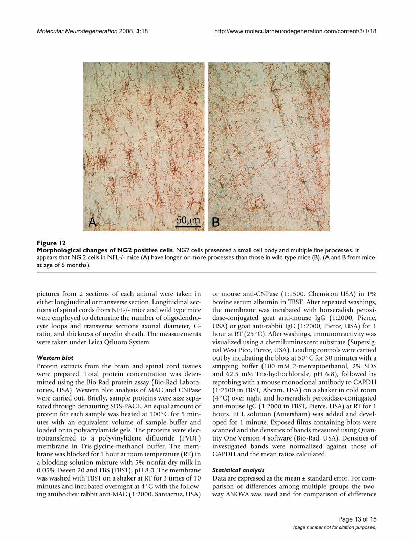
Elongation and increase of NG2 cell processesPhotos of NG2 cells are shown in Fig 12. The NG2 cellularprocesses in NFL-/- mice were longer and the cells hadmore branches (Fig. 12A) compared with those in wildtype mice (Fig.12B). No significant difference (p > 0.05)was found in the number of NG2 cells in the white matterregion containing corticospinal tract between NFL-/-(25.40 ± 2.61 at age of 2 months and 26.60 ± 2.30 at ageof 6 months) and wild type mice (23.20 ± 3.12 at age of 2months and 24.40 ± 4.00 at age of 6 months). Thenumber of intersecting points between NG2 cell branchesand 10 lines equally spanned in the view field wascounted from the pictures. There were significantly moreNG2 cellular processes crossing those fixed lines (p <0.01) in the white matter of the spinal cord in NFL-/- mice(162.5 ± 8.81 at age of 2 months and 171.60 ± 10.53 atage of 6 months) compared against wild type mice (120.5
Electron micrographs of myelinated axonsFigure 3Electron micrographs of myelinated axons. Electron micrographs show myelinated axons in the white matter region containing corticospinal tract in wild type (A) and NFL-/- (B) mice at the age of 2 months. Note the myelin thickness increased in NFL-/- mice (B).
Page 5 of 15(page number not for citation purposes)

Molecular Neurodegeneration 2008, 3:18 http://www.molecularneurodegeneration.com/content/3/1/18
Page 6 of 15(page number not for citation purposes)
Frequency distribution of myelinated axons according to their diametersFigure 4Frequency distribution of myelinated axons according to their diameters. Diameters of all myelinated axons were measured in the region containing the corticospinal tract at the lumbar segments of 2- and 6-month-old NFL-/- (N = 3 animals per age group) and control animals (N = 3 animals per age group). Data is presented based on diameters of all axons. Note the marked shift in distribution of axonal diameters towards smaller fibres. (*p < 0.05, **p < 0.01).

Molecular Neurodegeneration 2008, 3:18 http://www.molecularneurodegeneration.com/content/3/1/18
Page 7 of 15(page number not for citation purposes)
G-ratio of myelinated axonsFigure 5G-ratio of myelinated axons. Significant decreases in the G-ratio were measured in NFL-/- mice at both ages of 2 and 6 months (N = 3 animals per age group) in comparison to wild type control (N = 3 animals per age group). However, the decreases were significant only in those axons with smaller diameters in NFL-/- mice. (**p < 0.01).
Thickness of myelin sheathFigure 6Thickness of myelin sheath. The thicknesses of myelin sheath of axons of NFL-/- mice (N = 3 animals per age group) increased significantly in comparison to that of wild type controls (N = 3 animals per age group). (*p<0.05; **<0.01).

Molecular Neurodegeneration 2008, 3:18 http://www.molecularneurodegeneration.com/content/3/1/18
± 15.61 at age of 2 months and 129 ± 8.93 at age of 6months), suggesting that NG2 cells in NFL-/- mice had alarger number or longer processes.
5. No change in the expression of MAG and CNPaseMyelin associated glycoprotein (MAG) may play animportant role in axonal reorganization and adhesion,intermembrane spacing, signal transduction during glialcell differentiation, regulation of neurite outgrowth, andmaintenance of axon-myelin integrity; hence, we meas-ured the expression of MAG protein using Western blot.The results showed no significant difference in the cortexof brain or spinal cord between C57BL/6 and NFL-/- mice(Fig. 13) at the early stage of neuropathogenesis in theNFL-/- mice. The level of CNPase, an oligodendrocytemarker, may decrease together with myelin reduction invarious neurodegenerative diseases and experimental con-ditions. However, Western blot showed no significantalteration in CNPase expression in the brain or spinalcord when comparing wild type and NFL-/- mice at age of2 or 6 months (Fig. 13), indicating that the amount ofmyelin was similar in both groups.
DiscussionIn this study, we used an animal model in which neu-ropathological progress was initiated by targeted disrup-
tion of NFL subunit expression in neurons.Morphological distortion and degeneration in myelinatedaxons were observed in the white matter region contain-ing corticospinal tract in 2- and 6-month old NFL-/- mice.We have found previously that, until 6-month old, theseearly neuronal changes do not activate astrocytes ormicroglia. Instead, the environment has imposed aninhibitory effect on microglia [3]. However, NG2 cells atthat earlier period of neuropathological developmenthave the ability to sense and respond to the alterations inNFs of myelinated axons.
NFs may be a major determinant of axonal diameter,based on the correlation between NF contents in theaxonal cross-sections and axonal calibres [18]. It wasobserved that small unmyelinated axons contain few NFs[19] and that some small neurons lack morphologicallyidentifiable NFs [20,21]. This correlation persists duringaxonal degeneration and regeneration [22], and changesin NF transport correlate temporally with alterations inthe calibre of axons in regenerating nerves [23]. In a studyusing a transgenic approach, NFL was confirmed as amajor determinant of axonal calibre in the ventral root oflower motor neurons of the spinal cord [24]. In agreementwith this observation, our earlier study has reported thatthe distribution of diameters of myelinated axons of
Electron micrographs of the node of RanvierFigure 7Electron micrographs of the node of Ranvier. Electron micrographs show the node of Ranvier in the white matter region containing the corticospinal tract in wild type (A) and NFL-/- (B) mice at the age of 2 months. Note there were more unloading loops (B, arrow), which did not contact with the axonal membrane, in NFL-/- mice. The loading loops in wild type mice are shown in A (arrow).
Page 8 of 15(page number not for citation purposes)

Molecular Neurodegeneration 2008, 3:18 http://www.molecularneurodegeneration.com/content/3/1/18
upper motor neurons in the white matter of the spinalcord has shifted towards a smaller range in both 2- and 6-month old mice. This indicates that, after targeted disrup-tion of expression of NFL, most NFH proteins cannotmove into axons [3] resulting in an alteration in NF con-tents in myelinated axons and thereby inhibiting radialgrowth of the axons [24-28].
It is well accepted that the axon calibre controls the onsetand rate of myelin formation, i.e., the thickness of themyelin sheath normally increases with axon calibre [29],although some reports suggested no apparent correlationbetween myelination and axonal size [30]. In our results,the G-ratio has decreased significantly at both ages of 2and 6 months, suggesting a significant decrease in thediameter of axons and/or a significant increase in thethickness of myelin sheath. Further analyses showed thatthe alteration in NF contents in axons of NFL-/- mice hasresulted in both significant decrease in axonal diametersand significant increase in the thickness of myelin sheath.The myelin sheath from a larger axon would now have towrap around a smaller axon, resulting in an increase in thethickness of myelin sheath. More unloading loops in 2-month old mice also suggest more layer of myelin sheathwrapping around the axon. This implies that the altera-tion in NF contents in NFL-/- axons did not significantlychange the amount of myelin synthesis as well as the proc-esses of myelin wrapping. It is clear that signalling com-munication between the myelinating cells and neuronsmay not be determined by NF contents in axons.
In fact, our experimental results did not show any signs ofoligodendrocytes actively responding to axonal alteration.
CNPase (2', 3'-Cyclic Nucleotide 3'-Phosphodiesterase) isvirtually only expressed by oligodendrocytes in the CNSand by Schwann cells in the PNS. CNPase activity isdecreased in demyelinating diseases such as multiple scle-rosis. Myelin-associated glycoprotein (MAG), a trans-membrane glycoprotein, is selectively localized inperiaxonal Schwann cell and oligodendrocyte membranesof myelin sheaths, suggesting that it functions in glia-axoninteractions in both the PNS and CNS. Disruption of peri-axonal myelin and loss of MAG is characterized by demy-elination in rat spinal cord, which can be induced byinjection of lipopolysaccharide [31]. However, in ourobservation, there was no obvious difference in theamount of CNPase and MAG, indicating that oli-godendrocytes may not have responded to the alterationin axonal contents at early stage of development of NFL-/- mice.
While there was microglial inhibition, no obvious astro-cyte and oligodendrocyte reactions in the spinal cord ofNFL-/- mice at the age of 2 and 6 months were noted. The4th major non-neuronal cell type, NG2 cells, presentedtheir ability to sense and respond to axonal changes at anearlier time window. Normally, the processes of NG2expressing cells can closely contact nodes of Ranvier[5,32]. Some of this connection was lost as evidenced bythe reduction in the number of NG2 and Na+ channeldouble positive labelling in the NFL-/- neurons whereprofound redistribution of NFs occurred. Another sign ofNG2 response was the elongation and increase of theirprocesses. The purpose of this disconnection, processelongation and increase is unknown since the function ofNG2 cells is not yet defined. The NG2 processes may pro-vide a stable environment for axons and/or provide aninhibitory mechanism to prevent axonal sprouting at thenode of Ranvier [32]. But the results from NG2 knockoutmice showed that NG2 cells may be irrelevant for inhibi-tion or promotion of axonal growth in vivo [33]. There-fore, the loss of connection between NG2 cells and axonsmight induce the elongation and increase of NG2 cellprocesses in an attempt to re-establish the connection toneurons and provide protection. However, the signal toattract NG2 cell processes to contact axons may decreasesignificantly after targeted disruption of NFL expression inneurons, resulting in a failure in the connection betweenNG2 cells and the nodes of Ranvier. It is also possible thatNG2 cells may actively respond to alteration in NFL-/-axons and mediate further glial cell reaction in a laterstage. Nevertheless, it is still a mystery about the role ofearly responding NG2 cells in the chain of glial reaction inneurodegenerative diseases.
ConclusionOur results demonstrated that the structural configurationdetermined by the NFL gene is important for the mainte-
Numbers of loops at the node of RanvierFigure 8Numbers of loops at the node of Ranvier. The number of unloading loops, which have yet to contact with axonal membrane, was significantly increased in the NFL-/- mice near the age of 2 months.
Page 9 of 15(page number not for citation purposes)

Molecular Neurodegeneration 2008, 3:18 http://www.molecularneurodegeneration.com/content/3/1/18
nance of normal morphology of myelinated axons. Thealteration in axonal contents caused by disruption in NFLexpression may not be able to result in severe demyelina-tion but definitely leads to NG2 cell response. The NG2cells might serve as an early sensor to deliver informationof pathological changes in neurons to the local environ-ment.
MethodsAnimalsTwo and 6 month old male NFL-/- mice with C57BL/6background [24] (courtesy from Dr Julian, Canada) werehoused five to a cage and acclimated to a 12 hour shift inlight/dark cycle with free access to food and water. Intotal, 52 mice were used. For NFL-/- mice, five animals per
Electron micrographs of myelinated axons in NFL-/- miceFigure 9Electron micrographs of myelinated axons in NFL-/- mice. Obvious twist of myelinated axons can be observed (A). Accumulation of mitochondria in the axon (A, arrow) and accumulation of amorphological cytoplasm materials between myelin sheath and axon (A, asterisk) were found near the node of Ranvier. Some mitochondria became enlarged and vacuolated (B, asterisks). An obvious accumulation of mitochondria could be detected in certain region of axons (C, arrow), which may lead to a giant axonal sphere stuffed with mitochondria (D). Note the large number of mitochondria that were vacuolated. (A and B from mice at age of 2 months, C and D from mice at age of 6 months).
Page 10 of 15(page number not for citation purposes)

Molecular Neurodegeneration 2008, 3:18 http://www.molecularneurodegeneration.com/content/3/1/18
time interval were used for immunohistochemistricalstaining and 3 per time interval for electron microscopicstudy. Another five animals per time interval wereemployed for Western blot. The same number of C57BL/6J mice (Laboratory Animal Centre, Singapore) was usedas the control. All procedures involving animals wereaccording to guidelines of the Institutional Animal Careand Use Committee, National University of Singapore.
ImmunohistochemistryMice were perfused intra-aortically with ice-cold Ringer'ssolution (pH 7.4) and 2% PLP (pH 7.4). The brain andlumbar enlargement of the spinal cord were removed,postfixed in the same fixative for 4 hours at 4°C, andequilibrated in 30% sucrose overnight at 4°C. The coro-
nal sections (20 μm) of the brain and transverse and lon-gitudinal sections (20 μm) of the spinal cord were cryocut.Every 5th section was collected on slides. Sections wereblocked with 5% goat serum/PBS plus 0.1% Triton X-100before being incubated with primary antibodies.
For DAB staining, the incubation of rabbit antibodyagainst NG2 (1:2000, marker for NG2 cells) or rabbit anti-body against NFH (1:1000, marker for neurons), overnight at room temperature (RT) was followed by succes-sive incubations with biotin-conjugated anti-rabbit sec-ondary antibody (goat polyclonal, 1:200; ChemiconInternational, USA), ABC reagents (Vector Laboratories,USA), and SigmaFast DAB Peroxidase Substrate (Sigma-Aldrich, USA). Sections were counterstained with methyl
Double labelling of Na+ and K+ channelsFigure 10Double labelling of Na+ and K+ channels. Na+ channels (Red) in the centre of the node and K+ channels in both sides of the juxtaparanode (Green). In wild type mice (B), the double labelling appeared unique K-Na-K bowknot pattern while the per-centage of the unique pattern decreased significantly in NFL-/- mice (A and bar chart, *p < 0.05). (A and B from mice at age of 2 months, N = 3 animals per group).
Page 11 of 15(page number not for citation purposes)

Molecular Neurodegeneration 2008, 3:18 http://www.molecularneurodegeneration.com/content/3/1/18
green, before being dehydrated in graded ethanol, clearedin Histoclear, mounted with Vectashield mountingmedium (Stem cell technology, USA) and then observedunder a light microscope (Olympus BX51, Japan).
For immunofluorescent double labelling, rat antibodyagainst potassium channel Kv1.2 (1:200, Sigma, USA)and mouse antibody against sodium channel (pan) cloneK58/35 (1:200, Sigma, USA) were applied to sections for2 hours at RT. Rabbit antibody against NG2 (1:200) com-bined with rat antibody against sodium channel (pan)clone K58/35 (1:200, Sigma, USA) were applied to sec-tions for 2 hours at RT. Rat antibody against CD11b(1:1000, marker for microglia, BD Pharmingen, USA) wasapplied to spinal cord sections for 2 hours at RT. Then theFITC or Cy3-conjugated anti-rabbit or anti-rat secondaryantibody (goat polyclonal, 1:200; Chemicon Interna-tional, USA) was used and mounted with Vectashieldmounting medium counterstaining. The slides wereexamined under a confocal microscope (Olympus LSM510 Zeiss, Japan). Ten pictures from 10 sections of eachanimal were randomly taken from white matter with cor-ticospinal tract of the spinal cord. In the sections of dou-ble labelling of Na+ and K+ channels, the number of intactunique K-Na-K labelling pattern was counted. In the sec-tions of double labelling of NG2 and pan-Na channel, thenumber of positive double labelling was counted. Thelength of NG2 cell processes was also stereologically ana-lyzed by number of the point where NG2 cell processescross lines of square measuring units.
Electron microscopic studyFor conventional electron microscopy, the animals wereperfused transcardially with Ringer's solution and then2% paraformaldehyde containing 3% glutaraldehyde in0.1 M phosphate-buffer. Transverse and longitudinal sec-tions of the spinal cord were cut at 1 mm3 and immersedin the same fixative for 4 hours at 4°C. Tissues were rinsedin 0.1 M phosphate-buffer (with 5% sucrose) with 3changes at 10 minutes each. Post fixing of samples wasdone in 1% osmium tetroxide in 0.1 M phosphate-buffer(pH 7.4) for 2 hour at room temperature and dehydrationin an ascending ethanol series at room temperarure. Tis-sue blocks were infiltrated in 100% acetone: resin (1:6)for overnight and three changes of pure resin at series ofascending temperature (40°C, 50°C, and 55°C) for 1hour each. Samples were embedded in a pure aralditemixture at 60°C for 24 hours. Cross-section and longitu-dinal section of spinal cord blocks were cut into 99 nmthickness. Ultrathin sections were double stained in leadcitrate and 3% uranyl acetate, and then were viewed in aPhilips 120 Biotwin TEM (Fei company, USA).
For each fiber on cross-sections, the G ratio was calculatedas d/D, where the d was the diameter of axon proper andthe D were the outer diameter of the myelinated fiber. Theg-ratio of myelianted fibers was determined in cross sec-tions of spinal cord from NFL-/- mice and correspondingwild type mice. Randomly selected non-overlapping fieldswere photographed. Thickness of myelin sheath was cal-culated as half of the result of the outer diameter of themyelinated fibre minus the diameter of axon proper. Ten
Double labelling of Na+ channels and NG2 cellsFigure 11Double labelling of Na+ channels and NG2 cells. The micrographs show double labelling of Na channels (Red) and NG2 cells (Green). The number of yellow dots of double labelling was obviously reduced in the white matter of NFL-/- mice (A) in compared to that in wild type mice (B). (A and B from mice at age of 2 months, N = 3 animals per group).
Page 12 of 15(page number not for citation purposes)
Molecular Neurodegeneration 2008, 3:18 http://www.molecularneurodegeneration.com/content/3/1/18
pictures from 2 sections of each animal were taken ineither longitudinal or transverse section. Longitudinal sec-tions of spinal cords from NFL-/- mice and wild type micewere employed to determine the number of oligodendro-cyte loops and transverse sections axonal diameter, G-ratio, and thickness of myelin sheath. The measurementswere taken under Leica Qfluoro System.
Western blotProtein extracts from the brain and spinal cord tissueswere prepared. Total protein concentration was deter-mined using the Bio-Rad protein assay (Bio-Rad Labora-tories, USA). Western blot analysis of MAG and CNPasewere carried out. Briefly, sample proteins were size sepa-rated through denaturing SDS-PAGE. An equal amount ofprotein for each sample was heated at 100°C for 5 min-utes with an equivalent volume of sample buffer andloaded onto polyacrylamide gels. The proteins were elec-trotransferred to a polyvinylidene difluoride (PVDF)membrane in Tris-glycine-methanol buffer. The mem-brane was blocked for 1 hour at room temperature (RT) ina blocking solution mixture with 5% nonfat dry milk in0.05% Tween 20 and TBS (TBST), pH 8.0. The membranewas washed with TBST on a shaker at RT for 3 times of 10minutes and incubated overnight at 4°C with the follow-ing antibodies: rabbit anti-MAG (1:2000, Santacruz, USA)
or mouse anti-CNPase (1:1500, Chemicon USA) in 1%bovine serum albumin in TBST. After repeated washings,the membrane was incubated with horseradish peroxi-dase-conjugated goat anti-mouse IgG (1:2000, Pierce,USA) or goat anti-rabbit IgG (1:2000, Pierce, USA) for 1hour at RT (25°C). After washings, immunoreactivity wasvisualized using a chemiluminescent substrate (Supersig-nal West Pico, Pierce, USA). Loading controls were carriedout by incubating the blots at 50°C for 30 minutes with astripping buffer (100 mM 2-mercaptoethanol, 2% SDSand 62.5 mM Tris-hydrochloride, pH 6.8), followed byreprobing with a mouse monoclonal antibody to GAPDH(1:2500 in TBST, Abcam, USA) on a shaker in cold room(4°C) over night and horseradish peroxidase-conjugatedanti-mouse IgG (1:2000 in TBST, Pierce, USA) at RT for 1hours. ECL solution (Amersham) was added and devel-oped for 1 minute. Exposed films containing blots werescanned and the densities of bands measured using Quan-tity One Version 4 software (Bio-Rad, USA). Densities ofinvestigated bands were normalized against those ofGAPDH and the mean ratios calculated.
Statistical analysisData are expressed as the mean ± standard error. For com-parison of differences among multiple groups the two-way ANOVA was used and for comparison of difference
Morphological changes of NG2 positive cellsFigure 12Morphological changes of NG2 positive cells. NG2 cells presented a small cell body and multiple fine processes. It appears that NG 2 cells in NFL-/- mice (A) have longer or more processes than those in wild type mice (B). (A and B from mice at age of 6 months).
Page 13 of 15(page number not for citation purposes)

Molecular Neurodegeneration 2008, 3:18 http://www.molecularneurodegeneration.com/content/3/1/18
between two groups the t-test was used. Statistical signifi-cance was set at p < 0.05.
Competing interestsThe authors declare that they have no competing interests.
Authors' contributionsYJW carried out the electronic microscopy and Westernblot studies, participated in acquisition of data, per-formed the statistical analysis, and drafted the manu-script. YFT carried out the immunohistochemistry study,
participated in acquisition of data, and performed the sta-tistical analysis. ZCX and ZMB supervised YJW in experi-ments and participated in preparation of manuscript. BPHdesigned the study, supervised YJW and YFT in experi-ments and statistical analysis, and drafted the manuscript.All authors read and approved the final manuscript.
AcknowledgementsThis work was supported by a grant NMRC/07788/2003 from the National Medical Research Council, Singapore and a grant R121-000-119-750 from National University of Singapore. We are grateful to Dr George Yip and Ms Jasmin Qianru Lim, Department of Anatomy, Yong Loo Lin School of Med-
Western blot of MAG and CNPaseFigure 13Western blot of MAG and CNPase. There was no significant difference in MAG or CNPase expression in brain cortex and spinal cord between wild type and NFL-/- mice.
Page 14 of 15(page number not for citation purposes)

Molecular Neurodegeneration 2008, 3:18 http://www.molecularneurodegeneration.com/content/3/1/18
Publish with BioMed Central and every scientist can read your work free of charge
"BioMed Central will be the most significant development for disseminating the results of biomedical research in our lifetime."
Sir Paul Nurse, Cancer Research UK
Your research papers will be:
available free of charge to the entire biomedical community
peer reviewed and published immediately upon acceptance
cited in PubMed and archived on PubMed Central
yours — you keep the copyright
Submit your manuscript here:http://www.biomedcentral.com/info/publishing_adv.asp
BioMedcentral
icine, National University of Singapore, for their critical reading of this man-uscript, comments and helpful suggestions.
References1. Menzies FM, Grierson AJ, Cookson MR, Heath PR, Tomkins J, Figle-
wicz DA, Ince PG, Shaw PJ: Selective loss of neurofilamentexpression in Cu/Zn superoxide dismutase (SOD1) linkedamyotrophic lateral sclerosis. J Neurochem 2002, 82:1118-1128.
2. Wong NK, He BP, Strong MJ: Characterization of neuronalintermediate filament protein expression in cervical spinalmotor neurons in sporadic amyotrophic lateral sclerosis(ALS). J Neuropathol Exp Neurol 2000, 59:972-982.
3. Li ZH, Lu J, Tay SSW, Wu YJ, Strong MJ, He BP: Mice with targeteddisruption of neurofilament light subunit display formationof protein aggregation in motoneurons and down-regulationof complement receptor type 3 α-subunit in microglia in thespinal cord at their earlier age: a possible feature in pre-clin-ical development of neurodegenerative diseases. Brain Res2006, 1113:200-209.
4. McLean JR, Sanelli TR, Leystra-Lantz C, He BP, Strong MJ: Temporalprofiles of neuronal degeneration, glial proliferation, and celldeath in hNFL (+/+) and NFL (-/-) mice. Glia 2005, 52:59-69.
5. Butt AM, Duncan A, Hornby MF, Kirvell SL, Hunter A, Levine JM,Berry M: Cells expressing the NG2 antigen contact nodes ofRanvier in adult CNS white matter. Glia 1999, 26:84-91.
6. Karadottir R, Hamilton NB, Bakiri Y, Attwell D: Spiking and non-spiking classes of oligodendrocytes precursor glia in CNSwhite matter. Nat Neurosci 2008, 11:450-456.
7. Kukley M, Capetillo-Zarate E, Dietrich D: Vesicular glutamaterelease from axons in white matter. Nat Neurosci 2007,10:311-320.
8. Ziskin JL, Nishiyama A, Rubio M, Fukaya M, Bergles DE: Vesicularrelease of glutamate from unmyelinated axons in white mat-ter. Nat Neurosci 2007, 10:321-330.
9. Lin SC, Bergles DE: Synaptic signalling between neurons andglia. Glia 2004, 47:290-298.
10. Keirstead HS, Levine JM, Blakemore WF: Response of the oli-godendrocyte progenitor cell population (defined by NG2labelling) to demyelination of the adult spinal cord. Glia 1998,22:161-170.
11. Reynolds R, Dawson M, Papadopoulos D, Polito A, Di Bello IC, Pham-Dinh D, Levine J: The response of NG2-expressing oli-godendrocyte progenitors to demyelination in MOG EAEand MS. J Neurocytol 2002, 31:523-536.
12. Dawson MR, Levine JM, Reynolds R: NG2-expressing cells in thecentral nervous system: Are they oligodendroglial progeni-tors? J Neurosci 2000, 61:471-479.
13. Levine JM, Card JP: Light and electron microscopic localizationof a cell surface antigen (NG2) in the rat cerebellum: Associ-ation with smooth protoplasmic astrocytes. J Neurosci 1987,7:2711-2720.
14. Levine JM, Stincone F, Lee YS: Development and differentiationof glial precursor cells in the rat cerebellum. Glia 1993,7:307-321.
15. Nishiyama A, Chang A, Trapp BD: NG2+ glial cells: A novel glialcell population in the adult brain. J Neuropathol Exp Neurol 1999,58:1113-1124.
16. Levine JM: Increased expression of NG2 chondroitin-sulfateproteoglycan after brain injury. J Neurosci 1994,14(8):4716-4730.
17. Ong WY, Levine JM: A light and electron microscopic study ofNG2 chondroitin sulfate proteoglycan-positive oligodendro-cyte precursor cells in the normal and kainate-lesioned rathippocampus. Neuroscience 1999, 92:83-95.
18. Hoffman PN, Koo EH, Muma NA, Griffin JW, Price DL: Role of neu-rofilaments in the control of axonal caliber in myelinatednerve fibers. In Intrinsic Determinants of Neuronal Form and FunctionVolume 37. Edited by: Lasek RJ, Black MM. New York, Alan R. Liss;1988:389-402.
19. Friede R, Sarnorajski T: Axon caliber related to neurofilamentsand microtubules in sciatic nerve. Anat Rec 1970, 167:379-388.
20. Burgoyne RD, Cambray-Deakin MA: The cellular neurobiology ofneuronal development: the cerebellar granule cell. Brain ResRev 1988, 13:77-101.
21. Trojanowski JQ, Walkenstein N, Lee AMY: Expression of neurofil-ament subunits in neurons of the central and peripheralnervous system: an immunohistochemical study with mono-clonal antibodies. J Neurosci 1986, 6:650-660.
22. Hoffman PN, Griffin JW, Price DL: Control of axonal caliber byneurofilament transport. J Cell Biol 1984, 99:705-714.
23. Hoffman PN, Thompson GW, Griffin GW, Price DL: Changes inneurofilament transport coincide temporally with altera-tions in the caliber of axons in regenerating motor fibers. JCell Biol 1985, 101:1332-1340.
24. Zhu Q, Couillard-Despres S, Julien JP: Delayed maturation ofregenerating myelinated axons in mice lacking neurofila-ments. Exp Neurol 1997, 148:299-316.
25. Elder GA, Friedrich VL, Bosco P, Kang C, Gourov A, Tu P-H, Lee VM-Y, Lazzarini RA: Absence of the midsized neurofilament subu-nit decreases axonal diameter, levels of light neurofilament(NF-L) and neurofilament content. J Cell Biol 1998,141:727-739.
26. Elder GA, Friedrich VL, Kang C, Bosco P, Gourov A, Tu P-H, ZhangB, Lee VM-Y, Lazzarini RA: Requirement of heavy neurofilamentsubunit in the development of axons with large calibers. J CellBiol 1998, 143:195-205.
27. Eyer J, Peterson A: Neurofilament-deficient axons and perikar-yal aggregates in viable transgenic mice expressing a neuro-filament-beta-galactosidase fusion protein. Neuron 1994,12:389-405.
28. Zhu Q, Lindenbaum M, Levavasseur F, Jacomy H, Julien JP: Disrup-tion of the NF-H gene increases axonal microtubule contentand velocity of neurofilament transport: relief of axonopathyresulting from the toxin beta, beta'-iminodipropionitrile. JCell Biol 1998, 143:183-193.
29. Raval-Fernandes S, Rome LH: Role of axonal components duringmyelination. Microsc Res Tech 1998, 41:379-392.
30. Duncan ID, Griffiths IR, Munz M: "Shaking pups": A disorder ofcentral myelination in the spaniel dog. III. Quantitativeaspects of glia and myelin in the spinal cord and optic nerve.Neuropathol Appl Neurobiol 1983, 9:355-368.
31. Felts PA, Woolston AM, Fernando HB, Asquith S, Gregson NA, MizziOJ, Smith KJ: Inflammation and primary demyelinationinduced by the intraspinal injection of lipopolysaccharide.Brain 2005, 128:1649-1666.
32. Huang JK, Phillips GR, Roth AD, Pedraza L, Shan W, Belkaid W, Mi S,Fex-Svenningsen A, Florens L, Yates JR 3rd, Colman DR: Glial mem-branes at the node of Ranvier prevent neurite outgrowth.Science 2005, 310:1813-1817.
33. Hossain-Ibrahim MK, Rezajooi K, Stallcup WB, Lieberman AR, Ander-son PN: Analysis of axonal regeneration in the central andperipheral nervous systems of the NG2-deficient mouse.BMC Neurosci 2007, 8:80.
Page 15 of 15(page number not for citation purposes)
Copyright © 2022 FDOKUMEN