Pseudorabies Virus Us9 Directs Axonal Sorting of Viral Capsids
10
Published Ahead of Print 8 August 2007. 2007, 81(20):11363. DOI: 10.1128/JVI.01281-07. J. Virol. W. Enquist M. G. Lyman, B. Feierbach, D. Curanovic, M. Bisher and L. Sorting of Viral Capsids Pseudorabies Virus Us9 Directs Axonal http://jvi.asm.org/content/81/20/11363 Updated information and services can be found at: These include: SUPPLEMENTAL MATERIAL Supplemental material REFERENCES http://jvi.asm.org/content/81/20/11363#ref-list-1 at: This article cites 30 articles, 20 of which can be accessed free CONTENT ALERTS more» articles cite this article), Receive: RSS Feeds, eTOCs, free email alerts (when new http://journals.asm.org/site/misc/reprints.xhtml Information about commercial reprint orders: http://journals.asm.org/site/subscriptions/ To subscribe to to another ASM Journal go to: on April 28, 2014 by guest http://jvi.asm.org/ Downloaded from on April 28, 2014 by guest http://jvi.asm.org/ Downloaded from
Transcript of Pseudorabies Virus Us9 Directs Axonal Sorting of Viral Capsids
Published Ahead of Print 8 August 2007. 2007, 81(20):11363. DOI: 10.1128/JVI.01281-07. J. Virol.
W. EnquistM. G. Lyman, B. Feierbach, D. Curanovic, M. Bisher and L. Sorting of Viral CapsidsPseudorabies Virus Us9 Directs Axonal
http://jvi.asm.org/content/81/20/11363Updated information and services can be found at:
These include:
SUPPLEMENTAL MATERIAL Supplemental material
REFERENCEShttp://jvi.asm.org/content/81/20/11363#ref-list-1at:
This article cites 30 articles, 20 of which can be accessed free
CONTENT ALERTS more»articles cite this article),
Receive: RSS Feeds, eTOCs, free email alerts (when new
http://journals.asm.org/site/misc/reprints.xhtmlInformation about commercial reprint orders: http://journals.asm.org/site/subscriptions/To subscribe to to another ASM Journal go to:
on April 28, 2014 by guest
http://jvi.asm.org/
Dow
nloaded from
on April 28, 2014 by guest
http://jvi.asm.org/
Dow
nloaded from
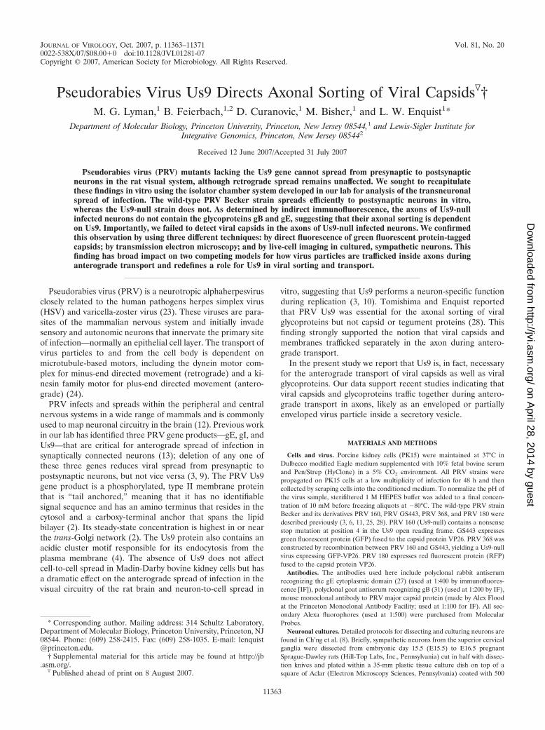
JOURNAL OF VIROLOGY, Oct. 2007, p. 11363–11371 Vol. 81, No. 200022-538X/07/$08.00�0 doi:10.1128/JVI.01281-07Copyright © 2007, American Society for Microbiology. All Rights Reserved.
Pseudorabies Virus Us9 Directs Axonal Sorting of Viral Capsids�†M. G. Lyman,1 B. Feierbach,1,2 D. Curanovic,1 M. Bisher,1 and L. W. Enquist1*
Department of Molecular Biology, Princeton University, Princeton, New Jersey 08544,1 and Lewis-Sigler Institute forIntegrative Genomics, Princeton, New Jersey 085442
Received 12 June 2007/Accepted 31 July 2007
Pseudorabies virus (PRV) mutants lacking the Us9 gene cannot spread from presynaptic to postsynapticneurons in the rat visual system, although retrograde spread remains unaffected. We sought to recapitulatethese findings in vitro using the isolator chamber system developed in our lab for analysis of the transneuronalspread of infection. The wild-type PRV Becker strain spreads efficiently to postsynaptic neurons in vitro,whereas the Us9-null strain does not. As determined by indirect immunofluorescence, the axons of Us9-nullinfected neurons do not contain the glycoproteins gB and gE, suggesting that their axonal sorting is dependenton Us9. Importantly, we failed to detect viral capsids in the axons of Us9-null infected neurons. We confirmedthis observation by using three different techniques: by direct fluorescence of green fluorescent protein-taggedcapsids; by transmission electron microscopy; and by live-cell imaging in cultured, sympathetic neurons. Thisfinding has broad impact on two competing models for how virus particles are trafficked inside axons duringanterograde transport and redefines a role for Us9 in viral sorting and transport.
Pseudorabies virus (PRV) is a neurotropic alphaherpesvirusclosely related to the human pathogens herpes simplex virus(HSV) and varicella-zoster virus (23). These viruses are para-sites of the mammalian nervous system and initially invadesensory and autonomic neurons that innervate the primary siteof infection—normally an epithelial cell layer. The transport ofvirus particles to and from the cell body is dependent onmicrotubule-based motors, including the dynein motor com-plex for minus-end directed movement (retrograde) and a ki-nesin family motor for plus-end directed movement (antero-grade) (24).
PRV infects and spreads within the peripheral and centralnervous systems in a wide range of mammals and is commonlyused to map neuronal circuitry in the brain (12). Previous workin our lab has identified three PRV gene products—gE, gI, andUs9—that are critical for anterograde spread of infection insynaptically connected neurons (13); deletion of any one ofthese three genes reduces viral spread from presynaptic topostsynaptic neurons, but not vice versa (3, 9). The PRV Us9gene product is a phosphorylated, type II membrane proteinthat is “tail anchored,” meaning that it has no identifiablesignal sequence and has an amino terminus that resides in thecytosol and a carboxy-terminal anchor that spans the lipidbilayer (2). Its steady-state concentration is highest in or nearthe trans-Golgi network (2). The Us9 protein also contains anacidic cluster motif responsible for its endocytosis from theplasma membrane (4). The absence of Us9 does not affectcell-to-cell spread in Madin-Darby bovine kidney cells but hasa dramatic effect on the anterograde spread of infection in thevisual circuitry of the rat brain and neuron-to-cell spread in
vitro, suggesting that Us9 performs a neuron-specific functionduring replication (3, 10). Tomishima and Enquist reportedthat PRV Us9 was essential for the axonal sorting of viralglycoproteins but not capsid or tegument proteins (28). Thisfinding strongly supported the notion that viral capsids andmembranes trafficked separately in the axon during antero-grade transport.
In the present study we report that Us9 is, in fact, necessaryfor the anterograde transport of viral capsids as well as viralglycoproteins. Our data support recent studies indicating thatviral capsids and glycoproteins traffic together during antero-grade transport in axons, likely as an enveloped or partiallyenveloped virus particle inside a secretory vesicle.
MATERIALS AND METHODS
Cells and virus. Porcine kidney cells (PK15) were maintained at 37°C inDulbecco modified Eagle medium supplemented with 10% fetal bovine serumand Pen/Strep (HyClone) in a 5% CO2 environment. All PRV strains werepropagated on PK15 cells at a low multiplicity of infection for 48 h and thencollected by scraping cells into the conditioned medium. To normalize the pH ofthe virus sample, sterifiltered 1 M HEPES buffer was added to a final concen-tration of 10 mM before freezing aliquots at �80°C. The wild-type PRV strainBecker and its derivatives PRV 160, PRV GS443, PRV 368, and PRV 180 weredescribed previously (3, 6, 11, 25, 28). PRV 160 (Us9-null) contains a nonsensestop mutation at position 4 in the Us9 open reading frame. GS443 expressesgreen fluorescent protein (GFP) fused to the capsid protein VP26. PRV 368 wasconstructed by recombination between PRV 160 and GS443, yielding a Us9-nullvirus expressing GFP-VP26. PRV 180 expresses red fluorescent protein (RFP)fused to the capsid protein VP26.
Antibodies. The antibodies used here include polyclonal rabbit antiserumrecognizing the gE cytoplasmic domain (27) (used at 1:400 by immunofluores-cence [IF]), polyclonal goat antiserum recognizing gB (31) (used at 1:200 by IF),mouse monoclonal antibody to PRV major capsid protein (made by Alex Floodat the Princeton Monoclonal Antibody Facility; used at 1:100 for IF). All sec-ondary Alexa fluorophores (used at 1:500) were purchased from MolecularProbes.
Neuronal cultures. Detailed protocols for dissecting and culturing neurons arefound in Ch’ng et al. (8). Briefly, sympathetic neurons from the superior cervicalganglia were dissected from embryonic day 15.5 (E15.5) to E16.5 pregnantSprague-Dawley rats (Hill-Top Labs, Inc., Pennsylvania) cut in half with dissec-tion knives and plated within a 35-mm plastic tissue culture dish on top of asquare of Aclar (Electron Microscopy Sciences, Pennsylvania) coated with 500
* Corresponding author. Mailing address: 314 Schultz Laboratory,Department of Molecular Biology, Princeton University, Princeton, NJ08544. Phone: (609) 258-2415. Fax: (609) 258-1035. E-mail: [email protected].
† Supplemental material for this article may be found at http://jb.asm.org/.
� Published ahead of print on 8 August 2007.
11363
on April 28, 2014 by guest
http://jvi.asm.org/
Dow
nloaded from
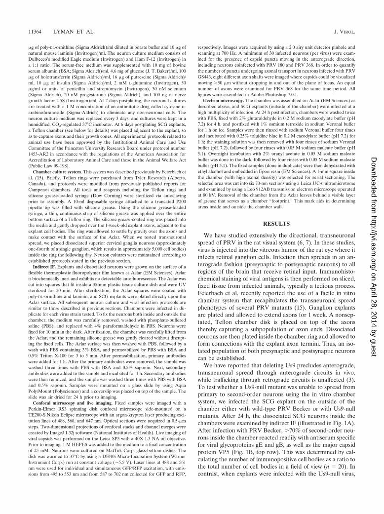
�g of poly-DL-ornithine (Sigma Aldrich)/ml diluted in borate buffer and 10 �g ofnatural mouse laminin (Invitrogen)/ml. The neuron culture medium consists ofDulbecco’s modified Eagle medium (Invitrogen) and Ham F-12 (Invitrogen) ina 1:1 ratio. The serum-free medium was supplemented with 10 mg of bovineserum albumin (BSA; Sigma Aldrich)/ml, 4.6 mg of glucose (J. T. Baker)/ml, 100�g of holotransferrin (Sigma Aldrich)/ml, 16 �g of putrescine (Sigma Aldrich)/ml, 10 �g of insulin (Sigma Aldrich)/ml, 2 mM L-glutamine (Invitrogen), 50�g/ml or units of penicillin and streptomycin (Invitrogen), 30 nM selenium(Sigma Aldrich), 20 nM progesterone (Sigma Aldrich), and 100 ng of nervegrowth factor 2.5S (Invitrogen)/ml. At 2 days postplating, the neuronal culturesare treated with a 1 M concentration of an antimitotic drug called cytosine-D-arabinofuranoside (Sigma-Aldrich) to eliminate any non-neuronal cells. Theneuron culture medium was replaced every 3 days, and cultures were kept in ahumidified, CO2-regulated 37°C incubator. At 6 days postplating SCG explants,a Teflon chamber (see below for details) was placed adjacent to the explant, soas to capture axons and their growth cones. All experimental protocols related toanimal use have been approved by the Institutional Animal Care and UseCommittee of the Princeton University Research Board under protocol number1453-AR2 in accordance with the regulations of the American Association forAccreditation of Laboratory Animal Care and those in the Animal Welfare Act(Public Law 99-198).
Chamber culture system. This system was described previously by Feierbach etal. (15). Briefly, Teflon rings were purchased from Tyler Research (Alberta,Canada), and protocols were modified from previously published reports forCampenot chambers. All tools and reagents including the Teflon rings andsilicone grease-loaded syringe (Dow Corning) were sterilized via autoclavingprior to assembly. A 10-ml disposable syringe attached to a truncated P200pipette tip was filled with silicone grease. Using the silicone grease-loadedsyringe, a thin, continuous strip of silicone grease was applied over the entirebottom surface of a Teflon ring. The silicone grease-coated ring was placed intothe media and gently dropped over the 1-week-old explant axons, adjacent to theexplant cell bodies. The ring was allowed to settle by gravity over the axons andmake contact with the surface of the Aclar. When we tested transneuronalspread, we placed dissociated superior cervical ganglia neurons (approximatelyone-fourth of a single ganglion, which results in approximately 5,000 cell bodies)inside the ring the following day. Neuron cultures were maintained according toestablished protocols stated in the previous section.
Indirect IF. Explants and dissociated neurons were grown on the surface of aflexible thermoplastic fluoropolymer film known as Aclar (EM Sciences). Aclaris biochemically inert and exhibits no detectable autofluorescence. The Aclar wascut into squares that fit inside a 35-mm plastic tissue culture dish and were UVsterilized for 20 min. After sterilization, the Aclar squares were coated withpoly-DL-ornithine and laminin, and SCG explants were plated directly upon theAclar surface. All subsequent neuron culture and viral infection protocols aresimilar to those described in previous sections. Chambers were infected in du-plicate for each virus strain tested. To fix the neurons both inside and outside thechamber, the medium was carefully removed, washed with phosphate-bufferedsaline (PBS), and replaced with 4% paraformaldehyde in PBS. Neurons werefixed for 10 min in the dark. After fixation, the chamber was carefully lifted fromthe Aclar, and the remaining silicone grease was gently cleared without disrupt-ing the fixed cells. The Aclar surface was then washed with PBS, followed by awash with PBS containing 3% BSA, and permeabilized by PBS with BSA and0.5% Triton X-100 for 3 to 5 min. After permeabilization, primary antibodieswere added for 1 h. After the primary antibodies were removed, the sample waswashed three times with PBS with BSA and 0.5% saponin. Next, secondaryantibodies were added to the sample and incubated for 1 h. Secondary antibodieswere then removed, and the sample was washed three times with PBS with BSAand 0.5% saponin. Samples were mounted on a glass slide by using AquaPoly/Mount (Polysciences) and a coverslip was placed on top of the sample. Theslide was air dried for 24 h prior to imaging.
Confocal microscopy and live imaging. Fixed samples were imaged with aPerkin-Elmer RS3 spinning disk confocal microscope side-mounted on aTE200-S Nikon Eclipse microscope with an argon-krypton laser producing exci-tation lines of 488, 568, and 647 nm. Optical sections were acquired in 0.5-�msteps. Two-dimensional projections of confocal stacks and channel merges werecreated by ImageJ 1.32j software (National Institutes of Health). Live imaging ofviral capsids was performed on the Leica SP5 with a 40X 1.3 NA oil objective.Prior to imaging, 1 M HEPES was added to the medium to a final concentrationof 25 mM. Neurons were cultured on MatTek Corp. glass-bottom dishes. Thedish was warmed to 37°C by using a DH40i Micro-Incubation System (WarnerInstrument Corp.) run at constant voltage (�5.5 V). Laser lines at 488 and 561nm were used for individual and simultaneous GFP/RFP excitation, with emis-sions from 495 to 553 nm and from 587 to 702 nm collected for GFP and RFP,
respectively. Images were acquired by using a 2.0 airy unit detector pinhole andscanning at 700 Hz. A minimum of 30 infected neurons (per virus) were exam-ined for the presence of capsid puncta moving in the anterograde direction,including neurons coinfected with PRV 180 and PRV 368. In order to quantifythe number of puncta undergoing axonal transport in neurons infected with PRVGS443, eight different axon shafts were imaged where capsids could be visualizedmoving �50 �m without dropping in and out of the plane of focus. An equalnumber of axons were examined for PRV 368 for the same time period. Allfigures were assembled in Adobe Photoshop 7.0.1.
Electron microscopy. The chamber was assembled on Aclar (EM Sciences) asdescribed above, and SCG explants (outside of the chamber) were infected at ahigh multiplicity of infection. At 24 h postinfection, chambers were washed twicewith PBS, fixed with 2% glutaraldehyde in 0.2 M sodium cacodylate buffer (pH7.2) for 4 h, and postfixed with 1% osmium tetroxide in sodium Veronal bufferfor 1 h on ice. Samples were then rinsed with sodium Veronal buffer four timesand incubated with 0.25% toluidine blue in 0.2 M cacodylate buffer (pH 7.2) for1 h; the staining solution was then removed with four rinses of sodium Veronalbuffer (pH 7.2), followed by four rinses with 0.05 M sodium maleate buffer (pH5.1). Overnight incubation with 2% uranyl acetate in 0.05 M sodium maleatebuffer was done in the dark, followed by four rinses with 0.05 M sodium maleatebuffer (pH 5.1). The fixed samples (done in duplicate) were then dehydrated withethyl alcohol and embedded in Epon resin (EM Sciences). A 1-mm square insidethe chamber (with high axonal density) was selected for serial sectioning. Theselected area was cut into six 70-nm sections using a Leica UC-6 ultramicrotomeand examined by using a Leo 912AB transmission electron microscope operatedat 80 kV. Removal of the chamber from the Aclar leaves behind a visible layerof grease that serves as a chamber “footprint.” This mark aids in determiningareas inside and outside the chamber wall.
RESULTS
We have studied extensively the directional, transneuronalspread of PRV in the rat visual system (6, 7). In these studies,virus is injected into the vitreous humor of the rat eye where itinfects retinal ganglion cells. Infection then spreads in an an-terograde fashion (presynaptic to postsynaptic neurons) to allregions of the brain that receive retinal input. Immunohisto-chemical staining of viral antigens is then performed on sliced,fixed tissue from infected animals, typically a tedious process.Feierbach et al. recently reported the use of a facile in vitrochamber system that recapitulates the transneuronal spreadphenotypes of several PRV mutants (15). Ganglion explantsare plated and allowed to extend axons for 1 week. A nonsep-tated, Teflon chamber disk is placed on top of the axonsthereby capturing a subpopulation of axon ends. Dissociatedneurons are then plated inside the chamber ring and allowed toform connections with the explant axon termini. Thus, an iso-lated population of both presynaptic and postsynaptic neuronscan be established.
We have reported that deleting Us9 precludes anterograde,transneuronal spread through anterograde circuits in vivo,while trafficking through retrograde circuits is unaffected (3).To test whether a Us9-null mutant was unable to spread fromprimary to second-order neurons using the in vitro chambersystem, we infected the SCG explant on the outside of thechamber either with wild-type PRV Becker or with Us9-nullmutants. After 24 h, the dissociated SCG neurons inside thechambers were examined by indirect IF (illustrated in Fig. 1A).After infection with PRV Becker, �70% of second-order neu-rons inside the chamber reacted readily with antiserum specificfor viral glycoproteins gE and gB, as well as the major capsidprotein VP5 (Fig. 1B, top row). This was determined by cal-culating the number of immunopositive cell bodies as a ratio tothe total number of cell bodies in a field of view (n � 20). Incontrast, when explants were infected with the Us9-null virus,
11364 LYMAN ET AL. J. VIROL.
on April 28, 2014 by guest
http://jvi.asm.org/
Dow
nloaded from

dissociated neurons inside the chamber ring showed no reac-tivity with gE, gB, or VP5 (Fig. 1B, middle row). However,Us9-null mutants were capable of efficient infection of explantneurons as determined by strong reactivity with antiserumagainst gE, gB, and VP5 (Fig. 1B, bottom row). We did note a
uniform, nonspecific “speckle” pattern in some samples la-beled with the anti-gE antibody. This pattern was present inmock-infected cells and corresponded to areas of high celldensity (e.g., near the SCG explant). However, it was not aconfounding factor in the interpretation of our results. Overall,
FIG. 1. Us9 is essential for transneuronal spread in vitro. (A) Diagram illustrating the isolator chamber system in which one-half of an SCGexplant is plated on Aclar and allowed to extend neurites. A Teflon chamber ring is then placed on top of preformed axons, capturing asubpopulation of axon ends. Dissociated SCG neurons are then plated inside the chamber, where they mature and form synaptic connections withaxons emanating from the explant. Dissociated SCG neurons inside the chamber are equivalent to one-fourth of an SCG ganglia, which isapproximately 5,000 neurons. All chambers were prepared in duplicate. The red arrowhead denotes where the images in panel B were taken inreference to the SCG explant. (B) Explants were infected on the outside of the chamber with wild-type PRV Becker or PRV 160 (Us9-null). At24 h postinfection, both the explant and the dissociated neurons inside the chamber were fixed and labeled with antibodies that recognize the viralglycoproteins gE and gB, as well as the major capsid protein VP5. Postsynaptic neurons in contact with the Becker-infected SCG explant stainedfor viral glycoprotein and capsid proteins (top row). No labeling of viral antigen was detected in postsynaptic neurons in contact with the explantinfected with PRV 160 (middle row), although the explant itself showed extensive infection (bottom row).
VOL. 81, 2007 Us9 IS ESSENTIAL FOR AXONAL TARGETING OF CAPSIDS 11365
on April 28, 2014 by guest
http://jvi.asm.org/
Dow
nloaded from
these findings are consistent with the inability of Us9-null in-fections to spread through anterograde circuitry of the ratvisual system (3).
Tomishima and Enquist speculated that the Us9 transneu-ronal spread phenotype reflected the lack of sorting of viralglycoproteins into the axon of infected neurons. However, theyreported that in Us9-null infections, capsids, and tegument
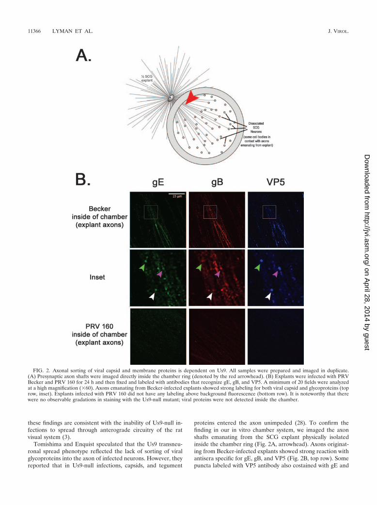
proteins entered the axon unimpeded (28). To confirm thefinding in our in vitro chamber system, we imaged the axonshafts emanating from the SCG explant physically isolatedinside the chamber ring (Fig. 2A, arrowhead). Axons originat-ing from Becker-infected explants showed strong reaction withantisera specific for gE, gB, and VP5 (Fig. 2B, top row). Somepuncta labeled with VP5 antibody also costained with gE and
FIG. 2. Axonal sorting of viral capsid and membrane proteins is dependent on Us9. All samples were prepared and imaged in duplicate.(A) Presynaptic axon shafts were imaged directly inside the chamber ring (denoted by the red arrowhead). (B) Explants were infected with PRVBecker and PRV 160 for 24 h and then fixed and labeled with antibodies that recognize gE, gB, and VP5. A minimum of 20 fields were analyzedat a high magnification (�60). Axons emanating from Becker-infected explants showed strong labeling for both viral capsid and glycoproteins (toprow, inset). Explants infected with PRV 160 did not have any labeling above background fluorescence (bottom row). It is noteworthy that therewere no observable gradations in staining with the Us9-null mutant; viral proteins were not detected inside the chamber.
11366 LYMAN ET AL. J. VIROL.
on April 28, 2014 by guest
http://jvi.asm.org/
Dow
nloaded from
gB antisera, suggesting that capsids were transported togetherwith viral glycoproteins (Fig. 2B, inset). In contrast, axonsoriginating from explants infected with the Us9-null mutantdid not react with antisera against gE or gB, which is consistentwith previous observations (28). Surprisingly, we did not detectany reaction with VP5 antibodies, suggesting that sorting ofviral capsids into axons is, in fact, dependent on Us9 (Fig. 2B,bottom row).
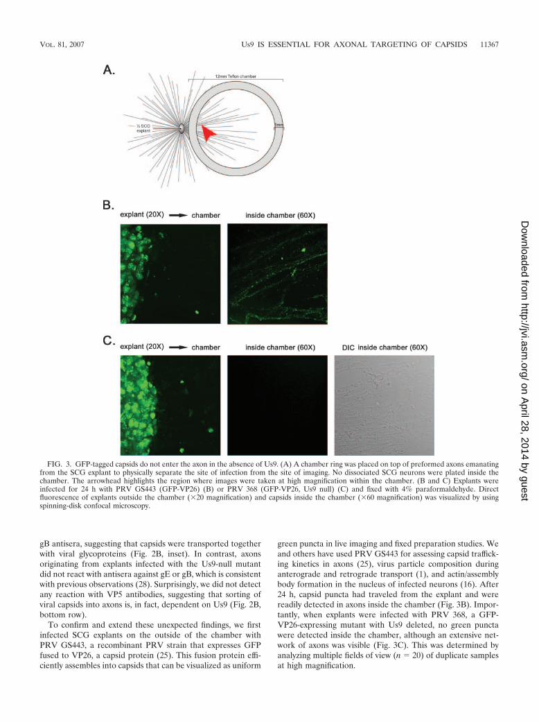
To confirm and extend these unexpected findings, we firstinfected SCG explants on the outside of the chamber withPRV GS443, a recombinant PRV strain that expresses GFPfused to VP26, a capsid protein (25). This fusion protein effi-ciently assembles into capsids that can be visualized as uniform
green puncta in live imaging and fixed preparation studies. Weand others have used PRV GS443 for assessing capsid traffick-ing kinetics in axons (25), virus particle composition duringanterograde and retrograde transport (1), and actin/assemblybody formation in the nucleus of infected neurons (16). After24 h, capsid puncta had traveled from the explant and werereadily detected in axons inside the chamber (Fig. 3B). Impor-tantly, when explants were infected with PRV 368, a GFP-VP26-expressing mutant with Us9 deleted, no green punctawere detected inside the chamber, although an extensive net-work of axons was visible (Fig. 3C). This was determined byanalyzing multiple fields of view (n � 20) of duplicate samplesat high magnification.
FIG. 3. GFP-tagged capsids do not enter the axon in the absence of Us9. (A) A chamber ring was placed on top of preformed axons emanatingfrom the SCG explant to physically separate the site of infection from the site of imaging. No dissociated SCG neurons were plated inside thechamber. The arrowhead highlights the region where images were taken at high magnification within the chamber. (B and C) Explants wereinfected for 24 h with PRV GS443 (GFP-VP26) (B) or PRV 368 (GFP-VP26, Us9 null) (C) and fixed with 4% paraformaldehyde. Directfluorescence of explants outside the chamber (�20 magnification) and capsids inside the chamber (�60 magnification) was visualized by usingspinning-disk confocal microscopy.
VOL. 81, 2007 Us9 IS ESSENTIAL FOR AXONAL TARGETING OF CAPSIDS 11367
on April 28, 2014 by guest
http://jvi.asm.org/
Dow
nloaded from

We next used transmission electron microscopy to deter-mine whether capsids were present in the distal axons of in-fected explants. Explants on the outside of the chamber wereinfected with PRV Becker or a Us9-null mutant and thenimaged directly inside the chamber ring 24 h postinfection.Capsids were detected readily in the distal axons of explantsinfected with PRV Becker. All were found within a membra-nous vesicle and contained a proteinaceous tegument layerjuxtaposed to the capsid (Fig. 4A). This morphology is consis-tent with other ultrastructural studies examining PRV infec-tion of primary neurons and tissue culture cells (15, 17, 30).Importantly, when explants were infected with the Us9-nullmutant, no capsid structures could be detected inside thechamber ring despite an extensive search (Fig. 4B).
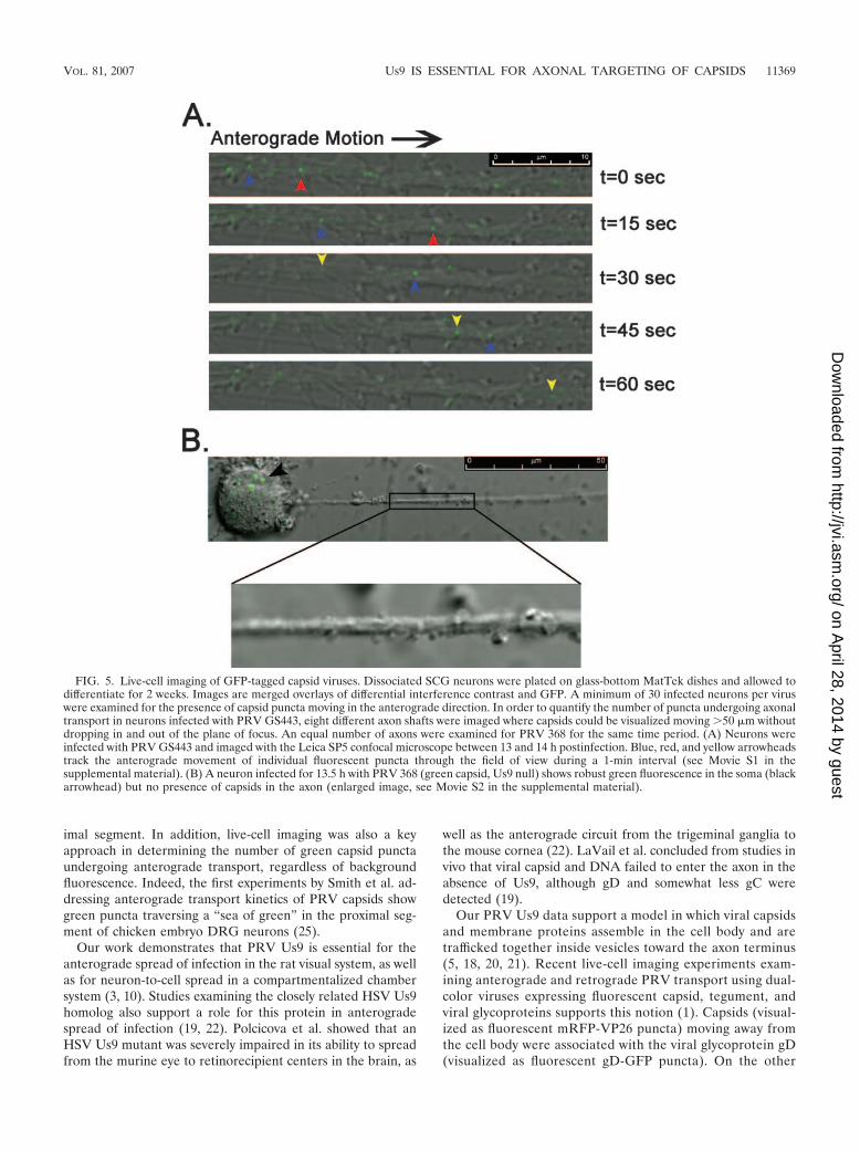
We next used a live-cell imaging approach in order to quan-tify the number of capsids moving in the axons of dissociatedSCG neurons (as deduced by GFP-VP26 puncta visible in the
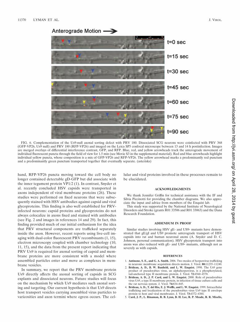
confocal microscope). This technique enabled us to distinguishGFP-VP26 puncta moving by fast-axonal transport in the an-terograde or retrograde direction from those that were notmoving or from background fluorescence. Dissociated neuronswere infected with PRV GS443 or PRV 368 and imaged be-tween 13 and 14 h postinfection. A total of 128 GFP-VP26puncta were observed moving in the anterograde direction inPRV GS443-infected neurons, entering the field of view by, onaverage, 4.6 � 1.6 capsids/min (Fig. 5A; see also Movie S1 inthe supplemental material). Despite extensive analyses, nocapsid puncta were observed in the axon of neurons infectedwith PRV 368, although GFP-VP26 signal was clearly visible inthe soma (Fig. 5B; see also Movie S2 in the supplementalmaterial). To test whether the axonal sorting defect in PRV368 was due to the lack of Us9 protein, we coinfected neuronswith PRV 368 and PRV 180 (a Becker recombinant expressingmRFP-VP26) (11). We could easily detect green, red, andyellow puncta moving in the anterograde direction in coin-fected neurons (Fig. 6 and see also Movie S3 in the supple-mental material). We conclude that PRV 368-infected neuronscan sort and move GFP-VP26 capsid puncta in their axonswhen complemented in trans by a Us9-expressing virus recom-binant.
DISCUSSION
We report here that the axonal targeting of PRV capsids isdependent on the viral membrane protein Us9. We confirmedthis observation by indirect IF (Fig. 2), direct fluorescence ofGFP-tagged capsids (Fig. 3), transmission electron microscopy(Fig. 4), and live-cell imaging in cultured, sympathetic neurons(Fig. 5 and 6). Our findings raise a key question: why did weobtain such disparate results using essentially the same re-agents as Tomishima and Enquist (14, 28)? In short, theanswer is that imaging the proximal axon of infected neurons,as was done in the previous studies, can be misinterpreted,particularly when indirect IF is used at late times after infec-tion. For example, Tomishima and Enquist fixed neurons in-fected with PRV GS443 and PRV 368 at 17 hpi and usedanti-EGFP antibodies to enhance GFP signal in axons (28).We believe that the signal observed by Tomishima and Enquistwas unassembled GFP-VP26 protein and not assembled cap-sids. This free GFP-VP26 in the proximal axon confoundedinterpretation of “capsid” localization in axons infected with aUs9-null virus, especially when images were obtained at lowmagnification (14, 28). We first came to this idea from liveneuron imaging studies with PRV GS443 and PRV 368. Weconsistently observed a diffuse green fluorescent signal build-ing in the proximal axon late in infection (�12 h postinfection).Indeed, by 16 h postinfection, this diffuse signal was strongenough in the proximal segment that we could visualize theconnection of cell bodies to cognate axons. The general phe-nomenon of entry of unassembled structural proteins into theproximal segment of axons during infection also may explainthe bright, but noticeably diffuse signal of the major capsidprotein VP5 reported by Tomishima and Enquist (28).
Furthermore, in our studies, a serendipitous advantage ofusing the in vitro chamber system was that the site of imagingwas several millimeters from the SCG explant, automaticallyforcing us to image in the mid-distal axon instead of the prox-
FIG. 4. Axons are devoid of enveloped virus particles during aUs9-null infection. Explants on the outside of the chamber were in-fected for 24 h with PRV Becker or PRV 160 (Us9-null), and axonsinside the chamber were visualized by transmission electron micros-copy. Samples were examined in duplicate. A 1-mm square with highaxonal density inside the chamber was selected for sectioning by theultramicrotome. Six serial sections (70 nm apart) were scrutinized forthe presence of virus particles. (A) Enveloped virus particles weredetected in the distal-axon of Becker-infected explants but were notpresent in explants infected with PRV 160 (B).
11368 LYMAN ET AL. J. VIROL.
on April 28, 2014 by guest
http://jvi.asm.org/
Dow
nloaded from
imal segment. In addition, live-cell imaging was also a keyapproach in determining the number of green capsid punctaundergoing anterograde transport, regardless of backgroundfluorescence. Indeed, the first experiments by Smith et al. ad-dressing anterograde transport kinetics of PRV capsids showgreen puncta traversing a “sea of green” in the proximal seg-ment of chicken embryo DRG neurons (25).
Our work demonstrates that PRV Us9 is essential for theanterograde spread of infection in the rat visual system, as wellas for neuron-to-cell spread in a compartmentalized chambersystem (3, 10). Studies examining the closely related HSV Us9homolog also support a role for this protein in anterogradespread of infection (19, 22). Polcicova et al. showed that anHSV Us9 mutant was severely impaired in its ability to spreadfrom the murine eye to retinorecipient centers in the brain, as
well as the anterograde circuit from the trigeminal ganglia tothe mouse cornea (22). LaVail et al. concluded from studies invivo that viral capsid and DNA failed to enter the axon in theabsence of Us9, although gD and somewhat less gC weredetected (19).
Our PRV Us9 data support a model in which viral capsidsand membrane proteins assemble in the cell body and aretrafficked together inside vesicles toward the axon terminus(5, 18, 20, 21). Recent live-cell imaging experiments exam-ining anterograde and retrograde PRV transport using dual-color viruses expressing fluorescent capsid, tegument, andviral glycoproteins supports this notion (1). Capsids (visual-ized as fluorescent mRFP-VP26 puncta) moving away fromthe cell body were associated with the viral glycoprotein gD(visualized as fluorescent gD-GFP puncta). On the other
FIG. 5. Live-cell imaging of GFP-tagged capsid viruses. Dissociated SCG neurons were plated on glass-bottom MatTek dishes and allowed todifferentiate for 2 weeks. Images are merged overlays of differential interference contrast and GFP. A minimum of 30 infected neurons per viruswere examined for the presence of capsid puncta moving in the anterograde direction. In order to quantify the number of puncta undergoing axonaltransport in neurons infected with PRV GS443, eight different axon shafts were imaged where capsids could be visualized moving �50 �m withoutdropping in and out of the plane of focus. An equal number of axons were examined for PRV 368 for the same time period. (A) Neurons wereinfected with PRV GS443 and imaged with the Leica SP5 confocal microscope between 13 and 14 h postinfection. Blue, red, and yellow arrowheadstrack the anterograde movement of individual fluorescent puncta through the field of view during a 1-min interval (see Movie S1 in thesupplemental material). (B) A neuron infected for 13.5 h with PRV 368 (green capsid, Us9 null) shows robust green fluorescence in the soma (blackarrowhead) but no presence of capsids in the axon (enlarged image, see Movie S2 in the supplemental material).
VOL. 81, 2007 Us9 IS ESSENTIAL FOR AXONAL TARGETING OF CAPSIDS 11369
on April 28, 2014 by guest
http://jvi.asm.org/
Dow
nloaded from
hand, RFP-VP26 puncta moving toward the cell body nolonger contained detectable gD-GFP but did associate withthe inner tegument protein VP1/2 (1). In contrast, Snyder etal. recently concluded HSV capsids were transported inaxons independent of viral membrane proteins (26). Thesestudies were performed on fixed neurons that were subse-quently stained with HSV antibodies against capsid and viralglycoproteins. This finding is also well established for PRV-infected neurons: capsid proteins and glycoproteins do notalways colocalize in axons fixed and stained with antibodies(see Fig. 2 and images in references 14 and 29). In fact, thisfinding provided much of our initial enthusiasm for the ideathat PRV structural components are trafficked separatelyinside the axon. However, recent reports using live-cell im-aging with dual-color fluorescent PRV recombinants (1, 15),electron microscopy coupled with chamber technology (10,11, 15), and the data from the present report indicating thatPRV Us9 is required for axonal sorting of capsid and mem-brane proteins are more consistent with a model whereassembled particles enter and move as complexes in mem-brane vesicles.
In summary, we report that the PRV membrane proteinUs9 directly affects the axonal sorting of capsids in SCGexplants and dissociated neurons. Future studies will focuson the mechanism by which Us9 mediates such axonal sort-ing and targeting. Our current hypothesis is that Us9 directshost transport vesicles carrying assembled virus particles tovaricosities and axon termini where egress occurs. The cel-
lular and viral proteins involved in these processes remain tobe elucidated.
ACKNOWLEDGMENTS
We thank Jennifer Griffin for technical assistance with the IF andSilvia Piccinotti for providing the chamber diagrams. We also appre-ciate the input and advice from members of the Enquist lab.
This study was supported by the National Institute of NeurologicalDisorders and Stroke (grants R01 33506 and R01 33063) and the DanaResearch Foundation.
ADDENDUM IN PROOF
Similar studies involving HSV gE- and US9- mutants have demon-strated that gE/gI and US9 promote anterograde transport of HSVcapsids into rat and human neuronal axons (A. Snyder and D. C.Johnson, personal communication). HSV glycoprotein transport intoaxons was also reduced with gE- and US9- mutants, although not asseverely as with capsids.
REFERENCES
1. Antinone, S. E., and G. A. Smith. 2006. Two modes of herpesvirus traffickingin neurons: membrane acquisition directs motion. J. Virol. 80:11235–11240.
2. Brideau, A. D., B. W. Banfield, and L. W. Enquist. 1998. The Us9 geneproduct of pseudorabies virus, an alphaherpesvirus, is a phosphorylated,tail-anchored type II membrane protein. J. Virol. 72:4560–4570.
3. Brideau, A. D., J. P. Card, and L. W. Enquist. 2000. Role of pseudorabiesvirus Us9, a type II membrane protein, in infection of tissue culture cells andthe rat nervous system. J. Virol. 74:834–845.
4. Brideau, A. D., T. del Rio, E. J. Wolffe, and L. W. Enquist. 1999. Intracellulartrafficking and localization of the pseudorabies virus Us9 type II envelopeprotein to host and viral membranes. J. Virol. 73:4372–4384.
5. Card, J. P., L. Rinaman, R. B. Lynn, B. H. Lee, R. P. Meade, R. R. Miselis,
FIG. 6. Complementation of the Us9-null axonal sorting defect with PRV 180. Dissociated SCG neurons were coinfected with PRV 368(GFP-VP26, Us9 null) and PRV 180 (RFP-VP26) and imaged on the Leica SP5 confocal microscope between 13 and 14 h postinfection. Imagesare merged overlays of differential interference contrast, GFP, and RFP. Blue, red, and yellow arrowheads track the anterograde movement ofindividual fluorescent puncta through the field of view for 1.5 min (see Movie S3 in the supplemental material). Red and blue arrowheads highlightindividual yellow puncta, whose composition is a mix of GFP-VP26 and RFP-VP26. The yellow arrowhead marks a predominantly red punctumand a predominantly green punctum transported together that eventually separate. (asterisks)
11370 LYMAN ET AL. J. VIROL.
on April 28, 2014 by guest
http://jvi.asm.org/
Dow
nloaded from
and L. W. Enquist. 1993. Pseudorabies virus infection of the rat centralnervous system: ultrastructural characterization of viral replication, trans-port, and pathogenesis. J. Neurosci. 13:2515–2539.
6. Card, J. P., L. Rinaman, J. S. Schwaber, R. R. Miselis, M. E. Whealy, A. K.Robbins, and L. W. Enquist. 1990. Neurotropic properties of pseudorabiesvirus: uptake and transneuronal passage in the rat central nervous system.J. Neurosci. 10:1974–1994.
7. Card, J. P., M. E. Whealy, A. K. Robbins, R. Y. Moore, and L. W. Enquist.1991. Two alpha-herpesvirus strains are transported differentially in therodent visual system. Neuron 6:957–969.
8. Ch’ng, T., E. A. Flood, and L. W. Enquist. 2004. Culturing primary andtransformed neuronal cells for studying pseudorabies virus infection, p. 299–316. In P. M. Lieberman (ed.), Methods in molecular biology. The HumanaPress, Inc., Totoway, NJ.
9. Ch’ng, T. H., and L. W. Enquist. 2005. Efficient axonal localization ofalphaherpesvirus structural proteins in cultured sympathetic neurons re-quires viral glycoprotein E. J. Virol. 79:8835–8846.
10. Ch’ng, T. H., and L. W. Enquist. 2005. Neuron-to-cell spread of pseudora-bies virus in a compartmented neuronal culture system. J. Virol. 79:10875–10889.
11. del Rio, T., T. H. Ch’ng, E. A. Flood, S. P. Gross, and L. W. Enquist. 2005.Heterogeneity of a fluorescent tegument component in single pseudorabiesvirus virions and enveloped axonal assemblies. J. Virol. 79:3903–3919.
12. Enquist, L. W., and J. P. Card. 2003. Recent advances in the use of neuro-tropic viruses for circuit analysis. Curr. Opin. Neurobiol. 13:603–606.
13. Enquist, L. W., P. J. Husak, B. W. Banfield, and G. A. Smith. 1998. Infectionand spread of alphaherpesviruses in the nervous system. Adv. Virus Res.51:237–347.
14. Enquist, L. W., M. J. Tomishima, S. Gross, and G. A. Smith. 2002. Direc-tional spread of an alpha-herpesvirus in the nervous system. Vet. Microbiol.86:5–16.
15. Feierbach, B., M. Bisher, J. Goodhouse, and L. W. Enquist. 2007. In vitroanalysis of trans-neuronal spread of an alpha-herpesvirus infection in pe-ripheral nervous system neurons. J. Virol. 81:6846–6857.
16. Feierbach, B., S. Piccinotti, M. Bisher, W. Denk, and L. W. Enquist. 2006.Alpha-herpesvirus infection induces the formation of nuclear actin filaments.PLoS Pathog. 2:e85.
17. Granzow, H., B. G. Klupp, W. Fuchs, J. Veits, N. Osterrieder, and T. C.Mettenleiter. 2001. Egress of alphaherpesviruses: comparative ultrastruc-tural study. J. Virol. 75:3675–3684.
18. Kritas, S. K., H. J. Nauwynck, and M. B. Pensaert. 1995. Dissemination ofwild-type and gC-, gE-and gI-deleted mutants of Aujeszky’s disease virus in
the maxillary nerve and trigeminal ganglion of pigs after intranasal inocula-tion. J. Gen. Virol. 76(Pt. 8):2063–2066.
19. Lavail, J. H., A. N. Tauscher, A. Sucher, O. Harrabi, and R. Brandimarti.2007. Viral regulation of the long distance axonal transport of herpes simplexvirus nucleocapsid. Neuroscience 146:974–985.
20. Lycke, E., B. Hamark, M. Johansson, A. Krotochwil, J. Lycke, and B. Sven-nerholm. 1988. Herpes simplex virus infection of the human sensory neuron.An electron microscopy study. Arch. Virol. 101:87–104.
21. Lycke, E., K. Kristensson, B. Svennerholm, A. Vahlne, and R. Ziegler. 1984.Uptake and transport of herpes simplex virus in neurites of rat dorsal rootganglia cells in culture. J. Gen. Virol. 65(Pt. 1):55–64.
22. Polcicova, K., P. S. Biswas, K. Banerjee, T. W. Wisner, B. T. Rouse, and D. C.Johnson. 2005. Herpes keratitis in the absence of anterograde transport ofvirus from sensory ganglia to the cornea. Proc. Natl. Acad. Sci. USA 102:11462–11467.
23. Pomeranz, L. E., A. E. Reynolds, and C. J. Hengartner. 2005. Molecularbiology of pseudorabies virus: impact on neurovirology and veterinary med-icine. Microbiol. Mol. Biol. Rev. 69:462–500.
24. Smith, G. A., and L. W. Enquist. 2002. Break ins and break outs: viralinteractions with the cytoskeleton of mammalian cells. Annu. Rev. Cell Dev.Biol. 18:135–161.
25. Smith, G. A., S. P. Gross, and L. W. Enquist. 2001. Herpesviruses usebidirectional fast-axonal transport to spread in sensory neurons. Proc. Natl.Acad. Sci. USA 98:3466–3470.
26. Snyder, A., T. W. Wisner, and D. C. Johnson. 2006. Herpes simplex viruscapsids are transported in neuronal axons without an envelope containingthe viral glycoproteins. J. Virol. 80:11165–11177.
27. Tirabassi, R. S., and L. W. Enquist. 2000. Role of the pseudorabies virus gIcytoplasmic domain in neuroinvasion, virulence, and posttranslational N-linked glycosylation. J. Virol. 74:3505–3516.
28. Tomishima, M. J., and L. W. Enquist. 2001. A conserved alpha-herpesvirusprotein necessary for axonal localization of viral membrane proteins. J. CellBiol. 154:741–752.
29. Tomishima, M. J., G. A. Smith, and L. W. Enquist. 2001. Sorting andtransport of alpha herpesviruses in axons. Traffic 2:429–436.
30. Watson, D. H., W. C. Russell, and P. Wildy. 1963. Electron microscopicparticle counts on herpesvirus using the phosphotungstate negative stainingtechnique. Virology 19:250–260.
31. Whealy, M. E., J. P. Card, A. K. Robbins, J. R. Dubin, H. J. Rziha, and L. W.Enquist. 1993. Specific pseudorabies virus infection of the rat visual systemrequires both gI and gp63 glycoproteins. J. Virol. 67:3786–3797.
VOL. 81, 2007 Us9 IS ESSENTIAL FOR AXONAL TARGETING OF CAPSIDS 11371
on April 28, 2014 by guest
http://jvi.asm.org/
Dow
nloaded from