Spinal Cord Injury Reduces the Efficacy of Pseudorabies Virus Labeling of Sympathetic Preganglionic...
11
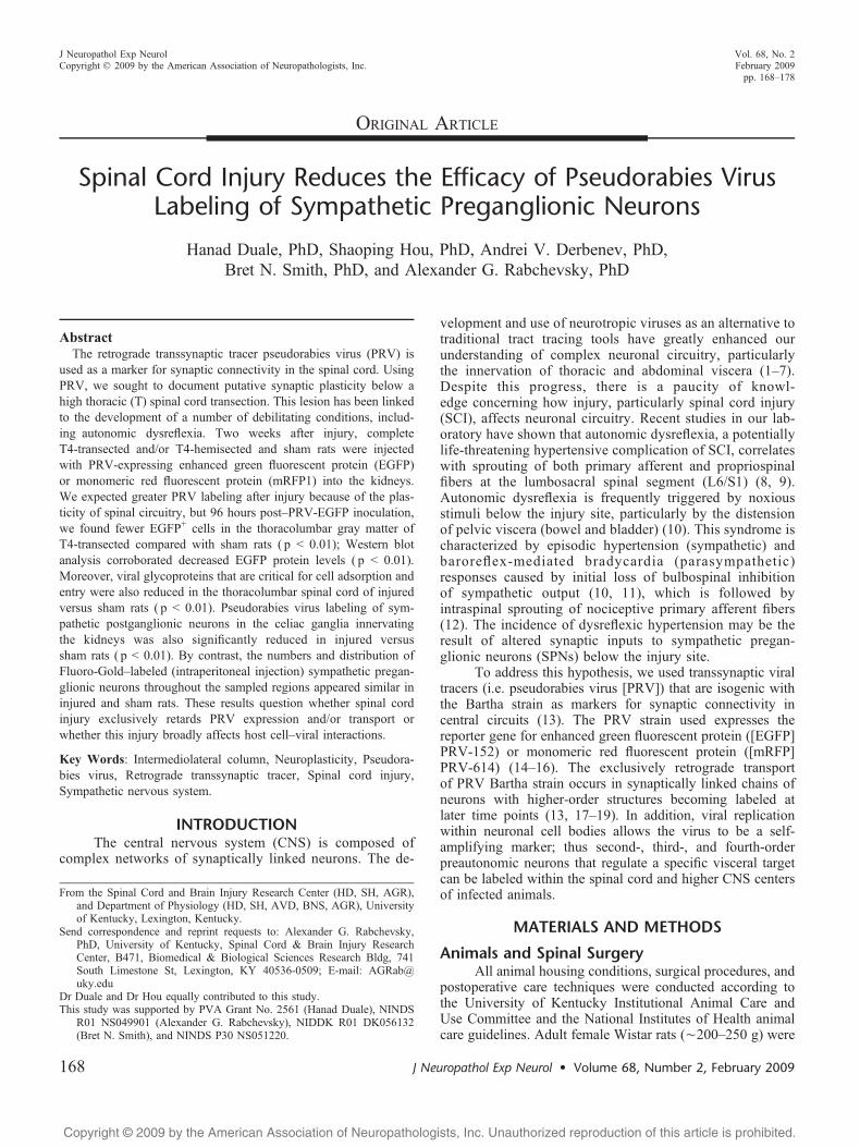
Copyright @ 2009 by the American Association of Neuropathologists, Inc. Unauthorized reproduction of this article is prohibited. ORIGINAL ARTICLE Spinal Cord Injury Reduces the Efficacy of Pseudorabies Virus Labeling of Sympathetic Preganglionic Neurons Hanad Duale, PhD, Shaoping Hou, PhD, Andrei V. Derbenev, PhD, Bret N. Smith, PhD, and Alexander G. Rabchevsky, PhD Abstract The retrograde transsynaptic tracer pseudorabies virus (PRV) is used as a marker for synaptic connectivity in the spinal cord. Using PRV, we sought to document putative synaptic plasticity below a high thoracic (T) spinal cord transection. This lesion has been linked to the development of a number of debilitating conditions, includ- ing autonomic dysreflexia. Two weeks after injury, complete T4-transected and/or T4-hemisected and sham rats were injected with PRV-expressing enhanced green fluorescent protein (EGFP) or monomeric red fluorescent protein (mRFP1) into the kidneys. We expected greater PRV labeling after injury because of the plas- ticity of spinal circuitry, but 96 hours postYPRV-EGFP inoculation, we found fewer EGFP + cells in the thoracolumbar gray matter of T4-transected compared with sham rats ( p G 0.01); Western blot analysis corroborated decreased EGFP protein levels ( p G 0.01). Moreover, viral glycoproteins that are critical for cell adsorption and entry were also reduced in the thoracolumbar spinal cord of injured versus sham rats ( p G 0.01). Pseudorabies virus labeling of sym- pathetic postganglionic neurons in the celiac ganglia innervating the kidneys was also significantly reduced in injured versus sham rats ( p G 0.01). By contrast, the numbers and distribution of Fluoro-GoldYlabeled (intraperitoneal injection) sympathetic pregan- glionic neurons throughout the sampled regions appeared similar in injured and sham rats. These results question whether spinal cord injury exclusively retards PRV expression and/or transport or whether this injury broadly affects host cellYviral interactions. Key Words: Intermediolateral column, Neuroplasticity, Pseudora- bies virus, Retrograde transsynaptic tracer, Spinal cord injury, Sympathetic nervous system. INTRODUCTION The central nervous system (CNS) is composed of complex networks of synaptically linked neurons. The de- velopment and use of neurotropic viruses as an alternative to traditional tract tracing tools have greatly enhanced our understanding of complex neuronal circuitry, particularly the innervation of thoracic and abdominal viscera (1Y7). Despite this progress, there is a paucity of knowl- edge concerning how injury, particularly spinal cord injury (SCI), affects neuronal circuitry. Recent studies in our lab- oratory have shown that autonomic dysreflexia, a potentially life-threatening hypertensive complication of SCI, correlates with sprouting of both primary afferent and propriospinal fibers at the lumbosacral spinal segment (L6/S1) (8, 9). Autonomic dysreflexia is frequently triggered by noxious stimuli below the injury site, particularly by the distension of pelvic viscera (bowel and bladder) (10). This syndrome is characterized by episodic hypertension (sympathetic) and baroreflex-mediated bradycardia (parasympathetic) responses caused by initial loss of bulbospinal inhibition of sympathetic output (10, 11), which is followed by intraspinal sprouting of nociceptive primary afferent fibers (12). The incidence of dysreflexic hypertension may be the result of altered synaptic inputs to sympathetic pregan- glionic neurons (SPNs) below the injury site. To address this hypothesis, we used transsynaptic viral tracers (i.e. pseudorabies virus [PRV]) that are isogenic with the Bartha strain as markers for synaptic connectivity in central circuits (13). The PRV strain used expresses the reporter gene for enhanced green fluorescent protein ([EGFP] PRV-152) or monomeric red fluorescent protein ([mRFP] PRV-614) (14Y16). The exclusively retrograde transport of PRV Bartha strain occurs in synaptically linked chains of neurons with higher-order structures becoming labeled at later time points (13, 17Y19). In addition, viral replication within neuronal cell bodies allows the virus to be a self- amplifying marker; thus second-, third-, and fourth-order preautonomic neurons that regulate a specific visceral target can be labeled within the spinal cord and higher CNS centers of infected animals. MATERIALS AND METHODS Animals and Spinal Surgery All animal housing conditions, surgical procedures, and postoperative care techniques were conducted according to the University of Kentucky Institutional Animal Care and Use Committee and the National Institutes of Health animal care guidelines. Adult female Wistar rats (È200Y250 g) were J Neuropathol Exp Neurol Volume 68, Number 2, February 2009 168 J Neuropathol Exp Neurol Copyright Ó 2009 by the American Association of Neuropathologists, Inc. Vol. 68, No. 2 February 2009 pp. 168Y178 From the Spinal Cord and Brain Injury Research Center (HD, SH, AGR), and Department of Physiology (HD, SH, AVD, BNS, AGR), University of Kentucky, Lexington, Kentucky. Send correspondence and reprint requests to: Alexander G. Rabchevsky, PhD, University of Kentucky, Spinal Cord & Brain Injury Research Center, B471, Biomedical & Biological Sciences Research Bldg, 741 South Limestone St, Lexington, KY 40536-0509; E-mail: AGRab@ uky.edu Dr Duale and Dr Hou equally contributed to this study. This study was supported by PVA Grant No. 2561 (Hanad Duale), NINDS R01 NS049901 (Alexander G. Rabchevsky), NIDDK R01 DK056132 (Bret N. Smith), and NINDS P30 NS051220.
-
Upload
independent -
Category
Documents
-
view
1 -
download
0
Transcript of Spinal Cord Injury Reduces the Efficacy of Pseudorabies Virus Labeling of Sympathetic Preganglionic...
Copyright @ 2009 by the American Association of Neuropathologists, Inc. Unauthorized reproduction of this article is prohibited.
ORIGINAL ARTICLE
Spinal Cord Injury Reduces the Efficacy of Pseudorabies VirusLabeling of Sympathetic Preganglionic Neurons
Hanad Duale, PhD, Shaoping Hou, PhD, Andrei V. Derbenev, PhD,Bret N. Smith, PhD, and Alexander G. Rabchevsky, PhD
AbstractThe retrograde transsynaptic tracer pseudorabies virus (PRV) is
used as a marker for synaptic connectivity in the spinal cord. UsingPRV, we sought to document putative synaptic plasticity below ahigh thoracic (T) spinal cord transection. This lesion has been linkedto the development of a number of debilitating conditions, includ-ing autonomic dysreflexia. Two weeks after injury, completeT4-transected and/or T4-hemisected and sham rats were injectedwith PRV-expressing enhanced green fluorescent protein (EGFP)or monomeric red fluorescent protein (mRFP1) into the kidneys.We expected greater PRV labeling after injury because of the plas-ticity of spinal circuitry, but 96 hours postYPRV-EGFP inoculation,we found fewer EGFP+ cells in the thoracolumbar gray matter ofT4-transected compared with sham rats ( p G 0.01); Western blotanalysis corroborated decreased EGFP protein levels ( p G 0.01).Moreover, viral glycoproteins that are critical for cell adsorption andentry were also reduced in the thoracolumbar spinal cord of injuredversus sham rats ( p G 0.01). Pseudorabies virus labeling of sym-pathetic postganglionic neurons in the celiac ganglia innervatingthe kidneys was also significantly reduced in injured versussham rats ( p G 0.01). By contrast, the numbers and distribution ofFluoro-GoldYlabeled (intraperitoneal injection) sympathetic pregan-glionic neurons throughout the sampled regions appeared similar ininjured and sham rats. These results question whether spinal cordinjury exclusively retards PRV expression and/or transport orwhether this injury broadly affects host cellYviral interactions.
Key Words: Intermediolateral column, Neuroplasticity, Pseudora-bies virus, Retrograde transsynaptic tracer, Spinal cord injury,Sympathetic nervous system.
INTRODUCTIONThe central nervous system (CNS) is composed of
complex networks of synaptically linked neurons. The de-
velopment and use of neurotropic viruses as an alternative totraditional tract tracing tools have greatly enhanced ourunderstanding of complex neuronal circuitry, particularlythe innervation of thoracic and abdominal viscera (1Y7).Despite this progress, there is a paucity of knowl-edge concerning how injury, particularly spinal cord injury(SCI), affects neuronal circuitry. Recent studies in our lab-oratory have shown that autonomic dysreflexia, a potentiallylife-threatening hypertensive complication of SCI, correlateswith sprouting of both primary afferent and propriospinalfibers at the lumbosacral spinal segment (L6/S1) (8, 9).Autonomic dysreflexia is frequently triggered by noxiousstimuli below the injury site, particularly by the distensionof pelvic viscera (bowel and bladder) (10). This syndrome ischaracterized by episodic hypertension (sympathetic) andbaroreflex-mediated bradycardia (parasympathetic)responses caused by initial loss of bulbospinal inhibitionof sympathetic output (10, 11), which is followed byintraspinal sprouting of nociceptive primary afferent fibers(12). The incidence of dysreflexic hypertension may be theresult of altered synaptic inputs to sympathetic pregan-glionic neurons (SPNs) below the injury site.
To address this hypothesis, we used transsynaptic viraltracers (i.e. pseudorabies virus [PRV]) that are isogenic withthe Bartha strain as markers for synaptic connectivity incentral circuits (13). The PRV strain used expresses thereporter gene for enhanced green fluorescent protein ([EGFP]PRV-152) or monomeric red fluorescent protein ([mRFP]PRV-614) (14Y16). The exclusively retrograde transportof PRV Bartha strain occurs in synaptically linked chains ofneurons with higher-order structures becoming labeled atlater time points (13, 17Y19). In addition, viral replicationwithin neuronal cell bodies allows the virus to be a self-amplifying marker; thus second-, third-, and fourth-orderpreautonomic neurons that regulate a specific visceral targetcan be labeled within the spinal cord and higher CNS centersof infected animals.
MATERIALS AND METHODS
Animals and Spinal SurgeryAll animal housing conditions, surgical procedures, and
postoperative care techniques were conducted according tothe University of Kentucky Institutional Animal Care andUse Committee and the National Institutes of Health animalcare guidelines. Adult female Wistar rats (È200Y250 g) were
J Neuropathol Exp Neurol � Volume 68, Number 2, February 2009168
J Neuropathol Exp NeurolCopyright � 2009 by the American Association of Neuropathologists, Inc.
Vol. 68, No. 2February 2009pp. 168Y178
From the Spinal Cord and Brain Injury Research Center (HD, SH, AGR),and Department of Physiology (HD, SH, AVD, BNS, AGR), Universityof Kentucky, Lexington, Kentucky.
Send correspondence and reprint requests to: Alexander G. Rabchevsky,PhD, University of Kentucky, Spinal Cord & Brain Injury ResearchCenter, B471, Biomedical & Biological Sciences Research Bldg, 741South Limestone St, Lexington, KY 40536-0509; E-mail: [email protected]
Dr Duale and Dr Hou equally contributed to this study.This study was supported by PVA Grant No. 2561 (Hanad Duale), NINDS
R01 NS049901 (Alexander G. Rabchevsky), NIDDK R01 DK056132(Bret N. Smith), and NINDS P30 NS051220.
Copyright @ 2009 by the American Association of Neuropathologists, Inc. Unauthorized reproduction of this article is prohibited.
anesthetized with a mixture of ketamine (80 mg/kg, intra-peritoneally [i.p.]; Fort Dodge Animal Health, Fort Dodge,IA) and xylazine (10 mg/kg, i.p.; Butler, Columbus, OH).The injured groups received either complete T4-transection(n = 26) or left-sided T4-hemisection (n = 12) with a sterilescalpel blade after T3 vertebra laminectomy, in contrast to thesham group (n = 26), which received only T3 laminectomy(Table 1), as previously described (8, 9).
After surgical operations were complete, the erectorspinae muscles were sutured with 3-0 Vycril (Ethicon,Sommerfield, NJ), the field was disinfected with povidone-iodine solution (Nova Plus, Irving, TX), and the skinwas closed with Michel wound clips (Roboz, Gaithersburg,MD). For postoperative care, animals were given 20 mLRinger’s lactate solution (Baxter Healthcare, Deerfield, IL)and 33 mg/kg cefazolin (Apothecon, Bristol-Myers Squibb,Princeton, NJ) subcutaneously immediately after surgery, andfor injured rats twice daily for up to 10 days to maintainhydration and control bladder infection. Buprenorphine(0.035 mg/kg; Reckitt Benckiser, UK) was also administeredsubcutaneously once after recovery from anesthesia and twicedaily for the next 3 days to control postoperative pain. Blad-ders of injured rats were manually expressed twice daily untilautomatic bladder-emptying reflex developed at about 10days postinjury.
PRV InjectionsTwo weeks after T4-transection, T4-hemisection, and
sham laminectomy, animals were reanesthetized with a mix-ture of ketamine (80 mg/kg, i.p.) and xylazine (10 mg/kg,i.p.), and a laparotomy was performed to expose theabdominal and pelvic contents. With the aid of a dissectingstereomicroscope, 3 KL of PRV-152 (green, 108 plaque-forming units/mL) were injected into the left kidney of sham(n = 18) and T4-transected (n = 18) rats at 3 sites (1 KL/site)using a 30-gauge Hamilton syringe (Hamilton, Reno, NV)(Table 1). Injections were uniformly placed at sites on the
longitudinal midline of the convex surface of the kidney (5).Injection sites were located by dividing this midline in thirdsand injecting at the rostral and caudal end of the middle third.Alternatively, 3 KL of PRV-614 (red, 108 plaque-formingunit/mL) were injected similarly into the left kidney of sham(n = 3) and T4-transected (n = 3) rats at 3 sites as a controlfor reporter gene expression (Table 1). For the T4-hemisectedrats, 3 KL of PRV-152 (n = 3) and 3 KL of PRV-614 (n = 3)were injected into both the left and right kidneys, respectively(Table 1). To control for potential injury-mediated reportergene expression, the T4-hemisection paradigm was reversed(e.g. 3 KL of PRV-152 [n = 3] and 3 KL of PRV-614 [n = 3]into both the right and left kidneys, respectively) (Table 1).For distal colon injections, the rostral section of the exposeddistal colon was elevated and packed with gauze. A total of12 KL PRV-152 was injected at 6 sites (2 KL/site) in sham(n = 3) and T4-transected rats (n = 3) using a 30-gaugeHamilton syringe with a sharp beveled tip into the ventraland lateral surfaces of the distal colon 25 to 40 mm from theanus (20). The needle was run into the viscus walltangentially to the surface beneath the serosa and either overor into the muscle layer, taking care not to penetrate into thelumen. The needle was inserted 4 to 10 mm and withdrawn1 to 2 mm before the virus solution was slowly injected (20).The needle was held in place for 5 minutes and slowlywithdrawn under a dry cotton-tipped stick to absorb reflux;this was followed by lavage of the peritoneal space withsterile saline. Injection sites were sealed with a drop of tissueadhesive (3M Vetbond, St Paul, MN). To control for PRVspecificity, naive rats (control; n = 2) received an i.p.injection of 3 KL of PRV-152 (Table 1). Materials that cameinto contact with PRV during surgery were cleaned withClorox bleach (0.05%) solution and properly discarded asbiohazard materials. Animals were maintained in a biosafetylevel 2 facility (72 hours, 84 hours, or 96 hours) untileuthanasia before perfusion fixation for histology or rapidisolation of fresh spinal tissues for molecular biology. Oneweek before perfusion, selected sham (n = 2) and injured(n = 2) rats were given an i.p. injection of Fluoro-Gold(0.2 mL of 1.5% in saline; Biotium, Hayward, CA) to labelall sympathetic preganglionic neurons in the intermedio-lateral cell column (21).
Perfusion and Tissue ProcessingAnimals designated for histology (n = 58; Table 2)
were overdosed with sodium pentobarbital (150 mg/kg;Abbott, Chicago, IL) and perfused transcardially with0.1 mol/L phosphate buffered saline (PBS), pH 7.4, followedby 4% paraformaldehyde in PBS. The spinal cord from theconus medullaris to the transection site was removed,postfixed for 4 hours, rinsed in 0.2 mol/L PB overnight,and cryoprotected for at least 48 hours in 20% sucrose in0.1 mol/L PBS. The caudal and rostral limits of the dissectedspinal cords sampled were between 0.5 mm rostral of theconus medullaris (ÈS4) and 6 cm rostral at the T5 segment.The cords were then divided into two 3-cm portions of caudal(S4YT13; lumbosacral) and rostral segments (T12YT5;thoracic) and embedded in gum tragacanth (Sigma-Aldrich,St Louis, MO) in 20% sucrose/PBS for cryosectioning, as
TABLE 1. Total Number of Rats Used in the DifferentExperimental ParadigmsInjection Groups T4-Transection T4-Hemisection Sham Naive
PRV-GFP,left kidney (n = 39)
18 3 18 V
PRV-GFP,right kidney (n = 3)
V 3 V V
PRV-GFP,distal colon (n = 6)
3 V 3 V
PRV-RFP,left kidney (n = 9)
3 3 3 V
PRV-RFP,right kidney (n = 3)
V 3 V V
Fluoro-Gold,intraperitoneal (n = 4)
2 V 2 V
PRV specificitycontrol (n = 2)
V V V 2
Total (n = 66) 26 12 26 2
GFP, green fluorescent protein; PRV, pseudorabies virus; RFP, red fluorescentprotein.
J Neuropathol Exp Neurol � Volume 68, Number 2, February 2009 Intraspinal Plasticity After Spinal Injury
� 2009 American Association of Neuropathologists, Inc. 169

Copyright @ 2009 by the American Association of Neuropathologists, Inc. Unauthorized reproduction of this article is prohibited.
previously detailed (9). Both thoracic and lumbosacralsegments of cord were serially cryosectioned in the lon-gitudinal horizontal plane at 50 Km and consecutivelymounted onto glass slides (Superfrost plus, Fisher Scientific,Pittsburgh, PA) in 5 series of 5 slides. This rendered 250-Kmseparations between adjacent serial sections within a serieson each slide (9).
ImmunohistochemistryFor anti-PRV double immunostaining with PRV-614,
slides with mounted longitudinal sections were thawed andpreincubated in 0.1 mol/L PBS containing 0.5% Triton-X and5% normal donkey serum (Vector Laboratories, Burlingame,CA) for 1 hour, followed by incubation with rabbitantiYPRV-132 (1:1000; courtesy of Dr Patrick Card) in samebuffer overnight at 4-C. The slides were then rinsed beforeapplying goat anti-rabbit conjugated to AMCA (JacksonImmunoResearch Laboratories, West Grove, PA; 1:200) for3 hours at room temperature. After final rinses, slides werecoverslipped using Vectashield mounting medium (Vector)and sealed with Cutex nail hardener (Jackson, WY).
Quantification of PRV-152+ Cells in theSpinal Cord
For PRV-152+ cell quantification after 96 hours post-inoculation, our region of interest was the caudal 2 cm of thethoracic spinal segments, corresponding to T8 to T13 ofsham (n = 4) versus T4-transected rats (n = 4) (Table 2).Previous studies had shown that EGFP-labeled cells afterPRV-152 inoculation into the kidneys were mainly dis-tributed from T6 to T13, with the highest cellular distributionrestricted to T8 to T13 (22Y24). Serial longitudinal horizontalsections containing the dorsoventral extent of the intermedio-lateral column, which incorporates most SPNs, were selected.To avoid counting the same cell twice, every other collected
serial section was used; thus, the sections were separated by100 Km. This corresponded to quantifying 3 alternating serialsections within the intermediolateral column. All quantifica-tions were conducted in a blinded fashion using the Bioquantimage analysis program (Nova Prime, V6.70.10; BioquantImage Analysis Corp, Nashville, TN) using establishedmethods (9). In brief, under an Olympus BX51 fluorescencemicroscope (Olympus Corp, Melville, NY), live imagescorresponding to bilateral gray matter regions in our area ofinterest (T8YT13) were circumscribed at 4� magnification(10� eye piece), and images were captured using anOptronics digital video camera (Optronics Corp, Goleta,CA). This corresponded to approximately 5.8 mm2 for shamsand 6 mm2 for injured cords, depending upon dorsoventralplane. In conjunction with Microcode II stage encoders(Boeckler Instruments, Tucson, AZ) and using a 60� oil-immersed objective (10� eye piece), all EGFP+ cells withinthe circumscribed area through the section plane werecounted. Although counting all objects within a circum-scribed area is a valid and efficient method, it is, however,prone to overcounting (25). To minimize overcounting, weused the correction factor first computed by Abercrombie(26): T/T + h, where T is the section thickness and h is themean diameter of the objects along the z axis (25, 26).Photomicrographs in all figures were optimized for finalproduction by adjusting only the brightness and contrastusing Adobe Photoshop 7.0 (Adobe Systems Inc, San Jose,CA). All graphs were created with DeltaGraph 5.4 (Red RockSoftware, Inc, Salt Lake City, UT).
Spinal Cord Tissue Extraction and Protein AssayInjured and sham animals designated for Western blot
analysis (n = 8, Table 2) were killed with carbon dioxide anddecapitated at 96 hours after PRV-152 inoculation into theleft kidney. The spinal cords (T5-conus medullaris) wererapidly removed (27) and placed on an ice-cold dissectingplate containing precooled Triton lysis buffer pH 7.4 (1%triton, 20 mmol/L Tris-HCl, 150 mmol/L NaCl, 5 mmol/LEGTA, 10 mmol/L EDTA, 10% glycerol) with proteaseinhibitors (Complete Mini Protease Inhibitor Cocktail tablet).The spinal cords were dissected into 2-cm segments, centeredbetween the T8 and T13 spinal rootlets, and homogenizedin 1 mL of Triton lysis buffer with protease inhibitors. Sam-ples were then briefly sonicated and vortexed at 14,000 rpmfor 30 minutes at 4-C, and the supernatants were collectedfor protein assay. Protein concentration was determined byBio-Rad DC Protein Assay (Bio-Rad, Hercules, CA), withsample solutions diluted to contain 1 mg/mL of protein forimmunoblotting.
Celiac Ganglia Tissue Extraction andPRV+ Neuronal Quantification
The celiac plexus is situated around and between theceliac artery and the superior mesenteric artery and extendsdorsally between the adrenal glands and the cranial half ofthe kidneys (28). The left celiac ganglion is crescent shapedand lies on the lateral side of the celiac and superiormesenteric artery; the right celiac ganglion is triangular,smaller than the left one, and lies dorsal to the inferior vena
TABLE 2. Celiac Ganglia and Spinal Tissues Used in theDifferent Experimental GroupsExperimental Paradigm T4-Transection T4-Hemisection Sham Naive
Histology, 48 hourspostYPRVinoculation*
3 V 3 V
Histology, 72 hourspostYPRVinoculation*†
5 4 5 V
Histology, 84 hourspostYPRVinoculation†
2 4 2 V
Histology, 96 hourspostYPRVinoculation†
10 4 10 2
Histology, 168 hourspostYFG injection
2 V 2 V
Western blot analysis,96 hours postYPRVinoculation
4 V 4 V
Total (n = 66) 26 12 26 2
*Includes celiac ganglia.†Includes both PRV-152 (green) and PRV-614 (red).FG, Fluoro-Gold; PRV, pseudorabies virus.
Duale et al J Neuropathol Exp Neurol � Volume 68, Number 2, February 2009
� 2009 American Association of Neuropathologists, Inc.170

Copyright @ 2009 by the American Association of Neuropathologists, Inc. Unauthorized reproduction of this article is prohibited.
cava (28). To facilitate visualization and isolation of thePRV-infected ganglia, injured and sham animals were killedwith carbon dioxide and immediately decapitated. Both theleft (PRV+) and right (PRV-) celiac ganglion were isolatedand submerged in ice-cold 4% paraformaldehyde in 0.1mol/L PBS for 48 hours. Postfixed ganglia were rinsed in0.2 mol/L PB overnight and cryoprotected for at least48 hours in 20% sucrose in 0.1 mol/L PBS. In addition, thespinal cords were also extracted, postfixed, and processed asdescribed to verify CNS labeling. As previously described,the ganglia were embedded in gum tragacanth and seriallycryosectioned in the longitudinal horizontal plane at 20 Kmand consecutively mounted onto glass slides in 5 series of5 adjacent slides. This rendered 100-Km separations betweenserial sections within a series on each slide (9). To verifyputative ganglionic neurons, selected serial sections werestained with cresyl violet. In brief, chosen slide series weresubjected to sequential rehydration followed by cresyl violetstain (cresyl violet acetate, Sigma) for 5 minutes, followed bya sequential dehydration phase. Stained sections were main-tained in Citrisolv (Fisher Scientific) to clear excess cresylviolet and subsequently coverslipped with Permount (FisherScientific) mounting medium.
For quantification of PRV-152+ sympathetic postgan-glionic neurons in the left celiac ganglion, a total of 12 rats(Table 2) were used and divided into injured (n = 6) andsham (n = 6) groups. These groups were further subdividedby inoculation time (48 hours, n = 6 [including injured andsham]; 72 hours, n = 6). Preliminary data revealed that PRVlabeling of the left celiac ganglion (right celiac ganglionacted as a negative control) was optimal at 72 hours versus
48 hours postYviral inoculation into the left kidney. Con-sequently, the total number of PRV-152+ cells in the leftceliac ganglion at 72 hours postYleft kidney inoculation wasquantified using the Bioquant image analysis program,similar to quantification in the thoracic cord. Notably,however, because of the small size of the ganglia, wequantified each sequential section throughout adjacent serialslides. For each section, the cross-sectional area of the entireleft ganglion was circumscribed at 4� magnification (10�eye piece), and images were captured using an Optronics
FIGURE 1. (A, B) Photomicrographs representing the progression of pseudorabies virus (PRV) labeling of cells in thethoracolumbar spinal cord after PRV-152 inoculation into the left kidney of sham rats at 72 hours (A) and 96 hours (B)postinoculation. The top of each panel represents the side of kidney injections. (C, D) Chronic spinal cord injury dramaticallyaltered the PRV labeling pattern in (C) sham versus (D) T4-transected (T4TX) rats at 96 hours postYPRV-152 inoculation into theleft kidney. Note the conspicuous diminished labeling after T4TX compared with the sham controls. Scale bars = (A, B) 500 Km;(C, D) 100 Km.
FIGURE 2. Quantitative comparisons of enhanced green fluo-rescent proteinYpositive (EGFP+) cells between T4-transectedand sham rats throughout the thoracolumbar spinal cord(T8YT13) after pseudorabies virus (PRV)-152 injection into theleft kidney at 96 hours postinoculation. Bars represent mean TSD. *p G 0.01.
J Neuropathol Exp Neurol � Volume 68, Number 2, February 2009 Intraspinal Plasticity After Spinal Injury
� 2009 American Association of Neuropathologists, Inc. 171

Copyright @ 2009 by the American Association of Neuropathologists, Inc. Unauthorized reproduction of this article is prohibited.
digital video camera. Only a subpopulation of celiacsympathetic postganglionic neurons innervates the kidneys,so all EGFP+ cells within the circumscribed area of everysection were counted using a 60� oil-immersed objective(10� eye piece). To correct for possible overcounting, weused the Abercrombie factor as previously described.
Western Blot Analysis of PRV-152 andPRV-Associated Proteins VP-5 and gB to C
To measure the relative expression levels of EGFP(there is no commercially available antibody againstmRFP1), PRV-associated major capsid coat protein (VP-5),and glycoproteins B to C (gBYC), spinal cord samples (50Kg, 40 Kg, and 40 Kg, respectively) corresponding to bothinjured (n = 4) and shams (n = 4) were run on an sodiumdodecyl sulfate/PAGE Precast gel (EGFP: 12% Bis-Tris
Criterion XT Precast gel, Bio-Rad; gBYC and VP-5: 4Y12%Bis-Tris Criterion XT, Bio-Rad), using established method-ologies (29). Afterward, the proteins were transferred onto apolyvinylidene diflouride (PVDF) membrane (Immuno-blotPVDF membrane-0.2 Km, Bio-Rad) using a semidry electro-transferring unit set at 15 V for 15 minutes. Preliminaryexperiments established protein concentration curves toensure that quantified bands were in the linear range asmeasured with the Li-Cor Odyssey Infrared Imaging System(Li-Cor Biotechnology, Lincoln, NE). The membranes wereincubated in Tris-buffered saline (TBS, pH 7.4) blockingsolution with 5% milk (Great Value instant nonfat dry milk,Wal-Mart) for 1 hour at room temperature. For the detectionof EGFP, a rabbit polyclonal anti-GFP antibody (-ab290,Abcam, Cambridge, MA) was used at a dilution of 1:1000 inTBS-Triton blocking solution with 5% milk for overnight
FIGURE 3. Chronic T4-transection (T4TX) markedly decreased the pseudorabies virus (PRV) labeling pattern in the thoracolumbarspinal cord, irrespective of the reporter gene (PRV-152 or PRV-RFP) used in sham (A) versus T4TX (B) rats. Injury-induced aberrantPRV-RFP expression is in part caused by compromised viral uptake because anti-PRV (>-PRV) double immunostaining in the samesections revealed more labeled neurons in sham (C) versus T4TX (D) rats. By contrast, Fluoro-Gold (FG) labeling in thethoracolumbar spinal cord of sham (E) versus chronic T4TX (F) rats seemed unaltered. Scale bar = 100 Km and applies to allphotographs.
Duale et al J Neuropathol Exp Neurol � Volume 68, Number 2, February 2009
� 2009 American Association of Neuropathologists, Inc.172

Copyright @ 2009 by the American Association of Neuropathologists, Inc. Unauthorized reproduction of this article is prohibited.
at 4-C. A goat anti-rabbit secondary conjugated to an infrareddye (1:5000, IRDye800CW, Rockland Immunochemicals,Gilbertsville, PA) was then applied for 1 hour at roomtemperature. After drying, the membranes were then imagedand quantified using the Li-Cor Odyssey Infrared ImagingSystem. To detect PRV associated VP-5 (154 kd) and gB toC proteins (NB: the glycoproteins B and C have similar sizeband È70 kd), a rabbit polyclonal anti-PRV (RB132, kindlydonated by Dr. Patrick Card, University of Pittsburgh) wasused at a dilution of 1:1000 in TBS-Triton blocking solutionwith 5% milk overnight at 4-C. A goat anti-rabbit secondaryconjugated to an infrared dye (1:5000, IRDye800CW, Rock-land Immunochemicals) was then applied for 1 hour at roomtemperature. To standardize protein loading, EGFP, VP-5,and/or gB to C immunoreactive PVDF membranes weresubsequently stripped (i.e. remove primary and secondaryantibodies from the membrane) and reprobed with mousemonoclonal antiYA-actin (1:500, Sigma). In brief, thePVDF membrane was bathed in a stripping buffercontaining 25 mmol/L glycine HCl, pH 2, and 1% sodiumdodecyl sulfate for 1 hour and replaced with fresh bufferevery 15 minutes. After stripping, the PVDF membranewas washed extensively in PBS + 0.1% Tween-20 (3 �5 minutes) and subsequently incubated for 1 hour in TBSblocking solution with 5% milk at room temperature. For thedetection of A-actin, mouse monoclonal antiYA-actin wasused at a dilution of 1:500 in TBS-Triton with 5% milk for
overnight incubation at 4-C. A goat anti-mouse secondaryconjugated to an infrared dye (1:5000, IRDye800CW, Rock-land Immunochemicals) was then applied for 1 hour at roomtemperature, and the A-actin+ PVDF membrane was visual-ized with Li-Cor Odyssey Infrared Imaging System.
Statistical AnalysisStatistical analyses between spinal-transected and non-
transected rats were performed using Stat-View (SASInstitute, Cary, NC). Unpaired Student t-test was usedbetween nontransected and injured groups. Significancethroughout all experiments was set at p G 0.05. Data arerepresented as mean T SD.
RESULTS
Effects of T4-Spinal Transection on EGFP/RFPExpression Patterns of Neurons in theThoracolumbar Spinal Cord
The PRV cellular labeling pattern observed in thethoracolumbar spinal cord after PRV-152 injection into theleft kidney includes the labeling of ipsilateral SPNs andinterneurons at 72 hours postinoculation (Fig. 1A). By 96hours postinoculation, there were extensive EGFP+ cells fromT6 to T13, with the highest numbers concentrated at T8 toT13 (Fig. 1B). At 2 weeks after complete T4-transection,
FIGURE 4. Expression patterns of pseudorabies virus (PRV)Yinfected neurons in the lumbosacral (parasympathetic) andthoracolumbar (sympathetic) spinal cord after viral injections into the distal colon (A, B) or kidneys (C, D), respectively. ChronicT4-transection (T4TX) did not seem to affect PRV-152 uptake and/or expression patterns in sham (A) versus T4TX (B) rats at96 hours postinoculation. By contrast, left-sided T4-hemisected spinal cords (C, D) seemed to show reduced PRV-152 expression/uptake in the ipsilateral spinal cord (Ipsi) compared with PRV-614 labeling in the contralateral side (Contra) at 72 hours (C) and84 hours (D) postinoculation into the left kidney (PRV-152) and right kidney (PRV-614), respectively. Scale bar = 500 Km andapplies to all panels.
J Neuropathol Exp Neurol � Volume 68, Number 2, February 2009 Intraspinal Plasticity After Spinal Injury
� 2009 American Association of Neuropathologists, Inc. 173

Copyright @ 2009 by the American Association of Neuropathologists, Inc. Unauthorized reproduction of this article is prohibited.
FIGURE 5. Relative expression levels of enhanced green fluorescent protein (EGFP) and pseudorabies virus (PRV)Yassociatedproteins in the thoracolumbar spinal cords of sham versus T4-transected (T4TX) rats. The dramatic reduction of PRV-152+ cellsafter injury in the thoracolumbar spinal cord was corroborated by Western blot analysis of (A) EGFP protein levels (arbitrary unit[AU]) in T4TX versus sham rats at 96 hours postYPRV-152 inoculation into the left kidney. (B) Protein blots (EGFP and A actin)from T4TX versus sham rats. (C) Viral glycoprotein B to C (gBYC) expression levels (AU), an important component for host-celladsorption and entry, were significantly reduced in T4TX versus sham rats at 96 hours postYPRV-152 inoculation. (D) Protein blots(gBYC and A actin) from T4TX versus sham rats. (E) The expression levels of major capsid coat protein (VP-5; AU), an importantelement in the construction of the PRV capsid, was significantly reduced in T4TX versus sham rats at 96 hours postYPRV-152inoculation. (F) Protein blots (VP-5 and A actin) from T4TX versus sham rats. Bars represent mean T SD. *p G 0.01.
Duale et al J Neuropathol Exp Neurol � Volume 68, Number 2, February 2009
� 2009 American Association of Neuropathologists, Inc.174

Copyright @ 2009 by the American Association of Neuropathologists, Inc. Unauthorized reproduction of this article is prohibited.
however, EGFP+ cells from T8 to T13 were markedlyreduced compared with sham rats (Figs. 1C, D). Moreover,semiquantitative Western blot analysis of EGFP expressionin the thoracolumbar spinal cord showed a significantreduction in injured versus sham rats (F1,6 = 13.61; p G0.01) (Fig. 2). To rule out the possibility that reduced PRVlabeling after injury is limited to specific reporter genes (i.e.EGFP), we injected PRV-RFP into the left kidney of shamversus injured rats and observed a similar outcome as for thePRV-152 paradigm (Figs. 3A, B). Furthermore, anti-PRVimmunostaining in the same sections appeared to show fewerlabeled neurons in injured versus sham rats at 96 hourspostinoculation (Figs. 3C, D). Conversely, intraperitonealinjection of Fluoro-Gold and subsequent labeling of SPNsthroughout the thoracolumbar spinal cord appeared unalteredin sham and injured rats (Figs. 3E, F).
Effects of T4-Spinal Transection on EGFPExpression Patterns of Neurons in theLumbosacral Spinal Cord
To assess whether SCI differentially affects thesympathetic versus parasympathetic autonomic branches,we injected PRV-152 into the distal colon (T4-transected,n = 3; sham, n = 3), which receives parasympatheticinnervation from the sacral parasympathetic preganglionicneurons in the lumbosacral (L6/S1) spinal cord of rats. At 96hours postYPRV-152 inoculation into the distal colon, therewas no qualitative difference in EGFP+ cellular labeling
pattern in the L6/S1 spinal cord between sham and injuredrats (Figs. 4A, B).
Effects of T4-Hemisection on EGFP/RFPExpression Patterns of Neurons in theThoracolumbar Spinal Cord
To rule out the possibility of compromised PRV uptakebeing limited to complete spinal cord transection, left-sidedT4-hemisected rats were injected with PRV-152 (green;injured side) and PRV-614 (red; intact side) into the leftand right kidneys, respectively (Table 2). At 72 hours (n = 2)and 84 hours (n = 2) postinoculation, spinal cord tissueipsilateral to the injury (left side) seemed to show fewerPRV-152+ cells compared with the PRV-614+ cells in thecontralateral uninjured spinal cord (Figs. 4C, D). Similarly,when the injection paradigm was reversed (i.e. PRV-614[injured side] into the left kidney and PRV-152 [intact side]into right kidney), we observed the same phenomena aspreviously described at 72 hours (n = 2) and 84 hours (n = 2)postinoculation (data not shown). Notably, in both injectionparadigms at 96 hours postinoculation (total; n = 4), weobserved considerable intermingling of PRV-152+ and PRV-614+ cells in the ipsilateral and contralateral thoracolumbarspinal cord (data not shown).
Expression Levels of EGFP and PRV-AssociatedProteins in the Thoracolumbar Spinal Cord
The significant reduction in EGFP+ cells in thethoracolumbar spinal cord of injured rats after left kidney
FIGURE 6. Photomicrographs of cresyl violetYstained left celiac ganglion (A) and pseudorabies virus (PRV) labeling pattern in leftceliac ganglion after PRV-152 inoculation into left kidney of a sham rat (B) at 72 hours. A subpopulation of celiac ganglionicneurons innervates the kidneys. Scale bar = 200 Km and applies to both images. (C) Quantitative comparison of enhanced greenfluorescent proteinYpositive (EGFP+) cells between T4-transected and sham rats in the left celiac ganglia after PRV-152 injectioninto the left kidney at 72 hours postinoculation. Bars represent mean T SD. *p G 0.01.
J Neuropathol Exp Neurol � Volume 68, Number 2, February 2009 Intraspinal Plasticity After Spinal Injury
� 2009 American Association of Neuropathologists, Inc. 175

Copyright @ 2009 by the American Association of Neuropathologists, Inc. Unauthorized reproduction of this article is prohibited.
PRV injection (Fig. 2) was corroborated by Western blotanalysis that showed a significant decrease in EGFP proteinlevels (p G 0.01) (Figs. 5A, B). This reduced EGFP ex-pression could be the result of either injury-induced disrup-tion in transcription and translational mechanisms or reducedviral uptake at the synapses. Because immunostaining forPRV seemed reduced (Figs. 3C, D), the most likely ex-planation is reduced viral uptake. Uptake of PRV initiallydepends upon the adsorption of virions to a heparin sulfateproteoglycan moiety on the host cell surface, which ismediated by glycoprotein C (gC) (30), whereas glycoproteinsB and D (gB and gD) are involved in cell surface penetration(31). Analysis of viral gB to C protein levels at 96 hourspostinoculation showed a significant reduction (p G 0.01) ininjured compared with sham rats (Figs. 5C, D). Importantly,VP-5, a critical protein that is involved in viral capsidformation (and thus viral assembly), was also significantlyreduced (p G 0.01) in injured versus sham rats (Figs. 5E, F).
Effects of T4-Spinal Transection on EGFPExpression Patterns of Neurons in theCeliac Ganglia
Because PRV labeling of postganglionic neurons in thespinal cord was retarded by SCI, we sought to determinewhether PRV transport in the sympathetic postganglionicneurons that innervate the kidneys was also affected by SCI.Therefore, PRV-152 was injected into the left kidney to labelsympathetic postganglionic neurons in the left celiac gan-glion. The PRV-152 expression patterns in the left celiacganglion indicated that a subpopulation of celiac neuronswas labeled in both injured and sham tissue samples(Figs. 6A, B). This was expected because the ganglia alsoinnervate other organs. Notably, we did not observe any PRVlabeling in the right celiac ganglion (i.e. the negative control).Quantitative analysis revealed significantly less PRV labelingin the left celiac ganglion of injured versus sham rats at72 hours postYviral inoculation (p G 0.01) (Fig. 6C).
DISCUSSIONNumerous studies have demonstrated the use of PRV
as a transsynaptic tracer in the rats’ CNS (5, 14, 16, 32), butSCI-induced plasticity of kidney-related sympathetic andcolon-related parasympathetic neural pathways has not beenpreviously studied using PRV. The present study usedtranssynaptic labeling with PRV to investigate putative axonremodeling after SCI, a crucial component in the develop-ment of autonomic dysreflexia (8, 9).
PRV Labeling in the Thoracolumbar Spinal CordThe patterns of labeling in the thoracolumbar (T6YL1)
spinal cord of sham rats after PRV inoculation into the leftkidney at 72, 84, and 96 hours postinjection were comparableto those in previous studies (22, 24). At 72 hours post-inoculation, PRV labeling was restricted predominately tosecond-order sympathetic preganglionic neurons; by 84 hours,third-order interneurons were labeled; and by 96 hours, theentire thoracolumbar cord seemed labeled (Figs. 1A, B); dis-crete regions proximal to the central canal of the lumbosacralspinal cord were also labeled (unpublished observations,
H. Duale, PhD, September 2007). By contrast, the pattern ofPRV labeling in these regions after chronic SCI was dra-matically altered. We analyzed spinal tissues infected after96 hours rather than 84 hours or 72 hours because theWestern blot signals from the earlier time points were in-sufficient for quantification. Counting of PRV+ cells in fixedspinal tissues was also conducted in tissue 96 hours postYPRV inoculation to maintain consistency. The markedreduction in PRV labeling corresponded with decreasedexpression of viral glycoproteins (gBYC) that facilitate viraladsorption and entry into the cell in sham versus injured rats.In contrast, intraperitoneal injection of Fluoro-Gold andsubsequent labeling pattern of SPNs throughout the thoraco-lumbar spinal cord seemed unchanged in sham versus injuredrats (Figs. 3E, F), indicating that the reduced PRV was notcaused by loss of autonomic motor neurons in injured spinalcords. The differences observed between Fluoro-Gold andPRV labeling in the thoracolumbar spinal cord may becaused by differential uptake mechanisms, that is, Fluoro-Gold is taken up via pinocytosis, whereas PRV uptake isreceptor mediated. We cannot rule out the possibility, how-ever, that altered intracellular transport and/or transcription/translation of viral protein products also contributed to theattenuation of PRV labeling after injury. Accordingly, in theleft-sided T4-hemisection paradigm, we observed at 72 hoursand 84 hours but not at 96 hours postYPRV inoculation amarked reduction in PRV-152 labeling in the ipsilateralspinal cord compared with the contralateral uninjured sidelabeled with PRV-RFP (Figs. 4C, D). Moreover, this obser-vation was independent of the PRV reporter gene expressed(i.e. PRV-RFP or PRV-152) (Figs. 1C, D; 3A, B).
PRV Labeling in the Celiac GangliaSpinal cord injury leads to disruption of the descending
spinal cardiovascular pathways resulting in sympathetichypoactivity and unopposed prevalence of the intact vagalparasympathetic control (33). Sympathetic hypoactivityresults in low resting blood pressure, loss of regular adapt-ability of blood pressure, and disturbed reflex control (34).The loss of descending supraspinal sympathetic excitatoryand inhibitory control (35), in addition to morphologicalalterations of the sympathetic preganglionic neurons, couldaccount for reduced uptake of PRV from the peripheral ner-vous system (36). Anecdotal evidence suggests that post-ganglionic neurons are altered by SCI because reducedsympathetic activity is associated with low plasma adrenalineand noradrenaline levels (37, 38). The celiac ganglia providethe principle autonomic (i.e. sympathetic) innervation of thekidneys (39), and quantitative analysis of left celiac ganglionafter viral inoculation into the left kidney revealed signifi-cantly less PRV labeling of sympathetic postganglionicneurons in injured compared with sham rats. These data im-ply that sympathetic postganglionic neurons are also suscep-tible to the effects of centrally mediated injury. A plausibleexplanation for the perturbation of PRV uptake at the post-ganglionic synapse is reduced neuronal activity caused bydiminished afferent input from denervated preganglionic neu-rons, which may adversely influence the expression profile ofPRV-specific cell receptors.
Duale et al J Neuropathol Exp Neurol � Volume 68, Number 2, February 2009
� 2009 American Association of Neuropathologists, Inc.176

Copyright @ 2009 by the American Association of Neuropathologists, Inc. Unauthorized reproduction of this article is prohibited.
PRV Labeling in the Lumbosacral Spinal CordBecause painful bowel distention is one of the main
triggers of autonomic dysreflexia (10, 11), we sought toidentify whether the parasympathetic innervation of the distalcolon is similarly altered by SCI as sympathetic innervationof the kidneys. Notably, in 1989, de Groat and Kawatani (40)had demonstrated that unilateral parasympathetic denervationof the cat bladder caused significant reorganization (i.e.hyperactivity) of the sympathetic efferent output to the pelvicganglion, leading to the conversion of sympathetic inhibitoryto excitatory input to the denervated bladder. After PRV-152inoculation into the distal colon, we observed no qualitativedifference in the PRV labeling pattern of colon-related (i.e.sacral parasympathetic preganglionic) neurons in shamversus injured rats. This lack of difference may indicate thedifferential effect of SCI on the sympathetic versus para-sympathetic branches of the autonomic nervous system.Alternatively, our findings after PRV injection into the distalcolon might be explained by the distance from the injury.Krassioukov and Weaver (36) showed that after completemidthoracic transection, injury-mediated morphologicalchanges in the SPN continued as far as 6 spinal segmentscaudal from the injury site, whereas in our paradigm, ap-proximately 15 spinal segments separated the T4-transectionsite and the lumbosacral spinal cord. Importantly, Vizzardet al (41) assessed the relative contribution of both the spinal(i.e. sympathetic) and vagal (i.e. parasympathetic) pathwaysin PRV transport to the brain after midthoracic (T8)transection. After PRV inoculation into the distal colon,PRV-infected cells were detected in the caudal medullaryregions (vagal pathway) in both sham and injured rats; how-ever, labeling in the Barrington nucleus (spinal pathway) (42)was eliminated or significantly reduced. Regrettably, thestudy by Vizzard et al (41) did not assess PRV labelingpatterns in the lumbosacral spinal cord after SCI. Neverthe-less, in agreement with the current study, this infers differ-ential influences of SCI on the distinct autonomic pathwaysthat regulate pelvic visceral function.
Methodological ConsiderationsModern anatomical studies of neuronal interactions and
fiber pathways in the CNS rely on the use of transneuronaltracers that transport through live axons in the anterograde orretrograde direction. Conventional neuronal tracers such aswheat germ agglutinin conjugated to horseradish peroxidase(43) and fast blue (44) are popular because they offer severaltechnical advantages including high sensitivity and relativelysimple methodology, but these methods have limitationsincluding weak labeling, color fading, and nonspecific spreadto adjacent cells (45, 46). A major advantage of usingrecombinant viruses as tracers is their ability to migratetranssynaptically in the retrograde direction and replicatewithin the neuron. Thus, they function as self-amplifying cellmarkers, ensuring a high signal-to-noise ratio using fluores-cent transgene expression or standard immunohistochemistryprocedures, allowing dual immunolabeling of virus andneuronal phenotype in the same reaction. Nevertheless,recombinant viruses as tract tracers have limitations. Forexample, the appropriate receptor/ligand for viral adsorption
and entry into the cell needs to be present for effectivelabeling. As demonstrated in the current study, the receptor/ligand density and binding kinetics might be changed in CNSinjury. Indeed, with the advent of viral-mediated genetherapy, numerous studies have touted its potential therapeu-tic value in various pathological conditions (47Y49). In viewof the present study, care should be taken when assessingresults using such an approach in spinal injury models, notonly in terms of gene expression, but also viral uptake. Thebroader implication is that gene therapy strategies for treatingthe injured spinal cord must take into consideration thatreduced expression levels of cell receptors required for viraladsorption and entry may limit the efficacy of transgeneexpression, and hence mitigate potential neuroanatomic and/or behavioral outcome measures.
Collectively, our results demonstrate that after chronicSCI, kidney-related sympathetic preganglionic and postgan-glionic neurons undergo physiological changes that signifi-cantly retard PRV but not Fluoro-Gold uptake. In contrast,the uptake of PRV by colon-related parasympathetic neuronsin the lumbosacral spinal cord seemed unaffected by SCI. Animportant consideration is whether SCI exclusively retardsPRV expression or other host cellYviral interactions and/orwhether axonal transport in sympathetic circuits only isaltered after SCI.
ACKNOWLEDGMENTSThe authors thank Drs Lynn Enquist (Princeton
University), Peter Strick, and Patrick Card at the Centerfor Neuroanatomy with Neurotropic Viruses (University ofPittsburgh) for providing reagents and technical expertise inthe production of the PRV-152. The authors also thankDr Roger Tsien (University of California San Diego) forthe mRFP1 and Dr Bruce Banfield (University of Colorado)for the PRV-614. In addition, the authors thank Drs JamesGeddes and George Smith (University of Kentucky) forcritical review of the manuscript.
REFERENCES1. Standish A, Enquist LW, Escardo JA, et al. Central neuronal circuit
innervating the rat heart defined by transneuronal transport of pseudora-bies virus. J Neurosci 1995;15:1998Y2012
2. Jansen AS, Hoffman JL, Loewy AD. CNS sites involved in sympatheticand parasympathetic control of the pancreas: A viral tracing study. BrainRes 1997;766:29Y38
3. Hadziefendic S, Haxhiu MA. CNS innervation of vagal preganglionicneurons controlling peripheral airways: A transneuronal labeling studyusing pseudorabies virus. J Auton Nerv Syst 1999;76:135Y45
4. Nadelhaft I, Vera PL, Card JP, et al. Central nervous system neuronslabelled following the injection of pseudorabies virus into the rat urinarybladder. Neurosci Lett 1992;143:271Y74
5. Tang X, Neckel ND, Schramm LP. Spinal interneurons infected by renalinjection of pseudorabies virus in the rat. Brain Res 2004;1004:1Y7
6. Buijs RM, la Fleur SE, Wortel J, et al. The suprachiasmatic nucleusbalances sympathetic and parasympathetic output to peripheral organsthrough separate preautonomic neurons. J Comp Neurol 2003;464:36Y48
7. Glatzer NR, Hasney CP, Bhaskaran MD, et al. Synaptic and morpho-logic properties in vitro of premotor rat nucleus tractus solitarius neuronslabeled transneuronally from the stomach. J Comp Neurol 2003;464:525Y39
8. Cameron AA, Smith GM, Randall DC, et al. Genetic manipulation ofintraspinal plasticity after spinal cord injury alters the severity ofautonomic dysreflexia. J Neurosci 2006;26:2923Y32
J Neuropathol Exp Neurol � Volume 68, Number 2, February 2009 Intraspinal Plasticity After Spinal Injury
� 2009 American Association of Neuropathologists, Inc. 177
Copyright @ 2009 by the American Association of Neuropathologists, Inc. Unauthorized reproduction of this article is prohibited.
9. Hou S, Duale H, Cameron AA, et al. Plasticity of lumbosacralpropriospinal neurons is associated with the development of autonomicdysreflexia after thoracic spinal cord transection. J Comp Neurol 2008;509:382Y99
10. Karlsson AK. Autonomic dysreflexia. Spinal Cord 1999;37:383Y9111. Lindan R, Joiner E, Freehafer AA, et al. Incidence and clinical features
of autonomic dysreflexia in patients with spinal cord injury. Paraplegia1980;18:285Y92
12. Krenz NR, Meakin SO, Krassioukov AV, et al. Neutralizing intraspinalnerve growth factor blocks autonomic dysreflexia caused by spinal cordinjury. J Neurosci 1999;19:7405Y14
13. Card JP, Rinaman L, Lynn RB, et al. Pseudorabies virus infection of therat central nervous system: Ultrastructural characterization of viralreplication, transport, and pathogenesis. J Neurosci 1993;13:2515Y39
14. Smith BN, Banfield BW, Smeraski CA, et al. Pseudorabies virusexpressing enhanced green fluorescent protein: A tool for in vitroelectrophysiological analysis of transsynaptically labeled neurons inidentified central nervous system circuits. Proc Natl Acad Sci U S A2000;97:9264Y69
15. Campbell RE, Tour O, Palmer AE, et al. A monomeric red fluorescentprotein. Proc Natl Acad Sci U S A 2002;99:7877Y82
16. Banfield BW, Kaufman JD, Randall JA, et al. Development ofpseudorabies virus strains expressing red fluorescent proteins: Newtools for multisynaptic labeling applications. J Virol 2003;77:10106Y12
17. Jansen AS, Farwell DG, Loewy AD. Specificity of pseudorabies virus asa retrograde marker of sympathetic preganglionic neurons: Implicationsfor transneuronal labeling studies. Brain Res 1993;617:103Y12
18. Ch’ng TH, Spear PG, Struyf F, et al. Glycoprotein DYindependentspread of pseudorabies virus infection in cultured peripheral nervoussystem neurons in a compartmented system. J Virol 2007;81:10742Y57
19. Pickard GE, Smeraski CA, Tomlinson CC, et al. Intravitreal injection ofthe attenuated pseudorabies virus PRV Bartha results in infection of thehamster suprachiasmatic nucleus only by retrograde transsynaptic trans-port via autonomic circuits. J Neurosci 2002;22:2701Y10
20. Valentino RJ, Kosboth M, Colflesh M, et al. Transneuronal labelingfrom the rat distal colon: Anatomic evidence for regulation of distalcolon function by a pontine corticotropin-releasing factor system.J Comp Neurol 2000;417:399Y414
21. Anderson CR, Edwards SL. Intraperitoneal injections of Fluorogold re-liably labels all sympathetic preganglionic neurons in the rat. J NeurosciMethods 1994;53:137Y41
22. Zermann DH, Ishigooka M, Doggweiler-Wiygul R, et al. Centralautonomic innervation of the kidney. What can we learn from atransneuronal tracing study in an animal model? J Urol 2005;173:1033Y38
23. Weiss ML, Chowdhury SI, Patel KP, et al. Neural circuitry of thekidney: NO-containing neurons. Brain Res 2001;919:269Y82
24. Huang J, Weiss ML. Characterization of the central cell groupsregulating the kidney in the rat. Brain Res 1999;845:77Y91
25. Guillery RW. On counting and counting errors. J Comp Neurol 2002;447:1Y7
26. Abercrombie M. Estimation of nuclear population from microtomesections. Anat Rec 1946;94:239Y47
27. Sullivan PG, Rabchevsky AG, Keller JN, et al. Intrinsic differences inbrain and spinal cord mitochondria: Implication for therapeutic inter-ventions. J Comp Neurol 2004;474:524Y34
28. Hamer DW, Santer RM. Anatomy and blood supply of the coeliac-superior mesenteric ganglion complex of the rat. Anat Embryol (Berl)1981;162:353Y62
29. Deng Y, Thompson BM, Gao X, et al. Temporal relationship ofperoxynitrite-induced oxidative damage, calpain-mediated cytoskeletal
degradation and neurodegeneration after traumatic brain injury. ExpNeurol 2007;205:154Y65
30. Mettenleiter TC, Zsak L, Zuckermann F, et al. Interaction of glyco-protein gIII with a cellular heparinlike substance mediates adsorption ofpseudorabies virus. J Virol 1990;64:278Y86
31. Rauh I, Mettenleiter TC. Pseudorabies virus glycoproteins gII and gp50are essential for virus penetration. J Virol 1991;65:5348Y56
32. Card JP, Rinaman L, Schwaber JS, et al. Neurotropic properties ofpseudorabies virus: Uptake and transneuronal passage in the rat centralnervous system. J Neurosci 1990;10:1974Y94
33. Furlan JC, Fehlings MG, Shannon P, et al. Descending vasomotorpathways in humans: Correlation between axonal preservation andcardiovascular dysfunction after spinal cord injury. J Neurotrauma2003;20:1351Y63
34. Teasell RW, Arnold JM, Krassioukov A, et al. Cardiovascularconsequences of loss of supraspinal control of the sympathetic nervoussystem after spinal cord injury. Arch Phys Med Rehabil 2000;81:506Y16
35. Claus-Walker J, Halstead LS. Metabolic and endocrine changes in spinalcord injury: II (section 1). Consequences of partial decentralization ofthe autonomic nervous system. Arch Phys Med Rehabil 1982;63:569Y75
36. Krassioukov AV, Weaver LC. Morphological changes in sympatheticpreganglionic neurons after spinal cord injury in rats. Neuroscience1996;70:211Y25
37. Wallin BG, Stjernberg L. Sympathetic activity in man after spinal cordinjury. Outflow to skin below the lesion. Brain 1984;107(Pt 1):183Y98
38. Maiorov DN, Weaver LC, Krassioukov AV. Relationship betweensympathetic activity and arterial pressure in conscious spinal rats. Am JPhysiol 1997;272:H625YH631
39. Gattone VH 2nd, Marfurt CF, Dallie S. Extrinsic innervation of the ratkidney: A retrograde tracing study. Am J Physiol 1986;250:F189YF196
40. de Groat WC, Kawatani M. Reorganization of sympathetic preganglionicconnections in cat bladder ganglia following parasympathetic denerva-tion. J Physiol 1989;409:431Y49
41. Vizzard MA, Brisson M, de Groat WC. Transneuronal labeling ofneurons in the adult rat central nervous system following inoculation ofpseudorabies virus into the colon. Cell Tissue Res 2000;299:9Y26
42. Pavcovich LA, Yang M, Miselis RR, et al. Novel role for the pontinemicturition center, Barrington’s nucleus: Evidence for coordination ofcolonic and forebrain activity. Brain Res 1998;784:355Y61
43. Nauta HJ, Pritz MB, Lasek RJ. Afferents to the rat caudoputamenstudied with horseradish peroxidase. An evaluation of a retrogradeneuroanatomical research method. Brain Res 1974;67:219Y38
44. Kuypers HG, Bentivoglio M, van der Kooy D, et al. Retrogradetransport of bisbenzimide and propidium iodide through axons to theirparent cell bodies. Neurosci Lett 1979;12:1Y7
45. Kuypers HG, Ugolini G. Viruses as transneuronal tracers. TrendsNeurosci 1990;13:71Y75
46. Ugolini G, Kuypers HG. Collaterals of corticospinal and pyramidalfibres to the pontine grey demonstrated by a new application of thefluorescent fibre labelling technique. Brain Res 1986;365:211Y27
47. Su C, Na M, Chen J, et al. Gene-viral cancer therapy using dual-regulated oncolytic adenovirus with antiangiogenesis gene for increasedefficacy. Mol Cancer Res 2008;6:568Y75
48. Nicklin SA, Reynolds PN, Brosnan MJ, et al. Analysis of cell-specificpromoters for viral gene therapy targeted at the vascular endothelium.Hypertension 2001;38:65Y70
49. Ruitenberg MJ, Plant GW, Hamers FP, et al. Ex vivo adenoviralvectorYmediated neurotrophin gene transfer to olfactory ensheathingglia: Effects on rubrospinal tract regeneration, lesion size, and functionalrecovery after implantation in the injured rat spinal cord. J Neurosci2003;23:7045Y58
Duale et al J Neuropathol Exp Neurol � Volume 68, Number 2, February 2009
� 2009 American Association of Neuropathologists, Inc.178