1979 Changes in axonal numbers in developing human trochlear nerve
Upload
independentCategory
view
2download
0
10.1128/MCB.24.18.8195-8209.2004.
2004, 24(18):8195. DOI:Mol. Cell. Biol. Bender, Celin Chacko and Cynthia T. McMurrayParisi, Erling Seeberg, Ioannis Dragatsis, Kelly Doyle, Anna McNiven, Ruedi Aebersold, Michael Hayden, Joseph E.Mandavilli, Bennett Van Houten, Scott Zeitlin, Mark Legendre-Guillemin, Peter S. McPherson, Bhaskar S.Ure, Lars Eide, David D. Tran, Brent T. Vrieze, Valerie Eugenia Trushina, Roy B. Dyer, John D. Badger II, Daren and In VitroTrafficking in Mammalian Neurons In Vivo Mutant Huntingtin Impairs Axonal
http://mcb.asm.org/content/24/18/8195Updated information and services can be found at:
These include:
REFERENCEShttp://mcb.asm.org/content/24/18/8195#ref-list-1at:
This article cites 53 articles, 20 of which can be accessed free
CONTENT ALERTS more»articles cite this article),
Receive: RSS Feeds, eTOCs, free email alerts (when new
http://journals.asm.org/site/misc/reprints.xhtmlInformation about commercial reprint orders: http://journals.asm.org/site/subscriptions/To subscribe to to another ASM Journal go to:
on Decem
ber 19, 2013 by guesthttp://m
cb.asm.org/
Dow
nloaded from
on Decem
ber 19, 2013 by guesthttp://m
cb.asm.org/
Dow
nloaded from
MOLECULAR AND CELLULAR BIOLOGY, Sept. 2004, p. 8195–8209 Vol. 24, No. 180270-7306/04/$08.00�0 DOI: 10.1128/MCB.24.18.8195–8209.2004Copyright © 2004, American Society for Microbiology. All Rights Reserved.
Mutant Huntingtin Impairs Axonal Trafficking in Mammalian NeuronsIn Vivo and In Vitro
Eugenia Trushina,1† Roy B. Dyer,1† John D. Badger II,1 Daren Ure,2 Lars Eide,1,3 David D. Tran,4Brent T. Vrieze,1 Valerie Legendre-Guillemin,5 Peter S. McPherson,5 Bhaskar S. Mandavilli,6Bennett Van Houten,6 Scott Zeitlin,7 Mark McNiven,8 Ruedi Aebersold,9 Michael Hayden,10
Joseph E. Parisi,11 Erling Seeberg,3 Ioannis Dragatsis,12 Kelly Doyle,1 Anna Bender,1Celin Chacko,1 and Cynthia T. McMurray1,4,8*
Department of Molecular Pharmacology and Experimental Therapeutics,1 Division of Neuroimmunology,2 Neuroscience Program,Mayo Medical School,4 Department of Biochemistry and Molecular Biology,8 and Departments of Laboratory Medicine andPathology and Neurology,11 Mayo Clinic and Foundation, Rochester, Minnesota; Department of Neuroscience, University of
Virginia, Charlottesville, Virginia7; Institute for Systems Biology, Seattle, Washington9; Department of Medical Genetics, Universityof British Columbia, Vancouver,10 and Department of Neurology and Neurosurgery, Montreal
Neurological Institute, McGill University, Montreal,5 Canada; Centre of Molecular Biology and Neuroscienceand Department of Molecular Biology, Institute of Medical Microbiology, University of Oslo, The National
Hospital, Oslo, Norway3; National Institute of Environmental Health Sciences, NationalInstitutes of Health, Raleigh, North Carolina6; and Department of Physiology,
University of Tennessee Health Science Center, Memphis, Tennessee12
Received 24 March 2004/Returned for modification 25 May 2004/Accepted 28 June 2004
Recent data in invertebrates demonstrated that huntingtin (htt) is essential for fast axonal trafficking. Here,we provide direct and functional evidence that htt is involved in fast axonal trafficking in mammals. Moreover,expression of full-length mutant htt (mhtt) impairs vesicular and mitochondrial trafficking in mammalianneurons in vitro and in whole animals in vivo. Particularly, mitochondria become progressively immobilizedand stop more frequently in neurons from transgenic animals. These defects occurred early in developmentprior to the onset of measurable neurological or mitochondrial abnormalities. Consistent with a progressiveloss of function, wild-type htt, trafficking motors, and mitochondrial components were selectively sequesteredby mhtt in human Huntington’s disease-affected brain. Data provide a model for how loss of htt function causestoxicity; mhtt-mediated aggregation sequesters htt and components of trafficking machinery leading to loss ofmitochondrial motility and eventual mitochondrial dysfunction.
Huntington’s disease (HD) is a progressive neurodegenera-tive disorder caused by a CAG repeat expansion mutation inthe coding region of a novel gene. The mechanism of HD is un-known. However, most data suggest that polyglutamine-medi-ated aggregation contributes to the pathology (32). Studies ofhuman brain (14), mouse models (48), and cells (8, 28) dem-onstrate that mutant huntingtin (mhtt) binds and sequesters itsnormal counterpart as well as many cellular proteins (41). Butwhether pathophysiology results from a loss of normal functionor a gain of a new function in adult neurons is not well under-stood.
A major gap in our understanding of the disease mechanismis the absence of a known function for normal huntingtin (htt).Emerging evidence suggests that htt is likely to be a multifunc-tional protein that can mediate transactions in both the nu-cleus and the cytoplasm. Transcriptional dysfunction caused bymhtt has been proposed to lead to toxicity. The mutation infull-length htt prevents its normal ability to bind and sequestera repressor of brain-derived neurotrophic factor expression,reducing the availability of brain-derived neurotrophic factor
to striatal neurons (54). The N-terminal, truncated form ofmhtt can bind to and interfere with nuclear factors such asCREB (51), CREB binding protein (30, 39), corepressor (22),and transcriptional activator Sp1 (12, 23).
Cytoplasmic dysfunction has also been implicated as a toxicmechanism. Recently, novel data obtained with Drosophila (17)and isolated squid axoplasm (42) have provided direct evi-dence that htt is an essential protein involved in fast axonaltrafficking. Additionally, these data demonstrate that the mu-tation in htt causes trafficking abnormalities. Reduction of httexpression in Drosophila caused axonal transport defects inlarval nerves and the same neurodegenerative phenotype inadult eyes as expression of mutant dynein or p150Glued (17). Ininvertebrate models for HD, expression of truncated proteinswith an expanded glutamine tract impairs vesicle movement andpromotes vesicle accumulation in axons, though the detailedmolecular mechanism remains illusive (17, 42). These data areconsistent with many reports linking htt to trafficking proteinsand motors (9, 15, 19, 21, 24, 25, 44). Therefore, a possible sce-nario of trafficking dysfunction could include mhtt-dependenttitration of soluble motor proteins into aggregates (17). Intrigu-ingly, the presence of truncated htt or androgen receptor with ex-panded polyglutamine regions caused similar defects in organelletrafficking in the isolated squid axoplasm in the absence of thenucleus, protein synthesis, and detectable aggregation (42).
* Corresponding author. Mailing address: Department of MolecularPharmacology, Mayo Clinic, 200 First St. SW, Rochester, MN 55905.Phone: (507) 284-1597. Fax: (507) 284-9111. E-mail: [email protected].
† E.T. and R.B.D. contributed equally.
8195
on Decem
ber 19, 2013 by guesthttp://m
cb.asm.org/
Dow
nloaded from
It is now critical to establish whether mhtt-related traffickingdefects observed in invertebrates or in vitro systems also occurin mammals. Moreover, trafficking defects were measured ininvertebrate models after expression of artificially truncatedproteins. To exclude any assumptions about cleavage and N-terminal fragment generation, it is also important to measuretrafficking in models expressing full-length mhtt in vivo. This isa crucial point since pathological features of the protein canchange with truncation (52). Short N-terminal fragments areknown to be toxic. However, recent data from mice, cells, andhumans suggest that the full-length protein initiates cell deathbefore the generation of N-terminal fragments in the cyto-plasm (14, 20, 43).
We have previously been demonstrated that expression offull-length mhtt initiates toxicity in the cytoplasm of primarystriatal neurons (43). To test whether htt is an essential proteinfor fast axonal trafficking in mammals, we investigated theeffect of reduced htt expression or expression of mhtt on ve-sicular dynamics in whole animals and primary striatal neuronsfrom mice. Mouse models were chosen to test both effects of apathogenic length of glutamine tract and the dosage of ex-pressed htt. Our data from both mouse neurons and humanHD-affected brain suggest that trafficking defects are caused byabnormal protein interactions of mhtt that lead to titration ofwild-type htt and motor proteins from soluble pools. Theseeffects not only inhibited motility of vesicles but also of or-ganelles, notably mitochondria. Taken together, the data pro-vide a testable model for toxicity; mhtt-mediated aggregationcauses loss of vesicular trafficking and progressive immobiliza-tion of mitochondria.
MATERIALS AND METHODS
Human brain tissue extraction. Brain tissue was obtained from the MayoClinic (Rochester, Minn.) and the Harvard Brain Tissue Resource Center(McLean Hospital, Belmont, Mass.). Tissue was extracted based on solubility byutilization of stronger denaturing conditions (2% sodium dodecyl sulfate [SDS],3 M urea, and 1 mM dithiothreitol) as previously described (14). The resultingsupernatants, S1 and S3, represented soluble and poorly soluble fractions, re-spectively.
Size exclusion chromatography. Protein from brain extractions was quantifiedby the Bradford assay using bovine serum albumin as a standard. An equalamount of total protein (16 mg) was resolved on a 2.5-cm by 100-cm column ofSepharose CL-2B (linear fractionation range, 70,000 to 40,000,000 Da) at a flowrate of 0.5 ml/min in 50 mM Tris (pH 8.0), 150 mM NaCl, and 1 mM EDTA at4°C. The column was extensively washed (3 column volumes) in buffer after eachrun. The column was precalibrated with high-range molecular weight (MW)markers (Pharmacia, Piscataway, N.J.). Calibration standards included dextranblue (�2,000 kDa), thyroglobulin (698 kDa), ferritin (418 kDa), catalase (206kDa), and aldolase (167 kDa), as previously described (14). Due to the largefractionation range of the column, dextran blue (�2,000 kDa) was resolved.Therefore, the void volume of 100 ml was determined by the elution profile thatcontained aggregates of an estimated size of 3 � 108 Da. To determine thefractionation profiles of select proteins, an equal volume (80 �l) of each fractionwas analyzed by blotting with the following antibodies: monoclonal anti-htt 2166(1:1,200; Chemicon, Temecula, Calif.), kinesin MC44 (1:5,000; M. McNiven,Mayo Foundation, Rochester, Minn.), dynactin p150Glued (1:250; BD Transduc-tion Labs, San Diego, Calif.), �-tubulin Tu-01 (1:1,000; Zymed, San Francisco,Calif.), �III-tubulin 5G8 (1:20,000; Promega, Madison, Wis.), glyceraldehyde-3-phosphate dehydrogenase (GAPDH) (1:6,000; Chemicon), actin (1:200; SantaCruz, Santa Cruz, Calif.), and Ran C-20 (1:200; Santa Cruz). Monomeric httrefers to the elution volume at which a monomeric form of htt is expected basedon the calibration standards and htt’s predicted MW of 350,000, based on itsprimary amino acid sequence (3,144 amino acids).
For some experiments, equal total protein amounts (20 to 30 �g) of the S1 andS3 extracts were analyzed by direct immunoblotting without prior size exclusion
chromatography. This analysis reveals the enrichment of a specific protein in thesoluble (i.e., S1) or poorly soluble (i.e., S3) pool.
Immunoprecipitation. htt antibody 2166 (Chemicon) was conjugated to Dyna-beads protein G (Dynal Inc., Lake Success, N.Y.). Ascites fluid (100 �l at 2 to 3mg/ml of immunoglobulin G) was mixed with 450 �l of protein G beads that werepreviously washed three times with 1 ml of 0.1 M sodium phosphate buffer, pH7.0, and incubated at room temperature for 20 min. Following capture, the beadswere washed three times with 1 ml of 0.1 M sodium phosphate buffer, pH 7.0.Beads were prepared for cross-linking by washing three times in 1 ml of 0.2 Mtriethanolamine, pH 8.2, and resuspended in 1 ml of 20 mM dimethyl pimelimi-date and dihydrochloride in 0.2 M triethanolamine, pH 8.2. The suspension wasincubated at room temperature for 30 min. The beads were washed for 15 minwith rocking in 1 ml of 50 mM Tris, pH 7.5, and then they were washed threetimes with 1 ml of 0.1% Tween 20 in phosphate-buffered saline (PBS) andresuspended in PBS containing 0.1% Tween 20 and 0.02% sodium azide. Forimmunoprecipitation, 10 �l of beads (either unconjugated or conjugated to2166) were combined with 450 �g of S1 extract in 1 ml of radioimmunoprecipi-tation assay buffer or with 1 ml of gel filtration fractions (reconstituted toradioimmunoprecipitation assay buffer conditions) and incubated at 4°C over-night with rocking. Beads were washed three times with 1 ml of ice-cold TNTbuffer (10 mM Tris, pH 8.0, 140 mM NaCl, 0.1% Triton X-100). Beads werewashed once with PBS, and proteins were eluted in 50 �l of SDS-polyacrylamidegel electrophoresis sample buffer (58.3 mM Tris, pH 6.8, 1.67% SDS, 5% glyc-erol, 2.5% 2-mercaptoethanol, 0.002% bromophenol blue) by heating 1 h at56°C.
Neuronal cell cultures and mouse models. All procedures involving animalswere approved by the Institutional Animal Care and Use Committee. The fol-lowing mouse models were used: control FVB/N model (3) with seven glu-tamines in the mouse endogenous htt homologue, and the homozygous trans-genic model with full-length human HD cDNA containing 16 (HD16) (33) or 72(HD72) (20) CAG repeats (both models were constructed using the FVB/Nmouse strain). We also used a mouse model with conditional knockout (KO) ofthe murine htt gene, R1ag5 (10). We have established two colonies of transgenicmice, Hdhflox/flox and R1ag5-cre/�; Hdh�/�, that allow the conditional elimina-tion of htt. The Hdhflox/flox mice contain an engineered Hdh gene flanked by two34-bp loxP sites that permit cleavage by a recombinase from bacteriophage P1(Cre). The R1ag5-cre/�; Hdh�/� mice contain a single allele of the mouseendogenous htt gene that allows mice to develop normally. However, these micealso contain an engineered Cre recombinase that catalyzes the deletion of anintervening DNA sequence located between the two loxP sites in the htt gene.Cre expression is under the control of the calmodulin kinase 2a promoter. Lossof htt is limited to sites of the Camk2a expression that occurs throughout theneurons in the forebrain and in the cerebellum. Crossing of the Hdhflox/flox andR1ag5-cre/�; Hdh�/� mice will produce progeny in which the single endogenousmouse gene containing the lox sites is cleaved and deleted, and this occurs whenthe Cre recombinase becomes active near birth. Consequently, the htt proteinexpression is normal in the brains of progeny from zygote formation until em-bryonic day 15 (E15) but is increasingly diminished as the mouse Hdh homologueis inactivated due to Cre expression. Mutant mice (Hdhflox/�; cre/�, hereafterreferred to as KO mice) were screened for the presence of Hdhflox allele, Hdhnull allele, and cre transgene according to previously published methods (10). Allexperiments were performed in neurons from mice that were originally heterozy-gous for the htt allele (Hdhflox/�; cre/�). Thus, at E17, when the experiments withKO neurons were performed, the levels of htt expression must be reduced bymore than 50% due to initiation of cre/lox recombination. Littermates Hdhflox/�;Hdh�/� (hereafter referred to as Flox/�) were used as a control. Estimation ofhtt loss in striatum of KO mice at E17 was done by Western blotting of striatalextracts and showed the reduction in protein levels below 50%.
Striatal neurons were plated according to the protocol published previously(43). Briefly, mice were anesthetized with ether on gestational day 17, and fetuseswere rapidly removed. Fetal brains were extracted and placed in sterile HEPES-buffered saline (HBS) (pH 7.3). The ventral part of the medial ganglionic emi-nence (the developmental precursor to the striatum) was dissected under amicroscope in sterile conditions. Tissue was minced and placed in 10 �l of0.3-mg/ml trypsin (type XII-S from bovine pancreas) in HBS for 30 min at roomtemperature. After two washes in HBS, the dissociated tissue was triturated inDulbecco’s modified Eagle’s medium containing 10% Ham’s F-12 with glutamine(Gibco/BRL, Grand Island, N.Y.), 10% heat-inactivated fetal calf serum (Hy-Clone Laboratories, Logan, Utah), and 1� pen/strep antibiotic mixture. Cellswere counted and diluted to 3 � 105 cells/ml, and 2 ml of this stock was placedin each well of a six-well dish containing glass coverslips coated with poly-L-ornithine (1 mg in 2 ml of sterile borate buffer, pH 8.4). Plated cells weremaintained in an incubator with 5% CO2 at 37°C. After 72 h in culture medium,
8196 TRUSHINA ET AL. MOL. CELL. BIOL.
on Decem
ber 19, 2013 by guesthttp://m
cb.asm.org/
Dow
nloaded from
containing serum was replaced with a serum-free Neurobasal-based medium(without glutamine; Gibco/BRL) containing 1� pen/strep antibiotic mixture and1� B27 supplement (Gibco/BRL) to reduce the proliferation of astrocytes. Allexperiments were performed 6 to 7 days after plating. For experiments with KOmice, neurons were plated from individual pups, and tissue was used for DNAextraction and genotyping as described above.
Differential interference contrast (DIC) microscopy. Coverslips with platedneurons were mounted in a chamber containing serum-free Ham’s F-12K me-dium (Sigma, St. Louis, Mo.) and placed on a heated microscope stage (37°C).Cells were imaged using a Zeiss Axiophot microscope (Carl Zeiss Inc., Thorn-wood, N.Y.) with a 63� Plan-APO DIC lens (1.4 numeric aperture [n.a.]) and a4� extender. Vesicular movement within the neurites was recorded for 30 minusing a Hamamatsu Newvicon camera (Hamamatsu Phototonics K.K.,Hamamatsu, Japan) at 24-h mode and played at 2-h mode. The frame capturerate was five per 2 s. Images were collected on an Apple Desktop G3/300Minitower with a Targa 2000 PCI video card. Video images were analyzed usingAdobe Premiere Raid array software. Particles were visualized by adjusting theprism to achieve the desired shadowing. Analysis was performed by a blindinvestigator; the movies were encoded by numbers not by phenotype. For everyneuron, the amount of visible particles was calculated and sorted depending onshape (round or elongated) and pattern of motion. For every particle observed,we recorded the time it spent in motion or stationary period along with directionof motion (anterograde [A] or retrograde [R]). Depending on the pattern ofmotion, the particle was considered to move smoothly if it did not stop over thedistance of 50 �m (the average length of the neurite observed in the movie wasabout 60 �m). The particle was considered stationary if it did not move over thetime of the whole movie. A particle that moved back and forth without netdisplacement was counted as saltatory. Many particles underwent stop-and-gomotion where they moved, stopped, and then resumed movement often changingthe direction. For both round and elongated particles, velocities were estimatedby measuring distance and the time the particle spent in motion. If the sameparticle performed multiple short-term movements in both directions (stop-and-go), velocities in A and R directions were calculated independently and aver-aged.
Identification of axons and dendrites in neuronal preparations. Typicallyaxons were chosen for motility measurements. However, real-time imaging re-quired presence of living cells that could not be fixed and probed until after theexperiment. To distinguish between axons and dendrites in living neurons, wefirst identified axons based upon known morphological characteristics such asuniform thickness and lack of branching. After the morphological selection,neurons were postfixed and immunostained with anti-MAP2 antibody (UpstateBiotechnology, Lake Placid, N.Y.) or Tau (C-17) antibody (Santa Cruz) asdescribed earlier (43). We calculated the percentage of correct identifications inthree separate experiments. Morphological characteristics of neurites yielded acorrect assignment of an axon in roughly 85% of cases.
Mitochondria staining in neurons. For experiments with mitochondria traf-ficking in living neurons, cells were treated for 5 min with tetramethylrhodaminemethyl ester (TMRM) (Molecular Probes, Eugene, Oreg.) (10 nM in medium).The experiments were performed using a LSM 510 confocal microscope (CarlZeiss Inc., Thornwood, N.Y.) with a Plan-Apochromat 100� (1.4 n.a.) oil ob-jective. However, the confocal aperture was opened to allow the fluorescencefrom the entire thickness of the cell to be collected. Utilization of a laserscanning microscope allowed photo bleaching of the sample to be avoided.During imaging, cells were kept at low excitation intensity (laser power was 25%and transmission was 0.1%). Cells were incubated at 37°C during time of re-cording. All recordings were started 5 min after the coverslip was placed on themicroscopic stage to allow equilibration of the sample. The Ar/Kr laser was setup to 568 nm for excitation; emission was set up to 585 nm and greater. TMRMwas washed away with fresh F-12K medium prior to imaging, and images ofmitochondria were taken every 1 s at the highest scan speed (0.9 sec) for 10 min.A total of 600 images was recorded per cell with frame capturing being one persecond.
Movies were analyzed using LSM 510 software that allowed animation of 600images into a movie. Analysis of mitochondria movement was performed by fourblind investigators. Each mitochondrion was traced from the first frame of themovie to the last. The time and the distance that mitochondrion traveled in axonor spent in stationary state was recorded. For every organelle, the average speedthat it moved in A or R direction was estimated. Data were averaged for eachphenotype. We also created the pattern of motion for randomly selected mito-chondria moving in smooth or stop-and-go mode where time that mitochondrionspent in motion or stationary state was plotted versus distance.
Statistical analysis. Vesicles observed by DIC optics were evaluated for nettransport in one direction using a chi-square test. Data were compared to an
expected distribution of 50:50 for A to R movement and round to elongatedparticles, and statistically significant differences reflected net transport in onedirection. Comparison of mean velocities was done using a multicomparisonBonferroni t test; comparison of fluoro-gold (FG) uptake was done using anunpaired two-tailed t test. In all cases, differences were considered statisticallysignificant where P was �0.05.
Immunostaining. Immunostaining was performed as previously described(43). Primary antibodies were polyclonal goat anti-htt antibodies N18 and C20(1:50; Santa Cruz), kinesin heavy chain MC44 polyclonal rabbit antibody (1:1,000; M. McNiven, Mayo Foundation), monoclonal mouse p150Glued (1:750;Transduction Laboratories, San Diego, Calif.), mouse monoclonal anti-GAPDH(1:6,000; Chemicon), and polyclonal rabbit �-synapsin (1:500; kind gift of P.McPherson). Secondary antibodies were rabbit anti-goat Cy5 (1:120; Zymed);goat anti-mouse Cy5 (1:1,500; Amersham), goat anti-mouse tetramethylmodam-ine (TMR) (1:400), and goat anti-rabbit TMR (1:50; Molecular Probes).
Cell death measurement. Before and after the recording of the vesiculartrafficking, neurons were cytochemically labeled with the DNA dye Hoechst33258 (2.5 �g/ml, 5 min), and neurons were scored as healthy or apoptotic bymorphological criteria. Neurons were scored as apoptotic only when they had apyknotic and/or fragmented nuclei and degenerated or absent neuritis.
Confocal imaging. For colocalization studies, cells were imaged using a LSM510 confocal laser scanning microscope (Carl Zeiss Inc.) with a 100� oil objec-tive (1.4 n.a.) with optical section set to �0.5 �m.
Electron microscopy. For conventional electron microscopy, cells were fixedusing cacodylate, sucrose, and glutaraldehyde mixture. Cells were incubated in1% osmium tetroxide, dehydrated in a graded series of ethanol, and embeddedin Quetol 651 (Ted Pella, Inc). Thin sections (0.09 to 0.1 �m) were cut parallelto the ventral surface of the cells using a diamond knife (Diatome US) and anUltracut E microtome (Reichert-Jung, Vienna, Austria). Sections were collectedon copper grids, poststained with lead citrate, and viewed at �60 kV with a JEOL1200 transmission electron microscope (JEOL USA).
Estimation of mitochondrial and nuclear DNA damage. Quantitative PCR wasutilized to determine the DNA damage in neurons (2). This technique enablesthe quantitative measurement of both nuclear and mitochondrial DNA in theextracts from the brains of control and transgenic mice. The technique is basedon the premise that DNA lesions, including oxidative damage such as strandbreaks, base modifications, and AP sites, will block the progression of the poly-merase resulting in the decreased amplification of the target sequence. DNAfrom striatal tissue of control (FVB/N), HD16, and HD72 mice of 8 to 9 weeksof age was extracted with a genomic DNA extraction kit (QIAGEN, Chatsworth,Va.) using the protocol supplied with the kit. DNA estimation was done using aPicoGreen double-stranded DNA (dsDNA) quantitation kit (Molecular Probes).Free PicoGreen dye is essentially nonfluorescent and exhibits 1,000-fold fluo-rescence enhancement upon binding to dsDNA at excitation and emission wave-lengths of 480 and 530 nm, respectively. The assay displays a linear correlationbetween dsDNA quantity and fluorescence, being extremely sensitive (detectionrange extending from 25 pg/ml to 1 �g/ml). Quantitative PCR was done using aprotocol described previously (2) except that the quantitation of PCR productswas done using PicoGreen dye. The primer sequences used were as follows: forthe 6.5-kb nuclear gene, �-polymerase, 5-TAT CTC TCT TCC TCT TCA CTTCTC CCC TGG-3 and 5-CGT GAT GCC GCC GTT GAG GGT CTC CTG-3; for the 10-kb mitochondrial genome, 5-GAG AGA TTT TAT GGG TGTAAT GCG G-3 and 5-GCC AGC CTG ACC CAT AGC CAT AAT AT-3; andfor the 117-bp small mitochondrial fragment, 5-CCC AGC TAC TAC CATCAT TCA AGT-3 and 5-GAT GGT TTG GGA GAT TGG TTG ATG-3.
DNA lesion frequencies were calculated as described previously (2). Briefly,the amplification of damaged samples (AD) was normalized to the amplificationof a nondamaged control (AO) resulting in a relative amplification ratio. Assum-ing a random distribution of lesions and using the Poisson equation [f(x) �e���x/x!, where � is the average lesion frequency for the nondamaged template(i.e., the zero class; x � 0)], the average lesion per DNA strand was determinedby the following equation: � � �ln AD/A0.
ATP measurements. Plated neurons were lysed with ice-cold perchloric acid(5%). The clarified supernatant was neutralized, and ATP was quantified usingSigma’s lucigenin/luciferase ATP detection kit. The total amount of ATP ex-tracted was correlated with the protein amount.
FG neurotracer experiments. Control and HD72 5-month-old mice were in-jected with 0.2 �l of 4% fluorogold (FG) in water over a 4-min time period. Micewere deeply anesthetized with injection of 10 �l of 8-mg/ml ketamine–1-mg/mlxylazine per g of the body weight. FG was injected into both hemispheres in thesubstantia nigra using a stereotaxic device. Injections were performed with aHamilton gastight 10-�l syringe. The anterior-posterior, medial lateral, and dor-soventral coordinates relative to the bregma were as follows: anterior-posterior,
VOL. 24, 2004 mhtt IMPAIRS AXONAL TRAFFICKING 8197
on Decem
ber 19, 2013 by guesthttp://m
cb.asm.org/
Dow
nloaded from
�3.2 mm; medial lateral, 1.5 mm; dorsoventral, �3.8 mm. Animals werecardioperfused with 4% paraformaldehyde solution after 24, 48, or 72 h postin-jection. Brains were removed and placed overnight in freshly prepared 4%paraformaldehyde. Sections were cut through the caudate-putamen region andthrough the sight of injection in the frontal plane at 30 �m thick with an OxfordVibratome. The sections were mounted on glass slides and air dried over night.FG was detected using UV illumination (360- to 370-nm excitation, 420- to460-nm emission) with an Olympus AX70 microscope under 40� or 10� mag-nification. We first identified the accuracy of injection. If injection was placedcorrectly, analysis of FG staining in 10 consecutive striatal slices was performed.As a reference point we took the slice where lateral ventricles joined the dorsalthird ventricle that appears as a “flap” on the slice. Five consecutive slices beforeand after the reference slice were compared to one another regarding intensityof FG staining in the striatum. Thus, intensity of staining was compared incontrol and HD72 mice in brain slices that precisely represented the same brainregions, were of the same thickness, and allowed observation of most of thecaudate putamen (striatum) region. For each slice imaged under 40� magnifi-cation, the total number of neurons accumulating FG in their cell bodies wascounted in the same area in the striatum. Numbers were collected from 10 slicesand averaged per mouse. The mean number of the cell bodies accumulating FGwas calculated for five control and five HD72 mice for 1- and 3-day time points.
Mass spectrometry. S3 extracts were prepared from the caudate-putamenregion of human HD-affected brain 3882 by a method previously described (14).Gel filtration fractions from S3 extracts were pooled, concentrated and desaltedby phenol-ether (36), separated by SDS-polyacrylamide gel electrophoresis, andstained with Coomassie blue. The gel lane was then cut horizontally into 37even-sized gel slices that were processed for trypsin digestion as described pre-viously (46). Extracted, trypsin-digested peptides were separated on a BioBasicC18 reversed-phase column and analyzed on a CapLC Q-TOF mass spectrometer(MicroMass) as described previously (46). The MS/MS data was peaklisted(MassLynx; MicroMass) and submitted to Mascot (MatrixScience) software fordatabase search analysis against the National Center for Biotechnology Infor-mation nonredundant database. Only peptides with a confidence level of 95% orgreater (as defined by Mascot software) were accepted. Peptides at the low endof the confidence interval were subjected to a second round of analysis by manualinspection.
RESULTS
Vesicles are progressively immobilized in neurons fromtransgenic mice expressing mhtt with increasing polyglu-tamine length. To evaluate whether expression of mhtt altersfast axonal trafficking, we performed real-time imaging in pri-mary embryonic (E17) striatal neurons isolated from controland transgenic mice expressing either a high level of htt with16 glutamines (HD16) (33) or a low level of mhtt with 72glutamines (HD72) (20) (Fig. 1). The use of mouse modelsoffered several advantages. First, trafficking could be moni-tored in primary neurons from brain regions that are mostaffected in HD. Further, these mice expressed the full-lengthmhtt as is found in vivo. Therefore, we made no assumptions asto what cleavage products, if any, were present in the cells atthe time of measurements. In these experiments, all mice weregenerated in identical genetic backgrounds allowing accuratecomparison.
Vesicle motility was measured using DIC optics allowingreal-time imaging of individual cells. Particles were detectedonly by optical shadowing in the absence of any mechanical orchemical treatment (Fig. 1A and B; also unpublished data). Inall experiments, neurons were maintained under physiologicalconditions during the time of recording (30 min). Imaging wasprimarily performed in neurites that, by known morphologicalcriteria (e.g., lack of branching and uniform thickness), weredefined as axons. Neurite origin after DIC recording was, inrandom cases, confirmed by immunostaining with tau antibody.
We observed two types of moving particles that were defined
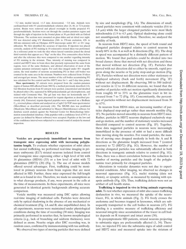
by size and morphology (Fig. 1A). The dimensions of small,round particles were consistent with endocytic vesicles (0.6 to1.8 �m) while the larger elongated particles were most likelymitochondria (1.9 to 4.5 �m). Optical shadowing alone couldnot unambiguously identify them. Therefore, we analyzed themotility of both.
In neurons from HD72 mice, the speed of both round andelongated particles dropped relative to control neurons bynearly 60% in the A as well as R directions (Fig. 1E). The dropin speed was accompanied by a distinctly different pattern ofmotion. We found that particles could be divided into twobroad classes: those that moved with net direction and thosethat moved without net direction (Fig. 1F). Particles thatmoved with net direction did so either by smooth, rapid pro-gress along the neurite or by jerky stop-and-go motion (Fig.1F). Particles without net direction were either stationary ordisplayed saltatory (back and forth) movement (Fig. 1F)without net displacement. By observing 300 to 500 individ-ual vesicles in 12 to 16 different neurons, we found that thenumber of particles with net motion significantly diminishedfrom roughly 69 to 33% as the glutamine tract in htt in-creased from 7 to 72 (Fig. 1F). Concomitantly, the percent-age of particles without net displacement increased from 30to 67%.
In neurons from HD16 mice, an increasing number of par-ticles displayed stop-and-go rather than smooth motility (Fig.1F). No particles moved smoothly in neurons from HD72 mice.Rather, particles in HD72 neurons displayed exclusively stop-and-go motion, and the number of stationary vesicles increasedthreefold compared to control neurons (Fig. 1F). Together,these data suggested particles in neurons were increasinglyimmobilized in the presence of mhtt or had a more difficulttime moving along the neurites. For round particles, the num-ber of moving ones dropped modestly in both the A and Rdirections as the glutamine length increased from 7 (controlneurons) to 72 (HD72) (Fig. 1G). However, the number ofmoving elongated particles was substantially affected in bothdirections in transgenic animals relative to control (Fig. 1G).Thus, there was a direct correlation between the reduction innumber of moving particles and the length of the polyglu-tamine tract primarily for elongated particles.
Alterations in vesicular dynamics were not due to experi-mental conditions. After imaging, we observed no changes inneuronal appearance (Fig. 1C), nuclei staining (data notshown), or synaptic activity, as measured by staining with syn-apsin antibody (Fig. 1D). Thus, trafficking defects were not anartifact of cell health status.
Trafficking is impaired in vivo in living animals expressingmhtt. To test whether expression of mhtt also causes traffickingdysfunction in vivo, we measured the uptake of the neuro-tracer, FG, in whole animals (Fig. 2). FG freely crosses theaxolemma and becomes trapped in lysosomes, which are sub-sequently transported to the cell bodies in neurons (47). FGlabeling is a sensitive measure of trafficking efficiency andneuronal integrity since accumulation of the tracer in cell bod-ies depends on R transport and intact axons (38).
In presymptomatic HD patients, striatal neurons projectingto substantia nigra are preferentially affected (1, 34). There-fore, we injected FG into the substantia nigra of adult controland HD72 mice and measured uptake into the striatum of
8198 TRUSHINA ET AL. MOL. CELL. BIOL.
on Decem
ber 19, 2013 by guesthttp://m
cb.asm.org/
Dow
nloaded from
5-month-old animals (Fig. 2). At this age, HD72 mice do notdisplay any movement disorder and are indistinguishable fromcontrol animals (20). After the injection, mice were allowed torecover and were sacrificed at 1 to 3 days. Trafficking wasmeasured by the intensity of staining in the neuronal cell bod-ies in the striatum with time (Fig. 2B to F).
We found striking differences in striatal staining betweencontrol and HD72 mice at 1 day after injection in all animalstested (n � 10) (Fig. 2B, E, and F). At this time, the numberof neurons that accumulated FG in the cell body in the stria-tum of HD72 mice was less than 25% of that for control
animals. By 3 days, differences in staining intensity betweencontrol and HD72 mice were not significant. These data pro-vided direct evidence that expression of mhtt slows vesiculartrafficking not only in embryonic neurons in vitro but also inadult animals in vivo. Moreover, impairment of trafficking oc-curred before measurable onset of symptoms associated withneuronal loss in HD72 mice (20).
Mitochondrial movement is impaired in neurons fromtransgenic mice expressing mhtt. Analysis of data obtained inDIC experiments indicated that the motility of elongated par-ticles was most affected in neurons expressing mhtt. To test
FIG. 1. mhtt expression causes inhibition of trafficking in neurons of transgenic mice as visualized by DIC optics. (A) Examples of round andelongated particles as they appear using DIC optics. Images are shown at different magnification (top, 63� with 4� extender; bottom, 100� with4� extender and 2� zoom). White arrows indicate round particles, black arrows indicate elongated particles. (B) Time-lapse DIC images ofparticle motility taken 1 s apart. A round vesicle moving towards the cell body is indicated by an arrow. The line is an arbitrary point of reference.Technical details are described in Materials and Methods. (C) DIC imaging does not affect neuronal health. Phase images (63� oil DIC, 1.4 n.a.)of primary striatal neuronal cultures before and after 30 min of DIC imaging. Scale bar, 20 �m. (D) DIC imaging does not affect synaptic activity.Primary striatal neurons immunostained with synapsin antibody before and after 30 min of DIC imaging (63� oil DIC, 1.4 n.a.). Scale bar, 20 �m.Analysis of speed (panel E), pattern of motion (panel F), and number of moving round and elongated particles (panel G) in striatal neurons fromcontrol (C) and transgenic mice expressing htt with 16 (HD16) or 72 (HD72) glutamines obtained using DIC optics. (E) Velocity of particles inmicrometers per second. N, number of neurites observed; �, P � 0.0001; ��, P � 0.01. Thirty to 50 particles were randomly selected for evaluationin every group. (F) Analysis of pattern of motion includes all particles observed (round and elongated). SL, saltatory (back and forth without netdirection); ST, stationary (no motion); SM, smooth (without stops over the range of 50 �m); S-G, stop-and-go (the particle stops for a period oftime before resuming motion); P, number of particles evaluated; �, P � 0.0001. (G) The percentage of particles moving in neurites in the A orR direction is significantly lower in neurons from HD72 mice. �, P � 0.0001. Statistical analysis in panels F and G was done by chi-square test.
VOL. 24, 2004 mhtt IMPAIRS AXONAL TRAFFICKING 8199
on Decem
ber 19, 2013 by guesthttp://m
cb.asm.org/
Dow
nloaded from

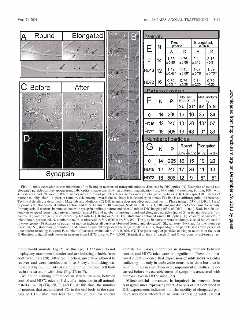
whether the motility of organelles was also affected by theexpression of mhtt, we identified mitochondria by specificstaining with the cationic dye TMRM and examined their dy-namics using fluorescence imaging (6) (Fig. 3A and B). Thedye itself does not alter mitochondrial function or motility (6),and the rates obtained for elongated particles in DIC experi-ments were similar to ones obtained using TMRM.
We found that the majority of mitochondria (roughly 70%)in neurons were stationary and tended to be located near thecell body (Fig. 3A). Therefore, we restricted measurementsand analyses to mitochondria moving in neurites (Fig. 3Aand B; also unpublished data). Neurons were incubated withTMRM and imaged every second over 10 to 15 min on theheated microscopic stage. Indeed, we found that trafficking ofmitochondria was impaired in primary neurons from HD72mice compared to control.
The speed of mitochondria was calculated using two meth-ods. In method 1, the average speed was measured by dividingthe total distance by the total time of mitochondrial movementduring the observation period (Fig. 3C, bottom). In method 2,
we calculated the speed only when the mitochondrion moved(Fig. 3C, top).
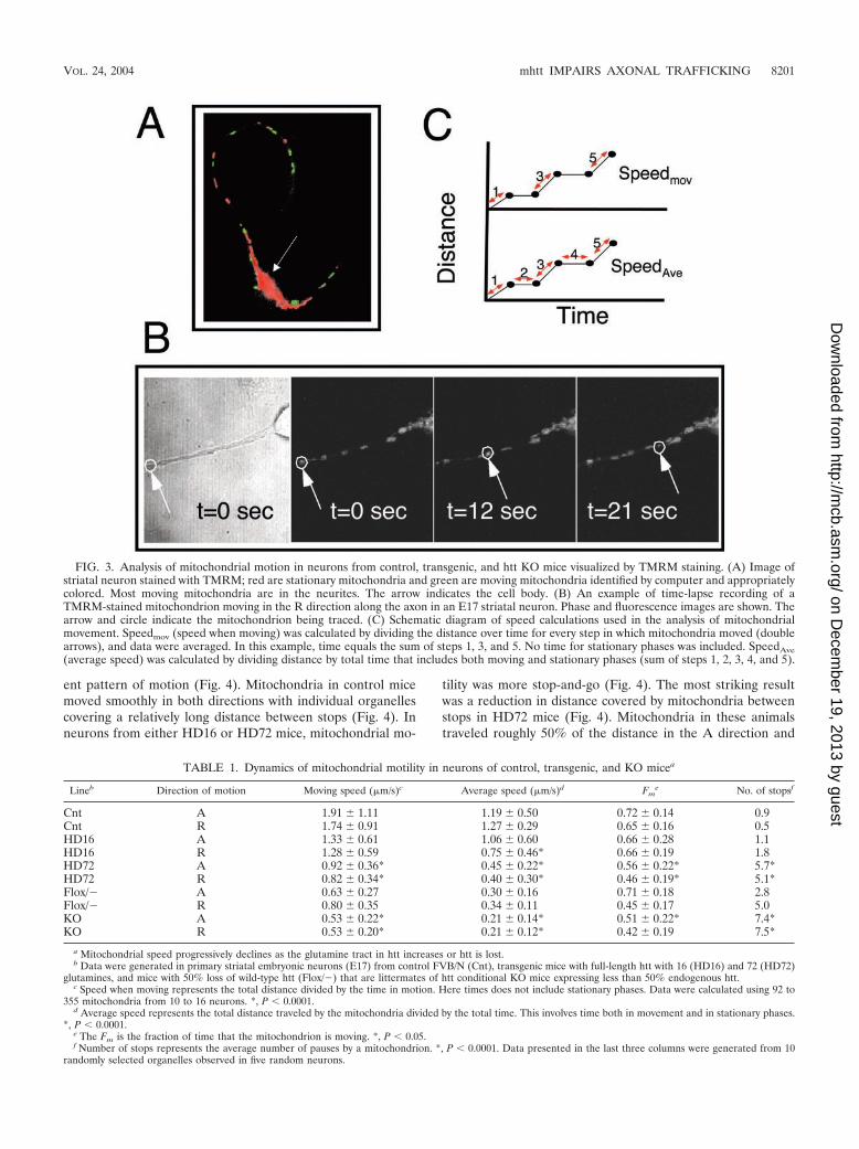
Quantification of vesicular dynamics of 300 to 530 mitochon-dria by method 1 confirmed that they moved with diminishedaverage speed in HD72 mice relative to control (Table 1). Theaverage speed of mitochondria progressively decreased in bothdirections (up to 70%) as the glutamine tract increased from 7to 72 (Table 1). Similarly, with method 2, we found that whenmitochondria moved, their actual speed was 50% slower inHD72 mice compared to control (Table 1). Thus, mitochon-dria had an increasingly difficult time moving along neurites inthe presence of mhtt, and the degree of diminished motilitydirectly correlated with the glutamine length.
Mitochondria not only moved slowly in HD72 neurons, theyalso stopped more frequently. We found that the number ofstops increased with the glutamine length (Table 1). This isalso reflected in the fraction of time mitochondrial spent inmotion, which was significantly smaller in neurons from HD72mice (Table 1). Thus, the inhibition of mitochondria speed inHD16 and HD72 mice was accompanied by a distinctly differ-
FIG. 2. Retrograde transport of FG into striatum is slow in mice expressing mhtt. (A) Injection scheme for FG uptake. FG was injected intosubstantia nigra, and uptake in striatum was monitored with time. (B) Quantification of the number of neurons that accumulate FG in the cellbodies in control and HD72 mice 1 and 3 days after the injection. Horizontal lines represent 0, 50, 100, etc. cells labeled/mouse. Black bars, controlFVB/N mice; white bars, HD72 mice; �, P � 0.01 (t test). (C) Retrograde uptake of FG in striatum of control mice 3 days after the injection. Thebox indicates the area in the striatum that was used for quantification in panel B. Scale bar, 200 �m. (D) View of cortical neurons with FGaccumulation in the cell bodies. Scale bar, 50 �m. (E, F) FG uptake in the striatum of control (E) and HD72 (F) mice 1 day after the injection.Neuronal cell bodies labeled with FG are seen as bright circular structures in tissue. Scale bar, 50 �m.
8200 TRUSHINA ET AL. MOL. CELL. BIOL.
on Decem
ber 19, 2013 by guesthttp://m
cb.asm.org/
Dow
nloaded from
ent pattern of motion (Fig. 4). Mitochondria in control micemoved smoothly in both directions with individual organellescovering a relatively long distance between stops (Fig. 4). Inneurons from either HD16 or HD72 mice, mitochondrial mo-
tility was more stop-and-go (Fig. 4). The most striking resultwas a reduction in distance covered by mitochondria betweenstops in HD72 mice (Fig. 4). Mitochondria in these animalstraveled roughly 50% of the distance in the A direction and
FIG. 3. Analysis of mitochondrial motion in neurons from control, transgenic, and htt KO mice visualized by TMRM staining. (A) Image ofstriatal neuron stained with TMRM; red are stationary mitochondria and green are moving mitochondria identified by computer and appropriatelycolored. Most moving mitochondria are in the neurites. The arrow indicates the cell body. (B) An example of time-lapse recording of aTMRM-stained mitochondrion moving in the R direction along the axon in an E17 striatal neuron. Phase and fluorescence images are shown. Thearrow and circle indicate the mitochondrion being traced. (C) Schematic diagram of speed calculations used in the analysis of mitochondrialmovement. Speedmov (speed when moving) was calculated by dividing the distance over time for every step in which mitochondria moved (doublearrows), and data were averaged. In this example, time equals the sum of steps 1, 3, and 5. No time for stationary phases was included. SpeedAve(average speed) was calculated by dividing distance by total time that includes both moving and stationary phases (sum of steps 1, 2, 3, 4, and 5).
TABLE 1. Dynamics of mitochondrial motility in neurons of control, transgenic, and KO micea
Lineb Direction of motion Moving speed (�m/s)c Average speed (�m/s)d Fme No. of stopsf
Cnt A 1.91 1.11 1.19 0.50 0.72 0.14 0.9Cnt R 1.74 0.91 1.27 0.29 0.65 0.16 0.5HD16 A 1.33 0.61 1.06 0.60 0.66 0.28 1.1HD16 R 1.28 0.59 0.75 0.46* 0.66 0.19 1.8HD72 A 0.92 0.36* 0.45 0.22* 0.56 0.22* 5.7*HD72 R 0.82 0.34* 0.40 0.30* 0.46 0.19* 5.1*Flox/� A 0.63 0.27 0.30 0.16 0.71 0.18 2.8Flox/� R 0.80 0.35 0.34 0.11 0.45 0.17 5.0KO A 0.53 0.22* 0.21 0.14* 0.51 0.22* 7.4*KO R 0.53 0.20* 0.21 0.12* 0.42 0.19 7.5*
a Mitochondrial speed progressively declines as the glutamine tract in htt increases or htt is lost.b Data were generated in primary striatal embryonic neurons (E17) from control FVB/N (Cnt), transgenic mice with full-length htt with 16 (HD16) and 72 (HD72)
glutamines, and mice with 50% loss of wild-type htt (Flox/�) that are littermates of htt conditional KO mice expressing less than 50% endogenous htt.c Speed when moving represents the total distance divided by the time in motion. Here times does not include stationary phases. Data were calculated using 92 to
355 mitochondria from 10 to 16 neurons. *, P � 0.0001.d Average speed represents the total distance traveled by the mitochondria divided by the total time. This involves time both in movement and in stationary phases.
*, P � 0.0001.e The Fm is the fraction of time that the mitochondrion is moving. *, P � 0.05.f Number of stops represents the average number of pauses by a mitochondrion. *, P � 0.0001. Data presented in the last three columns were generated from 10
randomly selected organelles observed in five random neurons.
VOL. 24, 2004 mhtt IMPAIRS AXONAL TRAFFICKING 8201
on Decem
ber 19, 2013 by guesthttp://m
cb.asm.org/
Dow
nloaded from
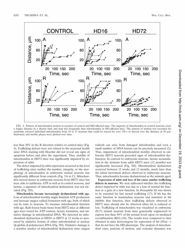
less than 30% in the R direction relative to control mice (Fig.4). Trafficking defects were not related to the neuronal healthsince DNA staining with Hoechst did not reveal any signs ofapoptosis before and after the experiment. Thus, motility ofmitochondria in HD72 mice was significantly impaired by ex-pression of mhtt.
The defect imparted by mhtt expression occurred at the levelof trafficking since neither the number, integrity, or the mor-phology of mitochondria in embryonic striatal neurons wassignificantly different from controls (Fig. 5A to C). Mitochon-dria moved slower in embryonic neurons from HD72 mice butwere able to synthesize ATP as well as in control neurons, andlactate, a signature of mitochondrial dysfunction, was not ele-vated (Fig. 5D).
Mitochondria become increasingly dysfunctional with age.Loss of mitochondrial motility might diminish ATP productionand increase oxygen radical formation with age, both of whichcan be toxic to neurons. To measure mitochondrial functionwith age, flash frozen brain tissue from HD72 mice at differentages were tested for ATP content, lactate formation, and oxi-dative damage in mitochondrial DNA. We detected no mito-chondrial dysfunction in HD16 or HD72 at 12 weeks as mea-sured by oxidative lesions of either mitochondrial or nuclear(�-globin, �-polymerase) DNA (Fig. 5D). Oxidative damage isa sensitive marker of mitochondrial dysfunction since oxygen
radicals can arise from damaged mitochondria and even asmall number of DNA lesions can be precisely measured (2).Thus, impairment of mitochondrial motility observed in em-bryonic HD72 neurons preceded signs of mitochondrial dys-function. In contrast to embryonic neurons, lactate accumula-tion in the striatum from adult HD72 mice (12 months) wassignificantly increased (Fig. 5D). Mitochondrial dysfunctionoccurred between 12 weeks and 12 months, much later thanthe initial movement defects observed in embryonic neurons.Thus, mitochondria became dysfunctional as the animals aged.
Expression of mhtt and loss of htt cause similar traffickingdefects in neurons. We next addressed whether the traffickingdefect imparted by mhtt was due to a loss of normal htt func-tion or a gain of a new function. In Drosophila htt was shownto be essential for fast axonal trafficking (17). If htt has thesame function in mammalian neurons and mutation in httinhibits that function, then trafficking defects observed inHD72 mice should also be observed when htt is reduced orlost. Trafficking of mitochondria was visualized in embryonic(E17) striatal neurons from mice genetically engineered toexpress less than 50% of htt normal levels upon cre-mediatedrecombination (KO) (10). The results were compared to dataobtained in mice heterozygous for expression of htt (Flox/�)that do not have the HD phenotype. The analysis of mitochon-drial rates, patterns of motion, and vesicular dynamics was
FIG. 4. Pattern of mitochondria motion in neurites of control and HD-affected mice. The majority of mitochondria in control neurons covera longer distance in a shorter time and stop less frequently than mitochondria in HD-affected mice. The pattern of motion was recorded forrandomly selected individual mitochondria from 10 to 15 neurons that could be traced for over 150 s or moved over the distance of 50 �m.Stationary and mobile phases were plotted over time.
8202 TRUSHINA ET AL. MOL. CELL. BIOL.
on Decem
ber 19, 2013 by guesthttp://m
cb.asm.org/
Dow
nloaded from

performed using TMRM staining and the protocol identical tothe one used for HD72 neurons. Indeed, we found that reduc-tion of wild-type htt below 50% of normal levels decreasedspeed of mitochondria in neurons from KO mice (see Flox/�and KO speed data in Table 1), increased number of stops (seedata for stops in Table 1), decreased time that mitochondriaspent in motion (see Fm data in Table 1), and decreased dis-tances mitochondria covered between stops (data not shown).Thus, reduction of htt in the neurons altered mitochondrialdynamics in both the A and R directions. Trafficking defectswere significant in KO neurons compared to Flox/�, suggest-
ing a dose-dependent effect. These data demonstrate that nor-mal htt plays a direct role in mitochondrial trafficking. Inembryonic neurons, loss of wild-type htt caused the sametrafficking defects as expression of mhtt in cells that wereotherwise normal.
mhtt titrates motor proteins from soluble pool to poorlysoluble protofibrillar complexes in human HD-affected brain.mhtt is prone to aggregation and forms inclusions in human,mouse, and cellular systems (27). Thus, mhtt may impair ve-sicular trafficking by sequestering normal htt and/or traffickingmachinery. To test whether such sequestration occurs in hu-
FIG. 5. Ultrastructure and function of mitochondria from control and HD-affected mice. Mitochondria morphology was not altered inembryonic striatal neurons (E17) in control (A) or HD72 (B) mice. Scale bar, 10 nm. There was also no detection of abnormal mitochondriaaccumulation in neuritis in HD72 neurons (C) compared to control (not shown). Scale bar, 1 �m. M, mitochondrion. (D) Trafficking defectsprecede mitochondrial dysfunction in HD72 mice. Mitochondria function (ATP synthesis and lactate accumulation) was not altered in embryonic(E17) striatal neurons in HD72 mice compared to control despite detected abnormalities in mitochondrial trafficking. At the same time, increasedlactate accumulation was measured in adult HD72 mice (12 months). There was also no difference in mitochondrial DNA damage in striatal tissuefrom control, HD16, and HD72 mice at 12 weeks. Estimated mitochondrial (MT) and nuclear (�-polymerase and �-globin) DNA damage wasmeasured by quantitative PCR in striatal tissues (see Materials and Methods). ATPHD/ATPC is the ratio of ATP generated within E17 culturesof HD16 or HD72 animals (ATPHD) to that of control neurons (ATPC). LactateHD/LactateC is the ratio of lactate measured in E17 striatal culturesor striatal tissue from HD72 mice (lactateHD) to that of control mice (lactateC). Age is indicated.
VOL. 24, 2004 mhtt IMPAIRS AXONAL TRAFFICKING 8203
on Decem
ber 19, 2013 by guesthttp://m
cb.asm.org/
Dow
nloaded from

mans, we isolated extracts from human control and HD-in-fected brain and tested whether trafficking-related proteinswere associated with mhtt in disease tissue. The mutation of httis known to alter interaction with several partners (18). One ofthem is htt-associated protein 1 (HAP1) that interacts with thekey trafficking proteins dynactin p150Glued, a kinesin heavychain homologue, and tubulin (15, 24). By using a differentialextraction procedure (14) in combination with size exclusionchromatography and mass spectrometry, we addressed wheth-er soluble aggregates from human HD-affected brain con-tained components of vesicular trafficking.
We first characterized the composition of aggregated com-plexes using mass spectrometry. To examine htt-containingcomplexes from brain tissue, we developed and applied anextraction method that allows the isolation of htt-containingcomplexes from control and HD-affected brain tissue based ontheir solubility (Fig. 6A) (14). By using this method, we havepreviously shown that wild-type htt is found in soluble (S1)extracts while mhtt is primarily found in poorly soluble (S3)protofibrillar complexes. Therefore, mass spectrometry wasperformed on the S3 fraction from HD-affected brain. The S3extract from control brain contained virtually no htt. There-
FIG. 6. Components of the trafficking machinery are sequestered in human HD-affected brain. (A) Extraction scheme used to resolve proteinsbased on their solubility. Age- and gender-matched human brain tissues in HD-affected (C/P, CTX) and spared (CBL, HIP) regions were preparedas described in Materials and Methods. The S1 fraction contains proteins that are the most soluble, S2 contains proteins of intermediate solubility,and S3 represents the least soluble proteins that are specific to the HD-affected brain. CTX, cerebral cortex; CBL, cerebellar cortex; C/P, caudateand putamen; HIP, hippocampus; RIPA, radioimmunoprecipitation assay buffer. (B) Distribution of cytoplasmic, nonmetabolic proteins identifiedby mass spectrometry in gel filtration fractions of S3 extracts of HD-affected caudate and putamen. Note that mitochondrial and cytoskeletalproteins constitute the largest portion of this pool. (C) Redistribution of cytoskeleton and trafficking motors from S1 to S3 in HD-affected brain.Gel filtration profiles of htt, �-tubulin (TB), neuronal specific �III-tubulin (�TB), kinesin (KN), and dynactin p150Glued (DN) in the S1 and S3extracts of HD-affected caudate and putamen. Equal total protein amounts were resolved by size exclusion chromatography, and then an equalvolume of each fraction was analyzed by immunoblotting. Data from two human control brains (M2 and 3867), age- and gender-matched with twohuman HD-affected brains (AR98189 and 3882), are shown. The asterisk indicates the predicted migration of a htt monomer. (D) Migration ofnontrafficking proteins, actin (AC), GAPDH (GD), and Ran (RN) was unaffected in HD-affected compared to control brain.
8204 TRUSHINA ET AL. MOL. CELL. BIOL.
on Decem
ber 19, 2013 by guesthttp://m
cb.asm.org/
Dow
nloaded from
fore, htt-containing complexes were not present in the S3 ofcontrol brain and we could not perform a comparative analysis.
Mass spectrometry sequencing revealed that the S3 aggre-gate fraction from HD-affected brain contained both nuclear(�8%) and cytoplasmic (�92%) proteins (unpublished data).Nuclear proteins included histones, leucine acidic nuclear pro-tein, transportins, elongation factors, and Sir12 histone de-acetylase. Sixty percent of the cytoplasmic proteins were met-abolic enzymes or proteins of unknown function (Fig. 6B).However, the remaining 40% primarily comprised traffickingmotors, mitochondrial proteins, endosomal proteins, and cyto-skeletal components (Fig. 6B) Overall, these data suggest thatcomponents of the vesicular trafficking machinery and cargowere well represented in aggregated fractions from humanHD-affected brain.
We next tested whether key components of the traffickingmachinery codistributed into S3 extracts together with mhtt inhuman HD-affected brain. To test this hypothesis, the totalprotein amount from the soluble (S1) and poorly soluble (S3)extracts from control and HD-affected brain were resolved bysize exclusion chromatography. An equal volume of the elutedfractions was analyzed by Western analysis to determine thepresence and size distribution of the proteins of interest.
As previously shown, htt was extracted as a soluble protein(S1) in control brain, while in the HD-affected brain, it wasconsistently depleted in the S1 fraction and redistributed intothe poorly soluble S3 fraction (Fig. 6C) (13). Most of the httreactivity appeared to migrate near its monomer MW range(Fig. 6C). htt was present in control S1 but consistently absentin comparable extracts for HD S1. Instead, normal htt wasredistributed into the poorly soluble S3 fraction of HD-af-fected brain (Fig. 6C, compare the migration from S1 and S3for htt).
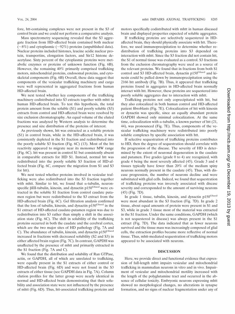
We next tested whether proteins involved in vesicular traf-ficking were also redistributed into the S3 fraction togetherwith mhtt. Similar to htt, we found that �-tubulin, neuron-specific �III-tubulin, kinesin, and dynactin p150Glued were ex-tracted in the soluble S1 fraction from control caudate puta-men region but were redistributed to the S3 extract from theHD-affected brain (Fig. 6C). Gel filtration analysis confirmedthat the loss of tubulin, kinesin, and dynactin p150Glued in theS1 extract of HD-affected caudate-putamen region was due toredistribution into S3 rather than simply a shift in the associ-ation state (Fig. 6C). The shift in solubility of the traffickingproteins occurred in both the caudate and the cerebral cortex,which are the two major sites of HD pathology (Fig. 7A andC). The abundance of tubulin, kinesin, and dynactin p150Glued
increased in fractions with decreased solubility (S2 and S3) ineither affected brain region (Fig. 7C). In contrast, GAPDH wasunaffected by the presence of mhtt and primarily extracted inthe S1 fraction (Fig. 7A and C).
We found that the distribution and solubility of Ran GTPase,actin, or GAPDH, all of which are unrelated to trafficking,were equally present in the S1 extracts of either control orHD-affected brain (Fig. 6D) and were not found in the S3extracts of either tissue (see GAPDH data in Fig. 7A). Columnelution profiles for the latter group were nearly identical innormal and HD-affected brain demonstrating that their solu-bility and association state were not influenced by the presenceof mhtt (Fig. 6D). Thus, htt-associated trafficking proteins and
motors specifically codistributed with mhtt in human diseasedbrain and displayed properties expected of soluble aggregates.
If trafficking proteins are selectively sequestered in HD-affected brain, they should physically associate with htt. There-fore, we used immunoprecipitation to determine whether re-distribution of trafficking proteins into S3 depended oninteraction with mhtt. Since the S3 fraction did not contain htt,the S1 of normal tissue was evaluated as a control. S3 fractionsfrom the exclusion chromatography were used as a source ofthe relevant proteins. We found that in fractions from both S1control and S3 HD-affected brain, dynactin p150Glued and ki-nesin could be pulled down by immunoprecipitation using the2166 htt antibody (Fig. 7B). Thus, it appeared that traffickingproteins found in aggregates in HD-affected brain normallyinteract with htt. However, these proteins are sequestered intopoorly soluble aggregates due to interaction with mhtt.
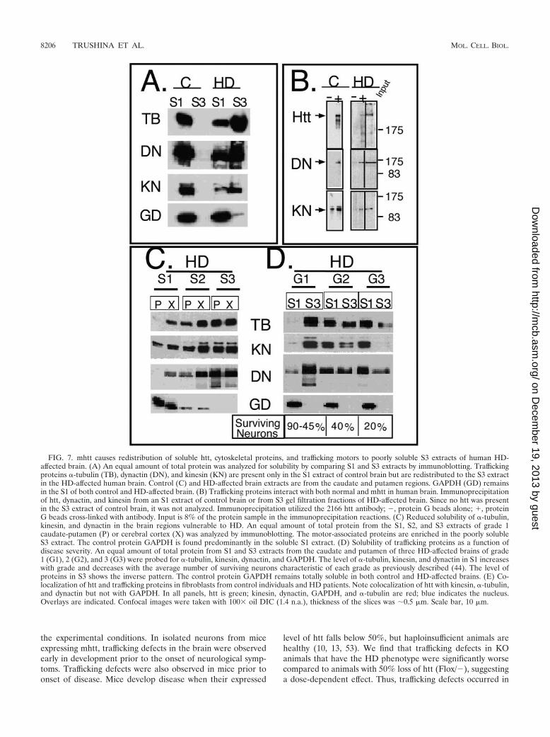
Trafficking proteins not only coprecipitated with htt, butthey also colocalized in both human control and HD-affectedpatient fibroblasts (Fig. 7E). Colocalization of htt with kinesinor dynactin was specific, since an equally abundant proteinGAPDH showed only minimal colocalization. At the sametime, colocalization with �-tubulin, a known partner of htt (21,44), was pronounced (Fig. 7E). Thus, components of the ve-sicular trafficking machinery were redistributed into poorlysoluble complexes by specific association with htt.
If selective sequestration of trafficking proteins contributesto HD, then the degree of sequestration should correlate withthe progression of the disease. The severity of HD is deter-mined by the extent of neuronal degeneration in the caudateand putamen. Five grades (grade 0 to 4) are recognized, withgrade 4 being the most severely affected (45). Grade 3 and 4tissues typically have fewer than 20% of the medium spinyneurons normally present in the caudate (45). Thus, with dis-ease progression, the number of neurons decline and weresubstituted with glial cells. Indeed, we found that sequestrationof trafficking proteins was inversely associated with diseaseseverity and corresponded to the amount of surviving neurons(45) (Fig. 7D).
In grade 1 tissue, tubulin, kinesin, and dynactin p150Glued
were most abundant in the S3 fraction (Fig. 7D). In grade 2tissue, about equal amounts of protein were present in S1 andS3, while in grade 3 tissue most of the material was extractedin the S1 fraction. Under the same conditions, GAPDH (whichis not sequestered in disease) was always present in the S1extract (Fig. 7D). The data indicated that as fewer neuronssurvived and the tissue mass was increasingly composed of glialcells, the extraction profiles became more reflective of normaltissue. Thus, mhtt-mediated sequestration of trafficking motorsappeared to be associated with neurons.
DISCUSSION
Here, we provide direct and functional evidence that expres-sion of full-length mhtt impairs vesicular and mitochondrialtrafficking in mammalian neurons in vitro and in vivo. Impair-ment of vesicular and mitochondrial motility increased withthe length of the polyglutamine tract and occurred in the ab-sence of cellular toxicity. Embryonic neurons expressing mhttshowed no morphological changes, no alterations in synapseformation, and no signs of nuclear fragmentation under any of
VOL. 24, 2004 mhtt IMPAIRS AXONAL TRAFFICKING 8205
on Decem
ber 19, 2013 by guesthttp://m
cb.asm.org/
Dow
nloaded from
the experimental conditions. In isolated neurons from miceexpressing mhtt, trafficking defects in the brain were observedearly in development prior to the onset of neurological symp-toms. Trafficking defects were also observed in mice prior toonset of disease. Mice develop disease when their expressed
level of htt falls below 50%, but haploinsufficient animals arehealthy (10, 13, 53). We find that trafficking defects in KOanimals that have the HD phenotype were significantly worsecompared to animals with 50% loss of htt (Flox/�), suggestinga dose-dependent effect. Thus, trafficking defects occurred in
FIG. 7. mhtt causes redistribution of soluble htt, cytoskeletal proteins, and trafficking motors to poorly soluble S3 extracts of human HD-affected brain. (A) An equal amount of total protein was analyzed for solubility by comparing S1 and S3 extracts by immunoblotting. Traffickingproteins �-tubulin (TB), dynactin (DN), and kinesin (KN) are present only in the S1 extract of control brain but are redistributed to the S3 extractin the HD-affected human brain. Control (C) and HD-affected brain extracts are from the caudate and putamen regions. GAPDH (GD) remainsin the S1 of both control and HD-affected brain. (B) Trafficking proteins interact with both normal and mhtt in human brain. Immunoprecipitationof htt, dynactin, and kinesin from an S1 extract of control brain or from S3 gel filtration fractions of HD-affected brain. Since no htt was presentin the S3 extract of control brain, it was not analyzed. Immunoprecipitation utilized the 2166 htt antibody; �, protein G beads alone; �, proteinG beads cross-linked with antibody. Input is 8% of the protein sample in the immunoprecipitation reactions. (C) Reduced solubility of �-tubulin,kinesin, and dynactin in the brain regions vulnerable to HD. An equal amount of total protein from the S1, S2, and S3 extracts of grade 1caudate-putamen (P) or cerebral cortex (X) was analyzed by immunoblotting. The motor-associated proteins are enriched in the poorly solubleS3 extract. The control protein GAPDH is found predominantly in the soluble S1 extract. (D) Solubility of trafficking proteins as a function ofdisease severity. An equal amount of total protein from S1 and S3 extracts from the caudate and putamen of three HD-affected brains of grade1 (G1), 2 (G2), and 3 (G3) were probed for �-tubulin, kinesin, dynactin, and GAPDH. The level of �-tubulin, kinesin, and dynactin in S1 increaseswith grade and decreases with the average number of surviving neurons characteristic of each grade as previously described (44). The level ofproteins in S3 shows the inverse pattern. The control protein GAPDH remains totally soluble in both control and HD-affected brains. (E) Co-localization of htt and trafficking proteins in fibroblasts from control individuals and HD patients. Note colocalization of htt with kinesin, �-tubulin,and dynactin but not with GAPDH. In all panels, htt is green; kinesin, dynactin, GAPDH, and �-tubulin are red; blue indicates the nucleus.Overlays are indicated. Confocal images were taken with 100� oil DIC (1.4 n.a.), thickness of the slices was �0.5 �m. Scale bar, 10 �m.
8206 TRUSHINA ET AL. MOL. CELL. BIOL.
on Decem
ber 19, 2013 by guesthttp://m
cb.asm.org/
Dow
nloaded from
animals and in embryonic neurons prior to the onset of disease.Inhibition of trafficking by mhtt is likely to be a direct effectsince impairment of trafficking by mutant polyglutamine pro-tein can occur in a cell-free system (i.e., squid axoplasm), whichlacks a nucleus and in which protein synthesis does not occur(42).
Data presented here also suggest a normal role for htt intrafficking. We find that mice expressing less than 50% of httdisplay a severe trafficking defect, suggesting that normal traf-ficking requires the presence of htt. In fact, the traffickingdefects in these animals are more extensive than those express-ing mhtt. These results are consistent with the predictions ofearlier genetics studies. Both White et al. (49) and Nasir et al.(29) demonstrated that mice lacking htt die early in embryo-genesis but that behavioral and developmental defects can bepartially rescued by one allele of mhtt (49). Consistent withthese data, we find that expression of mhtt impairs traffickingrelative to control mice but partially restores the defect in theconditional KO animals. Thus, the impairment of traffickingthat we observe cannot be a simple loss of htt function. Pro-gressive alteration in vesicular dynamics is likely to arise fromtwo sources. mhtt may functionally inactivate trafficking mo-tors as well as inhibit a normal function of htt in trafficking.Such a model is supported by data from human brain. We findthat mhtt redistributes htt and soluble components of vesiculartrafficking machinery, such as kinesin and the dynein-associ-ated protein dynactin p150Glued, into poorly soluble aggre-gates.
These data raise the issue of how toxicity might arise from atrafficking defect. Our data as well as others suggest that neu-rite outgrowth and maintenance may be affected by impairedtrafficking. Szebenyi and colleagues (42) have shown that trun-cated, mutant androgen receptor inhibits full neurite out-growth in SH-SY5Y cells induced to differentiate to a neuronal
phenotype with retinoic acid and brain-derived neurotrophicfactor. These events preceded signs of cellular toxicity, indi-cating that trafficking impairment and dendritic effects areprimary events in pathogenesis. Indeed, we have previouslyshown that full-length mhtt resides in the cytoplasm and ini-tiates toxicity there (43). Expression of full-length mhtt causesneurite retraction, collapse of cytoskeleton, and commitmentto cell death before nuclear entry.
We demonstrate that trafficking of mitochondria is particu-larly affected by expression of mhtt. Therefore, the morpho-logical defects in dendrites observed in HD could be the resultof inefficient delivery of mitochondria to the growth cone dur-ing neurite formation or to the periphery of mature neurites.Although the previous studies in the invertebrate systems didnot analyze mitochondria per se, we demonstrate that mito-chondrial trafficking is slower and less fluid in either embryonicneurons from htt KO mice or those from transgenic animalsexpressing full-length mhtt. Additionally, recent data in theDrosophila model have identified a protein required for mito-chondrial transport to the synapse (40). This protein, Milton,has a high homology to HAP1, a protein known to interactmore strongly with mhtt than with wild-type htt (25). We alsofind that mitochondrial proteins succinate dehydrogenase, glu-taminase, ATP synthase �- and �-chain, and outer mitochon-drial membrane translocase were among those identified bymass spectrometry sequencing of mhtt-containing complexesfrom human HD-affected brains.
ATP production is an essential component of cell survival.Thus, impairment of mitochondrial function could deplete en-ergy stores in neurons. Further, mitochondria are required tosupport synaptic connections and formation of growth cones.The loss of motility might prevent mitochondria from being inthe right place at the right time. While trafficking defects oc-curred in the embryo prior to any sign of neuropathology or
FIG. 7—Continued.
VOL. 24, 2004 mhtt IMPAIRS AXONAL TRAFFICKING 8207
on Decem
ber 19, 2013 by guesthttp://m
cb.asm.org/
Dow
nloaded from

mitochondrial dysfunction, mitochondrial dysfunction was de-tected between 12 weeks and 12 months of age. These dataprovide a link between a defined function of htt and a long-standing model for mitochondrial involvement in HD (26). Inhuman HD patients, magnetic resonance imaging confirmsthat creatine (a free radical scavenger, a substrate for theenzyme creatine kinase, and a precursor for ATP) was de-pleted and lactate was increased (35). In rats, systemic ad-ministration of the mitochondrial complex II inhibitor, 3-ni-tropropionic acid, causes neurobehavioral and pathologicalabnormalities consistent with HD (4). In HD patients, thecaudate has severe deficiencies in mitochondrial complexes IIand III (5, 16). Mitochondria from human HD lymphoblastsare more sensitive to stress (31, 37). Thus, disruption of mito-chondrial function is consistent with HD pathophysiology inboth animals and humans. Vesicular trafficking defects arelikely to be a general hallmark of aggregation disorders. Mouseand fly models of Alzheimer’s disease (11) and amyotrophiclateral sclerosis have declines in intracellular transport andabnormal accumulation of neurofilaments, actin, and tubulin(7, 50).
Taken together, these data provide a model as to how cyto-plasmic dysfunction and loss of a normal htt trafficking func-tion could lead to toxicity. mhtt redistributes htt and soluble
trafficking components into protofibrillar complexes that im-pair motility of vesicles and organelles. With time, sequestra-tion of mitochondrial proteins along with defective traffickingmight lead to failure of ATP synthesis, energy depletion, andultimately cell death (Fig. 8).
ACKNOWLEDGMENTS
This work was supported by the Mayo Foundation, National Insti-tutes of Health, grants NS40738 and DK 43694-01; National ScienceFoundation grant IBN 9728120 (to C.T.M.); Canadian Institutes ofHealth Research grant mitochondria-15396 (to P.S.M.; the Valorisa-tion Recherche Quebec, Genome Quebec and Genome Canada); andThe Norwegian Research Council (FUGE). Human brain tissue wasgenerously provided by the Harvard Brain Tissue Resource Center,which is supported in part by PHS grant MH/NS 31862.
We thank Line Roy, Nathalie Hamel, Daniel Boismenu, Alex Bell,and John Bergeron of the Montreal Proteomics Centre for help withsequencing, Eugene Krueger for technical assistance in DIC imaging,Alexey A. Leontovich for help with statistical analysis, and TimothyFarnham for help with manuscript preparation.
REFERENCES
1. Albin, R. L., A. Reiner, K. D. Anderson, L. S. Dure IV, B. Handelin, R.Balfour, W. O. Whetsell, Jr., J. B. Penney, and A. B. Young. 1992. Prefer-ential loss of striato-external pallidal projection neurons in presymptomaticHuntington’s disease. Ann. Neurol. 31:425–430.
2. Ayala-Torres, S., Y. Chen, T. Svoboda, J. Rosenblatt, and B. Van Houten.2000. Analysis of gene-specific DNA damage and repair using quantitativePCR. Methods Companion Methods Enzymol. 22:135–147.
3. Barnes, G. T., M. P. Duyao, C. M. Ambrose, S. McNeil, F. Persichetti, J.Srinidhi, J. F. Gusella, and M. E. MacDonald. 1994. Mouse Huntington’sdisease gene homolog (Hdh). Somat. Cell Mol. Genet. 20:87–97.
4. Beal, M. F., R. J. Ferrante, K. J. Swartz, and N. W. Kowall. 1991. Chronicquinolinic acid lesions in rats closely resemble Huntington’s disease. J. Neu-rosci. 11:1649–1659.
5. Brennan, W. A., Jr., E. D. Bird, and J. R. Aprille. 1985. Regional mitochon-drial respiratory activity in Huntington’s disease brain. J. Neurochem. 44:1948–1950.
6. Chen, L. B. 1988. Mitochondrial membrane potential in living cells. Annu.Rev. Cell Biol. 4:155–181.
7. Collard, J. F., F. Cote, and J. P. Julien. 1995. Defective axonal transport ina transgenic mouse model of amyotrophic lateral sclerosis. Nature 375:61–64.
8. De Cristofaro, T., A. Affaitati, E. V. Feliciello, A. Avvedimento, and S.Varrone. 2000. Polyglutamine-mediated aggregation and cell death. Bio-chem. Biophys. Res. Commun. 272:816–821.
9. DiFiglia, M., E. Sapp, K. Chase, C. Schwarz, A. Meloni, C. Young, E. Martin,J. P. Vonsattel, R. Carraway, S. A. Reeves, F. M. Boyce, and N. Aronin. 1995.Huntingtin is a cytoplasmic protein associated with vesicles in human and ratbrain neurons. Neuron 14:1075–1081.
10. Dragatsis, I., M. S. Levine, and S. Zeitlin. 2000. Inactivation of Hdh in thebrain and testis results in progressive neurodegeneration and sterility inmice. Nat. Genet. 26:300–306.
11. Drouet, B., M. Pincon-Raymond, J. Chambaz, and T. Pillot. 2000. Molecularbasis of Alzheimer’s disease. Cell. Mol. Life Sci. 57:705–715.
12. Dunah, A. W., H. Jeong, A. Griffin, Y. M. Kim, D. G. Standaert, S. M.Hersch, M. M. Mouradian, A. B. Young, N. Tanese, and D. Krainc. 2002. Sp1and TAFII130 transcriptional activity disrupted in early Huntington’s dis-ease. Science 296:2238–2243.
13. Duyao, M. P., A. B. Auerbach, A. Ryan, F. Persichetti, G. T. Barnes, S. M.McNeil, P. Ge, J.-P. Vonsattel, J. F. Gusella, A. L. Joyner, and M. E.MacDonald. 1995. Homozygous inactivation of the mouse Hdh gene doesnot produce a Huntington’s disease-like phenotype. Science 269:407–410.
14. Dyer, R. B., and C. T. McMurray. 2001. Mutant protein in Huntington’sdisease is resistant to proteolysis in affected brain. Nat. Genet. 29:270–278.
15. Engelender, S., A. H. Sharp, V. Colomer, M. K. Tokito, A. Lanahan, P.Worley, E. L. Holzbaur, and C. A. Ross. 1997. Huntingtin-associated protein1 (HAP1) interacts with the p150Glued subunit of dynactin. Hum. Mol.Genet. 13:2205–2212.
16. Gu, M., M. T. Gash, V. M. Mann, F. Javoy-Agid, J. M. Cooper, and A. H.Schapira. 1996. Mitochondrial defect in Huntington’s disease caudate nu-cleus. Ann. Neurol. 39:385–389.
17. Gunawardena, S., L. S. Her, R. G. Brusch, R. A., Laymon, I. R. Niesman, B.Gordesky-Gold, L. Sintasath, N. M. Bonini, and L. S. B. Goldstein. 2003.Disruption of axonal transport by loss of huntingtin or expression of patho-genic polyQ proteins in Drosophila. Neuron 40:25–40.
18. Harjes, P., and E. E. Wanker. 2003. The hunt for huntingtin function:
FIG. 8. Proposed mechanism of neuronal dysfunction in HD.Model for HD pathogenesis that includes inhibition of mhtt proteol-ysis and initiation of aggregation that involves specific cellular targetsalong with wild-type htt. Sequestration leads to disruption of cytoskel-eton, vesicular and organelle trafficking, transcriptional dysfunction,and eventually causes mitochondrial dysfunction and neuronal death.
8208 TRUSHINA ET AL. MOL. CELL. BIOL.
on Decem
ber 19, 2013 by guesthttp://m
cb.asm.org/
Dow
nloaded from
interaction partners tell many different stories. Trends Biochem. Sci. 28:425–433.
19. Hilditch-Maguire, P., F. Trettel, L. A. Passani, A. Auerbach, F. Persichetti,and M. E. MacDonald. 2000. Huntingtin: an iron-regulated protein essentialfor normal nuclear and perinuclear organelles. Hum. Mol. Genet. 9:2789–2797.
20. Hodgson, J. G., N. Agopyan, C.-A. Gutekunst, B. R. Leavitt, F. LePiane, R.Singararja, D. J. Smith, N. Bissada, K. McCutcheon, J. Nasir, L. Jamot,X.-J. Li, M. E. Stevens, E. Rosemond, J. C. Roder, A. G. Phillips, E. M.Rubin, S. M. Hersch, and M. R. Hayden. 1999. A YAC mouse model forHuntington’s disease with full-length mutant huntingtin, cytoplasmic toxicity,and selective striatal neurodegeneration. Neuron 23:181–192.
21. Hoffner, G., P. Kahlem, and P. Djian. 2002. Perinuclear localization ofhuntingtin as a consequence of its binding to microtubules through an in-teraction with �-tubulin: relevance to Huntington’s disease. J. Cell Sci. 115:941–948.
22. Kegel, K. B., A. R. Meloni, Y. Yi, Y. J. Kim, E. Doyle, B. G. Cuiffo, E. Sapp,Y. Wang, Z. H. Qin, J. D. Chen, J. R. Nevins, N. Aronin, and M. DiFiglia.2002. Huntingtin is present in the nucleus, interacts with the transcriptionalcorepressor C-terminal binding protein, and represses transcription. J. Biol.Chem. 277:7466–7476.
23. Li, S.-H., A. L. Cheng, H. Zhou, S. Lam, M. Rao, H. Li, and X.-J. Li. 2002.Interaction of Huntington disease protein with transcriptional activator Sp1.Mol. Cell. Biol. 22:1277–1287.
24. Li, S. H., C. A. Gutekunst, S. M. Hersch, and X. J. Li. 1998. Interaction ofhuntingtin-associated protein with dynactin p150Glued. J. Neurosci. 18:1261–1269.
25. Li, X. J., S. H. Li, A. H. Sharp, F. C. Nucifora, Jr., G. Schilling, A. Lanahan,P. Worley, S. H. Snyder, and C. A. Ross. 1995. A huntingtin-associatedprotein enriched in brain with implications for pathology. Nature 378:398–402.
26. Manfredi, G., and M. F. Beal. 2000. The role of mitochondria in the patho-genesis of neurodegenerative diseases. Brain Pathol. 10:462–472.
27. Michalik, A., and C. Van Broeckhoven. 2003. Pathogenesis of polyglutaminedisorders: aggregation revisited. Hum. Mol. Genet. 12:R173–R186.
28. Narain, Y., A. Wyttenbach, J. Rankin, R. A. Furlong, and D. C. Rubinsztein.1999. A molecular investigation of true dominance in Huntington’s disease.J. Med. Genet. 36:739–746.
29. Nasir, J., S. B. Floresco, J. R. O’Kusky, V. M. Diewert, J. M. Richman, J.Zeisler, A. Borowski, J. D. Marth, A. G. Phillips, and M. R. Hayden. 1995.Targeted disruption of the Huntington’s disease gene results in embryoniclethality and behavioral and morphological changes in heterozygotes. Cell81:811–823.
30. Nucifora, F. C., Jr., M. Sasaki, M. F. Peters, H. Huang, J. K. Cooper, M.Yamada, H. Takahashi, S. Tsuji, J. Troncoso, V. L. Dawson, T. M. Dawson,and C. A. Ross. 2001. Interference by huntingtin and atrophin-1 with cbp-mediated transcription leading to cellular toxicity. Science 291:2423–2428.
31. Panov, A. V., C. A. Gutekunst, B. R. Leavitt, M. R. Hayden, J. R. Burke, W. J.Strittmatter, and J. T. Greenamyre. 2002. Early mitochondrial calcium de-fects in Huntington’s disease are a direct effect of polyglutamines. Nat.Neurosci. 5:731–736.
32. Perutz, M. F., and A. H. Windle. 2001. Cause of neural death in neurode-generative diseases attributable to expansion of glutamine repeats. Nature412:143–144.
33. Reddy, P. H., M. Williams, V. Charles, L. Garrett, L. Pike-Buchanan, W. O.Whetsell, Jr., G. Miller, and D. A. Tagle. 1998. Behavioral abnormalities andselective neuronal loss in HD transgenic mice expressing mutated full-lengthHD cDNA. Nat. Genet. 20:198–202.
34. Reiner, A., R. L. Albin, K. D. Anderson, C. J. D’Amato, J. B. Penney, andA. B. Young. 1988. Differential loss of striatal projection neurons in Hun-tington disease. Proc. Natl. Acad. Sci. USA 85:5733–5737.
35. Sanchez-Pernaute, R., J. M. Garcia-Segura, A. del Barrio Alba, J. Viano, andJ. G. de Yebenes. 1999. Clinical correlates of striatal 1H MRS in Hunting-ton’s disease. Neurology 53:806–812.
36. Sauve, D. M., D. T. Ho, and M. Roberge. 1995. Concentration of diluteprotein for gel electrophoresis. Anal. Biochem. 10:382–383.
37. Sawa, A., G. W. Wiegand, J. Cooper, R. L. Margolis, A. H. Sharp, J. F.Lawler, Jr., J. T. Greenamyre, S. H. Snyder, and C. A. Ross. 1999. Increased
apoptosis of Huntington disease lymphoblasts associated with repeat length-dependent mitochondrial depolarization. Nat. Med. 5:1194–1198.
38. Schmued, L. 1990. Fluoro-gold and 4-acetamido-4-isothiocyanostilbene-2,2-disulfonic acid: use of substituted stilbenes in neuroanatomical studies,p. 317–330. In Methods in neuroscience, vol. 3. Academic Press, Inc., NewYork, N.Y.
39. Steffan, J. S., A. Kazantsev, O. Spasic-Boskovic, M. Greenwald, Y. Z. Zhu, H.Gohler, E. E. Wanker, G. P. Bates, D. E. Housman, and L. M. Thompson.2000. The Huntington’s disease protein interacts with p53 and CREB-bind-ing protein and represses transcription. Proc. Natl. Acad. Sci. USA 97:6763–6768.
40. Stowers, R. S., L. J. Megeath, J. Gorska-Andrzejak, I. A. Meinertzhagen, andT. L. Schwarz. 2002. Axonal transport of mitochondria to synapses dependson milton, a novel Drosophila protein. Neuron 36:1063–1077.
41. Suhr, S. T., M. C. Senut, J. P. Whitelegge, K. F. Faull, D. B. Cuizon, andF. H. Gage. 2001. Identities of sequestered proteins in aggregates from cellswith induced polyglutamine expression. J. Cell Biol. 153:283–294.
42. Szebenyi, G., G. A. Morfini, A. Babcock, M. Gould, K. Selkoe, D. L. Stenoien,M. Young, P. W. Faber, M. E. MacDonald, M. J. McPhaul, and S. T. Brady.2003. Neuropathogenic forms of huntingtin and androgen receptor inhibitfast axonal transport. Neuron 40:41–52.
43. Trushina, E., M. P. Heldebrant, C. M. Perez-Terzic, R. Bortolon, I. V.Kovtun, J. D. Badger II, A. Terzic, A. Estevez, A. J. Windebank, R. B. Dyer,J. Yao, and C. T. McMurray. 2003. Microtubule destabilization and nuclearentry are sequential steps leading to toxicity in Huntington’s disease. Proc.Natl. Acad. Sci. USA 100:12171–12176.
44. Tukamoto, T., N. Nukina, K. Ide, and I. Kanazawa. 1997. Huntington’sdisease gene product, huntingtin, associates with microtubules in vitro. Mol.Brain Res. 51:8–14.
45. Vonsattel, J. P., R. H. Myers, T. J. Stevens, R. J. Ferrante, E. D. Bird, andE. P. Richardson, Jr. 1985. Neuropathological classification of Huntington’sdisease. J. Neuropathol. Exp. Neurol. 44:559–577.
46. Wasiak, S., V. Legendre-Guillemin, R. Puertollano, F. Blondeau, M. Girard,E. de Heuvel, D. Boismenu, A. W. Bell, J. S. Bonifacino, and P. S. McPher-son. 2002. Enthoprotin: a novel clathrin-associated protein identifiedthrough subcellular proteomics. J. Cell Biol. 158:855–862.
47. Wessendorf, M. W. 1991. Fluoro-gold: composition and mechanism of up-take. Brain Res. 553:35–148.
48. Wheeler, V. C., J. K. White, C. A. Gutekunst, V. Vrbanac, M. Weaver, X. J.Li, S. H. Li, H. Yi, J. P. Vonsattel, J. F. Gusella, S. Hersch, W. Auerbach,A. L. Joyner, and M. E. MacDonald. 2000. Long glutamine tracts causenuclear localization of a novel form of huntingtin in medium spiny striatalneurons in HdhQ92 and HdhQ111 knock-in mice. Hum. Mol. Genet. 9:503–513.
49. White, J. K., W. Auerbach, M. P. Duyao, J. P. Vonsattel, J. F. Gusella, A. L.Joyner, and M. E. MacDonald. 1997. Huntingtin is required for neurogenesisand is not impaired by the Huntington’s disease CAG expansion. Nat. Genet.17:404–410.
50. Williamson, T. L., and D. W. Cleveland. 1999. Slowing of axonal transport isa very early event in the toxicity of ALS-linked SOD1 mutants to motorneurons. Nat. Neurosci. 2:50–56.
51. Wyttenbach, A., J. Swartz, H. Kita, T. Thykjaer, J. Carmichael, J. Bradley,R. Brown, M. Maxwell, A. Schapira, T. F. Orntoft, K. Kato, and D. C.Rubinsztein. 2001. Polyglutamine expansions cause decreased CRE-medi-ated transcription and early gene expression changes prior to cell death in aninducible cell model of Huntington’s disease. Hum. Mol. Genet. 10:1829–1845.
52. Yu, Z. X., S. H. Li, J. Evans, A. Pillarisetti, H. Li, and X. J. Li. 2003. Mutanthuntingtin causes context-dependent neurodegeneration in mice with Hun-tington’s disease. J. Neurosci. 23:2193–2202.
53. Zeitlin, S., J. P. Liu, D. L. Chapman, V. E. Papaioannou, and A. Efstratiadis.1995. Increased apoptosis and early embryonic lethality in mice nullizygousfor the Huntington’s disease gene homologue. Nat. Genet. 11:155–163.
54. Zuccato, C., M. Tartari, A. Crotti, D. Goffredo, M. Valenza, L. Conti, T.Cataudella, B. R. Leavitt, M. R. Hayden, T. Timmusk, D. Rigamonti, and E.Cattaneo. 2003. Huntingtin interacts with REST/NRSF to modulate thetranscription of NRSE-controlled neuronal genes. Nat. Genet. 35:13–24.
VOL. 24, 2004 mhtt IMPAIRS AXONAL TRAFFICKING 8209
on Decem
ber 19, 2013 by guesthttp://m
cb.asm.org/
Dow
nloaded from
Copyright © 2022 FDOKUMEN