New Alleles of the Yeast MPS1 Gene Reveal Multiple Requirements in Spindle Pole Body Duplication
16
Molecular Biology of the Cell Vol. 9, 759 –774, April 1998 New Alleles of the Yeast MPS1 Gene Reveal Multiple Requirements in Spindle Pole Body Duplication Amy R. Schutz* and Mark Winey Department of Molecular, Cellular, and Developmental Biology, University of Colorado, Boulder, Colorado 80309-0347 Submitted November 24, 1997; Accepted January 28, 1998 Monitoring Editor: David Drubin In Saccharomyces cerevisiae, the Mps1p protein kinase is critical for both spindle pole body (SPB) duplication and the mitotic spindle assembly checkpoint. The mps1–1 mutation causes failure early in SPB duplication, and because the spindle assembly checkpoint is also compromised, mps1–1 cells proceed with a monopolar mitosis and rapidly lose viability. Here we report the genetic and molecular characterization of mps1–1 and five new temperature-sensitive alleles of MPS1. Each of the six alleles contains a single point mutation in the region of the gene encoding the protein kinase domain. The mutations affect several residues conserved among protein kinases, most notably the invariant glutamate in subdomain III. In vivo and in vitro kinase activity of the six epitope-tagged mutant proteins varies widely. Only two display appreciable in vitro activity, and interestingly, this activity is not thermolabile under the assay conditions used. While five of the six alleles cause SPB duplication to fail early, yielding cells with a single SPB, mps1–737 cells proceed into SPB duplication and assemble a second SPB that is structur- ally defective. This phenotype, together with the observation of intragenic complemen- tation between this unique allele and two others, suggests that Mps1p is required for multiple events in SPB duplication. INTRODUCTION The MPS1 gene of Saccharomyces cerevisiae encodes an essential protein kinase with roles in spindle pole body (SPB) 1 duplication and in the spindle assembly check- point (Winey et al., 1991; Weiss and Winey, 1996). The N terminus of Mps1p is unique, and its C-terminal kinase domain shares greatest homology with the catalytic do- mains of the mammalian kinases esk and PYT/TTK and the Arabidopsis thaliana kinase PPK1 (Poch et al., 1994; Lauze ´ et al., 1995; Kwart, personal communication). Across the kinase domain, these proteins exhibit approx- imately 40% identity and 60% similarity, but functional homology has not yet been demonstrated (Bachant and Winey, personal communication). Murine esk and hu- man PYT/TTK are nearly identical kinases that are ex- pressed primarily in proliferating cells, either trans- formed or stem cells (Douville et al., 1992; Mills et al., 1992; Lindberg et al., 1993; Hogg et al., 1994). Like these mammalian enzymes, Mps1p is a dual-specificity kinase. Mps1p will phosphorylate itself and the exogenous sub- strate myelin basic protein (MBP) on serine and threo- nine and, to a lesser extent, will autophosphorylate ty- rosine residues (Lauze ´ et al., 1995). Duplication of the SPB, the organelle that acts as the centrosome or microtubule-organizing center in bud- ding yeast, requires the Mps1p kinase. The SPB is a trilaminar structure that remains embedded in the nuclear envelope throughout the life cycle of the yeast, and it is duplicated once during G 1 of each cell cycle to provide the two poles of the mitotic spindle. Electron microscopic examination reveals several visible events in duplication (Byers and Goetsch, 1974; Winey and Byers, 1993). The first step observed is satellite forma- tion, the appearance of a clump of material thought to be the precursor of the new SPB. The satellite forms on the cytoplasmic surface of the half-bridge, a thickened * Present address: Monsanto Company, 800 North Lindbergh Boulevard, St. Louis, MO 63167. ² Corresponding author. 1 Abbreviations used: GFP, green fluorescent protein; GST, gluta- thione-S-transferase; MBP, myelin basic protein; mps, monopo- lar spindle; SPB, spindle pole body; ts, temperature-sensitive for growth. © 1998 by The American Society for Cell Biology 759
-
Upload
independent -
Category
Documents
-
view
0 -
download
0
Transcript of New Alleles of the Yeast MPS1 Gene Reveal Multiple Requirements in Spindle Pole Body Duplication

Molecular Biology of the CellVol. 9, 759–774, April 1998
New Alleles of the Yeast MPS1 Gene Reveal MultipleRequirements in Spindle Pole Body DuplicationAmy R. Schutz* and Mark Winey†
Department of Molecular, Cellular, and Developmental Biology, University of Colorado,Boulder, Colorado 80309-0347
Submitted November 24, 1997; Accepted January 28, 1998Monitoring Editor: David Drubin
In Saccharomyces cerevisiae, the Mps1p protein kinase is critical for both spindle pole body(SPB) duplication and the mitotic spindle assembly checkpoint. The mps1–1 mutationcauses failure early in SPB duplication, and because the spindle assembly checkpoint isalso compromised, mps1–1 cells proceed with a monopolar mitosis and rapidly loseviability. Here we report the genetic and molecular characterization of mps1–1 and fivenew temperature-sensitive alleles of MPS1. Each of the six alleles contains a single pointmutation in the region of the gene encoding the protein kinase domain. The mutationsaffect several residues conserved among protein kinases, most notably the invariantglutamate in subdomain III. In vivo and in vitro kinase activity of the six epitope-taggedmutant proteins varies widely. Only two display appreciable in vitro activity, andinterestingly, this activity is not thermolabile under the assay conditions used. While fiveof the six alleles cause SPB duplication to fail early, yielding cells with a single SPB,mps1–737 cells proceed into SPB duplication and assemble a second SPB that is structur-ally defective. This phenotype, together with the observation of intragenic complemen-tation between this unique allele and two others, suggests that Mps1p is required formultiple events in SPB duplication.
INTRODUCTION
The MPS1 gene of Saccharomyces cerevisiae encodes anessential protein kinase with roles in spindle pole body(SPB)1 duplication and in the spindle assembly check-point (Winey et al., 1991; Weiss and Winey, 1996). The Nterminus of Mps1p is unique, and its C-terminal kinasedomain shares greatest homology with the catalytic do-mains of the mammalian kinases esk and PYT/TTK andthe Arabidopsis thaliana kinase PPK1 (Poch et al., 1994;Lauze et al., 1995; Kwart, personal communication).Across the kinase domain, these proteins exhibit approx-imately 40% identity and 60% similarity, but functionalhomology has not yet been demonstrated (Bachant andWiney, personal communication). Murine esk and hu-
man PYT/TTK are nearly identical kinases that are ex-pressed primarily in proliferating cells, either trans-formed or stem cells (Douville et al., 1992; Mills et al.,1992; Lindberg et al., 1993; Hogg et al., 1994). Like thesemammalian enzymes, Mps1p is a dual-specificity kinase.Mps1p will phosphorylate itself and the exogenous sub-strate myelin basic protein (MBP) on serine and threo-nine and, to a lesser extent, will autophosphorylate ty-rosine residues (Lauze et al., 1995).
Duplication of the SPB, the organelle that acts as thecentrosome or microtubule-organizing center in bud-ding yeast, requires the Mps1p kinase. The SPB is atrilaminar structure that remains embedded in thenuclear envelope throughout the life cycle of the yeast,and it is duplicated once during G1 of each cell cycle toprovide the two poles of the mitotic spindle. Electronmicroscopic examination reveals several visible eventsin duplication (Byers and Goetsch, 1974; Winey andByers, 1993). The first step observed is satellite forma-tion, the appearance of a clump of material thought tobe the precursor of the new SPB. The satellite forms onthe cytoplasmic surface of the half-bridge, a thickened
* Present address: Monsanto Company, 800 North LindberghBoulevard, St. Louis, MO 63167.
†Corresponding author.1 Abbreviations used: GFP, green fluorescent protein; GST, gluta-
thione-S-transferase; MBP, myelin basic protein; mps, monopo-lar spindle; SPB, spindle pole body; ts, temperature-sensitive forgrowth.
© 1998 by The American Society for Cell Biology 759

region of nuclear envelope adjacent to the SPB. Thissatellite-bearing SPB morphology is also observedwhen cells are arrested at Start in G1 by exposure tothe mating pheromone a-factor. Duplication is com-pleted after passage through Start, yielding side-by-side SPBs joined together by a complete bridge. Later,the SPBs separate and migrate apart to organize themitotic spindle.
Cells with the temperature-sensitive-for-growth (ts)mps1–1 mutation fail early in SPB duplication at thenonpermissive temperature and form a monopolarspindle (Winey et al., 1991). Electron microscopy ofthese cells reveals a single, large SPB with an enlargedand very prominent half-bridge structure. Order-of-function experiments indicate that MPS1 is requiredfor the transition from satellite-bearing to side-by-sideSPBs. When mps1–1 cells are first arrested in G1 withsatellite-bearing SPBs by a-factor treatment and thenreleased from that block at the nonpermissive temper-ature, they fail in SPB duplication (Winey et al., 1991).Maintenance or stability of the satellite appears to beaffected, because the existing satellite from a-factorarrest is lost. Mutations that affect SPB duplicationlater in the pathway have also been identified. In mps2or ndc1 mutants, duplication proceeds much fartherbut produces a defective SPB that is not inserted intothe nuclear envelope (Winey et al., 1991, 1993).
Mps1p is essential for successful SPB duplication inevery cell cycle. In addition, this protein performs anonessential function in the spindle assembly check-point. This checkpoint initiates a mitotic cell cyclearrest in response to SPB duplication failure or micro-tubule-depolymerizing drugs, preventing aberrantcell division (Hoyt et al., 1991; Li and Murray, 1991;Weiss and Winey, 1996). At the nonpermissive tem-perature, checkpoint function is lost and mps1–1 cellsthat have failed in SPB duplication cannot arrest. In-stead, they carry out a monopolar mitosis with grossdefects in chromosome segregation, leading to a dras-tic drop in viability (Winey et al., 1991). Unlike the roleof Mps1p in SPB duplication, its checkpoint function isonly required when spindle integrity has been com-promised (Weiss and Winey, 1996). Strong overex-pression of wild-type, epitope-tagged Mps1p causesmitotic arrest through ectopic activation of the check-point, and this arrest correlates with phosphorylationof the checkpoint protein Mad1p (Hardwick et al.,1996).
We have characterized a collection of ts alleles of theMPS1 gene hoping to learn more about the functionsof the protein kinase it encodes. A total of 15 isolateswere sequenced and found to define exactly six allelesof the gene. Each contains a single mutation in theprotein kinase domain. Several conserved residues areaffected, and these changes cause varying effects onthe in vitro and in vivo enzymatic activity of epitope-tagged Mps1p. Most mps1 mutants display the single
enlarged SPB observed previously (Winey et al., 1991),but the SPB morphology in mps1–737 cells is strikinglydifferent. These cells produce a second, defective SPBsimilar to that reported for mps2 and ndc1 mutants(Winey et al., 1991, 1993). This genetic and molecularanalysis of MPS1 is not only informative about thestructure and function of protein kinases, but alsoreveals a previously unknown requirement for Mps1pin SPB duplication.
MATERIALS AND METHODS
Yeast Strains, Cell Culture, and Genetic TechniquesThe yeast strains used in this study are listed in Table 1. Yeastculture and genetic and molecular techniques were as described byAusubel et al. (1994) and Elble (1992). Escherichia coli DH5a (Sikorskiand Hieter, 1989) was cultured and transformed as described byAusubel et al. (1994). Temperature-sensitive mps1 isolates were suc-cessively outcrossed to S288c-derived wild-type strains (Table 1) toeliminate any irrelevant ts mutations present in these heavily mu-tagenized strains (generally, four to six outcrosses).
Cell Synchronization and Release from ArrestCells were arrested in G1 with a-factor (7–10 mM) obtained by acustom peptide synthesis using F-MOC chemistry on a peptidesynthesizer (model 488, Applied Biosystems, Foster City, CA). Effi-ciency of arrest was monitored by budding index determination.Arrest was deemed successful if greater than 90% of a brieflysonicated sample of 100–200 cells was unbudded, and was laterconfirmed by flow cytometric determination of DNA content. Cellswere rinsed and released from the arrest into growth mediumwithout a-factor that was equilibrated to the temperature beingused for the release. Reentry into the cell cycle was monitored bybudding index and flow cytometry.
Fine-Structure Mapping of Mutant LesionsThe mitotic recombination assay was performed essentially as de-scribed by Mannis and Mortimer (1964). Homoallelic or heteroallelicdiploids were cultured overnight in YPD (rich) liquid medium, andapproximately 107 cells were plated on YPD agar. Plates were thenexposed to a 4-krad dose of x-irradiation in a Torrex 120D x-rayinspection system (EG&G Astrophysics Research, Princeton, NJ).Experiments were performed in duplicate, and nonirradiated con-trols were also performed for comparison of spontaneous and in-duced recombination. After x-ray exposure, plates were incubatedat the nonpermissive temperature for 48 h. Mitotic recombinationevents were assessed by counting the colonies that appeared at thenonpermissive temperature.
The assay was calibrated using sequenced alleles of the HIS4 gene(Mathison and Culbertson, 1985). Cells mutant for HIS4 are unableto grow without supplemental histidine. Heteroallelic diploids wereconstructed that carried lesions 40 or 95 nucleotides apart (his4–209and his4–212 alleles, or his4–519 and his4–504, respectively; Table1). Cultures of these diploids were plated on medium lacking his-tidine and subjected to x-irradiation. Mutations only 40 nucleotidesapart (his4–209 and his4–212) could be clearly resolved by thisassay. This distance is similar to Mannis and Mortimer’s (1964)initial estimate of 45 nucleotides as the limit of resolution.
Gapped plasmid repair was performed as described (Rothstein,1991), making use of various unique restriction sites in thepRS316-derived plasmid pMPS1DB. For some strains, the lesionwas further mapped within the kinase domain by inducing re-combination between the chromosome and an MPS1 plasmidtruncated at the KpnI site in the kinase domain (Lauze et al.,
A.R. Schutz and M. Winey
Molecular Biology of the Cell760

1995). x-Irradiation was performed as described above for themitotic recombination assay.
Nucleic Acid TechniquesDNA was manipulated by standard techniques as described byAusubel et al. (1994). Plasmid DNA was prepared from E. coli withWizard prep kits (Promega, Madison, WI).Plasmid Constructs. An EcoRI–SalI restriction fragment containingthe MPS1 gene was cloned into a centromeric, URA3-markedpRS316 vector (Sikorski and Hieter, 1989; Lauze et al., 1995), and thisclone was modified as follows to create pMPS1DB. The EcoRI–NotIfragment was removed from the polylinker, the ends filled in withKlenow, and the plasmid religated; this eliminated the BamHI site inthe linker, making the site in the gene unique. Later this clone wasmodified by partial digestion with Asp718 (an isoschizimer of KpnI),followed by fill-in and religation to destroy the KpnI site. This clone,which now contained unique BamHI, KpnI, and MroI sites in theprotein kinase domain, was named pMPS1DBK. DNA fragmentscontaining the individual mps1 mutations were placed into thisconstruct.
Fusions were made between the mps1 alleles and the Schistosomajaponicum glutathione-S-transferase (GST) gene in the vectorpEG(KT) under the control of the inducible GAL1 promoter (Mitch-ell et al., 1993; Lauze et al., 1995). For each mutant clone, a ;3-kilobase (kb) HincII fragment containing mps1 was excised andinserted at the SmaI site of pEG(KT). This created GST::mps1 fusionscontaining all but the first two amino acids of the open readingframe.Polymerase Chain Reaction (PCR) Amplification and DNA Se-quencing. For sequencing of the mutant lesions, yeast genomic DNAisolated by the method of Hoffman and Winston (1987) was used astemplate for standard PCR reactions performed with Taq polymer-ase (Promega) in a PTC-100 thermal cycler (MJ Research, Water-town, MA). Custom oligos synthesized by Life Technologies (Gaith-ersburg, MD) were used to amplify portions of the MPS1 geneencoding the kinase domain. The products of four independentamplifications were pooled and then sequenced on both strands,using the Sequenase PCR Product Sequencing Kit (United StatesBiochemical, Cleveland, OH) as directed by the supplier. For lesionsthat had been mapped to one-half of the kinase domain, only thatregion was sequenced; for the others, the entire kinase domain wassequenced. Once the lesions were identified, these regions wereamplified, and the products of several independent reactions werepooled and cloned into an otherwise wild-type copy of MPS1 in theplasmid pMPS1DBK using unique BamHI, KpnI, and MroI restrictionsites. These clones were then sequenced to confirm the presence ofthe mutant lesion and absence of PCR-derived errors. Sequencing ofdouble-stranded plasmid was performed with a Sequenase 2.0 kit(United States Biochemical) using 1 pmol template and 2 pmolprimer, annealed by coprecipitation in ethanol after alkaline dena-turation of the template.
PrimersFor amplification: BSK1 59-GAA TCT TTT CAT TAT TG-39, MPS D59-CGA AAA AAT AGA ACT TTT GGG-39, MPS I 59-CCA CATCAT TGA GAT CAT C-39, and MPS J 59-GCA TAA ACG CAA ATCACG C-39. For sequencing: BKS1 59-CAG TTT CCA AGG CCAAAA-39, BSK1 59-GAA TCT TTT CAT TAT TG-39, MPS F 59-CGGCAT TCC TAA TAA GG-39, MPS H 59-GTT TTA GTG AAA GGTATC-39, MPS K 59-GTA AAT GAC TCC CAG TAC G-39, and MPSL 59-TCC CAA TTT GAG TTT CAC G-39.
Cytological TechniquesYeast cells were prepared for flow cytometry as described by Hutterand Eipel (1979) using the DNA stain propidium iodide (SigmaChemical, St. Louis, MO). Stained cells were analyzed on a FACScan
flow cytometer (Becton Dickinson, San Jose, CA) using the LYSYSsoftware package to obtain and analyze data.
Yeast cells were prepared for thin sectioning as described byByers and Goetsch (1974, 1975). After sectioning, some sections werestained a second time with uranyl acetate to increase contrast. Serialsections were viewed on a Philips CM10 electron microscope (Phil-ips Electronic Instruments, Mahwah, NJ). Immunofluorescent stain-ing of microtubules was performed as described by Kilmartin andAdams (1984) as modified by Jacobs et al. (1988), using the ratmonoclonal antibody YOL1/34 (anti-a-tubulin; Accurate Chemical,Westbury, NY).
The yeast strain IAY18, which carries a SPC42 deletion allele(spc42D1::LEU2) and also a green fluorescent protein (GFP)-taggedallele of SPC42 integrated into the chromosome and present in threecopies (TRP1::SPC42-GFP), was obtained from J. Kilmartin and I.Adams (Medical Research Council, Cambridge, England). The de-letion and GFP alleles were subsequently crossed into all six mps1mutant backgrounds. GFP fluorescence was used to observe SPBs inthese cells after growth at the permissive and nonpermissive tem-peratures. Cells were observed live or after a 5-min fixation withformaldehyde and staining of DNA with 4,6-amidino-2-phenylin-dole (DAPI). Cells were viewed with a Zeiss Universal microscope(Carl Zeiss, Oberkochen, Germany). Images were obtained using aCCD camera (Empix Imaging, Mississauga, Canada), IMAGE-640frame-grabber board (Matrox, Dorval, Canada), and MetaMorphsoftware (Universal Imaging, West Chester, PA).
Protein Techniques and Kinase AssaysFor expression of the GST-tagged fusion proteins in the wild-typeyeast strain FLY14A (Table 1) transformed with tagged plasmids,cells were cultured in selective medium (for retention of the plas-mid) containing 2% raffinose, a sugar that does not repress the GAL1promoter as glucose would. Because protein synthesis or stabilitycould potentially be thermolabile, all cultures were grown at 25°C.Galactose was added to the medium to a final concentration of 4%when the OD600 reached 0.5–0.8 to induce protein production, andcultures were induced for 6–8 h. For Western blots, cells were lysedin 13 Laemmli sample buffer (Ausubel et al., 1994) by vortexingwith 0.45- to 0.52-mm glass beads for 5 min at 4°C. Lysates werethen separated on 8% SDS-PAGE gels (Ausubel et al., 1994) andelectrophoretically transferred to polyvinylidene difluoride (PVDF)membranes. GST fusions were detected on Western blots with goatanti-GST primary antibody (Pharmacia Biotech, Piscataway, NJ)diluted 1:1000, alkaline phosphatase-conjugated secondary anti-body, and the NBT/BCIP color reaction assay (Promega, Madison,WI).
For kinase assays with GST-Mps1p fusion proteins, cells werecultured and induced as described above. Cells were collected andwashed once in cold water, and cell pellets were quick frozen inliquid nitrogen and stored at 270°C. For small-scale purification(for one or a few kinase assays, from up to 100 ml of culture) cellswere disrupted by vortexing for 5 min at 4°C in 0.5 ml buffer B(containing protease and phosphatase inhibitors; described in Lauzeet al., 1995) with 300 ml of 0.45- to 0.52-mm glass beads. When alarger amount of protein was desired, cells were lysed in the samebuffer by passage through a French pressure cell press (AmericanInstrument, Silver Spring, MD). After lysis, the lysate was clarifiedby centrifuging for 10 min at 5,000–10,000 3 g at 4°C, and thesupernatant was incubated with glutathione-Sepharose (Pharmacia)for 1–2 h at 4°C. The resin was washed three times in buffer B, threetimes in buffer B1, and twice in nondetergent buffer (Lauze et al.,1995). This material was used for kinase assays as described inLauze et al. (1995), with two modifications: assays were performedat 25°C or 35°C and were carried out for 10–15 min.
Kinase assay material was separated on 15% Anderson SDS-PAGEgels (Anderson et al., 1973). The top half of the gel was subjected toelectrophoretic transfer onto a PVDF membrane, while the lower halfwas stained with Coomassie Brilliant Blue (Ausubel et al., 1994). Phos-
MPS1 alleles in yeast
Vol. 9, April 1998 761

phorylation in both halves was scanned and quantitated with a Storm860 phosphorimager (Molecular Dynamics, Sunnyvale, CA) with theImageQuaNT analysis package. GST-Mps1 protein levels on the blotportion were then quantitated using 1:1000 anti-GST antibody andalkaline-phosphatase secondary antibody, with the alkaline phospha-tase substrate Attophos (JBL Scientific, Santa Clara, CA) used fordetection and analysis by the phosphorimager.
RESULTS
Six Alleles of MPS1 with Mutations in the KinaseDomainWiney et al. (1991) isolated the mps1–1 allele in a cyto-logical screen for mutants with defects in microtubuleorganization. Similar screens carried out in other labora-tories have identified ts isolates that fail to complementthe temperature sensitivity of mps1–1 and could repre-sent new alleles of the gene. These were designated 6(from D. Koshland, Carnegie Institute of Washington,Baltimore, MD); 412, 422 (J.V. Kilmartin, Medical Re-
search Council, Cambridge, England); 3704, 3497, and3796 (T. Huffaker, Cornell University, Ithaca, NY). Acollection of ts yeast strains (Hartwell, 1967) was alsoscreened in this laboratory for additional strains thatcould not complement mps1–1, yielding isolates 502, 585,676, 688, 737, 773, 1169, and 1237 (Siewert, personalcommunication). Complete outcrossing and character-ization of all new isolates was impractical due to theirnumber and origin. Ten of the 14 strains with non-complementing mutations were drawn from the samecollection of ts strains (Hartwell, 1967; Siewert andKilmartin isolates) and could therefore have been redun-dant.
To quickly determine the minimum number of al-leles represented by these isolates, we employed anx-ray–induced mitotic recombination assay (Mannisand Mortimer, 1964) and assigned the strains to mi-totic recombination groups (the mps1–6 isolate was
Table 1. Yeast strain list
Strain Genotype Source
256 RM124 a his4-519 ura2 leu1 ade1 arg4 M. Culbertsona
508 5007/2 a his4-212 ade2-1 (ochre) M. Culbertsona
1640 R39 a his4-209 HOL1 M. Culbertsona
1870 LM739-1B a his4-504 leu2-3 ura3-52 M. Culbertsona
AS102-5b a mps1-585 lys2 leu2-3 This studyb
AS106-8b a mps1-773 tyr1 This studyb
AS107-1a a mps1-1169 tyr1 This studyb
AS112-5b a mps1-502 lys2-801 ura3-52 This studyb
AS114-2a a mps1-676 lys2-801 This studyb
AS115-4c a mps1-688 lys2-801 ura3-52 This studyb
AS121-1a a mps1-422 lys2 leu2-3,112 ura3-52 his3D200 trp1D1 This studyc
AS123-4a a mps1-3497 leu2-3 This studyd
AS126-2b a mps1-1237 ura3-52 leu2-3,112 his3D200 Schutz et al. (1997)AS126-5a a mps1-1237 ura3-52 leu2-3,112 lys2 This studyb
AS127-1a a mps1-412 ura3-52 his3D200 leu2-3,112 trp1D1 This studyc
AS127-1d a mps1-412 ura3-52 his3D200 leu2-3,112 trp1D1 This studyc
AS131-2d a mps1-3796 ura3-52 his3 leu2-3,112 This studyd
AS131-5b a mps1-3796 ura3-52 his3 trp1D1 This studyd
AS132-3a a mps1-737 ura3-52 his3D200 leu2-3,112 This studyb
AS132-8c a mps1-737 ura3-52 his3D200 leu2-3,112 lys2 This studyb
AS132-10a a mps1-737 ura3-52 his3D200 leu2-3,112 lys2 This studyb
AS181-2b a mps1-6 ura3-52 leu2-3,112 trp1D1 This studye
AS234-GFP a mps1-737 spc42D1::LEU2 TRP1::SPC42-GFP leu2-3,112 trp1D1 his3 ura3-52 ade2 This studyAS235-GFP a mps1-3796 spc42D1::LEU2 TRP1::SPC42-GFP leu2-3,112 trp1D1 his3 ura3-52 ade2 This studyDB5 a/a mps1::HIS3/MPS1 ura3-52/ura3-52 leu2-3,112/leu2-3,112 This studyDBY3704 a mps1-3704 his4 T. Huffakerd
FLY14A a ura3-52 trp1 leu2 pep4-3 prb1-1122 prc1-407 bar1 F. Lucaf
S288c a gal2 R.K. Mortimerg
Wx241-17a a mps1-1 ura3-52 leu2-3 his3D200 Schutz et al. (1997)
a University of Wisconsin, Madison, WI.b This mps1 allele originally isolated by E.A. Siewert, University of Colorado, Boulder, CO.c This mps1 allele originally isolated by J.V. Kilmartin, MRC, Cambridge, England.d This mps1 allele originally isolated by T. Huffaker, Cornell University, Ithaca, NY.e This mps1 allele originally isolated by D. Koshland, Carnegie Institute of Washington, Baltimore, MD.f University of Colorado, Boulder, CO.g University of California, Berkeley, CA.
A.R. Schutz and M. Winey
Molecular Biology of the Cell762

not obtained until much later and was not included inthe recombination analysis). For this assay, heteroal-lelic diploids and homoallelic control strains were con-structed. Heteroallelic diploids carried two alleles ofMPS1 that might or might not be different, and ho-moallelic controls carried two identical alleles derivedfrom the same isolate. Diploids were x-irradiated tocause chromosome breakage, thereby inducing mitoticrecombination events. Mitotic recombination couldhave given rise to two distinct outcomes: if the geneticlesions in question were located in different regions ofMPS1, recombination or gene conversion in betweenthe lesions could have reconstructed a wild-type copyof the gene, generating Ts1 (temperature-resistant)cells; if the lesions were identical or fell very closetogether in the gene, no Ts1 cells would have beengenerated.
Using this assay, a variety of mps1 isolate combina-tions were tested for their ability to recombine. Dip-loids were plated on rich medium, exposed to a 4-kraddose of x-irradiation, and incubateded at the nonper-missive temperature (see MATERIALS AND METH-ODS). As predicted, two distinct classes were found(Table 2). One class displayed obvious recombination(a low level of spontaneous recombination and a dra-matic increase upon x-irradiation), indicating that theisolate pair in question was nonallelic. The other classdisplayed very little or no recombination, indicatingthat these isolate pairs were either allelic or containedtwo mutant lesions that were very close together. Allisolate pairs that failed to recombine were placed intothe same mitotic recombination group. The isolatesfell into five recombination groups and therefore de-fined at least five different alleles of MPS1 (Table 3). In
all cases, recombination results were internally consis-tent for all members of a group that were tested. Threegroups contained multiple isolates, while two hadonly a single member. One representative of eachgroup was chosen for complete outcrossing and fur-ther characterization (Table 3), and all isolates werelater sequenced.
Gapped plasmid repair was used to map the molec-ular lesions in these alleles to particular regions of thegene (Rothstein, 1991). Restriction enzymes were usedto create small gaps in a plasmid carrying the wild-type MPS1 gene, and gapped constructs were thentransformed into yeast strains carrying various mps1mutations (see MATERIALS AND METHODS). Ineach case, gap repair data (our unpublished results)indicated that the molecular lesion occurred betweenthe BamHI and MroI restriction sites in MPS1, whichbracket the region encoding the protein kinase domain(Figure 1). In some cases, mutations were further lo-calized within the kinase domain. The plasmidpMPS1-KpnID (Lauze et al., 1995) contains a deletionallele of the gene that is truncated at the KpnI site inthe middle of the kinase domain, and cannot comple-ment mps1 mutations. Mutant strains harboring thisplasmid were x-irradiated to induce chromosomebreakage, similar to the mitotic recombination assaydescribed above, resulting in plasmid-to-chromosomeconversion (see MATERIALS AND METHODS). Be-cause of the KpnI deletion, the plasmid could onlysupply wild-type coding information for the 59-half ofthe kinase domain. The mps1–412 allele was rescuedby recombination, indicating that the lesion lies 59 tothe KpnI site. Both mps1–1 and mps1–1237 were not
Table 2. x-Ray-induced mitotic recombination matrix
Isolatepairs 1 412 422 502 585 676 688 737 1237 3497 3704 3796
1 2 1 1 1 1 1 1 1 1 1 1 1412 2 1 2 1 2 1 1 1 1 1422 2 1 2 1 1 2502 2 1 2 1 1 2585 2 1 2 1 1676 2 1 1 2688 2 1 1737 2 1 1 1 1
1237 2 2 2 13497 2 2 137043796 2
Isolate numbers are listed at the left and across the top of the grid. Each position in the matrix indicates 1 for recombination or 2 for failureto recombine between the two isolates corresponding to that position. Failure to recombine was defined as the production of # 4 Ts1 coloniesfollowing after x-irradiation, and the smallest number of Ts1 colonies generated for any nonallelic pair was 164 (from approximately 107 cellsirradiated; see MATERIALS AND METHODS). Not every combination was tested; as recombination groups emerged, some redundantisolates were eliminated. Isolates 773 and 1169 are not listed in this table because they were placed in the same recombination group with1 in the very first round of testing and not examined further. If no result is indicated, that particular isolate combination was not tested.
MPS1 alleles in yeast
Vol. 9, April 1998 763

rescued, indicating that these alleles carry lesions 39 toKpnI.
The area identified by gap repair spanned approxi-mately 800 base pairs (bp) and contained the entirekinase domain, and in some cases the location of themutation was restricted to one-half of this region. Avariety of primers were used for PCR amplificationand DNA sequence analysis of the appropriate re-gions (see MATERIALS AND METHODS). To deter-mine the exact mutation, sequence from the mutantswas compared with the sequence generated usingwild-type S288c (Table 1) genomic DNA as template.All members of each mitotic recombination groupwere sequenced in parallel. The mps1–6 isolate wasobtained much later than the others and was neverexamined by gap repair. Instead, we proceeded di-rectly to sequencing of the kinase domain to search fora mutation.
Every mutant strain contained a single base substi-tution in the region of MPS1 that encodes the proteinkinase domain (Figure 1). Together, the five recombi-nation groups (Table 3) and mps1–6 define exactly six
alleles of the gene. In all cases, members of a mitoticrecombination group shared identical changes. Thisresult was somewhat surprising, considering that thelargest recombination group (IV, with six members)was made up of isolates identified in three differentlaboratories and drawn from two independent ts mu-tant collections. Three types of amino acid substitu-tions were found (Figure 2): glutamate replaced bylysine (three alleles); cysteine replaced by tyrosine(two alleles); and proline replaced by serine (one al-lele). Several residues that are conserved among manyprotein kinases were affected, particularly the gluta-mate at position 486 (E486K; mps1–737) and proline atposition 608 (P608S; mps1–1237). All mutations, withthe exception of mps1–412, affected residues that areidentical in Mps1p and its closest homologs, esk, PYT/TTK, and PPK1 (Figure 2).
After sequencing, PCR products containing the in-dividual mutations were placed into a pristine, un-mutagenized copy of MPS1 in a centromeric vector,and the inserts were subsequently sequenced to con-firm the presence of the correct single nucleotidechange. The cloned mutant alleles were introducedinto a mps1–1 mutant strain and into a strain carryinga null allele of MPS1 to confirm that these single basechanges were sufficient to confer temperature sensitiv-ity (Lauze et al., 1995). Each mutant clone allowedgrowth of the cells up to approximately the correcttemperature level, although each strain grew at aslightly higher temperature than expected. Centro-meric plasmids may be present at two or three copiesin a yeast cell, and this increased dosage of mutantalleles appeared to provide a slight growth advantage.
Phenotypic Analysis of mps1 Mutant StrainsYeast strains carrying the ts alleles (Table 1) wereexamined carefully, and their behavior was comparedwith that of the prototypic allele, mps1–1. These stud-ies were performed using one fully outcrossed repre-sentative from each mitotic recombination group (Ta-
Figure 1. Strategy for identification of mutant lesions. The regionof MPS1 that encodes the protein kinase consensus domain lies nearthe C terminus of the gene. Gapped plasmid repair data indicatethat all mutations occur within this 800-bp region bounded by theBamHI and MroI restriction sites. The positions of unique BamHI,KpnI, and MroI restriction sites are shown. Arrows indicate theposition and orientation of the oligonucleotide primers used forgenomic PCR and for DNA sequence analysis (see MATERIALSAND METHODS). The location of each mutant lesion is markedwith an X.
Table 3. The mps1 mitotic recombination groups
Group MembersNonpermissive
temperatureIntragenic
complementation
I 1*, 773, 1169 30°CII 412*, 585, 688 34°C With 737III 737* 34°C With 412, 3796IV 422, 502, 676, 1237*, 3497, 3704 34°CV 3796* 34°C With 737
6* 30°C
The 14 isolates were subdivided into five mitotic recombination groups. One isolate, mps1-6, was not examined by this method. Therepresentative of each group that was chosen for further analysis is marked with an asterisk. When mps1-737 is combined with either mps1-412or mps1-3796 in a diploid cell, partial intragenic complementation occurs. The diploid can now grow at 36-37°C, which is normally lethal foreach allele alone. These diploids remain ts at 38°C.
A.R. Schutz and M. Winey
Molecular Biology of the Cell764

ble 3) and the mps1–6 allele. All proved to be recessive,and introduction of the wild-type MPS1 gene on aplasmid fully restored normal growth. During con-struction of the collection of heteroallelic diploids formitotic recombination assays, partial intrageniccomplementation between some alleles became evi-dent (Table 3). When a diploid cell carried the mps1–737 allele in combination with either mps1–412 ormps1–3796, this cell was able to grow at temperaturesof 36–37°C, which normally were lethal for each allelealone and for all other heteroallelic diploids. Becausethis complementation was partial and diploids couldnot grow at 38°C, the mitotic recombination assaycould still be performed. No other allele combinationsexhibited this effect.
For each allele, several phenotypes were examined:cell cycle behavior at the nonpermissive temperature,as judged by flow cytometry and budding index de-termination; cell viability and formation of diploidsafter a brief shift to the nonpermissive temperature;and SPB morphology as determined by electron mi-croscopy. In general, the behavior of the alleles wasvery similar. In all cases, when cells were shifted to the
nonpermissive temperature, SPB duplication failed.Because MPS1 is required for the spindle assemblycheckpoint (Weiss and Winey, 1996), there was no cellcycle arrest in response to the spindle defect, and cellsproceeded through an aberrant mitosis. This eventgenerated aploids, aneuploids, and cells of increasedploidy and produced a distinctive profile when theDNA content of cells in the culture was monitored byflow cytometry (Figure 3). Not surprisingly, this aber-rant mitosis was usually a lethal event. For all alleles,at least 90% of cell viability was lost after a 4-h incu-bation at the nonpermissive temperature of 37°C. Acharacteristic feature of monopolar spindle mutants isthat after brief exposure of a haploid culture to thenonpermissive temperature, surviving cells often re-cover with a diploid DNA content (Winey et al., 1991).This phenomenon, referred to as diploidization or en-domitosis, is thought to occur because the monopolarspindle segregates all the chromosomes to that singlepole, creating a diploid and an aploid cell (see Thomasand Botstein, 1986). In mps1–1 cultures, the few sur-viving cells are generally diploid (Winey et al., 1991).This behavior was observed for all alleles, but the
Figure 2. The six ts alleles each contain a single mutation in the kinase domain. The amino acid sequence of the Mps1p protein kinasedomain is shown. Above the sequence, the 11 subdomains of the kinase domain and the residues conserved among the family ofserine-threonine kinases are listed (Ser/Thr; see Hanks et al., 1988). Uppercase letters indicate highly conserved residues, and lowercaseletters indicate less well conserved ones. The italicized E in subdomain V denotes a residue conserved in some dual-specificity kinases(Lindberg et al., 1992). The amino acid changes caused by each mps1 mutation and its corresponding allele number are noted above the Mps1psequence, and the altered residue in Mps1p is underlined. Below the Mps1p sequence are those for the kinase domains of esk, PYT/TTK, andPPK1 (Douville et al., 1992; Lindberg et al., 1992; Kwart, personal communication). Identity between Mps1p and the other kinases is indicatedby black boxes, and similarity is indicated by gray boxes. A homolog of MPS1 has also been found in Schizosaccharomyces pombe (He and Sazer,personal communication) but is not shown here. The sequences given here begin at the following amino acid numbers: Mps1p, position 437;esk, position 521; PYT/TTK, position 506; PPK1, position 144.
MPS1 alleles in yeast
Vol. 9, April 1998 765
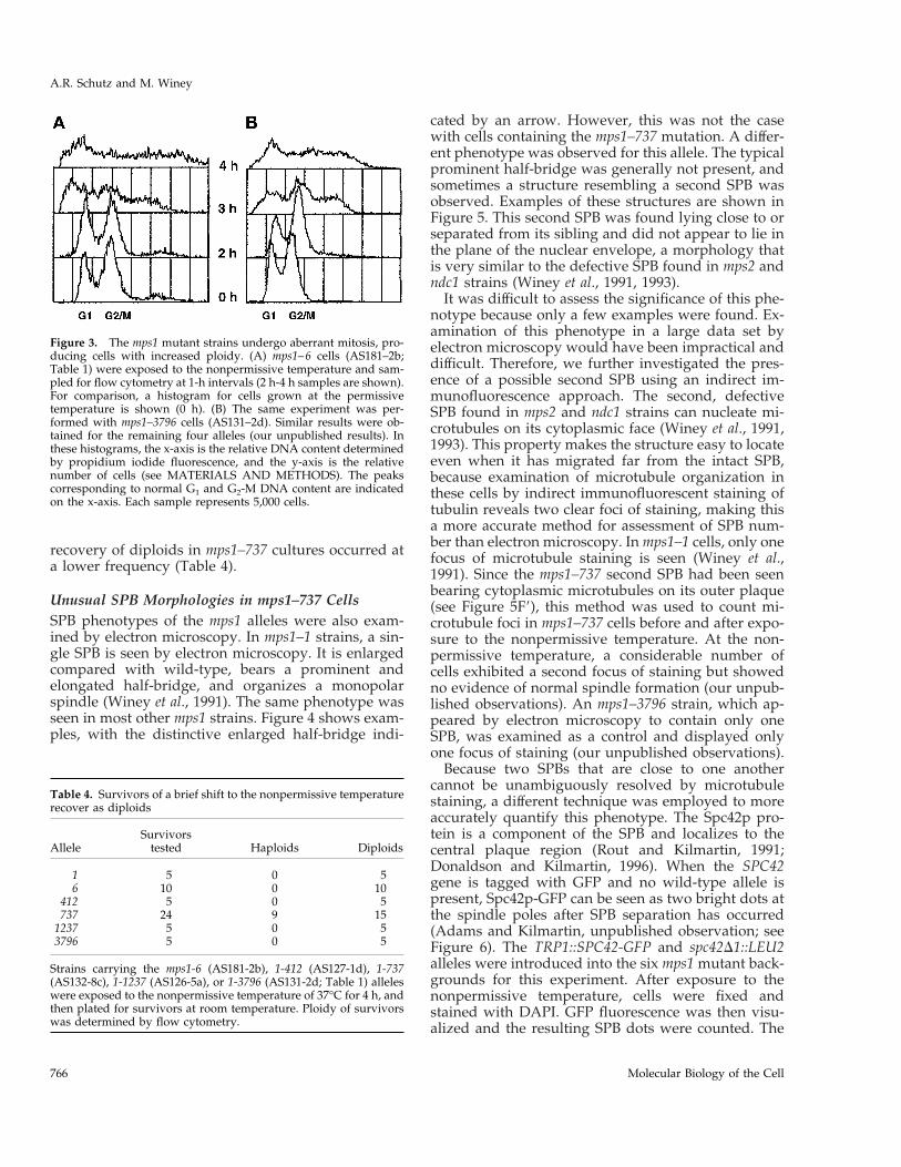
recovery of diploids in mps1–737 cultures occurred ata lower frequency (Table 4).
Unusual SPB Morphologies in mps1–737 CellsSPB phenotypes of the mps1 alleles were also exam-ined by electron microscopy. In mps1–1 strains, a sin-gle SPB is seen by electron microscopy. It is enlargedcompared with wild-type, bears a prominent andelongated half-bridge, and organizes a monopolarspindle (Winey et al., 1991). The same phenotype wasseen in most other mps1 strains. Figure 4 shows exam-ples, with the distinctive enlarged half-bridge indi-
cated by an arrow. However, this was not the casewith cells containing the mps1–737 mutation. A differ-ent phenotype was observed for this allele. The typicalprominent half-bridge was generally not present, andsometimes a structure resembling a second SPB wasobserved. Examples of these structures are shown inFigure 5. This second SPB was found lying close to orseparated from its sibling and did not appear to lie inthe plane of the nuclear envelope, a morphology thatis very similar to the defective SPB found in mps2 andndc1 strains (Winey et al., 1991, 1993).
It was difficult to assess the significance of this phe-notype because only a few examples were found. Ex-amination of this phenotype in a large data set byelectron microscopy would have been impractical anddifficult. Therefore, we further investigated the pres-ence of a possible second SPB using an indirect im-munofluorescence approach. The second, defectiveSPB found in mps2 and ndc1 strains can nucleate mi-crotubules on its cytoplasmic face (Winey et al., 1991,1993). This property makes the structure easy to locateeven when it has migrated far from the intact SPB,because examination of microtubule organization inthese cells by indirect immunofluorescent staining oftubulin reveals two clear foci of staining, making thisa more accurate method for assessment of SPB num-ber than electron microscopy. In mps1–1 cells, only onefocus of microtubule staining is seen (Winey et al.,1991). Since the mps1–737 second SPB had been seenbearing cytoplasmic microtubules on its outer plaque(see Figure 5F9), this method was used to count mi-crotubule foci in mps1–737 cells before and after expo-sure to the nonpermissive temperature. At the non-permissive temperature, a considerable number ofcells exhibited a second focus of staining but showedno evidence of normal spindle formation (our unpub-lished observations). An mps1–3796 strain, which ap-peared by electron microscopy to contain only oneSPB, was examined as a control and displayed onlyone focus of staining (our unpublished observations).
Because two SPBs that are close to one anothercannot be unambiguously resolved by microtubulestaining, a different technique was employed to moreaccurately quantify this phenotype. The Spc42p pro-tein is a component of the SPB and localizes to thecentral plaque region (Rout and Kilmartin, 1991;Donaldson and Kilmartin, 1996). When the SPC42gene is tagged with GFP and no wild-type allele ispresent, Spc42p-GFP can be seen as two bright dots atthe spindle poles after SPB separation has occurred(Adams and Kilmartin, unpublished observation; seeFigure 6). The TRP1::SPC42-GFP and spc42D1::LEU2alleles were introduced into the six mps1 mutant back-grounds for this experiment. After exposure to thenonpermissive temperature, cells were fixed andstained with DAPI. GFP fluorescence was then visu-alized and the resulting SPB dots were counted. The
Figure 3. The mps1 mutant strains undergo aberrant mitosis, pro-ducing cells with increased ploidy. (A) mps1–6 cells (AS181–2b;Table 1) were exposed to the nonpermissive temperature and sam-pled for flow cytometry at 1-h intervals (2 h-4 h samples are shown).For comparison, a histogram for cells grown at the permissivetemperature is shown (0 h). (B) The same experiment was per-formed with mps1–3796 cells (AS131–2d). Similar results were ob-tained for the remaining four alleles (our unpublished results). Inthese histograms, the x-axis is the relative DNA content determinedby propidium iodide fluorescence, and the y-axis is the relativenumber of cells (see MATERIALS AND METHODS). The peakscorresponding to normal G1 and G2-M DNA content are indicatedon the x-axis. Each sample represents 5,000 cells.
Table 4. Survivors of a brief shift to the nonpermissive temperaturerecover as diploids
AlleleSurvivors
tested Haploids Diploids
1 5 0 56 10 0 10
412 5 0 5737 24 9 15
1237 5 0 53796 5 0 5
Strains carrying the mps1-6 (AS181-2b), 1-412 (AS127-1d), 1-737(AS132-8c), 1-1237 (AS126-5a), or 1-3796 (AS131-2d; Table 1) alleleswere exposed to the nonpermissive temperature of 37°C for 4 h, andthen plated for survivors at room temperature. Ploidy of survivorswas determined by flow cytometry.
A.R. Schutz and M. Winey
Molecular Biology of the Cell766

numbers collected and phenotypes observed areshown in Figure 6. For five mps1 alleles, very few largebudded cells displayed two dots of fluorescence at thenonpermissive temperature. Most cells contained asingle SPB, which was usually found near the budneck (as in Figure 6B). In contrast, nearly all mps1–737cells contained a second SPB as evidenced by two fociof Spc42-GFP fluorescence, confirming the previousmicrotubule staining and electron microscopic results(Figure 6C). The two SPBs were often found fairlyclose to one another—in 64% of cells with two dots,both dots were located in the same cell body. Gener-ally, the SPBs were found in the cell body that con-tained the majority of DAPI staining, although aploidbodies with only mitochondrial DNA staining thatcontained an SPB were observed (our unpublishedresults).
The presence of two dots of Spc42p-GFP fluores-cence in almost all mps1–737 cells indicated that theycould proceed partially through SPB duplication andassemble a defective SPB. This unexpected phenotypesuggests an additional, later requirement for MPS1 inthe duplication process, but it was possible that themps1–737 phenotype arose from an early structuralmistake in satellite formation that precluded completeduplication. Cells with the mps1–1 mutation fail toform a satellite at the nonpermissive temperature andalso fail to maintain a satellite deposited by pretreat-ment with a-factor (Winey et al., 1991). If a defect insatellite formation were responsible for the mps1–737SPB morphology, then a-factor pretreatment to supplythe cell with a normal satellite before exposure to thenonpermissive temperature should correct the defect.However, mps1–737 cells treated in this manner still
failed in SPB duplication. Severe DNA missegregationwas seen by flow cytometry, and aberrant microtubulearrays with no spindle or connection formed betweenthe two foci of staining were again observed by im-munofluorescence (our unpublished observations).These results are consistent with a later requirementfor MPS1 function SPB duplication.
Characterization of the Mutant ProteinsAn N-terminal GST epitope tag was used to monitorand purify mutant Mps1p proteins (Mitchell et al.,1993). The mutant alleles were tagged as described byLauze et al. (1995) by excision of a HincII fragmentcontaining the gene from each centromeric mutantclone and placement into the GST vector (see MATE-RIALS AND METHODS). The resulting constructscontained all but the first two codons of the openreading frame, and overproduction of tagged fusionproteins could be induced by addition of galactose tothe growth medium. The tagged proteins are referredto as GM- followed by WT (for the wild-type enzyme)or by the allele number. Previously, overexpression ofGM-WT has been shown to cause a mitotic arrest byectopic activation of the spindle assembly checkpoint(Hardwick et al., 1996). The overexpression phenotypeof the tagged mutant proteins in the wild-type strainFLY14A (Table 1) was examined. When expression ofeach was induced with galactose at 25°C and cell cycledistribution of the induced cultures monitored by flowcytometry, some degree of G2/M delay or arrest wasalso observed (our unpublished observations). Nosuch response to galactose was evident in culturesexpressing GST from the pEG(KT) vector without in-
Figure 4. Most mps1 alleles share the mps1–1 SPB phenotype. (A) In mps1–1 cells, a single SPB with very prominent half-bridge (arrow) isseen at the nonpermissive temperature (Winey et al., 1991). (B and C) Strains with the mps1–6 (B; AS181–2b) or mps1–3796 (C; AS131–2d)mutations were shifted to the nonpermissive temperature for 4 h, and their SPB phenotypes were examined by electron microscopy. Bothstrains exhibited the same SPB morphology seen in panel A: a single enlarged SPB with prominent half-bridge (arrows). This was alsoobserved in mps1–412 and mps1–1237 strains (our unpublished results). Bar, 0.1 mm.
MPS1 alleles in yeast
Vol. 9, April 1998 767

sert. This indicated that the tagged mutant enzymesmust possess sufficient in vivo kinase activity to acti-vate the spindle assembly checkpoint in this experi-mental situation.
The GST-Mps1p fusion protein has a predicted mo-lecular weight of 112 kDa. However, the wild-type–tagged protein has been shown to migrate at a higherapparent molecular weight on a Western blot as abroad and somewhat indistinct band (Lauze et al.,1995). After treatment with protein phosphatases thisband shifts down to the predicted molecular weight,suggesting that altered gel mobility is largely a resultof phosphorylation. The observation that a catalyti-cally inactive “kinase dead” form exhibits no bandshift indicates that autophosphorylation is likely to beresponsible (Lauze et al., 1995). When the mutant pro-teins were overexpressed, we observed that gel mo-bility varied greatly between the different mutant pro-teins but was reproducible for any given mutantprotein (Figure 7A). Some proteins, such as GM-412and GM-3796, migrated similarly to GM-WT. Othersmigrated more quickly, with the most dramatic exam-ple being GM-1. This protein migrated as a single tightband at about the correct predicted molecular weight,similar to the migration of the catalytically inactivatedprotein (Lauze et al., 1995). Other fusions displayedintermediate changes in gel mobility. As with thewild-type protein, mobility differences very likelyarose from varying degrees of autophosphorylation,and so they may reflect different levels of kinase ac-tivity in the cell. The mutant proteins were generallypresent at lower levels than GM-WT.
Figure 5. Cells carrying the mps1–737 allele display an unusualSPB morphology. The mps1–737 strains AS132–3a or AS132–8c (Ta-ble 1) were exposed to the nonpermissive temperature for 3–4 h andexamined by electron microscopy. In images A through D, the entirestructure is seen in one thin section. The pairs of images in E and Frepresent two sections from the same nucleus, where relevant struc-tures are seen in more than one section. A second SPB was found infour of 26 nuclei examined. (A and B) In most cells, a single SPBwith a shorter and much less prominent half-bridge was found. (Cand D) In each image, one complete SPB with enlarged half-bridgeis present, and a second, structurally incomplete SPB (arrow) isperched at the end of the half-bridge on its cytoplasmic face. Thesecond SPB has clear central and outer plaques. This morphologywas observed in three cells. (E and E9) These images are twoadjacent serial sections through the same nucleus. The intact SPB isseen in E, and the adjacent section (E9) reveals an aberrant SPB nextto it (arrow). (F and F9) One normal SPB, which lies on an invagi-nation of the nuclear envelope, is seen in F. In F9, the defective SPBis seen in another section from the same nucleus. This SPB hasmigrated away from its sibling to a distal location on the outersurface of the nuclear envelope, and microtubules can be seenemanating from its cytoplasmic face (arrowhead). This phenotypewas observed once. Bar, 0.1 mm.
A.R. Schutz and M. Winey
Molecular Biology of the Cell768

Kinase Activity of the Mutant ProteinsThe in vitro kinase activities of all six mutant proteinswere compared with that of wild type. For this exper-iment, wild-type cells (FLY14A; Table 1) with vectorcarrying no insert, GM-WT, or GST-tagged mutantalleles were induced for 8 h to produce sufficient pro-tein and then collected and lysed. Tagged proteinswere purified and used for kinase assays (Figure 7B).In this experiment, approximately equal amounts ofprotein were present in each lane, as determined byWestern blotting (see MATERIALS AND METHODS).Both autophosphorylation (shown) and activityagainst the exogenous substrate myelin basic protein(our unpublished results) were tested and behavedsimilarly. No kinase activity was detected from thecontrol strain (pEG(KT)), whereas substantial activitycould be attributed to GM-WT. The mutant proteinsvaried in their activity. Most appeared to be largelyinactive in vitro. Only two proteins, GM-6 and GM-1237, demonstrated significant kinase activity. Thesetwo mutant enzymes were examined more closely.
Because these mutations cause a ts phenotype in thecell, we investigated the possible thermolability of theGM-6 and GM-1237 mutant proteins in kinase assaysperformed at 35°C, a temperature that is nonpermis-sive for both corresponding mutant strains. Whenwild-type protein is assayed at this temperature, au-tophosphorylation signal is unchanged, althoughphosphorylation of MBP is somewhat reduced. Tocompare wild-type and mutant kinase activities, thethree purified proteins were assayed in parallel at25°C and 35°C (Figure 8). After separation of kinaseassay material on SDS-PAGE gels, the portion of thegel containing GST-tagged Mps1p was Western blot-ted. Autophosphorylation and phosphorylation ofMBP were quantitated, and similar Mps1p proteinloadings were confirmed by Western blot using a
Figure 6. mps1–737 cells contain two dots of Spc42p-GFP fluores-cence, indicating that two SPBs are present. Strains that carry vari-ous mps1 alleles and the GFP-tagged allele of SPC42 were shifted to37°C for 4 h, briefly fixed, and stained with DAPI to visualize DNA.DAPI and GFP fluorescence were then observed. Only large buddedcells with evidence of DNA missegregation were scored. The num-ber of dots of GFP fluorescence in a given cell reflects the number ofSPBs. (A) At 25°C, large-budded cells display normal DNA segre-gation patterns (DAPI), and one SPB dot (Spc42p-GFP) localizes witheach region of DAPI staining. Shown here are mps1–3796 and mps1–737 strains (AS235-GFP and AS234-GFP, respectively; Table 1). (B)In the mps1–3796 strain at 37°C, aberrant DNA segregation becomesapparent in large-budded cells. Only one SPB dot can be found inmost cells, and it is often located close to the bud neck. Similarresults were observed for all alleles except mps1–737. (C) Whenshifted to 37°C, mps1–737 cells behave differently. DNA segregationpatterns become aberrant, but most cells clearly contain two distinctSPB dots. Both dots are generally found in the same cell body, andone is often located in a region that does not overlap with DAPIstaining (arrow). (D) Number of SPB dots observed in all six mutantbackgrounds.
MPS1 alleles in yeast
Vol. 9, April 1998 769

chemifluorescent detection substrate (see MATERI-ALS AND METHODS). Two trends were apparentfrom the data (Figure 8). First, the activities of GM-6and GM-1237 were consistently lower than that ofGM-WT. Second, both of the mutant kinases clearlyretained substantial activity at 35°C; they were notobviously thermolabile in this in vitro assay. For thecorresponding mps1–6 and mps1–1237 mutant strains,the nonpermissive temperature is 34°C. This resultsuggests that thermolability of these mutant proteinsmay not arise from a general loss of kinase activity,but may involve interactions with other factors withinthe cell.
DISCUSSION
We have described the phenotypic and molecularcharacterization of six alleles of the MPS1 gene. Eachallele contains a single point mutation in the regionencoding the protein kinase domain, and these lesionsaffect the activity of the resulting mutant kinases indifferent ways. Five of the six alleles cause the sametype of early SPB duplication failure. However, cells
carrying the mps1–737 allele proceed farther in dupli-cation before they fail. This phenotype suggests thatthe Mps1p kinase is required for multiple events inSPB duplication.
The Mutant LesionsThe fact that all six ts mutations occur in the C-termi-nal protein kinase domain indicates that Mps1p kinaseactivity is likely to be essential for its physiologicalrole. This collection of alleles probably represents avariety of defects in ATP binding, substrate binding,and catalytic activity; however, most alleles producethe same SPB duplication phenotype, and all are sim-ilarly defective for the spindle assembly checkpoint.The location of all lesions in the kinase domain alsosuggests that the methods used to identify these al-leles were somewhat limited. In all cases, temperaturesensitivity and failure to complement the originalmps1–1 allele were requirements for identification. Iso-lates that displayed some degree of intragenic comple-mentation with mps1–1 could easily have been over-looked, along with any nonconditional mps1 mutants.In fact, point mutations in the N-terminal region havenow been identified, and these mps1 mutants have noconditional phenotype. They are lethal in combinationwith a null allele of CIN8, but display no defects inSPB duplication (Geiser et al., 1997; Winey and Chen,unpublished observation).
Figure 7. Autophosphorylation by GST-tagged mutant proteinsvaries greatly. Plasmids carrying the GST-tagged mps1 alleles weretransformed into the wild-type strain FLY14A (Table 1), and expres-sion of fusion proteins was induced as described (see MATERIALSAND METHODS). (A) GST fusions were detected on Western blotsof whole cell lysates with anti-GST antibody. The control lane (pEG)shows cells carrying the GST vector without insert. The taggedwild-type protein (GM-WT) is phosphorylated, causing it to migrateabove its predicted molecular weight of 112 kDa (Lauze et al., 1995).(B) GST fusions were affinity-purified with glutathione-Sepharoseand used for in vitro kinase assays at 25°C. Material was thenseparated on an SDS-PAGE gel and blotted. Proteins loadings areapproximately equal across the gel, as determined by Western blot(our unpublished results).
Figure 8. In vitro activity of the GM-6 and GM-1237 mutant pro-teins is not thermolabile. Affinity-purified GM-WT, GM-6, and GM-1237 kinases were assayed in vitro at 25°C and 35°C, and thenseparated on an SDS-PAGE gel and blotted. This elevated assaytemperature would be nonpermissive for growth of both mps1–6and mps1–1237 mutant strains. (A) Autophosphorylation activity ofthe proteins at both assay temperatures. (B) Substrate-level phos-phorylation of myelin basic protein (MBP). For each pair of samples(25°C and 35°C), protein loadings were approximately equal (deter-mined by Western blot).
A.R. Schutz and M. Winey
Molecular Biology of the Cell770

The six mutations reported here also provide infor-mation about the correlation between Mps1p structureand function. Often ts mutations in protein kinases arelocated in structural transition regions that lie in be-tween conserved motifs—a suitable location for mu-tations that cause instability and thermolability of theprotein without entirely destroying catalytic activity[for examples, see Lorincz and Reed (1986); Carr et al.(1989); Hollingsworth et al. (1992)]. However, many ofthe mps1 lesions defy this paradigm. Of the six muta-tions, three affect residues located in conserved orinvariant sequence motifs in the protein kinase cata-lytic domain (see Figure 2). The most drastic of theseis alteration of the invariant glutamate residue at po-sition 486 in subdomain III to a lysine, in mps1–737.Normally, this residue and Lys468 in subdomain IIwould be predicted to cooperate in formation of thedocking site for Mg-ATP (Taylor et al., 1992). Such anextreme change might be predicted to abolish kinaseactivity, but this mutant protein can still supportgrowth, although mps1–737 cells grow slowly even atthe permissive temperature. Perhaps another gluta-mate residue nearby can compensate, possibly Glu488or Glu491.
Another conserved residue affected is Pro608, whichlies in the peptide-binding lobe of the protein in sub-domain VIII and is altered to serine in the mps1–1237allele. This proline residue is somewhat conserved
among kinases in general, and identical in Mps1p, esk,PYT/TTK, and PPK1. The residues in subdomain VIIIappear to be particularly important for enzyme–sub-strate interactions. In a variety of kinases, this regionparticipates in the formation of two side chain pocketsin the kinase structure that interact with substrateresidues in the immediate vicinity of the amino acidthat will be phosphorylated, contributing to substrateselectivity and orientation (Songyang et al., 1994,1996). The alteration of Glu517 to lysine in mps1–3796also affects a somewhat conserved residue, one that isfound in many dual-specificity kinases (Lindberg etal., 1992). The remaining three mutations occur at po-sitions that are not conserved among protein kinases,but two of these affect residues that are identical in esk,PYT/TTK, and PPK1.
These six mutations cause a variety of effects on thein vivo and in vitro activity of GST-Mps1p fusionproteins. All six mutant kinases must possess somedegree of activity in vivo, because they supportgrowth at the permissive temperature and can cause aG2-M bias in the cell cycle distribution of wild-typecells when they are overexpressed. Their gel mobilitysuggests that autophosphorylation activity variesgreatly; however, the significance of autophosphory-lation for protein function is not yet known. Kinaseassay experiments with GST fusions show that in vitroactivity also varies, but does not correlate well with
Figure 9. Proposed pathway for SPB duplication. In this schematic, the central and outer plaques of the SPB are depicted, with cytoplasmicand nuclear microtubules drawn above and below the SPB, respectively. The bracketed structure is a duplication intermediate that is inferredbut has not been reported. Execution point experiments with mps1–1, mps2–1, and ndc1–1 mutants (Winey et al., 1991, 1993) indicate wherein the pathway these mutations cause SPB duplication to fail. The mps1–1 mutation causes failure early on and is marked with an asteriskto indicate that four of the other five alleles share this SPB morphology. However, the mps1–737 mutation allows duplication to proceedfarther before failure occurs and has been placed here at the same point in the pathway as mps2–1 and ndc1–1 because of their phenotypicsimilarity. Mutation of CDC37 also causes a later failure in SPB duplication, but generates a different, partially duplicated structure (Schutzet al., 1997). The two distinct mps1 mutant phenotypes suggest that this gene is required continuously or at multiple times during SPBduplication.
MPS1 alleles in yeast
Vol. 9, April 1998 771

gel mobility as an assessment of activity in vivo, withthe exception that proteins with little or no gel shift areinactive in this in vitro assay. The observation thatmany mutant Mps1p proteins are inactive in vitro isnot unexpected, since their folding may be compro-mised enough that they cannot withstand lysis andpurification. Similar results have been obtained withmutant forms of the yeast cAMP-dependent proteinkinase that are partially or even fully functional in thecell (Gibbs and Zoller, 1991).
The behavior of the GM-6 and GM-1237 mutantenzymes in vitro raises interesting questions about thenature of their temperature-sensitive defects. Both fu-sions display significant in vitro kinase activity, butthis activity does not appear to be thermolabile whenthe assays are performed at a temperature that isnonpermissive for growth of the corresponding mu-tant strains. This suggests that the ts defect does notarise soley from temperature-induced denaturation oraggregation of the protein, but may involve a morecomplex mechanism, such as disruption of interac-tions with other proteins. These two mutations couldaffect the ability of the kinase to bind substrates, acti-vators, or regulatory subunits. Such defects would notbe apparent when purified GM-6 or GM-1237 is as-sayed; accessory proteins and relevant substrateswould not be present, and regulatory signals such asphosphorylation would already have been delivered.Such a model is particularly appealing when informa-
tion about substrate-binding motifs is taken into ac-count. The proline-to-serine substitution in Mps1–1237p occurs very close to residues critical for bindingof substrate residues that lie in the 22 or 11 positionsrelative to the phosphorylated amino acid, and theMps1–6p substitution is near another group of resi-dues also important for binding at the 22 position(Songyang et al., 1994, 1996). These two perturbationscould weaken or disrupt binding of relevant sub-strates in a similar manner. If this mechanism is thesource of temperature sensitivity, the mps1–6 andmps1–1237 alleles could prove useful genetically foridentification of important interacting factors, partic-ularly substrates.
Implications of the mps1–737 PhenotypeThe mps1–737 allele is interesting because of the na-ture of its mutation (which drastically alters the highlyconserved Glu486 residue), but even more so becauseof its phenotype. Most of the mps1 alleles describedhere closely resemble the original allele, mps1–1, andcause early failure in SPB duplication. In sharp con-trast, cells carrying the mps1–737 allele are able to forma second, aberrant SPB. Why does this occur, and whatdoes it indicate about the role of MPS1 in SPB dupli-cation? This may not be a simplistic loss-of-functionsituation. We suggest that the Mps1p kinase is actuallyrequired for multiple events in SPB duplication (Fig-ure 9), and that the mps1–737 allele represents a sep-aration or alteration of those functions. Different mu-tations might affect activity of the kinase towarddifferent substrates or its ability to respond correctly toregulatory signals, revealing phenotypic nuances. TheMps1–737p mutant enzyme may be competent to meetthe previously identified early requirement for thekinase (Winey et al., 1991) but unable to carry out alater function. Failure to meet this later requirementwould result in the mps1–737 phenotype. The assem-bly of an aberrant second SPB with central and outerplaques and cytoplasmic microtubules, which closelyresembles the mps2 and ndc1 morphologies (Winey etal., 1991, 1993), is consistent with this type of previ-ously unrecognized late requirement for MPS1. A laterfunction for MPS1 in SPB duplication would not havebeen uncovered without this unique allele; no othergenetic or pharmacological tools currently exist thatreversibly block SPB duplication between the satellite-bearing and duplicated side-by-side stages.
Multiple requirements could also provide a simplemechanism for the intragenic complementation ob-served when the mps1–737 allele is combined witheither mps1–412 or mps1–3796 (Figure 10). It appearsthat Mps1–737p can supply only the earlier duplica-tion function. Like mps1–737, mps1–412 and mps1–3796 could also be separation-of-function alleles thatcannot meet the early requirement but would be com-
Figure 10. A model for intragenic complementation between mps1alleles. The intragenic complementation observed between mps1–737 and either the mps1–412 or mps1–3796 alleles could be a result ofmultiple requirements for Mps1p during SPB duplication. Themps1–737 gene product, Mps1–737p, may be competent to meet theearly requirement reported previously (Winey et al., 1991) but ap-parently cannot perform an important later function. Mps1–412pand Mps1–3796p clearly fail to meet the early requirement, but it ispossible that they are competent to perform the later function that islost in mps1–737 cells. If so, the combination of two partially func-tional kinases in mps1–737/mps1–412 or mps1–737/mps1–3796 dip-loid cells could meet both requirements, allowing the cell to grow ata higher temperature. According to this model, the three remainingmps1 alleles would be defective for both functions and thus wouldnot display intragenic complementation. All six mps1 alleles aredefective for the spindle assembly checkpoint function (Weiss andWiney, 1996).
A.R. Schutz and M. Winey
Molecular Biology of the Cell772

petent to carry out the later SPB duplication function ifgiven the opportunity. When combined in a diploidcell, Mps1–737p could cooperate with either Mps1–412p or Mps1–3796p, together supplying both func-tions and permitting growth at a higher temperature.A partial loss of Mps1p function may also occur underdifferent circumstances. The Cdc37p protein is re-quired to promote the activity of several protein ki-nases in yeast (Gerber et al., 1995; Dey et al., 1996;Schutz et al., 1997). Mutation of CDC37 causes a sig-nificant decrease in Mps1p kinase activity and resultsin a late failure in SPB duplication (Schutz et al., 1997).However, the partially duplicated SPB morphologyseen is different than that observed for any other du-plication mutants (Figure 9). The observation that dif-ferent perturbations of Mps1p function correlate withdifferent mutant SPB morphologies argues stronglythat this kinase is required for multiple events duringthe duplication process, or possibly throughout a win-dow of time in G1.
The differing mps1 mutant phenotypes also invitespeculation about the reversibility of SPB duplicationproblems. Diploidization by surviving cells of mostmutant strains after brief exposure to the nonpermis-sive temperature indicates that SPB duplication failureis irreversible in these cells. Presumably, a second polecan no longer be made, and the single, enlarged SPB isleft to carry out mitosis alone, sometimes pulling allthe chromosomes to that pole and creating a viablediploid. Cells with the mps1–737 mutation are able torecover as viable haploids more frequently than ob-served for other alleles. Does this indicate that theirdifferent SPB defects can be reversed? Cells carryingthe mps2–1 or ndc1–1 mutations, which share the sameSPB morphology as mps1–737, also recover from ex-posure to the nonpermissive temperature as a mixtureof both haploids and diploids (Winey et al., 1991;Thomas and Botstein, 1986). This correlation suggeststhat perhaps the defective second SPB in these cells isnot always a dead-end product, but in some cases canbe remodeled or built upon to produce a fully func-tional SPB.
The continued analysis of strains with mutant allelesof MPS1 and other genes important for SPB duplica-tion should yield further insight into this process.Furthermore, analysis of the regulation of MpsIp, andidentification of relevant substrates in SPB duplicationand spindle checkpoint control, should lead us to adeeper understanding of the duplication process andof the roles of this important kinase in the SPB cycleand in cell cycle control.
ACKNOWLEDGMENTS
We thank Ian Adams and John Kilmartin for the GFP-tagged alleleof SPC42; Tim Huffaker, John Kilmartin, Doug Koshland, and Eliz-abeth Siewert for mps1 isolates; and Mike Culbertson for his4 strains.
We also thank Andrea Castillo for critical reading of the manuscript;Tom Giddings and Estelle Steiner for technical advice and assis-tance; and the members of the Winey laboratory for their input. Thiswork was initiated under an American Cancer Society grant(MV63940) and completed with support from the National Institutesof Health (NIH GM-51312). Additional support was provided by anAmerican Cancer Society Junior Faculty Research Award (A70760)and the Pew Scholars Program in the Biomedical Sciences award(P0020SC) to M.W. A.R.S. was supported by an NIH training grant(GM-07135), National Science Foundation Predoctoral Fellowship,and Truman Scholarship.
REFERENCES
Anderson, C.W., Baum, P.R., and Gesteland, R.F. (1973). Processingof adenovirus-2 induced proteins. J. Virol. 12, 241–252.
Ausubel, F.M., Brent, R., Kingston.R.E., Moore, D.D., Seidman, J.G.,Smith, J.A., and Struhl, K. (1994). Current Protocols in MolecularBiology. New York: John Wiley & Sons.
Byers, B., and Goetsch, L. (1974). Duplication of spindle plaques andintegration of the yeast cell cycle. Cold Spring Harbor Symp. Quant.Biol. 38, 123–131.
Byers, B., and Goetsch, L. (1975). Behavior of the spindles andspindle plaques in the cell cycle and conjugation of Saccharomycescerevisiae. J. Bacteriol. 124, 511–523.
Carr, A.M., MacNeill, S.A., Hayles, J., and Nurse, P. (1989). Molec-ular cloning and sequence analysis of mutant alleles of the fissionyeast cdc21 protein kinase gene: implications for cdc2 protein struc-ture and function. Mol. Gen. Genetics 218, 41–49.
Dey, B., Lightbody, J.J., and Boschelli, F. (1996). CDC37 is requiredfor p60v-src activity in yeast. Mol. Biol. Cell 7, 1405–1417.
Donaldson, A.D., and Kilmartin, J.V. (1996). Spc42p: a phosphory-lated component of the S cerevisiae spindle pole body (SPB) with anessential function during SPB duplication. J. Cell Biol. 132, 887–901.
Douville, E.M.J., Afar, D.E.H., Howell, B.W., Letwin, K., Tannock,L., Ben-david, Y., Pawson, T., and Bell, J.C. (1992). Multiple cDNAsencoding the esk kinase predict transmembrane and intracellularenzyme isoforms. Mol. Cell. Biol. 12, 2681–2689.
Elble, R. (1992). A simple and efficient procedure for transformationof yeasts. BioFeedBack 13, 18–20.
Geiser, J.R., Schott, E.J., Kingsbury, T.J., Cole, N.B., Totis, L.J., Bhat-tacharyya, G., He, L., and Hoyt, M.A. (1997). Saccharomyces cerevisiaegenes required in the absence of the CIN8-encoded spindle motoract in functionally diverse mitotic pathways. Mol. Biol. Cell 8,1035–1050.
Gerber, M.R., Farrell, A., Deshaies, R.J., Herskowitz, I., and Morgan,D.O. (1995). Cdc37 is required for association of the protein kinaseCdc28 with G1 and mitotic cyclins. Proc. Natl. Acad. Sci. USA 92,4651–4655.
Gibbs, C.S., and Zoller, M.J. (1991). Rational scanning mutagenesisof a protein kinase identifies functional regions involved in catalysisand substrate interactions. J. Biol. Chem. 266, 8923–8931.
Hanks, S.K., Quinn, A.M., and Hunter, T. (1988). The protein kinasefamily: conserved features and deduced phylogeny of the catalyticdomains. Science 241, 42–52.
Hardwick, K.G., Weiss, E., Luca, F.C., Winey, M., and Murray, A.W.(1996). Activation of the budding yeast spindle assembly checkpointwithout mitotic spindle disruption. Science 273, 953–956.
Hartwell, L.H. (1967). Macromolecular synthesis in temperature-sensitive mutants of yeast. J. Bacteriol. 93, 1662–1670.
MPS1 alleles in yeast
Vol. 9, April 1998 773

Hoffman, C.S., and Winston, F. (1987). A ten-minute DNA prepara-tion from yeast efficiently releases autonomous plasmids for trans-formation of Escherichia coli. Gene 57, 267–272.
Hogg, D., Guidos, C., Bailey, D., Amendola, A., Groves, T., David-son, J., Schmandt, R., and Mills, G. (1994). Cell cycle dependentregulation of the protein kinase TTK. Oncogene 9, 89–96.
Hollingsworth, R.E. Jr., Ostroff, R.M., Klein, M.B., Niswander, L.A.,and Sclafani, R.A. (1992). Molecular genetic studies of the Cdc7protein kinase and induced mutagenesis in yeast. Genetics 132,53–62.
Hoyt, M.A., Totis, L., and Roberts, B.T. (1991). S. cerevisiae genesrequired for cell cycle arrest in response to loss of microtubulefunction. Cell 66, 507–517.
Hutter, K.J., and Eipel, H.E. (1979). Microbial determination by flowcytometry. J. Gen. Microbiol. 113, 369–375.
Jacobs, C.W., Adams, A.E.M., Szaniszlo, P.J., and Pringle, J.R. (1988).Functions of microtubules in the Saccharomyces cerevisiae cell cycle.J. Cell Biol. 107, 1409–1426.
Kilmartin, J.V., and Adams, A.E.M. (1984). Structural rearrange-ments of tubulin and actin during the cell cycle of the yeast Saccha-romyces. J. Cell Biol. 98, 922–933.
Lauze, E., Stoelcker, B.S., Luca, F.C., Weiss, E., Schutz, A.R., andWiney, M. (1995). Yeast spindle pole body duplication gene MPS1encodes an essential dual specificity protein kinase. EMBO J. 14,1655–1663.
Li, R., and Murray, A.W. (1991). Feedback control in mitosis inbudding yeast. Cell 66, 519–531.
Lindberg, R.A., Quinn, A.M., and Hunter, T. (1992). Dual specificityprotein kinases: will any hydroxyl do? Trends Biochem. Sci. 17,114–119.
Lindberg, R.A., Fischer, W.H., and Hunter, T. (1993). Characteriza-tion of a human protein threonine kinase isolated by screening anexpression library with antibodies to phosphotyrosine. Oncogene 8,351–359.
Lorincz, A.T., and Reed, S.I. (1986). Sequence analysis of tempera-ture-sensitive mutations in the Saccharomyces cerevisiae gene CDC28.Mol. Cell. Biol. 6, 4099–4103.
Mannis, T.R., and Mortimer, R.K. (1964). Allelic mapping in yeast byx-ray induced mitotic reversion. Science 143, 581–582.
Mathison, L., and Culbertson, M.R. (1985). Suppressible and non-suppressible 11 G-C base pair insertions induced by ICR-170 at thehis4 locus in Saccharomyces cerevisiae. Mol. Cell. Biol. 5, 2247–2256.
Mitchell, D.A, Marshall, T.K., and Deschenes, R.J. (1993). Vectors forthe inducible expression of glutathione S-transferase fusion proteinsin yeast. Yeast 9, 715–723.
Mills, G.B., Schmandt, R., McGill, M., Amendola, A., Hill, M., Ja-cobs, K., May, C., Rodricks, A., Campbell, S., and Hogg, D. (1992).Expression of TTK, a novel human protein kinase, is associated withcell proliferation. J. Biol. Chem. 267, 16000–16006.
Poch, O., Schwob, E., de Fraipont, F., Camasses, A., Bordonne, R.,and Martin, R.P. (1994). RPK1, an essential yeast protein kinaseinvolved in the regulation of the onset of mitosis, shows homologyto mammalian dual-specificity kinases. Mol. Gen. Genet. 243, 641–653.
Rothstein, R. (1991). Targeting, disruption, replacement, and allelerescue: integrative DNA transformation in yeast. Methods Enzymol.194, 281–301.
Rout, M.P., and Kilmartin, J.V. (1991). Yeast spindle pole bodycomponents. Cold Spring Harbor Symp. Quant. Biol. 61, 687–692.
Schutz, A.R., Giddings, T.H., Jr., Steiner, E., and Winey, M. (1997).The yeast CDC37 gene interacts with MPS1 and is required forproper execution of spindle pole body duplication. J. Cell Biol. 136,969–982.
Sikorski, R.S., and Hieter, P. (1989). A system of shuttle vectors andyeast host strains designed for efficient manipulation of DNA inSaccharomyces cerevisiae. Genetics 122, 19–27.
Songyang, Z., Blechner, S., Hoagland, N., Hoekstra, M.F., Piwnica-Worms, H., and Cantley, L.C. (1994). Use of an oriented peptidelibrary to determine the optimal substrates of protein kinases. Curr.Biol. 4, 973–982.
Songyang, Z. et al. (1996). A structural basis for substrate specifici-ties of protein Ser/Thr kinases: primary sequence preference ofcasein kinases I and II, NIMA, phosphorylase kinase, calmodulin-dependent kinase II, CDK5, and Erk1. Mol. Cell. Biol. 16, 6486–6493.
Taylor, S.S., Knighton, D.R., Zheng, J., Ten Eyck, L.F., and Sowadski,J.M. (1992). Structural framework for the protein kinase family.Annu. Rev. Cell Biol. 8, 429–462.
Thomas, J.H., and Botstein, D. (1986). A gene required for theseparation of chromosomes on the spindle apparatus in yeast. Cell44, 65–76.
Weiss, E., and Winey, M. (1996). The Saccharomyces cerevisiae spindlepole body duplication gene MPS1 is part of a mitotic checkpoint.J. Cell Biol. 132, 111–123.
Winey, M., and Byers, B. (1993). Assembly and functions of thespindle pole body in budding yeast. Trends Genet. 9, 300–304.
Winey, M., Goetsch, L., Baum, P., and Byers, B. (1991). MPS1 andMPS2: novel yeast genes defining distinct steps of spindle pole bodyduplication. J. Cell Biol. 114, 745–754.
Winey, M., Hoyt, M.A., Chan, C., Goetsch, L., Botstein, D., andByers, B. (1993). NDC1: a nuclear envelope component required foryeast spindle pole body duplication. J. Cell Biol. 122, 743–751.
A.R. Schutz and M. Winey
Molecular Biology of the Cell774