Mycobacterium indicus pranii and Mycobacterium bovis BCG lead to differential macrophage activation...
11
Mycobacterium indicus pranii and Mycobacterium bovis BCG lead to differential macrophage activation in Toll-like receptor-dependent manner Pawan Kumar, 1 Rohit Tyagi, 1 Gobardhan Das 2,† and Sangeeta Bhaskar 1 1 National Institute of Immunology, New Delhi, and 2 International Centre for Genetic Engineering and Biotechnology, New Delhi, India doi:10.1111/imm.12306 Received 13 December 2013; revised 26 March 2014; accepted 22 April 2014. † Present address: Special Centre for Molecu- lar Medicine, Jawaharlal Nehru University, New Delhi 110067, India Correspondence: Sangeeta Bhaskar, PDC-I, National Institute of Immunology, Aruna Asaf Ali Marg, New Delhi-110067, India. Email: [email protected]. Senior author: Sangeeta Bhaskar Summary Mycobacterium indicus pranii (MIP) is an atypical mycobacterial species possessing strong immunomodulatory properties. It is a potent vaccine candidate against tuberculosis, promotes Th1 immune response and pro- tects mice from tumours. In previous studies, we demonstrated higher protective efficacy of MIP against experimental tuberculosis as compared with bacillus Calmette–Gu erin (BCG). Since macrophages play an impor- tant role in the pathology of mycobacterial diseases and cancer, in the present study, we evaluated the MIP in live and killed form for macro- phage activation potential, compared it with BCG and investigated the underlying mechanisms. High levels of tumour necrosis factor-a, interleu- kin-12p40 (IL-12p40), IL-6 and nitric oxide were produced by MIP-stimu- lated macrophages as compared with BCG-stimulated macrophages. Prominent up-regulation of co-stimulatory molecules CD40, CD80 and CD86 was also observed in response to MIP. Loss of response in MyD88- deficient macrophages showed that both MIP and BCG activate the mac- rophages in a MyD88-dependent manner. MyD88 signalling pathway cul- minates in nuclear factor-jB/activator protein-1 (NF-jB/AP-1) activation and higher activation of NF-jB/AP-1 was observed in response to MIP. With the help of pharmacological inhibitors and Toll-like receptor (TLR) -deficient macrophages, we observed the role of TLR2, TLR4 and intracel- lular TLRs in MIP-mediated macrophage activation. Stimulation of HEK293 cells expressing TLR2 in homodimeric or heterodimeric form showed that MIP has a distinctly higher level of TLR2 agonist activity compared with BCG. Further experiments suggested that TLR2 ligands are well exposed in MIP whereas they are obscured in BCG. Our findings establish the higher macrophage activation potential of MIP compared with BCG and delineate the underlying mechanism. Keywords: bacterial; cytokines; Toll-like receptors. Introduction Mycobacterium indicus pranii (MIP), earlier known as Mycobacterium w, was selected from a panel of mycobac- teria for its ability to evoke a cell-mediated immune response against Mycobacterium leprae. 1,2 Its use as an adjunct to standard chemotherapy against multibacillary leprosy resulted in significantly enhanced bacillary clear- ance and shortened recovery time. Protective effects against leprosy and tuberculosis were also observed in the household contacts of leprosy patients. 3–5 MIP was further evaluated against Mycobacterium tuberculosis and provided protection against experimental tuberculosis in both BCG-responder as well as non-responder strains of mice. 6 Consistent with the beneficial effects shown against tuberculosis and leprosy, MIP shares antigenic determi- nants with M. leprae and M. tuberculosis. 7 Underscoring its uniqueness, recent studies have placed MIP between slow-growing and fast-growing species. 8 Further studies suggested that MIP possesses strong immunomodulatory properties and enhances the immune response when used as an adjuvant along with a recombinant anti-human ª 2014 John Wiley & Sons Ltd, Immunology, 143, 258–268 258 IMMUNOLOGY ORIGINAL ARTICLE
-
Upload
independent -
Category
Documents
-
view
1 -
download
0
Transcript of Mycobacterium indicus pranii and Mycobacterium bovis BCG lead to differential macrophage activation...

Mycobacterium indicus pranii and Mycobacterium bovis BCG lead to
differential macrophage activation in Toll-like receptor-dependentmanner
Pawan Kumar,1 Rohit Tyagi,1
Gobardhan Das2,† and Sangeeta
Bhaskar1
1National Institute of Immunology, New
Delhi, and 2International Centre for Genetic
Engineering and Biotechnology, New Delhi,
India
doi:10.1111/imm.12306
Received 13 December 2013; revised 26
March 2014; accepted 22 April 2014.†Present address: Special Centre for Molecu-
lar Medicine, Jawaharlal Nehru University,
New Delhi 110067, India
Correspondence: Sangeeta Bhaskar, PDC-I,
National Institute of Immunology, Aruna
Asaf Ali Marg, New Delhi-110067, India.
Email: [email protected].
Senior author: Sangeeta Bhaskar
Summary
Mycobacterium indicus pranii (MIP) is an atypical mycobacterial species
possessing strong immunomodulatory properties. It is a potent vaccine
candidate against tuberculosis, promotes Th1 immune response and pro-
tects mice from tumours. In previous studies, we demonstrated higher
protective efficacy of MIP against experimental tuberculosis as compared
with bacillus Calmette–Gu�erin (BCG). Since macrophages play an impor-
tant role in the pathology of mycobacterial diseases and cancer, in the
present study, we evaluated the MIP in live and killed form for macro-
phage activation potential, compared it with BCG and investigated the
underlying mechanisms. High levels of tumour necrosis factor-a, interleu-
kin-12p40 (IL-12p40), IL-6 and nitric oxide were produced by MIP-stimu-
lated macrophages as compared with BCG-stimulated macrophages.
Prominent up-regulation of co-stimulatory molecules CD40, CD80 and
CD86 was also observed in response to MIP. Loss of response in MyD88-
deficient macrophages showed that both MIP and BCG activate the mac-
rophages in a MyD88-dependent manner. MyD88 signalling pathway cul-
minates in nuclear factor-jB/activator protein-1 (NF-jB/AP-1) activation
and higher activation of NF-jB/AP-1 was observed in response to MIP.
With the help of pharmacological inhibitors and Toll-like receptor (TLR)
-deficient macrophages, we observed the role of TLR2, TLR4 and intracel-
lular TLRs in MIP-mediated macrophage activation. Stimulation of
HEK293 cells expressing TLR2 in homodimeric or heterodimeric form
showed that MIP has a distinctly higher level of TLR2 agonist activity
compared with BCG. Further experiments suggested that TLR2 ligands
are well exposed in MIP whereas they are obscured in BCG. Our findings
establish the higher macrophage activation potential of MIP compared
with BCG and delineate the underlying mechanism.
Keywords: bacterial; cytokines; Toll-like receptors.
Introduction
Mycobacterium indicus pranii (MIP), earlier known as
Mycobacterium w, was selected from a panel of mycobac-
teria for its ability to evoke a cell-mediated immune
response against Mycobacterium leprae.1,2 Its use as an
adjunct to standard chemotherapy against multibacillary
leprosy resulted in significantly enhanced bacillary clear-
ance and shortened recovery time. Protective effects
against leprosy and tuberculosis were also observed in the
household contacts of leprosy patients.3–5 MIP was
further evaluated against Mycobacterium tuberculosis and
provided protection against experimental tuberculosis in
both BCG-responder as well as non-responder strains of
mice.6 Consistent with the beneficial effects shown against
tuberculosis and leprosy, MIP shares antigenic determi-
nants with M. leprae and M. tuberculosis.7 Underscoring
its uniqueness, recent studies have placed MIP between
slow-growing and fast-growing species.8 Further studies
suggested that MIP possesses strong immunomodulatory
properties and enhances the immune response when used
as an adjuvant along with a recombinant anti-human
ª 2014 John Wiley & Sons Ltd, Immunology, 143, 258–268258
IMMUNOLOGY OR IG INAL ART ICLE
chorionic gonadotrophin vaccine.9 Heat-killed MIP is
approved for human use and is available commercially.
Macrophages are major phagocytic cells strategically
located throughout the body to clear invading pathogens
and cellular debris. Classically activated macrophages pro-
duce pro-inflammatory cytokines, reactive nitrogen spe-
cies, antimicrobial peptides and offer protection against a
variety of bacteria, protozoa and viruses.10 Pathogenic
bacteria such as M. tuberculosis, however, have evolved
the mechanisms to evade the microbicidal activities of
macrophages. Mycobacterium tuberculosis inhibits the
phagolysosomal fusion and uses the phagosomal compart-
ments as niches for its growth and multiplication. Simi-
larly, the Leishmania parasite exploits the macrophages
for its growth and multiplication. Interestingly, MIP-acti-
vated macrophages have been shown to contain the Leish-
mania parasite in an inducible nitric oxide synthase-
dependent manner.11 Classically activated macrophages
can also lyse tumour cells and activate CD8+ T cells to
offer protection from tumour.12,13 However, macrophages
possess a high degree of plasticity and their normal func-
tion is altered in neoplastic diseases. Growing tumours
promote M2 macrophages and take advantage of their tis-
sue-repairing and homeostatic properties. Tumour-associ-
ated macrophages produce high levels of vascular
endothelial growth factor, interleukin-8 (IL-8) and IL-10,
which support tumour growth.14 Accordingly, high infil-
tration of tumour-associated macrophages is associated
with poor prognosis of malignant disease.
In previous studies, we observed that live MIP provides
significantly strong protection against tuberculosis in mice
and guinea pigs compared with killed MIP and bacillus
Calmette–Gu�erin (BCG). Although MIP is approved for
human use in heat-killed form, it is a non-pathogenic
mycobacterium and is cleared from the host within
4–6 weeks. We therefore used MIP in both live and killed
forms in our studies. Analysis of the immune response
revealed that MIP promotes the Th1 response and leads
to interferon-c production by lung-infiltrating immune
cells.15,16 The whole genome sequence of MIP has been
published recently and consistent with our findings in sil-
ico analysis demonstrated a much higher fraction of puta-
tive antigenic proteins in MIP compared with BCG.17
Given the importance of Th1 immune response in protec-
tion against cancer, we also evaluated MIP for cancer
immunotherapy. Significant protection from tumour and
activation of an anti-tumour immune response were
observed in MIP-treated mice.18 Similar findings have
been reported by others and the role of interferon-c has
been demonstrated in the MIP-mediated anti-tumour
response.19 Since macrophages play a central role in the
pathology of mycobacterial diseases and cancer, in the
present study, we evaluated MIP in both live and heat-
killed form for macrophage activation potential and
examined the underlying mechanism. As BCG is a widely
used vaccine against tuberculosis and is also effective
against superficial bladder cancer,20 we included BCG in
this study and compared it with MIP. Our results showed
higher activation of macrophage by live MIP compared
with killed MIP and BCG. Both MIP and BCG activated
the macrophages in MyD88 and nuclear factor-jB/activa-tor protein-1 (NF-jB/AP1) -dependent manner. With the
help of pharmacological inhibitors and Toll-like receptor
(TLR) -deficient macrophages, we noted the role of
TLR2, TLR4 and intracellular TLRs in macrophage activa-
tion by MIP. Using HEK293 cells expressing TLR2, we
further observed that MIP and BCG have differential
TLR2 engagement properties. Our findings demonstrate
higher macrophage activation potential of MIP and impli-
cate the role of TLRs in differential activation of macro-
phages by MIP and BCG.
Materials and methods
Ethics statement
All animal experiments described in this manuscript were
approved by the institutional animal ethics committee of
the National Institute of Immunology, New Delhi and
performed in accordance with the guidelines from the
same: IAEC approval no. 205/08/11.
Mice
Six- to eight-week-old inbred C57BL/6, IL10�/� and
MyD88�/� mice were obtained from the animal facility
of the National Institute of Immunology, New Delhi.
TLR2�/� and TLR4�/� mice were a kind gift from Dr
Ruslan Medzhitov (Yale University School of Medicine,
New Haven, CT) to Dr Gobardhan Das and were main-
tained at the International Centre for Genetic Engineering
and Biotechnology, New Delhi.
Mycobacteria
Glycerol stocks of MIP and BCG Danish 1331 were
thawed and cultured in 7H9 medium containing 0�05%Tween-80 and 0�05% glycerol, and supplemented with
10% albumin-dextrose-catalase. MIP was cultured in a
shaker incubator (100 r.p.m.) and BCG was grown as a
standing culture, with intermittent manual shaking, at
36°. Log-phase cultures were harvested by centrifugation
at 2000 g and washed in PBS containing 3% fetal bovine
serum (PBS-3). Bacterial aggregates were removed from
BCG suspension by additional centrifugation of suspen-
sion at 50 g for 10 min. Purity of cultures was tested by
plating MIP and BCG cultures on nutrient agar plates.
Counts of MIP and BCG were determined on the basis of
optical density. Heat-killed MIP was prepared by auto-
claving bacterial suspension at 15 psi for 15 min.
ª 2014 John Wiley & Sons Ltd, Immunology, 143, 258–268 259
Differential macrophage activation by MIP and BCG

Isolation of peritoneal macrophages
One millilitre aged thioglycollate broth was given intra-
peritoneally per mouse and peritoneal exudate cells were
harvested after 96 hr from sacrificed mice. Macrophages
were purified on the basis of plastic adherence. Peritoneal
exudate cells were plated in cell culture flasks in RPMI-
1640 medium supplemented with 10% fetal bovine serum
and 1% penicillin-streptomycin solution (RPMI-10). After
2 hr, non-adherent cells were removed by three or four
washes with PBS and macrophages were harvested by
incubating the cells with non-enzymatic cell dissociation
solution followed by scraping.
Stimulation of RAW 264.7 macrophages, peritoneal mac-rophages and HEK 293 cells
RAW 264.7 macrophages were maintained in RPMI-10
medium. For stimulation, 3�0 9 105 cells were plated per
well in 96-well flat-bottom plates in duplicate. Heat-killed
MIP (MIP-K), live MIP (MIP-L) and BCG were added to
the culture at the indicated multiplicities of stimulation
(MOS) and culture supernatants were collected after
24 hr. For kinetics studies, MIP-K, MIP-L and BCG were
used at MOS of 10 and culture supernatants were col-
lected after 12, 24 and 36 hr. For peritoneal macrophage
stimulation, 1�5 9 106 cells were plated per well of 24-
well plates and stimulated with MIP-K, MIP-L or BCG at
the indicated MOS in the presence of 20 U/ml recombi-
nant murine interferon-c. Culture supernatants were col-
lected after 24 hr and stored at �20°. 293tlr2, 293tlr2/1,and 293tlr2/6 cells were purchased from Invivogen (San
Diego, CA) and maintained in Dulbecco’s modified
Eagle’s medium supplemented with 10% fetal bovine
serum (DMEM-10), 1% penicillin-streptomycin solution,
10�0 lg/ml blasticidin S and 100 lg/ml Normocin. For
stimulation, 1�0 9 105 cells were plated per well in
96-well flat-bottom plates and MIP-K, MIP-L or BCG
was added in native or sonicated form at the MOS of 50.
Culture supernatants were collected at 48 hr and analysed
for IL-8.
Measurement of cytokines and nitric oxide
Cytokine level in the culture supernatants was deter-
mined according to the instruction-manual provided with
the ELISA kits purchased from BD Biosciences (San
Diego, CA) or e-Bioscience (San Diego, CA). Nitric oxide
(NO) level was determined with the help of Griess
reagents system and optical density was measured at
540 nm.
Flow cytometry
RAW 264.7 cells were cultured in 24-well plates (1.5x10⁶
cells/well) and stimulated with MIP-K, MIP-L and BCG
at MOS of 10 in the presence of 20 U/ml interferon-c.Cells were harvested after 24 hr and incubated with phy-
coerythrin-conjugated anti-mouse CD40, CD80, and
CD86 (purchased from BD Biosciences or e-Bioscience)
and washed with ice-cold PBS. Stained cells were run on
BD LSR Flow Cytometer and data were analysed with
CYFLOGIC 1.2.1 software (CyFlo Ltd, Turku, Finland).
Analysis of NF-jB/AP-1 activation
For analysis of NF-jB/AP-1 activation, 1�0 9 105 RAW-
Blue cells (purchased from Invivogen) were cultured per
well of 96-well plates in DMEM-10 supplemented with
200 lg/ml Zeocin and 100 lg/ml Normocin and stimu-
lated with MIP or BCG and the indicated MOS. Culture
supernatants were collected after 24 hr and secreted
embryonic alkaline phosphatase (SEAP) level was deter-
mined with QUANTI-Blue substrate (Invivogen), accord-
ing to the instructions provide by the supplier.
Inhibition of NF-jB/AP-1 and TLR signalling
RAW 264.7 cells were cultured in 96-well plates (39105
cells/well) in RPMI-10 medium and incubated with curc-
umin, Cli095, OxPAPC or chloroquine, for inhibition of
NF-jB/AP-1, TLR4, TLR2 and TLR4, and intracellular
TLRs, respectively, at indicated concentrations. After 1 hr,
MIP and BCG were added to the culture. Culture super-
natants were collected after 24 hr and analysed for
tumour necrosis factor-a (TNF-a) level.
Sonication of bacteria
The MIP-K, MIP-L and BCG suspended in DMEM-10
were sonicated for 10 cycles (1 min on, 1 min off) at
60% power using a Sonoplus sonicator (Bandelin Elec-
tronics, Berlin, Germany).
Statistical analysis
All statistical analyses were performed with the help of
GRAPHPAD PRISM 5.0 software (GraphPad, San Diego, CA).
Data were analysed by one-way analysis of variance with
Tukey’s multiple comparison test applied post analysis. A
P value of < 0�05 was considered significant.
Results
MIP-stimulated macrophages produced higher levelsof pro-inflammatory cytokines compared with BCG-stimulated macrophages
Peritoneal macrophages were stimulated with MIP-K,
MIP-L or BCG at increasing MOS and culture superna-
tants were analysed for pro-inflammatory cytokines.
ª 2014 John Wiley & Sons Ltd, Immunology, 143, 258–268260
P. Kumar et al.

Significantly high levels of TNF-a, IL-12p40 and IL-6
were produced by MIP-L-stimulated macrophages com-
pared with MIP-K-stimulated and BCG-
stimulated macrophages (Fig. 1a–c). Moderate levels of
TNF-a, IL-12p40 and IL-6 were produced in response to
MIP-K. Both MIP-K and MIP-L induced significantly
high levels of these cytokines compared with BCG. Mac-
rophage killing in response to MIP and BCG, as deter-
mined on the basis of propidium iodide staining, was
observed to be comparable with unstimulated macro-
phages (data not shown). It indicated that MIP and BCG
do not show significant cytotoxicity against macrophages
at MOS of ≤ 20. Further, MIP-K-stimulated and MIP-
L-stimulated macrophages produced high levels of NO
compared with BCG-stimulated macrophages but the dif-
ference was not significant (Fig. 1d). Reactive nitrogen
species are important effector molecules produced by
activated macrophages to kill intracellular pathogens and
tumour cells. MIP-K, MIP-L and BCG led to dose-depen-
dent increases in the production of pro-inflammatory
cytokines and NO by macrophages.
RAW 264.7 macrophages were also evaluated for their
response to MIP and BCG. RAW 264.7 is an adherent
murine monocyte/macrophage cell line derived from
Abelson murine leukaemia virus-induced tumour.21 RAW
264.7 cells produced TNF-a in a dose-dependent manner
when stimulated with MIP-K, MIP-L and BCG. Level of
TNF-a produced in response to MIP-L was significantly
high compared with that produced in response to MIP-K
and BCG (Fig. 1e). As BCG inhibits phagolysosomal
fusion to some extent, we wondered if BCG could induce
a higher response at a later time-point. But time-kinetics
of TNF-a production by RAW 264.7 macrophages in
response to MIP and BCG showed that MIP-L induces a
higher level of TNF-a at all time-points (Fig. 1f). These
data showed that MIP leads to significantly high levels of
pro-inflammatory cytokine production by primary mac-
rophage as well as RAW 264.7 macrophage cell line as
compared with BCG.
MIP-stimulated macrophages showed higher up-regulation of co-stimulatory molecules compared withBCG-stimulated macrophages
Antigen-presenting cells up-regulate the expression of
co-stimulatory molecules upon activation. These co-
stimulatory molecules interact with their cognate recep-
tors and lead to activation of the adaptive immune
response.22 We asked whether macrophages stimulated
with MIP-K, MIP-L or BCG up-regulates the expression
(a)7500
6000
4500
3000
1500
0
5x10x ***
***
***
******
***
***
**20x
5x10x20x
5x10x20x
5x10x20x
5x350030002500
1200
800
400
0
0 12 24 36
(f)(e)(d)
20002000
4000
4000
BCG
6000 ControlMIP-K
BCGMIP-L6000
8000
Time (hrs)
00
10x20x
MIP-K MIP-L BCG
(b) (c)5000
4000
3000
2000
1000
0Con
MIP-K MIP-L BCG
ns
0
20
40
60
80 ns
Con
MIP-K MIP-L BCGCon
MIP-K MIP-LCon
MIP-K MIP-L BCGCon
TN
F-α
(pg
/ml)
TN
F-α
(pg
/ml)
NO
(uM
)
TN
F-α
(pg
/ml)
IL-1
2p40
(pg
/ml)
IL-6
(pg
/ml)
Figure 1. Production of pro-inflammatory cytokines and nitric oxide (NO) by heat-killed Mycobacterium indicus pranii (MIP-K), live Mycobacte-
rium indicus pranii (MIP-L) and bacillus Calmette–Gu�erin (BCG) -stimulated macrophages. Peritoneal macrophages were stimulated with
MIP-K, MIP-L and BCG and culture supernatants were analysed by ELISA. Higher levels of tumour necrosis factor-a (TNF-a), interleukin-12p40(IL-12p40) and IL-6 production was observed in response to MIP-L. MIP-K led to moderate level of these cytokines (a–c). High level of NO was
also observed in MIP-L and MIP-K-stimulated macrophages compared with BCG, but difference was not significant. (d). RAW 264.7 macrophag-
es were also stimulated with MIP-K, MIP-L and BCG, and a high level of TNF-a was produced in response to MIP-L. A moderate level of TNF-
a was produced by MIP-K-stimulated RAW 264.7 macrophages (e). Time-kinetics of RAW 264.7 macrophage response to MIP-K, MIP-L and
BCC was studied and at all time-points, higher levels of TNF-a production were observed in response to MIP-L followed by MIP-K (f). Data
shown are mean � SEM of three independent experiments. **P < 0�01, ***P < 0�001 and ns, not significant.
ª 2014 John Wiley & Sons Ltd, Immunology, 143, 258–268 261
Differential macrophage activation by MIP and BCG

of CD40, CD80 and CD86. To examine this, RAW
264.7 macrophages were stimulated with MIP-K, MIP-L
and BCG for 24 hr, stained with fluorochrome-conju-
gated antibodies, and analysed by flow cytometry. Sub-
stantially high expression of CD40, CD80 and CD86
was observed in the macrophages stimulated with MIP-
L (Fig. 2). Maximum up-regulation was observed in the
expression of CD40 among all three molecules analysed.
Moderate expression of co-stimulatory molecules was
observed in MIP-K-stimulated macrophages. Cytokine
and co-stimulatory markers data established the high
activation of macrophages by MIP compared with
BCG.
Differential macrophage activation in response toMIP and BCG is not mediated by IL-10
Interleukin-10 is an immune-suppressive cytokine with
pleiotropic effects including down-regulation of co-stimu-
latory molecules and decreased production of pro-inflam-
matory cytokines by antigen-presenting cells. Pathogenic
mycobacteria and BCG are known to induce IL-10 pro-
duction by dendritic cells. Interleukin-10 acts in an auto-
crine manner to impair dendritic cell activation and
antigen-presenting functions.23,24 We examined the puta-
tive role of IL-10 in the differential response of macro-
phages to MIP and BCG. But we did not observe a
Con
trol
MIP
-K
FL2–H FL2–H FL2–H
FL2–HFL2–HFL2–H
FL2–H
FL2–H
CD40 CD80 CD86
(a) (b)
(c)
(d)CD86
CD80
CD40
*
*1500
1000
500
0
1500
1000
500
0
Con
MIP
-KM
IP-L
BCG
Con
MIP
-KM
IP-L
BCG
Con
MIP
-KM
IP-L
BCG
1500
1000
500
0
cMF
IcM
FI
cMF
I
FL2–H FL2–H
FL2–H FL2–H
MIP
-L
BC
G
Figure 2. Expression of activation markers by macrophages stimulated with heat-killed Mycobacterium indicus pranii (MIP-K), live Mycobacterium
indicus pranii (MIP-L) and bacillus Calmette–Gu�erin (BCG). RAW 264.7 macrophages were stimulated with MIP-K, MIP-L and BCG at a multi-
plicity of stimulation of 10. Cells were stained with phycoerythrin-conjugated anti-mouse CD40, CD80 and CD86 monoclonal antibodies and anal-
ysed by flow cytometry. MIP-L was leading to higher up-regulation of co-stimulatory molecules. Moderate up-regulation was observed in response
to MIP-K. Representative histograms are shown. Composite Mean Fluorescence Intensity (= % positive cells 9 mean fluorescence intensity of posi-
tive cells) is also shown as a bar graph. Data shown are mean � SEM of two independent experiments. *P < 0�05.
ª 2014 John Wiley & Sons Ltd, Immunology, 143, 258–268262
P. Kumar et al.
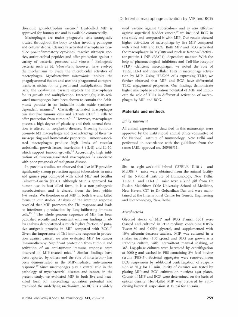
significant increase in IL-10 production by MIP-K-, MIP-
L- and BCG-stimulated macrophages compared with
unstimulated macrophages (data not shown). This indi-
cated that IL-10 has no role in the differential activation
of macrophages by MIP and BCG. It was further con-
firmed with the help of IL-10-deficient macrophages.
MIP-K, MIP-L and BCG led to similar responses from
wild-type and IL-10-deficient macrophages, as indicated
by TNF-a and IL-12p40 production (Fig. 3a,b). This
ruled out involvement of IL-10 differential activation of
macrophages by MIP and BCG.
Both MIP and BCG activated the macrophagesthrough MyD88- and NF-jB/AP-1-dependentpathway
Toll-like receptors are evolutionarily conserved pattern
recognition receptors playing major roles in the recogni-
tion of microbes and their products by immune cells.
MyD88 adapter protein is associated with cytoplasmic tail
of TLRs and passes information inside cells.25,26 To exam-
ine the role of MyD88 in macrophage activation, wild-
type and MyD88-deficient macrophages were stimulated
with MIP-K, MIP-L and BCG and culture supernatants
were analysed for pro-inflammatory cytokines. Loss of
response to MIP and BCG as indicated by drastically
reduced TNF-a and IL-12p40 production, was observed
in MyD88-deficient macrophages (Fig. 4a,b). This showed
that both MIP and BCG activate the macrophages in a
MyD88-dependent manner.
Signalling through MyD88 culminates in the transloca-
tion of NF-jB and AP-1 into nucleus and subsequent
transcription of inducible genes including those encoding
for pro-inflammatory cytokines.26 With the help of NF-
jB/AP-1-inducible SEAP-expressing Raw-Blue cells, we
noted significantly high NF-jB/AP-1 activation in
response to MIP-L (Fig. 4c). Dose-dependent increase in
SEAP level was induced by both MIP and BCG. The role
of NF-jB/AP-1 in MIP- and BCG-mediated macrophage
activation was further confirmed by stimulating RAW
264.7 macrophages in the presence of curcumin – an
inhibitor of NF-jB/AP-1.27 Reduced production of TNF-
a in response to MIP and BCG was observed in the pres-
ence of curcumin (Fig. 4d). These results showed that
both MIP and BCG activate the macrophage through an
MyD88- and NF-jB/AP-1-dependent pathway.
TLR2, TLR4 and intracellular TLRs have distinct rolesin MIP-mediated macrophage activation
Loss of response in MyD88-deficient macrophages indi-
cated the major role of TLRs in macrophage activation by
MIP. Contribution of different TLRs in MIP-mediated
macrophage activation was therefore further examined.
RAW 264.7 macrophages were stimulated with MIP and
BCG in the presence of pharmacological inhibitors of
TLR4 (Cli095), TLR2 and TLR4 (OxPAPC) and intracel-
lular TLRs (chloroquine)28 and TNF-a level was exam-
ined in the culture supernatants. Diminished TNF-aproduction by MIP-K-, MIP-L- and BCG-stimulated
macrophages was noted in the presence of Cli095
(Fig. 5a). This indicated the role of TLR4 in MIP- and
BCG-mediated macrophage activation. Decreased TNF-aproduction in response to MIP-L was also observed in
the presence of chloroquine, indicating the role of intra-
cellular TLRs in macrophage activation by MIP-L
(Fig. 5b). However, a dramatic reduction in the TNF-aproduction by MIP-K-, MIP-L- and BCG-stimulated
macrophage was observed in the presence of OxPAPC,
which indicated the major role of TLR2 in MIP-mediated
and BCG-mediated macrophage activation (Fig. 5c).
The role of TLRs in macrophage activation by MIP was
further confirmed with the help of macrophages isolated
from TLR2�/� and TLR4�/� mice. Consistent with the
5000
4000
3000
2000
1000
0
4000
3000
2000
400
300
200
100
IL-1
2p40
(pg
/ml)
0
Con MIP-K MIP-L
Wild Type
IL-10 KO
Wild Type
IL-10 KO
ns
(a)
(b) ns
ns
ns
ns
ns
BCG
Con MIP-K MIP-L BCG
TN
F-α
(pg
/ml)
Figure 3. Response of interleukin-10 deficient (IL-10�/�) and
wild-type macrophages to heat-killed Mycobacterium indicus pranii
(MIP-K), live Mycobacterium indicus pranii (MIP-L) and bacillus
Calmette–Gu�erin (BCG). Peritoneal macrophages from IL-10�/� and
wild-type mice were stimulated with MIP-K, MIP-L and BCG at a
multiplicity of stimulation of 10. A similar response in terms of
tumour necrosis factor-a (TNF-a) and IL-12-p40 production was
observed in IL-10�/� and wild-type macrophages. Data shown are
mean � SEM of three independent experiments. ns, not significant.
ª 2014 John Wiley & Sons Ltd, Immunology, 143, 258–268 263
Differential macrophage activation by MIP and BCG

Con MIP-K MIP-L BCG
OD
(65
0 nm
)
(c) 5x10x20x
0·6
0·4
0·2
0·0
***
***
5000
4000
3000
2000
1000
0Con MIP-K MIP-L BCG
(a)MyD88 KO
Wild Type
TN
F-α
(pg
/ml)
***
*
Con MIP-K MIP-L BCG
(b)MyD88 KO
Wild Type
***4000
30002000400
300
200
100
0
IL-1
2p40
(pg
/ml)
Con MIP-K MIP-L BCG
ns
(d)
TN
F-α
(pg
/ml)
**
*
7500
6000
4500
3000
1500
0
0 uM
5 uM
20 uM
Figure 4. Role of MyD88 and nuclear factor-jB/activator protein 1 ((NF-jB/AP-1) in heat-killed Mycobacterium indicus pranii (MIP-K), live
Mycobacterium indicus pranii (MIP-L) and bacillus Calmette–Gu�erin (BCG) mediated activation of macrophages. Peritoneal macrophages from
MyD88�/� and wild-type mice were stimulated with MIP-K, MIP-L and BCG. Response to MIP and BCG was lost in MyD88�/� macrophages,
as shown by drastically reduced levels of tumour necrosis factor-a (TNF-a) and interleukin-12 p40 (IL-12p40) in culture supernatants (a,b). Role
of NF-jB/AP-1 in macrophage activation by MIP or BCG was determined by stimulating RAW-Blue cells, which express NF-jB/AP-1 inducible
secreted alkaline phosphatase (SEAP). Higher levels of SEAP activity were observed in response to MIP-L (c). The role of NF-jB/AP-1 in MIP-
mediated macrophage activation was determined with the help of pharmacological inhibitor. RAW 264.7 macrophages were incubated with curc-
umin at the indicated concentration for 30 min and then stimulated with MIP-K, MIP-L and BCG. Dose-dependent decrease in TNF-a produc-
tion in response to MIP-K, MIP-L and BCG was observed in the presence of curcumin (d). Data shown are mean � SEM of three independent
experiments. *P < 0�05, **P < 0�01, ***P < 0�001 and ns, not significant.
(a) 6000
4500
3000
1500
0Con LPS MIP-K MIP-L BCG
MediumCli 095
TN
F-α
(pg
/ml)
(b)
Con CpG MIP-K MIP-L BCG
6000
4500
3000
1500
0
MediumChloroquine
TN
F-α
(pg
/ml)
(c)
Con Pam LPS MIP-K MIP-L BCG
6000
4500
3000
1500
0
MediumOxPAPC
TN
F-α
(pg
/ml)
***
***
Figure 5. Role of Toll-like receptors (TLRs) in macrophage activation by heat-killed Mycobacterium indicus pranii (MIP-K), live Mycobacterium
indicus pranii (MIP-L) and bacillus Calmette–Gu�erin (BCG). RAW 264.7 macrophages were incubated with 1�5 lg/ml Cli095 (TLR4 inhibitor),
80 lg/ml OxPAPC (TLR2 and TLR4 inhibitor) or 20 lM chloroquine (intracellular TLR inhibitor) for 60 min. MIP-K, MIP-L and BCG were
subsequently added at multiplicities of infection of 10 and culture supernatants were analysed for tumour necrosis factor-a (TNF-a) after 24 hr.
Diminished levels of TNF-a production by MIP-K, MIP-L and BCG stimulated macropahges were observed in the presence of Cli095 (a).
Decreased TNF-a production by MIP-L-stimulated macrophages was also observed in the presence of chloroquine (b). Dramatic reduction
in TNF-a production by MIP-K, MIP-L and BCG-stimulated macrophages was observed in the presence of OxPAPC (c). Pam-Pam3CSK4,
LPS-lipopolysachharide, CpG-CpG DNA were used as positive controls. Data shown are mean � SEM of three independent experiments.
***P < 0�001 and ns, not significant.
ª 2014 John Wiley & Sons Ltd, Immunology, 143, 258–268264
P. Kumar et al.

results obtained with pharmacological inhibitors,
decreased production of TNF-a and IL-12p40 in response
to MIP and BCG was observed in TLR4-deficient macro-
phages. A dramatic reduction in TNF-a and IL-12p40
production in response to MIP and BCG stimulation,
however, was observed in TLR2-deficient macrophages
(Fig. 6a,b). These results confirmed the major role of
TLR2 in macrophage activation by MIP and BCG.
MIP-K, MIP-L and BCG have differing TLR2 agonistproperty
Toll-like receptor 2 can recognize a wide array of micro-
bial molecules, which include peptidoglycans, lipoteichoic
acid, lipoarabinomannan, zymosan and lipoproteins. The
remarkable ability of TLR2 to function as a homodimer
or as heterodimer with TLR1 and TLR6 is responsible for
the recognition of a wide spectrum of microbial mole-
cules by TLR2.29 Since the above studies showed the
major role of TLR2 in macrophage activation by MIP
and BCG, we further delineated the TLR2 engagement by
MIP and BCG. A well-established HEK293-TLR system
was used to examine this.30 HEK293 cells expressing mur-
ine TLR2/2 homodimer (293tlr2/2 cells), TLR2/1 hetero-
dimer (293tlr2/1 cells) and TLR2/6 heterodimer (293tlr2/
6 cells) were stimulated with MIP-K, MIP-L or BCG in
native or sonicated form and culture supernatants were
analysed for IL-8. The sonicated form was used to expose
the potential TLR ligands, which might be hidden or
inaccessible in the native form of mycobacteria.
Interleukin-8 production by 293tlr2/2, 293tlr2/1 and
293tlr2/6 cells in response to native MIP-L indicated that
native MIP-L engages all three forms of TLR2, namely
TLR2/2, TLR2/1 and TLR2/6 (Fig. 7a,b,c, respectively). In
comparison to 293tlr2/2 cells and 293tlr2/6 cells, the
293tlr2/1 cells showed a high response to native MIP-L,
indicating a higher level of TLR2/1 ligands in native MIP-
L. Further, native MIP-L induced significantly higher lev-
els of IL-8 by 293tlr2/2, 293tlr2/1 and 293tlr2/6 cells in
comparison to native MIP-K and BCG, suggesting a
higher level of TLR2 ligands in native MIP-L. Compara-
ble levels of IL-8 were produced in response to native
MIP-L and sonicated MIP-L, indicating that TLR2 ligands
are well exposed in the native MIP-L and sonication does
not help to further increase the availability of TLR2
ligands. Native MIP-K showed decreased engagement of
TLR2/1, TLR2/6 and TLR2/2 compared with MIP-L. Sur-
prisingly, BCG, when used in native form, showed mini-
mal TLR2 agonist properties. Comparable levels of IL-8
were produced by unstimulated and native BCG-stimu-
lated 293tlr2/2, 293tlr2/1 and 293tlr2/6 cells. Interestingly,
however, when used in sonicated form, BCG engaged all
three form of TLR2. This showed that TLR2 ligands in
native BCG are not well exposed and sonication increases
the availability of ligands leading to substantial IL-8 pro-
duction. Increased IL-8 production was also observed
with sonicated MIP-K as compared with its native MIP-
K, suggesting that level of exposed TLR2 ligands decreases
due to autoclaving. Taken together, these results demon-
strate the different TLR2/2, TLR2/1, TLR2/6 agonist prop-
erty of MIP-L, MIP-K and BCG. In native form, MIP-L
has a higher level of TLR2 agonist activity followed by
MIP-K. BCG in native form is inefficient in engaging
TLR2 expressed in HEK 293 cells.
Discussion
Initially evaluated as an immune-therapeutic against lep-
rosy, MIP has generated a lot of interest because of its
strong immunomodulatory activity and the promising
results obtained against tuberculosis,9,31. In our previous
studies, where MIP was evaluated as a vaccine candidate
against tuberculosis, we found significantly higher protec-
tion in MIP-treated animals compared with those treated
with BCG.15 Strong Th1 response induced by MIP
7500
5000
9000
6000
3000
25002000
1500
1000
500
200
400
600
0
Wild TypeTLR2 KOTLR4 KO
Wild TypeTLR2 KOTLR4 KO
(a)
(b)
0
Con LPS MIP-K MIP-L BCG
Con Pam
Pam
LPS MIP-K MIP-L BCG
TN
F-α
(pg
/ml)
IL-1
2p40
(pg
/ml)
ns
ns***
***
ns
ns
nsns
Figure 6. Response of Toll-like receptor (TLR) -deficient macro-
phages to heat-killed Mycobacterium indicus pranii (MIP-K), live
Mycobacterium indicus pranii (MIP-L) and bacillus Calmette–Gu�erin
(BCG). Peritoneal macrophages from TLR2�/�, TLR4�/� and wild-
type mice were stimulated with MIP-K, MIP-L and BCG at multi-
plicities of infection of 10. Culture supernatants were analysed for
tumour necrosis factor-a (TNF-a) and interleukin-12 p40 (IL-12p40)
by ELISA. Diminished production of TNF-a and IL-12p40 in
response to MIP-K, MIP-L and BCG was observed in TLR4�/� mac-
rophages. Dramatic reduction in the TNF-a and IL-12p40 produc-
tion in response to MIP-K, MIP-L and BCG was observed in
TLR2�/� macrophages. Pam (Pam3CSK4, 10 ng/ml) and LPS (Lipo-
polysaccharide, 10 ng/ml) were used as positive controls. Data shown
are mean � SEM of two independent experiments. ***P < 0�001and ns, not significant.
ª 2014 John Wiley & Sons Ltd, Immunology, 143, 258–268 265
Differential macrophage activation by MIP and BCG

prompted us to evaluate it for cancer immunotherapy.
Encouraging results including lower tumour burden,
higher immune cell infiltration in tumour mass, and
higher T-cell and NK-cell cytotoxicity against tumour
cells were observed in MIP-treated mice.18 Macrophages
are important components of the mononuclear phagocy-
tic system, strategically located throughout the body to
ingest and process foreign materials and to recruit other
immune cells to the site of insult. M1 macrophages pro-
duce pro-inflammatory cytokines and reactive nitrogen
intermediates, and offer protection against infectious dis-
eases and neoplastic conditions. M2 macrophages on the
other hand have regenerative and tissue-repairing func-
tions and have been implicated in a variety of diseases.32
Our results show that a significantly higher level of pro-
inflammatory cytokines and nitric oxide were produced
from macrophages stimulated with MIP-L. MIP-K also
showed a higher response compared with BCG. Higher
activation of macrophages by MIP can explain the benefi-
cial effects of MIP seen against leprosy and tuberculosis.
Activated macrophages can potentially destroy the intra-
cellular pathogens and can process and present the anti-
gens to T cells leading to activation of the adaptive
immune response.
In addition to production of cytokines and presenta-
tion of processed antigens, expression of co-stimulatory
molecules is indispensible for activation of the adaptive
immune response.33 Co-stimulatory molecules, namely
CD40, CD80 and CD86, bind with their receptors and
provide the necessary signal for activation of adaptive
immune response. Our results show that MIP-L leads to
the highest up-regulation of CD40, CD80 and CD86 by
macrophages, followed by MIP-K. BCG induced lower
expression of these markers. In the absence of an optimal
cytokine milieu and co-stimulatory molecules, activation
of adaptive responses would be impaired and this may
partly explain the poor efficacy of BCG vaccine.
Toll-like receptors play an important role in the recog-
nition of lipoarabinomannans and various other micro-
bial molecules.34 Engagement of TLRs has a wide range
of effects, which include production of pro-inflammatory
cytokines and up-regulation of co-stimulatory molecules,
enhanced antigen presentation by antigen-presenting cells
and differentiation of monocytes into macrophages and
dendritic cells.35–37 MyD88 is an adapter molecule located
downstream to TLRs (except TLR3) and is involved in
the intracellular passage of the information.25 In the
absence of MyD88 adapter protein, both innate and adap-
tive immune responses are impaired. Our results demon-
strate the crucial role of MyD88 in macrophage activation
by MIP or BCG. Using the TLR signalling inhibitors, we
established the role of different TLRs in the activation of
macrophage by MIP. TLR2 had a prominent role in mac-
rophage activation by MIP or BCG, even though TLR4
400
800
600
200
0
ns
**
***
***
*
SonicatedNative
(a)
Con SPL BCGMIP-LMIP-KIL
-8 (
pg/m
l)
nsns
*
*
**
0Pam
2000
4000
6000
8000
10 000 SonicatedNative
(b)
Con BCGMIP-LMIP-K
IL-8
(pg
/ml)
ns***
***
****
FSL1
3000
2400
1800
1200
600
0
SonicatedNative
(c)
Con BCGMIP-LMIP-K
IL-8
(pg
/ml)
Figure 7. Delineation of Toll-like receptor (TLR2) signalling by heat-killed Mycobacterium indicus pranii (MIP-K), live Mycobacterium indicus
pranii (MIP-L) and bacillus Calmette–Gu�erin (BCG). HEK293 cells expressing TLR2/2, TLR2/1 or TLR2/6 (a, b and c, respectively) were stimu-
lated with MIP-K, MIP-L and BCG in native or sonicated form at a multiplicity of infection of 50. Culture supernatants were collected after
48 hr and analysed for interleukin-8 (IL-8) by ELISA. Higher levels of IL-8 production from 293tlr2/2, 293tlr2/1 and 293tlr2/6 cells were observed
in response to native and sonicated MIP-L. Maximum level of IL-8 was produced by 293tlr2/1 cells. Native MIP-K led to moderate levels of IL-8
from 293tlr2/2, 293tlr2/1 and 293tlr2/6 cells. Native BCG did not lead to IL-8 production from either of 293tlr2/2, 293tlr2/1 and 293tlr2/6 cells,
indicating that TLR2 ligands are not well exposed in native BCG. Substantial levels of IL-8 were produced by 293tlr2/2, 293tlr2/1 and 293tlr2/6
cells in response to sonicated BCG. SPL-Streptococcus pneumoniae lysate (TLR2/2 ligand, MOS = 50), Pam-Pam3CSK4 (TLR2/1 ligand, 0�2 lg/ml), FSL1 (TLR2/6 ligand, 1 lg/ml) were used as positive controls. Data shown are mean � SEM of three independent experiments. *P < 0�05,**P < 0�01, ***P < 0�001 and ns, not significant.
ª 2014 John Wiley & Sons Ltd, Immunology, 143, 258–268266
P. Kumar et al.

and intracellular TLRs were also important in macro-
phage activation. Response of TLR knockout macrophag-
es to MIP and BCG stimulation further confirmed the
results obtained with TLR signalling inhibitor and estab-
lished the major role of TLR2 in macrophage activation
by MIP and BCG.
Toll-like receptor 2 can recognize a variety of microbial
molecules, including peptidoglycans, lipoteichoic acid,
lipoarabinomannan, zymosan and lipoprotein. This
remarkable ability of TLR2 to recognize different types of
molecules is owing to its cooperation with other TLRs.
Accordingly, TLR2 may function as homodimer (TLR2/2)
or as heterodimer with TLR1 (TLR2/1) or TLR6 (TLR2/
6) and it increases the spectrum of potential TLR2
ligands.29 Given that TLR2 has a major role in activation
of macrophage by MIP and BCG, we further delineated
the TLR2 signalling by MIP and BCG. HEK293-express-
ing murine TLR2 alone or in combination with TLR1 or
TLR6 were used for this. In concurrence with superior
macrophage activation, MIP-L show higher levels of
TLR2/2, TLR2/1 and TLR2/6 agonistic activity compared
with MIP-K and BCG. Decreased ability of MIP-K to
engage TLR2 and to subsequently activate the macro-
phages can be explained because heat treatment will
denature the potent TLR ligands. Interestingly, despite
showing TLR2 engagement in macrophages, BCG did not
engage either of TLR2/2, TLR2/1 or TLR2/6 in HEK 293
cells. We speculated that TLR2 agonists are not in
exposed form in the BCG which resulted in to lack of
response in TLR2-expressing HEK 293 cells. To address
this, we used MIP and BCG in sonicated form. Interest-
ingly, when used in sonicated form, BCG led to a sub-
stantial response from TLR2-expressing HEK cells. Hence,
even though BCG has potent TLR2 ligands, they are not
in exposed form and cannot engage respective TLRs effi-
ciently. Hiding of putative TLR ligands can be an impor-
tant strategy of pathogenic mycobacteria to evade
immune-recognition. BCG also inhibits phagolysosomal
fusion, which would further prevent degradation of bacte-
rial component and subsequent release of putative ligands
including the ones which stimulate intracellular TLRs.
Clearly, MIP-L has distinctly higher levels of TLR2
ligands, which seems to be well exposed. BCG also has
putative TLR2 ligands but these ligands are not in
exposed form and therefore cannot engage the TLR2 effi-
ciently. Taken together, our findings establish the superior
activation of macrophages by MIP-L followed by MIP-K
compared with BCG and implicates the role of TLRs in
differential response of macrophages to MIP and BCG.
Acknowledgements
PK and RT performed the experiments; PK, GD and SB
designed the study; PK and SB prepared the manuscript.
This work was supported by the core research grant from
the National Institute of Immunology, New Delhi, India.
Disclosures
The authors declare no commercial or financial conflict
of interest.
References
1 Saxena VK, Singh US, Singh AK. Bacteriological study of a rapidly growing strain of
Mycobacterium. Lepr India 1978; 50:588–96.
2 Deo MG. Anti-leprosy vaccines – field trials and future prospects. Indian J Lepr 1984;
56:764–75.
3 Talwar GP, Zaheer SA, Mukherjee R et al. Immunotherapeutic effects of a vaccine
based on a saprophytic cultivable mycobacterium, Mycobacterium w in multibacillary
leprosy patients. Vaccine 1990; 8:121–9.
4 Katoch K, Katoch VM, Natrajan M et al. Treatment of bacilliferous BL/LL cases with
combined chemotherapy and immunotherapy. Int J Lepr Other Mycobact Dis 1995;
63:202–12.
5 Sharma P, Misra RS, Kar HK, Mukherjee A, Poricha D, Kaur H, Mukherjee R, Rani R.
Mycobacterium w vaccine, a useful adjuvant to multidrug therapy in multibacillary lep-
rosy: a report on hospital based immunotherapeutic clinical trials with a follow-up of
1–7 years after treatment. Lepr Rev 2000; 71:179–92.
6 Singh IG, Mukherjee R, Talwar GP. Resistance to intravenous inoculation of Mycobacte-
rium tuberculosis H37Rv in mice of different inbred strains following immunization
with a leprosy vaccine based on Mycobacterium w. Vaccine 1991; 9:10–4.
7 Yadava A, Mukherjee R. An immunodominant 30-kDa antigen of a candidate anti-
leprosy vaccine, Mycobacterium w, shares T and B cell determinants with M. leprae and
M. tuberculosis. Med Microbiol Immunol 1993; 182:243–53.
8 Saini V, Raghuvanshi S, Talwar GP, Ahmed N, Khurana JP, Hasnain SE, Tyagi AK.
Polyphasic taxonomic analysis establishes Mycobacterium indicus pranii as a distinct spe-
cies. PLoS ONE 2009; 4:e6263.
9 Purswani S, Talwar GP, Vohra R, Pal R, Panda AK, Lohiya NK, Gupta JC. Mycobacte-
rium indicus pranii is a potent immunomodulator for a recombinant vaccine against
human chorionic gonadotropin. J Reprod Immunol 2011; 91:24–30.
10 Murray PJ, Wynn TA. Protective and pathogenic functions of macrophage subsets. Nat
Rev Immunol 2011; 11:723–37.
11 Adhikari A, Majumder S, Banerjee S, Gupta G, Bhattacharya P, Majumdar SB, Saha B,
Majumdar S. Mycobacterium indicus pranii (Mw)-mediated protection against visceral
leishmaniasis: involvement of TLR4 signalling. J Antimicrob Chemother 2012; 67:
2892–902.
12 Taniguchi H, Shimada Y, Sawachi K et al. Lipopolysaccharide-activated alveolar macro-
phages having cytotoxicity toward lung tumor cells through cell-to-cell binding-depen-
dent mechanism. Anticancer Res 2010; 30:3159–65.
13 Tseng D, Volkmer JP, Willingham SB et al. Anti-CD47 antibody-mediated phagocytosis
of cancer by macrophages primes an effective antitumor T-cell response. Proc Natl Acad
Sci U S A 2013; 110:11103–8.
14 Mantovani A, Schioppa T, Porta C, Allavena P, Sica A. Role of tumor-associated mac-
rophages in tumor progression and invasion. Cancer Metastasis Rev 2006; 25:315–22.
15 Gupta A, Geetha N, Mani J, Upadhyay P, Katoch VM, Natrajan M, Katoch VM,
Bhaskar S. Immunogenicity and protective efficacy of “Mycobacterium w” against
Mycobacterium tuberculosis in mice immunized with live versus heat-killed M. w by the
aerosol or parenteral route. Infect Immun 2009; 77:223–31.
16 Gupta A, Ahmad FJ, Ahmad F, Gupta UD, Natarajan M, Katoch VM, Bhaskar S.
Protective efficacy of Mycobacterium indicus pranii against tuberculosis and underlying
local lung immune responses in guinea pig model. Vaccine 2012; 30:6198–209.
17 Saini V, Raghuvanshi S, Khurana JP, Ahmed N, Hasnain SE, Tyagi AK. Massive gene
acquisitions in Mycobacterium indicus pranii provide a perspective on mycobacterial
evolution. Nucleic Acids Res 2012; 40:10832–50.
18 Ahmad F, Mani J, Kumar P, Haridas S, Upadhyay P, Bhaskar S. Activation of anti-
tumor immune response and reduction of regulatory T cells with Mycobacterium indicus
pranii (MIP) therapy in tumor bearing mice. PLoS ONE 2011; 6:e25424.
19 Rakshit S, Ponnusamy M, Papanna S, Saha B, Ahmed A, Nandi D. Immunotherapeutic
efficacy of Mycobacterium indicus pranii in eliciting anti-tumor T cell responses: critical
roles of IFNc. Int J Cancer 2012; 130:865–75.
20 Alexandroff AB, Jackson AM, O’Donnell MA, James K. BCG immunotherapy of blad-
der cancer: 20 years on. Lancet 1999; 353:1689–94.
ª 2014 John Wiley & Sons Ltd, Immunology, 143, 258–268 267
Differential macrophage activation by MIP and BCG

21 Raschke WC, Baird S, Ralph P, Nakoinz I. Functional macrophage cell lines trans-
formed by Abelson leukemia virus. Cell 1978; 15:261–7.
22 Medzhitov R, Janeway CA Jr. Innate immunity: impact on the adaptive immune
response. Curr Opin Immunol 1997; 9:4–9.
23 Demangel C, Bertolino P, Britton WJ. Autocrine IL-10 impairs dendritic cell (DC)-
derived immune responses to mycobacterial infection by suppressing DC trafficking to
draining lymph nodes and local IL-12 production. Eur J Immunol 2002; 32:994–1002.
24 Geijtenbeek TB, Van Vliet SJ, Koppel EA, Sanchez-Hernandez M, Vandenbroucke-
Grauls CM, Appelmelk B, Van Kooyk Y. Mycobacteria target DC-SIGN to suppress
dendritic cell function. J Exp Med 2003; 197:7–17.
25 Akira S, Takeda K. Toll-like receptor signalling. Nat Rev Immunol 2004; 4:499–511.
26 Janssens S, Beyaert R. A universal role for MyD88 in TLR/IL-1R-mediated signaling.
Trends Biochem Sci 2002; 27:474–82.
27 Han SS, Keum YS, Seo HJ, Surh YJ. Curcumin suppresses activation of NF-jB and
AP-1 induced by phorbol ester in cultured human promyelocytic leukemia cells.
J Biochem Mol Biol 2002; 35:337–42.
28 Forgione N, Tropepe V. Toll-like signaling and the cytokine IL-6 regulate histone
deacetylase dependent neuronal survival. PLoS ONE 2012; 7:e41033.
29 Hajjar AM, O’Mahony DS, Ozinsky A, Underhill DM, Aderem A, Klebanoff SJ, Wilson
CB. Cutting edge: functional interactions between toll-like receptor (TLR) 2 and TLR1
or TLR6 in response to phenol-soluble modulin. J Immunol 2001; 166:15–9.
30 Huang LY, Dumontelle JL, Zolodz M, Deora A, Mozier NM, Golding B. Use of toll-like
receptor assays to detect and identify microbial contaminants in biological products.
J Clin Microbiol 2009; 47:3427–34.
31 Faujdar J, Gupta P, Natrajan M, Das R, Chauhan DS, Katoch VM, Gupta UD. Myco-
bacterium indicus pranii as stand-alone or adjunct immunotherapeutic in treatment of
experimental animal tuberculosis. Indian J Med Res 2011; 134:696–703.
32 Mills CD. M1 and M2 macrophages: oracles of health and disease. Crit Rev Immunol
2012; 32:463–88.
33 Vyas JM, Van der Veen AG, Ploegh HL. The known unknowns of antigen processing
and presentation. Nat Rev Immunol 2008; 8:607–18.
34 Takeda K, Akira S. Toll-like receptors in innate immunity. Int Immunol 2005; 17:1–14.
35 Datta SK, Raz E. Induction of antigen cross-presentation by Toll-like receptors. Springer
Semin Immunopathol 2005; 26:247–55.
36 Krutzik SR, Tan B, Li H et al. TLR activation triggers the rapid differentiation of
monocytes into macrophages and dendritic cells. Nat Med 2005; 11:653–60.
37 Kawai T, Akira S. The role of pattern-recognition receptors in innate immunity: update
on Toll-like receptors. Nat Immunol 2010; 11:373–84.
ª 2014 John Wiley & Sons Ltd, Immunology, 143, 258–268268
P. Kumar et al.