Mitochondria and calcium flux as targets of neuroprotection caused by minocycline in cerebellar...
12
Author's personal copy Mitochondria and calcium flux as targets of neuroprotection caused by minocycline in cerebellar granule cells Eva Maria Garcia-Martinez a,b , Sara Sanz-Blasco c , Andonis Karachitos d , Manuel J. Bandez e , Francisco J. Fernandez-Gomez a , Sergio Perez-Alvarez a , Raquel Maria Melero Fernandez de Mera a , Maria J. Jordan f , Norberto Aguirre g , Maria F. Galindo h , Carlos Villalobos c , Ana Navarro e , Hanna Kmita d , Joaquı ´n Jorda ´n a,i, * a Neurofarmacologı´a, Dpto Ciencias Me ´dicas, Facultad de Medicina, UCLM, Albacete, Spain b Servicio de Farmacia Hospitalaria, Complejo Hospitalario Universitario de Albacete, Albacete, Spain c Instituto de Biologı´a y Gene ´tica Molecular (IBGM), University Valladolid-CSIC, Valladolid, Spain d Laboratory of Bioenergetics, Institute of Molecular Biology and Biotechnology, Faculty of Biology, Adam Mickiewicz University, 61-614 Poznan, Umultowska 89, Poland e Bioquı´mica y Biologı´a Molecular, Facultad de Medicina, University Ca ´diz, Ca ´diz, Spain f Instituto Murciano de Investigacio ´n y Desarrollo Agrario y Alimentario, La Alberca, Murcia, Spain g Farmacologı´a, Facultad de Medicina, University of Navarra, Pamplona, Spain h Unidad Pfizer-Castilla-La Mancha de Neuropsicofarmacologı´a Translacional, Complejo Hospitalario Universitario de Albacete, Albacete, Spain i Centro Regional de Investigaciones Biome ´dicas, Universidad de Castilla-La Mancha (UCLM), Albacete, Spain Biochemical Pharmacology 79 (2010) 239–250 ARTICLE INFO Article history: Received 4 June 2009 Accepted 29 July 2009 Keywords: Anti-oxidant Apoptosis Voltage-dependent anion channel Uncoupler Aequorin Bioluminescence ABSTRACT Minocycline, an antibiotic of the tetracycline family, has attracted considerable interest for its theoretical therapeutic applications in neurodegenerative diseases. However, the mechanism of action underlying its effect remains elusive. Here we have studied the effect of minocycline under excitotoxic conditions. Fluorescence and bioluminescence imaging studies in rat cerebellar granular neuron cultures using fura2/AM and mitochondria-targeted aequorin revealed that minocycline, at concentrations higher than those shown to block inflammation and inflammation-induced neuronal death, inhibited NMDA- induced cytosolic and mitochondrial rises in Ca 2+ concentrations in a reversible manner. Moreover, minocycline added in the course of NMDA stimulation decreased Ca 2+ intracellular levels, but not when induced by depolarization with a high K + medium. We also found that minocycline, at the same concentrations, partially depolarized mitochondria by about 5–30 mV, prevented mitochondrial Ca 2+ uptake under conditions of environmental stress, and abrogated NMDA-induced reactive oxygen species (ROS) formation. Consistently, minocycline also abrogates the rise in ROS induced by 75 mM Ca 2+ in isolated brain mitochondria. In search for the mechanism of mitochondrial depolarization, we found that minocycline markedly inhibited state 3 respiration of rat brain mitochondria, although distinctly increased oxygen uptake in state 4. Minocycline inhibited NADH–cytochrome c reductase and cytochrome c oxidase activities, whereas the activity of succinate–cytochrome c reductase was not modified, suggesting selective inhibition of complexes I and IV. Finally, minocycline affected activity of voltage-dependent anion channel (VDAC) as determined in the reconstituted system. Taken together, our results indicate that mitochondria are a critical factor in minocycline-mediated neuroprotection. ß 2009 Elsevier Inc. All rights reserved. Abbreviations: DPPH , 2,2-diphenyl-1-picrylhydrazyl; TPTZ, 2,4,6-tripyridyl-S-triazine; CM-H 2 DCFDA, 5-(and 6-)-chloromethyl-2,7-dichlorodihydrofluorescein diacetate; ANT, adenine nucleotide translocase; ALS, amyotrophic lateral sclerosis; FCCP, carbonylcyanide-4-(trifluoromethoxy)-phenylhydrazone; FRAP, ferric reducing ability of plasma; [Ca 2+ ] cyt , cytosolic Ca 2+ concentrations; DFCA, fluorescein diacetate; GFP, green fluorescence protein; Het, hydroethidine; [Ca 2+ ] m , mitochondrial Ca 2+ concentrations; DCm, mitochondrial inner membrane potential; MPTP, mitochondrial permeability transition pore; NMDA, N-methyl-D-aspartic acid; ROI, region of interest; ROS, reactive oxygen species; O 2 , superoxide anion radical; TMRM, tetramethylrhodamine methyl ester; VDAC, voltage-dependent anion channel. * Corresponding author at: Grupo de Neurofarmacologı ´a, Departamento de Ciencias Me ´ dicas, Universidad Castilla-La Mancha-Centro Regional de Investigaciones Biome ´ dicas, Avda Almansa 14, Albacete 02006, Spain. Tel.: +34 967 599200; fax: +34 967599327. E-mail address: [email protected] (J. Jorda ´ n). Contents lists available at ScienceDirect Biochemical Pharmacology journal homepage: www.elsevier.com/locate/biochempharm 0006-2952/$ – see front matter ß 2009 Elsevier Inc. All rights reserved. doi:10.1016/j.bcp.2009.07.028
-
Upload
independent -
Category
Documents
-
view
1 -
download
0
Transcript of Mitochondria and calcium flux as targets of neuroprotection caused by minocycline in cerebellar...

Author's personal copy
Mitochondria and calcium flux as targets of neuroprotection caused byminocycline in cerebellar granule cells
Eva Maria Garcia-Martinez a,b, Sara Sanz-Blasco c, Andonis Karachitos d, Manuel J. Bandez e,Francisco J. Fernandez-Gomez a, Sergio Perez-Alvarez a, Raquel Maria Melero Fernandez de Mera a,Maria J. Jordan f, Norberto Aguirre g, Maria F. Galindo h, Carlos Villalobos c, Ana Navarro e,Hanna Kmita d, Joaquın Jordan a,i,*a Neurofarmacologıa, Dpto Ciencias Medicas, Facultad de Medicina, UCLM, Albacete, Spainb Servicio de Farmacia Hospitalaria, Complejo Hospitalario Universitario de Albacete, Albacete, Spainc Instituto de Biologıa y Genetica Molecular (IBGM), University Valladolid-CSIC, Valladolid, Spaind Laboratory of Bioenergetics, Institute of Molecular Biology and Biotechnology, Faculty of Biology, Adam Mickiewicz University, 61-614 Poznan, Umultowska 89, Polande Bioquımica y Biologıa Molecular, Facultad de Medicina, University Cadiz, Cadiz, Spainf Instituto Murciano de Investigacion y Desarrollo Agrario y Alimentario, La Alberca, Murcia, Spaing Farmacologıa, Facultad de Medicina, University of Navarra, Pamplona, Spainh Unidad Pfizer-Castilla-La Mancha de Neuropsicofarmacologıa Translacional, Complejo Hospitalario Universitario de Albacete, Albacete, Spaini Centro Regional de Investigaciones Biomedicas, Universidad de Castilla-La Mancha (UCLM), Albacete, Spain
Biochemical Pharmacology 79 (2010) 239–250
A R T I C L E I N F O
Article history:
Received 4 June 2009
Accepted 29 July 2009
Keywords:
Anti-oxidant
Apoptosis
Voltage-dependent anion channel
Uncoupler
Aequorin
Bioluminescence
A B S T R A C T
Minocycline, an antibiotic of the tetracycline family, has attracted considerable interest for its theoretical
therapeutic applications in neurodegenerative diseases. However, the mechanism of action underlying
its effect remains elusive. Here we have studied the effect of minocycline under excitotoxic conditions.
Fluorescence and bioluminescence imaging studies in rat cerebellar granular neuron cultures using
fura2/AM and mitochondria-targeted aequorin revealed that minocycline, at concentrations higher than
those shown to block inflammation and inflammation-induced neuronal death, inhibited NMDA-
induced cytosolic and mitochondrial rises in Ca2+ concentrations in a reversible manner. Moreover,
minocycline added in the course of NMDA stimulation decreased Ca2+ intracellular levels, but not when
induced by depolarization with a high K+ medium. We also found that minocycline, at the same
concentrations, partially depolarized mitochondria by about 5–30 mV, prevented mitochondrial Ca2+
uptake under conditions of environmental stress, and abrogated NMDA-induced reactive oxygen species
(ROS) formation. Consistently, minocycline also abrogates the rise in ROS induced by 75 mM Ca2+ in
isolated brain mitochondria. In search for the mechanism of mitochondrial depolarization, we found that
minocycline markedly inhibited state 3 respiration of rat brain mitochondria, although distinctly
increased oxygen uptake in state 4. Minocycline inhibited NADH–cytochrome c reductase and
cytochrome c oxidase activities, whereas the activity of succinate–cytochrome c reductase was not
modified, suggesting selective inhibition of complexes I and IV. Finally, minocycline affected activity of
voltage-dependent anion channel (VDAC) as determined in the reconstituted system. Taken together,
our results indicate that mitochondria are a critical factor in minocycline-mediated neuroprotection.
� 2009 Elsevier Inc. All rights reserved.
Abbreviations: DPPH�, 2,2-diphenyl-1-picrylhydrazyl; TPTZ, 2,4,6-tripyridyl-S-triazine; CM-H2DCFDA, 5-(and 6-)-chloromethyl-2,7-dichlorodihydrofluorescein diacetate;
ANT, adenine nucleotide translocase; ALS, amyotrophic lateral sclerosis; FCCP, carbonylcyanide-4-(trifluoromethoxy)-phenylhydrazone; FRAP, ferric reducing ability of
plasma; [Ca2+]cyt, cytosolic Ca2+ concentrations; DFCA, fluorescein diacetate; GFP, green fluorescence protein; Het, hydroethidine; [Ca2+]m, mitochondrial Ca2+
concentrations; DCm, mitochondrial inner membrane potential; MPTP, mitochondrial permeability transition pore; NMDA, N-methyl-D-aspartic acid; ROI, region of interest;
ROS, reactive oxygen species; �O2�, superoxide anion radical; TMRM, tetramethylrhodamine methyl ester; VDAC, voltage-dependent anion channel.
* Corresponding author at: Grupo de Neurofarmacologıa, Departamento de Ciencias Medicas, Universidad Castilla-La Mancha-Centro Regional de Investigaciones
Biomedicas, Avda Almansa 14, Albacete 02006, Spain. Tel.: +34 967 599200; fax: +34 967599327.
E-mail address: [email protected] (J. Jordan).
Contents lists available at ScienceDirect
Biochemical Pharmacology
journal homepage: www.e lsev ier .com/ locate /b iochempharm
0006-2952/$ – see front matter � 2009 Elsevier Inc. All rights reserved.
doi:10.1016/j.bcp.2009.07.028

Author's personal copy
1. Introduction
Minocycline, a semi-synthetic derivate of tetracycline, is underscientific debate as a potential therapeutic agent in neurologicaldisease processes (for review see [1,2]). Support for this newapplication of a time-tested antibiotic comes from observationsthat minocycline readily crosses the blood–brain barrier to thegreatest extent of all of the tetracyclines and is well tolerated. Itaffords neuroprotection in experimental models of Parkinson’sdisease, Huntington’s disease, multiple sclerosis, amyotrophiclateral sclerosis (ALS), and acute inflammation after brain traumaor cerebral ischemia [1,3]. Although there are inconsistent reportsthat show detrimental effects in different models of neurodegen-eration [4,5], numerous studies have focused on the mechanismsunderlying minocycline-mediated cell protection. However, thepossible mechanisms described so far are poorly understood andneed to be clarified.
We and others have postulated that mitochondria could be thepharmacological target affected by minocycline to afford cyto-protection. Proton-translocation by components of the respiratorychain generates the mitochondrial inner membrane potential(DCm). This transmembrane potential can be used to phosphor-ylate ADP to ATP or for Ca2+ uptake into mitochondria [6]. Whenadded to isolated mitochondria, minocycline is able to decrease theDCm, possibly preventing mitochondrial permeability transitionpore (MPTP) opening and the subsequent release of cytochrome c.Furthermore, DCm is considered an important factor in themaintenance of mitochondrial homeostasis: both Ca2+ uptake [7]and production of reactive oxygen species (ROS) are attenuateddue to a decrease in DCm [8]. Thus, small changes in DCm maycontribute to the cytoprotection mechanisms. Mitochondrialdepolarization, caused by low concentrations of mitochondrialuncouplers, has been shown to be protective against NMDA-induced neuronal death [9].
Mounting evidence indicates that ROS-mediated stress plays amajor role in the pathogenesis of many neurodegenerativedisorders. Accordingly, it has been shown that excitotoxicity,which plays a critical role in several neurological pathologies,promotes significant increases in cytosolic Ca2+ concentrations([Ca2+]cyt), ROS production, oxidative damage, and ultimately celldeath [10,11]. In addition, mitochondrial Ca2+ overload plays apivotal role in many cell death models including excitotoxicity[11]. Specifically, it has been reported that glutamate-inducedneurotoxicity depends on mitochondrial Ca2+ uptake ([Ca2+]m)[9,12]. Under stress conditions, there is an early increase in [Ca2+]m
that may contribute to mitochondrial dysfunction [13,14] andMPTP opening, a point-of-no-return step in the intrinsic pathwayof apoptotic cell death [15,16].
Nowadays, the molecular composition of the MPTP is still underdebate. Numerous independent studies suggest that the MPTPconsists of a voltage-dependent anion channel (VDAC) within theouter membrane, and adenine nucleotide translocase (ANT) withinthe inner membrane. The mechanism of VDAC contribution toMPTP still remains elusive. The available data indicate that pro-apoptotic stimuli can trigger a state of high or low conductance ofVDAC. It has also been suggested that anti-apoptotic activity mayinvolve an inhibitory VDAC interaction with MPTP constituentswhich appears critical for mitochondrial metabolite transport,volume regulation, and apoptosis (for review see [17–19]). On theother hand, there is also evidence indicating that VDAC partici-pates in signal generation without direct involvement in sub-sequent processes (e.g. [20]).
Herein, we have designed several experiments to understand themechanisms responsible for minocycline-mediated neuroprotec-tion under excitotoxic conditions. After NMDA receptor activation,we analyzed minocycline’s effect on two of the earliest events which
can tip the scales in favour of neurodegeneration. Close attentionwas paid to the entry of Ca2+ through the plasma membrane,mitochondrial Ca2+ uptake, and generation of ROS. We also studieddifferent parameters describing mitochondrial functions. Ourresults suggest that minocycline, although at concentrations higherthan those shown to block inflammation and inflammation-inducedneuronal death [21], prevents NMDA-induced excitotoxicity bydiminishing NMDA-induced Ca2+ entry and altering the DCm andmitochondrial Ca2+ uptake. The latter may be caused by activitychanges in VDAC as well as in the respiratory chain.
2. Materials and methods
2.1. Chemicals
Fura2, hydroethidine, CM-H2DCFDA, TMRM and celenterazinwere obtained from Invitrogen (Barcelona, Spain). MitochondrialAEQ construct was a gift from Prof. Brulet (Paris, France). Culturemedia, sera and antibiotics are from Lonza. If not indicatedotherwise, all other chemicals were purchased from Sigma orMerck.
2.2. Animals
For cerebellar granule cell cultures, 5–7-day-old Wistar ratswere used. Brain mitochondria were isolated from 3-month-oldmale Wistar rats. All animals were housed at 22 � 2 8C with 12 h/12 h light/dark cycles and with full access to water and food. Animalexperiments were carried out in accordance with the GuidingPrinciples for Research Involving Animals and Human Beings of theAmerican Physiological Society, the Guidelines of the European UnionCouncil (86/609/CEE), and the Spanish regulations (BOE 67/8509-12,1988) for the use of laboratory animals. Approval was obtained fromthe Scientific Committee of the Universities of Cadiz, Castilla-LaMancha, and Valladolid.
2.3. Cell cultures
Cerebellar granule cells were obtained as reported previously[22], plated on poly-L-lysine coated, 12 mm diameter glasscoverslips. They were cultured in high-glucose, low K+, Dulbecco’smodified Eagle’s medium (DMEM, Gibco, Spain) plus 10% fetalbovine serum, 5% horse serum, 100 mg/ml penicillin, and 100 mg/ml streptomycin for 2 days. Then the culture medium was replacedby Sato’s medium plus 5% horse serum (to avoid excessiveproliferation of glia) and cultured for 2–4 days before experiments.
2.4. Cell viability studies
To study NMDA-induced toxicity, neuronal cultures maintained7 days in vitro were incubated for 30 min with 100 mM NMDA,2 mM Ca2+/Na+ and 10 mM glycine in saline solution. Treatmentswere terminated by washing cells three times with Krebs solutionbefore incubating with regular medium. Minocycline was added10 min before NMDA at different concentrations and maintainedduring the whole experiment. Cell viability was analyzed 24 h laterusing the fluorescein diacetate/propidium iodide double-stainingprocedure. Living and dead cells were counted in 3–4 coverslipsper condition (a total of 300–450 cells per coverslip) andnormalized to corresponding controls. At least three independentexperiments were used for quantification.
2.5. Fluorescence imaging of cytosolic [Ca2+]
Coverslips containing cerebellar granule cells were incubated instandard medium containing (in mM) NaCl 145, KCl 5, CaCl2 1,
E.M. Garcia-Martinez et al. / Biochemical Pharmacology 79 (2010) 239–250240

Author's personal copy
MgCl2 1, glucose 10, Hepes 10 (pH 7.42), and loaded with 4 mMfura2/AM for 60 min at room temperature. Coverslips were thenplaced on the heated stage of an inverted Zeiss microscope(Axiovert S100 TV), and while continuously perfused with thesame pre-warmed standard medium, epi-illuminated alternatelyat 340 and 380 nm. Light emitted above 520 nm was recorded withan OrcaER Hamamatsu camera (Hamamtsu Photonics, Hama-matsu, Japan). Pixel-by-pixel ratios of consecutive frames werecaptured, and [Ca2+]cyt was estimated from these ratios aspreviously reported [23].
2.6. Bioluminescence imaging of mitochondrial [Ca2+]
Cerebellar granule cells were transfected with plasmidscontaining GFP-aequorin targeted to mitochondria [24] using aNucleofector II1 device and the VPG-1003 transfection kit (AmaxaBiosystems, Cologne, Germany). After 24 h, cells were incubatedfor 1 h with 1 mM coelenterazine, washed, and placed into aperfusion chamber set at 37 8C under a Zeiss Axiovert S100 TVmicroscope. They were then perfused at 5–10 ml/min with testsolutions based on the standard perfusion solution pre-warmed to37 8C as described above. At the end of each experiment, cells werepermeabilized with 0.1 mM digitonin in 10 mM CaCl2 in order torelease all the residual aequorin counts. Bioluminescence imageswere taken with a Hamamatsu VIM photon counting camerahandled with an Argus-20 image processor. Photonic emissionswere integrated for 10 s periods. Photons/cell in each image wasquantified using the Hamamatsu Aquacosmos software. Totalcounts per region of interest (ROI) ranged between 2 � 103 and2 � 105, and noise was (mean � SD) 1 � 1 counts per second (c.p.s.)per typical cell area (2000 pixels). Data were first quantified as ratesof photoluminescence emission/total c.p.s. remaining at each time (%of remaining counts) and divided by the integration period (L/LTOTAL
in s�1). Emission values of less than 4 c.p.s. were not used forcalculations. Calibrations in terms of [Ca2+]mit were performed aspreviously reported [23]. Briefly, the total luminescence for everyROI was computed by adding up the values of all images. In addition,the following values were registered for every time point (t):L = luminescence emission at time t; sum (L) = SL values from t = 0 tot; LTOTAL (LT1) = total luminescence remaining at time t = total(L) � sum (L); % remaining luminescence = 100 � LTOTAL/total (L);and ratio (R) = L/LT1. Finally, [Ca2+] was calculated using thefollowing algorithm:
½Ca2þ� ðin MÞ ¼ ½Rþ ðR� KTRÞ þ 1�=½ðKR � ðR� KRÞ�;
where R = L/LT1 � l)](1/n), using the constant values KR, KTR, n and lreported previously (see [23] for a detailed description). In someexperiments, cells were permeabilized with 20 mM digitonin in‘‘intracellular’’ medium with the following composition: 130 mMKCl, 10 mM NaCl, 1 mM MgCl2, 1 mM K3PO4, 0.2 mM EGTA, 1 mMATP, 20 mM ADP, 2 mM succinate, 20 mM Hepes/KOH, pH 6.8. Cellswere then incubated for 5 min with the same medium containing200 nM Ca2+ (buffered with EGTA), with or without minocycline.Finally, perfusion was switched to ‘‘intracellular’’ medium contain-ing 5 mM Ca2+, with or without minocycline.
2.7. Mitochondrial potential
The effects of treatment on DC in intact cerebellar granulecells were estimated by fluorescence imaging of cells loaded withthe DC sensitive probe tetramethylrhodamine methyl ester(TMRM), one the most sensitive probes available [25]. Briefly,cerebellar granule cells were loaded with 10 nM TMRM for10 min, washed with a standard, external medium, and placed onthe perfusion chamber of a Zeiss Axiovert S100 TV invertedmicroscope. Cells were then continuously perfused with pre-
warmed (37 8C) external medium, with and without treatments.Fluorescence images were taken at 10 s intervals with aHamamatsu VIM photon counting camera handled with anArgus-20 image processor. Traces from individual cells werenormalized relative to the value before the addition of eithervehicle or treatment and averaged. Background fluorescenceafter collapse of the mitochondrial potential induced by 10 mMFCCP was subtracted. Further details have been reportedpreviously [26].
2.8. Superoxide intracellular production
Superoxide production was monitored using hydroethidine(HEt, Molecular Probes) as described previously [27]. Coverslipscontaining cerebellar granule cells were incubated in standardmedium containing (in mM): NaCl 145, KCl 5, CaCl2 1, MgCl2 1,glucose 10, and Hepes 10 (pH 7.42) and loaded with 10 mMhydroethidine. Next, coverslips were placed on the heated stage ofan inverted Nikon (Eclipse TE2000) microscope. Light emittedabove 535 nm was recorded with an emission filter of 635 nm(Omega Optical) with an OrcaER Hamamatsu camera (HamamatsuPhotonics, Hamamatsu, Japan). Background was subtracted.Frames were recorded every 10 s over a 7 min period. Linearregression of fluorescence data was obtained of each condition andthe slope of the best fitting line was taken as an index of superoxideanion radical (�O2
�) production.
2.9. Isolation of mitochondria and preparation of mitochondrial
membranes
Brain mitochondria were isolated from the whole rat brain in asmall Potter homogenizer with a Teflon pestle. Homogenizationwas carried out in a medium of 230 mM mannitol, 70 mM sucrose,1.0 mM EDTA, and 10 mM Tris–HCl, pH 7.40, at a ratio of 9 ml ofhomogenization medium/g of tissue. The homogenate wascentrifuged at 700 � g for 10 min and the supernatant at8000 � g for 10 min to precipitate mitochondria that were washedin the same conditions [28]. The obtained mitochondrial suspen-sions of about 20 mg protein/ml were used immediately afterisolation for the determination of respiration or frozen in liquid N2
and kept at �80 8C. Sub-mitochondrial membranes (disruptedmitochondria) were obtained from brain mitochondria that weretwice frozen-and-thawed and homogenized each time by passagethrough a tuberculin needle. The procedure yielded a preparationof disrupted mitochondria with 0.20–0.24 nmol cytochrome aa3/mg protein, subsequently used for the determination of enzymaticactivity of the respiratory complexes and the presence of markersof oxidative damage. The protein content of the samples wasdetermined using Folin reagent and bovine serum albumin as thestandard. To study the effects of minocycline, the brain-coupledmitochondria or sub-mitochondrial membranes were added orincubated for 5 min at 4 8C, respectively, with 0, 75, 100, 125, or150 mM minocycline.
2.10. Mitochondrial oxygen uptake
The oxygen uptake of rat brain mitochondria (0.7–0.8 mg/ml)was determined with a Clark electrode in a 1.5 ml chamber at30 8C, in an air-saturated reaction medium consisting of: 0.23 Mmannitol, 0.07 M sucrose, 20 mM Tris–HCl, 1 mM EDTA, 5 mMphosphate, 4 mM MgCl2, at pH 7.4. Respiration rates weredetermined with 5 mM malate–5 mM glutamate or 6 mM succi-nate as substrates, and state 3 respiration was established byaddition of 0.5 mM ADP. Mitochondria oxygen uptake was assayedin uncoupled mitochondria with 1 mM FCCP. Three determinationswere performed for each value.
E.M. Garcia-Martinez et al. / Biochemical Pharmacology 79 (2010) 239–250 241

Author's personal copy
2.11. Measurement of ROS levels in mitochondrial suspension
Mitochondrial ROS production was evaluated with the fluor-escent probe 5-(and 6-)-chloromethyl-2,7-dichlorodihydrofluor-escein diacetate (CM-H2DCFDA) in isolated mitochondrialsuspension. CM-H2DCFDA is a reduced form of fluoresceindiacetate (DFCA) that requires oxidation before becoming fluor-escent. Therefore, any measurable change in fluorescence intensitycan be directly related to changes in free radical activity, mainlythe generation of hydrogen peroxide and hydroxyl radicals.Mitochondrial suspensions were incubated with CM-H2DCFDA(10 mg/ml), and fluorescence intensity was measured in a SpectraMax Gemini XS (Molecular Devices, Sunnyvale, CA). Brainmitochondria were challenged with 75 mM Ca2+ to induce ROSformation during 30 min with or without minocycline. Experi-ments were performed in 96-multiwell plates including foursamples for each experimental point. The average of relative DFCAfluorescence from at least three separate mitochondrial prepara-tions was determined. Results are expressed as mean � SEM values.
2.12. Mitochondrial electron transfer activity
The enzymatic activity of complexes I–III, II–III, and IV weredetermined spectrophotometrically at 30 8C with sub-mitochon-drial membranes suspended in 60 mM phosphate buffer (pH 7.4)with corresponding substrates added. For NADH–cytochrome c
reductase (the activity of complexes I and III) and succinate–cytochrome c reductase (the activity of complexes II and III),mitochondrial membranes were added with 0.20 mM NADH or5.0 mM succinate as substrates, 0.10 mM cytochrome c3+, and1.0 mM KCN. The enzymatic activity determined at 550 nm(e = 19 mM�1 cm�1), with the results expressed as nmol cyto-chrome c reduced/min �mg protein. Cytochrome oxidase (com-plex IV) was determined in the same buffer with 0.05 mMcytochrome c2+ added, prepared by reduction with ascorbate,and filtration across a Sephadex G25 column. The rate ofcytochrome c oxidation was calculated as the first order reactionconstant (k0) per mg protein, and expressed as nmol cytochrome c
oxidized at 10 mM cytochrome c/min �mg protein. This resultdetermines the electron transfer rates of physiological respiration[29]. Three determinations for each value.
2.13. DPPH radical-scavenging activity
The scavenging activity of minocycline was measured accord-ing to the method described by Brand-Williams et al. [30] withsome modifications. Briefly, 500 ml of minocycline methanolicsolutions (100 mM) at different concentrations (12.5–150 ml/ml)were added to 1 ml of 2,2-diphenyl-1-picrylhydrazyl (DPPH�)methanolic solution (0.1 mM). Estimated time of reaction (20 min)was determined considering the reduction of absorbance at517 nm (monitored every 5 min) until the reaction curve reacheda plateau. The absorbance was measured at room temperature, indarkness, with a blank (500 ml of sample plus 1 ml of methanol).The absorbance of the control (500 ml of methanol in 1 ml of DPPH�
solution) was measured daily. All of the assays were conducted intriplicate. The percentage activity for the DPPH� technique wascalculated according to % discoloration = [1 � (Abs sample/Abscontrol)] � 100.
2.14. Ferric reducing ability assay
The ferric reducing ability of minocycline was measuredaccording to the method developed by Benzie and Strain [31].Anti-oxidant compounds are able to reduce ferric compounds toferrous-TPTZ, which develops a blue color with a maximum
absorption at 593 nm. FRAP reagent was freshly prepared from300 mM acetate buffer (pH 3.6), 10 mM 2,4,6-tripyridyl-S-triazine(TPTZ) made up in 40 mM HCl, and 20 mM FeCl3�6H2O solution. Allthree solutions were mixed together in the ratio of 10:1:1 (v/v/v).An aliquot of 40 ml of minocycline was added to 1.2 ml of FRAPreagent. The absorption of the reaction mixture was measured at593 nm after 2 min of incubation at 37 8C. Measurements wereperformed in triplicate. Fresh working solutions of known Fe (II)concentrations (FeSO4�7H2O, 0–2 mM) were used for calibration.The anti-oxidant capacity, based upon the ability to reduce ferricions of each sample, was calculated from the linear calibrationcurve and expressed as mmol FeSO4 equivalents.
2.15. Isolation of VDAC from Saccharomyces cerevisiae mitochondria
The yeast Saccharomyces cerevisiae M3 cells (MATa, lys2, his4,trp1, ade2, leu2, ura3) [32] were grown at 28 8C in YPG medium (1%yeast extract, 2% peptone, 3% glycerol) at pH 5.5 to OD546 of 2.2.Mitochondria and the mitochondrial outer membrane wereisolated according to the published procedure [33]. The obtainedouter membrane pellet was suspended in the solubilization buffercontaining 3% Triton X-100, 10 mM Tris–HCl (pH 7.0) and 1 mMEGTA, and was incubated for 30 min at 0 8C. The suspension wasthen loaded onto a dry hydroxyapatite/celite column [34] and thefirst two fractions were collected. Before reconstitution, thepreparations of VDAC were checked by Western blotting withanti-yeast anti-serum for the presence of marker proteins ofmitochondrial compartments as well as for VDAC (not shown).
2.16. Conductance measurements in planar phospholipid membranes
Membranes were formed from soybean asolectin suspended atconcentration of 25 mg/ml in n-decane, across a circular hole(aperture diameter about 250 mm) in the thin wall of a Delrinchamber (Warner Instruments) separating two compartments(cis–trans) filled with unbuffered 1 M KCl, pH 7.0. The chamber wasconnected to the recording equipment through a matched pair ofAg–AgCl electrodes. Preparations of VDAC were added in smallaliquots (2–3 ml) to the cis compartment. Cis also refers to thecompartment where the voltage was held. Signals were amplifiedby a BLM-120 bilayer amplifier (Bio-LOGIC Science Instruments)and computer software was used for data collection. The averageamount of analyzed insertion events for a given set of conditionswas 70.
2.17. Statistics
When only two means were compared, Student’s t-test wasused. For more than two groups, statistical significance of the datawas assessed by analysis of the variance (ANOVA) and comparedusing Tukey’s test. Differences were considered significant atp < 0.05. For the conductance measurements in planar phospho-lipid membranes, the differences between calculated values ofaverage conductance were tested by t-test (at a = 0.01) forstatistical significance.
3. Results
3.1. Minocycline and cell viability
Consistent with previous data from our laboratory and else-where, minocycline afforded cytoprotection to neuronal cultureswhen exposed to excitotoxic conditions. Cerebellar granular cellswere treated with minocycline at different concentrations (1–150 mM) and challenged with NMDA (100 mM) during 30 min.Twenty-four hours later, neuronal viability was evaluated by using
E.M. Garcia-Martinez et al. / Biochemical Pharmacology 79 (2010) 239–250242
Author's personal copy
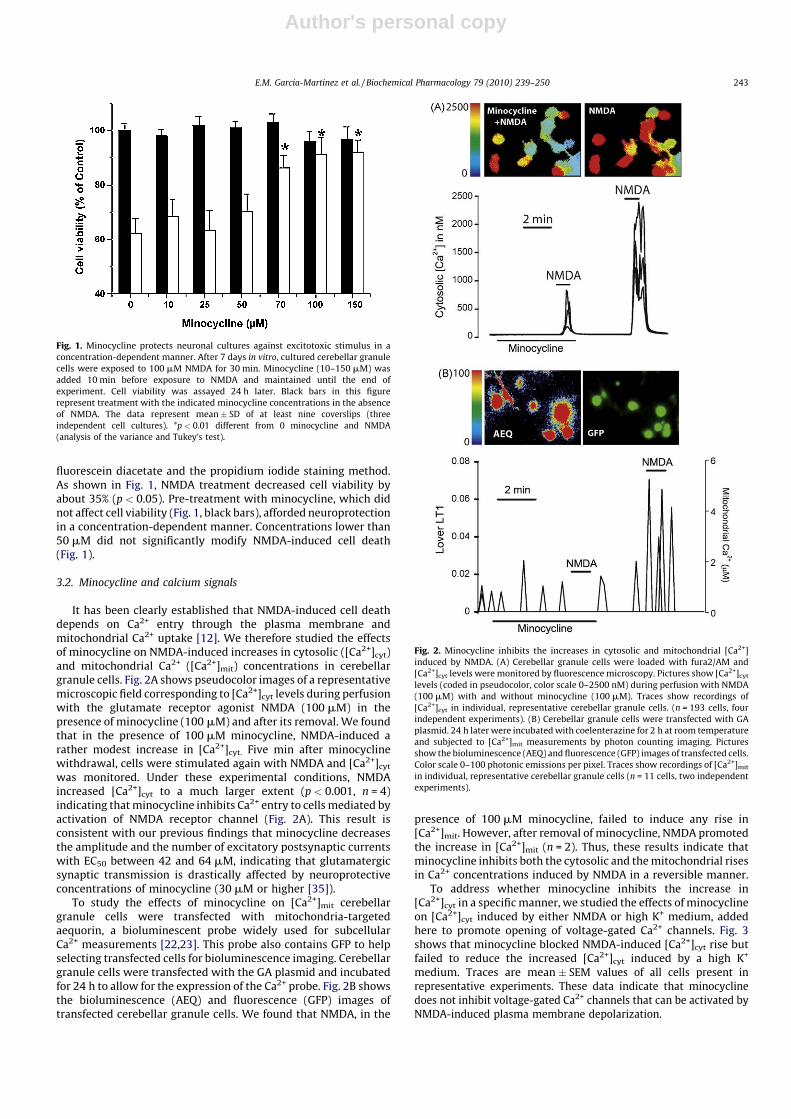
fluorescein diacetate and the propidium iodide staining method.As shown in Fig. 1, NMDA treatment decreased cell viability byabout 35% (p < 0.05). Pre-treatment with minocycline, which didnot affect cell viability (Fig. 1, black bars), afforded neuroprotectionin a concentration-dependent manner. Concentrations lower than50 mM did not significantly modify NMDA-induced cell death(Fig. 1).
3.2. Minocycline and calcium signals
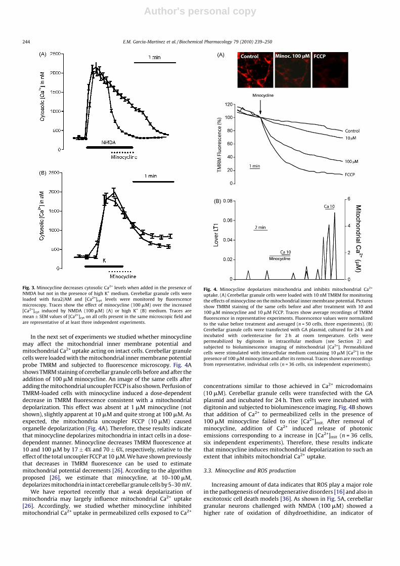
It has been clearly established that NMDA-induced cell deathdepends on Ca2+ entry through the plasma membrane andmitochondrial Ca2+ uptake [12]. We therefore studied the effectsof minocycline on NMDA-induced increases in cytosolic ([Ca2+]cyt)and mitochondrial Ca2+ ([Ca2+]mit) concentrations in cerebellargranule cells. Fig. 2A shows pseudocolor images of a representativemicroscopic field corresponding to [Ca2+]cyt levels during perfusionwith the glutamate receptor agonist NMDA (100 mM) in thepresence of minocycline (100 mM) and after its removal. We foundthat in the presence of 100 mM minocycline, NMDA-induced arather modest increase in [Ca2+]cyt. Five min after minocyclinewithdrawal, cells were stimulated again with NMDA and [Ca2+]cyt
was monitored. Under these experimental conditions, NMDAincreased [Ca2+]cyt to a much larger extent (p < 0.001, n = 4)indicating that minocycline inhibits Ca2+ entry to cells mediated byactivation of NMDA receptor channel (Fig. 2A). This result isconsistent with our previous findings that minocycline decreasesthe amplitude and the number of excitatory postsynaptic currentswith EC50 between 42 and 64 mM, indicating that glutamatergicsynaptic transmission is drastically affected by neuroprotectiveconcentrations of minocycline (30 mM or higher [35]).
To study the effects of minocycline on [Ca2+]mit cerebellargranule cells were transfected with mitochondria-targetedaequorin, a bioluminescent probe widely used for subcellularCa2+ measurements [22,23]. This probe also contains GFP to helpselecting transfected cells for bioluminescence imaging. Cerebellargranule cells were transfected with the GA plasmid and incubatedfor 24 h to allow for the expression of the Ca2+ probe. Fig. 2B showsthe bioluminescence (AEQ) and fluorescence (GFP) images oftransfected cerebellar granule cells. We found that NMDA, in the
presence of 100 mM minocycline, failed to induce any rise in[Ca2+]mit. However, after removal of minocycline, NMDA promotedthe increase in [Ca2+]mit (n = 2). Thus, these results indicate thatminocycline inhibits both the cytosolic and the mitochondrial risesin Ca2+ concentrations induced by NMDA in a reversible manner.
To address whether minocycline inhibits the increase in[Ca2+]cyt in a specific manner, we studied the effects of minocyclineon [Ca2+]cyt induced by either NMDA or high K+ medium, addedhere to promote opening of voltage-gated Ca2+ channels. Fig. 3shows that minocycline blocked NMDA-induced [Ca2+]cyt rise butfailed to reduce the increased [Ca2+]cyt induced by a high K+
medium. Traces are mean � SEM values of all cells present inrepresentative experiments. These data indicate that minocyclinedoes not inhibit voltage-gated Ca2+ channels that can be activated byNMDA-induced plasma membrane depolarization.
Fig. 2. Minocycline inhibits the increases in cytosolic and mitochondrial [Ca2+]
induced by NMDA. (A) Cerebellar granule cells were loaded with fura2/AM and
[Ca2+]cyt levels were monitored by fluorescence microscopy. Pictures show [Ca2+]cyt
levels (coded in pseudocolor, color scale 0–2500 nM) during perfusion with NMDA
(100 mM) with and without minocycline (100 mM). Traces show recordings of
[Ca2+]cyt in individual, representative cerebellar granule cells. (n = 193 cells, four
independent experiments). (B) Cerebellar granule cells were transfected with GA
plasmid. 24 h later were incubated with coelenterazine for 2 h at room temperature
and subjected to [Ca2+]mit measurements by photon counting imaging. Pictures
show the bioluminescence (AEQ) and fluorescence (GFP) images of transfected cells.
Color scale 0–100 photonic emissions per pixel. Traces show recordings of [Ca2+]mit
in individual, representative cerebellar granule cells (n = 11 cells, two independent
experiments).
Fig. 1. Minocycline protects neuronal cultures against excitotoxic stimulus in a
concentration-dependent manner. After 7 days in vitro, cultured cerebellar granule
cells were exposed to 100 mM NMDA for 30 min. Minocycline (10–150 mM) was
added 10 min before exposure to NMDA and maintained until the end of
experiment. Cell viability was assayed 24 h later. Black bars in this figure
represent treatment with the indicated minocycline concentrations in the absence
of NMDA. The data represent mean � SD of at least nine coverslips (three
independent cell cultures). *p < 0.01 different from 0 minocycline and NMDA
(analysis of the variance and Tukey’s test).
E.M. Garcia-Martinez et al. / Biochemical Pharmacology 79 (2010) 239–250 243
Author's personal copy
In the next set of experiments we studied whether minocyclinemay affect the mitochondrial inner membrane potential andmitochondrial Ca2+ uptake acting on intact cells. Cerebellar granulecells were loaded with the mitochondrial inner membrane potentialprobe TMRM and subjected to fluorescence microscopy. Fig. 4Ashows TMRM staining of cerebellar granule cells before and after theaddition of 100 mM minocycline. An image of the same cells afteradding the mitochondrial uncoupler FCCP is also shown. Perfusion ofTMRM-loaded cells with minocycline induced a dose-dependentdecrease in TMRM fluorescence consistent with a mitochondrialdepolarization. This effect was absent at 1 mM minocycline (notshown), slightly apparent at 10 mM and quite strong at 100 mM. Asexpected, the mitochondria uncoupler FCCP (10 mM) causedorganelle depolarization (Fig. 4A). Therefore, these results indicatethat minocycline depolarizes mitochondria in intact cells in a dose-dependent manner. Minocycline decreases TMRM fluorescence at10 and 100 mM by 17� 4% and 70� 6%, respectively, relative to theeffect of the total uncoupler FCCP at 10 mM. We have shown previouslythat decreases in TMRM fluorescence can be used to estimatemitochondrial potential decrements [26]. According to the algorithmproposed [26], we estimate that minocycline, at 10–100 mM,depolarizes mitochondria in intact cerebellar granule cells by 5–30 mV.
We have reported recently that a weak depolarization ofmitochondria may largely influence mitochondrial Ca2+ uptake[26]. Accordingly, we studied whether minocycline inhibitedmitochondrial Ca2+ uptake in permeabilized cells exposed to Ca2+
concentrations similar to those achieved in Ca2+ microdomains(10 mM). Cerebellar granule cells were transfected with the GAplasmid and incubated for 24 h. Then cells were incubated withdigitonin and subjected to bioluminescence imaging. Fig. 4B showsthat addition of Ca2+ to permeabilized cells in the presence of100 mM minocycline failed to rise [Ca2+]mit. After removal ofminocycline, addition of Ca2+ induced release of photonicemissions corresponding to a increase in [Ca2+]mit (n = 36 cells,six independent experiments). Therefore, these results indicatethat minocycline induces mitochondrial depolarization to such anextent that inhibits mitochondrial Ca2+ uptake.
3.3. Minocycline and ROS production
Increasing amount of data indicates that ROS play a major rolein the pathogenesis of neurodegenerative disorders [16] and also inexcitotoxic cell death models [36]. As shown in Fig. 5A, cerebellargranular neurons challenged with NMDA (100 mM) showed ahigher rate of oxidation of dihydroethidine, an indicator of
Fig. 4. Minocycline depolarizes mitochondria and inhibits mitochondrial Ca2+
uptake. (A) Cerebellar granule cells were loaded with 10 nM TMRM for monitoring
the effects of minocycline on the mitochondrial inner membrane potential. Pictures
show TMRM staining of the same cells before and after treatment with 10 and
100 mM minocycline and 10 mM FCCP. Traces show average recordings of TMRM
fluorescence in representative experiments. Fluorescence values were normalized
to the value before treatment and averaged (n = 50 cells, three experiments). (B)
Cerebellar granule cells were transfected with GA plasmid, cultured for 24 h and
incubated with coelenterazine for 2 h at room temperature. Cells were
permeabilized by digitonin in intracellular medium (see Section 2) and
subjected to bioluminescence imaging of mitochondrial [Ca2+]. Permeabilized
cells were stimulated with intracellular medium containing 10 mM [Ca2+] in the
presence of 100 mM minocycline and after its removal. Traces shown are recordings
from representative, individual cells (n = 36 cells, six independent experiments).
Fig. 3. Minocycline decreases cytosolic Ca2+ levels when added in the presence of
NMDA but not in the presence of high K+ medium. Cerebellar granule cells were
loaded with fura2/AM and [Ca2+]cyt levels were monitored by fluorescence
microscopy. Traces show the effect of minocycline (100 mM) over the increased
[Ca2+]cyt induced by NMDA (100 mM) (A) or high K+ (B) medium. Traces are
mean � SEM values of [Ca2+]cyt on all cells present in the same microscopic field and
are representative of at least three independent experiments.
E.M. Garcia-Martinez et al. / Biochemical Pharmacology 79 (2010) 239–250244

Author's personal copy
superoxide anion production. Specifically, cerebellar granularneurons presented a basal superoxide production rate of0.019 � 0.0023 A.F.U./min (n = 78). The addition of NMDA(100 mM) induced an increase in the rate of superoxide production,to a value of 4.18 � 0.9645 A.F.U./min (n = 186). The effect that wasabrogated by minocycline in a concentration-dependent manner.
In order to ascertain whether minocycline blocked ROS produc-tion at the mitochondrial level, we used CM-H2DCFDA to measureperoxide-like formation in brain isolated mitochondria challengedwith high Ca2+. First, minocycline decreased basal mitochondrialROS production (Fig. 5B). The addition of 75 mM Ca2+ to themitochondrial suspension resulted in a significant increase in theproduction of ROS (2.8-fold increase). Minocycline inhibited thisincrease (Fig. 5B). In addition, the mitochondrial uncoupler FCCP(10 mM) was also able to inhibit the increase in ROS productioninduced by the applied concentration of Ca2+ (Fig. 5B).
Taking into account these results, we went further to explorewhether minocycline might function as a superoxide dismutase(SOD) or a catalase mimetic in a cell free system. Our resultsrevealed that minocycline does not show any of the abovementioned enzymatic activities (data not shown). Interestingly,and consistent with previous data, minocycline showed adiphenylpicrylhydrazyl (DPPH) radical scavenger activity [37],an effect that reached statistical significance at a concentration of25 mM and above (Fig. 5C). Furthermore, our data also confirmedthat minocycline-mediated the reduction of Fe3+ to Fe2+, atconcentrations higher than 10 mM (Fig. 5D).
3.4. Minocycline-induced effects on mitochondrial function
The O2 uptake of isolated rat brain mitochondria was determinedin resting state, in phosphorylating state and in uncoupled state
triggered by FCCP. Malate–glutamate and succinate were used asrespiratory substrates in the presence of minocycline in the range of75–125 mM. Figs. 6 and 7 present typical recordings of the oxygenuptake obtained in the presence of 125 mM minocycline forphosphorylating state (Fig. 6) and resting and uncoupled states(Fig. 7). Minocycline increased the oxygen uptake in resting state, by
Fig. 5. Minocycline presents anti-oxidant functions. (A) Minocycline declines intracellular superoxide production rate induced by NDMA (100 mM) in cerebellar granular
cells. The slopes of the lines fitting the fluorescence intensity changes (an index of the rate of superoxide production) were individually measured and averaged. At least 84
individual cells were averaged to calculate the slope. The mean slope values were used to generate a theoretical line using the following equation: y = ax. The figure represents
generated lines of O2� production in both basal conditions and during 100 mM NMDA with and without minocycline (1, 10, 50 and 100 mM). (B) Minocycline abrogates ROS
production caused by 75 mM Ca2+ in brain isolated mitochondria. DFCA fluorescence was measured 30 min after the addition of Ca2+ to mitochondrial suspensions. Data are
mean � SD, of at least four different preparations. Columns marked with the same small letters denotes values not significantly different (p > 0.05). (C and D) Minocycline showed a
DPPH radical scavenger activity (C) and mediates the reduction of Fe3+ to Fe2+ (FRAP) (D). The data represent mean � SD of at least four or three independent experiments. *p < 0.05,
**p < 0.001 vs. control conditions (without minocycline).
Fig. 6. Mitochondrial oxygen uptake of rat brain mitochondria in phosphorylating
state. Respiratory rates were determined at 30 8C. Additions: mitochondria (0.7–
0.8 mg/ml), 5 mM malate–5 mM glutamate, 6 mM succinate, 0.5 mM ADP and
125 mM minocycline. The numbers near the traces indicates oxygen uptake in ng-at
O/min �mg protein.
E.M. Garcia-Martinez et al. / Biochemical Pharmacology 79 (2010) 239–250 245

Author's personal copy
about 80% in the case of malate–glutamate as respiratory substrateand by about 60% with succinate as respiratory substrate butsimultaneously decreased the uncoupling capacity of FCCP by about50% for the both respiratory substrates. However, in phosphorylat-ing state minocycline caused decrease in oxygen uptake, by about60% in the case of malate–glutamate as respiratory substrate and byabout 50% with succinate as respiratory substrate. The effect ofminocycline on the oxygen uptake by brain mitochondria inuncoupled state in the presence of malate–glutamate and succinateas respiratory substrates were similar to that observed forphosphorylating state and the calculated levels of inhibition wereabout 50% and 45%, respectively. The inhibition was not caused byminocycline-mediated rupture of the mitochondrial outer mem-brane as minocycline did not cause increase in cytochrome c release(not shown). Fig. 8 summarizes the effect of different concentrationsof minocycline on the oxygen uptake during the studied respiratorystates. Independently of the studied respiratory substrate weobserved minocycline concentration-dependent inhibition of phos-phorylating and uncoupled states and stimulation of resting state.However, the obtained calibration curves differed in shape betweenmalate–glutamate and succinate that suggests differences of theapplied mechanisms. Therefore we analyzed the influence ofminocycline on enzymatic activities of the respiratory chaincomplexes I + III (NADH–cytochrome c reductase), II + III (succi-nate–cytochrome c reductase), and IV (cytochrome c oxidase). Asshown in Fig. 9, minocycline in the range of 75–150 mM inhibitedmarkedly complexes I + III and IV. Thus minocycline appeared toaffect the oxygen uptake supported by malate–glutamate at thelevel of complexes I and IV and the oxygen uptake supported bysuccinate only at the level of complex IV.
3.5. Minocycline affects VDAC conductance and voltage dependence
There is a growing amount of data showing that the voltage-dependent anion channel is a dynamic regulator, or governor, ofmitochondrial functions (for review see [17,18]). Therefore wehave studied the effects of minocycline on channel-formingactivity of VDAC reconstituted into lipid bilayers. It was achievedby adding VDAC preparation to the aqueous phase of one side (socalled cis side) of a black membrane bilayer made of asolectin. It is
well known that VDAC behavior in planar phospholipid mem-branes made of asolectin is voltage-dependent and symmetrical[38–40], meaning that the channel closes at about the same rateand to about the same extent depending on the applied potentialvalue but regardless its sign. Thus, VDAC reconstituted into planarphospholipid membranes displays the ability to adopt a fully openstate and multiple closed states of significantly smaller conduc-
Fig. 8. Effects of minocycline on the mitochondrial oxygen uptake of rat brain
mitochondria in state 4, state 3 (0.5 mM ADP) and state 3 u (0.1 mM FCCP) (n = 3).
Respiratory rates are determined with mitochondria (0.7–0.8 mg/ml) at 30 8C.
(Panel A) 5 mM malate, 5 mM glutamate as respiratory substrate; (panel B) 10 mM
succinate as substrate.
Fig. 9. Effects of minocycline on the electron transfer activities of rat brain
mitochondrial membranes. NADH–cytochrome c reductase activity (complexes I–
III) and succinate–cytochrome c reductase activity (complexes II + III) expressed as
nmol cytochrome c reduced/min �mg protein; and cytochrome oxidase activity
(complex IV) expressed as nmol cytochrome c oxidized at 10 mM cytochrome c/
min �mg protein (n = 3).
Fig. 7. Mitochondrial oxygen uptake of rat brain mitochondria in resting state and in
uncoupled state induced by FCCP. Respiratory rates were determined at 30 8C.
Additions: mitochondria (0.7–0.8 mg/ml) 5 mM malate–5 mM glutamate, 6 mM
succinate, 0.1 mM FCCP and 125 mM minocycline. The numbers near the traces
indicates oxygen uptake in ng-at O/min �mg protein (n = 3).
E.M. Garcia-Martinez et al. / Biochemical Pharmacology 79 (2010) 239–250246

Author's personal copy
tance that differ in permeability. The VDAC protein was isolatedfrom mitochondria of the yeast S. cerevisiae that contain only onechannel-forming VDAC isoform [41] and are commonly used as amodel to study mitochondrial biology. Since 100 mM minocyclinedid not change formation and stability of the planar phospholipidmembrane (not shown), we perform comparative experiments atdifferent values of the applied positive and negative potential andin the presence of different concentrations of minocycline. Theobtained distributions of conductances in 1 M KCl were used tobuild histograms and to calculate values of average conductance ofVDAC under a given experimental conditions.
The effect imposed by minocycline on VDAC conductance didnot depend on the sign of the applied potential and was clearlyvisible under conditions of the dominance of VDAC fully open state,i.e., at a membrane potential of 10 mV. Examples of single channelinsertions at a membrane potential of +10 mV shown in Fig. 10Aindicate that unexpectedly minocycline caused flickering of VDACconductance in the second or even millisecond time scaledependently of minocycline concentration. Single channel inser-tions were then used to create histograms. Fig. 10B presentshistograms obtained at +10 mV and in the presence of increasingconcentrations of minocycline. Addition of minocycline distinctlychanged distribution of VDAC conductance and consequentlycaused decrease in calculated values of average conductancedependently of its concentration (Fig. 10C). The calculated valuesof average conductance at +10 mV were as follows (in nS):3.9 � 0.1 nS (in the absence of minocycline), 3.4 � 0.3 (10 mMminocycline), 3.2 � 0.4 (50 mM minocycline) and 2.9 � 0.4(100 mM minocycline). This denotes a serious change in a functionalstate of VDAC towards lower conductance states as the dominance ofVDAC closure observed at +70 mV corresponded to an averageconductance of 1.9 � 0.2 nS. The observed effect imposed byminocycline on VDAC conductance could result from changes involtage dependence of the channel. Voltage dependence can beobserved in reconstitution systems and is defined as the probability ofa channel occurrence in a given conductance state depending on thevalue of the applied potential [32,42]. As shown in Fig. 10D, in theabsence as well as in the presence of minocycline an increase in thevalue of the applied potential facilitated closure of the studiedchannels that resulted in a decrease of the value of an averageconductance calculated for a given potential (G). This, in turn, causeda decrease in the value of the ratio G/G0, where G0 denotes an averageconductance at the lowest applied potential (10 mV). The sign of theapplied potential did not influence these changes. However, in thepresence of minocycline the VDAC voltage dependence was lesspronounced and the weakening was concentration-dependent.
4. Discussion
The present data, built upon our group’s previous work[22,35,43], posits the intricate involvement of mitochondria inminocycline-mediated cytoprotection for neuronal cells. Followingexcitotoxic NMDA receptor activation, the accumulation of Ca2+
and the generation of ROS are modulated as minocycline targetsmitochondria. Intervention with minocycline may prove useful asthese are two events that occur early in neurodegenerativeprocesses.
Consistent with previous observations [44,45], minocyclineafforded cytoprotection to neuronal cultures under conditions ofNMDA receptor-mediated excitotoxicity, although at a concentra-tion range higher than that needed to block inflammation andinflammation-induced neuronal death [21]. In cultures pre-treatedwith minocycline, the NMDA-induced [Ca2+]cyt rise was dramati-cally inhibited. We can exclude this effect as a consequence of thewell established association between tetracyclines and Ca2+ buffercapacity [43]. Minocycline failed to modify the intracellular levels
of Ca2+ in resting conditions, and did not attenuate a Ca2+ increasefollowing cellular depolarization with 50 mM KCl. Our data alsorevealed that this effect was reversible, demonstrated whenminocycline removal allowed neurons to respond to NMDA in asimilar extent as control cell cultures. Furthermore, minocyclineactually decreased [Ca2+]cyt during the course of NMDA receptorstimulation. This later result might explain recent studies showingminocycline blocking centrally mediated hyperalgesia induced byintrathecal NMDA [46], or shed some light on the favourable effectsof minocycline in a therapeutic window between 6 and 24 h after astroke [47].
Minocycline does more than just preventing Ca2+ fluxes at theplasma membrane. It also reduced [Ca2+]mit overload, a pivotalrole in excitotoxicity and other models of cell death [9,12,15]. Byusing bioluminescence imaging with mitochondria-targetedaequorin, we directly observed the effects of minocycline onNMDA-induced mitochondrial Ca2+ overload. This effectremained in permeabilized cells as minocycline abrogatedmitochondrial Ca2+ uptake induced by Ca2+ concentrations thatsurpass the activation of the mitochondrial Ca2+ uniporter.Supporting the hypothesis that small changes in mitochondrialpotential may dramatically influence mitochondrial Ca2+ uptake,minocycline, at 100 mM, decreased by about 5–30 mV the DCm incerebellar granular cells. Therefore, minocycline reduces theelectrochemical driving force required for mitochondrial Ca2+
uptake, resulting in the prevention of [Ca2+]mit overload inexcitotoxicity. Accordingly, prevention of mitochondrial Ca2+
uptake by the mitochondrial Ca2+ uncoupler FCCP largelyabrogated NMDA-induced cell death [9]. Therefore, minocyclineinhibit mitochondrial Ca2+ overload induced by NMDA acting bothat the plasma membrane level and at the mitochondrial level.These two actions seem independent as FCCP does not preventNMDA-induced Ca2+ influx (data not shown).
Minocycline also weakened ROS production, both in culturedneurons and in isolated mitochondria. Minocycline reduced therate of increase of hydroethidine oxidation in neurons exposed toNMDA. Minocycline also reduced peroxide-like generation inisolated mitochondria suspensions when challenged with 75 mMCa2+. Similar to minocycline’s effect on DCm, this anti-oxidantaction was mimicked by the mitochondrial uncoupler FCCP. Wecannot exclude a structure-dependent anti-oxidant effect ofminocycline, since previous findings [48] have shown a directDPPH radical-scavenging property and the capacity to reduce Fe3+.However, such properties failed to afford SOD or catalase activity ofminocycline. We have previously shown that minocycline does notprevent malonate-induced peroxide formation in cerebellargranular cell [22], and is unable to prevent ROS-mediated MPTPopening that supporting the above statement [43].
Consistent with a role of mitochondria as a target ofminocycline-mediated neuroprotection, we found that minocy-cline affects distinctly activity of the respiratory chain although theobserved effect is rather complicated. First, minocycline inhibitscomplex I and complex IV of the respiratory chain. Interestingly,only these two complexes are true proton pumps within therespiratory chain able to physically move protons across amembrane [49]. Nevertheless, the observed inhibition occurs inphosphorylating and uncoupled states supported by malate–glutamate or by succinate. On the other hand, the inhibition limitsuncoupling capacity of uncoupler (FCCP) although minocyclineadded during resting state stimulate the oxygen uptake that mightbe caused by uncoupling as minocycline is able to decrease theinner membrane potential. However, the uncoupling effect israther weak when compared to that of FCCP. The apparentcontradictory effects of minocycline on mitochondrial respiration(i.e., stimulation vs. inhibition) have been, in part, previouslyreported for others drugs such as local anaesthetics [50]. Since
E.M. Garcia-Martinez et al. / Biochemical Pharmacology 79 (2010) 239–250 247

Author's personal copy
activation of NMDA receptors responsible for excitotoxicityimposes a high energetic demand on the in situ mitochondria[51] one could hypothesize that minocycline exerts its neuropro-tective effect by affecting bioenergetic functions of mitochondria.Indeed, this effect could be responsible for the recent report
showing that minocycline has a harmful effect on patients withALS [52]. Accordingly, it was proposed by Kupsch et al. [53] thatminocycline impairs mitochondria functions causing Dc dissipa-tion. Subsequently, deenergized mitochondria lose the ability toundergo Ca2+-triggered MPTP resulting in cell death.
Fig. 10. The effect of minocycline on reconstituted VDAC. (A) Examples of single channel insertions in the presence of increasing minocycline concentrations at a membrane
potential of +10 mV. (B) Histograms of VDAC conductances obtained in the presence of increasing minocycline concentrations at a membrane potential of +10 mV. P (G) is the
probability that a given conductance increment G is observed. (C) VDAC average conductance as a function of minocycline concentration. Values of average conductance were
calculated at a membrane potential of +10 mV. The dominance of VDAC closure at a membrane potential of +70 mV corresponded to an average conductance of 1.9 � 0.2 nS.
(D) The dependence of VDAC on the applied potential in the presence of minocycline increasing concentrations. G/G0 is the ratio of the conductance (G) at a given voltage and the
conductance (G0) at the lowest applied potential (10 mV). The average amount of analyzed insertion events for a given conditions used to calculate average conductance was 70. The
obtained values of average conductance were statistically different.
E.M. Garcia-Martinez et al. / Biochemical Pharmacology 79 (2010) 239–250248

Author's personal copy
Minocycline might also act as a modulator of the voltage-dependent anion channel. Minocycline added to a reconstitutedsystem caused VDAC transition into lower conductance states anddecreased the voltage dependence in a concentration-dependentway. Thus, in the presence of minocycline the voltage dependenceof reconstituted VDAC is weaker although simultaneously thechannel is modified to assume lower conductance states thatsuggests an effect of minocycline on VDAC gating mechanism [39].Furthermore flickering of VDAC conductance observed in thepresence of minocycline might result from VDAC transientblockade/closure [54]. This, in turn, could have anti-apoptoticeffects [19]. Moreover, VDAC closing is accompanied by a change ofits selectivity toward cations (e.g. [38,40]). However, it has beenrecently proven that VDAC also has a cation selective open state[55]. Therefore, it was proposed by Mannella and Kinnally [18] thatVDAC modulators triggering a transition to lower conductancestates might not be acting to close but to open. Nevertheless in thepresence of minocycline, the permeability of the mitochondrialouter membrane that is mediated by VDAC might be affected, thuscontributing to neuroprotection. In support of this view is theneuroprotective activity of rasagiline, a known anti-Parkinsoniandrug, related to its ability to modulate VDAC [56]. Noteworthy,minocycline does not change formation or stability of the planarphospholipid membrane as measured by reconstitution experi-ments. This implies the absence of nonspecific perturbations oflipid membranes by minocycline. However, it cannot be excludedthat minocycline acts at the protein–lipid interface causingredistribution of membrane lateral pressure sensed by specificmembrane proteins [57].
In conclusion, the results obtained support a pleiotropicmechanism of minocycline neuroprotective activity that involvesdirect interaction of minocycline with proteins (e.g. with VDAC),minocycline-mediated changes in the interface between lipids andmembrane proteins (e.g. VDAC, NMDA receptor, complexes of therespiratory chain) and anti-oxidant effects of minocycline thatcontrol protein activity (e.g. MPTP opening). Although furtherinvestigations are required to clarify these mechanisms based in
our results (Fig. 11) we can propose that minocycline affects VDACactivity, as well as the mitochondrial respiratory chain at the levelof complexes I and IV and NMDA-induced entry of Ca2+. As a result,minocycline may contribute to change the mitochondrial innermembrane potential, to ROS formation, and to mitochondrial Ca2+
uptake. The effects of minocycline on mitochondria complementits influence on NMDA-mediated Ca2+ influx into the cytosol, aschanges in cytosolic Ca2+ concentration may in turn affectmitochondrial respiration, potentially leading to the preventionof NMDA-mediated excitotoxicity that is also conveyed bymitochondria; as lowering of cytosolic Ca2+ concentration inhibitsformation and opening of MPTP crucial for apoptotic cell death.Simultaneously, formation and opening of MPTP may be inhibitedby minocycline directly and/or indirectly due to its intricate effecton mitochondria. Therefore, under pathological conditions, min-ocycline may prove beneficial in preventing apoptotic signallingvia stabilization of mitochondria-mediated signal cascades.
Acknowledgments
We are in debit to Ichiro Ikuta and Prof. Pedro Tranque forreview and recommendations. Dr. Mercedes Lorenzo and AymaraValdivia are thanked for their help with catalase and SOD activitiesexperiments. We are grateful to Vanesa Guijarro and SandraArteaga for technical assistance and Piotr Bednarczyk for his help.This work was supported by SAF2008-05143-C03-1 from CICYT:Investigacion sobre drogodependiencias. Ministerio de Sanidad yConsumo (04005-00) and PI2007/55 Consejerıa de Sanidad fromJunta de Comunidades de Castilla-La Mancha (to J.J.) and by ‘‘CCMObra Social y Cultural-FISCAM’’ and ‘‘Incorporacion de gruposemergentes’’ FIS CARLOS III (to M.F.G.). RM M-F is a Bellow FPU. S.P-A. is a fellow from the Spanish Ministerio de Sanidad y Consumo.SAF2008-05143-C03-2 to N.A. M.J.J thanks financial support fromthe European Social Fund.
References
[1] Blum D, Chtarto A, Tenenbaum L, Brotchi J, Levivier M. Clinical potential ofminocycline for neurodegenerative disorders. Neurobiol Dis 2004;17:359–66.
[2] Jordan J, Fernandez-Gomez FJ, Ramos M, Ikuta I, Aguirre N, Galindo MF.Minocycline and cytoprotection: shedding new light on a shadowy contro-versy. Curr Drug Deliv 2007;4:225–31.
[3] Kim HS, Suh YH. Minocycline and neurodegenerative diseases. Behav Brain Res2009;196:168–79.
[4] Mievis S, Levivier M, Communi D, Vassart G, Brotchi J, Ledent C, et al. Lack ofminocycline efficiency in genetic models of Huntington’s disease. NeuromolMed 2007;9:47–54.
[5] Goni-Allo B, Ramos M, Jordan J, Aguirre N. In vivo studies on the protective roleof minocycline against excitotoxicity caused by malonate or N-methyl-d-aspartate. Exp Neurol 2005;191:326–30.
[6] Chalmers S, Nicholls DG. The relationship between free and total calciumconcentrations in the matrix of liver and brain mitochondria. J Biol Chem2003;278:19062–70.
[7] White RJ, Reynolds IJ. Mitochondria accumulate Ca2+ following intense glu-tamate stimulation of cultured rat forebrain neurons. J Physiol 1997;498(Pt1):31–47.
[8] Votyakova TV, Reynolds IJ. DeltaPsi(m)-dependent and -independent produc-tion of reactive oxygen species by rat brain mitochondria. J Neurochem2001;79:266–77.
[9] Pivovarova NB, Nguyen HV, Winters CA, Brantner CA, Smith CL, Andrews SB.Excitotoxic calcium overload in a subpopulation of mitochondria triggersdelayed death in hippocampal neurons. J Neurosci 2004;24:5611–22.
[10] Lafon-Cazal M, Pietri S, Culcasi M, Bockaert J. NMDA-dependent superoxideproduction and neurotoxicity. Nature 1993;364:535–7.
[11] Reynolds IJ, Hastings TG. Glutamate induces the production of reactive oxygenspecies in cultured forebrain neurons following NMDA receptor activation. JNeurosci 1995;15:3318–27.
[12] Stout AK, Raphael HM, Kanterewicz BI, Klann E, Reynolds IJ. Glutamate-induced neuron death requires mitochondrial calcium uptake. Nat Neurosci1998;1:366–73.
[13] Kristian T, Siesjo BK. Calcium in ischemic cell death. Stroke 1998;29:705–18.[14] Zaidan E, Sims NR. The calcium content of mitochondria from brain subregions
following short-term forebrain ischemia and recirculation in the rat. J Neu-rochem 1994;63:1812–9.
Fig. 11. A model of minocycline-mediated neuroprotection. Minocycline inhibits
Ca2+ entry supported by NMDA receptor being a hallmark of excitotoxicity. In
addition, minocycline modulates both VDAC and the respiratory chain (RC) leading
to mitochondrial depolarization. Mitochondrial depolarization diminishes the
driving force for mitochondrial Ca2+ uptake through the mitochondrial Ca2+
uniporter (MCU), which prevents mitochondrial Ca2+ overload, ROS production and
apoptosis execution. These pleiotropic effects may contribute to explain the
neuroprotection afforded by minocycline.
E.M. Garcia-Martinez et al. / Biochemical Pharmacology 79 (2010) 239–250 249

Author's personal copy
[15] Norenberg MD, Rao KV. The mitochondrial permeability transition in neuro-logic disease. Neurochem Int 2007;50:983–97.
[16] Bernardi P, Scorrano L, Colonna R, Petronilli V, Di Lisa F. Mitochondria and celldeath. Mechanistic aspects and methodological issues. Eur J Biochem1999;264:687–701.
[17] Lemasters JJ, Holmuhamedov E. Voltage-dependent anion channel (VDAC) asmitochondrial governator—thinking outside the box. Biochim Biophys Acta2006;1762:181–90.
[18] Mannella CA, Kinnally KW. Reflections on VDAC as a voltage-gated channeland a mitochondrial regulator. J Bioenerg Biomembr 2008;40:149–55.
[19] Kroemer G, Galluzzi L, Brenner C. Mitochondrial membrane permeabilizationin cell death. Physiol Rev 2007;87:99–163.
[20] Rostovtseva TK, Bezrukov SM. VDAC regulation: role of cytosolic proteins andmitochondrial lipids. J Bioenerg Biomembr 2008;40:163–70.
[21] Tikka TM, Koistinaho JE. Minocycline provides neuroprotection against N-methyl-D-aspartate neurotoxicity by inhibiting microglia. J Immunol 2001;166:7527–33.
[22] Fernandez-Gomez FJ, Gomez-Lazaro M, Pastor D, Calvo S, Aguirre N, GalindoMF, et al. Minocycline fails to protect cerebellar granular cell cultures againstmalonate-induced cell death. Neurobiol Dis 2005;20:384–91.
[23] Nunez L, Senovilla L, Sanz-Blasco S, Chamero P, Alonso MT, Villalobos C, et al.Bioluminescence imaging of mitochondrial Ca2+ dynamics in soma and neur-ites of individual adult mouse sympathetic neurons. J Physiol 2007;580:385–95.
[24] Rogers KL, Stinnakre J, Agulhon C, Jublot D, Shorte SL, Kremer EJ, et al.Visualization of local Ca2+ dynamics with genetically encoded bioluminescentreporters. Eur J Neurosci 2005;21:597–610.
[25] Voronina SG, Barrow SL, Gerasimenko OV, Petersen OH, Tepikin AV. Effects ofsecretagogues and bile acids on mitochondrial membrane potential of pan-creatic acinar cells: comparison of different modes of evaluating DeltaPsim. JBiol Chem 2004;279:27327–38.
[26] Nunez L, Valero RA, Senovilla L, Sanz-Blasco S, Garcia-Sancho J, Villalobos C.Cell proliferation depends on mitochondrial Ca2+ uptake: inhibition by sali-cylate. J Physiol 2006;571:57–73.
[27] Bindokas VP, Jordan J, Lee CC, Miller RJ. Superoxide production in rat hippo-campal neurons: selective imaging with hydroethidine. J Neurosci 1996;16:1324–36.
[28] Navarro A, Boveris A. Rat brain and liver mitochondria develop oxidative stressand lose enzymatic activities on aging. Am J Physiol Regul Integr Comp Physiol2004;287:R1244–9.
[29] Navarro A, Gomez C, Lopez-Cepero JM, Boveris A. Beneficial effects of mod-erate exercise on mice aging: survival, behavior, oxidative stress, and mito-chondrial electron transfer. Am J Physiol Regul Integr Comp Physiol 2004;286:R505–11.
[30] Brand-Williams W, Cuvelier ME, Berset C. Use of free radical method toevaluate antioxidant activity. LWT-Food Sci Technol 1995;28:25–30.
[31] Benzie IF, Strain JJ. The ferric reducing ability of plasma (FRAP) as a measure of‘‘antioxidant power’’: the FRAP assay. Anal Biochem 1996;239:70–6.
[32] Schein SJ, Colombini M, Finkelstein A. Reconstitution in planar lipid bilayers ofa voltage-dependent anion-selective channel obtained from parameciummitochondria. J Membr Biol 1976;30:99–120.
[33] Daum G, Gasser SM, Schatz G. Import of proteins into mitochondria. Energy-dependent, two-step processing of the intermembrane space enzyme cyto-chrome b2 by isolated yeast mitochondria. J Biol Chem 1982;257:13075–80.
[34] de Pinto V, Prezioso G, Palmieri F. A simple and rapid method for thepurification of the mitochondrial porin from mammalian tissues. BiochimBiophys Acta 1987;905:499–502.
[35] Gonzalez JC, Egea J, Del Carmen Godino M, Fernandez-Gomez FJ, Sanchez-Prieto J, Gandia L, et al. Neuroprotectant minocycline depresses glutamatergicneurotransmission and Ca(2+) signalling in hippocampal neurons. Eur J Neu-rosci 2007;26:2481–95.
[36] Dykens JA, Stern A, Trenkner E. Mechanism of kainate toxicity to cerebellarneurons in vitro is analogous to reperfusion tissue injury. J Neurochem1987;49:1222–8.
[37] Morimoto N, Shimazawa M, Yamashima T, Nagai H, Hara H. Minocyclineinhibits oxidative stress and decreases in vitro and in vivo ischemic neuronaldamage. Brain Res 2005;1044:8–15.
[38] Colombini M, Blachly-Dyson E, Forte M. VDAC, a channel in the outer mito-chondrial membrane. Ion Channels 1996;4:169–202.
[39] Karachitos A, Galganska H, Wojtkowska M, Budzinska M, Stobienia O, BartoszG, et al. Cu,Zn-superoxide dismutase is necessary for proper function of VDACin Saccharomyces cerevisiae cells. FEBS Lett 2009;583:449–55.
[40] Benz R. Permeation of hydrophilic solutes through mitochondrial outer mem-branes: review on mitochondrial porins. Biochim Biophys Acta 1994;1197:167–96.
[41] Blachly-Dyson E, Song J, Wolfgang WJ, Colombini M, Forte M. Multicopysuppressors of phenotypes resulting from the absence of yeast VDAC encodea VDAC-like protein. Mol Cell Biol 1997;17:5727–38.
[42] Liu MY, Colombini M. A soluble mitochondrial protein increases the voltagedependence of the mitochondrial channel, VDAC. J Bioenerg Biomembr1992;24:41–6.
[43] Fernandez-Gomez FJ, Galindo MF, Gomez-Lazaro M, Gonzalez-Garcia C, CenaV, Aguirre N, et al. Involvement of mitochondrial potential and calciumbuffering capacity in minocycline cytoprotective actions. Neuroscience2005;133:959–67.
[44] Tikka T, Fiebich BL, Goldsteins G, Keinanen R, Koistinaho J. Minocycline, atetracycline derivative, is neuroprotective against excitotoxicity by inhibitingactivation and proliferation of microglia. J Neurosci 2001;21:2580–8.
[45] Pi R, Li W, Lee NT, Chan HH, Pu Y, Chan LN, et al. Minocycline preventsglutamate-induced apoptosis of cerebellar granule neurons by differentialregulation of p38 and Akt pathways. J Neurochem 2004;91:1219–30.
[46] Hua XY, Svensson CI, Matsui T, Fitzsimmons B, Yaksh TL, Webb M. Intrathecalminocycline attenuates peripheral inflammation-induced hyperalgesia byinhibiting p38 MAPK in spinal microglia. Eur J Neurosci 2005;22:2431–40.
[47] Lampl Y, Boaz M, Gilad R, Lorberboym M, Dabby R, Rapoport A, et al. Minocy-cline treatment in acute stroke: an open-label, evaluator-blinded study.Neurology 2007;69:1404–10.
[48] Kraus RL, Pasieczny R, Lariosa-Willingham K, Turner MS, Jiang A, Trauger JW.Antioxidant properties of minocycline: neuroprotection in an oxidative stressassay and direct radical-scavenging activity. J Neurochem 2005;94:819–27.
[49] Nicholls DG, Ferguson SJ. Bioenergetics 3. London: Academic Press; 2002.[50] Sun X, Garlid KD. On the mechanism by which bupivacaine conducts protons
across the membranes of mitochondria and liposomes. J Biol Chem 1992;267:19147–54.
[51] Yadava N, Nicholls DG. Spare respiratory capacity rather than oxidative stressregulates glutamate excitotoxicity after partial respiratory inhibition of mito-chondrial complex I with rotenone. J Neurosci 2007;27:7310–7.
[52] Gordon PH, Moore DH, Miller RG, Florence JM, Verheijde JL, Doorish C, et al.Efficacy of minocycline in patients with amyotrophic lateral sclerosis: a phaseIII randomised trial. Lancet Neurol 2007;6:1045–53.
[53] Kupsch K, Hertel S, Kreutzmann P, Wolf G, Wallesch CW, Siemen D, et al. Impair-ment of mitochondrial function by minocycline. FEBS J 2009;276:1729–38.
[54] Tan W, Loke YH, Stein CA, Miller P, Colombini M. Phosphorothioate oligonu-cleotides block the VDAC channel. Biophys J 2007;93:1184–91.
[55] Pavlov E, Grigoriev SM, Dejean LM, Zweihorn CL, Mannella CA, Kinnally KW.The mitochondrial channel VDAC has a cation-selective open state. BiochimBiophys Acta 2005;1710:96–102.
[56] Youdim MB, Weinstock M. Molecular basis of neuroprotective activities ofrasagiline and the anti-Alzheimer drug TV3326 [(N-propargyl-(3R)aminoin-dan-5-YL)-ethyl methyl carbamate]. Cell Mol Neurobiol 2001;21:555–73.
[57] Cantor RS. The lateral pressure profile in membranes: a physical mechanism ofgeneral anesthesia. Biochemistry 1997;36:2339–44.
E.M. Garcia-Martinez et al. / Biochemical Pharmacology 79 (2010) 239–250250