A pilot open-label trial of minocycline in patients with autism and regressive features
9
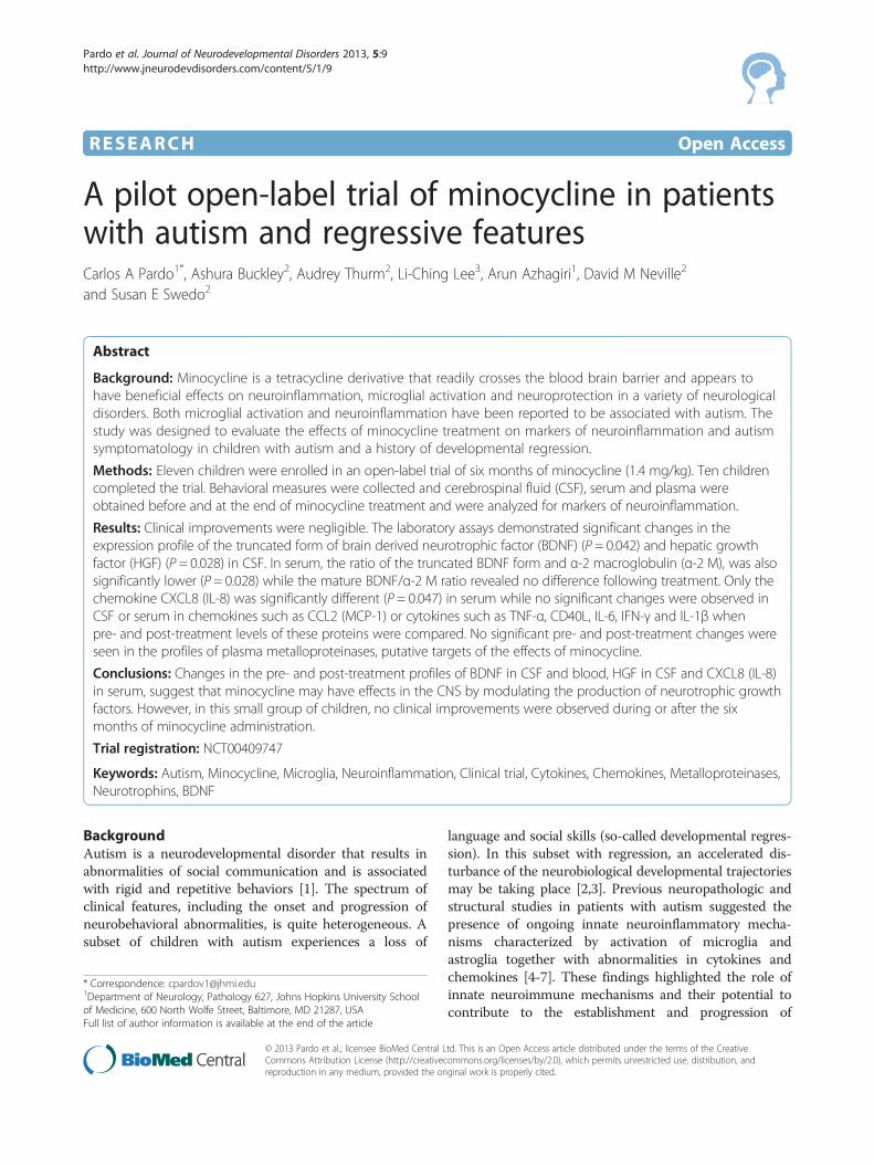
RESEARCH Open Access A pilot open-label trial of minocycline in patients with autism and regressive features Carlos A Pardo 1* , Ashura Buckley 2 , Audrey Thurm 2 , Li-Ching Lee 3 , Arun Azhagiri 1 , David M Neville 2 and Susan E Swedo 2 Abstract Background: Minocycline is a tetracycline derivative that readily crosses the blood brain barrier and appears to have beneficial effects on neuroinflammation, microglial activation and neuroprotection in a variety of neurological disorders. Both microglial activation and neuroinflammation have been reported to be associated with autism. The study was designed to evaluate the effects of minocycline treatment on markers of neuroinflammation and autism symptomatology in children with autism and a history of developmental regression. Methods: Eleven children were enrolled in an open-label trial of six months of minocycline (1.4 mg/kg). Ten children completed the trial. Behavioral measures were collected and cerebrospinal fluid (CSF), serum and plasma were obtained before and at the end of minocycline treatment and were analyzed for markers of neuroinflammation. Results: Clinical improvements were negligible. The laboratory assays demonstrated significant changes in the expression profile of the truncated form of brain derived neurotrophic factor (BDNF) (P = 0.042) and hepatic growth factor (HGF) (P = 0.028) in CSF. In serum, the ratio of the truncated BDNF form and α-2 macroglobulin (α-2 M), was also significantly lower (P = 0.028) while the mature BDNF/α-2 M ratio revealed no difference following treatment. Only the chemokine CXCL8 (IL-8) was significantly different (P = 0.047) in serum while no significant changes were observed in CSF or serum in chemokines such as CCL2 (MCP-1) or cytokines such as TNF-α, CD40L, IL-6, IFN-γ and IL-1β when pre- and post-treatment levels of these proteins were compared. No significant pre- and post-treatment changes were seen in the profiles of plasma metalloproteinases, putative targets of the effects of minocycline. Conclusions: Changes in the pre- and post-treatment profiles of BDNF in CSF and blood, HGF in CSF and CXCL8 (IL-8) in serum, suggest that minocycline may have effects in the CNS by modulating the production of neurotrophic growth factors. However, in this small group of children, no clinical improvements were observed during or after the six months of minocycline administration. Trial registration: NCT00409747 Keywords: Autism, Minocycline, Microglia, Neuroinflammation, Clinical trial, Cytokines, Chemokines, Metalloproteinases, Neurotrophins, BDNF Background Autism is a neurodevelopmental disorder that results in abnormalities of social communication and is associated with rigid and repetitive behaviors [1]. The spectrum of clinical features, including the onset and progression of neurobehavioral abnormalities, is quite heterogeneous. A subset of children with autism experiences a loss of language and social skills (so-called developmental regres- sion). In this subset with regression, an accelerated dis- turbance of the neurobiological developmental trajectories may be taking place [2,3]. Previous neuropathologic and structural studies in patients with autism suggested the presence of ongoing innate neuroinflammatory mecha- nisms characterized by activation of microglia and astroglia together with abnormalities in cytokines and chemokines [4-7]. These findings highlighted the role of innate neuroimmune mechanisms and their potential to contribute to the establishment and progression of * Correspondence: [email protected] 1 Department of Neurology, Pathology 627, Johns Hopkins University School of Medicine, 600 North Wolfe Street, Baltimore, MD 21287, USA Full list of author information is available at the end of the article © 2013 Pardo et al.; licensee BioMed Central Ltd. This is an Open Access article distributed under the terms of the Creative Commons Attribution License (http://creativecommons.org/licenses/by/2.0), which permits unrestricted use, distribution, and reproduction in any medium, provided the original work is properly cited. Pardo et al. Journal of Neurodevelopmental Disorders 2013, 5:9 http://www.jneurodevdisorders.com/content/5/1/9
-
Upload
johnshopkins -
Category
Documents
-
view
2 -
download
0
Transcript of A pilot open-label trial of minocycline in patients with autism and regressive features
Pardo et al. Journal of Neurodevelopmental Disorders 2013, 5:9http://www.jneurodevdisorders.com/content/5/1/9
RESEARCH Open Access
A pilot open-label trial of minocycline in patientswith autism and regressive featuresCarlos A Pardo1*, Ashura Buckley2, Audrey Thurm2, Li-Ching Lee3, Arun Azhagiri1, David M Neville2
and Susan E Swedo2
Abstract
Background: Minocycline is a tetracycline derivative that readily crosses the blood brain barrier and appears tohave beneficial effects on neuroinflammation, microglial activation and neuroprotection in a variety of neurologicaldisorders. Both microglial activation and neuroinflammation have been reported to be associated with autism. Thestudy was designed to evaluate the effects of minocycline treatment on markers of neuroinflammation and autismsymptomatology in children with autism and a history of developmental regression.
Methods: Eleven children were enrolled in an open-label trial of six months of minocycline (1.4 mg/kg). Ten childrencompleted the trial. Behavioral measures were collected and cerebrospinal fluid (CSF), serum and plasma wereobtained before and at the end of minocycline treatment and were analyzed for markers of neuroinflammation.
Results: Clinical improvements were negligible. The laboratory assays demonstrated significant changes in theexpression profile of the truncated form of brain derived neurotrophic factor (BDNF) (P = 0.042) and hepatic growthfactor (HGF) (P = 0.028) in CSF. In serum, the ratio of the truncated BDNF form and α-2 macroglobulin (α-2 M), was alsosignificantly lower (P = 0.028) while the mature BDNF/α-2 M ratio revealed no difference following treatment. Only thechemokine CXCL8 (IL-8) was significantly different (P = 0.047) in serum while no significant changes were observed inCSF or serum in chemokines such as CCL2 (MCP-1) or cytokines such as TNF-α, CD40L, IL-6, IFN-γ and IL-1β whenpre- and post-treatment levels of these proteins were compared. No significant pre- and post-treatment changes wereseen in the profiles of plasma metalloproteinases, putative targets of the effects of minocycline.
Conclusions: Changes in the pre- and post-treatment profiles of BDNF in CSF and blood, HGF in CSF and CXCL8 (IL-8)in serum, suggest that minocycline may have effects in the CNS by modulating the production of neurotrophic growthfactors. However, in this small group of children, no clinical improvements were observed during or after the sixmonths of minocycline administration.
Trial registration: NCT00409747
Keywords: Autism, Minocycline, Microglia, Neuroinflammation, Clinical trial, Cytokines, Chemokines, Metalloproteinases,Neurotrophins, BDNF
BackgroundAutism is a neurodevelopmental disorder that results inabnormalities of social communication and is associatedwith rigid and repetitive behaviors [1]. The spectrum ofclinical features, including the onset and progression ofneurobehavioral abnormalities, is quite heterogeneous. Asubset of children with autism experiences a loss of
* Correspondence: [email protected] of Neurology, Pathology 627, Johns Hopkins University Schoolof Medicine, 600 North Wolfe Street, Baltimore, MD 21287, USAFull list of author information is available at the end of the article
© 2013 Pardo et al.; licensee BioMed Central LCommons Attribution License (http://creativecreproduction in any medium, provided the or
language and social skills (so-called developmental regres-sion). In this subset with regression, an accelerated dis-turbance of the neurobiological developmental trajectoriesmay be taking place [2,3]. Previous neuropathologic andstructural studies in patients with autism suggested thepresence of ongoing innate neuroinflammatory mecha-nisms characterized by activation of microglia andastroglia together with abnormalities in cytokines andchemokines [4-7]. These findings highlighted the role ofinnate neuroimmune mechanisms and their potential tocontribute to the establishment and progression of
td. This is an Open Access article distributed under the terms of the Creativeommons.org/licenses/by/2.0), which permits unrestricted use, distribution, andiginal work is properly cited.
Pardo et al. Journal of Neurodevelopmental Disorders 2013, 5:9 Page 2 of 9http://www.jneurodevdisorders.com/content/5/1/9
regressive features in autism by influencing processes ofneuronal function, neuronal-glial interaction, and synapticplasticity. Under normal conditions, microglia are integralcomponents of brain homeostasis involved in synaptic plas-ticity, neurogenesis, axon guidance, and cortical organization[8-11]. However, in response to direct injury or to neuronaldysfunction, activated microglia can elaborate numerous cy-tokines and cytotoxins that can further damage neurons[12]. In autism, microglial activation has been shown tooccur in regions of the central nervous system such as thecerebellum and the frontal and anterior cingular cortices[6,7], areas of the brain involved in the neurobehavioral ab-normalities that characterize this disorder [13].Minocycline is a semisynthetic derivative of tetracyc-
line that readily crosses the blood brain barrier. Inaddition to being an antibacterial agent, it has directneuroprotective effects [14] as well as anti-inflammatoryproperties. It also has a proven tolerability and safetyprofile for clinical use [15]. In recent years, it has be-come one of the most investigated therapeutic candi-dates for modulation of microglial activation and innateneuroinflammatory pathways in neurological disorders,although contradictory results have been obtained in re-cent clinical trials [16,17]. The exact mechanism bywhich minocycline modifies inflammation is not fullyunderstood. The drug likely targets multiple inflammatorypathways via complex interactions with proteins integralto the inflammatory cascade [18]. This independent mod-ulatory function on neuroinflammation is, at least in part,mediated via microglial inhibition [19].The effect of minocycline on neuroinflammation and its
potential role as a neuroprotectant has been evaluated in ani-mal models of stroke, neurodegeneration, neuroimmunity,and neuroinfection [20]. Most of these studies showed thatminocycline decreases microglial activation, modulates path-ways involved in neuroinflammation, such as cytokine andchemokine networks (for example, interleukin-6 (IL-6), inter-leukin 1β (IL-1β) and tumor necrosis α (TNFα)), and re-duces the activity of selected metalloproteinases (MMPs)[21,22]. The neuroprotective role of minocycline in thesemodels has been attributed to anti-apoptotic andanti-oxidant properties [23]. Interestingly, minocyclineappeared to regulate behavior and reduced the impact ofstress on neuronal activation and working memory bymodulation of microglial activation in a model of stress[24]. Based on the potential anti-inflammatory andneuroprotective properties shown by minocycline in ani-mal models, therapeutic trials in humans have been car-ried out in a number of neuroinflammatory diseases suchas multiple sclerosis [25] and neurodegenerative [26-28],and cerebrovascular diseases [29]. Recent studies in a mousemodel of fragile X (Fmr1 KO), a neurodevelopmental dis-order that shares some neurological and neurobiological fea-tures with autism, demonstrated that minocycline was able
to promote dendritic spine maturation via inhibitory effectson MMP-9 expression and that these effects were accom-panied by improvements in the behavioral performance oftreated animals [30]. Furthermore, an open-label add-ontreatment trial with minocycline in patients with fragile Xshowed behavioral benefits without major side effects [31].On the other hand, while amyotrophic lateral sclerosis(ALS) studies in animal models showed great promise [32],as did the results of phase I and II clinical trials [16,17] a re-cently completed phase III randomized clinical trial showedadverse effects of the medication [17] in this disorder.The potential usefulness of minocycline in a variety
of neurological disorders involving neuroinflammationcoupled with the above findings suggestive of innateneuroinflammatory pathways in autism, led to the hy-pothesis that minocycline could be useful in the treat-ment of patients that exhibit the regressive subtype ofautism. Accordingly, we designed an open-label pilottrial study to evaluate the effects of minocycline onmarkers of neuroinflammation and to assess its impacton the clinical profile of children with regressiveautism.
MethodsStudy design and subjectsAn open-label pilot trial of minocycline therapy for chil-dren with autism and a history of regression was ap-proved by the National Institutes of Health (NIH)Institutional Review Board (NCT00409747). Consent forparticipating in the pilot trial was obtained from the par-ents or legal guardian. Minocycline dosage was calcu-lated for each subject at 1.4 mg/kg per day. This dosewas based on the Bonelli et al. 2004 study of minocyclinein Huntington’s Disease, which used 100 mg/day in adultsubjects [33]. Vitamin B6 at a dose of 0.6 mg/kg twicedaily was given in conjunction with minocycline adminis-tration to mitigate the potential for vestibular side effects.Subjects were referred and evaluated at the Intramural
Autism Research Program of the National Institute ofMental Health, Bethesda, Maryland. Children were eli-gible for inclusion if they were between three and twelveyears of age, met research criteria for a diagnosis of aut-ism, had a history of developmental regression, and werestable on all behavioral and/or medical therapies. Autismdiagnoses were made using the Autism Diagnostic Inter-view - Revised (ADI-R) [34] or a Toddler version, theAutism Diagnostic Observation Schedule [35], and clin-ical judgment based on DSM-IV criteria for autistic dis-order. The diagnostic evaluations also included cognitivetesting (described below), and information from theVineland Adaptive Behavior Scales, Second Edition(VABS) [36]. Regression was defined as language loss(loss of at least three spontaneously meaningful words)and/or nonverbal communication/social loss (loss of

Pardo et al. Journal of Neurodevelopmental Disorders 2013, 5:9 Page 3 of 9http://www.jneurodevdisorders.com/content/5/1/9
more than one nonverbal communicative behavior). Re-gression histories were confirmed by the ADI-R and amodified Regression Validation Interview [37].Subjects were excluded for known genetic defects,
based on medical records or clinical genetics testing,prematurity of less than 32 weeks gestation or small forgestational age status, serious neurologic disorders (forexample, cerebral palsy or uncontrolled epilepsy), evi-dence of renal insufficiency, hepatic disease or auto-immune disorder, or presence of a first degree relativewith systemic lupus erythematous. Patients were also ex-cluded if they were taking any medications that werecontraindicated with either minocycline or vitamin B6.Eleven children (9 boys, 2 girls; mean age 7.19 years;
range 3 to 12 years) were enrolled in the trial. By clinicalhistory, loss of social and communication skills had oc-curred at a mean age of 18.4 months (range 8 to 28 -months). Ten children completed the six-month open-labeltrial; one child dropped out after three months because ofparental concerns about side effects.Clinical and behavioral assessments were performed at
baseline and at post-treatment during the trial, and dataare presented in Table 1. At baseline, eight participantswere administered the Mullen Scales of Early Learning[38] and three participants were administered the Differ-ential Ability Scales Second Edition (DAS-II) [39]. Sincemany of the children who were assessed with the MullenScales were out of age range or below the ‘floors’ of thestandard scales, a nonverbal developmental quotient(NVDQ) was used to describe intellectual functioning.The NVDQ is the ratio of IQ of age equivalent/chrono-logical age for the average of the nonverbal portions of
Table 1 Patient sample description
Subject Pretreatmentage (months)
Sex Age atonset ofregression(months)
Non-verbalIQ/DQ
CGI- b
1 99 F 27 30
2a 64 M 13 60
3 69 M 12 33
4 153 M 19 77
5 91 M 8 58
6 66 M 18 103
7 128 F 15 25
8 112 M 14 33
9 49 M 18 59
10 38 M 22 90
11 83 M 18 32
M± SD 4.6
Effect SizeaDenotes child that discontinued the study at 3 months.Note: Effect size was calculated for the within-subjects effect using the following fo
the test. For the three participants administered theDAS-II, the nonverbal cognitive score was the standardscore from the nonverbal reasoning domain.The severity of core autism symptoms was rated at base-
line and monthly intervals using the Clinical Global Im-pression Severity Scale (CGI-S) [40], and change wasrecorded with the CGI-Improvement (CGI-I). In addition,adaptive functioning was measured using the VABS. Par-ents were queried about medication side effects atmonthly intervals.Serum, plasma and cerebrospinal fluid (CSF) samples
were obtained at baseline and following six months ofminocycline administration. CSF was obtained by lum-bar puncture under fasting conditions, while the childwas sedated with propofol. Venous blood samples wereobtained at the same time as the CSF and aliquots ofboth were sent for immediate analysis of routine labora-tory tests at NIH or stored (−80°C) for research analyses.Subjects also underwent monthly blood tests for evaluat-ing potential hematological, metabolic, or hepatic sideeffects.The cytokines TNFα, IFN-γ, IL-1α, IL-1β, IL-2, IL-3,
IL-4, IL-5, IL-6, IL-7, IL-10, IL-12(p40), IL 12(p70), IL-13, IL-15, GM-CSF and the chemokines CCL2 (MCP-1),CCL3 (MIP-1α), CCL5 (Rantes), CXCL8 (IL-8), CCL11(Eotaxin) and CXCL10 (IP-10) were measured in theserum and CSF at baseline and after six months treat-ment with minocycline. Additional assays included com-parisons of baseline and six months concentrations ofplasma and CSF leptin, CD40 ligand and neurotrophicfactors such as brain neurotrophic growth factor (BDNF),hepatic growth factor [HGF], and glial derived growth
Clinical assessment
aseline CGI- post-treatment Vineland- ABCbaseline
Vineland-ABCpost-treatment
6 5 55 54
5 4a 68 73a
5 5 45 44
5 5 56 55
3 3 60 56
4 4 70 71
5 5 55 46
5 5 53 56
4 4 66 63
4 3 70 66
5 5 48 57
± 0.8 4.4 ± 0.8 58.7 ± 8.8 58.3 ± 9.3
- −0.3 - −0.1
rmula: ES = (MPost - MBL)/SDBL). CGI, Clinical Global Impression.
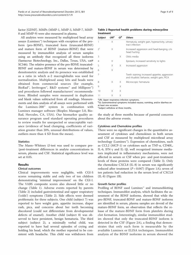
Table 2 Reported health problems during minocyclinetreatment
Subject URIa GIb Others
1 X Hematuria, weight gain, hyperactivity, urinarytract infection
2c X Increased aggression and head-banging, c/ohead hurting
3 X Otitis media
4 X Epistaxis, increased sensitivity to lights
5 X X Increased aggression
6 X
7 Teeth staining; increased appetite, aggressionand ritualistic behavior, weight gain, PICA
8 X X Microscopic hematuria
9 X
10 X X
11 X XaURI, Upper respiratory infection symptoms.bGI, Gastrointestinal symptoms included nausea, vomiting, and/or diarrhea onat least one occasion.cSubject withdrew at three months.
Pardo et al. Journal of Neurodevelopmental Disorders 2013, 5:9 Page 4 of 9http://www.jneurodevdisorders.com/content/5/1/9
factor [GDNF]. MMPs (MMP-1, MMP-3, MMP-7, MMP-8 and MMP-9) were also measured in plasma.All analytes were measured by multiplexed bead array
assays (Luminex™) techniques with exception of the pro-form (pro-BDNF), truncated form (truncated-BDNF)and mature form of BDNF (mature-BDNF) that weremeasured by immunoblot analysis of serum samplesusing an antibody that recognized all three isoforms(Santacruz Biotechnology, Inc., Dallas, Texas, USA, cat#SC546). The relative presence of the pro-BDNF, truncated-BDNF and mature-BDNF in serum was determined bydensitometric analysis and its presence was establishedas a ratio in which α-2 macroglobulin was used fornormalization. Multiplexed assay kits and beads wereobtained from commercial sources (for example,BioRad™, Invitrogen™, R&D systems™ and Millipore™)and procedures followed manufacturers’ recommenda-tions. Blinded samples were measured in duplicatesand blank values subtracted from all readings. Measure-ments and data analysis of all assays were performed withthe Luminex-200™ system in combination withLuminex manager software (Bioplex Manager 5.0, Bio-Rad, Hercules, CA, USA). Our biomarker quality as-surance program used standard operating proceduresto review results for unexpected or unacceptable vari-ance (evidence of bead clumping, coefficients of vari-ation greater than 20%, unusual distributions of values,outliers more than 4 SD from the mean).
StatisticsThe Mann–Whitney U-test was used to compare pre-/post-treatment differences in analyte concentrations inserum, plasma and CSF. Statistical significance level wasset at 0.05.
ResultsClinical outcomesClinical improvements were negligible, with CGI-Sscores remaining stable and only two of ten childrendemonstrating ‘minimal improvement’ on the CGI-I.The VABS composite scores also showed little or nochange (Table 1). Adverse events reported by parents(Table 2) included gastrointestinal and upper respiratory(‘colds’) symptoms (Table 2). Side effects were deemedproblematic for three subjects. One child (subject 7) wasreported to have weight gain, appetite increase, diaperrash, pica, and concerns about her teeth becomingdiscolored (could not differentiate from developmentaldefects of enamel). Another child (subject 8) was ob-served to have persistent, benign hematuria. The thirdsubject (subject 2), a minimally verbal child, wasreported to have had several episodes of crying andholding his head, which the mother reported to be con-sistent with headache. This child was withdrawn from
the study at three months because of parental concernsabout the adverse events.
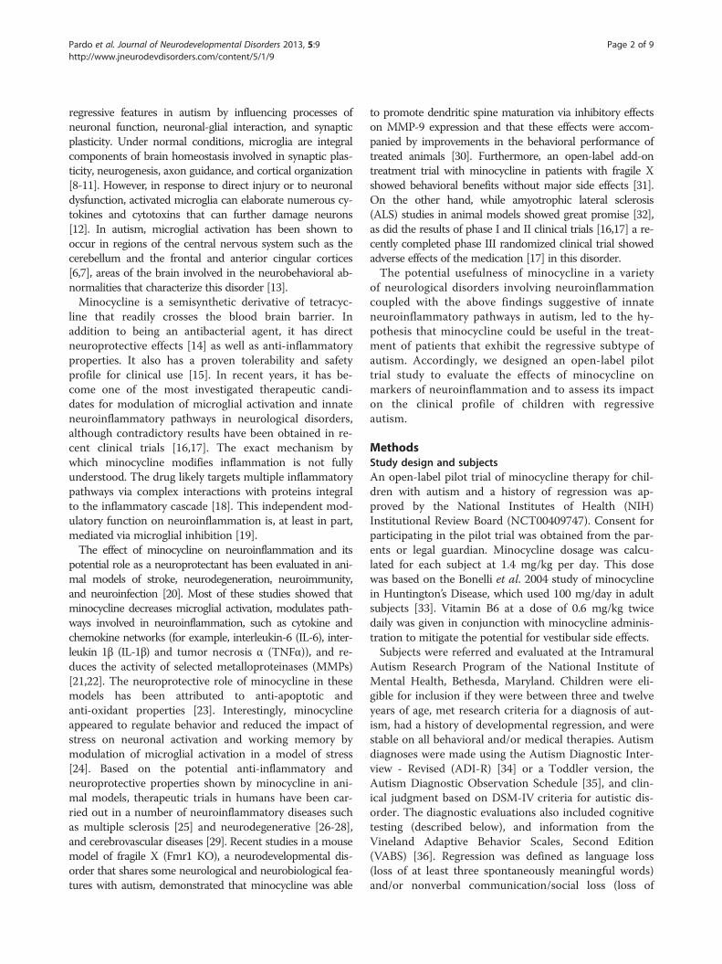
Cytokines and Chemokine profilesThere were no significant changes in the quantitative as-sessment of cytokines and chemokines in both serumand CSF as measured by multiplexed microbead arraytechnology (Luminex™). Expression of chemokines, suchas CCL2 (MCP-1) or cytokines such as TNF-α, CD40L,IL-6, IFN-γ and IL-1β, well recognized immune media-tors implicated in inflammatory mechanisms, were notaffected in serum or CSF when pre- and post-treatmentlevels of these proteins were compared (Table 3). Onlythe chemokine CXCL8 (IL-8) in serum was significantlyreduced after treatment (P = 0.047) (Figure 1A); seven often patients had reduction in the serum level of CXCL8(IL-8) (Figure 1B).
Growth factor profilesProfiling of BDNF used Luminex™ and immunoblottingtechniques. Immunoblot analysis, which facilitates the as-sessment of the BDNF isoforms, showed that while thepro-BDNF, truncated-BDNF and mature-BDNF isoformsare identified in serum, plasma samples are devoid of themature-BDNF form, an observation that reflects the re-lease of the mature-BDNF form from platelets duringclot formation. Interestingly, similar immunoblot stud-ies showed that only the truncated-BDNF isoform isdetected in the CSF (Figure 2A), a finding that demon-strates that only such form is measurable by theavailable Luminex or ELISA techniques. Immunoblotanalysis of the BDNF isoforms in serum showed the
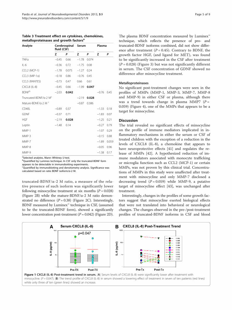
Table 3 Treatment effect on cytokines, chemokines,metalloproteinases and growth factorsa
Analyte Cerebrospinalfluid (CSF)
Serum Plasma
Z P Z P Z P
TNFα −0.45 0.66 −1.78 0.074
IL-6 −0.36 0.72 −1.75 0.08
CCL2 (MCP-1) −1.78 0.075 −1.27 0.24
CCL3 (MIP-1α) −0.18 0.86 −0.76 0.45
CCL5 (RANTES) −0.73 0.47 0.66 0.61
CXCL8 (IL-8) −0.45 0.66 −1.99 0.047
BDNFb −2.03 0.042 −0.76 0.45
Truncated-BDNF/α-2 Mc −2.19 0.028
Mature-BDNF/α-2 M c −0.87 0.386
CD40L −0.89 0.37 −1.33 0.18
GDNF −0.37 0.71 −1.83 0.07
HGF −2.19 0.028 −1.25 0.21
Leptin −1.48 0.14 −0.27 0.79
MMP-1 −1.07 0.29
MMP-3 −0.15 0.88
MMP-7 −1.89 0.059
MMP-8 −0.05 0.96
MMP-9 −1.38 0.17aSelected analytes, Mann–Whitney U-test.bQuantified by Luminex technique. In CSF only the truncated-BDNF formappears to be detectable in immunobotting experiments.cQuantified by immunoblotting and densitometry analysis. Significance wascalculated based on ratio BDNF isoform/α-2 M.
Pardo et al. Journal of Neurodevelopmental Disorders 2013, 5:9 Page 5 of 9http://www.jneurodevdisorders.com/content/5/1/9
truncated-BDNF/α-2 M ratio, a measure of the rela-tive presence of such isoform was significantly lowerfollowing minocycline treatment at six months (P = 0.028)(Figure 2B) while the mature-BDNF/α-2 M ratio demon-strated no difference (P = 0.38) (Figure 2C). Interestingly,BDNF measured by Luminex™ technique in CSF, (assumedto be the truncated-BDNF form), showed a significantlylower concentration post-treatment (P = 0.042) (Figure 2D).
Figure 1 CXCL8 (IL-8) Post-treatment trend in serum. (A) Serum levelsminocycline. (P = 0.047). (B) The trend profile of CXCL8 (IL-8) in serum showwhile only three of ten (green lines) showed an increase.
The plasma BDNF concentration measured by Luminex™technique, which reflects the presence of pro- andtruncated-BDNF isoforms combined, did not show differ-ence after treatment (P = 0.45). Contrary to BDNF, thegrowth factor HGF, (and ligand for MET), was foundto be significantly increased in the CSF after treatment(P = 0.028) (Figure 3) but was not significantly differentin serum. The CSF concentration of GDNF showed nodifference after minocycline treatment.
MetalloproteinasesNo significant post-treatment changes were seen in theprofiles of MMPs (MMP-1, MMP-3, MMP-7, MMP-8and MMP-9) in either CSF or plasma, although therewas a trend towards change in plasma MMP7 (P =0.059) (Figure 4), one of the MMPs that appears to be atarget for minocycline.
DiscussionThe trial revealed no significant effects of minocyclineon the profile of immune mediators implicated in in-flammatory mechanisms in either the serum or CSF oftreated children with the exception of a reduction in thelevels of CXCL8 (IL-8), a chemokine that appears tohave neuroprotective effects [41] and regulates the re-lease of MMPs [42]. A hypothesized reduction of im-mune modulators associated with monocyte traffickingor microglia function such as CCL2 (MCP-1) or certainMMPs, was not proven by this clinical trial. Concentra-tions of MMPs in this study were unaffected after treat-ment with minocycline and only MMP-7 disclosed adecreasing trend (P = 0.059) while MMP-9, a putativetarget of minocycline effect [43], was unchanged aftertreatment.Interestingly, changes in the profiles of some growth fac-
tors suggest that minocycline exerted biological effectsthat were not translated into behavioral or neurologicalchanges. The changes observed in the pre-/post-treatmentprofiles of truncated-BDNF isoforms in CSF and blood
of CXCL8 (IL-8) were significantly lower after treatment withed a lowering effect of treatment in seven of ten patients (red lines)

Figure 2 Profiles of expression of brain derived neurotrophic factor in serum and cerebrospinal fluid. (A) Immunoblot analysis of brainderived neurotrophic factor (BDNF) isoforms in serum disclosed the presence of the pro-form (32 kD), truncated-form (28 kD) and mature form(14 kD) while plasma disclosed mostly the presence of the BDNF pro-form and truncated-form. The truncated-BDNF form was the only isoformseen in cerebrospinal fluid (CSF). (B)The BDNF truncated-form was significantly decreased after treatment with minocycline (P = 0.028) a trendthat was observed in six of ten patients (red lines). (C) Although the mature form appeared to be decreased after treatment the differencepre- and post-treatment did not reach significance (P = 0.38). (D) BDNF in CSF measured by multiplexed array technique, which detects mostlythe truncated-BNDF isoform, was significantly lowered after treatment (P = 0.042).
Pardo et al. Journal of Neurodevelopmental Disorders 2013, 5:9 Page 6 of 9http://www.jneurodevdisorders.com/content/5/1/9
and HGF in CSF, are intriguing as they may suggestthat higher doses of minocycline could potentiallymodulate the proteolytic processing of some neuro-trophic growth factors. BDNF pro-form and truncated-form appear to be critical for mechanisms of synaptic
plasticity. Both forms are not necessarily inactive inter-mediates in the biosynthesis of BDNF but rather bio-logically active forms with opposite functions withrespect to the mature-BDNF form that may be involved inthe neurobiological abnormalities that underlie cognitive
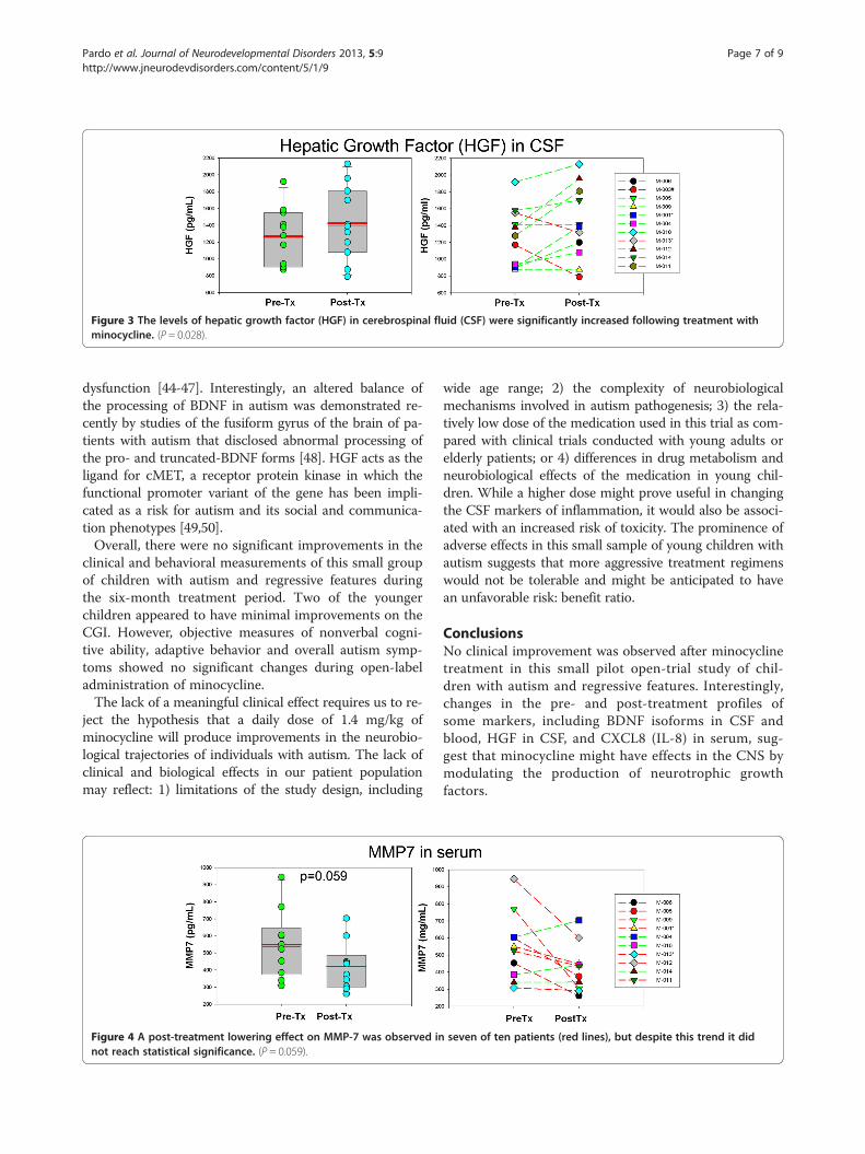
Figure 3 The levels of hepatic growth factor (HGF) in cerebrospinal fluid (CSF) were significantly increased following treatment withminocycline. (P = 0.028).
Pardo et al. Journal of Neurodevelopmental Disorders 2013, 5:9 Page 7 of 9http://www.jneurodevdisorders.com/content/5/1/9
dysfunction [44-47]. Interestingly, an altered balance ofthe processing of BDNF in autism was demonstrated re-cently by studies of the fusiform gyrus of the brain of pa-tients with autism that disclosed abnormal processing ofthe pro- and truncated-BDNF forms [48]. HGF acts as theligand for cMET, a receptor protein kinase in which thefunctional promoter variant of the gene has been impli-cated as a risk for autism and its social and communica-tion phenotypes [49,50].Overall, there were no significant improvements in the
clinical and behavioral measurements of this small groupof children with autism and regressive features duringthe six-month treatment period. Two of the youngerchildren appeared to have minimal improvements on theCGI. However, objective measures of nonverbal cogni-tive ability, adaptive behavior and overall autism symp-toms showed no significant changes during open-labeladministration of minocycline.The lack of a meaningful clinical effect requires us to re-
ject the hypothesis that a daily dose of 1.4 mg/kg ofminocycline will produce improvements in the neurobio-logical trajectories of individuals with autism. The lack ofclinical and biological effects in our patient populationmay reflect: 1) limitations of the study design, including
Figure 4 A post-treatment lowering effect on MMP-7 was observed innot reach statistical significance. (P = 0.059).
wide age range; 2) the complexity of neurobiologicalmechanisms involved in autism pathogenesis; 3) the rela-tively low dose of the medication used in this trial as com-pared with clinical trials conducted with young adults orelderly patients; or 4) differences in drug metabolism andneurobiological effects of the medication in young chil-dren. While a higher dose might prove useful in changingthe CSF markers of inflammation, it would also be associ-ated with an increased risk of toxicity. The prominence ofadverse effects in this small sample of young children withautism suggests that more aggressive treatment regimenswould not be tolerable and might be anticipated to havean unfavorable risk: benefit ratio.
ConclusionsNo clinical improvement was observed after minocyclinetreatment in this small pilot open-trial study of chil-dren with autism and regressive features. Interestingly,changes in the pre- and post-treatment profiles ofsome markers, including BDNF isoforms in CSF andblood, HGF in CSF, and CXCL8 (IL-8) in serum, sug-gest that minocycline might have effects in the CNS bymodulating the production of neurotrophic growthfactors.
seven of ten patients (red lines), but despite this trend it did
Pardo et al. Journal of Neurodevelopmental Disorders 2013, 5:9 Page 8 of 9http://www.jneurodevdisorders.com/content/5/1/9
AbbreviationsALS: Amyotrophic lateral sclerosis; BDNF: Brain derived neurotrophic factor;CGI-I: Clinical Global Impression Improvement Scale; CGI-S: Clinical GlobalImpression Severity Scale; CSF: Cerebrospinal fluid; GDNF: Glial derivedgrowth factor; HGF: Hepatic growth factor; MMPs: Metalloproteinases;NIH: National Institutes of Health; NVDQ: Nonverbal developmental quotient;VABS: Vineland Adaptive Behavior Scales; α-2 M: α-2 macroglobulin.
Competing interestsThe authors declare that they have no competing interests.
Authors’ contributionsCAP, SS, AT and DN conceived and participated in the design of the study.AB and AT participated in the evaluation and clinical care of patients. AAcarried out immunoassays and quantitative immunoblots. LCL performed thestatistical analysis. CAP, AB, AT and SS drafted the manuscript. Themanuscript was reviewed and approved by all authors.
AcknowledgementsThe research was funded by the Intramural Research Program of theNational Institute of Mental Health. Dr. Pardo’s laboratory received supportfrom NIH contract HHSN271200700179, The Bart McLean Fund forNeuroimmunology Research and The Peter Emch Fund for Autism Research.The content is solely the responsibility of the authors and does notnecessarily represent the official views of the National Institute of MentalHealth or the National Institutes of Health.
Author details1Department of Neurology, Pathology 627, Johns Hopkins University Schoolof Medicine, 600 North Wolfe Street, Baltimore, MD 21287, USA. 2Pediatrics &Developmental Neuroscience Branch, National Institute of Mental Health, 10Center Drive, Bldg 10/RM 1C250/MSC 1255, Bethesda, MD 20892, USA.3Department of Epidemiology, Johns Hopkins University Bloomberg Schoolof Public Health, 615 N. Wolfe Street, Room E6032, Baltimore, MD 21205,USA.
Received: 27 November 2012 Accepted: 20 March 2013Published: 8 April 2013
References1. Rapin I: Autism. N Engl J Med 1997, 337:97–104.2. Ashwood P, Krakowiak P, Hertz-Picciotto I, Hansen R, Pessah I, Van de WJ:
Elevated plasma cytokines in autism spectrum disorders provideevidence of immune dysfunction and are associated with impairedbehavioral outcome. Brain Behav Immun 2011, 25:40–45.
3. Ozonoff S, Iosif AM, Baguio F, Cook IC, Hill MM, Hutman T, Rogers SJ, RozgaA, Sangha S, Sigman M, Steinfeld MB, Young GS: A prospective study ofthe emergence of early behavioral signs of autism. J Am Acad ChildAdolesc Psychiatry 2010, 49:256–266.
4. Laurence JA, Fatemi SH: Glial fibrillary acidic protein is elevated insuperior frontal, parietal and cerebellar cortices of autistic subjects.Cerebellum 2005, 4:206–210.
5. Li X, Chauhan A, Sheikh AM, Patil S, Chauhan V, Li XM, Ji L, Brown T, MalikM: Elevated immune response in the brain of autistic patients.J Neuroimmunol 2009, 207:111–116.
6. Morgan JT, Chana G, Pardo CA, Achim C, Semendeferi K, Buckwalter J,Courchesne E, Everall IP: Microglial activation and increased microglialdensity observed in the dorsolateral prefrontal cortex in autism. BiolPsychiatry 2010, 68:368–376.
7. Vargas DL, Nascimbene C, Krishnan C, Zimmerman AW, Pardo CA:Neuroglial activation and neuroinflammation in the brain of patientswith autism. Ann Neurol 2005, 57:67–81.
8. Ransohoff RM: Chemokines and chemokine receptors: standing at thecrossroads of immunobiology and neurobiology. Immunity 2009, 31:711–721.
9. Tremblay ME, Stevens B, Sierra A, Wake H, Bessis A, Nimmerjahn A: The roleof microglia in the healthy brain. J Neurosci 2011, 31:16064–16069.
10. Kettenmann H, Hanisch UK, Noda M, Verkhratsky A: Physiology ofmicroglia. Physiol Rev 2011, 91:461–553.
11. Sierra A, Encinas JM, Deudero JJ, Chnacey JH, Enikolopov G, Overstreet-Wadiche LS, Tsirka SE, Maletic-Savatic M: Microglia shape adult
hippocampal neurogenesis through apoptosis-coupled phagocytosis. CellStem Cell 2010, 7:483–495.
12. Derecki NC, Cronk JC, Kipnis J: The role of microglia in brain maintanance:Implications for Rett syndrome. Trends Immunol 2013, 34:144–150.
13. Amaral DG, Schumann CM, Nordahl CW: Neuroanatomy of autism. TrendsNeurosci 2008, 31:137–145.
14. Alano CC, Kauppinen TM, Valls AV, Swanson RA: Minocycline inhibits poly(ADP-ribose) polymerase-1 at nanomolar concentrations. Proc Natl AcadSci USA 2006, 103:9685–9690.
15. Blum D, Chtarto A, Tenenbaum L, Brotchi J, Levivier M: Clinical potential ofminocycline for neurodegenerative disorders. Neurobiol Dis 2004, 17:359–366.
16. Gordon PH, Moore DH, Gelinas DF, Qualls C, Meister ME, Werner J, Mendoza M,Mass J, Kushner G, Miller RG: Placebo-controlled phase I/II studies ofminocycline in amyotrophic lateral sclerosis. Neurology 2004, 62:1845–1847.
17. Gordon PH, Moore DH, Miller RG, Florence JM, Verheijde JL, Doorish C,Hilton JF, Spitalny GM, Macarthur RB, Mitsumoto H, Neville HE, Boylan K,Mozaffar T, Belsh JM, Ravits J, Bedlack RS, Graves MC, McCluskey LF, BarohnRJ, Tandan R, Western ALS Study Group: Efficacy of minocycline inpatients with amyotrophic lateral sclerosis: a phase III randomised trial.Lancet Neurol 2007, 6:1045–1053.
18. Kim HS, Suh YH: Minocycline and neurodegenerative diseases. BehavBrain Res 2009, 196:168–179.
19. Nutile-McMenemy N, Elfenbein A, Deleo JA: Minocycline decreases in vitromicroglial motility, beta1-integrin, and Kv1.3 channel expression.J Neurochem 2007, 103:2035–2046.
20. Plane JM, Shen Y, Pleasure DE, Deng W: Prospects for minocyclineneuroprotection. Arch Neurol 2010, 67:1442–1448.
21. Orsucci D, Calsolaro V, Mancuso M, Siciliano G: Neuroprotective effects oftetracyclines: molecular targets, animal models and human disease. CNSNeurol Disord Drug Targets 2009, 8:222–231.
22. Yong VW, Wells J, Giuliani F, Casha S, Power C, Metz LM: The promise ofminocycline in neurology. Lancet Neurol 2004, 3:744–751.
23. Plane JM, Liu R, Wang TW, Silverstein FS, Parent JM: Neonatal hypoxic-ischemic injury increases forebrain subventricular zone neurogenesis inthe mouse. Neurobiol Dis 2004, 16:585–595.
24. Hinwood M, Morandini J, Day TA, Walker FR: Evidence that microgliamediate the neurobiological effects of chronic psychological stress onthe medial prefrontal cortex. Cereb Cortex 2012, 22:1442–1454.
25. Metz LM, Zhang Y, Yeung M, Patry DG, Bell RB, Stoian CA, Yong VW, PattenSB, Duquette P, Antel JP, Mitchell JR: Minocycline reduces gadolinium-enhancing magnetic resonance imaging lesions in multiple sclerosis.Ann Neurol 2004, 55:756.
26. Galpern WR, Singhal AB: Neuroprotection: lessons from a spectrum ofneurological disorders. Int J Stroke 2006, 1:97–99.
27. Gordon PH, Cheung YK, Levin B, Andrews H, Doorish C, Macarthur RB, MontesJ, Bednarz K, Florence J, Rowin J, Boylan K, Mozaffar T, Tandan R, Mitsumoto H,Kelvin EA, Chapin J, Bedlack R, Rivner M, McCluskey LF, Pestronk A, Graves M,Sorenson EJ, Barohn RJ, Belsh JM, Lou JS, Levine T, Saperstein D, Miller RG,Scelsa SN, Combination Drug Selection Trial Study Group: A novel, efficient,randomized selection trial comparing combinations of drug therapy forALS. Amyotroph Lateral Scler 2008, 9:212–222.
28. Lewitt PA, Guttman M, Tetrud JW, Tuite PJ, Mori A, Chaikin P, Sussman NM:Adenosine A2A receptor antagonist istradefylline (KW-6002) reduces"off" time in Parkinson's disease: a double-blind, randomized,multicenter clinical trial (6002-US-005). Ann Neurol 2008, 63:295–302.
29. Hayakawa K, Mishima K, Nozako M, Hazekawa M, Mishima S, Fujioka M,Orito K, Egashira N, Iwasaki K, Fujiwara M: Delayed treatment withminocycline ameliorates neurologic impairment through activatedmicroglia expressing a high-mobility group box1-inhibiting mechanism.Stroke 2008, 39:951–958.
30. Bilousova TV, Dansie L, Ngo M, Aye J, Charles JR, Ethell DW, Ethell IM:Minocycline promotes dendritic spine maturation and improves behaviouralperformance in the fragile X mouse model. J Med Genet 2009, 46:94–102.
31. Paribello C, Tao L, Folino A, Berry-Kravis E, Tranfaglia M, Ethell IM, Ethell DW:Open-label add-on treatment trial of minocycline in fragile X syndrome.BMC Neurol 2010, 10:91.
32. Kriz J, Nguyen MD, Julien JP:Minocycline slows disease progression in amouse model of amyotrophic lateral sclerosis. Neurobiol Dis 2002, 10:268–278.
33. Bonelli RM, Hodl AK, Hofmann P, Kapfhammer HP: Neuroprotection inHuntington's disease: a 2-year study on minocycline. Int ClinPsychopharmacol 2004, 19:337–342.

Pardo et al. Journal of Neurodevelopmental Disorders 2013, 5:9 Page 9 of 9http://www.jneurodevdisorders.com/content/5/1/9
34. Le Couteur A, Lord C, Rutter M: Autism Diagnostic Interview-Revised (ADI-R).Los Angeles: Western Psychological Services; 2003.
35. Lord C, Risi S, Lambrecht L, Cook EH Jr, Leventhal BL, DiLavore PC, Pickles A,Rutter M: The autism diagnostic observation schedule-generic: astandard measure of social and communication deficits associated withthe spectrum of autism. J Autism Dev Disord 2000, 30:205–223.
36. Sparrow SS, Cichetti D, Balla D: Vineland Adaptive Behavior Scales. CirclePines, MN: American Guidance Service; 2005.
37. Luyster R, Richler J, Risi S, Hsu WL, Dawson G, Bernier R, Dunn M, HepburnS, Hyman SL, McMahon WM, Goudie-Nice J, Minshew N, Rogers S, SigmanM, Spence MA, Goldberg WA, Tager-Flusberg H, Volkmar FR, Lord C: Earlyregression in social communication in autism spectrum disorders: aCPEA Study. Dev Neuropsychol 2005, 27:311–336.
38. Mullen E: Infant Mullen Scales of Early Learning. Circle Pines, MN: AmericanGuidance Service; 1995.
39. Elliott C: Manual for the Differential Ability Scales. San Antonio, TX: PearsonAssessments; 2007.
40. Guy W: ECDEU Assessment Manual of Psychopharmacology-Revised. Rockville,MD: Psychopharmacology Research Branch; 1976.
41. Hussein MH, Daoud GA, Kakita H, Kato S, Goto T, Kamei M, Goto K, NobataM, Ozaki Y, Ito T, Fukuda S, Kato I, Suzuki S, Sobajima H, Hara F, HashimotoT, Togari H: High cerebrospinal fluid antioxidants and interleukin 8 areprotective of hypoxic brain damage in newborns. Free Radic Res 2010,44:422–429.
42. Thirumangalakudi L, Yin L, Rao HV, Grammas P: IL-8 induces expression ofmatrix metalloproteinases, cell cycle and pro-apoptotic proteins, and celldeath in cultured neurons. J Alzheimers Dis 2007, 11:305–311.
43. Switzer JA, Hess DC, Ergul A, Waller JL, Machado LS, Portik-Dobos V,Pettigrew LC, Clark WM, Fagan SC: Matrix metalloproteinase-9 in anexploratory trial of intravenous minocycline for acute ischemic stroke.Stroke 2011, 42:2633–2635.
44. Bekinschtein P, Cammarota M, Izquierdo I, Medina JH: BDNF and memoryformation and storage. Neuroscientist 2008, 14:147–156.
45. Yang J, Siao CJ, Nagappan G, Marinic T, Jing D, McGrath K, Chen ZY, MarkW, Tessarollo L, Lee FS, Lu B, Hempstead BL: Neuronal release of proBDNF.Nat Neurosci 2009, 12:113–115.
46. Carlino D, Leone E, Di Cola F, Baj G, Marin R, Dinelli G, Tongiorgi E, DeVanna M: Low serum truncated-BDNF isoform correlates with highercognitive impairment in schizophrenia. J Psychiatr Res 2011, 45:273–279.
47. Carlino D, De Vanna M, Tongiorgi E: Is altered BDNF biosynthesis ageneral feature in patients with cognitive dysfunctions? Neuroscientist2012. doi:10.1177/1073858412469444. Epub ahead of print.
48. Garcia KLP, Yu G, Nicolini C, Michalski B, Garzon DJ, Chiu VS, Tongiorgi E,Szatmari P, Fahnestock M: Altered balance of proteolytic isoforms ofpro-brain-derived neurotrophic factor in autism. J Neuropath Exp Neurol2012, 71:289–297.
49. Campbell DB, Sutcliffe JS, Ebert PJ, Militerni R, Bravaccio C, Trillo S, Elia M,Schneider C, Melmed R, Sacco R, Persico AM, Levitt P: A genetic variantthat disrupts MET transcription is associated with autism. Proc Natl AcadSci USA 2006, 103:16834–16839.
50. Buie T, Campbell DB, Fuchs GJ III, Furuta GT, Levy J, Vandewater J, Whitaker AH,Atkins D, Bauman ML, Beaudet AL, Carr EG, Gershon MD, Hyman SL, Jirapinyo P,Jyonouchi H, Kooros K, Kushak R, Levitt P, Levy SE, Lewis JD, Murray KF,Natowicz MR, Sabra A, Wershil BK, Weston SC, Zeltzer L, Winter H: Evaluation,diagnosis, and treatment of gastrointestinal disorders in individuals withASDs: a consensus report. Pediatrics 2010, 125(Suppl 1):S1–S18.
doi:10.1186/1866-1955-5-9Cite this article as: Pardo et al.: A pilot open-label trial of minocycline inpatients with autism and regressive features. Journal ofNeurodevelopmental Disorders 2013 5:9.
Submit your next manuscript to BioMed Centraland take full advantage of:
• Convenient online submission
• Thorough peer review
• No space constraints or color figure charges
• Immediate publication on acceptance
• Inclusion in PubMed, CAS, Scopus and Google Scholar
• Research which is freely available for redistribution
Submit your manuscript at www.biomedcentral.com/submit