Microarray analysis of potential genes in the pathogenesis of ...
Upload
un-lincolnCategory
view
7download
0
JOURNAL OF VIROLOGY, Jan. 2010, p. 532–542 Vol. 84, No. 10022-538X/10/$12.00 doi:10.1128/JVI.01698-09Copyright © 2010, American Society for Microbiology. All Rights Reserved.
Microarray Analysis of Paramecium bursaria ChlorellaVirus 1 Transcription�†
Giane M. Yanai-Balser,1 Garry A. Duncan,2 James D. Eudy,3 Dong Wang,4 Xiao Li,5Irina V. Agarkova,1 David D. Dunigan,1,6 and James L. Van Etten1,6*
Department of Plant Pathology, University of Nebraska, Lincoln, Nebraska 68583-07221; Biology Department,Nebraska Wesleyan University, Lincoln, Nebraska 68504-27942; Department of Genetics, Cell Biology andAnatomy, University of Nebraska Medical Center, Omaha, Nebraska 68198-54553; Statistics Department,
University of Nebraska, Lincoln, Nebraska 68583-09634; Biomedical Engineering and Biotechnology,University of Massachusetts, Lowell, Massachusetts 018545; and Nebraska Center for Virology,
University of Nebraska, Lincoln, Nebraska 68583-09006
Received 12 August 2009/Accepted 7 October 2009
Paramecium bursaria chlorella virus 1 (PBCV-1), a member of the family Phycodnaviridae, is a large double-stranded DNA, plaque-forming virus that infects the unicellular green alga Chlorella sp. strain NC64A. The330-kb PBCV-1 genome is predicted to encode 365 proteins and 11 tRNAs. To monitor global transcriptionduring PBCV-1 replication, a microarray containing 50-mer probes to the PBCV-1 365 protein-encoding genes(CDSs) was constructed. Competitive hybridization experiments were conducted by using cDNAs from poly(A)-containing RNAs obtained from cells at seven time points after virus infection. The results led to the followingconclusions: (i) the PBCV-1 replication cycle is temporally programmed and regulated; (ii) 360 (99%) of thearrayed PBCV-1 CDSs were expressed at some time in the virus life cycle in the laboratory; (iii) 227 (62%) ofthe CDSs were expressed before virus DNA synthesis begins; (iv) these 227 CDSs were grouped into two classes:127 transcripts disappeared prior to initiation of virus DNA synthesis (considered early), and 100 transcriptswere still detected after virus DNA synthesis begins (considered early/late); (v) 133 (36%) of the CDSs wereexpressed after virus DNA synthesis begins (considered late); and (vi) expression of most late CDSs isinhibited by adding the DNA replication inhibitor, aphidicolin, prior to virus infection. This study provides thefirst comprehensive evaluation of virus gene expression during the PBCV-1 life cycle.
Paramecium bursaria chlorella virus 1 (PBCV-1), the proto-type of the genus Chlorovirus (family Phycodnaviridae), is alarge, icosahedral (190 nm in diameter), plaque-forming virusthat infects the unicellular, eukaryotic green alga Chlorella sp.strain NC64A. The PBCV-1 virion has a lipid membrane lo-cated inside an outer glycoprotein capsid. The 330-kb genomeis a linear, nonpermutated, double-stranded DNA (dsDNA)molecule with covalently closed hairpin ends that has approx-imately 365 protein encoding genes (CDSs), as well as 11tRNA encoding genes (reviewed in references 34, 39, and 40).The CDSs are evenly distributed on both strands and inter-genic space is minimal (typically fewer than 100 nucleotides);the exception is a 1,788-bp sequence in the middle of thegenome that encodes the tRNA genes. Approximately 35% ofthe 365 PBCV-1 gene products resemble proteins in the publicdatabases.
PBCV-1 initiates infection by attaching rapidly and specifi-cally to the cell wall of its host (22), probably at a unique virusvertex (4, 26). Attachment is immediately followed by host cellwall degradation by a virus-packaged enzyme(s) at the point ofcontact. After wall degradation, the viral internal membranepresumably fuses with the host membrane, causing host mem-
brane depolarization (9), potassium ion efflux (25), and anincrease in the cytoplasm pH (2). These events are predicted tofacilitate entry of the viral DNA and virion-associated proteinsinto the cell. PBCV-1 lacks a gene encoding a recognizableRNA polymerase or a subunit of it, and RNA polymeraseactivity is not detected in PBCV-1 virions. Therefore, viralDNA and virion-associated proteins are predicted to migrateto the nucleus, and early viral transcription is detected 5 to 10min postinfection (p.i.), presumably by commandeering a hostRNA polymerase(s) (possibly RNA polymerase II) (14, 29).Virus DNA synthesis begins 60 to 90 min p.i., followed by virusassembly at 3 to 5 h p.i. in localized regions of the cytoplasm,called virus assembly centers (21). At 6 to 8 h p.i., virus-induced host cell lysis occurs resulting in release of progenyvirions (�1,000 viruses/cell, �25% of which are infectious).These events are depicted in Fig. 1.
To initiate PBCV-1 transcription, the host RNA poly-merase(s), possibly in combination with a virus transcriptionfactor(s), must recognize virus DNA promoter sequences. Re-cently, three short nucleotide sequences were identified in pu-tative virus promoter regions (150 bp upstream and 50 bpdownstream of the ATG translation site) that are conserved inPBCV-1 and other Chlorovirus members (7). PBCV-1 CDSsare not spatially clustered on the genome by either temporal orfunctional class, suggesting that transcription regulation mustoccur via cis- and possible trans-acting regulatory elements.
To understand the dynamics of PBCV-1 global gene expres-sion during virus replication, we constructed a microarray con-taining 50-mer probes to each of the 365 PBCV-1 CDSs.
* Corresponding author. Mailing address: Nebraska Center for Vi-rology, University of Nebraska, Lincoln, NE 68583-0900. Phone: (402)472-3168. Fax: (402) 472-3323. E-mail: [email protected].
† Supplemental material for this article may be found at http://jvi.asm.org/.
� Published ahead of print on 14 October 2009.
532

cDNAs from poly(A)-containing RNAs isolated from cells atseven times after PBCV-1 infection were competitively hybrid-ized against a reference sample on the microarray. To furtherdelineate early and late gene expression, cells were treatedwith the DNA replication inhibitor, aphidicolin, prior to infec-tion. The results provide the first comprehensive transcrip-tional map of the virus genome, conferring insights about thecharacterization of each PBCV-1 CDS, the majority of whichhave unknown functions. In addition, the microarray data sug-gest that viral DNA replication plays a significant role in thetemporal regulation of gene expression.
MATERIALS AND METHODS
RNA isolation and drug treatment. Chlorella strain NC64A cells (108 cells/ml)were infected with PBCV-1 at a multiplicity of infection of 5 to ensure synchro-nous infection. Uninfected cells and cells at 20, 40, 60, 90, 120, 240, and 360 minp.i. were harvested by centrifugation (4,000 rpm) for 5 min at 4°C and disruptedwith glass beads (0.25 to 0.30 mm in diameter) by using a bead beater (DisruptorGenie; Scientific Industries, Bohemia, NY) in the presence of TRIzol (Invitro-gen, Carlsbad, CA). RNAs were isolated by using the Absolutely RNA miniprepkit (Stratagene, La Jolla, CA) according to the manufacturer’s instructions. RNAintegrity was verified in denaturing 1% agarose gels by monitoring host cytoplas-mic and chloroplast rRNAs. Total RNA was quantified with a NanoDrop spec-trophotometer (NanoDrop Technologies, Wilmington, DE).
To determine the effect of virus DNA synthesis on virus gene expression,aphidicolin (20 �g/ml) was added to the cells 15 min prior to infection, and
samples were collected at the same times after infection as described above.Control samples were obtained from infected nontreated cells at the same times.Preliminary experiments to determine optimal drug dosage and time of applica-tion indicated that 20 �g of aphidicolin/ml completely inhibits DNA synthesis in15 min.
Microarray construction and hybridization. The PBCV-1 genome is predictedto have 365 CDSs and 11 tRNA encoding genes. The tRNAs sequences were notincluded in the microarrays. Fifty-mer probes representing each CDS in thePBCV-1 genome were designed and synthesized by MWG Biotech (Ebersberg,Germany) (20 to 80% GC, with a melting temperature of 60 to 80°C). A tablewith the probes’ sequences is in Supplement S1 in the supplemental material.Probes were spotted onto CMT-GAPS silane-coated slides (Corning, Lowell,MA) by using Omnigrid 100 (Genomic Solutions, Ann Arbor, MI) according tothe manufacturer’s instructions. Probes were printed in quadruplicates on everyslide. For each time point, 20 �g of total RNA was reverse transcribed by usingoligo(dT) as a primer, and cDNAs were labeled with either Cy3- or Cy5-dUTP(GE Healthcare, Piscataway, NJ) with the SuperScript indirect cDNA labelingsystem (Invitrogen) according to the supplier’s directions. The reference sample,for the time course experiments, consisted of a pool of transcripts obtained bymixing equal amounts of total RNA from each time point. Competitivehybridization experiments were conducted for each sample against the ref-erence sample (15, 18, 41). For the aphidicolin experiments, a direct com-parison was carried out with each treated sample versus the correspondinguntreated infected control.
Labeled cDNAs were resuspended in 40 �l of preheated (68°C) Ambionhybridization buffer 2 (Ambion, Austin, TX). Arrays were hybridized (42°C) for16 h in a Corning hybridization chamber (Corning, Lowell, MA). The slides werewashed twice (42°C) in 2� SSC (1� SSC is 0.15 M sodium chloride plus 0.015 Msodium citrate)–0.5% sodium dodecyl sulfate (SDS) for 15 min, followed by two
FIG. 1. Timeline representing the PBCV-1 life cycle in Chlorella strain NC64A. Numbers represent minutes after infection. CDSs expressedbefore viral DNA synthesis begins were classified as early (black arrow), CDSs expressed after DNA synthesis begins were classified as late (whitearrow), and CDSs expressed before and after DNA synthesis begins were classified as early/late (arrow with diagonal lines). Electron micrographsA and B were reproduced with permission from Meints et al. (22), and micrographs C and D were reproduced with permission from Meints etal. (21).
VOL. 84, 2010 CHLORELLA VIRUS PBCV-1 TRANSCRIPTION 533

washes in 0.5� SSC–0.5% SDS for 15 min. Slides were then dried by low-speedcentrifugation and subjected to fluorescence detection with an Axon 4000Bscanner (Molecular Devices, Sunnyvale, CA).
Microarray analysis. Results from three independent experiments were ana-lyzed by using the GenePix Pro v.6.0 software (Molecular Devices) and TIGRmicroarray software suite (TM4) (28). Several transformations were performedto eliminate low-quality data, to normalize the measured intensities using theLowess algorithm, and to regulate the standard deviation of the intensity of theCy5/Cy3 ratio across blocks. CDSs that displayed statistically significant modu-lation were identified by a one-way analysis of variance, using P values of �0.01as a cutoff. For the aphidicolin experiments, significant analysis of microarray(33) was used to identify CDSs with statistically significant changes in expressioncompared to an untreated infected sample (false discovery rate, �5%). CDSswith similar expression profiles were grouped into different clusters with a K-means algorithm by using Euclidean distance and 50 maximum iterations.PBCV-1 microarray data sets were deposited at NCBI’s Gene Omnibus Express(GEO) under the accession number GSE18421.
RESULTS AND DISCUSSION
Microarray quality check. To evaluate our probes, equalamounts of PBCV-1 genomic DNA (2 �g) were labeled in twoindependent reactions with either Cy3-dCTP or Cy5-dCTP(GE Healthcare) by using random primers (Invitrogen). Thetwo reactions were then competitively hybridized with theprobes in the microarray. No difference was detected in hy-bridization of the two DNA samples on all spots (results notshown), indicating the probes were specific and also excludingany preferential hybridization to one of the dyes. In addition,to check for host cross-hybridization, PBCV-1 DNA (2 �g) waslabeled with Cy3-dCTP and host Chlorella strain NC64A DNA(2 �g) with Cy5-dCTP. Only two PBCV-1 CDSs (A260R andA625R) hybridized with host DNA; however, these two CDSare false positives since recently available Chlorella strainNC64A genome sequence did not have detectable homologoussequences.
PBCV-1 transcription program. RNAs were isolated frominfected cells at 20, 40, 60, 90, 120, 240, and 360 min p.i.Competitive hybridization results from each time point againstthe RNA reference pool revealed that transcripts of 360 (99%)of the 365 PBCV-1 CDSs display statistically significant varia-tion in at least one of the experimental time points (Fig. 2).CDSs A60L, A328L, A482R, and A646L did not pass statisticaltests in the time course experiment. CDS A689L was not spot-ted onto the array because it was accidentally omitted duringthe probe synthesis. The gene expression analysis was based onrelative levels, rather than absolute levels of expression. Map-ping the PBCV-1 transcription pattern to the genome revealedno large regions that were biased as to time of expression,indicating that gene expression in PBCV-1 is mostly controlledby multiple initiation sites (Fig. 3). However, a few early or lateCDSs clustered in small regions of the genome, as can be seenin shaded areas in Fig. 3. Interestingly, these small regions arealso clustered and conserved in another sequenced chlorovirus(NY-2A) that infects the same host chlorella (results notshown). Unlike the phage T4 genome, where most late CDSsare located in contiguous regions on the same DNA strand(18), PBCV-1 expression did not show a strong strand-specificbias. In addition, there is no relationship between time ofexpression and G�C content of the genome (Fig. 3).
Classification of PBCV-1 CDSs was based on when the tran-script was detected by the microarray. Globally, the 360 statis-tically significant CDSs were grouped into three classes (Fig.
4): (i) 227 (62%) of the CDSs were expressed before viralDNA synthesis begins at 60 to 90 min p.i.; (ii) these 227 CDSswere divided into two classes: transcripts of 127 CDSs disap-peared prior to initiation of virus DNA synthesis (consideredearly), while transcripts of 100 CDSs were still detected aftervirus DNA synthesis begins (considered early/late); (iii) tran-scripts of 133 (37%) CDSs were detected after virus DNAsynthesis begins (considered late). Functional categorization ofPBCV-1 CDSs was reported elsewhere (8), and the functionaldistribution compared to each transcriptional category is sum-marized in Fig. 4 and 5. Forty-four of the PBCV-1 encodedproteins have been expressed, and recombinant proteins wereshown to be functional enzymes. These are indicated with anasterisk in Fig. 5. The functions of the remaining CDSs areeither putative or unknown.
A previously described putative promoter sequence (AATGACA) and a similar sequence (ATGACAA) (7, 14) weredetected in 50 early or early/late PBCV-1 CDSs. However,promoter sequences for most early, early/late, and late CDSsremain unidentified.
Early CDSs. A total of 127 (35%) of the 360 PBCV-1 CDSswere expressed early, 20 to 60 min p.i. (see Supplement S2 inthe supplemental material). Sixty-one percent of the earlyCDSs have no known function. Many of the early CDSs arepredicted to encode the machinery for the virus to begin DNAreplication. In fact, PBCV-1 encodes seven proteins involvedin DNA replication, recombination and repair that were ex-pressed early including: � DNA polymerase (A185R), super-family III helicase (A456L), DNA topoisomerase II (A583L),RNase H (A399R), and PCNA (A574L). A pyrimidine dimer-specific glycosylase (A50L), a well-characterized DNA repairenzyme involved in pyrimidine photodimer excision (20), wasalso expressed early. Additional PBCV-1 encoded proteinsinvolved in virus DNA synthesis and DNA recombination werein the early/late class including, DNA primase (A468R), a 5�-3�exonuclease (A166R) and a second PCNA (A193L). ThePBCV-1 genome contains methylated nucleotides, both N6-methyl adenine and 5-methylcytosine (35). Therefore, it is notsurprising that the virus encodes three functional DNA meth-yltransferases that were transcribed early: two enzymes thatform N6-methyladenine (A251R and A581R) and one thatforms 5-methylcytosine (A517L).
PBCV-1 DNA synthesis also requires large quantities of de-oxynucleotide triphosphates (dNTPs) that cannot be accountedfor simply by recycling deoxynucleotides from host DNA. By4 h p.i., the total amount of DNA in the cell increases fourfolddue to viral DNA synthesis (37). To guarantee a supply ofdNTPs in nonproliferating host cells, large DNA viruses, in-cluding PBCV-1, encode proteins involved in dNTP biosynthe-sis: dUTP pyrophosphatase (A551L), thioredoxin (A427L),thymidylate synthase X (A674R), and cytosine deaminase(A200R) CDSs were transcribed early. Additional dNTP syn-thesizing CDSs in the early/late class included aspartate tran-scarbamylase (A169R), both subunits of ribonucleotide reduc-tase (A476R and A629R), glutaredoxin (A438L), and dCMPdeaminase (A596R).
Several PBCV-1 CDSs predicted to encode proteins in-volved in transcription were also expressed early. These pro-teins included three putative transcription factors (TFIIB[A107L], TFIID [A552R], and TFIIS [A125L]), two helicases
534 YANAI-BALSER ET AL. J. VIROL.
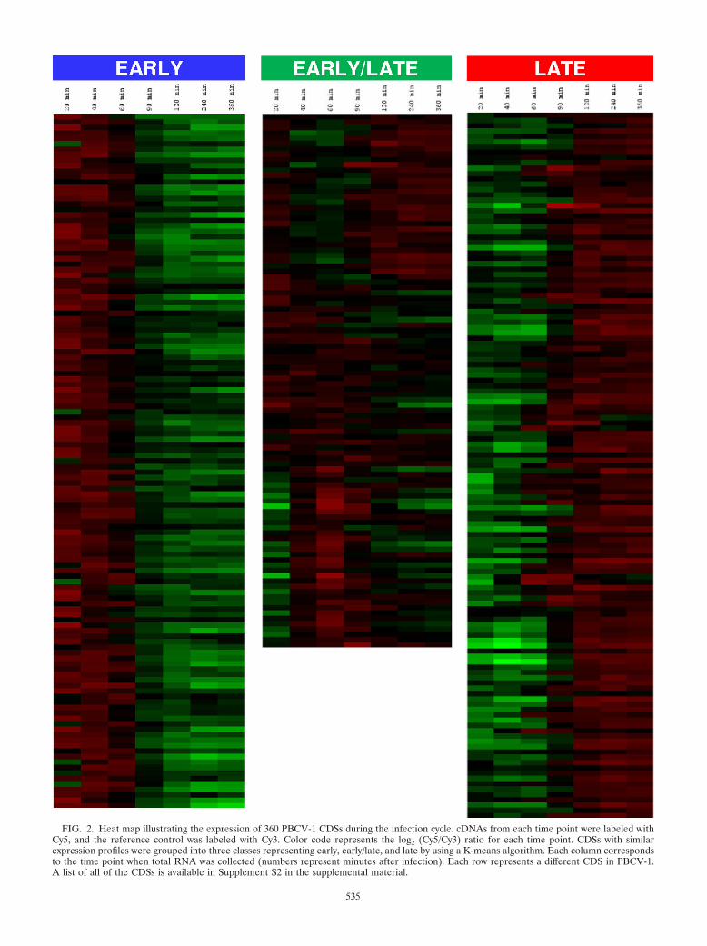
FIG. 2. Heat map illustrating the expression of 360 PBCV-1 CDSs during the infection cycle. cDNAs from each time point were labeled withCy5, and the reference control was labeled with Cy3. Color code represents the log2 (Cy5/Cy3) ratio for each time point. CDSs with similarexpression profiles were grouped into three classes representing early, early/late, and late by using a K-means algorithm. Each column correspondsto the time point when total RNA was collected (numbers represent minutes after infection). Each row represents a different CDS in PBCV-1.A list of all of the CDSs is available in Supplement S2 in the supplemental material.
535

(SWI/SNF helicase [A548L] and superfamily II helicase[A241R]), and RNase III (A464R). The genes for two enzymesinvolved in mRNA capping, an RNA triphosphatase (A449R)and a guanylyltransferase (A103R), were also transcribedearly. The products of at least some of these early CDSs areundoubtedly involved in the switching of virus early gene tran-scription to late gene transcription.
A few PBCV-1 enzymes involved in protein synthesis and
protein degradation were transcribed early, including transla-tion elongation factor-3 (A666L), ubiquitin C-terminal hydro-lase (A105L), Skp1 protein (A39L), SCF-E3 ubiquitin ligase(A481L), a zinc metallopeptidase (A604L), and an ATPase(AAA � class) (A44L).
Unlike other viruses, PBCV-1 encodes at least part, if not all, ofthe machinery required to glycosylate its major capsid protein(19), including five glycosyltransferases (11, 38, 43). Further-
FIG. 3. Mapping of the PBCV-1 transcriptome. Blue arrows, early CDSs; green arrows, early/late CDSs; red arrows, late CDSs. Arrow pointsindicate the transcription direction. Shaded areas indicate small transcription CDS clusters that are also conserved in another chlorovirus (NY-2A),except for the areas marked with asterisks. The middle circle shows the G�C content of the genome. Note that the PBCV-1 genome is linear, anda circular map was generated for illustration purposes only.
536 YANAI-BALSER ET AL. J. VIROL.

more, glycosylation of the virus major capsid protein probablyoccurs independently of the host endoplasmic reticulum-Golgisystem (19). All of the glycosyltransferase CDSs were ex-pressed early (A64R, A111/114R, A219/222/226R, A473L, andA546L).
The chlorella viruses are also unusual because they encodeenzymes involved in sugar metabolism. Two PBCV-1-encodedenzymes synthesize GDP-L-fucose from GDP-D-mannose,GDP-D-mannose dehydratase (A118R), and fucose synthase(A295L) (10, 32) and three enzymes, glucosamine synthetase(A100R), UDP-glucose dehydrogenase (A609L), and hyaluro-nan synthase (A98R), contribute to the synthesis of hyaluro-nan, a linear polysaccharide composed of alternating �-1,4-glucuronic acid and �-1,3-N-acetylglucosamine residues (6,16). The CDSs for these five enzymes were expressed early.
Early/late CDSs. A total of 100 (27%) CDSs were classifiedas early/late, 67 (67%) of which have unknown function (seeSupplement S2 in the supplemental material). This class con-tains CDSs that were expressed before 60 min p.i., but whosetranscripts were also present after PBCV-1 DNA synthesisbegins. At least three mechanisms can lead to classification ofCDSs into the early/late class: (i) the CDSs were transcribedboth before and after viral DNA replication begins; (ii) theCDSs were only transcribed prior to initiation of virus DNAsynthesis, but complete degradation of their transcripts onlyoccurred after DNA synthesis begins; and (iii) the CDSs en-code polycistronic mRNAs, e.g., one could have a dicistronicmRNA in which one CDS is required for an early function andthe other CDS is required for a late function. To add to thecomplexity, �30 transcripts were detected early, disappeared,
FIG. 4. (A) PBCV-1 global expression pattern. (B) Distribution of CDSs according to their putative function and time of expression. CDSproducts with unknown function are not listed in this graph.
VOL. 84, 2010 CHLORELLA VIRUS PBCV-1 TRANSCRIPTION 537

and then reappeared as late transcripts (these CDSs aremarked in Supplement S2 in the supplemental material). Asimilar phenomenon has been reported in transcriptional stud-ies with the bacteriophage T4 (18) and Red Sea bream iridovi-rus (17).
In addition to the early/late CDSs described in the precedingsection, the most striking feature of this early/late class was thepresence of many genes encoding proteins associated withgenome integration, including five GIY-YIG endonucleases(A134L, A287R, A315L, A495R, and A651L) and four HNHendonucleases (A354R, A422R, A478L, and A490L). Also,one of the two transposases (A366L) coded by PBCV-1 wasexpressed early/late. The functions of these proteins in thePBCV-1 life cycle are unknown.
Late CDSs. A total of 133 (37%) of the 360 CDSs wereclassified as late CDSs (see Supplement S2 in the supplementalmaterial), 74% of which have no match in the public databases.The expectation is that most late CDS products are involved ineither virus capsid assembly, DNA packaging, or virus releaseor are packaged in the virus particles. Indeed, an SDS-poly-acrylamide gel electrophoresis mass spectrometry proteomicanalysis of purified virions indicated that 118 PBCV-1 geneproducts were detected in the virions (D. D. Dunigan et al.,unpublished results). Of these 118 proteins, 83 had their cor-responding CDS transcribed late, 29 were transcribed early/
late, while only 6 virion-associated proteins were expressedearly.
At least nine late CDSs are predicted to have their geneproducts serving a purely structural role in the virion includingthe major capsid protein A430L (12) and A140/145R, which isassociated with the unique vertex of the virus (26). Otherputative structural proteins are A189/192R, A363R, A384L,A540L, A558L, A561L, and A622L. Homologs of all of theseproteins are present in all of the chloroviruses (Dunigan et al.,unpublished). In addition to structural proteins, a DNA-bind-ing protein (A437L), predicted to aid in neutralization of thevirus dsDNA, was in the virion, and its gene was transcribedlate.
Several CDSs encoding proteins with putative enzyme func-tions were expressed late, and their gene products were pack-aged in the PBCV-1 virion. Presumably, these enzymes arereleased into the cell during virus infection and aid in estab-lishing infection. These proteins include an ATPase (A561L),a superfamily II helicase (A363R), a SWI/SNF chromatin re-modeling complex subunit (A189/192R), and a SET domain-containing histone 3, Lys27 methyltransferase (named vSET)(A612L). The vSET protein is predicted to aid in the rapidinhibition of host transcription during virus infection (24). Twosite-specific (restriction) endonucleases (A252 and A579L)were also expressed as late CDSs, and their proteins are pack-
FIG. 5. Heat map showing distribution of selected CDSs according to their putative functional class. cDNAs from each time point were labeledwith Cy5, and the reference control was labeled with Cy3. The color code represents the log2 (Cy5/Cy3) ratio for each time point. Each columncorresponds to the time point when total RNA was collected (numbers represent minutes after infection). Each row represents a different PBCV-1CDS. Recombinant proteins have been characterized for the CDSs marked with an asterisk.
538 YANAI-BALSER ET AL. J. VIROL.

aged in the virion. The restriction endonucleases are involvedin host chromosome degradation, which begins within a fewminutes after virus infection (1). Thus, PBCV-1 has at leasttwo avenues to inhibit host transcription and subvert the hostRNA polymerase (presumably RNA polymerase II) for virustranscription: virus packaged restriction endonucleases and avSET histone 3, Lys27 methyltransferase.
Five of the eight PBCV-1-encoded Ser/Thr protein kinaseCDSs were expressed late (A34R, A277L, A278L, A282L, andA614L). With the exception of A277L, the other four kinaseswere packaged in the virion (Dunigan et al., unpublished). Adual specific phosphatase (A305L) was also expressed late, andthe protein is present in the virion. These results indicate thatPBCV-1 has the potential to release several protein kinase/phosphatase proteins into the cell during infection; these en-zymes are probably involved in regulatory mechanisms.
PBCV-1 encodes five proteins that degrade polysaccharides,and presumably some of these encoded proteins are involvedin host cell wall degradation either during virus entry or inaiding lysis of the cell wall during virus release (30). With theexception of � 1,3-glucanase (A94L) that was expressed early,the remaining four CDSs were transcribed late. The late CDSsencode a �- and -1,4-linked glucuronic lyase (A215L), twochitinases (A260R and A181/182R), and one chitosanase(A292L).
Finally, 49 late gene products were not detected in thevirion. Fifteen of these proteins have a putative function in-cluding, for example, a DNA packaging ATPase (A392R) thatis predicted to be involved in packaging DNA into the virion.Interestingly, some CDSs that were expected to be transcribedearly because of their putative involvement in the DNA repli-cation were transcribed late. These CDSs encode a replicationfactor C protein (A417L), which is similar to one of the tworeplication factor C proteins in Archaea, an ATP-dependentDNA ligase (A544R), and a deoxynucleoside kinase (A416R).However, none of these proteins were detected in the virion(Dunigan et al., unpublished).
Aphidicolin treatment. To confirm the early and late classi-fication of PBCV-1 CDSs, aphidicolin was used to block viralDNA synthesis. The drug was added to the culture 15 min priorto the addition of PBCV-1, and cells were harvested at thesame times after infection used in the previous experiments(20, 40, 60, 90, 120, 240, and 360 min p.i.). Each sample wasanalyzed by using the corresponding infected nontreated sam-ple as a control.
Expression of 179 (49%) CDSs was inhibited by aphidicolin(see Supplement S2 in the supplemental material). Of these179, 14 were expressed early, 57 were expressed early/late, and108 were expressed late. This experiment established that tran-scription of most late CDSs relies on the synthesis of viralDNA. In contrast, expression of 181 CDSs was not affected byaphidicolin. Of these 181, 113 were early, 43 were early/late,and 25 were late. Three CDSs (A328R, A482R, and A464L)were not previously classified as early, early/late, or late be-cause they did not pass statistical tests. However, they wereexpressed in the presence of aphidicolin, which indicates thatthese CDSs might be early. Collectively, expression of mostearly CDSs was not affected by aphidicolin and expression oflate CDSs was inhibited by the drug. We have no explanationfor why 14 early CDSs and 57 early/late CDSs were affected by
aphidicolin, nor do we know why 25 late CDSs were not af-fected by aphidicolin.
Verification of the microarray results. Three independentsets of experiments support the microarray results. (i) Recom-binant proteins from 19 PBCV-1 CDSs have been biochemi-cally characterized previously, and their genes were Northernblotted to determine when they were expressed. Sixteen ofthese results completely agree with the microarray experi-ments. The other three Northern analyses were similar, but notidentical, to the microarray analyses. One is a glycosyltrans-ferase (A64R), which Northern analysis indicated is expressedfrom 45 to 360 min p.i. (11), whereas the microarray analysisindicated that the CDS was expressed only between 40 and 60min p.i. The second CDS is a chitinase (A181/182R), whichNorthern results indicate transcription occurs between 30 and360 min p.i. (30), whereas the microarray analysis showed thetranscript was present from 60 to 360 min p.i. The third CDSis a dCMP deaminase (A596R), which Northern analysis indi-cated expression occurs from 30 to 120 min p.i. (42); the mi-croarray experiments detected the transcript at 40 to 60 minp.i. and then again from 240 to 360 min p.i. This discontinuityin expression has also been observed with a few other virusCDSs, and it has been observed in both Northern blots andin the microarray results (e.g., ornithine/arginine decarboxyl-ase [A207R] [23]). It is important to note that the microarraysmeasured relative levels of the transcript, and this fact couldexplain the few differences mentioned above.
(ii) The 46.2-Mb genome of the PBCV-1 host, ChlorellaNC64A, was recently sequenced to ninefold coverage (http://genome.jgipsf.org/ChlNC64A_1/ChlNC64A_1.info.html). Thisprompted us to initiate a transcriptome analysis of ChlorellaNC64A and PBCV-1-infected cells by shotgun sequencingcDNA derived from poly(A)� RNA using a new high-through-put sequencing instrument. To date, we have two sets of se-quences, one from uninfected Chlorella NC64A and one fromcells at 20 min p.i. We compared the 20-min p.i. viral microar-ray results with the infected 20-min p.i. viral transcriptomesequencing results. Of the 172 CDSs detected by the microar-ray analysis at 20 min p.i., expression of 159 CDSs (92%) weredetected in the transcriptome study using a 200-fold coveragecutoff (G. M. Yanai-Balser et al., unpublished results).
(iii) Kawasaki et al. (14) identified 22 immediate-earlyPBCV-1 CDSs (expressed at 5 to 10 min p.i.). Our microarrayresults indicated that 20 of these 22 CDSs were expressedearly. CDS A689L was not present in our array, and CDSA312L was classified as early/late and was not detected at 20min p.i., which is the earliest time point in the present study. Inthe later case, the difference could be due to the relativemeasurement of the transcript.
Do late PBCV-1 mRNAs have a poly(A) tail? Both the mi-croarray results and the preliminary transcriptome sequenc-ing of PBCV-1 depended on cDNA synthesis using an oli-go(dT) primer. We mention this issue for two reasons. First,about 20 years ago we conducted a set of pulse-labeling exper-iments wherein PBCV-1-infected cells were incubated with[H3]adenine for 30-min periods (36). Poly(A)� and poly(A)
RNAs were separated on an oligo(dT)-cellulose column, andthe radioactivity was determined. The results indicated that 22to 26% of the radioactivity eluted in the poly(A)� fractionfrom cells infected from 0 to 30 min and from 30 to 60 min p.i.
VOL. 84, 2010 CHLORELLA VIRUS PBCV-1 TRANSCRIPTION 539

In contrast, 6.1, 4.5, and 2.5% of the radioactivity eluted in thepoly(A)� fractions at 60 to 90, 90 to 120, and 120 to 240 minp.i., respectively. Therefore, we suggested that PBCV-1 earlymRNAs probably contain poly(A) tails and late mRNAs mightlack them. However, there are other explanations for theseearlier results, e.g., the pool size of unlabeled adenine certainlyincreases dramatically during infection; consequently, therecould be a large dilution effect on the added [H3]adeninecompared to the controls. In addition, we now know thatPBCV-1 infection leads to rapid depolarization of the hostplasma membrane, which causes an immediate decrease inadenine transport into the infected cell (2). Both of theseissues undoubtedly influenced our previous results.
Second, a report by Kawasaki et al. (14) also suggests thatthere may be a shift in poly(A)� RNAs during chlorella virusreplication. Twenty-two PBCV-1 immediate-early CDSs wereanalyzed, and the transcripts gradually decreased in size after20 min p.i., suggesting weakening or cessation of poly(A) poly-merase activity.
The current manuscript provides evidence that most, if notall, PBCV-1 mRNAs have a poly(A) tail because expression of99% of the PBCV-1 CDSs was detected in our microarrayexperiments. Furthermore, many of the CDSs we identifiedwere clearly expressed late and packaged in the virion.
PBCV-1 transcription is complex. As mentioned above, weand others have characterized several individual PBCV-1 geneproducts. In most of these reports, a Northern analysis wasconducted when studying a specific CDS. The transcriptionpatterns of �50% of these CDSs are more complicated thanjust obtaining a single hybridizing RNA band of the predictedsize at specific times. That is, full-length gene DNA probesoften hybridize to mRNA transcripts that are 40 to 60% largerthan the CDS itself, suggesting that PBCV-1 might have poly-cistronic mRNAs. In addition, some probes not only hybridizeto mRNAs of the expected size, but they also hybridize tolarger mRNAs at other times in the virus replication cycle.These complex patterns occur even with single-stranded DNAprobes (e.g., the potassium ion channel CDS, kcv) (13). Onedifference between the previous Northern analyses and themicroarray results is that hybridization to total RNA was usedin the Northern analyses and in the research described in thepresent study the cDNAs were synthesized from poly(A)�
RNAs.We examined four of the CDSs that produced larger tran-
scripts than expected to determine whether expression of theirflanking CDSs were identical in the microarray experiments tothe target CDS, possibly suggesting a polycistronic mRNA. Acommon expression pattern was obtained for two of the fourCDSs. The RNase III (A464R) mRNA has a predicted size of825 nucleotides (nt). However, in a Northern blot the RNaseIII hybridizing band is �1,300 nt. Its two adjacent CDSs are204 nt (A462R) and 354 nt (A465R) and, like A464R, both ofthem are expressed early. The combined sizes of them with theRNase III CDS are compatible with either a dicistronic or evena tricistronic mRNA. The other example is the potassium ionchannel CDS (kcv, A250R) that Northern analysis indicatedhas a complex expression pattern, at early times A250R isexpressed as a large message and at late stages as a monocis-tronic mRNA (13). However, when the early transcript for thisCDS was mapped, the start and the stop sites were within the
adjacent CDSs, so A250R is clearly not a polycistronic mRNA.The CDSs surrounding the other two CDSs with larger tran-scripts than expected, fucose synthase (A295L) and dCMPdeaminase (A596R), were expressed at different times than thetarget CDS.
It is clear that a detailed transcription analysis using a high-throughput sequencing system of PBCV-1-infected cells will berequired to precisely determine promoter and terminator sites,as well as splicing regions of PBCV-1 transcription. This anal-ysis should include the use of random primers in addition tooligo(dT) primers and also allow the detection of PBCV-1encoded small RNAs.
Microarray results with other large dsDNA viruses.PBCV-1 expressed 99% of its CDSs at some point in the viruslife cycle during infection in laboratory conditions. Similarresults were obtained for other large DNA viruses such asvaccinia virus and monkeypox viruses in which �95% of theirCDSs are expressed in cell cultures (3, 27). Transcription anal-ysis using microarrays was also performed with two marine fishiridoviruses: (i) Singapore grouper iridovirus expressed 97% ofits CDSs in cell culture (31), and (ii) Red Sea bream iridovirusexpressed 96% of its CDSs during in vitro infection (17).Therefore, it appears that most CDSs are expressed in theselarge dsDNA viruses, even in laboratory conditions.
The temporal categorization of the transcripts in large vi-ruses varies. Different studies classify virus transcripts in dif-ferent ways, usually subdividing early CDSs into more specificcategories. For example, Red Sea bream iridoviruses has �9%of its CDSs classified as immediate-early, �43% classified asearly, and �41% classified as late (17). T4 bacteriophage tran-scripts are divided into immediate-early (42%), delayed early(�12%), middle early (21%), and late (�22%) (18). Poxvirustranscription occurs in the cytoplasm of the infected cell and isprogrammed by a virus-encoded RNA polymerase and time-specific transcription factors to generate three transcriptioncategories: early, intermediate, and late (5). Using tiling arraytechnology, a recent study reveals immediate-early transcriptsin vaccinia virus (3). An additional microarray-based classifi-cation of vaccinia virus and monkeypox virus gene expressiondivided the virus CDSs into early and late categories only(�50% of the CDSs in each class). The method used in thislater study categorized CDSs according to the time the tran-script is first detected, so the intermediate class (expressedearly and also late) was not distinguished, implying that thisclass was a subgroup of the early CDSs (27). For PBCV-1, weclassified a portion of the early CDSs as early/late when thetranscripts were detected before and after the onset of viralDNA synthesis.
Conclusion. For the first time, a global mRNA transcriptionprofile was conducted for a chlorella virus. The PBCV-1 lifecycle is temporally programmed. This regulation is controlledby a precise gene expression pattern, where the time of tran-scription is dictated by initiation of viral DNA replication,which begins 60 to 90 min p.i. Early CDSs were transcribedbetween 20 and 60 min p.i., and products of the majority of theearly CDSs are responsible for providing the machinery forviral DNA replication. Late CDSs were transcribed after 60min p.i., and most of them were dependent on initiation of viralDNA synthesis, since aphidicolin blocked the expression ofmost late CDSs. Products of many late CDSs serve either a
540 YANAI-BALSER ET AL. J. VIROL.

structural role or were packaged in the virion, presumably toaid in virus infection. A total of 46% of the early transcriptswere still detected after DNA synthesis begins and were calledearly/late CDSs, indicating a complex mechanism for mRNAmaturation and degradation. Some of these early/late CDSsmay only be synthesized early, but their transcripts were notdegraded until after DNA synthesis begins. However, this sce-nario probably does not apply to all early/late CDSs becauseseveral early-late CDS products were packaged in the virion.These CDSs may also be transcribed as late CDSs.
The present study reveals that PBCV-1 gene expression timeis independent of GC content, as well as the transcriptiondirection and CDS location in the genome. It is anticipatedthat the temporal transcription map will provide clues to thefunctions of the large category of viral proteins whose func-tions are not yet known and to the events that must be orches-trated for a successful viral infection.
ACKNOWLEDGMENTS
We thank James Gurnon for technical help and maintaining theculture conditions and Tong Yin, Justin Jackson, and other membersof the Microarray Core Facility for technical support. We also thankthe Drug Synthesis and Chemistry Branch, Developmental Therapeu-tics Program, Division of Cancer Treatment and Diagnosis, NationalCancer Institute, for generously providing aphidicolin.
This research was supported in part by Public Health Service grantGM32441 (J.L.V.E.) and NIH grant P21RR15635 from the COBREprogram of the National center for Research Resources (J.L.V.E.).The UNMC Microarray Core Facility receives partial support fromNIH grant P20 RR016469 from the INBRE program (G.A.D. andJ.D.E.).
REFERENCES
1. Agarkova, I. V., D. D. Dunigan, and J. L. Van Etten. 2006. Virion-associatedrestriction endonucleases of chloroviruses. J. Virol. 80:8114–8123.
2. Agarkova, I. V., D. Dunigan, J. Gurnon, T. Greiner, J. Barres, G. Thiel, andJ. L. Van Etten. 2008. Chlorovirus-mediated membrane depolarization ofchlorella alters secondary active transport of solutes. J. Virol. 82:12181–12190.
3. Assarsson, E., J. A. Greenbaum, M. Sundstrom, L. Schaffer, J. A. Hammond,V. Pasquetto, C. Oseroff, R. C. Hendrickson, E. J. Lefkowitz, D. C. Tscharke,J. Sidney, H. M. Grey, S. R. Head, B. Peters, and A. Sette. 2008. Kineticanalysis of a complete poxvirus transcriptome reveals an immediate-earlyclass of genes. Proc. Natl. Acad. Sci. USA 105:2140–2145.
4. Cherrier, M. V., V. A. Kostyuchenko, C. Xiao, V. D. Bowman, A. J. Battisti,X. Yan, P. R. Chipman, T. S. Baker, J. L. Van Etten, and M. G. Rossmann.2009. An icosahedral algal virus has a complex unique vertex decorated by aspike. Proc. Natl. Acad. Sci. USA 106:11085–11090.
5. Condit, R. C. 2007. Vaccinia, Inc.: probing the functional substrate of pox-viral replication factories. Cell Host Microbe 2:205–207.
6. DeAngelis, P., W. Jing, M. V. Graves, D. E. Burbank, and J. L. Van Etten.1997. Hyaluronan synthase of chlorella virus PBCV-1. Science 278:1800–1803.
7. Fitzgerald, L. A., P. T. Boucher, G. M. Yanai-Balser, K. Suhre, M. V. Graves,and J. L. Van Etten. 2008. Putative gene promoter sequences in the chlorellaviruses. Virology 380:388–393.
8. Fitzgerald, L. A., M. V. Graves, X. Li, T. Feldblyum, W. C. Nierman, andJ. L. Van Etten. 2007. Sequence and annotation of the 369-kb NY-2A andthe 345-kb AR158 viruses that infect Chlorella NC64A. Virology 358:472–484.
9. Frohns, F., A. Kasmann, D. Kramer, B. Schafer, M. Mehmel, M. Kang, J. L.Van Etten, S. Gazzarrini, A. Moroni, and G. Thiel. 2006. Potassium ionchannels of chlorella viruses cause rapid depolarization of host cells duringinfection. J. Virol. 80:2437–2444.
10. Fruscione, F., L. Sturla, G. A. Duncan, J. L. Van Etten, P. Valbuzzi, A. DeFlora, E. D. Zanni, and M. Tonetti. 2008. Differential role of NADP� andNADPH in the activity and structure of GDP-D-mannose 4,6-dehydratasefrom two chlorella viruses. J. Biol. Chem. 283:184–193.
11. Graves, M. V., C. T. Bernardt, R. Cerny, and J. L. Van Etten. 2001. Molec-ular and genetic evidence for a virus-encoded glycosyltransferase involved inprotein glycosylation. Virology 285:332–345.
12. Graves, M. V., and R. H. Meints. 1992. Characterization of the major capsid
protein and cloning of its gene from algal virus PBCV-1. Virology 188:198–207.
13. Kang, M., M. Graves, M. Mehmel, A. Moroni, S. Gazzarrini, G. Thiel, J. R.Gurnon, and J. L. Van Etten. 2004. Genetic diversity in chlorella virusesflanking kcv, a gene that encodes a potassium ion channel protein. Virology326:150–159.
14. Kawasaki, T., M. Tanaka, M. Fujie, S. Usami, and T. Yamada. 2004. Im-mediate early genes expressed in chlorovirus infections. Virology 318:214–223.
15. Knapen, D., L. Vergauwen, K. Laukens, and R. Blust. 2009. Best practicesfor hybridization design in two-colour microarray analysis. Trends Biotech-nol. 27:406–414.
16. Landstein, D., M. V. Graves, D. E. Burbank, P. DeAngelis, and J. L. VanEtten. 1998. Chlorella virus PBCV-1 encodes functional glutamine:fructose-6-phosphate amidotransferase and UDP-glucose dehydrogenase enzymes.Virology 250:388–396.
17. Lua, D. T., M. Yasuike, I. Hirono, and T. Aoki. 2005. Transcription programof Red Sea bream iridovirus as revealed by DNA microarrays. J. Virol.79:15151–15164.
18. Luke, K., A. Radek, X. Liu, J. Campbell, M. Uzan, R. Haselkorn, and Y.Kogan. 2002. Microarray analysis of gene expression during bacteriophageT4 infection. Virology 299:182–191.
19. Markine-Goriaynoff, N., L. Gillet, J. L. Van Etten, H. Korres, N. Verma, andA. Vanderplasschen. 2004. Glycosyltransferases encoded by viruses. J. Gen.Virol. 85:2741–2754.
20. McCullough, A. K., M. T. Romberg, S. Nyaga, Y. Wei, T. G. Wood, J. S.Taylor, J. L. Van Etten, M. L. Dodson, and R. S. Lloyd. 1998. Characteriza-tion of a novel cis-syn and trans-syn II pyrimidine dimer glycosylase/AP lyasefrom a eukaryotic algal virus, PBCV-1. J. Biol. Chem. 273:13136–13142.
21. Meints, R. H., K. Lee, and J. L. Van Etten. 1986. Assembly site of the virusPBCV-1 in a chlorella-like green alga: ultrastructural studies. Virology 154:240–245.
22. Meints, R. H., K. Lee, D. E. Burbank, and J. L. Van Etten. 1984. Infectionof a chlorella-like alga with the virus, PBCV-1: ultrastructural studies. Vi-rology 138:341–346.
23. Morehead, T. A., J. R. Gurnon, B. Adams, K. W. Nickerson, L. A. Fitzgerald,and J. L. Van Etten. 2002. Ornithine decarboxylase encoded by chlorellavirus PBCV-1. Virology 301:165–175.
24. Mujtaba, S., K. L. Manzur, J. R. Gurnon, M. Kang, J. L. Van Etten, andM.-M. Zhou. 2008. Epigenetic transcriptional repression of cellular genes bya viral SET protein. Nat. Cell Biol. 10:1114–1122.
25. Neupartl, M., C. Meyer, I. Woll, F. Frohns, M. Kang, J. L. Van Etten, D.Kramer, B. Hertel, A. Moroni, and G. Thiel. 2008. Chlorella viruses evoke arapid release of K� from host cells during the early phase of infection.Virology 372:340–348.
26. Onimatsu, H., K. Suganuma, S. Uenoyama, and T. Yamada. 2006. C-termi-nal repetitive motifs in Vp130 present at the unique vertex of the chloroviruscapsid are essential for binding to the host chlorella cell wall. Virology353:433–442.
27. Rubins, K. H., L. E. Hensley, G. W. Bell, C. Wang, E. J. Lefkowitz, P. O.Brown, and D. A. Relman. 2008. Comparative analysis of viral gene expres-sion programs during poxvirus infection: a transcriptional map of the vac-cinia and monkeypox genomes. PLoS ONE 3:e2628.
28. Saeed, A. I., V. Sharov, J. White, J. Li, W. Liang, N. Bhagabati, J. Braisted,M. Klapa, T. Currier, M. Thiagarajan, A. Sturn, M. Snuffin, A. Rezantsev,D. Popov, A. Ryltsov, E. Kostukovich, I. Borisovsky, Z. Liu, A. Vinsavich, V.Trush, and J. Quackenbush. 2003. TM4: a free, open-source system formicroarray data management and analysis. BioTechniques 34:374–378.
29. Schuster, A. M., L. Girton, D. E. Burbank, and J. L. Van Etten. 1986.Infection of a chlorella-like alga with the virus PBCV-1: transcriptionalstudies. Virology 148:181–189.
30. Sun, L., B. Adams, J. R. Gurnon, Y. Ye, and J. L. Van Etten. 1999. Charac-terization of two chitinase genes and one chitosanase gene encoded bychlorella virus PBCV-1. Virology 263:376–387.
31. Teng, Y., Z. Hou, J. Gong, H. Liu, X. Xie, L. Zhang, X. Chen, and Q. W. Qin.2008. Whole-genome transcriptional profiles of a novel marine fish iridovi-rus, Singapore grouper iridovirus (SGIV) in virus-infected grouper spleencell cultures and in orange-spotted grouper, Epinephulus coioides. Virology377:39–48.
32. Tonetti, M., D. Zanardi, J. R. Gurnon, F. Fruscione, A. Armirotti, G.Damonte, L. Sturla, A. De Flora, and J. L. Van Etten. 2003. Chlorella virusPBCV-1 encodes two enzymes involved in the biosynthesis of GDP-L-fucoseand GDP-D-rhamnose. J. Biol. Chem. 278:21672–21677.
33. Tusher, V. G., R. Tibshirani, and G. Chu. 2001. Significance analysis ofmicroarrays applied to the ionizing radiation response. Proc. Natl. Acad. Sci.USA 98:5116–5121.
34. Van Etten, J. L. 2003. Unusual lifestyle of giant chlorella viruses. Annu. Rev.Genet. 37:153–195.
35. Van Etten, J. L., A. M. Schuster, L. Girton, D. E. Burbank, D. Swinton, andS. Hattman. 1985. DNA methylation of viruses infecting a eukaryotic chlo-rella-like green alga. Nucleic Acids Res. 13:3471–3478.
36. Van Etten, J. L., A. M. Schuster, and R. H. Meints. 1988. Viruses of eukary-
VOL. 84, 2010 CHLORELLA VIRUS PBCV-1 TRANSCRIPTION 541

otic chlorella-like algae, p. 411–428. In Y. Koltin and M. J. Leibowitz (ed.),Viruses of fungi and simple eukaryotes. Marcel Dekker, Inc., New York, NY.
37. Van Etten, J. L., D. E. Burbank, J. Joshi, and R. H. Meints. 1984. DNAsynthesis in a chlorella-like alga following infection with the virus PBCV-1.Virology 134:443–449.
38. Wang, I.-N., Y. Li, Q. Que, M. Bhattacharya, L. C. Lane, W. G. Chaney, andJ. L. Van Etten. 1993. Evidence for virus-encoded glycosylation specificity.Proc. Natl. Acad. Sci. USA 90:3840–3844.
39. Wilson, W. H., J. L. Van Etten, and M. J. Allen. 2008. The Phycodnaviri-dae: the story of how tiny giants rule the world, p. 1–42. In J. L. Van Etten(ed.), Lesser known large dsDNA viruses. Springer-Verlag, Berlin,Germany.
40. Yamada, T., H. Onimatsu, and J. L. Van Etten. 2006. Chlorella viruses, p.293–336. In K. Maramorosch and A. J. Shatkin (ed.), Advances in virusresearch. Elsevier Academic Press, Inc., San Diego, CA.
41. Yang, Y. H., and T. Speed. 2002. Design issues for cDNA microarray exper-iments. Nat. Rev. 3:579–588.
42. Zhang, Y., F. Maley, G. F. Maley, G. Duncan, D. D. Dunigan, and J. L. VanEtten. 2007. Chloroviruses encode a bifunctional dCMP-dCTP deaminasethat produces two key intermediates in dTTP formation. J. Virol. 81:7662–7671.
43. Zhang, Y., Y. Xiang, J. L. Van Etten, and M. G. Rossmann. 2007. Structureand function of a chlorella virus PBCV-1 encoded glycosyltransferase. Struc-ture 15:1031–1039.
542 YANAI-BALSER ET AL. J. VIROL.
Copyright © 2022 FDOKUMEN