Melanoma Antigen11 Inhibits the Hypoxia-Inducible Factor Prolyl Hydroxylase 2 and Activates Hypoxic...
10
2009;69:616-624. Cancer Res Olga Aprelikova, Silvia Pandolfi, Sean Tackett, et al. Prolyl Hydroxylase 2 and Activates Hypoxic Response Melanoma Antigen-11 Inhibits the Hypoxia-Inducible Factor Updated version http://cancerres.aacrjournals.org/content/69/2/616 Access the most recent version of this article at: Material Supplementary http://cancerres.aacrjournals.org/content/suppl/2009/01/15/69.2.616.DC1.html Access the most recent supplemental material at: Cited Articles http://cancerres.aacrjournals.org/content/69/2/616.full.html#ref-list-1 This article cites by 45 articles, 33 of which you can access for free at: Citing articles http://cancerres.aacrjournals.org/content/69/2/616.full.html#related-urls This article has been cited by 8 HighWire-hosted articles. Access the articles at: E-mail alerts related to this article or journal. Sign up to receive free email-alerts Subscriptions Reprints and . [email protected] Department at To order reprints of this article or to subscribe to the journal, contact the AACR Publications Permissions . [email protected] Department at To request permission to re-use all or part of this article, contact the AACR Publications Research. on April 7, 2014. © 2009 American Association for Cancer cancerres.aacrjournals.org Downloaded from Research. on April 7, 2014. © 2009 American Association for Cancer cancerres.aacrjournals.org Downloaded from
Transcript of Melanoma Antigen11 Inhibits the Hypoxia-Inducible Factor Prolyl Hydroxylase 2 and Activates Hypoxic...
2009;69:616-624. Cancer Res Olga Aprelikova, Silvia Pandolfi, Sean Tackett, et al. Prolyl Hydroxylase 2 and Activates Hypoxic ResponseMelanoma Antigen-11 Inhibits the Hypoxia-Inducible Factor
Updated version
http://cancerres.aacrjournals.org/content/69/2/616
Access the most recent version of this article at:
Material
Supplementary
http://cancerres.aacrjournals.org/content/suppl/2009/01/15/69.2.616.DC1.html
Access the most recent supplemental material at:
Cited Articles
http://cancerres.aacrjournals.org/content/69/2/616.full.html#ref-list-1
This article cites by 45 articles, 33 of which you can access for free at:
Citing articles
http://cancerres.aacrjournals.org/content/69/2/616.full.html#related-urls
This article has been cited by 8 HighWire-hosted articles. Access the articles at:
E-mail alerts related to this article or journal.Sign up to receive free email-alerts
Subscriptions
Reprints and
To order reprints of this article or to subscribe to the journal, contact the AACR Publications
Permissions
To request permission to re-use all or part of this article, contact the AACR Publications
Research. on April 7, 2014. © 2009 American Association for Cancercancerres.aacrjournals.org Downloaded from Research.
on April 7, 2014. © 2009 American Association for Cancercancerres.aacrjournals.org Downloaded from
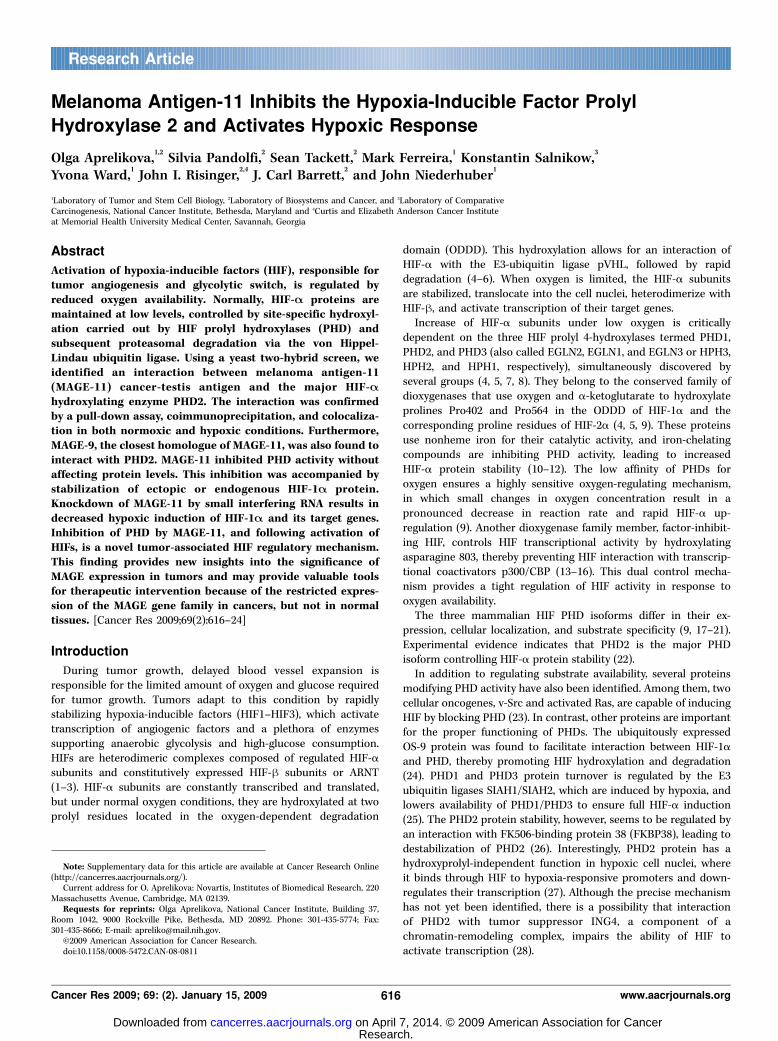
Melanoma Antigen-11 Inhibits the Hypoxia-Inducible Factor Prolyl
Hydroxylase 2 and Activates Hypoxic Response
Olga Aprelikova,1,2
Silvia Pandolfi,2Sean Tackett,
2Mark Ferreira,
1Konstantin Salnikow,
3
Yvona Ward,1John I. Risinger,
2,4J. Carl Barrett,
2and John Niederhuber
1
1Laboratory of Tumor and Stem Cell Biology, 2Laboratory of Biosystems and Cancer, and 3Laboratory of ComparativeCarcinogenesis, National Cancer Institute, Bethesda, Maryland and 4Curtis and Elizabeth Anderson Cancer Instituteat Memorial Health University Medical Center, Savannah, Georgia
Abstract
Activation of hypoxia-inducible factors (HIF), responsible fortumor angiogenesis and glycolytic switch, is regulated byreduced oxygen availability. Normally, HIF-A proteins aremaintained at low levels, controlled by site-specific hydroxyl-ation carried out by HIF prolyl hydroxylases (PHD) andsubsequent proteasomal degradation via the von Hippel-Lindau ubiquitin ligase. Using a yeast two-hybrid screen, weidentified an interaction between melanoma antigen-11(MAGE-11) cancer-testis antigen and the major HIF-Ahydroxylating enzyme PHD2. The interaction was confirmedby a pull-down assay, coimmunoprecipitation, and colocaliza-tion in both normoxic and hypoxic conditions. Furthermore,MAGE-9, the closest homologue of MAGE-11, was also found tointeract with PHD2. MAGE-11 inhibited PHD activity withoutaffecting protein levels. This inhibition was accompanied bystabilization of ectopic or endogenous HIF-1A protein.Knockdown of MAGE-11 by small interfering RNA results indecreased hypoxic induction of HIF-1A and its target genes.Inhibition of PHD by MAGE-11, and following activation ofHIFs, is a novel tumor-associated HIF regulatory mechanism.This finding provides new insights into the significance ofMAGE expression in tumors and may provide valuable toolsfor therapeutic intervention because of the restricted expres-sion of the MAGE gene family in cancers, but not in normaltissues. [Cancer Res 2009;69(2):616–24]
Introduction
During tumor growth, delayed blood vessel expansion isresponsible for the limited amount of oxygen and glucose requiredfor tumor growth. Tumors adapt to this condition by rapidlystabilizing hypoxia-inducible factors (HIF1–HIF3), which activatetranscription of angiogenic factors and a plethora of enzymessupporting anaerobic glycolysis and high-glucose consumption.HIFs are heterodimeric complexes composed of regulated HIF-asubunits and constitutively expressed HIF-h subunits or ARNT(1–3). HIF-a subunits are constantly transcribed and translated,but under normal oxygen conditions, they are hydroxylated at twoprolyl residues located in the oxygen-dependent degradation
domain (ODDD). This hydroxylation allows for an interaction ofHIF-a with the E3-ubiquitin ligase pVHL, followed by rapiddegradation (4–6). When oxygen is limited, the HIF-a subunitsare stabilized, translocate into the cell nuclei, heterodimerize withHIF-h, and activate transcription of their target genes.Increase of HIF-a subunits under low oxygen is critically
dependent on the three HIF prolyl 4-hydroxylases termed PHD1,PHD2, and PHD3 (also called EGLN2, EGLN1, and EGLN3 or HPH3,HPH2, and HPH1, respectively), simultaneously discovered byseveral groups (4, 5, 7, 8). They belong to the conserved family ofdioxygenases that use oxygen and a-ketoglutarate to hydroxylateprolines Pro402 and Pro564 in the ODDD of HIF-1a and thecorresponding proline residues of HIF-2a (4, 5, 9). These proteinsuse nonheme iron for their catalytic activity, and iron-chelatingcompounds are inhibiting PHD activity, leading to increasedHIF-a protein stability (10–12). The low affinity of PHDs foroxygen ensures a highly sensitive oxygen-regulating mechanism,in which small changes in oxygen concentration result in apronounced decrease in reaction rate and rapid HIF-a up-regulation (9). Another dioxygenase family member, factor-inhibit-ing HIF, controls HIF transcriptional activity by hydroxylatingasparagine 803, thereby preventing HIF interaction with transcrip-tional coactivators p300/CBP (13–16). This dual control mecha-nism provides a tight regulation of HIF activity in response tooxygen availability.The three mammalian HIF PHD isoforms differ in their ex-
pression, cellular localization, and substrate specificity (9, 17–21).Experimental evidence indicates that PHD2 is the major PHDisoform controlling HIF-a protein stability (22).In addition to regulating substrate availability, several proteins
modifying PHD activity have also been identified. Among them, twocellular oncogenes, v-Src and activated Ras, are capable of inducingHIF by blocking PHD (23). In contrast, other proteins are importantfor the proper functioning of PHDs. The ubiquitously expressedOS-9 protein was found to facilitate interaction between HIF-1aand PHD, thereby promoting HIF hydroxylation and degradation(24). PHD1 and PHD3 protein turnover is regulated by the E3ubiquitin ligases SIAH1/SIAH2, which are induced by hypoxia, andlowers availability of PHD1/PHD3 to ensure full HIF-a induction(25). The PHD2 protein stability, however, seems to be regulated byan interaction with FK506-binding protein 38 (FKBP38), leading todestabilization of PHD2 (26). Interestingly, PHD2 protein has ahydroxyprolyl-independent function in hypoxic cell nuclei, whereit binds through HIF to hypoxia-responsive promoters and down-regulates their transcription (27). Although the precise mechanismhas not yet been identified, there is a possibility that interactionof PHD2 with tumor suppressor ING4, a component of achromatin-remodeling complex, impairs the ability of HIF toactivate transcription (28).
Note: Supplementary data for this article are available at Cancer Research Online(http://cancerres.aacrjournals.org/).Current address for O. Aprelikova: Novartis, Institutes of Biomedical Research, 220
Massachusetts Avenue, Cambridge, MA 02139.Requests for reprints: Olga Aprelikova, National Cancer Institute, Building 37,
Room 1042, 9000 Rockville Pike, Bethesda, MD 20892. Phone: 301-435-5774; Fax:301-435-8666; E-mail: [email protected].
I2009 American Association for Cancer Research.doi:10.1158/0008-5472.CAN-08-0811
Cancer Res 2009; 69: (2). January 15, 2009 616 www.aacrjournals.org
Research Article
Research. on April 7, 2014. © 2009 American Association for Cancercancerres.aacrjournals.org Downloaded from
To better understand the regulation of PHDs in hypoxic tumorcells, we performed a yeast two-hybrid screen, using PHD2 as a baitand a library prepared from PC3 prostate cancer cells subjected to24 hours of hypoxia (1% oxygen). This screen identified some of theknown PHD2 interacting proteins. Importantly, we found a novelinteraction of PHD2 with melanoma antigen-11 (MAGE-11 orMAGE-A11). MAGE-11 is a member of a family of 12 MAGE-Aproteins, representing a group of cancer-testis antigens expressedin some embryonic tissues but not in normal adult organs, with theexception of testis and placenta, and aberrantly reexpressed indifferent types of tumors. We focused on discovering what effectsMAGE-11 protein may have on PHD2 because the MAGE-A familyrepresents an ideal target for therapeutic intervention due to itsrestricted expression pattern. We found that MAGE-11 interactswith PHD2 in both normoxic and hypoxic conditions. MAGE-11protein itself is not regulated by hypoxia and does not changePHD2 protein levels but rather suppresses PHD2 activity. Down-regulation of MAGE-11 by RNA interference results in diminishedHIF-1a induction and transcriptional activity in hypoxia. Given thecommon re-expression of MAGE and other cancer-testis antigensin cancers, this finding provides a novel mechanism of action ofthese proteins.
Materials and Methods
Yeast two-hybrid screen. Twelve different PHD2 fragments, cloned intopGBT.superB vector (Myriad Genetics), were used as bait constructs in the
yeast two-hybrid analysis. All baits were first tested in a yeast one-hybridassay to eliminate self-activation of transcription. The activation domain
prey library was made in pGAD.PN2 vector (Myriad Genetics) using the PC3
prostate cancer cell line exposed to 1% oxygen for 24 h. Yeast strain PNY200was used to maintain the bait plasmids, and yeast strain BK100 was used to
maintain the prey constructs. After mating, colonies were selected using
two auxotrophic marker genes, HIS3 and ADE2. To confirm the interactions,
the bait and prey plasmids were cotransfected into yeast and interactionwas verified using the third reporter gene, LacZ.
Cell lines and reagents. HeLa, U2OS, A549, 786-O, MDA-MB435, and293T cells were maintained in DMEM (high glucose) supplemented with
10% fetal bovine serum (FBS; Invitrogen) and penicillin/streptomycin(Invitrogen) in humidified air containing 5% CO2 at 37jC. Hypoxiatreatment was carried out in a modular incubator chamber (Billaps-
Rothenberg) with the mixture containing different percentage of oxygenand 5% CO2 and balanced with nitrogen. Deferoxamine mesylate (DFO),
O-phenanthroline, proteasomal inhibitor MG132, and dimethyloxallyl
glycine were purchased from Sigma-Aldrich, Inc.
Plasmids. MAGE-11 amino acids 2-429 were PCR-amplified from ahuman testis cDNA library (Clontech) and subcloned into the 3� Flag-
CMV10 expression vector (Sigma) or pCR2.1-TOPO (Invitrogen) for in vitro
translation or into pGEX5x-1 (Amersham) for expression in Escherichia coli .
Full-length MAGE-4 and MAGE-9 clones were part of the Open BiosystemsHuman ORF collection in the pDONR223 gateway entry vector. These clones
were transferred into the destination vector pDEST-510 with a COOH
terminal Flag tag (National Cancer Institute), using homologous recombi-
nation with a Gateway LR Clonase II enzyme mix (Invitrogen). All plasmidswere sequenced to verify the authenticity.
The pSG5-MAGE-11 plasmid was kindly provided by Dr. Elizabeth Wilson
(University of North Carolina at Chapel Hill). The GHO-plasmid wasfrom Dr. M.C. Simon (University of Pennsylvania). V5-PHD2 was from
Dr. R. Bruick (University of Texas Southwestern Medical Center), and Flag-
tagged PHD1-PHD3 were from Dr. F.S. Lee (University of Pennsylvania).
Cell transfection, immunoprecipitation, and immunoblotting. Cellswere transfected with an Effectene reagent (Qiagen). Briefly, cells were
trypsinized and counted, and 2 � 106 for 293T or 1.5 � 106 cells for othercell lines were mixed with the DNA-transfection reagent complex made
with 1 to 2 Ag DNA, according to the manufacturer’s instructions, and
plated on a 60-mm plate. For immunoprecipitation experiments, the
transfection reactions were scaled up to 100-mm plates. For coimmuno-
precipitation, cells were lysed in NP40 buffer containing 50 mmol/L Tris-
HCl (pH 7.5), 150 mmol/L NaCl, 0.5% NP40, and protease (Sigma) and
phosphatase (Pierce) inhibitors. Total protein (300 Ag) was used forimmunoprecipitation. Cell lysates were precleared with normal mouse IgG
and protein A/G Sepharose (Santa Cruz). The agarose conjugated with anti-
V5 antibody (Invitrogen) was washed several times with NP40 buffer and
blocked with 5% bovine serum albumin (BSA) for 1 h, and 30 AL were usedto collect the protein complexes for 4 to 16 h at 4jC. The agarose waswashed with NP40 buffer thrice, and bound proteins were resolved in 10%
SDS-PAGE. Immunoprecipitation of endogenous PHD2 and MAGE-11 from
HeLa cells was performed with f1 mg of total protein. The lysates wereprecleared with A/G Sepharose (Santa Cruz), and complexes of PHD2 were
immunoprecipitated with 5 Ag of anti-PHD2 antibody (Novus Biologicals)The complexes were collected with 40 AL of protein A/G Sepharose for 1 h at4jC and washed four times with lysis buffer supplemented with 1 mg/mLBSA and twice with the same buffer without BSA.
The nuclear extracts were prepared by scraping cells into cold PBS
followed by a low-speed centrifugation. Cell pellets were resuspended in
400 AL of cold hypotonic buffer [10 mmol/L Tris-HCl (pH 7.5), 1.5 mmol/LMgCl2, 10 mmol/L KCl, 0.5 mmol/L DTT, 0.2 mmol/L phenylmethylsulfonyl
fluoride (PMSF)] and allowed to swell on ice for 10 min. After
centrifugation, the supernatant fraction was discarded and the pellets were
resuspended in 50 to 70 AL of high-salt buffer [20 mmol/L of Tris-HCl(pH 7.5), 25% glycerol, 420 mmol/L NaCl, 1.5 mmol/L MgCl2, 0.2 mmol/L
EDTA, 0.5 mmol/L DTT, 0.2 mmol/L PMSF] and incubated on ice for 20 min
followed by high-speed centrifugation.
The antibodies used for Western blotting were anti–Flag-horseradishperoxidase (HRP; Sigma), anti–V5-HRP (Invitrogen), anti-PHD2 rabbit
polyclonal (Novus Biologicals), anti–HIF-1a (BD Biosciences), anti-HA
(Cell Signaling), anti–MAGE-11 (MabAb94, kindly provided by Dr. Wilson,University of North Carolina at Chapel Hill), anti–von Hippel-Lindau (VHL;
BD Biosciences), anti–a-tubulin (Sigma), and anti-actin (Sigma).Small interfering RNA transfections. Small interfering RNAs (siRNA)
for MAGE-11 knockdown were purchased from Dharmacon (D-017280-03),as well as nonsilencing control siRNA 2. The duplexes were transfected into
HeLa or U2OS cells, as described before (29), except that HeLa cells were
transfected with 100 nmol/L of siRNA duplex. Stable transfection of MDA-
MB435 cell line was performed with retroviral vector pSM2, expressingshMAGE11 in the context of mir-30 (Open Biosystems, clone V2HS_37013)
or with the nonsilencing short hairpin RNA mir control (Open Biosystems,
RHS1703) followed by selection with 2 Ag/mL of puromycin.VHL-binding assay. After transfection, cells were scraped in ice-cold
reaction buffer containing 20 mmol/L Tris-HCl (pH 7.5), 5 mmol/L KCl,
1.5 mmol/L MgCl2, and protease inhibitor cocktail and disrupted by
sonication. Synthetic peptide containing 19 HIF-1a amino acids, DLDLEM-LAPYIPMDDDFQL or DLDLEMLAP(OH)YIPMDDDFQL, biotinylated on
NH2 terminus (2 Ag/reaction) were first captured with streptavidin-conjugated magnetic beads in 200 AL of NP40 buffer for 2 h. The beadswere washed twice in NP40 buffer and twice in the reaction buffer andincubated with 50 to 100 Ag of total protein in the presence of 0.5 mmol/La-ketoglutarate, 0.1 mmol/L FeCl2, and 0.5 mmol/L ascorbate for 30 min at30jC. The beads were washed thrice with NP40 buffer and incubated with10 AL of in vitro translated HA-VHL for 1 h at 4jC. The beads were washedthrice with NP40 buffer, and bound HA-VHL was released with SDS loading
buffer, resolved in 10% SDS-PAGE, and identified with anti-HA antibody.
Quantitative real-time PCR. Total RNA was purified with Trizol reagent(Invitrogen), and 4 Ag were converted into cDNA using M-MLV reversetranscriptase and oligo dT primer (Invitrogen). cDNA (1 AL) was used forPCR with PCR master mix (Applied Biosystems), and 1.25 AL of 20� primer-probe mix were used for individual TaqMan assays (Applied Biosystems).Every measurement was done in triplicate. The amount of h-actintranscripts was determined in separate reactions, and the final data were
normalized to the amount of h-actin and presented as a fold changecompared with control cells. MAGE-11 primers used for PCR in quantitativereverse transcription–PCR (RT-PCR) reaction were spanning the boundaries
MAGE-11 Inhibits PHD2 Activity
www.aacrjournals.org 617 Cancer Res 2009; 69: (2). January 15, 2009
Research. on April 7, 2014. © 2009 American Association for Cancercancerres.aacrjournals.org Downloaded from
of exons 2 and 3 (Applied Biosystems, Hs00377815_m1), bHLHB2 (exons
2 and 3; Applied Biosystems, Hs00186419_m1), and NDRG1 (exons 13 and
14; Applied Biosystems, Hs00608389_m1).Luciferase reporter assay. Cells were split at 0.5 � 105 into 12-well
plates and transfected on the next day with 200 ng of luciferase-reporter
plasmid, 5 ng of RL-CMV, 200 ng of HIF-1a, 10 ng of V5-PHD2, and 100 ng ofFlag-MAGE-11 using Fugene 6 Reagent (Roche). Whenever one of the DNAcomponents was eliminated, the total amounts of DNA were balanced with
pcDNA3 empty vector. All transfections were performed in triplicates. Two
days after transfection, cells were lysed in passive lysis buffer (Promega) and20 AL were used to measure the activity of firefly and Renilla luciferase withdual luciferase reporter assay system (Promega).
Confocal microscopy. HeLa cells were grown on coverslips and, on thenext day, were placed in 0.5% oxygen for 16 h or left untreated. Cellswere fixed with 4% paraformaldehyde for 10 min and then treated with
0.1 mol/L glycin for 1 h. After three washings with PBS, cells were
permeabilized with 0.2% Triton X-100 for 4 min, washed again, and
blocked with 10% normal goat serum in PBS. Primary antibody anti–MAGE-11 (affinity purified rabbit polyclonal) and anti-PHD2 (mouse
polyclonal, Novus Biologicals) at 1:100 dilution in the blocking solution
were applied overnight at 4jC. Secondary antibody anti-mouse labeledwith Alexa 486 or anti-rabbit labeled with Alexa 568 (both fromInvitrogen) was diluted at 1:500 with the blocking solution and incubated
with cells for 1 h at room temperature. Cell nuclei were stained with 4¶,6-diamidino-2-phenylindole using Prolong Gold Antifade Reagent (Invitro-gen). Stained cells were observed on a Zeiss Axiovert 100M microscope
equipped with a 100�/1.3oil Plan NeoFluar objective. Confocal imageswere obtained using an LSM510 scanning laser microscope (Zeiss).
Controls containing no primary antibody were performed in parallel
samples and showed no staining.
Results
Identification of PHD2-MAGE-11 interaction. Most solidtumors exhibit some degree of oxygen deprivation. Therefore, wesought to identify proteins that regulate PHD activity underconditions of limited oxygen supply. We performed a yeast two-hybrid analysis using PHD2 as bait, and a prey library made fromhypoxic PC3 prostate cancer cells exposed to 1% oxygen for24 h. A total of 12 fragments of PHD2 was used in an effort tomaximize likely binding partners. Of these, we found multipleinteractions of PHD2 bait constructs (spanning the region of aminoacids 70-420) with the HIF-2a protein. Interestingly, we did notfind an interaction with HIF-1a in this screen, implying either thatthe interaction of PHD2 with HIF-2a protein is more stable thanwith HIF-1a or that HIF-1a may require some additionalmodifications compromised in the yeast strains used in the study.Furthermore, in agreement with the recent publication of Barthand colleagues (26), we found three different PHD2 baits, alllocated between amino acids 1 and 180, interacting with multipleFKBP38 clones. In addition, we found a novel interaction of PHD2(amino acids 202-301) with the melanoma antigen MAGE-11. Thenormal or tumor-specific functions of MAGE-A proteins are poorlyunderstood. MAGE-11 was recently identified as a scaffolding
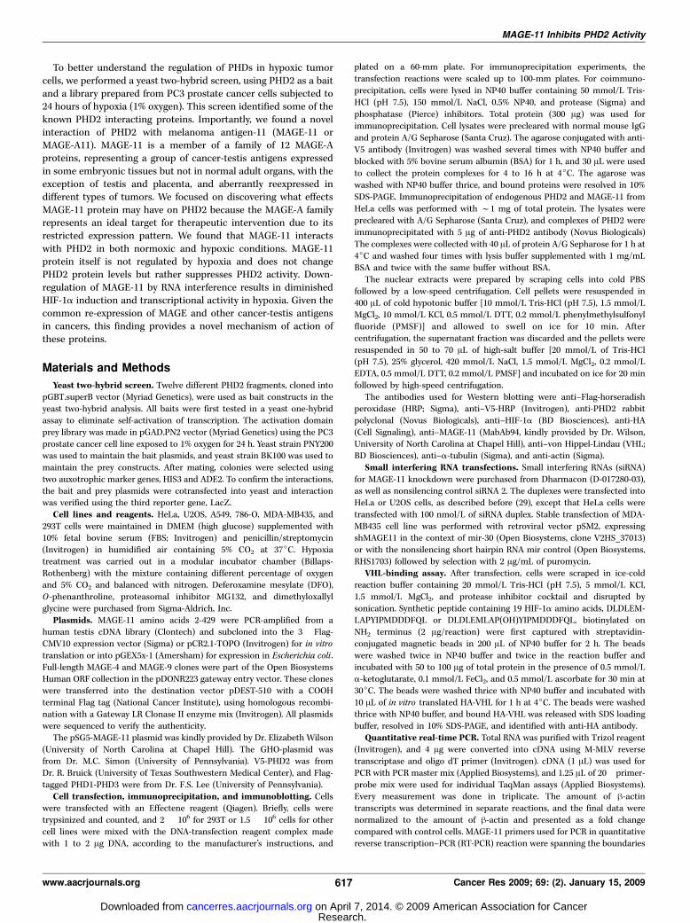
Figure 1. MAGE-11 interaction with PHD2. A, Flag-MAGE-11 and V5-PHD2 were cotransfected into 293Tcells and, 2 d later, treated with 1.5% oxygen or left untreatedfor 16 h. Immunoprecipitation was performed with anti-V5 agarose, followed by Western blotting with anti-Flag antibodies. B, coimmunoprecipitation of endogenousMAGE-11 and PHD2 from HeLa cell extracts. Protein complexes were immunoprecipitated with anti-PHD2 antibody or normal rabbit IgG, as a negative control, followedby Western blotting with anti-MAGE-11 (top ) or anti-PHD2 (bottom ) antibody. C, MAGE-11 was overexpressed in 293T cells with Flag-PHD1 or Flag-PHD3 andimmunoprecipitated with anti-Flag or normal mouse IgG, followed by Western blotting with anti–MAGE-11 antibody. D, three different Flag-tagged MAGE-A genes wereoverexpressed in 293T cells alone or in combination with V5-PHD2 and immunoprecipitated with anti-V5 agarose followed by Western blotting with anti-Flag antibody.
Cancer Research
Cancer Res 2009; 69: (2). January 15, 2009 618 www.aacrjournals.org
Research. on April 7, 2014. © 2009 American Association for Cancercancerres.aacrjournals.org Downloaded from
protein involved in the androgen receptor (AR) signaling pathway(30), but neither a link to hypoxia nor a regulation of HIFs has beenidentified and tested.We confirmed the interaction between PHD2 and MAGE-11 by a
pull-down assay using purified GST-MAGE-11 fusion protein and35S-labeled in vitro translated PHD2 (Supplementary Fig. S1A) orlysates of 293T cells overexpressing PHD2 (Supplementary Fig.S1B). We further confirmed the association between MAGE-11 andPHD2, using 293T cells cotransfected with expression vectorsencoding Flag-tagged MAGE-11 and V5 epitope-tagged PHD2.Furthermore, we examined the effect of oxygen on the interaction.Immunoprecipitation with anti-V5 antibodies showed specificbinding of PHD2 and MAGE-11, whereas no MAGE-11 was foundin the immunoprecipitates of 293T cells transfected with an emptyvector instead of PHD2 (Fig. 1A). Interestingly, the oxygen level hadno effect on the interaction, as MAGE-11 coprecipitated with PHD2equally well in hypoxic and in normoxic conditions. To copreci-pitate endogenous MAGE-11 and PHD2, we first tested expressionof MAGE-11 protein in a wide panel of cell lines. We found thatHeLa cells expressed the highest levels of MAGE-11 (data notshown). For this study, we used a rabbit polyclonal antibody raisedagainst a unique polypeptide in the NH2 terminus of MAGE-11 (31).Immunoprecipitation with anti-PHD2 antibody followed by West-ern blotting with anti–MAGE-11 antibody confirmed that these twoproteins bind to each other in vivo (Fig. 1B).The bait PHD2 fragment used in the yeast two-hybrid screen
(amino acids 202-301) is located outside of the catalytic domain inthe region of moderate conservation between PHD paraloguesinvolved in substrate recognition (32). Therefore, we tested twoother PHD family members and found that both PHD1 and PHD3also interact with MAGE-11 (Fig. 1C).The fragment of MAGE-11 identified in the yeast two-hybrid
screen spanned the region of amino acids 212-421. This fragmentrepresents a MAGE homology domain conserved among all MAGEfamily members. To find out if other members of the MAGE-Afamily would also interact with PHD2, we cloned two other genes,MAGE-4 and MAGE-9. The phylogenetic analysis of the MAGEdomains showed that MAGE-9 is the closest homologue of theMAGE-11 protein (33). We found that MAGE-9 was readily detectedin a complex with PHD2 by coimmunoprecipitation, whereasMAGE-4 was not (Fig. 1D). This study showed that there is certainselectivity in the interaction of different MAGE-A proteins with
PHD2 and that other MAGE family members may act in the samemanner as MAGE-11.MAGE-11 colocalizes with PHD2. To further characterize the
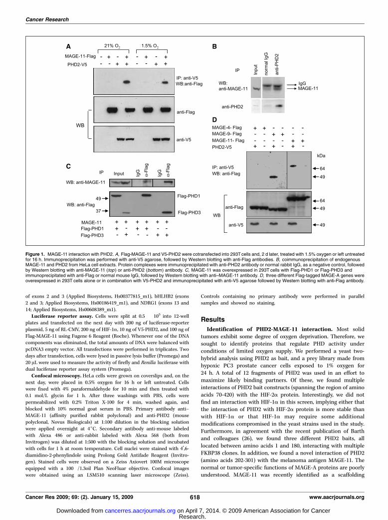
interaction between MAGE-11 and PHD2, we examined theircellular localizations. Previous studies have shown that MAGE-11 islocalized in both nuclear and cytoplasmic fractions with higheramounts found in cell nuclei (30, 34). To investigate MAGE-11localization under low oxygen, HeLa cells were grown undernormoxic or hypoxic conditions and immunostained with affinitypurified rabbit polyclonal antibody raised against MAGE-A11 (31)and a commercially available polyclonal anti-PHD2 antibodyproduced in mouse. Confocal microscopy showed that, inagreement with previous publications, MAGE-11 is located in bothnuclei and cytoplasm of HeLa cells with preferential nuclearstaining that does not change in the conditions of low oxygen.Normoxic HeLa cells show robust colocalization of MAGE-11 withPHD2 proteins in cytoplasm and less, but still obvious, colocaliza-tion was observed under hypoxic conditions (Fig. 2).MAGE-11 is degraded by the ubiquitin-proteasome system
but not through PHD-VHL pathway. Having confirmed thatPHD2 and MAGE-11 physically interact in the cell, we sought tostudy what function this interaction may have. First, we examinedwhether MAGE-11 was a hydroxylation target of PHD2. Although itis well established that HIF-a family members are substrates ofPHD hydroxylation required for VHL E3 ubiquitin ligase recogni-tion, there may be other proteins that are degraded through thesame mechanism. Indeed, the large subunit of RNA polymerase IIrequires hydroxylation of a specific proline residue and VHLbinding to undergo polyubiquitination and degradation (35). Allknown PHD substrate proteins consist of a LXXLAP consensusmotif in which the last proline is the hydroxyl group acceptor. Theprimary sequence of MAGE-11 has six amino acids starting atposition 160, LXXLPA, similar to the HIF consensus but differing inthe position of the acceptor. Due to the fact that several studieshave shown a certain tolerance divergence in this consensus(36, 37), we tested if MAGE-11 may be a substrate of PHD2 and maydegrade through VHL-dependent ubiquitination. We examinedMAGE-11 levels in 293T cells after treatment with the proteasomalinhibitor MG132 and found that the abundance of MAGE-11protein increased, and it was also associated with a ladder of highmolecular weight bands typical for polyubiquitinated proteins(Supplementary Fig. S2A). This phenomenon was independent of
Figure 2. PHD2 colocalizes withMAGE-11 in HeLa cells. Confocalmicroscopy of HeLa cells exposed to 0.5%or 21% oxygen and stained with affinitypurified anti–MAGE-11 rabbit antibody andanti-PHD2 mouse antibody followed bysecondary anti-rabbit Alexa 568 (greenfluorescence ) and anti-mouse-Alexa 486(red fluorescence ) antibody.
MAGE-11 Inhibits PHD2 Activity
www.aacrjournals.org 619 Cancer Res 2009; 69: (2). January 15, 2009
Research. on April 7, 2014. © 2009 American Association for Cancercancerres.aacrjournals.org Downloaded from
whether or not PHD2 was overexpressed together with MAGE-11(Supplementary Fig. S2A). The inhibition of endogenous PHDactivity by hypoxia or by the iron-chelating agents DFO andO-phenanthroline did not affect MAGE-11 accumulation either(Supplementary Fig. S2B). When we expressed MAGE-11 in theVHL-negative 786-O cell line, we also observed induction ofMAGE-11 protein by proteasome inhibition, implying that VHL isnot required for proteasomal degradation of MAGE-11 (Supple-mentary Fig. S2C). We further expanded this observation toendogenous MAGE-11 and noted that, in HeLa cells, MAGE-11levels were also induced by MG132 (Supplementary Fig. S2D) butwere unresponsive to hypoxia or PHD inhibition (SupplementaryFig. S2E).MAGE-11 inhibits PHD activity. Several proteins that modulate
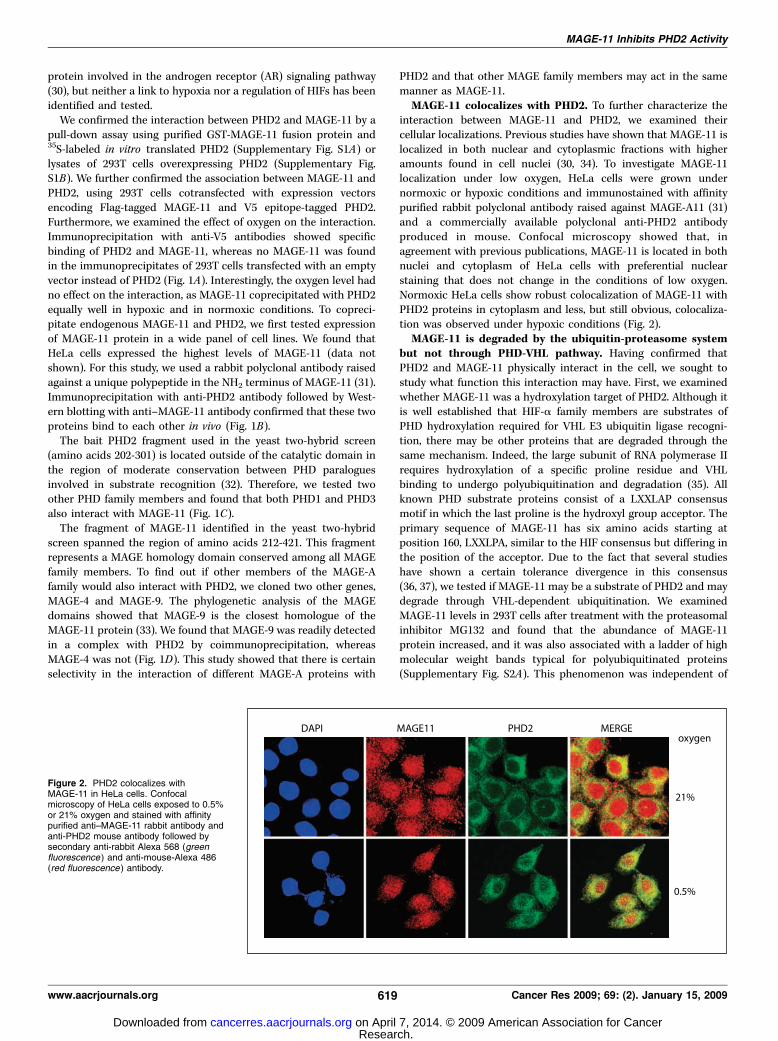
PHD activity either by bridging PHDs with HIF-a (OS-9; ref. 24),destabilizing the PHD2 protein (FKBP38; ref. 26), or causingsuppression of HIF transcriptional activity under hypoxia (ING4;ref. 28) have been identified. To understand the function ofMAGE-11 in regulation of PHD2, we first studied the effect ofMAGE-11 on the protein levels of PHD2. There was no effect onPHD2 protein levels when it was ectopically coexpressed withthe increasing amounts of MAGE-11 in HeLa or Hep3B cells(Fig. 3A and B, bottom). Similarly, there was no change inendogenous PHD2 protein levels in normoxic or hypoxic
MDA-MB435 (Fig. 3C) or 293 cells overexpressing MAGE-11(data not shown). We then tested the effect of MAGE-11 on PHDactivity by using a VHL-binding assay. This in vitro reaction usesa 19–amino acid peptide derived from the ODDD of HIF-1acontaining Pro564, which was hydroxylated by cell extractsexpressing increasing amounts of MAGE11, and the extent ofhydroxylation was measured by interaction with the in vitrotranslated HA-VHL protein. Figure 3A shows that an increase inMAGE-11 in cell extracts results in decreased binding of VHL (top).This effect was specific because MAGE-4 did not change PHDactivity (Fig. 3B). Because pVHL binds only to the hydroxylatedpeptide, these results indicate that PHD activity is suppressed byMAGE-11 protein.When PHD2 activity is blocked by RNA interference or small
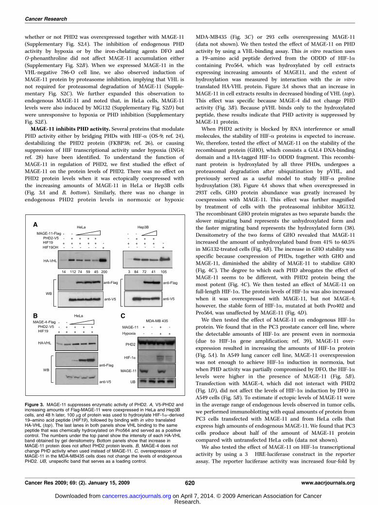
molecules, the stability of HIF-a proteins is expected to increase.We, therefore, tested the effect of MAGE-11 on the stability of therecombinant protein (GHO), which consists a GAL4 DNA-bindingdomain and a HA-tagged HIF-1a ODDD fragment. This recombi-nant protein is hydroxylated by all three PHDs, undergoes aproteasomal degradation after ubiquitination by pVHL, andpreviously served as a useful model to study HIF-a prolinehydroxylation (38). Figure 4A shows that when overexpressed in293T cells, GHO protein abundance was greatly increased bycoexpression with MAGE-11. This effect was further magnifiedby treatment of cells with the proteasomal inhibitor MG132.The recombinant GHO protein migrates as two separate bands: theslower migrating band represents the unhydroxylated form andthe faster migrating band represents the hydroxylated form (38).Densitometry of the two forms of GHO revealed that MAGE-11increased the amount of unhydroxylated band from 41% to 60.5%in MG132-treated cells (Fig. 4B). The increase in GHO stability wasspecific because coexpression of PHDs, together with GHO andMAGE-11, diminished the ability of MAGE-11 to stabilize GHO(Fig. 4C). The degree to which each PHD abrogates the effect ofMAGE-11 seems to be different, with PHD2 protein being themost potent (Fig. 4C). We then tested an effect of MAGE-11 onfull-length HIF-1a. The protein levels of HIF-1a was also increasedwhen it was overexpressed with MAGE-11, but not MAGE-4;however, the stable form of HIF-1a, mutated at both Pro402 andPro564, was unaffected by MAGE-11 (Fig. 4D).We then tested the effect of MAGE-11 on endogenous HIF-1a
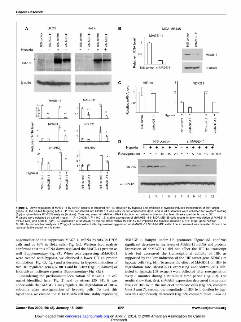
protein. We found that in the PC3 prostate cancer cell line, wherethe detectable amounts of HIF-1a are present even in normoxia(due to HIF-1a gene amplification; ref. 39), MAGE-11 over-expression resulted in increasing the amounts of HIF-1a protein(Fig. 5A). In A549 lung cancer cell line, MAGE-11 overexpressionwas not enough to achieve HIF-1a induction in normoxia, butwhen PHD activity was partially compromised by DFO, the HIF-1alevels were higher in the presence of MAGE-11 (Fig. 5B).Transfection with MAGE-4, which did not interact with PHD2(Fig. 1D), did not affect the levels of HIF-1a induction by DFO inA549 cells (Fig. 5B). To estimate if ectopic levels of MAGE-11 werein the average range of endogenous levels observed in tumor cells,we performed immunoblotting with equal amounts of protein fromPC3 cells transfected with MAGE-11 and from HeLa cells thatexpress high amounts of endogenous MAGE-11. We found that PC3cells produce about half of the amount of MAGE-11 proteincompared with untransfected HeLa cells (data not shown).We also tested the effect of MAGE-11 on HIF-1a transcriptional
activity by using a 3� HRE-luciferase construct in the reporterassay. The reporter luciferase activity was increased four-fold by
Figure 3. MAGE-11 suppresses enzymatic activity of PHD2. A, V5-PHD2 andincreasing amounts of Flag-MAGE-11 were coexpressed in HeLa and Hep3Bcells, and 48 h later, 100 Ag of protein was used to hydroxylate HIF-1a–derived19–amino acid peptide (HIF19 ), followed by binding with in vitro translatedHA-VHL (top ). The last lanes in both panels show VHL binding to the samepeptide that was chemically hydroxylated on Pro564 and served as a positivecontrol. The numbers under the top panel show the intensity of each HA-VHLband obtained by gel densitometry. Bottom panels show that increase inMAGE-11 protein does not affect PHD2 protein levels. B, MAGE-4 does notchange PHD activity when used instead of MAGE-11. C, overexpression ofMAGE-11 in the MDA-MB435 cells does not change the levels of endogenousPHD2. UB, unspecific band that serves as a loading control.
Cancer Research
Cancer Res 2009; 69: (2). January 15, 2009 620 www.aacrjournals.org
Research. on April 7, 2014. © 2009 American Association for Cancercancerres.aacrjournals.org Downloaded from
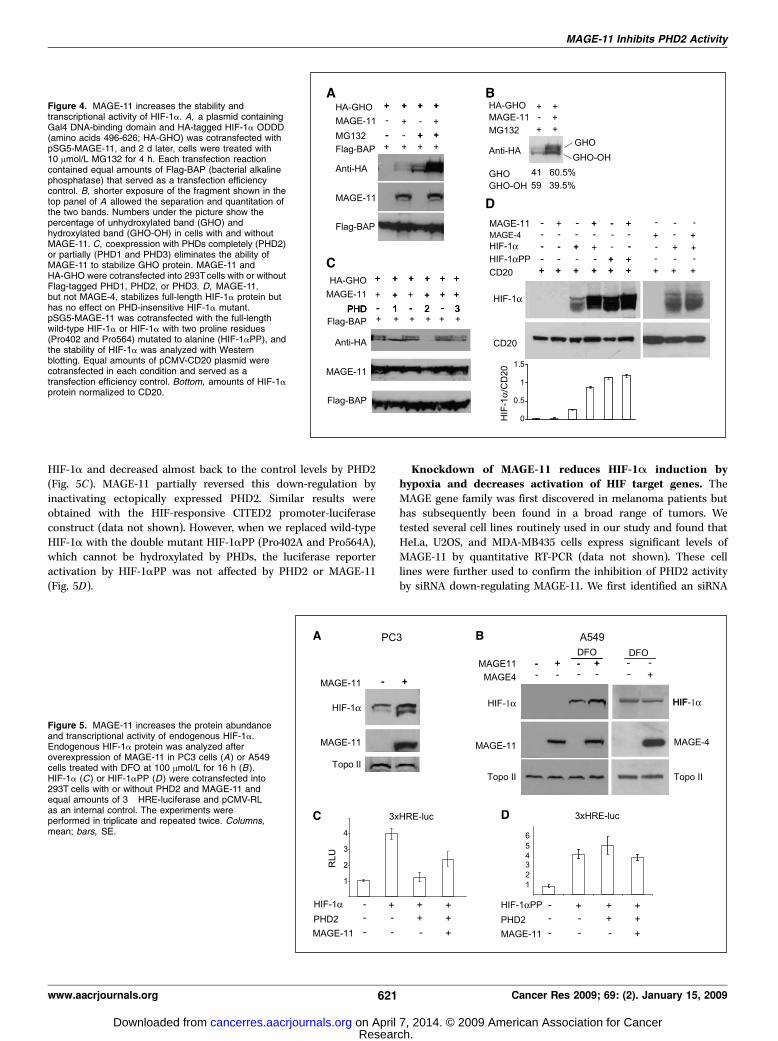
HIF-1a and decreased almost back to the control levels by PHD2(Fig. 5C). MAGE-11 partially reversed this down-regulation byinactivating ectopically expressed PHD2. Similar results wereobtained with the HIF-responsive CITED2 promoter-luciferaseconstruct (data not shown). However, when we replaced wild-typeHIF-1a with the double mutant HIF-1aPP (Pro402A and Pro564A),which cannot be hydroxylated by PHDs, the luciferase reporteractivation by HIF-1aPP was not affected by PHD2 or MAGE-11(Fig. 5D).
Knockdown of MAGE-11 reduces HIF-1A induction byhypoxia and decreases activation of HIF target genes. TheMAGE gene family was first discovered in melanoma patients buthas subsequently been found in a broad range of tumors. Wetested several cell lines routinely used in our study and found thatHeLa, U2OS, and MDA-MB435 cells express significant levels ofMAGE-11 by quantitative RT-PCR (data not shown). These celllines were further used to confirm the inhibition of PHD2 activityby siRNA down-regulating MAGE-11. We first identified an siRNA
Figure 4. MAGE-11 increases the stability andtranscriptional activity of HIF-1a. A, a plasmid containingGal4 DNA-binding domain and HA-tagged HIF-1a ODDD(amino acids 496-626; HA-GHO) was cotransfected withpSG5-MAGE-11, and 2 d later, cells were treated with10 Amol/L MG132 for 4 h. Each transfection reactioncontained equal amounts of Flag-BAP (bacterial alkalinephosphatase) that served as a transfection efficiencycontrol. B, shorter exposure of the fragment shown in thetop panel of A allowed the separation and quantitation ofthe two bands. Numbers under the picture show thepercentage of unhydroxylated band (GHO) andhydroxylated band (GHO-OH) in cells with and withoutMAGE-11. C, coexpression with PHDs completely (PHD2)or partially (PHD1 and PHD3) eliminates the ability ofMAGE-11 to stabilize GHO protein. MAGE-11 andHA-GHO were cotransfected into 293Tcells with or withoutFlag-tagged PHD1, PHD2, or PHD3. D, MAGE-11,but not MAGE-4, stabilizes full-length HIF-1a protein buthas no effect on PHD-insensitive HIF-1a mutant.pSG5-MAGE-11 was cotransfected with the full-lengthwild-type HIF-1a or HIF-1a with two proline residues(Pro402 and Pro564) mutated to alanine (HIF-1aPP), andthe stability of HIF-1a was analyzed with Westernblotting. Equal amounts of pCMV-CD20 plasmid werecotransfected in each condition and served as atransfection efficiency control. Bottom, amounts of HIF-1aprotein normalized to CD20.
Figure 5. MAGE-11 increases the protein abundanceand transcriptional activity of endogenous HIF-1a.Endogenous HIF-1a protein was analyzed afteroverexpression of MAGE-11 in PC3 cells (A) or A549cells treated with DFO at 100 Amol/L for 16 h (B ).HIF-1a (C ) or HIF-1aPP (D ) were cotransfected into293T cells with or without PHD2 and MAGE-11 andequal amounts of 3� HRE-luciferase and pCMV-RLas an internal control. The experiments wereperformed in triplicate and repeated twice. Columns,mean; bars, SE.
MAGE-11 Inhibits PHD2 Activity
www.aacrjournals.org 621 Cancer Res 2009; 69: (2). January 15, 2009
Research. on April 7, 2014. © 2009 American Association for Cancercancerres.aacrjournals.org Downloaded from
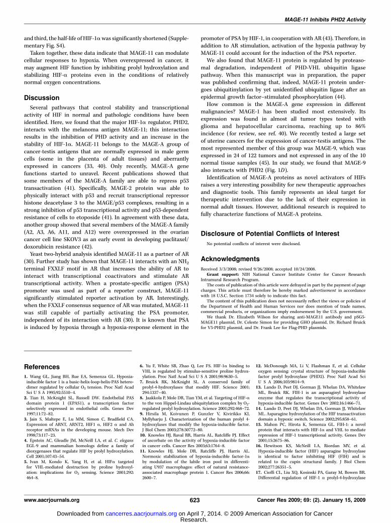
oligonucleotide that suppresses MAGE-11 mRNA by 90% in U2OScells and by 60% in HeLa cells (Fig. 6A). Western blot analysisconfirmed that this siRNA down-regulated the MAGE-11 protein aswell (Supplementary Fig. S3). When cells expressing siMAGE-11were treated with hypoxia, we observed a lower HIF-1a proteinstimulation (Fig. 6A, top) and a decrease in hypoxic induction oftwo HIF-regulated genes, NDRG1 and bHLHB2 (Fig. 6A, bottom) orHRE-driven luciferase reporter (Supplementary Fig. S3B).Considering the predominant localization of MAGE-11 in cell
nuclei identified here (Fig. 2) and by others (30, 34), it wasconceivable that MAGE-11 may regulate the degradation of HIF-asubunits after reoxygenation of hypoxic cells. To test thishypothesis, we created the MDA-MB435 cell line, stably expressing
shMAGE-11 hairpin under U6 promoter. Figure 6B confirmssignificant decrease in the levels of MAGE-11 mRNA and protein.Expression of shMAGE-11 did not affect the HIF-1a transcriptlevels but decreased the transcriptional activity of HIF, assupported by the low induction of the HIF target gene NDRG1 inhypoxic cells (Fig. 6C). To assess the effect of MAGE-11 on HIF-1adegradation rate, shMAGE-11 expressing and control cells sub-jected to hypoxia (1% oxygen) were collected after reoxygenationevery 5 minutes during a 20-minute time period (Fig. 6D). Theresults show that, first, shMAGE expression decreased the proteinlevels of HIF-1a in the nuclei of normoxic cells (Fig. 6D , comparelanes 1 and 7), second, the magnitude of HIF-1a induction by hyp-oxia was significantly decreased (Fig. 6D , compare lanes 2 and 8),
Figure 6. Down-regulation of MAGE-11 by siRNA results in impaired HIF-1a induction by hypoxia and inhibition of hypoxia-induced transcription of HIF targetgenes. A, the siRNA targeting MAGE-11 was transfected into U2OS or HeLa cells for two consecutive days, and in 24 h samples were collected for Western blotting(top ) or quantitative RT-PCR analysis (bottom ). Columns, mean of relative mRNA induction normalized to h-actin of at least three experiments; bars, SE.P values were obtained by paired t tests. **, P < 0.005; *, P < 0.01. B, stable expression of shMAGE-11 in MDA-MB435 cells results in down-regulation of MAGE-11mRNA (left) and protein (right ). C, expression of shMAGE-11 did not affect mRNA for HIF-1a but impaired the hypoxic induction of HIF target gene NDRG1.D, HIF-1a immunoblot analysis of 25 Ag of nuclear extract after hypoxia-reoxygenation of shMAGE-11 MDA-MB435 cells. The experiment was repeated thrice. Therepresentative experiment is shown.
Cancer Research
Cancer Res 2009; 69: (2). January 15, 2009 622 www.aacrjournals.org
Research. on April 7, 2014. © 2009 American Association for Cancercancerres.aacrjournals.org Downloaded from
and third, the half-life of HIF-1awas significantly shortened (Supple-mentary Fig. S4).Taken together, these data indicate that MAGE-11 can modulate
cellular responses to hypoxia. When overexpressed in cancer, itmay augment HIF function by inhibiting prolyl hydroxylation andstabilizing HIF-a proteins even in the conditions of relativelynormal oxygen concentrations.
Discussion
Several pathways that control stability and transcriptionalactivity of HIF in normal and pathologic conditions have beenidentified. Here, we found that the major HIF-1a regulator, PHD2,interacts with the melanoma antigen MAGE-11; this interactionresults in the inhibition of PHD activity and an increase in thestability of HIF-1a. MAGE-11 belongs to the MAGE-A group ofcancer-testis antigens that are normally expressed in male germcells (some in the placenta of adult tissues) and aberrantlyexpressed in cancers (33, 40). Only recently, MAGE-A genefunctions started to unravel. Recent publications showed thatsome members of the MAGE-A family are able to repress p53transactivation (41). Specifically, MAGE-2 protein was able tophysically interact with p53 and recruit transcriptional repressorhistone deacetylase 3 to the MAGE/p53 complexes, resulting in astrong inhibition of p53 transcriptional activity and p53-dependentresistance of cells to etoposide (41). In agreement with these data,another group showed that several members of the MAGE-A family(A2, A3, A6, A11, and A12) were overexpressed in the ovariancancer cell line SKOV3 as an early event in developing paclitaxel/doxorubicin resistance (42).Yeast two-hybrid analysis identified MAGE-11 as a partner of AR
(30). Further study has shown that MAGE-11 interacts with an NH2terminal FXXLF motif in AR that increases the ability of AR tointeract with transcriptional coactivators and stimulate ARtranscriptional activity. When a prostate-specific antigen (PSA)promoter was used as part of a reporter construct, MAGE-11significantly stimulated reporter activation by AR. Interestingly,when the FXXLF consensus sequence of AR was mutated, MAGE-11was still capable of partially activating the PSA promoter,independent of its interaction with AR (30). It is known that PSAis induced by hypoxia through a hypoxia-response element in the
promoter of PSA by HIF-1, in cooperation with AR (43). Therefore, inaddition to AR stimulation, activation of the hypoxia pathway byMAGE-11 could account for the induction of the PSA reporter.We also found that MAGE-11 protein is regulated by proteaso-
mal degradation, independent of PHD-VHL ubiquitin ligasepathway. When this manuscript was in preparation, the paperwas published confirming that, indeed, MAGE-11 protein under-goes ubiquitinylation by yet unidentified ubiquitin ligase after anepidermal growth factor–stimulated phosphorylation (44).How common is the MAGE-A gene expression in different
malignancies? MAGE-1 has been studied most extensively. Itsexpression was found in almost all tumor types tested withglioma and hepatocellular carcinoma, reaching up to 86%incidence ( for review, see ref. 40). We recently tested a large setof uterine cancers for the expression of cancer-testis antigens. Themost represented member of this group was MAGE-9, which wasexpressed in 24 of 122 tumors and not expressed in any of the 10normal tissue samples (45). In our study, we found that MAGE-9also interacts with PHD2 (Fig. 1D).Identification of MAGE-A proteins as novel activators of HIFs
raises a very interesting possibility for new therapeutic approachesand diagnostic tools. This family represents an ideal target fortherapeutic intervention due to the lack of their expression innormal adult tissues. However, additional research is required tofully characterize functions of MAGE-A proteins.
Disclosure of Potential Conflicts of Interest
No potential conflicts of interest were disclosed.
Acknowledgments
Received 3/3/2008; revised 9/26/2008; accepted 10/24/2008.Grant support: NIH National Cancer Institute Center for Cancer Research
Intramural Research Program.The costs of publication of this article were defrayed in part by the payment of page
charges. This article must therefore be hereby marked advertisement in accordancewith 18 U.S.C. Section 1734 solely to indicate this fact.The content of this publication does not necessarily reflect the views or policies of
the Department of Health and Human Services nor does mention of trade names,commercial products, or organizations imply endorsement by the U.S. government.We thank Dr. Elizabeth Wilson for sharing anti-MAGE11 antibody and pSG5-
MAGE11 plasmid, Dr. Celeste Simon for providing GHO plasmid, Dr. Richard Bruickfor V5-PHD2 plasmid, and Dr. Frank Lee for Flag-PHD plasmids.
MAGE-11 Inhibits PHD2 Activity
www.aacrjournals.org 623 Cancer Res 2009; 69: (2). January 15, 2009
References
1. Wang GL, Jiang BH, Rue EA, Semenza GL. Hypoxia-inducible factor 1 is a basic-helix-loop-helix-PAS hetero-dimer regulated by cellular O2 tension. Proc Natl AcadSci U S A 1995;92:5510–4.2. Tian H, McKnight SL, Russell DW. Endothelial PASdomain protein 1 (EPAS1), a transcription factorselectively expressed in endothelial cells. Genes Dev1997;11:72–82.3. Jain S, Maltepe E, Lu MM, Simon C, Bradfield CA.Expression of ARNT, ARNT2, HIF1 a, HIF2 a and Ahreceptor mRNAs in the developing mouse. Mech Dev1998;73:117–23.4. Epstein AC, Gleadle JM, McNeill LA, et al. C. elegansEGL-9 and mammalian homologs define a family ofdioxygenases that regulate HIF by prolyl hydroxylation.Cell 2001;107:43–54.5. Ivan M, Kondo K, Yang H, et al. HIFa targetedfor VHL-mediated destruction by proline hydroxyl-ation: implications for O2 sensing. Science 2001;292:464–8.
6. Yu F, White SB, Zhao Q, Lee FS. HIF-1a binding toVHL is regulated by stimulus-sensitive proline hydrox-ylation. Proc Natl Acad Sci U S A 2001;98:9630–5.7. Bruick RK, McKnight SL. A conserved family ofprolyl-4-hydroxylases that modify HIF. Science 2001;294:1337–40.8. Jaakkola P, Mole DR, Tian YM, et al. Targeting of HIF-ato the von Hippel-Lindau ubiquitylation complex by O2-regulated prolyl hydroxylation. Science 2001;292:468–72.9. Hirsila M, Koivunen P, Gunzler V, Kivirikko KI,Myllyharju J. Characterization of the human prolyl 4-hydroxylases that modify the hypoxia-inducible factor.J Biol Chem 2003;278:30772–80.10. Knowles HJ, Raval RR, Harris AL, Ratcliffe PJ. Effectof ascorbate on the activity of hypoxia-inducible factorin cancer cells. Cancer Res 2003;63:1764–8.11. Knowles HJ, Mole DR, Ratcliffe PJ, Harris AL.Normoxic stabilization of hypoxia-inducible factor-1aby modulation of the labile iron pool in differenti-ating U937 macrophages: effect of natural resistance-associated macrophage protein 1. Cancer Res 2006;66:2600–7.
12. McDonough MA, Li V, Flashman E, et al. Cellularoxygen sensing: crystal structure of hypoxia-induciblefactor prolyl hydroxylase (PHD2). Proc Natl Acad SciU S A 2006;103:9814–9.13. Lando D, Peet DJ, Gorman JJ, Whelan DA, WhitelawML, Bruick RK. FIH-1 is an asparaginyl hydroxylaseenzyme that regulates the transcriptional activity ofhypoxia-inducible factor. Genes Dev 2002;16:1466–71.14. Lando D, Peet DJ, Whelan DA, Gorman JJ, WhitelawML. Asparagine hydroxylation of the HIF transactivationdomain a hypoxic switch. Science 2002;295:858–61.15. Mahon PC, Hirota K, Semenza GL. FIH-1: a novelprotein that interacts with HIF-1a and VHL to mediaterepression of HIF-1 transcriptional activity. Genes Dev2001;15:2675–86.16. Hewitson KS, McNeill LA, Riordan MV, et al.Hypoxia-inducible factor (HIF) asparagine hydroxylaseis identical to factor inhibiting HIF (FIH) and isrelated to the cupin structural family. J Biol Chem2002;277:26351–5.17. Cioffi CL, Liu XQ, Kosinski PA, Garay M, Bowen BR.Differential regulation of HIF-1 a prolyl-4-hydroxylase
Research. on April 7, 2014. © 2009 American Association for Cancercancerres.aacrjournals.org Downloaded from
Cancer Research
Cancer Res 2009; 69: (2). January 15, 2009 624 www.aacrjournals.org
genes by hypoxia in human cardiovascular cells.Biochem Biophys Res Commun 2003;303:947–53.18. Lieb ME, Menzies K, Moschella MC, Ni R, TaubmanMB. Mammalian EGLN genes have distinct patterns ofmRNA expression and regulation. Biochem Cell Biol2002;80:421–6.19. Metzen E, Berchner-Pfannschmidt U, Stengel P, et al.Intracellular localisation of human HIF-1 a hydroxy-lases: implications for oxygen sensing. J Cell Sci 2003;116:1319–26.20. D’Angelo G, Duplan E, Boyer N, Vigne P, Frelin C.Hypoxia up-regulates prolyl hydroxylase activity: afeedback mechanism that limits HIF-1 responses duringreoxygenation. J Biol Chem 2003;278:38183–7.21. del Peso L, Castellanos MC, Temes E, et al. The vonHippel Lindau/hypoxia-inducible factor (HIF) pathwayregulates the transcription of the HIF-proline hydroxy-lase genes in response to low oxygen. J Biol Chem 2003;278:48690–5.22. Berra E, Benizri E, Ginouves A, Volmat V, Roux D,Pouyssegur J. HIF prolyl-hydroxylase 2 is the key oxygensensor setting low steady-state levels of HIF-1a innormoxia. EMBO J 2003;22:4082–90.23. Chan DA, Sutphin PD, Denko NC, Giaccia AJ. Role ofprolyl hydroxylation in oncogenically stabilized hypoxia-inducible factor-1a. J Biol Chem 2002;277:40112–7.24. Baek JH, Mahon PC, Oh J, et al. OS-9 interacts withhypoxia-inducible factor 1a and prolyl hydroxylases topromote oxygen-dependent degradation of HIF-1a. MolCell 2005;17:503–12.25. Nakayama K, Frew IJ, Hagensen M, et al. Siah2regulates stability of prolyl-hydroxylases, controls HIF1aabundance, and modulates physiological responses tohypoxia. Cell 2004;117:941–52.26. Barth S, Nesper J, Hasgall PA, et al. The peptidylprolyl cis/trans isomerase FKBP38 determines hyp-oxia-inducible transcription factor prolyl-4-hydroxy-lase PHD2 protein stability. Mol Cell Biol 2007;27:3758–68.
27. To KK, Huang LE. Suppression of hypoxia-induc-ible factor 1a (HIF-1a) transcriptional activity by theHIF prolyl hydroxylase EGLN1. J Biol Chem 2005;280:38102–7.28. Ozer A, Wu LC, Bruick RK. The candidate tumorsuppressor ING4 represses activation of the hypoxiainducible factor (HIF). Proc Natl Acad Sci U S A 2005;102:7481–6.29. Aprelikova O, Wood M, Tackett S, et al. Role of ETStranscription factors in the hypoxia-inducible factor-2target gene selection. Cancer Res 2006;66:5641–7.30. Bai S, He B, Wilson EM. Melanoma antigen geneprotein MAGE-11 regulates androgen receptor functionby modulating the interdomain interaction. Mol CellBiol 2005;25:1238–57.31. Bai S, Grossman G, Yuan L, et al. Hormone controland expression of androgen receptor coregulatorMAGE-11 in human endometrium during the windowof receptivity to embryo implantation. Mol Hum Reprod2008;14:107–16.32. Villar D, Vara-Vega A, Landazuri MO, Del Peso L.Identification of a region on hypoxia-inducible-factorprolyl 4-hydroxylases that determines their specificityfor the oxygen degradation domains. Biochem J 2007;408:231–40.33. Barker PA, Salehi A. The MAGE proteins: emergingroles in cell cycle progression, apoptosis, and neuro-genetic disease. J Neurosci Res 2002;67:705–12.34. Jurk M, Kremmer E, Schwarz U, Forster R, WinnackerEL. MAGE-11 protein is highly conserved in higherorganisms and located predominantly in the nucleus.Int J Cancer 1998;75:762–6.35. Kuznetsova AV, Meller J, Schnell PO, et al. vonHippel-Lindau protein binds hyperphosphorylated largesubunit of RNA polymerase II through a prolinehydroxylation motif and targets it for ubiquitination.Proc Natl Acad Sci U S A 2003;100:2706–11.36. Huang J, Zhao Q, Mooney SM, Lee FS. Sequencedeterminants in hypoxia-inducible factor-1a for hydrox-
ylation by the prolyl hydroxylases PHD1, PHD2, andPHD3. J Biol Chem 2002;277:39792–800.37. Landazuri MO, Vara-Vega A, Viton M, Cuevas Y, delPeso L. Analysis of HIF-prolyl hydroxylases binding tosubstrates. Biochem Biophys Res Commun 2006;351:313–20.38. Pan Y, Mansfield KD, Bertozzi CC, et al. Multiplefactors affecting cellular redox status and energymetabolism modulate hypoxia-inducible factor prolylhydroxylase activity in vivo and in vitro . Mol Cell Biol2007;27:912–25.39. Saramaki OR, Savinainen KJ, Nupponen NN, Bratt O,Visakorpi T. Amplification of hypoxia-inducible factor1a gene in prostate cancer. Cancer Genet Cytogenet2001;128:31–4.40. Gillespie AM, Coleman RE. The potential of melano-ma antigen expression in cancer therapy. Cancer TreatRev 1999;25:219–27.41. Monte M, Simonatto M, Peche LY, et al. MAGE-Atumor antigens target p53 transactivation functionthrough histone deacetylase recruitment and conferresistance to chemotherapeutic agents. Proc Natl AcadSci U S A 2006;103:11160–5.42. Duan Z, Duan Y, Lamendola DE, et al. Overexpres-sion of MAGE/GAGE genes in paclitaxel/doxorubicin-resistant human cancer cell lines. Clin Cancer Res 2003;9:2778–85.43. Horii K, Suzuki Y, Kondo Y, et al. Androgen-dependent gene expression of prostate-specific antigenis enhanced synergistically by hypoxia in humanprostate cancer cells. Mol Cancer Res 2007;5:383–91.44. Bai S, Wilson EM. EGF dependent phosphorylationand ubiquitinylation of MAGE-11 regulates its interac-tion with the androgen receptor. Mol Cell Biol 2008;28:1947–63.45. Risinger JI, Chandramouli GV, Maxwell GL, et al.Global expression analysis of cancer/testis genes inuterine cancers reveals a high incidence of BORISexpression. Clin Cancer Res 2007;13:1713–9.
Research. on April 7, 2014. © 2009 American Association for Cancercancerres.aacrjournals.org Downloaded from