PROLYL OLIGOPEPTIDASE IN MULTIPLE SCLEROSIS Anne ...
86
PROLYL OLIGOPEPTIDASE IN MULTIPLE SCLEROSIS Anne Penttinen University of Helsinki Faculty of Pharmacy Division of Pharmacology and Toxicology April 2010
-
Upload
khangminh22 -
Category
Documents
-
view
1 -
download
0
Transcript of PROLYL OLIGOPEPTIDASE IN MULTIPLE SCLEROSIS Anne ...

PROLYL OLIGOPEPTIDASE IN MULTIPLE SCLEROSIS
Anne Penttinen University of Helsinki Faculty of Pharmacy Division of Pharmacology and Toxicology April 2010

2
CONTENTS
PART I: LITERATURE REVIEW ................................................................................... 1
PROLYL OLIGOPEPTIDASE IN MULTIPLE SCLEROSIS ........................................ 1
1 INTRODUCTION ..................................................................................................... 1
2 PROLYL OLIGOPEPTIDASE ................................................................................. 2
2.1 Structure, activity and genetics .......................................................................... 2
2.2 Distribution ........................................................................................................ 4
2.3 Regulation .......................................................................................................... 5
2.4 Physiological role of POP .................................................................................. 6
2.5 Substrates and inhibitors .................................................................................... 6
3 MULTIPLE SCLEROSIS ......................................................................................... 8
3.1 The course of the disease ................................................................................... 8
3.2 Components associated with pathophysiology of multiple sclerosis ............... 10
3.2.1 Inflammatory cells .................................................................................... 10
3.2.2 Cytokines and chemokines ....................................................................... 13
3.2.3 Matrix metalloproteinases ......................................................................... 15
3.2.4 Oxidative stress ......................................................................................... 17
3.2.5 Pathogens and molecular mimicry ............................................................ 18
3.2.6 Genes ........................................................................................................ 19
3.2.7 Other factors ............................................................................................. 19
4 PROLYL OLIGOPEPTIDASE IN INFLAMMATION ......................................... 20
4.1 Immunological role of peptide hormone substrates ......................................... 20
4.1.1 Angiotensins ............................................................................................. 21
4.1.2 Bradykinin ................................................................................................ 22
4.1.3 Substance P ............................................................................................... 23
4.2 POP and chronic pulmonary inflammatory diseases ....................................... 24
4.3 POP and bronchiolitis obliterans syndrome (BOS) after lung transplant ........ 27
4.4 POP and rheumatoid arthritis ........................................................................... 28
4.5 POP and delayed type of allergic inflammation induced by Mycobacterium tuberculosis ................................................................................................................. 29
4.6 POP and systemic lupus erythematosus ........................................................... 30

3
4.7 POP and antifibrotic peptide Ac-SDKP ........................................................... 31
5 PROLYL OLIGOPEPTIDASE IN NEURODEGENERATION ............................ 32
5.1 POP substrates and neurodegeneration ............................................................ 33
5.2 Microglial toxicity ............................................................................................ 34
5.3 Neuronal apoptosis ........................................................................................... 34
5.4 Activation induced cell death in T cells ........................................................... 36
6 CONCLUSION ....................................................................................................... 38
PART II: EXPERIMENTAL PART ............................................................................... 39
PROLYL OLIGOPEPTIDASE ACTIVITY IN MULTIPLE SCLEROSIS AND CHARACTERIZATION OF ITS ENDOGENOUS INHIBITOR ................................. 39
1 INTRODUCTION ................................................................................................... 39
2 MATERIALS .......................................................................................................... 41
2.1 Compounds and reagents ................................................................................. 41
2.2 Solutions ........................................................................................................... 43
2.2.1 Solutions for prolyl endopeptidase activity measurements ...................... 43
2.2.2 Solutions for affinity chromatography ...................................................... 44
2.2.3 Solutions for albumin removal ................................................................. 45
2.2.4 Solutions for anion exchange chromatography ........................................ 45
2.2.5 Solutions for hydrophobic interaction chromatography ........................... 45
2.2.6 Solutions for electrophoresis .................................................................... 46
2.2.7 Solutions for protein staining .................................................................... 46
2.2.8 Solutions for Western blotting .................................................................. 47
2.2.9 Other solutions .......................................................................................... 47
2.3 Gels for sodium dodecyl sulphate-polyacrylamide gel electrophoresis (SDS-PAGE) ......................................................................................................................... 48
2.4 Patients and controls ........................................................................................ 48
2.5 Serum and CSF samples................................................................................... 49
3 METHODS .............................................................................................................. 49
3.1 Assays for prolyl endopeptidase (PE) activity ................................................. 49
3.2 Measurement of the endogenous POP inhibitor in serum samples .................. 50
3.3 Characterization of the endogenous POP inhibitor .......................................... 50
3.3.1 Protein concentration determination ......................................................... 50
3.3.2 Molecular size estimation by ultrafiltration .............................................. 51

4
3.3.3 Albumin removal ...................................................................................... 51
3.3.4 The effect of temperature on inhibitory capacity of serum ...................... 51
3.3.5 Generation of POP-sepharose coupled chromatographic column ............ 52
3.3.6 Purification of the endogenous POP inhibitor by affinity column ........... 52
3.3.7 Anion exchange chromatography ............................................................. 52
3.3.8 Hydrophobic interaction chromatography ................................................ 53
3.3.9 SDS-PAGE and western plotting .............................................................. 53
3.4 Data analysis .................................................................................................... 54
4 RESULTS ................................................................................................................ 55
4.1 POP activity levels ........................................................................................... 55
4.2 Endogenous POP inhibitor ............................................................................... 56
4.3 Characterization of the endogenous POP inhibitor .......................................... 57
5 DISCUSSION .......................................................................................................... 67
6 CONCLUSION ....................................................................................................... 73
REFERENCES ............................................................................................................... 74

1
PART I: LITERATURE REVIEW
PROLYL OLIGOPEPTIDASE IN MULTIPLE SCLEROSIS
1 INTRODUCTION
Prolyl oligopeptidase (POP, E.C. 3.4.21.26) cleaves short peptides, of less than 30
amino acid long, at the C-side of an internal proline (Fülöp et al. 1998). POP is a
member of the prolyl oligopeptidase family of serine proteases, and has been associated
with many pathophysiological processes, such as neurodegeneration, depression and
hypertension (Brant et al. 2007; Garcia-Horsman et al. 2007; Männistö et al. 2007).
POP seems to have a role in inflammatory diseases, such as rheumatoid arthritis and
mycobacterium tuberculosis induced delayed-type allergic inflammation, and in
diseases, that have a notable inflammatory and/or neurodegenerative component
(Hashimoto et al. 2001, Kagewaga et al. 2004; Brand et al. 2007; Gaggar et al. 2009).
Thus, POP function may have a connection to both the inflammatory and the
neurodegenerative phase of multiple sclerosis (MS). At the moment there are no studies
that have been focused on POP function in MS. A preliminary study in a Spanish cohort
reported altered POP activity in plasma samples of patients with relapsing-remitting
multiple sclerosis (RR-MS) (Tenorio-Laranga et al. 2010). However, there is a number
of studies that associates matrix metalloproteinases (MMPs) with the course of MS
disease (inter alia Bar-Or et al. 2003). MMPs and POP have been shown to work in
concert in a chronic neutrophilic inflammation in the airways (Gaggar et al. 2009).
Some of the peptide hormones suggested to be regulated by POP have also been shown
to be mediators in inflammation (Lühder et al. 2009).
POP may exhibit highest concentration among brain peptidases (Polgár 2002). POP
mRNA is significantly expressed in activated microglial cells that have been associated
with neurodegenerative disorders (Klegeris et al. 2008). Other studies have linked POP
to neurodegeneration based on its possible role in regulation of neuronal apoptosis

2
(Männistö et al. 2007). A peptidomic study of porcine brain homogenates found
fragments of myelin basic protein to be POP substrates (Brandt et al. 2005). In addition,
cytoplasmic POP has been shown to be a regulator of the inositol phosphate signaling
pathway (Garcia-Horsman et al. 2007). Disturbances in this pathway have been
implicated in mood disorders, such as depression, a common symptom of MS.
This literature review focuses on the POP functions interpreted to be implicated in
inflammatory and neurodegenerative processes, since these processes are the most
relevant in the course of MS disease. The aim of this review is to clarify the role of POP
activity alterations in the pathology of this autoimmune disease.
2 PROLYL OLIGOPEPTIDASE
The POP family proteins seem to be of ancient origin (Venäläinen et al. 2004). POP is
distributed in archaeal, bacterial and eukaryotic species, but it has not been found in
fungi. The high conservation of POP family enzyme sequences in different species
suggests their presence in the last universal common ancestor (LUCA). This
conservation points strongly to the importance of these enzymes in physiological
processes.
2.1 Structure, activity and genetics
Mammal POP is a cylinder shaped 80 kDa globular soluble protein of 710 amino acids
constituted by two domains (Fülöp et al. 1998) (Fig.1). Both N- and C-termini of the
polypeptide chain constitute the peptidase domain (residues 1-72 and 428-710) which
presents an α/β-hydrolase fold and contains the catalytic triad (Ser554, Asp641 and
His680). The peptidase domain is covalently attached to a seven-bladed β-propeller
domain (residues 73-427). The active site is located in a large cavity at the interface of
the two domains. POP interacts maximally with six amino acids residues of the

3
substrate peptide, and specificity is provided by stacking a tryptophan residue against
the substrate proline ring (Fülöp et al. 1998; Männistö et al. 2007). The β-propeller
domain has been suggested to operate as a gating filter that controls the entrance of the
substrates, a mechanism by which POP is proposed to distinguish between large
proteins from unstructured peptides (Fülöp et al. 2000).
Fig. 1 POP is a cylinder shaped 80 kDa globular soluble protein constituted by peptidase and β-propeller domains. In a picture Z-Pro-Prolinal, a specific POP inhibitor, is bound at the active site of the enzyme (Protein Data Bank code 1qfs).
POP cleaves mainly peptides shorter than 30-mer at the carboxyl group of an internal
proline residues (Fülop et al. 1998, Polgar 2002). It is also able to hydrolyze after a
residue of alanine, although at much slower catalytic rate. In addition, cleavages of Ala-
Thr and Val-Gly bonds in octadecaneuropeptide and Cys-X bond in humanin have also
been reported (Myöhänen et al. 2009). POP is the only proline-specific endopeptidase
currently known in mammals (Polgár 2002). Proline is an imino rather than an amino
acid, and thus most peptidases are unable to hydrolyze the peptide bond at that position
(Fig.2) (Cunninham and O’Connor 1997; Polgár 2002).

4
Fig. 2 Molecular structure of amino acids and proline. a. General amino acid structure b. Proline. The secondary nature of the nitrogen containing moiety makes proline an imino acid (Cunninham and O’Connor 1997).
The POP gene, Prep, has been cloned from several sources including human
lymphocytes or a human T cell line MOLT4 (Kimura et al. 1999; Odaka et al. 2002).
The structure and localization of the mouse POP gene has been accomplished. This
gene contains 15 exons (Kimura et al. 1999; Garcia-Horsman et al. 2007; Männistö et
al. 2007). The peptidase domain of the enzyme is coded by exons 1-3 and 10-15 and the
propeller domain by exons 3-10. A national centre for biotechnology information
(NCBI) gene data bank search reveals only one POP gene in human with similar
structure of 15 exons. This gene is located at chromosome 6q22. Although POP appears
to be a single copy of gene, different forms of POP, arising from post-translational
modifications and/or alternative gene products, may account for the variety of functions
and locations of the enzyme activity (Kimura et al. 1999; Garcia-Horsman et al. 2007).
2.2 Distribution
POP was first discovered 1971 in human uterus homogenates, where it cleaves the Pro-
Leu bond of oxytocin (Walter et al. 1971). POP is mainly cytosolic, but lower
activity/expression has been described e.g. in nuclei, mitochondria, microtubules,
membranes and extracellular space (Irazusta et al. 2002; Schulz et al. 2005; Garcia-
Horsman et al. 2007; Myöhänen et al. 2008; Myöhänen et al. 2009). Proliferating cells
exhibit enhanced activity. Highest activities have been reported in cancerous tissues
(Goossens et al. 1996). Presence of POP has been reported in immune cells, such as
lymphocytes (T cells), macrophages and neutrophils (Gaggar et al. 2008; O’Reilly et al.
2009). Variable activity/expression, depending on the method used, have been measured

5
from fibroblasts, epithelial cells, endothelial cells, thrombocytes, platelets, microglia,
astrocytes, oligodendrocytes, heart, vasculature, kidneys, adipose tissue, testis, rectum,
spleen, thymys, bone marrow and lung (Goossens et al. 1996; Odaka et al. 2002;
Cavasin et al. 2004; Garcia-Horsman et al. 2007; Klegeris et al. 2008; Myöhänen et al.
2009). POP activities measured in blood, CSF, seminal fluid and prostate fluid are much
lower than the activities measured in tissues.
The enzyme is found in all mature brain regions except for corpus callosum, and it has
been detected in specific neuronal cells and absent in glial cells (Schulz et al. 2005;
Männistö et al. 2007). The cerebral and cerebellar cortical neurons have been shown to
be especially enriched of POP (Garcia-Horsman et al. 2007; Männistö et al. 2007;
Myöhänen et al. 2009). Significant activity has been measured also in striatum,
hypothalamus, hippocampus and amygdala. When POP distribution was investigated by
high-throughput gene profiling, the POP mRNA levels were however similar in
different brain areas (BioGPS).
Very often POP-like activity has not been fully identified (Garcia-Horsman et al. 2007).
POP activity measured with specific substrates/inhibitors may not be exclusively
associated to soluble POP, since possible posttranslational modifications and/or
different/alternative gene products (membrane form, secreted form) may react similarly
(Cunninham and O’Connor 1997; Garcia-Horsman et al. 2007).
2.3 Regulation
The mechanisms by which POP expression and its enzymatic activity are regulated are
not currently known (Garcia-Horsman et al. 2007; Männistö et al. 2007). It is suggested
that POP expression is developmentally regulated since mRNA/enzyme activity levels
change with the animal age in tissue specific manner. Also environmental conditions
seem to modify POP expression. POP regulation through a redox-sensitive modification
has been suggested, since the enzyme is known to be inactivated (via formation of
disulfide bonds) in oxidative conditions (thiol groups, oxidized gluthathione). Other

6
endogenous mechanisms that may modulate POP activity or expression are steroids,
substrate phosphorylation and biological inhibitors in concert with polyamines. At the
gene level there might exist multiple initiation sites, although there are no identified
POP variants.
2.4 Physiological role of POP
Ubiquitous distribution of POP points to its importance in the general protein
degradation process that focuses on peptide fragments containing pro-X bonds (Mantle
et al. 1996). Extracellular POP is believed to inactivate peptide hormones and
neuropeptides (Roβner et al. 2005; Schulz et al. 2005). Intracellular POP may be part of
signaling pathways possible by regulating cellular phosphatases (Männistö et al. 2007;
Klegeris et al. 2008). This peptide phosphorylation/dephosphorylation could alter
peptide affinity to phosphatases, or even to POP, enabling activation or inactivation
depending on the cellular conditions. Perinuclear POP associated with the tubulin
cytoskeleton may have importance for physiological functions, such as axonal transport
and protein secretion (Roβner et al. 2005; Schulz et al. 2005). Changes in POP mRNA
levels affect on cell regeneration and tissue differentiation (Männistö et al. 2007).
Decreased or increased enzyme activity has been implicated in many patophysiological
processes (Mantle et al. 1996). However, the real physiological role of POP is unknown
(Odaka et al. 2002).
2.5 Substrates and inhibitors
In vitro all naturally occurring proline containing small peptides are potential POP
substrates (Garcia-Horsman et al. 2007). In vivo the situation is more complicated, since
cellular location or secondary modifications may restrict the peptide-peptidase
interaction. Many in vitro targets have not been confirmed in vivo. POP has been
suggested to participate in the degradation/maturation of oxytocin, vasopressin,
substance P, angiotensins, bradykinin, neurotensin, luteinizing hormone-releasing

7
hormone, and thyrotropin-releasing hormone (Yoshimoto et al. 1983; Kalwant et al.
1991; Cunningham and O’Connor 1997). The strongest in vivo evidence is for substance
P, tyrotrophin releasing hormone (TRH), gonadotrophin-releasing hormone (GnRH)
and arginin vasopressin (AVP) (Garcia-Horsman et al. 2007; Männistö et al. 2007).
Also fragments of structural proteins (e.g. actin, myelin, collagen) and proteins
connected to inositol pathaway have been reported as POP substrates (Brandt et al.
2005; Gaggar et al. 2006; Garcia-Horsman et al. 2007).
There are reports of the existence of an endogenous POP inhibitor, but until this date it
has not been fully identified (Yoshimoto 1982; Salers 1994; Cunninham and O’Connor
1997). Yoshimoto et al. have reported a POP inhibitor (≈MW 6.5 kDa) in porcine
pancreas. This inhibitor was very stable against temperature, pH and trichloroacetic acid
treatment. Also an endogenous inhibitor (≈MW 6.5 kDa) in neonatal rat pancreatic β-
cells has been partly characterized by Salers. This cytosolic inhibitor inhibited
fluorogenic substrate degradation by partly purified POP in a competitive manner. The
inhibitor was detectable only in neonatal rats. A similar inhibitor has been reported in
the sperm of Halocynthia roretzi (Cunninham and O’Connor 1997). The compound was
purified and reported to have a molecular weight of 6.5 kDa. An octadecapeptide was
isolated from bovine brain homogenates that demonstrated inhibitory capacity against
POP obtained from Flavobacterium meningosepticum. Gebhard et al. have reported two
proteins, PSKP-1 and PSKP-2 (MW 6,7 kDa and 6,6 kDa) isolated from the skin of
Phyllomedusa sauvagii, as good prolyl oligopeptidase inhibitors (Gebhard et al. 2004).
These proteins were found to possess in vitro inhibitory activity towards a prolyl
oligopeptidase from bovine serum. However, they also reported residual activity of 24-
32 % after the inhibition, which they concluded to be caused by another type of prolyl
oligopeptidase in serum. An endogenous inhibitor is suggested to control proteolysis by
POP.
Synthetic POP inhibitors have been of pharmacological interest as memory enhancers
(Männistö et al. 2007). Most of these POP inhibitors are substrate-like compounds that
resemble three amino acid long peptides, interacting with three substrate binding sites
(S1, S2 and S3) of the enzyme. The majority of them contain a proline or proline

8
analogue residues at their P1 and P2 sites. Some of these inhibitors may have actions
beyond the enzyme inhibition. Also natural compounds of microbial or plant origin
have been tested as POP inhibitors (Kim et al. 2001).
3 MULTIPLE SCLEROSIS
MS is an autoimmune disease of central nervous system (CNS) characterized by focal T
cell, B cell and macrophage infiltrates, demyelination and axonal injury (Bar-Or et al.
1999; Steinman 2001; Friese and Fugger 2005). This inflammatory and degenerative
disease is possible triggered by a complex interplay of infectious, genetic and
environmental factors (Ascherio and Munger 2007a,b; Sawai 2010). The disease usually
begins between the ages 20 – 40 and affects women twice as often as men. Symptoms
of the disease, e.g. paralysis, sensory disturbances, lack of coordination and visual
impairment, are caused by loss of neurological function.
3.1 The course of the disease
The clinical course of MS varies greatly among individuals likely due to the complexity
of the triggering factors (Schmidt 1999; Ascherio and Munger 2007a,b; Sawai 2010).
MS is generally categorized as being either relapsing-remitting or primary-progressive
in onset (Bar-Or et al. 1999). The RR form is characterized by series of attacks and
recoveries; the progressive form involves gradual clinical decline. The course of RR-
MS may later change to a secondary progressive form. MS commonly begins with an
autoimmune “attack” against components of the myelin sheath (Sadovnick et al. 1996;
Steinman 2001; Friese and Fuger 2005; Sawai et al. 2010). This “attack” may last from
a few days to weeks, and it is followed by remission that can last from months to years.
The earlier phase of disease is mainly mediated by an autoimmune reaction (focal
inflammation), whereas the subsequent chronic phase of disease develops due to
degeneration of myelin sheath and underlying axon (Steinman 2001). RR-MS, the most

9
common form of the disease, seems to follow cycles of immunological activation as a
result of T cell attack (exacerbation), which is then followed by a suppressor response
that down-regulates inflammation (remission) (Ziçaber et al. 1998; Polmán et al. 2005).
The inflammatory autoimmune reaction in CNS is mainly mediated by auto-reactive T
cells, but other immune cells are also involved (Mahad and Ransohoff 2003). T cells, B
cells or macrophages may be activated by a foreign microbe, self-protein or microbial
superantigen (Steinman 2001). Once activated lymphocytes extravasate, they must pass
through a barrier of extracellular matrix, comprised of type IV collagen, and vessels of
blood-brain barrier (BBB). T cells are capable to bind to vessels of the BBB only when
inflammation activates the blood-vessel endothelium. Bleaching BBB allows
inflammatory cells to attack into the white matter. Myelin damage and impaired
electrical conduction along the axon are caused by combined effects of cytotoxic cells,
complement activation, autoantibodies and cytokines (Schmidt 1999; Steinman 2001).
Resulting lesions are found predominantly in the periventricular white matter followed
by the optic nerve and chiasm, pons, the cerebellar peduncles, medulla oblongata and
the spinal cord. The axon loss in the spinal cord and spinal cord atrophy correlate most
strongly with neurological impairment.
Experimental autoimmune encephalomyelitis (EAE), an animal model of multiple
sclerosis, is generated by subcutaneous injection of antigenic peptides from myelin
proteins e.g. myelin basic protein (MBP), proteolipid protein (PLP) and myelin
oligodendrocyte glycoprotein (MOG) or by transfer of CD4+ (CD8+) T cells reactive
with these peptides (Bar-Or et al. 1999; Ransohoff 2009). Symptoms of the disease
(tissue inflammation, motor weakness) develop in two weeks after immunization when
immune cells accumulate in the animal’s spinal cord. These T cells educated in the
periphery by exposure to antigen identify similar or identical antigens in the brain.
Thus, T cell attack that is usually protective is now destructive creating multifocal
perivascular inflammatory infiltrates in CNS.

10
3.2 Components associated with pathophysiology of multiple sclerosis
3.2.1 Inflammatory cells
The role of autoreactive T cells for the pathophysiology of MS is well established
(Ziçaber et al. 1998; Mossberg et al. 2009). However, a number of studies suggest that
components of innate immune system, such as monocytes/macrophages and
granulocytes, may also mediate MS pathology.
T cells (cellular immunity) can be divided into CD4+ and CD8+ expressing T cells
(Friese and Fugger 2005). CD4+ T-helper cells (Th) recognize peptides that are
presented by major histocompatibility comples (MHC) class II molecules on antigen
presenting cells (APC). Differentiated no longer naïve Th cells can be divided into the
functional subsets Th1 (proinflammatory) and Th2 (e.g. antibody class-switching) based
on the cytokines they produce (Bar-Or et al. 1999; Friese and Fugger 2005). Former
category protects against intracellular and latter against extracellular pathogens. CD8+
cytotoxic T cells mainly attack peptides from endogenously synthesized antigens
presented by MHC class I molecules and may persist as memory cells. The focus in MS
research has been mainly on CD4+ T cells, but also CD8+ cells seem to have significant
role in the disease.
Efficient activation of naïve T cells is dependent on an antigen specific signal delivered
through T cell receptor (TCR) and co-stimulatory signal that induce T cell to secrete
cytokines (Bar-Or et al. 1999). Autoreactive T cells can be found in the peripheral blood
of normal individuals. In MS blood, these cells are however in an enhanced activation
state expressing IL-2 receptor on their surface as a mark of activation (DeFreitas et al.
1986; Bar-Or et al. 1999). They also may be less dependent on coactivation. In addition,
the levels of these T cells are found to be higher in MS blood and CSF (Navikas and
Link 1996). In the course of MS, activated T cells attack to the components of the
myelin sheet (Steinman 2001). These include not only myelin basic protein (MBP),

11
proteolipid protein (PLP) and myelin oligodendrocte glycoprotein (MOG), but also
stress proteins such as αB crystalline.
Regulatory T cells (Treg) are CD4+ T cells that express CD25, the alpha chain of IL-2
receptor, and high levels of the transcription factor forkhead box P3 (FoxP3) (Royal III
et al. 2009). Circulating Treg cells are produced either centrally in the thymus or in the
peripheral circulation and may convert to memory T cells after stimulation. The effect
of Treg cells on symptoms of MS disease is thought to be ameliorating due to
suppression of immune system.
B cells, that recognize antigen in its native form using B cell receptor (BCR) or
membrane bound immunoglobulin, have been associated with MS through their ability
to produce antibodies or autoantibodies against for example to myelin proteins and
lipids (Franciotta et al. 2008). This response damages tissue by recruiting immune cells
and by activation of the complement pathway (Steinman 2001; Franciotta et al. 2008).
In MS, B cells take part to antigen uptake, processing, and presentation to T cells
(Klawiter and Cross 2007). They produce cytokines, stimulate T cells and recruit and
target cells to inflammatory sites. B cells also break up oligodendroglial and other
central nervous system cells via antibody ± complement. Demyelination may be a result
of myelin opsonization by antibodies.
Clonal expansion of B cells and the production of oligoclonal IgG in the brain and CSF
of patients with MS have been suggested to be an evidence of the immune-mediated
pathogenesis of the disease and suggest a possible infectious cause (Franciotta et al.
2008). In MS B cells aggregate lymphoid-like structures in the target organ. Some
studies have found that many B cells that are infected with Epstein-Barr virus (EBV)
accumulate in intrameningeal follicles and in white matter lesions, and according to one
hypothesis might be the target of a cytotoxic immune response.
Granulocytes (neutrophils, eosinophils, basophils) are endowed with an oxygen radical-
forming enzyme, the NADPH-oxidase, which reduces molecular oxygen to form several

12
reactive oxygen species (respiratory burst) (Mossberg et al. 2009). Initial radical formed
by NADPH is superoxide anion.
Peripheral mononuclear neutrophils (PMNs) are able to affect both immune response
and inflammation (Ziçaber et al. 1998). Activated neutrophils can produce oxygen and
nitrogen species and cytokines or react by releasing proteases (Semple et al. 2009).
PMNs in the peripheral blood (PB) of MS patients can be activated mainly by
inflammatory cytokines, which induce expression of receptors for chemokines and other
chemotactic peptides on PMNs (Ziçaber et al. 1998). Also the expression of CD10,
CD13 antigen, and CD11b/CD18 molecules on PMN cell surface increases which often
is associated with the course of exacerbation.
Monocytes, which become macrophages when they infiltrate into tissues, are suggested
to be final mediators in MS since in vivo depletion of monocytes have been shown to
lead marked suppression of the disease (Bar-Or et al. 2003). As a consequence
lymphocytes did not infiltrate into the CNS parenchyma.
Microglia are scavenger cells that resemble tissue macrophages and are able to remove
debris resulting from injury, infection and disease (McGeer and McGeer 2003). Tissue-
based phagocytes are originally developed from monocytes, and when they migrate into
tissues they provide a first line of defense. Microglia comprise 10%–12% of the total
cell number of the brain (Dobos et al. 2010). These cells strongly interact with
astrocytes, neurons, and blood vessels. When activated, their morphology change, and
they start to secrete proinflammatory cytokines. INF-γ, mainly produced by T cells, is
the main cytokine that is account to the activation of microglia (McGeer and McGeer
2003). Respiratory burst system of activated microglia is the most abundant source of
oxygen free radicals.
The presence of reactive microglial cells has been described in variety of
neurodegenerative diseases, including MS (Klegeris et al. 2008). Microglial
neurotoxicity is likely mediated by a combination of different toxins i.e. glutamate,

13
quinolinic acid, TNF-α, soluble FasL, and ROS. Also various proteolytic enzymes have
been identified as candidate neurotoxins (McGeer and McGeer 2003).
3.2.2 Cytokines and chemokines
Cytokines are signaling molecules that regulate immune responses and inflammatory
reactions (Navikas and Link 1996). The balance between proinflammatory cytokines
(e.g. IFN-γ, TNF-α, LT-α and 1L-12) and immunosuppressive cytokines (TGF-P, IL-
10) may determine the pathology in MS (Navikas and Link 1996; Steinman 2001).
Some cytokines are produced by a variety of cells, whereas others are secreted only by
specific cells. They may act alone or in synergy with other cytokines. Specific cytokines
are capable to stimulate cell proliferation, cytokine production, the expression of
adhesion or MHC class II molecules. They also have a role in T-cell homing and
induction of cell death. The concerted effects of proinflammatory cytokines may
contribute to demyelination in MS.
Chemokines, small chemotactic cytokines, play a critical role in leucocyte recruitment
to the site of inflammation. (Sørensen and Sellebjerg 2001; Mahad and Ransohoff
2003). Chemokines also modulate immunity by regulating T cell polarization, induction
of respiratory burst, apoptosis, angiogenesis, mitosis, tumor metastasis, wound healing
and secretion of cytokines and extracellular matrix proteases. Certain chemokines and
chemokine receptors have been linked with relapse periods of MS, such as
ligand/receptor pairs CCL5/CCR5 and CXCL10/CXCR3 (Semple e al. 2009). CCL2
has also been of interest to research (Mahad and Ransohoff 2003; Semple e al. 2009).
Animal models have demonstrated increased CCL2 mRNA during EAE relapses, and a
study with CCL2-/- and CCR2-/-- mice showed both impaired immune cell recruitment
during the course of EAE and attenuated clinical symptoms compared with wild-type
animals. CCL2 is present in both active and chronic active lesions in MS. Figure 3
illustrates the inflammatory reactions in brain triggered by CCL2 (Fig.3)

14
Reboldi and colleges investigated first stage of immune-cell entry to CNS by focusing
on interleukin 17–producing T helper cells (TH17) (Reboldi et al. 2009). These cells
express CCR6, a chemokine receptor for CCL20, on their surface. The authors found
that CCR6-knockout mice were resistant to developing EAE even thought they show
normal TH17-cell responses: Transferred CCR6-expressing T cells from wild-type mice
were sufficient to initiate inflammation in the CNS of CCR6-knockout mice before
disease onset/immunization triggering a massive CCR6-independent recruitment of
effector T cells across activated parenchymal vessels. After T cell transfer antigen
activated CCR6-deficient T cells were again able to invade the CNS; T cells bind only
to inflamed vessels of the BBB. These T cells accumulated in CSF secreting choroids
plexus, whose epithelial cells express CCL20 which in turn signals through CCR6 for
TH17 cells. Thus, the CCR6-CCL20 axis in the choroid plexus seems to control immune
surveillance of the CNS.
Figure 3 The roles of CCL2/CCR2 in brain inflammation. CCL2 induces the recruitment of macrophages and neural precursor cells, production of cytokines, and direct alteration of the expression of endothelial cell tight-junction proteins to increase blood–brain barrier (BBB) permeability (Semple et al. 2009).
The most important chemoattractants for neutrophils are ELR+ CXC chemokines
(glutamic acid-leucine-arginine positive) including IL-8 (CXCL8) and growth related
oncogenes GRO-α, GRO-β and GRO-γ (CXC1, CXCL2 and CXCL3). These
chemokines act through CXC chemokine receptor 1 and 2 (CXCR1 and CXCR2)
(Weathington et al. 2006; Gaggar et al. 2008). Figure 4 illustrates the multiple functions
mediated by CXCR2 signaling in CNS. CXCL13 is not found in normal CNS (Klawiter

15
and Cross 2006), but it is present only on infiltrating cells in actively demyelinating MS
lesions. CXCL13 has a major role in the formation and maintenance of B-cell follicles
in secondary and tertiary lymphoid organs. CXCL13 acts via CXCR5, which is
expressed on B cells and subsets of T cells.
Fig. 4 CXCR2 signaling in the CNS. CXCR2 is the main receptor involved in neutrophil chemotaxis (Semple et al. 2009).
3.2.3 Matrix metalloproteinases
The matrix metalloproteinases (MMPs) comprises a large subfamily of endopeptidases
that share structural domains (Hartung and Kieseier 2000). The mature CNS normally
contains only moderate levels of most MMPs, but some of them become upregulated in
neurological diseases, including MS (Bar-Or 2003). MMPs can degrade all protein
components of the extracellular matrix, such as collagen, elastin, fibrinonectin, and
laminin (Hartung and Kieseier 2000). Any imbalance in favor of inhibitors may lead to
fibrotic processes, whereas any increase in enzymatic activity will result in tissue
destruction or cell invasion.

16
In the course of MS MMPs are involved in the degradation of the extracellular matrix
facilitating immune cell transmigration into the CNS (Steinman et al. 2001; Correale
and de los Milagros Bassani Molinas 2003; Manicone and McGuire 2008). In addition,
MMPs have been reported to degrade proteins of myelin sheath. The enzymes may also
release of proinflammatory cytokines and participate in regulating the expression of the
cell death signaling molecule FasL (Hartung and Kieseier 2000). Several MMPs
including MMP-2, 3 and 9 have been shown to contribute to microglial toxicity
(Klegeris et al. 2008).
In MS brain tissues MMP-2, MMP-7, MMP-9 and MMP-12 have been reported to be
elevated (Hartung and Kieseier 2000; Bar-Or et al. 2003; Correale and de los Milagros
Bassani Molinas 2003). MMP upregulation correlates with the disease course. MMP-2
and MMP-9 (Gelatinase A and B), detectable also in the CSF of MS patients, play a key
role in penetration of the extracellular matrix (Steinman et al. 2001). The presence of
MMP-9 in the perivascular infiltrate is associated with disruption of the type IV
collagen-positive basement membrane, which is critical in the opening of the BBB.
There is growing body of evidence that MMPs are key to the pathogenesis of
inflammatory demyelination (Hartung and Kieseier 2000; Bar-Or et al. 2003). In the
CNS, the intracerebral injections or induction of MMP-2, MMP-7, MMP-8 and MMP-9
results in breakdown of the ECM, leucocyte recruitment, and opening of BBB in a rat
model. In EAE study, increased levels of MMP-9 are detectable in the CSF of diseased
animals. Also increased mRNA expression patterns of MMP-7 and MMP-9 have been
found in the inflamed CNS. MMP inhibition by tissue inhibitors of matrix
metalloproteases (TIMPs) can block TNF-α and thereby downregulate the induction of
adhesion molecules (Steinman et al. 2001).
Bar-Or et al. found also a pattern of MMP expression in different cellular populations
(Bar-Or et al. 2003). Certain MMP members were enriched in B cells, whereas others
were prominent in T cells. However, the majority of MMPs were enriched in
monocytes, which also demonstrated the most rapid BBB transmigration in a BBB
model compared to T and B cells.

17
The regulation of MMP activity is controlled at three different levels: gene
transcription, pro-enzyme activation and activity of TIMPs (Hartung and Kieseier
2000). The activation of zymogens involves multiple cleavage steps after which the
activated forms are subject to inhibition by TIMPs. Proteinase inhibitors, such as α2-
macroglobulin, also play a regulatory role. Corticosteroids and progesterone are known
to suppress transcription.
3.2.4 Oxidative stress
The imbalance between cellular production of reactive oxygen species (ROS) and the
inability of cells to defend against them is called oxidative stress (Gilgyn-Sherki et al.
2004). ROS oxidize important cellular components, such as lipids, proteins, and DNA
causing mitochondrial and ultimately cellular death (Gilgyn-Sherki et al. 2004; Rintoul
et al. 2006). The production of the reactive oxygen, and nitrogen species, is an inherent
character of activated inflammatory cells. The most common cellular free radicals are
hydroxyl radical (OH-), superoxide radical (O2-), and nitric monoxide (NO-). Free
radicals may also activate transcription factors, e.g. nuclear transcription factor-kappa B
(NF-κB) that upregulates the expression of some MS related genes. Post-mitotic glial
and neuronal cells are particularly sensitive to free radicals.
It is thought that the inflammatory environment in CNS of MS patients leads to the
generation of oxygen and nitrogen free radicals (Ortiz et al. 2009). Inflammation can
lead to oxidative stress and conversely, increase on ROS could produce inflammation.
A number of studies of patients with MS have shown increased free radical activity
and/or deficiencies in antioxidant enzymes (e.g. glutathione peroxidase and superoxide
dismutase) compared with healthy people (Naidoo and Knapp 1992; Gilgyn-Sherki et
al. 2004). Nitric oxide metabolites (nitrates/nitrites), lipid peroxidation products (e.g.
malondialdehyde), and diene conjugates are found at higher levels in patients with MS.

18
Both in vitro and in vivo studies have shown evidence of the protective role of
antioxidant therapy against ROS destruction (Marracci et al. 2002). For example
Marracci and coworkers demonstrated that alpha lipoic acid (ALA), an antioxidant
capable of crossing BBB, can suppress and treat EAE in mice. ALA was administered
to SJL mice 7 days after immunization with PLP 139-151 peptide and complete
Freund’s adjuvant (CFA). As a result, spinal cord of ALA-treated mice had reduced
demyelination and axonal loss. Also a number of CD3+ T cells and CD11b+
monocyte/macrophage cells within the spinal cord diminished markedly. The
mechanism by which ALA inhibits destructive T cell trafficking into the spinal cord
was suggested to be the inhibition of MMP activity.
3.2.5 Pathogens and molecular mimicry
The lesions found in multiple sclerosis are inflamed, but do not contain an evident
pathogen (Steinman 2001). Sequencing the human genome and the genomes of various
microbes, has demonstrated that biological organisms share many genes. Certain
pathogens are comprised of proteins containing peptide sequences that mimic
autoantigen epitopes (Bar-Or et al. 1999; Steinman 2001). Thus, the immune system, in
targeting a structure on foreign microbe, may mistakenly self-attack. Many microbial
protein sequences share homologies with structural components of myelin sheet.
Antigen presenting cells (APCs) present these conserved peptides in periphery
activating autoreactive T cells (TCR cross reactivity), which in CNS recognize the
autoantigens in myelin sheath structure (Bar-Or et al. 1999). Also antibodies produced
by B cells can cross react with components of the myelin sheath and microbial
sequences (Steinman 2001). Molecular mimicry has been suggested both initiate and
potentiate MS (Bar-Or et al. 1999).
Viral infections may trigger relapses in MS (Steinman 2001). Viruses such as
herpesvirus 6, influenza, measles, papilloma virus and EBV virus all contain genes
encoding sequences that share homology with myelin sheet peptides.

19
3.2.6 Genes
Susceptibility to MS is linked to genes in the major histocompatibility complex (MHC)
on chromosome 63–5 (Steinman 2001). Both HLA class I and II alleles contribute to
disease (Friese and Fugger 2005). However, the alleles for class II genes, HLA-DR and
HLA-DQ, are the strongest risk factor (Steinman 2001). Other HLA complex genes are
genes for TNF, components of the complement cascade and myelin oligodendroglial
glycoprotein. A number of other genes have been implicated in MS pathology or
susceptibility, including immunoglobulin Fc receptors, interleukin 1 beta and 6,
apolipoprotein E (APO-E) and osteopontin genes (Steinman 2001; Gilgun-Sherki et al.
2004). Also polymorphisms in various human MMP genes (MMP-1, MMP-3, MMP-9
and MMP12) have been investigated, and as a result MMP9 and MMP12
polymorphisms are found to modify the disease course (Fernandes et al. 2009;
Mirowska-Guzel et al. 2009). Polymorphism in the ACE gene is associated with
susceptibility to MS (Platten et al. 2009).
3.2.7 Other factors
Glutamate is an excitatory neurotransmitter (Gilgun-Sherki et al. 2004). During
inflammation lymphocytes, brain microglia and macrophages release excessive amounts
of glutamate (Steinman 2001). Glutamatergic activity in the extracellular space may
also be increased since oxidative stress reduces the efficiency of glutamate transporter
(Gilgun-Sherki et al. 2004). Glutamate can cause necrotic damage to the cells in CNS
via increased fluxes of calcium (Steinman 2001; Gilgun-Sherki et al. 2004). Myelin
producing oligodendrocytes are especially vulnerable to glutamate excitotoxicity.
Blocking AMPA (α-amino-3-hydroxy-5-methyl-4-isoxazoleppropionic acid) or kainate
receptors has ameliorated symptoms in EAE.
Epidemiological studies have demonstrated that individuals with low vitamin D levels
have a higher risk of MS (Royal III et al. 2009). This factor has been associated with
high prevalence of MS in northern hemisphere. In EAE studies, vitamin D has been

20
demonstrated to suppress proinflammatory cytokine production and to increase
secretion of anti-inflammatory cytokines. These immune modulatory properties of
vitamin D are partly mediated through effects on Treg cells. The study by Royal et al.
found correlations between percentages of Treg cell subtypes and levels of vitamin D
metabolites. The protective effect of vitamin D seems to be more efficient in women.
A proteomic study by Han et al. reported the changes in proteins in three histological
types of MS lesions (acute plaque, chronic active plaque and chronic plaque) compared
to control sample (Han et al. 2008). The same study identified proteins involved in
blood coagulation (tissue factor, PCI, thrombospondin, fibronectin and vitronectin) to
be present in MS lesions (Han et al. 2008). It was also demonstrated that inhibition of
certain coagulation factors ameliorates EAE. It is to note that this study found altered
POP levels in MS lesions compared to healthy tissues.
4 PROLYL OLIGOPEPTIDASE IN INFLAMMATION
The classical immune system consists of inflammatory components such as antigen
presenting cells (APC), T cells, B cells (acquired immunity) and granulocytes and
natural killer cells (innate immunity) (Lühder et al. 2009). POP is not yet considered to
be part of this system but it has been speculated it to be involved in activation of cell-
mediated immunity, autoimmune, and inflammatory responses at various levels
(Klegeris et al. 2008).
4.1 Immunological role of peptide hormone substrates
In addition to classical inflammatory components, there are inflammatory mediators
which were originally discovered as regulators in the nervous or cardiovascular systems
(Lühder et al. 2009). Substance P (SP), vasoactive intestinal peptide (VIP),
neuropeptide Y (NPY), bradykinin and renin-angiotensin system (RAS) components all

21
play a role in the immune system by modulating T cell responses, APC migration and
also BBB function. These small peptides, most of them alleged POP substrates, may
play an important role in immunology. Previously, POP has been suggested to regulate
blood pressure by participating in the RAS system through metabolism of bradykinin
and angiotensins I and II (Welches et al. 1993; Fülop et al. 1998; Polgár 2002), although
there have been some concerns (Garcia-Horsman et al. 2007). Interestingly, also both
proteomic and transcriptional analyses of multiple sclerosis lesions have revealed
modulation of the renin-angiotensin and the opposing kallikrein-kinin pathways
(Schulze-Topphoff et al. 2009).
4.1.1 Angiotensins
POP might be involved in degradation of biologically active angiotensin II and
subsequent generation of angiotensin1-7 (Ang1-7) (Skidgel 1992; Garcia-Horsman et al.
2007). Thus, it is appealing that POP may have influence on renin-angiotensin-system
(RAS).
Angiotensin II (Ang II) mediates its effects mainly through AT1R and AT2R (Lühder et
al. 2009). AT1R may activate multiple hemodynamic-independent cellular pathways
resulting in proliferation, apoptosis, inflammation, extra-cellular matrix (EMC)
remodeling and increased ROS production. A majority of these processes are attributed
to ROS production, that Ang II enhances by up-regulating and phosphorylating tissue
specific NAD(P)H subunits. Redox-sensitive inflammatory responses include e.g.
increased expression of CCL2 leading to phagocyte chemoattractance via CCR2. In
vitro RAS blockade has been shown to reduce Ang II induced release of chemokines in
monocytes. Also dendritic cell (DC) and APC functions may alter via ATR1 signaling.
The levels of angiotensin converting enzyme (ACE) are elevated in multiple sclerosis
(Constantinescu et al. 1995; Linz et al. 1999). In MS serum increased ACE activity
correlates with plaque volume in MRI scans (Lühder et al. 2009). ACE, that generates
Ang II, participates in T cell stimulation by certain peptides and influences the

22
permeability of BBB. ACE inhibitors (captopril, telmisartan) and renin inhibitor
(aliskeren) have been shown to suppress certain immune functions in EAE models
(Constantinescu et al. 1995; Lühder et al. 2009). In a proteomic study, in which up-
regulation of RAS components in brain lesions of MS patients was observed, both
preventive and therapeutic treatment with ACE inhibitors suppressed autoreactive Th1
and Th17 cells and promoted CD4 positive FoxP3 positive Treg in an antigen specific
manner (Platten et al. 2009). In another EAE study lymphocytes from captopril treated
Lewis rats at the peak of disease severity had attenuated responses to stimulants MBP
and concanavalin A (ConA) (Constantinescu et al. 1995). Telmisartan has been shown
to reveal modulation of T cell responses in experimental autoimmune uveitis (Lühder et
al. 2009).
Hemorphins are bioactive peptides derived from hemoglobin hydrolysis (Brand et al.
2005). The members of hemorphin family can also interact with the renin-angiotensin
system, e.g. VV-hemorphin-7, an in vitro –POP substrate, is known to inhibit ACE
enzyme.
This data suggest a pivotal role of the RAS in autoimmune inflammation of the central
nervous system.
4.1.2 Bradykinin
Kinins belong to a family of bioactive octa- to decapeptides generated from kininogens
in a stepwise cleavage process (Schulze-Topphoff et al. 2009). Bradykinin is susceptible
to degradation by variety of exo- and endopeptidases, and it is inactivated when any of
its peptide bonds are hydrolyzed (Skidgel 1992). In vitro both POP and ACE cleave
bradykinin at the carboxy-terminal after proline residue. There is evidence that POP is
involved directly or indirectly in bradykinin cleavage in vivo (Garcia-Horsman et al.
2007).

23
Kallikreins cleave kininogens to bradykinin that acts on the endothelium and facilitates
the migration of lymphocytes to the CNS (Schulze-Topphoff et al. 2009). The
inflammatory responses are passed on via two G protein–coupled receptors kinin
receptor B1 (Bdkrb1) and B2 (Bdkrb2). Bdkrb1 does not express in immune cells under
physiological conditions, whereas Bdkrb2 is ubiquitously expressed. Bradykinin
mediates its effect primarily trough Bdkrb2, and subsequent proteases generate des-
Arg9-bradykinin, acting via Bdkrb1. Bdkrb1 expression has been found on brain
endothelial cells, T lymphocytes and parenchymal CD3+ T cells within perivascular
lesions from individuals with MS.
The activation of the kallikrein-kinin system in EAE models has been reported (Lühder
et al. 2009). In the mouse model it was found upregulation of Bdkrb1, bradykinin, des-
Arg9-bradykinin, kallikrein-1 and kallikrein-6 and low-molecular-weight kininogens in
CSF and CNS tissue (Schulze-Topphoff et al. 2009). A specific Bdkrb1 agonist
markedly attenuated clinical symptoms in SJL mice, whereas a specific Bdkrb1
antagonist resulted in earlier onset and greater severity of the disease. Bdkrb1 was
identified as a specific modulator of immune cell entry (Th17) into the CNS and its
expression on mononuclear cells from peripheral blood positively correlated with the
expanded disability status (EDSS) of MS patients.
4.1.3 Substance P
An excitatory neurotransmitter Substance P (SP), also a suggested POP substrate, has
mainly proinflammatory effects on multiple levels of immune responses (Nessler et al.
2006). A study using adoptive transfer EAE model linked the function of SP and
autoimmunity. It showed that mice treated with a neurokinin-1 receptor (SP receptor)
antagonist before disease onset had reduced severity of the disease. The outcome was
related to a decreased expression of the adhesion molecules (ICAM-1 and VCAM-1) on
CNS endothelia and decreased secretion of proinflammatory Th1 cytokines.

24
4.2 POP and chronic pulmonary inflammatory diseases
Chronic obstructive pulmonary disease (COPD) is primarily a systemic inflammatory
disease of the lung that is induced by noxious agents (e.g. cigarette smoke). It results in
airflow limitation that is not fully reversible and is characterized by emphysema and
chronic bronchitis (Shapiro et al. 2005; O’Reilly et al. 2009). On the other hand cystic
fibrosis (CF), an autosomal recessive disorder, is caused by a mutation in the gene
encoding for a membrane protein, the cystic fibrosis transmembrane conductance
regulator (CFTR), which functions as an ion channel (Sheppard and Nicholson 2002).
CF affects multiple organs, including lungs.
Neutrophils seem to be important mediators in COPD and CF (Weathington et al. 2006;
Gaggar et al. 2008; O’Reilly 2009a). Chronic neutrophilic inflammation in the airways
propagates damage by oxidant injury and the release of proteolytic enzymes causing
tissue injury and end-organ dysfunction. POP has been identified in lung parenchyma,
broncoalveolar lavage (BAL) fluid, pulmonary macrophages, and it is also present in
the cytoplasm of neutrophils, where it is concentrated in granule-like structures (Gaggar
et al. 2008; O’Reilly et al. 2009b).
The fragments of extra cellular matrix (ECM) play an important role in inflammatory
cell recruitment to the lung (Gaggar et al. 2008). Intratracheally administered collagen
fragments have been reported to cause accumulation of pulmonary neutrophils. Non-
specific-derived fragments have also been connected to neutrophil chemotaxis in
murine models. In addition elastin fragments ending with Pro-Gly have been described
to cause fibroplast, monocyte, and to some extent neutrophil chemotaxis.
Chemical or enzymatic breakdown of collagen releases a tripeptide, N-acetylated
proline-glycine-proline (N-α-PGP) that has been shown to be chemotactic for
neutrophils both in vitro and in vivo (Weathington et al. 2006; Gaggar et al. 2008). The
effect of N-α-PGP was tested in a transwell chemotaxis system, which demonstrated
that N-α-PGP was active on neutrophils, whereas PGG as a control was not. Also a
specific neutrophil recruitment into the airways was seen when C57 BI/6J mice were

25
exposed to N-α-PGP. Only the number of neutrophils in lungs increased when other cell
types were unaffected. The initial neutrophil influx did, however, depend on ELR+
chemokines, but was maintained by N-α-PGP until neutrophils were absent. Also
nonacetylated PGP has been shown to be a neutrophil chemoattractant, although in vitro
four to seven times less potent than N-α-PGP. Chemoattractance was also demonstrated
in a murine model of pneumonic tularemia (Gaggar et al. 2008).
Structural homology to CXC chemokines is suggested to be the mechanism behind
chemoattractance of ECM derived peptides (Weathington et al. 2006). Several ELR+
CXC chemokines (CXCL1, CXCL2 and CXCL3) contain a conserved PPGPH sequence
immediately N-terminal to the third structural cysteine. IL-8 possesses the sequence
ESGPH in this position (Downs et al. 2001). This GP motif can be found in all
neutrophil specific chemokines, and the PGP motif is characteristic for several EMC
proteins, such as collagen, collagen-like domains of elastin, and surfactant proteins A
and D (Weathington et al. 2006). It has been shown that neutrophil chemotaxis to N-α-
PGP and PGP is dependent on the CXC chemokine receptors (Weathington et al. 2006;
Gaggar et al. 2008). Both ECM fragments act on IL-8 receptors CXCR1 and CXCR2
and they may cause release of superoxide via CXCR1 binding (Fig. 5).
Fig. 5 N-α-PGP, a POP product, acts on IL-8 receptors CXCR1 and CXCR2 (Migration wordpress.com).

26
PGP production has been shown to correlate with the POP activity (Gaggar et al. 2008).
In a murine model of airway inflammation, following intratracheal protease delivery to
lungs, any protease alone did not generate PGP, but when either MMP-8 or MMP-9 was
combined with POP, PGP was generated. A study by Gaggar et al. (2008) demonstrated
that N-α-PGP and PGP levels were increased in sputum samples of CF patients. They
also found that POP activity was increased 5-fold in CF sputum compared with healthy
controls. In addition, they observed increased levels of HNE (human neutrophil
elastase), MMP-8, MMP-9, MMP-11 and MMP-12. When CF sputum was incubated
with collagen, PGP was generated. It was concluded that CF sputum contains the
proteolytic enzymes necessary for generation of PGP from intact collagen. PGP levels
in clinical samples decreased during inpatient therapy.
O’Reilly et al. investigated COPD sputum ex vivo and found similar results: COPD
sputum was capable to generate both N-α-PGP and PGP, and this was dependent on
synergistic actions of MMPs and POP (Reilly et al. 2009a). Additionally, these peptides
were detected mainly in diseased sputum samples compared with healthy samples. PGP
levels were also found higher in serum of COPD patients compared with controls.
O’Reilly et al. hypothesized that N-α-PGP and PGP levels may increase during disease
exacerbation. As a conclusion, they suggested these peptides as biomarkers or
therapeutic targets for COPD.
The tripeptides generated by concerted action of MMPs and POP during inflammation
are subsequently capable to attract neutrophils, which may lead to self-perpetuating
cycle of neutrophilic inflammation (Gaggar et al. 2008). The airway epithelium serves
as a barrier that protects collagen from protease cleavage. However, during disease
progression, the epithelium gets damaged, and thus allows proteases to access the
underlying ECM. Stimulated human neutrophils have also been shown to generate PGP,
which proves that neutrophils contain all the enzymes necessary for this process
(O’Reilly et al. 2009b). These cells also appear to contain an enzymatic activity which
acetylates PGP to N-α-PGP.

27
POP is the only enzyme that is directly capable of releasing PGP from the often
repeated “PPGP” motif in collagen, but it cannot liberate it from intact collagen (Gaggar
et al. 2008). Intact collagen forms a helical trimmer and cleavage by MMP-1 or MMP-9
exposes previously hidden PGP-containing strands. Thus, MMPs seem to act in concert
for generating an optimal substrate for POP (Fig. 6) (Weathington et al. 2006; Gaggar et
al. 2008).
PGP generation can be partly inhibited by MMP-8 and MMP-9 antagonists, but POP
inhibition alone completely blocks the generation (Gaggar et al. 2008). Thus, POP has a
significant role in the pathology of pulmonary neutrophilic inflammation. Neutrophilic
inflammation and matrix destruction and remodeling can be seen in a variety of chronic
inflammatory diseases (O’Reilly et al. 2009b).
Fig. 6 The generation of PGP, a POP (PE) product. Activated neutrophils secrete metalloproteinases (MMP-8 and MMP-9), which denature and cleave collagen to suitable fragments for POP cleavage to generate PGP. The PGP generated is chemoattractant on neutrophils. The process can go on in a cycle (Gaggar et al. 2008).
4.3 POP and bronchiolitis obliterans syndrome (BOS) after lung transplant
POP activity has been shown to be elevated in lavage fluid from lung transplant patients
with chronic allograft rejection (Hardison et al. 2009). Hardison and coworkers reported
increased levels of both IL-8 and PGP, the neutrophil chemoattractants, and MMP-8

28
and MMP-9 and POP, the enzymes responsible for generating PGP, in bronchiolitis
obliterans syndrome (BOS) patient bronchoalveolar lavage (BAL) fluid.
Chronic allograft rejection accounts for poor rates of lung transplant patient survival
(Hardison et al. 2009). The clinical correlate of this condition is known as bronchiolitis
obliterans syndrome (BOS). Histologically it manifests as obliterative bronchiolitis
(OB) damaging epithelial cells and sub-epithelial structures of airways. Neutrophils and
chemokines, specifically ELR+ CXC chemokines, have notable roles in the
development of this chronic pathology. Proteases were also suspected to be involved
since there is a high degree of matrix remodeling during disease progression.
Hardison and coworkers demonstrated that the enzymes required for PGP generation are
present at both increased levels and activity in BOS samples compared with other
transplant populations (Hardison et al. 2009). Their data also confirmed the concerted
activity of MMP-9 and POP. POP activity in the samples also correlated with PGP
levels, and the change in PGP level correlated with the change in lung function. BAL
samples demonstrated increased levels of IL-8 and PGP (only the nonacetylated form).
By using specific antibodies against PGP and IL-8 they demonstrated that these
chemokines are major chemoattractants in BOS BAL fluid. Their data also
demonstrated a possible change in chemokine predominance seen during the
development of BOS. PGP may thus be the main chemoattractant at the time of
diagnosis. The study emphasized the influence of a matrix-derived neutrophil
chemoattractant in posttransplantation BOS.
4.4 POP and rheumatoid arthritis
Rheumatoid arthritis (RA) is an autoimmune disease characterized by destruction of
articular cartilage and subchondral bone (Hashimoto et al. 2001). Increased POP levels
in synovial membrane preparations from patients suffering rheumatoid arthritis has been
reported in earlier studies (Cunninham and O’Connor 1997). Hashimoto and coworkers
investigated the activities of various proteinases (cysteine, serine, aspartic, and MMPs)

29
in cell-free knee synovial fluids of patients with RA and patients with osteoarthritis
(OA; non-allergic inflammatory disease) and found more than 19-fold higher activity of
cathepsin B and about 6-fold higher activity of POP compared to those found in
synovial fluid of OA.
Previous documents have demonstrated that joint destruction occurs due to elevated
levels of active forms of the proteolytic enzymes that degrade cartilage aggrecan
proteoglycan and bone collagens (Hashimoto et al. 2001). Cathepsin B (cysteine
protease) in RA fluid is able to degrade collagen, and this degradation was shown to be
suppressed by the addition of a specific inhibitor. This indicated that cathepsin B may
participate in joint destruction of RA. MMP levels detected in the RA synovial fluid as
well as in the rheumatoid synovial tissues were low, possible due to an acidic micro
environment. The characteristic differences in the synovial fluids between RA and OA
remained unclear. The cells that secreted these enzymes were not identified. Authors
concluded that the assays of cathepsin B and POP might be useful for diagnosis and
prognosis of RA.
4.5 POP and delayed type of allergic inflammation induced by Mycobacterium tuberculosis
Kakewaga and colleges studied the proteinase release into the foci of mucobacterium
tuberculosis (M.tuber) –induced delayed-type allergic inflammation in mice in order to
determine what kinds of proteinases are secreted into the foci of allergic-inflammation
involving delayed-type hypersensitivity reaction (Kagewaga et al. 2004). They observed
significant activities of cathepsin B and POP in the washing-fluids of subcutaneous
inflammatory foci of M.tuber –induced delayed-type allergic-inflammation, but not
M.tuber –induced acute-inflammation. Thus, this secretion of cathepsin B and POP was
likely due to the immune response involving delayed-type hypersensitivity, but not
acute-inflammation. The activities of these enzymes in the washing fluids of
inflammatory foci in the M.tuber –induced delayed-type allergic-inflammation
increased time-dependently.

30
Delayed-type hypersensitivity, mediated by antigen stimulated antigen-specific T-
lymphocytes, participates in various allergic-inflammatory diseases (Kagewaga et al.
2004). By secreting cytokines, these activated antigen specific T-lymphocytes activate
macrophages, granulocytic cells, and lymphocytes. Th1-lymphocytes support the
activation of macrophages, and the activated macrophages enhance the inflammatory
response in delayed-type hypersensitivity e.g. by releasing Cathepsin B and POP.
M.tuber was used in this study as a specific antigen, since it mediates Th1-lymphocyte-
macrophage cooperation system via cytokine secretion to induce typical delayed-type
hypersensitivity. Mice were injected subcutaneously with emulsion consisting of M.
tuber and Freund’s incomplete adjuvant and after 14 days, the same emulsion was re-
injected subcutaneously into the focus of inflammation. A specific cathepsin B inhibitor
was demonstrated to suppress both swelling and cathepsin B activity in the footpad
having M. tuber.-induced delayed-type allergic-inflammation.
4.6 POP and systemic lupus erythematosus
Systemic lupus erythematosus (SLE) is a chronic autoimmune connective tissue disease
(Rahman and Isenberg 2008). Since some earlier studies have indicated that hydrolytic
enzymes participate in the pathogenesis of immunoallergic disturbances, Hagihara and
coworkers investigated the peptidase activities in plasma and tissues of the New
Zealand Black (NZB) mouse as an animal model of human systemic lupus
erythematosus (Hagihara et al. 1987).
In this study total POP activity was found to be lower in plasma of NZB mice than in
the control mice (BALB/c mice) (Hagihara et al. 1987). On the other hand during
maturational development POP activity in spleen increased gradually with age. The
increase in POP activity in the spleen supported the previous report by Aoyagi et al,
who also had noted changes in the POP activities in the spleen of an animal model of
lupus erythematosus (hybrids of NZB and New Zealand White mice) (Hagihara et al.
1987; Cunninham and O’Connor 1997). The activity of POP had progressively
increased with age in the hybrid mice with lupus, in contrast to decreased activities in

31
control mice. In mouse kidney tissue homogenates Hagihara and colleges found no
difference in POP activity between two animal groups. The results indicate
biochemically measurable relation between POP and the developmental
immunological abnormalities.
4.7 POP and antifibrotic peptide Ac-SDKP
POP is considered to be the main enzyme involved in the generation of N-acetyl-seryl-
aspartyl-lysyl-proline (Ac-SDKP) most probably from thymosin-β4 (Cavasin et al.
2004). Ac-SDKP is an antifibrotic tetrapeptide, which is further hydrolyzed almost
exclusively by ACE. Treatment with ACE inhibitors can increase its plasma
concentration 5-fold. Hence, many effects of ACE inhibitors seem to be similar to that
of a Ac-SDKP surplus (Rasoul et al. 2004). Rasoul and coworkers demonstrated, using
male Sprague-Dawley rats, that Ac-SDKP inhibits cell proliferation, left ventricle (LV)
inflammatory cell infiltration (macrophages/monocytes and mast cells) and expression
of TGF-β (transforming growth factor-β1), CTGF (connective tissue growth factor) and
collagen deposition in the LV. Ac-SDKP was also able to prevent the effects of Ang II
infusion, but it did not alter blood pressure (Rasoul et al. 2004; Cavasin et al. 2004).
Other studies have demonstrated that Ac-SDKP possesses angiogenic activity in vitro
and in vivo. In animal models Ac-SDKP is able to enhance revascularization after
myocardial infarction, or hind limb ischemia (Rossdeutsch et al. 2008). This hind limb
revascularization was shown to be dependent on the induction of monocyte
chemoattractant protein-I (MCP-1).
It is presumed that ubiquitously distributed thymosin-β4 is the precursor of Ac-SDKP,
since it possesses the sequence Ac-SDKP in its N-terminus (Cavasin et al. 2004). POP
has been demonstrated to be the enzyme responsible for production of Ac-SDKP in
vitro and in vivo. Since thymosin-β4 has 43 amino-acids, it is likely that initial
hydrolysis to smaller peptides (under 30 amino acids) is necessary for POP to produce
the tetrapeptide in a second cleavage reaction. Ac-SDKP levels have been shown to be

32
significantly lower in rats treated with POP inhibitors, whereas ACE inhibitors
significantly increase endogenous levels of Ac-SDKP.
Thymosin-β4 cleavage by POP and formation of Ac-SDKP can happen locally in any
organ (Cavasin et al. 2004). Ac-SDKP may prove to be an important mediator in
inflammation acting as an anti-inflammatory cytokine.
5 PROLYL OLIGOPEPTIDASE IN NEURODEGENERATION
Neuroinflammatory hypothesis of neurodegenerative diseases proposes that
inflammation contributes to the pathogenesis in a number of neurological disorders
(Klegeris et al. 2008). However, it is not known whether neurodegeneration is a result
of inflammation or from a more direct destructive function. Other factors, such as
oxidative stress have been implicated in neuropathology (Schliebs 2004). Disturbances
in mitochondrial function, cell signaling, neurotrophic support or protein folding
ultimately lead to initiation of neuronal cell signaling cascades. The degenerative events
are usually accompanied by counteracting regenerative mechanisms (Hashimoto 2003;
Schliebs 2004).
Numerous studies have demonstrated that altered levels of POP activity are associated
with neurodegenerative disorders (Cunninham and O’Connor 1997). Mantle and
coworkers reported decreased POP activity levels in Alzheimer’s disease (AD), Lewy
body dementia, Parkinson’s disease and Huntington’s disease in human cortical brain
tissue samples (grey/white matter) (Mantle et al. 1996). They concluded that the
reduction in POP activity in all these diseases is a mark of generalized process of
neurodegeneration. Loss of enzyme activity may thus be an early response to cellular
stress, e.g. resulting from oxidative conditions.
33
5.1 POP substrates and neurodegeneration
From a MSMS peptide analysis, Brand and colleges studied the changes in peptide
masses upon in vitro POP inhibition of brain homogenates and found fragments of
myelin basic protein to be substrates of POP (Brand et al. 2005). This finding indicates
that POP might be a part of the demyelination process.
Some authors have suggested that neuropeptide substrates (AVP, TRH, SP, α-MSH and
neurotensin) are indeed implicated in neurodegenerative diseases, although mainly in
memory function. However, some peptides like AVP, have been reported to protect
from neuronal death (Tolde et al. 1997; Brand et al. 2007; Garcia-Horsman 2007).
Consequently, the degradation of these peptides may accelerate the aging process.
POP has been suggested to participate in processing of the amyloid precursor protein
(APP) (Roβner et al. 2005). Roβner and coworkers studied the effects of APP
overexpression, increased β-amyloid concentrations and β-amyloid plaque formation on
the expression or enzymatic activity of POP, in the brains of APP transgenic Tg2576
mice (Roβner et al. 2005). These mice are characterized by 5 to 7-fold overexpression
of human APP695. They were able to observe increased hippocampal protein levels and
enzymatic activity of POP in adult Tg2576 mice, compared to wild type mice. This
hippocampal POP level in adult transgenic mice was as high as in aged control mice
(“accelerated aging’’). This phenomenom was not present in cortex or cerebellum. Their
results indicated that hippocampal POP activity is modulated by soluble/oligomeric or
fibrillar β-amyloid peptides. The increased POP expression in hippocampus was parallel
with the development of memory deficits and it predicted β-amyloid plaque formation.
Neurons most actively expressing POP in AD brain showed morphological
characteristics of neurodegeneration.

34
5.2 Microglial toxicity
Supernatants of stimulated THP-1 cells (transformed human mononuclear cells) and
human microglial cells have been shown to be lethal to cultured human neuroblastoma
SH-SY5Y cells (Klegeris et al. 2008). Based on this fact, Klegeris and coworkers
investigated microglia and their surrogate, THP-1 cells, in order to identify the proteins
with potential of microglial toxicity. In these studies POP mRNA was found also to be
significantly increased in both cell types.
When proteins were identified by SILAC (stable isotope labeling by amino acids in cell
culture) methodology, POP was one of the proteins significantly up-regulated in the
culture media compared to control cell line (Klegeris et al. 2008). Microglial
stimulators, like LPS, INF-γ, or α-synuclein alone, did not up-regulate extracellular
POP activity, but combinations of them did. The same supernatants were also analyzed
by MTT assay (method of transcriptional and translational assay), and a statistically
significant correlation between supernatant toxicity towards SH-SY5Y cells and POP
activity was found. In these results cell survival decreased in a POP concentration
dependent manner. Specific POP inhibitors were found to be only partially protective,
which lead to the conclusion that other toxic components were are also involved in this
process.
5.3 Neuronal apoptosis
Abnormal apoptosis has been associated with the course of number of human diseases,
including autoimmune and neurodegenerative disorders (Katsube et al. 1999). POP has
been suggested to be a regulator of neuronal apoptosis, and some POP inhibitors, e.g.
ONO-1603, indeed demonstrate neuroprotective character in various studies.
Katsube et al. reported that ONO-1603 delays age-induced apoptosis of rat cerebral
cortical cells and cerebellar granule cells (Katsube et al. 1999). Age-induced apoptosis
is a process where neuronal cells are cultured in the absence of medium change and

35
glucose supplement causing them to die apoptotically. This process in cerebellar
neurons is inhibited by antagonists of the N-methyl-d-aspartate (NMDA) receptors and
protected by antioxidants, which points to the importance of glutamatergic activity.
However, POP activity seems to be unaltered during age-induced apoptosis (Katsube et
al. 1999). Previously Katsube and coworkers have shown that this POP inhibitor
protects immature cerebellar granule cells also from potassium deprivation-induced cell
death, which is supposed to be mediated by increased muscarinic activity. These
processes are closely associated with overexpression of GAPDH (Glyceraldehyde-3-
phosphate dehydrogenase).
GAPDH has multiple roles in cellular activities (Katsube et al. 1999; Puttonen et al.
2006). Translocation of overexpressed GAPDH to nucleus is linked to apoptosis of
neurons. GAPDH has also been found to bind the proteins that are up-regulated in
certain neurodegenerative diseases. This implies that it may mediate the translocation of
these disease gene products to the nucleus (Katsube et al. 1999). Additionally it has
been suggested that the apoptosis seen in neurodegenerative diseases may be mediated
by GAPDH. Thus, GAPDH may act as an intracellular sensor of oxidative stress.
Puttonen and coworkers investigated two POP inhibitors, JTP-4819 and Z-Pro-Prolinal,
and their ability to inhibit 6-OHDA- and Ara-C-induced GAPDH translocation into the
particulate cell fraction containing nuclei and mitochondria (Puttonen et al. 2006). The
production of reactive oxygen species (ROS) as well as cell viability were determined in
the presence of these inhibitors, as well as the content of p53, an indicator of cell stress,
and Bcl-2, an antiapoptotic protein, were measured. This study demonstrated that Z-
Pro-Prolinal was able to retain GAPDH in the cytosol after treatment with 6-OHDA in
CV1-P fibroblasts, when there was no notable difference in GAPDH location in control
SH-SY5Y cells. However, neither of these POP inhibitors was able to inhibit Ara-C-
induced GAPDH translocation. The antioxidant effect of POP inhibitors was observed
in 6-OHDA-treated CV1-P cells but not in SH-SY5Y cells. Both Z-Pro-Prolinal and
JTP-4819 prevented toxin-induced elevation of ROS in CV1-P cells but did not enhance
the viability of the cells. The level of Bcl-2 was elevated by Z-Pro-Prolinal in the
particulate fraction of both CV1-P cells and SH-SY5Y cells whereas JTP-4819

36
increased the levels only in CV1-P cells. 6-OHDA induced increase in the levels of p53
was not inhibited by the POP inhibitors. It was concluded that POP may be involved in
translocation of GAPDH to nuclei in cells undergoing apoptosis, and POP inhibitors
may counteract oxidative stress factors (ROS) in certain cell types (Puttonen et al. 2006;
Männistö 2007).
The antiapoptotic effect of POP inhibitors may not be due to POP dependant peptide
cleavage inactivation (Katsube et al. 1999). For example the neuroprotective of effect of
ONO-1603 seem to be associated with inhibition of GAPDH mRNA and protein
overexpression. There is however complex interaction between GAPDH, IP3 and POP,
which all are linked to inositol pathway (Puttonen et al. 2006), where no direct peptide
processing seems to be involved. IP3-receptor activation is the signal to cells to commit
apoptosis; releasing Ca2+ amplifies the reaction. The suggested mechanism by which
POP inhibitors may inhibit GAPDH mediated nuclear translocation are either by
inhibiting the formation of GAPDH-Siah1 complex and/or by the activating PI-3-
kinase that translocates GAPDH back to the cytosol.
5.4 Activation induced cell death in T cells
An activation-induced cell death (AICD) is triggered by T cell receptor (TCR)
stimulation with antigen that activates resting T cells (Odaka et al. 2002). When
activated T cells are re-stimulated with antigen through TCR, T cells undergo apoptosis.
This process is believed to play an important role in controlling the autoreactive T cells
in thymus or in periphery.
POP is expressed in thymus and spleen (Odaka et al. 2002). Odaka and coworkers
examined POP activity in subpopulations of murine T cells in order to define the role of
this enzyme in T cells. Thymocytes were prepared from thymus glands of BALB/c
mice, and partly used as unfractionated thymocytes. The earliest thymocytes express
neither CD4 nor CD8 at their surface, and are classified as double-negative (CD4-CD8-)
cells. During their development they become double-positive thymocytes (CD4+CD8+).

37
Mature single-positive (CD4+CD8- or CD4-CD8+) thymocytes are released from the
thymus to peripheral tissues. It was found that POP activities in mature thymocytes or
peripheral T cells to be lower compared with immature thymocytes (Fig. 7). POP
activity in stimulated peripheral T cells increased in a time-dependent manner.
Fig. 7 POP activity in subpopulations of T cells (BALC/c mice). POP activities in the mature thymocytes or peripheral T cells are lower compared with immature thymocytes. POP activity is totally inhibited by ZPP (Odaka et al. 2002) (abbreviations: DN - double negative, DP - double positive, SP - single positive; ZZP - Z-Pro-Prolinal).
Odaka and coworkers also tested the hypothesis that T cells with high POP activity are
sensitive to AICD in murine T cell hybridomas, thymocytes, and activated peripheral T
cells using specific POP inhibitors (Odaka et al. 2002).
Murine T cell hybridoma N3-6-71 was found to express high POP activity (Odaka et al.
2002). When cells were pretreated or untreated (control) with POP inhibitor before
stimulation with anti-CD3 antibody, most of the control T cells died, whereas AICD
was inhibited in cells pretreated with POP inhibitor. Thus, pretreatment of murine T cell
hybridomas with POP inhibitor resulted in their resistance to AICD. During AICD, POP
activity in cells was unchanged. POP inhibition also interfered with inducible activation
of caspase-8 and caspase-3.

38
When BALB/c mice thymocytes pretreated or untreated with POP inhibitor were
stimulated with anti-CD3 and anti-CD28 antibodies, more than one third of untreated
thymocytes died, while thymocytes pretreated with POP inhibitor showed decreased cell
death (Odaka et al. 2002). Similar results were obtained in peripheral T cells stimulated
with Con A. Thus, pretreatment with POP inhibitor prevented TCR mediated apoptosis
in thymocytes and in activated peripheral T cells.
The results of this study suggest that T cells expressing high POP activity are
susceptible to ACID. The authors considered that POP might be responsible for the
regulation of metabolism of substance P, which in turn contributes to T cell
proliferation in an autocrine fashion, during AICD.
6 CONCLUSION
A preliminary study in a Spanish cohort found altered POP activity levels and the levels
of its endogenous inhibitor in plasma samples of RR-MS patients (Tenorio-Laranga et
al. 2010). Furthermore, a number of studies have plausibly connected POP function to
inflammation and tissue/cell destruction. Since POP is highly expressed in brain, it is
presumable that it also plays a role in the course of MS disease. The importance of this
role is more difficult to evaluate due to multiple components involved in the
pathogenesis of MS disease. In addition, the importance of particular enzyme in vivo
depends on its localization, access to substrate and the presence of other substrates and
peptidases (Skidgel 1992). The activity of enzyme may become apparent only when
other enzymes hydrolyzing same substrate are inhibited. However, it seems probable
that POP could be one of the components associated with pathogenesis of MS.

39
PART II: EXPERIMENTAL PART
PROLYL OLIGOPEPTIDASE ACTIVITY IN MULTIPLE SCLEROSIS AND CHARACTERIZATION OF ITS ENDOGENOUS INHIBITOR
1 INTRODUCTION
Prolyl oligopeptidase (POP; prolyl endopeptidase, EC 3.4.21.26) is a member of the
prolyl oligopeptidase family of serine proteases (clan SC, family S9) (Rawlings et al.
1991; Venäläinen et al. 2004). The enzyme consists of a peptidase domain, which
contains its catalytic triad (Ser 554, His 680, Asp 641) and a seven-bladed β-propeller
domain (Fülöp et al. 1998). POP mainly cleaves peptides shorter than 30 a.a. at the
carboxyl site of an internal proline. The enzyme is presumed to participate in the
maturation and degradation of many peptide hormones and neuropeptides, such as
angiotensins, kinins, vasopressin and substance P (Cunninham and O’Connor 1997;
Garcia-Horsman et al. 2007; Männistö et al. 2007). POP has been reported also to
degrade structural protein fragments (Brandt et al. 2005; Gaggar et al. 2008; Tenorio-
Laranga et al. 2010).
MS is a chronic autoimmune disease characterized by inflammatory demyelination and
axonal degeneration leading to a severe and progressive neurological impairment
(Mahad and Ransohoff, 2003). Inflammation and degeneration seem to dominate at
different phases of the disease (Steinman 2001; Rovaris et al. 2006). In most MS
patients, this disorder is characterized by a relapsing–remitting course (RR-MS) (Sawai
et al. 2010)
In earlier studies POP has been shown to have a role in inflammatory diseases, such as
rheumatoid arthritis and chronic obstructive pulmonary disease (COPD) (Hashimoto et
al. 2001; Weathington et al. 2006). Also POP function is associated with
neurodegeneration based on its possible role in regulation of neuronal apoptosis and

40
secretion by activated microglial cells (Männistö et al. 2007; Klegeris et al. 2008). Since
POP may exhibit highest concentration among brain peptidases, and it has been reported
to degrade fragments of myelin basic protein, it seemed relevant to investigate the role
of POP in MS (Polgár 2002; Brandt et al. 2005).
A preliminary study in a Spanish cohort indicated that the plasma levels of POP activity
and its endogenous inhibitor are altered in patients with relapsing remitting multiple
sclerosis (RR-MS) (Tenorio-Laranga et al. 2010). The POP activity levels in plasma
from patients were significantly lower compared to healthy controls, and such decreased
activity could be reverted by the addition of reductants. The study also showed that the
levels of the endogenous POP inhibitor were significantly increased in plasmas of
patients with RR-MS. This study also found a relationship between POP plasma activity
and severity of the disease. However, this study was limited to a relatively small
number of cases.
The first objective of this study was to evaluate the POP activity levels in serum and
cerebrospinal fluid (CSF) from a much larger sample of RR-MS patients and healthy
controls in a Finnish population. The levels of the endogenous POP inhibitor in serum
were also considered to be evaluated. For these objectives, sample of MS patients and
healthy controls were obtained from the laboratory of Doc. PhD. Laura Airas from the
Department of Neurology, Turku University hospital.
A second objective of this study was to isolate, characterize and identify the putative
circulating endogenous POP inhibitor. In order to accomplish these goals, different
biochemical and –biophysical techniques were applied.

41
2 MATERIALS
2.1 Compounds and reagents
Acetic acid glacial 100 % (Merck, Darmstadt, Germany, Lot K2911103)
Acrylamide/bis-Acrylamide 40 % (w/v) (Sigma-Aldrich Inc., St. Louis, USA, Lot
038K6060/039K6024)
Albumin standard. Source: bovine serum albumin (BSA) 2 mg/ml (Thermo
Scientific/Pierce, Rockford IL, USA, Lot K6134878)
Ammoniumpersulphate for electroforesis (Sigma-Aldrich Inc., St. Louis, USA, Lot
113K0656)
Anti-bovine serum albumin (BSA)-agarose (antibody developed in rabbit, IgG fraction
of antiserum) (Sigma-Aldrich Inc., St. Louis, USA)
N-Benzyloxycarbonyl-Glycyl-Prolyl-7-amino-4-methylcoumarin (Z-Gly-Pro-AMC)
(Bachem AG, Bubendorf, Switzerland)
Bio-Rad protein assay dye reagent concentrate (Bio-Rad Laboratories, Hercules, CA,
USA)
Chicken antihuman POP IgY antibody (Department of Pharmacology and Toxicology,
University of Helsinki, Helsinki, Finland)
Dimethyl sulfoxide (DMSO) (Sigma-Aldrich, St. Louis, USA)
1.4-Dithio-DL-threitol (DTT) (Bachem AG, Bubendorf, Switzerland, Lot 9004958)

42
Ethanol 99 % (Etax Aa, Altia Oyj, Rajamäki, Finland)
Formaldehyde 37 % (Merck, Darmstadt, Germany, Lot K24900203/ Sigma Aldrich
Inc., St. Louis, USA, Lot SZB93500)
Hydrochloric acid 37 % fuming (Merck KGaA, Darmstadt, Germany)
Immuno pure antibody, host: rabbit, anti-antigen: chicken IgY (H+L) (Pierce
Biotechnology, Rockford, USA, Lot JG1153873)
JTP-4819. (–)-(2S)-1-benzylaminocarbonyl-[(2S)-2-2-glycoloyl-pyrrolidinyl]-2-
pyrrolidine carboxamide (Department of Pharmaceutical Chemistry, University of
Kuopio, Kuopio, Finland)
2-Mercaptoethanol (Sigma-Aldrich, France, Lot 083K0054)
Methanol (Mallinkrod + Baker R.V., Deventer, Netherland)
Potassium phosphate monobasic (Sigma-Aldrich, Steinheim, Germany, Lot 097K0067)
Recombinant porcine POP (rPOP) (Department of Pharmacology and Toxicology,
University of Helsinki, Helsinki, Finland)
Sodium acetate anhydrous (Merck, Darmstadt, Germany Lot 410TA666068)
Sodium acetate trihydrate (Merck, Darmstadt, Germany, Lot 2323185)
Sodium carbonas decahydrate (Riedel-de-Haën Gmbh, Seelze, Germany, Lot 7107A)
Sodium chloride (Merck, Darmstadt, Germany, Lot K39486504849)

43
Sodium dihydrogen phosphate monohydrate (Merck KGaA, Darmstadt, Germany, Lot
A916846743)
Sodium dodecyl sulphate 99 % (Sigma Aldrich Inc., St Louis, USA, Lot 014K0116)
Sodium phosphate dibasic dehydrate (Sigma-Aldrich, Steinheim, Germany, Lot 3320)
Sodium sulphate (Sigma Aldrich, St. Louis, USA, Lot 089K0005)
Sodium thiosulphate pentahydrate (Sigma Aldrich, St. Louis, USA, Lot 049K0310)
Tris(hydroxymethyl)aminomethane (Tris) (Fluka Chemie AG, Buchs, Switzerland, Lot
93350)
TEMED (N,N,N,N’-tetramethylenediamine) (Sigma Aldrich/YA-Kemia OY, Helsinki,
Finland, Lot 87689)
Triton-X-100 (Merck KGaA, Darmstadt, Germany)
Tween®20 (Sigma Aldrich Inc, St Louis, USA, Lot 125K0102)
2.2 Solutions
2.2.1 Solutions for prolyl endopeptidase activity measurements
Incubation buffer (0.1 M Na-K-Phosphate buffer, pH 7): To 0.1 M Na2HPO4 was added
0.1 M KH2PO4 adjusting pH at 7
DTT stock 100 mM: DTT dissolved in MilliQ water

44
JTP-4819 stock 2 µM: JTP-4819 dissolved in MilliQ water
Purified porcine POP stock 0.1 mg/ml: rPOP dissolved in phosphate buffer
Purified porcine POP solution for experiments 5 µg/ml: rPOP stock solution diluted in
incubation buffer
Substrate solution (10 mM Z-Gly-Pro-AMC): Z-Gly-Pro-AMC dissolved in DMSO
2.2.2 Solutions for affinity chromatography
Activation buffer (1mM hydrochloric acid): 37 % hydrochloric acid diluted in MilliQ
water
Ligand solution (0.2 M sodium carbonate, 0.5 M sodium chloride, pH 8.3): salts
dissolved in MilliQ water, pH adjusted
Stopping solution (1 M sodium-acetate buffer, pH 4.2): sodium-acetate dissolved in
MilliQ water and pH adjusted with acetic acid
Washing buffer (0.5 M ethanolamine, 0.5 M sodium chloride, pH 8.3): substances
dissolved in MilliQ water, pH adjusted with hydrochloric acid
Deactivation buffer (0.1 M acetate, 0.5 M sodium chloride, pH 4): substances dissolved
in MilliQ water, pH adjusted with acetic acid
Equilibration buffer (50 mM sodium phosphate, 100 mM sodium chloride, pH 7): salts
dissolved in MilliQ water, pH adjusted
Elution buffer (50 mM sodium dihydrogen phosphate, 1 M sodium chloride, pH 7): salts
dissolved in MilliQ water, pH adjusted

45
2.2.3 Solutions for albumin removal
Acidic solution (50 mM sodium acetate, 2 M sodium chloride, pH 4): substances
dissolved in MilliQ, pH adjusted
Alkaline solution (50 mM Tris, 2 M sodium chloride, pH 10): substances dissolved in
MilliQ water, pH adjusted
Neutral solution (50 mM sodium phosphate, 100 mM sodium chloride, pH 7)
2.2.4 Solutions for anion exchange chromatography
Buffer A (50 mM sodium phosphate, pH 7): sodium phosphate dissolved in MilliQ
water, pH adjusted
Buffer B (50 mM sodium phosphate NaH2PO4, 500mM sodium chloride, pH 7): salts
dissolved in MilliQ water, pH adjusted
Washing buffer (50 mM sodium phosphate, 2 M sodium chloride, pH 7): salts dissolved
in MilliQ water, pH adjusted
2.2.5 Solutions for hydrophobic interaction chromatography
Buffer A (20 mM sodium phosphate, 1.0 M sodium sulphate, pH 7.2): salts dissolved in
MilliQ water, pH adjusted
Buffer B (0.25 mM sodium phosphate, pH 7.2): sodium phosphate dissolved in MilliQ
water, pH adjusted

46
2.2.6 Solutions for electrophoresis
Ammoniumpersulfate (APS) 20 % stock: APS dissolved in MilliQ water
Loading buffer: 2% SDS, 70% glycerol, 10mM 2-mercaptoethanol, 0.005%
bromophenol blue, 100 mM Tris · HCl, pH 6.8
Running buffer (10 x concentrate): 3 % Tris, 14.4 % glycine, 1 % SDS in MilliQ water
(Institute of Biotechnology, University of Helsinki, Helsinki, Finland)
Running buffer: 10 % running buffer concentrate, 1 % SDS in MilliQ water
Sodium dodecyl sulphate 10 % (SDS, C12 grade): SDS dissolved in MilliQ water
2.2.7 Solutions for protein staining
Coomassie Staining:
Coomassie blue 2 %: acetic acid glacial/methanol/water (7:40:53), 2 % bromophenyl
blue
Silver staining:
Developing solution: 2 % sodium carbonate, 0.04 % formaldehyde in MilliQ water,
formaldehyde added just before use
Fixing solution: 99 % ethanol/ acetic acid glacial/MilliQ water (45:5:50)

47
Silver nitrate 0.1 %: silver nitrate dissolved in MilliQ water
Sodium thiosulfate 0.02 %: sodium thiosulphate dissolved in MilliQ water
Stop solution (1 % acetic acid): acetic acid glacial diluted in MilliQ water
Tris·HCl 1 M, pH 6.8: Tris dissolved in MilliQ water, pH adjusted
Tris·HCl 1.5 M, pH 8.8: Tris dissolved in MilliQ water, pH adjusted
2.2.8 Solutions for Western blotting
Milk solution 5 %: dry skimmed milk dissolved in TTBS 0.1 %
Tris-buffered saline (10 x TBS concentrate, pH 7.4): 3 % Tris, 8 % sodium chloride, 0.2
% potassium chloride in MilliQ water, pH adjusted with hydrochloric acid (Institute of
Biotechnology, University of Helsinki, Helsinki, Finland)
TTBS 0.1%: 10 % 10 x TBS concentrate, 0.1% Tween®20 in MilliQ water
Western blotting buffer: 10 % 10 x running buffer concentrate, 20 % methanol in
MilliQ water
2.2.9 Other solutions
Sodium hydroxide 1 M: 5 M sodium hydroxide diluted in MilliQ water
Sodium hydroxide 5 M: sodium hydroxide dissolved in MilliQ water (Institute of
Biotechnology, University of Helsinki, Helsinki, Finland)

48
2.3 Gels for sodium dodecyl sulphate-polyacrylamide gel electrophoresis (SDS-PAGE)
Resolving gel 7 %: 7 % acrylamide, 375 mM Tris, 0.1 % SDS, 0.1 % APS, 0.0825 %
TEMED in MilliQ water
Resolving gel 12 %: 12 % acrylamide, 375 mM Tris, 0.1 % SDS, 0.1 % APS, 0.0413 %
TEMED in MilliQ water
Staking gel 5 %: 5 % acrylamide, 125 mM Tris, 0.1 % SDS, 0.1 % APS, 0,099 %
TEMED in MilliQ water
2.4 Patients and controls
Fourteen patients who had confirmed clinical diagnosis of MS according to modified
McDonald criteria (Polman et al. 2005) were used in this study. Concretely all the
subjects were clinically classified as having relapsing-remitting form of the disease
according to the criteria described by Lublin (Lublin et al. 1996). Eleven of the patients
were females, three were males. The age range was from 19 to 42 years with the mean
age of 28.6 years (Table 1). All the patients were on interferon-β therapy.
Twenty eight healthy volunteers (19 females and 9 males) from the age of 19 to 80
years old (mean 47.3 years) were also recruited for this study as control (Table 1). None
of them was diagnosed with any neurological illness or any chronic disease.
Table 1 Age range of patients
Age (years) < 20 20-35 36-60 > 60 total MS patients 13 1 14 Healthy controls 1 11 11 5 28
49
2.5 Serum and CSF samples
Paired serum and CSF samples were collected during the years 2006-2008 and
immediately stored at -80°C after acquisition. The samples were collected from the
Hospital of Turku and provided for the present study in collaboration with the
Department of Neurology from the University of Turku. The study was approved by the
Ethical Committee of Southwest Finland and followed local and European regulations.
3 METHODS
3.1 Assays for prolyl endopeptidase (PE) activity
Serum samples (n=42) were analyzed after one freezing cycle. POP activity was
assayed by measuring the fluorescence from the amino methyl coumarin (AMC) release
due to the cleavage of the substrate Z-Gly-Pro-AMC. It had to be noted that there are
two different peptidases in serum with prolyl endopeptidase (PE) activity: POP and a
POP specific inhibitor insensitive endopeptidase or also called Z-Pro-Prolinal
insensitive prolyl oligopeptidase (ZIP) (Breen et al. 2004). ZIP function has been
identified as fibrinogen activated protein α (FAPα; also called seprase) (Birney and
O’Connor 2001; Collins et al. 2004).
For PE activity measurements, serum aliquots (25 µl) were incubated in 0.1 M Na-K-
Phosphate buffer (KH2PO4/Na2HPO4· 2H2O, pH 7.0) by triplicate in 96 well microtiter
plate and Z-Gly-Pro-AMC was added after 20 minutes of pre-warming at 37°C to reach
the final concentration of 200 µM. Free AMC fluorescence was determined by using
Wallac 1420 Victor2TM multilabel counter (PerkinElmer Life Sciences) with excitation
and emission filters of 355 and 460 nm, respectively. The kinetic was carried out during
90 min at 37°C after the substrate addition. ZIP activity was determined as the
remaining activity after adding the POP specific inhibitor JTP-4819 (diluted to 50 nM).

50
POP activity was subsequently calculated by subtraction of ZIP activity from PE
activity.
The effect of reductant agents on serum POP activity (n=44) was also investigated
following the same procedure described above but with the addition of DTT to 5 mM.
The cerebrospinal fluid (CSF) samples (n=21) were analyzed in the presence of
reductants (5 mM DTT) after one freezing cycle by duplicate following the same
procedure described for serum samples, but the CSF volume was 100 µl per well. All
the PE activity detected was due to POP, since the addition of inhibitor decrease the
liberation of AMC to zero. Therefore additional assays using POP inhibitors were not
necessary.
3.2 Measurement of the endogenous POP inhibitor in serum samples
Serum samples (n=40) were analyzed after second freezing cycle. The levels of the
endogenous POP inhibitor were determined as the decrease of the recombinant POP
(rPOP) activity upon addition of 25 µl of serum. The assays were done by triplicate
using 75 ng of rPOP. The serum PE activity was less than 5 % in all the cases. Bovine
serum albumin (BSA 0.1 mg/ml) was added to reaction mixture in order to reduce
variability among the assays. The inhibitory capacity is expressed as % of decrease in
POP activity compared to basal rPOP value.
3.3 Characterization of the endogenous POP inhibitor
3.3.1 Protein concentration determination
The protein concentration in the samples was determined by Bradford method using
BSA as a standard.

51
3.3.2 Molecular size estimation by ultrafiltration
An aliquot of serum sample was loaded onto upper chamber of a filtration devise with
PVC filter of 10 kDa cut off (Microcon Ultracel) and centrifuged at 10,000 g for 30 min
at 4°C. The filtered fraction was kept on ice, and the retained fraction washed three
times with 1-2 volumes of 100 mM Na-K-phosphate buffer, pH 7. After the process
both fractions were assayed for POP inhibitory capacity.
3.3.3 Albumin removal
The albumin removal was carried out using anti serum albumin antibody coupled to
agarose bead. An aliquot was placed in the antibody-agarose mixture diluted with
neutral buffer (50 mM NaH2PO4, 100 mM NaCl, pH 7) and incubated during 15 min at
4°C. After that the mixture was centrifuged at 500 g during 2 min and the supernatant
was pippetted out and applied to a fresh agarose bead batch one more time. To
regenerate the antibody-agarose mixture, it was carefully washed at low pH (50 mM
Sodium acetate, 2 M NaCl, pH 4), high pH (50 mM Tris, 2 M NaCl, pH 10) and
neutralized with neutral buffer (50 mM NaH2PO4, 100 mM NaCl, pH 7).
3.3.4 The effect of temperature on inhibitory capacity of serum
The proteins in serum were inactivated by heat in a water bath. Serum samples were
kept in a bath for 15 min at 45°C, 60°C or 5 min 100°C, after that the pre-heated
fractions were assayed for the endogenous inhibitor level as described before.

52
3.3.5 Generation of POP-sepharose coupled chromatographic column
Purified in E.coli expression system, recombinant porcine POP in solution (0,1mg/ml)
was buffer exchanged to ligand buffer (0.2 M NaCO3, 0.5 M NaCl, pH 8.3) using
Millipore falcon filters (MWCO 30,000 kDa). Hi Trap NHS-activated HP column (0.7 ×
2.5 cm) was activated with ice cold HCl and 1 ml of POP in ligand buffer solution was
loaded onto the column. The coupling reaction was carried out for 20 min at room
temperature (RT).
The coupling reaction was stopped by washing and incubating the column with 0.5 M
ethanolamine, 0.5 M NaCl, pH 8.3 during 20 min at RT. The ethanolamine buffer was
washed with 0.1 M Acetate, 0.5 M NaCl, pH 4 and equilibrated with 50 mM NaH2PO4,
100 mM NaCl, pH 7.
3.3.6 Purification of the endogenous POP inhibitor by affinity column
An aliquot (200 µl) of the albumin free sample was loaded in the POP-sepharose
coupled column. The column was sealed and incubated on ice during 10 min. After the
incubation the column was washed with neutral buffer (50 mM NaH2PO4, 100 mM
NaCl, pH 7) and eluted in batch with 50 mM NaH2PO4, 1 M NaCl, pH 7. The whole
process was repeated with 2 more aliquots. All the resulting washed fractions or eluted
fractions were pooled and concentrated. The resulting fractions (wash and elution
fractions) were analyzed by SDS-polyacrylamide gels and for endogenous POP
inhibitor level.
3.3.7 Anion exchange chromatography
Albumin free serum samples, containing endogenous POP inhibitor, were subjected to
anion exchange chromatography using HiTrap DEAE FF (weak anion exchange, 0.7 x
2.5 cm, Amersham bioscience AB, Uppsala, Sweden) or alternatively HiTrap QFF
53
column (strong anion exchange, 5 x 10 mm, Amersham Biosciences Inc., Piscataway,
NJ, USA) that were equilibrated with buffer A (50 mM NaH2PO4, pH 7). The
purification process was done in ÄKTA prime protein purification system (Amersham
bioscience, Uppsala, Sweden). The sample (1 ml) was loaded onto the column and the
elution was performed at a flow rate of 1 ml/min using buffer A for 10 min, linear
gradient from 0 mM to 500 mM NaCl in 50 mM phosphate buffer, pH 7 for next 10 min
followed by buffer B (50 mM NaH2PO4, 500 mM NaCl, pH 7) for 10 (-20) min. The
protocol was carried out at 4°C. The elution was monitored by UV absorbance at 280
nm using the Prime view software (Amersham biosciences, Uppsala, Sweden). Based
on the UV absorbance profile (protein amount) fractions were chosen for POP activity
and inhibitory capacity assays.
3.3.8 Hydrophobic interaction chromatography
Albumin free serum samples were subjected to hydrophobic interaction chromatography
(HIC) using HiTrap Phenyl-sepharose™ High Performance column (0.7 x 2.5 cm,
Amersham Biosciences AB, Uppsala, Sweden). The procedure was carried out similarly
to anion exchange chromatography with following changes. The column was
equilibrated with buffer A (20 mM NaH2PO4, pH 7.2, 1 M Na2SO4) and the elution was
performed using buffer A, linear gradient of 1 to 0 M Na2SO4 in 20mM – 0,25 M
phosphate buffer, pH 7.2 followed by buffer B (0,25 mM NaH2PO4, pH 7.2). The
protocol was carried out in RT.
3.3.9 SDS-PAGE and western plotting
Samples were diluted with loading buffer (100 mM Tris · HCl, pH 6.8, 70 % glycerol,
2 % SDS, 0,005 % bromophenol blue, 10 mM β-mercaptoethanol) and loaded into 7 or
12 % polyacrylamide/bisacrylamide Tris·HCl discontinuous gels. Gels were stained for
protein or transferred electrophoretically to nitrocellulose membrane for plotting.

54
For Coomassie protein staining, gels were fixed and stained with 2 % Coomassie blue in
acetic acid : methanol : water (7:40:53) overnight. Background was destained with
several changes of ethanol : acetic acid : water (40:5:45), until clear background. Silver
staining was done by incubating the gel during 30 min ethanol : acetic acid : water
(40:5:45), washed with water for 30 min (two changes), sensitized with 0.02 %
sodiumthiosulphate for 1-2 min, and incubated with 0.1 % silver nitrate for 20 min at
RT. Gels were then rinsed twice with distilled water, and bands visualized by incubating
for 1-2 min with 2 % sodium carbonate, 0.04 % formaldehyde.
Western blotting was performed under standard conditions using primary antibody
chicken anti-POP IgY diluted to 1:5000 (with 0.1 % TTBS), and the anti-chicken-
horseradish-peroxidase complex diluted to 1:50000 (horseradish peroxidase conjugated
rabbit anti-chicken secondary antibody solution) (Pierce Biotechnology, Rockford,
USA). Protein visualization was performed using Super signal West Pico -kit (Thermo
scientic, Rockford, USA) following manufacturer’s instructions. The immunoreactivity
was detected by Gene gnome (Vacutec Life Sciences, JHB, South Africa) and
GeneSnap Imaging (Syngene, Cambridge, UK).
3.4 Data analysis
The kinetic data was processed by Microsoft Office Excel (Microsoft Corporation).
Statistical analyses were performed using GraphPad Prism version 5.02 for Windows,
GraphPad Software, San Diego California USA. A two-tailed t-test was used for
assessing the significance of the difference between healthy and diseased sample
groups. A linear regression analysis was used for evaluating a correlation between
different factors. Results are expressed as means ± S.E.M. Statistical significance was
defined as p < 0.05.
55
4 RESULTS
4.1 POP activity levels
POP activity levels in the serum samples were found to be lower to those observed in
plasma samples on the previous study for both healthy controls and patients with RR-
MS (Fig. 1a). Nevertheless, the levels of POP detected in MS serum were consistently
lower when compared to controls. The statistical significance of this difference was
however low due mostly to the low levels of absolute activity in both groups
(p=0.4249).
Serum POP activity levels measured in the presence of the reductant agent DTT were
notably higher in both groups (Fig. 1b). The POP activity in MS serum seemed to be
slightly higher compared with the healthy controls. However, the statistical significance
of the difference was still low (p=0.2588).
By the assay procedure used it was not possible to measure the POP activity levels in
CSF without DTT because of the very low amounts of the active enzyme. With DTT the
activity was detectable, but still very low. However, the POP activity levels in CSF
seemed to be decreased in CSF from MS patients compared to that of control patients.
Again, the difference between the groups had a statistical low significance (p=0.3795)
(Fig. 1c).
A correlation between POP activity levels and the age and sex of a patient could not be
found in this study neither in serum nor in CSF (data not shown).

56
0.00
0.02
0.04
0.06
0.08
0.10
0.12Control serum (n=28)MS serum (n=14)
POP
activ
ity 1
/s
0.0
0.5
1.0
1.5
2.0
2.5
3.0Control serum with DTT (n=25)MS Serum with DTT (n=14)
POP
activ
ity 1
/s
0.0
0.2
0.4
0.6
0.8
1.0
1.2
1.4Control CSF with DTT(n=13)MS CSF with DTT (n=8)
POP
activ
ity 1
/s
a
b
c
Fig. 1 POP activity levels in serum (a,b) and cerebrospinal fluid (CSF) samples (c) from MS patients and healthy controls. a, basal POP activity. b and c, POP activity in the presence of DTT (5 mM). Data represents the mean ± SEM of kinetic fluorescence assay measurements on 25 µl of serum or 100 µl of CSF.
4.2 Endogenous POP inhibitor
Serum inhibitory capacity over recombinant porcine POP (rPOP), which indicates the
presence of an endogenous POP inhibitor, was one of the interests of this research.
Figure 2 shows the difference in the inhibitory capacity of serum between MS patients
and healthy controls. The data suggested that MS serum might have stronger inhibitory

57
capacity over rPOP compared with the effect of control serum, but this difference had
low statistical significance (p=0.4878). There was no correlation between the age or sex
of a patient and the inhibitory capacity of serum (data not shown).
0
10
20
30
40
50
Control serum (n=26)MS serum (n=14)
Inhi
bito
ry c
apac
ity (%
) ove
r rP
OP
Fig. 2 Inhibitory capacity of serum over recombinant porcine POP (rPOP). Inhibitory capacity (mean ± SEM) was measured after preincubating rPOP (75 ng) with 25 µl of serum, and it is expressed as % of decrease in POP activity compared to basal POP activity value.
4.3 Characterization of the endogenous POP inhibitor
The inhibitory capacity of different volumes of serum over recombinant porcine POP
was measured after pre-incubating recombinant POP (75 ng) with 0, 12.5 µl, 25 µl, 50
µl and 75 µl of serum. A clear positive correlation was found between the volume of
serum used and the inhibitory capacity in serum (Fig. 3). Bigger volumes of serum
possessed bigger inhibitory capacity over rPOP.

58
0 5 10 15 20 25 30 35 40 45 50 55 60 65 70 750
20
40
60
80
100Inhibitory capacity (%)
Volume of serum (µl)
inhi
bito
ry c
apac
ity (%
) ove
r rP
OP
Fig. 3 Dose response curve of seral POP inhibitor over recombinant porcine POP (rPOP). Inhibitory capacity (mean ± SEM) was measured after preincubating rPOP with different amounts of serum as indicated.
To try to establish the nature of this POP inhibitor, the effect of DTT (5 mM) and
Triton-X-100 (1 %) on the inhibitory capacity was measured (Fig. 4). The inhibitory
capacity in serum did not decrease with DTT demonstrating that the inhibition was not a
redox process. On the other hand the addition of Triton X-100 potentiated the inhibitory
effect of serum, suggesting that the inhibition might be dependent on ionic interactions
between inhibitor and POP.
0
20
40
60
80
100serumserum with DTTserum with Triton-X
inhi
bito
ry c
apac
ity (%
) ove
r rP
OP
Fig. 4 Effect of DTT and Triton X-100 on the serum inhibitory capacity over recombinant porcine POP (rPOP). Inhibitory capacity was measured after preincubating rPOP (75 ng) with 50 µl of serum, in the presence of DTT (5 mM) and Triton-X-100 (1 %). The mean of inhibitory capacity is shown ± SEM, and it is expressed as % of decrease in POP activity compared to basal POP value.

59
The serum was subject of different treatments in order to learn about the biochemical
features of the endogenous POP inhibitor. Albumin, the most abundant protein in
serum, was removed from serum samples by anti bovine serum albumin antibodies.
This albumin free serum was compared with original serum in terms of its inhibitory
capacity over recombinant porcine POP. The loss of albumin did not affect to serum’s
capacity to inhibit POP (Fig.5). The serum was fractionated by ultrafiltration with a cut-
off filter of 10 kDa. The inhibitory capacity did not decreased in the >10 kDa fraction.
On the other hand, the <10 kDa fraction showed no effect on POP activity. These assay
results indicated that the endogenous POP inhibitor is bigger than 10 kDa in size and
probably a protein rather than a short peptide.
-20
0
20
40
60
80
100
serumalbumin free serumretained serumfiltered serum
Separated w ith f ilter (MWCO 10 kDa)
Inhi
bito
ry c
apac
ity (%
) ove
r rPO
P
Fig. 5 The inhibitory capacity of serum, albumin free serum and molecular filtered fractionations of serum over recombinant porcine POP (rPOP). Albumin was removed by anti bovine serum antibodies and fractionated by ultrafiltration with molecular weight cut off 10 kDa filter. Inhibitory capacity (mean ± SEM) was measured after preincubating POP (75 ng) with 50 µl of intact, albumin free, and > 10 kDa and < 10 kDa fractions. Values are expressed as % of decrease in POP activity compared to basal POP activity.
The endogenous POP inhibitor, which was now expected to be a protein according to its
molecular weight, was further investigated by affinity chromatography. An albumin free
serum sample was loaded onto the POP-Sepharose coupled column (recombinant POP
coupled via primary amines) which allowed the inhibitory factor (probably a protein or
large molecule) in serum to interact with linked POP and be retained in the column. The
retained fraction was eluted in batch, and the resulting wash and elution fractions were
further investigated. The inhibitory factor was expected to be present in the eluted

60
fraction (the interacted proteins). The fraction obtained from the wash was loaded again
to the column in order to maximize the yield. Still, the efficiency of the column was
low, since only a small fraction of total amount of inhibitor was bound to the column as
demonstrated by inhibitory capacity assay. The bound fraction showed only slight
inhibitory capacity whereas it was moderate in the wash fraction. The elution fraction
(bound proteins), as a pure sample, were subsequently subjected to SDS-PAGE and
silver staining.
From this fraction the bands that appeared after staining were very weak. There was a
band around 80 kDa and also a weak band at higher mol.wt, which leaded to think of
the possibility of protein complex of some sort. The sample was concentrated by
acetone precipitation and re-analyzed by SDS-page and silver staining. However, the
protein content was still too low.
The hypothesis of the protein character of the endogenous POP inhibitor was tested by
preheating the serum samples before determining the inhibitory capacity in serum. From
the results (Fig. 5) it can be seen that the inhibitory capacity remained at the original
levels at 45ºC and 60ºC, but at 100ºC the capacity was decreased to one third of the
original value. Since generally proteins start to denature above 40°C and lose their
biological activity, the presumed inhibitory protein was expected to inactivate due to
heat in this experiment. However, these results suggested that if the inhibitor is a
protein, it has a considerable resistance to heat. Heat resistant proteins indeed have been
described.

61
0
20
40
60
80
100serum
45°C 60°C 100°C
preheated serumIn
hibi
tory
cap
acity
(%) o
ver r
POP
Fig. 5 The effect of temperature on the inhibitory capacity of serum on recombinant porcine POP (rPOP). Inhibitory capacity (mean ± SEM) was determined after preincubating POP (75 ng) with 50 µl of serum/preheated serum and it is expressed as % of decrease in POP activity compared to basal POP value.
The proteins of albumin free serum were subsequently fractionated by chromatography
using an anion exchange column (HiTrap DEAE FF). The resulting fractions were
investigated for inhibitory capacity (and for POP activity/protein) in order to find the
fraction(s) that possess the endogenous POP inhibitor. Fractions of interest were used as
a sample for the next chromatographic step. The following section presents the results
of the most relevant purification steps.
Figure 6 shows the DEAE chromatogram of an albumin fee serum sample. Both POP
activity and inhibitory capacity were assayed from several fractions. POP activity
(triangles) was found in two peaks, which essentially coincided with the inhibitor
capacity profile (squares). There was however a slight shift in the peak position for POP
activity and inhibitory capacity. This was considered as a possible mark of the existence
of protein complex POP-inhibitor. High inhibitory capacity fractions 16 and 17 were
chosen for further investigation.

62
Fig. 6 DEAE chromatogram of an albumin-free serum sample and subsequent measurements of POP activity and inhibitory capacity from fractions 1-4 and 14-29. POP activity and inhibitory capacity were measured as described in methods in 50 µl of sample.
The complexity of the sample fractions (16 and 17) was evaluated by silver staining of
SDS-PAGE, which revealed that the fractions were too complex for protein
identification by mass spectrometry (data not shown). Western plot was used to
determine the presence of POP protein in the fractions. Figure 7 demonstrates that the
peak did not contained POP at measuring levels. Accordingly, the POP activity
measured in these samples was likely attributed to ZIP. The western result strongly
argues against the presence of a POP inhibitor complex.
Fig. 7 Western blotting of the fractions 16 and 17 from DEAE chromatography. Recombinant porcine POP (rPOP, 0.02 µg) was used as a positive control. Sample proteins were probed with specific chicken anti-POP antibody linked to horseradish-peroxidase conjugated rabbit anti-chicken and detected with chemiluminescent substrate. (MWm – molecular weight marker)
0
10000
20000
30000
40000
50000
60000
70000
05
101520253035404550
0 2 4 6 8 10 12 14 16 18 20 22 24 26 28 30
mA
u/in
hibi
tory
cap
acity
%
Volume (ml)
Protein
Inhibitory capacity %POP activity
AM
C fl
uore
scen
ce
100 % B
0 % B

63
However, in order to confirm a lack of POP and inhibitor complex in this conditions,
POP inhibitor peak fractions from DEAE were mixed with a sample of pure
recombinant POP and re-subjected to DEAE to look for co-purification (Fig.8). POP
activity was only eluted with the inhibitor fractions. However, western plots did not
revealed POP presence in these fractions (Fig. 9).
Fig. 8 DEAE chromatogram of an albumin free serum sample mixed with recombinant porcine POP (rPOP, 1 µg) and subsequent measurement for POP activity from all the fractions. POP activity was measured as described above using sample volume of 50 µl.
Fig. 9 Western blotting of the fractions 15-19 from DEAE chromatography. Recombinant porcine POP (rPOP, 0.02 µg) was used as a positive control. Original FPLC sample was mixed with extra rPOP (1 µg). Sample proteins were probed with chicken anti-POP antibody linked to horseradish-peroxidase conjugated rabbit anti-chicken and detected with chemiluminescent substrate. (MWm – molecular weight marker)
0
10000
20000
30000
40000
50000
60000
70000
0
5
10
15
20
25
30
35
40
45
50
0 2 4 6 8 10 12 14 16 18 20 22 24 26 28 30AM
C fl
uore
scen
ce
mAu
Protein
POP activity
Volume (ml)
100 % B
0 % B

64
The results from silver staining of these fractions (15 – 17) showed no band at 80 kDa
corresponding to POP (Fig. 10). However, the fractions contained multiple other
proteins.
Fig. 10 Silver staining of fractions 15–17 from DEAE chromatography. Recombinant porcine POP (rPOP, 0.02 µl) was used as a positive control. Original FPLC sample was mixed with extra rPOP (1 µg). (MWm – molecular weight marker)
Since the results obtained so far were not conclusive to isolate the serum inhibitor
hydrophobic interaction chromatography (HIC) was subsequently tested. HIC is a
typical high selectivity technique for purifying biological macromolecules.
Chromatography of albumin free serum was performed using a Phenyl Sepharose
column as shown in Fig.11. After chromatography, the inhibitory capacity of fraction
concentrates 20-23, 25-27 and 30-33 was assayed finding a peak of inhibitory capacity
on fraction 22, which coincides with a protein peak (Fig. 11). Fractions 20, 21, 23 and
25 (25 as a negative control) were subjected to SDS-PAGE (7 % resolving gel) and
silver staining. The polyacrylamide gels revealed that the fractions were still very
complex with too many protein bands, making difficult to identify the protein
corresponding to the inhibitor. The fractions 21 and 22 were pooled and further
processed.

65
Fig. 11 Phenyl Sepharose chromatogram of an albumin free serum sample and subsequent measurement for inhibitory capacity from concentrated fractions 20-23, 25-27 and 30-33. Inhibitory capacity was measured as described above using sample volume of 50 µl.
Chromatography upon a Q Sepharose column was then performed. The pooled sample
from previous HIC run (21 and 22) was buffer exchanged and applied to the column
(Fig. 12). Inhibitory capacity was tested in concentrated pools of fractions 1-2 (1), 9-10
(2), 15-19 (3), 20 (4) and 21-23 (5) according to the protein peaks eluted (Fig 13). The
pooled fraction number 3 demonstrated the highest inhibitory capacity (10 %) and was
subjected to SDS-PAGE (12 % resolving gel) and protein staining together along with
the original sample. Figure 14 presents electrophoresis gel after staining. The original
sample from phenyl sepharose run showed still be complex. However, analysis of the Q
sepharose fraction revealed a single band after coomassie-blue staining and expressed
few minor additional bands after silver staining. This fraction was thus considered as a
highly purified fraction. The two major bands from this fraction (1. and 2. in Fig. 14)
were excised and analyzed by mass spectrometry for identification.
05101520253035404550
0102030405060708090
100
0 2 4 6 8 10 12 14 16 18 20 22 24 26 28 30 32 34 36 38 40
Protein
Inhibitory capacity
Inhi
bito
ry c
apac
ity %
Volume (ml)
A
mA
u100 % B
0 % B

66
Fig. 12 Q sepharose chromatogram of an albumin free serum sample (pooled fractions 21 and 22).
Fig. 13 Inhibitory capacity of 5 pooled fractions of Q sepharose run. Inhibitory capacity was measured as described above using sample volume of 50 µl. [fractions: 1-2 (1), 9-10 (2), 15-19 (3), 20 (4) and 21-23 (5)]
0
100
0,00,20,40,60,81,01,21,41,61,82,0
0 2 4 6 8 10 12 14 16 18 20 22 24 26 28 30
mA
u
Volume (ml)
Protein
% B
-2
0
2
4
6
8
10
12
1 2 3 4 5
Inhi
bito
ry c
apac
ity %
ove
r rP
OP
Pooled fractions
Inhibitory capacity % over rPOP
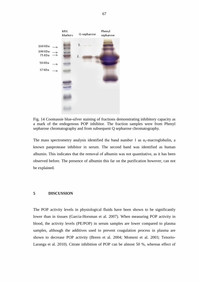
67
Fig. 14 Coomassie blue-silver staining of fractions demonstrating inhibitory capacity as a mark of the endogenous POP inhibitor. The fraction samples were from Phenyl sepharose chromatography and from subsequent Q sepharose chromatography.
The mass spectrometry analysis identified the band number 1 as α2-macroglobulin, a
known panprotease inhibitor in serum. The second band was identified as human
albumin. This indicates that the removal of albumin was not quantitative, as it has been
observed before. The presence of albumin this far on the purification however, can not
be explained.
5 DISCUSSION
The POP activity levels in physiological fluids have been shown to be significantly
lower than in tissues (Garcia-Horsman et al. 2007). When measuring POP activity in
blood, the activity levels (PE/POP) in serum samples are lower compared to plasma
samples, although the additives used to prevent coagulation process in plasma are
shown to decrease POP activity (Breen et al. 2004; Momeni et al. 2003; Tenorio-
Laranga et al. 2010). Citrate inhibition of POP can be almost 50 %, whereas effect of

68
EDTA is moderate (Tenorio-Laranga et al. 2010). It was considered these problems
could have been circumvented by using serum instead plasma as a sample. However, in
turn the observations here indicated that POP activity in serum is very low, making
more difficult to reach statistical significance when comparing diseased and healthy
samples. Furthermore, POP activity levels in CSF were found particularly low.
Proline-containing oligopeptide substrates were previously thought to be specific for
POP, until in 1997 it was shown a second enzyme (ZIP) in bovine plasma that cleaves
POP substrates in vitro (Cunningham and O’Connor; Birney and O’Connor 2001).
However, the actual substrates of ZIP in vivo have not been described. This enzyme has
later been identified as a fibroblast activated protein (FAP, seprase), although whether
the serum ZIP really is a product of the FAP gene has not been confirmed (Collins et al.
2004, Garcia-Horsman et al. 2007). The POP activity has been shown to be 40 % of the
PE activity in plasma (Breen et al. 2004, Tenorio-Laranga et al. 2010). In the present
study the proportion of POP activity in serum accounted just 2 % of the PE total
activity. This is not in agreement with previous information. However, when POP
activity was measured in the presence of a reductant as DTT, the activity rose to be over
50 % of the total activity. Oxidative deactivation of POP is well documented and it is
thought to be due to oxidation of certain cysteine residue, or residues, involved in
substrate binding or product exit from active site (Garcia Horsman et al. 2007). From
the data in this study it can be concluded that in serum POP is substantially deactivated
most probably dependent on cysteine oxidation. The results also indicate that this
process is related to coagulation, which is the main difference between serum and
plasma preparation. It is concluded that in order to measure POP levels in serum is
recommended to perform assay in the presence of a reductant agent.
Many factors can alter POP activity and thus assay results and the conclusions of the
results. Sample collection, preparation and assay conditions have been shown to affect
POP activity and stability (Momeni et al. 2003). Storage at room temperature (20̊C)
causes rapid decline in enzyme activity. Storage at – 80˚C has not been shown to
decrease POP activity, but freezing and thawing cycles of samples does. The first
freeze-thawing cycle produces the biggest reduction in enzyme activity being less

69
notable in subsequent cycles. Furthermore, the activity is sensitive to organic solvents,
usually used to dissolve synthetic substrates and inhibitors, and they have been shown
to influence the PE assay sensitivity. In this study DMSO was used, since it shows no
significant effect on activity values.
All the differences observed in POP activities (serum, serum with DTT, CSF) between
healthy controls and patients with RR-MS in this study were of very low statistical
significance. However, the differences in blood POP activities between study groups
were similar to preliminary study in the Spanish cohort (Tenorio-Laranga et al. 2010).
When serum POP activity measurements were performed with DTT, it showed clearly
that the POP enzyme was present in serum, but that it had lost its enzymatic activity
most probably due to harsh oxidative conditions possible as a result of clothing process.
This is substantiated by the fact that the effect of DTT on POP activity in serum was
notable both in MS and in control samples, whereas in the preliminary study, in plasma,
this DTT effect was more clearly observed in MS compared to control samples,
although the trend was similar. Furthermore, the POP activities in serum with DTT
seem to be similar to the POP activities in plasma with DTT. It might as well be that the
additives in plasma may (indirectly) protect the enzyme from oxidation after sample
collection or they may even restore some activity.
The POP activities in CSF samples were very low, which was in line with the previous
studies measuring such activities in various diseases (Hagihara and Nagatsu 1987;
Momeni et al. 2003). It would have been interesting to be able to measure the enzymatic
activity without the reductant in order to evaluate the oxidative state in CNS. The results
showed that there was a POP enzyme present in CSF, but that it has oxidized
(inactivated) in both study groups to the level under the detection limit of the procedure
since DTT activated to measuring values. It is worth noting that the variability of the
activity numbers measured in CSF samples was relatively wide, while this was not the
case for serum samples when DTT was present.
Even though the oxidative stress is linked to MS, the notable difference in serum POP
activities with and without DTT in both study groups suggests that serum samples in

70
some point may have been exposed to oxidative/inactivating conditions before the
activity measurement. POP activity has been shown to be sensitive to thiol reagents and
oxygen oxidation; the effects that are mediated by the oxidation of the Cys255 residue
of the catalytic triad (Polgár 2002). The activity measurements were done in batches,
and there was no observable difference in day-to-day POP activity values. The serum
used in the assays was not collected in the purposes of this study, instead it was material
saved after clinical laboratory testing in the years of 2006 to 2008.
Another factor that can decrease POP activity in the patient samples is the level of the
endogenous POP inhibitor. In the preliminary study there was a significant difference
between healthy controls and MS patients in the inhibitory capacity of plasmas
(Tenorio-Laranga et al. 2010). In this study the difference was similar; MS serum
seemed to possess the bigger inhibitory capacity over purified recombinant POP than
control serum. However, the difference was too small to be statistically significant.
Nevertheless, this inhibitory effect could not be reversed by DTT, thus the presence of
this inhibitory factor itself, is not sufficient to explain the POP inactivation seen in the
results. The inhibitory factor inactivates POP enzyme in a reversible manner since POP
can be still activated with higher affinity substrates.
In addition almost all the patients were on interferon-β (INF-β) therapy. Currently it is
not known if this cytokine medication has an effect on POP activity levels. A study by
Bayer et al. that investigated INF-β regulated proteins (by 2-DE-DIGE and MS) in
CNS, did not report altered POP expression (Beyer et al. 2009). The changes detected
were mainly cytoskeletal and transport proteins. However, it is known that POP
interacts with cytoskeletal proteins, and probably also with transport factors (Schulz et
al. 2005).
The preliminary study found significant decrease in POP activity according to age in
healthy subjects, but this was not true in the case of MS patients, where the activity was
already low in young patients with no further change at older age (Tenorio-Laranga et
al. 2010). In this study such a difference could not be observed, most due to the fact of
the low activities in all the samples. Also the mean age was higher among the control

71
patients. It is worth of noting that activity measurements were done in reaction
mixtures, so the POP concentration (activity) in vivo should be bigger.
This part of the study was performed initially measuring the levels of POP activity and
the levels of its endogenous inhibitor in all samples blind, that is, the origin of every
sample was not known until all samples were assayed. The different phases in MS
disease might cause fluctuation to the measured parameters, when in control samples
these levels are expected to be more stable. A longitudinal study or a defined sample
collection period would sharpen the results. In this study there was no correlation
between the age of a patient and measured parameters, but the preliminary study
reported decreasing POP activity levels along aging.
The strong point of this study was the biochemical characterization of the endogenous
POP inhibitor in serum. There was no previous information on the nature this factor. It
was suspected however, that inhibitor would be a peptide or a polyamine based on
previous suggestions (see Garcia-Horsman et al. 2007). The studies that have
investigated an endogenous inhibitor in animal tissue samples have isolated a small
peptide (Salers 1994; Yoshimoto 1982). However, one of the first experiments in this
study characterized the inhibitor to a molecular weight of above 10 kDa. This was the
first difference from previous studies. However, the results here found that the factor is
temperature resistant, similarly to previous studies, supporting the possible peptidic
nature. These two findings seem to be contradictory.
In trying to isolate the inhibitor, there were applied different chromatographic methods.
Affinity chromatography showed to be effective in binding the inhibitor, but it was
found to have of extremely low yield which precluded a confident identification and the
addition of further purification step.
The chromatographic sequence, DEAE, Q-Sepharose and phenyl-Sepharose was found
to be adequate for a good purification with enough yield for identification. These steps
finally leaded to the identification of the serum POP inhibitor as human albumin or α2-
macroglobulin. The activity assay of pure recombinant POP in the presence of purified

72
albumin (BSA) shows no difference with the control ruling out this protein as POP
inhibitor. Thus, it was concluded that the inhibitory factor in serum was indeed α2-
macroglobulin. Despite that this has to be verified by direct assay it is very likely that
this protein is indeed actual inhibitor.
α2-Macroglobulin has been described as inhibitory protein, that denaturates at high
temperature, 66°C (Kalwant and Porter 1991; Borth 1992; Yamamoto et al. 1996).
Furthermore it is known that substrate binding can increase temperature resistance. The
relative resistance to 100°C might have been due to partial renaturation at the
temperature of inhibition assay (37°C). Alternatively, the remaining inhibitory capacity
after high temperature incubation may also lead to think on the possibility of other
molecule in serum inhibiting POP, which may not been revealed after purification
process, and remains to be discovered. It can be speculated that this molecule, if small,
might be bound to a bigger protein precluding its filtering through a 10 kDa filter.
On the other hand α2-macroglobulin belongs to a family of α-macroglobulins that
comprise 8-10 % of total serum protein (Borth 1992). It can mediate reversible or
irreversible inhibition of proteins of diverse biological functions. α2-Macroglobulin
binds virtually any proteinase, whether self or foreign, primarily through ε(γ-glutamyl)
lysine bonds. In blood it functions as an inhibitor of coagulation by inhibiting thrombin
and inhibits fibrinolysis by inhibiting plasmin and kallikrein (DeBoer et al. 1993). It
serves as a humoral defence barrier against pathogens in the blood and tissues of
vertebrates (Borth 1992). Human α2–macroglobulin, coded by gene A2M, is implicated
e.g. in pathogenesis of Alzheimer’s disease based on its ability to mediate the clearance
and degradation β-amyloid (Blacker et al. 1998). It is also expressed in brains and
upregulated with its receptor LRP, during acute phase brain injury.
It is also important to mention that α2-macroglobulin has been found altered in MS
(Jensen et al. 2004). Significantly lower concentrations of native α2-macroglobulin and
significantly higher concentrations of transformed α2-macroglobulin were found in
plasmas of MS patients. Also immunochemical alteration due to impaired α2-

73
macroglobulin stability has been reported from patients with MS (Gunnarsson et al.
2000).
Kalwant and Porter have previously reported that α2–macroglobulin inhibits POP
moderately at 10 mM concentration (in the presence of DTT) (Kalwant and Porter
1991). If the inhibitory capacity seen in this study was all caused by α2–macroglobulin,
α2–macroglobulin inhibits POP notably. But this needs to be proven by additional tests.
The therapeutic aspect of this POP inhibition is complicated, since by affecting α2–
macroglobulin levels multiple other factors will also be affected, but it opens a new line
of the relationship between POP function and MS.
6 CONCLUSION
The first part of this study investigated POP activity levels and the levels of its inhibitor
in patient samples from healthy controls and patients with RR-MS. Although, low
statistical significance was found, the data are somehow in concordance with the
previously reported results in a Spanish cohort. By themselves, this result justify a more
comprehensive study of plasma POP activities in a Finnish larger sample to confirm the
hypothesis of link between the level of POP activity and the cause or effect of the
disease.
The second part of the study was a step by step characterization of the endogenous POP
inhibitor in serum samples. After biochemical characterization and series of
chromatographic runs, a mass spectrometry analysis revealed the inhibitor to be α2 –
macroglobulin, a panprotease inhibitor in serum. This is of relevance not just because
confirm the observed effect of α2 –macroglobulin on POP, but because it substantiate a
relationship between POP and MS. The results found justify further studies on the role
of POP in complex mechanisms of homeostasis and how this is broken in disease.

74
REFERENCES Ascherio A, Munger KL: Environmental risk factors for multiple sclerosis. Part I: The role of infection. Ann Neurol 61: 288–299, 2007 Ascherio A, Munger KL: Environmental risk factors for multiple sclerosis. Part II: Noninfectious factors. Ann Neurol 61: 504–513, 2007 Bar-Or A, Oliveira EML, Anderson DE, Hafler DA: Molecular pathogenesis of multiple sclerosis. J Neuroimm 100: 252–259, 1999 Bar-Or A, Nuttall RK, Duddy M, Alter A, Kim HJ, Ifergan I, Pennington CJ, Bourgoin P, Edwards DR, Young VW: Analysis of all matrix metalloproteinase members in leucocytes emphasize monocytes as major inflammatory mediators in multiple sclerosis. Brain: 2738–2749, 2003 Beyer S, Raether G, Stadler K, Hoffrogge R, Scharf C, Rolfs A, Mix E, Strauss U: Interferon-β modulates protein synthesis in the central nervous system. J Neuroimm 213: 31–38, 2009 Blacker D, Wilcox MA, Laird NM, Rodes L, Horvath SM, Go RCP, Perry R, Watson Jr. B, Bassett SS, McInnis MG, Albert MS, Hyman BT, Tanzi RE: Alpha-2 macroglobulin is genetically associated with Alzheimer disease. Nature Genet 19: 357–360, 1998 Borth W: α2-Macroglobulin, a multifunctional binding protein with targeting characteristics. FASEB J 6: 3345–3353, 1992 Brandt I, Vriendt KD, Devreese B, Van Beeumen J, Van Dongen W, Augustyns K, De Meester I, Scharpé S, Lambeir A-M: Search for substrates for prolyl oligopeptidase in porcine brain. Peptides 26: 2536–2546, 2005 Brandt I, Scharpé S, Lambeir AM: Suggested functions for prolyl oligopeptidase: A puzzling paradox. Clini Chim Acta 377: 50–61, 2007 Breen G, Harwood AJ, Gregory K, Sinclair M, Collier D, St Clair D, Williams RSB: Two peptidase activities decrease in treated bipolar disorder not schizophrenic patients. Bipolar Disord 6: 156–161, 2004 Cavasin MA, Rhaleb NE, Yang XP, Carretero OA: Prolyl oligopeptidase is involved in release of the antifibrotic peptide Ac-SDKP. Hypertension 43: 1140–1145, 2004 Constantinescu CS, Ventura E, Hilliard B, Rostami A: Effects of angiotensin converting enzyme inhibitor captopril on experimental autoimmune encephalomyelitis. Immunopharm Immunot 17: 471–491, 1995

75
Correale J, de los Milagros Bassani Molinas M: Temporal variations of adhesion molecules and matrix metalloproteinases in the course of MS. J Neuroimm 140: 198–209, 2003 Cunninham DF, O’Connor B: Proline specific peptidases. BBA Protein Struct Mol Enzym 1343: 160–186, 1997 De Boer JP, Creasey AA, Chang A, Abbink JJ, Roem D, Eerenberg AJM, Hack CE, Taylor Jr. FB: Alpha-2 macroglobulin functions as an inhibitor of fibrinolytic, clotting and neutrophilic proteinases in sepsis, studies using a baboon model. Infect Immun 61: 5035–5043, 1993 DeFreitas EC, Sandberg-Wollheim M, Schonely K, Boufal M, Kaprowski H: Regulation of interleukin receptors on T cells from multiple sclerosis patients. Immunology 83: 2637–2641, 1986 Dobos N, Korf J, Luiten PGM, Eisel ULM: Neuroinflammation in Alzheimer’s disease and major depression. Biol Psychiat 67:503–504, 2010 Downs JT, Lane CL, Nestor NB, McLellan TV, Kelly MA, Karam GA, Mezes PS, Pelletier JP, Otterness IG: Analysis of collagenase-cleavage of type II collagen using a neoepitope ELISA. J Immunol Methods 247: 25–34, 2001 Fernandes KSS, Guimaraes Brum D, Sandrim VS, Tostes Guerreiro CT, Antunes Barreira A, Tanus-Santos JE: Matrix metalloproteinase-9 genotypes and haplotypes are associated with multiple sclerosis and with the degree of disability of the disease. J Neuroimm 214: 128–131, 2009 Franciotta D, Salvetti M, Lolli F, Serafini B, Aloisi B: B cells and multiple sclerosis. Lancet Neurol 7: 852–58, 2008 Friese MA, Fugger L: Autoreactive CD8+ T cells in multiple sclerosis: a new target for therapy? Brain 128: 1747–1763, 2005 Fülöp V, Böcskei Z, Polgár L: Prolyl oligopeptidase: an unusual beta-propeller domain regulates proteolysis. Cell 94: 161–170, 1998 Fülöp V, Zoltán S, Polgár L: Catalysis of serine oligopeptidases is controlled by a gating filter mechanism. EMBO Rep 1: 227–281, 2000 Gaggar A, Jackson PL, Noerager BD, O’Reilly PJ, McQuaid DB, Rowe SM, Clancy JP, Blalock JE: A novel proteolytic cascade generates an extracellular matrix-derived chemoattractant in chronic neutrophilic inflammation. J Immunol 180: 5662–5669, 2008 Garcia-Horsman JA, Männistö PT, Venäläinen JI: On the role of prolyl oligopeptidase in health and disease. Neuropeptides 41: 1-24, 2007

76
Gebhard LG, Carrizo FU, Stern AL, Burgardt N, Faivovich J, Lavilla E, Ermácora MR: A Kazal prolyl endopeptidase inhibitor isolated from the skin of phyllomedusa sauvagii. Eur J Biochem 271: 2117–2126, 2004 Gilgun-Sherki Y, Melamed E, Offen D: The role of oxidative stress in the pathogenesis of multiple sclerosis: The need for effective antioxidant theory. J Neurol 251: 261–268, 2004 Goossens F, De Meester I, Vanhoof G, Scharpé S: Distribution of prolyl oligopeptidase in human peripheral tissues and body fluids. Eur J Clin Chem Clin Biochem 34: 17–22, 1996 Gunnarsson M, Stigbrand T, Jensen PEH: Decreased stability of a2-macroglobulin purified from patients with multiple sclerosis. Acta Neurol Scand 2000: 101: 270–272, 2000 Hagihara M, Nagatsu T: Post-proline cleaving enzyme in human cerebrospinal fluid from control patients and parkinsonian patients. Biochem Med Metab Biol 38: 387–391, 1987 Han MH, Hwang SI, Roy DB, Lundgren DH, Price1 JV, Ousman SS, Fernald GH, Gerlitz B, Robinson WH, Baranzini SE, Grinnell BW, Raine CS, Sobel RA, Han DK, Steinman L: Proteomic analysis of active multiple sclerosis lesions reveals therapeutic targets. Nature 451: 1076–1108, 2008 Hardison MT, Galin FS, Calderon CE, Djekic UV, Parker SB, Wille KM, Jackson PL, Oster RA, Young KR, Blalock JE, Gaggar A: The presence of a matrix-derived neutrophil chemoattractant in bronchiolitis obliterans syndrome after lung transplantation. J Immunol 182: 4423–4431, 2009 Hashimoto M, Rockenstein E, Crews L, Maslias E: Role of protein aggregation in mitochondrial dysfunction and neurogeneration in Alzheimer’s and Parkinson’s diseases. Neuromol Med 4: 21–35, 2003 Hashimoto Y, Kakegawa H, Narita Y, Hachiya Y, Hayakawa T, Kos J, Turk V, Katunumai N: Significance of cathepsin B accumulation in synovial fluid of rheumatoid arthritis. Biochem Biophys Res Commun 283: 334–339, 2001 Hartung HP, Kieseier BC: The role of matrix metalloproteinases in autoimmune damage to the central and peripheral nervous system. J Neuroimm 107: 140–147, 2000 Irazusta J, Larrinaga G, González-Maeso J, Gil J, Meana JJ, Casis L: Distribution of prolyl endopeptidase activities in rat and human brain. Neurochem Int 40: 337–345, 2002 Jensen PE, Humle Jørgensen S, Datta P, Sørensen PS: Significantly increased fractions of transformed to total alpha2-macroglobulin concentrations in plasma from patients with multiple sclerosis. Biochim Biophys Acta 1690: 203–207, 2004

77
Kakegawa H, Matano Y, Inubushi T, Katunuma N: Significant accumulations of cathepsin B and prolylendopeptidase in inflammatory focus of delayed-type hypersensitivity induced by mucobacterium tuberculosis in mice. Biochem Biophys Res Commun 316: 78–84, 2004 Kalwant S, Porter AG: Purification and characterization of human brain prolyl endopeptidase. Biochem J: 276: 237–244, 1991 Karamyan VT, Speth RC: Enzymatic pathways of the brain renin–angiotensin system: Unsolved problems and continuing challenges. Regul Peptides 143: 15–27, 2007 Katsube N, Sunaga K, Aishita H, Chuang DM, Ishitani R: ONO-1613, a potential antidementia drug, delays age-induced apoptosis and suppresses overexpression of glyceraldehyde-3-phosphate dehydrogenase in cultured central nervous system neurons. J Pharmacol Exp Ther 288: 6–13, 1999 Kim JH, Kim SI, Song KS: Prolyl endopeptidase inhibitors from green tea. Arch Pharm Res 24: 292–296, 2001 Kimura A, Yoshida I, Tagaki N, Takahashi T: Structure and localization of the mouse prolyl oligopeptidase gene. J Biol Chem 274: 24047–24053, 1999 Klawiter EC, Cross AH: B cells: No longer the nondominant arm of multiple sclerosis. Curr Neurol Neurosci Rep 7: 231–238, 2007 Klegeris A, Li J, Bammler TK, Jin J, Zhu D, Kashima DT, Pan S, Hashioka S, Maguire J, McGeer PL, Zhang J: Prolyl endopeptidase is revealed following SILAC analysis to be a novel mediator of human microglial and THP-1 cell neurotoxicity. Glia 56: 675–685, 2008 Linz W, Wohlfart P, Schölkens BA, Malinski T, Wiemer G: Interactions among ACE, kinins and NO. Cardiovasc Res 43: 549–561, 1999 Lublin FD, Reingold SC: Defining the clinical course of multiple sclerosis. Results of an international survey. Neurology 46: 907–911, 1996 Lühder F, Lee DH, Gold R, Stegbauer J, Linker RA: Small but powerful: Short peptide hormones and their role in autoimmune inflammation. J Neuroimm 217: 1–7, 2009 Mahad DJ, Ransohoff RM: The role of MCP-1 (CCL2) and CCR2 in multiple sclerosis and experimental autoimmune encephalomyelitis (EAE). Semin Immunol 15: 23–32, 2003 Manicone AM, McGuire JK: Matrix metalloproteinases as modulators of inflammation. Semin Cell Dev Biol 19: 34–41, 2008

78
Mantle D, Falkous G, Ishiura S, Blanchard PJ, Perry EK: Comparison of proline endopeptidase activity in brain tissue from normal cases and cases, with Alzheimer’s disease, Levy body dementia, Parkinson’s disease and Huntington’s disease. Clin Chim Acta 249: 129–139, 1996 Marracci GH, Jones RE, McKeon GP, Bourdette DN: Alpha lipoic acid inhibits T cell migration into the spinal cord and suppresses and treats experimental autoimmune encephalomyelitis. J Neuroimm 131: 104–114, 2002 Migration.wordpress. Internet search 20.02.2010: http://migration.wordpress.com/2007/05/22/rp-story-of-the-chemokine-mimicking-collagen-fragment/ Mirowska-Guzel D, Gromadzka G, Czlonkowski A, Czlonkowska A: Association of MMP1, MMP3, MMP9, and MMP12 polymorphisms with risk and clinical course of multiple sclerosis in a Polish population. J Neuroimm 214: 113–117, 2009 Momeni N, Yoshimoto T, Ryberg B, Sandberg-Wollheim M, Grubb A: Factors influencing analysis of prolyl endopeptidase in human blood and cerebrospinal fluid: increase in assay sensitivity. Scand J Clin Lab Invest 63: 387–396, 2003 Mossberg N, Movitz C, Hellstrand K, Bergström T, Nilsson S, Andersen O: Oxygen radical production in leucocytes and disease severity in multiple sclerosis. J Neuroimm 213: 131–134, 2009 Myöhänen TT, Venäläinen JI, Garcia-Horsman JA, Piltonen M, Männistö PT: Cellular and subcellular distribution of rat brain prolyl oligopeptidase and its association with specific neuronal neurotransmitters. J Comp Neurol 507: 1694–1708, 2008 Myöhänen T, García-Horsman JA, Tenorio-Laranga J, Männistö PT: Issues about the physiological functions of prolyl oligopeptidase based on its discordant spatial association with substrates and inconsistencies among mRNA, protein levels, and enzymatic activity. J Histochem Cytochem 57: 831–848, 2009 Männistö PT, Venäläinen J, Jalkanen A, Garcia-Horsman JA: Prolyl Oligopeptidase: A potential target for the treatment of cognitive disorders. Drug News Perspect 20: 293– 305, 2007 Naidoo R, Knapp ML: Studies of lipid peroxidation products in cerebrospinal fluid and serum in multiple sclerosis and other conditions. Clin Chem 38: 2449–2454, 1992 Navikas V, Link H: Review: Cytokines and the pathogenesis of multiple sclerosis. J Neurosci Res 45:322–333, 1996

79
Nessler S, Stadelmann C, Bittner A, Schlegel K, Gronen F, Brueck W, Hemmer B, Sommer N: Suppression of autoimmune encephalomyelitis by a neurokinin-1 receptor antagonist — A putative role for substance P in CNS inflammation. J Neuroimm 179: 1–8, 2006 O’Reilly P, Jackson PL, Noerager B, Parker S, Dransfield M, Gaggar A, Blalock JE: N-α-PGP and PGP, potential biomarkers and therapeutic targets for COPD. Respir Res 10:38, 2009 O’Reilly PJ, Hardison MT, Jackson PL, XU X, Snelgrove RJ, Gaggar A, Galin FS, Blalock JE: Neutrophils contain prolyl endopeptidase and generate the chemotactic peptide, PGP, from collagen. J Neuroimm 217: 51–54, 2009 Ortiz GG, Macias-Islas MÁ, Pacheco-Moisés FP, Cruz-Ramos JA, Sustersik S, Barba EA, Aguayo A: Oxidative stress is increased in serum from Mexican patients with relapsing-remitting multiple sclerosis. Dis Marker 26: 35–39, 2009 Platten M, Youssef S, Hur EM, Ho PP, Han MH, Lanz TV, Phillips LK, Goldstein MJ, Bhat R, Raine CS, Sobel RA, Steinman L: Blocking angiotensin-converting enzyme induces potent regulatory T cells and modulates TH1- and TH17-mediated autoimmunity. PNAS 106: 14948–14953, 2009 Polgár L: The prolyl oligopeptidase family. Cell Mol Life Sci 59: 349–362, 2002 Polman CH, Reingold SC, Edan G, Filippi M, Hartung HP, Kappos L, Lublin FD, Metz LM, McFarland HF, O’Connor PW, Sandberg-Wollheim M, Thompson AJ, Weinshenker BG, Wolinsky JS: Diagnostic Criteria for Multiple Sclerosis: 2005 Revisions to the “McDonald Criteria”. Ann Neurol 58: 840–846, 2005 Puttonen KA, Lehtonen Š, Raasmaja A, Männistö PT: A prolyl oligopeptidase inhibitor, Z-Pro-Prolinal, inhibits glyceraldehyde-3-phosphate dehydrogenase translocation and production of reactive oxygen species in CV1-P cells exposed to 6-hydroxydopamine. Toxicol Vitro 20: 1446–1454, 2006 Rahman A, Isenberg D: Systemic Lupus Erythematosus. New Engl J Med 358: 929–939, 2008 Ransohoff RM: In the beginning. Nature 462: 41–42, 2009 Rasoul S, Carreteroa OA, Peng H, Cavasin MA, Zhuoa J, Sanchez-Mendoza A, Brigstock DR, Rhaleb NE: Antifibrotic effect of Ac-SDKP and angiotensin-converting enzyme inhibition in hypertension. J Hypertens 22: 593–603, 2004 Rawlings ND, Polgár L, Barrett AJ: A new family of serine type peptidases related to prolyl oligopeptidase. Biochem J 279: 907–908, 1991

80
Reboldi A, Coisne C, Baumjohann D, Benvenuto F, Bottinelli D, Lira S, Uccelli A, Lanzavecchia A, Engelhardt B, Sallusto F: C-C chemokine receptor 6–regulated entry of TH-17 cells into the CNS through the choroid plexus is required for the initiation of EAE. Nat Immunol 10: 514–523, 2009 Rintoul GL, Bennett VJ, Papaconstandinou NA, Reynolds IJ: Nitric oxide inhibits mitochondrial movement inforebrain neurons associated with disruption of mitochondrial membrane potential. J Neurochem 97: 800–806, 2006 Roβner S, Schulz I, Zeitschel U, Schliebs R, Bigl V, Demuth HU: Brain prolylendopeptidase in aging, APP transgenic mice and Alzheimer’s disease. Neurochem Res 30: 695–702, 2005 Rossdeutsch A, Smart N, Riley PR: Thymosin β4 and Ac-SDKP: Tools to mend a broken heart. J Mol Med 86: 29–35, 2008 Rovaris M, Confavreux C, Furlan R, Kappos L, Comi G, Filippi M: Secondary progressive multiple sclerosis: current knowledge and future challenges. Lancet Neurol 5: 343–354, 2006 Salers P: Evidence for the presence of prolyl oligopeptidase and its endogenous inhibitor in neonatal rat pancreatic β-cells. Regul Peptides 50: 235–245, 1994 Schliebs R: Molecular mechanisms of neurogeneration and neuroprotection – experimental approaches and the diseased brain. Int J Devl Neuroscience 22: 441–442, 2004 Schmidt Stephan: Candidate autoantigens in multiple sclerosis. Mult Scler 5: 147–160, 1999 Schulz I, Zeitschel U, Rudolph T, Ruiz-Carrillo D, Rahveld JU, Gerhartz B, Bigl V, Demuth HU, Roβner S: Subcellular localization suggests novel functions for prolyl endopeptidase in protein secretion. J Neurochem 94: 970–979, 2005 Schulze-Topphoff U, Prat A, Prozorovski T, Siffrin V, Paterka M, Herz J, Bendix I, Ifergan I, Schadock I, A Mori M, Van Horssen J, Schröter F, Smorodchenko A, Han MH, Bader M, Steinman L, Aktas O, Zipp F: Activation of kinin receptor B1 limits encephalitogenic T lymphocyte recruitment to the central nervous system. Nat Med 15: 788–793, 2009 Semple BD, Kossmann T, Morganti-Kossmann MC: Role of chemokines in CNS health and pathology: A focus on the CCL2/CCR2 and CXCL8/CXCR2 networks. J Cereb Blood Flow Metab 3: 459–473, 2010 Setsu S, Umemura H, Mori M, Satoh M, Hayakawa S, Kodera Y, Tomonaga T, Kuwabara S, Nomura F: Serum levels of complement C4 fragments correlate with disease activity in multiple. J Neuroimm 218: 112–115, 2010

81
Shapiro SD, Ingenito EP: The pathogenesis of chronic obstructive pulmonary disease: advances in the past 100 years. Am J Respir Cell Mol Biol 32: 367–372, 2005 Sheppard MN, Nicholson AG: The pathology of cystic fibrosis. Curr Diagn Pathol 8: 50–59, 2002 Skidgel RA: Bradykinin-degrading enzymes: Structure, function, distribution, and potential roles in cardiovascular pharmacology. J Cardiovasc Pharmacol 20: S4–S9, 1992 Sørensen TL, Sellebjerg F: Distinct chemokine receptor and cytokine expression profile in secondary progressive MS. Neurology 57: 1371–1376, 2001 Steinman L: Multiple sclerosis: a two-stage disease. Nat Immunol 2: 762–764, 2001 Tenorio-Laranga J, Burgal M, Coret-Ferrer F, Casanova-Estruch B, Garcia-Horsman JA: Prolyl oligopeptidase activity is inhibited in blood-plasmas of relapsing-remitting multiple sclerosis patients. J Neuroinflamm 7: 23, 2010 Tolde K, Shinoda M, Iwamoko Y, Fujiwara T, Okamiya K, Uemura A: A novel endopeptidase inhibitor, JTP-4819, with potential for treating Alzheimers disease. Behav Brain Res 83: 147–151, 1997 Venäläinen JI, Juvonen RO, Männistö PT: Evolutionary relationships of the prolyl oligopeptidase family enzymes. Eur J Biochem 271: 2705–2715, 2004 Walter R, Shlank H, Glass JD, Schwartz IL, Kerennyj TD: Leucylglycinamide released from oxytocin by human uterine enzyme. Science 173:827–829, 1971 Weathington NM, van Houwelingen AH, Noerager BD, Jackson PL, Kraneveld AD, Galin FS, Folkerts G, Nijkamp FP, Blalock JE: A novel peptide CXCR ligand derived from extracellular matrix degradation during airway inflammation. Nat Med 12: 317–323, 2006 Welches WR, Brosnihan KB, Ferrario CM: A comparison of the properties and enzymatic activities of three angiotensin processing enzymes: angiotensin converting enzyme, prolyl endopeptidase and neutral endopeptidase. Life Sci 52: 1461–1480, 1993 Yamamoto T, Arakawa H, Ikai A, Hirotsu S, Tasum M: FT-IR study on thermal denaturation processes of ovomacroglobulin and α2-macroglobulin. J Mol Struct 384: 149–157, 1996 Yoshimoto T, Tsukumo K, Takatsuka N, Tsuru D: An inhibitor for post-proline cleaving enzyme, distribution, and partial purification from porcine pancreas. J Pharmacobiodyn 5: 734–740, 1982

82
Yuceyar N, Taşkiran D, Sağduyu A: Serum and cerebrospinal fluid nitrite and nitrate levels in relapsing-remitting and secondary progressive multiple sclerosis patients. Clin Neurol Neurosur 103: 206–211, 2001 Ziçaber J, Pásnik J, Baj Z, Pokoca L, Chmielewski H, Tchorzewski H: The immunoregulatory abilities of polymorphonuclear neutrophils in the course of multiple sclerosis. Mediat Inflamm 7: 335–338, 1998

![Marriages of Princess Anne County, Virginia : [1749-1821]](https://static.fdokumen.com/doc/165x107/6334e066a6138719eb0b4446/marriages-of-princess-anne-county-virginia-1749-1821.jpg)