Substrate Noise Analysis and Techniques for ... - DSpace@MIT
Upload
independentCategory
view
1download
0
Chemistry & Biology
Article
Profiling Mechanisms of Alkane HydroxylaseActivity In Vivo Using the DiagnosticSubstrate NorcaraneElena A. Rozhkova-Novosad,1 Jong-Chan Chae,2 Gerben J. Zylstra,2 Erin M. Bertrand,3
Marselle Alexander-Ozinskas,3 Dayi Deng,1 Luke A. Moe,5 Jan B. van Beilen,4 Michael Danahy,3
John T. Groves,1,* and Rachel N. Austin3,*1 Department of Chemistry, Princeton University, Princeton, NJ 08544, USA2 Biotechnology Center for Agriculture and the Environment, Rutgers University, New Brunswick, NJ 08901, USA3 Department of Chemistry and Program in Environmental Studies, Bates College, Lewiston, ME 04240, USA4 Institute of Biotechnology, ETH Honggerberg, CH-8093 Zurich, Switzerland5 Department of Biochemistry, University of Wisconsin, Madison, WI 53706, USA*Correspondence: [email protected] (J.T.G.), [email protected] (R.N.A.)
DOI 10.1016/j.chembiol.2006.12.007
SUMMARY
Mechanistically informative chemical probesare used to characterize the activity of func-tional alkane hydroxylases in whole cells. Nor-carane is a substrate used to reveal the lifetimeof radical intermediates formed during alkaneoxidation. Results from oxidations of this probewith organisms that contain the two mostprevalent medium-chain-length alkane-oxidiz-ing metalloenzymes, alkane u-monooxygenase(AlkB) and cytochrome P450 (CYP), are re-ported. The results—radical lifetimes of 1–7 nsfor AlkB and less than 100 ps for CYP—indicatethat these two classes of enzymes are mecha-nistically distinguishable and that whole-cellmechanistic assays can identify the activehydroxylase. The oxidation of norcarane by sev-eral recently isolated strains (Hydrocarboni-phaga effusa AP103, rJ4, and rJ5, whosealkane-oxidizing enzymes have not yet beenidentified) is also reported. Radical lifetimes of1–3 ns are observed, consistent with these or-ganisms containing an AlkB-like enzyme andinconsistent with their employing a CYP-likeenzyme for growth on hydrocarbons.
INTRODUCTION
Alkanes are ubiquitous, entering the environment through
natural oil seepages, oil spills, and biological production.
Microorganisms have been exposed to alkanes for eons
and have evolved methods for extracting metabolic en-
ergy and cell-building material from this energy-rich class
of compounds. More than 200 different species of fungi
and bacteria that are capable of oxidizing alkanes have
been identified [1–3], but in relatively few cases has the en-
zyme responsible for the chemistry been determined [4].
Chemistry & Biology 14, 165
Six enzyme families are known to be capable of oxidizing
unactivated alkanes, or, more generally, to hydroxylate
unactivated C-H bonds. These families are the alkane
u-monooxygenases (AlkB) [5], the soluble methane mono-
oxygenases (sMMO)/toluene monooxygenases (TMO) [1,
6–8], the particulate methane monooxygenases (pMMO)
[9], cytochrome P450 (CYP) [10], napthalene dioxgenase
(NDO) [11], and TauD and related a-ketoacid-dependent
hydroxylases [12]. While all families reductively activate
dioxygen to hydroxylate their cognate substrates, they dif-
fer markedly in their active site coordination chemistry,
cellular location, and cofactor content.
The majority of medium- and long-chain alkanes are
oxidized either by AlkB or CYP [1, 13–15]. CYPs contain
a thiolate-ligated heme active site and can be either
membrane spanning or soluble [10]. AlkB contains a diiron
active site with a histidine-rich coordination environment
[16] and is predicted by sequence analysis and gene fu-
sion studies to have six membrane-spanning helices
that hold the diiron active site in place near the inner
membrane-cytoplasm interface [17]. Although AlkB can
be purified, maintaining activity in the purified state is
difficult.
The molecular mechanisms of oxygen activation for
some of these metalloenzymes are well studied. Heme-
oxygenases, such as CYP, hydroxylate inert hydrocarbon
substrates by using a high-valent oxoiron(IV) porphyrin
p-cation-radical intermediate similar to peroxidase com-
pound I [10]. The consensus mechanism for oxygen acti-
vation and transfer involves a hydrogen atom abstrac-
tion-oxygen rebound pathway [10, 18]. Hydroxylation of
the very unreactive C-H bond of methane by the non-
heme diiron enzyme sMMO has many similarities to the
P450 mechanism [19–22], in part because the active
oxidant, a m-oxo-diiron(IV) intermediate called compound
Q, is poised at the same net oxidation state as the oxo-
iron(IV) porphyrin p-cation-radical species produced by
the heme-oxygenases. AlkB has been postulated to follow
an sMMO-like reaction mechanism [23–25]; however, the
details of the reaction cycle for AlkB have not yet been elu-
cidated.
–172, February 2007 ª2007 Elsevier Ltd All rights reserved 165

Chemistry & Biology
Profiling Alkane Hydroxylase Mechanisms In Vivo
Figure 1. Mechanisms of Oxygenation of the Probe Norcarane by the Reactive Oxo-Iron Intermediate, L-FeIV = O
Pathway A, which dominates the chemistry reported in this paper, represents C-H bond homolysis to generate a substrate-based radical. Pathways
B and C, which are not significant for the oxo-iron intermediate studies in this paper, involve either electron transfer (et) from the substrate to
the iron complex to give a substrate-based cation (7 and 8), or reionization of product alcohols 5 and 6. Both pathways could produce the minor
product 9.
In principle, reaction mechanisms are characteristic of
specific active sites; thus, mechanistic analysis should
provide information on active site structure. To this end,
the reactivity of the chemical probe bicyclo[4.1.0]heptane
(norcarane) was examined with whole cells harboring var-
ious alkane hydroxylase enzymes. This study was de-
signed to test the hypothesis that hydroxylases that oxi-
dize medium-chain alkanes can be identified by their
reaction mechanisms, that previously unidentified alkane
hydroxylases can be characterized based on their respec-
tive mechanistic features, and that, furthermore, these
identifications can take place when whole cells are used.
Norcarane (1) is a mechanistically diagnostic radical
clock substrate that is useful for distinguishing between
radical and cationic intermediates formed during hydrox-
ylation reactions. Furthermore, based on product distribu-
tion and the known intramolecular rearrangement rate, the
lifetime of a putative substrate radical can be determined.
An overview of the rearrangement chemistry of norcarane
is provided in Figure 1. Norcarane has been applied suc-
cessfully as a probe of reaction mechanisms in both
heme and non-heme oxygenases [19, 20, 22, 23, 25,
26]. Well-established conditions for the analysis of regio-
and stereoisomers of norcaranols and rearranged prod-
ucts are an additional advantage of this substrate. Be-
cause these product profiles can distinguish between
AlkB homologs and CYP homologs that are present,
166 Chemistry & Biology 14, 165–172, February 2007 ª2007 E
they provide a useful tool for determining which enzyme
is predominantly active in vivo.
In the present study, the hydroxylation of norcarane by
several different alkane-oxidizing organisms was exam-
ined. Of these, five organisms are known to harbor an
AlkB-like metalloenzyme: the g-proteobacterium Acineto-
bacter venetianus RAG-1 (ATCC 31012) [27], the b-proteo-
bacterium Burkholderia cepacia UCB 717 (ATCC 25416)
[5], Rhodococcus erythropolis NRRL B-16531 (ATCC
15960) [5, 28], a Pseudomonas putida clone that expresses
AlkB from P. putida GPo1 [29], and a P. putida clone that
expresses an AlkB homolog from the marine organism Al-
canovorax borkumensis AP1 [29]. Three of the organisms
are known to harbor a CYP metalloenzyme: Curvularia
lunata (ATCC 12017) [30], a P. putida clone expressing
CYP153A6 [14], and R. erythropolis, which contains genes
for both enzyme types. Three organisms examined repre-
sent recently identified species whose alkane-hydroxylat-
ing mechanisms have yet to be determined: the g-proteo-
bacterium Hydrocarboniphaga effusa AP103 [31] and the
taxonomically related strains, rJ4 and rJ5 [32]. The sub-
strate radical lifetimes obtained with AlkB- and CYP-con-
taining strains are consistent within each enzyme class,
but they are dramatically distinct between the two enzyme
classes. The differences between the enzyme classes are
sufficiently pronounced that the hydroxylases active in the
three recently identified species can be predicted to be
lsevier Ltd All rights reserved

Chemistry & Biology
Profiling Alkane Hydroxylase Mechanisms In Vivo
Table 1. Representative Product Distribution for Whole-Cell Bacterial Oxidation of Norcarane and Radical Lifetime
Producta
GPo1
Cell-free
Extract
AlkB
from
GPo1 in
GPo12
AlkB
from
AP1 in
GPo12
A.
venetianus
RAG-1
B.
cepacia
R.
erythropolis AP103 rJ4 rJ5
CYP
153A6
CYP
153A6
Cell-free
Extract
C.
lunata
5 21 12 11 14.5 9.9 21 15 6.4 5.2 72.5 79 50
6b 0 0 0 0.6 0 0 0.6 0.5 0.1 4.0 0 29
10 43 57 48 3.0 0 0 26.5 26 7.1 17 16 16
11 2.4 7.5 9.8 27.5 28 42 6.1 16 4.3 4.0 3.0 0
12 0 0.5 0 11 24 14 9.8 21 20.5 2.3 1.3 3
13 2.6 11 8.8 32 11 16 27 19 50.4 0 0 0
4 31 12 21.5 11 26 7 15 11 12.4 0.20 0.1 1
9 0 0 0.5 0 0 0 0 0 0 0.20 0.8 0
Radicallifetime, nsc
7 4 9 2 3 1 3 2 2 0.01 0.006 0.06
Values reported are for all norcarane-derived products and are given in percent.a Product yields (percent) are reported relative to the total amount of norcarane-dervived products and were determined by digital
integration of the total ion current signal of the gas chromagraph mass spectrometer by using the resident HP ChemStation
software. Product identity was confirmed by retention time and fragmentation pattern compared to authentic standards. Products
reported in less than 1% relative yield were determined by comparing ion chromatograms of the characteristic ions to those ofdilute standard mixtures.b Endo-3-norcaranol (10) and exo-2-norcaranol (6) peaks overlapped; their relative contributions were estimated based on
intensities of m/z 111 and 112.c Uncertainty in the radical lifetime is generally ±1 ns for AlkB and ±10 ps for CYP.
mechanistically and structurally like AlkB, even though
there is very little else known about these isolates.
RESULTS AND DISCUSSION
In Vivo Detection of AlkB
Previous work [23, 25] has shown that the archetypal AlkB
from P. putida GPo1, under a range of tested conditions,
catalyzed the oxidation of norcarane via H atom abstrac-
tion to generate a substrate-based radical that persists
for 1–18 ns, as indicated by the distribution of products.
A characteristic distribution of products includes 6%–
35% of the ring-opened product, 3-hydroxymethylcyclo-
hexene (4), 9%–20% of the ring-closed product endo-
2-norcaranol (5), and 20%–60% of the mechanistically
uninformative product exo-3-norcaranol (10). The cationic
product 3-cyclohepten-1-ol (9) was rarely seen. Would
other bacterial strains known to contain an AlkB enzyme
also show the same mechanistic behavior when given
norcarane as a substrate? If so, the use of diagnostic sub-
strates could provide the basis for a mechanistic profiling
screen. Known AlkB enzyme systems from two separate
bacterial hosts—P. putida GPo1 and Alcanovorax borku-
mensis AP1 (a marine g-proteobacterium that grows
almost exclusively on n-alkanes)—were tested in vivo to
ascertain whether they give the same or similar product
ratios and radical lifetimes when oxygenating norcarane
as does AlkB in cell-free assays. These AlkBs were stud-
ied in P. putida GPo12, which is a P. putida GPo1 knock-
out that lacks the ability to use octane as its sole carbon
Chemistry & Biology 14, 165–
source, but can express alkane hydroxylases from a
variety of sources [29].
Table 1 presents results from the oxidation of norcarane
by these two enzymes. With P. putida hosting AlkB from
GPo1, 12% of all norcarane-derived products were 3-
hydroxymethylcyclohexene (4), which is clear evidence
of a radical-based mechanism with an average radical
lifetime with norcarane of 4 ns. With P. putida hosting
the AlkB from AP1, the rearranged product (4) accounted
for more than 20% of the norcarane-derived products,
resulting in an average radical lifetime of 9 ns. Small
amounts, less than 1%, of the cationic product 3-cyclo-
hepten-1-ol (9) were also detected, which may reflect
subtle differences in the active site structures of the
AlkB homologs [25].
To test whether assay conditions in the cell differed from
those in extracellular assays, a norcarane oxidation exper-
iment was done by using cell lysate from P. putida GPo12
expressing AlkB from P. putida GPo1. Under the condi-
tions of this assay, the radical product 3-hydroxymethyl-
cyclohexene (4) comprised 31.2% of the products, leading
to an estimated radical lifetime of 7 ns. This result is similar
to that obtained in the in vivo assay and confirms the
comparability of in vivo and in vitro approaches.
Both the mechanisms and radical lifetimes of these
two enzymes, as assayed by the in vivo screen, are com-
pletely consistent with AlkB-like behavior in cell-free
extracts (Table 1). This result suggests that the in vivo
screening method can be useful for identifying active
AlkB hydroxylases.
172, February 2007 ª2007 Elsevier Ltd All rights reserved 167
Chemistry & Biology
Profiling Alkane Hydroxylase Mechanisms In Vivo
In Vivo Detection of CYP
Very little work exists characterizing the reaction mecha-
nisms of CYPs in vivo [33]. From prior studies with purified
CYP isozymes, it is known that the mechanism of CYP-
catalyzed alkane oxidation proceeds through a very
short-lived substrate radical—a radical whose lifetime is
in the range of tens of picoseconds, not nanoseconds as
has been seen with AlkB [22, 34].
To determine whether CYPs in vivo would show the
same mechanistic behavior with norcarane as in purified
systems, CYP153A6 from Mycobacterium sp. strain
HXN-1500 as well as its required electron transfer part-
ners, ferredoxin and a ferredoxin reductase, were cloned
into P. putida GPo12. Norcarane was metabolized, yield-
ing primarily (72.5%) 2-endo-norcaranol (5) (Table 1). Small
amounts of the ring-opened product, 3-hydroxymethylcy-
clohxene (4), and the cation product (9) were detected.
Product 4 appeared as a small shoulder on the 2-endo-
norcaranol peak in the GCMS spectrum, making quantifi-
cation difficult, although characteristic ions (m/e 94, 81,
and 79; 81 is more intense than 79) make identification
straightforward. The peak areas were used to quantify
the regions delineated by these key ions. Based on the
product yields, an estimation of the lifetime of the norcar-
ane radical was determined to be �10 ps in the catalysis
by CYP153A6 in P. putida GPo12. Small amounts of
cationic products are frequently seen with purified CYP
reactions and may indicate electron transfer that com-
petes with OH rebound or reionization of the product
alcohols [22].
A norcarane oxidation experiment was done with cell
lysate from P. putida GPo12 expressing CYP153A6 from
Mycobacterium sp. strain HXN-1500. Under the conditions
of this assay, the radical product 3-hydroxymethylcyclo-
hexene (4) comprised �0.1% of the products, leading to
an estimated radical lifetime of 6 ps. This result is similar
to that obtained in the in vivo assay and also to recent
studies from our laboratories with another diagnostic
substrate, bicyclohexane, and CYP-expressing cells and
cell-free extracts [33].
Identifying Active Hydroxylases in In Vivo Assays
in Cases in which Identifications Based on Genetic
Data Have Been Made
Having successfully demonstrated that AlkB and CYP in
in vivo assays give product yields consistent with cell-
free extracts or purified systems, work was undertaken
to identify the metalloenzymes functional in oxygenating
alkanes in microorganisms.
A. venetianus RAG-1 has an alkB gene that is closely
related to the alkB gene of Acinetobacter sp. M1 and is
distantly related to alkB from P. putida GPo1. The growth
of A. venetianus RAG-1 on hydrocarbons has been exten-
sively characterized [35–37]. The oxidation of norcarane by
A. venetianus RAG-1 resulted in 3-hydroxymethylcyclo-
hexene (4) and did not result in the cationic ring-opened
product cyclohept-3-ene-1-ol (9), nor the corresponding
ketone (15), confirming the radical-centered pathway of
this oxygenation reaction with a radical lifetime of �2 ns.
168 Chemistry & Biology 14, 165–172, February 2007 ª2007 El
Results from the oxidation of norcarane by A. venetianus
are summarized in Table 1.
B. cepacia UCB 717 (ATCC 25416) is a food- and water-
borne human pathogen [38]. It is known to degrade
n-alkanes, polycyclic aromatic hydrocarbons, and other
environmental contaminants, including tetrachloroethy-
lene and 2,4,5-trichlorophenoxyacetic acid, the main
component in Agent Orange [38]. It contains an alkB
homolog, distantly related to that of P. putida GPo1 [39].
Norcarane was hydroxylated by B. cepacia. More than
25% of all norcarane-derived products were the rear-
ranged alcohol (4), leading to an estimated radical lifetime
of 3 ns. No ring-opened cationic products were detected.
Results from the oxidation of norcarane by B. cepacia are
outlined in Table 1.
R. erythropolis NRRL B-16531 (ATCC 15960) is a Gram-
positive soil bacterium [39] known to degrade halogenated
monocyclic aromatic hydrocarbons as well as medium-
chain-length alkanes [5]. It contains at least four alkB
homologs [28, 39] that are distantly related to that of
P. putida GPo1 [29]. Protein expression data for this
organism have been equivocal. While initial reports indi-
cated that only one AlkB-like enzyme is active [28],
a more recent report suggests that none were active and,
instead, a CYP was responsible for the alkane oxidation
activity detected [15]. Our results are consistent with al-
kane hydroxylation by an AlkB homolog. 3-hydroxymethyl-
cyclohexene (4) was a clear product from the oxidation of
norcarane, comprising 7% of the total norcarane-derived
products and resulting in a radical lifetime of 1 ns. No
cationic ring-opened products were detected. Results
from the oxidation of norcarane by R. erythropolis are
shown in Table 1.
C. lunata is a CYP-containing fungus that hydroxylates
alkanes [40]. Under the assay conditions, primarily un-
rearranged norcaranols and only a small amount of 3-
hydroxymethylcyclohexene, 4, were detected. Because
3-hydroxymethylcyclohexene elutes near 2-endo-norcar-
anol, quantification of 3-hydroxymethylcyclohexene was
difficult, although the characteristic ions (m/e 94, 81, and
79; 81 is more intense than 79) were clearly visible. The
radical lifetime for C. lunata was estimated as 60 ps,
which is fully consistent with that for a CYP system.
Thus, in all of these cases, the enzyme identified by the
mechanistic screen as transforming the probe molecule
matched the available genetic information.
Identifying Active Hydroxylases in In Vivo Assays
in Cases in which No Identifications Have Been Made
Can the in vivo approach be useful for identifying enzymes
and reaction mechanisms of hydroxylases for which
biochemical or genetic information has not yet been
obtained or has proven difficult to obtain? To test this
proposal, norcarane oxidation was examined with three
strains of the recently identified genus Hydrocarboni-
phaga (specifically H. effusa AP103 [31], rJ4, and rJ5
[32]), isolated from geographically distant areas. These
strains are able to grow on medium-length n-alkanes
and are taxonomically close to Nevskia ramosa, a
sevier Ltd All rights reserved
Chemistry & Biology
Profiling Alkane Hydroxylase Mechanisms In Vivo
freshwater bacterium that was formerly the sole represen-
tative of an independent deep branch of the g subclass of
Proteobacteria [41, 42], reportedly capable of aerobically
degrading toluene [41]. However, the metabolic pathways
responsible for n-alkane degradation have not yet been
characterized in these bacteria.
All three strains were capable of transforming norcar-
ane. The data show norcarane-derived products, notably
endo- and exo-2-norcaranols, 5 and 6, and their C-3
regioisomers, 10 and 11. Ketones 12 and 13 were also
identified. Overoxidation of the rearranged alcohol (4)
with the rJ5 strain resulted in cyclohex-2-en-carboxalde-
hyde (14). The yield of the aldehyde (14) was added to
the total rearrangement product for the radical lifetime
evaluation, assuming that peak area was proportional to
concentration. The observed radical lifetimes were 3 ns
for H. effusa AP103, 2 ns for rJ4, and 2 ns for the rJ5
strain (Table 1). Neither the cationic ring-opened product
cyclohept-3-ene-1-ol (9) nor the corresponding ketone
was detected with any of the strains tested. Thus, all three
of these organisms show behavior that is consistent
with the presence of an AlkB-like enzyme and inconsis-
tent with the presence of an active CYP-like enzyme
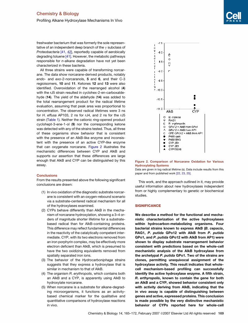
that can oxygenate norcarane. Figure 2 illustrates the
mechanistic differences between CYP and AlkB and
supports our assertion that these differences are large
enough that AlkB and CYP can be distinguished by this
assay.
Conclusions
From the results presented above the following significant
conclusions are drawn:
(1) In vivo oxidation of the diagnostic substrate norcar-
ane is consistent with an oxygen-rebound scenario
via a substrate-centered radical mechanism for all
of the hydroxylases examined.
(2) CYPs behave differently than AlkB in the mecha-
nism of norcarane hydroxylation, showing a 3–4 or-
ders of magnitude shorter lifetime for a substrate-
based radical than for AlkB-containing proteins.
This difference may reflect fundamental differences
in the reactivity of the catalytically competent inter-
mediate. CYP, with its two electrons removed from
an iron porphyrin complex, may be effectively more
electron deficient than AlkB, which is presumed to
have the two oxidizing equivalents removed from
spatially separated iron ions.
(3) The behavior of the Hydrocarboniphaga strains
suggests that they express a hydroxylase that is
similar in mechanism to that of AlkB.
(4) The organism R. erythropolis, which contains both
an AlkB and a CYP, is apparently using AlkB to
hydroxylate norcarane.
(5) When norcarane is a substrate for alkane-degrad-
ing microorganisms, it functions as an activity-
based chemical marker for the qualitative and
quantitative comparisons of hydroxylase reactions
in vivo.
Chemistry & Biology 14, 165
This work, and the approach outlined in it, may provide
useful information about new hydroxylases independent
from or highly complementary to genetic or biochemical
studies.
SIGNIFICANCE
We describe a method for the functional and mecha-
nistic characterization of the active hydroxylases
within hydrocarbon-metabolizing organisms. Four
bacterial strains known to express AlkB (B. cepacia,
RAG1, P. putida GPo12 with AlkB from P. putida
GPo1, and P. putida GPo12 with AlkB from AP1) were
shown to display substrate rearrangement behavior
consistent with predictions based on the whole-cell
mechanistic analysis of the AlkB hydroxylase from
the archetypal P. putida GPo1. Two of the strains are
clones, permitting unequivocal assignment of the
hydroxylase activity. This result indicates that whole-
cell mechanism-based profiling can successfully
identify the active hydroxylase enzyme. A fifth strain,
R. erthyropolis, known to contain the gene for both
an AlkB and a CYP, showed behavior consistent only
with activity deriving from AlkB, indicating that the
in vivo assay is capable of distinguishing between
genes and active, expressed proteins. This conclusion
is made possible by the very distinctive mechanistic
behavior of CYPs reported here for whole-cell
Figure 2. Comparison of Norcarane Oxidation for Various
Hydroxylating Systems
Data are given in log radical lifetime (s). Data include results from this
paper and from published work [22, 23, 25].
–172, February 2007 ª2007 Elsevier Ltd All rights reserved 169

Chemistry & Biology
Profiling Alkane Hydroxylase Mechanisms In Vivo
incubations with two microbial strains (the fungus
C. lunata and the P. putida GPo12 with CYP153A6).
Again, the use of a clone makes the assignment of
the chemistry observed for CYP unequivocal. The
chemistry assigned to CYP is also consistent with the
mechanistic behavior observed in purified enzymes.
Finally, for the novel and as yet uncharacterized organ-
isms, the mechanistic in vivo assay enables one to pre-
dict that the hydroxylases they use for hydrocarbon
metabolism are AlkB like and are not CYPs.
EXPERIMENTAL PROCEDURES
Materials
Unless otherwise specified, all reagents were from Sigma-Aldrich. LB,
TSB, EDTA, and chloramphenicol were obtained from Fisher Scientific
(Fair Lawn, NJ). All chemicals and solvents were Optima grade and
were used without further purification.
GC-MS analyses were performed on an HP GC-MS (5890/ 5989B)
with an RtxR-5Sil MS capillary column (Restek Corp.) or an HP GC-
MS (6890N/5973) with an HP-5MS Agilent column.
Norcarane, bicyclo[4.1.0]heptane (1), was synthesized by the
Simmons-Smith reaction as previously described [43]. Authentic sam-
ples of the product C2 (5 and 6) and C3 norcaranols (10 and 11), the
corresponding ketones (12 and 13), and the rearrangement products
3-cyclohepten-1-ol (9) and 3-hydroxymethylcyclohexene (4) were
prepared according to published procedures [44–47]. The purity of
each product was assessed by 1H NMR and GC-MS.
Bacterial Strains: Growth Conditions, Alkane Induction,
and In Vivo Activity Assay
H. effusa AP103 was previously isolated by our laboratory [31].
H. effusa rJ4 and rJ5 strains were generous gifts from Dr. K. Watanabe.
B. cepacia UCB 717 (ATCC 25416), R. erythropolis NRRL B-16531
(ATCC 15960), A. venetianus RAG-1 (ATCC 31012), and C. lunata
were obtained from the American Type Culture Collection. P. putida
with appropriate plasmids was generated according to previously
published procedures [5, 29].
B. cepacia UCB 717, R. erythropolis NRRL B-16531, and A. venetia-
nus RAG-1 were cultivated in LB medium at 30�C. H. effusa strains
were grown in TSB medium at 30�C. In the case of the RAG-1 strain,
the medium was supplemented with chloramphenicol at a final concen-
tration of 25 mg/ml. After 20–40 hr of growth in shake flasks at 200 rpm,
cells were harvested by centrifugation, resuspended in MSB (minimum
salt basal) [48] medium, and supplied with an appropriate n-alkane
(hexadecane for B. cepacia UCB 717 and dodecane for other strains)
via a suspended glass bulb. Cultures were allowed to grow while being
shaken for 24 hr at 30�C.
P. putida Gpo12 with plasmids containing AlkB homologs from
P. putida GPo1, AP1, and the CYP153A6 from Mycobacterium sp.
strain HXN-1500 were grown in MSB medium supplemented with oc-
tane introduced through the vapor phase by placing the substrate in
a glass bulb. When the optical density reached 1.0 at 600 nm, the oc-
tane was removed and diagnostic substrate (75 ml) was added to the
medium via a fresh glass bulb.
After induction with the appropriate n-alkane, the cultures were
harvested by centrifugation, washed twice, and resuspended in the
assay buffer (50 mM sodium-phosphate [pH 7.4]). The cell density
was adjusted to between an optical density of 1.2 and 1.5 at 600 nm.
A 2.5 ml portion of the cell suspension was transferred to a Falcon
tube, 8 ml of the diagnostic substrate was added, and the resulting sus-
pension was mixed well by vortexing. The tubes were gently shaken at
85 rpm at 25�C. After 18 hr of incubation, 0.5 ml aliquots were taken,
cells were separated by centrifugation, and resulting supernatant was
extracted by vortex mixing for 1 min with 300 ml ethyl acetate. The
phases were separated by centrifugation for 1 min at 14,000 rpm. Com-
170 Chemistry & Biology 14, 165–172, February 2007 ª2007 El
bined organic phases were dried by anhydrous sodium sulfate and
were analyzed by GC-MS. Products were identified by comparing the
observed peaks to the retention times and fragmentation patterns of
authentic standards. Alternatively, 75 ml of the substrate was placed
in a glass bulb hanging over 50 ml of cells suspended in buffer after
induction with hydrocarbons. The larger volume facilitated product
identification, and the shorter incubation time decreased the produc-
tion of overoxidation products. Technical constraints largely dictated
the method used. The cells were incubated for 6–8 hr, centrifuged for
15 min at 8000 3 g, and extracted three times with an equal volume
of ethyl acetate. The organic phases were combined and dried through
anhydrous sodium sulfate. The organic phase was concentrated on
a rotary evaporator and was analyzed by GC-MS.
Norcarane was found to be a good substrate for all enzyme systems
tested, generally yielding more than 25% conversion of substrate to
products over the course of the experiment. Small amounts of second-
ary oxidation products resulting from norcarane desaturation were
detected in a minority of samples. These alcohols and epoxides were
chromatographically distinct from the radical alcohol and did not affect
the results.
C. lunata: Growth Conditions, Alkane Induction,
and In Vivo Activity Assay
C. lunata was grown in rabbit food medium. The medium was prepared
by combining 1.25 g rabbit food pellets with 50 ml Milli-Q water and
heating the solution until boiling. The solution steeped for 30 min and
was vacuum filtered before autoclaving. C. lunata was inoculated
into 50 ml rabbit food broth medium and was grown at 25�C and
300 rpm. Experiments were carried out after �2–3 days of growth.
Substrate (200 ml) and additional rabbit food medium (10 ml) were
added directly to 50 ml growing culture. The culture was incubated
with substrate at 25�C and 300 rpm overnight, and it was then filtered
through cheese cloth. This filtrate was then extracted three times
with ethyl acetate, concentrated, assayed by GC-MS, and analyzed
as described above. Control experiments were also done for each
experiment with C. lunata; all reaction components, except for the
organism, were added to the media and incubated.
Cell-free Extract: Preparation of Extract and In Vivo
Activity Assay
P. putida GPo12 cells expressing AlkB from P. putida GPo1 were grown
on octane to an optical density of 2 at 600 nm and were centrifuged at
8000 rpm for 15 min. The pellet was sonicated in a buffer that contained
50 mM potassium phosphate (pH 7.4), 5% glycerol, 1 mM DTT (dithio-
threitol), and 200 mM PMSF (phenylmethylsulfonyl fluoride) for 1 min
total in 5 s bursts, followed by centrifugation at 4200 rpm for 25 min.
The supernatant was decanted and used for assays.
Cell-free assays were done by adding 1 ml supernatant to a glass
vial with Teflon-lined screw-top caps with 1 ml of 1 M DTT and 2 ml sub-
strate. The vial was capped with a Teflon seal and shaken at 30�C
briefly. The reaction was initiated by the addition of 12 mmol NADH
and was incubated with shaking for 2 hr. After 2 hr, the reaction was
quenched and extracted with an equal volume of chloroform.
Radical Lifetime Determinations
The radical lifetimes were calculated from the following equation:
radical lifetime =
1
krearrangement½unrearranged products�
radical rearranged products
!: (1)
The rebound rate is the inverse of the radical lifetime. The rate con-
stant 2 3 108 s�1 for the norcaran-2-yl radical ring opening was calcu-
lated from product ratios for the reaction of 2-chloronorcarane with
tri-n-butyl-tin hydride as the radical trap [49, 50]. Unrearranged prod-
ucts include both isomers of 2-norcarnol (5 and 6) and 2 norcaranone
(12) when present. Hydroxymethylcyclohexene (4) is the primary rear-
ranged product; however, for rJ5, cyclohex-2-en-carbaldehyde (14)
was detected and included as a rearranged product.
sevier Ltd All rights reserved

Chemistry & Biology
Profiling Alkane Hydroxylase Mechanisms In Vivo
General Comments
Whole-cell oxidation experiments were repeated at least three times for
each strain. GC-MS runs were at least duplicated. Control experiments
in which potential products were provided to cultures as substrates
were routinely done to ensure that assays would permit detection
and quantification of all norcarane-derived products. In cases in which
secondary oxidation occurred, the products (ketones or aldehydes)
were included in the calculation of the radical lifetime.
ACKNOWLEDGMENTS
Financial support from the National Science Foundation through the
Environmental Molecular Science Institute CEBIC (Center for Environ-
mental Bioinorganic Chemistry at Princeton University) CHE-0221978
(G.J.Z., R.N.A., and J.T.G.), CHE-0316301 (J.T.G.), MCB-0078465,
and CHE-0116233 (R.N.A.); the Camille and Henry Dreyfus Foundation
(R.N.A.); and the National Institutes of Health GM-32698 (J.T.G.) and
GM072506 (R.N.A.) is gratefully acknowledged. We also thank
Dr. John Eng and Dr. Dorothy Little for expert technical assistance
with mass spectrometry and Charlotte Lehmann for technical assis-
tance with cell culturing and experiments.
Received: August 11, 2006
Revised: December 5, 2006
Accepted: December 6, 2006
Published: February 23, 2007
REFERENCES
1. van Beilen, J.B., Li, Z., Duetz, W.A., Smits, T.H.M., and Witholt, B.
(2003). Diversity of alkane hydroxylase systems in the environ-
ment. Oil Gas Sci. Technol. 58, 427–440.
2. Atlas, R.M., and Bartha, R. (1973). Fate and effects of polluting
petroleum in the marine environment. Residue Rev. 49, 49–85.
3. Aislabie, J.M., Balks, M.R., Foght, J.M., and Waterhouse, E.J.
(2004). Hydrocarbon spills on Antarctic soils: effects and manage-
ment. Environ. Sci. Technol. 38, 1265–1274.
4. Harayama, S., Kasai, Y., and Hara, A. (2004). Microbial communi-
ties in oil-contaminated seawater. Curr. Opin. Biotechnol. 15,
2005–2014.
5. Smits, T.H.M., Rothlisberger, M., Witholt, B., and van Beilen, J.B.
(1999). Molecular screening for alkane hydroxylase genes in
gram-negative and gram-positive strains. Environ. Microbiol. 1,
307–317.
6. Baik, M.H., Newcomb, M., Friesner, R., and Lippard, S.J. (2003).
Mechanistic studies on the hydroxylation of methane by methane
monooxygenase. Chem. Rev. 103, 2385–2419.
7. Sluis, M., Sayavedra-Soto, L.A., and Arp, D.J. (2002). Molecular
analysis of the soluble butane monooxygenase from ‘Pseudomo-
nas butanovora’. Microbiology 148, 3617–3629.
8. Pikus, J.D., Studts, J.M., Achim, C., Kauffmann, K.E., Munck, E.,
Steffan, R.J., McClay, K., and Fox, B.G. (1996). Recombinant
toluene4-monooxygenase: catalytic and mossbauer studies of
the purified diiron and Rieske components of a four protein com-
plex. Biochemistry 35, 9106–9119.
9. Semrau, J.D., Christoserdov, A., Lebron, J., Costello, A., Davag-
nino, J., Kenna, E., Holmes, A.J., Finch, R., Murrell, J.C., and
Lidstrom, M.E. (1995). Particulate methane monooxygenase
genes in methanotrophs. J. Bacteriol. 177, 3071–3079.
10. Groves, J.T. (2005). Models and Mechanisms of Cytochrome P450
Action (New York: Kluwer Academic/Plenum Publishers).
11. Resnick, S.M., Lee, K., and Gibson, D.T. (1996). Diverse reactions
catalyzed by naphthalene dioxygenase from Pseudomonas sp
strain NCIB 9816. J. Ind. Microbiol. 17, 438–457.
Chemistry & Biology 14, 165–
12. Rice, J.C., Barr, E.W., Tirupati, B., Bollinger, J.M., and Krebs, C.
(2003). The first direct characterization of a high-valent iron
intermediate in the reaction of an a-ketoglutarate-dependent diox-
ygenase: a high-spin Fe(IV) complex in taurine/a-ketoglutarate
dioxygenase (TauD) from Escherichia coli. Biochemistry 42,
7497–7508.
13. Bell, S.G., Hoskins, N., Xu, F., Caprotti, D., Rao, Z., and Wong, L.L.
(2006). Cytochrome P450 enzymes from the metabolically diverse
bacterium Rhodopseudomonas palustris. Biochem. Biophys. Res.
Commun. 342, 191–196.
14. van Beilen, J.B., Holtackers, R., Luscher, D., Bauer, U., Witholt, B.,
and Duetz, W.A. (2005). Biocatalytic production of perillyl alcohol
from limonene by using a novel Mycobacterium sp cytochrome
P450 alkane hydroxylase expressed in Pseudomonas putida.
Appl. Environ. Microbiol. 71, 1737–1744.
15. van Beilen, J.B., Funhoff, E.G., van Loon, A., Just, A., Kaysser, L.,
Bouza, M., Holtackers, R., Rothlisberger, M., Li, Z., and Witholt, B.
(2006). Cytochrome P450 alkane hydroxylases of the CYP153
family are common in alkane-degrading eubacteria lacking inte-
gral membrane alkane hydroxylases. Appl. Environ. Microbiol.
72, 59–65.
16. Shanklin, J., Achim, C., Schmidt, H., Fox, B.G., and Munck, E.
(1997). Mossbauer studies of alkane omega-hydroxylase: evi-
dence for a diiron cluster in an integral-membrane enzyme. Proc.
Natl. Acad. Sci. USA 94, 2981–2986.
17. van Beilen, J.B., Penninga, D., and Witholt, B. (1992). Topology of
the membrane-bound alkane hydroxylase of Pseudomonas oleo-
vorans. J. Biol. Chem. 267, 9194–9201.
18. Groves, J.T. (2003). The bioinorganic chemistry of iron in oxy-
genases and supramolecular assemblies. Proc. Natl. Acad. Sci.
USA 100, 3569–3574.
19. Newcomb, M., Shen, R., Lu, Y., Coon, M.J., Hollenberg, P.F.,
Kopp, D.A., and Lippard, S.J. (2002). Evaluation of norcarane as
a probe for radicals in cytochome P450- and and soluble methane
monooxygenase-catalyzed hydroxylation reactions. J. Am. Chem.
Soc. 124, 6879–6886.
20. Brazeau, B.J., Austin, R.N., Tarr, C., Groves, J.T., and Lipscomb,
J.D. (2001). Intermediate Q from soluble methane monooxygenase
hydroxylates the mechanistic substrate probe norcarane: evi-
dence for a stepwise reaction. J. Am. Chem. Soc. 123, 11831–
11837.
21. Kopp, D., and Lippard, S.J. (2002). Soluble methane monooxyge-
nase: activation of dioxygen and methane. Curr. Opin. Chem. Biol.
6, 568–576.
22. Auclair, K., Hu, Z., Little, D.M., Ortiz de Montellano, P.R., and
Groves, J.T. (2002). Revisiting the mechanism of P450 enzymes
with the radical clocks norcarane and spiro[2,5]octane. J. Am.
Chem. Soc. 124, 6020–6027.
23. Austin, R.N., Chang, H.K., Zylstra, G.J., and Groves, J.T. (2000).
The non-heme diiron alkane monooxygenase of Pseudomonas
oleovorans (AlkB) hydroxylates via a substrate radical intermedi-
ate. J. Am. Chem. Soc. 122, 11747–11748.
24. Austin, R.N., Buzzi, K., Kim, E., Zylstra, G.J., and Groves, J.T.
(2003). Xylene monooxygenase, a membrane-spanning non-
heme diiron enzyme that hydroxylates hydrocarbons via a sub-
strate radical intermediate. J. Biol. Inorg. Chem. 8, 733–740.
25. Bertrand, E., Sakai, R., Rozhkova-Novosad, E., Moe, L., Fox, B.G.,
Groves, J.T., and Austin, R.N. (2005). Reaction mechanisms of
non-heme diiron hydroxylases characterized in whole cells.
J. Inorg. Biochem. 99, 1998–2006.
26. Moe, L.A., Hu, Z., Deng, D., Austin, R.N., Groves, J.T., and Fox,
B.G. (2004). Remarkable aliphatic hydroxylation by diiron enzyme
toluene 4-monooxygenase in reactions with radical/cation
diagnostic probes norcarane, 1,1-dimethylcyclopropane, and
1,1-diethylcyclopropane. Biochemistry 43, 15688–15701.
172, February 2007 ª2007 Elsevier Ltd All rights reserved 171

Chemistry & Biology
Profiling Alkane Hydroxylase Mechanisms In Vivo
27. Vaneechoutte, M., Tjernberg, I., Baldi, F., Pepi, M., Fani, R.,
Sullivan, E.R., van der Toorn, J., and Dijkshoorn, L. (1999).
Oil-degrading Acinetobacter strain RAG-1 and strains described
as ‘Acinetobacter venetianus sp. Nov’ belong to the same geno-
mic species. Res. Microbiol. 150, 69–73.
28. Whyte, L.G., Smits, T.H.M., Labbe, D., Witholt, B., Grer, C.W., and
van Beilen, J.B. (2002). Gene cloning and characterization of
multiple alkane hydroxylase sytstems in Rhodococcus strains
Q15 and NRRL B-16531. Appl. Environ. Microbiol. 68, 5933–5942.
29. Smits, T.H.M., Balada, S.B., Witholt, B., and van Beilen, J.B.
(2002). Functional analysis of alkane hydroxylases from gram-
negative and gram-positive bacteria. J. Bacteriol. 184, 1733–1742.
30. Matthew, J., and Sisler, H.D. (1984). Effects of sterol biosynthesis-
inhibiting (SBI) fungicides on cytochrome P-450 oxygenations in
fungi. Pestic. Biochem. Physiol. 22, 262–275.
31. Palleroni, N.J., Port, A.M., Chang, H.K., and Zylstra, G.J. (2004).
Hydrocarboniphaga effusa gen. Nov. sp. Nov., a novel member
of gamma-Proteobacteria active in alkane and aromatic hydrocar-
bon degradation. Int. J. Syst. Evol. Microbiol. 54, 1203–1207.
32. Watanabe, K., Teramoto, M., and Harayama, S. (1999). An out-
break of nonflocculating catabolic populations caused the break-
down of a phenol-digesting activated-sludge process. Appl.
Environ. Microbiol. 65, 2813–2819.
33. Austin, R.N., Deng, D., Jiang, Y., Luddy, K., van Beilen, J.B., Ortiz
de Montellano, P.R., and Groves, J.T. (2006). The diagnostic sub-
strate bicyclohexane reveals a radical mechanism for bacterial
cytochrome P450 in whole cells. Angew. Chem. Int. Ed. Engl. 45,
8192–8194.
34. Jiang, Y., He, X., and Ortiz de Montellano, P.R. (2006). Radical in-
termediates in the catalytic oxidation of hydrocarbons by bacterial
and human cytochrome P450 enzymes. Biochemistry 45, 533–542.
35. Di Cello, F., Pepi, M., Baldi, F., and Fani, R. (1997). Molecular char-
acterization of an n-alkane-degrading bacterial community and
identification of a new species, Acinetobacter venetianus. Res.
Microbiol. 148, 237–249.
36. Bach, H., Berdichevsky, Y., and Gutnick, D. (2003). An exocellular
protein from the oil-degrading microbe Acinetobacter venetianus
RAG-1 enhances the emulsifying activity of the polymeric bioemul-
sifier emulsan. Appl. Environ. Microbiol. 69, 2608–2615.
37. Nakar, D., and Gutnick, D.L. (2003). Involvement of a protein tyro-
sine kinase in production of the polymeric bioemulsifier emulsan
172 Chemistry & Biology 14, 165–172, February 2007 ª2007
from the oil-degrading strain Acinetobacter lwoffii RAG-1. J. Bac-
teriol. 185, 1001–1009.
38. Coenye, T., and Vandamme, P. (2003). Diversity and significance
of Burkholderia species occupying diverse ecological niches.
Environ. Microbiol. 5, 719–729.
39. Margesin, R., Labbe, D., Schinner, F., Greer, C.W., and Whyte,
L.G. (2003). Characterization of hydrocarbon-degrading microbial
populations in contaminated and pristine alpine soils. Appl. Envi-
ron. Microbiol. 69, 3085–3092.
40. Sonomoto, K., Hoq, M.M., Tanaka, A., and Fukui, S. (1983). 11B-
Hydroxylation of cortexelone (Reichstein Compound S) to hydro-
cortisone by Curvularia lunata entrapped in photo-cross-linked
resin gels. Appl. Environ. Microbiol. 45, 436–443.
41. Sturmeyer, H., Overmann, J., Babenzien, H.D., and Cypionka, H.
(1998). Ecophysiological and phylogenetic studies of Nevskia
ramosa in pure culture. Appl. Environ. Microbiol. 64, 1890–1894.
42. Glokner, F.O., Babenzien, H.D., and Amann, R. (1998). Phylogeny
and identification in situ of Nevskia ramosa. Appl. Microbiol. Bio-
technol. 64, 1895–1901.
43. LeGoff, E. (1964). Cyclopropanes from easily prepared highly
active zinc-copper couple dibromomethane + olefins. J. Org.
Chem. 29, 2048–2050.
44. Simmons, H.E., and Smith, R.D. (1959). A new synthesis of cyclo-
propanes. J. Am. Chem. Soc. 81, 4256–4264.
45. Dauben, W.G., and Berezin, G.H. (1963). Preparation of cyclic
cyclopropylcarbinols. J. Am. Chem. Soc. 85, 468–472.
46. Akermark, B., Hanson, S., Rein, T., and Vagberg, J. (1989). Palla-
dium-catalyzed allylic acetoxylation – an exploratory study of the
influence of added acids. J. Organomet. Chem. 369, 433–444.
47. Snider, B., and Rodini, D. (1980). Dialkylaluminum chloride cata-
lyzed ene reactions of aldehydes. Synthesis of ipsenol. Tetrahe-
dron Lett. 21, 1815–1818.
48. Stanier, R.Y., Palleron, N.J., and Doudorof, M. (1966). The aerobic
pseudomonas: a taxonomic study. J. Gen. Microbiol. 43, 159–271.
49. Friedrich, E.C., and Holmstead, R.L. (1972). Cyclopropylcarbinyl-
allylcarbinyl radical rearrangements in simple bicyclo[3.1.0]hexyl
and bicyclo[4.1.0]heptyl systems. J. Org. Chem. 37, 2550–2554.
50. Rahm, A., Amardeil, R., and Degueil-Castaing, M. (1989). Use of
high pressure in the reduction of organic chlorides with tri-n-butyl-
tin hydride. Organomet. Chem. 371, C4–C8.
Elsevier Ltd All rights reserved
Copyright © 2022 FDOKUMEN