A nanoparticle-organic memory field-effect transistor behaving ...
Upload
sgbcollegeCategory
view
2download
0
Sukhen Das, et al. Int J Pharm 2015; 5(2): 600-609 ISSN 2249-1848
www.pharmascholars.com 600
Research Article CODEN: IJPNL6
MARSILEA MINUTA(L.) PLANT EXTRACT MEDIATED SYNTHESIS OF GOLD
NANOPARTICLE FOR CATALYTIC AND ANTIMICROBIAL APPLICATIONS
Niranjan Bala1,2, Arpan Kool1, Pradip Thakur1,3, Sukhen Das1,4*, Papiya Nandy5, Ruma Basu6
1Department of Physics, Jadavpur University, Kolkata- 700032, India. 2Department of Botany, Sreegopal Banerjee College, Hooghly-712503, India. 3Department of Physics, Netaji Nagar College for Women, Kolkata-700092,India. 4Department of Physics, Indian Institute of Engineering Science and Technology, Howrah-
711103, India. 5Centre for Interdisciplinary Research and Education, 404B Jodhpur Park, Kolkata-700068,
India. 6Department of Physics, Jogamaya Devi College. Kolkata-700026, India
*Corresponding author e-mail: [email protected]
ABSTRACT
Gold nanoparticle (GNP) was synthesized using the plant extract of Marsilea minuta (L.) for catalytic and
antibacterial applications. Formation of nanoparticle was confirmed by various physical and chemical
characterization techniques. Morphology of synthesized NPs was analyzed using electron microscopy. GNP
synthesized via this method was highly stable and their size was found to be ~ 25 nm. Green synthesized GNPs
showed well catalytic activity in complete reduction of toxic para-nitrophenol (PNP) to para-aminophenol (PAP).
GNP synthesized by this method showed better antibacterial potency against Escherichia coli and Staphylococcous
aureus. It was observed that GNP synthesized via this method showed synergistic effect of chemically synthesized
GNP and plant extract on antimicrobial activity.
Keywords: Gold nanoparticles; green synthesis; catalyst; para-nitrophenol; antimicrobial activity; synergistic effect.
INTRODUCTION
During the course of improvement of nano-science,
GNP drew the attention of research workers for their
unique optical [1-2], chemical [3-4] and
photochemical activities [5-6]. Along with these GNP
were applied in biological fields like nano-medicine
[7], drug delivery [8-9] and cancer therapy [10-11]
for their noble nature and easily tunable size.
Predictable synthetic approaches of gold
nanoparticles are mainly involves toxic chemicals.
Recently another approach has been become very
attractive that using different organic compounds
present in plants [12-13] like phenolic compounds,
flavonoids, alkaloids, organic acids [14-16, 17-23]
which can reduce Au3+ or Au1+ into Au0 by donating
electron and also stabilize the newly synthesized
GNPs simultaneously. Green synthesis of
nanoparticles have drawn attention not only for
avoiding the use of toxic chemicals during synthesis
procedure, but for the reason that these nanoparticles
are biocompatible, nontoxic, ecofriendly in nature
and suitable for biomedical applications [14-16, 24].
Nitro-aromatic compounds like nitrophenols were
abundantly distributed in environment [25]. PNP is
one of the major members within the nitrophenols
family produced as a by-product during industrial
manufacture of pesticide, herbicide and synthetic
dyes [26]. It was applied in agriculture, dyes and
pigments production, manufacturing fungicide for
leather [27-28]. PNP was also used for production of
drugs like acetaminophen, a non- aspiring pain
reliever, parathion and fluoridifen like pesticide [29]
International Journal of Pharmacy Journal Homepage: http://www.pharmascholars.com
Sukhen Das, et al. Int J Pharm 2015; 5(2): 600-609 ISSN 2249-1848
www.pharmascholars.com 601
PNP showed toxicity on aquatic organisms likes
algae, invertebrate, fish [30-32]. Widespread use of
nitro-phenol PNP results great hazards for all life
forms of the ecosystem ranging from unicellular
organism to human beings. Mainly, in water bodies
these aromatic compounds reduces the photosynthetic
potential of aquatic organisms by interfering the
entering sun lights and soluble gas balance, prompts
in the destruction of aquatic ecosystem [33]
In the present study inexpensive, highly stable GNP
was synthesized by one step method using crude
extract of a pteridophytic plant Marsilea minuta.
Formation of the particles was confirmed by uv-
visible spectroscopy (uv-vis) and x-ray diffraction
(XRD). Morphology and size of NP were analyzed
using dynamic light scattering (DLS), FESEM and
HRTEM. Chemical constituents of synthesized NPs
were analyzed by FTIR. Stability to salinity of these
NP was compared with chemically synthesized GNP.
Catalytic activity of biosynthesized GNP was studied
in the reduction of environmentally toxic PNP and
potency for antibacterial activities was examined
against Escherichia coli and Stphylococcus aureus
microorganism. Mechanism for antibacterial activity
was confirmed by FESEM studies.
MATERIALS AND METHODS
Materials: Marsilea minuta (Indian native name:
Sushni) a pteridophytic plant collected from Jadavpur
University campus. Tetrachloroauric (III) acid
(HAuCl4) was purchased from Sigma Aldrich. PNP
was purchased from Merck (Germany). All other
chemicals used in this experiment were purchased
from Merck (India).
Plant extract preparation: 1 gm of Marsilea leaves
were washed 4-5 times thoroughly with distilled
water and then surface sterilized with 90% ethanol.
Subsequently, boiled with 20 ml double distilled
water for 5 min and cooled to room temperature.
Then the plant extract (pH7) was filtered with
Whatman filter paper no.1and stored at room
temperature for further use.
Synthesis of gold nanoparticles: 2 ml aqueous
solution of 0.01 M of Tetrachloroauric (III) acid
(HAuCl4) was mixed with 100 µl of the plant extract
under continuous stirring. Initially, the colour of the
solution was yellowish and after 30 min of
continuous stirring red wine colour of GNP appeared
indicating the formation of GNP (Fig. 1). The
reaction mixture was allowed to 4 hours for complete
reduction of gold ions. Optimal condition for the
synthesis of green GNPs was checked against
different concentrations of plant extract to HAuCl4
within a range of 25-200μl. Citrate capped
chemically synthesized GNP was obtained following
the protocol of Polavarapu and Xu (2009) [34]
Characterizations: Uv-Vis light spectra of
synthesized nanoparticles were recorded in λ25
spectrophotometer (Parkin Elmer, Germany). Particle
size and particle distribution of aqueous solution of
GNPs were analyzed by DLS with the help of Zeta-
sizer-5000 (Malvern Instruments, UK). Morphology
and particle size analyzed using FESEM (Inspect
F50, FEI, Netherland) and HRTEM (JEM – 2100
HRTEM, JEOL, Japan). For FESEM study GNP
solution was drop casted on glass cover slip and NP
solution was sprayed on copper grid for HRTEM
analysis. XRD pattern of synthesized green GNP was
analyzed in the range of 2θ ~35-80° using powder
diffractometer (D8, Bruker AXS,) by Cu Kα radiation
(α = 0.15425 nm). Biosynthesized GNPs solution was
drop casted over cover glass and dried at 60°C for 2
hour for XRD analysis. FTIR spectroscopy recorded
using a JASCO FTIR instrument-410 in the range of
4000-400 cm-1 to investigate the chemical
constituents of the plant extract and synthesized GNP
after centrifugation and washing. The pellets were
first prepared in Potassium Bromide (KBr) and
samples were sprayed on it by drop casting.
Antimicrobial study: Investigate the effect of GNP
on bacteria, antimicrobial studies were performed on
Escherichia coli DH5α (MTCC 1652) and
Staphylococcus aureus (MTCC 96). Antimicrobial
effect of the plant extracts and GNPs synthesized via
green and chemical routes were studied following the
standard protocol. 0.5 mg of lyophilized GNP was
dissolved in 1ml of autoclaved water and different
amount of the solution and plant extract (50-400 µl)
were added to cultures of bacteria (107 CFU/ml) in 5
ml nutrient broth (0.5 % peptone, 0.1 % beef extract,
0.2 % yeast extract, 0.5 % NaCl, pH 7). So, the
resultant GNPs concentration in nutrient broth was 5-
40 µg/ml. The cultures were then incubated at 37 °C
for 24 h. Growth inhibition was followed by plating
50 µl of the treated culture on nutrient agar plates
(nutrient broth with 1.5 % agar as the solidifying
agent). Bacterial colonies were counted and
compared with control after 24 h incubation at 37 °C.
The whole experiment was repeated thrice to ensure
reproducible data. The same experiment was
conducted with corresponding amounts of chemically
synthesized GNP and crude plant extract of M.
minuta.
The growth of bacteria was evaluated by counting
colony forming unit (CFU) on agar plate. The
Sukhen Das, et al. Int J Pharm 2015; 5(2): 600-609 ISSN 2249-1848
www.pharmascholars.com 602
antibacterial efficiency was calculated using the
equation 1.
(1)
Where, M is the mortality rate (%), B is the mean
number of bacteria on the control samples
(CFU/sample) and C is the mean number of bacteria
on the treated samples (CFU/sample).
Minimum bactericidal concentration (MBC) values
were calculated using the standard plate count
technique [35] with increasing concentrations of
GNP.
Relative decrease of MBC values were calculated by
the equation 2.
Where, X is relative decrease in MBC value, A is the
mean value of chemically synthesized GNP/ crude
plant extract and B is the mean MBC value of green
synthesized GNP.
Mechanism behind GNP induced bacterial death was
investigated by FESEM. For FESEM study bacterial
samples were prepared following the standard
protocol [36-37] protocol. Briefly, bacterial sample in
mid exponential phase (107 CFU/ml) were treated
with 20 µg/ml of green GNP for 12 hour at 37°C.
Control was prepared under similar condition but
without GNP. Bacterial samples were then fixed with
2% glutaraldehyde. After washing with water, 1 μl
was placed on a silicon platelet (Plano, Wetzlar,
Germany). Samples were passed through a gradient
of ethanol dehydration steps followed by staining
with 3% ethanolic solution (25 %) of uranyl acetate.
Finally, the samples were washed with 0.1 M
phosphate buffer solution (pH 7.2) and sputter coated
with gold [36]. Microscopy was performed with
Inspect F50 (FEI, Netherlands).
Catalytic Activity: Catalytic property of
synthesized GNP was investigated by the reduction
of para-nitrophenol (PNP) with sodium borohydrate.
PNP is a common toxic byproduct of pesticide,
herbicide and synthetic dye production [26]. PNP
was easily reduced to para-aminophenol (PAP) by
NaHB4 in presence of gold in nano forms. [38].
Catalytic measurements were studied following the
protocols of Pougon, Z. D. [39].
Stability to NaCl: Agglomeration is a big problem
for colloidal nanoparticles. Stability of
biosynthesized GNP was compared to chemically
synthesized GNP. Stability of these particles were
checked against very high salt concentration (5 M
NaCl) following the protocol of S Pandey et al.[15]
RESULT AND DISCUSSION
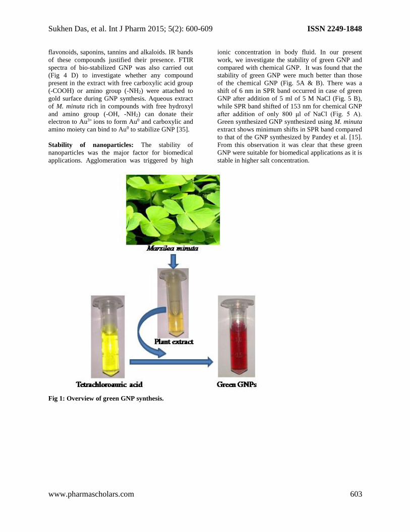
Effect of crude plant extract on GNP synthesis:
GNP synthesis started after addition of aqueous plant
extract to HAuCl4. Initial colour of the reaction
mixture was yellowish and gradually became reddish.
Reduction of HAuCl4 was complete after 4 h and
wine red coloured green GNP was formed (Fig 1).
Surface Plasmon resonance (SPR) band at 522 nm
confirmed the formation of GNP by UV-VIS spectra
(Fig. 2A). It was evident from the figure that
complete synthesis of GNP took around 4 hours, the
particles were quite stable and no agglomeration was
detected even after 30 days. GNP formation started at
above 1 % (v/v) plant extracts (Fig: 2B). With higher
plant extract concentration SPR band intensity
increased gradually indicating formation of more
GNP and optimized at a concentration of 5% plant
extract. Further addition of plant extract to the
reaction mixture showed red shifting of SPR band. It
was observed that 0.005M of HAuCl4 is the
minimum concentration for the green synthesis of
GNP and GNP population optimizes at a
concentration of 0.01M (Fig: 2C). Further increase in
HAuCl4 concentration did not show any significant
difference, which means that the bioactive
compounds present in plant extract was unable to
synthesize more particles. To synthesize GNP (‹40
nm) for biomedical application nearly 5 % (v/v) plant
extract and 0.01M HAuCl4 were required.
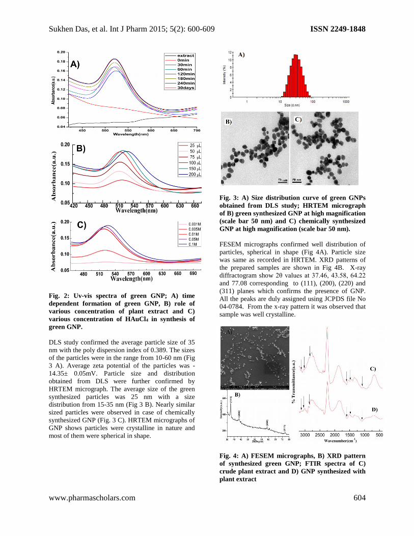
Functional groups involved in GNP synthesis:
FTIR spectrum of aqueous extract of M. minuta
showed presence of characteristic bands for several
functional groups (Fig. 4 C). IR peaks for phenolic –
O-H Stretch were observed at around 3154 cm−1
and 3122 cm−1. Presence of phenolics were
confirmed by C-C=C symmetric stretch, C-C=C
asymmetric stretch, C=C-H asymmetric stretch and
aromatic C-H bend at 1596, 1474, 3022 and 2848
cm−1 respectively [14]. Amines were also present in
the plant extract. Presence of aromatic amines (-
C6H5NH2) and aliphatic amines (R-NH2) were
confirmed by 1118 cm−1 and 1390 cm−1 bands. IR
band at 1040 cm−1 supports the presence of carbonyl
(>C=O) groups of carboxylic acids. These findings
are supported by some previous reports on
phytochemical profiling of M. minuta [35]. Aqueous
extract of plant contains phenolic compounds,
Sukhen Das, et al. Int J Pharm 2015; 5(2): 600-609 ISSN 2249-1848
www.pharmascholars.com 603
flavonoids, saponins, tannins and alkaloids. IR bands
of these compounds justified their presence. FTIR
spectra of bio-stabilized GNP was also carried out
(Fig 4 D) to investigate whether any compound
present in the extract with free carboxylic acid group
(-COOH) or amino group (-NH2) were attached to
gold surface during GNP synthesis. Aqueous extract
of M. minuta rich in compounds with free hydroxyl
and amino group (-OH, -NH2) can donate their
electron to Au3+ ions to form Au0 and carboxylic and
amino moiety can bind to Au0 to stabilize GNP [35].
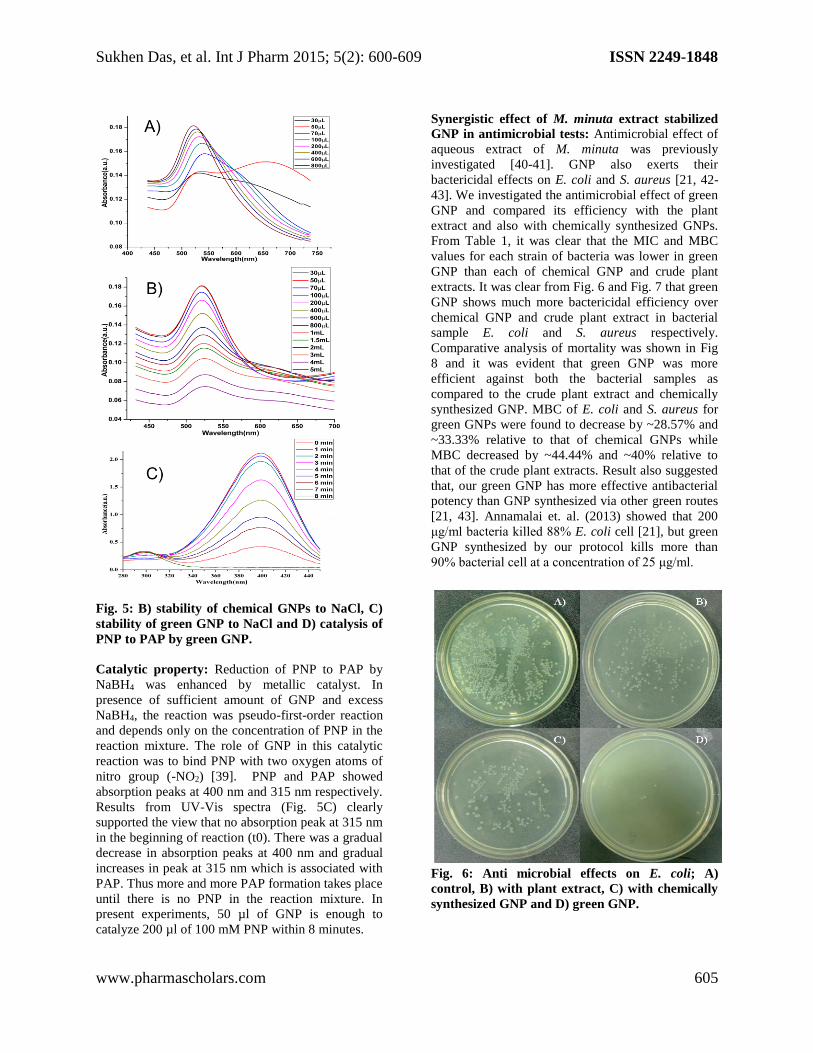
Stability of nanoparticles: The stability of
nanoparticles was the major factor for biomedical
applications. Agglomeration was triggered by high
ionic concentration in body fluid. In our present
work, we investigate the stability of green GNP and
compared with chemical GNP. It was found that the
stability of green GNP were much better than those
of the chemical GNP (Fig. 5A & B). There was a
shift of 6 nm in SPR band occurred in case of green
GNP after addition of 5 ml of 5 M NaCl (Fig. 5 B),
while SPR band shifted of 153 nm for chemical GNP
after addition of only 800 μl of NaCl (Fig. 5 A).
Green synthesized GNP synthesized using M. minuta
extract shows minimum shifts in SPR band compared
to that of the GNP synthesized by Pandey et al. [15].
From this observation it was clear that these green
GNP were suitable for biomedical applications as it is
stable in higher salt concentration.
Fig 1: Overview of green GNP synthesis.
Sukhen Das, et al. Int J Pharm 2015; 5(2): 600-609 ISSN 2249-1848
www.pharmascholars.com 604
Fig. 2: Uv-vis spectra of green GNP; A) time
dependent formation of green GNP, B) role of
various concentration of plant extract and C)
various concentration of HAuCl4 in synthesis of
green GNP.
DLS study confirmed the average particle size of 35
nm with the poly dispersion index of 0.389. The sizes
of the particles were in the range from 10-60 nm (Fig
3 A). Average zeta potential of the particles was -
14.35± 0.05mV. Particle size and distribution
obtained from DLS were further confirmed by
HRTEM micrograph. The average size of the green
synthesized particles was 25 nm with a size
distribution from 15-35 nm (Fig 3 B). Nearly similar
sized particles were observed in case of chemically
synthesized GNP (Fig. 3 C). HRTEM micrographs of
GNP shows particles were crystalline in nature and
most of them were spherical in shape.
Fig. 3: A) Size distribution curve of green GNPs
obtained from DLS study; HRTEM micrograph
of B) green synthesized GNP at high magnification
(scale bar 50 nm) and C) chemically synthesized
GNP at high magnification (scale bar 50 nm).
FESEM micrographs confirmed well distribution of
particles, spherical in shape (Fig 4A). Particle size
was same as recorded in HRTEM. XRD patterns of
the prepared samples are shown in Fig 4B. X-ray
diffractogram show 2θ values at 37.46, 43.58, 64.22
and 77.08 corresponding to (111), (200), (220) and
(311) planes which confirms the presence of GNP.
All the peaks are duly assigned using JCPDS file No
04-0784. From the x-ray pattern it was observed that
sample was well crystalline.
Fig. 4: A) FESEM micrographs, B) XRD pattern
of synthesized green GNP; FTIR spectra of C)
crude plant extract and D) GNP synthesized with
plant extract
Sukhen Das, et al. Int J Pharm 2015; 5(2): 600-609 ISSN 2249-1848
www.pharmascholars.com 605
Fig. 5: B) stability of chemical GNPs to NaCl, C)
stability of green GNP to NaCl and D) catalysis of
PNP to PAP by green GNP.
Catalytic property: Reduction of PNP to PAP by
NaBH4 was enhanced by metallic catalyst. In
presence of sufficient amount of GNP and excess
NaBH4, the reaction was pseudo-first-order reaction
and depends only on the concentration of PNP in the
reaction mixture. The role of GNP in this catalytic
reaction was to bind PNP with two oxygen atoms of
nitro group (-NO2) [39]. PNP and PAP showed
absorption peaks at 400 nm and 315 nm respectively.
Results from UV-Vis spectra (Fig. 5C) clearly
supported the view that no absorption peak at 315 nm
in the beginning of reaction (t0). There was a gradual
decrease in absorption peaks at 400 nm and gradual
increases in peak at 315 nm which is associated with
PAP. Thus more and more PAP formation takes place
until there is no PNP in the reaction mixture. In
present experiments, 50 µl of GNP is enough to
catalyze 200 µl of 100 mM PNP within 8 minutes.
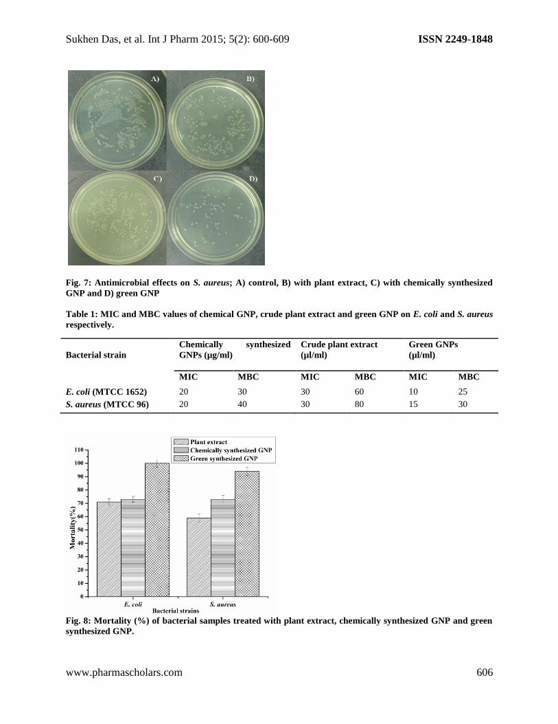
Synergistic effect of M. minuta extract stabilized
GNP in antimicrobial tests: Antimicrobial effect of
aqueous extract of M. minuta was previously
investigated [40-41]. GNP also exerts their
bactericidal effects on E. coli and S. aureus [21, 42-
43]. We investigated the antimicrobial effect of green
GNP and compared its efficiency with the plant
extract and also with chemically synthesized GNPs.
From Table 1, it was clear that the MIC and MBC
values for each strain of bacteria was lower in green
GNP than each of chemical GNP and crude plant
extracts. It was clear from Fig. 6 and Fig. 7 that green
GNP shows much more bactericidal efficiency over
chemical GNP and crude plant extract in bacterial
sample E. coli and S. aureus respectively.
Comparative analysis of mortality was shown in Fig
8 and it was evident that green GNP was more
efficient against both the bacterial samples as
compared to the crude plant extract and chemically
synthesized GNP. MBC of E. coli and S. aureus for
green GNPs were found to decrease by ~28.57% and
~33.33% relative to that of chemical GNPs while
MBC decreased by ~44.44% and ~40% relative to
that of the crude plant extracts. Result also suggested
that, our green GNP has more effective antibacterial
potency than GNP synthesized via other green routes
[21, 43]. Annamalai et. al. (2013) showed that 200
μg/ml bacteria killed 88% E. coli cell [21], but green
GNP synthesized by our protocol kills more than
90% bacterial cell at a concentration of 25 μg/ml.
Fig. 6: Anti microbial effects on E. coli; A)
control, B) with plant extract, C) with chemically
synthesized GNP and D) green GNP.
Sukhen Das, et al. Int J Pharm 2015; 5(2): 600-609 ISSN 2249-1848
www.pharmascholars.com 606
Fig. 7: Antimicrobial effects on S. aureus; A) control, B) with plant extract, C) with chemically synthesized
GNP and D) green GNP
Table 1: MIC and MBC values of chemical GNP, crude plant extract and green GNP on E. coli and S. aureus
respectively.
Fig. 8: Mortality (%) of bacterial samples treated with plant extract, chemically synthesized GNP and green
synthesized GNP.
Bacterial strain
Chemically synthesized
GNPs (µg/ml)
Crude plant extract
(µl/ml)
Green GNPs
(µl/ml)
MIC MBC MIC MBC MIC MBC
E. coli (MTCC 1652) 20 30 30 60 10 25
S. aureus (MTCC 96) 20 40 30 80 15 30
Sukhen Das, et al. Int J Pharm 2015; 5(2): 600-609 ISSN 2249-1848
www.pharmascholars.com 607
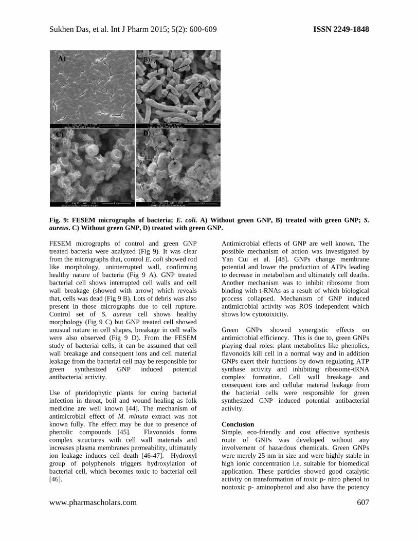
Fig. 9: FESEM micrographs of bacteria; E. coli. A) Without green GNP, B) treated with green GNP; S.
aureus. C) Without green GNP, D) treated with green GNP.
FESEM micrographs of control and green GNP
treated bacteria were analyzed (Fig 9). It was clear
from the micrographs that, control E. coli showed rod
like morphology, uninterrupted wall, confirming
healthy nature of bacteria (Fig 9 A). GNP treated
bacterial cell shows interrupted cell walls and cell
wall breakage (showed with arrow) which reveals
that, cells was dead (Fig 9 B). Lots of debris was also
present in those micrographs due to cell rupture.
Control set of S. aureus cell shows healthy
morphology (Fig 9 C) but GNP treated cell showed
unusual nature in cell shapes, breakage in cell walls
were also observed (Fig 9 D). From the FESEM
study of bacterial cells, it can be assumed that cell
wall breakage and consequent ions and cell material
leakage from the bacterial cell may be responsible for
green synthesized GNP induced potential
antibacterial activity.
Use of pteridophytic plants for curing bacterial
infection in throat, boil and wound healing as folk
medicine are well known [44]. The mechanism of
antimicrobial effect of M. minuta extract was not
known fully. The effect may be due to presence of
phenolic compounds [45]. Flavonoids forms
complex structures with cell wall materials and
increases plasma membranes permeability, ultimately
ion leakage induces cell death [46-47]. Hydroxyl
group of polyphenols triggers hydroxylation of
bacterial cell, which becomes toxic to bacterial cell
[46].
Antimicrobial effects of GNP are well known. The
possible mechanism of action was investigated by
Yan Cui et al. [48]. GNPs change membrane
potential and lower the production of ATPs leading
to decrease in metabolism and ultimately cell deaths.
Another mechanism was to inhibit ribosome from
binding with t-RNAs as a result of which biological
process collapsed. Mechanism of GNP induced
antimicrobial activity was ROS independent which
shows low cytotoixicity.
Green GNPs showed synergistic effects on
antimicrobial efficiency. This is due to, green GNPs
playing dual roles: plant metabolites like phenolics,
flavonoids kill cell in a normal way and in addition
GNPs exert their functions by down regulating ATP
synthase activity and inhibiting ribosome-tRNA
complex formation. Cell wall breakage and
consequent ions and cellular material leakage from
the bacterial cells were responsible for green
synthesized GNP induced potential antibacterial
activity.
Conclusion
Simple, eco-friendly and cost effective synthesis
route of GNPs was developed without any
involvement of hazardous chemicals. Green GNPs
were merely 25 nm in size and were highly stable in
high ionic concentration i.e. suitable for biomedical
application. These particles showed good catalytic
activity on transformation of toxic p- nitro phenol to
nontoxic p- aminophenol and also have the potency
Sukhen Das, et al. Int J Pharm 2015; 5(2): 600-609 ISSN 2249-1848
www.pharmascholars.com 608
to kill bacteria more efficiently than chemically
synthesized GNPs. GNPs synthesized using M.
minuta shows better stability, antimicrobial potency
compared to other green routes. It would act as a very
effective catalyst and antimicrobial agent for
biomedical applications.
Acknowledgement: We would like to thank Defence
Research and Development Organization (DRDO),
Defiance Ministry, India, for the financial support.
Reference:
[1] Rai S, Ikram A, Sahai S, Dass S, Shrivastav R, Satsangi VR. RSC Adv, 2014; 4: 17671-17679.
[2] Gonzalez AL, Noguezand C, Barnard AS. J Mater Chem C, 2013; 1: 3150-3157.
[3] Salcedo ARM, Sevilla FB. Phil Sci Lett, 2013; 6: 90-96.
[4] Cheng W, Dong S, Wang E. Langmuir, 2002; 18: 9947-9952.
[5] Chandrasekharan N, Kamat PV. J Phys Chem B, 2000; 104: 10851–10857.
[6] Olya ME, Pirkarami A, Soleimani M, Bahmaei M. J Environ Manage, 2013; 121: 210-219.
[7] Dreaden EC, Alkilany AM, Huang X, Murphy CJ, El-Sayed MA. Chem Soc Rev, 2012; 41: 2740-2779.
[8] Cheng J, Gu YJ, Cheng SH, Wong WT. J Biomed Nanotechnol, 2013; 9: 1362-9.
[9] Han G, Ghosh P, Rotello VM. Nanomedicine, 2007; 2: 113-23.
[10] Jain S, Bch MB, Hirst DG, O'Sullivan JM. Br J Radiol, 2012; 85: 101–113.
[11] Cai W, Gao, Hong H, Sun J. Nanotechnol Sci Appl, 2008; 1: 17–32.
[12] Harne S, Sharma AK, Dhaygude M, Joglekar S, Kodam K, Hudlikar M. Colloid Surface B, 2012; 95: 284-8.
[13] Narayanan KB, Sakthivel N, J Microbiol Biotechnol, 2013; 23: 1287-92.
[14] Das RK, Gogoi1 N, Babu PJ, Sharma P, Mahanta C, Bora U. Adv Mater Phys Chem, 2012; 2: 275-281.
[15] Pandey S, Oza G, Mewada A, Sharon M. Arch Appl Sci Res, 2012; 4: 1135-1141.
[16] Boruah SK, Boruah PK, Sarma P, Medhi C, Medhi OK. Adv Mat Lett, 2012; 3: 481-486.
[17] Song JY, Jang HK, Kim BS. Proc Biochem, 2009; 44: 1133-1138.
[18] Sen IK, Maity K, Islam SS. Carbohydr Polym, 2013; 91: 518-28.
[19] Aromal SA, Philip D. Spectrochim Acta A Mol Biomol Spectrosc, 2012; 97: 1-5.
[20] Kumar KM, Mandal BK, Sinha M, Krishnakumar V. Spectrochim Acta A Mol Biomol Spectrosc, 2012; 86:
490-94.
[21] Annamalai A, Christina VL, Sudha D, Kalpana M, Lakshmi PT. Colloids Surf B Biointerfaces, 2013; 108: 60-
65.
[22] Aromal SA, Babu KVD, Philip D. Spectrochim Acta A Mol Biomol Spectrosc, 2012; 96: 1025-30.
[23] Rajan A, MeenaKumari A, Philip D. Spectrochim Acta A Mol Biomol Spectrosc, 2014; 118: 793-99.
[24] Das SK, Dickinson C, Lafir F, Broughamc DF, Marsili E. Green Chem, 2012; 14: 1322.
[25] Bhatti ZI, Toda H, Furukawa K. Water Research, 2002; 36:1135–1142.
[26] Vincent T, Guibal E. Langmuir, 2003; 19: 8475−8483.
[27] Toxicological profile for nitrophenols: 2-nitrophenol and 4-nitrophenol. Agency for Toxic Substances and
Disease Registry (ATSDR). US Department of Health and Human Services, Public Health Service, Atlanta, USA,
1992. http://www.atsdr.cdc.gov/toxprofiles/tp50-p.pdf
[28] US EPA, Report no. EPA/738/F-97/016. US EPA, Washington, DC, 1997.
[29] Rogers KR, Van Emon JM. US EPA Report no. EPA/600/A-93/074, USA, 1993.
[30] IPCS CICAD for Mononitrophenols – Document no. 20, WHO, Geneva, Switzerland, 2000.
http://www.epa.gov/hpv/pubs/summaries/4ntrophn/c14390rt.pdf
[31] Megharaj M, Pearson HW, Venkateswarl K. Pest Biochem Physiol, 1991; 40: 266-273.
[32] National Toxicology Program (NTP). Technical Report Series No. 417, US Department of Health and Human
Services, Public Health Service, National Institutes of Health, 1994.
[33] Zhu S, Jiao S, Liu Z, Pang G, Feng S. Environ Sci Nano, 2014; 1: 172.
[34] Polavarapu L, Xu QH. Nanotechnology, 2009; 20: 185606 (7pp)
[35] John De Britto A, Gracelin DHS, Kumar PBJR. Int J Pharm Bio Sci, 2013; 4: 800-805.
[36] Bagchi B, Kar S, Dey SK, Bhandary S, Roy D, Mukhopadhyay TK, Das S, Nandy P. Colloids and Surfaces B:
Biointerfaces, 2013; 108: 358–365.
[37] Hartmann M, Berditsch M, Hawecker J, Ardakani MF, Gerthsen D, Ulrich AS. Antimicrob Agents Chemother,
2010; 54: 3132–3142.
[38] Kuroda K, Ishida T, Haruta M. J Mol Catal A Chem, 2009; 298: 7−11.
Sukhen Das, et al. Int J Pharm 2015; 5(2): 600-609 ISSN 2249-1848
www.pharmascholars.com 609
[39] Pozun ZD, Rodenbusch SE, Keller E, Tran K, Tang W, Stevenson KJ, Henkelman G. J Phys Chem C, 2013;
117: 7598−7604.
[40] Parihar P, Parihar L, Bohra A. J Microbiol Antimicrob, 2010; 2: 19-22.
[41] Parihar P, Daswani L, Bohra A. Indian Fern J, 2003; 20: 48-50.
[42] Lima E, Guerra R, Lara V, Guzman A. Chem Cent J, 2013; 7: 11.
[43] Lokina S, Narayanan V. Chem Sci Trans, 2013; 2: 105-110.
[44] Banerjee RD, Sen SP. Ecol Bot, 1980; 34: 284-298.
[45] Bala N, Saha S, Chakraborty M, Maiti M, Das S, Basu R, Nandy P. RSC Adv, 2015; 5: 4993-5003
[46] Cowan MM. Clin Microbiol Rev, 1999; 12: 564–582.
[47] Walsh SE, Maillard JY, Russel AD, Catrenich CE, Charbonneau AL, Bartol RG. J Appl Microbiol, 2003; 94:
240–247.
[48] Cui Y, Zhao Y, Tianb Y, Zhang W, Lua X, Jiang X. Biomaterials, 2012; 33: 2327-2333.
Copyright © 2022 FDOKUMEN