Novel nanoparticle-based drug delivery system for neural ...
167
HAL Id: tel-02406015 https://tel.archives-ouvertes.fr/tel-02406015 Submitted on 12 Dec 2019 HAL is a multi-disciplinary open access archive for the deposit and dissemination of sci- entific research documents, whether they are pub- lished or not. The documents may come from teaching and research institutions in France or abroad, or from public or private research centers. L’archive ouverte pluridisciplinaire HAL, est destinée au dépôt et à la diffusion de documents scientifiques de niveau recherche, publiés ou non, émanant des établissements d’enseignement et de recherche français ou étrangers, des laboratoires publics ou privés. Novel nanoparticle-based drug delivery system for neural stem cell targeting and differentiation Dario Carradori To cite this version: Dario Carradori. Novel nanoparticle-based drug delivery system for neural stem cell targeting and differentiation. Human health and pathology. Université d’Angers; Université catholique de Louvain (1970-..), 2017. English. NNT : 2017ANGE0056. tel-02406015
-
Upload
khangminh22 -
Category
Documents
-
view
3 -
download
0
Transcript of Novel nanoparticle-based drug delivery system for neural ...

HAL Id: tel-02406015https://tel.archives-ouvertes.fr/tel-02406015
Submitted on 12 Dec 2019
HAL is a multi-disciplinary open accessarchive for the deposit and dissemination of sci-entific research documents, whether they are pub-lished or not. The documents may come fromteaching and research institutions in France orabroad, or from public or private research centers.
L’archive ouverte pluridisciplinaire HAL, estdestinée au dépôt et à la diffusion de documentsscientifiques de niveau recherche, publiés ou non,émanant des établissements d’enseignement et derecherche français ou étrangers, des laboratoirespublics ou privés.
Novel nanoparticle-based drug delivery system forneural stem cell targeting and differentiation
Dario Carradori
To cite this version:Dario Carradori. Novel nanoparticle-based drug delivery system for neural stem cell targeting anddifferentiation. Human health and pathology. Université d’Angers; Université catholique de Louvain(1970-..), 2017. English. �NNT : 2017ANGE0056�. �tel-02406015�

Dario CARRADORI
Thesis presented for the obtainment of Doctor Degree from the Université d'Angers (UA) and from Université catholique de Louvain (UCL) Under the label of Université Bretagne Loire Doctoral school : Biology-Health Discipline : Biomolecules, Drug delivery, Nanomedicine, Neurodegenerative diseases, Pharmacology Speciality : Neuroscience Unité de recherche : Micro et Nanomédecines Biomimétiques Presented in public : 21.09.2017
Novel nanoparticle-based drug delivery system for neural stem cell targeting and
differentiation
JURY Reviewers: Patrick COUVREUR, Professor, Institut Galien Paris-Sud, Faculté Pharmacie Paris-Sud, Châtenay-Malabry, France Lino FERREIRA, Professor, UC Biotech, Parque Tecnológico de Cantanhede, Cantanhede, Portugal Examinators: Véronique PREAT, Professor, Louvain Drug Research Institut, Université catholique de Louvain, Brussels, Belgium Patrick SAULNIER, Professor, Micro et Nanomédecines Biomimétiques, Université d’Angers, Angers, France Thesis directors: Joël EYER, Statutory Researcher, Micro et Nanomédecines Biomimétiques, Université d’Angers, Angers, France Anne DES RIEUX, Professor, Louvain Drug Research Institut, Université catholique de Louvain, Brussels, Belgium President: Françoise VAN BAMBEKE, Professor, Louvain Drug Research Institut, Université catholique de Louvain, Brussels, Belgium

Foreword
i
Foreword
Neural stem cells (NSCs) are located in restricted neurogenic areas of the central nervous
system, called niches, where they can undergo self-renewal and differentiate into specialized
neuronal cells (astrocytes, oligodendrocytes and neurons). NSC differentiation plays a crucial
role in the neurogenesis process, both during the development and the maintenance of the
mammalian central nervous system (CNS). Several studies have demonstrated that
neurogenesis stimulation resulted in the partial recovery of injured neuronal networks and in
increased behavioral performances (e.g., learning and memory). Consequently, due to their
impact on neurogenesis, NSCs are a potential tool to treat CNS diseases characterized by
progressive lesion of neuronal tissue and deterioration of neurological functions.
NSCs have been investigated to replace injured cells and to restore neurological functions by
their differentiation into specialized neuronal cells. Different strategies have been developed
which are mostly based on NSC transplantation after in vitro manipulation of exogenous
NSCs (isolated from their niches or induced from other somatic cells). Thirty-seven NSC-
based clinical trials are currently on going for gliomas, ischemic stroke, amyotrophic lateral
sclerosis, spinal cord injury and Parkinson’s disease, but none have reached phase III yet. The
limited translation is probably due to transplantation-associated issues such as access to cell
source and post-grafting cell survival.
In situ differentiation of endogenous NSCs is a promising strategy to avoid transplantation-
associated issues. Nevertheless, no work based on in situ differentiation of endogenous NSCs
has reached the clinical phase yet and the lack of NSC-targeting systems mostly promoted the
development of non-selective therapies.
Endogenous NSC targeting would increase the efficacy and the safety of in situ NSC
differentiation treatments by enhancing drug bioavailability at the targeted site and limiting
drug counter effects. Thus, the development of drug delivery systems that selectively reach
NSC niches and induce NSC differentiation in situ would greatly improve the current NSC-
based therapies.

Foreword
ii
AIM AND SCOPE OF THE WORK
The aim of this work was to contribute to the development of NSC-based therapies by
providing a drug delivery system able both to target endogenous NSCs and to induce their
differentiation in situ by bioactive molecule stimulation.
The goals of this project were i) to develop and characterize a drug delivery system targeting
NSCs ii) to investigate the mechanisms behind the selective interaction NSCs-drug delivery
system and iii) to induce endogenous NSC differentiation in situ by delivering a bioactive
molecule.
ORGANIZATION OF THE MANUSCRIPT
Chapter 1 – Introduction establishes the context of NSC differentiation, from the general
field of NSC biology to the current NSC-based therapies aiming at the treatment of CNS
diseases. Secondly, it explores the contribution of nanomedicine to improve NSC-based
therapies with a particular emphasis on the potential role of the lipid nanocapsules.
Chapter 2 – NFL-lipid nanocapsules for brain neural stem cell targeting in vitro and in
vivo shows the study we made to produce a NSC-targeting drug delivery system by adsorbing
the peptide NFL-TBS.40-63 (NFL) on the surface of lipid nanocapsules (LNCs). NFL-LNCs
were physicochemically characterized and their NSC-targeting efficiency was evaluated in
vitro and in vivo on NSCs of both brain and spinal cord. NFL-LNCs selectively interact with
NSCs of the brain while they do not with NSCs of the spinal cord.
Chapter 3 – The characteristics of neural stem cell plasma membrane affect their
interaction with NFL-lipid nanocapsules shows the study we made to investigate the reasons
behind the selective interactions between NFL-LNCs and NSCs of the brain. We
characterized the plasma membrane lipid compositions of NSCs from the brain and from the
spinal cord highlighting significant differences between the two types of cells. The lipid
composition modulates plasma membrane properties, such as fluidity and permeability, which
are involved in the interactions nanoparticle-cell. Consequently, we measured their variation
before and after NSC incubation with NFL, LNCs and NFL-LNCs. We finally identified some
of the factors modulating NFL-LNC preferential interaction by comparing the results between
NSCs from the brain and from the spinal cord.

Foreword
iii
Chapter 4 – Retinoic acid-loaded NFL-lipid nanocapsules induce neural stem cell
differentiation towards the oligodendrocyte lineage shows the study we made to induce the
differentiation of NSCs from the brain via bioactive molecule stimulation. Retinoic acid (RA)
was selected among several drugs for its neurogenic potential as well as compatibility with
LNCs. We encapsulated RA into NFL-LNCs and we evaluate the NSC differentiation efficacy
in vitro and in vivo. In vitro, the incubation with RA-loaded NFL-LNCs induces a significant
NSC differentiation in oligodendrocytes, while in vivo, focal demyelinated rats have been
treated with RA-loaded NFL-LNCs but the evaluation of the therapeutic effect is ongoing.
Chapter 5 – Discussion highlights the main achievements of the thesis, describes the
implications of the new findings and explores the possible improvements which can be made.
NFL-LNC represents a great novelty in the field of NSC-based therapy due to the versatility
of the LNCs (e.g. encapsulation of different types of bioactive molecules) which makes NFL-
LNC potentially suitable for many therapeutic applications, even though a few points remain
to be improved (e.g., the way of administration).

List of abbreviations
iv
LIST OF ABBREVIATIONS
AD Alzheimer’s disease
ALS Amyotrophic Lateral Sclerosis
bFGF Basic Fibroblast Growth Factor
CC Central Canal
CC-NSCs Central Canal Neural Stem Cells
Chol Cholesterol
CNP 2',3'-Cyclic-Nucleotide 3'-Phosphodiesterase
CNS Central Nervous System
CPPs Cell-Penetrating Peptides
DCX Doubleortin
DiD DiIC18(5) solid (1,1'-Dioctadecyl-3,3,3',3'-Tetramethylindodicarbocyanine,
4-Chlorobenzenesulfonate Salt)
EGF Epidermal Growth Factor
ELFEFs Extremely Low-Frequency Electromagnetic Fields
eSM egg SphingoMyelin
Endo Endogenous
Exo Exogenous
FD Fluorescent Dextran
FDA Food and Drug Administration
GalC GalactoCerebroside
GFAP Glial Fibrillary Acid Protein
GP Generalized Polarization
IS Ischemic Stroke
LNCs Lipid NanoCapsules
MAP2 Microtubule-Associated Protein 2
MBP Myelin Basic Protein
MIRB Molday ION Rhodamine B
Msi1 Musashi-1
n.b. new-born
NeuroD Neurogenetic Differentiation
NeuN Neuronal nuclear epitope
NFs NeuroFilaments

List of abbreviations
v
NFL NFL-TBS.40-63
NFL-LNCs NFL-TBS.40-63 functionalized Lipid NanoCapsules
NG2 Chondroitin sulfate proteoglycan neuron/glia antigen 2
NSCs Neural Stem Cells
NSE Neuron-Specific Enolase
Pax6 Paired box gene 6
PCL PolyCaproLactone
PD Parkinson’s disease
PDI PolyDispersity Index
PIT Phase Inversion Temperature
PLA PolyLactic Acid
PLGA Poly(Lactic-co-Glycolic Acid)
POPC 1-Palmitoyl-2-Oleoyl-sn-glycero-3-PhosphoCholine
PSA-NCAM PolySiAlylated-Neural Cell Adhesion Molecule
RA Retinoic Acid
SCI Spinal Cord Injury
SGZ SubGranular Zone
SPION SuperParamagnetic Iron Oxide Nanoparticles
Sox2 SRY-related HMG-box gene
SVZ SubVentricular Zone
SVZ-NSCs SubVentricular Zone Neural Stem Cells
TH Thyrosine-Hydroxylase
USPION Ultrasmall SuperParamagnetic Iron Oxide Nanoparticles

vi
Remerciements
Mes remerciements vont à toutes ces personnes qui m'ont permis de commencer ce doctorat,
de le continuer et, enfin, de voir sa fin! J'ai eu beaucoup de chance de travailler avec des
gens comme vous, extrêmement compétents et efficaces.
Merci à Angers, où j’ai passé la première année et demie de mon doctorat, à la douceur
angevine, au vin et aux châteaux!
J'aimerais remercier le directeur de ma thèse, le Dr. Joël Eyer, pour m'avoir donné
l'opportunité de travailler au sein de son laboratoire, et mon superviseur, le Pr. Patrick
Saulnier, pour m'avoir transmis son enthousiasme et sa positivité.
Merci au Dr. Claire Lepinoux-Chambaud, pour m’avoir aidé au début de mon doctorat, et à
Kristell Barreau, pour avoir partagé avec moi ce début de doctorat. Merci au Dr. Franck
Letournel, au Dr. Catherine Fressinaud, au Dr. Guillaume Bastiat et au Dr. Jean-Christophe
Gimel pour leurs conseils et leur professionnalité. Merci à Aurélien Contini, Saikrishna
Kandalam, Lisa Terranova, Emilie André, Marion Toucheteau et toute la famille NanoFar à
Angers. Merci à Nolwenn Lautram, Maryne Jaffré et Edith Greleau pour leur disponibilité et
leur aide. Merci à Hélène Malhaire, Marion Pitorre, Carl Simonsson et tous les membres du
MiNT.
Un merci spécial au Dr. Edward Milbank (il migliore), au Dr. Giovanna Lollo (la delizia al
limone), au Dr. Carmelina Angotti (papà castoro) et au Dr. Gabriela Ullio (mariiiia) pour
leur amitié, leur soutien, nos litiges, nos voyages ensemble, la Sicile et les "Divanetti".
Merci à Bruxelles, où j'ai passé les dernières deux années et demie de ma thèse, au chocolat,
à la bière et aux frites.
Un grand merci à tous les membres de l’ADDB avec qui ça a été un plaisir et un honneur de
travailler.
Je remercie mon co-directeur de thèse, le Pr. Anne des Rieux, pour son exubérance et son
énergie, ainsi que mon co-superviseur, le Pr. Véronique Préat, pour sa diplomatie et son
pragmatisme.

vii
Merci à mes collègues de bureau, le Dr. Fabienne Danhier, le Dr. John Bianco et Pauline De
Bert, pour les chats, leurs conseils, la musique, les snacks et la bonne compagnie.
Merci à Valentina Kalichuk et Loïc Germain pour leur soutien et leur humour. Merci à
Nikolaos Tsakiris pour ses blagues et son karaoke et merci au Dr. Neha Shrestha pour les
snapchat et les momos.
Merci au Pr. Rita Vanbever, à Nathalie Lecouturier, au Dr. Gaëlle Vandermeulen, au Dr.
Cristina Loira Pastorizia, à Pallavi Ganipineni, à Audrey Smith, à Marie-Julie Guichard, au
Dr. Laure Lambricht, à Janske Nel, à Sohaib Mahri, à Kifah Nasr, à Luc Randolph, à
Natalija Tatic, à Yning Xu et à Michelle Zhao. Merci à mes stagiaires Zahraa El Azabi,
Hanane Choaibi et Ariane Mwema.
Merci à mes compagnons de jogging Thibaut Fourniols et Yoann Montigaud avec qui j'atteins
le plus haut degré de sportivité de ma vie!
Un super grand merci à Bernard Ucakar, Kevin Vanvarenberg et Murielle Cailler, pour leur
bonne humeur, leur aide indispensable, leur sympathie, leur professionnalisme et leur
précision.
Merci au Pr. Marie-Paule Mingeot-Leclercq, au Pr. Giulio Muccioli, au Prof. Veronique
Miron, au Dr. Mireille Al Houayek, à Andreia Giro Dos Santos, au Dr. Julien Masquelier, à
Adrien Paquot, et à tous nos collaborateurs.
Un merci spécial au Dr. Ana Beloqui (la reina), à Chiara Bastiancich (questo mese non ho
voglia di fare « niente ») et à Alessandra Lopes (le fou de rire) pour leur amitié, leurs
conseils, leur soutien, les pauses café, les repas et les sorties ensemble. Merci !
Grâce au soutien inconditionnel et toujours présent de ma famille, de Luigi et de Silvia. Vous
avez dû me supporter, avec mes angoisses, mes peurs…mais vous avez toujours trouvé un
moyen de m'aider et de me rester proche malgré les problèmes et les difficultés que vous aviez
aussi. Merci.

Table of contents
viii
FOREWORD..........................................................................................................................i-iii
LIST OF ABBREVIATIONS.................................................................................................iv-v
TABLE OF CONTENTS
CHAPTER 1 – INTRODUCTION
1. Preface.............................................................................................................................3
2. Neural stem cells.............................................................................................................4
3. Neural stem cell differentiation.......................................................................................8
4. In situ NSC differentiation via endogenous NSC targeting..........................................28
5. Aim of the thesis...........................................................................................................30
6. Plan of the PhD.............................................................................................................31
7. References.....................................................................................................................33
CHAPTER 2 – NFL-LIPID NANOCAPSULES FOR BRAIN NEURAL STEM CELL
TARGETING IN VITRO AND IN VIVO
1. Preface...........................................................................................................................48
2. Abstract.........................................................................................................................49
3. Introduction...................................................................................................................50
4. Materials and Methods..................................................................................................52
5. Results and discussions.................................................................................................56
6. Conclusions...................................................................................................................67
7. References.....................................................................................................................69
8. Supplementary data.......................................................................................................73

Table of contents
ix
CHAPTER 3 – THE CHARACTERISTICS OF NEURAL STEM CELL PLASMA
MEMBRANE AFFECT THEIR INTERACTION WITH NFL-LIPID NANOCAPSULES
1. Preface...........................................................................................................................80
2. Abstract.........................................................................................................................81
3. Introduction...................................................................................................................82
4. Materials and Methods..................................................................................................84
5. Results and discussion..................................................................................................91
6. Conclusion..................................................................................................................101
7. References...................................................................................................................102
8. Supplementary data.....................................................................................................106
CHAPTER 4 – RETINOIC ACID-LOADED NFL-LIPID NANOCAPSULES INDUCE
NEURAL STEM CELL DIFFERENTIATION TOWARDS THE OLIGODENDROCYTE
LINEAGE
1. Preface.........................................................................................................................109
2. Abstract.......................................................................................................................110
3. Introduction.................................................................................................................111
4. Materials and Methods................................................................................................113
5. Results and discussion................................................................................................117
6. Conclusion..................................................................................................................123
7. References...................................................................................................................124
8. Supplementary data.....................................................................................................127
CHAPTER 5 – DISCUSSION
1. Main achievements.....................................................................................................131
2. Perspectives.................................................................................................................134
3. Conclusion..................................................................................................................137
4. References...................................................................................................................138

Table of contents
x
ANNEX
1. The carbocyanine dye DiD labels in vitro and in vivo neural stem cells of the
subventricular zone as well as myelinated structures following in vivo injection in the
lateral ventricle
2. Curriculum vitae

Chapter 1 - Introduction
1
CHAPTER 1
INTRODUCTION*
*Adapted from
Carradori, D.; Eyer, J.; Saulnier, P.; Préat, V.; des Rieux, A. The therapeutic contribution of
nanomedicine to treat neurodegenerative diseases via neural stem cell differentiation.
Biomaterials 2017, 123, 77-91.

Chapter 1 - Introduction
2
TABLE OF CONTENTS
1. PREFACE
2. NEURAL STEM CELLS
2.1 Definition of neural stem cells
2.2 Biologic functions
2.3 Neurogenesis
2.4 Therapeutic significance in CNS diseases
3. NEURAL STEM CELL DIFFERENTIATION
3.1 Exogenous and endogenous NSC-based strategies
3.2 Stimulation of NSC differentiation
3.3 Clinical trials
3.4 Challenges
3.5 Conclusions
4. NANOMEDICINES FOR NSC DIFFERENTIATION
4.1 Definition of nanomedicine
4.2 Classification of the nanomedicines
4.3 Advantages of nanomedicine in NSC differentiation-based therapies
4.4 Current nanomedicine-based studies aiming at NSC differentiation for
therapeutic purposes
4.5 Conclusions
5. IN SITU NSC DIFFERENTIATION VIA ENDOGENOUS NSC TARGETING
5.1 Endogenous NSC targeting
5.2 The NFL-TBS.40-63 peptide
6. AIM OF THE THESIS
7. PLAN OF THE PHD
8. REFERENCES

Chapter 1 - Introduction
3
1. PREFACE
The dogma of a static brain was destroyed when Smart and Leblond showed for the first time
that glial cells are dividing throughout the mouse brain parenchyma [1]
. A few years later,
Altman and Das reported the migration of postnatally born neuroblasts from the
subventricular zone to the olfactory bulb, providing the first strong evidence of neurogenesis
in the adult brain [2]
. Important discoveries were made in the following decades, such as the
presence of adult-born neurons in the dentate gyrus of rats [3]
and in the vocal control nucleus
of birds [4]
, but the perception of neurogenesis has drastically changed only since the 1990s.
One of the most important discoveries was the observation that the proliferation of progenitor
cells, and the subsequent number of newborn neurons, was dynamic. Several factors such as
hormonal stress [5]
, age [6]
, or alcohol [7]
could modulate this process. The improvement of
immunohistological techniques represented another step forward in the description of
neurogenesis by providing more sensitive analyses [8,9]
. Moreover, the ability to isolate,
cultivate, and differentiate neural precursor cells in vitro provided crucial data on the cellular
and biomolecular mechanisms involved in adult neurogenesis [10,11]
.
Meanwhile, the discovery of adult neurogenesis also showed the limits of this physiologic
process. Indeed, neurogenesis is restricted to small neurogenic areas of the central nervous
system and its impact on the adult organism is very limited. Neural cells can migrate, from the
neurogenic regions to the injured areas of the brain, and differentiate into the damaged cell
phenotype but a very little percentage (around 0.2%) is functionally replaced [12,13]
.
The identification of neural stem cells and their important role in adult neurogenesis
motivated researchers to explore the regenerative potential of these cells.

Chapter 1 - Introduction
4
2. NEURAL STEM CELLS
2.1 Definition
Neural stem cells (NSCs) are defined as “stem cell derived from any part of the nervous
system and which primarily make cells expressing neural markers (those of astrocytes,
oligodendrocytes and neurons) in in vitro culture” [14]
. This definition is based on
retrospective in vitro studies [11,15,16]
which highlighted NSC peculiar biological properties:
self-renewal and differentiation into specialized neural cells (neurons, astrocytes and
oligodendrocytes) [17,18]
.
2.2 Biological functions
NSCs are located in restricted areas of the central nervous system (CNS), called niches, such
as the central canal (CC), in the spinal cord, and the subventricular zone (SVZ) and the
subgranular zone (SGZ), in the brain (Figure 1). Here, NSCs play a crucial role in the
neurogenesis process both in the development and in the adulthood of the organism by
generating, maintaining and regenerating the CNS via their differentiation into specialized
neural cells.
Figure 1. NSC niches in the CNS. Brain. Localization of the subgranular (SGZ) and subventricular (SVZ)
zones in adult human brain. A, lateral section of the brain. B, frontal section of the brain. C, subventricular zone,
highlighted in red. D, subgranular zone, highlighted in red. Spinal cord. Localization of central canal (CC) in
spinal cord. E, spinal cord left lateral view. F, spinal cord cross section. G, central canal, highlighted in red.

Chapter 1 - Introduction
5
Direct and indirect mechanisms were described to regulate the behavior of NSCs [19]
.
Extrinsic signals can bind to NSC plasma membrane receptors or penetrate via specific
channels and trigger intracellular cascades inducing modifications in gene expression. The
secretions of the choroid plexus such as insulin-like growth factor 1 (IGF1) [20,21]
or ions such
as calcium [22]
are examples of extrinsic signals that can modulate NSC differentiation by their
interaction with IGF1 receptor and Ca+2
channels, respectively. Intrinsic regulatory processes
can also direct gene expression in NSCs by affecting transcriptional factors. Sonic hedgehog
is the major activating ligand to initiate Hedgehog signalling in the brain and has been shown
to play an important role in NSC proliferation and differentiation [23]
. Both the extrinsic and
intrinsic pathways are interconnected. Consequently, the identification of the origin of the
niche signals is challenging.
Although NSCs are multipotent in vitro, recent genetic fate-mapping and clonal lineage-
tracing of NSCs have highlighted the lack of similarities between NSC differentiation in vitro
and in vivo [24]
. The niche environment seems to limit adult NSC differentiation. In the adult
SGZ, NSCs can generate dentate gyrus granular cells while in the adult SVZ, NSCs produce
neuroblasts which migrate to the olfactory bulb where they differentiate into interneurons [25]
.
Moreover, localization in the niches would determine the type of cells derived from NSCs. In
the SVZ, ventral NSCs mostly develop into calbindin-expressing cells, whereas dorsal NSCs
develop into tyrosine-hydroxylase-expressing cells [26]
(long-axon and dopaminergic neuron
markers, respectively). In the SGZ, the adult NSC population reacts differently to
environmental stimuli depending on their lineage [27]
. Learning and memory processes are
strictly related to adult neurogenesis in the SVZ [28-30]
, while SGZ-NSC-derived granule cells
of the dentate gyrus have been implicated in long-term spatial memory and pattern separation
[31,32]. Transplanted NSCs are also able to release immunomodulatory and neurotrophic factors
(bystander effect) such as nerve growth factor, brain-derived growth factor, and leukemia
inhibiting factor [33]
.
2.3 Neurogenesis
Adult neurogenesis was described both in the SGZ and in the SVZ (Figure 2) of the brain
while it does not occur in adult spinal cord [34]
. Neurogenesis consists of several
developmental stages (proliferation, migration, differentiation and integration) that are
characterized by distinct cell phenotypes. SGZ and SVZ do not have the same precursors;

Chapter 1 - Introduction
6
consequently, there is not a unique nomenclature to identify cells involved in the adult
neurogenesis [27,35,36]
.
Figure 2. Neurogenesis in the SVZ. A, Cellular organization of the SVZ: lateral ventricle, ependymal layer
(composed by type E cells or ependymal cells), subependymal layer (composed by type B, type C and type A
cells) and brain parenchyma (composed by astrocytes, oligodendrocytes and blood vessels). B, NSC
differentiation in the SVZ: type B cells undergo self-renewal or differentiate in type C cells which become type
A cells. C, neurogenesis steps in the SVZ: neuroblasts proliferate in the SVZ and then migrate to the olfactory
bulb where they differentiate in neurons and integrate with the existing cellular network.
Nevertheless, several markers are used to detect the different cell phenotypes involved in the
neurogenic process [37,38]
(Table 1). Nestin was the first marker described to identify stem-
like/precursor cells and is the most widely used [39]
. Nestin is a specific class of intermediate
filament proteins that are expressed in non-differentiated cells. Class III β tubulin (Tuj1) [40]
, a
protein expressed in post-mitotic neuron cytoskeleton, doublecortin (DCX) [41]
, which
encodes a microtubule-associated protein present in migrating neuroblasts, and the
polysialylated-neural cell adhesion molecule (PSA-NCAM), which is a product of post-
translational modification of NCAM, are the most accepted markers for early neurons [42]
.

Chapter 1 - Introduction
7
Mature neurons are identified by the microtubule-associated protein (MAP-2), the neuron-
specific enolase (NSE) and the neural-specific nuclear protein (NeuN) [43,44]
. Some non-neural
cells can also be positive for the markers mentioned above. To avoid ambiguous interpretation
of the results, it is recommended to perform multiple staining including non-neural markers
such as glial fibrillary acidic protein (GFAP) [45]
or calcium binding proteins (S100 and
S100b) [46]
for astrocytes, and 2',3'-cyclic-nucleotide 3'-phosphodiesterase (CNP) or myelin
basic protein (MBP) for oligodendrocytes [47]
.
Table 1. Markers for adult neurogenesis.
Type of cells Markers
Neural stem cells/Progenitors Nestin [39]
SRY-related HMG-box gene (Sox2) [48]
Musashi-1 (Msi1) [49]
Paired box gene 6 (Pax6) [50]
Prominin (CD133) [51]
Glial fibrillary acid protein (GFAP) [45]
NG2-glia positive cells Chondroitin sulfate proteoglycan neuron/glia antigen 2 (NG2) [52]
Neuronal lineage (early) III β tubulin (TUJ1) [40]
Doublecortin (DCX, C-18) [41]
Polysialylated-neural cell adhesion molecule (PSA-NCAM) [42]
Neurogenetic Differentiation (NeuroD) [42]
Neuronal lineage (mature) Neuronal nuclear epitope (NeuN) [44]
Microtubule-associated protein 2 (MAP2) [43]
Neuron-specific enolase (NSE) [43]
Calbindin [53]
Thyrosine-hydroxylase (TH) [54]
Calretinin [53]
Neurofilaments (NF) [53]
Astrocytic lineage Glial fibrillary acid protein (GFAP) [45]
Calcium binding proteins (S100/S100b) [46]
Glutammate-aspartate transporter (EAAT1) [55]
Glial lineage Galactocerebroside (GalC) [56]
2',3'-cyclic-nucleotide 3'-phosphodiesterase (CNP) [47]
Myelin basic protein (MBP) [47]
2.4 Therapeutic significance in CNS diseases
The therapeutic relevance of NSCs was investigated after several studies clearly demonstrated
that the inhibition of neurogenesis decreased neurological functions [57,58]
, while its
stimulation resulted in behavioural performance recovery (e.g., learning and memory tasks)
[59,60]. Consequently, adult neurogenesis modulation could have a positive impact on the
patients affected by CNS diseases, which are mostly characterized by neurological function
deterioration, such as Alzheimer’s (memory impairment) and Parkinson’s (motor impairment)
diseases. In this regard, strategies that are effectively able to restore distinct aspects of adult

Chapter 1 - Introduction
8
neurogenesis are of specific interest for future treatments. Although the differentiation of
NSCs is regulated by many physiological stimuli, these cells are considered to be key
determinants in neurogenesis. Consequently, NSCs have been investigated to replace injured
cells and to restore neurological functions by their differentiation into specialized neural cells.
The discovery of adult neurogenesis had a significant impact on regenerative medicine by
overturning the long-held dogma that mammalian CNS cannot regenerate and renew itself.
The stimulation of the neurogenesis process can result in the partial recovery of injured neural
networks and in the enhancement of behavioural performances. The identification of NSCs
and their role in adult neurogenesis motivated researchers to explore the regenerative potential
of these cells for the treatment of the CNS diseases characterized by progressive lesion/loss of
neural tissue and deterioration of neurological functions.
3. NEURAL STEM CELL DIFFERENTIATION
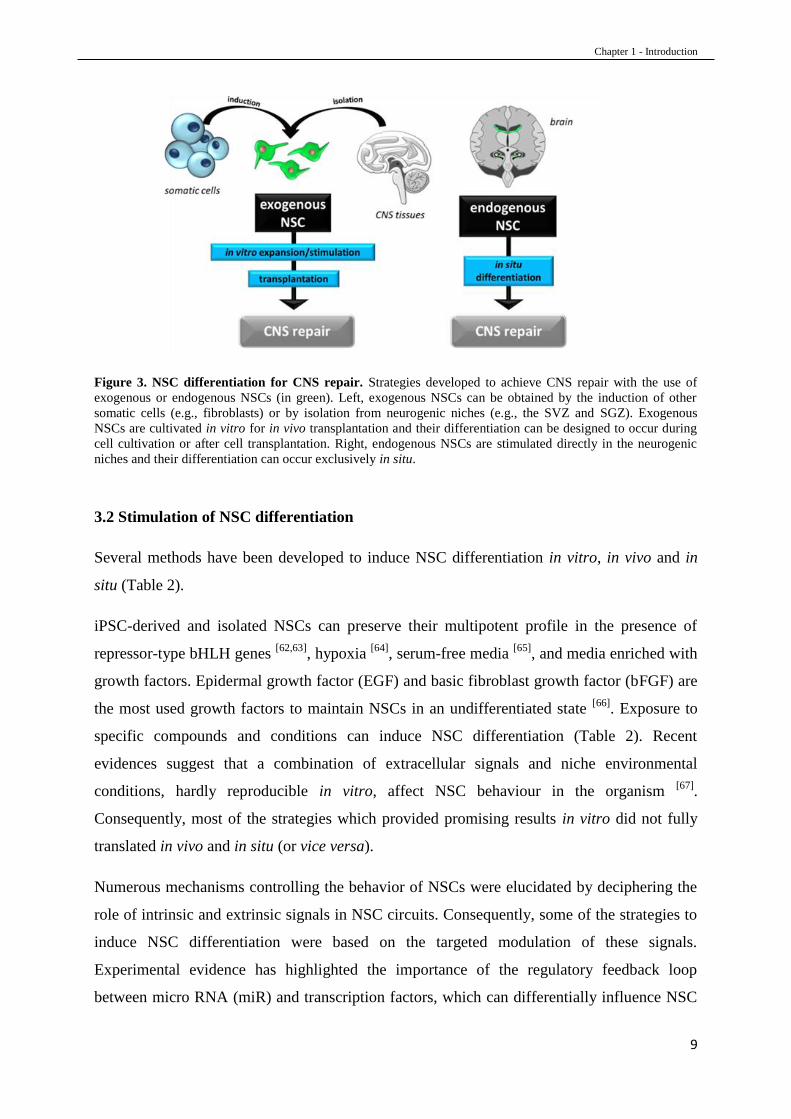
3.1 Exogenous and endogenous NSC-based strategies
In the last 20 years, several protocols have been developed to cultivate and differentiate NSCs
in vitro after their isolation from CNS niches (isolated NSCs) or after derivation from
pluripotent restored adult somatic cells (induced pluripotent stem cell-derived NSCs, iPSC-
derived NSCs) [61]
. However, the development of standard procedures to induce NSC
differentiation in vivo or in situ has not yet been established. CNS repair via NSC
differentiation can be achieved by following different strategies, which essentially depend on
whether NSCs are exogenous or endogenous (Figure 3). iPSC-derived and isolated NSCs are
considered exogenous NSCs when stimulated in vitro and transplanted in vivo. NSCs are
considered endogenous when their differentiation is stimulated in situ.
Chapter 1 - Introduction
9
Figure 3. NSC differentiation for CNS repair. Strategies developed to achieve CNS repair with the use of
exogenous or endogenous NSCs (in green). Left, exogenous NSCs can be obtained by the induction of other
somatic cells (e.g., fibroblasts) or by isolation from neurogenic niches (e.g., the SVZ and SGZ). Exogenous
NSCs are cultivated in vitro for in vivo transplantation and their differentiation can be designed to occur during
cell cultivation or after cell transplantation. Right, endogenous NSCs are stimulated directly in the neurogenic
niches and their differentiation can occur exclusively in situ.
3.2 Stimulation of NSC differentiation
Several methods have been developed to induce NSC differentiation in vitro, in vivo and in
situ (Table 2).
iPSC-derived and isolated NSCs can preserve their multipotent profile in the presence of
repressor-type bHLH genes [62,63]
, hypoxia [64]
, serum-free media [65]
, and media enriched with
growth factors. Epidermal growth factor (EGF) and basic fibroblast growth factor (bFGF) are
the most used growth factors to maintain NSCs in an undifferentiated state [66]
. Exposure to
specific compounds and conditions can induce NSC differentiation (Table 2). Recent
evidences suggest that a combination of extracellular signals and niche environmental
conditions, hardly reproducible in vitro, affect NSC behaviour in the organism [67]
.
Consequently, most of the strategies which provided promising results in vitro did not fully
translated in vivo and in situ (or vice versa).
Numerous mechanisms controlling the behavior of NSCs were elucidated by deciphering the
role of intrinsic and extrinsic signals in NSC circuits. Consequently, some of the strategies to
induce NSC differentiation were based on the targeted modulation of these signals.
Experimental evidence has highlighted the importance of the regulatory feedback loop
between micro RNA (miR) and transcription factors, which can differentially influence NSC

Chapter 1 - Introduction
10
behaviour [68]
. miR upregulation via miR-9 transfection [69]
or miR-195 downregulation via
MBD1 gene-expressing lentivirus [70]
can increase embryonic mouse NSC differentiation into
neurons and astrocytes. The signals from cell-cell contact were also identified as modulators
of NSC differentiation: co-cultures of NSCs and protoplasmic astrocytes or amniotic cells
promote embryonic rat NSC differentiation into neurons [71]
. The alteration of epigenetic
marks is an important NSC lineage modulator as well. The overexpression of activator-type
bHLH genes such as Mash1, neurogenin2, and NeuroD promotes neuronal-specific gene
expression while it inhibits glia-specific gene expression [63]
. Deciphering cell circuits and
their regulatory signal network provides crucial information for NSC differentiation strategies
[72].
In addition to endogenous modulators, pharmacological agents were found to impact NSC
behaviour. Incubation with 4-aminothiazoles (e.g., neuropathiazol) or oleanoic acid led to
TuJ1 (neuronal marker) expression in 90% of cells in treated primary neural progenitor cells
isolated from adult rat hippocampus and NSCs isolated from the embryonic striatum of mice
respectively [73]
. Valproic acid [74,75]
stimulates NSC differentiation by the inhibition of
histone deacetylase and its combination with other active molecules (e.g., retinoids)
significantly increased the percentage of MAP-2 (neuronal marker) positive cells in vitro [76]
.
The impact of addictive drugs on NSC differentiation and neurogenesis was also
demonstrated [77]
. Morphine promotes astrocyte differentiation [78]
while D-amphetamine [79]
and opioid peptides [80]
increase neuron differentiation in vitro and in situ, in the hippocampus
of adult mice.
NSC differentiation can also be stimulated by extremely low-frequency electromagnetic fields
(ELFEFs) which affect several biological parameters such as the intracellular calcium level.
ELFEFs upregulate the Ca2+
channels and increase the Ca2+
influx which induces the
signalling cascade associated with the promoter of specific bHLH that control NSC
differentiation [22,81]
. NSCs isolated from the brain cortex of new-born mice and exposed to
ELFEFs showed more cells positive for neuronal markers (+11.8% of MAP-2 and +11.9% of
beta III tubulin) compared to the controls [82]
.
Strategies aiming at in situ differentiation of endogenous NSCs would bypass issues related to
exogenous NSC transplantation, such as supply of exogenous NSCs (e.g., ethical concerns),
immune response against allogeneic transplantation and post-grafting cell viability.
Consequently, the administration of active compounds that would stimulate NSC

Chapter 1 - Introduction
11
differentiation in situ is the current most common approach (instead of NSC transplantation)
to enhance neurological function in neurodegenerative disease animal models (Table 2).
Table 2. NSC differentiation in vitro and in vivo (from exogenous or endogenous NSCs)
Study
design Molecule/Condition Type of cells/area Outcomes Year
in
vitro
neuropathiazol
primary neural progenitor
cells isolated from adult rat
hippocampus
+ neurons 2006 [74]
ELFEFs NSCs isolated from the
brain cortex of nb mice + neurons 2008
[82]
NSC co-culture with
astrocytes or amniotic cells
NSCs isolated from
embryonic rates + neurons 2012
[71]
miR-9 transfection NSCs isolated from
embryonic mice
+ neurons
+ astrocytes 2013
[69]
MBD1-expressing lentivirus NSCs isolated from
embryonic mice
+ neurons
+ astrocytes 2013
[70]
BDNF NSCs isolated from the
forebrain cortex of nb mice
+ neurons
+ oligodendrocytes 2013
[100]
oleanolic acid NSCs isolated from the
embryonic mouse striatum + neurons 2015
[73]
valproic acid NSCs isolated from
embryonic rat forebrains + neurons
2015 [76]
;
2008 [75]
all-trans-retinoic acid NSCs isolated from
embryonic rat forebrains + neurons
2015 [76]
;
1998 [92]
1,25-Dihydroxyvitamin D3 NSCs isolated from adult
mouse brain + oligodendrocytes 2015
[79]
opioid peptides NSCs isolated from
embryonic rat striatum + neurons 2015
[80]
NSC transplantation human NSCs prevention of further
cognitive deterioration 2015
[101]
Exo
NSCs
growth factor-
overexpressing NSC
transplantation
NSCs isolated from
transgenic nb mouse
hippocampus
AD deficit and synaptic
density recovery 2016
[102]
Endo
NSCs
morphine adult mouse hippocampus + astrocytes 2015 [78]
D-amphetamine adult mouse hippocampus + neurons 2013 [79]
ELFEFs adult mouse hippocampus + neurons 2010 [98]
retroviral Zfp488 adult mouse corpus
callosum
motor function
restoration 2011
[84]
perfluorooctane sulfonate cortical tissues of neonatal
mice
+ neurons
+ oligodendrocytes 2013
[85]
PCDHIIx shRNA/siRNA lateral ventricle of mice + neurons 2014 [103]
simvastatin injured areas of adult rat
brain
neurological function
enanchement 2015
[83]
ketamine nb rat SVZ + neurons 2015 [104]
nicotine adult rat hippocampus + neurons 2015 [105]
long noncoding RNA Pnky embryonic and postnatal
mouse brain + neurons 2015
[106]
oxygen supply modulation developing cerebral cortex
of mice + radial glia 2016
[97]
ELFEFs, extremely low-frequency electromagnetic fields; Exo, exogenous; endo, endogenous; nb, new-born; SVZ, subventricular zone.

Chapter 1 - Introduction
12
Simvastatin increased the percentage of MAP-2 (neuronal marker) and GFAP (astrocytic
marker) positive cells in a rat traumatic brain injury model [83]
. Its administration enhanced
neurological functions such as sensory functions, motor functions, beam balance performance,
and reflexes. The corpus callosum in mice exhibiting cuprizone-induced demyelination
showed a significant increase in Olig2 (oligodendrocytic marker) positive cells after treatment
with retroviral Zfp488 [84]
. The overexpression of Zfp488 protein (activator of
oligodendrocyte differentiation) induced a significant motor function restoration in
demyelination-injured mice.
Perfluorooctane sulfonate induced neuronal and oligodendrocytic NSC differentiation in
healthy mice, probably via PPARγ nuclear receptor activation, a pathway also involved in the
retinoid-induced cascade for NSC differentiation [85]
.
Retinoic acid (RA) has an important role in neurogenesis by inducing neurite outgrowth and
neural differentiation from various cell sources such as embryonic stem cells [86,87]
and
mesenchymal stem cells [88,89]
. Several studies demonstrated that RA increases the expression
of neural markers after its incubation with NSCs [76, 90]
. RA signalling is transduced by two
families of nuclear receptors, the retinoic acid receptors (RARs) and the retinoid X receptors
(RXRs) [91,92]
. RAR and RXR work as the heterodimer complex RAR/RXR and have 3
different isotypes (α, β and γ). While RARs are activated by all-trans and 9-cis RA, RXR is
activated only by 9-cis RA. Unbound retinoid receptors repress transcription through the
recruitment of corepressors such as NCoR and SMRT [91]
. Those corepressors recruit in turn
histone deacetylase protein complexes and Polycomb repressive complex 2, resulting in
chromatin condensation and gene silencing. The interaction between RA and RAR/RXR
induces receptor conformational changes which cause the dissociation of the corepressors and
recruitment of transcriptional coactivators such as SRC/p160 family, p300/CBP and CARM-1
[92]. Coactivators induce histone acetylation and recruitment of ATP-dependent remodelling
complexes which leads to the displacement of impeding nucleosomes within the proximal
promoter region. The displacement facilitates the access to the general transcription
machinery by promoters including RNA polymerase II [91,92]
. Since RXR is able to form
heterodimers with other nuclear receptors, including other peroxisome proliferator-activated
receptors (PPARs), cross-talk with other signalling pathways mediated by PPAR can also
occur. Additionally, ROS can induce NSC differentiation in a RA-independent way by RARα
stabilization.

Chapter 1 - Introduction
13
In vitro, the dissolution in aqueous solutions (e.g., cell media) increases RA susceptibility to
the oxidative damage of light, air, and temperature (>90°C). Moreover, retinoids are adsorbed
by many of the glass and plastic wares commonly used in cell culture, impacting control of
the administrated doses and reproducibility of the experiments [94]
. In vivo, chronic
administration of RA showed progressive decline of its plasmatic concentrations (probably
due to a progressive impairment of gastrointestinal uptake) as well as poor bioavailability
(associated with acquired mutations either of RAR or CRABP) and stability (pH sensitivity)
[95]. One successful strategy to overcome these issues is to incorporate RA into nanoparticles.
For instance, RA encapsulation into nanovectors showed an enhanced therapeutic effect in the
treatment of cancer or ischemia compared to its free form, highlighting the interest of RA-
based systems [96]
.
Figure 4. Cell regulation via retinoic acid (RA). More than 95% of circulating retinoic acid is bound to the retinoid bounding protein 4 (RBP4) which allows the
penetration into the cell via the transmembrane protein stimulated by retinoic acid 6 (STRA6). The complex RA-
RBP4-STRA6 dissociates in the cytoplasm and RA binds to the cellular retinoic acid-binding protein (CRABP)
which carry RA into the nucleus. However, free retinoic acid can cross the phospholipid bilayer of the plasmatic
membrane and of the nucleus also without any transporter. Once in the nucleus, RA binds to heterodimer
complex of retinoic acid receptor–retinoic x receptor (RAR/RXR) and recruits the transcriptional co-activators
cAMP-response-element-binding protein (CBP) and protein 300 (p300). RAR/RXR in combination with CBP
and p300 act with other factors to regulate signal transduction pathways and cell-differentiation.
Another approach to induce NSC differentiation is the oxygen supply modulation at the site of
neurogenic niches. It was recently showed by Lange et al. that angiogenesis is linked to
neurogenesis during cortical development [97]
. They demonstrated that selective perturbation

Chapter 1 - Introduction
14
of brain angiogenesis in embryos increased NSC expansion by hypoxia while exposure to
increased oxygen levels stimulated NSC differentiation.
The in vivo application of ELFEFs on C57b1/6 mice promoted proliferation and
differentiation of hippocampal NSCs [98]
. NSC differentiated into neurons, which were
functionally integrated in the dentate gyrus network 30 days after the ELFEF treatment.
Spatial learning and memory were enhanced, highlighting the important therapeutic
implications of ELFEFs for the treatment of neurodegenerative diseases. Moreover, it was
demonstrated that ELFEFs increased the survival of hippocampal newborn cells [99]
.
Altogether, there is concurrent information demonstrating that NSC differentiation is a valid
approach to enhance neurological functions during neurodegenerative diseases. The
therapeutic effect achieved in neurologically compromised animal models (e.g., restoration of
cognitive functions), together with the stimulation of the neurogenic process and
neuroprotection in healthy animal models via NSC differentiation, provide precious insight
for the clinical translation of NSC-based therapeutic strategy.
3.3 Clinical trials
Depending on the results obtained during pre-clinical studies, NSC differentiation-based
therapies have been recently translated into the clinic [107]
. Thirty-seven NSC-based clinical
trials are currently on going, involving patients affected by gliomas, ischemic stroke (IS),
amyotrophic lateral sclerosis (ALS), spinal cord injury (SCI) and Parkinson’s disease (PD)
[108]. Surprisingly, most of these therapies aimed at in vivo differentiation of transplanted
exogenous NSCs (Table 3), while most of the pre-clinical studies focused on in situ
differentiation of endogenous NSCs (Table 2). In the clinical trial NCT02117635, CTX0E03
cells (a human neuronal stem cell line) were stereotaxically injected by in the striatum of IS
patients (site of lesion) (Phase I). The treatment promoted a partial recovery of neurologic
functions [109]
but no anatomical modifications. This would suggest that NSCs do not directly
differentiate into neurons but rather act as cellular mediators by secreting paracrine factors.
The aim of the clinical trial NCT01640067 (Phase I) was to assess the safety of NSCs and
their efficacy. The transplantation of foetal NSCs in the spinal cord of ALS patients stopped
the progression of the disease for up to 18 months and did not cause side effect [110]
. No
mechanistic study was performed in vivo to explain this result, but the preservation of NSC

Chapter 1 - Introduction
15
multipotency was demonstrated in vitro after the recovery of remaining NSCs in the syringe
used for the injection, and culture of transplanted NSCs.
Transplantation of genetically modified NSCs has also been used for the treatment of gliomas
and is being evaluated in clinical trials [111]
. The strategies consisted by using genetically
modified NSCs as vehicles to target tumor cells without harming healthy brain tissue. Very
promising pre-clinical studies showed that NSC-based oncolytic virus delivery [112]
and iPSC-
derived NSCs engineered with therapeutic/diagnostic transgenes [113]
were able to suppress
tumour growth and to significantly extend the survival of glioblastoma-bearing mice. In
another study (NCT01172964), E.Coli cytosine deaminase-expressing NSCs were co-injected
with 5-fluorocytosine (5-FU) intra-cerebrally. The objective was to facilitate the conversion
of 5-FU into its active form (fluorouracil) directly in the tumor. Another approach was to
perform an intracranial injection at the tumor site of carboxylesterase-expressing NSCs to
increase glioblastoma cell sensitivity to irinotecan hydrochloride, an anti-cancer drug
(Camptosar) (NCT02192359).
Unfortunately, more detailed information regarding the results and efficiency of these clinical
trials are not available. It seems that none of the described clinical treatments caused severe
adverse events. To the extent of our knowledge, no NSC-based therapy aiming at treating
neurodegenerative diseases reached phase III yet. Focusing on strategies based on in situ
stimulation of endogenous NSC differentiation could provide promising alternatives that
might be easier to translate into therapy for the treatment of neurodegenerative diseases.
Table 3. NSC-based clinical trials
Targeted disease Cell type Approach Identification Phase
ALS exo NSC transplantation NCT01640067 I (finished 2015)
SCI exo NSC transplantation NCT02326662 I/II (ongoing 2017)
SCI exo NSC transplantation NCT01772810 I (recruiting 2017)
IS exo NSC transplantation NCT02117635 II (ongoing 2017)
PD exo NSC transplantation NCT02452723 I (recruiting 2017)
ALS endo NSC in situ stimulation NCT00397423 II (completed 2007)
gliomas g. m. NSC transplantation NCT01172964 I (completed 2015)
gliomas g. m. NSC transplantation NCT02192359 I (recruiting 2017)
ALS, amyotrophic lateral sclerosis; SCI, spinal cord injury; IS, ischemic stroke; PD, Parkinson’s disease; exo, exogenous; endo, endogenous; g. m., genetically modified. Resource: https://clinicaltrials.gov.

Chapter 1 - Introduction
16
3.4 Challenges
Despite important and encouraging progress, the intrinsic complexity of the CNS still
precludes the potential of many NSC-based therapeutic approaches. The structural fragility of
the CNS limits invasive approaches whereas the stage, the area, and the type of the pathology
strongly influence the impact and the effect of the treatments [114,115]
.
One limiting factor is the lack of correlation between in vitro and in vivo NSC behavior. The
ability of NSCs to differentiate into specialized cellular lineages depends on their
microenvironment. Understanding the chemical and physical signals as well as the cell-cell
interactions represents the most important challenge to dynamically modulate NSC
differentiation in vivo [67]
.
Another challenge is the control of the biological activity of these cells following their
transplantation or stimulation. In many clinical trials involving NSCs, little is known about
the mechanisms, the location, and the extent of the modulation of neurogenesis. The
development of methods able both to induce NSC differentiation and to track NSC
differentiating progeny would represent a promising strategy to understand NSC behavior and
to design the most appropriate NSC-based therapy.
Additional problems are associated to exogenous NSC therapies, which are based on in vitro
NSC cultivation followed by in vivo NSC transplantation. The incidence of tumors is one of
the most important concerns in NSC transplantation. Tumor development has been rarely
reported in the majority of the described stem cell-transplantation-based clinical trials [116]
but
it is not unheard of [117]
. Moreover, the strict procedures of the cell culture (e.g., xeno-free
environment) and the risk of adaptive genetic changes during the passages [118]
, in vitro,
together with cell survival, graft rejection and cell source issues [119]
, in vivo, make the clinical
translation more difficult.
The current inability of medical science and fundamental research to provide information and
solutions to these problems represents a significant risk for the patients, and thus impairs the
clinical translation of NSC research.
At the same time, under the pressure of the public and the media, the population has
overestimated expectations about the ability of NSC transplantation to cure neurodegenerative
diseases. Patients have been exposed to severe risks due to clinical trials with incomplete
scientific knowledge, e.g., in 2013 with the “Caso Stamina” [120]
. Cattaneo and Bonfanti

Chapter 1 - Introduction
17
highlighted the importance of a constructive dialogue between science and society, which
should bypass the media [121]
.
3.5 Conclusion
NSC differentiation, and consequently neurogenesis, can be stimulated either by endogenous
modulators or pharmacological agents. Several methods have been developed to induce NSC
differentiation which resulted in therapeutic effects in the treatment of CNS diseases and,
some of them were included in clinical trials. Despite the rapid clinical translation, several
issues are still challenging with this therapeutic approach. The intrinsic complexity of the
CNS, the lack of in vitro-in vivo correlation, transplantation-associated issues, and the
biological activity control remain difficult obstacles to overcome by conventional medicine.
Considering those issues, nanomedicines provide promising solutions to solve some of these
problems.
4. NANOMEDICINE FOR NSC DIFFERENTIATION
4.1 Definition of nanomedicine
Nanomedicine is the medical and pharmaceutical application of nanotechnology and its main
objective is the improvement of conventional therapies by providing new skills and/or
overcoming the limitations associated with conventional pharmaceutical forms.
While several regulatory authorities and medical agencies worldwide (e.g., European
Medicines Agency [122]
and European Science Foundation [123]
) converge on this definition of
nanomedicine, the term “nanotechnology” is still a matter of discussion. One of the main
reasons of the disagreement is that “the term “nano” is, however, somewhat confusing since it
does not designate the same reality for the physicist, the chemist and the biologist” [124]
.
The National Science and Technology Council defines nanotechnology as the “science,
engineering, and technology conducted at the nanoscale, which is about 1 to 100
nanometers.” [125]
while the European Commission defines it as the “study of phenomena and
fine-tuning of materials at atomic, molecular and macromolecular scales, where properties
differ significantly from those at a larger scale” [126]
.

Chapter 1 - Introduction
18
According to our bibliographic research and scientific experience, the term “nanotechnology”
would define those areas of science and engineering where both particular phenomena (e.g.,
plasmon resonance) and specific properties (e.g., high surface area) are present when
materials, structures and devices are at nanometre scale dimensions exclusively (generally
smaller than 500 nm).
4.2 Classification of the nanomedicines
Nanomedicine can use either miniaturized medical devices for imaging/clinical evaluation,
called nanodevices [127]
, or nanoscale systems for therapy/theranostic, called nanomedicines.
The classification of the nanomedicines (Figure 5) depends on whether they are biological
carriers, such as virus and bacteria [128,129]
, or they are made of nanomaterials. While a
nanocrystalline material produces nanoscale crystals [130]
which are entirely composed by the
drug, nanostructured materials are engineered polymeric or non-polymeric compounds that
provide drug nanocarriers [131]
with specific shapes and functionalities.
Figure 5. Different types of nanomedicines.

Chapter 1 - Introduction
19
Among those nanostructured carriers, the lipid-based nanoparticles have attracted increasing
interest due to their high degree of biocompatibility and versatility [132]
. In fact, most of the
excipients used to produce lipid-based nanoparticles are phospholipids, triglycerides and
cholesterol which are already used in FDA-approved therapeutics. Moreover, the presence of
both oily and aqueous compartments made those nanoparticles suitable for hydrophilic,
lipophilic and amphiphilic drug delivery.
Lipid micelles, nanoemulsions, liposomes, solid lipid nanoparticles and lipid nanocapsules
(LNCs) are some of the most studied lipid-based nanoparticles for therapeutic applications.
They are produced by different methods and show different properties (Table 4).
Table 4. Main advantages/disadvantages of lipid-based nanoparticles
Type Method of production Advantages Disadvantages
Nanoemulsions
high pressure
homogenization
low energy
emulsification method at
constant temperature
phase inversion
temperature method
spontaneous
solvent free
Ostwald ripening
risks of erythrocyte lysis
Micelles
concentration of
surfactant above the
critical micelle
concentration
spontaneous
solvent free
small size
low stability
low drug-loading
Liposomes
Bengham’s method (dry
film hydration with
aqueous media)
dry film hydration with
organic solvent
highly biomimetic
semi-spontaneous
solvents
low stability
big size
short half-life
Solid-lipid nanoparticles
high pressure
homogenization
microemulsion
double emulsification
easy to scale-up
highly stable
solvent free
controlled release
not for fragile drugs
multi-steps
LNCs phase inversion
temperature method
solvent free
highly stable
easy to scale-up
multi-steps
high temperatures
Liposomes [133]
are the first nanomedicine successfully translated to clinic applications [134]
.
They are 50 nm-5 µm vesicles which can be composed by one (monolamellar) or more
(multilamellar) aqueous compartments delimited by a phospholipid bilayer. Due to their
highly biomimetic composition and structure, liposomes are also used in cellular membrane-
modelling to study the interactions between compounds and cells [135]
.
Micelles [136]
are colloidal solutions consisting in a mixture of water, oil and surfactant.
Depending on whether the dispersant phase is aqueous or oily, micelles can be classic or

Chapter 1 - Introduction
20
reverse, respectively. The most important characteristic of these formulations is that lipid
micelles form spontaneously when the surfactant reaches the critic micellar concentration.
Moreover, there is a dynamic equilibrium with a constant exchange of monomers between
dispersant phase and lipid micelles.
Nanoemulsions [137]
are 50-200 nm globular droplets either of oil in water (O/W) or of water
in oil (W/O). Nanoemulsions have been included into different pharmaceutical forms, such as
spray, creams and foams which have been administered by different routes as well (e.g., oral,
intravenous and pulmonary) [138]
.
Both nanoemulsions and liposomes can form semi-spontaneously by mixing the components
at specific ratios and by defining the final size with one or more additional steps (e.g.,
sonication or extrusion).
Solid-lipid nanoparticles [139]
are 50 nm-1 µm colloidal carriers composed by lipids that are in
a solid state both at body and room temperatures, making these nanoparticles very stable as
well as suitable for lyophilisation. Moreover, solid lipids allow a better control of drug release
because the diffusion of a molecule through a solid instead of liquid phase should be
considerably lower.
Lipid nanocapsules (LNCs) [140]
are 20 to 200 nm negatively charged nanocarriers presenting
a hybrid structure between polymeric nanoparticles and liposomes. LNCs typically have an
oily core composed of Labrafac® WL 1349 (a triglyceride mixture of capric and caprylic
acids, liquid at room and body temperatures), surrounded by a surfactant shell of Solutol HS®
(a PEG derivative mixture of PEG 660 and PEG 660 hydroxystearate) and, by Lipoid® S75-3
(a lecithin composed of 70% phosphatidylcholine soya bean lecithin and 30% stearic, oleic,
linoleic and linolenic acids). LNC composition and structure are extremely versatile and they
can be modified according to their application without affecting the main properties of the
system. Depending on whether the drug to be encapsulated and delivered has a low solubility
in Labrafac® or is hydrophilic, LNCs’ core can be produced by using different triglyceride
mixtures (e.g., Captex 8000® [141]
) or replaced by an aqueous core (e.g., via polyurea
bidimensional network [142]
or micelles encapsulation [143]
), respectively. LNCs are also
suitable for shell functionalization and modification. The utilisation of longer chain of PEG
(e.g., PEG1500 [144]
) significantly increases LNC stealth properties and blood half-life while the
insertion of lipopolysaccharides [145]
shifts the surface charge from negative to positive values.
LNCs’ ligand functionalization (e.g., via peptide adsorption [146]
or grafting [147]
) enhances

Chapter 1 - Introduction
21
LNC-cells interactions and provides targeting properties to the system. Those nanoparticles
have been used to encapsulate several type of bioactive molecules (e.g., cytotoxic [148]
, nucleic
acids [149]
, food complements [150]
) as well as tracking compounds (e.g., fluorescent dyes [151]
and radioactive molecules [152]
). These nanocarriers have been administered in animal models
and showed different biodistributions depending on the route of administration (e.g.,
intravenous [152]
, intracranial [146]
, oral [153]
) and on their shell structure.
LNCs are produced by a solvent-free method named the phase inversion temperature (PIT)
method (Figure 6). The PIT is a range of temperature, affected by the saline concentration of
the aqueous solution, in which the surfactant of the emulsion shows the hydrophilic lipophilic
balance (HLB)in equilibrium. Solutol HS® solubility changes with the temperature: at high
temperatures (>80°C) it is lipophilic (dehydration of the polyoxyethylene chains) while at low
temperatures (<60°C), Solutol HS® is hydrophilic. A fast cooling and dilution of the initial
mixture of Labrafac®, Solutol HS® and Lipoid® during the PIT results in a final nanoscale
carrier formulation which is kinetically more stable than the initial emulsion.
Figure 6. The phase-inversion temperature method to produce LNCs. The phase inversion temperature (PIT) method is a solvent free and low energy process. Solutol HS®, Lipoid®
and Labrafac® are mixed together. Then, the emulsion is heated and cooled between 60 °C and 90 °C. Higher
temperatures lead to water in oil emulsions (dehydration of the polar surfactant heads) while lower temperatures
lead to oil in water emulsions (hydration of the polar surfactant heads). After 3 temperature cycles, rapid dilution
with cold water (4 °C) is performed at temperature corresponding to the PIT (between 72 °C and 76 °C). The
final LNCs show a lipophilic core (Labrafac®) and an hydrophilic surface (Solutol HS® + Lipoid®).

Chapter 1 - Introduction
22
4.3 Advantages of nanomedicines in NSC differentiation-based therapies
Nanomedicines have emerged to overcome some of the limitations cited above that are
showed by conventional medicines.
Nanostructured scaffolds are promising candidates to mimic the in vivo extracellular
conditions by selecting appropriate nanoscale material and architecture; thus they can be used
to increase the correlation between in vitro and in vivo NSC behaviour. They could increase
the viability of the transplanted NSCs and ensure their differentiation. Moreover,
nanostructured scaffold could boost neurogenesis in non-neurogenetic regions (e.g., the
striatum of PD patients [154]
) and be associated to active drugs (e.g. chemotrophic proteins
[155]) which can guide and maintain the migration and integration of NSC-differentiated cells
via sustained drug release.
Some of the molecules used to stimulate NSC differentiation listed in Table 2 can be poorly
soluble in water (vitamins) or highly sensitive (proteins). The nanoscale size reduction of low
soluble drugs (e.g., simvastatin [156]
) improves the dissolution rate of the molecules in aqueous
media, facilitating their administration. The nanoencapsulation protects growth factors from
the environment [157]
and increases their levels in the CNS, preserving their activity [158]
.
Another critical factor is the capacity of a molecule to reach the dose in the required
timeframe to allow a therapeutic NSC differentiation. Drug release can be controlled by
different nanoparticulate systems (e.g., nanospheres or nanocapsules), the structure (e.g.,
monolayer or multilayers) and composition of the system (e.g., chitosan or hyaluronic acid-
based polymers) [159]
. Drug association with nanoparticulate systems led to an increased half-
life and bioavailability compared to the free forms (e.g., for retinoic acid [160]
). Furthermore,
the nanoscale size can enhance the cellular uptake of the drug [161]
.
Nanotechnology-based real-time imaging can be used to develop non-invasive tools for the
monitoring of NSC differentiation dynamics after in vivo transplantation. The real-time
traceability of NSCs offers spatial and temporal information of the processes involved in
differentiation, as well as the interactions between exogenous NSCs and endogenous cells.
Consequently, it would improve the knowledge about the mechanisms of NSC differentiation.
The development of systems able to specifically target and stimulate endogenous NSC
differentiation is a promising solution to overcome the previously described transplantation-
associated issues (3.4 Challenges). Nanomedicines are suitable for surface modifications by

Chapter 1 - Introduction
23
covalent or non-covalent grafting. Nanomedicine-associated drugs could cross the blood-brain
barrier e.g. via OX26 [162]
or lipoprotein [163]
functionalization and reach endogenous NSCs
via a NSC-targeting molecule. Moreover, selective targeting would allow the administration
of smaller doses by increasing the efficacy of the drug and decreasing its side effects.
However, it has been clearly established that size, shape and composition of nanomedicines
play an important role on the safety of human health by directly impacting their biological
reactivity and accumulation/clearance in the body [164]
. Size reduction and increase of surface
area can induce an inflammatory response and genotoxicity for a same dose of medicine [165]
.
Indeed, one of the critical point is the potential activation of the immune system. It could
nullify the expected therapeutic effect of nanomedicines (e.g., by macrophage sequestration)
or induce acute immunotoxicity (e.g., anaphylactic and hypersensitivity reactions) [166]
.
Nevertheless, strategies can be used to limit the negative impact of nanoparticles on the
immune system by modifying their size, by using less-immunogenic materials and by
modifying their surface. For instance, the PEGylation of nanoparticle surface is widely used
to reduce opsonisation and thus to “hide” nanoparticles from the immune system recognition
[167].
4.4 Current nanomedicine-based studies aiming at NSC differentiation for therapeutic
purposes
Nanostructured scaffolds have been developed for in vivo transplantation of NSCs. The first
carbon-nanotube structured PLGA matrix made by Landers et al. induced the differentiation
of iPSC-derived NSCs into neuronal cells after electric stimuli in vitro [168]
. This
nanostructured scaffold is a promising candidate to improve cell survival and functional
integration in patients with neurodegenerative diseases who are receiving NSC transplantation
(e.g., PD). Recently, Hoveizi et al. produced PLA/gelatin nanofibers seeded with iPSC-
derived NSCs to investigate the influence of the nanostructured scaffold on NSC
differentiation [169]
. The authors demonstrated that iPSC-derived NSC were able to attach,
proliferate, and differentiate on the PLA/gelatin fibers and that the system was a potential cell
carrier for transplantation. In another work, Raspa et al. used self-assembling peptides (Ac-
FAQ) in association with poly(ε-caprolactone)- poly(D,L-lactide-co-glycolide) (PCL–PLGA)
to produce electrospun fibers [170]
. The nanofibrous systems were highly biocompatible in vivo
when implanted in rats and they promoted NSC differentiation in vitro after NSCs were
seeded onto flat electrospun covered coverslips.

Chapter 1 - Introduction
24
Bernardino and Ferreira were the first to produce retinoic acid-loaded nanoparticles [171,172]
.
Pro-neurogenic gene expression was increased after the intracranial injection of nanoparticles
into the mouse SVZ due to the activation of nuclear retinoic acid receptors. Recently, a
neuroprotective effect and an enhanced vascular regulation induced by their formulation was
reported in PD [173]
and IS [96]
mouse models, respectively. Moreover, they also showed the
advantage of the synergy between blue light exposure and light-reactive RA-loaded
nanoparticles which potentiates neurogenesis in the SVZ [174]
. Curcumin-loaded nanoparticles
can also modulate NSC differentiation and are associated with the recovery of functional
deficits in an AD rat model [175]
. The administration of these nanoparticles via intraperitoneal
injection increased the expression of genes involved in neuronal differentiation (neurogenin,
neuroD1, etc.) and reversed learning and memory impairments probably via the activation of
the Wnt/β-catenin pathway. Papadimitriou et al. developed two different types of polymeric
nanoparticles, crosslinked to form a nanogel or self-assembled to form a block micelle
system, which were loaded with retinoic acid and tested in vitro on NSCs of the SVZ of mice
[176]. The authors demonstrated that both the nanogel and the block micelle system reached the
cytoplasm and ensured a higher bioavailability of the retinoic acid, which increased the NSC
differentiation into MAP-2 (neuronal marker) positive cells.
Fe3O4 magnetic nanoparticles in association with ELFEFs enhanced neural [177]
and
osteogenic [178]
differentiation of bone marrow-derived mesenchymal stem cells. Since
ELFEFs already showed the efficacy of inducing NSC neural differentiation, its association
with magnetic nanoparticles could potentially be even more efficient at stimulating NSC
differentiation. Genome editing of iPSCs via nanoparticle-based drug delivery systems has
also been developed as a promising reprogramming strategy for personalized medicine
[179,180].
Direct delivery of mRNA or microRNA into human iPSCs have provided human models for
specific disease phenotypes, including neurodegenerative diseases, which are useful to design
the most appropriate therapy by understanding their mechanisms and pathogenesis [181]
.
Moreover, genome editing of iPSC-derived NSCs using nanomedicines would supply an
unlimited source of any human cell type, avoiding the cross-species issues of animal-derived
models, and most of the ethical concerns related to stem cells (e.g., the utilization of human
embryos) [182]
. Direct delivery of nucleic acids to the CNS increased neuron regeneration
and/or slow the progression of neurological impairments. Although transfection methods

Chapter 1 - Introduction
25
mediated by viral vectors are efficiently applied to induce NSC differentiation (as shown in
3.2 [69,70,85]
), from a clinical point of view, non-viral vectors are preferred [183]
.
Nanomedicines provide non-viral vectors, such as nanoparticle-based systems, which are
suitable for cell reprogramming. Li et al. induced mature neuron differentiation via a
biodegradable nanoparticle-mediated transfection method [184]
. They delivered neurogenin 2
(bHLH transcription factor) to transplanted human fetal tissue-derived NSCs in the lesion site
of a rat brain and generated a significantly larger number of neurofilament (neuronal marker)
positive cells.
Saravia et al. reported for the first time the ability of a nanoparticle-based formulation to
deliver miR-124 and to modulate the endogenous neurogenic niche in PD animal model [185]
.
They demonstrated not only neurogenesis at the SVZ-olfactory bulb axis but also the
migration and maturation of new neurons into the lesioned striatum and the enhancement of
the motor functions in PD-like mice. Fernandez and Chari recently achieved the highest
transfection level (54%) reported so far on NSCs [186]
. They demonstrated that the association
between DNA microcircles, which are small DNA vectors without a bacterial backbone, and
magnetic nanoparticles resulted in a sustained gene expression for 4 weeks. These results are
really promising and bide well for a clinical translation of their system.
Recently, gold nanoparticles with deoxythymidine oligonucleotides Gd(III) and Cy3 have
been shown to be useful for MRI imaging of transplanted NSCs [187]
. A majority of
transplanted NSCs (71%) was detectable in the brain over 2 weeks post-transplantation. In
another work, the differentiation peak time (12 days post-transplantation) and the
migration/apoptosis phases of the transplanted NSCs were identified [188]
. The authors
developed a polymeric nanovehicle that induced NSC differentiation (loaded with retinoic
acid) and was detectable in real-time imaging (bicistronic vector TUPIS) [188]
. Umashankar et
al. proposed a live-imaging method to monitor superparamagnetic iron oxide/ Molday ION
Rhodamine B (USPIO/MIRB)-labelled NSCs after transplantation [189]
. NSCs were incubated
with USPIO/MIRB nanoparticles and then identified by dual magnetic resonance and optical
imaging. Although USPIO/MIRB may have advantageous labelling and detection features for
NSC tracking, the immunoresponse produced in vivo needs further examinations before their
utilization in the clinic. Jiráková et al. demonstrated that poly-L-lysine-γFe2O3 coated
nanoparticles are a potential tool for the detection and monitoring of transplanted iPSC-
derived NSCs [190]
. Contrarily to cobalt zinc ferrite coated nanoparticles, poly-L-lysine-

Chapter 1 - Introduction
26
γFe2O3 coated nanoparticles did not affect cell proliferation and differentiation. By making
NSCs detectable by magnetic resonance without affecting the cellular behaviour, they provide
a suitable non-invasive tool for cell tracking in NSC-based therapies.
Table 5. Nanomedicine-based approaches for the modulation of NSC differentiation
Strategy System Study design Outcomes Reference
nanostructure
d scaffold DNA-peptide nanotubes in vitro
NSC differentiation in
neurons [193]
2014
carbon nanotubes in vivo actuation of NSC
differentiation [168]
2014
graphene nanofibers in vitro NSC differentiation in
oligodendrocytes [194]
2014
PLA/gelatin nanofibers in vitro iPSC differentiation in
neuronal-like cells [169]
2016
rolled graphene oxide foams
and electric stimuli in vitro
NSC proliferation and
differentiation [195]
2016
patterned porous silicon
photonic crystals in vitro NSC differentiation [196]
2016
self-assembling peptide -PCL–
PLGA nanofibers in vivo
implantation
NSC proliferation and
differentiation in neurons
and oligodendrocytes
[170] 2016
self-assembling peptide nanofibers
in vivo
implantation robust survival and neurite
outgrowth [197]
2016
salmon fibrin fibers in vivo
implantation
NSC proliferation and
differentiation, vessel
growth
[198] 2016
collagen scaffold tethered with
a collagen-binding epidermal
growth factor receptor
antibody
in vivo
implantation
retain NSCs at the injury
sites and promote neuronal
differentiation
[202] 2017
aligned nanofibrous PLGA
scaffolds in vitro
NSC proliferation and
differentiation [203]
2017
nanoparticula
te drug
delivery
system
nilo1-titanium dioxide
nanoparticles in vitro
NSC recognition in co-
culture [191]
2011
neurogenin2-loaded
biodegradable nanoparticles in vitro
increased neurofilamnet
expression [184]
2016
polymeric nanoparticle-based
nanogel loaded in retinoic acid in vitro
increased number of MAP-
2 positive cells [176]
2016
polymeric nanoparticle-based
bloc micelle system loaded in
retinoic acid in vitro
increased number of MAP-
2 positive cells [176]
2016
DNA microcircle magnetic
nanoparticles in vitro sustained gene expression [186]
2016
retinoic-acid loaded polymeric
nanoparticles in vivo
(intracranial)
NSC differentiation,
neuroprotection, AD deficit
recovery
[174] 2016; [173]
2015; [172]
2012
miR-124-nanoparticles in vivo
(intracerebral)
neurogenic niche
modulation, PD deficit
recovery
[185] 2016

Chapter 1 - Introduction
27
curcumin-loaded nanoparticles
in vivo (intraperitonea
l and
intracranial)
NSC differentiation in
neurons, AD deficit
recovery
[175] 2013
Nurr1 plasmid DNA/Rex1
siRNA-silica nanoparticles in vitro
iPSC differentiation in
dopaminergic neurons [204]
2017
SDF-1 PLGA nanoparticles
in vivo
(intracranial
injections)
NSC recruitment at brain
lesioned site [205]
2017
nanotechnolo
gy-based real-
time imaging
bicistronic vector TUPIS
functionalized nanovehicle in vivo NSC differentiation imaging [188]
2015
deoxythymidine
oligonucleotides Gd(III)/Cy3
functionalized gold
nanoparticles
in vivo NSC differentiation imaging [187] 2016
USPIO/MIRB nanoparticle in vivo tracking of NSC behavior
after neuronal
transplantation
[189] 2016
DNA–gadolinium–gold
nanoparticles in vivo
in vivo T1 magnetic
resonance imaging of
transplanted NSC
[199] 2016
poly-L-lysine-γFe2O3 coated
nanoparticles in vivo
in vivo magnetic resonance
imaging of iPSC-derived
NSC
[190] 2016
poly(aspartic acid-
dimethylethanediamine)
SPION-loaded micelles
in vivo MRI tracking of NSCs [206]
2017
mixed 3D graphene oxide-
encapsulated gold nanoparticle in vitro
NSC differentiation
monitoring [200]
2013
nanotopographical siRNA
delivery in vitro
NSC differentiation in
neurons [192]
2013
gold nanoparticle-decorated
scaffold in vitro
NSC differentiation in
neurons [201]
2015
hybrid polyacrilammide-
chitosan scaffolds grafted with
PLGA nanoparticles grafted
with transactivator of
transcription von Hippel-
Lindau peptide
in vitro iPSC differentiation in
neurons [207]
2017
PLA, polylactic acid; PCL, polycaprolactone; PLGA, poly(lactic-co-glycolic acid); USPIO, ultrasmall superparamagnetic iron oxide ;
MIRB, Molday ION Rhodamine B; SPION, Superparamagnetic iron oxide nanoparticles.
4.5 Conclusion
The field of nanomedicines offers many tools to overcome the limitations of the conventional
NSC differentiation-based treatments, as well as the comprehension of the biological
mechanisms behind NSC differentiation. Nanostructured scaffolds, nanoparticulate drug
delivery systems, and nanotechnology-based real-time imaging have successfully improved
NSC-based therapies. Nevertheless, the design of NSC-targeting systems could significantly
increase the safety and the efficacy of those therapies.

Chapter 1 - Introduction
28
5. IN SITU NSC DIFFERENTIATION VIA ENDOGENOUS NSC TARGETING
5.1 Endogenous NSC targeting
The development of systems able to target endogenous NSCs and to induce their
differentiation in situ represents a promising strategy to overcome conventional medicine-
associated limits as well as transplantation-associated issues. In particular, the risk of death or
rejection of transplanted NSCs would be totally excluded. Also, the procedural limitations
derived from in vitro manipulation of NSCs before their transplantation (e.g., the cultivation
in restricted conditions or the risk of genetic modifications) would be avoided. Moreover,
endogenous NSC targeting would increase the efficacy of the treatments by enhancing the
highly efficient localized delivery and, consequently, limiting off target effects. Considering
that people affected by neurodegenerative diseases are often physically debilitated, the
development of less invasive strategies, such as in situ NSC differentiation via targeting
nanomedicines, could be a successful approach.
Figure 6. In situ NSC differentiation in the brain.
Endogenous NSCs of the subventricular zone of the brain (in green) could be stimulated to differentiate in
specialized neuronal cells (neurons, astrocytes and oligodendrocytes) and, consequently, they could replace (in
light blue) the damaged cells present in the surrounding lesioned areas (in red).

Chapter 1 - Introduction
29
Endogenous NSC differentiation is considered one of the most promising approaches for the
treatment of neurodegenerative diseases. However, not one system based on this approach has
yet reached the clinical phase. The lack of NSC-targeting molecules is the primarily limitation
toward the development of selective systems. Only one nanoparticle-based system has been
designed to target endogenous NSCs and to deliver active molecules directly to the
neurogenic niches: titanium dioxide nanoparticles coupled to Nilo1 [208]
. Although these
nanoparticles selectively interacted with NSCs in vitro [208]
, no further information is available
on the in vivo efficacy of such system. Thus, significant results have been achieved in vitro
for several nanoparticulate drug delivery systems, but the in vivo proof-of-principle is lacking.
Recently, for the first time, a new peptide, NFL-TBS.40-63, has been identified as selectively
targeting endogenous NSCs of the brain in vitro and in vivo [209]
. It represents an alternative
and promising molecule to design nanomedicines targeting endogenous NSCs.
5.2 The NFL-TBS.40-63 peptide
The NFL-TBS.40-63 (NFL) peptide is a positively charged (+2) 24-aminoacid peptide
corresponding to the tubulin-binding site located on the light subunit of neurofilaments [210]
which are the major component of the neural cytoskeleton. The peptide, in its free form or
associated with nanoparticles, can massively enter glioblastoma cells via active pathways (in
vitro [211]
) and reduces the tumor size in animal bearing glioblastoma (in vivo [212]
) by
disrupting the microtubule network of the tumor cells. NFL uptake by healthy cells
(astrocytes and neurons) is not significant and shows no major toxicity on these cells. Recent
works have also shown that NFL penetrates oligodendrocytes by clathrin-dependent
endocytosis promoting their cellular growth, differentiation and survival in vitro [213,214]
.
Physicochemical studies carried on NFL indicated a strong correlation structure – activity (by
alanine-scanning assay) and a dependency of the ratio alpha helix/beta sheet to the
environmental conditions (by circular dichroism) [209]
.
Interestingly, NFL penetrates massively into NSCs of the subventricular zone (SVZ-NSCs) of
the brain by direct translocation, increasing their adhesion and differentiation (in vitro, Figure
6A). It also localises in the NSC niche of the SVZ of the brain after intra-lateral ventricle
injection (in vivo, Figure 6B) [209]
. Although NFL has a high affinity for SVZ-NSCs, it does
not induce yet an identified therapeutic effect in vivo. In addition, the strong correlation
structure – activity limits chemical coupling between NFL and bioactive molecules that would
be able to induce SVZ-NSC differentiation.

Chapter 1 - Introduction
30
Figure 7. NSC targeting by the peptide NFL in vitro and in vivo.
A) Confocal microscopy of neurospheres incubated with 20 µmol/l 5-FAM-labeled NFL (green) immunostained
with anti-α-tubulin (red) to reveal the microtubule network. The nuclei were stained with DAPI (blue). Scale
bars = 20 µm. B) Confocal microscope localization of the 5-FAM-labeled NFL (green) in the subventricular
zone of adult rats 1 hour after its injection in the right lateral ventricle. Immunofluorescence analysis of nestin
(red; neural stem cells), and DAPI (blue). LV, lateral ventricle. CP, caudoputamen. Scale bars = 10 µm. [209]
Although the direct therapeutic application of the peptide is still not excluded for NSC
differentiation, its association with a drug delivery system could potentially be a useful
strategy for selective delivery of bioactive molecules and differentiation of NSCs. Moreover,
NFL was recently related to the cell-penetrating peptides (CPPs) such as TAT [215]
and VIM
[216] (due to the characteristics it shares with them such as the positive charge, the low
molecular weight and the balance between endocytosis and direct translocation in cell
penetration [217]
) which have been successfully used to enhance the cellular delivery of a large
variety of cargos including nanoparticles [218]
.
6. AIM OF THE THESIS
The safety and the efficacy of the current NSC-based therapies would be greatly increased by
the development of endogenous NSC-targeting drug delivery systems.
According to the information present in the scientific literature and summarized in this
chapter, on one hand, NFL would represent a promising NSC-targeting ligand to functionalize
drug delivery systems aiming at NSC targeting and differentiation. On the other hand, LNCs
are a versatile drug delivery system suitable for targeting compound functionalization that
does not necessarily requires covalent cross-linking.
LV
CP

Chapter 1 - Introduction
31
Consequently, our hypothesis is that a NSC-targeting drug delivery system, able to target
endogenous NSCs and induce NSC differentiation via bioactive molecule delivery could
be produced by the combination of both NFL and LNCs. Retinoic acid would be a potent
candidate to induce NSC differentiation as well as a compatible drug (lipophilic) to be
encapsulated and delivered by LNCs.
7. PLAN OF THE PHD
The objective of this work is to provide a drug delivery system able both to target and to
induce NSC differentiation for therapeutic purposes.
1) To produce a NSC-targeting nanovector. We adsorbed the NSC-targeting peptide NFL-
TBS.40-63 (NFL) on the surface of the lipid nanocapsules (LNCs) by simple incubation.
NFL-LNCs has been physicochemically characterize in size, PDI and zeta-potential. The
stability of the system as well as of the binding between NFL and LNCs have been evaluated
at different physiologic-mimicking conditions.
2) To evaluate NFL-LNC targeting efficiency in vitro and in vivo. NFL-LNC have been
labelled with DiD to trace the nanoparticles during our tests. In vitro, NFL-LNC has been
incubated with primary neural stem cells (NSCs) isolated from the subventricular zone of the
brain (SVZ-NSCs) and from the central canal of the spinal cord (CC-NSCs). The targeting
efficiency has been evaluated both by flow cytometry and by confocal imaging. In vivo, NFL-
LNC has been injected either in the right lateral ventricle of the brain or at T10 of the spinal
cord of adult rats.
NFL-LNC

Chapter 1 - Introduction
32
3) To evaluate the therapeutic potential of NFL-LNCs by encapsulating and delivering
retinoic acid to endogenous NSCs. Retinoic acid (RA) was selected among several active
molecules for its neurogenic potentials as well as compatibility with LNCs. RA was loaded
into NFL-LNCs and tested in vitro on primary SVZ-NSCs. The differentiation efficiency has
been evaluated by microscopic morphologic observation and immunocytochemistry. In vivo,
RA-loaded NFL-LNCs have been injected in the demyelinated brain of adult rats. The
differentiation efficiency is under evaluation by immunohistochemistry.

Chapter 1 - Introduction
33
8. REFERENCES
[1] Smart, I.; Leblond, C. P. Evidence for division and transformations of neuroglia cells in the mouse brain, as
derived from radioautography after injection of thymidine-H3. Journal of Comparative Neurology 1961, 116 (3),
349-367.
[2] Altman, J.; Das, G. D. Autoradiographic and histological evidence of postnatal hippocampal neurogenesis in
rats. Journal of Comparative Neurology 1965, 124 (3), 319-335.
[3] Kaplan, M. S.; Hinds, J. W. Neurogenesis in the adult rat: electron microscopic analysis of light
radioautographs. Science 1977, 197 (4308), 1092-1094.
[4] Goldman, S. A.; Nottebohm, F. Neuronal production, migration, and differentiation in a vocal control
nucleus of the adult female canary brain. Proceedings of the National Academy of Sciences 1983, 80 (8), 2390-
2394.
[5] Gould, E.; Cameron, H. A.; Daniels, D. C.; Woolley, C. S.; McEwen, B. S. Adrenal hormones suppress cell
division in the adult rat dentate gyrus. The journal of neuroscience 1992, 12 (9), 3642-3650.
[6] Kuhn, H. G.; Dickinson-Anson, H.; Gage, F. H. Neurogenesis in the dentate gyrus of the adult rat: age-
related decrease of neuronal progenitor proliferation. The journal of neuroscience 1996, 16 (6), 2027-2033.
[7] Nixon, K.; Crews, F. T. Binge ethanol exposure decreases neurogenesis in adult rat hippocampus. Journal of
neurochemistry 2002, 83 (5), 1087-1093.
[8] Kempermann, G.; Kuhn, H. G.; Gage, F. H. More hippocampal neurons in adult mice living in an enriched
environment. Nature 1997, 386 (6624), 493-495.
[9] Kuhn, H. G.; Winkler, J. G.; Kempermann, G.; Thal, L. J.; Gage, F. H. Epidermal growth factor and
fibroblast growth factor-2 have different effects on neural progenitors in the adult rat brain. The journal of
neuroscience 1997, 17 (15), 5820-5829.
[10] Kilpatrick, T. J.; Bartlett, P. F. Cloning and growth of multipotential neural precursors: requirements for
proliferation and differentiation. Neuron 1993, 10 (2), 255-265.
[11] Reynolds, B. A.; Weiss, S. Generation of neurons and astrocytes from isolated cells of the adult mammalian
central nervous system. Science 1992, 255 (5052), 1707-1710.
[12] Thored, P. J.; Wood, J.; Arvidsson, A.; Cammenga, J. Z.; Kokaia, Z.; Lindvall, O. Long-term neuroblast
migration along blood vessels in an area with transient angiogenesis and increased vascularization after stroke.
Stroke 2007, 38 (11), 3032-3039.
[13] Arvidsson, A.; Collin, T.; Kirik, D.; Kokaia, Z.; Lindvall, O. Neuronal replacement from endogenous
precursors in the adult brain after stroke. Nature medicine 2002, 8 (9), 963-970.
[14] Murrell, W.; Palmero, E.; Bianco, J.; Stangeland, B.; Joel, M.; Paulson, L.; Thiede, B.; Grieg, Z.; Ramsnes,
I.; Skjellegrind, H. K. Expansion of multipotent stem cells from the adult human brain. PloS one 2013, 8 (8),
e71334.
[15] Kukekov, V. G.; Laywell, E. D.; Suslov, O.; Davies, K.; Scheffler, B.; Thomas, L. B.; O'brien, T. F.;
Kusakabe, M.; Steindler, D. A. Multipotent stem/progenitor cells with similar properties arise from two
neurogenic regions of adult human brain. Experimental neurology 1999, 156 (2), 333-344.
[16] Palmer, T. D.; Markakis, E. A.; Willhoite, A. R.; Safar, F.; Gage, F. H. Fibroblast growth factor-2 activates
a latent neurogenic program in neural stem cells from diverse regions of the adult CNS. The journal of
neuroscience 1999, 19 (19), 8487-8497.
[17] Shenghui, H. E.; Nakada, D.; Morrison, S. J. Mechanisms of stem cell self-renewal. Annual Review of Cell
and Developmental 2009, 25, 377-406.
[18] Wagers, A. J.; Weissman, I. L. Plasticity of adult stem cells. Cell 2004, 116 (5), 639-648.

Chapter 1 - Introduction
34
[19] Christian, K. M.; Song, H.; Ming, G. l. Functions and dysfunctions of adult hippocampal neurogenesis.
Annual review of neuroscience 2014, 37, 243-262.
[20] Lehtinen, M. K.; Zappaterra, M. W.; Chen, X.; Yang, Y. J.; Hill, A. D.; Lun, M.; Maynard, T.; Gonzalez,
D.; Kim, S.; Ye, P. The cerebrospinal fluid provides a proliferative niche for neural progenitor cells. Neuron
2011, 69 (5), 893-905.
[21] Silva-Vargas, V.; Maldonado-Soto, A. R.; Mizrak, D.; Codega, P.; Doetsch, F. Age-dependent niche signals
from the choroid plexus regulate adult neural stem cells. Cell stem cell 2016, 19 (5), 643-652.
[22] Grassi, C.; D’Ascenzo, M.; Torsello, A.; Martinotti, G.; Wolf, F.; Cittadini, A.; Azzena, G. B. Effects of
50Hz electromagnetic fields on voltage-gated Ca 2+ channels and their role in modulation of neuroendocrine cell
proliferation and death. Cell Calcium 2004, 35 (4), 307-315.
[23] Gonçalves, J. T.; Schafer, S. T.; Gage, F. H. Adult neurogenesis in the hippocampus: From stem cells to
behavior. Cell 2016, 167 (4), 897-914.
[24] Bonaguidi, M. A.; Wheeler, M. A.; Shapiro, J. S.; Stadel, R. P.; Sun, G. J.; Ming, G. l.; Song, H. In vivo
clonal analysis reveals self-renewing and multipotent adult neural stem cell characteristics. Cell 2011, 145 (7),
1142-1155.
[25] Calzolari, F.; Michel, J.; Baumgart, E. V.; Theis, F.; Götz, M.; Ninkovic, J. Fast clonal expansion and
limited neural stem cell self-renewal in the adult subependymal zone. Nature neuroscience 2015, 18 (4), 490-
492.
[26] Yamaguchi, M.; Seki, T.; Imayoshi, I.; Tamamaki, N.; Hayashi, Y.; Tatebayashi, Y.; Hitoshi, S. Neural
stem cells and neuro/gliogenesis in the central nervous system: understanding the structural and functional
plasticity of the developing, mature, and diseased brain. The Journal of Physiological Sciences 2016, 66 (3), 197-
206.
[27] Lim, D. A.; Alvarez-Buylla, A. The adult ventricular-subventricular zone (V-SVZ) and olfactory bulb (OB)
neurogenesis. Cold Spring Harbor perspectives in biology 2016, 8 (5), a018820.
[28] Merkle, F. T.; Mirzadeh, Z.; Alvarez-Buylla, A. Mosaic organization of neural stem cells in the adult brain.
Science 2007, 317 (5836), 381-384.
[29] DeCarolis, N. A.; Mechanic, M.; Petrik, D.; Carlton, A.; Ables, J. L.; Malhotra, S.; Bachoo, R.; G+Âtz, M.;
Lagace, D. C.; Eisch, A. J. In vivo contribution of nestin- and GLAST-lineage cells to adult hippocampal
neurogenesis. Hippocampus 2013, 23 (8), 708-719.
[30] Deng, W.; Aimone, J. B.; Gage, F. H. New neurons and new memories: how does adult hippocampal
neurogenesis affect learning and memory? Nature Reviews Neuroscience 2010, 11 (5), 339-350.
[31] Sahay, A.; Scobie, K. N.; Hill, A. S.; O'carroll, C. M.; Kheirbek, M. A.; Burghardt, N. S.; Fenton, A. A.;
Dranovsky, A.; Hen, R. Increasing adult hippocampal neurogenesis is sufficient to improve pattern separation.
Nature 2011, 472 (7344), 466-470.
[32] Aimone, J. B.; Deng, W.; Gage, F. H. Resolving new memories: a critical look at the dentate gyrus, adult
neurogenesis, and pattern separation. Neuron 2011, 70 (4), 589-596.
[33] Butti, E.; Cusimano, M.; Bacigaluppi, M.; Martino, G. Neurogenic and non-neurogenic functions of
endogenous neural stem cells. Adult neurogenesis twenty years later: physiological function versus brain repair
2015, 75.
[34] Qin, Y.; Zhang, W.; Yang, P. Current states of endogenous stem cells in adult spinal cord. Journal of
neuroscience research 2015, 93 (3), 391-398.
[35] Götz, M.; Nakafuku, M.; Petrik, D. Neurogenesis in the Developing and Adult Brain-Similarities and Key
Differences. Cold Spring Harbor perspectives in biology 2016, 8 (7), a018853.
[36] und Halbach, O. v. B. Immunohistological markers for proliferative events, gliogenesis, and neurogenesis
within the adult hippocampus. Cell and tissue research 2011, 345 (1), 1-19.

Chapter 1 - Introduction
35
[37] Abrous, D. N.; Koehl, M.; Le Moal, M. Adult neurogenesis: from precursors to network and physiology.
Physiological reviews 2005, 85 (2), 523-569.
[38] Chaker, Z.; Codega, P.; Doetsch, F. A mosaic world: puzzles revealed by adult neural stem cell
heterogeneity. Wiley Interdisciplinary Reviews: Developmental Biology 2016, 5 (6), 640-658.
[39] Lendahl, U.; Zimmerman, L. B.; McKay, R. D. CNS stem cells express a new class of intermediate filament
protein. Cell 1990, 60 (4), 585-595.
[40] Roskams, A. J. I.; Cai, X.; Ronnett, G. V. Expression of neuron-specific beta-III tubulin during olfactory
neurogenesis in the embryonic and adult rat. Neuroscience 1998, 83 (1), 191-200.
[41] Rao, M. S.; Shetty, A. K. Efficacy of doublecortin as a marker to analyse the absolute number anddendritic
growth of newly generated neurons in the adult dentate gyrus. European Journal of Neuroscience 2004, 19 (2),
234-246.
[42] Seki, T. Expression patterns of immature neuronal markers PSA-NCAM, CRMP-4 and NeuroD in the
hippocampus of young adult and aged rodents. Journal of neuroscience research 2002, 70 (3), 327-334.
[43] Gage, F. H.; Coates, P. W.; Palmer, T. D.; Kuhn, H. G.; Fisher, L. J.; Suhonen, J. O.; Peterson, D. A.; Suhr,
S. T.; Ray, J. Survival and differentiation of adult neuronal progenitor cells transplanted to the adult brain.
Proceedings of the National Academy of Sciences 1995, 92 (25), 11879-11883.
[44] Weyer, A.; Schilling, K. Developmental and cell type-specific expression of the neuronal marker NeuN in
the murine cerebellum. Journal of neuroscience research 2003, 73 (3), 400-409.
[45] Liedtke, W.; Edelmann, W.; Bieri, P. L.; Chiu, F. C.; Cowan, N. J.; Kucherlapati, R.; Raine, C. S. GFAP is
necessary for the integrity of CNS white matter architecture and long-term maintenance of myelination. Neuron
1996, 17 (4), 607-615.
[46] Ghandour, M. S.; Langley, O. K.; Labourdette, G.; Vincendon, G.; Gombos, G. Specific and artefactual
cellular localizations of S100 protein: an astrocyte marker in rat cerebellum. Developmental neuroscience 1981,
4 (1), 66-78.
[47] Brunner, C.; Lassmann, H.; Waehneldt, T. V.; Matthieu, J.; Linington, C. Differential ultrastructural
localization of myelin basic protein, myelin/oligodendroglial glycoprotein, and 2’-, 3’-cyclic nucleotide 3’-
phosphodiesterase in the CNS of adult rats. Journal of neurochemistry 1989, 52 (1), 296-304.
[48] Ellis, P.; Fagan, B. M.; Magness, S. T.; Hutton, S.; Taranova, O.; Hayashi, S.; McMahon, A.; Rao, M.;
Pevny, L. SOX2, a persistent marker for multipotential neural stem cells derived from embryonic stem cells, the
embryo or the adult. Developmental neuroscience 2005, 26 (2-4), 148-165.
[49] Kanemura, Y.; Yamasaki, M.; Mori, K.; Fujikawa, H.; Hayashi, H.; Nakano, A.; Matsumoto, T.; Tamura,
K.; Arita, N.; Sakakibara, S. i. Musashi1, an evolutionarily conserved neural RNA-binding protein, is a versatile
marker of human glioma cells in determining their cellular origin, malignancy, and proliferative activity.
Differentiation 2001, 68 (2-3), 141-152.
[50] Sansom, S. N.; Griffiths, D. S.; Faedo, A.; Kleinjan, D. J.; Ruan, Y.; Smith, J.; Van Heyningen, V.;
Rubenstein, J. L.; Livesey, F. J. The level of the transcription factor Pax6 is essential for controlling the balance
between neural stem cell self-renewal and neurogenesis. PLoS Genet 2009, 5 (6), e1000511.
[51] Lee, A.; Kessler, J. D.; Read, T. A.; Kaiser, C.; Corbeil, D.; Huttner, W. B.; Johnson, J. E.; Wechsler-Reya,
R. J. Isolation of neural stem cells from the postnatal cerebellum. Nature neuroscience 2005, 8 (6), 723-729.
[52] Dimou, L.; Gallo, V. NG2-glia and their functions in the central nervous system. Glia 2015, 63 (8), 1429-
1451.
[53] Sayegh, A. I.; Ritter, R. C. Morphology and distribution of nitric oxide synthase-, neurokinin-1 receptor-,
calretinin-, calbindin-, and neurofilament-M-immunoreactive neurons in the myenteric and submucosal plexuses
of the rat small intestine. The Anatomical Record Part A: Discoveries in Molecular, Cellular, and Evolutionary
Biology 2003, 271 (1), 209-216.
Chapter 1 - Introduction
36
[54] Van den Pol, A. N.; Herbst, R. S.; Powell, J. F. Tyrosine hydroxylase-immunoreactive neurons of the
hypothalamus: a light and electron microscopic study. Neuroscience 1984, 13 (4), 1117-1156.
[55] Chen, W.; Aoki, C.; Mahadomrongkul, V.; Gruber, C. E.; Wang, G. J.; Blitzblau, R.; Irwin, N.; Rosenberg,
P. A. Expression of a variant form of the glutamate transporter GLT1 in neuronal cultures and in neurons and
astrocytes in the rat brain. Journal of Neuroscience 2002, 22 (6), 2142-2152.
[56] Ranscht, B.; Clapshaw, P. A.; Price, J.; Noble, M.; Seifert, W. Development of oligodendrocytes and
Schwann cells studied with a monoclonal antibody against galactocerebroside. Proceedings of the National
Academy of Sciences 1982, 79 (8), 2709-2713.
[57] Demars, M.; Hu, Y.; Gadadhar, A.; Lazarov, O. Impaired neurogenesis is an early event in the etiology of
familial Alzheimer's disease in transgenic mice. Journal of neuroscience research 2010, 88 (10), 2103-2117.
[58] Hollands, C.; Bartolotti, N.; Lazarov, O. Alzheimer's Disease and Hippocampal Adult Neurogenesis;
Exploring Shared Mechanisms. Frontiers in neuroscience 2016, 10.
[59] Giuliani, D.; Neri, L.; Canalini, F.; Calevro, A.; Ottani, A.; Vandini, E.; Sena, P.; Zaffe, D.; Guarini, S.
NDP-α-MSH induces intense neurogenesis and cognitive recovery in Alzheimer transgenic mice through
activation of melanocortin MC 4 receptors. Molecular and Cellular Neuroscience 2015, 67, 13-21.
[60] Jin, H.; Pei, L.; Shu, X.; Yang, X.; Yan, T.; Wu, Y.; Wei, N.; Yan, H.; Wang, S.; Yao, C. Therapeutic
intervention of learning and memory decays by salidroside stimulation of neurogenesis in aging. Molecular
neurobiology 2016, 53 (2), 851-866.
[61] Thier, M.; Wörsdörfer, P.; Lakes, Y. B.; Gorris, R.; Herms, S.; Opitz, T.; Seiferling, D.; Quandel, T.;
Hoffmann, P.; Nöthen, M. M. Direct conversion of fibroblasts into stably expandable neural stem cells. Cell
stem cell 2012, 10 (4), 473-479.
[62] Kageyama, R.; Ohtsuka, T.; Hatakeyama, J.; Ohsawa, R. Roles of bHLH genes in neural stem cell
differentiation. Experimental cell research 2005, 306 (2), 343-348.
[63] Imayoshi, I.; Ishidate, F.; Kageyama, R. Real-time imaging of bHLH transcription factors reveals their
dynamic control in the multipotency and fate choice of neural stem cells. Frontiers in cellular neuroscience 2015,
9.
[64] Ito, K.; Suda, T. Metabolic requirements for the maintenance of self-renewing stem cells. Nature reviews
Molecular cell biology 2014, 15 (4), 243-256.
[65] Carpenter, M. K.; Cui, X.; Hu, Z. Y.; Jackson, J.; Sherman, S.; Seiger, A.; Wahlberg, L. U. In vitro
expansion of a multipotent population of human neural progenitor cells. Experimental neurology 1999, 158 (2),
265-278.
[66] Gage, F. H. Mammalian neural stem cells. Science 2000, 287 (5457), 1433-1438.
[67] Gattazzo, F.; Urciuolo, A.; Bonaldo, P. Extracellular matrix: a dynamic microenvironment for stem cell
niche. Biochimica et Biophysica Acta (BBA)-General Subjects 2014, 1840 (8), 2506-2519.
[68] Shimazaki, T.; Okano, H. Heterochronic microRNAs in temporal specification of neural stem cells:
application toward rejuvenation. npj Aging and Mechanisms of Disease 2016, 2, 15014.
[69] Zhao, C.; Sun, G.; Li, S.; Shi, Y. A feedback regulatory loop involving microRNA-9 and nuclear receptor
TLX in neural stem cell fate determination. Nature structural & molecular biology 2009, 16 (4), 365-371.
[70] Liu, C.; Teng, Z. Q.; McQuate, A. L.; Jobe, E. M.; Christ, C. C.; von Hoyningen-Huene, S. J.; Reyes, M. D.;
Polich, E. D.; Xing, Y.; Li, Y. An epigenetic feedback regulatory loop involving microRNA-195 and MBD1
governs neural stem cell differentiation. PloS one 2013, 8 (1), e51436.
[71] Liu, Y.; Wang, L.; Long, Z.; Zeng, L.; Wu, Y. Protoplasmic astrocytes enhance the ability of neural stem
cells to differentiate into neurons in vitro. PloS one 2012, 7 (5), e38243.
[72] Lairson, L. L.; Lyssiotis, C. A.; Zhu, S.; Schultz, P. G. Small Molecule-Based Approaches to Adult Stem
Cell Therapies. Pharmacology and Toxicology 2013, 53.

Chapter 1 - Introduction
37
[73] Ning, Y.; Huang, J.; Kalionis, B.; Bian, Q.; Dong, J.; Wu, J.; Tai, X.; Xia, S.; Shen, Z. Oleanolic Acid
Induces Differentiation of Neural Stem Cells to Neurons: An Involvement of Transcription Factor Nkx-2.5. Stem
cells international 2015, 2015, 1.
[74] Warashina, M.; Min, K. H.; Kuwabara, T.; Huynh, A.; Gage, F. H.; Schultz, P. G.; Ding, S. A synthetic
small molecule that induces neuronal differentiation of adult hippocampal neural progenitor cells. Angewandte
Chemie International Edition 2006, 45 (4), 591-593.
[75] Jung, G. A.; Yoon, J. Y.; Moon, B. S.; Yang, D. H.; Kim, H. Y.; Lee, S. H.; Bryja, V.; Arenas, E.; Choi, K.
Y. Valproic acid induces differentiation and inhibition of proliferation in neural progenitor cells via the beta-
catenin-Ras-ERK-p21Cip/WAF1 pathway. BMC cell biology 2008, 9 (1), 66.
[76] Chu, T.; Zhou, H.; Wang, T.; Lu, L.; Li, F.; Liu, B.; Kong, X.; Feng, S. In vitro characteristics of Valproic
acid and all-trans-retinoic acid and their combined use in promoting neuronal differentiation while suppressing
astrocytic differentiation in neural stem cells. Brain research 2015, 1596, 31-47.
[77] Xu, C.; Loh, H. H.; Law, P. Y. Effects of addictive drugs on adult neural stem/progenitor cells. Cellular and
Molecular Life Sciences 2016, 73 (2), 327-348.
[78] Xu, C.; Zheng, H.; Loh, H. H.; Law, P. Morphine Promotes Astrocyte-Preferential Differentiation of Mouse
Hippocampal Progenitor Cells via PKCε-Dependent ERK Activation and TRBP Phosphorylation. Stem Cells
2015, 33 (9), 2762-2772.
[79] Dabe, E. C.; Majdak, P.; Bhattacharya, T. K.; Miller, D. S.; Rhodes, J. S. Chronic D-amphetamine
administered from childhood to adulthood dose-dependently increases the survival of new neurons in the
hippocampus of male C57BL/6J mice. Neuroscience 2013, 231, 125-135.
[80] Trivedi, M.; Zhang, Y.; Lopez-Toledano, M.; Clarke, A.; Deth, R. Differential neurogenic effects of casein-
derived opioid peptides on neuronal stem cells: implications for redox-based epigenetic changes. The Journal of
Nutritional Biochemistry 2016, 37, 39-46.
[81] Leone, L.; Fusco, S.; Mastrodonato, A.; Piacentini, R.; Barbati, S. A.; Zaffina, S.; Pani, G.; Podda, M. V.;
Grassi, C. Epigenetic modulation of adult hippocampal neurogenesis by extremely low-frequency
electromagnetic fields. Molecular neurobiology 2014, 49 (3), 1472-1486.
[82] Piacentini, R.; Ripoli, C.; Mezzogori, D.; Azzena, G. B.; Grassi, C. Extremely low-frequency
electromagnetic fields promote in vitro neurogenesis via upregulation of Ca+2 channel activity. Journal of
cellular physiology 2008, 215 (1), 129-139.
[83] Xie, C.; Cong, D.; Wang, X.; Wang, Y.; Liang, H.; Zhang, X.; Huang, Q. The effect of simvastatin
treatment on proliferation and differentiation of neural stem cells after traumatic brain injury. Brain research
2015, 1602, 1-8.
[84] Ibrahim, W. N. W.; Tofighi, R.; Onishchenko, N.; Rebellato, P.; Bose, R.; Uhlén, P.; Ceccatelli, S.
Perfluorooctane sulfonate induces neuronal and oligodendrocytic differentiation in neural stem cells and alters
the expression of PPARγ in vitro and in vivo. Toxicology and applied pharmacology 2013, 269 (1), 51-60.
[85] Soundarapandian, M. M.; Selvaraj, V.; Lo, U. G.; Golub, M. S.; Feldman, D. H.; Pleasure, D. E.; Deng, W.
Zfp488 promotes oligodendrocyte differentiation of neural progenitor cells in adult mice after demyelination.
Scientific reports 2011, 1.
[86] Bain, G.; Ray, W. J.; Yao, M.; Gottlieb, D. I. Retinoic acid promotes neural and represses mesodermal gene
expression in mouse embryonic stem cells in culture. Biochemical and biophysical research communications
1996, 223 (3), 691-694.
[87] Okada, Y.; Shimazaki, T.; Sobue, G.; Okano, H. Retinoic-acid-concentration-dependent acquisition of
neural cell identity during in vitro differentiation of mouse embryonic stem cells. Developmental biology 2004,
275 (1), 124-142.
[88] Gong, M.; Bi, Y.; Jiang, W.; Zhang, Y.; Chen, L.; Hou, N.; Chen, J.; Li, T. Retinoic acid receptor beta
mediates all-trans retinoic acid facilitation of mesenchymal stem cells neuronal differentiation. The international
journal of biochemistry & cell biology 2013, 45 (4), 866-875.

Chapter 1 - Introduction
38
[89] Zhang, S.; Chen, X.; Hu, Y.; Wu, J.; Cao, Q.; Chen, S.; Gao, Y. All-trans retinoic acid modulates Wnt3A-
induced osteogenic differentiation of mesenchymal stem cells via activating the PI3K/AKT/GSK3 signalling
pathway. Molecular and cellular endocrinology 2016, 422, 243-253.
[90] Gudas, L. J.; Wagner, J. A. Retinoids regulate stem cell differentiation. Journal of cellular physiology 2011,
226 (2), 322-330.
[91] Bastien, Julie, and Cécile Rochette-Egly. "Nuclear retinoid receptors and the transcription of retinoid-target
genes." Gene 328 (2004): 1-16.
[92] Cunningham, Thomas J., and Gregg Duester. "Mechanisms of retinoic acid signalling and its roles in organ
and limb development." Nature reviews Molecular cell biology 16.2 (2015): 110-123.
[93] Sharow, K. A.; Temkin, B.; Asson-Batres, M. A. Retinoic acid stability in stem cell cultures. International
Journal of Developmental Biology 2012, 56 (4), 273-278.
[94] Wohl, C. A.; Weiss, S. Retinoic acid enhances neuronal proliferation and astroglial differentiation in
cultures of CNS stem cell-derived precursors. Journal of neurobiology 1998, 37 (2), 281-290.
[95] Muindi, J.; Frankel, S.; Miller, W. J.; Jakubowski, A.; Scheinberg, D. A.; Young, C.; Dmitrovsky, E.;
Warrell, R. J. Continuous treatment with all-trans retinoic acid causes a progressive reduction in plasma drug
concentrations: implications for relapse and retinoid" resistance" in patients with acute promyelocytic leukemia
[published erratum appears in Blood 1992 Aug 1; 80 (3): 855]. Blood 1992, 79 (2), 299-303.
[96] R. Ferreira, M.C. Fonseca, T. Santos, J. Sargento-Freitas, R. Tjeng, F. Paiva, M. Castelo-Branco, L.
Ferreira, L. Bernardino, Retinoic acid-loaded polymeric nanoparticles enhance vascular regulation of neural stem
cell survival and differentiation after ischaemia, Nanoscale 8 (2016) 8126-8137.
[97] Lange, C.; Garcia, M. T.; Decimo, I.; Bifari, F.; Eelen, G.; Quaegebeur, A.; Boon, R.; Zhao, H.; Boeckx, B.;
Chang, J. Relief of hypoxia by angiogenesis promotes neural stem cell differentiation by targeting glycolysis.
The EMBO journal 2016, 35 (9), 924-941.
[98] Cuccurazzu, B.; Leone, L.; Podda, M. V.; Piacentini, R.; Riccardi, E.; Ripoli, C.; Azzena, G. B.; Grassi, C.
Exposure to extremely low-frequency (50Hz) electromagnetic fields enhances adult hippocampal neurogenesis
in C57BL/6 mice. Experimental neurology 2010, 226 (1), 173-182.
[99] Podda, M. V.; Leone, L.; Barbati, S. A.; Mastrodonato, A.; Li Puma, D. D.; Piacentini, R.; Grassi, C.
Extremely low-frequency electromagnetic fields enhance the survival of newborn neurons in the mouse
hippocampus. European Journal of Neuroscience 2014, 39 (6), 893-903.
[100] Wu, C. C.; Lien, C. C.; Hou, W. H.; Chiang, P. M.; Tsai, K. J. Gain of BDNF function in engrafted
neural stem cells promotes the therapeutic potential for Alzheimer’s disease. Scientific Reports 2016, 6.
[101] Lilja, A. M.; Malmsten, L.; Röjdner, J.; Voytenko, L.; Verkhratsky, A.; Ogren, S. O.; Nordberg, A.;
Marutle, A. Neural Stem Cell Transplant-Induced Effect on Neurogenesis and Cognition in Alzheimer Tg2576
Mice Is Inhibited by Concomitant Treatment with Amyloid-Lowering or Cholinergic 7 Nicotinic Receptor
Drugs. Neural plasticity 2015, 2015.
[102] Chen, B.; Wang, X.; Wang, Z.; Wang, Y.; Chen, L.; Luo, Z. Brain-derived neurotrophic factor stimulates
proliferation and differentiation of neural stem cells, possibly by triggering the Wnt/β-catenin signaling pathway.
Journal of neuroscience research 2013, 91 (1), 30-41.
[103] Zhang, P.; Wu, C.; Liu, N.; Niu, L.; Yan, Z.; Feng, Y.; Xu, R. Protocadherin 11 x regulates differentiation
and proliferation of neural stem cell in vitro and in vivo. Journal of Molecular Neuroscience 2014, 54 (2), 199-
210.
[104] Huang, H.; Liu, L.; Li, B.; Zhao, P. P.; Xu, C. M.; Zhu, Y. Z.; Zhou, C. H.; Wu, Y. Q. Ketamine Interferes
with the Proliferation and Differentiation of Neural Stem Cells in the Subventricular Zone of Neonatal Rats.
Cellular Physiology and Biochemistry 2015, 35 (1), 315-325.

Chapter 1 - Introduction
39
[105] Cohen, A.; Soleiman, M. T.; Talia, R.; Koob, G. F.; George, O.; Mandyam, C. D. Extended access nicotine
self-administration with periodic deprivation increases immature neurons in the hippocampus.
Psychopharmacology 2015, 232 (2), 453-463.
[106] Ramos, A. D.; Andersen, R. E.; Liu, S. J.; Nowakowski, T. J.; Hong, S. J.; Gertz, C. C.; Salinas, R. D.;
Zarabi, H.; Kriegstein, A. R.; Lim, D. A. The long noncoding RNA Pnky regulates neuronal differentiation of
embryonic and postnatal neural stem cells. Cell stem cell 2015, 16 (4), 439-447.
[107] Giusto, E.; Donegà, M.; Cossetti, C.; Pluchino, S. Neuro-immune interactions of neural stem cell
transplants: from animal disease models to human trials. Experimental neurology 2014, 260, 19-32.
[108] Barreau, K.; Lépinoux-Chambaud, C.; Joël, E. Review of Clinical Trials Using Neural Stem Cells. JSM
Biotechnology & Biomedical Engineering 2016, 3 (3).
[109] Qiao, L. y.; Huang, F. j.; Zhao, M.; Xie, J. h.; Shi, J.; Wang, J.; Lin, X. z.; Zuo, H.; Wang, Y. l.; Geng, T.
c. A two-year follow-up study of cotransplantation with neural stem/progenitor cells and mesenchymal stromal
cells in ischemic stroke patients. Cell transplantation 2014, 23 (1), S65-S72.
[110] Mazzini, L.; Gelati, M.; Profico, D. C.; Sgaravizzi, G.; Pensi, M. P.; Muzi, G.; Ricciolini, C.; Nodari, L.
R.; Carletti, S.; Giorgi, C. Human neural stem cell transplantation in ALS: initial results from a phase I trial.
Journal of translational medicine 2015, 13 (1), 17.
[111] Namba, H.; Kawaji, H.; Yamasaki, T. Use of genetically engineered stem cells for glioma therapy
(Review). Oncology letters 2016, 11 (1), 9-15.
[112] Morshed, R. A.; Gutova, M.; Juliano, J.; Barish, M. E.; Hawkins-Daarud, A.; Oganesyan, D.; Vazgen, K.;
Yang, T.; Annala, A.; Ahmed, A. U. Analysis of glioblastoma tumor coverage by oncolytic virus-loaded neural
stem cells using MRI-based tracking and histological reconstruction. Cancer gene therapy 2015, 22 (1), 55-61.
[113] Bagó, J. R.; Alfonso-Pecchio, A.; Okolie, O.; Dumitru, R.; Rinkenbaugh, A.; Baldwin, A. S.; Miller, C. R.;
Magness, S. T.; Hingtgen, S. D. Therapeutically engineered induced neural stem cells are tumour-homing and
inhibit progression of glioblastoma. Nature communications 2016, 7.
[114] Weil, Z. M.; Norman, G. J.; DeVries, A. C.; Nelson, R. J. The injured nervous system: a Darwinian
perspective. Progress in neurobiology 2008, 86 (1), 48-59.
[115] Bonfanti, L. From hydra regeneration to human brain structural plasticity: a long trip through narrowing
roads. The Scientific World Journal 2011, 11, 1270-1299.
[116] Trounson, Alan, and Natalie D. DeWitt. "Pluripotent stem cells progressing to the clinic." Nature Reviews
Molecular Cell Biology 17.3 (2016): 194-200.
[117] Amariglio, Ninette, et al. "Donor-derived brain tumor following neural stem cell transplantation in an
ataxia telangiectasia patient." PLoS Med 6.2 (2009): e1000029.
[118] Simonson, O. E.; Domogatskaya, A.; Volchkov, P.; Rodin, S. The safety of human pluripotent stem cells
in clinical treatment. Annals of medicine 2015, 47 (5), 370-380.
[119] Herberts, C. A.; Kwa, M. S.; Hermsen, H. P. Risk factors in the development of stem cell therapy. Journal
of translational medicine 2011, 9 (1), 29.
[120] Abbott, A. Italian stem-cell trial based on flawed data. Nature News doi 2013, 10.
[121] Cattaneo, E.; Bonfanti, L. Therapeutic potential of neural stem cells: greater in people’s perception than in
their brains? 2014.
[122] European Medicines Agency,
http://www.ema.europa.eu/docs/en_GB/document_library/Regulatory_and_procedural_guideline/2010
/01/WC500069728.pdf (Accessed 14 April 2017)

Chapter 1 - Introduction
40
[123] European Science Foundation,
http://archives.esf.org/fileadmin/Public_documents/Publications/Nanomedicine.pdf (Accessed 14 April
2017)
[124] Couvreur, P. "Nanoparticles in drug delivery: past, present and future." Advanced drug delivery reviews
65.1 (2013): 21-23.
[125] National Science and Technology Council, http://www.nano.gov/nanotech-101/what/definition
(Accessed 14 April 2017)
[126] European Commission http://ec.europa.eu/research/industrial_technologies/policy_en.html (Accessed 14 April 2017)
[127] Alapan, Yunus, Kutay Icoz, and Umut A. Gurkan. "Micro-and nanodevices integrated with biomolecular
probes." Biotechnology advances 33.8 (2015): 1727-1743.
[128] Saccardo, Paolo, et al. Development of artificial viruses for nanomedicine and gene therapy. (2015).
[129] Watanabe, Kunihiko. Bacterial membrane vesicles (MVs): novel tools as nature-and nano-carriers for
immunogenic antigen, enzyme support, and drug delivery. Applied microbiology and biotechnology 100.23
(2016): 9837-9843.
[130] Shah, Dhaval A., Sharad B. Murdande, and Rutesh H. Dave. A review: pharmaceutical and
pharmacokinetic aspect of nanocrystalline suspensions. Journal of Pharmaceutical Sciences (2015).
[131] Castro, Emilio, and Arun Kumar. Nanoparticles in drug delivery systems. Nanomedicine in drug delivery
(2013): 1-22.
[132] Matougui, Nada, et al. Lipid-based nanoformulations for peptide delivery. International journal of
pharmaceutics 502.1 (2016): 80-97.
[133] Allen, Theresa M., and Pieter R. Cullis. Liposomal drug delivery systems: from concept to clinical
applications. Advanced drug delivery reviews 65.1 (2013): 36-48.
[134] Bulbake, Upendra, et al. Liposomal Formulations in Clinical Use: An Updated Review. Pharmaceutics 9.2
(2017): 12.
[135] Pabst, Georg, et al., eds. Liposomes, lipid bilayers and model membranes: from basic research to
application. CRC Press, 2014.
[136] Gill, Kanwaldeep K., Amal Kaddoumi, and Sami Nazzal. PEG–lipid micelles as drug carriers:
physiochemical attributes, formulation principles and biological implication. Journal of drug targeting 23.3
(2015): 222-231.
[137] Gupta, Ankur, et al. Nanoemulsions: formation, properties and applications. Soft matter 12.11 (2016):
2826-2841.
[138] Singh, Yuvraj, et al. Nanoemulsion: Concepts, development and applications in drug delivery. Journal of
Controlled Release (2017).
[139] Weber, S., A. Zimmer, and J. Pardeike. Solid lipid nanoparticles (SLN) and nanostructured lipid carriers
(NLC) for pulmonary application: a review of the state of the art. European Journal of Pharmaceutics and
Biopharmaceutics 86.1 (2014): 7-22.
[140] Huynh, Ngoc Trinh, et al. Lipid nanocapsules: a new platform for nanomedicine. International journal of
pharmaceutics 379.2 (2009): 201-209.
[141] Roger, Emilie, et al. Reciprocal competition between lipid nanocapsules and P-gp for paclitaxel transport
across Caco-2 cells. European Journal of Pharmaceutical Sciences 40.5 (2010): 422-429.
[142] Anton, Nicolas, et al. Aqueous-core lipid nanocapsules for encapsulating fragile hydrophilic and/or
lipophilic molecules. Langmuir 25.19 (2009): 11413-11419.

Chapter 1 - Introduction
41
[143] Allard, Emilie, et al. Lipid nanocapsules loaded with an organometallic tamoxifen derivative as a novel
drug-carrier system for experimental malignant gliomas. Journal of Controlled Release 130.2 (2008): 146-153.
[144] Béduneau, Arnaud, et al. Pegylated nanocapsules produced by an organic solvent-free method: Evaluation
of their stealth properties. Pharmaceutical research 23.9 (2006): 2190-2199.
[145] Hirsjärvi, Samuli, et al. Surface modification of lipid nanocapsules with polysaccharides: from
physicochemical characteristics to in vivo aspects. Acta biomaterialia 9.5 (2013): 6686-6693.
[146] Balzeau, Julien, et al. The effect of functionalizing lipid nanocapsules with NFL-TBS. 40-63 peptide on
their uptake by glioblastoma cells. Biomaterials 34.13 (2013): 3381-3389.
[147] Hirsjärvi, Samuli, et al. Tumour targeting of lipid nanocapsules grafted with cRGD peptides. European
Journal of Pharmaceutics and Biopharmaceutics 87.1 (2014): 152-159.
[148] Groo, Anne-Claire, et al. Fate of paclitaxel lipid nanocapsules in intestinal mucus in view of their oral
delivery. International journal of nanomedicine 8.1 (2013): 4291.
[149] David, Stephanie, et al. siRNA LNCs–a novel platform of lipid nanocapsules for systemic siRNA
administration. European Journal of Pharmaceutics and Biopharmaceutics 81.2 (2012): 448-452.
[150] Barras, A., et al. Formulation and characterization of polyphenol-loaded lipid nanocapsules. International
journal of pharmaceutics 379.2 (2009): 270-277.
[151] Bastiat, Guillaume, et al. A new tool to ensure the fluorescent dye labeling stability of nanocarriers: a real
challenge for fluorescence imaging. Journal of Controlled Release 170.3 (2013): 334-342.
[152] Ballot, Sandrine, et al. 99mTc/188Re-labelled lipid nanocapsules as promising radiotracers for imaging
and therapy: formulation and biodistribution. European journal of nuclear medicine and molecular imaging 33.5
(2006): 602-607.
[153] Peltier, Sandra, et al. Enhanced oral paclitaxel bioavailability after administration of paclitaxel-loaded lipid
nanocapsules. Pharmaceutical research 23.6 (2006): 1243-1250.
[154] Xu, Chi, et al. Morphine Promotes Astrocyte‐Preferential Differentiation of Mouse Hippocampal
Progenitor Cells via PKCε‐Dependent ERK Activation and TRBP Phosphorylation. Stem Cells 33.9 (2015):
2762-2772.
[155] E. Tamariz, A.C. Wan, Y.S. Pek, M. Giordano, G. Hernández-Padrón, A. Varela-Echavarría, I. Velasco,
V.M. Castaño, Delivery of chemotropic proteins and improvement of dopaminergic neuron outgrowth through a
thixotropic hybrid nano-gel, J. Mater. Sci. Mater. Med. 22 (2011) 2097-2110.
[156] A. Fattahi, J. Karimi-Sabet, A. Keshavarz, A. Golzary, M. Rafiee-Tehrani, F. Dorkoosh, Preparation and
characterization of simvastatin nanoparticles using rapid expansion of supercritical solution (RESS) with
trifluoromethane, J. Supercrit. Fluids 107 (2016) 469-478.
[157] Z. Liu, X. Gao, T. Kang, M. Jiang, D. Miao, G. Gu, Q. Hu, Q. Song, L. Yao, Y. Tu, H. Chen, X. Jiang, B6
peptide-modified PEG-PLA nanoparticles for enhanced brain delivery of neuroprotective peptide, J. Chen,
Bioconj. Chem. 24 (2013) 997-1007.
[158] I. Khalin, R. Alyautdin, T.W. Wong, J. Gnanou, G. Kocherga, J. Kreuter, Brain-derived neurotrophic
factor delivered to the brain using poly (lactide-co-glycolide) nanoparticles improves neurological and cognitive
outcome in mice with traumatic brain injury, Drug Deliv. 23 (2016) 3520-3528.
[159] N. Kamaly, B. Yameen, J. Wu, O.C. Farokhzad, Degradable controlled-release polymers and polymeric
nanoparticles: Mechanisms of controlling drug release, Chem. Rev. 116 (2016) 2602-2663.
[160] L. Hu, X. Tang, F. Cui, Solid lipid nanoparticles (SLNs) to improve oral bioavailability of poorly soluble
drugs, J. Pharm. Pharmacol. 56 (2004) 1527-1535.
[161] H. Hoshyar, S. Gray, H. Han, G. Bao, The effect of nanoparticle size on in vivo pharmacokinetics and
cellular interaction, Nanomedicine 11 (2016) 673-692.

Chapter 1 - Introduction
42
[162] A. Béduneau, P. Saulnier, F. Hindré, A. Clavreul, J.C. Leroux, J.B. Benoit, Design of targeted lipid
nanocapsules by conjugation of whole antibodies and antibody Fab’fragments, Biomaterials 28 (2007) 4978-
4990.
[163] J. Kreuter, T. Hekmatara, S. Dreis, T. Vogel, S. Gelperina, K. Langer, Covalent attachment of
apolipoprotein AI and apolipoprotein B-100 to albumin nanoparticles enables drug transport into the brain, J.
Control. Release, 118 (2007) 54–58.
[164] H. Godwin, C. Nameth, D. Avery, L.L. Bergeson, D. Bernard, E. Beryt, W. Boyes, S. Brown, A.J.
Clippinger, Y. Cohen, M. Doa, C.O. Hendren, P. Holden, K. Houck, A.B. Kane, F. Klaessig, T. Kodas, R.
Landsiedel, I. Lynch, T. Malloy, M.B. Miller, J. Muller, G. Oberdorster, E.J. Petersen, R.C. Pleus, P. Sayre, V.
Stone, K.M. Sullivan, J. Tentschert, P. Wallis, A.E. Nel, Nanomaterial categorization for assessing risk potential
to facilitate regulatory decision-making , ACS Nano 9 (2015) 3409-3417.
[165] V. Iswarya, J. Manivannan, A. De, S. Paul, R. Roy, J.B. Johnson, R. Kundu, N. Chandrasekaran, A.
Mukherjee, A. Mukherjee, Surface capping and size-dependent toxicity of gold nanoparticles on different trophic
levels, Environ. Sci. Pollu.t Res. Int. 23 (2016) 4844-4858.
[166] M.A. Dobrovolskaia, M. Shurin, A.A. Shvedova, Current understanding of interactions between
nanoparticles and the immune system, Toxicol. Appl. Pharmacol. 15 (2016) 78-89.
[167] Q. Jiao, L. Li, Q. Mu, Q. Zhang, Immunomodulation of nanoparticles in nanomedicine applications,
Biomed. Res. Int. (2014) 426028. doi: 10.1155/2014/426028.
[168] J. Landers, J.T. Turner, G. Heden, A.L. Carlson, N.K. Bennett, P.V. Moghe, A.V. Neimark, Carbon
nanotube composites as multifunctional substrates for in situ actuation of differentiation of human neural stem
cells, Adv. Healthc. Mater. 3 (2014) 1745-1752.
[169] E. Hoveizi, S. Ebrahimi-Barough, S. Tavakol, K. Sanamiri, In Vitro Differentiation of Human iPS Cells
into Neural like Cells on a Biomimetic Polyurea , Mol. Neurobiol. (2016) 1-7.
[170] A. Raspa, A. Marchini, R. Pugliese, M. Mauri, M. Maleki, R. Vasita, F. Gelain, A biocompatibility study
of new nanofibrous scaffolds for nervous system regeneration, Nanoscale 8 (2016) 253-265.
[171] J. Maia, T. Santos, S. Aday, F. Agasse, L. Cortes, J.O. Malva, L. Bernardino, L. Ferreira, Controlling the
neuronal differentiation of stem cells by the intracellular delivery of retinoic acid-loaded nanoparticles, ACS
Nano 5 (2010) 97-106.
[172] T. Santos, R. Ferreira, J. Maia, F. Agasse, S. Xapelli, L. Cortes, J. Bragança, J.O. Malva, L. Ferreira, L.
Bernardino, Polymeric nanoparticles to control the differentiation of neural stem cells in the subventricular zone
of the brain, ACS Nano 6 (2012) 10463-10474.
[173] M. Esteves, A.C. Cristóvão, T. Saraiva, S.M. Rocha, G. Baltazar, L. Ferreira, L. Bernardino, Retinoic acid-
loaded polymeric nanoparticles induce neuroprotection in a mouse model for Parkinson's disease, Front. Aging
Neurosci. 7 (2015).
[174] Santos, Tiago, et al. "Blue light potentiates neurogenesis induced by retinoic acid-loaded responsive
nanoparticles." Acta Biomaterialia (2017).
[175] S.K. Tiwari, S. Agarwal, B. Seth, A. Yadav, S. Nair, P. Bhatnagar, M. Karmakar, M. Kumari, L.K.
Chauhan, D.K. Patel, V. Srivastava, D. Singh, S.K. Gupta, A. Tripathi, R.K. Chaturvedi, K.C. Gupta, Curcumin-
loaded nanoparticles potently induce adult neurogenesis and reverse cognitive deficits in Alzheimer's disease
model via canonical Wnt/β-catenin pathway, ACS Nano 8 (2013) 76-103.
[176] S.A. Papadimitriou, M.P. Robin, D. Ceric, R.K. O'Reilly, S. Marino, M. Resmini, Fluorescent polymeric
nanovehicles for neural stem cell modulation, Nanoscale 8 (2016) 17340-17349.
[177] Y.K. Choi, D.H. Lee, Y.K. Seo, H. Jung, J.K. Park, H. Cho, Stimulation of Neural Differentiation in
Human Bone Marrow Mesenchymal Stem Cells by Extremely Low-Frequency Electromagnetic Fields
Incorporated with MNPs, Appl. Biochem. Biotechnol. 174 (2014) 1233-1245.

Chapter 1 - Introduction
43
[178] M.O. Kim, H. Jung, S.C. Kim, J.K. Park, Y.K. Seo, Electromagnetic fields and nanomagnetic particles
increase the osteogenic differentiation of human bone marrow-derived mesenchymal stem cells, Int. J. Mol.
Med. 35 (2015) 153-160.
[179] M. Silva, L. Daheron, H. Hurley, K. Bure, R. Barker, A.J. Carr, D. Williams, H.W. Kim, A. French, P.J.
Coffey, J.J. Cooper-White, B. Reeve, M. Rao, E.Y. Snyder, K.S. Ng, B.E. Mead, J.A. Smith, J.M. Karp, D.A.
Brindley, I. Wall, Generating iPSCs: Translating Cell Reprogramming Science into Scalable and Robust
Biomanufacturing Strategies, Cell Stem cell 16 (2015) 13–17.
[180] W.T. Hendriks, C.R. Warren, C.A. Cowan, Genome Editing in Human Pluripotent Stem Cells:
Approaches, Pitfalls, and Solutions, Cell Stem Cell 18 (2016) 53-65.
[181] K. Saha, R. Jaenisch, Technical challenges in using human induced pluripotent stem cells to model
disease, Cell Stem Cell 5 (2009) 584-95.
[182] K.M. Haston, S. Finkbeiner, Clinical Trials in a Dish: The Potential of Pluripotent Stem Cells to Develop
Therapies for Neurodegenerative Diseases, Annu. Rev. Pharmacol. Toxicol. 56 (2016) 489–510.
[183] J.Y. Tan, D.L. Sellers, B. Pham, S.H. Pun, P.J. Horner, Non-Viral Nucleic Acid Delivery Strategies to the
Central Nervous System, Front. Mol. Neurosci. 9 (2016) 108.
[184] X. Li, S.Y. Tzeng, X. Liu, M. Tammia, Y.H. Cheng, A. Rolfe, D. Sun, N. Zhang, J.J. Green, X. Wen, H.Q.
Mao, Nanoparticle-mediated transcriptional modification enhances neuronal differentiation of human neural
stem cells following transplantation in rat brain, Biomaterials,84 (2016) 157-166.
[185] C. Saraiva, J. Paiva, T. Santos, L. Ferreira, L. Bernardino, MicroRNA-124 loaded nanoparticles enhance
brain repair in Parkinson's disease, J. Control. Rel. 235 (2016) 291-305.
[186] A.R. Fernandes, D.M. Chari, Part I: Minicircle vector technology limits DNA size restrictions on ex vivo
gene delivery using nanoparticle vectors: Overcoming a translational barrier in neural stem cell therapy, J.
Control. Rel. 238 (2016) 289-299.
[187] F.J. Nicholls, M.W. Rotz, H. Ghuman, K.W. MacRenaris, T.J. Meade, M. Modo, DNA-gadolinium-gold
nanoparticles for in vivo T1 MR imaging of transplanted human neural stem cells, Biomaterials 77 (2016) 291-
306.
[188] Z. Wang, Y. Wang, Z. Wang, J. Zhao, J.S. Gutkind, A. Srivatsan, G. Zhang, H.S. Liao, X. Fu, A. Jin, X.
Tong, G. Niu, X. Chen, Polymeric Nanovehicle Regulated Spatiotemporal Real-Time Imaging of the
Differentiation Dynamics of Transplanted Neural Stem Cells after Traumatic Brain Injury, ACS Nano 9 (2015)
6683-6695.
[189] A. Umashankar, M.J. Corenblum, S. Ray, M. Valdez, E.S. Yoshimaru, T.P. Trouard, L. Madhavan, Effects
of the iron oxide nanoparticle Molday ION Rhodamine B on the viability and regenerative function of neural
stem cells: relevance to clinical translation, Int. J. Nanomedicine 11 (2016) 1731-1748.
[190] K. Jiráková, M. Šeneklová, D. Jirák, K. Turnovcová, M. Vosmanská, M. Babič, D. Horák, P. Veverka, P.
Jendelová, The effect of magnetic nanoparticles on neuronal differentiation of induced pluripotent stem cell-
derived neural precursors, Int. J. Nanomedicine 11 (2016) 6267-6281.
[191] G. Elvira, B. Moreno, I.D. Valle, J.A. Garcia-Sanz, M. Canillas, E. Chinarro, J.R. Jurado, A.J. Silva,
Targeting neural stem cells with titanium dioxide nanoparticles coupled to specific monoclonal antibodies, J.
Biomater. App. 26 (2011) 1069-1089.
[192] A. Solanki, S. Shah, P.T. Yin, K.B. Lee, Nanotopography-mediated reverse uptake for siRNA delivery into
neural stem cells to enhance neuronal differentiation, Sci. Rep. 3 (2013).
[193] N. Stephanopoulos, R. Freeman, H.A. North, S. Sur, S.J. Jeong, F. Tantakitti, J.A. Kessler, S.I. Stupp,
Bioactive DNA-peptide nanotubes enhance the differentiation of neural stem cells into neurons, Nano Lett. 15
(2014) 603-609.
[194] S. Shah, P.T. Yin, T.M. Uehara, S.T. Chueng, L. Yang, K.B. Lee, Guiding stem cell differentiation into
oligodendrocytes using graphene-nanofiber hybrid scaffolds, Adv. Mater. 26 (2014) 3673-3680.

Chapter 1 - Introduction
44
[195] O. Akhavan, E. Ghaderi, S.A. Shirazian, R. Rahighi, Rolled graphene oxide foams as three-dimensional
scaffolds for growth of neural fibers using electrical stimulation of stem cells, Carbon 97 (2016) 71-77.
[196] T.H. Huang, Y. Pei, D. Zhang, Y. Li, K.A. Kilian, Patterned porous silicon photonic crystals with modular
surface chemistry for spatial control of neural stem cell differentiation, Nanoscale 8 (2016) 10891-10895.
[197] N.L. Francis, N.K. Bennett, A. Halikere, Z.P. Pang, P.V. Moghe, Self-Assembling Peptide Nanofiber
Scaffolds for 3-D Reprogramming and Transplantation of Human Pluripotent Stem Cell-Derived Neurons, ACS
Biomater. Sci. Eng. 2 (2016) 1030-1038.
[198] J. Arulmoli, H.J. Wright, D.T. Phan, U. Sheth, R.A. Que, G.A. Botten, M. Keating, E.L. Botvinick, M.M.
Pathak, T. Zarembinski, D.S. Yanni, O.V. Razorenova, C.C. Hughes, L.A. Flanagan, Combination scaffolds of
salmon fibrin, hyaluronic acid, and laminin for human neural stem cell and vascular tissue engineering, Acta
Biomaterialia 43 (2016): 122-138.
[199] F.J. Nicholls, M.W. Rotz, H. Ghuman, K.W. MacRenaris, T.J. Meade, M. Modo, DNA-gadolinium-gold
nanoparticles for in vivo T1 MR imaging of transplanted human neural stem cells, Biomaterials 77 (2016) 291-
306.
[200] T.H. Kim, K.B. Lee, J.W. Choi, 3D graphene oxide-encapsulated gold nanoparticles to detect neural stem
cell differentiation, Biomaterials 34 (2013) 8660-8670.
[201] Baranes, Koby, et al. Gold nanoparticle-decorated scaffolds promote neuronal differentiation and
maturation. Nano letters 16.5 (2015): 2916-2920.
[202] Xu, Bai, et al. A Dual Functional Scaffold Tethered with EGFR Antibody Promotes Neural Stem Cell
Retention and Neuronal Differentiation for Spinal Cord Injury Repair. Advanced Healthcare Materials (2017).
[203] Sperling, Laura E., et al. "Influence of random and oriented electrospun fibrous poly (lactic‐co‐glycolic
acid) scaffolds on neural differentiation of mouse embryonic stem cells." Journal of Biomedical Materials
Research Part A (2017).
[204] Chang, Jen-Hsuan, et al. Dual Delivery of siRNA and Plasmid DNA using Mesoporous Silica
Nanoparticles to Differentiate Induced Pluripotent Stem Cells into Dopaminergic Neurons. Journal of Materials
Chemistry B (2017).
[205] Zamproni, Laura N., et al. Injection of SDF-1 loaded nanoparticles following traumatic brain injury
stimulates neural stem cell recruitment."International journal of pharmaceutics 519.1 (2017): 323-331.
[206] Mohammadi, Marzieh, et al. Biocompatible polymersomes-based cancer theranostics: Towards
multifunctional nanomedicine. International journal of pharmaceutics (2017).
[207] Kuo, Yung-Chih, and Chun-Wei Chen. Neuroregeneration of Induced Pluripotent Stem Cells in
Polyacrylamide-Chitosan Inverted Colloidal Crystal Scaffolds with Poly (lactide-co-glycolide) Nanoparticles
and Transactivator of Transcription von Hippel-Lindau Peptide. Tissue Engineering Part A (2017).
[208] G. Elvira, B. Moreno, I.D. Valle, J.A. Garcia-Sanz, M. Canillas, E. Chinarro, J.R. Jurado, A.J. Silva,
Targeting neural stem cells with titanium dioxide nanoparticles coupled to specific monoclonal antibodies, J.
Biomater. App. 26 (2011) 1069-1089.
[209] Lépinoux-Chambaud, Claire, Kristell Barreau, and Joël Eyer. The Neurofilament‐Derived Peptide NFL‐TBS. 40‐63 Targets Neural Stem Cells and Affects Their Properties. Stem cells translational medicine 5.7
(2016): 901-913.
[210] Berges, Raphael, et al. Structure-function analysis of the glioma targeting NFL-TBS. 40-63 peptide
corresponding to the tubulin-binding site on the light neurofilament subunit. PloS one 7.11 (2012): e49436.
[211] Lépinoux-Chambaud, Claire, and Joël Eyer. The NFL-TBS. 40-63 anti-glioblastoma peptide enters
selectively in glioma cells by endocytosis. International journal of pharmaceutics 454.2 (2013): 738-747.
[212] Rivalin, Romain, et al. The NFL-TBS. 40-63 anti-glioblastoma peptide disrupts microtubule and
mitochondrial networks in the T98G glioma cell line. PloS one 9.6 (2014): e98473.
Chapter 1 - Introduction
45
[213] Fressinaud, Catherine, and Joël Eyer. Neurofilaments and NFL-TBS. 40–63 peptide penetrate
oligodendrocytes through clathrin-dependent endocytosis to promote their growth and survival in vitro.
Neuroscience 298 (2015): 42-51.
[214] Fressinaud, Catherine, and Joël Eyer. Neurofilament‐tubulin binding site peptide NFL‐TBS. 40–63
increases the differentiation of oligodendrocytes in vitro and partially prevents them from lysophosphatidyl
choline toxiciy. Journal of neuroscience research 92.2 (2014): 243-253.
[215] Rizzuti, Mafalda, et al. Therapeutic applications of the cell-penetrating HIV-1 Tat peptide. Drug discovery
today 20.1 (2015): 76-85.
[216] Saini, Avneet, et al. Insights on the structural characteristics of Vim-TBS (58-81) peptide for future
applications as a cell penetrating peptide. Bioscience trends 7.5 (2013): 209-220.
[217] Bechara, Chérine, and Sandrine Sagan. Cell-penetrating peptides: 20years later, where do we stand?. FEBS
letters 587.12 (2013): 1693-1702.
[218] Copolovici, Dana Maria, et al. Cell-penetrating peptides: design, synthesis, and applications. ACS nano 8.3
(2014): 1972-1994.

Chapter 2 – NFL-lipid nanocapsules for brain neural stem cell targeting in vitro and in vivo
46
CHAPTER 2
NFL-LIPID NANOCAPSULES FOR BRAIN
NEURAL STEM CELL TARGETING *
*Adapted from
Carradori, D.; Saulnier, P.; Préat, V.; Des Rieux, A.; Eyer, J. NFL-lipid nanocapsules for
brain neural stem cell targeting in vitro and in vivo. Journal of Controlled Release 2016, 238,
253-262.

Chapter 2 – NFL-lipid nanocapsules for brain neural stem cell targeting in vitro and in vivo
47
TABLE OF CONTENTS
1. PREFACE
2. ABSTRACT
3. INTRODUCTION
4. MATERIALS AND METHODS
4.1 Materials
4.2 Preparation and characterization of LNC presenting CPP at their surface
4.3 Impact of CPP on LNC interactions with NSC primary cultures
4.4 Impact of NFL on LNC specific interactions with NSC in vivo.
4.5 Statistical analyses
5. RESULTS AND DISCUSSION
5.1 Enhancement of LNC interactions with NSC
5.2 Characterization of NFL interactions with LNC
5.3 Interactions between NFL-LNC and NSC in vitro
5.4 In vivo targeting of SVZ-NSC by NFL-LNC
6. CONCLUSION
7. REFERENCES
8. SUPPLEMENTARY DATA

Chapter 2 – NFL-lipid nanocapsules for brain neural stem cell targeting in vitro and in vivo
48
1. PREFACE
Although endogenous NSC differentiation is considered one of the most promising strategies
for the treatment of neurodegenerative diseases, the lack of NSC-targeting molecules
primarily promotes the development of non-selective systems.
Thus, the aim of this work was to produce a pharmaceutical system able to target endogenous
neural stem cells (NSCs) and to potentially deliver a wide choice of bioactive compounds.
Lipid nanocapsules (LNCs) were selected as drug delivery system due to their high
biocompatibility and versatility while cell-penetrating peptides (TAT and VIM), known to
increase nanoparticle-uptake, and the peptide NFL-TBS.40-63 (NFL), known for its NSCs-
targeting property, were used to functionalize the system. The evaluation of the targeting
efficiency of the different combinations LNCs-peptides was performed in vitro and only the
most efficient was tested in vivo. Primary NSCs from the brain (subventricular zone) or from
the spinal cord (central canal) were used for the in vitro tests while injections in the lateral
ventricle of the brain or in the T10 of the spinal cord were performed for the in vivo
evaluations.
We showed that only LNCs on which NFL was stably adsorbed, NFL-LNCs, were able to
interact in vitro and in vivo with NSCs of the brain while the other peptides did not show such
a unique functionalization of the nanoparticles. Surprisingly, when the NFL-LNCs were tested
on NSCs of the spinal cord, they failed to penetrate in these cells.
This study indicates for the first time the possibility to selectively interact with NSCs of the
brain in vitro and in vivo with a versatile drug delivery system. Moreover, the preferential
interaction towards NSCs of the brain over NSCs of the spinal cord suggests interesting
differences between the two types of cells which would worth to investigate.

Chapter 2 – NFL-lipid nanocapsules for brain neural stem cell targeting in vitro and in vivo
49
2. ABSTRACT
The replacement of injured neurons by the selective stimulation of neural stem cells in situ
represents a potential therapeutic strategy in the treatment of neurodegenerative diseases. The
peptide NFL-TBS.40-63 showed specific interactions towards neural stem cells of the
subventricular zone. The aim of our work was to produce a NFL-based drug delivery system
able to target neural stem cells through the selective affinity between the peptide and these
cells. NFL-TBS.40-63 (NFL) was adsorbed on lipid nanocapsules (LNCs) whom targeting
efficiency was evaluated on neural stem cells from the subventricular zone (brain) and from
the central canal (spinal cord). NFL-LNC were incubated with primary neural stem cells in
vitro and injected either in adult rat’s brain (right lateral ventricle) or spinal cord (T10) in
vivo. NFL-LNC interactions with neural stem cells were different depending on the origin of
the cells. NFL-LNC showed a preferential uptake by neural stem cells of the brain while they
did not interact with neural stem cells of the spinal cord. The results obtained in vivo
correlated with the results observed in vitro. NFL-LNCs are thus a promising therapeutic tool
to selectively deliver bioactive molecules to brain neural stem cells.

Chapter 2 – NFL-lipid nanocapsules for brain neural stem cell targeting in vitro and in vivo
50
3. INTRODUCTION
Stem cells are characterized by self-renewal [1] and differentiation into specialized cells [2].
Stem cell classification can be related to their developmental stage (embryonic, fetal or adult)
[3], plasticity (totipotent, pluripotent, multipotent or unipotent) [4] and anatomical
localization [5]. In an adult mammalian organism, neural stem cells (NSC) are multipotent
self-renewing cells, localized in the central nervous system, that are able to differentiate into
neurons, astrocytes and oligodendrocytes [6]. NSC are confined in places called niches such
as the subventricular (SVZ) and the subgranular zones in the brain [7], and central canal (CC)
in the spinal cord [8]. Neurogenesis can occur during the adulthood in these niches [9]. NSC
can be isolated and cultivated as floating aggregates named neurospheres [10]. The absence of
growing factors (EGF and FGF) as well as the presence of serum induces NSC to differentiate
into specialized neuronal cells [11]. The regenerative potential of these cells [12], due to their
differentiation property, makes them a suitable tool for the treatment of neurodegenerative
diseases [13]. NSC can potentially replace and repair injured neurons [14]. The most common
NSC-based therapeutic approach consists in allogenic NSC transplantation at the site of
interest [15-18]. Unfortunately this method is strongly limited by the dangers their
transplantation represents (development of metastases, immune-mediated rejections, death of
the transplanted cells [19]) and by their availability as well [20]. Thus, new strategies have
emerged aiming at the in situ stimulation of endogenous NSC to induce their differentiation
[21, 22]. Although this approach would solve most of the issues associated with NSC
transplantation, the lack of selective NSC targeting greatly slows the progresses in that
direction.
Recently NFL-TBS.40-63 (NFL), a synthetic 24-aminoacid peptide corresponding to the
tubulin-binding site of the neurofilament light subunit [23], shows a strong specificity for
NSC of the SVZ (SVZ-NSC) [24]. In vitro, NFL penetrated massively in SVZ-NSC by direct
translocation increasing their adhesion and differentiation whereas it did not enter in
astrocytes and neurons. In vivo, after intra-lateral ventricle injection (in rat brain), it localized
specifically in the SVZ-NSC niche. NFL induced no toxicity both in vitro and in vivo. NFL
was also tested as anti-glioblastoma drug, alone [25, 26] or associated with nanoparticulate
systems [27]. The active uptake of the peptide by cancerous cells induced the inhibition of the
tumor growth because of the disruption of microtubule and mitochondrial networks [28]. The
cytotoxic effect was not observed on healthy cells. The peptide also showed in vitro affinity
for oligodendrocytes, in which it penetrated by clathrin-dependent endocytosis promoting the

Chapter 2 – NFL-lipid nanocapsules for brain neural stem cell targeting in vitro and in vivo
51
cellular growth and survival [29, 30]. The reason of the affinity between NFL and these three
kinds of cells (SVZ-NSC, glioblastoma cells and oligodendrocytes) is not well understood
yet, neither the reason of the different mechanisms of penetration (passive in the first case and
active in the second and third case). Physicochemical studies carried on NFL indicated a
strong correlation structure – activity (by alanine-scanning assay) and a dependency of the
ratio alpha helix/beta sheet to the environmental conditions (by circular dichroism) [31].
Although NFL has high affinity for SVZ-NSC, it did not induce yet an identified therapeutic
effect. Moreover, the strong correlation structure – activity [31] limits chemical coupling
between NFL and bioactive molecules that would be able to induce SVZ-NSC differentiation.
While a direct therapeutic application of the peptide is still not excluded, its association with a
drug delivery system could potentially be a useful strategy for selective delivery of bioactive
molecules to NSC.
Lipid nanocapsules (LNC) are the drug delivery system that has been selected for this
purpose. The excipients used to produce LNC are approved by the FDA (for oral, topical and
parenteral administration). The formulation is highly stable and the process of production is
organic solvent-free and follows a phase inversion temperature method [32-35]. LNC are a
versatile vector promoting a nanoencapsulation or a nanoassociation of a wide range of
therapeutics (hydrophilic, lipophilic or amphiphilic [36]). Furthermore this nanoparticulate
system can be easily made traceable by fluorescent labelling. DiD is one of the most used
fluorescent dye for LNC tracking [37] and its compatibility with in vivo experiment (e.g. for
intra-lateral ventricle injection in the brain) is well known [38]. NFL was recently related to
the cell-penetrating peptides (CPP) [26] due the characteristics it shares with them: the
positive charge, the low molecular weight and the balance between endocytosis and direct
translocation in cell penetration [39]. CPP have been successfully used to enhance the cellular
delivery of a large variety of cargos including nanoparticles [40]. Hence we decided to
compare LNC presenting NFL at their surface (NFL-LNC) with LNC decorated with other
described CPP such as TAT [41] and VIM [42].
Thus, the objective of this work was to evaluate whether the LNC-based drug delivery system
is able to selectively interact with NSC. NFL-LNC targeting efficiency was tested on NSC
from the SVZ (brain) and from the CC (spinal cord) in vitro. The mechanism of interaction
between NFL-LNC and NSC was characterized in vitro. Then, NFL-LNC were injected in
adult rat brain (right lateral ventricle) and spinal cord (T10) to determine if they specifically
target endogenous NSC.

Chapter 2 – NFL-lipid nanocapsules for brain neural stem cell targeting in vitro and in vivo
52
4. MATERIALS AND METHODS
4.1 Materials
Biotinylated NFL-TBS.40-63 (NFL), biotinylated VIM-TBS.58-81 (VIM), biotinylated TAT
(TAT), 5-FAM-labeled scramble NFL (SCR NFL), 5-FAM-labeled TAT (fluoTAT), 5-FAM-
labeled NFL-TBS.40-63 (fluoNFL) and 5-FAM-labeled VIM-TBS.58-81 (fluoVIM) were
purchased from GeneCust (Luxembourg, Luxembourg). Labrafac® was purchased from
Gattefosse SA (Saint-Priest, France). Lipoïd® was purchased from Lipoid Gmbh
(Ludwigshafen, Germany). Solutol HS was purchased from BASF (Ludwigshafen, Germany).
Sodium chloride (NaCl) was purchased from Prolabo (Fontenay-sous-bois, France). D-
glucose, Phalloidin-Rhodamine and primary antibodies mouse anti-alpha tubulin, anti-nestin,
anti-GFAP and anti-vimentin were purchased from Sigma (Saint-Louis, Missouri). DiD' solid;
DiIC18(5) solid (1,1'-Dioctadecyl-3,3,3',3'-Tetramethylindodicarbocyanine, 4-
Chlorobenzenesulfonate Salt) (DiD), secondary antibodies anti-mouse Alexa Fluor 488, Alexa
Fluor 568 and DAPI, HEPES, Pen/Strept, Na Pyruvate, B27, MEM alpha (no nucleosides),
0.05 % Trypsin-EDTA (1x), DNAse and ProLong Gold antifade were purchased from
Thermo Fisher Scientific (Waltham, Massachusetts). EGF and bFGF were purchased from
Tebu-Bio (Le Perray en Yvelines, France). BD CellTak was purchased from Corning Inc
(New York, New York). BCA Uptima was purchased from Interchim (Montluçon, France).
CellTiter 96® AQueous One Solution Cell Proliferation Assay was purchased from Promega
(Fitchburg, Wisconsin). Amicon Ultra-0.5 ml 100K filters were purchased from Merck
Millipore (Billerica, Massachusetts). The isolation of NSC and in vivo experiments were
performed according to Directive 2010/63/EU, to guidelines of the French and Belgian
Government following the approval by the local committee of Pays de la Loire for Ethic on
Animal Experiments or by the ethical committee for animal care of the faculty of medicine of
the Université catholique de Louvain.
4.2 Preparation and characterization of LNC presenting CPP at their surface
Preparation of DiD-labeled lipid nanocapsule stock solution (stock-LNC). Stock-LNC was
prepared according to the protocol developed by Heurtault et al. [32]. Briefly, 0.846 g Solutol
HS15, 0.075 g Lipoïd®, 0.089 g NaCl, 1.028 g Labrafac® and 2.962 ml of water were mixed
under magnetic stirring for 5 min at 30 °C. Temperature cycles (a minimum of 3) of
progressive heating/cooling were done between 60 °C and 90 °C. During the cooling of the
last cycle, at 80 °C 27.5 µl of DiD solution (1 mg/ml in absolute ethanol) and at 74 °C 12.5 ml

Chapter 2 – NFL-lipid nanocapsules for brain neural stem cell targeting in vitro and in vivo
53
of cold water were added, under high speed stirring, respectively. The nanoparticles were
filtered by 0.2 µm filter and stored at 4 °C.
Preparation of CPP DiD-labeled lipid nanocapsules (CPP-LNC). TAT-, VIM- and NFL-
LNC were produced by incubating 369 µl of 1 mM peptide solution (in water) overnight with
1 ml of stock-LNC under gentle stirring. LNC without peptide (negative control) were
produced incubating 369 µl of water overnight with 1 ml of DiD-labelled lipid nanocapsule
stock solution under gentle stirring.
CPP-LNC physicochemical properties. Size, z-potential and PDI of CPP-LNC were
characterized using a Malvern Zetasizer Nano Serie DTS 1060 (Malvern Instruments). For the
measurement of size and PDI, nanoparticles were diluted 1/100 (v/v) in water. For the
measurement of z-potential, nanoparticles were diluted 1/100 (v/v) in NaCl 10 mM. Peptide
concentration at LNC surface was indirectly measured by BCA Uptima quantification [27].
Briefly, CPP-LNC were filtered by centrifugation at 4000 g during 30 min using an Amicon
Ultra-0.5 ml 100K filter. The water phase containing the unbound peptide was collected. The
positive control consisted of free peptide (NFL, TAT or VIM).
Characterization of NFL interactions with LNC. NFL-LNC size was measured after dilution
1/100 (v/v) in water or in NaCl 1 M. Neutral LNC (nLNC) and NFL-nLNC were prepared
following the same protocol as described above but without Lipoïd®. Size and PDI were
measured in different concentrations of Tris or NaCl (0, 0.005, 0.05, 0.15, 0.25, 0.5 and 1 M).
4.3 Impact of CPP on LNC interactions with NSC primary cultures
Isolation of SVZ-NSC. SVZ-NSC were isolated according to a protocol developed by Guo et
al. [43]. Briefly, new-born rats (from 1 to 5 day-old) were sacrificed by decapitation. The
brain was removed and put in dissection buffer (1.25 ml of D-glucose 1 M, 750 µl of HEPES
and 500 µl Pen/Strept in 50 ml of HBSS medium). It was then cut into 400 µm coronal
sections and the SVZ was dissected and placed in 1 ml of dissection buffer. The tissue was
vortexed and centrifuged for 5 min at 344 g. The supernatant was removed and 1 ml of stem
cell culture medium (1.25 ml of D-glucose 1 M, 750 µl of HEPES, 500 µl Pen/Strept, 500 µl
Na Pyruvate, 500 µl of B27 and 10 ng/ml of EGF in 50 ml of MEM alpha no nucleosides
medium) was added. The pellet was mechanically triturated using a 26 G needle. The cells
were seeded at 2.105 to 3.10
5 cells/Petri 60 mm in 5 ml of cell culture medium. SVZ-NSC

Chapter 2 – NFL-lipid nanocapsules for brain neural stem cell targeting in vitro and in vivo
54
were cultivated as floating neurospheres which appeared after 5 days. The percentage of NSC
(Nestin positive cells) was between 71 and 76 % (Supplementary data 1).
Isolation of CC-NSC. CC-NSC were isolated according to a protocol developed by Hugnot
[44]. Briefly new-born rats were sacrificed by decapitation. The spinal cords were removed
and put in a 1.5 ml tube with 500 µl of HBSS (two spinal cords/tube). In each tube, 130 µl of
hyaluronidase, 130 µl of trypsin and 25 µl of DNAse were added. The samples were
incubated at 37 °C for 30 min, transferred in a 15 ml tube containing 12 ml of HBSS and
centrifuged at 380 g for 5 min. The supernatant was eliminated and the pellet was suspended
in 600 µl of HBSS and mechanically dissociated. A 40 µm filter was used to filter the
suspension which was then centrifuged at 380 g for 5 min. The supernatant was eliminated
and the pellet was suspended in 2 ml of 25 % sucrose solution. The suspension was
centrifuged at 780 g for 30 min. The supernatant was removed and 1 ml of stem cell culture
medium (with 10 ng of bFGF) was added. The cells were seeded at 2.105 to 3.10
5 cells/T25
flask and floating neurospheres appeared after 5 days. The percentage of NSC was between
75 and 82 % (Supplementary data 1).
Cell viability. SVZ-NSC and CC-NSC viability was measured following incubation during 24
hours with NFL, LNC and NFL-LNC at different concentrations (0.63, 1.26 and 2.52 mg/ml)
and with 10 mM sodium azide in the presence of 6 mM 2-deoxy-D-glucose to deplete cellular
ATP (ATP depl solution) using CellTiter 96® AQueous One Solution Cell Proliferation
assay.
FACS analyses. BD FACSCalibur™ flow cytometer (BD Biosciences) was used to measure
the percentage of 5-FAM positive NSC (after incubation with fluorescent CPP) or DiD
positive NSC (after their incubation with nanoparticle formulations). The fluorescent CPP
and the nanoparticles were used at a final concentration of 10 µM and of 1.26 mg/ml in cell
culture medium respectively. All the in vitro experiments were done using 6 to 7-day old
neurospheres with a concentration of +/- 2900 neurospheres/ml (corresponding to +/-
4.105cells/ml). Before FACS analyses, neurospheres were washed 3 times with PBS,
dissociated with a 26 G needle and re-suspended in 50 μg/mL propidium iodide.
Characterization of the interactions between the CPP or CPP-LNC and SVZ-NSC. 5-FAM-
labeled CPP (NFL, SCR NFL, VIM or TAT) were incubated 1 h with SVZ-NSC. FACS was
used to measure the percentage of 5-FAM positive cells. CPP-LNC (TAT-LNC or NFL-LNC)
were incubated 1 h with SVZ-NSC. FACS was used to measure the percentage of DiD

Chapter 2 – NFL-lipid nanocapsules for brain neural stem cell targeting in vitro and in vivo
55
positive NSC. FluoNFL or NFL-LNC were incubated during 5, 20 and 60 min with SVZ-NSC
or CC-NSC to evaluate the kinetic of interaction with the cells. To study the mechanism of
interactions between NFL or NFL-LNC and NSC, SVZ-NSC and CC-NSC were pre-
incubated at 4 °C or pre-treated to deplete cellular ATP for 30 min before to be incubated for
1 h with fluoNFL or NFL-LNC. To study the effect of NFL pre-treatment on SVZ-NSC
affinity towards CPP and nanoparticles, SVZ-NSC were pre-treated for 5 min with NFL
solution before to be incubated for 5 min with 5-FAM-labeled CPP (NFL or VIM) or
nanoparticle formulations (LNC and NFL-LNC). Pre-treatment was also performed with VIM
as control. FACS was used to measure the percentage of 5-FAM and DiD positive NSC
respectively.
Confocal microscopy evaluation of NFL-LNC association to NSC. Neurospheres from SVZ-
NSC or CC-NSC were incubated 1 h with NFL-LNC. Neurospheres were washed 3 times
with cell medium and fixed with 4 % PFA for 15 min. They were then incubated 2 h with 100
nM rhodamine-phalloidin, and mounted with VECTASHIELD® antifade mounting medium.
The visualization of NFL-LNC association with NSC was also performed on adherent SVZ-
NSC which were seeded on lamellae coated with BD CellTak. Pictures were taken with a
Zeiss Cell Observer Spinning Disk microscope (Carl Zeiss). Two pictures/condition were
taken randomly.
4.4 Impact of NFL on LNC specific interactions with NSC in vivo
Injection of NFL-LNC in the brain right ventricle. Six week-old Sprague Dawley female rats
were anesthetized using 1 µl/g of ketamine:xylazine (50:50) by intraperitoneal injection. The
rat head was stabilized in a stereotactic frame. Twenty µl of LNC or NFL-LNC (126 mg/ml)
were injected at -0.8 mm anteroposterior, 1.5 mm mediolateral and 4 mm dorsoventral respect
to bregma using a convection-enhanced delivery (CED) microinjector (Harvard Apparatus) at
0.5 µl/min (32 G needle). Brains were retrieved 1 h or 6 h after injection and cryopreserved.
Twelve µm sections were collected and fixed in methanol for 10 min. Samples were stained
against nestin (mouse anti-nestin, 1/50), vimentin (mouse anti-vimentin, 1/200) or glial
fibrillary acid protein (GFAP) (mouse anti-GFAP, 1/200). Antigen detection was performed
with Alexa Fluor 488 anti-mouse IgG (1/200) at 4 °C. DAPI (1/300 in PBS) was used to stain
cell nuclei. Sections were mounted with ProLong Gold antifade and pictures were acquired by
TCS-SP8 Leica (Leica Microsystems) confocal microscopy. A minimum of 5
pictures/condition were taken.

Chapter 2 – NFL-lipid nanocapsules for brain neural stem cell targeting in vitro and in vivo
56
Injections of NFL-LNC in the spinal cord. Six week-old Sprague Dawley female rats were
anesthetized with 80 µl of Temgesic® (0.03 mg/ml) by intraperitoneal injection. Isoflurane
(2.5 %) was administered during the whole operation by aerosol. Vertebral laminectomy was
performed at T10. Five µl of LNC or NFL LNC (126 mg/ml) were injected at 0.5 mm
mediolateral and 1.2 mm dorsoventral respect to T10 midline using a CED microinjector
(Harvard Apparatus) at 0.8 µl/min (33 G). Twelve µm sections were collected and fixed in
methanol for 10 min. Samples were stained against nestin, vimentin and GFAP. Sections were
finally covered with VECTASHIELD® and fixed on microscope slides. Pictures were taken
with a Zeiss Cell Observer Spinning Disk microscope (Carl Zeiss). Two pictures/condition
were taken randomly.
4.5 Statistical analyses
All the experiments were repeated at least 3 times. Error bars represent standard error of the
mean (SEM). p* < 0.05, p** < 0.01 and p*** < 0.001 were calculated with Mann-Whitney
test by using Prism 5.00 (GraphPad software, San Diego, CA).
5. RESULTS AND DISCUSSION
To selectively target endogenous NSC, NFL was selected among several cell-penetrating
peptides (CPP) and was adsorbed at the surface of LNC.
5.1 Enhancement of LNC interactions with NSC
We compared different CPP to select the best peptide to functionalize LNC and to efficiently
interact with NSC. NFL was tested because of its specificity towards SVZ-NSC [24], TAT for
its detailed description in the literature [45], VIM for its ability to target the cell nucleus [42]
and SCR NFL for its amino-acid composition, which is the same as NFL but in a random
sequence [31]. The selection of the most appropriate CPP was done in three steps: evaluating
i) its ability to interact with SVZ-NSC, ii) its capacity to interact with LNC surface and iii)
CPP-LNC efficiency to interact with SVZ-NSC. NFL, VIM and TAT, but not SCR NFL,
interacted with SVZ-NSC in vitro (Figure 1A). As already demonstrated for glioblastoma
cells [31], SCR NFL showed a lower cellular uptake compared to NFL. The interaction with
SVZ-NSC seems to be influenced also by the amino-acid organization of the peptides.
Seventy to 93 % of the cells were positive for 5-FAM after 1 h of incubation with NFL, VIM
and TAT and no significant differences were observed among them. Then, NFL, VIM and

Chapter 2 – NFL-lipid nanocapsules for brain neural stem cell targeting in vitro and in vivo
57
TAT were incubated with LNC. NFL and TAT significantly increased LNC size and z-
potential (+4 to +9 nm and +2 to +3 mV respectively) but not VIM (Figure 1B). After
centrifugation of the nanoparticles, 54.1 % of NFL, 100 % of VIM and 61.7 % of TAT were
recovered (Figure 1B). NFL and TAT interacted with LNC surface but not VIM. VIM could
present specific folds [46], which hide its positively charged residues and prevent its
interaction with the LNC surface. The percentage of non-bound NFL we observed is similar
to the one reported by Balzeau et al. (54.1% versus 51.7 %, respectively) [27]. While less
TAT was adsorbed at LNC surface, the LNC size increase was higher in presence of TAT
than when incubated with NFL. We hypothesized that the size increase of LNC in presence of
TAT or NFL is not necessarily linked to the absolute amount of peptide adsorbed at the LNC
surface. Indeed, a different conformation of TAT at the surface of the LNC and a higher
hydration of the peptide due to its more positive profile (+8 for TAT and +2 for NFL) could
increase the hydrodynamic size of the LNC in a higher extent compared to NFL. Moreover,
the spatial accumulation of the peptide and water molecules could hide the active sites of TAT
from the NSC membrane preventing the interaction between TAT-LNC and SVZ-NSC.
NFL-LNC and TAT-LNC were incubated with primary cultures of SVZ-NSC. Significantly
more cells were positive for DiD when LNC were decorated with NFL (60 %) than with TAT
(10 %) (Figure 1C). Thus, TAT did not provide LNC the affinity for SVZ-NSC observed with
TAT alone.
SCR NFL, VIM and TAT were sequentially eliminated because of their low affinity for SVZ-
NSC, inability to interact with LNC surface and lack of interaction of the final CPP-based
system with SVZ-NSC, respectively. We confirmed the ability of NFL to interact with SVZ-
NSC [24] and to adsorb on LNC surface [27]. Moreover we showed that this peptide
conferred to LNC the ability to interact with SVZ-NSC.

Chapter 2 – NFL-lipid nanocapsules for brain neural stem cell targeting in vitro and in vivo
58
Figure 1: Enhancement of LNC interactions with NSC via CPP functionalization. A: 5-FAM-labelled NFL,
scramble NFL, VIM and TAT were incubated at 37 °C for 1 h with SVZ-NSC. B: NFL, VIM and TAT were
incubated overnight with LNC and the resulting CPP-LNC were physicochemical characterized (p* < 0.05, p**
< 0.01 and p*** < 0.001 compared to LNC, n=12). C: NFL-LNC and TAT-LNC were incubated at 37 °C for 1 h
with SVZ-NSC (p*** < 0.001 compared to LNC, n=7).
5.2 Characterization of NFL interaction with LNC
LNC have a size of 60 nm ± 4, a PDI of 0.01 and a z- potential of -8.5 mV ± 1.5 (Table 1).
Adsorption of NFL significantly increased LNC size (+ 6 nm) and PDI as well as z-potential
(+ 2.5 mV). The experimental conditions are comparable (particle concentrations, continuous
phase viscosity, electrolyte concentrations, temperature) and the accuracy of the apparatus is
+/- 2% of the size of the nanoparticle (www.malvern.com). Thus, a difference of 6 nm (10 %
of LNC size) is significant and it is a reliable indication of NFL adsorption on LNC surface.
NFL did not change neutral LNC (nLNC) physicochemical properties (Table 1).
The influence of NFL on DiD-labelling was evaluated as well as the stability of the system in
different conditions. Whatever the incubation time and the lipidic phase, no significant
amount of DiD leaked from the nanocapsules (Supplementary Data 2), confirming the
correspondence DiD – LNC in FACS and confocal evaluations. No variation was observed in
the physicochemical properties of NFL-LNC when incubated in cell culture medium and
artificial cerebrospinal fluid (aCSF[47]) (Supplementary Data 3).

Chapter 2 – NFL-lipid nanocapsules for brain neural stem cell targeting in vitro and in vivo
59
Table 1. Physiochemical characteristics of the nanoparticles
Size and PDI were characterized by DLS diluting the nanoparticles 1/100 (v/v) in water. The z-potential was
measured by diluting the nanoparticles 1/100 (v/v) in NaCl 10 mM (p* < 0.05, p** < 0.01 and p*** < 0.001
compared to LNC, n=12).
The adsorption of NFL on LNC surface is due to the interaction between the peptide and the
polar PEG groups of the Solutol®, as previously shown by tensiometry studies [27]. Since the
peptide is positively charged and the LNC is slightly negatively charged, we hypothesized
that ionic interactions drove NFL adsorption at LNC surface. To study the importance of
electrostatic interactions in NFL – LNC association, NFL-LNC were incubated in 1 M NaCl.
NFL-LNC size (66 nm) significantly decreased to the LNC size (60 nm) (Figure 2A). We can
suppose that the hydration of the peptide is significantly modified by the electrolytes leading
to a modification of its secondary structure (the hydrodynamic diameter of the whole particle
is lower). It can be also hypothesized that the peptide was removed from the nanoparticle
surface by the high ionic strength of the solution. We tested then the ability of the peptide to
interact with LNC presenting a neutral surface (nLNC). The resulting formulation did not
show any significant physicochemical variation compared to nLNC alone, suggesting that no
peptide adsorbed on LNC surface. These results underlined the critical role played by
electrostatic interactions between NFL and LNC. As a consequence, NFL could be washed
away from the surface or replaced by other molecules/ions [48], so NFL-LNC were incubated
at different salt concentrations. The size of NFL-LNC was unaffected by an increase of ionic
strength up to 0.25 M of NaCl and 0.005 M of Tris buffer (Figure 2B). Higher concentrations
of these salts affected the size of the system decreasing it. Tris and NaCl have the same ionic
strength (1 M) at pH 7 but Tris impacted NFL-LNC more than NaCl. This could be an
indication that the interactions between NFL and LNC do not depend only on electrostatic
interactions. So-called “weak interactions” (e.g. Wan der Waals forces) could also be
involved in NFL and LNC interactions and cannot be neglected. Importantly, in the
experimental settings used in this study, NFL-LNC interactions were preserved.
size (nm) PDI
z-potential (mV)
LNC
60 ± 4 0.01
-8.5 ± 1.5
NFL-LNC 66 ± 4* 0.18-0.21***
-6.0 ± 1.5**
nLNC 65 ± 1.6 0.136 -1.5 ± 1.5
NFL-nLNC 67 ± 0.6 0.112 -1.5± 2

Chapter 2 – NFL-lipid nanocapsules for brain neural stem cell targeting in vitro and in vivo
60
Figure 2: Impact of salt concentration on NFL interaction with LNC. A: LNC and NFL-LNC size was
measured before and after incubation with 1 M NaCl (p* < 0.05, n=18). B: LNC and NFL-LNC size was
measured in different salts (NaCl and Tris) and at different salt concentrations (p* < 0.05 compared to LNC,
n=3).
5.3 Interactions between NFL-LNC and NSC in vitro
First, the impact of NFL, LNC and NFL-LNC at different concentrations on NSC viability
was evaluated for 24h of incubation. Whatever the condition, viability of SVZ-NSC was at
least 100 % (Figure 3A). Viability of CC-NSC (Figure 3B) was generally lower than the one
observed for SVZ-NSC but stayed above 70%. Incubation of CC-NSC with NFL-LNC at the
highest concentration (2.52 mg/ml) decreased NSC viability (70 %), while for the other
conditions, it remained above 80 %. Moreover, no significant difference was observed
between the percentage of propidium iodide positive NSC treated cells and the control in
FACS analyses (data not shown). So, NFL, LNC and NFL-LNC showed no significant

Chapter 2 – NFL-lipid nanocapsules for brain neural stem cell targeting in vitro and in vivo
61
toxicity on SVZ-NSC at any concentrations while NFL-LNC significantly decreased the
viability of CC-NSC only at the highest concentration.
Figure 3: Impact of NFL, LNC and NFL-LNC on NSC viability. A: viability of SVZ-NSC evaluated by MTS
assay after 24-h incubation with different compounds (NFL, LNC, NFL-LNC, and ATP depl solution) at
different concentrations. B: viability of CC-NSC evaluated by MTS assay after 24-hour incubation with different
compounds (NFL, LNC, NFL-LNC, and ATP depl solution) at different concentrations (p* < 0.05 compared to
CTRL, n=3). NFL concentrations in 0.63, 1.26, and 2.52 mg/ml of NFL-LNC correspond to 1.35, 2.7, and 5.4
µM respectively.
Then, the kinetic of NFL, LNC and NFL-LNC interactions with NSC was analysed and the
impact of energy deprivation on their interaction was evaluated. NFL interacted very quickly
with SVZ-NSC (after 5 min 43 % of SVZ-NSC were positive to 5-FAM) while it was slower
with CC-NSC (less than 5 % were positive to 5-FAM) (Figure 4A top). After 20 min, the
percentage of 5-FAM positive CC-NSC was similar to the percentage of 5-FAM positive
SVZ-NSC (54 %). From 20 min to 1 h, the percentage of 5-FAM positive cells steadily
increased and no difference were observed between CC- and SVZ-NSC, to reach 80 % of
positive cells. The percentage of 5-FAM positive SVZ-NSC was not affected by the energetic
conditions (Figure 4A bottom): 37 °C (normal condition, n.c.), ATP depletion (ATP depl) and
4 °C. The interaction between NFL and CC-NSC were significantly decreased (more than 50
%) when the incubation was performed at 4 °C (30 % of 5-FAM positive cells). Thus, the
kinetic of interactions between NFL and NSC was influenced by cell origin and energetic
conditions for CC-NSC. CPP can penetrate plasmatic membranes by different pathways,

Chapter 2 – NFL-lipid nanocapsules for brain neural stem cell targeting in vitro and in vivo
62
mostly by energy-independent mechanisms [39, 40] but the mechanisms are cell type-
dependent. For instance, NFL penetrates passively in SVZ-NSC but actively in
oligodendrocytes and glioblastoma cells [24, 26, 29]. We hypothesized that plasma membrane
composition could change depending on cell type and consequently, impact cell-CPP
interactions. Up to now, only the plasma membrane of SVZ-NSC has been partially
characterized [49]. Thus, so far, it is not possible to compare the membrane composition of
both types of cells and to draw conclusions about its influence.
Figure 4: Interaction between NFL, LNC, and NFL-LNC and NSC. A, top: kinetic of peptide internalization
by SVZ-NSC and CC-NSC over time (p** < 0.01 compared to CC-NSC, n=3). A, bottom: peptide
internalization by SVZ-NSC and CC-NSC at different energetic conditions (p** < 0.01, n=3). B, top: kinetic of
LNC internalization by SVZ-NSC and CC-NSC over time. B, bottom: LNC internalization by SVZ-NSC and
CC-NSC at different energetic conditions. C, top: kinetic of NFL-LNC internalization by SVZ-NSC and CC-
NSC over time (p* < 0.05 and p** < 0.01 compared to CC-NSC, n=3). C, bottom: NFL-LNC internalization by
SVZ-NSC and CC-NSC at different energetic conditions (p* < 0.05 and p** < 0.01, n=3).
LNC did not interact with NSC, whatever the cell type and the incubation time (Figure 4B).
Only 12 % of SVZ-NSC were positive for DiD after 1 h of incubation with LNC. LNC were
reported to interact with cancer cells [50] and mesenchymal stem cells [51] while they did not
with NSC. NSC could have different proteins/lipids involved in specific pathways of
internalization compared to these cells. It has already been demonstrated that nanoparticle
uptake can be affected also by the cell type [52]: e.g. HepG2 cells have no endogenous
caveolin, so they are unable to uptake nanoparticles by caveolae mediated endocytosis [53].
NFL-LNC showed an affinity only for SVZ-NSC (Figure 4C). The kinetic of interaction
between NFL-LNC and SVZ-NSC was similar to the one observed for NFL (Figure 4C, top).

Chapter 2 – NFL-lipid nanocapsules for brain neural stem cell targeting in vitro and in vivo
63
After 5 min, more than 35 % of SVZ-NSC were positive for DiD. The percentage of positive
cells increased over time from 47 % after 20 min, to 60 % after 1 h. The interaction between
NFL-LNC and SVZ-NSC were not significantly affected by the energetic conditions of the
incubation (Figure 4C, bottom). The percentage of the cells positive to DiD remained around
50 %. Thus, only NFL conferred LNC targeting toward SVZ-NSC. NFL adsorption at LNC
surface allowed LNC to interact with SVZ-NSC via energetic-independent mechanisms, but
no interaction of LNC was observed with CC-NSC despite the presence of NFL. As
hypothesized for NFL alone, SVZ-NSC and CC-NSC could have a different lipid/protein
membrane composition that could account for these results.
We hypothesized that the presence of NFL-specific receptors at the SVZ-NSC surface may be
responsible for the rapid association of NFL or NFL-LNC with SVZ-NSC. To test this
hypothesis, we evaluated the effect of cell pre-incubation with NFL to determine if it would
be possible to saturate those potential receptors or interaction sites. SVZ-NSC were first pre-
treated with NFL for 5 min and then incubated with VIM, NFL, LNC or NFL-LNC. The
effect of the pre-treatment with NFL was measured comparing the percentage of 5-FAM
(NFL and VIM) or DiD (LNC and NFL-LNC) positive SVZ-NSC between pre-treated and not
pre-treated cells. The percentage of cells that interacted with NFL or VIM significantly
increased (Figure 5A) when they were pre-treated with NFL (from 50% to more than 80%)
but not after VIM pre-treatment (Figure 5A). If NFL and NSC interactions were receptor-
mediated, we expected that peptide internalization would have been lower after NFL pre-
treatment. Since it was not the case, NFL did not penetrate NSC via receptor-mediated
pathways. NFL and VIM could have different mechanisms of interaction with NSC. The
percentage of 5-FAM positive cells was similar after 1-hour incubation with NFL or VIM
while it was differently affected by 5-min incubation with these peptides (47 % for NFL and
15 % for VIM). We hypothesize that NFL pre-treatment could modify the cell membrane
structure and thus increase peptide penetration while VIM pre-treatment could not. As for the
adsorption on LNC surface, VIM could present specific folds that hide its positively charged
residues and consequently affect its interaction with the plasmatic membrane.
To the contrary of NFL and VIM, LNC and NFL-LNC interactions with SVZ-NSC were not
affected by pre-incubation with NFL or VIM (Figure 5B). NFL and VIM have a mean
diameter of 2 nm (2.7 kDa) while nanoparticle size was around 65 nm. The effect of CPP on
the plasma membrane (e.g. the production of pores already reported for antimicrobial peptides
[51]) might only affect the penetration of small molecules. The same experiment was

Chapter 2 – NFL-lipid nanocapsules for brain neural stem cell targeting in vitro and in vivo
64
performed also on CC-NSC and no significant effect was observed at any condition (data not
shown).
Figure 5: Effect of NFL pre-treatment on SVZ-NSC affinity for NFL and NFL-LNC. A: percentage of 5-
FAM positive SVZ-NSC after 5-min incubation with NFL or VIM following pre-treatment with NFL or VIM
(p* < 0.05, n=3). B: percentage of DiD positive SVZ-NSC after their incubation with LNC or NFL-LNC
following pre-treatment with NFL or VIM.
To localize NFL-LNC after incubation with NSC, NSC treated with NFL-LNC were observed
by confocal microscopy. No DiD signal was detected in SVZ-NSC neurospheres (Figure 6A)
when they were incubated with LNC, while DiD was clearly visible when the incubation was
performed with NFL-LNC. The same observations were made on adherent SVZ-NSC (Figure
6B). Both floating and adherent NSC cultures were tested to evaluate the influence of the
spatial organization of the cells on the interaction with NFL-LNC. No significant difference
was observed between the three-dimensional (floating) and the bi-dimensional (adherent)
organisation of SVZ-NSC. Moreover, culture of adherent NSC is an in vitro model widely
used that can be employed to assess the potential of bioactive molecule-loaded NFL-LNC on
NSC differentiation. NFL-LNC can be seen around NSC (Figure 6C) and in the cells (Figure
6D). It is crucial to know where the NFL-LNCs are localized in the cells to choose
appropriate active biomolecules for the design of future NFL-LNC-based therapeutic systems.
As observed by FACS, no NFL-LNCs were associated with CC-NSC (Supplementary data 4).

Chapter 2 – NFL-lipid nanocapsules for brain neural stem cell targeting in vitro and in vivo
65
Figure 6: Visualization of interactions between NFL-LNC and SVZ-NSC. A: SVZ-NSC neurospheres were
incubated 1 h with LNC and with NFL-LNC (blue is DAPI, cell nucleus; green is Rh-Ph, F-actin, cytoskeleton;
red is DiD, LNC). Barr = 50 µm. B: SVZ-NSC adherent cells were incubated 1 h with LNC and NFL-LNC (blue
is DAPI, cell nucleus; green is alpha-tubulin, cytoskeleton; red is DiD, LNC). Scale Bar = 50 µm for A and B. C
and D: Orthogonal sections of SVZ-NSC neurospheres incubated 1 h with NFL-LNC (blue is DAPI, cell
nucleus; green is Rh-Ph, F-actin, cytoskeleton; red is DiD, LNC). C: NFL-LNC localization on SVZ NSC
surface. D: NFL-LNC localization into SVZ-NSC cytoplasm. Scale Bar = 10 µm for C-D.
5.4 In vivo targeting of SVZ-NSC by NFL-LNC
We demonstrated that NFL-LNC targeted SVZ-NSC in vitro but this result had to be
confirmed in vivo. Thus, we injected LNC and NFL-LNC in rat brain right ventricle (Figure
7). One and 6 h post-injection, brains were retrieved and LNC and NFL-LNC localization was
analysed by confocal microscopy. In our work we used different markers (GFAP, vimentin
and nestin) to identify cellular components of the SVZ (astrocytes, ependymal cells and NSC,
respectively). According to the cell architecture of the SVZ [54], cells expressing nestin
correspond to a subpopulation of astrocytes (type B1), which reside adjacent to the ependymal
cells (type E). In rodents, type B1 cells proliferate and give rise to progenitor cells (type C),

Chapter 2 – NFL-lipid nanocapsules for brain neural stem cell targeting in vitro and in vivo
66
which in turn generate neuroblasts (type A) that afterwards differentiate into neurons [55]. For
that reason they are considered bona fide NSC.
LNCs were still visible in the ventricle 1 h post-injection only when LNC were decorated with
NFL (Figure 7B). The preferential localization of NFL-LNC on nestin positive cells was also
visible 6 h after the injection (Figure 7C). NFL-LNCs were co-localized with nestin-positive
cells (NSC) but not with GFAP- or vimentin-positive cells (astrocytes and ependymal cells,
respectively) (Figure 7D). These observations confirmed the specificity of NFL-LNC
interactions with SVZ-NSC observed in vitro. The initial accumulation of NFL-LNC in the
ventricle, as observed 1 h post-injection (Figure 7B), might be due to a spatial obstruction in
the ventricle resulted from the first interactions nanoparticles-cells. Over time, cerebrospinal
fluid flow probably washed away unbound NFL-LNC. Thus, only NFL-LNC that interacted
with SVZ-NSCs were visible (Figure 7C). NFL-LNCs were not observed in the left ventricle
6 h after their injection in the brain (Supplementary data 5). NFL-LNCs were not able to
diffuse in the ventricular cavity at the doses we tested. DiD signal was limited to the
ependymal and sub-ependymal layer and no diffusion was observed in the parenchyma while
opposite results were obtained testing DiD alone [38] suggesting the integrity of our system.
In vitro results were also confirmed for CC-NSC: no NFL-LNCs were visible in the spinal
cord (Supplementary data 6).
In situ NSC differentiation stimulated by nanoparticle drug delivery systems is a relatively
new strategy. One article described nanoparticle-driven NSC differentiation in situ [56] and
only one study, to the best of our knowledge, reported nanoparticles targeting endogenous
NSC [57]. Titanium dioxide nanoparticles decorated with Nilo1 (monoclonal antibody
specifically targeting NSC) successfully targeted NSC in vitro but no further information was
available regarding in vivo targeting. In this work we demonstrated for the first time according
to our knowledge that NFL-LNC are able to target SVZ-NSC in vitro and in vivo.
The objective of this work was to demonstrate in vivo the specific affinity of NFL-LNC
towards SVZ-NSC. In that perspective, nanoparticles were administered by intracranial
injection. Nevertheless, for an easier translation to clinical applications, less invasive routes of
administration, e.g. nasal [58], should be explored. NFL-LNC biodistribution and crossing of
the blood-brain barrier were not investigated. But we believe that mucosal delivery might be
more promising than systemic administration for LNC delivery into the brain. Thus, NFL-
LNC represents a versatile drug delivery system able to carry a wide range of therapeutics

Chapter 2 – NFL-lipid nanocapsules for brain neural stem cell targeting in vitro and in vivo
67
(hydrophilic, lipophilic and amphiphilic) in one of the most neurogenic region of the
organism, the SVZ. NFL-LNC could be a useful tool in the development of SVZ-NSC-based
therapeutic approaches for the treatment of several neurodegenerative diseases.
Figure 7: In vivo targeting of SVZ-NSC by NFL-LNC. A: right lateral ventricle (blue is DAPI, cell nucleus).
Scale Bar = 200 µm. B e C: section of the right lateral ventricle 1 h (B) or 6 h (C) after LNC or NFL-LNC
injection. (blue is DAPI, cell nucleus; green is Alexa 488, nestin, bona fide NSC; red is DiD, LNC). Scale Bar =
20 µm. D: co-localisation of NFL-LNC with different cell populations present in the right lateral ventricle 6 h
after NFL-LNC injection (blue is DAPI, nucleus; green is Alexa 488, nestin/GFAP/vimentin; red is DiD, LNC).
The pictures were taken in different areas of the SVZ. Scale Bar = 10 µm. CP, caudoputamen; LV, lateral
ventricle; LS, lateral septal nucleus.
6. CONCLUSION
We developed a drug delivery system that selectively targets endogenous SVZ NSC in vitro
and in vivo. NFL was selected among several cell-penetrating peptides. NFL interacted with
LNC by electrostatic interactions and entered NSC via an energy-independent mechanism. A

Chapter 2 – NFL-lipid nanocapsules for brain neural stem cell targeting in vitro and in vivo
68
sustained interaction of NFL-LNC with the NSC of the SVZ was observed. For therapeutic
applications this system can be loaded with active biomolecules that could induce in situ
differentiation of endogenous NSC. NFL-LNC is thus a promising drug delivery system for
stem cell-based therapy for the treatment of neurodegenerative diseases.
ACKNOWLEDGMENTS
Dario Carradori is supported by NanoFar "European Doctorate in Nanomedicine" EMJD
programme funded by EACEA. This work is also supported by AFM (Association Française
contre les Myopathies), by ARC (Association de Recherche sur le Cancer), by CIMATH
(Région des Pays-de-la-Loire), and by MATWIN (Maturation & Accelerating Translation
With Industry) to J. Eyer. This work is supported by grants from the Université Catholique de
Louvain (F.S.R), Fonds National de la Recherche Scientifique (F.R.S.-FNRS). A. des Rieux is
a F.R.S.-FNRS Research Associate.
The authors acknowledge PACeM (Plateforme d’Analyses Cellulaire et Moléculaire), SCIAM
(Service commun d’Imageries et d’Analyses Microscopiques), SCAUH (Service Commun
d’Animalerie Hospitalo-Universitaire) and LICR (Ludwig Institute for Cancer Research) for
their technical assistance and all of the members of LNBT, MINT and ADDB.

Chapter 2 – NFL-lipid nanocapsules for brain neural stem cell targeting in vitro and in vivo
69
7. REFERENCES
[1] Shenghui, H. E., Nakada, D., & Morrison, S. J. (2009). Mechanisms of stem cell self-renewal. Annual Review
of Cell and Developmental, 25, 377-406.
[2] Wagers, A. J., & Weissman, I. L. (2004). Plasticity of adult stem cells. Cell, 116(5), 639-648.
[3] Bissels, U., Eckardt, D., & Bosio, A. (2013). Characterization and classification of stem cells, in: G Steinhoff
(Ed), Regenerative Medicine: From Protocol to Patient, Springer Netherlands, Dordrecht, 155-176
[4] Mariano, E. D., Teixeira, M. J., Marie, S. K. N., & Lepski, G. (2015). Adult stem cells in neural repair:
Current options, limitations and perspectives. World journal of stem cells, 7(2), 477.
[5] Morrison, S. J., & Spradling, A. C. (2008). Stem cells and niches: mechanisms that promote stem cell
maintenance throughout life. Cell, 132(4), 598-611.
[6] Gage, F. H. (2000). Mammalian neural stem cells. Science, 287(5457), 1433-1438.
[7] Fuentealba, L. C., Obernier, K., & Alvarez-Buylla, A. (2012). Adult neural stem cells bridge their niche. Cell
stem cell, 10(6), 698-708.
[8] Sabelström, H., Stenudd, M., & Frisén, J. (2014). Neural stem cells in the adult spinal cord. Experimental
neurology, 260, 44-49.
[9] Bond, A. M., Ming, G. L., & Song, H. (2015). Adult mammalian neural stem cells and neurogenesis: five
decades later. Cell stem cell, 17(4), 385-395.
[10] Gil‐Perotín, S., Duran‐Moreno, M., Cebrián‐Silla, A., Ramírez, M., García‐Belda, P., & García‐Verdugo, J.
M. (2013). Adult neural stem cells from the subventricular zone: a review of the neurosphere assay. The
Anatomical Record, 296(9), 1435-1452.
[11] Hinsch, K., & Zupanc, G. K. (2006). Isolation, cultivation, and differentiation of neural stem cells from
adult fish brain. Journal of neuroscience methods, 158(1), 75-88.
[12] Bond, A. M., Ming, G. L., & Song, H. (2015). Adult mammalian neural stem cells and neurogenesis: five
decades later. Cell stem cell, 17(4), 385-395.
[13] Suksuphew, S., & Noisa, P. (2015). Neural stem cells could serve as a therapeutic material for age-related
neurodegenerative diseases. World journal of stem cells, 7(2), 502.
[14] Mendonça, L. S., Nóbrega, C., Hirai, H., Kaspar, B. K., & de Almeida, L. P. (2015). Transplantation of
cerebellar neural stem cells improves motor coordination and neuropathology in Machado-Joseph disease mice.
Brain, 138(2), 320-335.
[15] Iwai, H., Shimada, H., Nishimura, S., Kobayashi, Y., Itakura, G., Hori, K., et al. (2015). Allogeneic Neural
Stem/Progenitor Cells Derived From Embryonic Stem Cells Promote Functional Recovery After Transplantation
Into Injured Spinal Cord of Nonhuman Primates. Stem cells translational medicine, sctm-2014.
[16] Zhang, W., Gu, G. J., Shen, X., Zhang, Q., Wang, G. M., & Wang, P. J. (2015). Neural stem cell
transplantation enhances mitochondrial biogenesis in a transgenic mouse model of Alzheimer's disease–like
pathology. Neurobiology of aging, 36(3), 1282-1292.
[17] Lindvall, O. (2015). Treatment of Parkinson's disease using cell transplantation. Philosophical Transactions
of the Royal Society B, 370(1680), 20140370.
[18] Kim, J. A., Ha, S., Shin, K. Y., Kim, S., Lee, K. J., Chong, Y. H. et al. (2015). Neural stem cell
transplantation at critical period improves learning and memory through restoring synaptic impairment in
Alzheimer’s disease mouse model. Cell death & disease, 6(6), e1789
[19] Herberts, C. A., Kwa, M. S., & Hermsen, H. P. (2011). Risk factors in the development of stem cell
therapy. Journal of Translational Medicine, 9(1), 29.
[20] Lo, B., & Parham, L. (2013, July). Ethical issues in stem cell research. Endocrine Society.

Chapter 2 – NFL-lipid nanocapsules for brain neural stem cell targeting in vitro and in vivo
70
[21] Rochette-Egly, C. (2015). Retinoic acid signaling and mouse embryonic stem cell differentiation: Cross talk
between genomic and non-genomic effects of RA. Biochimica et Biophysica Acta (BBA)-Molecular and Cell
Biology of Lipids, 1851(1), 66-75.
[22] Chambers, S. M., Mica, Y., Lee, G., Studer, L., & Tomishima, M. J. (2016). Dual-SMAD inhibition/WNT
activation-based methods to induce neural crest and derivatives from human pluripotent stem cells. Human
Embryonic Stem Cell Protocols, 329-343.
[23] Bocquet A, Berges R, Frank R, Robert P, Peterson AC, and Eyer J. Neurofilaments bind tubulin and
modulate its polymerization. Journal of Neuroscience 2009; 29:11043-11054.
[24] Lépinoux-Chambaud C, Barreau K, Eyer J. The neurofilament-derived peptide NFL-TBS.40-63 targets
neural stem cells and affects their properties. Stem Cells Transl Med. 2016 May 13. pii: sctm.2015-0221.
[25] Berges, R., Balzeau, J., Peterson, A. C., & Eyer, J. (2012). A tubulin binding peptide targets glioma cells
disrupting their microtubules, blocking migration, and inducing apoptosis. Molecular Therapy, 20(7), 1367-
1377.
[26] Lépinoux-Chambaud, C., & Eyer, J. (2013). The NFL-TBS. 40-63 anti-glioblastoma peptide enters
selectively in glioma cells by endocytosis. International journal of pharmaceutics, 454(2), 738-747.
[27] Balzeau, J., Pinier, M., Berges, R., Saulnier, P., Benoit, J. P., & Eyer, J. (2013). The effect of
functionalizing lipid nanocapsules with NFL-TBS. 40-63 peptide on their uptake by glioblastoma cells.
Biomaterials, 34(13), 3381-3389.
[28] Rivalin, R., Lepinoux-Chambaud, C., Eyer, J., & Savagner, F. (2014). The NFL-TBS. 40-63 anti-
glioblastoma peptide disrupts microtubule and mitochondrial networks in the T98G glioma cell line. PloS ONE,
9(6), e98473.
[29] Fressinaud, C., & Eyer, J. (2015). Neurofilaments and NFL-TBS. 40–63 peptide penetrate oligodendrocytes
through clathrin-dependent endocytosis to promote their growth and survival in vitro. Neuroscience, 298, 42-51.
[30] Fressinaud, C., & Eyer, J. (2014). Neurofilament‐tubulin binding site peptide NFL‐TBS. 40–63 increases
the differentiation of oligodendrocytes in vitro and partially prevents them from lysophosphatidyl choline
toxiciy. Journal of neuroscience research, 92(2), 243-253.
[31] Berges, R., Balzeau, J., Takahashi, M., Prevost, C., & Eyer, J. (2012). Structure-function analysis of the
glioma targeting NFL-TBS. 40-63 peptide corresponding to the tubulin-binding site on the light neurofilament
subunit. PLoS ONE 7(11): e49436. doi:10.1371/journal.pone.0049436
[32] Heurtault B, Saulnier P, Pech B, Proust JE, Richard J, & Benoit JP. Lipidic nanocapsules: preparation
process and use as Drug Delivery Systems. Patent No. WO02688000.
[33] Minkov, I., Ivanova, T., Panaiotov, I., Proust, J., & Saulnier, P. (2005). Reorganization of lipid
nanocapsules at air–water interface: Part 2. Properties of the formed surface film. Colloids and Surfaces B:
Biointerfaces, 44(4), 197-203.
[34] Shinoda, K., & Saito, H. (1969). The stability of O/W type emulsions as functions of temperature and the
HLB of emulsifiers: the emulsification by PIT-method. Journal of Colloid and Interface Science, 30(2), 258-
263.
[35] Huynh NT, Passirani C, Saulnier P, and Benoit JP. Lipid nanocapsules: A new platform for nanomedicine.
International Journal of Pharmaceutics 2009; 379: 201-209.
[36] Anton, N., Saulnier, P., Gaillard, C., Porcher, E., Vrignaud, S., & Benoit, J. P. (2009). Aqueous-core lipid
nanocapsules for encapsulating fragile hydrophilic and/or lipophilic molecules. Langmuir, 25(19), 11413-11419.
[37] Bastiat, G., Pritz, C. O., Roider, C., Fouchet, F., Lignières, E., Jesacher, A. et al. (2013). A new tool to
ensure the fluorescent dye labeling stability of nanocarriers: a real challenge for fluorescence imaging. Journal
of Controlled Release, 170(3), 334-342.

Chapter 2 – NFL-lipid nanocapsules for brain neural stem cell targeting in vitro and in vivo
71
[38] Carradori, D., Barreau, K., & Eyer, J. (2016). The carbocyanine dye DiD labels in vitro and in vivo neural
stem cells of the subventricular zone as well as myelinated structures following in vivo injection in the lateral
ventricle. Journal of neuroscience research, 94(2), 139-148.
[39] Ramsey, J. D., & Flynn, N. H. (2015). Cell-penetrating peptides transport therapeutics into cells.
Pharmacology & therapeutics, 154, 78-86.
[40] Farkhani, S. M., Valizadeh, A., Karami, H., Mohammadi, S., Sohrabi, N., & Badrzadeh, F. (2014). Cell
penetrating peptides: efficient vectors for delivery of nanoparticles, nanocarriers, therapeutic and diagnostic
molecules. Peptides, 57, 78-94.
[41] Lee, D., Pacheco, S., & Liu, M. (2014). Biological effects of Tat cell-penetrating peptide: a multifunctional
Trojan horse?. Nanomedicine, 9(1), 5-7.
[42] Balzeau, J., Peterson, A., & Eyer, J. (2012). The vimentin-tubulin binding site peptide (Vim-TBS. 58-81)
crosses the plasma membrane and enters the nuclei of human glioma cells. International journal of
pharmaceutics, 423(1), 77-83.
[43] Guo, W., Patzlaff, N. E., Jobe, E. M., & Zhao, X. (2012). Isolation of multipotent neural stem or progenitor
cells from both the dentate gyrus and subventricular zone of a single adult mouse. Nature protocols, 7(11), 2005-
2012.
[44] Hugnot, J. P. (2013). Isolate and culture neural stem cells from the mouse adult spinal cord. In Neural
Progenitor Cells (pp. 53-63). Humana Press.
[45] Rizzuti, M., Nizzardo, M., Zanetta, C., Ramirez, A., & Corti, S. (2015). Therapeutic applications of the cell-
penetrating HIV-1 Tat peptide. Drug discovery today, 20(1), 76-85.
[46] Saini, A., R. Jaswal, R., Negi, R., & S. Nandel, F. (2013). Insights on the structural characteristics of Vim-
TBS (58-81) peptide for future applications as a cell penetrating peptide. Bioscience trends, 7(5), 209-220.
[47] Synthetic aCSF was prepared according to Hájos, N., & Mody, I. (2009). Establishing a physiological
environment for visualized in vitro brain slice recordings by increasing oxygen supply and modifying aCSF
content. Journal of neuroscience methods, 183(2), 107-113.
[48] Sperling, R. A., & Parak, W. J. (2010). Surface modification, functionalization and bioconjugation of
colloidal inorganic nanoparticles. Philosophical Transactions of the Royal Society of London A: Mathematical,
Physical and Engineering Sciences, 368(1915), 1333-1383.
[49] Langelier, B., Linard, A., Bordat, C., Lavialle, M., & Heberden, C. (2010). Long chain‐polyunsaturated
fatty acids modulate membrane phospholipid composition and protein localization in lipid rafts of neural stem
cell cultures. Journal of cellular biochemistry, 110(6), 1356-1364.
[50] Hirsjärvi, S., Belloche, C., Hindré, F., Garcion, E., & Benoit, J. P. (2014). Tumour targeting of lipid
nanocapsules grafted with cRGD peptides. European Journal of Pharmaceutics and Biopharmaceutics, 87(1),
152-159.
[51] Clavreul, A., Montagu, A., Lainé, A. L., Tétaud, C., Lautram, N., Franconi, F. et al. (2015). Targeting and
treatment of glioblastomas with human mesenchymal stem cells carrying ferrociphenol lipid nanocapsules.
International journal of nanomedicine, 10, 1259.
[52] Kou, L., Sun, J., Zhai, Y., & He, Z. (2013). The endocytosis and intracellular fate of nanomedicines:
Implication for rational design. Asian Journal of Pharmaceutical Sciences, 8(1), 1-10.
[53] Fujimoto, T., Kogo, H., Nomura, R., & Une, T. (2000). Isoforms of caveolin-1 and caveolar structure.
Journal of Cell Science, 113(19), 3509-3517.
[54] Capilla‐Gonzalez, V., Cebrian‐Silla, A., Guerrero‐Cazares, H., Garcia‐Verdugo, J. M., & Quiñones‐Hinojosa, A. (2014). Age‐related changes in astrocytic and ependymal cells of the subventricular zone. Glia,
62(5), 790-803.

Chapter 2 – NFL-lipid nanocapsules for brain neural stem cell targeting in vitro and in vivo
72
[55] Garcıa-Verdugo, J. M., Doetsch, F., Wichterle, H., Lim, D. A., & Alvarez-Buylla, A. (1998). Architecture
and cell types of the adult subventricular zone: in search of the stem cells. J. Neurobiol, 36, 234-248.
[56] Santos, T., Ferreira, R., Maia, J., Agasse, F., Xapelli, S., Cortes, L. et al. (2012). Polymeric nanoparticles to
control the differentiation of neural stem cells in the subventricular zone of the brain. ACS nano, 6(12), 10463-
10474.
[57] Elvira, G., Moreno, B., del Valle, I., Garcia–Sanz, J. A., Canillas, M., Chinarro, E. et al. (2011). Targeting
neural stem cells with titanium dioxide nanoparticles coupled to specific monoclonal antibodies. Journal of
biomaterials applications, 0885328210393294.
[58] Gartziandia, O., Egusquiaguirre, S. P., Bianco, J., Pedraz, J. L., Igartua, M., Hernandez, R. M., et al. (2016).
Nanoparticle transport across in vitro olfactory cell monolayers. International journal of pharmaceutics, 499(1),
81-89.

Chapter 2 – NFL-lipid nanocapsules for brain neural stem cell targeting in vitro and in vivo
73
8. SUPPLEMENTARY DATA
Supplementary data 1: Qualitative analysis of NSC cultures. SVZ-NSC and CC-NSC (2900
neurospheres/ml corresponding to 4*105cells/ml) were fixed in PFA 4 % and incubated 30
min in Triton 0.05 %. NSC were treated 1 h in BSA 1 % and subsequently NSC were
incubated 2 hours with anti-nestin primary antibody (1/50 in BSA 1 %) and 2 hours with
Alexa fluor 488 secondary antibody (1/200 in BSA 1 %). BD FACSverse™ flow cytometer
was used to measure the % of Alexa fluor 488 +
NSC.
Supplementary data 1: Percentage of NSC in primary NSC cultures. SVZ-NSC and CC-
NSC positive to nestin.
Supplementary Data 2: DiD-labelling stability. The stability of the labelling was evaluated
following the protocol developed by Bastiat et al. [37]. Briefly, 1 ml of NFL-LNC suspension
was mixed with 1 ml of a lipidic solution (Labrafac® WL 1349, Captex® 200 or ethyl oleate).
The mixture was incubated 6 h and centrifuged (4800 g, 30 min) until the separation of the
phases was obtained. DiD fluorescence intensity was measured in the nanoparticle solution at
644/665 nm by Spectramax M2 (Molecular Devices).
Supplementary data 2: Stability of DiD-labeling of LNC. Fluorescence intensity of LNC
and NFL-LNC was measured at 644/665 nm before and after incubation in lipidic solutions
(Labrafac®, Captex 200® or EO).
- -

Chapter 2 – NFL-lipid nanocapsules for brain neural stem cell targeting in vitro and in vivo
74
Supplementary Data 3: NFL-LNC physicochemical property stability at different conditions.
The size, z-potential and PDI of NFL-LNC were measured over the time at 4 °C, at room
temperature (not diluted), in cell culture media, and in artificial cerebrospinal fluid (aCSF) at
37 °C (diluted 1/100).
Supplementary data 3: NFL-LNC stability at different conditions. A: NFL-LNC stability
over time. The size was measured after incubation at 4 °C or room temperature over time (p*
< 0.05 compared to day 1, n=3). B: Stability of NFL-LNC in cell culture medium. Size of
LNC and NFL-LNC were measured over time after dilution in cell culture medium (1/100) at
37 °C. C: Stability of NFL-LNC in aCSF. Size of LNC and NFL-LNC were measured over
time after dilution in cell aCSF (1/100) at 37 °C.
Supplementary data 4: Interactions between NFL-LNC and CC-NSC by confocal
imaging. CC-NSC neurospheres incubated 1 h with LNC and with NFL-LNC (blue is DAPI,
cell nucleus; green is Rh-Ph, F-actin, cytoskeleton; red is DiD, LNC). Scale bar = 50 µm.
NFL
-LN
C
LN
C
C
trl
Merge Dapi Rh-Ph DiD

Chapter 2 – NFL-lipid nanocapsules for brain neural stem cell targeting in vitro and in vivo
75
Supplementary data 5: Sections of the left lateral ventricle. Section of the left lateral
ventricle 6 h after the injection of NFL-LNC in the right lateral ventricle (blue is DAPI, cell
nucleus; red is DiD, LNC). Scale bar = 50 µm. LV, lateral ventricle; CP, caudoputament; LS,
lateral septal nucleus.
Chapter 2 – NFL-lipid nanocapsules for brain neural stem cell targeting in vitro and in vivo
76
Supplementary data 6: Spinal cord central canal 1 hour after NFL-LNC injection. A:
site of the injection performed with Harvard Apparatus. B: coronal sections of the spinal cord.
C: central canal of the spinal cord 1 h after NFL-LNC injection (blue is DAPI, cell nucleus).
Scale bar = 200 µm. D: high magnification of the central canal (CC) of the spinal 1 h after
NFL-LNC injection (blue is DAPI, cell nucleus; red is DiD, LNC). Scale bar = 50 µm.
Supplementary Data 7: composition of LNC. LNC are classically composed of 3 principal
components: the oily phase (Labrafac®), the aqueous phase (distilled water) and the nonionic
surfactant (Solutol® HS). Labrafac® is composed of triglycerides of capric and caprylic
acids. Solutol® HS is a mixture of free PEG 660 and PEG 660 hydroxystearate. The aqueous
phase consists of distilled water plus NaCl. In this study, another surfactant, Lipoid®,
composed of 69% phosphatidylcholine soya bean lecithin, is used in small proportions. Each
component has different roles on LNC formulation (see Table s1).
Table s1. Role of the compounds in the formulation of LNCs.
Compounds Effects
Solutol® Major influence on LNC formation and stability
Labrafac®
Increase of LNC size
NaCl Decrease of phase-inversion temperature
Lipoid®
Stabilizing the LNC rigid shell and slight decrease of the z-potential

Chapter 2 – NFL-lipid nanocapsules for brain neural stem cell targeting in vitro and in vivo
77
Supplementary Data 8: peptide properties.
Table s2. NFL, SCR NFL, VIM, and TAT properties
Peptide Sequence Molecular
weight
Iso-
electric
point
Net charge
at pH 7
NFL YSSYSAPVSSSLSVRRSYSSSSGS 2488.58
g/mo 9.87 pH +2
SCR NFL SLGSPSSSVRASYSSSRSYVYSSS 2488.58
g/mo 9.87 pH +2
VIM GGAYVT RSSAVRLRSSVPGVRLLQ 2529.9
g/mol 12.11 pH +4
TAT GRKKRRQRRRPPG 1647.94
g/mol 12.81 pH +8

Chapter 3 – The characteristics of neural stem cell plasma membrane affect their interaction with NFL-lipid nanocapsules
78
CHAPTER 3
THE CHARACTERISTICS OF NEURAL
STEM CELL PLASMA MEMBRANE
AFFECT THEIR INTERACTION WITH NFL-
LIPID NANOCAPSULES *
*Adapted from
Carradori, D.; Dos Santos, A.G.; Masquelier, J.; Paquot, A.; Saulnier, P.; Eyer, J.; Préat, V.;
Muccioli, G.; Mingeot-Leclerq, M.P.; des Rieux, A. In progress.

Chapter 3 – The characteristics of neural stem cell plasma membrane affect their interaction with NFL-lipid nanocapsules
79
TABLE OF CONTENTS
1. PREFACE
2. ABSTRACT
3. INTRODUCTION
4. MATERIALS AND METHODS
4.1 Materials
4.2 Preparation of lipid nanocapsules
4.3 Preparation of the liposomes
4.4 LNC and liposome physicochemical characterization
4.5 Isolation of NSCs
4.6 Isolation of NSC plasma membranes
4.7 Lipid composition of NSC plasma membranes
4.8 Fluidity characterization of NSC plasma membranes
4.9 Impact of NFL, LNCs and NFL-LNCs on membrane fluidity
4.10 Permeability characterization of NSC plasma membranes
4.11 Impact of NFL, LNCs and NFL-LNCs on plasma membranes permeability
4.12 Statistical analyses
5. RESULTS AND DISCUSSION
5.1 Characterization of LNCs and liposomes
5.2 Plasma membrane lipidic composition depends of NSC origin
5.3 NSC plasma membrane fluidity impacts interactions between NSCs and LNCs
and NFL-LNCs
5.4 NFL affects NSC plasma membrane permeability
6. CONCLUSIONS
7. REFERENCES
8. SUPPLEMENTARY DATA

Chapter 3 – The characteristics of neural stem cell plasma membrane affect their interaction with NFL-lipid nanocapsules
80
1. PREFACE
In our previous work (Chapter 2), we produced a neural stem cell (NSC)-targeting drug
delivery system by adsorbing the peptide NFL-TBS.40-63 (NFL) on the surface of lipid
nanocapsules (LNCs). NFL-LNCs interact with NSCs of the subventricular zone (SVZ-NSCs)
in vitro and in vivo while they do not interact with NSCs of the central canal (CC-NSCs). This
result was surprising as no data were found in the literature that could explain this difference.
To understand how the characteristics of each NSC type could impact so dramatically their
interactions with NFL-LNC, we compared the plasma membrane of SVZ-NSCs and CC-
NSCs.
Although many plasma membrane characteristics could play a crucial role in the interaction
between compounds and cells (such as membrane protein, lateral/transversal asymmetry),
passive internalization is mostly related to the lipidic composition and organisation of the cell
membrane.
As NFL and NFL-LNC showed passive translocation and energetic-independent interactions
with SVZ-NSCs respectively, our hypothesis was that the lipidic composition of the two types
of NSCs was different and would thus modulate the interaction with NFL-LNC by impacting
the fluidity and rigidity of their membrane.
The aim of this work was to characterize the plasma membrane of both type of cells and to
identify the factor(s) modulating the selective interaction between NFL-LNC and SVZ-NSCs.

Chapter 3 – The characteristics of neural stem cell plasma membrane affect their interaction with NFL-lipid nanocapsules
81
2. ABSTRACT
NFL-LNC is a drug delivery system penetrating NSCs of the subventricular zone (SVZ-
NSCs) of the brain, in vitro and in vivo, but not NSCs of the central canal (CC-NSCs) of the
spinal cord. The objective of the present work was to investigate the reason(s) of the selective
interaction between NFL-LNCs and SVZ-NSCs, focusing on their lipid plasma membrane
characterization. A significantly higher proportion of ceramides and a lower content in
phosphatidylcholines were detected in SVZ-NSCs compared to CC-NSCs while no significant
differences in the amount of phospholipids and cholesterol were observed. Lipid composition
is known to influence plasma membrane characteristics modulating the interactions
nanoparticle-cell. Consequently, we measured physical state and permeability (by generalized
polarization of laurdan and permeability to fluorescent dextran respectively) and their
variation after NSC incubation with NFL, LNCs and NFL-LNCs. The study showed a
significantly different generalized polarization of laurdan between the two types of cells and a
cell-dependent impact of LNCs on the physical state. While the steady state of the
permeability between SVZ-NSCs and CC-NSCs was similar, SVZ-NSCs showed a significant
increase of the permeability after incubation with NFL. We concluded that the different
amount of phosphatidylcholines and ceramides detected in CC-NSCs and SVZ-NSCs could
play a role in the interaction towards NFL-LNCs by impacting the physical state and the
permeability of the plasma membranes.
Chapter 3 – The characteristics of neural stem cell plasma membrane affect their interaction with NFL-lipid nanocapsules
82
3. INTRODUCTION
Neural stem cells (NSCs) are located in restricted neurogenic areas of the central nervous
system (CNS) such as the central canal (CC), in the spinal cord, and the subventricular zone
(SVZ), in the brain. NSCs can undergo self-renewal and differentiate into specialized neural
cells (astrocytes, oligodendrocytes and neurons) [1]. NSC differentiation plays a crucial role
in the neurogenesis process during both the development and the maintenance of the
mammalian CNS [2]. The therapeutic relevance of NSCs rose when many studies clearly
demonstrated that the stimulation of the neurogenesis resulted in the partial recovery of
injured neuronal networks [3] and in the enhancement of behavioral performances (e.g.,
learning and memory) [4]. Their impact on neurogenesis made NSCs an attractive tool for the
treatment of CNS diseases characterized by progressive lesion/loss of neural tissue and
deterioration of neurological functions [5]. Consequently, NSCs have been investigated to
replace injured cells and to restore neurological functions. Different strategies have been
developed which essentially depend on whether NSCs are exogenous (induced from other
somatic cells or isolated from neurogenic niches) or endogenous (niche-localized) [6].
Exogenous NSC differentiation occurs after transplantation of in vitro expanded/stimulated
cells while endogenous NSC differentiation occurs in situ after local stimulation. In situ
differentiation of endogenous NSCs remains one of the most promising strategies for the
treatment of neurodegenerative diseases since it avoids both the transplantation-associated
issues (e.g., cell sources and cell viability) [7] and the procedural limitations derived from the
in vitro manipulation of exogenous NSCs (e.g., the cultivation in restricted conditions or the
risk of genetic modifications) [8]. Although many drugs and drug delivery systems have been
tested in CNS disease animal models to induce in situ differentiation of endogenous NSCs
[9,10], no treatment based on this strategy has reached the clinical phase yet [11]. The lack of
NSC-targeting molecules mostly promoted non-selective systems that might present off-target
associated side-effect or inefficiency.
Recently, the 24-aminoacid peptide NFL-TBS.40-63 (NFL), corresponding to the tubulin-
binding site of the neurofilament light subunit [12], showed a strong interaction with NSCs of
the SVZ (SVZ-NSCs) of the brain [13]. In vitro, NFL massively penetrated SVZ-NSCs by
direct translocation and induced NSC differentiation while in vivo, it localized in SVZ-NSC
niches after intra-lateral ventricle injection in rat brain. NFL was not detected in astrocytes
and neurons and it showed no significant toxicity both in vitro and in vivo. NFL was also
tested as anti-glioblastoma drug, alone [14,15] or in association with lipid nanocapsules [16].

Chapter 3 – The characteristics of neural stem cell plasma membrane affect their interaction with NFL-lipid nanocapsules
83
Interestingly, the peptide showed a different mechanism of internalization in the glioblastoma
cells compared to SVZ-NSCs. NFL was actively taken up and induced tumour growth
inhibition via microtubule and mitochondrial network disruption [17] while no major toxicity
was observed on healthy cells (rat astrocytes and neurons). NFL actively penetrates in
oligodendrocytes by clathrin-dependent endocytosis and induces their differentiation in vitro
[18]. The reason behind the selective interactions between NFL and these three types of cells
(SVZ-NSCs, glioblastoma cells and oligodendrocytes) or the different mechanisms of
penetration (passive for SVZ-NSCs and active for glioblastoma and oligodendrocytes) is not
well understood yet.
NFL can be associated to cell-penetrating peptides due to the positive charge, low molecular
weight and balance between endocytosis and direct translocation in cell penetration
mechanisms [19]. Physicochemical analysis performed by alanine-scanning assay and
circular dichroism indicated a strong structure-activity correlation and a dependency to the
environmental conditions of the ratio alpha helix/beta sheet, respectively [20]. Although NFL
massively penetrates SVZ-NSCs, it did not show yet an identified therapeutic effect in vivo on
these cells. Moreover, the strong structure-activity correlation limits chemical coupling
between NFL and bioactive molecules. A direct therapeutic application of the peptide is still
not excluded but its association with a drug delivery system represents a potential strategy for
the selective delivery of bioactive molecules to NSCs.
Lipid nanocapsules (LNCs) are a versatile drug delivery system due to their long stability
(more than 18 months), ability to deliver both lipophilic and hydrophilic drugs, and
biocompatibility [21]. They are produced by a solvent-free method, called phase-inversion
temperature method, and they have a lipid core of caprylic/capric triglycerides (Labrafac®)
and a shell of lecithin (Lipoid®) and PEG600/PEG stearate derivatives (Kolliphor®).
In our previous work [22], we adsorbed the peptide NFL on LNC surface producing a drug
delivery system, NFL-LNC, able to target SVZ-NSCs in vitro and in vivo. Interestingly, NFL-
LNCs did not interact with NSCs from the central canal (CC-NSCs) of the spinal cord. In
vitro, the analysis of the association between NFL/LNCs/NFL-LNCs and SVZ-NSCs/CC-
NSCs highlighted significant differences in the kinetic and in the mechanism of interaction.
NFL interacted faster with SVZ-NSCs (5 min, 43% cells) than with CC-NSCs (5 min, < 5%
cells) but from 20 min to 1 h the values were similar for both types of cells (from 54% to
80%). LNCs were not detected in significant amounts whatever the cell type and incubation

Chapter 3 – The characteristics of neural stem cell plasma membrane affect their interaction with NFL-lipid nanocapsules
84
time while NFL-LNCs interacted only with SVZ-NSCs, following the same kinetic than NFL
alone. The mechanistic study of the association process showed that the interactions of NFL
and NFL-LNCs with SVZ-NSCs did not shown any significant variation when incubated at
37 °C, 4 °C or in ATP depletion while NFL interaction with CC-NSCs significantly decreased
at 4 °C exclusively. Moreover, pre-incubations with NFL increased the penetration of the
peptide itself in SVZ-NSCs but not in CC-NSCs.
These results highlight the differences between SVZ-NSC and CC-NSC interactions with
NFL and NFL-LNCs. We hypothesized that they could be related to different plasma
membrane characteristics. Unfortunately, it was not possible to find information in the
literature to support this hypothesis. Up to now, only the lipids and the proteins of brain NSCs
have been characterized but no distinction was made between SVZ-NSCs and the other brain
NSCs (e.g., from the subgranular zone) [23].
Thus, the objective of this work was to characterize and compare the plasma membranes of
SVZ-NSCs and CC-NSCs to identify the possible factor(s) modulating the interactions with
NFL-LNCs.
4. MATERIALS AND METHODS
4.1 Materials
Biotinylated NFL-TBS.40-63 (NFL) was purchased from GeneCust (Luxembourg,
Luxembourg). Labrafac® was purchased from Gattefosse SA (Saint-Priest, France). Lipoïd®
was purchased from Lipoid Gmbh (Ludwigshafen, Germany). Kolliphor HS® was purchased
from Sigma (Saint-Louis, Missouri, USA). Sodium chloride (NaCl) was purchased from
Prolabo (Fontenay-sous-bois, France). Egg sphingomyelin (eSM), 1-palmitoyl-2-oleoyl-sn-
glycero-3-phosphocholine (POPC), and cholesterol (Chol) were purchased from Avanti Polar
Lipids (Birmingham, Alabama, USA). 6-dodecanoyl-2-dimethyl-aminonaphthalene (Laurdan)
was purchased from Molecular Probes (Carlsbad, California, USA). 3137-0050 oak ridge
tubes were purchased from Thermo Fisher Scientific (Waltham, Massachusetts, USA). DAPI,
HEPES, Pen/Strept, Na Pyruvate, B27, MEM alpha (no nucleosides), 0.05 % Trypsin-EDTA
(1x), DNAse and ProLong Gold antifade were purchased from Thermo Fisher Scientific
(Waltham, Massachusetts, USA). Fluorescein isothiocyanate–dextran 4 kDa (FD4), 10KDa
(FD10) and 40 kDa (FD40) were purchased from Sigma (Saint-Louis, Missouri, USA). EGF

Chapter 3 – The characteristics of neural stem cell plasma membrane affect their interaction with NFL-lipid nanocapsules
85
and bFGF were purchased from Tebu-Bio (Le Perray en Yvelines, France). BD CellTak was
purchased from Corning Inc (New York, New York, USA). BCA Uptima was purchased from
Interchim (Montluçon, France). CellTiter 96® AQueous One Solution Cell Proliferation
Assay was purchased from Promega (Fitchburg, Wisconsin, USA). Amicon Ultra-0.5 ml
100K filters were purchased from Merck Millipore (Billerica, Massachusetts, USA). The
isolation of NSCs were performed according to Directive 2010/63/EU, to guidelines of the
French and Belgian Government following the approval by the ethical committee for animal
care of the faculty of medicine of the University catholique de Louvain.
4.2 Preparation of lipidic nanocapsules
LNC-stock was prepared according to the protocol developed by Heurtault et al. [24]. Briefly,
0.846 g Kolliphor HS®, 0.075 g Lipoïd®, 0.089 g NaCl, 1.028 g Labrafac® and 2.962 ml of
water were mixed under magnetic stirring for 5 min at 40°C. Temperature cycles (a minimum
of 3) of progressive heating/cooling were done between 60°C and 90°C. During the cooling of
the last cycle, 12.5 ml of water (4°C) were added at 74°C under high speed stirring. The
nanoparticles were filtered on 0.2 µm filter and stored at 4°C. NFL-LNCs were produced by
incubating 369 µl of 1 mM NFL solution (in water) overnight with 1 ml of stock-LNC under
gently stirring. LNCs without peptide (control) were produced by incubating 369 µl of water
overnight with 1 ml of LNC-stock solution under gently stirring.
4.3 Preparation of liposomes
Liposomes were made according to the protocol described by Hope et al. [25]. The amount of
lipids and Laurdan required to reach a final concentration of 10 mM and 0.01 mM,
respectively, was dissolved in 1 ml of chloroform. After drying the lipid mixture with
nitrogen, the lipid film was placed overnight in a vacuum desiccator to remove remaining
solvent traces. The lipid film was hydrated with 1 ml of phosphate-free buffer (10 mM Tris,
0.1 M NaCl, pH 7) and incubated at 50°C for 30 min. The suspension was subjected to five
cycles of freezing/thawing to obtain multilamellar vesicles. Liposomes were obtained by
extrusion through polycarbonate filters (Nucleopore Track-Etch Membrane Whatman®,
Brentford, UK) with a pore diameter of 0.1 µm. Phospholipid concentrations were determined
according to the protocol developed by Bartlett G.R. [26]. Liposomes were stored at 4°C and
used within 2 days after their preparation. Different lipid molar ratios of POPC/eSM/Chol
were used (Table 1).

Chapter 3 – The characteristics of neural stem cell plasma membrane affect their interaction with NFL-lipid nanocapsules
86
Table 1. Lipid composition of the liposomes
Liposomes POPC (%) eSM (%) Chol (%)
1 33 33 33
2 15 75 15
3 70 15 15
4 15 15 70
5 0 50 50
6 50 50 0
7 50 0 50
8 50 25 25
9 25 50 25
10 25 25 50
Egg sphingomyelin (eSM), 1-palmitoyl-2-oleoyl-sn-glycero-3-phosphocholine (POPC), and cholesterol (Chol).
4.4 LNC and liposome physicochemical characterization.
Size, -potential and PDI of LNCs and liposomes were characterized using a Malvern
Zetasizer Nano Serie DTS 1060 (Malvern Instruments). For the measurement of size and PDI,
samples were diluted 1/100 (v/v) in water. For the measurement of -potential, samples were
diluted 1/100 (v/v) in NaCl 10 mM.
4.5 Isolation of NSCs
SVZ-NSC isolation. SVZ-NSCs were isolated according to a protocol presented by Guo et al.
[27]. Briefly, new-born rats (from 1 to 5 day-old) were sacrificed by decapitation and the
brain was removed and put in the dissection buffer (1.25 ml of D-glucose 1 M, 750 µl of
HEPES and 500 µl Pen/Strept in 50 ml of HBSS medium). Brains were cut into 400 µm
coronal sections and the SVZ was dissected and placed in 1.5 ml tubes with 0.5 ml of
dissection buffer. The tubes were centrifuged for 5 min at 400 g. The supernatant was
removed and 1 ml of stem cell culture medium (1.25 ml of D-glucose 1 M, 750 µl of HEPES,
500 µl Pen/Strept, 500 µl Na Pyruvate, 500 µl of B27 and 10 ng/ml of EGF in 50 ml of MEM
alpha no nucleosides medium) was added. The pellet was mechanically triturated using a 26 G
needle and the cells were seeded at 2*105 to 3*10
5 cells/T75 flask in 12 ml of cell culture
medium.

Chapter 3 – The characteristics of neural stem cell plasma membrane affect their interaction with NFL-lipid nanocapsules
87
CC-NSC isolation. CC-NSCs were isolated according to a protocol described by Hugnot [28].
Briefly new-born rats were sacrificed by decapitation. The spinal cords were removed and put
in a 1.5 ml tube with 500 µl of HBSS (two spinal cords/tube). In each tube, 130 µl of
hyaluronidase, 130 µl of trypsin and 25 µl of DNAse were added. The samples were
incubated at 37 °C for 30 min, transferred in a 15 ml tube containing 12 ml of HBSS and
centrifuged at 380 g for 5 min. The supernatant was eliminated and the pellet was suspended
in 600 µl of HBSS and mechanically dissociated. A 40 µm filter was used to filter the
suspension which was then centrifuged at 380 g for 5 min. The supernatant was eliminated
and the pellet was suspended in 2 ml of 25 % sucrose solution. The suspension was
centrifuged at 780 g for 30 min. The supernatant was removed and 1 ml of stem cell culture
medium (with 10 ng of bFGF) was added. The cells were seeded at 2*105 to 3*10
5 cells/T75
flask in 12 ml of cell culture medium.
The isolation of SVZ-NSCs and CC-NSCs has been performed on the same animals. NSCs
were cultivated as floating neurospheres, which appeared after 5 days, and used 7 days after
their isolation.
4.6 Isolation of NSC plasma membranes
The isolation of NSC plasma membrane was performed following the protocol presented by
Susky et al. [29]. Briefly, 3*107
SVZ-NSCs or CC-NSCs were collected in a 50 ml falcon and
centrifuged (5804R centrifuge, Eppendorf) for 5 min at 500 g. Supernatants were eliminated
and 200 µl of lysis buffer (730 mM sucrose, 20 mM Tris-HCl, 100 µM EDTA) were added.
The cells were left on ice during 30 min. Then, 30 ml of distilled water were added, keeping
the tube on ice for another 30 min. Samples were centrifuged for 5 min at 800 g; supernatants
were recovered and centrifuged again for 5 min at 800 g. Supernatants were transferred in a
3137-0050 oak ridge tube and centrifuged (Avanti J-E, Beckman) for 10 min at 10000 g (rotor
JA-20, Beckman). Supernatants were recovered and centrifuged again for 10 min at 10000 g
before to be recovered and centrifuged for 20 min at 25000 g. The pellets were suspended in
15 ml of ice-cold starting buffer (SB) (225 mM mannitol, 75 mM sucrose, and 30 mM Tris-
HCl) and centrifuged for 20 min at 25000 g. The pellets were recovered in 0.5 ml of plasma
membrane resuspension buffer (5mM Bis-Tris and 0.2 mM EDTA, pH6), placed on the top of
a sucrose gradient (from the bottom: 3 ml of sucrose 53%, 4 ml of sucrose 43%, and 4 ml of
sucrose 38%) in a Ultra-Clear centrifuge tube (ref 344059) and centrifuged (Optima XL-
100K, Beckman) 2 h and 30 min at 95000 g (rotor SW40, Beckman). Centrifugation in

Chapter 3 – The characteristics of neural stem cell plasma membrane affect their interaction with NFL-lipid nanocapsules
88
sucrose gradient resulted in three bands (from the bottom: plasma membrane, mitochondria
and plasma-membrane associated microdomains). Top and bottom bands were recovered
using a pipette. The bands were diluted up to 1 ml in SB.
4.7 Lipidic composition of NSC plasma membranes
Phospholipid concentration in NSC PMs was determined by phosphorus assay following the
protocol described by Bartlett G.R. [26] while the cholesterol concentration was determined
by Amplex® Red Cholesterol Assay Kit (Thermo Fisher Scientific) following supplier
instructions. HPLC-MS preliminary data have been obtained by extracting the lipids from the
PM and analyzing them as described by Mutemberezi et al. [30]. The lipid fraction was
analyzed by a LTQ-Orbitrap mass spectrometer (ThermoFisher Scientific) coupled to an
Accela HPLC system (ThermoFisher Scientific). Analyte separation was achieved using a C-
18 Supelguard pre-column and a kinetex LC-18 column (5μm, 4.6 × 150 mm) (Phenomenex)
and a gradient of acidified methanol and water.
4.8 Fluidity characterization of NSC plasma membranes
NSC plasma membrane fluidity was studied by measuring the generalized polarization (GP)
of Laurdan, a lipophilic fluorescent probe, according to the method presented by Parasassi et
al. [31]. Briefly, 1*106/ml of SVZ-NSCs or CC-NSCs were incubated with 1% BSA for 1 h
and then centrifuged at 500 g for 5 min. Pellets were suspended in 2.5 µM Laurdan in HBSS
and incubated for 45 min at room temperature. NSCs were centrifuged again at 500 g for 5
min and suspended in HBSS at 1*106/ml. The cell suspension was placed into a fluorometric
cuvette with a 10 mm optical path length. Laurdan emission spectra were recorded at 4°C or
at 37°C using λex = 360 nm, and λem = 440 nm (emission in blue, IB) and 490 nm (emission
in red, IR), where IB and IR are the fluorescence intensities emitted by Laurdan in the gel phase
and liquid crystalline phase, respectively. GP was calculated using the following formula:
GP = (IB-IR)/(IB+IR).
The risk of interferences between the lipid nanoparticles and Laurdan was avoided with the
elimination of the excess of the probe by NSC centrifugation and resuspension in fresh buffer
after plasma membrane labelling. The concentrations of the compounds were fixed below the
limit of toxicity previously evaluated [13,22].

Chapter 3 – The characteristics of neural stem cell plasma membrane affect their interaction with NFL-lipid nanocapsules
89
4.9 Impact of NFL, LNCs and NFL-LNCs on membrane fluidity
On cells. SVZ-NSCs and CC-NSCs were treated as described in 5.8. The impact of NFL,
LNCs and NFL-LNCs on NSC plasma membrane fluidity was studied by measuring the
variation of GP before and after incubations (5 min) with different concentration of NFL,
LNCs and NFL-LNCs. Laurdan emission spectra were recorded as previously described.
On liposomes. Liposomes (Table 1) were diluted 1/10 in 1 ml HBSS (final volume, 1 ml; final
lipid concentration, 1 mM) and were incubated (5 min) with NFL, LNCs and NFL-LNCs.
Laurdan emission spectra were recorded as previously described.
4.10 Permeability characterization of NSC plasma membranes
NSC plasma membrane permeability was analysed by measuring the penetration of
fluorescein isothiocyanate–dextrans (FD) into NSCs according to the method developed by
Jia et al. [32]. Briefly, SVZ-NSCs and CC-NSCs were seeded at 2900 neurospheres/ml
(corresponding to 2.5*105
cells/ml) in NSC medium with FD 4 kDa, FD10 kDa or FD 40 kDa
at 20 µM for 10 min. The neurospheres were centrifuged at 500 g for 5 min and the pellets
resuspended in FD-free NSC medium. Before BD FACSCalibur™ flow cytometer (BD
Biosciences) analysis, neurospheres were washed 3 times with PBS and dissociated with a 26
G needle.
4.11 Impact of NFL, LNCs and NFL-LNCs on plasma membrane permeability
SVZ-NSCs and CC-NSCs were treated as described in 4.10. Impact of NFL, LNCs and NFL-
LNCs on NSC plasma membrane permeability was analyzed by measuring the variation of
fluorescence before and after 5 min incubation with different concentrations of NFL, LNCs
and NFL-LNCs. SVZ-NSCs incubated with NFL at 20 µM in NSC medium supplemented
with FD4 at 20 µM were analyzed by confocal microscopy (Zeiss Cell Observer Spinning
Disk microscope, Carl Zeiss). Neurospheres were washed 3 times with PBS and fixed with 4
% PFA for 15 min. They were mounted with VECTASHIELD® antifade mounting medium
and 2 pictures/condition were taken randomly. To evaluate NSC permeability recovery, SVZ-
NSCs were seeded at 2900 neurospheres/ml (corresponding to 2.5*105
cells/ml) in FD-free
NSC medium and incubated at 37°C during 5 min with NFL at 20 µM. Neurospheres were
centrifuged at 500 g for 5 min, the pellet was resuspended in FD-free NSC medium and
incubated at 37°C for 6 h. Then, neurospheres were centrifuged at 500 g for 5 min,
resuspended in FD4 (20 µM)-supplemented NSC medium and incubated at 37°C for 5 min.

Chapter 3 – The characteristics of neural stem cell plasma membrane affect their interaction with NFL-lipid nanocapsules
90
Neurospheres were finally centrifuged at 500 g for 5 min and the pellet was suspended in FD-
free NSC medium. Before BD FACSCalibur™ flow cytometer (BD Biosciences) analysis,
neurospheres were washed 3 times with PBS and dissociated with a 26 G needle.
4.12 Statistical analyses
All the experiments were repeated at least 3 times. Error bars represent standard error of the
mean (SEM). p* < 0.05, p** < 0.01 and p*** < 0.001 were calculated with Mann-Whitney
test or with ANOVA one-way (with a Bonferroni correction to adapt the first order risk) by
using Prism 5.00 (GraphPad software, San Diego, CA).

Chapter 3 – The characteristics of neural stem cell plasma membrane affect their interaction with NFL-lipid nanocapsules
91
5 RESULTS AND DISCUSSION
5.1 Characterization of LNCs and liposomes
LNCs had a size of 59 nm ± 4 , a PDI of 0.08 and a zeta-potential of -9.0 mV ± 1.5 while the
adsorption of NFL increased LNC size (+ 6 nm) and PDI as well as -potential (+ 2 mV)
(Table 1), as previously observed [22]. Liposomes showed a size of 140 nm ± 10, a PDI of
0.15 and a -potential of -15.0 mV ± 6 (Table 2), as previously observed [33,34].
Table 2. Physiochemical characteristics of the nanoparticles
Size and PDI were characterized by DLS diluting the nanoparticles and liposomes 1/100 (v/v) in water. The -
potential was measured by diluting the nanoparticles and liposomes 1/100 (v/v) in NaCl 10 mM
5.2 Plasma membrane lipidic composition depends of NSC origin
Although many plasma membrane characteristics could play a crucial role in the interaction
between compounds and cells (such as lipid/protein composition, lipid rafts and transversal
asymmetry), passive internalization is mostly related to the lipidic composition of the cell
membrane [40]. As NFL and NFL-LNC showed passive translocation and interactions with
SVZ-NSCs, respectively [13,22], to decipher why NFL-LNCs would target SVZ-NSCs and
not CC-NSCs, we studied NSC plasma membrane lipid composition.
Plasma membranes from SVZ-NSCs and CC-NSCs have been isolated and collected from the
same new born rats to limit variabilities. The purity of NSC cultures was evaluated before
plasma membrane isolation by counting the percentage of Nestin+ cells (73.5 and 78.5 % for
SVZ-NSCs and CC-NSCs respectively). To the best of our knowledge, no NSC isolation
protocol provides cultures 100% Nestin+, consequently, the possible influence of non-NSCs
must be always taken into account. The amount of phospholipids detected after plasma
membrane isolation was similar and constant between SVZ- and CC-NSCs (Figure 1A).
size (nm) PDI
-potential (mV)
LNCs
59 ± 4 0.08
-9.0 ± 1.5
NFL-LNCs 65 ± 4 0.18-0.2
-7.0 ± 1.5
Liposomes 140 ± 10 0.15 -15 ± 6

Chapter 3 – The characteristics of neural stem cell plasma membrane affect their interaction with NFL-lipid nanocapsules
92
Cholesterol is a key component of animal cell membranes, present at different proportions,
depending on cell type. It can affect membrane mechanical properties (e.g., fluidity) as well
as permeability to water and small molecules [35]. Cholesterol concentrations were not
statistically different between SVZ-NSC and CC-NSC plasma membranes despite the higher
amount detected in SVZ-NSCs (Figure 1B), excluding a major role of this lipid in the
interactions between NFL-LNCs and NSCs.
Figure 1. Phospholipid and cholesterol quantification in SVZ-NSC and CC-NSC plasma membranes. A,
phospholipid quantification in SVZ-NSC and CC-NSC plasma membranes. B, cholesterol quantification in SVZ-
NSC and CC-NSC plasma membrane. p > 0.05. One way ANOVA test (Bonferroni comparison between SVZ-
NSCs and CC-NSCs). N=5, n=2 and n=3 for phospholipid and cholesterol quantification respectively.
The comparison of the lipidic composition of SVZ- and CC-NSC plasma membrane
highlighted differences in the ceramide and phosphatidylcholine content between the two
types of cells. On the one hand, a significantly higher amount of C18:0 ceramide was detected
in SVZ-NSCs as well as a higher content of other ceramides (C16:0, C18:1, C20:0, C22:0 and
C24:1) compared to CC-NSC plasma membrane (Figure 2A). On the other hand, a
significantly higher amount of C36:0, C38:1, C38:3, C38:4, C40:4 and C40:6
phosphatidylcholines were detected in CC-NSCs (Figure 2B) as well as a higher amount of
other phosphatidylcholines (C32:0, C34:0, C34:1, C36:2, C36:3, C36:4, C36:5, C38:2, C38:5
and C40:5) (Figure S1) compared to SVZ-NSCs. The quantification of sphingomyelins did
not show any significant difference between the two types of cells (Figure S2).

Chapter 3 – The characteristics of neural stem cell plasma membrane affect their interaction with NFL-lipid nanocapsules
93
Figure 2. Ceramides and phosphatidylcholines quantification in SVZ-NSC and CC-NSC plasma
membrane by HPLC-MS. A, ceramides. B, phosphatidylcholines. p* < 0.05. One way ANOVA test
(Bonferroni comparison between SVZ-NSCs and CC-NSCs). N=5, n=5.
Ceramides are a family of lipids composed by sphingosine and a fatty acid while
phosphatidylcholines are a class of phospholipids that incorporate choline as a head group.
Both of those lipids significantly affect membrane properties, such as fluidity and
permeability [36-38] in a dose-dependent form [39] which are critical for nanoparticle-cell
interactions [40]. Ceramide and phosphatidylcholine proportion in NSC membrane could thus
impact the ability of NFL-LNCs to target NSCs via its influence on NSC fluidity and
permeability.

Chapter 3 – The characteristics of neural stem cell plasma membrane affect their interaction with NFL-lipid nanocapsules
94
5.3 NSC plasma membrane fluidity impacts interactions between NSCs and both LNCs
and NFL-LNCs
We then evaluated and compared SVZ- and CC-NSC membrane fluidity and permeability
before and after incubations with NFL, LNCs and NFL-LNCs.
The fluidity of both SVZ- and CC-NSC plasma membranes is related to the GP of laurdan
whose maximal emission depends on the polarity of the environment [41]. GP is sensitive to
the water content of cellular membranes. Consequently, it can be used to quantitatively
determine the relative amount of solid and liquid phases in plasma membranes [42]. CC-NSC
GP was significantly higher (p*<0.05) than the GP value of SVZ whatever the temperature
(Figure 3), suggesting that SVZ-NSC plasma membrane was significantly more fluid than
CC-NSC plasma membrane and both were significantly more rigid at 4 °C (GP 0.53-0.65)
than at 37 °C (GP 0.32-0.40). This result showed that the physical state of NSCs was different
depending on the temperature and the cell type.
Figure 3. Fluidity characterization of SVZ-NSC and CC-NSC plasma membranes by the generalized
polarization (GP) of Laurdan. Fluidity of both SVZ-NSCs and CC-NSCs was measured at 37 °C or at 4 °C.
p* < 0.05, p** <0.01, p*** < 0.001. Mann-Whitney test. N=3, n=2.
Catapano et al. observed that ceramides increase the rigidity of lipid membranes [43] while
Pinto et al. showed that laurdan is excluded from the rigid ceramide-rich domains, resulting in
a higher fraction of the probe in the fluid phase [44]. We could thus hypothesize that the low
GP of SVZ-NSC membrane, despite the higher amount of ceramides, could be due to the
exclusion of laurdan from the rigid domains.

Chapter 3 – The characteristics of neural stem cell plasma membrane affect their interaction with NFL-lipid nanocapsules
95
The different fluidity of NSCs could be one of the factors regulating the selective targeting of
NFL-LNCs toward SVZ-NSCs. Variations of GP (ΔGP) provide information on the
interactions compound-cell [45]. So, SVZ-NSC and CC-NSC GPs were measured at 4° and
37 °C, before and after incubation with NFL, LNCs and NFL-LNCs at different
concentrations. The increase of ΔGP is usually associated with a lower bilayer packing and
higher polarity (increase of membrane fluidity) whereas a decrease of ΔGP corresponds to the
opposite [42].
NFL had no significant impact on NSC ΔGP whatever the condition (Figure 4) despite the
demonstration of the peptide penetration into SVZ-NSCs [13]. The dissociation of the
neurospheres in single cells, necessary to measure NSC GPs, could impact the integrity of the
plasma membranes. Consequently, we hypothesized that the effect on the fluidity induced by
the dissociation would hinder the effect of the peptide. Therefore, the action of NFL on SVZ-
NSC and CC-NSC membrane fluidity might be no detectable following this method.
LNCs and NFL-LNCs (≥ 1.26 mg/ml of nanoparticles) significantly decreased the ΔGP of
SVZ-NSC and CC-NSC membrane at 37 °C, while no major variation was measured at 4 °C
(Figure 4). Even if LNCs and NFL-LNCs increased the rigidity of both membranes, the effect
of the nanoparticles on SVZ-NSC GP (+ 40%) (Figure 4A) was significantly higher (p*
<0.05) than the effect on CC-NSC GP (+ 26%) (Figure 4B). When looking at the GP values
pre-incubation (Figure 3), it seemed that the lowest the GP value, the highest the impact of
LNCs and NFL-LNCs.
Thus, we propose that nanoparticles could preferentially interact with SVZ-NSCs compared
to CC-NSCs due to the higher ceramide content and lower phosphatidylcholine amount
resulting in different membrane fluidity.

Chapter 3 – The characteristics of neural stem cell plasma membrane affect their interaction with NFL-lipid nanocapsules
96
Figure 4. Impact of NFL, LNCs and NFL-LNCs on ΔGP of SVZ-NSC and CC-NSC lipid fluidity. The
impact on the plasma membrane fluidity was measured incubating SVZ-NSCs (A) and CC-NSCs (B) with NFL
(2.7, 10, and 20 µM), LNCs (0.63, 1.26 and 2.52 mg/ml) and NFL-LNCs (1.35 +0.63, 2.7+1.26, 5.4 +2.52, NFL
+ mg/ml LNCs) at 4 °C and 37 °C (ΔGP = GP before incubation – GP after incubation). P** < 0.01, p*** <
0.001. Mann-Whitney test. N=3, n=2.
On the one hand, we demonstrated that plasma membrane fluidity was significantly more
affected by LNCs and NFL-LNCs for SVZ-NSCs than for CC-NSCs. On the other hand, NFL
presence in the formulation did not show any major effect on the membrane fluidity, despite
the selective targeting brought by the peptide [22].

Chapter 3 – The characteristics of neural stem cell plasma membrane affect their interaction with NFL-lipid nanocapsules
97
To confirm the role of plasma membrane fluidity on NSC interaction with LNCs and NFL-
LNCs, we performed membrane-modelling using liposomes. Liposomes can mimic many
aspects of cellular membranes [46] and for that reason, they have already been used as models
to investigate the interactions between molecules/nanoparticles and cells [47,48]. Different
ratios of POPC, eSM and Chol were used to produce liposomes with different GPs (from 0.15
to 0.62) [49] (Figure 5A). The lipid mixture required to produce liposome 4 did not produce
the phospholipid bilayer, therefore liposome 4 was not used.
Liposome ΔGPs were measured to evaluate the role of membrane fluidity on the interactions
with NFL, LNCs and NFL-LNCs (Figure 5B). No trend related to the liposome composition
was observed. NFL did not show major effects on the ΔGP of the liposomes at this
concentration (2.7 µM) while LNCs and NFL-LNCs significantly increased the rigidity of
liposomes with GP lower than 0.448 (Figure 5B), as already shown in NSCs. Moreover, the
impact on the fluidity of NFL-LNCs was higher compared to LNCs as the liposome GPs were
low (liposome 6 and 3). NFL-LNCs decreased GP values of liposome 1 while LNCs and
NFL-LNCs decreased GP values of liposome 5. Also in this case, the effect depended on the
initial liposome fluidity but with an opposite impact on the GP: the higher the initial GP (≥
0.506), the higher the fluidizing effect of LNCs.
Liposomes with GPs between 0.33 and 0.4 did not show any significant ΔGPs after incubation
with the treatments. Interestingly, liposome GPs close to the SVZ-NSC GP were more
impacted compared to liposome GPs close to CC-NSC GP.
Using liposomes with different GPs as models for cell membranes highlighted the relation
between membrane fluidity and interaction with LNC. The lower the GP, the higher the
rigidity induced by LNCs, while the higher the GP, the higher the fluidity. The use of
liposomes with different ceramide and phosphatidylcholine content could be used to highlight
the relation between LNC impact on ΔGP and the variation of ceramide/phosphatidylcholine
ratios.
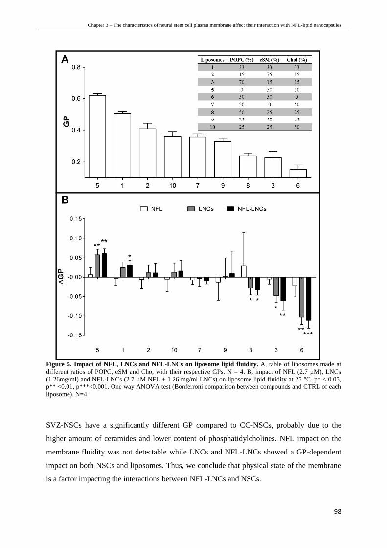
Chapter 3 – The characteristics of neural stem cell plasma membrane affect their interaction with NFL-lipid nanocapsules
98
Figure 5. Impact of NFL, LNCs and NFL-LNCs on liposome lipid fluidity. A, table of liposomes made at
different ratios of POPC, eSM and Cho, with their respective GPs. N = 4. B, impact of NFL (2.7 µM), LNCs
(1.26mg/ml) and NFL-LNCs (2.7 µM NFL + 1.26 mg/ml LNCs) on liposome lipid fluidity at 25 °C. p* < 0.05,
p** <0.01, p***<0.001. One way ANOVA test (Bonferroni comparison between compounds and CTRL of each
liposome). N=4.
SVZ-NSCs have a significantly different GP compared to CC-NSCs, probably due to the
higher amount of ceramides and lower content of phosphatidylcholines. NFL impact on the
membrane fluidity was not detectable while LNCs and NFL-LNCs showed a GP-dependent
impact on both NSCs and liposomes. Thus, we conclude that physical state of the membrane
is a factor impacting the interactions between NFL-LNCs and NSCs.

Chapter 3 – The characteristics of neural stem cell plasma membrane affect their interaction with NFL-lipid nanocapsules
99
5.4 NFL affects NSC plasma membrane permeability
Molecule internalization into cells is regulated, in part, by plasma membrane permeability
which, in turn, depends on its lipid composition [50]. Ceramides can increase plasma
membrane permeability by detergent solubilization effects, induction of nonlamellar
structures and generation of structural defects between rigid and fluid lipid phases [51].
Consequently, we hypothesized that SVZ-NSCs and CC-NSCs could have different plasma
membrane permeability due to the different amount of ceramides detected in those cells.
Plasma membrane permeability to FD4, FD10 and FD40 did not show any significant
difference between the two types of NSCs. The percentage of positive cells was close to the
baseline of the instrument (below 13%) for all conditions (Figure 6). Thus, the different
amount of ceramides detected in SVZ-NSCs and CC-NSCs would not be directly related to
the cell steady state permeability for those compounds. However, we cannot exclude different
NSC permeability for lower molecular weight molecules (< 4kDa).
Figure 6. Permeability of SVZ-NSCs and CC-NSCs to fluorescent dextrans. Percentage of positive NSCs to
different molecular weight fluorescent dextrans (FD4, FD10 and FD40) obtained by flux cytometry. N=4.
Hence, we measured the variation of NSC permeability before and after incubation with NFL,
LNCs and NFL-LNCs. While membrane permeability of both cell types to FD4, FD10 and
FD 40 did not show any major difference after incubation with LNCs and NFL-NCs (data not
shown), the treatment with NFL at 20 µM significantly increased the penetration of FD4 in
29% of SVZ-NSCs (Figure 7A). This result was also confirmed by confocal imaging (Figure
7B).
Cell penetrating peptides can produce pores of different sizes on the plasma membrane of the
cells, depending on the condition of incubation (e.g., concentration) [52]. Only 20 µM of NFL

Chapter 3 – The characteristics of neural stem cell plasma membrane affect their interaction with NFL-lipid nanocapsules
100
allowed the penetration of FD4 (4 kDa), suggesting the ability of the peptide to make pores
but only above this concentration. However, in our previous work we showed that pre-
incubations with 10 µM NFL allowed the penetration of 1.6-2.7 kDa molecules [22].
Consequently, we hypothesized that NFL could modulate SVZ-NSC plasma membrane
permeability by producing pores with concentration-depending sizes. Unfortunately it was not
possible to perform NSCs incubation with higher concentrations of NFL due to its toxicity
[13]. The modification of SVZ-NSC permeability by NFL was totally recovered after 6 hours
(Figure 7C), indicating that the effect of the peptide was transient.
Figure 7. Impact of NFL on NSC permeability. A, percentage of positive NSCs to fluorescent dextran (FD) of
different molecular weights (4, 10 and 40 kDa) after incubation with increasing concentration of NFL (2.7, 10, 20 µM). p* < 0.05. One way ANOVA test (Bonferroni comparison between all the columns with the respective
CTRL). N=4. B, confocal imaging of SVZ-NSCs treated with NFL 20 µM. Blue is DAPI, nucleus. Yellow is
fluorescein isothiocyanate, FD4. Scale barr = 50 µm. C, permeability recovery 6 hours after the treatment with
NFL 20 µM in presence of FD4. p* < 0.05, p** <0.01. Mann-Whitney test. N=3.
Although ceramides are associated with higher membrane permeability [51], no significant
difference was observed between the SVZ-NSC and CC-NSC steady state permeability for
FD4, FD10 and FD40. Despite LNCs and NFL-LNCs did not show any major effect, NFL
significantly increased the permeability of SVZ-NSCs, suggesting a preferential
permeabilizant effect of the peptide on those cells compared to CC-NSCs. Interestingly, the
higher (but not significant) presence of Chol observed in SVZ-NSC did not hinder the
permeabilizant effect of the peptide NFL despite the fact that this lipid usually decreases the
permeability of the plasma membranes [35]. Pae et al. showed that ceramides increase the

Chapter 3 – The characteristics of neural stem cell plasma membrane affect their interaction with NFL-lipid nanocapsules
101
uptake of arginine-rich CPPs while prevent the internalization of amphipathic CPPs [53]. Due
to the presence of arginines in the structure of NFL, we can hypothesize that the higher
amount of ceramides detected in SVZ-NSCs could enhances NFL penetration and thus impact
the permeability of those cells.
6. CONCLUSIONS
NFL-LNCs selectively interact with SVZ-NSCs and not with CC-NSCs. The characterization
of the plasma membrane of both cell types highlighted interesting differences in the lipid
compositions which, in turn, have an impact on the physical state and permeability of the
cells. Higher ceramide content as well as lower phosphatidylcholine amount were observed in
SVZ-NSC plasma membrane compared to CC-NSCs. The interaction between NSCs and
nanoparticles depends on the initial physical state of the plasma membrane while NFL
preferentially increases the permeability of SVZ-NSC plasma membrane. The implementation
of plasma membrane characterization (e.g., by proteomic) or the comparison with other cells
(e.g., glioblastoma cells) would provide additional information to understand and explain
NFL-LNC interaction towards SVZ-NSCs.
Acknowledgment
Dario Carradori is supported by NanoFar "European Doctorate in Nanomedicine" EMJD
programme funded by EACEA. This work is also supported by AFM (Association Française
contre les Myopathies), by ARC (Association de Recherche sur le Cancer), by Ligue contre le
cancer 49 and 85, and by MATWIN (Maturation & Accelerating Translation With Industry)
to J. Eyer. This work is supported by grants from the Université Catholique de Louvain
(F.S.R), Fonds National de la Recherche Scientifique (F.R.S.-FNRS). A. des Rieux is a
F.R.S.-FNRS Research Associate.

Chapter 3 – The characteristics of neural stem cell plasma membrane affect their interaction with NFL-lipid nanocapsules
102
7. REFERENCES
[1] Murrell, Wayne, et al. "Expansion of multipotent stem cells from the adult human brain."
PloS one 8.8 (2013): e71334.
[2] Bond, Allison M., Guo-li Ming, and Hongjun Song. "Adult mammalian neural stem cells
and neurogenesis: five decades later." Cell Stem Cell 17.4 (2015): 385-395.
[3] Arvidsson, Andreas, et al. "Neuronal replacement from endogenous precursors in the adult
brain after stroke." Nature medicine 8.9 (2002): 963-970.
[4] Giuliani, Daniela, et al. "NDP-α-MSH induces intense neurogenesis and cognitive
recovery in Alzheimer transgenic mice through activation of melanocortin MC 4 receptors."
Molecular and Cellular Neuroscience 67 (2015): 13-21.
[5] Kim, Seung U., Hong J. Lee, and Yun B. Kim. "Neural stem cell‐based treatment for
neurodegenerative diseases." Neuropathology 33.5 (2013): 491-504.
[6] Carradori, Dario, et al. "The therapeutic contribution of nanomedicine to treat
neurodegenerative diseases via neural stem cell differentiation." Biomaterials 123 (2017): 77-
91.
[7] Herberts, Carla A., Marcel SG Kwa, and Harm PH Hermsen. "Risk factors in the
development of stem cell therapy." Journal of translational medicine 9.1 (2011): 29.
[8] Simonson, Oscar E., et al. "The safety of human pluripotent stem cells in clinical
treatment." Annals of medicine 47.5 (2015): 370-380.
[9] Xie, Chuncheng, et al. "The effect of simvastatin treatment on proliferation and
differentiation of neural stem cells after traumatic brain injury." Brain research 1602 (2015):
1-8.
[10] Saraiva, C., et al. "MicroRNA-124 loaded nanoparticles enhance brain repair in
Parkinson's disease." Journal of Controlled Release 235 (2016): 291-305.
[11] Barreau, Kristell, Claire Lépinoux-Chambaud, and Eyer Joël. "Review of Clinical Trials
Using Neural Stem Cells." JSM Biotechnology & Biomedical Engineering 3.3 (2016).
[12] Bocquet, Arnaud, et al. "Neurofilaments bind tubulin and modulate its polymerization."
Journal of Neuroscience 29.35 (2009): 11043-11054.
[13] Lépinoux-Chambaud, Claire, Kristell Barreau, and Joël Eyer. "The Neurofilament‐
Derived Peptide NFL‐TBS. 40‐63 Targets Neural Stem Cells and Affects Their Properties."
Stem cells translational medicine 5.7 (2016): 901-913.
[14] Berges, Raphael, et al. "A tubulin binding peptide targets glioma cells disrupting their
microtubules, blocking migration, and inducing apoptosis." Molecular Therapy 20.7 (2012):
1367-1377.

Chapter 3 – The characteristics of neural stem cell plasma membrane affect their interaction with NFL-lipid nanocapsules
103
[15] Lépinoux-Chambaud, Claire, and Joël Eyer. "The NFL-TBS. 40-63 anti-glioblastoma
peptide enters selectively in glioma cells by endocytosis." International journal of
pharmaceutics 454.2 (2013): 738-747.
[16] Balzeau, Julien, et al. "The effect of functionalizing lipid nanocapsules with NFL-TBS.
40-63 peptide on their uptake by glioblastoma cells." Biomaterials 34.13 (2013): 3381-3389.
[17] Rivalin, Romain, et al. "The NFL-TBS. 40-63 anti-glioblastoma peptide disrupts
microtubule and mitochondrial networks in the T98G glioma cell line." PloS one 9.6 (2014):
e98473.
[18] Fressinaud, Catherine, and Joël Eyer. "Neurofilaments and NFL-TBS. 40–63 peptide
penetrate oligodendrocytes through clathrin-dependent endocytosis to promote their growth
and survival in vitro." Neuroscience 298 (2015): 42-51.
[19] Ramsey, Joshua D., and Nicholas H. Flynn. "Cell-penetrating peptides transport
therapeutics into cells." Pharmacology & therapeutics 154 (2015): 78-86.
[20] Berges, Raphael, et al. "Structure-function analysis of the glioma targeting NFL-TBS.
40-63 peptide corresponding to the tubulin-binding site on the light neurofilament subunit."
PloS one 7.11 (2012): e49436.
[21] Huynh, Ngoc Trinh, et al. "Lipid nanocapsules: a new platform for nanomedicine."
International journal of pharmaceutics 379.2 (2009): 201-209.
[22] Carradori, Dario, et al. "NFL-lipid nanocapsules for brain neural stem cell targeting in
vitro and in vivo." Journal of Controlled Release 238 (2016): 253-262.
[23] Langelier, Bénédicte, et al. "Long chain‐polyunsaturated fatty acids modulate membrane
phospholipid composition and protein localization in lipid rafts of neural stem cell cultures."
Journal of cellular biochemistry 110.6 (2010): 1356-1364.
[24] Heurtault B, Saulnier P, Pech B, Proust JE, Richard J, & Benoit JP. Lipidic
nanocapsules: preparation process and use as Drug Delivery Systems. Patent No.
WO02688000.
[25] Hope, M. J., et al. "Production of large unilamellar vesicles by a rapid extrusion
procedure. Characterization of size distribution, trapped volume and ability to maintain a
membrane potential." Biochimica et Biophysica Acta (BBA)-Biomembranes 812.1 (1985):
55-65.
[26] Bartlett, Grant R. "Phosphorus assay in column chromatography." Journal of Biological
Chemistry 234 (1959): 466-468.
[27] Guo, Weixiang, et al. "Isolation of multipotent neural stem or progenitor cells from both
the dentate gyrus and subventricular zone of a single adult mouse." Nature protocols 7.11
(2012): 2005-2012.

Chapter 3 – The characteristics of neural stem cell plasma membrane affect their interaction with NFL-lipid nanocapsules
104
[28] Hugnot, Jean-Philippe. "Isolate and culture neural stem cells from the mouse adult spinal
cord." Neural progenitor cells: methods and protocols (2013): 53-63.
[39] Suski, Jan M., et al. "Isolation of plasma membrane–associated membranes from rat
liver." nature protocols 9.2 (2014): 312-322.
[30] Mutemberezi, Valentin, et al. "Development and validation of an HPLC-MS method for
the simultaneous quantification of key oxysterols, endocannabinoids, and ceramides:
variations in metabolic syndrome." Analytical and bioanalytical chemistry 408.3 (2016): 733-
745.
[31] Parasassi, T., et al. "Quantitation of lipid phases in phospholipid vesicles by the
generalized polarization of Laurdan fluorescence." Biophysical journal 60.1 (1991): 179-189.
[32] Jia, Y., et al. "Comparison of cell membrane damage induced by the therapeutic
ultrasound on human breast cancer MCF-7 and MCF-7/ADR cells." Ultrason Sonochem.
(2015):128-35.
[33] Etzerodt, Thomas P., et al. "A GALA lipopeptide mediates pH-and membrane charge
dependent fusion with stable giant unilamellar vesicles." Soft Matter 8.21 (2012): 5933-5939.
[34] Cuco, Andreia, et al. "Interaction of the Alzheimer Aβ (25–35) peptide segment with
model membranes." Colloids and Surfaces B: Biointerfaces 141 (2016): 10-18.
[35] Magarkar, Aniket, et al. "Cholesterol level affects surface charge of lipid membranes in
saline solution." Scientific reports 4 (2014): 5005.
[36] van Blitterswijk, Wim J., et al. "Ceramide: second messenger or modulator of membrane
structure and dynamics?." Biochemical Journal 369.2 (2003): 199-211.
[37] Silva, Liana, et al. "Ceramide-platform formation and-induced biophysical changes in a
fluid phospholipid membrane." Molecular membrane biology 23.2 (2006): 137-148.
[38] Van Meer, Gerrit, Dennis R. Voelker, and Gerald W. Feigenson. "Membrane lipids:
where they are and how they behave." Nature reviews Molecular cell biology 9.2 (2008): 112-
124.
[39] Holopainen, Juha M., Jukka YA Lehtonen, and Paavo KJ Kinnunen. "Lipid
microdomains in dimyristoylphosphatidylcholine–ceramide liposomes." Chemistry and
physics of lipids 88.1 (1997): 1-13.
[40] Peetla, Chiranjeevi, Sivakumar Vijayaraghavalu, and Vinod Labhasetwar. "Biophysics of
cell membrane lipids in cancer drug resistance: Implications for drug transport and drug
delivery with nanoparticles." Advanced drug delivery reviews 65.13 (2013): 1686-1698.
[41] Owen, Dylan M., et al. "Quantitative imaging of membrane lipid order in cells and
organisms." Nature protocols 7.1 (2012): 24-35.

Chapter 3 – The characteristics of neural stem cell plasma membrane affect their interaction with NFL-lipid nanocapsules
105
[42] Bagatolli, L. A. "LAURDAN fluorescence properties in membranes: a journey from the
fluorometer to the microscope." Fluorescent methods to study biological membranes. Springer
Berlin Heidelberg, 2012. 3-35.
[43] Catapano, Elisa R., et al. "Solid character of membrane ceramides: a surface rheology
study of their mixtures with sphingomyelin." Biophysical journal 101.11 (2011): 2721-2730.
[44] Pinto, Sandra N., et al. "A combined fluorescence spectroscopy, confocal and 2-photon
microscopy approach to re-evaluate the properties of sphingolipid domains." Biochimica et
Biophysica Acta (BBA)-Biomembranes 1828.9 (2013): 2099-2110.
[45] Kovács, Eugenia, et al. "Interaction of gentamicin polycation with model and cell
membranes." Bioelectrochemistry 87 (2012): 230-235.
[46] Bangham, A. D., M. W. Hill, and N. G. A. Miller. "Preparation and use of liposomes as
models of biological membranes." Methods in membrane biology. Springer US, 1974. 1-68.
[47] Kristl, J., et al. "Interactions of solid lipid nanoparticles with model membranes and
leukocytes studied by EPR." International journal of pharmaceutics 256.1 (2003): 133-140.
[48] Van Meer, Gerrit, Dennis R. Voelker, and Gerald W. Feigenson. "Membrane lipids:
where they are and how they behave." Nature reviews Molecular cell biology 9.2 (2008): 112-
124.
[49] Madani, Fatemeh, et al. "Mechanisms of cellular uptake of cell-penetrating peptides."
Journal of Biophysics 2011 (2011).
[50] Smith, Dennis, et al. "Passive lipoidal diffusion and carrier-mediated cell uptake are both
important mechanisms of membrane permeation in drug disposition." Molecular
pharmaceutics 11.6 (2014): 1727-1738.
[51] Contreras, F-Xabier, et al. "Sphingosine increases the permeability of model and cell
membranes." Biophysical journal 90.11 (2006): 4085-4092.
[52] Herce, H. D., et al. "Arginine-rich peptides destabilize the plasma membrane, consistent
with a pore formation translocation mechanism of cell-penetrating peptides." Biophysical
journal 97.7 (2009): 1917-1925.
[53] Pae, Janely, et al. "Translocation of cell-penetrating peptides across the plasma
membrane is controlled by cholesterol and microenvironment created by membranous
proteins." Journal of Controlled Release 192 (2014): 103-113.

Chapter 3 – The characteristics of neural stem cell plasma membrane affect their interaction with NFL-lipid nanocapsules
106
8. SUPPLEMENTARY DATA
Figure S1. Lipid characterization of SVZ-NSC and CC-NSC plasma membranes by HPLC-MS. Quantification of different phosphatidylcholines in the plasma membrane of SVZ-NSCs and CC-NSCs. N=5,
n=5.
Figure S2. Lipid characterization of SVZ-NSC and CC-NSC plasma membranes by HPLC-MS. Quantification of different sphingomyelins in the plasma membrane of SVZ-NSCs and CC-NSCs. N=5, n=5.
Phosphatidylcholines
C32
:0
C32
:1
C32
:2
C34
:0
C34
:1
C36
:0
C36
:2
C36
:3
C36
:4
C36
:5
C38
:1
C38
:2
C38
:3
C38
:4
C38
:5
C38
:6
C40
:4
C40
:5
C40
:6
0
5
10
15
2020
35
5050
100
150
200
250
300
350
SVZ-NSCs CC-NSCs
rati
o
(AU
CP
C/A
UC
IS)

Chapter 4 – Retinoic acid-loaded NFL-lipid nanocapsules induce neural stem cell differentiation towards the oligodendrocyte lineage
107
CHAPTER 4
RETINOIC ACID -LOADED NFL-LIPID
NANOCAPSULES INDUCE NEURAL STEM
CELL DIFFERENTIATION TOWARDS THE
OLIGODENDROCYTE LINEAGE *
*Adapted from
Carradori, D.; Vanvarenberg, K.; Ucakar, B.; Beloqui, A.; Saulnier, P.; Eyer, J.; Préat, V.;
Miron, V.; des Rieux, A. In progress.

Chapter 4 – Retinoic acid-loaded NFL-lipid nanocapsules induce neural stem cell differentiation towards the oligodendrocyte lineage
108
TABLE OF CONTENTS
1. PREFACE
2. ABSTRACT
3. INTRODUCTION
4. MATERIALS AND METHODS
8.1 Materials
8.2 Preparation of RA-loaded NFL-LNCs
8.3 Characterization of LNCs
8.4 Isolation of SVZ-NSCs
8.5 Impact of RA-loaded NFL-LNCs on SVZ-NSC viability
8.6 Effect of RA-loaded NFL-LNCs on SVZ-NSC self-renewal
8.7 Influence of RA-loaded NFL-LNCs on SVZ-NSC differentiation
8.8 Impact on a focal brain white matter demyelinating lesion
8.9 Statistical analysis
5. RESULTS AND DISCUSSION
8.10 Characterization of LNCs
8.11 RA-loaded NFL-LNCs modulate SVZ-NSC self-renewal
8.12 RA-loaded NFL-LNCs affect SVZ-NSC differentiation
6. CONCLUSION
7. REFERENCES
8. SUPPLEMENTARY DATA

Chapter 4 – Retinoic acid-loaded NFL-lipid nanocapsules induce neural stem cell differentiation towards the oligodendrocyte lineage
109
1. PREFACE
Central nervous system (CNS) diseases are mostly characterized by the chronic loss and
lesion of specialized neural cells such as neurons, astrocytes and oligodendrocytes. In this
case, neural stem cells (NSCs) are of great interest due to their differentiation property by
which they could replace those lesioned cells. The therapeutic strategy based on the
stimulation of endogenous NSCs is one of the most promising because it would avoid some of
the limiting issues associated to exogenous NSC-based approaches (e.g., source of cells,
viability post-transplantation).
The production of a system able to target endogenous NSCs and to stimulate their
differentiation via the delivery of a bioactive molecule would represent an important step
forward in NSC-based therapy.
In our previous work, we developed and characterized a NSC-interacting drug delivery
system by adsorbing the peptide NFL-TBS.40-63 (NFL) on the surface of lipid nanocapsules
(LNCs). When injected in the brain, NFL-LNCs specifically co-localize with Nestin+ cells
which are considered the bona fide neural stem cells of the subventricular zone (SVZ-NSCs).
The SVZ is one of the most neurogenic regions of the CNS. It is thus an interesting target for
the modulation of NSC differentiation and NFL-LNC represents a promising way to deliver
bioactive drugs in this area.
The aim of this work was to provide the proof of concept of this hypothesis by loading
retinoic acid, a molecule active on NSC differentiation, into NFL-LNCs and inducing SVZ-
NSC differentiation towards specialized neural cells.

Chapter 4 – Retinoic acid-loaded NFL-lipid nanocapsules induce neural stem cell differentiation towards the oligodendrocyte lineage
110
2. ABSTRACT
Neural stem cells (NSCs) are located in restricted areas of the central nervous system such as
the subventricular zone (SVZ) of the brain where they undergo self-renew or differentiate into
specialized neural cells (neurons, astrocytes and oligodendrocytes). The stimulation of
endogenous NSCs is one of the most promising NSC-based therapeutic approaches to restore
neurological functions in patients affected by neurodegenerative diseases. Unfortunately, the
lack of drugs/systems able both to target and to induce NSC differentiation limits the clinical
translation of this strategy. Recently, the combination between the peptide NFL-TBS.40-63
(NFL) and lipid nanocapsules (LNCs) produced a nanoscale vector able to co-localize with
the NSCs of the SVZ (SVZ-NSCs). The aim of this work was to induce NSC differentiation
by delivering retinoic acid (RA) to SVZ-NSCs via NFL-LNCs. RA was successfully
encapsulated into NFL-LNC and RA-loaded NFL-LNCs were incubated with primary
newborn or adult rat SVZ-NSCs. The impact on NSC self-renewal and differentiation was
investigated, either by light microscopy or by immunocytochemistry (2 and 7 days post-
incubation). Treatment with RA-loaded NFL-LNCs significantly increased the number of
GalC+ (oligodendrocytic marker) cells while it decreased the number of Nestin
+ (neural stem
cell marker) cells. Then, RA-loaded NFL-LNCs were injected in an in vivo rat model of white
matter demyelination (lysolecithin injection in the caudoputamen) to evaluate the efficacy of
our system on remyelination stimulation. The analysis of brain tissues is ongoing.

Chapter 4 – Retinoic acid-loaded NFL-lipid nanocapsules induce neural stem cell differentiation towards the oligodendrocyte lineage
111
3. INTRODUCTION
CNS degenerative diseases are a heterogeneous group of disorders characterized by the
progressive development of lesions and loss of specialized neural cells such as neurons,
astrocytes and oligodendrocytes [1,2]. Oligodendrocytes are myelinating cells which
modulate the axonal conduction and provide trophic support to neurons [3]. Myelin
dysfunction and oligodendrocitic failure are primarily involved in the etiology of many
demyelinating CNS diseases (e.g., multiple sclerosis and multiple system atrophy) which have
high economic and social costs worldwide [4].
Neural stem cells (NSCs) have been investigated to treat such diseases since many studies
demonstrated partial neurological function recovery after their transplantation in animal
models [5]. NSCs are located in restricted neurogenic niches of the CNS (e.g., the central
canal (CC) in the spinal cord and the subventricular zone (SVZ) and the subgranular zone
(SGZ) in the brain). The therapeutic effect of NSCs is mostly related to their differentiation
and proliferation, properties by which they can replace damaged cells and enhance the
recovery of the affected areas [6]. Several NSC-based strategies have been developed to treat
CNS diseases which especially depend on whether NSCs are exogenous (induced from other
somatic cells or isolated from neurogenic niches) or endogenous (niche-localized) [7].
Basically, NSC differentiation is designed to occur either in vivo after transplantation of in
vitro expanded/stimulated exogenous NSCs or in situ after endogenous NSC stimulation
respectively.
In situ differentiation of endogenous NSCs is one of the most promising strategies to treat
demyelinating diseases because it would avoid both the issues associated to the
transplantation (e.g., cell sources and cell viability) [8] and the in vitro manipulations (e.g.,
risk of contamination and genetic modifications) of NSCs [9]. Nevertheless, the lack of drugs
and systems combining endogenous NSCs targeting and differentiation strongly slows down
the development of this therapeutic approach.
The peptide NFL-TBS.40-63 (NFL) is a 24-aminoacid peptide which corresponds to the
tubulin-binding site of the neurofilament light subunit [10]. It selectively penetrates SVZ-
NSCs by direct translocation in vitro while it localizes in SVZ-NSC niches after intra-lateral
ventricle injection in rat brain in vivo [11]. Although NFL targets SVZ-NSCs and induces
their differentiation into neurons and oligodendrocytes in vitro, it did not show yet an
identified therapeutic effect in vivo. Moreover, the strong structure-activity correlation

Chapter 4 – Retinoic acid-loaded NFL-lipid nanocapsules induce neural stem cell differentiation towards the oligodendrocyte lineage
112
evaluated by alanine-scanning assay and circular dichroism [12] limits chemical coupling
between NFL and bioactive molecules. While the direct therapeutic application of the peptide
to target and differentiate NSCs is still not excluded, its association with a drug delivery
system represents a potential strategy for the selective delivery of bioactive molecules to
SVZ-NSCs.
In a previous work [13], NFL was adsorbed on the surface of lipid nanocapsules (LNCs)
which are a nanoscale vector characterized by long stability (more than 18 month), FDA-
approved excipients, easy scale-up, versatility (compatible with lipophilic as well as
hydrophilic drugs) [14], and suitable surface functionalization (e.g. with targeting ligands)
[15]. The adsorption of NFL on the LNCs did not modify the NSC-targeting property of the
peptide which, on the contrary, was extended to the whole system. NFL-LNC interacts with
primary SVZ-NSCs, in vitro, and localizes on the NSCs of the SVZ after injection in the
lateral ventricle of rat, in vivo. The SVZ is one of the most important neurogenic regions of
the CNS that is involved in the origin/development of many CNS diseases [16-18]. Thus, the
targeting of this area via NFL-LNC would be a good opportunity to induce NSC
differentiation by delivering a bioactive molecule to SVZ-NSCs.
Retinoic acid (RA) is a lipophilic molecule that modulates neurogenesis by inducing neurite
outgrowth and differentiation into specialized neural cells from different cell sources, such as
embryonic stem cells [19-21] and mesenchymal stem cells [22-24]. In particular, RA has been
shown to induce the differentiation of pluripotent stem cells [25] and neural progenitor cells
[26] in myelinating oligodendrocytes. Although RA represents a promising bioactive drug to
stimulate NSC differentiation and treat demyelinating diseases [27], many factors limit its
utilisation as a free molecule in vitro, (e.g., its low solubility in aqueous media) and in vivo
(e.g., the progressive decline of its plasmatic concentrations). One strategy to overcome these
issues is to incorporate RA into nanoparticles. For instance, RA encapsulation in nanovectors
showed an enhanced therapeutic effect in the treatment of cancer [28] or ischemia [29],
highlighting the interest of RA-based systems.
Thus, the objectives of this work were (i) to encapsulate RA into NFL-LNCs and (ii) to
evaluate their efficacy to stimulate NSC differentiation, first in vitro, on primary SVZ-NSCs,
and then in vivo, and in a rat model of brain demyelinated lesions.

Chapter 4 – Retinoic acid-loaded NFL-lipid nanocapsules induce neural stem cell differentiation towards the oligodendrocyte lineage
113
4. MATERIALS AND METHODS
4.1 Materials
Biotinylated NFL-TBS.40-63 (NFL) was purchased from GeneCust (Luxembourg,
Luxembourg). Labrafac® provided by Gattefosse SA (Saint-Priest, France). Lipoïd® was
provided by Lipoid Gmbh (Ludwigshafen, Germany). Kolliphor HS®, L-α-Lysolecithin,
poly-D-Lysine hydrobromide, retinoic acid and Rhodamine B were purchased from Sigma
(Saint-Louis, Missouri). Sodium chloride (NaCl) was purchased from Prolabo (Fontenay-
sous-bois, France). Primary antibody mouse anti-GalC was purchased from Merck Millipore
(Billerica, Massachusetts, USA). Primary antibodies mouse anti-PAN neurofilament and
mouse anti-bIII tubulin were purchased from Biolegend (San Diego, California, USA).
Primary antibodyes rabbit anti-Nestin and rabbit anti-GFAP were purchased from Abcam
(Cambridge, United Kingdom). Secondary antibodyes Alexa 488 anti-mouse, Alexa 488 anti-
rabbit, Alexa 594 anti-mouse and Alexa 594 anti-rabbit were purchased from ThermoFisher
Scientific (Waltham, Massachusetts, USA). DiD' solid; DiIC18(5) solid (1,1'-Dioctadecyl-
3,3,3',3'-Tetramethylindodicarbocyanine, 4-Chlorobenzenesulfonate Salt) (DiD), DAPI,
HEPES, Pen/Strept, Na Pyruvate, B27, MEM alpha (no nucleosides), 0.05 % Trypsin-EDTA
(1x), DNAse and ProLong Gold antifade were purchased from Thermo Fisher Scientific. EGF
was purchased from Tebu-Bio (Le Perray en Yvelines, France). CellTiter 96® AQueous One
Solution Cell Proliferation Assay was purchased from Promega (Fitchburg, Wisconsin, USA).
Formaldeyde solution 37% was purchased from Carl Roth (Karlsruhe, Germany). Amicon
Ultra-0.5 ml 100K filters were purchased from Merck Millipore (Billerica, Massachusetts,
USA). The isolation of NSC and in vivo experiments were performed according to Directive
2010/63/EU, to guidelines of the Belgian Government following the approval by the ethical
committee for animal care of the faculty of medicine of the Université catholique de Louvain.
4.2 Preparation of RA-loaded NFL-LNCs
NFL-LNCs. Stok-LNCs were prepared following the protocol developed by Heurtault et al.
[30]. Briefly, Kolliphor HS15® (0.846 g), Lipoïd® (0.075 g), NaCl (0.089 g), Labrafac®
(1.028 g) and water (2.962 g) were mixed under gentle magnetic stirring at 30°C for 5 min.
The solution was progressively heated (90 °C) and cooled (60 °) three times. During the last
cooling, cold water (12.5 g at 4 °C) was added at 72-74 °C under high speed stirring. The
nanoparticles were filtered by 0.2 µm filter and stored at 4°C. NFL-LNCs were produced by
incubating 369 µl of 1 mM peptide solution (in water) overnight with 1 ml of stock-LNC

Chapter 4 – Retinoic acid-loaded NFL-lipid nanocapsules induce neural stem cell differentiation towards the oligodendrocyte lineage
114
under gently stirring while LNCs without peptide were produced incubating 369 µl of water.
LNC concentration: 126 mg/ml of nanoparticles. NFL-LNC concentration: 270 µM NFL +
126 mg/ml of nanoparticles.
RA-loaded NFL-LNCs. RA was loaded into LNCs by adding 50 µl of RA stock solution (20
mg/ml in DMSO) at 75 °C during the last cooling of LNC preparation. RA-loaded NFL-LNCs
were produced by following the same protocol as for NFL-LNCs. The concentration of RA-
loaded LNC stock solution was 400 µM RA + 126 mg/ml of nanoparticles and of RA-loaded
NFL-LNC stock solution was 400 µM RA + 270 µM NFL + 126 mg/ml of nanoparticles.
4.3 Characterization of LNC
Size, -potential and PDI of nanoparticles were characterized using a Malvern Zetasizer Nano
ZS (Malvern Instruments). For the measurement of size and PDI, samples were diluted 1/100
(v/v) in water. For the measurement of -potential, samples were diluted 1/100 (v/v) in NaCl
10 mM.
4.4 Isolation of SVZ-NSCs
SVZ-NSCs were isolated according to a protocol described by Guo et al. [31]. Briefly rats
from 5 day-old or 2 month-old were sacrificed by decapitation. The brain was removed and
put in dissection buffer (1.25 ml of D-glucose 1 M, 750 µl of HEPES and 500 µl Pen/Strept in
50 ml of HBSS medium). The brain was cut into coronal sections of 400 µm and the SVZ was
dissected and placed in 1 ml of dissection buffer. The tissue was centrifuged for 5 min at 344
g. The supernatant was removed to add 1 ml of stem cell culture medium (1.25 ml of D-
glucose 1 M, 750 µl of HEPES, 500 µl Pen/Strept, 500 µl Na Pyruvate, 500 µl of B27 and 10
ng/ml of EGF in 50 ml of MEM alpha no nucleosides medium). The pellet was vortexed and
mechanically triturated using a 26 G needle. The cells were seeded at 2*105 cells/Petri 60 mm
in 5 ml of cell culture medium. Either neurospheres or dissociated cells have been used during
this study, depending on the aim of the experiment.
4.5 Impact of RA-loaded NFL-LNCs on SVZ-NSC viability
Five days after isolation, neurospheres corresponding to 3*105 SVZ-NSCs/ml were incubated
for 1 h at 37 °C with RA-loaded NFL-LNCs or their controls (PBS, NFL, LNCs, NFL-LNCs,
RA and RA-LNCs) diluted 1/100 from stock solutions (final concentrations RA 4 µM, NFL
2.7 µM, LNCs 1.26 mg/ml, NFL-LNCs NFL 2.7 µM+ 1.26 mg/ml LNCs, RA-loaded LNCs

Chapter 4 – Retinoic acid-loaded NFL-lipid nanocapsules induce neural stem cell differentiation towards the oligodendrocyte lineage
115
RA 4 µM + 1.26 mg/ml LNCs, RA-loaded NFL-LNCs RA 4 µM + 2.7 µM NFL + 1.26
mg/ml LNCs). Then, the neurospheres were washed, dissociated by using a 26 G needle, and
seeded at 3*105 cells/ml on poly-D-Lysine hydrobromide (0.1 mg/ml in water) coated 96-well
plates. Cell viability was measured after 1h, 2 days and 7 days using the CellTiter 96®
AQueous One Solution Cell Proliferation assay as per supplier instructions.
4.6 Effect of RA-loaded NFL-LNCs on SVZ-NSC self-renewal
Five days after isolation, neurospheres were treated as described in 4.5. Then, the
neurospheres were seeded in 24-well plates at 3*105 cells/ml or dissociated using a 26 G
needle and seeded either at 3*105 cells/ml (1
st observation) or 1.5*10
5 (2
nd observation) on
poly-L-Lysine coated 24-well plates. Evaluation of SVZ-NSC self-renewal was performed 2
and 7 days after the treatment.
Un-dissociated neurospheres were counted manually using bright-filter microscopy (Evos XL
Core, Thermo Fisher Scientific). At least, 3 pictures/condition were taken.
Dissociated SVZ-NSCs were fixed with 4 % PFA for 10 min, treated 1 h with Triton 0.05% in
BSA 1% and incubated overnight with mouse anti-Nestin (1/250) primary antibody in BSA
1%. The cells were incubated 2 h with Alexa 488 (1/400) anti-mouse secondary antibody in
PBS and mounted with VECTASHIELD® antifade mounting medium (Vector Laboratories,
Burlingame, California, USA). An EVOS fluorescent microscope was used to take at least 3
pictures/conditions. Nestin+ cells were counted manually.
4.7 Influence of RA-loaded NFL-LNCs on SVZ-NSC differentiation
Five days after isolation, neurospheres were treated as described in 4.5. Then, the
neurospheres were dissociated by using a 26 G needle and seeded either at 3*105 cells/ml (1
st
observation) or 1.5*105
(2nd
observation)on poly-D-Lysine hydrobromide coated 24-well
plates.
Two days after treatment, SVZ-NSCs were fixed with 4 % PFA for 10 min, treated 1 h with
Triton 0.05% in BSA 1% and incubated overnight with rabbit anti-GFAP (1/1000) and mouse
anti-βIII tubulin (1/1000) primary antibodies in BSA 1%. The cells were incubated 2 h with
Alexa 488 (1/400) anti-mouse and Alexa 594 (1/400) anti-rabbit secondary antibodies in PBS.
Seven days after the treatment, SVZ-NSCs were fixed with 4 % PFA for 10 min, treated 1 h
with Triton 0.05% in BSA 1% for 30 and incubated overnight with (separately) mouse anti-
pan-neurofilament (panNF) (1/500) and mouse anti-GalC (1/500) primary antibodies in BSA

Chapter 4 – Retinoic acid-loaded NFL-lipid nanocapsules induce neural stem cell differentiation towards the oligodendrocyte lineage
116
1%. The cells were incubated 2 h with Alexa 488 anti-mouse (1/400) and Alexa 594 (1/400)
anti-mouse secondary antibodies in PBS.
Samples were then mounted with VECTASHIELD® antifade mounting medium and pictures
were acquired with an EVOS fluorescent microscope (at least 3 pictures/conditions). PanNF+
and GalC+ cells were counted manually.
4.8 Impact of RA-loaded NFL-LNCs on a focal brain white matter demyelinated lesion
Demyelinating lesion was induced in the caudoputamen of 2 month-old female Wistar rats by
stereotaxic injection in the right caudate putamen (ML: +2.1; AP: -0.8; DV: +4) of 2 μl of 1%
lysolecithin (0.5 µl/min) in PBS using a Harvard apparatus (Holliston, Massachusetts). Three
days after the lesion, the rats received 20 μl of RA-loaded NFL-LNCs (400 µM RA + 270 µM
NFL + 126 mg/ml LNCs) (0.5 µl/min) or their controls by a second stereotaxic injection in
the right lateral ventricle (ML: +1.5; AP: -0.8; DV: +4). Five days after the treatment (7 days
after the lesion), the animals were sacrified by overdose of isoflurane and intracardially
perfused with 4% paraformaldehyde in NaCl buffer. Brains were recovered, post-fixed 6
hours and cryoprotected in 30% sucrose before storage at −80 °C in OCT.
The impact of RA-loaded NFL-LNCs on focal brain white matter demyelinated lesion will be
evaluated on 20-µm brain section by comparing the number of oligodendrocytes (Olig1 and
Olig2+ cells) and the amount of myelin (MAG and MBP
+ cells) in brains treated with the
controls and with RA-loaded NFL-LNCs. This is currently ongoing.
4.9 Statistical analyses
All experiments were repeated at least 3 times. Error bars represent standard error of the mean
(SEM). p* < 0.05, p** < 0.01 and p*** < 0.001 were calculated with Mann-Whitney test or
with ANOVA one-way (Bonferroni correction to take into account the test multiplicity) by
using Prism 5.00 (GraphPad software, San Diego, CA).

Chapter 4 – Retinoic acid-loaded NFL-lipid nanocapsules induce neural stem cell differentiation towards the oligodendrocyte lineage
117
5. RESULTS AND DISCUSSION
5.1 Characterization of LNCs
Table 1. Physiochemical characteristics of the nanoparticles
Size and PDI were characterized by DLS diluting the nanoparticles 1/100 (v/v) in water. The -potential was
measured by diluting the nanoparticles 1/100 (v/v) in NaCl 10 mM. N=3, n=3
LNCs had a size of 57 nm ± 4 , a PDI of 0.027 and a -potential of -8.5 mV ± 1.5 while the
adsorption of NFL increased LNC size (+ 8 nm) and PDI as well as -potential (+ 4 mV)
(Table 1), as previously observed [22]. RA encapsulation did not influence LNC size and PDI
but slightly decreased -potential values (p > 0.05).
5.2 Impact of RA-loaded NFL-LNCs on SVZ-NSC viability
Cell viability was evaluated 1 hour, 2 days and 7 days after SVZ-NSCs incubation. These
time points were chosen to investigate acute (1 h) and chronic (2 d and 7 d) effect during the
evaluation of NSC self-renewal and differentiation. LNC concentrations were selected below
the threshold of toxicity [13].
None of the treatments had major effects on NSC viability (Figure 1A) as well as RA-loaded
NFL-LNCs at higher concentration (1/50) (Figure 1B).
Figure 1: Impact of RA-loaded NFL-LNCs or their controls on NSC viability. A: viability of SVZ-NSCs
incubated 1 hour with different treatments: RA (4 µM), NFL (2.7 µM), LNCs (1.26 mg/ml), NFL-LNCs ( 2.7
size (nm) PDI
-potential (mV)
LNCs
57 ± 4 0.027
-8.5 ± 1.5
NFL-LNCs 65 ± 4 0.175 -4.2 ± 0.5
RA-loaded LNCs 55 ± 3 0.093
-11.0 ± 2.5
RA-loaded NFL-LNCs 63 ± 4 0.2 -15 ± 6

Chapter 4 – Retinoic acid-loaded NFL-lipid nanocapsules induce neural stem cell differentiation towards the oligodendrocyte lineage
118
µM NFL + 1.26 mg/ml LNCs), RA-loaded LNCs (4 µM RA + 1.26 mg/ml LNCs) and RA-loaded NFL-LNCs (4
µM RA + 2.7 µM NFL and 1.26 mg/ml LNCs). The evaluation was performed 1 hour, 2 days and 7 days after
the incubation by MTS. B: viability of SVZ-NSCs incubated 1 hour with RA-loaded NFL-LNCs (400 µM RA +
270 µM NFL + 126 mg/ml LNCs) at different dilutions (1/50, 1/100, 1/500 and 1/1000). The evaluation was
performed 1 hour, 2 days and 7 days after the incubation by MTS assay. N=3, n=3. (p > 0.05).
5.3 RA-loaded NFL-LNCs modulate SVZ-NSC self-renewal
RA, NFL, NFL-LNCs and RA-loaded NFL-LNCs significantly decreased the number of
neurospheres after 2 days (Figure 2A). After 7 days, NFL, NFL-LNCs and RA-loaded NFL-
LNCs but not RA anymore significantly reduced the percentage of neurospheres (Figure 2B).
The highest decrease was observed for NFL-LNCs at 2 days post-treatment but the percentage
of neurospheres was not significantly lower anymore for this condition after 7 days.
Self-renewal is the ability of stem cells to proliferate by maintaining an undifferentiated state
and its loss typically accompanies stem cell differentiation into specialized cells [32]. The
negative impact of NFL on the neurosphere-forming property was already observed in SVZ-
NSCs [11] while the effect of RA on those cells was not described before, to the extent of our
knowledge. The reduction of self-renewal was higher 2 days than 7 days after the treatments,
suggesting the capacity of NSCs to partially recover their property. Only NFL, NFL-LNCs
and RA-loaded NFL-LNCs kept their negative modulation on SVZ-NSC self-renewal
probably due to the presence of the peptide either as free molecule or associated to LNCs
(NFL-LNCs and RA-loaded NFL-LNCs).
Another indication of NSC differentiation was the decrease of the percentage of Nestin+ cells
(Figure 2C-D, Figure 1S). NFL-LNCs and RA-loaded NFL-LNCs significantly decreased the
number of Nestin+
cells after 2 (Figure 2C) and 7 days (Figure 2D), while the other treatments
showed no major effect.
The absolute number of neurospheres and Nestin+
cells decreased from the 1st to the 2
nd (-20%
and -15% respectively) observation due to the percentage of NSCs that differentiate during
cultivation [33].

Chapter 4 – Retinoic acid-loaded NFL-lipid nanocapsules induce neural stem cell differentiation towards the oligodendrocyte lineage
119
Important in vitro NSC characteristics such as the capacity to form neurospheres [34] and to
express Nestin [35] are indicators of the balance proliferation-differentiation. The reduction of
those characteristics was an indication of SVZ-NSC transition from the undifferentiated state
to the differentiating state. Thus, NFL-LNCs and RA-loaded NFL-LNCs would induce NSC
differentiation.
Figure 2: Impact of RA-loaded NFL-LNCs on SVZ-NSC self-renewal. SVZ-NSCs have been incubated 1
hour with different treatments: RA (4 µM), NFL (2.7 µM), LNCs (1.26 mg/ml), NFL-LNCs ( 2.7 µM NFL +
1.26 mg/ml LNCs), RA-loaded LNCs (4 µM RA + 1.26 mg/ml LNCs) and RA-loaded NFL-LNCs (4 µM RA +
2.7 µM NFL and 1.26 mg/ml LNCs). A: number of neurospheres 2 day after the treatment. B: number of
neurospheres 7 days after the treatment. C: percentage of Nestin+ cells 2 days after the treatment. D: percentage
of Nestin+ cells 7 days after the treatment. . p* < 0.05, p** < 0.01, p*** < 0.001 compared to CTRL. One-way
ANOVA test. N=4. n=2 for A and B. N=4, n=3 for C and D.
5.4 RA-loaded NFL-LNCs affect SVZ-NSC differentiation
β III Tubulin is a marker associated to early neuronal developmental stages while
neurofilaments (NF) and galactocerebroside (GalC) are expressed by mature neurons and

Chapter 4 – Retinoic acid-loaded NFL-lipid nanocapsules induce neural stem cell differentiation towards the oligodendrocyte lineage
120
oligodendrocytes, respectively [36,37]. The glial fibrillary acid protein (GFAP) is commonly
used to identify mature astrocytes [38] but it is also an early marker for astroglia and
oligodendroglia due to their phenotype overlap at their early stage [39].
2 day time point (1st observation). The impact of RA-loaded NFL-LNCs on NSC
differentiation was evaluated by βIII Tubulin and GFAP immunostaining 2 days after
treatment. NFL and RA-loaded NFL-LNCs significantly increased the percentage of both β
III Tubulin+ and GFAP
+ cells (Figure 3).
We hypothesized that RA encapsulation in LNCs would allow SVZ-NSC differentiation in
GFAP+ and βIII Tubulin
+ cells. Indeed, RA as a free drug shows many limitations in vitro
such as precipitation in aqueous media and adsorption on glass and plastic wares.
RA-loaded NFL-LNC significantly increased the percentage of GFAP+ cells compared to free
RA but not to RA-loaded LNCs. GFAP+ cells induced by RA-loaded LNCs could be due to
the RA carried into the cells via the small amount of un-functionalized LNCs (10%) able to
penetrate SVZ-NSCs [13]. An increase of βIII Tubulin+
cells was observed when NSCs were
treated with NFL and RA-loaded NFL-LNC. We propose that the highest percentage of
GFAP+ and βIII Tubulin
+ cells observed when NSCs were treated with RA-loaded NFL-LNCs
could be due to a synergetic effect between NFL and RA. The ability of NFL to induce NSC
differentiation towards oligodendrocyte and neuronal phenotypes has already been reported
[11]. Consequently, the effect on both markers is not surprising as well as the low percentage
of GFAP+ cells induced by NFL-LNC could be due to the presence of the peptide on LNC
surface.
We thus hypothesised that RA-loaded NFL-LNCs showed higher percentage of GFAP+ cells
compared to NFL probably because of the synergy between NFL and RA. It would be
interesting to test the effect of RA-loaded LNCs in presence of NFL to confirm this
hypothesis.

Chapter 4 – Retinoic acid-loaded NFL-lipid nanocapsules induce neural stem cell differentiation towards the oligodendrocyte lineage
121
Figure 3: Impact of RA-loaded NFL-LNC on SVZ-NSC differentiation in GFAP
+ and β III
+ cells
evaluated at 2 day time point. SVZ-NSCs have been incubated 1 hour with different treatments: RA (4 µM),
NFL (2.7 µM), LNCs (1.26 mg/ml), NFL-LNCs (2.7 µM NFL + 1.26 mg/ml LNCs), RA-loaded LNCs (4 µM
RA + 1.26 mg/ml LNCs) and RA-loaded NFL-LNCs (4 µM RA + 2.7 µM NFL and 1.26 mg/ml LNCs). Cell
were immunostained for GFAP (green), β III Tubulin (red) and DAPI (blue). The number of positive cells has
been counted manually. p* < 0.05, p** < 0.01, p*** < 0.001. One way ANOVA test (Bonferroni post-test).
N=4, n=3.

Chapter 4 – Retinoic acid-loaded NFL-lipid nanocapsules induce neural stem cell differentiation towards the oligodendrocyte lineage
122
7 day time point. The percentage of GFAP+ and GalC
+ cells was evaluated 7 days after
treatment. The percentage of GalC+ cells significantly increased after incubation with RA-
loaded NFL-LNCs (Figure 4A) while no significant effect was observed for the other
treatments. Moreover, SVZ-NSCs treated with RA-loaded NFL-LNCs presented morphology
similar to immature oligodendrocytes 7 days after treatment (Figure 4B). The percentage of
GFAP+
cells was not significantly impacted whatever the condition (p > 0.05), even if
treatment of NSCs with RA-loaded LNCs and RA-NFL-LNCs tended to reduce the
percentage of GFAP+ cells while treatments with RA, NFL, LNCs and NFL-LNCs tended to
increase the number of GFAP+ cells (Figure 4A). Compared to the observation performed 2
day after the treatment, the percentage of GFAP+
increased for the CTRL (from 4 to 12%).
The co-expression of both markers remained below 15% for all conditions.
Figure 4: Impact of RA-loaded NFL-LNC on SVZ-NSC differentiation in GFAP
+ and GalC
+ cells
evaluated at 7 day time point. SVZ-NSCs have been incubated 1 hour with different treatments: RA (4 µM),
NFL (2.7 µM), LNCs (1.26 mg/ml), NFL-LNCs (2.7 µM NFL + 1.26 mg/ml LNCs), RA-loaded LNCs (4 µM
RA + 1.26 mg/ml LNCs) and RA-loaded NFL-LNCs (4 µM RA + 2.7 µM NFL and 1.26 mg/ml LNCs). Cells
were immunostained for GalC (yellow), GFAP (red) and DAPI (blue). A: the number of GalC+, GFAP
+ and
GalC/GFAP+ cells after treatment. p* < 0.05, p** < 0.01, p*** < 0.001 compared to CTRL. One-way ANOVA
test. N=4, n=3. B: confocal imaging of SVZ-NSCs treated with RA-loaded NFL-LNCs compared to non-treated
cells.
NF immunostaining was performed to measure the percentage of neuron-like cells 7 days
after the treatment of SVZ-NSCs but no neuron was detected whatever the condition (Figure

Chapter 4 – Retinoic acid-loaded NFL-lipid nanocapsules induce neural stem cell differentiation towards the oligodendrocyte lineage
123
2S). Longer treatments and late observations could be necessary to detect the neuronal
phenotype [40]
Although self-renewal and differentiation properties were evaluated on 5-day old rat NSCs,
RA-loaded NFL-LNCs incubated with SVZ-NSCs either from 5-day old or from 2-month old
rats did not show any significant difference in the percentage of GalC+ cells (Figure 3S).
Moreover, decreasing concentration of RA-loaded NFL-LNCs induced lower NSC
differentiation into oligodendrocytes (Figure 4S).
The evaluation of the differentiation has been performed by immunostaining with early and
mature specialized cell markers to observe the development of the process over time. The “2
days” time point was chosen because morphological modifications were already visible under
bright filter light while the “7 days” time point was one of the most frequent used to detect
NSC differentiation in the literature. RA-loaded NFL-LNCs increased the expression of early
(GFAP and β III Tub) and mature (GalC) glial markers and, in the same time, decreased the
number of neurospheres and Nestin+ cells. Moreover, it had similar differentiating effect on
both newborn and adult NSCs in vitro. Hence, RA-loaded NFL-LNCs would represent a
promising system to induce SVZ-NSC differentiation into oligodendrocytes and treat
demyelinating diseases. The in vivo proof of concept is ongoing.
6. CONCLUSION
Myelin dysfunction and oligodendrocitic loss are primarily involved in the etiology of many
demyelinating CNS diseases which have high economic and social costs worldwide. The
encapsulation of RA into NFL-LNCs provides a drug delivery system able to selectively
interact with SVZ-NSCs and to induce their differentiation into the oligodendrocyte lineage in
vitro. RA-loaded NFL-LNC represents a promising tool to treat demyelinating disease via
endogenous NSC differentiation. The impact of RA-loaded NFL-LNCs on focal brain white
matter demyelinating lesion in under evaluation.
Acknowledgment
Dario Carradori is supported by NanoFar "European Doctorate in Nanomedicine" EMJD
programme funded by EACEA. This work is supported by grants from the Université
Catholique de Louvain (Fonds Speciaux de Recherche, F.S.R). A. des Rieux is a F.R.S.-
FNRS (Fonds National de la Recherche Scientifique) Research Associate.

Chapter 4 – Retinoic acid-loaded NFL-lipid nanocapsules induce neural stem cell differentiation towards the oligodendrocyte lineage
124
7. REFERENCES
[1] Vadakkan, Kunjumon I. "Neurodegenerative disorders share common features of “loss of function” states of
a proposed mechanism of nervous system functions." Biomedicine & Pharmacotherapy 83 (2016): 412-430.
[2] Barnham, Kevin J., Colin L. Masters, and Ashley I. Bush. "Neurodegenerative diseases and oxidative stress."
Nature reviews Drug discovery 3.3 (2004): 205-214.
[3] Fünfschilling, Ursula, et al. "Glycolytic oligodendrocytes maintain myelin and long-term axonal integrity."
Nature 485.7399 (2012): 517-521.
[4] Ettle, Benjamin, Johannes CM Schlachetzki, and Jürgen Winkler. "Oligodendroglia and myelin in
neurodegenerative diseases: more than just bystanders?." Molecular neurobiology 53.5 (2016): 3046-3062.
[5] Vishwakarma, Sandeep K., et al. "Current concept in neural regeneration research: NSCs isolation,
characterization and transplantation in various neurodegenerative diseases and stroke: A review." Journal of
advanced research 5.3 (2014): 277-294.
[6] Murrell, Wayne, et al. "Expansion of multipotent stem cells from the adult human brain." PloS one 8.8
(2013): e71334. (2013): e71334.
[7] Carradori, Dario, et al. "The therapeutic contribution of nanomedicine to treat neurodegenerative diseases via
neural stem cell differentiation." Biomaterials 123 (2017): 77-91.
[8] Herberts, Carla A., Marcel SG Kwa, and Harm PH Hermsen. "Risk factors in the development of stem cell
therapy." Journal of translational medicine 9.1 (2011): 29.
[9] Simonson, Oscar E., et al. "The safety of human pluripotent stem cells in clinical treatment." Annals of
medicine 47.5 (2015): 370-380.
[10] Bocquet, Arnaud, et al. "Neurofilaments bind tubulin and modulate its polymerization." Journal of
Neuroscience 29.35 (2009): 11043-11054.
[11] Lépinoux-Chambaud, Claire, Kristell Barreau, and Joël Eyer. "The Neurofilament‐Derived Peptide NFL‐TBS. 40‐63 Targets Neural Stem Cells and Affects Their Properties." Stem cells translational medicine 5.7
(2016): 901-913.
[12] Berges, Raphael, et al. "Structure-function analysis of the glioma targeting NFL-TBS. 40-63 peptide
corresponding to the tubulin-binding site on the light neurofilament subunit." PloS one 7.11 (2012): e49436.
[13] Carradori, Dario, et al. "NFL-lipid nanocapsules for brain neural stem cell targeting in vitro and in vivo."
Journal of Controlled Release 238 (2016): 253-262.
[14] Huynh, Ngoc Trinh, et al. "Lipid nanocapsules: a new platform for nanomedicine." International journal of
pharmaceutics 379.2 (2009): 201-209.
[15] Béduneau, Arnaud, et al. "Design of targeted lipid nanocapsules by conjugation of whole antibodies and
antibody Fab’fragments." Biomaterials 28.33 (2007): 4978-4990.
[16] Curtis, Maurice A., Richard LM Faull, and Peter S. Eriksson. "The effect of neurodegenerative diseases on
the subventricular zone." Nature Reviews Neuroscience 8.9 (2007): 712-723
[17] Gonzalez-Perez, Oscar, and Arturo Alvarez-Buylla. "Oligodendrogenesis in the subventricular zone and the
role of epidermal growth factor." Brain research reviews 67.1 (2011): 147-156.
[18] Maki, Takakuni, et al. "Mechanisms of oligodendrocyte regeneration from ventricular-subventricular zone-
derived progenitor cells in white matter diseases." Frontiers in cellular neuroscience 7 (2013): 275.
[19] Corcoran, J.; Maden, M. Nerve growth factor acts via retinoic acid synthesis to stimulate neurite outgrowth.
Nature neuroscience 1999, 2 (4), 307-308.

Chapter 4 – Retinoic acid-loaded NFL-lipid nanocapsules induce neural stem cell differentiation towards the oligodendrocyte lineage
125
[20] Bain, G.; Ray, W. J.; Yao, M.; Gottlieb, D. I. Retinoic acid promotes neural and represses mesodermal gene
expression in mouse embryonic stem cells in culture. Biochemical and biophysical research communications
1996, 223 (3), 691-694.
[21] Okada, Y.; Shimazaki, T.; Sobue, G.; Okano, H. Retinoic-acid-concentration-dependent acquisition of
neural cell identity during in vitro differentiation of mouse embryonic stem cells. Developmental biology 2004,
275 (1), 124-142.
[22] Gong, M.; Bi, Y.; Jiang, W.; Zhang, Y.; Chen, L.; Hou, N.; Chen, J.; Li, T. Retinoic acid receptor beta
mediates all-trans retinoic acid facilitation of mesenchymal stem cells neuronal differentiation. The international
journal of biochemistry & cell biology 2013, 45 (4), 866-875.
[23] Zhang, S.; Chen, X.; Hu, Y.; Wu, J.; Cao, Q.; Chen, S.; Gao, Y. All-trans retinoic acid modulates Wnt3A-
induced osteogenic differentiation of mesenchymal stem cells via activating the PI3K/AKT/GSK3 signalling
pathway. Molecular and cellular endocrinology 2016, 422, 243-253.
[24] Su, Z. y.; Li, Y.; Zhao, X. l.; Zhang, M. All-trans retinoic acid promotes smooth muscle cell differentiation
of rabbit bone marrow-derived mesenchymal stem cells. Journal of Zhejiang University Science B 2010, 11 (7),
489-496.
[25] Douvaras, Panagiotis, et al. "Efficient generation of myelinating oligodendrocytes from primary progressive
multiple sclerosis patients by induced pluripotent stem cells." Stem Cell Reports 3.2 (2014): 250-259.
[26] Davis, Scott F., et al. "Isolation of adult rhesus neural stem and progenitor cells and differentiation into
immature oligodendrocytes." Stem cells and development 15.2 (2006): 191-199.
[27] Sharow, K. A.; Temkin, B.; Asson-Batres, M. A. Retinoic acid stability in stem cell cultures. International
Journal of Developmental Biology 2012, 56 (4), 273-278.
[28] Huang, Hai, et al. "Co-delivery of all-trans-retinoic acid enhances the anti-metastasis effect of albumin-
bound paclitaxel nanoparticles." Chemical Communications 53.1 (2017): 212-215.
[29] R. Ferreira, M.C. Fonseca, T. Santos, J. Sargento-Freitas, R. Tjeng, F. Paiva, M. Castelo-Branco, L.
Ferreira, L. Bernardino, Retinoic acid-loaded polymeric nanoparticles enhance vascular regulation of neural stem
cell survival and differentiation after ischaemia, Nanoscale 8 (2016) 8126-8137.
[30] Heurtault B, Saulnier P, Pech B, Proust JE, Richard J, & Benoit JP. Lipidic nanocapsules: preparation
process and use as Drug Delivery Systems. Patent No. WO02688000.
[31] Guo, Weixiang, et al. "Isolation of multipotent neural stem or progenitor cells from both the dentate gyrus
and subventricular zone of a single adult mouse." Nature protocols 7.11 (2012): 2005-2012.
[32] Ito, Keisuke, and Toshio Suda. "Metabolic requirements for the maintenance of self-renewing stem cells."
Nature reviews Molecular cell biology 15.4 (2014): 243-256.
[33] Xiong, Fangling, et al. "Optimal time for passaging neurospheres based on primary neural stem cell
cultures." Cytotechnology 63.6 (2011): 621-631.
[34] Pastrana, Erika, Violeta Silva-Vargas, and Fiona Doetsch. "Eyes wide open: a critical review of sphere-
formation as an assay for stem cells." Cell stem cell 8.5 (2011): 486-498.
[35] Johansson, Clas B., et al. "Identification of a neural stem cell in the adult mammalian central nervous
system." Cell 96.1 (1999): 25-34.
[36] Sayegh, Ayman I., and Robert C. Ritter. "Morphology and distribution of nitric oxide synthase‐, neurokinin‐1 receptor‐, calretinin‐, calbindin‐, and neurofilament‐M‐immunoreactive neurons in the myenteric and
submucosal plexuses of the rat small intestine." The Anatomical Record Part A: Discoveries in Molecular,
Cellular, and Evolutionary Biology 271.1 (2003): 209-216.
[37] Ranscht, B., et al. "Development of oligodendrocytes and Schwann cells studied with a monoclonal
antibody against galactocerebroside." Proceedings of the National Academy of Sciences 79.8 (1982): 2709-2713.

Chapter 4 – Retinoic acid-loaded NFL-lipid nanocapsules induce neural stem cell differentiation towards the oligodendrocyte lineage
126
[38] Xu, Kexing, et al. "Glial fibrillary acidic protein is necessary for mature astrocytes to react to β‐ amyloid."
Glia 25.4 (1999): 390-403.
[39] Badrah Alghamdi, Robert Fern. "Phenotype overlap in glial cell populations: astroglia, oligodendroglia and
NG-2 (+) cells." Frontiers in neuroanatomy 9 (2015).
[40] Mahabadi, Vahid Pirhajati, et al. "In Vitro Differentiation of Neural Stem Cells into Noradrenergic-Like
Cells." International journal of molecular and cellular medicine 4.1 (2015): 22.

Chapter 4 – Retinoic acid-loaded NFL-lipid nanocapsules induce neural stem cell differentiation towards the oligodendrocyte lineage
127
8. SUPPLEMENTARY DATA
Figure 1S: Impact of RA, NFL, LNCs, NFL-LNCs, RA-loaded LNCs and RA-loaded NFL-LNC on the
percentage of Nestin+ cells. SVZ-NSC have been incubated 1 hour with different compounds (RA 4 µM, NFL
2.7 µM, LNCs 1.26 mg/ml, NFL-LNCs NFL 2.7 µM+ 1.26 mg/ml LNCs, RA-loaded LNCs RA 4 µM + 1.26
mg/ml LNCs, RA-loaded NFL-LNCs RA 4 µM + 2.7 µM NFL + 1.26 mg/ml LNCs). The immunostaining was
performed 2 days (A) and 7 days (B) after the treatment. Blue: DAPI, nucleus. Yellow, Alexa 488, Nestin. Scale
barr: 100 µm.

Chapter 4 – Retinoic acid-loaded NFL-lipid nanocapsules induce neural stem cell differentiation towards the oligodendrocyte lineage
128
Figure2S: representative picture of NF expression in SVZ-NSC 7 days after treatment. SVZ-NSCs have
been incubated 1 hour with different compounds (RA 4 µM, NFL 2.7 µM, LNCs 1.26 mg/ml, NFL-LNCs NFL
2.7 µM+ 1.26 mg/ml LNCs, RA-loaded LNCs RA 4 µM + 1.26 mg/ml LNCs, RA-loaded NFL-LNCs RA 4 µM
+ 2.7 µM NFL + 1.26 mg/ml LNCs) and immunostained for NF (green) and DAPI (blue).
Figure3S: GalC expression in SVZ-NSC isolated from 5-day old and 2-month old rats 7 days after
treatment. Both types of SVZ-NSCs have been incubated 1 hour with RA-loaded NFL-LNCs (RA 4 µM + 1.26
mg/ml LNCs, RA-loaded NFL-LNCs RA 4 µM + 2.7 µM NFL + 1.26 mg/ml LNCs) and immunostained for
GalC. The number of GalC+ was evaluated by manual counting. N=3, n=3.

Chapter 4 – Retinoic acid-loaded NFL-lipid nanocapsules induce neural stem cell differentiation towards the oligodendrocyte lineage
129
Figure4S: GalC expression in SVZ-NSCs treated with RA-loaded NFL-LNCs at different concentrations.
SVZ-NSCs have been incubated 1 hour with RA-loaded NFL-LNCs (400 µM RA + 270 µM NFL + 126 mg/ml
LNCs) at different dilutions (1/50, 1/100, 1/500 and 1/1000). Cells were immunostained for GalC. The number
of GalC+ was evaluated by manual counting. N=3.

Discussion
130
Discussion
As reviewed in the introduction of this manuscript, the discovery of neural stem cells (NSCs)
and their contribution in the neurogenesis process has represented a step forward in the
regenerative medicine of the central nervous system (CNS). NSCs can undergo self-renewal
and differentiate into specialized neural cells such as neurons, astrocytes and
oligodendrocytes. Consequently, NSCs have been investigated to replace loss or lesioned
neuronal cells and to treat neurodegenerative diseases via their differentiation property.
Several NSC-based strategies have been developed which basically depend on whether the
differentiation is designed to occur after transplantation of in vitro cultivated exogenous NSCs
or in situ after endogenous NSC stimulation.
The exogenous NSC-based strategy shows many limitations derived either from the in vitro
manipulation of the cells (e.g., risk of contaminations and genetic modifications) or from their
in vivo transplantation (e.g., viability and immunoreactions). Endogenous NSC-based
approach would avoid all of these issues aiming at directly stimulating the NSCs present in
the niches of the CNS. Unfortunately, the lack of NSC-targeting molecules primarily
promoted non-selective drugs/systems which limited the medical translation.
A system able to target endogenous NSCs and to induce their differentiation in situ would
represent a promising therapeutic tool for the treatment of neurodegenerative diseases.
Moreover, it would increase the efficacy and the safety of the treatments by enhancing the
localized drug delivery and, consequently, by limiting the side effects.
The aim of this work was to contribute to the development of neural stem cell (NSC)-
based therapy by providing a drug delivery system able both to target endogenous NSCs
and to induce their differentiation in situ.
The goals of this thesis were i) to produce and characterize a drug delivery system targeting
NSCs, ii) to investigate the mechanism behind the selective interaction NSCs-drug delivery
system and iii) to induce endogenous NSC differentiation in situ by delivering a bioactive
molecule.

Discussion
131
1. MAIN ACHIEVEMENTS
We provided a drug delivery system penetrating neural stem cells of the subventricular
zone in vitro and co-localizing with Nestin+ cells after intra-lateral ventricle injection in
the brain in vivo.
Although there is no drug delivery system able to selectively target endogenous NSCs yet,
several nanoparticle-based therapeutics successfully induced NSC differentiation in vivo (see
Chapter 1, 4.4) [1-5]. Only one nanoparticle-based system showed efficient NSC targeting in
vitro but no further information is available in vivo (see Chapter 1, 5.1) [6]. NFL-LNC was
designed to target endogenous NSCs. Although the in vitro proof of principle must be
strengthen (e.g. by NSC co-culture with another neural cell type), the co-localization with
Nestin+ cells after intra-lateral ventricle injection in the brain in vivo is a promising result
concerning the ability of NFL-LNC to selectively interact with endogenous NSCs.
1) Nanoparticle functionalization with targeting ligands can be obtained following different
technics. Most of them require complex chemical manipulations of both the nanoparticle and
the targeting ligand to allow their covalent and stable combination (e.g., via nanoparticle
DSPE-PEG-maleimide post-insertion and peptide/protein thiolation [7]). NFL-TBS.40-63
(NFL) can be adsorbed on the surface of lipid nanocapsules (LNCs) by simple incubation.
The functionalization is due to electrostatic interactions and the binding between NFL and
LNCs is not significantly affected by the ionic force of the physiologic fluids. The major
physicochemical properties of NFL-LNC are stable over the time and at different physiologic-
mimicking conditions. All the excipients used for the formulation of LNCs are FDA approved
for oral, topical and parenteral administration. Hence, NFL-LNCs are easy to produce and
suitable for in vivo studies.
2) While it shows no interactions for NSCs of the central canal (CC-NSCs) of the spinal cord,
NFL-LNC co-localizes with NSCs from the subventricular zone (SVZ-NSCs) of the brain
which is one of the most neurogenic regions of the nervous system [8]. Most of the
neurodegenerative diseases induce a substantial downregulation of SVZ cell proliferation.
Consequently, this area is an important target for therapeutic intervention [9]. The interaction
with Nestin+ cells of the SVZ via NFL-LNC would offer the best opportunity to induce, in an
adult organism, NSC differentiation into neural cells and to study their migration and
integration into functional brain circuits.

Discussion
132
3) Due to the versatility of the carrier, NFL-LNC can deliver a wide range of molecules
(lipophilic, hydrophilic and amphiphilic compounds). Many drugs have shown different
impacts on NSC differentiation and most of them would be compatible with NFL-LNC. NSC
differentiation could be then oriented towards specific neural cell types (neurons, astrocytes or
oligodendrocytes) depending on the drug encapsulated in NFL-LNC and on the targeted
disease.
Thus, NFL-LNC is a versatile tool that could potentially be translated to clinic and treat
different types of neurodegenerative diseases via endogenous NSC differentiation.
We characterized the plasma membranes of SVZ-NSCs and CC-NSCs and we identified
some of the factors regulating the selective interactions between SVZ-NSCs and NFL-
LNC.
The evaluation of the plasma membrane lipid composition, fluidity and permeability
represents the first physicochemical characterization of both SVZ-NSCs and CC-NSCs, to the
best of our knowledge. The comparison between SVZ-NSCs and CC-NSCs, before and after
incubation with NFL, LNCs and NFL-LNCs, highlighted the role of plasma membrane
characteristics on the interactions between NSCs and these treatments.
1) Although there are similarities among all eukaryotic cellular membranes, each cell has
peculiar plasma membrane characteristics which provide unique features [10]. Significant
differences in the plasma membrane lipid composition of SVZ-NSCs and CC-NSCs were
observed. Consequently, the lipid contents differently impacted fluidity and permeability of
those two types of cells. On the one hand, changes in temperature and osmolarity cause
plasma membrane fluidity fluctuations which are critical to the initiation of the regulatory
reactions and environmental acclimation [11]. On the other hand, little variation in ceramide
content would impact several cellular processes such as peptide translocation, cell
proliferation, apoptosis and inflammation [12]. Consequently, SVZ-NSCs and CC-NSCs
could have different regulatory mechanisms as well as sensibility to the action of the
environmental stimuli. These data improve the general background of NSC biology and
provide the first physicochemical characterization of SVZ-NSCs and CC-NSCs allowing the
comprehension/prediction of their behavior at specific conditions.
2) The differences emerged between SVZ-NSCs and CC-NSCs provide new elements to
design alternative NSC-targeting strategies. For instance, liposomal formulations have been

Discussion
133
produced to target cancer cell plasma membranes (whose fluidity is higher than in healthy
cell) and decrease tumor proliferation [13]. Plasma membrane lipid composition and physical
state are peculiar characteristics to which a drug delivery system could be selectively directed.
3) NFL, LNCs and NFL-LNCs would preferentially interact with SVZ-NSCs because their
plasma membrane has peculiar characteristics. Consequently, other cells with similar
characteristics could be potentially targeted by NFL-LNCs. The comprehension of the
interactions nanoparticle-target is required for the safe use of nanomaterials in therapy. In the
event of a systemic administration of NFL-LNC, the information provided by this study could
anticipate or exclude eventual out-of-target interactions allowing a safer administration of
NFL-LNC.
Hence, this study contributes to the improvement of the NSC biology knowledge, it provides
crucial information to produce novel NSC-targeting systems and it identifies some of the
factors regulating SVZ-NSC targeting.
We induced SVZ-NSC differentiation towards oligodendrocyte lineage after treatment
with retinoic acid (RA)-loaded NFL-LNCs.
Most of the nanoparticle-based drug delivery systems induce significant in vitro NSC
differentiation after treatments longer than 8 hours (see Chapter 1, 4.4) [14,15]. RA-loaded
NFL-LNC is the first treatment able to induce significant in vitro NSC differentiation after 1
hour only, to the extent of our knowledge.
1) RA is a commercial and inexpensive drug which is a promising neurogenesis modulator. It
is loaded into LNCs by addition of this molecule into the excipient mixture. The high
lipophilicity of RA assures high drug encapsulation efficiency and retention inside the LNCs.
The surface functionalization with NFL is not affected by RA loading in LNCs. Thus, RA-
loaded NFL-LNC is low cost and easy to produce.
2) SVZ-NSCs differentiate into immature oligodendrocytes 1 week after incubation with RA-
loaded NFL-LNCs in vitro. Oligodendrocytes represent around 75% of all glial cells in the
adult CNS and their main function is the production of myelin [16]. The lesion/loss of
oligodendrocytes is related to many demyelinating diseases (e.g., multiple sclerosis) which
have high social and economic costs worldwide. The possibility to replace those damaged
cells via oligodendrocyte-oriented NSC differentiation would represent a promising
therapeutic strategy to treat those neurodegenerative diseases.

Discussion
134
3) The evaluation of the in vivo efficacy of RA-loaded NFL-LNC on lysolecithin
demyelinated animal model is ongoing. In the event that there will be a
recovery/remyelination of the affected areas due to NSC differentiation, RA-loaded NFL-
LNCs would represent the first system able both to target endogenous NSCs and to induce
their differentiation for therapeutic purpose. Thus, the aim of this thesis would have been fully
addressed. In the event that no biological effect is detected, the reason should be investigated.
On the one hand it could depend on the lack of correspondence between the in vitro and in
vivo behavior of NSCs, already observed for many other promising therapeutic approaches.
On the other hand it could be due to an inappropriate therapeutic schedule. In this case,
several parameters (such as the dose of RA-loaded NFL-LNC, the number of the injections,
the time-points of the treatment) could be adapted to induce a therapeutic effect.
2. PERSPECTIVES
Less invasive administration route would greatly increase the safety of NFL-LNC-based
therapies.
Although intracranial injections are currently used in therapy (e.g., brain cancer), less invasive
ways of administration could be explored. The intravenous administration would be suitable
in those neurodegenerative diseases presenting increased blood-brain barrier (BBB)
permeability (e.g., ALS [17]) which would allow NFL-LNC brain accumulation via passive
targeting. Alternatively, the intranasal administration of lipid-based nanoparticles has been
successfully performed to target the brain of PD patients [18]. Moreover, NFL-LNC
targeting/accumulation in SVZ-NSCs could be improved by increasing both the half-life of
the carrier (e.g., via LNC shell modification with longer chain PEG [19]) and the passage
through the BBB (e.g., via focused ultrasound permeabilization [20] or BBB-crossing ligand
surface functionalization [21]).
The detection of NFL-LNC during the biodistribution studies (e.g., via non-invasive
tomographic imaging or positron emission tomography) will depend on the type of tracking
agent used for LNC labelling (e.g., fluorescent [22] or radioactive [23]).
Further plasma membrane characterization would provide additional information on
the interactions between NFL-LNC and cells.

Discussion
135
Proteins have a crucial role in the transport, signaling and cell interactions with the
environment [24]. Therefore, the proteomic evaluation of SVZ-NSC and CC-NSC plasma
membranes would enrich the study on the factors modulating the interaction. Mass
spectroscopy-based methods combined with other technics (e.g., chromatographic or 2-D
electrophoresis [25]) are powerful tools for plasma membrane proteomic mapping and
expression before and after incubation with NFL-LNCs. Consequently, the impact of the
plasma membrane proteins on the interactions towards NFL-LNC, if any, would be revealed.
The characterization of the plasma membrane could be extended also to glioblastoma cells,
which are massively penetrated by NFL-LNCs via active mechanisms. In this work we
compared cells penetrated (SVZ-NSCs) or not (CC-NSCs) by NFL-LNCs, providing
information about the preferential interaction between NFL-LNCs and SVZ-NSCs. The
comparison of glioblastoma cells with SVZ-NSCs would provide information on the factor(s)
regulating the type of the interaction (energetic-dependent and energetic-independent
respectively).
The (co)encapsulation of other drugs in NFL-LNC could be used for the treatment of
many neurodegenerative diseases.
The versatility of NFL-LNCs allows the encapsulation and the delivery of a wide range of
active molecules to SVZ-NSCs. Lipophilic drugs are directly dissolved in the lipid phase
(e.g., Labrafac®) while hydrophilic molecules can be pre-encapsulated into micelles which
are then encapsulated into LNCs. The adaptation of the drug-encapsulation method to the
chemical profile of the molecule does not significantly impact LNC major properties.
The combination of RA and valproic acid increased the rate of NSC differentiation in vitro
due to their synergic effect [26]; thus, co-delivering those molecules via NFL-LNC could
result in a more rapid and/or enhanced recovery of the demyelinating disorders compared to
RA alone. Moreover, RA association to molecules known to promote neurite growth (e.g.,
brain-derived neurotrophic [27]) or neuroprotection (e.g., iron chelators [28]) would allow the
treatment of both demyelination and demyelination-associated injuries, such as axonal loss in
MS [29]. Importantly, inflammation often occurs during the development of demyelinating
disorders as a local response driven by microglia [30]. Co-delivery of RA and anti-
inflammatory molecules (e.g., nonsteroidal drugs [31]) via NFL-LNC could alleviate pain,
heat, redness, swelling and loss of function during the treatment of the diseases.

Discussion
136
Although RA-loaded NFL-LNCs show promising results for the treatment of demyelinating
diseases, it would be worth to investigate the potential of other drugs. The ability to orient
NSC differentiation towards specific neuronal cell types depending on the encapsulated
molecule would make NFL-LNC a multivalent tool to treat different neurodegenerative
diseases. While the combination of pramipexole, bone morphogenetic proteins 7 and growth
factors induced NSC differentiation into dopaminergic neurons in vitro [32], neurotrophin-3
promoted NSC differentiation into cholinergic neurons in vitro [33]. The encapsulation and
delivery of those molecules via NFL-LNC would represent a promising strategy for the
treatment of PD and AD respectively.
By providing strengthened pre-clinical studies, RA-loaded NFL-LNCs have the potential
to be tested in clinical trials.
RA-loaded NFL-LNCs induce NSC differentiation into GalC+ cells in vitro while they restore
the number of oligodendrocytes after focused white matter demyelination in vivo (preliminary
results). Thus, inflammatory demyelinating diseases (e.g., multiple sclerosis [34]) could be
the best target to evaluate the therapeutic effect of RA-loaded NFL-LNCs. However, before to
reach the clinic, toxicity as well as therapeutic doses of RA-loaded NFL-LNCs should be
determined in at least two different mammalian species (including one non-rodent species) to
estimate the safe starting dose in humans [35]. In parallel, RA-loaded NFL-LNCs must be
scalable and produced under good manufacturing practices (GMP) [36].
3. CONCLUSION
The safety and the efficacy of the current NSC-based therapies would be greatly increased by
the development of endogenous NSC-targeting drug delivery systems able both to target
endogenous NSC and to induce their differentiation in situ.
Here, we produced a stable and versatile drug delivery system directed towards endogenous
NSCs by simply adsorbing the NSC-targeting peptide NFL on the surface of LNCs. NFL-
LNC shows high affinity for endogenous SVZ-NSCs, representing the first drug delivery
system with potential NSC-targeting ability in vivo.

Discussion
137
We investigated the reasons of the preferential interaction performing a physicochemical
study of NSC plasma membranes which provided new information in NSC biology as well as
identified some of the factors modulating the preferential interaction.
We loaded RA into NFL-LNCs and we induced SVZ-NSC differentiated towards
oligodendrocyte lineage in vitro 1 week after only 1 hour treatment with RA-loaded NFL-
LNCs. The evaluation of the in vivo efficacy on demyelinated animal models is ongoing and,
in the case remyelination/recovery will occur, the aim of our work would be totally addressed.
Nevertheless, independently of the in vivo results, we significantly contributed to the
development of NSC-based therapy by providing a versatile drug delivery system able to
interact with endogenous NSCs and, potentially, to deliver a wide range of active drugs for
the treatment of neurodegenerative diseases.

Discussion
138
4. REFERENCES
[1] T. Santos, R. Ferreira, J. Maia, F. Agasse, S. Xapelli, L. Cortes, J. Bragança, J.O. Malva, L. Ferreira, L.
Bernardino, Polymeric nanoparticles to control the differentiation of neural stem cells in the subventricular zone
of the brain, ACS Nano 6 (2012) 10463-10474.
[2] C. Saraiva, J. Paiva, T. Santos, L. Ferreira, L. Bernardino, MicroRNA-124 loaded nanoparticles enhance
brain repair in Parkinson's disease, J. Control. Rel. 235 (2016) 291-305.
[3] S.K. Tiwari, S. Agarwal, B. Seth, A. Yadav, S. Nair, P. Bhatnagar, M. Karmakar, M. Kumari, L.K. Chauhan,
D.K. Patel, V. Srivastava, D. Singh, S.K. Gupta, A. Tripathi, R.K. Chaturvedi, K.C. Gupta, Curcumin-loaded
nanoparticles potently induce adult neurogenesis and reverse cognitive deficits in Alzheimer's disease model via
canonical Wnt/β-catenin pathway, ACS Nano 8 (2013) 76-103.
[4] Chang, Jen-Hsuan, et al. Dual Delivery of siRNA and Plasmid DNA using Mesoporous Silica Nanoparticles
to Differentiate Induced Pluripotent Stem Cells into Dopaminergic Neurons. Journal of Materials Chemistry B
(2017).
[5] Zamproni, Laura N., et al. Injection of SDF-1 loaded nanoparticles following traumatic brain injury
stimulates neural stem cell recruitment."International journal of pharmaceutics 519.1 (2017): 323-331.
[6] G. Elvira, B. Moreno, I.D. Valle, J.A. Garcia-Sanz, M. Canillas, E. Chinarro, J.R. Jurado, A.J. Silva,
Targeting neural stem cells with titanium dioxide nanoparticles coupled to specific monoclonal antibodies, J.
Biomater. App. 26 (2011) 1069-1089.
[7] Béduneau, Arnaud, et al. "Design of targeted lipid nanocapsules by conjugation of whole antibodies and
antibody Fab’fragments." Biomaterials 28.33 (2007): 4978-4990.
[8] Lim, Daniel A., and Arturo Alvarez-Buylla. "The adult ventricular–subventricular zone (V-SVZ) and
olfactory bulb (OB) neurogenesis." Cold Spring Harbor perspectives in biology 8.5 (2016): a018820.
[9] Curtis, Maurice A., Richard LM Faull, and Peter S. Eriksson. "The effect of neurodegenerative diseases on
the subventricular zone." Nature Reviews Neuroscience 8.9 (2007): 712-723.
[10] de la Serna, Jorge Bernardino, et al. "There Is No Simple Model of the Plasma Membrane Organization."
Frontiers in Cell and Developmental Biology 4 (2016).
[11] Los, Dmitry A., and Norio Murata. "Membrane fluidity and its roles in the perception of environmental
signals." Biochimica et Biophysica Acta (BBA)-Biomembranes 1666.1 (2004): 142-157.
[12] Mencarelli, Chiara, and Pilar Martinez–Martinez. "Ceramide function in the brain: when a slight tilt is
enough." Cellular and Molecular Life Sciences 70.2 (2013): 181-203.
[13] Garg, Sumit, et al. "Membrane fluidity in cancer cell membranes as a therapeutic target: validation using
BPM 31510." Biophysical Journal 108.2 (2015): 246a.
[14] J. Maia, T. Santos, S. Aday, F. Agasse, L. Cortes, J.O. Malva, L. Bernardino, L. Ferreira, Controlling the
neuronal differentiation of stem cells by the intracellular delivery of retinoic acid-loaded nanoparticles, ACS
Nano 5 (2010) 97-106.
[15] S.A. Papadimitriou, M.P. Robin, D. Ceric, R.K. O'Reilly, S. Marino, M. Resmini, Fluorescent polymeric
nanovehicles for neural stem cell modulation, Nanoscale 8 (2016) 17340-17349.
[16] de Hoz, Livia, and Mikael Simons. "The emerging functions of oligodendrocytes in regulating neuronal
network behaviour." Bioessays 37.1 (2015): 60-69.
[17] Brettschneider, Johannes, et al. "Axonal damage markers in cerebrospinal fluid are increased in ALS."
Neurology 66.6 (2006): 852-856.
[18] Gartziandia, Oihane, et al. "Chitosan coated nanostructured lipid carriers for brain delivery of proteins by
intranasal administration." Colloids and Surfaces B: Biointerfaces 134 (2015): 304-313.

Discussion
139
[19] Vonarbourg, A., et al. "Evaluation of pegylated lipid nanocapsules versus complement system activation
and macrophage uptake." Journal of biomedical materials research Part A 78.3 (2006): 620-628.
[20] Nance, Elizabeth, et al. "Non-invasive delivery of stealth, brain-penetrating nanoparticles across the blood−
brain barrier using MRI-guided focused ultrasound." Journal of controlled release 189 (2014): 123-132.
[21] Re, Francesca, et al. "Functionalization of liposomes with ApoE-derived peptides at different density affects
cellular uptake and drug transport across a blood-brain barrier model." Nanomedicine: Nanotechnology, Biology
and Medicine 7.5 (2011): 551-559.
[22] Bastiat, Guillaume, et al. "A new tool to ensure the fluorescent dye labeling stability of nanocarriers: a real
challenge for fluorescence imaging." Journal of Controlled Release 170.3 (2013): 334-342.
[23] Ballot, Sandrine, et al. "99mTc/188Re-labelled lipid nanocapsules as promising radiotracers for imaging
and therapy: formulation and biodistribution." European journal of nuclear medicine and molecular imaging 33.5
(2006): 602-607.
[24] Chattopadhyay, Amitabha, and Jean-Marie Ruysschaert. "Membrane proteins occupy a central role in
cellular physiology. Introduction." Biochimica et biophysica acta 1848.9 (2015): 1727.
[25] Zhao, Yingxin, et al. "Proteomic analysis of integral plasma membrane proteins." Analytical chemistry 76.7
(2004): 1817-1823.
[26] Jung, Gyung-Ah, et al. "Valproic acid induces differentiation and inhibition of proliferation in neural
progenitor cells via the beta-catenin-Ras-ERK-p21 Cip/WAF1 pathway." BMC cell biology 9.1 (2008): 66.
[27] Kamei, Naosuke, et al. "BDNF, NT-3, and NGF released from transplanted neural progenitor cells promote
corticospinal axon growth in organotypic cocultures." Spine 32.12 (2007): 1272-1278.
[28] Dexter, David T., et al. "Clinically available iron chelators induce neuroprotection in the 6-OHDA model of
Parkinson’s disease after peripheral administration." Journal of neural transmission 118.2 (2011): 223-231.
[29] Brück, Wolfgang. "The pathology of multiple sclerosis is the result of focal inflammatory demyelination
with axonal damage." Journal of neurology 252.5 (2005): v3-v9.
[30] Minghetti, Luisa. "Role of inflammation in neurodegenerative diseases." Current opinion in neurology 18.3
(2005): 315-321.
[31] Xu, Zhixiang, et al. "Design, synthesis and evaluation of a series of non-steroidal anti-inflammatory drug
conjugates as novel neuroinflammatory inhibitors." International immunopharmacology 25.2 (2015): 528-537.
[32] Yang, HongNa, et al. "Dopaminergic Neuronal Differentiation from the Forebrain-Derived Human Neural
Stem Cells Induced in Cultures by Using a Combination of BMP-7 and Pramipexole with Growth Factors."
Frontiers in neural circuits 10 (2016).
[33] Yan, Yu-hui, et al. "Neurotrophin-3 promotes proliferation and cholinergic neuronal differentiation of bone
marrow-derived neural stem cells via notch signaling pathway." Life sciences 166 (2016): 131-138.
[34] J.R. Plemel, W.Q. Liu and V.W. Yong. “Remyelination therapies: a new direction and challenge in multiple
sclerosis.” Nature Reviews Drug Discovery (2017) doi:10.1038/nrd.2017.115
[35] Atanasov, Atanas G., et al. "Discovery and resupply of pharmacologically active plant-derived natural
products: A review." Biotechnology advances 33.8 (2015): 1582-1614.
[36] http://ec.europa.eu/growth/sectors/chemicals/good-laboratory-practice_it

Annex
140
ANNEX

Annex
141

Annex
142

Annex
143

Annex
144

Annex
145

Annex
146

Annex
147

Annex
148

Annex
149

Annex
150

Annex
151
33 Avenue des Constellations
1200, Bruxelles, Belgium
+32 492933560
Date of birth: 25/06/1986
Citizenship: Italian
DARIO CARRADORI
Pharmacist, PhD Student
EDUCATION/FORMATION
10/2013-present: Double PhD in Life and Health Sciences at Université d’Angers (Angers, France) and in
Biomedical and Pharmaceutical Sciences at l’Université catholique de Louvain (Brussels, Belgium) funded by
the Erasmus Mundus Joint Doctorate in nanomedicine and pharmaceutical innovation- NanoFar (2013-2016)
Research project: "New nanoparticle-based drug delivery systems for neural stem cell targeting and
difefrentiation"
Thesis director: Dr. Joel Eyer. Thesis co-director: Dr. Anne des Rieux. Supervisors : Prof. Patrick Saulnier and
Prof. Véronique Préat
Research domain: drug delivery, nanomedicine, neural stem cells, neurodegenerative diseases
09/2006-05/2012: Combined Bachelor and Master in Pharmaceutical Chemistry and Technology at
Università degli Studi di Camerino (Camerino, Italy). Results: 107/110, 300 ECTS
Experimental Degree thesis (6 months) with Prof. Piera di Martino and Prof. Karine Andrieux: "Nanoparticles
for brain targeting"
Research domain: drug delivery, nanomedicine, neurodegenerative diseases
During this period:
Stage (6 months) at Institut Galien Paris-Sud (Chatenay-Malabry, France)
Stage (3 months) at Beijing Institut of Technology (Beijing, China)
Stage (2 months) at Jilin Agricultural University (Changchun, China)
CERTIFICATIONS
2014 Certification of “Design and Realization in animal experimentation” at Ecole Nationale Vétérinaire de
Nantes (Nantes, France)
2012 Habilitation to the profession of Pharmacist in Italy (Camerino, Italy)
2012 Master Degree in Pharmaceutical Chemistry and Technology
WORK EXPERIENCES
04/2013-08/2013: Ingénieur d’étude at Institut Galien Paris-Sud (Chatenay-Malabry, France). Research
domain: drug delivery, Alzheimer’s disease, blood-brain barrier, nanomedicine

Annex
152
09/2012-03/2013: Professional stage at Institut Galien Paris-Sud (Chatenay-Malabry, France). Research
domain: drug delivery, Alzheimer’s disease, blood-brain barrier, nanomedicine
2015 (6 months): Traineeship at Camerino Hospital Pharmacy (Camerino, Italy)
RESEACRH FIELDS
Drug delivery, Nanomedicine, Neural stem cells, Neurodegenerative diseases
JOB-RELATED SKILLS
LANGUAGES: Italian (mother tongue), English (Prof. working proficiency), French (Prof. working
proficiency), Spanish (A2 level).
INFORMATICS: Office, ImageJ, GraphPad Prism.
LABORATORY: Nanoparticle-based formulations, Drug delivery, Cell cultures (primary and immortalized),
Biostatistics (basic), Polymeric synthesis (basic), Fluorescence activated cell sorting, Immunocytochemistry,
Immunohistochemistry, Analytical chemistry, qPCR, ELISA, Electrophoresis, Purification methods (dialysis, gel
filtration, ultracentrifugation), In vivo experiments (biodistribution, pharmacological efficacy, surgery),
Scientific writing (original research articles, reviews, book chapter).
OTHERS: Networking, Team worker, Fast learner, Independent, Problem-solver, Efficient, Rigorous, Self-
critical.
SCHOLARSHIPS/GRANTS
2016 1-year scholarship by IRP (1 year)
2013 Erasmus Mundus Nanofar by EACEA (3 years)
2012 Professional stage abroad by UNICAM (6 months
2011 ERASMUS student mobility by EACEA and UNICAM (6 months)
2010 Extra-Europe student mobility by UNICAM (3 months)
2009 Interlink by MIUR (2 months)
CONGRESS CONTRIBUTION
ORAL PRESENTATIONS
- PhD Day 2017, Bruxelles (Belgium): “NFL-lipid nanocapsules target neural stem cells from
the brain in vitro and in vivo”.
- SFNano 2016, Paris (France): “New drug delivery system to target neural stem cells”
- SFNano 2015, Grenoble (France): “Mechanistic study on the interaction between PtX and
neural stem cell cellular membranes”.
POSTERS
- 14th European Symposium on Controlled Drug Delivery, Egmond aan Zee (The
Netherlands).2016.
- 4th Nanofar Autumn School, Nantes (France). 2015.

Annex
153
- 3rd
NanoFar Autumn School, Brussels (Belgium). 2014.
- CIMATH2, Nantes (France). 2014.
- 2nd
NanoFar Autumn School, Santiago de Compostela (Spain). 2013.
- Innovation in Drug Delivery: Advances in Local Drug Delivery, Pisa (Italy). 2013.
- NPMED13, Milan (Italy). 2013.
TEACHING SKILLS
- Ariane Mwema, 6 months (supervision of master student)
- Hanane Choaibi, 2 months (supervision of master student)
- Zahraa El Azabi, 6 months(supervision of master student)
PUBLICATIONS
- Bianco, J., Carradori, D., Deumens, R., des Rieux, A. (2017) Rapid serum-free isolation of
oligodendrocyte progenitor cells from adult rat spinal cord, Stem Cell Reviews and Reports,
1-14, [IF: 3.112]
- Carradori, D., Eyer, J., Saulnier, P., Préat, V., and des Rieux, A. (2017). The therapeutic
contribution of nanomedicine to treat neurodegenerative diseases via neural stem cell
differentiation. Biomaterials. 123: 77-91. [IF: 8.557]
- Beloqui, A., Alhouayek, M., Carradori, D., Vanvarenberg, K., Muccioli, G.G., Cani,P.D.,
and Préat, V. (2016). A mechanistic study on nanoparticle-mediated glucagon-like peptide-1
(GLP-1) secretion from enteroendocrine L cells for type 2 diabetes mellitus treatment..
Molecular Pharmaceutics. 13.12: 4222-4230. [IF: 4.384]
- Carradori, D., Saulnier, P., Préat, V., des Rieux, A., and Eyer, J. (2016). NFL-lipid
nanocapsules for brain neural stem cell targeting in vitro and in vivo. Journal of Controlled
Release, 238, 253-262. [IF: 7.705]
- Carradori, D., Gaudin, A., Brambilla, D., and Andrieux, K. (2016). Chapter Three-
Application of Nanomedicine to the CNS Diseases. International Review of Neurobiology.
130, 73-113 [IF: 2.457]
- Carradori, D., Barreau, K., and Eyer, J. (2016). The carbocyanine dye DiD labels in vitro
and in vivo neural stem cells of the subventricular zone as well as myelinated structures
following in vivo injection in the lateral ventricle. Journal of neuroscience research, 94(2),
139-148. [IF: 2.974]
- Carradori, D., Balducci, C., Brambilla, D., Re, F., Le Droumaguet, B., Flores, O., Alice
Gaudin, A., Mura, S., Forloni, G., Wandosell, F., Masserini, M., Nicolas, J., Couvreur, C.,
and Andrieux, K. A Nanoparticle-Based Therapy Leading to Memory Recovery in Alzheimer’s
Disease-like Transgenic Mouse Model. To submit

Annex
154
- Carradori, D., Dos Santos, A.G., Masquelier, J., Paquot, A.,Eyer, J., Saulnier, P., Préat, V.,
Muccioli, G., Mingeot-Leclercq M.P., des Rieux, A. The lipid composition of neural stem cell
plasma membrane could modulate their interaction with NFL-lipid nanocapsules. Manuscript
in preparation
- Carradori, D., Beloqui, A., Saulnier, P., Préat, V., Eyer, J., Miron, V., des Rieux, A. Retinoic
acid-loaded NFL-lipid nanocapsules induce neural stem cell differentiation towards the
oligodendrocyte lineage. Manuscript in preparation
- Xu, Y., Carradori, D., Préat, V., Beloqui, A. Size effect on lipid nanocapsules-mediated
GLP-1 secretion from enteroendocrine L cells. Manuscritp in prepration

Résumé Les cellules souches neurales (CSNs) se situent dans des régions spécifiques du système nerveux central qui sont appelées niches. Ces cellules sont capables de se répliquer ou se différentier en cellules neurales spécialisées (neurones, astrocytes et oligodendrocytes). C’est grâce à cette propriété de différentiation que les CSNs sont étudiées comme thérapie chez les patients atteints d’une maladie neurodégénérative. En effet, elles pourraient remplacer les cellules neurales altérées et ainsi restaurer les fonctions neurologiques. De nombreuses approches ont été développées afin de stimuler la différentiation des CSNs, dont la plus prometteuse est la différentiation des cellules endogènes directement au sein de leurs niches. Actuellement, il n’existe pas de molécule active ou de système thérapeutique qui cible les CSNs endogènes et qui induit leur différentiation simultanément. Le but de ce travail est de fournir un système de délivrance de molécules bioactives capable de cibler les CSNs endogènes et d'induire leur différenciation in situ. Nous avons développé et caractérisé des nanoparticules lipidiques (LNC), un système de délivrance très versatile. NFL-TBS.40-63, un peptide ciblant les CSNs, a été adsorbé à la surface des LNC afin de les diriger contre les CSNs endogènes. Nous avons observé que ces NFL-LNC ne ciblaient que les CSNs du cerveau et pas de la moelle. Afin d’étudier les interactions spécifiques entre les nanoparticules et les CSNs, nous avons caractérisé et comparé les propriétés de leur membrane plasmique. Enfin, nous avons encapsulé de l’acide rétinoïque, une molécule connue pour stimuler la différentiation des CSNs, dans les LNC-NFL et étudié leur impact sur la différentiation de CSNs in vitro et in vivo. Ce travail contribue au développement de thérapies efficaces et sures pour le traitement de maladies neurodégénératives à travers la différentiation de CSNs endogènes. Mots clés : Cellules souches neurales, Maladies neurodégénératives, Nanomédecine, Nanoparticules lipidiques, NFL-TBS.40-63, Zone subventriculaire, Membrane plasmique, Acide rétinoïque
Abstract Neural stem cells (NSCs) are located in specific regions of the central nervous system called niches. Those cells are able to self-renew and to differentiate into specialized neuronal cells (neurons, astrocytes and oligodendrocytes). Due to this differentiation property, NSCs are studied to replace neuronal cells and restore neurological functions in patients affected by neurodegenerative diseases. Several therapeutic approaches have been developed and endogenous NSC stimulation is one of the most promising. Currently, there is no active molecule or therapeutic system targeting endogenous CSNs and inducing their differentiation at the same time. The aim of the work was to provide a drug delivery system able both to target endogenous CSNs and to induce their differentiation in situ. Here, we developed and characterized lipidic nanoparticles (LNC) targeting endogenous NSCs. A peptide called NFL-TBS.40-63, known for its affinity towards NSCs, was adsorbed at the surface of LNC. We observed that NFL-LNC specifically targeted NSC from the brain and not from the spinal cord in vitro and in vivo. To explain this specificity, we characterized and compared NFL-LNC interactions with the plasmatic membrane of both cell types. Finally, we demonstrated that by loading retinoic acid in NFL-LNC we were able to induce brain NSC differentiation in vitro and in vivo. This work contributes to the development of efficient and safe therapies for the treatment of neurodegenerative disease via the differentiation of endogenous NSCs. Key Words: Neural stem cells, Neurodegenerative diseases, Nanomedicine, Lipid nanocapsules, NFL-TBS.40-63, Subventricular zone, Plasma membrane, Retinoic acid
L’Université Bretagne Loire
Novel nanoparticle-based drug delivery system for neural stem cell targeting and differentiation
Dario CARRADORI