Quantitative proteomic analysis of amphotericin B resistance in Leishmania infantum
Upload
independentCategory
view
6download
0
© 2014 Ribeiro et al. This work is published by Dove Medical Press Limited, and licensed under Creative Commons Attribution – Non Commercial (unported, v3.0) License. The full terms of the License are available at http://creativecommons.org/licenses/by-nc/3.0/. Non-commercial uses of the work are permitted without any further
permission from Dove Medical Press Limited, provided the work is properly attributed. Permissions beyond the scope of the License are administered by Dove Medical Press Limited. Information on how to request permission may be found at: http://www.dovepress.com/permissions.php
International Journal of Nanomedicine 2014:9 5341–5353
International Journal of Nanomedicine Dovepress
submit your manuscript | www.dovepress.com
Dovepress 5341
O r I g I N a l r e s e a r c h
open access to scientific and medical research
Open access Full Text article
http://dx.doi.org/10.2147/IJN.S68966
an optimized nanoparticle delivery system based on chitosan and chondroitin sulfate molecules reduces the toxicity of amphotericin B and is effective in treating tegumentary leishmaniasis
Tatiana g ribeiro1
Juçara r Franca1
leonardo l Fuscaldi1Mara l santos2
Mariana c Duarte3
Paula s lage3
Vivian T Martins4
lourena e costa3
simone Oa Fernandes1,5
Valbert N cardoso1,5
rachel O castilho1,6
Manuel soto7
carlos aP Tavares4
andré ag Faraco1,6
eduardo aF coelho3,8,*Miguel a chávez-Fumagalli3,*1Programa de Pós-graduação em ciências Farmacêuticas, Faculdade de Farmácia, 2Departamento de Morfologia, Instituto de ciências Biológicas, 3Programa de Pós-graduação em ciências da saúde: Infectologia e Medicina Tropical, Faculdade de Medicina, 4Departamento de Bioquímica e Imunologia, Instituto de ciências Biológicas, 5Departamento de análises clínicas e Toxicológicas, 6Departamento de Produtos Farmacêuticos, Faculdade de Farmácia, Universidade Federal de Minas gerais, Belo horizonte, Minas gerais, Brazil; 7centro de Biología Molecular severo Ochoa (csIc-UaM), Departamento de Biología Molecular, Universidad autónoma de Madrid, Madrid, spain; 8Departamento de Patologia clínica, cOlTec, Universidade Federal de Minas gerais, Belo horizonte, Minas gerais, Brazil
*These authors contributed equally to this work
Abstract: Amphotericin B (AmpB) is active against leishmaniasis, but its use is hampered due
to its high toxicity observed in patients. In this study, a nanoparticles-delivery system for AmpB
(NQC-AmpB), containing chitosan and chondroitin sulfate molecules, was evaluated in BALB/c
mice against Leishmania amazonensis. An in vivo biodistribution study, including biochemical
and toxicological evaluations, was performed to evaluate the toxicity of AmpB. Nanoparticles
were radiolabeled with technetium-99m and injected in mice. The products presented a similar
biodistribution in the liver, spleen, and kidneys of the animals. Free AmpB induced alterations
in the body weight of the mice, which, in the biochemical analysis, indicated hepatic and renal
injury, as well as morphological damage to the kidneys of the animals. In general, no significant
organic alteration was observed in the animals treated with NQC-AmpB. Mice were infected
with L. amazonensis and treated with the nanoparticles or free AmpB; then, parasitological and
immunological analyses were performed. The NQC-AmpB group, as compared to the control
groups, presented significant reductions in the lesion size and in the parasite burden in all evaluated
organs. These animals presented significantly higher levels of IFN-γ and IL-12, and low levels of
IL-4 and IL-10, when compared to the control groups. The NQC-AmpB system was effective in
reducing the infection in the animals, and proved to be effective in diminishing the toxicity evoked
by AmpB, which was observed when it was administered alone. In conclusion, NQC-AmpB could
be considered a viable possibility for future studies in the treatment of leishmaniasis.
Keywords: in vivo treatment, Leishmania amazonensis, nanoparticles, chitosan, chondroitin sulfate
IntroductionLeishmaniasis is a disease with a wide spectrum of clinical manifestations caused by
protozoa belonging to the Leishmania genus.1 Currently, nearly 350 million people
in 98 countries are at risk of contracting the infection,2 whereas between 700,000 and
1.2 million cases of cutaneous leishmaniasis, and about 500,000 cases of visceral
leishmaniasis, are diagnosed annually worldwide.3
The first choice to treat leishmaniasis has been the employ of pentavalent antimony;
however, the side effects, such as myalgias, arthralgias, pancreatitis, leukopenia, and
cardiotoxicity, are problems reported by patients.4,5 Amphotericin B (AmpB), a polyene
used in the treatment of the disease, is a highly hydrophobic antifungal drug. It is active
against Leishmania, but its clinical use is hampered mainly due to its high toxicity,
including nephrotoxicity, hemolysis, and liver damage, as well as nausea and fever.6,7
To improve the therapeutic index and reduce the toxicity of this drug, lipid-based
formulations have been developed. In this context, the World Health Organization
correspondence: Miguel a chávez Fumagalli/ eduardo aF coelholaboratório de Biotecnologia aplicada ao estudo das leishmanioses, Departamento de Patologia clínica, cOlTec, Universidade Federal de Minas gerais, avenida antônio carlos, 6627, 31270-901, Belo horizonte, Minas gerais, BrazilTel +55 31 3409 4983Fax +55 31 3409 4983email [email protected]/[email protected]
Journal name: International Journal of NanomedicineArticle Designation: Original ResearchYear: 2014Volume: 9Running head verso: Ribeiro et alRunning head recto: Nanoparticles containing amphotericin B against Leishmania amazonensisDOI: http://dx.doi.org/10.2147/IJN.S68966

International Journal of Nanomedicine 2014:9submit your manuscript | www.dovepress.com
Dovepress
Dovepress
5342
ribeiro et al
has recommended the use of liposomal AmpB.8 Despite the
improvement in the therapeutic index for its formulations,
its use still remains limited due to the high cost.9 Therefore,
the development of other delivery systems by incorporating
effective drugs to treat leishmaniasis, and aimed at reducing
their side effects, as well as presenting an accessible cost,
could be considered desirable.10
Delivery systems using nanospheres, liposomes, or micro-
spheres could result in a higher concentration and a slower
distribution of drugs in organs, such as the spleen, liver, and
kidneys.11–16 In one prior study performed by our group, an
optimized nanoparticles system composed of chitosan (Cs),
chondroitin sulfate (ChS), and AmpB (namely, NQC-AmpB);
was developed and evaluated to determine its in vitro antil-
eishmanial activity against extracellular promastigotes and
intracellular amastigotes of L. amazonensis and L. infantum.17 In
this study, the Cs nanoparticles (NQ), Cs–ChS nanoparticles
(NQC), and NQC-AmpB nanoparticles all presented an in
vitro anti leishmanial activity, associated with a low toxicity
in murine macrophages, as well as a null hemolytic activity in
type O+ human red blood cells, whereas the use of AmpB in
a free form evoked a high toxicity in the mammal cells. The
study concluded that the engineered NQC-AmpB nanoparticles
presented the best results against Leishmania, and led to the
possibility of this nanosystem being evaluated in in vivo models
against leishmaniasis.17
In this context, the present study aims to investigate
NQC-AmpB nanoparticles by evaluating their in vivo
biodistribution profile in mice, as well as toxicological and
biochemical parameters in the treated animals, to verify the
effectiveness of this system in treating mice infected with
L. amazonensis, in an attempt to find better chemotherapeutic
alternatives against leishmaniasis.
Materials and methodsethics statementExperiments were performed in compliance with the National
Guidelines of the Institutional Animal Care and Use Commit-
tee for the Ethical Handling of Research Animals (CEUA)
from the Federal University of Minas Gerais (UFMG) (Law
number 11.794, 2008), which approved this study under
protocol number 182/2012.
Mice Female BALB/c and Swiss mice (8 weeks age) were
obtained from the breeding facilities of the Department
of Biochemistry and Immunology, Institute of Biological
Sciences, UFMG, and were maintained under specific
pathogen-free conditions. All animals used in the present
study were euthanized at the end of the experiments, with
the toxicological, biochemical, parasitological, and/or
immunological analyses being used as the murine endpoints
in the experiments.
Preparation of NQ, NQc, and NQc-ampB nanoparticles All nanoparticles used in this study were prepared and
characterized by polyelectrolyte complexes technique, as
described.17
In vivo biodistribution studiesThe in vivo biodistribution studies were conducted using
nanoparticles radiolabeled with technetium-99m (99mTc). The
prepared nanoparticles were radiolabeled with 99mTc, accord-
ing to the following procedure. Briefly, 1 mL of aqueous
solution containing the nanoparticles (1 mg/mL), 10 µL of a
SnCl22H
2O solution (diluted in 0.25 N HCl 1 mg/mL), and
10 µL of a NaHB4 solution (diluted in 0.25 N NaOH 5 mg/mL)
were added to a vial, with the pH adjusted to 7.0. After, the
vial was sealed, and a vacuum was made. Next, an aliquot
containing 37 MBq of Na99mTcO4 was added to the mixture.
The solution was stirred at 60°C for 30 min and filtered using
a sterile syringe filter with a 0.45 µm pore size hydrophilic
PVDF membrane (Thomas Scientific, Swedesboro, NJ, USA).
Radiochemical purity of 99mTc nanoparticles was determined
by means of two chromatographic systems: ascending
chromatography on a silica gel strip (Merck KGaA, Darm-
stadt, Germany) using methyl ethyl ketone to determine the
amount of pure technetium (99mTcO4−); and by descending
chromatography on a strip of Whatman paper No. 1 that had
been previously saturated with 1% serum bovine albumin
solution, using saline to determine the amount of hydrolyzed
technetium (99mTcO2). Radioactivity was measured using an
automatic gamma counter (Wallac 1470 Wizard Gamma
Counter, Perkin Elmer, Turku, Finland).18–20
For scintigraphic images, Swiss mice (n=8 per group)
were assigned to one of the following groups: NQ, NQC, and
NQC-AmpB nanoparticles. Aliquots containing 3.7 MBq
of the respective 99mTc nanoparticles were administered
intravenously into the animals. At 0.5, 2, 4, 8, and 24 hours
after administration, mice were anesthetized and placed in
a supine position under a gamma camera (Mediso Medical
Imaging Systems, Budapest, Hungary) using a low-energy
high-resolution collimator. Images were acquired using a
256×256×16 matrix size, with a 20% energy window set at
140 keV for a period of 600 seconds. For the in vivo biodis-
tribution analyses, Swiss mice (n=8 per group) were placed
in those groups that received NQ, NQC, or NQC-AmpB

International Journal of Nanomedicine 2014:9 submit your manuscript | www.dovepress.com
Dovepress
Dovepress
5343
Nanoparticles containing amphotericin B against Leishmania amazonensis
nanoparticles. For this, 3.7 MBq of the respective 99mTc
nanoparticles were administered intravenously into the
animals, which were anesthetized and euthanized at 0.5, 2,
4, 8, and 24 hours, after this administration. Blood samples,
heart, lungs, spleen, liver, and kidneys were harvested for
analysis. Each organ or tissue was weighed, and its associ-
ated radioactivity was determined in an automatic gamma
counter. A standard dose, containing the same amount
of radioactivity injected into the animals, was analyzed
simultaneously and defined as 100% of radioactivity. The
results were expressed as the percentage of the injected
dose per gram of tissue (% ID/g) according to the follow-
ing equation:
% ID/g
cpm/g (tissue)
standard dose= ×100 (1)
In vivo toxicity studiesSwiss mice (n=8 per group) were treated daily, from day
0 to 10 days after the first administration of the dose, by
intravenous injection, as described below:
1. Control group: mice received 100 µL of a phosphate-
buffered saline (PBS) 1× solution.
2. AmpB group: mice received 100 µL of a solution contain-
ing 1 mg/kg body weight of AmpB.
3. NQ group: mice received 100 µL of Cs nanopar-
ticles, with the same amount of Cs in the NQC-AmpB
nanoparticles.
4. NQC group: mice received 100 µL of Cs–ChS nanopar-
ticles, with an equivalent distribution of these molecules
in the nanoparticles.
5. NQC-AmpB group: mice received 100 µL of AmpB-
loaded Cs–ChS nanoparticles containing 1 mg/kg of body
weight of AmpB.
During 12 days, variations in the body weight of the mice
were monitored, as was the time at which toxicity appeared
or death occurred. In addition, 2 days after the end of the
treatment, blood samples were collected for biochemical and
hematological analysis. The cardiac function was analyzed
by the dosage of creatine kinase-myocardial band isoen-
zyme (CK/MB); and the hepatic function was analyzed by
dosages of gamma-glutamyl transferase (GGT), aspartate
aminotransferase (AST), and alanine aminotransferase
(ALT). Nephrotoxicity was evaluated in the blood samples
by examining the levels of blood urea nitrogen (BUN) and
serum creatinine. All biochemical assays were performed
using commercial kits (Labtest Diagnostica, Minas Gerais,
Brazil) and an auto-analyzer apparatus (Thermo Plate TP
analyzer), according to manufacturer instructions.
For the histopathological analyses, heart, liver, and spleen
were removed from the animals, 2 days after the end of the
treatments. The organs were washed in a NaCl 0.9% solution,
and set in 10% buffered formalin. All tissues were embed-
ded in paraffin blocks, sectioned in 5 µm thickness using a
microtome (Leica RM2245; Leica Microsystems, Wetzlar,
Germany), and placed onto glass slides. After hematoxylin–
eosin staining, slides were observed and photos were taken
using an optical microscope.
In vivo treatmentExperiments were carried out using L. amazonensis
(IFLA/BR/1967/PH-8). Parasites were grown at 24°C in
Schneider’s medium (Sigma-Aldrich Co., St Louis, MO,
USA), supplemented with 10% heat-inactivated fetal bovine
serum (Sigma-Aldrich Co.), 20 mM L-glutamine, 200 U/mL
penicillin, and 100 µg/mL streptomycin, at pH 7.4. The
soluble Leishmania antigen (SLA) extract was prepared
from 1×10¹⁰ stationary-phase promastigotes, as previously
described.21
BALB/c mice (n=8 per group) were infected through
subcutaneous injection with 5×10⁶ stationary-phase pro-
mastigotes of L. amazonensis, and the development of
lesions was monitored using an electronic caliper (799–
6/150 model; Starrett®, Brazil). After the development of
ulcerated lesions (at approximately 87–94 days postinfec-
tion), the animals were divided into groups according to
lesion size to ensure similar average lesion sizes among
the treated groups. Animals were then treated daily for
10 days, receiving intravenous injections containing one
of the following regimens:
1. Control group: mice received 100 µL of a PBS 1×
solution.
2. AmpB group: mice received 100 µL of 1 mg/kg body
weight of AmpB.
3. NQ group: mice received 100 µL of Cs nanoparticles, at
equivalent amounts of Cs in relation to the NQC-AmpB
nanoparticles.
4. NQC group: mice received 100 µL of empty Cs–ChS
nanoparticles, at equal amounts of Cs–ChS in relation
to the NQC-AmpB nanoparticles.
5. NQC-AmpB group: mice received 100 µL of AmpB-
loaded Cs–ChS nanoparticles in the dosage of 1 mg/kg
body weight of AmpB.
During and after treatment, lesion size was followed
weekly to measure the diameter of the lesions.22 Further
evaluations, through the careful observation of the lesions –
including observations of the appearance of relapses and
nodules, as well as metastasis in other regions of the body of
International Journal of Nanomedicine 2014:9submit your manuscript | www.dovepress.com
Dovepress
Dovepress
5344
ribeiro et al
the animals – were also performed. The infected mice were
observed for an additional 30-day period after the interrup-
tion of treatment.
Animals were euthanized at 30 days after the end of
treatment. Efficacy was evaluated by measuring the lesion
size and the parasite load at the site of infection, as well as
in the spleen, liver, and draining lymph nodes (dLN), using
a limiting dilution technique.23 Briefly, organs were weighed
and homogenized, using a glass tissue grinder in sterile PBS
1×. Tissue debris was removed by centrifugation at 150× g,
and cells were concentrated by centrifugation at 2,000× g.
The pellet was resuspended in 1 mL of Schneider’s medium
supplemented with 20% fetal bovine serum. Two hundred
and twenty microliters of the resuspension was plated onto
96-well flat-bottom microtiter plates (Nunc, Nunclon,
Roskilde, Denmark), and diluted in log-fold serial dilutions
in supplemented Schneider’s culture medium with a 10−1 to
10−12 dilution. Each sample was plated in triplicate and read
7 days after the beginning of the culture, at 24°C. Pipette tips
were discarded after each dilution to avoid carrying adhered
parasites from one well to another. Results are expressed as
the negative log of the titer (ie, the dilution corresponding to
the last positive well) adjusted per microgram of tissue.
Splenocyte cultures and cytokine assays were performed
at 30 days after the end of the treatment, as previously
described.21 Briefly, single-cell preparations from spleen
tissue were plated in duplicate in 24-well plates (Nunc) at
5×10⁶ cells per mL. Cells were incubated in Dulbecco’s
Modified Eagle’s Medium (nonstimulated, background
control), or separately stimulated with SLA (50 µg⋅mL−1) or
with the respective nanoparticles (10 µg⋅mL−1), at 37°C in
5% CO2 for 48 h. IFN-γ, IL-4, IL-10, and IL-12 levels were
assessed in the supernatants by a sandwich enzyme-linked
immunosorbent assay (ELISA) provided in commercial kits
(BD OptEIA TM set mouse IFN-γ, IL-4, IL-10, and IL-12;
Pharmingen, San Diego, CA, USA), following the manufac-
turer’s instructions.
statistical analysisStatistical analyses were performed using GraphPad PrismTM
(version 6.0 for Windows). Results are expressed as mean ±
standard deviation. The normality and homogeneity of
the variance analysis of the data was performed using the
Kolmogorov–Smirnov and Bartlett’s tests, respectively.
In the biodistribution assays, two-way analysis of variance
(ANOVA) was used to compare differences between the
groups. To analyze the variation in body weight, the value
before treatment was used as the covariate,24 and one-way
ANOVA, followed by Bonferroni’s post-test, was used to
compare differences between the groups. To analyze bio-
chemical parameters, the computed normalized absorbance
ratios were used, by correcting the mean optical density
value for each sample from NQ, NQC, NQC-AmpB, and free
AmpB groups, and dividing it by the mean optical density
value from the control group. As a consequence, the normal-
ized absorbance ratios represent multiples of reactivity in
relation to the control group.25 To analyze the lesion size,
parasite burden, and immunological response between the
groups, the two-way ANOVA and the area under the curve,
followed by Bonferroni’s post-test, were used. Differences
were considered statistically significant when P0.05. Data
shown are representative of two independent experiments,
which presented similar results.
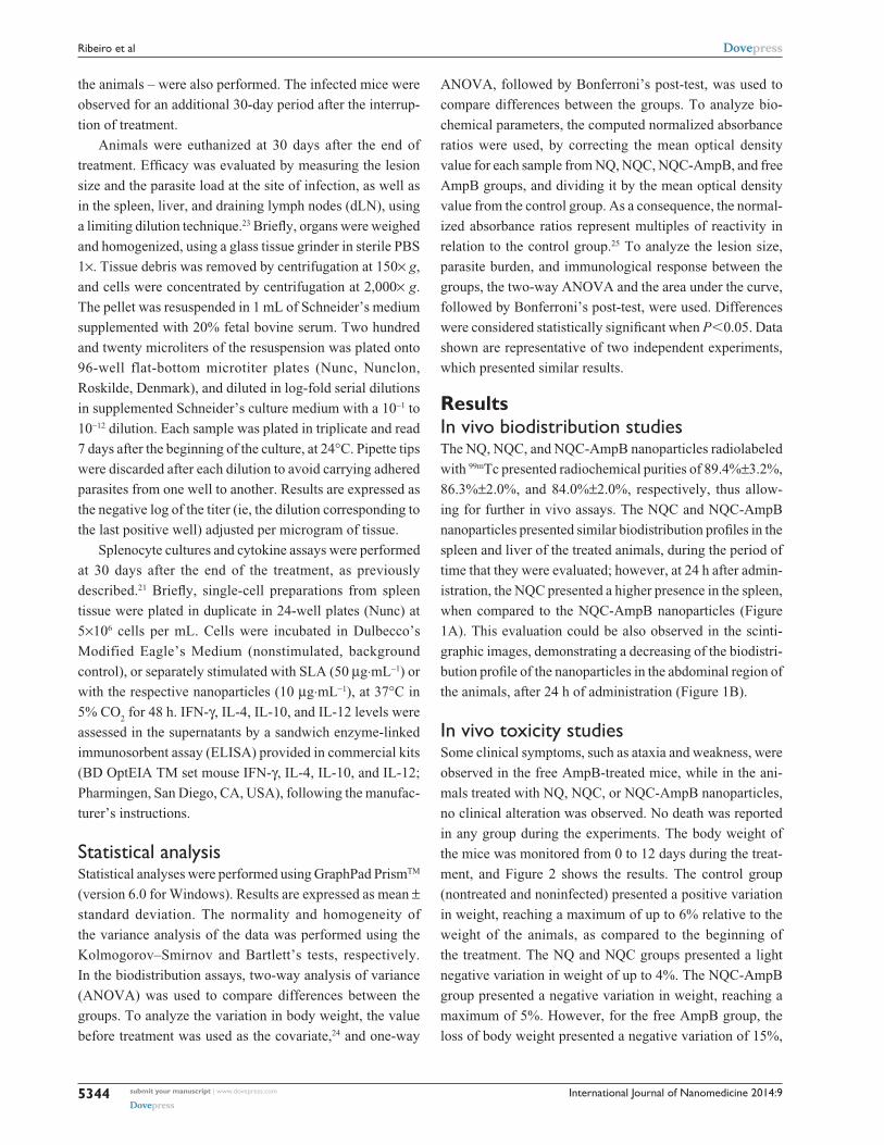
ResultsIn vivo biodistribution studiesThe NQ, NQC, and NQC-AmpB nanoparticles radiolabeled
with 99mTc presented radiochemical purities of 89.4%±3.2%,
86.3%±2.0%, and 84.0%±2.0%, respectively, thus allow-
ing for further in vivo assays. The NQC and NQC-AmpB
nanoparticles presented similar biodistribution profiles in the
spleen and liver of the treated animals, during the period of
time that they were evaluated; however, at 24 h after admin-
istration, the NQC presented a higher presence in the spleen,
when compared to the NQC-AmpB nanoparticles (Figure
1A). This evaluation could be also observed in the scinti-
graphic images, demonstrating a decreasing of the biodistri-
bution profile of the nanoparticles in the abdominal region of
the animals, after 24 h of administration (Figure 1B).
In vivo toxicity studiesSome clinical symptoms, such as ataxia and weakness, were
observed in the free AmpB-treated mice, while in the ani-
mals treated with NQ, NQC, or NQC-AmpB nanoparticles,
no clinical alteration was observed. No death was reported
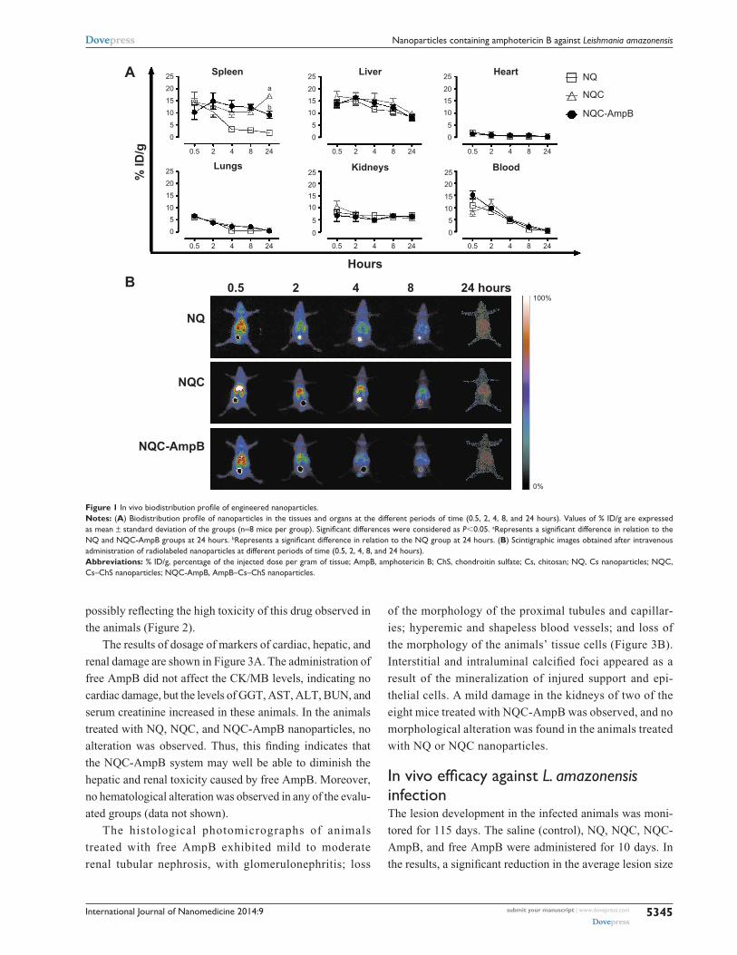
in any group during the experiments. The body weight of
the mice was monitored from 0 to 12 days during the treat-
ment, and Figure 2 shows the results. The control group
(nontreated and noninfected) presented a positive variation
in weight, reaching a maximum of up to 6% relative to the
weight of the animals, as compared to the beginning of
the treatment. The NQ and NQC groups presented a light
negative variation in weight of up to 4%. The NQC-AmpB
group presented a negative variation in weight, reaching a
maximum of 5%. However, for the free AmpB group, the
loss of body weight presented a negative variation of 15%,
International Journal of Nanomedicine 2014:9 submit your manuscript | www.dovepress.com
Dovepress
Dovepress
5345
Nanoparticles containing amphotericin B against Leishmania amazonensis
A
B
Spleen25
20
15
10
5
0
25
201510
5
0
0.5 2 4 8 24
25
20
15
105
0
0.5 2 4 8 24
25
201510
5
0
0.5 2 4 8 24
2520
15
105
0
0.5 2 4 8 24
0.5 2 4 8 24
25
201510
5
0
0.5 2 4 8 24
a
b
% ID
/g
Liver Heart NQ
NQC
NQC-AmpB
NQ
0.5 2 4 8 24 hours100%
0%
Hours
NQC
NQC-AmpB
Lungs Kidneys Blood
Figure 1 In vivo biodistribution profile of engineered nanoparticles. Notes: (A) Biodistribution profile of nanoparticles in the tissues and organs at the different periods of time (0.5, 2, 4, 8, and 24 hours). Values of % ID/g are expressed as mean ± standard deviation of the groups (n=8 mice per group). Significant differences were considered as P0.05. aRepresents a significant difference in relation to the NQ and NQc-ampB groups at 24 hours. bRepresents a significant difference in relation to the NQ group at 24 hours. (B) scintigraphic images obtained after intravenous administration of radiolabeled nanoparticles at different periods of time (0.5, 2, 4, 8, and 24 hours). Abbreviations: % ID/g, percentage of the injected dose per gram of tissue; AmpB, amphotericin B; ChS, chondroitin sulfate; Cs, chitosan; NQ, Cs nanoparticles; NQC, cs–chs nanoparticles; NQc-ampB, ampB–cs–chs nanoparticles.
possibly reflecting the high toxicity of this drug observed in
the animals (Figure 2).
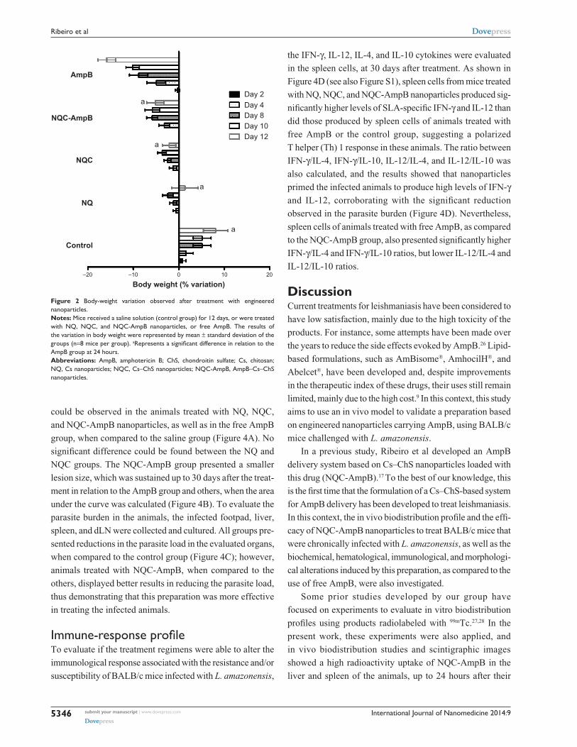
The results of dosage of markers of cardiac, hepatic, and
renal damage are shown in Figure 3A. The administration of
free AmpB did not affect the CK/MB levels, indicating no
cardiac damage, but the levels of GGT, AST, ALT, BUN, and
serum creatinine increased in these animals. In the animals
treated with NQ, NQC, and NQC-AmpB nanoparticles, no
alteration was observed. Thus, this finding indicates that
the NQC-AmpB system may well be able to diminish the
hepatic and renal toxicity caused by free AmpB. Moreover,
no hematological alteration was observed in any of the evalu-
ated groups (data not shown).
The histological photomicrographs of animals
treated with free AmpB exhibited mild to moderate
renal tubular nephrosis, with glomerulonephritis; loss
of the morphology of the proximal tubules and capillar-
ies; hyperemic and shapeless blood vessels; and loss of
the morphology of the animals’ tissue cells (Figure 3B).
Interstitial and intraluminal calcified foci appeared as a
result of the mineralization of injured support and epi-
thelial cells. A mild damage in the kidneys of two of the
eight mice treated with NQC-AmpB was observed, and no
morphological alteration was found in the animals treated
with NQ or NQC nanoparticles.
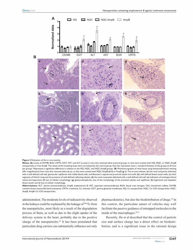
In vivo efficacy against L. amazonensis infectionThe lesion development in the infected animals was moni-
tored for 115 days. The saline (control), NQ, NQC, NQC-
AmpB, and free AmpB were administered for 10 days. In
the results, a significant reduction in the average lesion size
International Journal of Nanomedicine 2014:9submit your manuscript | www.dovepress.com
Dovepress
Dovepress
5346
ribeiro et al
could be observed in the animals treated with NQ, NQC,
and NQC-AmpB nanoparticles, as well as in the free AmpB
group, when compared to the saline group (Figure 4A). No
significant difference could be found between the NQ and
NQC groups. The NQC-AmpB group presented a smaller
lesion size, which was sustained up to 30 days after the treat-
ment in relation to the AmpB group and others, when the area
under the curve was calculated (Figure 4B). To evaluate the
parasite burden in the animals, the infected footpad, liver,
spleen, and dLN were collected and cultured. All groups pre-
sented reductions in the parasite load in the evaluated organs,
when compared to the control group (Figure 4C); however,
animals treated with NQC-AmpB, when compared to the
others, displayed better results in reducing the parasite load,
thus demonstrating that this preparation was more effective
in treating the infected animals.
Immune-response profileTo evaluate if the treatment regimens were able to alter the
immunological response associated with the resistance and/or
susceptibility of BALB/c mice infected with L. amazonensis,
the IFN-γ, IL-12, IL-4, and IL-10 cytokines were evaluated
in the spleen cells, at 30 days after treatment. As shown in
Figure 4D (see also Figure S1), spleen cells from mice treated
with NQ, NQC, and NQC-AmpB nanoparticles produced sig-
nificantly higher levels of SLA-specific IFN-γ and IL-12 than
did those produced by spleen cells of animals treated with
free AmpB or the control group, suggesting a polarized
T helper (Th) 1 response in these animals. The ratio between
IFN-γ/IL-4, IFN-γ/IL-10, IL-12/IL-4, and IL-12/IL-10 was
also calculated, and the results showed that nanoparticles
primed the infected animals to produce high levels of IFN-γ
and IL-12, corroborating with the significant reduction
observed in the parasite burden (Figure 4D). Nevertheless,
spleen cells of animals treated with free AmpB, as compared
to the NQC-AmpB group, also presented significantly higher
IFN-γ/IL-4 and IFN-γ/IL-10 ratios, but lower IL-12/IL-4 and
IL-12/IL-10 ratios.
DiscussionCurrent treatments for leishmaniasis have been considered to
have low satisfaction, mainly due to the high toxicity of the
products. For instance, some attempts have been made over
the years to reduce the side effects evoked by AmpB.26 Lipid-
based formulations, such as AmBisome®, AmhocilH®, and
Abelcet®, have been developed and, despite improvements
in the therapeutic index of these drugs, their uses still remain
limited, mainly due to the high cost.9 In this context, this study
aims to use an in vivo model to validate a preparation based
on engineered nanoparticles carrying AmpB, using BALB/c
mice challenged with L. amazonensis.
In a previous study, Ribeiro et al developed an AmpB
delivery system based on Cs–ChS nanoparticles loaded with
this drug (NQC-AmpB).17 To the best of our knowledge, this
is the first time that the formulation of a Cs–ChS-based system
for AmpB delivery has been developed to treat leishmaniasis.
In this context, the in vivo biodistribution profile and the effi-
cacy of NQC-AmpB nanoparticles to treat BALB/c mice that
were chronically infected with L. amazonensis, as well as the
biochemical, hematological, immunological, and morphologi-
cal alterations induced by this preparation, as compared to the
use of free AmpB, were also investigated.
Some prior studies developed by our group have
focused on experiments to evaluate in vitro biodistribution
profiles using products radiolabeled with 99mTc.27,28 In the
present work, these experiments were also applied, and
in vivo biodistribution studies and scintigraphic images
showed a high radioactivity uptake of NQC-AmpB in the
liver and spleen of the animals, up to 24 hours after their
Figure 2 Body-weight variation observed after treatment with engineered nanoparticles. Notes: Mice received a saline solution (control group) for 12 days, or were treated with NQ, NQc, and NQc-ampB nanoparticles, or free ampB. The results of the variation in body weight were represented by mean ± standard deviation of the groups (n=8 mice per group). aRepresents a significant difference in relation to the ampB group at 24 hours.Abbreviations: ampB, amphotericin B; chs, chondroitin sulfate; cs, chitosan; NQ, cs nanoparticles; NQc, cs–chs nanoparticles; NQc-ampB, ampB–cs–chs nanoparticles.
–20 –10 0
Body weight (% variation)
Control
NQ
NQC
NQC-AmpB
AmpB
Day 2Day 4Day 8Day 10Day 12
10 20
a
a
a
a
International Journal of Nanomedicine 2014:9 submit your manuscript | www.dovepress.com
Dovepress
Dovepress
5347
Nanoparticles containing amphotericin B against Leishmania amazonensis
Figure 3 evaluation of the in vivo toxicity. Notes: (A) levels of cK/MB, BUN, crTN, ggT, asT, and alT (x-axis) in mice that received saline (control group), or that were treated with NQ, NQc, or NQc-ampB nanoparticles or free ampB. The values of the treated groups were normalized by the control group. each bar represents mean ± standard deviation of the groups (n=8 mice per group). aRepresents a significant difference in relation to the NQ, NQC, and NQC-AmpB groups. (B) Photomicrographs of renal tissue using hematoxylin/eosin staining (40× magnification) from mice that received saline (a–c), or that were treated with NQc-ampB (d–f) or ampB (g–i). The arrows indicate: (a) the renal corpuscles delimited with a well-defined cell wall, glomerular capillaries with visible blood cells, and Bowman’s capsule and proximal tubule microvilli; (b) well-defined blood vessel walls; (c) thick segments of Henle’s loop and the presence of well-defined collecting tubules; (d) the renal corpuscles delimited with a well-defined cell wall; (e) indication of misshapen blood vessel and hyperemia; (f) loss of cellular morphology; (g) glomerulonephritis, loss of the morphology of the proximal tubules and capillaries; (h) hyperemia and shapeless blood vessel; and (i) loss of cellular morphology.Abbreviations: alT, alanine aminotransferase; ampB, amphotericin B; asT, aspartate aminotransferase; BUN, blood urea nitrogen; chs, chondroitin sulfate; cK/MB: creatine kinase-myocardial band isoenzyme; crTN, creatinine; cs, chitosan; ggT, gamma-glutamyl transferase; NQ, cs nanoparticles; NQc, cs–chs nanoparticles; NQc-ampB, ampB–cs–chs nanoparticles.
A
B
NQ
Nor
mal
ized
dat
aCK/MB
a d g
b e h
c f i
0
1
2
3a
a
a
a
a
4
5
GGT ALT AST BUN CRTN
NQC NQC-AmpB AmpB
administration. The moderate levels of radioactivity observed
in the kidneys could be explained by the leakage of 99mTc from
the nanoparticles, most likely as a result of the degradation
process of them, as well as due to the slight uptake of the
delivery system in the heart, probably due to the positive
charge of the nanoparticles.20 It has been postulated that
particulate drug carriers can substantially influence not only
pharmacokinetics, but also the biodistribution of drugs.29 In
this context, the particulate nature of vehicles may well
facilitate the passive guidance of entrapped molecules to the
inside of the macrophages.30,31
Recently, He et al described that the control of particle
size and surface charge has a direct effect on biodistri-
bution, and is a significant issue in the rational design

International Journal of Nanomedicine 2014:9submit your manuscript | www.dovepress.com
Dovepress
Dovepress
5348
ribeiro et al
A
C
B
D
12 Control NQ NQCNQC-AmpB AmpB
a
a,b
a,b a,b
a,b
a,b,c a,b,c
a,c a,c
a,b
aa
a
ca,bc
aa
a
a,b
a
a
a,b
11109876543210
1211109876543210
109876543210
00
10
20
30
40
50
Control
Footpad dLN
Control NQC NQ NQC-AmpB AmpB
Spleen Liver IFN-γ:IL-4 IFN-γ:IL-10 IL-12:IL-4 IL-12:IL-10
NQ NQC NQC-AmpB
AmpB87 94 101 105 108 112 115
Foot
pad
swel
ling
(mm
)Pa
rasi
te ti
ter (
Log 10
)
Rat
io o
f cyt
okin
epr
oduc
tion
AU
C
Days after infection
Figure 4 In vivo biological activity of engineered nanoparticles. Notes: Mice were subcutaneously infected with 5×10⁶ stationary-phase promastigotes of Leishmania amazonensis, and the course of infection was monitored for 115 days. When the animals developed ulcerated lesions presenting an average diameter of 2–4 mm, they were divided into groups according to lesion size. after, they were treated for 10 days (from day 97 to 107, with the therapeutic window represented as a gray square in A). (A) lesion size expressed as mean ± sD of the lesion size (n=8 mice per group). (B) The aUc of the footpad swelling of the different evaluated groups (represented on the x-axis) is represented. (C) The parasite burden in the infected footpads, spleen, liver, and dlN (represented on the x-axis) of the animals in the different evaluated groups. each bar represents the mean ± sD of the groups. (D) spleen-cell cultures were stimulated with sla (50 µg⋅ml−1) for 48 hours at 37°C, 5% CO2. The IFN-γ, Il-12, Il-4, and Il-10 levels were measured in the culture supernatants by enzyme-linked immunosorbent assay. The ratios of IFN-γ/Il-4, IFN-γ/Il-10, Il-12/Il-4, and Il-12/Il-10 are shown (represented on the x-axis). each bar represents the mean ± sD of the groups. aRepresents a significant difference in relation to the control group; brepresents a significant difference in relation to the NQ and NQC groups; crepresents a significant difference in relation to the free AmpB group.Abbreviations: ampB, amphotericin B; aUc, area under the curve; chs, chondroitin sulfate; cs, chitosan; dlN, draining lymph nodes; NQ, cs nanoparticles; NQc, cs–chs nanoparticles; NQc-ampB, ampB–cs–chs nanoparticles; sD, standard deviation; sla, soluble Leishmania antigen extract.
of drug nanocarriers.32 In the present study, all of the
obtained nanoparticles presented a positive charge and
sizes of approximately 79–136 nm.17 Furthermore, posi-
tively charged nanoparticles accumulate in mononuclear
phagocytes at about two-fold more than negatively charged
carriers.33,34 Danesh-Bahreini et al described that the posi-
tively charged nanoparticles, as compared to negative or
neutral charged nanoparticles, are more quickly taken up
by macrophages through phagocytosis.35
The mechanism of action proposed for the toxic effect of
AmpB is derived from its interaction with sterols in bilayer
membranes, such as cell walls, causing pore formation in
the membrane, in turn leading either to its destruction or to
the inhibition of membrane repair. In this context, AmpB
could also form pores in cholesterol-containing membranes,
explaining the high toxicity observed in the host cells.35
In a controlled drug-delivery system, an active product is
incorporated into a polymeric network structure in such a
way that the drug is slowly released and in a predefined
manner.36,37 Depending on the drug delivery and the appli-
cation route, the release time may be a few hours to several
years.38 In this context, it can be suggested that the reduced
toxicity of AmpB in the NQC-AmpB preparation observed
in this study could be attributed to the slower release of the
drug, when incorporated in the nanoparticles system, inside
the macrophages, in turn favoring its leishmanicidal activity
and the low toxicity.
The biodistribution profile observed in the present
study demonstrates higher accumulation of the NQC-
AmpB nanoparticles in the spleen and liver of the animals,
which corroborates with findings evaluating other nano-
structured systems, such as polymeric micelles39 and gold

International Journal of Nanomedicine 2014:9 submit your manuscript | www.dovepress.com
Dovepress
Dovepress
5349
Nanoparticles containing amphotericin B against Leishmania amazonensis
nanoparticles.40 Akiyama et al reported that ChS is able to
induce the Th1-type cytokine (such as IFN-γ, IL-2, and
IL-12) secretion and suppress the Th2-type cytokine (such
as IL-5 and IL-10) secretion in ovalbumin-sensitized sple-
nocytes of mice. The authors showed that O-sulfo groups
in the ChS molecule are important for the Th1-promoted
activity of murine splenocytes, in terms of the cytokine
production and Th1/Th2 balance.41 ChS has been found
in many tissues42 and cells,43–45 and has been reported to
interact with various biologically important molecules
and regulate their functions. It has been reported that the
Th1-promoted and Th2-inhibitory activity of ChS could
be associated with its binding to adhesion molecules, such
as L- and P-selectins, CD44, and chemokines. In the same
study, authors reported that the immunostimulatory activity
of ChS could be associated with its binding to L-selectins
expressed on T-cells’ surface.41 As the spleen is considered a
systemic organ of transit and homing of T-cells in mammal
hosts, one could speculate that the higher presence of the
NQC and NQC-AmpB nanoparticles in this organ, in detri-
ment to observed levels of NQ, could be due to the presence
of a moderate to high number of T-cells in this organ, which
could have their L-selectins adhered to ChS present in the
nanoparticles. This fact could explain, at least in part, the
higher presence of NQC and NQC-AmpB in the spleen of
the treated animals.
In the present study, the liver could be also considered a
site of accumulation of the nanoparticles in the mice; in this
context, a toxicological study was performed on this organ.
Clinically significant increases in hepatocyte-associated
serum enzymes were observed in the animals treated with free
AmpB, with a significant difference observed in the levels
of GGT, AST, and ALT between the groups that received
NQC-AmpB and free AmpB, which may well indicate a
decrease in the hepatic toxicity when AmpB is administered
in the NQC system, although used at the same dose.
The nephrotoxicity is considered perhaps the most
described adverse drug event for AmpB.46 The pathophysi-
ology and pharmacology of this activity have been well-
documented, and the proposed mechanism has been
described as being multifactorial.47,48 The BUN and serum
creatinine showed significant increases in the animals treated
with AmpB. The microscopic observation of kidneys of
animals treated with AmpB is in accordance with previous
studies.47–49 By contrast, in the animals treated with NQ,
NQC, and NQC-AmpB nanoparticles, no significant altera-
tion could be observed. It can therefore be concluded that the
decrease in toxicity of AmpB in the NQC-AmpB preparation,
in relation to free AmpB, may well have occurred because
AmpB encapsulated in the nanoparticles does not interact
well with the epithelial cell membranes within the kidney
tubule, which would minimize the nephrotoxicity. More
recently, other groups have used the same strategy to obtain
a less toxic product than free AmpB.50–54
In the present work, the clinical symptoms, as well as
hematological and biochemical alterations, were evaluated
in the animals after the intravenous administration of saline,
NQ, NQC, or NQC-AmpB nanoparticles, or free AmpB. As
found in prior reports,55,56 clinical symptoms such as ataxia,
weakness, and loss of body weight were associated with the
treatment using free AmpB. These animals presented a sig-
nificant loss of body weight when compared to the animals
that received NQC-AmpB, indicating that the controlled
release of this drug could well be responsible for the lower
incidence of toxic side effects.17 Mice that received NQ and
NQC treatment showed a loss of body weight, which could
be associated with the recent role of Cs, which is involved
in the modulation of adipokines.57
L. amazonensis is a member of the L. mexicana complex
and is the etiological agent for a broad spectrum of leishma-
niasis in South American countries.58 Among the causative
species of cutaneous leishmaniasis in Brazil, recent data
indicate that about 8% are attributed to L. amazonensis.59 The
present study evaluated the efficacy of NQ, NQC, and NQC-
AmpB nanoparticles in treating chronically L. amazonensis-
infected BALB/c mice, as compared to treatment with free
AmpB. Both the NQC-AmpB and AmpB treatments were
able to promote a significant reduction in both the lesion
size and parasite load of the infected animals. The lesion
size of the NQ and NQC groups was lower than the control
group, but higher than that found in the NQC-AmpB and free
AmpB groups, although the parasitism levels in the treated
mice with NQ and NQC were similar to that obtained in the
AmpB group, showing the antileishmanial activity of Cs and
its synergic effect with ChS.
The immune response of the treated and infected animals
was also evaluated. Mice treated with nanoparticles presented
higher levels of IFN-γ and IL-12, and lower levels of IL-4 and
IL-10. By contrast, control animals displayed high levels of
IL-4 and IL-10 in their splenic cultures. These results are in
concordance with Asthana et al, who showed that low lev-
els of IL-4 and IL-10, associated with a high production of
IFN-γ and IL-12 by splenic cultures of infected hamsters, are
related to the efficacy of the treatment using a template-based
nanoemulsion loaded with AmpB.33 Altogether, these results
indicate that treatment with NQC-AmpB nanoparticles was

International Journal of Nanomedicine 2014:9submit your manuscript | www.dovepress.com
Dovepress
Dovepress
5350
ribeiro et al
able to induce a Leishmania-specific Th1 immune response
in the treated mice.
Traditionally, the systemic treatment of leishmaniasis
has been with pentavalent antimony compounds (usually
performed using 20 mg per kg per day, given by intravenous
or intramuscular routes), over a 20-day period. This regimen
is able to cure about 85%–90% of the patients; however, the
treatment is associated with side toxic effects, such as elec-
trocardiographic changes, ventricular tachyarrhythmias, and
laboratory abnormalities, like elevated liver and pancreatic
enzymes, and bone marrow suppression.60,61 The development
of lipid formulations for AmpB has greatly reduced the toxic-
ity of this drug in the treatment of disease. In this context,
lipid-based AmpB products can be considered as alternatives
to treatments of leishmaniasis.17 In general, this formula-
tion is less nephrotoxic than free AmpB, since it is taken up
selectively by macrophages. Some adverse effects including
mild urticarial rash and renal impairment are resolved after
therapy.63 The liposomal formulation AmBisome®, the AmpB
colloidal dispersion (Amphocil™), and the AmpB lipid com-
plex (Abelcet®) have been used in the treatment.62–64 However,
the main restriction against the widespread use of the current
approved lipid-AmpB formulations is their high cost. So, the
search remains to obtain a low-cost formulation, which should
have also an effective activity against Leishmania.64
The NQC-AmpB nanoparticles described in this study
present characteristics that could be attractive to the treat-
ment of leishmaniasis. Firstly, the dosage employed of the
drug in this system is considered low (1 mg per kg per day),
which could be impacting in the null side effects observed
in the treated mice. Although the mouse model cannot be
extrapolated to the model, it is a good perspective that NQC-
AmpB could also be less toxic in the patients. Secondly,
although NQC-AmpB does not at present have an estimated
value of sale, due to the fact that this product is composed of
two known commercial products – ChS and Cs – one could
speculate that the NQC-AmpB nanoparticles may well have
a lesser cost in comparison to the other available commercial
lipid-AmpB formulations.17
Therapeutic and vaccine studies concerning leishmania-
sis call for the critical evaluation of the parasitological and
immunological parameters of the most common animal models
today. It is extremely important to optimize the conditions of
the artificial infection to the point where it can be firmly argued
that the conditions best represent those of the natural infec-
tion caused by the sand fly.65 The present study showed that
BALB/c mice, when infected with a highly infective inoculum,
developed an exponential parasite burden in organs such as
dLN, footpad, spleen, and liver, which suggests a chronic
infection in the animals, and makes it very difficult to control
the replication of parasites.66 In this context, one could specu-
late that even using a very effective treatment regimen, the
infected animals would not be able to clear all parasites in the
different organs. In this light, the NQC-AmpB nanoparticles
could be considered as effective therapeutic agents; once the
results obtained in the animals treated with this product were
compared with the data shown in the free-AmpB-treated group,
although not all parasites had been eliminated. However, addi-
tional studies are certainly necessary to improve the therapeutic
index of NQC-AmpB, either by increasing the number of doses
of the product, or the duration of the treatment; in order to
clear the largest possible number of parasites in the infected
animals. On the other hand, the evaluation of the treatment
in other mammal models could also be of interest, in order to
estimate the efficacy of the nanoparticles, in mammals that are
not as susceptible as BALB/c mice.
ConclusionIn conclusion, based on the results involving the in vivo
biodistribution, and with the evaluation of the biochemical,
toxicological, parasitological, and immunological para-
meters associated with the treatment of mice infected with
L. amazonensis, the NQC-AmpB nanoparticles could be
applied as an alternative AmpB delivery system, maintaining
the high activity of this drug against Leishmania, but reduc-
ing by significant levels its toxicity, compared to when it is
administered in a free form. Therefore, this new formulation
presents a high potential for use in future clinical studies
aimed at treating leishmaniasis.
AcknowledgmentsThis work was supported by grants from Pró-Reitoria de
Pesquisa from UFMG (Edital 01/2014), Instituto Nacional
de Ciência e Tecnologia em Nano-biofarmacêutica (INCT-
Nanobiofar), FAPEMIG (CBB-APQ-00496-11 and CBB-
APQ-00819-12), and CNPq (APQ-472090/2011-9 and
APQ-482976/2012-8). MACF is a grant recipient of
FAPEMIG/CAPES. EAFC, VNC, and AAGF are grant
recipients of CNPq. Eduardo AF Coelho and André AG
Faraco are co-senior authors of this study.
DisclosureThe authors report no conflicts of interest in this work.

International Journal of Nanomedicine 2014:9 submit your manuscript | www.dovepress.com
Dovepress
Dovepress
5351
Nanoparticles containing amphotericin B against Leishmania amazonensis
References 1. Desjeux P. Leishmaniasis: current situation and new perspectives. Comp
Immunol Microbiol Infect Dis. 2004;27(5):305–318. 2. World Health Organization (WHO). Control of the leishmaniases:
report of a meeting of the 399 WHO Expert Committee on the Control of Leishmaniases. 2010. Available from: http://whqlibdoc.who.int/trs/WHO_TRS_949_eng.pdf. Accessed October 9, 2014.
3. Alvar J, Vélez ID, Bern C, et al. Leishmaniasis worldwide and global estimates of its incidence. PloS One. 2012;7(5):e35671.
4. Grevelink SA, Lerner EA. Leishmaniasis. J Am Acad Dermatol. 1996; 34(2 Pt 1):257–272.
5. Croft SL, Coombs GH. Leishmaniasis – current chemotherapy and recent advances in the search for novel drugs. Trends Parasitol. 2003;19(11):502–508.
6. Annaloro C, Olivares C, Usardi P, et al. Retrospective evaluation of amphotericin B deoxycholate toxicity in a single centre series of haematopoietic stem cell transplantation recipients. J Antimicrob Chemother. 2009;63(3):625–626.
7. Denning DW. Therapeutic outcome of invasive aspergillosis. Clin Infect Dis. 1996;23(3):608–615.
8. Bern C, Adler-Moore J, Berenguer J, et al. Liposomal amphotericin B for the treatment of visceral leishmaniasis. Clin Infect Dis. 2006;43(7): 917–924.
9. Egger SS, Meier S, Leu C, et al. Drug interactions and adverse events associated with antimycotic drugs used for invasive aspergil-losis in hematopoietic SCT. Bone Marrow Transplant. 2010;45(7): 1197–1203.
10. Italia JL, Sharp A, Carter KC, Warn P, Kumar MN. Peroral amphot-ericin B polymer nanoparticles lead to comparable or superior in vivo antifungal activity to that of intravenous Ambisome® or Fungizone™. PLoS One. 2011;6(10):25744.
11. Gershkovich P, Wasan EK, Lin M, et al. Pharmacokinetics and biodis-tribution of amphotericin B in rats following oral administration in a novel lipid-based formulation. J Antimicrob Chemother. 2009;64(1): 101–108.
12. Bekersky I, Fielding RM, Dressler DE, Lee JW, Buell DN, Walsh TJ. Pharmacokinetics, excretion, and mass balance of liposomal ampho-tericin B (AmBisome) and amphotericin B deoxycholate in humans. Antimicrob Agents Chemother. 2002;46(3):828–833.
13. Fielding RM, Smith PC, Wang LH, Porter J, Guo LS. Comparative pharmacokinetics of amphotericin B after administration of a novel colloidal delivery system, ABCD, and a conventional formulation to rats. Antimicrob Agents Chemother. 1991;35(6):1208–1213.
14. Townsed RW, Zutshi A, Bekersky I. Biodistribution of 4-[(14)C]cholesterol-Ambisome following a single intravenous administration to rats. Drug Metab Dispos. 2001;29(5):681–685.
15. Saxena S, Ghosh PC. Biodistribution of amphotericin B when delivered through cholesterol hemisuccinate vesicles in normal and A. fumigatus infected mice. Phar Res. 2000;17(10):1236–1242.
16. Agrawal AK, Agrawal A, Pal A, Guru PY, Gupta CM. Superior che-motherapeutic efficacy of amphotericin B in tuftsin-bearing liposomes against Leishmania donovani infection in hamsters. J Drug Target. 2002;10:41–45.
17. Ribeiro TG, Chávez-Fumagalli MA, Valadares DG, et al. Novel target-ing using nanoparticles: an approach to the development of an effective anti-leishmanial drug-delivery system. Int J Nanomedicine. 2014;14: 877–890.
18. Soares DC, Ferreira TH, Ferreira Cde A, Cardoso VN, de Sousa EM. Boron nitride nanotubes radiolabeled with 99mTc: preparation, physi-cochemical characterization, biodistribution study, and scintigraphic imaging in Swiss mice. Int J Pharm. 2012;423(2):489–495.
19. Kean T, Thanou M. Biodegradation, biodistribution and toxicity of chitosan. Adv Drug Deliv Rev. 2010;62(1):3–11.
20. Banerjee T, Singh AK, Sharma RK, Maitra AN. Labeling efficiency and biodistribution of Technetium-99m labeled nanoparticles: interference by colloidal tin oxide particles. Int J Pharm. 2005;289(1–2):189–195.
21. Coelho EA, Tavares CA, Carvalho FA, et al. Immune responses induced by the Leishmania (Leishmania) donovani A2 antigen, but not by the LACK antigen, are protective against experimental Leishmania (Leish-mania) amazonensis infection. Infect Immun. 2003;71(7):3988–3994.
22. Nahrevanian H, Farahmand M, Aghighi Z, Assmar M, Amirkhani A. Pharmacological evaluation of anti-leishmanial activity by in vivo nitric oxide modulation in Balb/c mice infected with Leishmania major MRHO/IR/75/ER: An Iranian strain of cutaneous leishmaniasis. Exp Parasitol. 2007;116(3):233–240.
23. Buffet PA, Sulahian A, Garin YJ, Nassar N, Derouin F. Culture microtitration: a sensitive method for quantifying Leishmania infantum in tissues of infected mice. Antimicrob Agents Chemother. 1995;39(9):2167–2168.
24. Leite EA, Giuberti Cdos S, Wainstein AJ, et al. Acute toxicity of long-circulating and pH-sensitive liposomes containing cisplatin in mice after intraperitoneal administration. Life Sci. 2009;84(19–20):641–649.
25. Ramanakumar AV, Thomann P, Candeias JM, Ferreira S, Villa LL, Franco EL. Use of the normalized absorbance ratio as an internal standardization approach to minimize measurement error in enzyme-linked immunosorbent assays for diagnosis of human papillomavirus infection. J Clin Microbiol. 2010;48(3):791–796.
26. Tasdemir D, Kaiser M, Brun R, et al. Antitrypanosomal and leishmanicidal activities of flavonoids and their analogues: in vitro, in vivo, structure-activity relationship, and quantitative structure-activity relationship studies. Antimicrob Agents Chemother. 2006;50(4):1352–1364.
27. de Barros AL, Mota Ld, Soares DC, et al. Long-circulating, pH-sensitive liposomes versus long-circulating, non-pH-sensitive liposomes as a deliv-ery system for tumor identification. J Biomed Nanotechnol. 2013;9(9): 1636–1643.
28. Araújo JG, Mota Ld, Leite EA, et al. Biodistribution and antitumoral effect of long-circulating and pH-sensitive liposomal cisplatin admin-istered in Ehrlich tumor-bearing mice. Exp Biol Med (Maywood). 2011;236(7):808–815.
29. Martinez-Pomares L, Hanitsch LG, Stillion R, Keshav S, Gordon S. Expression of mannose receptor and ligands for its cysteine-rich domain in venous sinuses of human spleen. Lab Invest. 2005;85: 1238–1249.
30. Matsumura Y, Maeda H. A new concept for macromolecular therapeutics in cancer chemotherapy: mechanism of tumoritropic accumulation of proteins and the antitumor agent smancs. Cancer Res. 1986;46(12 Pt 1): 6387–6392.
31. Owais M, Gupta CM. Targeted drug delivery to macrophages in parasitic infections. Curr Drug Deliv. 2005;2(4):311–318.
32. He C, Hu Y, Yin L, Tang C, Yin C. Effects of particle size and surface charge on cellular uptake and biodistribution of polymeric nanoparticles. Biomaterials. 2010;31(13):3657–3666.
33. Asthana S, Jaiswal AK, Gupta PK, Pawar VK, Dube A, Chourasia MK. Immunoadjuvant chemotherapy of visceral leishmaniasis in hamsters using amphotericin B-encapsulated nanoemulsion template-based chitosan nanocapsules. Antimicrob Agents Chemother. 2013;57(4): 1714–1722.
34. Batrakova EV, Gendelman HE, Kabanov AV. Cell-mediated drugs delivery. Expert Opin Drug Deliv. 2011;8(4):415–433.
35. Danesh-Bahreini MA, Shokri J, Samiei A, Kamali-Sarvestani E, Barzegar-Jalali M, Mohammadi-Samani S. Nanovaccine for leishma-niasis: preparation of chitosan nanoparticles containing Leishmania superoxide dismutase and evaluation of its immunogenicity in BALB/c mice. Int J Nanomedicine. 2011;6:835–842.
36. Darole PS, Hegde DD, Nair HA. Formulation and evaluation of micro-emulsion based delivery system for amphotericin B. AAPS Pharm Sci Tech. 2008;9(1):122–128.
37. Colombo P, Santi P, Bettini R, Brazel CS, Peppas CA. Drug Release from Swelling-Controlled Systems. In: Wise DL, Brannon-Peppas L, Klibanov AM, et al, editors. Handbook of Pharmaceutical Con-trolled Release Technology. New York, NY: Marcel Dekker, Inc.; 2000:183–210.

International Journal of Nanomedicine 2014:9submit your manuscript | www.dovepress.com
Dovepress
Dovepress
5352
ribeiro et al
38. Jogani V, Jinturkar K, Vyas T, Misra A. Recent patents review on intranasal administration for CNS drug delivery. Recent Pat Drug Deliv Formul. 2008;2(1):25–40.
39. Koide H, Asai T, Hatanaka K, et al. Particle size-dependent trig-gering of accelerated blood clearance phenomenon. Int J Pharm. 2008;362(1–2):197–200.
40. De Jong WH, Hagens WI, Krystek P, Burger MC, Sips AJ, Geertsma RE. Particle size-dependent organ distribution of gold nanoparticles after intravenous administration. Biomaterials. 2008;29(12):1912–1919.
41. Akiyama H, Sakai S, Linhardt RJ, Goda Y, Toida T, Maitani T. Chondroitin sulphate structure affects its immunological activi-ties on murine splenocytes sensitized with ovalbumin. Biochem J. 2004;382:269–278.
42. Suzuki S, Satio H, Yamagata T, et al. Formation of three types of dis-ulphated disaccharides from chondroitin sulphates by chondroitinase digestion. J Biol Chem. 1968;243:1543–1550.
43. Ohhashi Y, Hasumi F, Mori Y. Comparative study on glycosamino-glycans synthesized in peripheral and peritoneal polymorphonuclear leucocytes from guinea pigs. Biochem J. 1984;217:199–207.
44. Stevens RL, Fox CC, Lichtenstein LM, Austen KF. Identification of chondroitin sulphate E proteoglycans and heparin proteoglycans in the secretory granules of human lung mast cells. Proc Natl Acad Sci U S A. 1988;85:2284–2287.
45. Petersen RL, Brandt E, Lindahl U, Spillmann D. Characterization of a neutrophil cell surface glycosaminoglycan that mediates binding of platelet factor 4. J Biol Chem. 1999;274:12376–12382.
46. Deray G. Amphotericin B nephrotoxicity. J Antimicrob Chemother. 2002;49 Suppl 1:37–41.
47. Yano T, Itoh Y, Kawamura E, et al. Amphotericin B-induced renal tubular cell injury is mediated by Na influx through ion-permeable pores and subsequent activation of mitogen-activated protein kinases and elevation of intracellular Ca2+ concentration. Antimicrob Agents Chemother. 2009;53(4):1420–1426.
48. Patel GP, Crank CW, Leikin JB. An evaluation of hepatotoxicity and nephrotoxicity of liposomal amphotericin B (L-AMB). J Med Toxicol. 2011;7(1):12–15.
49. Fielding RM, Singer AW, Wang LH, Babbar S, Guo LS. Relationship of pharmacokinetics and drug distribution in tissue to increased safety of amphotericin B colloidal dispersion in dogs. Antimicrob Agents Chemother. 1992;36(2):299–307.
50. Wertlake PT, Butler WT, Hill GJ 2nd, Utz JP. Nephrotoxic tubular damage and calcium deposition following amphotericin B therapy. Am J Pathol. 1963;43(3):449–457.
51. Mishra J, Dey A, Singh N, Somvanshi R, Singh S. Evaluation of toxicity and therapeutic efficacy of a new liposomal formulation of amphoteri-cin B in a mouse model. Indian J Med Res. 2013;137(4):767–776.
52. Prajapati VK, Awasthi K, Gautam S, et al. Targeted killing of Leish-mania donovani in vivo and in vitro with amphotericin B attached to functionalized carbon nanotubes. J Antimicrob Chemother. 2011; 66(4):874–879.
53. Nahar M, Jain NK. Preparation, characterization and evaluation of targeting potential of amphotericin B-loaded engineered PLGA nano-particles. Pharm Res. 2009;26(12):2588–2598.
54. Tiyaboonchai W, Limpeanchob N. Formulation and characterization of amphotericin B-chitosan-dextran sulfate nanoparticles. Int J Pharm. 2007;329(1–2):142–149.
55. Szoka FC Jr, Milholland D, Barza M. Effect of lipid composition and liposome size on toxicity and in vitro fungicidal activity of liposome-intercalated amphotericin B. Antimicrob Agents Chemother. 1987;31(3):421–429.
56. Boswell GW, Bekersky I, Buell D, Hiles R, Walsh TJ. Toxicologi-cal profile and pharmacokinetics of a unilamellar liposomal vesicle formulation of amphotericin B in rats. Antimicrob Agents Chemother. 1998;42(2):263–268.
57. Walsh AN, Sweeney T, Bahar B, O’Doherty JV. Multi-functional roles of chitosan as a potential protective agent against obesity. PLoS One. 2013;8(1):e53828.
58. Camara Coelho LI, Paes M, Guerra JA, et al. Characterization of Leish-mania spp. causing cutaneous leishmaniasis in Manaus, Amazonas, Brazil. Parasitol Res. 2011;108(3):671–677.
59. de Carvalho RF, Ribeiro IF, Miranda-Vilela AL, et al. Leishmanicidal activity of amphotericin B encapsulated in PLGA-DMSA nanoparticles to treat cutaneous leishmaniasis in C57BL/6 mice. Exp Parasitol. 2013; 135(2):217–222.
60. Herwaldt BL, Berman JD. Recommendations for treating leishmaniasis with sodium stibogluconate (Pentostam) and review of pertinent clinical studies. Am J Trop Med Hyg. 1992;46:296–306.
61. Solomon M, Pavlotsky F, Leshem E, Ephros M, Trau H, Schwartz E. Liposomal amphotericin B treatment of cutaneous leishma-niasis due to Leishmania tropica. J Eur Acad Dermatol Venereol. 2011;25:973–977.
62. Mohamed-Ahmed AH, Brocchini S, Croft SL. Recent advances in development of amphotericin B formulations for the treatment of visceral leishmaniasis. Curr Opin Infect Dis. 2012;25:695–702.
63. Dietze R1, Milan EP, Berman JD, et al. Treatment of Brazilian kala-azar with a short course of amphocil (amphotericin B cholesterol dispersion). Clin Infect Dis. 1993;17:981–986.
64. Sundar S, Murray HW. Cure of antimony-unresponsive Indian vis-ceral leishmaniasis with amphotericin B lipid complex. J Infect Dis. 1996;173:762–765.
65. Carrión J, Nieto A, Iborra S, et al. Immunohistological features of visceral leishmaniasis in BALB/c mice. Parasite Immunol. 2006; 28:173–183.
66. Oliveira DM, Costa MA, Chavez-Fumagalli MA, et al. Evaluation of parasitological and immunological parameters of Leishmania chagasi infection in BALB/c mice using different doses and routes of inocula-tion of parasites. Parasitol Res. 2012;110:1277–1285.

International Journal of Nanomedicine
Publish your work in this journal
Submit your manuscript here: http://www.dovepress.com/international-journal-of-nanomedicine-journal
The International Journal of Nanomedicine is an international, peer-reviewed journal focusing on the application of nanotechnology in diagnostics, therapeutics, and drug delivery systems throughout the biomedical field. This journal is indexed on PubMed Central, MedLine, CAS, SciSearch®, Current Contents®/Clinical Medicine,
Journal Citation Reports/Science Edition, EMBase, Scopus and the Elsevier Bibliographic databases. The manuscript management system is completely online and includes a very quick and fair peer-review system, which is all easy to use. Visit http://www.dovepress.com/testimonials.php to read real quotes from published authors.
International Journal of Nanomedicine 2014:9 submit your manuscript | www.dovepress.com
Dovepress
Dovepress
Dovepress
5353
Nanoparticles containing amphotericin B against Leishmania amazonensis
Cyt
okin
es (p
g/m
L)
1,500MediumSLANanoparticle
* *
1,000
500IF
N-γ
IL-1
2IL
-4IL
-10
IFN
-γIL
-12
IL-4
IL-1
0
IFN
-γIL
-12
IL-4
IL-1
0
IFN
-γIL
-12
IL-4
IL-1
0
IFN
-γIL
-12
IL-4
IL-1
0
0
Control NQ NQC NQC-AmpB AmpB
Figure S1 analysis of the cellular response. Notes: single-cell suspensions were obtained from the spleen of mice, 30 days after the end of the treatment, and cells were nonstimulated (medium; background control), or separately stimulated with sla (50 µg⋅ml−1), or separately stimulated with the respective nanoparticle (10 µg⋅ml−1), for 48 hours at 37°C, 5% CO2. IFN-γ, Il-12, Il-4, and Il-10 levels were measured in culture supernatants by capture enzyme-linked immunosorbent assay. Mean ± standard deviation of the cytokine levels was determined (n=8 mice per group). *Represents a significant increase (P0.05) in relation to the NQ, NQc, and NQc-ampB groups.Abbreviations: ampB, amphotericin B; chs, chondroitin sulfate; cs, chitosan; NQ, cs nanoparticles; NQc, cs–chs nanoparticles; NQc-ampB, ampB–cs–chs nanoparticles; sla, soluble Leishmania antigen extract.
Supplementary material
Copyright © 2022 FDOKUMEN