Multicenter Evaluation of a New Disk Agar Diffusion Method for Susceptibility Testing of Filamentous...
11
Published Ahead of Print 11 April 2007. 10.1128/JCM.00134-07. 2007, 45(6):1811. DOI: J. Clin. Microbiol. and A. Wang Gibbs M. A. Pfaller, S. Messer, M. Rinaldi, A. Fothergill, D. L. A. Espinel-Ingroff, B. Arthington-Skaggs, N. Iqbal, D. Ellis, B, and Caspofungin Posaconazole, Itraconazole, Amphotericin of Filamentous Fungi with Voriconazole, Diffusion Method for Susceptibility Testing Multicenter Evaluation of a New Disk Agar http://jcm.asm.org/content/45/6/1811 Updated information and services can be found at: These include: REFERENCES http://jcm.asm.org/content/45/6/1811#ref-list-1 at: This article cites 16 articles, 14 of which can be accessed free CONTENT ALERTS more» articles cite this article), Receive: RSS Feeds, eTOCs, free email alerts (when new http://journals.asm.org/site/misc/reprints.xhtml Information about commercial reprint orders: http://journals.asm.org/site/subscriptions/ To subscribe to to another ASM Journal go to: on September 30, 2014 by guest http://jcm.asm.org/ Downloaded from on September 30, 2014 by guest http://jcm.asm.org/ Downloaded from
-
Upload
independent -
Category
Documents
-
view
6 -
download
0
Transcript of Multicenter Evaluation of a New Disk Agar Diffusion Method for Susceptibility Testing of Filamentous...
Published Ahead of Print 11 April 2007. 10.1128/JCM.00134-07.
2007, 45(6):1811. DOI:J. Clin. Microbiol. and A. Wang
GibbsM. A. Pfaller, S. Messer, M. Rinaldi, A. Fothergill, D. L. A. Espinel-Ingroff, B. Arthington-Skaggs, N. Iqbal, D. Ellis, B, and CaspofunginPosaconazole, Itraconazole, Amphotericin of Filamentous Fungi with Voriconazole,Diffusion Method for Susceptibility Testing Multicenter Evaluation of a New Disk Agar
http://jcm.asm.org/content/45/6/1811Updated information and services can be found at:
These include:
REFERENCEShttp://jcm.asm.org/content/45/6/1811#ref-list-1at:
This article cites 16 articles, 14 of which can be accessed free
CONTENT ALERTS more»articles cite this article),
Receive: RSS Feeds, eTOCs, free email alerts (when new
http://journals.asm.org/site/misc/reprints.xhtmlInformation about commercial reprint orders: http://journals.asm.org/site/subscriptions/To subscribe to to another ASM Journal go to:
on Septem
ber 30, 2014 by guesthttp://jcm
.asm.org/
Dow
nloaded from
on Septem
ber 30, 2014 by guesthttp://jcm
.asm.org/
Dow
nloaded from
JOURNAL OF CLINICAL MICROBIOLOGY, June 2007, p. 1811–1820 Vol. 45, No. 60095-1137/07/$08.00�0 doi:10.1128/JCM.00134-07Copyright © 2007, American Society for Microbiology. All Rights Reserved.
Multicenter Evaluation of a New Disk Agar Diffusion Method forSusceptibility Testing of Filamentous Fungi with Voriconazole,Posaconazole, Itraconazole, Amphotericin B, and Caspofungin�
A. Espinel-Ingroff,1* B. Arthington-Skaggs,2 N. Iqbal,2 D. Ellis,3 M. A. Pfaller,4 S. Messer,4M. Rinaldi,5 A. Fothergill,5 D. L. Gibbs,6 and A. Wang6
Virginia Commonwealth University Medical Center, Richmond, Virginia1; Centers for Disease Control, Atlanta, Georgia2;Women’s and Children’s Hospital, North Adelaide, South Australia, Australia3; University of Iowa, Iowa City, Iowa4;
University of Texas, San Antonio, Texas5; and Giles Scientific Inc., Santa Barbara, California6
Received 18 January 2007/Returned for modification 9 March 2007/Accepted 2 April 2007
The purpose of this study was to correlate inhibition zone diameters, in millimeters (agar diffusion diskmethod), with the broth dilution MICs or minimum effective concentrations (MECs) (CLSI M38-A method) offive antifungal agents to identify optimal testing guidelines for disk mold testing. The following disk diffusiontesting parameters were evaluated for 555 isolates of the molds Absidia corymbifera, Aspergillus sp. (five species),Alternaria sp., Bipolaris spicifera, Fusarium sp. (three species), Mucor sp. (two species), Paecilomyces lilacinus,Rhizopus sp. (two species), and Scedosporium sp. (two species): (i) two media (supplemented Mueller-Hintonagar [2% dextrose and 0.5 �g/ml methylene blue] and plain Mueller-Hinton [MH] agar), (ii) three incubationtimes (16 to 24, 48, and 72 h), and (iii) seven disks (amphotericin B and itraconazole 10-�g disks, voriconazole1- and 10-�g disks, two sources of caspofungin 5-�g disks [BBL and Oxoid], and posaconazole 5-�g disks).MH agar supported better growth of all of the species tested (24 to 48 h). The reproducibility of zone diametersand their correlation with either MICs or MECs (caspofungin) were superior on MH agar (91 to 100% versus82 to 100%; R, 0.71 to 0.93 versus 0.53 to 0.96 for four of the five agents). Based on these results, the optimaltesting conditions for mold disk diffusion testing were (i) plain MH agar; (ii) incubation times of 16 to 24 h(zygomycetes), 24 h (Aspergillus fumigatus, A. flavus, and A. niger), and 48 h (other species); and (iii) theposaconazole 5-�g disk, voriconazole 1-�g disk, itraconazole 10-�g disk (for all except zygomycetes), BBLcaspofungin 5-�g disk, and amphotericin B 10-�g (zygomycetes only).
The Clinical and Laboratory Standards Institute (CLSI; for-merly the NCCLS) Subcommittee on Antifungal SusceptibilityTests has developed reproducible procedures for antifungalsusceptibility testing of molds by the broth microdilutionmethod (M38-A document) (3). An agar diffusion method hasbeen developed for yeasts by disk diffusion methodology (CLSIM44-A document for fluconazole and voriconazole) (2, 4, 6).Reference guidelines are not available for mold disk diffusiontesting. However, although infections caused by molds are notas common as yeast infections, an increased incidence of sys-temic infections caused by Aspergillus and more recently thezygomycetes and other species (Aspergillus, Fusarium, and Sce-dosporium) has been documented (16). Therefore, there is aneed for an easier and more economical standard method totest the susceptibility of mold isolates to available antifungalagents.
The overall objective of this study was to identify standardtesting guidelines for disk testing of molds (i) by determiningthe correlation between zone diameters in millimeters by a diskdiffusion method that were read at each of three incubationtimes with broth microdilution reference MICs (CLSI M38-Amethod) or MECs (minimum effective concentrations, caspo-fungin) (3), (ii) by determining the reproducibility of replicate
zone diameters obtained on 3 different days and under differ-ent testing conditions by the disk diffusion method, and (iii) bydetermining the performance of the disk diffusion method inidentifying resistant isolates. This study evaluated the following18 mold species (555 isolates): Absidia corymbifera, Aspergillussp. (five species), Alternaria sp., Bipolaris spicifera, Fusarium sp.(three species), Mucor sp. (two species), Paecilomyces lilacinus,Rhizopus sp. (two species), and Scedosporium sp. (two species).Because MIC or MEC breakpoints are not available for moldtesting, isolates were grouped as susceptible (MIC or MEC,�1 �g/ml), intermediate (MIC or MEC, 2 �g/ml) and resistant(MIC or MEC, �4 �g/ml) to determine the performance ofthe disk diffusion method for identifying resistant isolates.These categorical breakpoints were chosen to enable this de-termination; supporting clinical data are not available. Thesebreakpoints have not been approved by the CLSI, the FDA, orthe pharmaceutical companies.
MATERIALS AND METHODS
Study design. Two of the five laboratories received the same panel of 72isolates, including 4 isolates of each of the 18 species evaluated in the study and2 quality control (QC) isolates. Each isolate was tested with amphotericin B,caspofungin, itraconazole, posaconazole, and voriconazole by both the brothmicrodilution M38-A and disk diffusion assays on 3 different days to obtainreproducibility data. In addition, three of the five laboratories tested a total of483 isolates, including 5 to 56 isolates of each of the same 18 species with thesame antifungal agents by both the broth microdilution and disk diffusion pro-cedures. The study protocol included CLSI M38-A broth microdilution testingconditions (3) and a detailed description of the disk testing parameters to beevaluated: (i) two media, supplemented Mueller-Hinton agar (2% dextrose and
* Corresponding author. Mailing address: Virginia CommonwealthUniversity Medical Center, Richmond, VA 23298-0049. Phone: (804)828-5743. Fax: (804) 828-3097. E-mail: [email protected].
� Published ahead of print on 11 April 2007.
1811
on Septem
ber 30, 2014 by guesthttp://jcm
.asm.org/
Dow
nloaded from
0.5 �g/ml methylene blue [MGM]) and plain Mueller-Hinton (MH) agar; (ii)three incubation times (16 to 24, 48, and 72 h); and (iii) seven disks (amphoter-icin B and itraconazole 10-�g disks; voriconazole 1- and 10-�g disks; two sourcesof caspofungin 5-�g disks [BBL and Oxoid]; and posaconazole 5-�g disks).
Isolates. A total of 483 isolates were evaluated at three of the five centers;isolates for which the MICs of the five antifungal agents are high and low wereincluded. The set of 483 isolates included 15 Absidia corymbifera, 23 Alternariasp., 56 A. fumigatus, 38 A. flavus, 31 A. nidulans, 38 A. niger, 40 A. terreus, 18Bipolaris spicifera, 28 Fusarium moniliforme, 20 F. oxysporum, 29 F. solani, 17Mucor circinelloides, 5 M. ramosissimus, 37 Rhizopus arrhizus, 10 R. microsporusvar. rhizopodiformis, 24 Paecilomyces lilacinus, 28 Scedosporium apiospermum,and 26 S. prolificans isolates. In addition, two centers evaluated another set of 72isolates on 3 different days to obtain reproducibility data; these isolates includedfour isolates of each of the 18 species listed above. The 72 isolates were shippedto each of these two centers from the University of Texas Health Science Center,San Antonio. All of the isolates evaluated were clinical isolates submitted to theVirginia Commonwealth University Medical Center, Richmond; the Centers forDisease Control, Atlanta, GA; and the University of Texas Health ScienceCenter.
The CLSI QC isolates Candida krusei ATCC 6258 and Paecilomyces variotiiATCC MYA-3630 were tested each time a set of isolates was evaluated in eachof the five centers with each antifungal agent and by both methods. MICs for C.krusei ATCC 6258 were within the established MIC limits of the five antifungalagents (2, 5); the same applied for Paecilomyces variotii ATCC MYA-3630 withamphotericin B, itraconazole, and voriconazole (14); three posaconazole MICswere below the established MIC range from one laboratory for this QC isolate
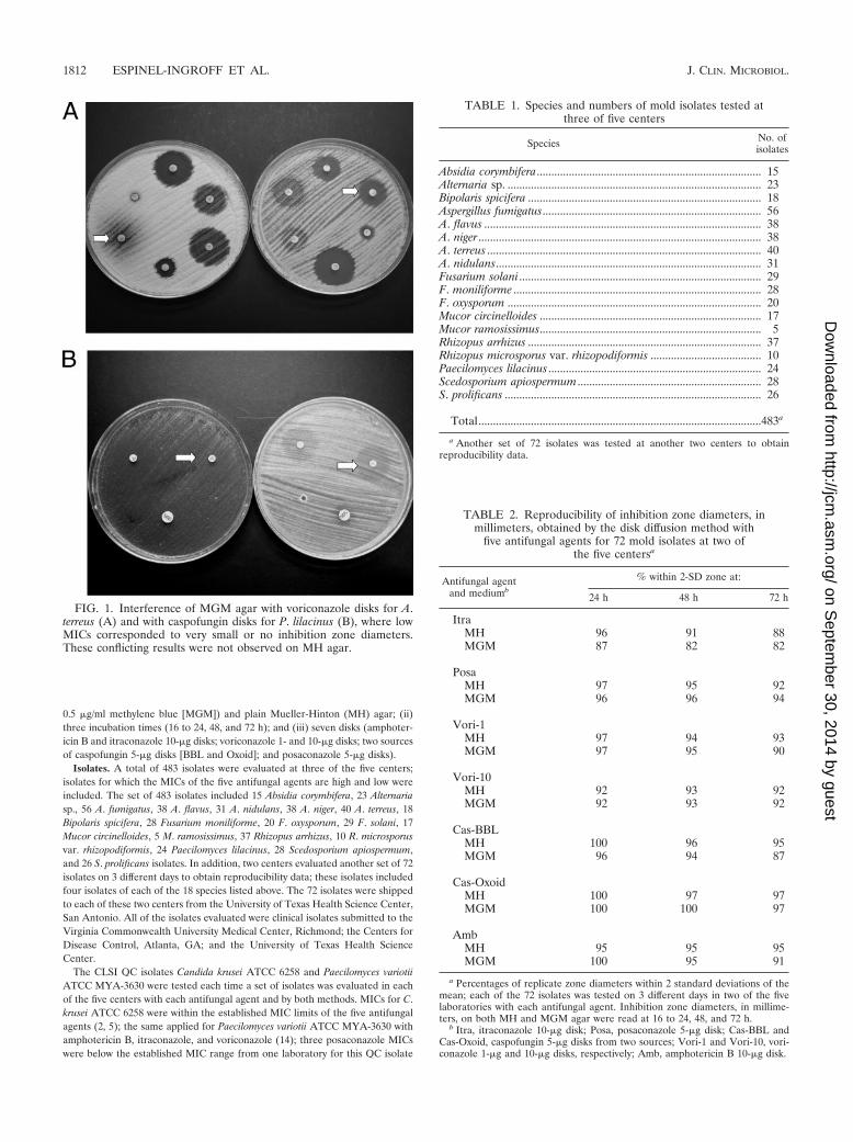
FIG. 1. Interference of MGM agar with voriconazole disks for A.terreus (A) and with caspofungin disks for P. lilacinus (B), where lowMICs corresponded to very small or no inhibition zone diameters.These conflicting results were not observed on MH agar.
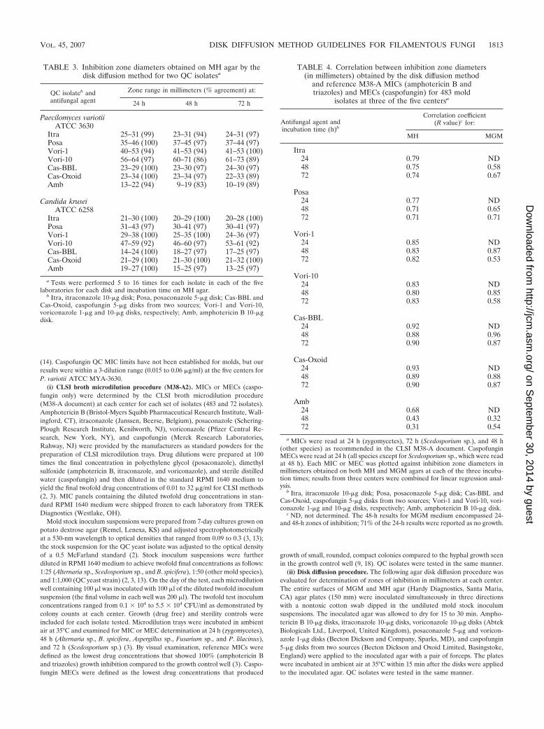
TABLE 1. Species and numbers of mold isolates tested atthree of five centers
Species No. ofisolates
Absidia corymbifera............................................................................. 15Alternaria sp. ....................................................................................... 23Bipolaris spicifera ................................................................................ 18Aspergillus fumigatus........................................................................... 56A. flavus ............................................................................................... 38A. niger ................................................................................................. 38A. terreus .............................................................................................. 40A. nidulans........................................................................................... 31Fusarium solani ................................................................................... 29F. moniliforme ..................................................................................... 28F. oxysporum ....................................................................................... 20Mucor circinelloides ............................................................................ 17Mucor ramosissimus............................................................................ 5Rhizopus arrhizus ................................................................................ 37Rhizopus microsporus var. rhizopodiformis ...................................... 10Paecilomyces lilacinus ......................................................................... 24Scedosporium apiospermum ............................................................... 28S. prolificans ........................................................................................ 26
Total.................................................................................................483a
a Another set of 72 isolates was tested at another two centers to obtainreproducibility data.
TABLE 2. Reproducibility of inhibition zone diameters, inmillimeters, obtained by the disk diffusion method with
five antifungal agents for 72 mold isolates at two ofthe five centersa
Antifungal agentand mediumb
% within 2-SD zone at:
24 h 48 h 72 h
ItraMH 96 91 88MGM 87 82 82
PosaMH 97 95 92MGM 96 96 94
Vori-1MH 97 94 93MGM 97 95 90
Vori-10MH 92 93 92MGM 92 93 92
Cas-BBLMH 100 96 95MGM 96 94 87
Cas-OxoidMH 100 97 97MGM 100 100 97
AmbMH 95 95 95MGM 100 95 91
a Percentages of replicate zone diameters within 2 standard deviations of themean; each of the 72 isolates was tested on 3 different days in two of the fivelaboratories with each antifungal agent. Inhibition zone diameters, in millime-ters, on both MH and MGM agar were read at 16 to 24, 48, and 72 h.
b Itra, itraconazole 10-�g disk; Posa, posaconazole 5-�g disk; Cas-BBL andCas-Oxoid, caspofungin 5-�g disks from two sources; Vori-1 and Vori-10, vori-conazole 1-�g and 10-�g disks, respectively; Amb, amphotericin B 10-�g disk.
1812 ESPINEL-INGROFF ET AL. J. CLIN. MICROBIOL.
on Septem
ber 30, 2014 by guesthttp://jcm
.asm.org/
Dow
nloaded from
(14). Caspofungin QC MIC limits have not been established for molds, but ourresults were within a 3-dilution range (0.015 to 0.06 �g/ml) at the five centers forP. variotii ATCC MYA-3630.
(i) CLSI broth microdilution procedure (M38-A2). MICs or MECs (caspo-fungin only) were determined by the CLSI broth microdilution procedure(M38-A document) at each center for each set of isolates (483 and 72 isolates).Amphotericin B (Bristol-Myers Squibb Pharmaceutical Research Institute, Wall-ingford, CT), itraconazole (Janssen, Beerse, Belgium), posaconazole (Schering-Plough Research Institute, Kenilworth, NJ), voriconazole (Pfizer Central Re-search, New York, NY), and caspofungin (Merck Research Laboratories,Rahway, NJ) were provided by the manufacturers as standard powders for thepreparation of CLSI microdilution trays. Drug dilutions were prepared at 100times the final concentration in polyethylene glycol (posaconazole), dimethylsulfoxide (amphotericin B, itraconazole, and voriconazole), and sterile distilledwater (caspofungin) and then diluted in the standard RPMI 1640 medium toyield the final twofold drug concentrations of 0.01 to 32 �g/ml for CLSI methods(2, 3). MIC panels containing the diluted twofold drug concentrations in stan-dard RPMI 1640 medium were shipped frozen to each laboratory from TREKDiagnostics (Westlake, OH).
Mold stock inoculum suspensions were prepared from 7-day cultures grown onpotato dextrose agar (Remel, Lenexa, KS) and adjusted spectrophotometricallyat a 530-nm wavelength to optical densities that ranged from 0.09 to 0.3 (3, 13);the stock suspension for the QC yeast isolate was adjusted to the optical densityof a 0.5 McFarland standard (2). Stock inoculum suspensions were furtherdiluted in RPMI 1640 medium to achieve twofold final concentrations as follows:1:25 (Alternaria sp., Scedosporium sp., and B. spicifera), 1:50 (other mold species),and 1:1,000 (QC yeast strain) (2, 3, 13). On the day of the test, each microdilutionwell containing 100 �l was inoculated with 100 �l of the diluted twofold inoculumsuspension (the final volume in each well was 200 �l). The twofold test inoculumconcentrations ranged from 0.1 � 104 to 5.5 � 104 CFU/ml as demonstrated bycolony counts at each center. Growth (drug free) and sterility controls wereincluded for each isolate tested. Microdilution trays were incubated in ambientair at 35°C and examined for MIC or MEC determination at 24 h (zygomycetes),48 h (Alternaria sp., B. spicifera, Aspergillus sp., Fusarium sp., and P. lilacinus),and 72 h (Scedosporium sp.) (3). By visual examination, reference MICs weredefined as the lowest drug concentrations that showed 100% (amphotericin Band triazoles) growth inhibition compared to the growth control well (3). Caspo-fungin MECs were defined as the lowest drug concentrations that produced
growth of small, rounded, compact colonies compared to the hyphal growth seenin the growth control well (9, 18). QC isolates were tested in the same manner.
(ii) Disk diffusion procedure. The following agar disk diffusion procedure wasevaluated for determination of zones of inhibition in millimeters at each center.The entire surfaces of MGM and MH agar (Hardy Diagnostics, Santa Maria,CA) agar plates (150 mm) were inoculated simultaneously in three directionswith a nontoxic cotton swab dipped in the undiluted mold stock inoculumsuspensions. The inoculated agar was allowed to dry for 15 to 30 min. Ampho-tericin B 10-�g disks, itraconazole 10-�g disks, voriconazole 10-�g disks (AbtekBiologicals Ltd., Liverpool, United Kingdom), posaconazole 5-�g and voricon-azole 1-�g disks (Becton Dickson and Company, Sparks, MD), and caspofungin5-�g disks from two sources (Becton Dickson and Oxoid Limited, Basingstoke,England) were applied to the inoculated agar with a pair of forceps. The plateswere incubated in ambient air at 35°C within 15 min after the disks were appliedto the inoculated agar. QC isolates were tested in the same manner.
TABLE 3. Inhibition zone diameters obtained on MH agar by thedisk diffusion method for two QC isolatesa
QC isolateb andantifungal agent
Zone range in millimeters (% agreement) at:
24 h 48 h 72 h
Paecilomyces variotiiATCC 3630
Itra 25–31 (99) 23–31 (94) 24–31 (97)Posa 35–46 (100) 37–45 (97) 37–44 (97)Vori-1 40–53 (94) 41–53 (94) 41–53 (100)Vori-10 56–64 (97) 60–71 (86) 61–73 (89)Cas-BBL 23–29 (100) 23–30 (97) 24–30 (97)Cas-Oxoid 23–34 (100) 23–34 (97) 22–33 (89)Amb 13–22 (94) 9–19 (83) 10–19 (89)
Candida kruseiATCC 6258
Itra 21–30 (100) 20–29 (100) 20–28 (100)Posa 31–43 (97) 30–41 (97) 30–41 (97)Vori-1 29–38 (100) 25–35 (100) 24–36 (97)Vori-10 47–59 (92) 46–60 (97) 53–61 (92)Cas-BBL 14–24 (100) 18–27 (97) 17–25 (97)Cas-Oxoid 21–29 (100) 21–30 (100) 21–32 (100)Amb 19–27 (100) 15–25 (97) 13–25 (97)
a Tests were performed 5 to 16 times for each isolate in each of the fivelaboratories for each disk and incubation time on MH agar.
b Itra, itraconazole 10-�g disk; Posa, posaconazole 5-�g disk; Cas-BBL andCas-Oxoid, caspofungin 5-�g disks from two sources; Vori-1 and Vori-10,voriconazole 1-�g and 10-�g disks, respectively; Amb, amphotericin B 10-�gdisk.
TABLE 4. Correlation between inhibition zone diameters(in millimeters) obtained by the disk diffusion method
and reference M38-A MICs (amphotericin B andtriazoles) and MECs (caspofungin) for 483 mold
isolates at three of the five centersa
Antifungal agent andincubation time (h)b
Correlation coefficient(R value)c for:
MH MGM
Itra24 0.79 ND48 0.75 0.5872 0.74 0.67
Posa24 0.77 ND48 0.71 0.6572 0.71 0.71
Vori-124 0.85 ND48 0.83 0.8772 0.82 0.53
Vori-1024 0.83 ND48 0.80 0.8572 0.83 0.58
Cas-BBL24 0.92 ND48 0.88 0.9672 0.90 0.87
Cas-Oxoid24 0.93 ND48 0.89 0.8872 0.90 0.87
Amb24 0.68 ND48 0.43 0.3272 0.31 0.54
a MICs were read at 24 h (zygomycetes), 72 h (Scedosporium sp.), and 48 h(other species) as recommended in the CLSI M38-A document. CaspofunginMECs were read at 24 h (all species except for Scedosporium sp., which were readat 48 h). Each MIC or MEC was plotted against inhibition zone diameters inmillimeters obtained on both MH and MGM agars at each of the three incuba-tion times; results from three centers were combined for linear regression anal-ysis.
b Itra, itraconazole 10-�g disk; Posa, posaconazole 5-�g disk; Cas-BBL andCas-Oxoid, caspofungin 5-�g disks from two sources; Vori-1 and Vori-10, vori-conazole 1-�g and 10-�g disks, respectively; Amb, amphotericin B 10-�g disk.
c ND, not determined. The 48-h results for MGM medium encompassed 24-and 48-h zones of inhibition; 71% of the 24-h results were reported as no growth.
VOL. 45, 2007 DISK DIFFUSION METHOD GUIDELINES FOR FILAMENTOUS FUNGI 1813
on Septem
ber 30, 2014 by guesthttp://jcm
.asm.org/
Dow
nloaded from
Zone diameters in the disk diffusion assay were measured to the nearest wholemillimeter at the point where there was a prominent reduction of growth (80%)after 16 to 24 h for zygomycetes and after 24, 48, and 72 h for the other species.Microcolonies inside the zone of inhibition were ignored when testing caspofun-gin and hyphal filaments bending over the inhibition zones and slight trailingaround the edges when testing the triazoles, but not for amphotericin B (Fig. 1).
Data analysis. MICs or MECs determined by the reference methods (at therecommended incubation times of 24 h for zygomycetes; 48 h for Aspergillus sp.,B. spicifera, Alternaria sp., Fusarium sp., and Paecilomyces sp.; and 72 h forScedosporium sp. [M38-A document]) were correlated with inhibition zone di-ameters (in millimeters) around disks; zones were obtained at three incubationtimes (16 to 24, 48, and 72 h). To obtain correlation results (R values), a linearregression analysis by the least-squares method (Pearson’s correlation coeffi-cient; MS Excel software) was performed by plotting zone diameters against theirrespective MIC or MEC endpoints (after log transformation). The reproducibil-ity of zone diameters obtained on 3 different days with each of four isolates of the18 species was evaluated by calculating the percentage of replicate zone diam-eters that were within 2 standard deviations of the mean (8). Reproducibilityvalues were not obtained as a range of � millimeter zone diameter measure-ments because millimeter variations in large zones tend to yield larger valuesthan the variations in smaller zones. Breakpoints are not available for anyantifungal agent versus molds. However, MICs below 1 �g/ml are usually re-ported for most Aspergillus sp. isolates with the five agents, for Scedosporiumapiospermum and P. lilacinus with posaconazole and voriconazole, for Alternariasp. and B. spicifera with the three triazoles, and for some zygomycete isolates withposaconazole and amphotericin B (7, 12, 19, 21). Because of that, we grouped assusceptible isolates for which the MICs or MECs were �1 �g/ml, as intermediateisolates for which the MICs or MECs were 2 �g/ml, and as resistant isolates forwhich the MICs or MECs were �4 �g/ml to analyze the performance of the diskmethod in identifying isolates with decreased susceptibility to the five antifungalagents evaluated. Scattergram plots of the MICs or MECs and correspondingdisk results of each antifungal agent were developed, and by using the error ratebounding method (17) the following tentative zone diameter categories wereassigned: susceptible, �17 mm (triazoles and caspofungin) and �15 mm (am-photericin B); intermediate, 14 to 16 mm (triazoles and caspofungin) and 13 to14 mm (amphotericin B); resistant, �13 mm (triazoles and caspofungin) and
�12 mm (amphotericin B). These tentative breakpoints were used to determinethe categorical agreement between the disk diffusion and MIC endpoints of eachdrug. Major errors were identified when the isolate was resistant by the diskmethod but susceptible by the MIC or MEC result, while minor errors wereidentified when there were shifts between susceptible and susceptible dose de-pendent or between susceptible dose dependent and resistant. Very major errorswere identified when the MIC or MEC result showed resistance and the diskresult showed susceptibility.
RESULTS AND DISCUSSION
Table 1 lists the number of isolates of each species that wereevaluated in the study to identify optimal testing guidelines fordisk diffusion testing of molds. These species are more com-monly associated with severe fungal infections in the immuno-compromised host. The antifungal agents included are themost important agents that have been licensed for the treat-ment or prevention of mold infections. The evaluation was tobe conducted with MGM agar, which is the medium that hasbeen selected by the CLSI for testing yeasts with voriconazoleand fluconazole by disk diffusion methodology (M44-A) (4).However, it was demonstrated early in this study that MGMmedium did not support good growth of most Aspergillus sp. at24 h, including A. fumigatus (60%), while zones of inhibitioncould be read at 24 h for most Aspergillus isolates and at 48 formost of the other species on MH agar (88%); similar resultshave been previously reported (8). In addition, interferencewith either caspofungin for P. lilacinus or voriconazole for A.terreus (Fig. 1) was observed, where low MICs corresponded tovery small or no inhibition zone diameters. These conflictingresults were not observed when disk diffusion tests were per-
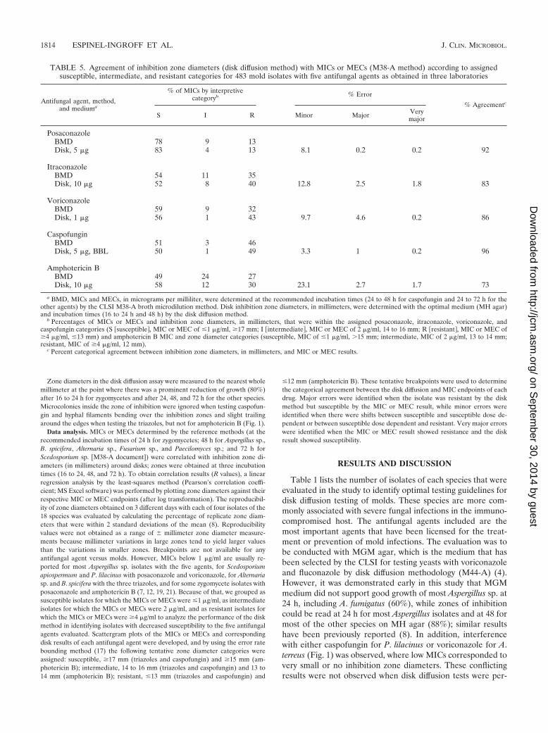
TABLE 5. Agreement of inhibition zone diameters (disk diffusion method) with MICs or MECs (M38-A method) according to assignedsusceptible, intermediate, and resistant categories for 483 mold isolates with five antifungal agents as obtained in three laboratories
Antifungal agent, method,and mediuma
% of MICs by interpretivecategoryb % Error
% Agreementc
S I R Minor Major Verymajor
PosaconazoleBMD 78 9 13Disk, 5 �g 83 4 13 8.1 0.2 0.2 92
ItraconazoleBMD 54 11 35Disk, 10 �g 52 8 40 12.8 2.5 1.8 83
VoriconazoleBMD 59 9 32Disk, 1 �g 56 1 43 9.7 4.6 0.2 86
CaspofunginBMD 51 3 46Disk, 5 �g, BBL 50 1 49 3.3 1 0.2 96
Amphotericin BBMD 49 24 27Disk, 10 �g 58 12 30 23.1 2.7 1.7 73
a BMD, MICs and MECs, in micrograms per milliliter, were determined at the recommended incubation times (24 to 48 h for caspofungin and 24 to 72 h for theother agents) by the CLSI M38-A broth microdilution method. Disk inhibition zone diameters, in millimeters, were determined with the optimal medium (MH agar)and incubation times (16 to 24 h and 48 h) by the disk diffusion method.
b Percentages of MICs or MECs and inhibition zone diameters, in millimeters, that were within the assigned posaconazole, itraconazole, voriconazole, andcaspofungin categories (S �susceptible�, MIC or MEC of �1 �g/ml, �17 mm; I �intermediate�, MIC or MEC of 2 �g/ml, 14 to 16 mm; R �resistant�, MIC or MEC of�4 �g/ml, �13 mm) and amphotericin B MIC and zone diameter categories (susceptible, MIC of �1 �g/ml, �15 mm; intermediate, MIC of 2 �g/ml, 13 to 14 mm;resistant, MIC of �4 �g/ml, 12 mm).
c Percent categorical agreement between inhibition zone diameters, in millimeters, and MIC or MEC results.
1814 ESPINEL-INGROFF ET AL. J. CLIN. MICROBIOL.
on Septem
ber 30, 2014 by guesthttp://jcm
.asm.org/
Dow
nloaded from
formed on MH agar. MGM agar also did not support thegrowth of most P. lilacinus and Alternaria isolates, even after72 h to 5 days of incubation, and slowed the determination ofzones from 48 to 72 h for B. spicifera and S. apiospermum. Ingeneral, zones were better defined and easier to read on MHagar than on MGM agar; inhibition zones could be determinedat 16 to 24 h for zygomycetes, at 24 h for most Aspergillusisolates (17 of 31 A. nidulans isolates [one center] and a few A.fumigatus and A. terreus isolates needed 48 h of incubation),and at 48 h for the other species. As discussed below, corre-lation and reproducibility results (R values) were superior atthese incubation times.
For the 555 isolates and both QC isolates, 95% of the inoc-ula were within the target range of 0.4 � 104 to 5 � 104
CFU/ml. Lower inoculum densities (0.1 � 104 to 0.3 � 104
CFU/ml) were mostly obtained with 22 isolates of A. flavus, A.niger, F. solani, and P. lilacinus and higher values (5.1 � 106 to7 � 106 CFU/ml) were obtained for three isolates of F. mo-niliforme and P. lilacinus at two centers. Similar reproducibilityresults for inoculum suspensions have been reported in priorcollaborative studies (92 to 95%) (10, 11). The reproducibilityof inoculum suspensions for Alternaria isolates has not beenpreviously evaluated, but our results were within the targetrange (optical density � 0.25 to 0.3).
Table 2 depicts the reproducibility of zone diameters (inmillimeters) obtained with the 72 isolates on 3 different dayswith each antifungal agent on both media. More than 15,000zone endpoints were analyzed. Overall reproducibility was
good with both media and at the three times of incubation, asdemonstrated by the high percentages (overall, 82 to 100%) ofreplicate zone diameters that were within 2 standard deviationsof the mean. The best reproducibility was observed with the 24-and 48-h results (91 to 100% on MH agar). By genus, thelowest reproducibility was observed for P. lilacinus with itra-conazole, voriconazole, and amphotericin B (78 to 84%), forFusarium sp. with itraconazole (83%), for Mucor sp. withposaconazole (86%), for Rhizopus sp. with itraconazole (89%),and for B. spicifera with caspofungin (81%); other reproduc-ibility values ranged from 90 to 100%. Results were lower onMGM than on MH agar; the exception was the results for thezygomycetes (70 to 86% on MH agar versus 91 to 95% onMGM), but not all isolates could be evaluated at 24 h due tolack of growth. Overall, our reproducibility results are similarto those obtained for an evaluation of posaconazole for As-pergillus sp. and the zygomycetes (8). Reproducibility resultsalso were comparable or superior to those obtained in othercollaborative studies for the development of the M38-A doc-ument (3, 10, 11).
Table 3 summarizes the zones of inhibition (in millimeters)obtained on MH agar with the two QC controls at the fivecenters; reference diameter ranges are not available for eitherQC isolate versus any antifungal on MH agar. In this study,most of the intervals between the high and low zone diameterlimits were within 6 to 11 mm; the reproducibility was 97 to100% at 24 to 72 h for C. krusei ATCC 6258 and at 24 h for P.variotii ATCC 3630 with most of the antifungal disks tested.
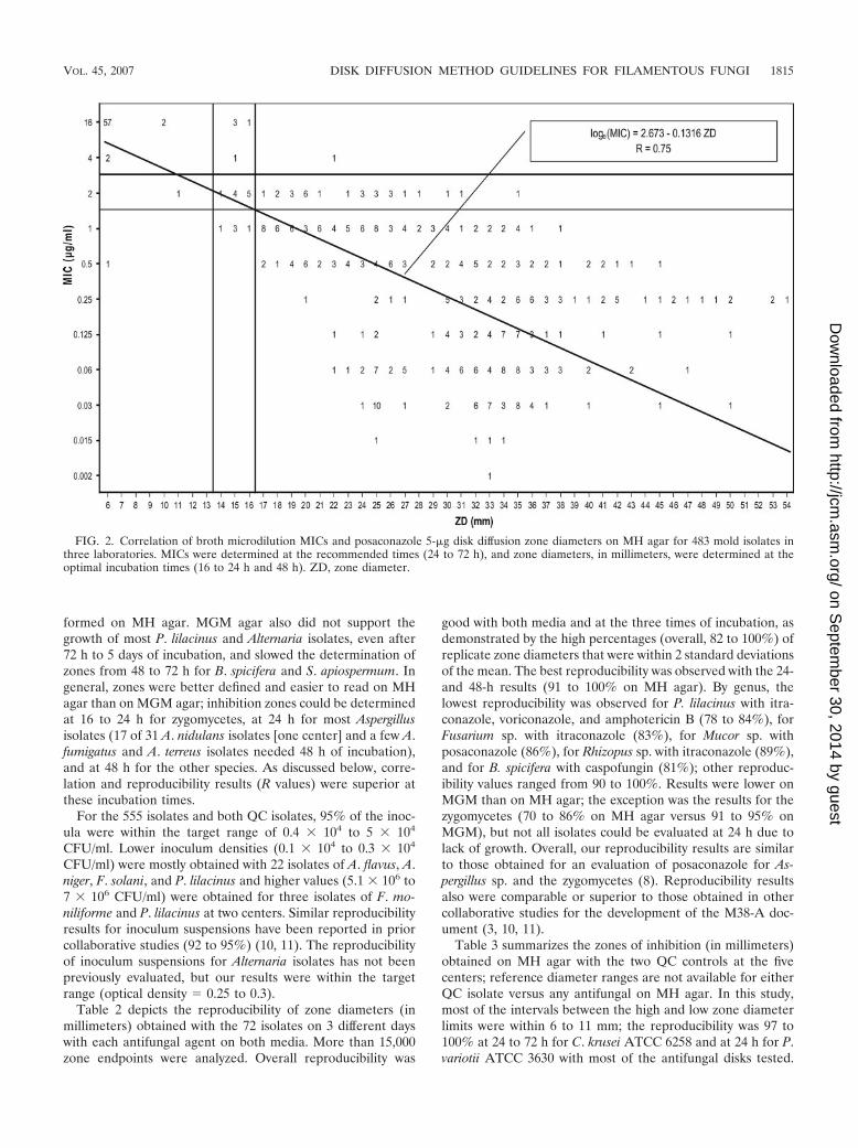
FIG. 2. Correlation of broth microdilution MICs and posaconazole 5-�g disk diffusion zone diameters on MH agar for 483 mold isolates inthree laboratories. MICs were determined at the recommended times (24 to 72 h), and zone diameters, in millimeters, were determined at theoptimal incubation times (16 to 24 h and 48 h). ZD, zone diameter.
VOL. 45, 2007 DISK DIFFUSION METHOD GUIDELINES FOR FILAMENTOUS FUNGI 1815
on Septem
ber 30, 2014 by guesthttp://jcm
.asm.org/
Dow
nloaded from
The CLSI zone diameter limit for C. krusei ATCC 6258 with avoriconazole 1-�g disk on the recommended MGM medium is16 to 25 mm (4); our range for that strain on MH agar wasconsistently larger (29 to 38 mm, 100%). Based on collabora-tive studies, tentative QC data for posaconazole (23 to 31 mm)and caspofungin (13 to 25 mm) on MGM agar are also avail-able for this QC strain. Our results were within the target zonediameter range for caspofungin (14 to 25 mm, 100%) with theBBL caspofungin disk on both agars, but a larger zone rangewas obtained with the Oxoid disk on MH agar (21 to 29 mm,100%). Similar results (larger zone ranges) were obtained withposaconazole for the QC yeast (31 to 43 mm at 24 to 72 h,97%). It appears that the yeast QC isolate could serve as acontrol isolate until a QC isolate(s) is selected and standardzone diameter limits are established for mold disk testing.
Table 4 summarizes the coefficients of correlation (linearregression analysis) between either MIC or MEC results andthe corresponding inhibition zone diameters, in millimeters(obtained with the seven disks), for the 483 isolates evaluatedat three of the five centers (Table 1). The correlation wassuperior for disks of both caspofungin and voriconazole; sim-ilar results were obtained for these agents on both the MGMand MH agars. However, for itraconazole and posaconazole,the correlation was higher on MH agar than on MGM agar;suitable R values (�0.7) were observed on MH agar comparedto the low R values (0.70) at some incubation times on MGMagar. Our correlation results were comparable to those re-
ported for Aspergillus sp. (8, 15, 20) and zygomycete (8, 15)isolates with voriconazole (R, 0.79) and posaconazole (R, 0.72to 0.82) disks. The lowest correlation results were obtained foramphotericin B, as previously reported (8), where zones wereconsistently small for isolates for which the amphotericin BMICs were correspondingly low and high.
The evaluation of a new method requires the determinationof breakpoint category agreement between the new and refer-ence methods. Since interpretive breakpoints are not availablefor molds, this type of analysis was performed with the tenta-tive breakpoints assigned in this study; these breakpoints wereuseful for illustration or comparison only. We grouped isolatesas resistant (MIC or MEC, �4 �g/ml), intermediate (MIC orMEC, 2 �g/ml), or susceptible (MIC or MEC, �1 �g/ml)based on reported in vitro data obtained with large numbers ofisolates (1, 7, 8, 12, 19, 21). Tentative zone diameter break-points were assigned by the error rate bounding method (17).Table 5 depicts the results of the breakpoint category analysisagreement for amphotericin B, itraconazole, posaconazole,and voriconazole (1-�g disk results only) inhibition zone di-ameters obtained at the optimal incubation times of 16 to 24h for zygomycetes, A. fumigatus, A. flavus, and A. niger and 48 hfor the other species on MH agar versus MICs obtained at 24 h(zygomycetes), 72 h (Scedosporium sp.), and 48 h (other spe-cies). Table 5 also provides the results of the comparison ofcaspofungin MECs from the first reading (16 to 24 h [zygomy-cetes], 48 h [Scedosporium sp.], and 24 h [other species]) with
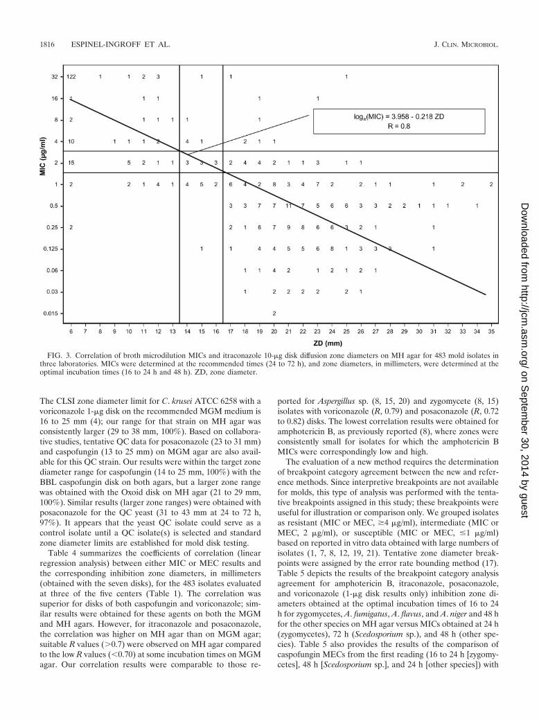
FIG. 3. Correlation of broth microdilution MICs and itraconazole 10-�g disk diffusion zone diameters on MH agar for 483 mold isolates inthree laboratories. MICs were determined at the recommended times (24 to 72 h), and zone diameters, in millimeters, were determined at theoptimal incubation times (16 to 24 h and 48 h). ZD, zone diameter.
1816 ESPINEL-INGROFF ET AL. J. CLIN. MICROBIOL.
on Septem
ber 30, 2014 by guesthttp://jcm
.asm.org/
Dow
nloaded from
zone diameters (BBL disk results only) also obtained on MHagar at the same incubation times as for the other four anti-fungal agents. Figures 2 to 6 depict the results of the regressionanalysis between either MICs or MECs and zone diameters forthe same five disks and by the same optimal testing conditionsdescribed above and in Table 5. Among the three laboratories,the percentage ranges of major and very major errors were 0.2to 6.4% and 0 to 2.7%, respectively, for the five disks; thepercentage range of overall categorical agreement was 81 to96% for posaconazole, caspofungin, and voriconazole 1-�gdisks and 65 to 88% for amphotericin B and itraconazole disks(data not shown in Table 5).
The categorical evaluation of the posaconazole 5-�g diskindicated that the percentages of major, very major, and minorerrors were low (0.2 and 8.1%) (Table 5). One susceptibleisolate was categorized as resistant (�13-mm diameter, majorerror), and one resistant isolate was categorized as susceptible(�17-mm diameter, very major error) (Fig. 2). Of the 39 minorerrors, 28 (72%) were among isolates for which the MIC was 2�g/ml (intermediate category); these 28 isolates (16 Fusariumsp. and 9 S. apiospermum isolates) were categorized as suscep-tible (�17-mm diameter). Agreement was higher with theposaconazole disk (92%) than with the itraconazole 10-�g disk(83%). Nine very major errors (1.8% false-susceptible values,�17-mm zone diameters) were observed with the itraconazoledisk (four A. corymbifera isolates, two A. niger isolates, two P.lilacinus isolates, and one S. apiospermum isolate) (Fig. 3). Ofthe 52 isolates for which the itraconazole MICs were 2 �g/ml
(intermediate category), 24 were categorized as resistant (�13mm; 19 zygomycete isolates), 9 were categorized as interme-diate (14- to 16-mm diameters), and 19 were categorized assusceptible (�17-mm diameters; mostly A. corymbifera and P.lilacinus). It is noteworthy that only two major errors wereobserved among the 203 Aspergillus isolates with itraconazoleand none were observed with posaconazole. Therefore, it ap-pears that the posaconazole disk (R range among the centers,0.71 to 0.78) is more suitable than the itraconazole disk (Rrange among the centers, 0.7 to 0.8), despite the latter’s higherR values, for testing of zygomycete isolates; both disks aresuitable for testing of the other species.
Two voriconazole disks were evaluated, which provided dif-ferent susceptible and resistant zone diameter thresholds (�17mm and �28 mm as susceptible and �13 mm and �25 mm asresistant, 1-�g and 10-�g disks, respectively). According to ourassigned categories, the performance of the voriconazole 1-�gdisk was superior (86% overall agreement; Table 5) to the10-�g disk (80% overall agreement; data not shown in Table5). Discrepancies with the 1-�g disk were mostly due to majorerrors (one very major error and 4.6% zone diameters of �13mm) (Fig. 4 and Table 5), but those with the 10-�g disk weredue to very major errors (9% zone diameters of �28 mm).Major errors with the 1-�g disk were observed for 13 Alternariasp. isolates (at one of the three centers) and 4 F. moniliformeisolates. Among the 43 isolates for which the voriconazole MICwas 2 �g/ml (intermediate category), 11 were categorized assusceptible (�17-mm zone diameters for Aspergillus sp. and S.
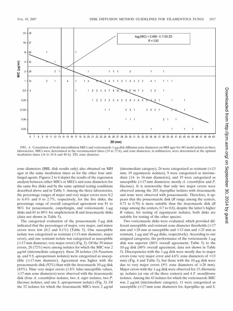
FIG. 4. Correlation of broth microdilution MICs and voriconazole 1-�g disk diffusion zone diameters on MH agar for 483 mold isolates in threelaboratories. MICs were determined at the recommended times (24 to 72 h), and zone diameters, in millimeters, were determined at the optimalincubation times (16 to 24 h and 48 h). ZD, zone diameter.
VOL. 45, 2007 DISK DIFFUSION METHOD GUIDELINES FOR FILAMENTOUS FUNGI 1817
on Septem
ber 30, 2014 by guesthttp://jcm
.asm.org/
Dow
nloaded from
apiospermum) and 32 isolates were categorized as resistant(�13 mm for 30 Fusarium isolates, 1 B. spicifera isolate, and 1Alternaria sp. isolate) with the 1-�g disk. In contrast, 95% ofthese 43 isolates were categorized as susceptible (�28-mmzone diameters) with the 10-�g disk (data not shown in Table5). In addition, the 1-�g disk was able to identify as resistant all26 S. prolificans and 84 zygomycete isolates, but the 10-�g diskmissed 19 of these isolates. Also, from a practical standpoint,only one voriconazole 10-�g disk could be tested per plate toavoid zone overlap (�70-mm zone diameters), while three orfour disks could be tested with the 1-�g disk (150-mm plate).Therefore, these results suggest that the 1-�g disk (R rangeamong the three centers, 0.77 to 0.88) should be the choice fortesting the susceptibilities of most mold isolates to voricon-azole. Since all zygomycete and S. prolificans isolates are re-sistant to voriconazole in this and other studies (12, 19), thereis no reason to test these isolates.
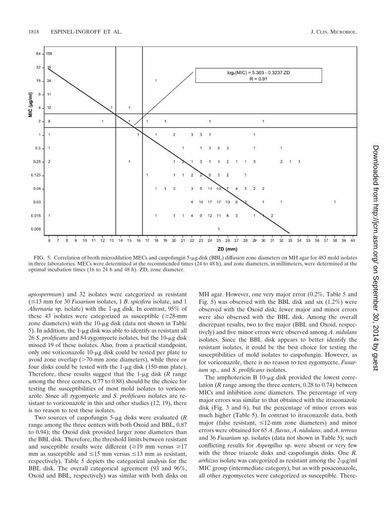
Two sources of caspofungin 5-�g disks were evaluated (Rrange among the three centers with both Oxoid and BBL, 0.87to 0.94); the Oxoid disk provided larger zone diameters thanthe BBL disk. Therefore, the threshold limits between resistantand susceptible results were different (�19 mm versus �17mm as susceptible and �15 mm versus �13 mm as resistant,respectively). Table 5 depicts the categorical analysis for theBBL disk. The overall categorical agreement (93 and 96%,Oxoid and BBL, respectively) was similar with both disks on
MH agar. However, one very major error (0.2%, Table 5 andFig. 5) was observed with the BBL disk and six (1.2%) wereobserved with the Oxoid disk; fewer major and minor errorswere also observed with the BBL disk. Among the overalldiscrepant results, two to five major (BBL and Oxoid, respec-tively) and five minor errors were observed among A. nidulansisolates. Since the BBL disk appears to better identify theresistant isolates, it could be the best choice for testing thesusceptibilities of mold isolates to caspofungin. However, asfor voriconazole, there is no reason to test zygomycete, Fusar-ium sp., and S. prolificans isolates.
The amphotericin B 10-�g disk provided the lowest corre-lation (R range among the three centers, 0.28 to 0.74) betweenMICs and inhibition zone diameters. The percentage of verymajor errors was similar to that obtained with the itraconazoledisk (Fig. 3 and 6), but the percentage of minor errors wasmuch higher (Table 5). In contrast to itraconazole data, bothmajor (false resistant, �12-mm zone diameters) and minorerrors were obtained for 65 A. flavus, A. nidulans, and A. terreusand 36 Fusarium sp. isolates (data not shown in Table 5); suchconflicting results for Aspergillus sp. were absent or very fewwith the three triazole disks and caspofungin disks. One R.arrhizus isolate was categorized as resistant among the 2-�g/mlMIC group (intermediate category), but as with posaconazole,all other zygomycetes were categorized as susceptible. There-
FIG. 5. Correlation of broth microdilution MECs and caspofungin 5-�g disk (BBL) diffusion zone diameters on MH agar for 483 mold isolatesin three laboratories. MECs were determined at the recommended times (24 to 48 h), and zone diameters, in millimeters, were determined at theoptimal incubation times (16 to 24 h and 48 h). ZD, zone diameter.
1818 ESPINEL-INGROFF ET AL. J. CLIN. MICROBIOL.
on Septem
ber 30, 2014 by guesthttp://jcm
.asm.org/
Dow
nloaded from
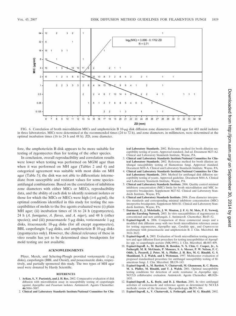
fore, the amphotericin B disk appears to be more suitable fortesting of zygomycetes than for testing of the other species.
In conclusion, overall reproducibility and correlation resultswere lower when testing was performed on MGM agar thanwhen it was performed on MH agar (Tables 2 and 4) andcategorical agreement was suitable with most disks on MHagar (Table 5); the disk was not able to differentiate interme-diate from susceptible and resistant values for some species-antifungal combinations. Based on the correlation of inhibitionzone diameters with either MICs or MECs, reproducibilitydata, and the ability of each disk to identify resistant isolates orthose for which the MICs or MECs were high (�4 �g/ml), theoptimal conditions identified in this study for testing the sus-ceptibilities of molds to the five agents evaluated were (i) plainMH agar; (ii) incubation times of 16 to 24 h (zygomycetes),24 h (A. fumigatus, A. flavus, and A. niger), and 48 h (otherspecies); and (iii) posaconazole 5-�g disks, voriconazole 1-�gdisks, itraconazole 10-�g disks (for all except zygomycetes),BBL caspofungin 5-�g disks, and amphotericin B 10-�g disks(zygomycetes only). However, the clinical relevance of these invitro results has yet to be determined since breakpoints formold testing are not available.
ACKNOWLEDGMENTS
Pfizer, Merck, and Schering-Plough provided voriconazole (1-�gdisks), caspofungin (BBL and Oxoid), and posaconazole disks, respec-tively, and partially sponsored this study. The two types of MH agarused were donated by Hardy Scientific.
REFERENCES
1. Arikan, S., V. Paetznick, and J. H. Rex. 2002. Comparative evaluation of diskdiffusion with microdilution assay in susceptibility testing of caspofunginagainst Aspergillus and Fusarium isolates. Antimicrob. Agents Chemother.46:3084–3087.
2. Clinical and Laboratory Standards Institute/National Committee for Clin-
ical Laboratory Standards. 2002. Reference method for broth dilution sus-ceptibility testing of yeasts. Approved standard, 2nd ed. Document M27-A2.Clinical and Laboratory Standards Institute, Wayne, PA.
3. Clinical and Laboratory Standards Institute/National Committee for Clin-ical Laboratory Standards. 2002. Reference method for broth dilution an-tifungal susceptibility testing of filamentous fungi. Approved standard.Document M38-A. Clinical and Laboratory Standards Institute, Wayne, PA.
4. Clinical and Laboratory Standards Institute/National Committee for Clin-ical Laboratory Standards. 2004. Method for antifungal disk diffusion sus-ceptibility testing of yeasts. Approved guideline. Document M44-A. Clinicaland Laboratory Standards Institute, Wayne, PA.
5. Clinical and Laboratory Standards Institute. 2006. Quality control minimalinhibitory concentration (MIC) limits for broth microdilution and MIC in-terpretive breakpoints. Supplement M27-S2. Clinical and Laboratory Stan-dards Institute, Wayne, PA.
6. Clinical and Laboratory Standards Institute. 2006. Zone diameter interpre-tive standards and corresponding minimal inhibitory concentration (MIC)interpretive breakpoints. Supplement M44-S1. Clinical and Laboratory Stan-dards Institute, Wayne, PA.
7. Dannaoui, E., J. Meletiadis, J. W. Mouton, J. F. G. M. Meis, P. E. Verweij,and the Eurofong Network. 2003. In vitro susceptibilities of zygomycetes toconventional and new antifungals. J. Antimicrob. Chemother. 51:45–52.
8. Espinel-Ingroff, A. 2006. Comparison of three commercial assays and amodified disk diffusion assay with two broth microdilution reference assaysfor testing zygomycetes, Aspergillus spp., Candida spp., and Cryptococcusneoformans with posaconazole and amphotericin B. J. Clin. Microbiol. 44:3616–3622.
9. Espinel-Ingroff, A. 2003. Evaluation of broth microdilution testing parame-ters and agar diffusion Etest procedure for testing susceptibilities of Aspergil-lus spp. to caspofungin acetate (MK-0991). J. Clin. Microbiol. 41:403–409.
10. Espinel-Ingroff, A., M. Bartlett, R. Bowden, N. X. Chin, C. Cooper, Jr., A.Fothergill, M. R. McGinnis, P. Menezes, S. A. Messer, P. W. Nelson, F. C.Odds, L. Pasarell, J. Peter, M. A. Pfaller, J. H. Rex, M. G. Rinaldi, G. S.Shankland, T. J. Walsh, and I. Weitzman. 1997. Multicenter evaluation ofproposed standardized procedure for antifungal susceptibility testing of fil-amentous fungi. J. Clin. Microbiol. 35:139–143.
11. Espinel-Ingroff, A., M. Bartlett, V. Chaturvedi, M. Ghannoum, K. C. Hazen,M. A. Pfaller, M. Rinaldi, and T. J. Walsh. 2001. Optimal susceptibilitytesting conditions for detection of azole resistance in Aspergillus spp.:NCCLS collaborative evaluation. Antimicrob. Agents Chemother. 45:1828–1835.
12. Espinel-Ingroff, A., K. Boyle, and D. J. Sheehan. 2001. In vitro antifungalactivities of voriconazole and reference agents as determined by NCCLSmethods: review of the literature. Mycopathologia 38:293–304.
13. Espinel-Ingroff, A., V. Chaturvedi, A. Fothergill, and M. G. Rinaldi. 2002.
FIG. 6. Correlation of broth microdilution MICs and amphotericin B 10-�g disk diffusion zone diameters on MH agar for 483 mold isolatesin three laboratories. MICs were determined at the recommended times (24 to 72 h), and zone diameters, in millimeters, were determined at theoptimal incubation times (16 to 24 h and 48 h). ZD, zone diameter.
VOL. 45, 2007 DISK DIFFUSION METHOD GUIDELINES FOR FILAMENTOUS FUNGI 1819
on Septem
ber 30, 2014 by guesthttp://jcm
.asm.org/
Dow
nloaded from
Optimal testing conditions for determining MICs and minimum fungicidalconcentrations of new and established antifungal agents for uncommonmoulds: NCCLS collaborative study. J. Clin. Microbiol. 40:3776–3781.
14. Espinel-Ingroff, A., A. Fothergill, M. Ghannoum, E. Manavathu, L. Ostrosky-Zeichner, M. A. Pfaller, M. Rinaldi, W. Schell, and T. Walsh. 2005. Qualitycontrol and reference guidelines for CLSI broth microdilution susceptibilitymethod (M38-A document) for amphotericin B, itraconazole, posaconazole,and voriconazole. J. Clin. Microbiol. 43:5243–5246.
15. Lopez-Oviedo, E., A. I. Aller, C. Martin, C. Castro, M. Ramirez, J. M.Peman, E. Canton, C. Almeida, and E. Martin-Mazuelos. 2006. Evaluationof disk diffusion method for determining posaconazole susceptibility offilamentous fungi: comparison with CLSI broth microdilution method. An-timicrob. Agents Chemother. 50:1108–1111.
16. Marr, K. A., R. A. Carter, F. Crippa, A. Wald, and L. Corey. 2002. Epide-miology and outcome of mould infections in hematopoietic stem cell trans-plant recipients. Clin. Infect. Dis. 34:909–917.
17. Metzler, D. M., and R. M. DeHaan. 1974. Susceptibility tests of anaerobicbacteria: statistical and clinical considerations. J. Infect. Dis. 130:588–594.
18. Odds, F. C., M. Motyl, R. Andrade, J. Bille, E. Canton, M. Cuenca-Estrella,A. Davidson, C. Durussel, D. Ellis, E. Foraker, A. W. Fothergill, M. A.Ghannoum, R. A. Giacobbe, M. Gobernado, R. Handke, M. Laverdiere, W.
Lee-Yang, W. G. Merz, L. Ostrosky-Zeichner, J. Peman, S. Perea, J. R.Perfect, M. A. Pfaller, L. Proia, J. H. Rex, M. G. Rinaldi, J.-L. Rodriguez-Tudela, W. A. Schell, C. Shields, D. A. Sutton, P. E. Verweij, and D. W.Warnock. 2004. Interlaboratory comparison of results of susceptibility test-ing with caspofungin against Candida and Aspergillus species. J. Clin. Micro-biol. 42:3475–3482.
19. Sabatelli, F., R. Patel, P. A. Mann, C. A. Mendrick, C. C. Norris, R. Hare, D.Loebenberg, T. A. Black, and P. M. McNicholas. 2006. In vitro activities ofposaconazole, fluconazole, itraconazole, voriconazole, and amphotericin Bagainst a large collection of clinically important molds and yeasts. Antimi-crob. Agents Chemother. 50:2009–2015.
20. Serrano, M. C., M. Ramirez, D. Morilla, A. Valverde, M. Chavez, A. Espinel-Ingroff, R. Claro, A. Fernandez, C. Almeida, and E. Martin-Mazuelos. 2004.A comparative study of the disc diffusion method with the broth microdilu-tion and Etest methods for voriconazole susceptibility testing of Aspergillusspp. J. Antimicrob. Chemother. 53:739–742.
21. Sun, Q. N., A. W. Fothergill, D. I. McCarthy, M. G. Rinaldi, and J. R.Graybill. 2002. In vitro activities of posaconazole, itraconazole, voriconazole,amphotericin B, and fluconazole against isolates of zygomycetes. Antimi-crob. Agents Chemother. 46.5:1581–1582.
1820 ESPINEL-INGROFF ET AL. J. CLIN. MICROBIOL.
on Septem
ber 30, 2014 by guesthttp://jcm
.asm.org/
Dow
nloaded from