Cu2+ probe of metal-ion binding sites in melanin using electron paramagnetic resonance spectroscopy

Microbes and Infection 9 (2007) 1114e1123www.elsevier.com/locate/micinf
Original article
Melanin biosynthesis in Madurella mycetomatis and its effect onsusceptibility to itraconazole and ketoconazole
Wendy W.J. van de Sande a,*, Johan de Kat a, Jojanneke Coppens b, Abdalla O.A. Ahmed c,Ahmed Fahal d, Henri Verbrugh a, Alex van Belkum a
a Erasmus MC, University Medical Centre Rotterdam, Department of Medical Microbiology and Infectious Diseases,Dr. Molewaterplein 40, 3015 GD Rotterdam, The Netherlands
b Erasmus MC, University Medical Centre Rotterdam, Department of Immunology, Dr. Molewaterplein 40, 3015 GD Rotterdam, The Netherlandsc King Saud University, College of Medicine, Department of Pathology and Microbiology, P.O. Box 2925, Riyadh 11461, Saudi Arabia
d Mycetoma Research Centre, University of Khartoum, Khartoum, Sudan
Received 16 March 2007; accepted 9 May 2007
Available online 18 May 2007
Abstract
One of the hallmarks of eumycetoma is the formation of fungal grains, which are secreted by multiple sinuses in infected tissues. Madurellamycetomatis grains are black. This black colour was shown to be due to the presence of melanin. Melanin can be produced through variousbiochemical pathways. It appeared that M. mycetomatis melanisation could be blocked by inhibitors of the pyo- and dihydroxynaphthalene(DHN)-melanin pathways but not by inhibitors of the dihydroxyphenylalanine (L-DOPA)-melanin pathway. Melanin isolated from M. myceto-matis cells provides in vitro protection against the killing effects of the oxidant permanganate and several antifungals. When melanin was addedto the culture medium, MICs were found to be 16-fold elevated in the case of itraconazole and 32-fold for ketoconazole. MICs for amphotericinB, fluconazole and voriconazole were not affected. Since itraconazole and ketoconazole are the main antifungal agents used to treat mycetoma,the clinical relevance of the in vitro rise in MIC should be studied further.� 2007 Elsevier Masson SAS. All rights reserved.
Keywords: Melanin; Antifungal susceptibility; Fungi; Azole
1. Introduction
One of the main characteristics of eumycetoma lesions isthe formation of grains that are secreted through draining si-nuses. Grains are encountered in different colours and textureswhich are all dependent on the nature of the causative agent[1]. Grains can be green, yellow, red or pink but the most fre-quently encountered ‘‘colours’’ are white or black [1]. Thegrains of the most common causal fungus, Madurella myceto-matis, are black (Fig. 1). M. mycetomatis grains consist of
* Corresponding author. Tel.: þ31 10 463 2176; fax: þ31 10 463 3875.
E-mail address: [email protected] (Wendy W.J. van de
Sande).
1286-4579/$ - see front matter � 2007 Elsevier Masson SAS. All rights reserv
doi:10.1016/j.micinf.2007.05.015
densely packed fungal mycelia embedded in a hard, brownmatrix composed of extra-cellular cement material [1,2]. Thecement material seems to be composed of an extra-cellularpigment and host tissue debris [1,2]. It is presumed thatthe black compound in the grain is melanin produced byM. mycetomatis.
Melanins are negatively charged, hydrophobic, macromo-lecular pigments formed by oxidative polymerization of phe-nolic or indolic compounds [3e5]. Melanin has been shownto protect micro-organisms against UV-radiation, enzymaticlysis, oxidants and killing by alveolar macrophages [3e5]. Ithas also been shown to chelate metal ions, to function asa physiological redox buffer, to provide structural rigidity tocell walls and to help store water and ions [3]. Melaninsmay also play a role in protecting against antimicrobial drugs
ed.

1115Wendy W.J. van de Sande et al. / Microbes and Infection 9 (2007) 1114e1123
[4]. The ability of certain microbes to produce melanin hasbeen linked to virulence and pathogenicity in their respectiveanimal or plant host species [6]. Notably, albino strains ofCryptococcus neoformans are less potent in establishing an in-fection than melanised strains [7e9]. Also, for the plant patho-gens Magnaporthe grisea and Colletotrichum lagenariumappresorial melanin is necessary for generating appropriate tur-gor and, hence, invasive power when infecting plant tissue [3].
Fungi may biosynthesise melanin by a variety of pathways.One of the most extensively studied melanised fungal patho-gens, C. neoformans, forms its melanin from diphenolic com-pounds such as 3,4-dihydroxyphenylalanine (L-DOPA) [6].Other fungi including Aspergillus fumigatus, Exophiala spp.,and Alternaria alternata biosynthesize melanin from polyke-tide compounds such as 1,3,6,8-tetrahydroxynaphthalene(1,3,6,8-THN) [6]. A third pathway has only recently been de-scribed in the cheese spoiling yeast Yarrowia lipolytica, thehuman pathogen C. neoformans and in bacterial species suchas Shewanella algae [10e12]. Use of this pathway leads tomelanin formation from tyrosine precursors but not from L-DOPA [10,11]. The main difference between this latter pyo-melanin and the other two melanins is that this pyomelaninis soluble. It is secreted by the fungus while DOPA e and1,8-dihydroxynaphthalene (DHN-) melanin are solid, insolu-ble and usually bound to the cell wall.
The aim of the present study was to identify the pathwaysused by M. mycetomatis to form its melanin and to investigate
Fig. 1. An example of a mycetoma-foot. The black grains within the lesions
are enlarged in figure B.
possible physiological roles of melanin during mycetomainfections.
2. Materials and methods
2.1. Immunohistochemistry
Deep tissue biopsies were taken from advanced mycetomalesions from 3 different patients and embedded in paraffin.Slides were rehydrated and boiled for 1 h in 6 M HCl to detectpossible melanisation of the grains. After boiling only thegrain structures were left, whereas the surrounding tissuehad dissolved. Non-boiled slides were used to determine ifmelanin was found outside the grains as well. All slideswere blocked by 2% BSA and 5% sucrose. Slides were incu-bated with melanin-specific antibody 6D2 (provided by Dr.Nosanchuk Albert Einstein College of Medicine, New York)for 1 h at room temperature at a concentration of 5 mg/ml.Monoclonal antibody 6D2 (IgM) was generated against mela-nin derived from C. neoformans, this antibody was not specificfor C. neoformans melanin since other types of melanin couldalso be detected. Non-melanised fungi were not bound by thisantibody [7,13]. Substitution of the primary antibody withbuffer was used as a negative control. Binding of the primaryantibody was detected by a FITC-labelled goat-anti-mouse an-tibody (CLB Biotechnology Services, Amsterdam, The Neth-erlands). The slides were mounted in DAPI-containingVectashield (Vector Laboratories Inc., Burlingame, CA,USA) and examined with a Zeiss Axioplan 2 imaging fluores-cence microscope (Zeiss, Gottingen, Germany).
2.2. Cultures
For this study 8 isolates of M. mycetomatis were used.These strains are part of a large culture collection from the Su-dan Mycetoma Research Centre (Khartoum, Sudan). Cultureswere maintained on Sabouraud dextrose agar (Difco Laborato-ries, Paris, France) by repeated subculture. For melanin isola-tion, strains were subcultured on minimal medium, which wascomposed of 2% agar (Difco Laboratories, Paris, France)15 mM glucose (Baker, Deventer, The Netherlands), 10 mMMgSO4 (Merck, Haarlem, The Netherlands), 29.4 mMKH2PO4 (VWR, Leuven, Belgium), 13 mM glycin (Sigma-Al-drich, Zwijndrecht, The Netherlands) and 80 mg/l gentamicin(Centrafarm, Etten-Leur, The Netherlands). To determinewhich precursors M. mycetomatis uses to form its melanin,the melanin-enhancers L-tyrosine and L-DOPA were addedto minimal medium in a concentration of 0.1 mM and1 mM, respectively. Inhibition of melanin synthesis was testedby adding the following inhibitors to the minimal medium:sulcotrione (Riedel-deHaen, Seelze, Germany), niacin(Sigma-Aldrich, Zwijndrecht, The Netherlands), glyphosate(Luxan, Elst, The Netherlands), tricyclazole (Riedel-deHaen)and phthalide (Acros Organics, Geel, Belgium). Concentra-tions tested were: 0 mg/l, 5 mg/l, 25 mg/l, 50 mg/l, 75 mg/l,100 mg/l and 200 mg/l. Strains were incubated for two months
1116 Wendy W.J. van de Sande et al. / Microbes and Infection 9 (2007) 1114e1123
at 37 �C. After such an incubation period, all control cultureswere clearly melanised.
2.3. Melanin isolation
Melanin for M. mycetomatis strain mm55, clinical isolate C.neoformans strain 106 and A. fumigatus ATCC 204305 wasisolated as described by Youngchim et al. [14]. In short, M.mycetomatis hyphae or C. neoformans yeast were first enzy-matically lysed to form protoplasts. The protoplasts were incu-bated in the denaturant 4.0 M guanidine thiocyanate togenerate dark particles, which were treated with proteinaseK to remove residual proteins. Finally, the pellet was boiledin 6.0 M HCl for 1 h to obtain pure melanin. Melanin concen-tration was measured by weighing dried mass. As a control,the dark particles were spotted onto glass slides and wereprobed with antibody 6D2 as described above.
2.4. Anti-oxidant activity
The anti-oxidant activity of the melanin was determined bya simple and rapid method [15]. This method was based on theability of a scavenger to inhibit the oxidation of 5-thio-2-nitro-benzoic acid (TNB) to 5.50dithio-2-nitrobenzoic acid (DTNB).To oxidize 60 mM TNB, 6.4 mM permanganate was used. Toscavenge this reaction, different amounts of melanin wereadded ranging from 80 mg/ml to 2.5 mg/ml melanin. As a con-trol, oxidation was also spectrophotometrically assessed by thedecrease of the permanganate concentration in solution ata wavelength of 525 nm.
2.5. Antifungal susceptibility
Antifungal susceptibilities were tested in duplicate usingthe Sensititre system (Trek Diagnostic Systems, Ltd.) as de-scribed by Van de Sande et al. [16]. Since M. mycetomatisdoes not form melanin in RPMI medium, 250 mg/ml of puri-fied melanin was added to each culture. Melanin was isolatedfrom M. mycetomatis strain mm55. The experiments were re-peated with 250 mg/ml melanin purified from C. neoformans,250 mg/ml melanin purified from A. fumigatus and 250 mg/ml bovine serum albumin (BSA) (Sigma-Aldrich, Zwijn-drecht, The Netherlands). MICs determined for isolates incu-bated without melanin were compared to those obtained inthe presence of melanin. A difference in the MIC was deemedsignificant if there was at least a two-fold difference betweenMICs obtained in the presence or absence of melanin for thesame strain.
2.6. Binding of antifungal agents to melanin
A volume of 200 microlitre of 800 mg/ml of amphotericin B(Bristol-Myers Squibb, Woerden, The Netherlands), itracona-zole (Janssen Pharmaceutical Products, Beerse, Belgium),ketoconazole (Janssen Pharmaceutical Products, Beerse,Belgium), fluconazole (Pfizer BV, Capelle aan de Ijsel, TheNetherlands) or voriconazole (Pfizer BV, Capelle aan de Ijsel,
The Netherlands) was incubated with various concentrationsof M. mycetomatis melanin (25 mg/ml, 2.5 mg/ml, 250 mg/ml, 25 mg/ml, 2.5 mg/ml) for 3 h at 37 �C. Melanin particleswere removed by centrifugation and the supernatant wasused to determine the concentration of the antifungal com-pound as was left in solution. Concentrations of antifungalagents were assessed with a standard large plate agar diffusionprocedure with diagnostic sensitivity test agar (Oxoid, Basing-stoke, UK) for amphotericin B, voriconazole and fluconazoleand with yeast-peptone agar (7 g/l yeast-nitrogen base, 7 g/l tryp-ticase peptone, 15 g/l glucose and 15 g/l agar) for itraconazoleand ketoconazole. As test organism Candida albicans (amphoter-icin B) and Candida pseudotropicalis (itraconazole, ketocona-zole, fluconazole and voriconazole) were used. Concentrationsfor the melanin-incubated samples were compared to a standardseries for each antifungal agent ranging from 0.25 to 4 mg/ml.
3. Results
3.1. M. mycetomatis produces melaninin the human host
M. mycetomatis produces black grains in the human host(Fig. 1). In order to determine whether the black colour ofthese grains is due to melanin, antibody 6D2 was used to de-tect melanin in tissue infected with M. mycetomatis. InFigs. 2A,B, a M. mycetomatis grain is seen, surrounded byneutrophils. When such a tissue section was stained with anti-body 6D2 it was seen that not the entire grain but essentiallyits rim and some individual hyphae within the grain were re-active and, consequently, considered melanised. Obviously,native human melanin was also found in the melanocytespresent in the patient’s skin (results not shown). Surprisingly,melanin was also detected inside the neutrophils surroundingthe fungal grain, which could suggests that fungal melanin,melanised fungal particles or non-polymerized precursors ofmelanin are phagocytosized. The precise role of this findingshould be further studied. Melanisation of the grain itselfwas only seen when tissue sections were boiled in 6 M HCl(Figs. 2D,E). This implies that melanin exposure needs to beenhanced prior to immunodetection, as was also describedfor Coccidioides posadasii where the lipid-rich spherule outerwall blocked reactivity of antibody 6D2 [13].
3.2. M. mycetomatis produces melanin via the pyo- andDHN-melanin pathways
M. mycetomatis was cultured in the presence of either themelanin inducers L-tyrosine and L-DOPA or the different mel-anin-inhibitors. L-DOPA appeared to be toxic to M. myceto-matis, since no growth was noted when it was added to themedium, even at low concentrations. This implied that L-DOPA was no useful precursor for M. mycetomatis melaninbiosynthesis. This was confirmed by cultivation in thepresence of both inhibitors of the L-DOPA-pathway (niacinand glyphosate). When either of these inhibitors was addedto the culture media, melanisation was similar as for the

1117Wendy W.J. van de Sande et al. / Microbes and Infection 9 (2007) 1114e1123
Fig. 2. Melanisation of M. mycetomatis grains in vivo. All figures represent grains found in human subcutaneous tissue of the foot. In figures A and B, grains are
stained with HE and Grocott respectively (magnification 100�). In figure C the same tissue is stained with melanin-specific antibody 6D2. In this figure melanin
was shown to be present in individual cells in the neutrophil zone but the grain itself was hardly stained (magnification 160�). To detect melanisation in the grain,
slides were pre-treated by boiling 1 h in 6 M HCl to remove all other cellular compounds and afterwards stained with antibody 6D2. In figure D melanisation is
seen within the grain itself (magnification 160�). No melanisation is seen when the primary antibody was replaced by PBS only (figure E, magnification 160�).
Within the grain individual hyphae are melanised (F, magnification 630�). Figure G represent a single hyphae within the grain (Grocott, magnification 1000�).
non-inhibited controls (Fig. 3). When examined by light-microscopy, the fungal cell walls showed clear melanisation.No inhibition of melanin synthesis was noted. These observa-tions confirmed that M. mycetomatis does not use theL-DOPA-melanin biosynthesis pathway to form its melanin.In contrast, cultivation in the presence of additional L-tyrosine(precursor of both the L-DOPA- and the pyo-melanin path-way) resulted in a light-brown colony showing melanised par-ticles and a secreted pigment. This secreted pigment turnedblack in the presence of ironsulfate. Cultivation in the pres-ence of an inhibitor of the pyo-pathway (sulcotrion) or with in-hibitors of the DHN-pathway (phthalide and tricyclazole)resulted in non-melanised phenotypes (Fig. 3). Melanisationof the cell wall vanished and the fungal hyphae appeared tobe elongated (Fig. 3). Inhibition of melanin synthesis wasnoted at concentrations of 25 mg/l sulcotrion or higher. Tricy-clazole was an even more potent inhibitor of the DHN-melaninpathway than phthalide was. Only 5 mg/l tricyclazole wasneeded to completely block melanin synthesis while 50 mg/lphthalide was needed to establish the same effect. This showsthat in case of M. mycetomatis tricyclazole is a stronger DHNpathway inhibitor than phthalide.
3.3. M. mycetomatis melanin provides protectionagainst oxidants
The purified melanin particles of M. mycetomatis strainmm55 resembled the particles seen in culture. The amount
of melanin was culture- and strain-dependent, meaning thatsome phase-variation was noted in the amount of melanin pro-duction in each isolate. In order to establish whether melaninprotects M. mycetomatis against reactive oxygen species pro-duced by the host, we investigated whether it scavenged oneof the strongest oxidants known, permanganate (Fig. 4).When melanin was added to the permanganate it was notedthat the more melanin was present, the less permanganatewas left. If less permanganate was present the oxidation ofTNB was less efficient since TNB was still measurable inthe solution. From this experiment it appeared that 200e250 mg melanin could effectively scavenge the oxidation ofTNB by permanganate.
3.4. M. mycetomatis melanin provides protectionagainst itraconazole and ketoconazole
Since M. mycetomatis melanin was shown to protect againstpermanganate, it might also protect against other agents thatare detrimental to the fungus, including antifungal agents.MIC measurements, with or without addition of melanin ex-tracted from M. mycetomatis, were performed for six antifun-gal agents, including the polyene amphotericin B, theantimetabolite 5-flucytosine and the azoles ketoconazole, itra-conazole, voriconazole and fluconazole (see Table 1). Thesame experiments were repeated with L-DOPA melanin ex-tracted from C. neoformans (Fig. 5), DHN melanin isolatedfrom A. fumigatus (results not shown) and albumin (results

1118 Wendy W.J. van de Sande et al. / Microbes and Infection 9 (2007) 1114e1123
Fig. 3. Melanin inhibition. In order to establish which pathway is used by M. mycetomatis to form its melanin, inhibition experiments with 5 melanin-inhibitors are
performed. Firstly, in figure A the wild type melanised isolate is shown. In figure B an isolate is shown which was inhibited with 50 mg/l glyphosate. As is seen in
this figure no difference was noted between the glyphosate inhibited isolate and the wild type in figure A. The same results were obtained when melanin-inhibitor
niacin was used. In figure D, an isolate is shown which was inhibited by 50 mg sulcotrion. Here it is seen that a non-melanised phenotype was obtained, further-
more a brown pigment was secreted into the culture medium. There was also no melanin formed when either 50 mg/l phthalide or 5 mg/l tricyclazole was added to
the culture medium (5 mg/l tricyclazole, figure E). The melanised phenotype was microscopically also different from the non-melanised phenotype. On figure C
a 400� magnification of the melanised phenotype is seen. The non-melanised phenotype is shown in figure F. This photo is made at a 400� magnification. The
cells are longer, less branched and slimmer than the melanised phenotype.
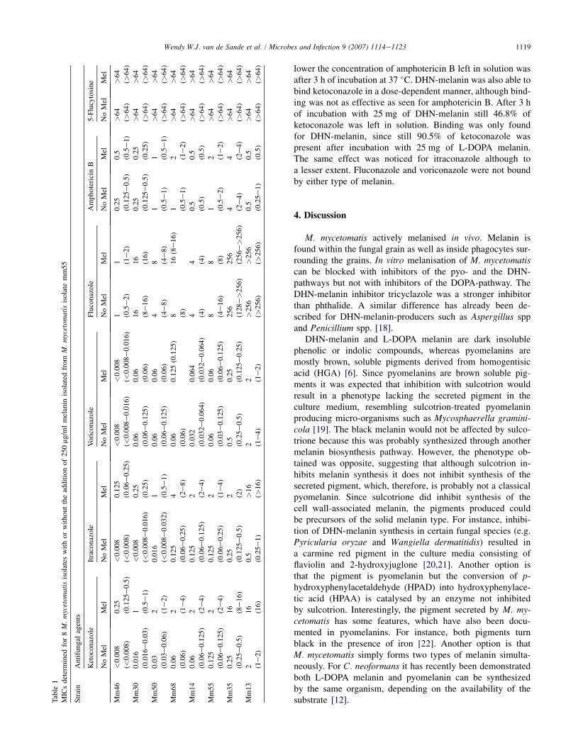
not shown). No significant differences (meaning less than onetwo-fold dilution step) in MIC were noted for amphotericin B,5-flucytosine, fluconazole and voriconazole upon preincuba-tion with either type of melanin or albumin. However, a signif-icant difference in the MICs for itraconazole and ketoconazole
Fig. 4. Antioxidant activity of melanin isolated from M. mycetomatis strain
mm55. Fungal melanin in different concentrations was incubated with
6.4 mM permanganate. After 10 min incubation TNB was added and mea-
sured. TNB oxidation was calculated by the following formula: (OD412 of
sample)/(OD412 of oxidized TNB) * 100% (-). The concentration permanga-
nate reduced by melanin was stated as well (6). Overall, the more permanga-
nate was reduced by melanin the less TNB was oxidized.
was documented in the presence of DHN-melanin isolatedfrom either M. mycetomatis (Fig. 5) or A. fumigatus (resultsnot shown). This effect was not documented when assayswere supplemented with L-DOPA melanin or BSA. Due tothe effect of M. mycetomatis melanin, itraconazole MICswere 16-fold increased, an even more significant increasewas noted for ketoconazole (32-fold). These elevations inMIC were comparable to the elevations found when A. fumiga-tus melanin was added. When taking into account that azoleMICs of 2 mg/ml or higher may be associated with failure oftherapy, this implies that upon melanin supplementation 6out of 8 and 5 out of 8 strains should be considered clinicallyresistant to ketoconazole and itraconazole, respectively [17].Without melanin supplementation, only a single strain shouldbe considered resistant to ketoconazole.
3.5. M. mycetomatis melanin binds amphotericin B,itraconazole and ketoconazole
Since M. mycetomatis MICs for itraconazole and ketocona-zole were increased with supplementation of melanin, it wasinvestigated if DHN- and L-DOPA-melanin bound amphoter-icin B, itraconazole, ketoconazole, fluconazole and voricona-zole. In Fig. 6 it is seen that amphotericin B is bound byboth DHN-melanin and L-DOPA melanin. Binding occurredin a dose-dependent manner, the more melanin present, the
1119Wendy W.J. van de Sande et al. / Microbes and Infection 9 (2007) 1114e1123
Table
1
MIC
sdet
erm
ined
for
8M
.m
ycet
omat
isis
ola
tes
wit
ho
rw
ith
out
the
add
itio
no
f2
50
mg
/ml
mel
anin
iso
late
dfr
omM
.m
ycet
omat
isis
olat
em
m5
5
Str
ain
An
tifu
nga
lag
ents
Ket
oco
naz
ole
Itra
con
azo
leV
ori
con
azo
leF
luco
naz
ole
Am
ph
ote
rici
nB
5-F
lucy
tosi
ne
No
Mel
Mel
No
Mel
Mel
No
Mel
Mel
No
Mel
Mel
No
Mel
Mel
No
Mel
Mel
Mm
46
<0
.00
8
(<0
.008
)
0.2
5
(0.1
25e
0.5
)
<0
.00
8
(<0
.008
)
0.1
25
(0.0
6e0
.25)
<0
.00
8
(<0
.008
e0
.016
)
<0
.008
(<0
.008
e0
.01
6)
1 (0.5
e2
)
1 (1e
2)
0.2
5
(0.1
25e
0.5
)
0.5
(0.5
e1
)
>6
4
(>6
4)
>6
4
(>6
4)
Mm
30
0.0
16
(0.0
16e
0.0
3)
1 (0.5
e1
)
<0
.00
8
(<0
.008
e0
.016
)
0.2
5
(0.2
5)
0.0
6
(0.0
6e0
.12
5)
0.0
6
(0.0
6)
16
(8e
16
)
16
(16
)
0.2
5
(0.1
25e
0.5
)
0.2
5
(0.2
5)
>6
4
(>6
4)
>6
4
(>6
4)
Mm
50
0.0
3
(0.0
3e0
.06
)
2 (1e
2)
0.0
16
(<0
.008
e0
.032
)
1 (0.5
e1
)
0.0
6
(0.0
6e0
.12
5)
0.0
6
(0.0
6)
4 (4e
8)
8 (4e
8)
1 (0.5
e1
)
1 (0.5
e1
)
>6
4
(>6
4)
>6
4
(>6
4)
Mm
68
0.0
6
(0.0
6)
2 (1e
4)
0.1
25
(0.0
6e0
.25
)
4 (2e
8)
0.0
6
(0.0
6)
0.1
25(0
.12
5)8 (8
)
16
(8e
16
)1 (0
.5e
1)
2 (1e
2)
>6
4
(>6
4)
>6
4
(>6
4)
Mm
14
0.0
6
(0.0
6e0
.12
5)
2 (2e
4)
0.1
25
(0.0
6e0
.12
5)
2 (2e
4)
0.0
32
(0.0
32e
0.0
64)
0.0
64
(0.0
32e
0.0
64)
4 (4)
4 (4)
0.5
(0.5
)
0.5
(0.5
)
>6
4
(>6
4)
>6
4
(>6
4)
Mm
55
0.1
25
(0.0
6e0
.12
5)
2 (2e
4)
0.1
25
(0.0
6e0
.25
)
2 (1e
4)
0.0
6
(0.0
3e0
.12
5)
0.0
6
(0.0
6e0
.125
)
8 (4e
16
)
8 (8)
1 (0.5
e2
)
2 (1e
2)
>6
4
(>6
4)
>6
4
(>6
4)
Mm
35
0.2
5
(0.2
5e0
.5)
16
(8e
16
)
0.2
5
(0.1
25e
0.5
)
2 (2)
0.5
(0.2
5e0
.5)
0.2
5
(0.1
25e
0.2
5)
25
6
(12
8e>
25
6)
25
6
(25
6e>
25
6)
4 (2e
4)
4 (2e
4)
>6
4
(>6
4)
>6
4
(>6
4)
Mm
13
2 (1e
2)
16
(16
)
0.5
(0.2
5e1
)
>1
6
(>1
6)
2 (1e
4)
2 (1e
2)
>2
56
(>2
56
)
>2
56
(>2
56
)
0.5
(0.2
5e1
)
0.5
(0.5
)
>6
4
(>6
4)
>6
4
(>6
4)
lower the concentration of amphotericin B left in solution wasafter 3 h of incubation at 37 �C. DHN-melanin was also able tobind ketoconazole in a dose-dependent manner, although bind-ing was not as effective as seen for amphotericin B. After 3 hof incubation with 25 mg of DHN-melanin still 46.8% ofketoconazole was left in solution. Binding was only foundfor DHN-melanin, since still 90.5% of ketoconazole waspresent after incubation with 25 mg of L-DOPA melanin.The same effect was noticed for itraconazole although toa lesser extent. Fluconazole and voriconazole were not boundby either type of melanin.
4. Discussion
M. mycetomatis actively melanised in vivo. Melanin isfound within the fungal grain as well as inside phagocytes sur-rounding the grains. In vitro melanisation of M. mycetomatiscan be blocked with inhibitors of the pyo- and the DHN-pathways but not with inhibitors of the DOPA-pathway. TheDHN-melanin inhibitor tricyclazole was a stronger inhibitorthan phthalide. A similar difference has already been de-scribed for DHN-melanin-producers such as Aspergillus sppand Penicillium spp. [18].
DHN-melanin and L-DOPA melanin are dark insolublephenolic or indolic compounds, whereas pyomelanins aremostly brown, soluble pigments derived from homogentisicacid (HGA) [6]. Since pyomelanins are brown soluble pig-ments it was expected that inhibition with sulcotrion wouldresult in a phenotype lacking the secreted pigment in theculture medium, resembling sulcotrion-treated pyomelaninproducing micro-organisms such as Mycosphaerella gramini-cola [19]. The black melanin would not be affected by sulco-trione because this was probably synthesized through anothermelanin biosynthesis pathway. However, the phenotype ob-tained was opposite, suggesting that although sulcotrion in-hibits melanin synthesis it does not inhibit synthesis of thesecreted pigment, which, therefore, is probably not a classicalpyomelanin. Since sulcotrione did inhibit synthesis of thecell wall-associated melanin, the pigments produced couldbe precursors of the solid melanin type. For instance, inhibi-tion of DHN-melanin synthesis in certain fungal species (e.g.Pyricularia oryzae and Wangiella dermatitidis) resulted ina carmine red pigment in the culture media consisting offlaviolin and 2-hydroxyjuglone [20,21]. Another option isthat the pigment is pyomelanin but the conversion of p-hydroxyphenylacetaldehyde (HPAD) into hydroxyphenylace-tic acid (HPAA) is catalysed by an enzyme not inhibitedby sulcotrion. Interestingly, the pigment secreted by M. my-cetomatis has some features, which have also been docu-mented in pyomelanins. For instance, both pigments turnblack in the presence of iron [22]. Another option is thatM. mycetomatis simply forms two types of melanin simulta-neously. For C. neoformans it has recently been demonstratedboth L-DOPA melanin and pyomelanin can be synthesizedby the same organism, depending on the availability of thesubstrate [12].

1120 Wendy W.J. van de Sande et al. / Microbes and Infection 9 (2007) 1114e1123
Fig. 5. MIC shift of M. mycetomatis DHN-melanin supplemented isolates (grey) and C. neoformans L-DOPA-melanin supplemented isolates (white) in comparison
with non-melanin supplemented isolates. The absolute difference in MIC was calculated by extracting the MIC for a certain strain without melanin supplemen-
tation from the MIC of that same strain with melanin supplementation. Differences were stated as the number of two-fold dilutions in difference. The average of
MIC difference for all strains tested was calculated and displayed in this figure. The dashed line in this figure shows the difference allowed between tests for the
same strain. Differences larger than one two-fold dilution are stated as significant. Furthermore in this figure the structure of the different antifungal agents are
shown. As is seen the structures of the azoles ketoconazole and itraconazole are very similar but they differ considerably from the structure formula’s of vorico-
nazole and fluconazole (which are also very similar to each other).
The DHN-melanin isolated from M. mycetomatis wasable to scavenge permanganate, one of the strongest oxi-dants known to modern chemists. During a mycetoma infec-tion, grains are usually in close contact with a layer ofneutrophils which actually secrete reactive oxidantspecies such as hypochlorous acid, hydrogen peroxide andreactive oxygen [23,24]. The fact that permanganate was ef-fectively melanin-scavenged implicates a strong protectivefunction for the melanin produced by M. mycetomatis duringinfection.
Supplementation of DHN-melanin in in vitro antifungalsusceptibility assays rendered M. mycetomatis less susceptibleto the antifungal agents itraconazole and ketoconazole, theprime agents used in clinical settings [1]. Not much hasbeen published on the effect of melanin on antifungal agents.Most of these studies were performed with melanised yeastcells versus non-melanised ones [25,26]. The fungi were cul-tured in chemically defined media to acquire a melanised phe-notype and were transferred afterwards into adjusted culturemedia to determine the MICs. Interestingly, the usually melan-ised isolates of C. neoformans and Histoplasma capsulatumwere not able to produce melanin in these test media, noteven when additional substrate was added [27,28]. During cul-tivation the melanised phenotype disappeared and all resultingoffspring was non-melanised [27]. This could explain why nodifferences in MICs were found. Similar to other fungi used inmelanin protection studies thus far, M. mycetomatis does notform melanin in RPMI culture media, the medium used in
the M38A protocol from the Clinical and Laboratory Stan-dards Institute (CLSI) during drug susceptibility testing[29]. In order to establish the effect of melanin on theMIC we chose to add purified M. mycetomatis melanin tothe culture medium as an in vitro simulation of the in vivosituation during infection. Furthermore, it was not feasibleto determine MICs on minimal medium. Melanisation tookat least one month and when melanised cells were inoculatedon a fresh minimal medium agar plate, the resulting colonywas not melanised. Only after a month of cultivation, melan-ised phenotypes were seen.
Although for other fungi no MIC shift was described, it wasshown that the time required for killing of melanised cells wasmuch longer [25,27,30]. This was thought to be the result ofbinding of antifungal agents to melanin [25,27,28,30]. Wehere demonstrate that co-incubation of DHN-melanin with ei-ther itraconazole or ketoconazole during susceptibility testingresults in an MIC shift. However, itraconazole and ketocona-zole are two azoles with high protein binding capacity. Invivo serum protein binding is more than 90% for both azoles.In vitro protein binding has been described to affect MICs forketoconazole, fluconazole and amphotericin B but not for itra-conazole [31]. When we performed our antifungal assay with250 mg/ml albumin, no shift in MIC was observed and wetherefore do not expect that the MIC shifts we observedwith melanin are confounded by low protein amounts. The ef-fect appeared melanin-specific since this increase was alsonoted when A. fumigatus DHN-melanin was added to the

1121Wendy W.J. van de Sande et al. / Microbes and Infection 9 (2007) 1114e1123
Fig. 6. Antifungal binding capacity of DHN-melanin isolated from M. mycetomatis (C) and L-DOPA-melanin isolated from C. neoformans (B). Fungal melanin
in different concentrations was incubated with 800 mg/ml of amphotericin B (A); ketoconazole (B); itraconazole (C); voriconazole (D); or fluconazole (E) for 3 h at
37 �C. After incubation melanin was pelleted and the concentration of each antifungal agent left in solution was determined with agar diffusion. Curves represent
the concentration antifungal agent left in solution after incubation with various concentrations of melanin compared to the same antifungal agent without melanin.
culture medium but no increase in MIC was noted when L-DOPA melanin was added. The fact that we did not documentan amphotericin B MIC shift for L-DOPA melanin was unex-pected since there is evidence that binding of amphotericin Bor caspofungin to L-DOPA melanin significantly reduced itsefficacy in time-kill assays [28]. In our own binding-assaywe also found that amphotericin B is bound by both L-DOPA- and DHN-melanin. This binding did not result ina MIC shift when our antifungal susceptibility assay was
supplemented with either type of melanin. In our assay themelanin remains in the solution during the whole experiment,in the time-kill assays performed by others melanin was re-moved after a period of incubation prior to MIC determina-tion. Apparently, when leaving melanin in the solution, theamphotericin B bound by melanin might still be able to inhibitM. mycetomatis growth. In previous studies no binding of theazoles voriconazole, fluconazole and itraconazole and the an-timetabolite 5-flucytosine to L-DOPA melanin was noted

1122 Wendy W.J. van de Sande et al. / Microbes and Infection 9 (2007) 1114e1123
[25,28]. This was in agreement with the results we obtained inour binding-assay. Only DHN-melanin was able to bind theazoles itraconazole and ketoconazole, no evidence of bindingto L-DOPA melanin was found in either the binding assay orthe MIC assay. This suggests that binding of itraconazole andketoconazole was specific for DHN-melanin, which couldexplain that the only other MIC shift for itraconazole underinfluence of melanin was described in a DHN-melanin-producing fungus (Wangiella dermatitidis) [32]. Since allazoles have a similar working mechanism, the differences inmelanin binding should be traceable to the molecular structureof the azoles. Itraconazole and ketoconazole, which are struc-turally similar, differ considerably from fluconazole and itsderivate voriconazole [33]. All azoles possess an azole-ring,the active domain of the molecule. The azole-ring of the imid-azole ketoconazole is slightly different from the other three,harbouring only two nitrogen atoms instead of three. The dif-ference in MIC effect is probably not functionally associatedwith the presence of an azole-ring. Interaction of melaninwith itraconazole and ketoconazole is probably mediated bythe dichlorobenzene ring or the long polyaromatic side chainor a combination of the two (see Fig. 5). Binding of the azolesto melanin in vitro results in a decrease in MIC because of thelack of accessibility to the fungus. However, by binding theazole to the grain an artificial ‘‘slow release’’ capsule mightbe created, resulting in a constant local release of the antifun-gal agent. This implies that the effect of the binding of itraco-nazole and ketoconazole to DHN-melanin in vitro is not easilytranslated to its effect in vivo. Further studies are needed to es-tablish the precise physiological role of melanin in the fungalgrain.
In conclusion, we demonstrated that M. mycetomatis ismelanised in vivo and we identified the biochemical pathwaysleading to this melanisation. Melanisation of M. mycetomatisoffers protection to strong oxidants and the antifungal agentsitraconazole and ketoconazole in vitro. Its protective role invivo still needs to be established.
Acknowledgements
We thank Dr. Nosanchuk from the Albert Einstein Collegeof Medicine in New York for the kind gift of melanin-specificantibody 6D2.
References
[1] A.O. Ahmed, W. van Leeuwen, A. Fahal, W. van de Sande, H. Verbrugh,
A. van Belkum, Mycetoma caused by Madurella mycetomatis: a ne-
glected infectious burden, Lancet Infect. Dis. 4 (2004) 566e574.
[2] D.B. Wethered, M.A. Markey, R.J. Hay, E.S. Mahgoub, S.A. Gumaa, Ul-
trastructural and immunogenic changes in the formation of mycetoma
grains, J. Med. Vet. Mycol. 25 (1987) 39e46.
[3] K. Langfelder, M. Streibel, B. Jahn, G. Haase, A.A. Brakhage, Biosyn-
thesis of fungal melanins and their importance for human pathogenic
fungi, Fungal Genet. Biol. 38 (2003) 143e158.
[4] A. Casadevall, A.L. Rosas, J.D. Nosanchuk, Melanin and virulence in
Cryptococcus neoformans, Curr. Opin. Microbiol. 3 (2000) 354e358.
[5] E.S. Jacobson, Pathogenic roles for fungal melanins, Clin. Microbiol.
Rev. 13 (2000) 708e717.
[6] J.D. Nosanchuk, A. Casadevall, The contribution of melanin to microbial
pathogenesis, Cell. Microbiol. 5 (2003) 203e223.
[7] A.L. Rosas, J.D. Nosanchuk, A. Casadevall, Passive immunization with mel-
anin-binding monoclonal antibodies prolongs survival of mice with lethal
Cryptococcus neoformans infection, Infect. Immun. 69 (2001) 3410e3412.
[8] K.J. Kwon-Chung, I. Polacheck, T.J. Popkin, Melanin-lacking mutants of
Cryptococcus neoformans and their virulence for mice, J. Bacteriol. 150
(1982) 1414e1421.
[9] J.D. Nosanchuk, R. Ovalle, A. Casadevall, Glyphosate inhibits melaniza-
tion of Cryptococcus neoformans and prolongs survival of mice after sys-
temic infection, J. Infect. Dis. 183 (2001) 1093e1099.
[10] C.E. Turick, L.S. Tisa, F. Caccavo Jr., Melanin production and use as
a soluble electron shuttle for Fe(III) oxide reduction and as a terminal
electron acceptor by Shewanella algae BrY, Appl. Environ. Microbiol.
68 (2002) 2436e2444.
[11] A. Carreira, L.M. Ferreira, V. Loureiro, Brown pigments produced by
Yarrowia lipolytica result from extracellular accumulation of homogenti-
sic acid, Appl. Environ. Microbiol. 67 (2001) 3463e3468.
[12] S. Frases, A. Salazar, E. Dadachova, A. Casadevall, Cryptococcus neofor-
mans can utilize the bacterial melanin precursor homogentisic acid for
fungal melanogenesis, Appl. Environ. Microbiol. 73 (2007) 615e621.
[13] J.D. Nosanchuk, J.J. Yu, C.Y. Hung, A. Casadevall, G.T. Cole, Cocci-
dioides posadasii produces melanin in vitro and during infection, Fungal
Genet. Biol. 44 (6) (2006) 517e520.
[14] S. Youngchim, R. Morris-Jones, R.J. Hay, A.J. Hamilton, Production of
melanin by Aspergillus fumigatus, J. Med. Microbiol. 53 (2004) 175e181.
[15] R.G.R. de Cassia, S.R. Pombeiro-Sponchiado, Antioxidant activity of the
melanin pigment extracted from Aspergillus nidulans, Biol. Pharm. Bull.
28 (2005) 1129e1131.
[16] W.W. van de Sande, A. Luijendijk, A.O. Ahmed, I.A. Bakker-Wouden-
berg, A. van Belkum, Testing of the in vitro susceptibilities of Madurella
mycetomatis to six antifungal agents by using the sensititre system in
comparison with a viability-based 2,3-bis(2-methoxy-4-nitro-5-sulfo-
phenyl)-5-[(phenylamino)carbonyl]-2H-tetrazolium hydroxide (XTT) as-
say and a modified NCCLS method, Antimicrob. Agents Chemother. 49
(2005) 1364e1368.
[17] S.G. Revankar, Therapy of infections caused by dematiaceous fungi,
Expert. Rev. Anti-Infect. Ther. 3 (2005) 601e612.
[18] M.H. Wheeler, M.A. Klich, The effects of tricyclazole, pyroquilon,
phthalide, and related fungicides on the production of conidial wall pig-
ments by Penicillium and Aspergillus species, Pestic. Biochem. Physiol.
52 (1995) 125e136.
[19] J. Keon, J. Hargreaves, Isolation and heterologous expression of a gene
encoding 4-hydroxyphenylpyruvate dioxygenase from the wheat leaf-
spot pathogen, Mycosphaerella graminicola, FEMS Microbiol. Lett.
161 (1998) 337e343.
[20] Y. Kurahaschi, R. Pontzen, Carpropamid: a new melanin biosynthesis in-
hibitor, Pflanzenschutz-Nachrichten Bayer 51 (1998) 245e256.
[21] P.A. Geis, M.H. Wheeler, P.J. Szaniszlo, Pentaketide metabolites of mel-
anin synthesis in the dematiaceous fungus Wangiella dermatitidis, Arch.
Microbiol. 137 (1984) 324e328.
[22] A. Gibello, E. Ferrer, J. Sanz, M. Martin, Polymer production by Klebsi-
ella pneumoniae 4-hydroxyphenylacetic acid hydroxylase genes cloned
in Escherichia coli, Appl. Environ. Microbiol. 61 (1995) 4167e4171.
[23] A.H. Fahal, E.A. el Toum, A.M. el Hassan, E.S. Mahgoub, S.A. Gumaa,
The host tissue reaction to Madurella mycetomatis: new classification,
J. Med. Vet. Mycol. 33 (1995) 15e17.
[24] R. Crameri, K. Blaser, Allergy and immunity to fungal infections and
colonization, Eur. Respir. J. 19 (2002) 151e157.
[25] D. van Duin, W. Cleare, O. Zaragoza, A. Casadevall, J.D. Nosanchuk, Ef-
fects of voriconazole on Cryptococcus neoformans, Antimicrob. Agents
Chemother. 48 (2004) 2014e2020.
[26] M.B. da Silva, A.F. Marques, J.D. Nosanchuk, A. Casadevall,
L.R. Travassos, C.P. Taborda, Melanin in the dimorphic fungal pathogen
Paracoccidioides brasiliensis: effects on phagocytosis, intracellular
resistance and drug susceptibility, Microbes Infect. 8 (2006) 197e205.

1123Wendy W.J. van de Sande et al. / Microbes and Infection 9 (2007) 1114e1123
[27] R. Ikeda, T. Sugita, E.S. Jacobson, T. Shinoda, Effects of melanin upon
susceptibility of Cryptococcus to antifungals, Microbiol. Immunol. 47
(2003) 271e277.
[28] D. van Duin, A. Casadevall, J.D. Nosanchuk, Melanization of Cryptococ-
cus neoformans and Histoplasma capsulatum reduces their susceptibili-
ties to amphotericin B and caspofungin, Antimicrob. Agents
Chemother. 46 (2002) 3394e3400.
[29] CLSI, Reference Method for Broth Dilution Antifungal Susceptibility
Testing of Filamentous Fungi; Approved Standard, National Committee
for Clinical Laboratory Standards, Wayne, Pennsylvania, USA, 2002,
Vol. 22.
[30] Y. Wang, A. Casadevall, Growth of Cryptococcus neoformans in presence
of L-dopa decreases its susceptibility to amphotericin B, Antimicrob.
Agents Chemother. 38 (1994) 2648e2650.
[31] M. Schafer-Korting, H.C. Korting, W. Rittler, W. Obermuller, Influence
of serum protein binding on the in vitro activity of anti-fungal agents, In-
fection 23 (1995) 292e297.
[32] A. Polak, D.M. Dixon, Loss of melanin in Wangiella dermatitidis does
not result in greater susceptibility to antifungal agents, Antimicrob.
Agents Chemother. 33 (1989) 1639e1640.
[33] R. Herbrecht, Voriconazole: therapeutic review of a new azole antifungal,
Expert. Rev. Anti-Infect. Ther. 2 (2004) 485e497.
Copyright © 2022 FDOKUMEN