Properties and Functions of Melanin Pigment from Klebsiella ...
Upload
independentCategory
view
0download
0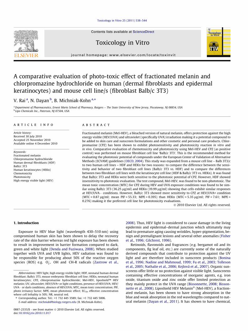
Toxicology in Vitro 25 (2011) 538–544
Contents lists available at ScienceDirect
Toxicology in Vitro
journal homepage: www.elsevier .com/locate / toxinvi t
A comparative evaluation of photo-toxic effect of fractionated melanin andchlorpromazine hydrochloride on human (dermal fibroblasts and epidermalkeratinocytes) and mouse cell line/s (fibroblast Balb/c 3T3)
V. Rai a, N. Dayan b, B. Michniak-Kohn a,⇑a Department of Pharmaceutics, Ernest Mario School of Pharmacy, Rutgers – The State University of New Jersey, Piscataway, NJ 08854, USAb Lipo Chemicals Inc., Paterson, NJ 07504, USA
a r t i c l e i n f o
Article history:Received 30 July 2010Accepted 25 November 2010Available online 4 December 2010
Keywords:Fractionated melaninChlorpromazine hydrochlorideHuman dermal fibroblasts (HDF)Balb/c 3T3Human keratinocytes (HEKn)ChemotoxicityPhototoxicityHigh energy visible light (HEV)
0887-2333/$ - see front matter � 2010 Elsevier Ltd. Adoi:10.1016/j.tiv.2010.11.017
Abbreviations: HEV light, high energy visible light;fibroblast; Balb/c 3T3, mouse embryonic fibroblast celkeratinocytes; CPZ, chlorpromazine hydrochloride;melanin; UV, ultraviolet; HEV/UVA+ or light conditionUVA� or dark conditions, absence of HEV/UVA; MTC, mphoto irritancy factor; MPE, mean phototoxic effect;where cell lethality is 50%; NR, neutral red.⇑ Corresponding author. Tel.: +1 732 445 3589; fax
E-mail address: [email protected] (B.
a b s t r a c t
Fractionated melanin (Mel-HEV), a bleached version of natural melanin, offers protection against the highenergy visible (HEV/UVA) and ultraviolet (specifically UVA) irradiation making it a potential compound tobe added to skin care and sunscreen formulations and other cosmetic and personal care products. Chlor-promazine (CPZ) has been shown to exhibit photosensitivity and phototoxicity reaction in vitro andin vivo. Comparative evaluation of chemotoxicity and phototoxicity using Mel-HEV and CPZ (as positivecontrol) was performed on mouse fibroblast cell line ‘Balb/c 3T3’. This is the recommended method forevaluating the phototoxic potential of compounds under the European Center of Validation of AlternativeMethods (ECVAM) guidelines (OECD, 2004). This study was expanded from a mouse cell line – Balb 3T3/cto two human cell lines – HDF and HEKn for two reasons: to compare the difference between the sensi-tivity and behavior of two fibroblast cell lines (Balb/c 3T3 vs. HDF) and to compare the differencesbetween two fibroblast cell lines with the keratinocyte cell line (HDF & Balb/c 3T3 vs. HEKn). It was foundthat Balb/c 3T3 and HEKn were both sensitive to the phototoxic potential of CPZ. However, HDF showedinsensitivity to phototoxic evaluation. The test compound, Mel-HEV, was found to be non-phototoxic. Themean toxic concentration (MTC) for CPZ during HEV and UVA exposure conditions was found to be sim-ilar using Balb/c 3T3 (36.25 lg/ml) and HEKn (39.99 lg/ml) showing that cells exhibit similar responsesat HEV/UVA� conditions. However, Balb/c 3T3 showed more sensitivity to CPZ at HEV/UVA+ condition(MTC = 0.87 lg/ml; mean PIF = 55.33; MPE = 0.395) than HEKn (MTC = 5.35 lg/ml; PIF = 7.61; MPE =0.276) making it the preferred cell line for phototoxicity evaluations.
� 2010 Elsevier Ltd. All rights reserved.
1. Introduction 2008). Thus, HEV light is considered to cause damage in the living
Exposure to HEV blue light (wavelength 430–510 nm) usingcompromised human skin has been shown to delay the recoveryrate of the skin barrier whereas red light exposure has been shownto result in improvement in barrier formation compared to dark,green and white light (Denda and Fuziwara, 2008). When studiedtogether with UVA and UVB lights, HEV radiation was found tobe responsible for producing about 50% of the reactive oxygenspecies (ROS) e.g. �O�2 , �OH and �CH-R radicals (Zastrow et al.,
ll rights reserved.
HDF, neonatal human dermall line; HEKn, neonatal humanMel-HEV, liposhield™ HEV
s, presence of HEV/UVA; HEV/ean toxic concentration; PIF,
EC50, effective concentration
: +1 732 445 5006.Michniak-Kohn).
epidermis and epidermal–dermal junction which ultimately maylead to premature aging causing wrinkles, hyper-pigmentation, be-nign and premalignant lesions and several other anomalies (Fisheret al., 1996; Gilchrest, 1996).
Retinoids, flavonoids and fragrances (e.g. bergamot oil and itscomponents, fig leaf oil, etc.) are currently some of the naturallyderived compounds that contribute to protection against the UVlight and are therefore included in sunscreen products (Boninaet al., 1996; Nadine and Mahmoud, 1999; Fu et al., 2003; Tollesonet al., 2005; Nathalie et al., 2006; Kejlová et al., 2007). Organic sun-screens offer little or no protection against visible light. Sunscreenscontaining effective concentrations of inorganic agents, e.g. ironoxide, titanium oxide and zinc oxide offer limited protection asthey mainly protect in the UVA range (Bissonnette, 2008; Bisson-nette et al., 2008). Liposhield HEV Melanin� (Mel-HEV), a fraction-ated melanin, has been shown to have strong absorption in theblue and weak absorption in the red wavelengths compared to nat-ural melanin (Dayan et al., 2011). It has shown to have chemical,
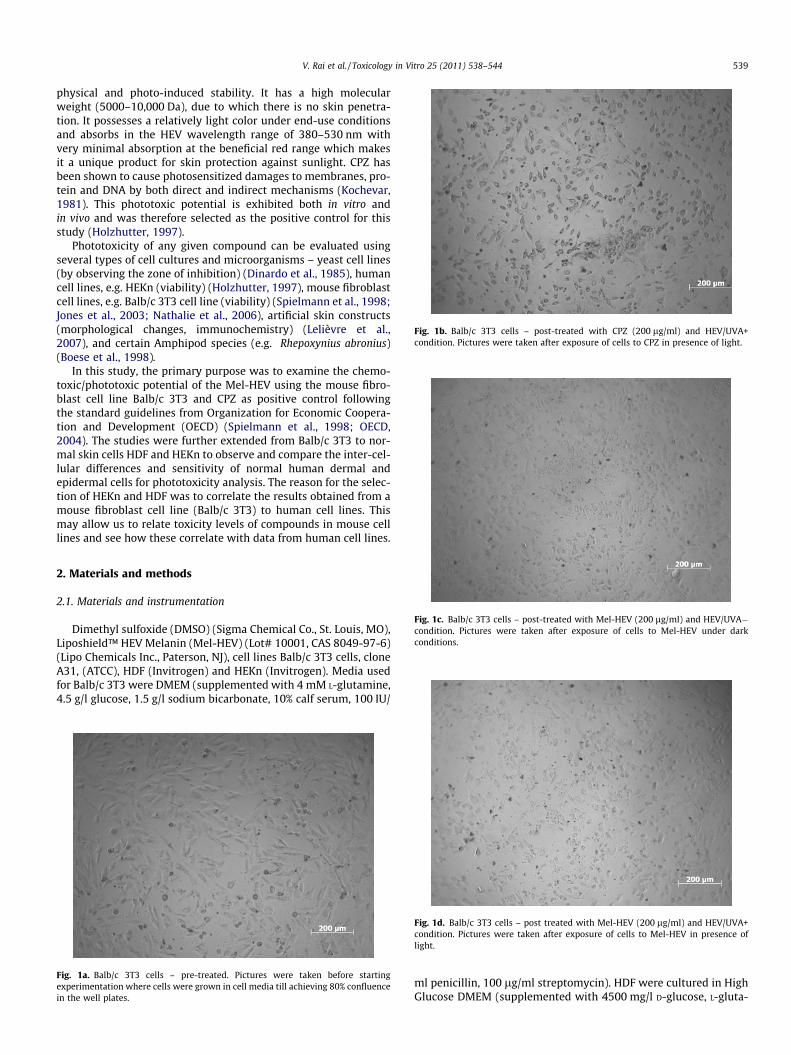
Fig. 1b. Balb/c 3T3 cells – post-treated with CPZ (200 lg/ml) and HEV/UVA+condition. Pictures were taken after exposure of cells to CPZ in presence of light.
V. Rai et al. / Toxicology in Vitro 25 (2011) 538–544 539
physical and photo-induced stability. It has a high molecularweight (5000–10,000 Da), due to which there is no skin penetra-tion. It possesses a relatively light color under end-use conditionsand absorbs in the HEV wavelength range of 380–530 nm withvery minimal absorption at the beneficial red range which makesit a unique product for skin protection against sunlight. CPZ hasbeen shown to cause photosensitized damages to membranes, pro-tein and DNA by both direct and indirect mechanisms (Kochevar,1981). This phototoxic potential is exhibited both in vitro andin vivo and was therefore selected as the positive control for thisstudy (Holzhutter, 1997).
Phototoxicity of any given compound can be evaluated usingseveral types of cell cultures and microorganisms – yeast cell lines(by observing the zone of inhibition) (Dinardo et al., 1985), humancell lines, e.g. HEKn (viability) (Holzhutter, 1997), mouse fibroblastcell lines, e.g. Balb/c 3T3 cell line (viability) (Spielmann et al., 1998;Jones et al., 2003; Nathalie et al., 2006), artificial skin constructs(morphological changes, immunochemistry) (Lelièvre et al.,2007), and certain Amphipod species (e.g. Rhepoxynius abronius)(Boese et al., 1998).
In this study, the primary purpose was to examine the chemo-toxic/phototoxic potential of the Mel-HEV using the mouse fibro-blast cell line Balb/c 3T3 and CPZ as positive control followingthe standard guidelines from Organization for Economic Coopera-tion and Development (OECD) (Spielmann et al., 1998; OECD,2004). The studies were further extended from Balb/c 3T3 to nor-mal skin cells HDF and HEKn to observe and compare the inter-cel-lular differences and sensitivity of normal human dermal andepidermal cells for phototoxicity analysis. The reason for the selec-tion of HEKn and HDF was to correlate the results obtained from amouse fibroblast cell line (Balb/c 3T3) to human cell lines. Thismay allow us to relate toxicity levels of compounds in mouse celllines and see how these correlate with data from human cell lines.
Fig. 1c. Balb/c 3T3 cells – post-treated with Mel-HEV (200 lg/ml) and HEV/UVA�condition. Pictures were taken after exposure of cells to Mel-HEV under darkconditions.
2. Materials and methods
2.1. Materials and instrumentation
Dimethyl sulfoxide (DMSO) (Sigma Chemical Co., St. Louis, MO),Liposhield™ HEV Melanin (Mel-HEV) (Lot# 10001, CAS 8049-97-6)(Lipo Chemicals Inc., Paterson, NJ), cell lines Balb/c 3T3 cells, cloneA31, (ATCC), HDF (Invitrogen) and HEKn (Invitrogen). Media usedfor Balb/c 3T3 were DMEM (supplemented with 4 mM L-glutamine,4.5 g/l glucose, 1.5 g/l sodium bicarbonate, 10% calf serum, 100 IU/
Fig. 1a. Balb/c 3T3 cells – pre-treated. Pictures were taken before startingexperimentation where cells were grown in cell media till achieving 80% confluencein the well plates.
Fig. 1d. Balb/c 3T3 cells – post treated with Mel-HEV (200 lg/ml) and HEV/UVA+condition. Pictures were taken after exposure of cells to Mel-HEV in presence oflight.
ml penicillin, 100 lg/ml streptomycin). HDF were cultured in HighGlucose DMEM (supplemented with 4500 mg/l D-glucose, L-gluta-
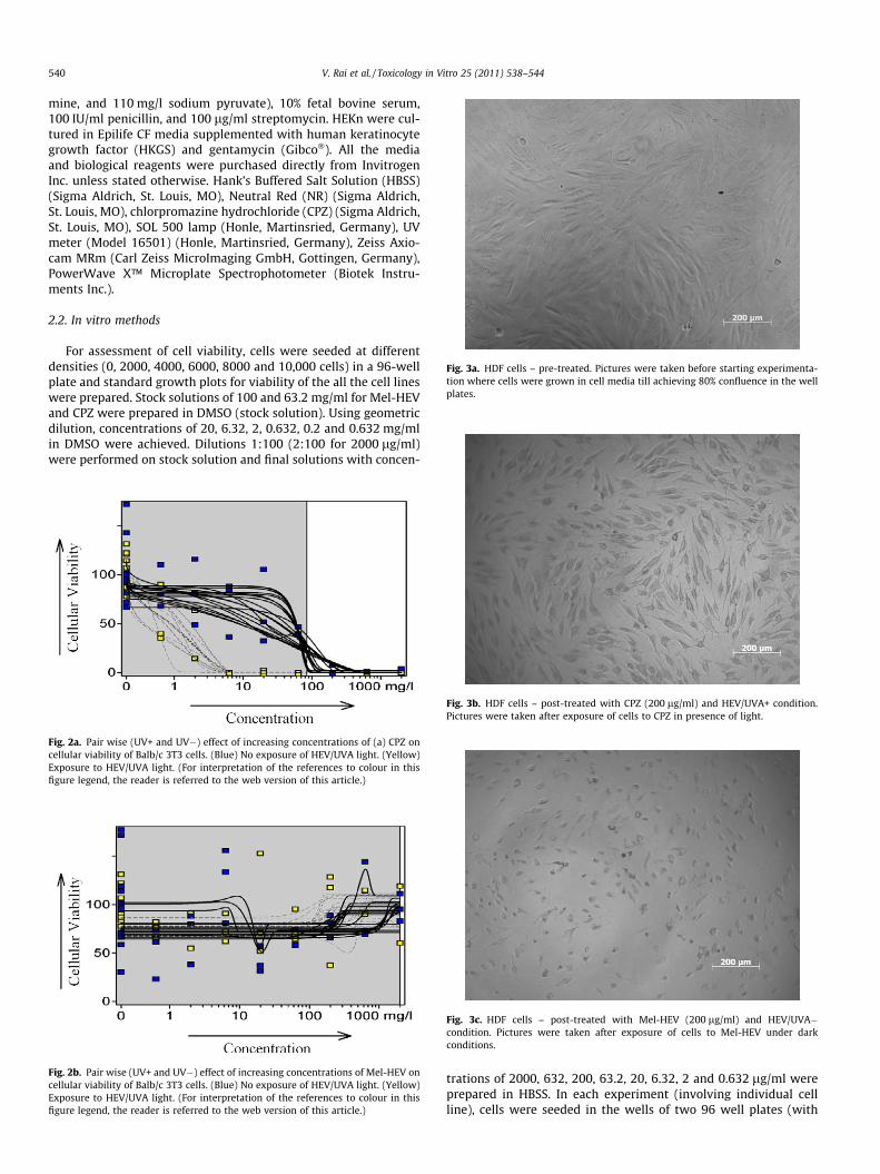
Fig. 3a. HDF cells – pre-treated. Pictures were taken before starting experimenta-tion where cells were grown in cell media till achieving 80% confluence in the wellplates.
540 V. Rai et al. / Toxicology in Vitro 25 (2011) 538–544
mine, and 110 mg/l sodium pyruvate), 10% fetal bovine serum,100 IU/ml penicillin, and 100 lg/ml streptomycin. HEKn were cul-tured in Epilife CF media supplemented with human keratinocytegrowth factor (HKGS) and gentamycin (Gibco�). All the mediaand biological reagents were purchased directly from InvitrogenInc. unless stated otherwise. Hank’s Buffered Salt Solution (HBSS)(Sigma Aldrich, St. Louis, MO), Neutral Red (NR) (Sigma Aldrich,St. Louis, MO), chlorpromazine hydrochloride (CPZ) (Sigma Aldrich,St. Louis, MO), SOL 500 lamp (Honle, Martinsried, Germany), UVmeter (Model 16501) (Honle, Martinsried, Germany), Zeiss Axio-cam MRm (Carl Zeiss MicroImaging GmbH, Gottingen, Germany),PowerWave X™ Microplate Spectrophotometer (Biotek Instru-ments Inc.).
2.2. In vitro methods
For assessment of cell viability, cells were seeded at differentdensities (0, 2000, 4000, 6000, 8000 and 10,000 cells) in a 96-wellplate and standard growth plots for viability of the all the cell lineswere prepared. Stock solutions of 100 and 63.2 mg/ml for Mel-HEVand CPZ were prepared in DMSO (stock solution). Using geometricdilution, concentrations of 20, 6.32, 2, 0.632, 0.2 and 0.632 mg/mlin DMSO were achieved. Dilutions 1:100 (2:100 for 2000 lg/ml)were performed on stock solution and final solutions with concen-
Fig. 3b. HDF cells – post-treated with CPZ (200 lg/ml) and HEV/UVA+ condition.Pictures were taken after exposure of cells to CPZ in presence of light.
Fig. 3c. HDF cells – post-treated with Mel-HEV (200 lg/ml) and HEV/UVA�condition. Pictures were taken after exposure of cells to Mel-HEV under darkconditions.
Fig. 2b. Pair wise (UV+ and UV�) effect of increasing concentrations of Mel-HEV oncellular viability of Balb/c 3T3 cells. (Blue) No exposure of HEV/UVA light. (Yellow)Exposure to HEV/UVA light. (For interpretation of the references to colour in thisfigure legend, the reader is referred to the web version of this article.)
Fig. 2a. Pair wise (UV+ and UV�) effect of increasing concentrations of (a) CPZ oncellular viability of Balb/c 3T3 cells. (Blue) No exposure of HEV/UVA light. (Yellow)Exposure to HEV/UVA light. (For interpretation of the references to colour in thisfigure legend, the reader is referred to the web version of this article.)
trations of 2000, 632, 200, 63.2, 20, 6.32, 2 and 0.632 lg/ml wereprepared in HBSS. In each experiment (involving individual cellline), cells were seeded in the wells of two 96 well plates (with
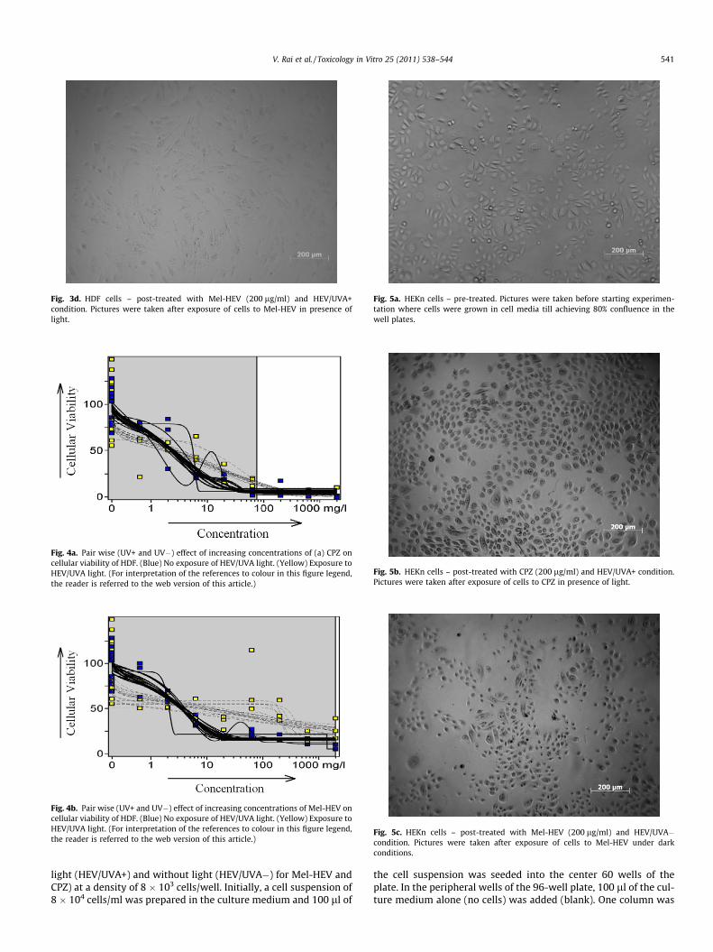
Fig. 3d. HDF cells – post-treated with Mel-HEV (200 lg/ml) and HEV/UVA+condition. Pictures were taken after exposure of cells to Mel-HEV in presence oflight.
Fig. 5a. HEKn cells – pre-treated. Pictures were taken before starting experimen-tation where cells were grown in cell media till achieving 80% confluence in thewell plates.
Fig. 5b. HEKn cells – post-treated with CPZ (200 lg/ml) and HEV/UVA+ condition.Pictures were taken after exposure of cells to CPZ in presence of light.
Fig. 5c. HEKn cells – post-treated with Mel-HEV (200 lg/ml) and HEV/UVA�condition. Pictures were taken after exposure of cells to Mel-HEV under darkconditions.
Fig. 4a. Pair wise (UV+ and UV�) effect of increasing concentrations of (a) CPZ oncellular viability of HDF. (Blue) No exposure of HEV/UVA light. (Yellow) Exposure toHEV/UVA light. (For interpretation of the references to colour in this figure legend,the reader is referred to the web version of this article.)
Fig. 4b. Pair wise (UV+ and UV�) effect of increasing concentrations of Mel-HEV oncellular viability of HDF. (Blue) No exposure of HEV/UVA light. (Yellow) Exposure toHEV/UVA light. (For interpretation of the references to colour in this figure legend,the reader is referred to the web version of this article.)
V. Rai et al. / Toxicology in Vitro 25 (2011) 538–544 541
light (HEV/UVA+) and without light (HEV/UVA�) for Mel-HEV andCPZ) at a density of 8 � 103 cells/well. Initially, a cell suspension of8 � 104 cells/ml was prepared in the culture medium and 100 ll of
the cell suspension was seeded into the center 60 wells of theplate. In the peripheral wells of the 96-well plate, 100 ll of the cul-ture medium alone (no cells) was added (blank). One column was

Fig. 5d. HEKn cells – post-treated with Mel-HEV (200 lg/ml) and HEV/UVA+condition. Pictures were taken after exposure of cells to Mel-HEV in presence oflight.
Table 2Photo Irritation Factor (PIF) and Mean Photo Effect (MPE) values for CPZ and Mel-HEVgenerated by Phototox Software (Version 2.0) in Balb/c 3T3 cells.
Chemical PIFmean
Toxicityprobability
MPE Toxicityprobability
Phototoxicpotential
CPZ 55.33 1.0 0.395 1.0 YesMel-HEV C1235⁄ 0 �0.050 0 No
C1235⁄ equals no phototoxicity potential (Spielmann et al., 1998; Peters and Hol-zhutter, 2002).
Table 3EC50 values for CPZ and Mel-HEV generated by Phototox Software (Version 2.0) inHDF cells.
Chemical HEV/UVA light EC50 mean EC50 variance
CPZ � 2.51 0.82CPZ + 3.13 2.33Mel-HEV � 3.53 0.42Mel-HEV + 53.7 83.2
542 V. Rai et al. / Toxicology in Vitro 25 (2011) 538–544
used as negative control (cells in media and DMSO). The plateswere incubated for 24 h at 37 �C, 5% CO2 to allow a semi-confluentmonolayer to form (Figs. 1a, 3a and 5a). After 24 h, the plates wereobserved under a microscope to confirm that the cells are growingand pictures were taken with a camera-equipped microscope(Zeiss AxioCam GmbH). The culture medium in the wells was dec-anted, cells were washed with HBSS and 100 ll of the test solu-tions/controls prepared in HBSS were added to the wells (n = 3).After addition of the compound solutions, the plates were incu-bated for 60 min at 37 �C and 5% CO2. After 60 min, the plates wereexposed to solar radiations (SOL 500 lamp equipped with a H1 fil-ter; Honle, Martinsried, Germany) of 4.08 mW/cm2 (measuredusing a calibrated UV meter type 16501; Honle, Martinsried, Ger-many) for 50 min through the plate lid leading to a cumulativedosage of 12.24 J/cm2. After exposure, the chemical solutions weredecanted and cell media was added. After 18–24 h, the cells wereobserved under a phase contrast microscope to evaluate the cyto-toxic effects of the test chemicals and record changes in morphol-ogy. Digital images were taken for qualitative evaluation. NR assayon cell viability was performed. For this purpose, NR solution(50 lg/ml) was centrifuged and filtered through 0.2 lm filter.The plates were decanted and 100 ll NR medium was added toeach well and the plates were incubated at 37 �C in a humidifiedatmosphere of 5% CO2 for 3 h. After 3 h the NR medium was re-moved and the cells were washed with 150 ll HBSS, which wasthen decanted. NR extractant solution (150 ll) was added to eachwell and the plates were shaken on a microtiter plate shaker for60 min. The absorbance of the resulting solution was measuredin a plate reader at 540 nm using the mean of the outer wells(blanks) as a reference. Data from spectroscopic analyses was thenfed into the Phototox Software (Peters and Holzhutter, 2002;OECD, 2010) to obtain graphs for cell viability at different concen-tration, half maximal effective concentration (EC50) which repre-sent the concentration of a compound where 50% of the cell
Table 1EC50 values for CPZ and Mel-HEV generated by Phototox Software (Version 2.0) inBalb/c 3T3 cells.
Chemical HEV/UVA light EC50 mean EC50 variance
CPZ � 36.25 20.95CPZ + 0.87 0.42Mel-HEV � – –Mel-HEV + – –
population exhibit a response, Photo Irritation Factor (PIF) andMean Photo Effect (MPE) (OECD, 2004). PIF is defined as the ratioof EC50 values obtained in dark vs. EC50 obtained in presence ofUV light. Mean Photo Effect (MPE) is a second measure to findphototoxicity of a given compound by averaging the value of thephoto effect between dark and light curve at different concentra-tion ranges (Holzhutter, 1997; Peters and Holzhutter, 2002).
3. Results
Mel-HEV and CPZ were tested in vitro for phototoxicity in hu-man skin cells lines (HDF and HEKn) and mouse cell line Balb/c3T3. During solution preparation in HBSS, Mel-HEV showed slightprecipitation at first two concentrations (2000 and 632 lg/ml)(data not shown) however, all lower concentrations were foundto be clear with no precipitation present. Similarly for CPZ, the firsttwo concentrations (2000 and 632 lg/ml) were slightly cloudy.The standard plot for Balb/c 3T3, HDF and HEKn by NR assay wasprepared and the regression coefficients (R2) for three cell lineswere found to be 0.8819, 0.9807 and 0.9274, respectively (n = 6).Cell images were taken pre- and post-treatment of chemicals inpresence and absence of HEV radiation (Figs. 1, 3 and 5). However,these images are provided only qualitative visual information onthe overall effect of the investigated compounds on cells. Quantita-tive analysis was performed by studying the cell viability at differ-ent concentrations of Mel-HEV and CPZ, both under exposure andno exposure (dark conditions) of HEV/UVA light. Based on evalua-tion parameters (test substance is considered non-phototoxic if ithas a PIF < 2 or an MPE < 0.1; ‘‘probably phototoxic’’ if it has aPIF > 2 and <5 or an MPE > 0.1 and <0.15; and ‘‘phototoxic’’ witha PIF > 5 or an MPE > 0.15) (Peters and Holzhutter, 2002). ForBalb/c 3T3 cell lines (Fig. 2a), it was observed that under dark con-ditions (blue dots represents the data and bold-lines representsstatistical curve fitting by Phototox version 2.0 software (Petersand Holzhutter, 2002)), Balb/c 3T3 cells showed a higher EC50
(36.25 ± 20.95 lg/ml) when exposed to CPZ compared to light con-ditions (0.87 ± 0.42 lg/ml) (Table 1) (yellow dots represents thedata and dashed-lines represents statistical curve fitting by Photo-tox version 2.0 software (Peters and Holzhutter, 2002)). The result-ing PIF Mean and MPE for CPZ in Balb/c 3T3 were found to be 55.33and 0.395, respectively exhibiting the toxicity potential of CPZ inpresence of light (Table 2). In HDF, little effect was observed due
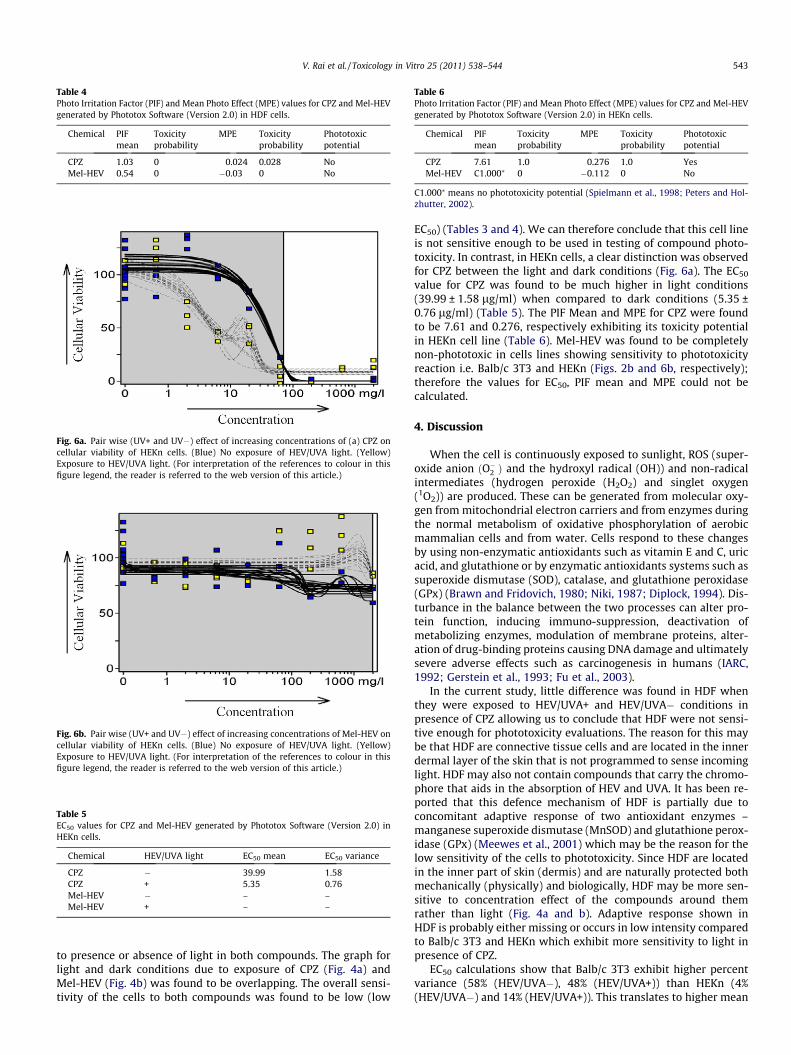
Fig. 6b. Pair wise (UV+ and UV�) effect of increasing concentrations of Mel-HEV oncellular viability of HEKn cells. (Blue) No exposure of HEV/UVA light. (Yellow)Exposure to HEV/UVA light. (For interpretation of the references to colour in thisfigure legend, the reader is referred to the web version of this article.)
Table 4Photo Irritation Factor (PIF) and Mean Photo Effect (MPE) values for CPZ and Mel-HEVgenerated by Phototox Software (Version 2.0) in HDF cells.
Chemical PIFmean
Toxicityprobability
MPE Toxicityprobability
Phototoxicpotential
CPZ 1.03 0 0.024 0.028 NoMel-HEV 0.54 0 �0.03 0 No
Fig. 6a. Pair wise (UV+ and UV�) effect of increasing concentrations of (a) CPZ oncellular viability of HEKn cells. (Blue) No exposure of HEV/UVA light. (Yellow)Exposure to HEV/UVA light. (For interpretation of the references to colour in thisfigure legend, the reader is referred to the web version of this article.)
Table 5EC50 values for CPZ and Mel-HEV generated by Phototox Software (Version 2.0) inHEKn cells.
Chemical HEV/UVA light EC50 mean EC50 variance
CPZ � 39.99 1.58CPZ + 5.35 0.76Mel-HEV � – –Mel-HEV + – –
Table 6Photo Irritation Factor (PIF) and Mean Photo Effect (MPE) values for CPZ and Mel-HEVgenerated by Phototox Software (Version 2.0) in HEKn cells.
Chemical PIFmean
Toxicityprobability
MPE Toxicityprobability
Phototoxicpotential
CPZ 7.61 1.0 0.276 1.0 YesMel-HEV C1.000⁄ 0 �0.112 0 No
C1.000⁄ means no phototoxicity potential (Spielmann et al., 1998; Peters and Hol-zhutter, 2002).
V. Rai et al. / Toxicology in Vitro 25 (2011) 538–544 543
to presence or absence of light in both compounds. The graph forlight and dark conditions due to exposure of CPZ (Fig. 4a) andMel-HEV (Fig. 4b) was found to be overlapping. The overall sensi-tivity of the cells to both compounds was found to be low (low
EC50) (Tables 3 and 4). We can therefore conclude that this cell lineis not sensitive enough to be used in testing of compound photo-toxicity. In contrast, in HEKn cells, a clear distinction was observedfor CPZ between the light and dark conditions (Fig. 6a). The EC50
value for CPZ was found to be much higher in light conditions(39.99 ± 1.58 lg/ml) when compared to dark conditions (5.35 ±0.76 lg/ml) (Table 5). The PIF Mean and MPE for CPZ were foundto be 7.61 and 0.276, respectively exhibiting its toxicity potentialin HEKn cell line (Table 6). Mel-HEV was found to be completelynon-phototoxic in cells lines showing sensitivity to phototoxicityreaction i.e. Balb/c 3T3 and HEKn (Figs. 2b and 6b, respectively);therefore the values for EC50, PIF mean and MPE could not becalculated.
4. Discussion
When the cell is continuously exposed to sunlight, ROS (super-oxide anion ðO��2 Þ and the hydroxyl radical (OH)) and non-radicalintermediates (hydrogen peroxide (H2O2) and singlet oxygen(1O2)) are produced. These can be generated from molecular oxy-gen from mitochondrial electron carriers and from enzymes duringthe normal metabolism of oxidative phosphorylation of aerobicmammalian cells and from water. Cells respond to these changesby using non-enzymatic antioxidants such as vitamin E and C, uricacid, and glutathione or by enzymatic antioxidants systems such assuperoxide dismutase (SOD), catalase, and glutathione peroxidase(GPx) (Brawn and Fridovich, 1980; Niki, 1987; Diplock, 1994). Dis-turbance in the balance between the two processes can alter pro-tein function, inducing immuno-suppression, deactivation ofmetabolizing enzymes, modulation of membrane proteins, alter-ation of drug-binding proteins causing DNA damage and ultimatelysevere adverse effects such as carcinogenesis in humans (IARC,1992; Gerstein et al., 1993; Fu et al., 2003).
In the current study, little difference was found in HDF whenthey were exposed to HEV/UVA+ and HEV/UVA� conditions inpresence of CPZ allowing us to conclude that HDF were not sensi-tive enough for phototoxicity evaluations. The reason for this maybe that HDF are connective tissue cells and are located in the innerdermal layer of the skin that is not programmed to sense incominglight. HDF may also not contain compounds that carry the chromo-phore that aids in the absorption of HEV and UVA. It has been re-ported that this defence mechanism of HDF is partially due toconcomitant adaptive response of two antioxidant enzymes –manganese superoxide dismutase (MnSOD) and glutathione perox-idase (GPx) (Meewes et al., 2001) which may be the reason for thelow sensitivity of the cells to phototoxicity. Since HDF are locatedin the inner part of skin (dermis) and are naturally protected bothmechanically (physically) and biologically, HDF may be more sen-sitive to concentration effect of the compounds around themrather than light (Fig. 4a and b). Adaptive response shown inHDF is probably either missing or occurs in low intensity comparedto Balb/c 3T3 and HEKn which exhibit more sensitivity to light inpresence of CPZ.
EC50 calculations show that Balb/c 3T3 exhibit higher percentvariance (58% (HEV/UVA�), 48% (HEV/UVA+)) than HEKn (4%(HEV/UVA�) and 14% (HEV/UVA+)). This translates to higher mean

544 V. Rai et al. / Toxicology in Vitro 25 (2011) 538–544
PIF and mean MPE values which are �7 times and �1.4 times high-er, respectively, for Balb/c 3T3 when compared to HEKn, making theBalb/c 3T3 cell line more useful for phototoxicity evaluations. Stud-ies show that under influence of UV and HEV radiation, p53-depen-dent cell death pathways and the mitogen-activated protein (MAP)kinase signaling cascade are more easily activated in Balb/c 3T3 celllines compared to cultured human keratinocytes ultimately causingcell death (Gerstein et al., 1993; Rittie and Fisher, 2002). It was alsofound that when Balb/c 3T3 were exposed to the phototoxic com-pound quinolone under UVA+ condition, reactive oxygen specieswere released triggering prostaglandin E2 (PGE2) and 6-keto-PGF1arelease from Balb/c 3T3 via protein kinase C (PKC) and a tyrosine ki-nase (TK) activation (Shimoda, 1998). Such anti-inflammatory cel-lular reaction could explain the extra sensitivity of Balb/c 3T3cells towards CPZ cells compared to HDF and HEKn cells.
Although the mean phototoxic concentrations for two cell linesare similar, the results are comparatively more precise (exhibitedless standard deviation) for HEKn due to less standard variationin EC50 and consecutive mean PIF and MPE values. Thus, when per-forming chemotoxicity studies with the purpose of finding a preciseEC50 value (at HEV/UVA� conditions), HEKn may prove to be a moreuseful cell line. However, this was not a full validation study and in-tended to check the toxicity potential of the Mel-HEV. Our studyfound that Balb/c 3T3 was the best evaluative cell line comparedto HDF and HEKn, which is concurrent with OECD guidelines. How-ever, in order to confirm the results, more known phototoxic testcompounds need to be evaluated using the same protocols.
In conclusion, Mel-HEV was found to be non-phototoxic in bothBalb/c 3T3 and HEKn cells even in presence of high concentrations(2000 lg/ml) of Mel-HEV. In contrast, cell viability vs. concentra-tion charts (Figs. 2 and 6) showed a slight elevation in cell viabilityat higher concentrations. It is predicted that the observed effect offractionated melanin can be due to its anti-oxidant and radicalscavenging properties, which may protect the cells from generat-ing reactive oxygen species hence causing phototoxicity. HEKnand Balb/c 3T3 were found to be sensitive to photosensitivity reac-tions using the positive control compound, CPZ. In contrast, the cellline HDF showed no sensitivity to the phototoxicity.
References
Bissonnette, R., 2008. Update on sunscreens: protection against visible light. SkinTherapy Letters 13, 5–7.
Bissonnette, R., Nigen, S., Bolduc, C., Mery, S., Nocera, T., 2008. Protection affordedby sunscreens containing inorganic sunscreening agents against blue lightsensitivity induced by aminolevulinic acid. Dermatologic Surgery 34, 1469–1476.
Boese, B.L., Lamberson, J.O., Swartz, R.C., Ozretich, R., Cole, F., 1998. Photoinducedtoxicity of PAHs and alkylated PAHs to a marine infaunal amphipod(Rhepoxynius abronius). Archives of Environmental Contamination andToxicity 34.
Bonina, F., Lanza, M., Montenegro, L., Puglisi, C., Tomaino, A., Trombetta, D., Castelli,F., Saija, A., 1996. Flavonoids as potential protective agents against photo-oxidative skin damage. International Journal of Pharmaceutics 145, 87–94.
Brawn, K., Fridovich, I., 1980. Superoxide radical and superoxide dismutases: threatand defense. Acta Physiologica Scandinavica 492, 9–18.
Dayan, N., Ballantyne, A., Ngo, T., Davis, J., Peterson, A., Larsen, K., Gray, J., Knaggs, H.,Gallas, J., 2011. Shielding the skin from high energy visible light-formulationsand aesthetics aspects. C&T Magazine, in press.
Denda, M., Fuziwara, S., 2008. Visible radiation affects epidermal permeabilitybarrier recovery: selective effects of red and blue light. Journal of InvestigativeDermatology 128, 1335–1336.
Dinardo, J.C., Wolf, B.A., Morrsi, W.E., Tenebaum, S., Schnetziger, R.W., 1985. Aquantitative in vitro assay for the evaluation of phototoxic potential of topicallyapplied materials. II. Modification and assessment of low level activity. Journalof Society of Cosmetic Chemist 36, 425–433.
Diplock, A.T., 1994. Antioxidants and free radical scavengers. In: Rice-Evans, C.A.,Burdon, R.H. (Eds.). Elsevier, Amsterdam, pp. 227–253.
Fisher, G.J., Datta, S.C., Talwar, H.S., Wang, Z.Q., Varani, J., Kang, S., Voorhees, J.J.,1996. Molecular basis of sun-induced premature skin ageing and retinoidantagonism. Nature 379, 335–339.
Fu, P.P., Cheng, S.H., Coop, L., Xia, Q., Culp, S.J., Totteson, W.H., Wamer, W.G.,Howard, P.C., 2003. Photoreaction, phototoxicity, and photocarcinogenicity ofretinoids. Journal of Environmental Science & Health, Part C – EnvironmentalCarcinogenesis & Ecotoxicology Reviews 21, 165–197.
Gerstein, A.D., Phillips, T.J., Rogers, G.S., Gilchrest, B.A., 1993. Wound healing andaging. Dermatologic Clinics 11, 749–757.
Gilchrest, B.A., 1996. A review of skin ageing and its medical therapy. British Journalof Dermatology 135, 867–875.
Holzhutter, H.-G., 1997. A general measure of in vitro phototoxicity derived frompairs of dose response curves and its use for predicting in vivo phototoxicity ofchemicals. Alternative to Animal Testing (ALTA) 25, 445–462.
IARC, 1992. Solar and ultraviolet radiation. IARC Monographs on the Evaluation ofCarcinogenic Risks to Humans. International Agency for Research on Cancer 55.
Jones, P.A., King, A.V., Earl, L.K., Lawrence, R.S., 2003. An assessment of thephototoxic hazard of a personal product ingredient using in vitro assays.Toxicology In Vitro 17, 471–480.
Kejlová, K., Jírová, D., Bendová, H., Kandárová, H., Weidenhoffer, Z., Kolárová, H.,Liebsch, M., 2007. Phototoxicity of bergamot oil assessed by in vitro techniquesin combination with human patch tests. Toxicology In Vitro 21, 1298–1303.
Kochevar, I.E., 1981. Phototoxicity mechanisms: chlorpromazine photosensitizeddamage to DNA and cell membranes. Journal of Investigative Dermatology 77,59–64.
Lelièvre, D., Justine, P., Christiaens, F., Bonaventure, N., Coutet, J., Marrot, L., Tinois-Tessonneaud, E., Cotovio, J., 2007. The EPISKIN phototoxicity assay (EPA):development of an in vitro tiered strategy to predict phototoxic potential.Alternatives to Animal Testing and Experimentation (AATEX) 14, 389–396.
Meewes, C., Brenneisen, P., Wenk, J., Kuhr, L., Ma, W., Alikoski, J., Poswig, A., Krieg, T.,Scharffetter-Kochanek, K., 2001. Adaptive antioxidant response protects dermalfibroblasts from UVA-induced phototoxicity. Free Radical Biology and Medicine30, 238–247.
Nadine, C., Mahmoud, R., 1999. Effects of all-trans retinoic acid on UVB-irradiatedhuman skin substitute. Journal of Cellular Physiology 181, 14–23.
Nathalie, D., Yannick, G., Caroline, B., Sandrine, D., Claude, F., Corinne, C., Pierre-Jacques, F., 2006. Assessment of the phototoxic hazard of some essential oilsusing modified 3T3 neutral red uptake assay. Toxicology In Vitro 20, 480–489.
Niki, E., 1987. Antioxidants in relation to lipidperoxidation. Chemistry and Physicsof Lipids 44, 227–253.
OECD, 2004. Organization for Economic Cooperation and Development Guidelinesfor the Testing of Chemicals/Section 4: Health Effects Test No. 432: In Vitro 3T3NRU Phototoxicity Test.
OECD, 2010. Chemicals Testing-Guidelines-Section 4: Software.Peters, B., Holzhutter, H.-G., 2002. In vitro phototoxicity testing: development and
validation of a new concentration response analysis software and biostatisticalanalyses related to the use of various prediction models. ALTA 30, 415–432.
Rittie, L., Fisher, G.J., 2002. UV-light-induced signal cascades and skin aging. AgingResearch Reviews 1, 705–720.
Shimoda, K., 1998. Mechanisms of quinolone phototoxicity. Toxicology Letters 102–103, 369–373.
Spielmann, H., Balls, M., Dupuis, J., Pape, W.J.W., Silva, O.D., Holzhütter, H.-G.,Gerberick, F., Liebsch, M., Lovell, W.W., Pfannenbecker, U., 1998. A study on UVfilter chemicals from Annex VII of European Union Directive 76/768/EEC, in thein vitro 3T3 NRU phototoxicity test. ALTA 26, 679–708.
Tolleson, W., Cherng, S.-H., Xia, Q., Boudreau, M., Yin, J., Wamer, W., Howard, P., Yu,H., Fu, P., 2005. Photodecomposition and Phototoxicity of Natural Retinoids, pp.147–155.
Zastrow, L., Groth, N., Klein, F., Kockott, D., Lademann, J., Ferrero, L., 2008. Detectionand Identification of Free Radicals Generated by UV and Visible Light in Ex VivoHuman Skin. In International Federation of Societies of Cosmetic Chemists, pp.207–214.
Copyright © 2022 FDOKUMEN