Hypothalamic Melanin-Concentrating Hormone Is Induced by Cold Exposure and Participates in the...
10
Hypothalamic Melanin-Concentrating Hormone Is Induced by Cold Exposure and Participates in the Control of Energy Expenditure in Rats MA ´ RCIO PEREIRA-DA-SILVA, MA ´ RCIO A. TORSONI, HUGO V. NOURANI, VIVIANE D. AUGUSTO, CLA ´ UDIO T. SOUZA, ALESSANDRA L. GASPARETTI, JOSE ´ B. CARVALHEIRA, GISLAINE VENTRUCCI, MARIA CRISTINA C. G. MARCONDES, ARIOVALDO P. CRUZ-NETO, MA ´ RIO J. A. SAAD, ANTONIO C. BOSCHERO, EVERARDO M. CARNEIRO, AND LI ´ CIO A. VELLOSO Departments of Internal Medicine (M.P., M.A.T., H.V.N., C.T.S., A.L.G., J.B.C., M.J.A.S., L.A.V.) and Physiology and Biophysics (V.A., G.V., M.C.C.G.M., A.C.B., E.M.C.), State University of Campinas; and Department of Zoology (A.P.C.-N.), Paulista State University, Campus Rio Claro–State University of Sao Paulo, 13083-970 Campinas SP, Brazil Short-term cold exposure of homeothermic animals leads to higher thermogenesis and food consumption accompanied by weight loss. An analysis of cDNA-macroarray was employed to identify candidate mRNA species that encode proteins in- volved in thermogenic adaptation to cold. A cDNA-macroar- ray analysis, confirmed by RT-PCR, immunoblot, and RIA, revealed that the hypothalamic expression of melanin- concentrating hormone (MCH) is enhanced by exposure of rats to cold environment. The blockade of hypothalamic MCH expression by antisense MCH oligonucleotide in cold-exposed rats promoted no changes in feeding behavior and body tem- perature. However, MCH blockade led to a significant drop in body weight, which was accompanied by decreased liver gly- cogen, increased relative body fat, increased absolute and relative interscapular brown adipose tissue mass, increased uncoupling protein 1 expression in brown adipose tissue, and increased consumption of lean body mass. Thus, increased hypothalamic MCH expression in rats exposed to cold may participate in the process that allows for efficient use of en- ergy for heat production during thermogenic adaptation to cold. (Endocrinology 144: 4831– 4840, 2003) C HRONIC EXPOSURE OF homeothermic animals to cold environment leads to several physiological ad- aptations that are required to maintain a stable body tem- perature (1–3). Among these adaptations, the increase of food consumption associated with an initial fall of body weight reflects the deviation of energy flow toward heat production, through a process of nonshivering thermogenesis (4, 5). Most of the adaptation control is coordinated by integrated re- sponses of neurons lying in different hypothalamic nuclei (6 – 8). Loss of coordinated control of food ingestion and energy expenditure is thought to be a central issue in the develop- ment of obesity, one of the most prevalent and life-threat- ening diseases of the modern world (9). A recent study has shown that defects in signaling of several hypothalamic neu- rotransmitters, such as neuropeptide Y, cocaine- and amphetamine-regulated transcript, agouti-related peptide, -MSH, pro-opiomelanocortin, melanin-concentrating hor- mone (MCH), and orexin (10), as well as defects of leptin and insulin signaling in hypothalamus, may lead to disturbances of feeding and weight control (11, 12). Because, under cold exposure, high food consumption is associated with weight loss and increased thermogenesis, we decided to use cDNA-macroarray analyses to identify can- didate hypothalamic proteins that are modulated in response to cold and, therefore, evaluate their participation in the processes that integrate energy expenditure and food consumption. Among 1176 different mRNA specificities analyzed, cold exposure induced the modulation of expression of 56 dif- ferent mRNA specificities in rat hypothalamus. Initial inter- est was drawn over the neurotransmitter MCH, a 19-amino- acid peptide, expressed mostly in neurons of the lateral hypothalamus (LH) and known to induce hyperphagia and weight gain, if intracerebroventricularly (ICV)-injected, in experimental animals (13, 14). Moreover, the lack of MCH signaling, by knocking out the expression of its receptor in the central nervous system, leads to moderate hypophagia and leanness (15, 16). Based on these facts, we decided to evaluate the role of MCH hyperexpression in the hypothal- amus of cold-exposed rats by blocking its protein synthesis using a phosphorthioate-modified MCH-specific antisense oligonucleotide. Materials and Methods Chemicals, antibodies, and oligodeoxynucleotides Reagents for SDS/PAGE and immunoblotting were obtained from Bio-Rad (Richmond, CA). HEPES, phenylmethylsulfonylfluoride, aprotinin, dithiothreitol, Triton X-100, Tween 20, glycerol, and BSA (fraction V) were from Sigma Chemical Co. (St. Louis, MO). Protein A-Sepharose 6MB was from Pharmacia (Uppsala, Sweden); 125 I-Pro- tein A and nitrocellulose membranes were from Amersham Corp. Abbreviations: AS, With antisense oligonucleotide treatment; BAT, brown adipose tissue; ICV, intracerebroventricular(ly); LH, lateral hy- pothalamus; MCH, melanin-concentrating hormone; RQ, respiratory quotient; RT, reverse transcription; S, with sense oligonucleotide treat- ment; T, temperature; UCP, uncoupling protein; VO 2 , rate of oxygen consumption; VCO 2 , rate of carbon dioxide production; WO, without oligonucleotide treatment. 0013-7227/03/$15.00/0 Endocrinology 144(11):4831– 4840 Printed in U.S.A. Copyright © 2003 by The Endocrine Society doi: 10.1210/en.2003-0243 4831
-
Upload
independent -
Category
Documents
-
view
0 -
download
0
Transcript of Hypothalamic Melanin-Concentrating Hormone Is Induced by Cold Exposure and Participates in the...

Hypothalamic Melanin-Concentrating Hormone IsInduced by Cold Exposure and Participates in theControl of Energy Expenditure in Rats
MARCIO PEREIRA-DA-SILVA, MARCIO A. TORSONI, HUGO V. NOURANI, VIVIANE D. AUGUSTO,CLAUDIO T. SOUZA, ALESSANDRA L. GASPARETTI, JOSE B. CARVALHEIRA,GISLAINE VENTRUCCI, MARIA CRISTINA C. G. MARCONDES, ARIOVALDO P. CRUZ-NETO,MARIO J. A. SAAD, ANTONIO C. BOSCHERO, EVERARDO M. CARNEIRO, AND LICIO A. VELLOSO
Departments of Internal Medicine (M.P., M.A.T., H.V.N., C.T.S., A.L.G., J.B.C., M.J.A.S., L.A.V.) and Physiology andBiophysics (V.A., G.V., M.C.C.G.M., A.C.B., E.M.C.), State University of Campinas; and Department of Zoology (A.P.C.-N.),Paulista State University, Campus Rio Claro–State University of Sao Paulo, 13083-970 Campinas SP, Brazil
Short-term cold exposure of homeothermic animals leads tohigher thermogenesis and food consumption accompanied byweight loss. An analysis of cDNA-macroarray was employed toidentify candidate mRNA species that encode proteins in-volved in thermogenic adaptation to cold. A cDNA-macroar-ray analysis, confirmed by RT-PCR, immunoblot, and RIA,revealed that the hypothalamic expression of melanin-concentrating hormone (MCH) is enhanced by exposure ofrats to cold environment. The blockade of hypothalamic MCHexpression by antisense MCH oligonucleotide in cold-exposedrats promoted no changes in feeding behavior and body tem-
perature. However, MCH blockade led to a significant drop inbody weight, which was accompanied by decreased liver gly-cogen, increased relative body fat, increased absolute andrelative interscapular brown adipose tissue mass, increaseduncoupling protein 1 expression in brown adipose tissue, andincreased consumption of lean body mass. Thus, increasedhypothalamic MCH expression in rats exposed to cold mayparticipate in the process that allows for efficient use of en-ergy for heat production during thermogenic adaptation tocold. (Endocrinology 144: 4831–4840, 2003)
CHRONIC EXPOSURE OF homeothermic animals tocold environment leads to several physiological ad-
aptations that are required to maintain a stable body tem-perature (1–3). Among these adaptations, the increase of foodconsumption associated with an initial fall of body weightreflects the deviation of energy flow toward heat production,through a process of nonshivering thermogenesis (4, 5). Mostof the adaptation control is coordinated by integrated re-sponses of neurons lying in different hypothalamic nuclei(6–8).
Loss of coordinated control of food ingestion and energyexpenditure is thought to be a central issue in the develop-ment of obesity, one of the most prevalent and life-threat-ening diseases of the modern world (9). A recent study hasshown that defects in signaling of several hypothalamic neu-rotransmitters, such as neuropeptide Y, cocaine- andamphetamine-regulated transcript, agouti-related peptide,�-MSH, pro-opiomelanocortin, melanin-concentrating hor-mone (MCH), and orexin (10), as well as defects of leptin andinsulin signaling in hypothalamus, may lead to disturbancesof feeding and weight control (11, 12).
Because, under cold exposure, high food consumption is
associated with weight loss and increased thermogenesis, wedecided to use cDNA-macroarray analyses to identify can-didate hypothalamic proteins that are modulated in responseto cold and, therefore, evaluate their participation in theprocesses that integrate energy expenditure and foodconsumption.
Among 1176 different mRNA specificities analyzed, coldexposure induced the modulation of expression of 56 dif-ferent mRNA specificities in rat hypothalamus. Initial inter-est was drawn over the neurotransmitter MCH, a 19-amino-acid peptide, expressed mostly in neurons of the lateralhypothalamus (LH) and known to induce hyperphagia andweight gain, if intracerebroventricularly (ICV)-injected, inexperimental animals (13, 14). Moreover, the lack of MCHsignaling, by knocking out the expression of its receptor inthe central nervous system, leads to moderate hypophagiaand leanness (15, 16). Based on these facts, we decided toevaluate the role of MCH hyperexpression in the hypothal-amus of cold-exposed rats by blocking its protein synthesisusing a phosphorthioate-modified MCH-specific antisenseoligonucleotide.
Materials and MethodsChemicals, antibodies, and oligodeoxynucleotides
Reagents for SDS/PAGE and immunoblotting were obtained fromBio-Rad (Richmond, CA). HEPES, phenylmethylsulfonylfluoride,aprotinin, dithiothreitol, Triton X-100, Tween 20, glycerol, and BSA(fraction V) were from Sigma Chemical Co. (St. Louis, MO). ProteinA-Sepharose 6MB was from Pharmacia (Uppsala, Sweden); 125I-Pro-tein A and nitrocellulose membranes were from Amersham Corp.
Abbreviations: AS, With antisense oligonucleotide treatment; BAT,brown adipose tissue; ICV, intracerebroventricular(ly); LH, lateral hy-pothalamus; MCH, melanin-concentrating hormone; RQ, respiratoryquotient; RT, reverse transcription; S, with sense oligonucleotide treat-ment; T, temperature; UCP, uncoupling protein; VO2, rate of oxygenconsumption; VCO2, rate of carbon dioxide production; WO, withoutoligonucleotide treatment.
0013-7227/03/$15.00/0 Endocrinology 144(11):4831–4840Printed in U.S.A. Copyright © 2003 by The Endocrine Society
doi: 10.1210/en.2003-0243
4831

(Aylesbury, UK). Microcon centrifugal filter devices, YN-3, werepurchased from Millipore Corp. (Bedford, MA). Antibody againstMCH (chicken anti-MCH, T-1506) was from BMA Biomedicals AG(Augst, Switzerland), and antibody against uncoupling protein(UCP)1 (goat anti-UCP1, sc-6529) was from Santa Cruz Biotechnol-ogy, Inc. (Santa Cruz, CA). Secondary antibodies, used as bridges,antigoat (G-4018), and anti-chicken (F-8888), as well as rat MCH(M-4542), were from Sigma Chemical Co. MCH RIA kit (RK 070-47)was from Phoenix Pharmaceuticals, Inc. (Belmont, CA). Chemicalsemployed in RT-PCR, primers for MCH (sense 5�-TAC GGA GCAGCA AAC A-3� and antisense 5�-ACA GCC AGA CTG AGG G-3�) and�-actin (sense 5�-ACC ACA GCT GAG AGG GAA ATC G-3� andantisense 5�-CAG TCC GCC TAG AAG CAT TTG C-3�), phosphor-thioate-modified oligonucleotides for MCH (sense 5�-CCC TCA GTCTGG CTG-3� and antisense 5�-ACA GCC AGA CTG AGG-3�), andTrizol reagent were obtained from Life Technologies (GIBCO BRL,Gaithersburg, MD). Sodium amobarbital was from Eli Lilly (India-napolis, IN). Atlas Rat 1.2 Array Kit was from Clontech Laboratories,Inc. (East Meadow Circle, Palo Alto, CA). Salmon sperm DNA wasfrom Sigma Chemical Co., and [�-33P]dATP was from AmershamCorp.
Experimental animals and cold-exposure protocols
Male Wistar rats (56–70 d old/200–260 g) from the University ofCampinas Central Animal Breeding Center were used in the experi-ments. The University’s Ethical Committee approved the protocols. Theanimals were maintained on a 12-h artificial light, 12-h dark cycle andkept in individual cages supplied with standard rodent chow and waterad libitum. After an acclimatizing period (3 d), rats were transferred tometabolic cages and randomly assigned to two treatment conditions:control and cold-exposed [temperature (T) �4 C]. Rats of the controlgroup were continuously maintained at 23 � 1 C, whereas rats of the T�4 C group were housed at 4 � 1 C. Protocols lasting 2 h and 2, 4, 8,and 16 d were employed. For hormone and biochemical determinations,samples were collected from eight rats of each group, and analyses wererun in duplicates. For cDNA preparation for macroarray analysis,mRNA extracted from the hypothalami of three rats of each group waspooled, reverse transcribed, and used in the hybridization protocol;three pools from each condition were evaluated. For the remainingexperiments, four rats of each group were analyzed, and the experimentswere run in duplicates or triplicates.
ICV cannulation and ICV oligonucleotide injection
Adult male Wistar rats were stereotaxically instrumented undersodium amobarbital (15 mg/kg body weight) anesthesia with chronicunilateral 26-gauge stainless steel indwelling guide cannulas, asep-tically placed into the lateral ventricle (0.2 mm posterior, 1.5 mmlateral, and 4.2 mm ventral to bregma) as previously described (13).After a 1-wk recovery period, all rats were kept in individual cagesand exposed either to 23 � 1 C (control) or to 4 � 1 C (T �4 C) during4 d. Sense and antisense MCH oligonucleotides were diluted in TEbuffer (10 mm Tris-HCl, 1 mm EDTA) and injected once a day at1000 h, with a total vol of 2.0 �l per dose (2.0 nmol/�l), 24, 48, and72 h after the onset of the experimental period. Rats were randomlyassigned to six different treatment conditions: control without oli-gonucleotide treatment (control WO); cold exposed without oligo-nucleotide treatment (T �4 C WO); control with sense oligonucleotidetreatment (control S); cold exposed with sense oligonucleotide treat-ment (T �4 C S); control with antisense oligonucleotide treatment(control AS); cold exposed with antisense oligonucleotide treatment(T �4 C AS).
Biochemical, hormonal, and metabolic characterization ofexperimental animals
During the animal model characterization, the measurements of foodintake, rectal temperature, and body weight were obtained at 1400 hdaily from experimental d 1 to experimental d 8, and on experimentald 16. During ICV injection protocol, the measurements were performeddaily during the whole experimental period. Rectal temperature wasmeasured with a Thermistor digital (HI-8753) high-precision thermom-
eter (Hanna Instruments, Inc., Woonsocket, RI) inserted 1.5 cm from theanus. Plasma glucose was determined by the glucose oxidase method(17), RIA was employed to measure serum insulin according to a pre-vious description (18), leptin concentrations were determined using acommercially available ELISA kit (Crystal Chem. Inc., Chicago, IL), andcorticosterone and TSH were detected by commercially available RIAkits (Amersham Biosciences, Biotrak, Aylesbury, UK). Glycogen wasmeasured, according to a previously described method (19), in liver andgastrocnemius muscle fragments, at experimental d 8 (during the animalmodel characterization) and at experimental d 4 (during ICV oligonu-cleotide treatment). Body carcass composition was determined as pre-viously described (20).
Metabolic measurements
Rates of oxygen consumption (VO2) and carbon dioxide production(VCO2) were measured by open-flow respirometry, with a set-up mod-ified from a previously published method (21). After 14 h fasting, ratswere placed individually into polyvinyl chloride respirometric cham-bers, which were then installed into a temperature-controlled cabinet(�0.1 C). Air was drawn over ascarite and drierite scrubbers to removeCO2 and H2O to a manifold, which divided the flow into four airstreams.Each stream was regulated at 1500–2500 ml/min, depending on theexperimental temperature. The constancy of the flow was periodicallychecked with a Cole-Palmer (Vernon Hills, IL) flow meter, and less then1% drift during the experiments was detected. Each stream fed the inletport of each of the four respirometric chambers, one of which containedno experimental animal and was used as a baseline reference. Air leavingeach respirometric chamber was directed into a computer-controlledmultiplexer, where four solenoid vales selected which flow would bedirected to a computer-controlled mass-flow meter (model 840; SierraInstruments, Inc., Monterey, CA). The mass-flow meter was adjusted todeliver a constant flow of 300 ml/min of air, sequentially through waterscrubbers, to the measurement cell of a CA-2A CO2 analyzer (SableSystems International, Las Vegas, NV), again through water and gasscrubbers, and finally to the measurement cell of a FC-1B O2 analyzer(Sable). Each chamber with a rat was sampled for 20 min. Thus, acomplete set of recording lasted for 80 min, and this sequence wasrepeated six times during the evaluation period. Experiments started at1000 h and finished at 1800 h. VO2 and VCO2 were calculated by theDatacan program, based in the equations of Withers (22) and wereexpressed as milliliters per hour per gram. Animals were weighed im-mediately before and after the metabolic measurements, and the averageof these two measurements was used as the body mass value for thesecalculations. The respiratory quotient (RQ) was calculated as:VCO2/VO2.
cDNA arrays analysis and RT-PCR
RNA preparation. Control and cold-exposed rat hypothalamus wereexcised and rapidly frozen in liquid nitrogen. Total RNA was ex-tracted using Trizol reagent, according to the manufacturer’s recom-mendations. Total RNA was quantified by spectrophotometry atA260 nm, and its integrity was determined from the A260:A280 nmratio. After 24 h, isolated total RNA was rendered genomic-DNA-freeby digestion with ribonuclease-free deoxyribonuclease (RQ1; Pro-mega, Madison, WI).
Array hybridization. The recommended protocol was followed in allsteps. Briefly, 1 �g poly(A)� RNA was converted into 33P-labeledfirst-strand cDNA by Moloney murine leukemia virus reverse tran-scriptase. Unincorporated 33P-labeled nucleotides were removed bychromatography using a NucleoSpin Extraction Spin Column(CLONTECH, Palo Alto, CA). Purified cDNA probes were hybridizedto the Atlas membranes. Hybridization occurred overnight at 68 C.After washing, membranes were sealed in hybridization bags andexposed to imaging plates for 1 d. After exposure, the imaging plateswere scanned using a BAS-1500 (Fujifilm, Fuji, Japan), and hybrid-ization signals were counted. Hybridization signals of each gene werenormalized by a positive control (signals of the housekeeping gene),and gene expression was compared between the preconditioning andcontrol groups. Genes exhibiting differential expression in the pre-conditioning group were selected only if the hybridization signals
4832 Endocrinology, November 2003, 144(11):4831–4840 Pereira-da-Silva et al. • MCH, Cold Exposure, and Energy Expenditure Control
were either increased or decreased by at least 2-fold, compared withthose of the control group.
Semiquantitative RT-PCR. Seven micrograms of total RNA, obtained fromhypothalamus of rats exposed, or not, to cold during 4 d, were reverse-transcribed with SuperScript reverse transcriptase (200 U/�l) usingoligo(deoxythymidine) (50 mm) in a 30-�l reaction vol [5� reversetranscription (RT) buffer, 10 mm deoxynucleotide triphosphate, and 40U/�l ribonuclease-free inhibitor]. The RTs involved a 50-min incubationat 42 C and a 15-min incubation at 70 C. After RT, 0.75 �l of the RTproduct was used in each PCR to a final vol of 50 �l (10� PCR buffer,1.0 mm deoxynucleotide triphosphate, 50 mm MgCl2, Taq polymerase,and sense and antisense primers for MCH and �-actin). The expressionof mRNA was determined by PCR, using the primers described above,and amplified a 323-bp DNA fragment of MCH and a 533-bp DNAfragment of �-actin. Triplicate reactions were done using an initial in-cubation at 94 C for 5 min, denaturation at 94 C for 1 min, followed byannealing at 50 and 57 C (MCH and �-actin, respectively) for 50 sec,extension at 72 C for 1 min, and final extension at 72 C for 7 min. Aftertitration between 16 and 32 cycles, 22 cycles was selected for quantifi-cation of MCH and �-actin. These PCR conditions were therefore usedin all subsequent experiments. All PCR experiments included a controltube with no RT step. PCR-amplified products were run on 2% Tris/acetic acid/EDTA agarose gels, and the DNA was visualized byethidium bromide staining. The size of the products was determinedusing a 1-kb plus DNA ladder (Life Technologies) as standard sizemarkers. Images of the bands were captured using a TFX 35M UVtransluminator (Life Technologies), and band intensity was quantifiedby digital densitometry (Scion Image software, Scion Corp., Frederick,MD). Results are expressed as the ratio of MCH/�-actin � sem, inarbitrary scanning units.
Protein analyses by Western blot and dot-blot
UCP1 was measured in brown adipose tissue (BAT) of rats sub-mitted to ICV cannulation and treated according to the followingprotocols: control WO; T �4 C WO; control S; T �4 C S; control AS;and T �4 C AS. Western blot analyses followed previously describedmethods (23, 24) using a specific anti-UCP1 polyclonal antibody. Theintensity of protein bands corresponding to UCP1 was quantified bydigital densitometry (Scion Image software) of the developed auto-radiographs. MCH was measured in hypothalamus of rats exposed,or not, to cold (without previous stereotaxic instrumentation), or inrats submitted to ICV cannulation and treated according to the sameprotocols as stated above for UCP1 measurements. For the detectionof MCH in hypothalamic protein extracts, a preparatory step ofprotein purification by filtration was performed using a MicroconYN-3 device. A method for specific peptide detection by dot-blot,previously described (25), was used. The signal obtained wasmeasured by digital densitometry (Scion Image software) of thedeveloped autoradiographs. Highly purified MCH was used ascontrol.
Determination of hypothalamic MCH by RIA
For hypothalamic MCH determination by RIA, hypothalami of ratsof each experimental group were homogenized in ice-cold buffercontaining 100 mm Tris (pH 7.6), 1% (vol/vol) Triton X-100, 150 mmNaCl, 0.1 mg/ml aprotinin, 2 mm phenylmethylsulfonylfluoride, 10mm Na3VO4, 100 mm NaF, 10 mm Na4P2O7, and 4 mm EDTA, usinga PTA 20S polytron generator (Brinkmann Instruments, Westbury,NY). Homogenates were then centrifuged at 12,000 � g for 20 min at�4 C. Protein concentration of supernatants was determined by theBradford method (26). Samples containing 2.0 mg/ml protein wereevaluated for the concentration of MCH using a RIA kit, following therecommendations of the manufacturer (Phoenix Pharmaceuticals,Inc.) (27).
Data presentation and statistical analysis
All numerical results are expressed as the mean � sem of theindicated number of experiments. The results of blots are presentedas direct comparisons of bands or dots in autoradiographs and quan-
tified by densitometry using the Scion Image software. Data wereanalyzed by the two-tailed unpaired Student’s t test or by repeat-measures ANOVA (one-way or two-way ANOVA) followed by posthoc analysis of significance (Bonferroni test) when appropriate, com-paring experimental and control groups. The level of significance wasset at P � 0.05.
ResultsMetabolic and hormonal characteristics of rats exposedto cold
Exposure of homeothermic animals to cold promoteschanges in several metabolic parameters that reflect phys-iological adaptations to the novel external environment.For decades, a number of studies have characterized thoseadaptations; and in the present study, we repeated someof those measurements to evidence the reliability of themodel employed. The results obtained during theevaluation of the metabolic parameters are summarized inTables 1 and 2. As a whole, the animal model hereinmatches previous characterizations of cold-exposed rats(28, 29). Of greater importance for the objectives of thepresent study, we emphasize that mean daily food intakewas approximately 70% increased in cold-exposed rats(P � 0.05), whereas no significant changes in body tem-perature were detected. Cumulative body weight varia-tion was positive in the control group, whereas a negativevariation on body weight was detected in cold-exposedanimals. From d 6 on, a stepwise recovery of body weightwas observed in cold-exposed rats; but at d 16, it was stilllower than at d 0.
cDNA-macroarray analyses
Poly (A)� RNA samples from the hypothalamus of con-trol and 4-d cold-exposed rats were prepared and used forcDNA expression array hybridization. Individual genesignal intensities were normalized to that of the house-keeping genes, according to the supplier’s instructions. Tominimize an animal-specific expression pattern, we com-
TABLE 1. Animal model characterization
Parameters Control T � 4 C
Food Intake (g/d) 19.47 � 0.49 33.00 � 0.72a
Temperature (C) 36.04 � 0.07 36.11 � 0.06Glucose (mg/dl) 91.06 � 0.35 92.29 � 0.31Insulin (ng/ml) 2.420 � 0.12 1.416 � 0.18a
Leptin (pg/ml) 2545.42 � 292.00 2013.60 � 252.95TSH (ng/ml) 13.27 � 1.31 13.32 � 1.53Corticosterone (ng/ml) 102.43 � 15.57 112.91 � 12.35Muscle glycogen
(% muscle weight)0.558 � 0.084 0.553 � 0.103
Liver glycogen(% liver weight)
7.488 � 0.531 17.773 � 3.984a
Body fat (% carcass weight) 12.354 � 933 6.526 � 0.536a
a P � 0.05 vs. Control.
TABLE 2. Cumulative body weight variation (g)
Mean weightat d 0 (g) d 1 d 4 d 8 d 16
Control 218 � 7 �2 � 1 �4 � 1 �7 � 2 �23 � 5T � 4 C 222 � 8 �10 � 2a �13 � 3a �9 � 3a �4 � 2a
a P � 0.05 vs. Control.
Pereira-da-Silva et al. • MCH, Cold Exposure, and Energy Expenditure Control Endocrinology, November 2003, 144(11):4831–4840 4833
bined equal amounts of RNA from three different animalsand analyzed six independent hybridization signals fromthree different arrays per experimental condition. Table 3presents the data of all genes that had their mRNA ex-
pression significantly modulated by cold exposure. Al-though several of the genes found to be regulated by coldexposure might play important roles in energy homeosta-sis, we elected MCH as the first to be studied, because of
TABLE 3. Results of macroarray analyses
mRNAs with increased hypothalamic expression in rats exposed to cold● 40S ribosomal protein S12 (RPS12)● 60S ribosomal protein L21 (RPL21)● Acetylcholine receptor, nicotinic, �4● Adenomatous polyposis coli protein (APC)● ADP-ribosylation factor 3 (ARF3)● A-RAF proto-oncogene● Calbindin D28; avian-type vitamin D-dependent; calcium-binding protein (CABP); spot 35 protein; CALB1● Carboxypeptidase E (CPE); CPH● Cathepsin L● Chymotrypsinogen B● Colipase (CLPS)● Dipeptidylpeptidase 6 (DPP6)● Elastase 2● Growth-accentuating protein 43 (GAP43); neuromodulin● Guanine nucleotide-binding protein G(I) � 1 subunit (GNA11); BPGTPB● Guanine nucleotide-binding protein G(O) � subunit (GNAO; GNA0)● Guanine nucleotide-binding regulatory, � subunit● Heat shock 90-kDa protein � (HSP90-�); HSP84; HSPCB● Mak; male germ cell-associated kinase; highly expressed at and after meiosis● Melanin-concentrating hormone (PMCH; MCH)● Mitochondrial ATP synthase � subunit (ATP5B)● MAPK2; PRKM2; MAPK1; extracellular signal-regulated kinase 2 (ERK2); ERT1● Muscle phosphofructokinase (PFKM)● Myelin basic protein (MBP)● Myelin proteolipid protein (PLP); lipophilin; DM20● Neuronal acetylcholine receptor protein � 2 subunit (NACHRB2); cholinergic receptor nicotinic � polipeptide 2 (CHRNB2)● N-methyl-D-aspartate receptor subtype 2B (NMDAR2B; NR2B); glutamate receptor subunit epsilon 2B (GRIN2B)● Nonmuscle cofilin (CFL1)● Nonprocessed neurexin II � major; nonprocessed neurexin II �● Nucleoside diphosphate kinase A (NDP kinase A; NDKA); tumor metastatic process-associated protein; metastasis inhibition factor
NM23; NME1● Pancreatic cholesterol esterase (CHOLE); bile salt-activated lipase (BAL); bile salt-stimulated lipase (BSSL); carboxyl ester lipase; sterol
esterase; pancreatic lysophospholipase● Pancreatic Triacylglycerol lipase● Pheochromocytoma-derived protein tyrosine phosphatase-like protein● Potassium channel RCK4, subunit, putative● Potassium voltage-gated channel shaker-related subfamily member 1 (KV1.1); KCNA1; RBK1● Proteasome component C3● Protein kinase C �1 (PKC-� 1; PRKCB1; PKCB); PKC-� II (PRKCB2)● Protein kinase C � type (PKC-�; PRKCD)● Ribosomal protein L13● Secretogranin II (SGII; SCG2); chromogranin C (CHGC)● Serine-threonine protein phosphatase 3 catalytic subunit � isoform (PPP3CA); PPP2B; calmodulin-dependent calcineurin A � (CALNA;
CNA1)● Sertoli cell cytochrome c oxipeptidase polypeptide 1 (COX1)● Sodium/potassium-transporting ATPase � 1 subunit (Na�/K� ATPase �1)● Sodium/potassium-transporting ATPase � 1 subunit (ATP1B1)● Soluble superoxide dismutase 1 (SOD 1)● Synaptotagmin X1 (SYT11)● Syntaxin 1B (STX1B)● Tissue inhibitor of metabolism metalloproteinase 2 (TIMP2)● Tyrosine 3 monooxygenase/tryptophan 5-monooxygenase activation protein � polypeptide (YWHAE); 14-3-3 protein �; protein kinase C
inhibitor protein 1 (KCIP1); mitochondrial import stimulation factor L subunit● Tyrosine 3 monooxygenase/tryptophan 5-monooxygenase activation protein � polypeptide (YWHAZ); 14-3-3 protein �/�; protein kinase C
inhibitor protein 1 (KCIP1); mitochondrial import stimulation factor S1 subunit● Tyrosine 3 monooxygenase/tryptophan 5-monooxygenase activation protein � polypeptide (YWHAH); 14-3-3 protein �; protein kinase C
inhibitor protein 1 (KCIP1)● Ubiquitous mitochondrial creatine kinase (U-MTCK; CKMT1); acidic-type mitochondrial creatine kinase (MIA-CK)mRNAs with decreased hypothalamic expression in rats exposed to cold● 25-kDa synaptosomal-associated protein (SNAP25); super protein (SUP)● Brain fatty acid-binding protein (B-FABP); FABP7; brain lipid-binding protein (BLBP)● Casein kinase I �; CK1d; 49-kDa isoform● CRH
4834 Endocrinology, November 2003, 144(11):4831–4840 Pereira-da-Silva et al. • MCH, Cold Exposure, and Energy Expenditure Control
its known participation in control of food ingestion andbody weight (12).
Hypothalamic MCH expression in rats exposed to cold
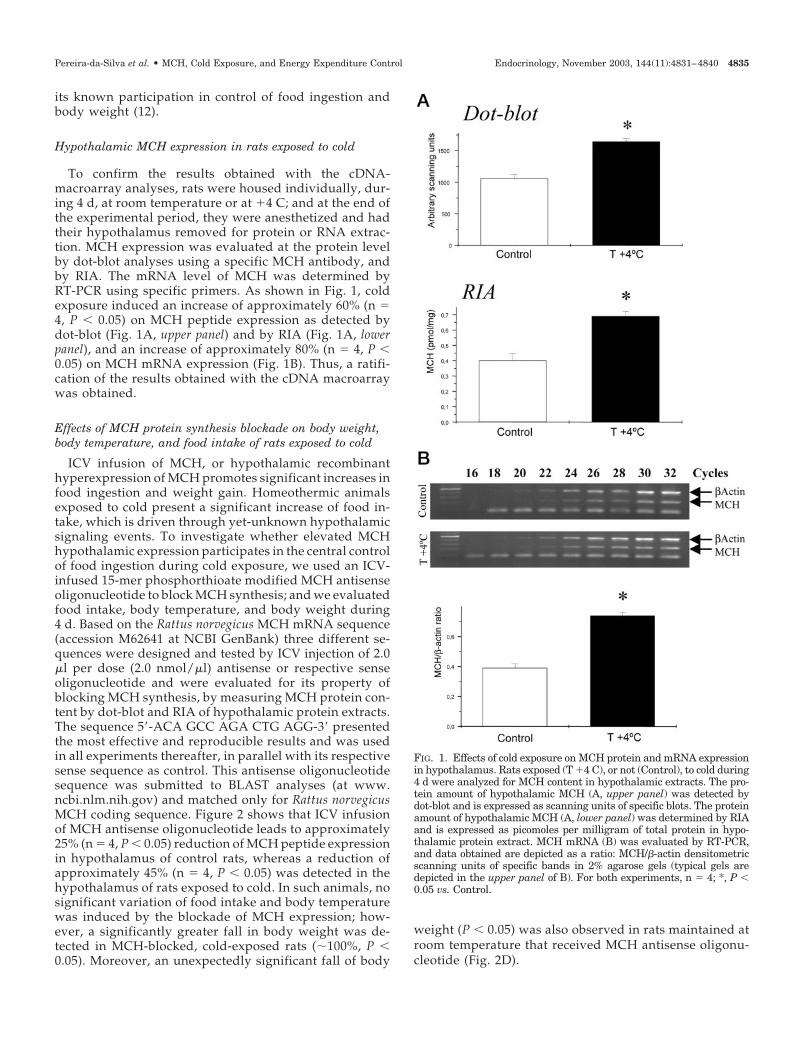
To confirm the results obtained with the cDNA-macroarray analyses, rats were housed individually, dur-ing 4 d, at room temperature or at �4 C; and at the end ofthe experimental period, they were anesthetized and hadtheir hypothalamus removed for protein or RNA extrac-tion. MCH expression was evaluated at the protein levelby dot-blot analyses using a specific MCH antibody, andby RIA. The mRNA level of MCH was determined byRT-PCR using specific primers. As shown in Fig. 1, coldexposure induced an increase of approximately 60% (n �4, P � 0.05) on MCH peptide expression as detected bydot-blot (Fig. 1A, upper panel) and by RIA (Fig. 1A, lowerpanel), and an increase of approximately 80% (n � 4, P �0.05) on MCH mRNA expression (Fig. 1B). Thus, a ratifi-cation of the results obtained with the cDNA macroarraywas obtained.
Effects of MCH protein synthesis blockade on body weight,body temperature, and food intake of rats exposed to cold
ICV infusion of MCH, or hypothalamic recombinanthyperexpression of MCH promotes significant increases infood ingestion and weight gain. Homeothermic animalsexposed to cold present a significant increase of food in-take, which is driven through yet-unknown hypothalamicsignaling events. To investigate whether elevated MCHhypothalamic expression participates in the central controlof food ingestion during cold exposure, we used an ICV-infused 15-mer phosphorthioate modified MCH antisenseoligonucleotide to block MCH synthesis; and we evaluatedfood intake, body temperature, and body weight during4 d. Based on the Rattus norvegicus MCH mRNA sequence(accession M62641 at NCBI GenBank) three different se-quences were designed and tested by ICV injection of 2.0�l per dose (2.0 nmol/�l) antisense or respective senseoligonucleotide and were evaluated for its property ofblocking MCH synthesis, by measuring MCH protein con-tent by dot-blot and RIA of hypothalamic protein extracts.The sequence 5�-ACA GCC AGA CTG AGG-3� presentedthe most effective and reproducible results and was usedin all experiments thereafter, in parallel with its respectivesense sequence as control. This antisense oligonucleotidesequence was submitted to BLAST analyses (at www.ncbi.nlm.nih.gov) and matched only for Rattus norvegicusMCH coding sequence. Figure 2 shows that ICV infusionof MCH antisense oligonucleotide leads to approximately25% (n � 4, P � 0.05) reduction of MCH peptide expressionin hypothalamus of control rats, whereas a reduction ofapproximately 45% (n � 4, P � 0.05) was detected in thehypothalamus of rats exposed to cold. In such animals, nosignificant variation of food intake and body temperaturewas induced by the blockade of MCH expression; how-ever, a significantly greater fall in body weight was de-tected in MCH-blocked, cold-exposed rats (�100%, P �0.05). Moreover, an unexpectedly significant fall of body
weight (P � 0.05) was also observed in rats maintained atroom temperature that received MCH antisense oligonu-cleotide (Fig. 2D).
FIG. 1. Effects of cold exposure on MCH protein and mRNA expressionin hypothalamus. Rats exposed (T �4 C), or not (Control), to cold during4 d were analyzed for MCH content in hypothalamic extracts. The pro-tein amount of hypothalamic MCH (A, upper panel) was detected bydot-blot and is expressed as scanning units of specific blots. The proteinamount of hypothalamic MCH (A, lower panel) was determined by RIAand is expressed as picomoles per milligram of total protein in hypo-thalamic protein extract. MCH mRNA (B) was evaluated by RT-PCR,and data obtained are depicted as a ratio: MCH/�-actin densitometricscanning units of specific bands in 2% agarose gels (typical gels aredepicted in the upper panel of B). For both experiments, n � 4; *, P �0.05 vs. Control.
Pereira-da-Silva et al. • MCH, Cold Exposure, and Energy Expenditure Control Endocrinology, November 2003, 144(11):4831–4840 4835

FIG. 2. Effects of ICV MCH antisense oligonucleotide treatment on MCH protein expression, food intake, body temperature, and body weightvariation. Rats were ICV cannulated and, after a recovery period, were treated once a day, at 1000 h, with sense (S) or antisense (AS) MCHoligonucleotide or a similar volume of saline (WO), with a total vol of 2.0 �l per dose (2.0 nmol/�l), 24, 48, and 72-h after the onset of theexperimental period. The experimental animals were randomly assigned to two groups, exposed (T �4 C) or not exposed (Control) to cold.Hypothalamic MCH protein content (A, upper panel) was detected by dot-blot and is expressed as scanning units of specific blots. The protein
4836 Endocrinology, November 2003, 144(11):4831–4840 Pereira-da-Silva et al. • MCH, Cold Exposure, and Energy Expenditure Control
Effects of MCH protein synthesis blockade on liver andmuscle glycogen contents and carcass composition
Because the blockade of hypothalamic MCH in cold-ex-posed rats produced a significantly increased fall of bodyweight, in the absence of major variations of food intake andbody temperature, we decided to evaluate the relativeamounts of body glycogen, fat, and lean mass stocks incontrol and cold-exposed rats, treated or not with MCHantisense oligonucleotide during 4 d. As depicted in Fig. 3,the exposure of rats to cold produced increases in liver(�400%, P � 0.05) and muscle (�50%, not significant) gly-cogen contents; however, if the rats exposed to cold weretreated with antisense oligonucleotide, lower levels of liver(�120%, P � 0.05, vs. T �4 C WO) and muscle (�20%, notsignificant) glycogen increments were detected. On the otherhand, relative body fat was significantly reduced in ratsexposed to cold; if cold-exposed rats were treated with an-tisense MCH oligonucleotide, higher amounts of relativebody fat were encountered (P � 0.05, vs. T �4 C WO). Finally,lean body mass was unaffected by cold exposure; however,under MCH blockade, cold-exposed animals consumed sig-nificantly higher amounts of relative lean body mass, whichdiffered from that observed in cold-exposed non-antisense-MCH-oligonucleotide-treated rats (P � 0.05).
Effects of MCH protein synthesis blockade on oxygenconsumption, carbon dioxide production, and RQ
Cold-exposed rats exhibited a significantly higher O2 con-sumption (3.24 � 0.26 vs. 1.54 � 0.02 ml/g�h for cold-exposedand control rats, respectively) and CO2 production (2.50 �0.22 vs. 1.21 � 0.04 ml/g�h for cold-exposed and control rats,respectively) than control rats. However, no major change inRQ was detected among the groups (0.77 � 0.01 vs. 0.78 �0.03 for cold-exposed and control rats, respectively). Theblockade of MCH expression with antisense oligonucleotidepromoted no significant changes in O2 consumption, CO2production, and RQ in both groups studied.
Effects of MCH protein synthesis blockade on BATUCP1 expression
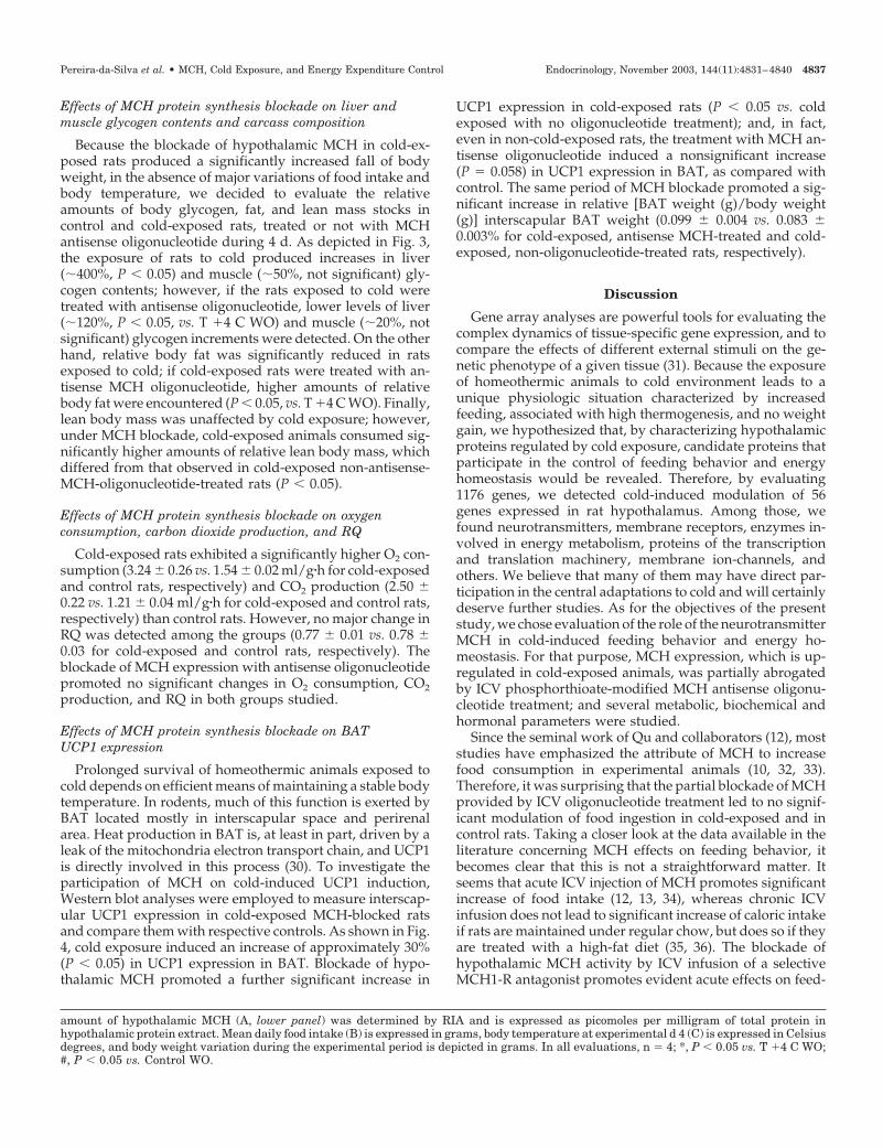
Prolonged survival of homeothermic animals exposed tocold depends on efficient means of maintaining a stable bodytemperature. In rodents, much of this function is exerted byBAT located mostly in interscapular space and perirenalarea. Heat production in BAT is, at least in part, driven by aleak of the mitochondria electron transport chain, and UCP1is directly involved in this process (30). To investigate theparticipation of MCH on cold-induced UCP1 induction,Western blot analyses were employed to measure interscap-ular UCP1 expression in cold-exposed MCH-blocked ratsand compare them with respective controls. As shown in Fig.4, cold exposure induced an increase of approximately 30%(P � 0.05) in UCP1 expression in BAT. Blockade of hypo-thalamic MCH promoted a further significant increase in
UCP1 expression in cold-exposed rats (P � 0.05 vs. coldexposed with no oligonucleotide treatment); and, in fact,even in non-cold-exposed rats, the treatment with MCH an-tisense oligonucleotide induced a nonsignificant increase(P � 0.058) in UCP1 expression in BAT, as compared withcontrol. The same period of MCH blockade promoted a sig-nificant increase in relative [BAT weight (g)/body weight(g)] interscapular BAT weight (0.099 � 0.004 vs. 0.083 �0.003% for cold-exposed, antisense MCH-treated and cold-exposed, non-oligonucleotide-treated rats, respectively).
Discussion
Gene array analyses are powerful tools for evaluating thecomplex dynamics of tissue-specific gene expression, and tocompare the effects of different external stimuli on the ge-netic phenotype of a given tissue (31). Because the exposureof homeothermic animals to cold environment leads to aunique physiologic situation characterized by increasedfeeding, associated with high thermogenesis, and no weightgain, we hypothesized that, by characterizing hypothalamicproteins regulated by cold exposure, candidate proteins thatparticipate in the control of feeding behavior and energyhomeostasis would be revealed. Therefore, by evaluating1176 genes, we detected cold-induced modulation of 56genes expressed in rat hypothalamus. Among those, wefound neurotransmitters, membrane receptors, enzymes in-volved in energy metabolism, proteins of the transcriptionand translation machinery, membrane ion-channels, andothers. We believe that many of them may have direct par-ticipation in the central adaptations to cold and will certainlydeserve further studies. As for the objectives of the presentstudy, we chose evaluation of the role of the neurotransmitterMCH in cold-induced feeding behavior and energy ho-meostasis. For that purpose, MCH expression, which is up-regulated in cold-exposed animals, was partially abrogatedby ICV phosphorthioate-modified MCH antisense oligonu-cleotide treatment; and several metabolic, biochemical andhormonal parameters were studied.
Since the seminal work of Qu and collaborators (12), moststudies have emphasized the attribute of MCH to increasefood consumption in experimental animals (10, 32, 33).Therefore, it was surprising that the partial blockade of MCHprovided by ICV oligonucleotide treatment led to no signif-icant modulation of food ingestion in cold-exposed and incontrol rats. Taking a closer look at the data available in theliterature concerning MCH effects on feeding behavior, itbecomes clear that this is not a straightforward matter. Itseems that acute ICV injection of MCH promotes significantincrease of food intake (12, 13, 34), whereas chronic ICVinfusion does not lead to significant increase of caloric intakeif rats are maintained under regular chow, but does so if theyare treated with a high-fat diet (35, 36). The blockade ofhypothalamic MCH activity by ICV infusion of a selectiveMCH1-R antagonist promotes evident acute effects on feed-
amount of hypothalamic MCH (A, lower panel) was determined by RIA and is expressed as picomoles per milligram of total protein inhypothalamic protein extract. Mean daily food intake (B) is expressed in grams, body temperature at experimental d 4 (C) is expressed in Celsiusdegrees, and body weight variation during the experimental period is depicted in grams. In all evaluations, n � 4; *, P � 0.05 vs. T �4 C WO;#, P � 0.05 vs. Control WO.
Pereira-da-Silva et al. • MCH, Cold Exposure, and Energy Expenditure Control Endocrinology, November 2003, 144(11):4831–4840 4837

ing inhibition and only moderate effects after chronic treat-ment (37). The recombinant hyperexpression of MCH alsoleads to increased food ingestion and obesity, but the mag-nitude of feeding augment is not as exuberant as in acutelyMCH-treated animals (10). Finally, targeted-disruption ofMCH1-R results in hyperphagia and resistance to diet-induced obesity (7, 15). Thus, it seems that the orexigeniceffects of MCH in hypothalamus are dependent on the du-ration of stimulus and on receptor subtype, and that some ofthe approaches used to modulate MCH action produce sub-stantial changes of body weight that are not accompanied bysimilar regulation of food intake.
Body temperature was another parameter evaluated in thepresent study. Normal rats exposed to cold environment (�4C) present an early fall in body temperature, reaching a nadirof approximately 33 C (rectal) after 2–6 h and returning toeuthermia after 24–48 h at most (28, 29). After 4 d of MCHblockade, no significant changes in rectal temperature weredetected in cold-exposed and control rats. These findings arein accordance with previous pharmacological and recombi-nant approaches undertaken to study MCH function in hy-pothalamus (12, 13, 34, 37).
Different from the two previous parameters evaluated,body weight was significantly influenced by the partialblockade of MCH. As in the present study, most previouslypublished data show that exposure of rats to �4 C leads to
an initial loss of body weight, which is followed by gradualrecovery. However, even after several weeks, cold-exposedrats do not reach the same rate of weight gain as theirmatched controls maintained at room temperature. In thepresent experiments, MCH-blocked, cold-exposed rats pre-sented a significantly greater weight loss than their controls,and even non-cold-exposed rats treated with MCH antisenseoligonucleotide underwent significant weight loss.
Because the blockade of MCH expression produced sig-nificantly greater weight loss than in respective controls,which was not accompanied by changes in food intake andbody temperature, we decided to measure body energystocks, oxygen consumption, carbon dioxide production,RQ, and UCP1 expression in BAT, as indirect means of es-timating the flow of the energy that was lost in MCH-blockedanimals, and the participation of BAT in thermogenic adap-tation after cold exposure and MCH signaling interference.The data herein presented reveals that blockade of MCHreduces liver and muscle glycogen content, increases relativeamounts of body fat, and decreases relative amounts of leanmass of cold-exposed rats. Moreover, in MCH-blocked rats,the amount of UCP1 in BAT, which is significantly stimu-lated by cold exposure, suffers a further and significant in-crement, which is accompanied by an increase in the relativeweight of BAT. All these changes occurred in the absence of
FIG. 3. Effects of hypothalamic MCHblockade on liver and muscle glycogencontent and relative fat and lean bodymass. Rats were ICV cannulated and,after a recovery period, were treatedonce a day at 1000 h, with antisense(AS) MCH oligonucleotide with a totalvol of 2.0 �l per dose (2.0 nmol/�l), 24,48, and 72 h after the onset of the ex-perimental period, or with a similar vol-ume of saline (WO). The experimentalanimals were randomly assigned to twogroups, exposed (T �4 C) or not exposed(Control) to cold. Liver (A) and muscle(B) glycogen are expressed as milli-grams per 100 grams of tissue. Relativebody fat (C) and lean mass (D) are ex-pressed as percent of body weight. In allmeasurements, n � 4; *, P � 0.05 vs. T�4 C WO.
4838 Endocrinology, November 2003, 144(11):4831–4840 Pereira-da-Silva et al. • MCH, Cold Exposure, and Energy Expenditure Control
major modifications of O2 consumption, CO2 production,and RQ.
BAT is a specialized organ that actively participates in theproduction of heat. Hypothalamic nuclei stimulate BAT ac-tivity and UCP1 expression by sympathetic inputs. In a re-cent study, the origin of sympathetic connections that inner-vate BAT has been mapped to paraventricular nucleus, LH,prefornical area, and retrochiasmatic nucleus (38). Thus, asone of the predominant neurotransmitters expressed in theLH, MCH may participate in the control of UCP1 expressionand BAT activity in certain conditions. Based on the presentfindings, it seems that, during cold exposure, the optimalexpression of UCP1, which is required for full thermogenicactivity of BAT, is dependent on increased MCH expressionin hypothalamus. Once MCH expression is disrupted, theanimals may loose the capacity of tight regulation of energyexpenditure for heat production, and increased UCP1 may bea reflection of that.
Energy expenditure by motor activity is another possiblemechanism involved in MCH-dependent body weight con-trol. According to Marsh and colleagues (15), the disruptionof MCH1-R gene produces a phenotype of leanness, motorhyperactivity, and hyperphagia. Apparently, most of theincreased metabolic rate of this animal model is attributableto hyperactivity, because it was detected during the darkphase of the light-dark cycle. This observation may explainwhy many studies, including the present one, that attempted
to characterize the role of hypothalamic MCH signaling onfood intake and body weight control, encountered nostraight correlation between variation in caloric intake andbody weight. This may also explain why, in the presentstudy, no significant changes in O2 consumption, CO2 pro-duction, and RQ were detected, because the rats were re-strained during the measurements.
Finally, a recent study (39) provides, by indirect means,further support for a role of MCH in energy expenditurecontrol. The treatment of rodents with the fatty acid synthaseinhibitor C75 was shown to induce hypophagia accompa-nied by a nonproportional loss of body weight, i.e. C75-treated rats lose weight to a lesser extent than food-deprivedrats, although both groups have no caloric intake. Theseanimals express less hypothalamic neuropeptide Y and Ag-outi-related peptide, and more pro-opiomelanocortin andcocaine- and amphetamine-regulated transcript, than theircontrols. However, most important, in our opinion, is the factthat they express higher levels of hypothalamic MCH. Takentogether with the data herein presented, it seems that, duringsome situations that provoke catabolism, the levels of hy-pothalamic MCH are increased to provide means for mini-mizing energy expenditure during adversity.
In conclusion, the present study suggests that hyperex-pression of MCH in hypothalamus of cold-exposed rats par-ticipates in the fine tuning of energy expenditure, providingmeans for optimal heat production with minimal caloricwastage. Apparently, under chronic cold exposure, the roleof MCH in the induction of hyperphagia is minimal or ab-sent. These data reinforce the potentiality of MCH as a targetfor pharmacological therapy of obesity and related diseases,and provide novel support for the concept of dichotomizedcentral control of thermogenesis and food intake.
Acknowledgments
Received February 24, 2003. Accepted July 8, 2003.Address all correspondence and requests for reprints to: Lıcio A.
Velloso, Departamento de Clınica Medica, FCM-UNICAMP, 13083-970Campinas SP, Brazil. E-mail: [email protected].
This study was granted by Fundacao de Amparo a Ciencia do Estadode Sao Paulo. M.P.S. was a recipient of a scholarship from Coordenaçãode Aperfeiçoamento de Pessoal de Nıvel Superior.
References
1. Arancibia S, Rage F, Astier H, Tapia-Arancibia L 1996 Neuroendocrine andautonomous mechanisms underlying thermoregulation in cold environment.Neuroendocrinology 64:257–267
2. Horvath TL, Warden CH, Hajos M, Lombardi A, Goglia F, Diano S 1999 Brainuncoupling protein 2: uncoupled neuronal mitochondria predict thermal syn-apses in homeostatic centers. J Neurosci 19:10417–10427
3. Vinter J, Hull D, Batt RA, Tyler DD 1988 The effect of limit feeding onthermogenesis and thermoregulation in genetically obese (ob/ob) mice duringcold exposure. Int J Obes 12:111–117
4. Melnyk A, Himms-Hagen J 1998 Temperature-dependent feeding: lack of rolefor leptin and defect in brown adipose tissue-ablated obese mice. Am J Physiol274:R1131–R1135
5. Yahata T, Kuroshima A 1989 Metabolic cold acclimation after repetitive in-termittent cold exposure in rat. Jpn J Physiol 39:215–228
6. Baffi JS, Palkovits M 2000 Fine topography of brain areas activated by coldstress. A fos immunohistochemical study in rats. Neuroendocrinology 72:102–113
7. Chen XM, Hosono T, Yoda T, Fukuda Y, Kanosue K 1998 Efferent projectionfrom the preoptic area for the control of non-shivering thermogenesis in rats.J Physiol 512:883–892
8. Ferreira ML, Marubayashi U, Coimbra CC 1999 The medial preoptic area
FIG. 4. Effects of hypothalamic MCH blockade on BAT UCP1 proteinexpression. Rats were ICV cannulated and, after a recovery period,were treated once a day, at 1000 h, with sense (S) or antisense (AS)MCH oligonucleotide or a similar volume of saline (WO), with a totalvol of 2.0 �l per dose (2.0 nmol/�l), 24, 48, and 72-h after the onset ofthe experimental period. The experimental animals were randomlyassigned to two groups, exposed (T �4 C) or not exposed (Control) tocold. At the end of the experimental period, the rats were anesthe-tized, BAT was excised and homogenized, and 0.2 mg of total proteinwas separated by SDS-PAGE, transferred to nitrocellulose mem-branes, and blotted with UCP1 antibodies. Specific bands were mea-sured by densitometry, and results obtained are depicted as scanningunits. n � 4; *, P � 0.05 vs. T �4 C WO.
Pereira-da-Silva et al. • MCH, Cold Exposure, and Energy Expenditure Control Endocrinology, November 2003, 144(11):4831–4840 4839
modulates the increase in plasma glucose and free fatty acid mobilizationinduced by acute cold exposure. Brain Res Bull 49:189–193
9. Spiegelman BM, Flier JS 2001 Obesity and the regulation of energy balance.Cell 104:531–543
10. Ludwig DS, Tritos NA, Mastaitis JW, Kulkarni R, Kokkotou E, Elmquist J,Lowell B, Flier JS, Maratos-Flier E 2001 Melanin-concentrating hormoneoverexpression in transgenic mice leads to obesity and insulin resistance. J ClinInvest 107:379–386
11. Inui A 1999 Feeding and body-weight regulation by hypothalamic neuropep-tides—mediation of the actions of leptin. Trends Neurosci 22:62–67
12. Qu D, Ludwig DS, Gammeltoft S, Piper M, Pelleymounter MA, Cullen MJ,Mathes WF, Przypek R, Kanarek R, Maratos-Flier E 1996 A role for melanin-concentrating hormone in the central regulation of feeding behaviour. Nature380:243–247
13. Della-Zuana O, Presse F, Ortola C, Duhault J, Nahon JL, Levens N 2002 Acuteand chronic administration of melanin-concentrating hormone enhances foodintake and body weight in Wistar and Sprague-Dawley rats. Int J Obes RelatMetab Disord 26:1289–1295
14. Rossi M, Beak SA, Choi SJ, Small CJ, Morgan DG, Ghatei MA, Smith DM,Bloom SR 1999 Investigation of the feeding effects of melanin concentratinghormone on food intake—action independent of galanin and the melanocortinreceptors. Brain Res 846:164–170
15. Marsh DJ, Weingarth DT, Novi DE, Chen HY, Trumbauer ME, Chen AS,Guan XM, Jiang MM, Feng Y, Camacho RE, Shen Z, Frazier EG, Yu H,Metzger JM, Kuca SJ, Shearman LP, Gopal-Truter S, MacNeil DJ, Strack AM,MacIntyre DE, Van der Ploeg LH, Qian S 2002 Melanin-concentrating hor-mone 1 receptor-deficient mice are lean, hyperactive, and hyperphagic andhave altered metabolism. Proc Natl Acad Sci USA 99:3240–3245
16. Shimada M, Tritos NA, Lowell BB, Flier JS, Maratos-Flier E 1998 Mice lackingmelanin-concentrating hormone are hypophagic and lean. Nature 396:670–674
17. Trinder P 1969 Determination of blood glucose using an oxidase-peroxidasesystem with a non-carcinogenic chromogen. J Clin Pathol 22:158–161
18. Scott AM, Atwater I, Rojas E 1981 A method for the simultaneous measure-ment of insulin release and B cell membrane potential in single mouse isletsof Langerhans. Diabetologia 21:470–475
19. Pimenta WP, Saad MJ, Paccola GM, Piccinato CE, Foss MC 1989 Effect of oralglucose on peripheral muscle fuel metabolism in fasted men. Braz J Med BiolRes 22:465–476
20. Kumar MV, Shimokawa T, Nagy TR, Lane MD 2002 Differential effects of acentrally acting fatty acid synthase inhibitor in lean and obese mice. Proc NatlAcad Sci USA 99:1921–1925
21. Thomas DW, Blondel J, Perret P, Lambrechts MM, Speakman JR 2001 En-ergetic and fitness costs of mismatching resource supply and demand inseasonally breeding birds. Science 291:2598–2600
22. Withers PC 1977 Measurement of VO2, VCO2, and evaporative water loss witha flow-through mask. J Appl Physiol 42:120–123
23. Araujo EP, Amaral ME, Souza CT, Bordin S, Ferreira F, Saad MJ, BoscheroAC, Magalhaes EC, Velloso LA 2002 Blockade of IRS1 in isolated rat pancreaticislets improves glucose-induced insulin secretion. FEBS Lett 531:437–442
24. Luciano E, Carneiro EM, Carvalho CR, Carvalheira JB, Peres SB, Reis MA,Saad MJ, Boschero AC, Velloso LA 2002 Endurance training improves re-
sponsiveness to insulin and modulates insulin signal transduction through thephosphatidylinositol 3-kinase/Akt-1 pathway. Eur J Endocrinol 147:149–157
25. Nagy G, Szekeres G, Kvell K, Berki T, Nemeth P 2002 Development andcharacterisation of a monoclonal antibody family against aquaporin 1 (AQP1)and aquaporin 4 (AQP4). Pathol Oncol Res 8:115–124
26. Bradford MM 1976 A rapid and sensitive method for the quantitation ofmicrogram quantities of protein utilizing the principle of protein-dye binding.Anal Biochem 72:248–254
27. Mondal MS, Nakazato M, Matsukura S 2002 Characterization of orexins(hypocretins) and melanin-concentrating hormone in genetically obese mice.Regul Pept 104:21–25
28. Cunningham JJ, Gulino MA, Meara PA, Bode HH 1985 Enhanced hepaticinsulin sensitivity and peripheral glucose uptake in cold acclimating rats.Endocrinology 117:1585–1589
29. Vallerand AL, Perusse F, Bukowiecki LJ 1987 Cold exposure potentiates theeffect of insulin on in vivo glucose uptake. Am J Physiol 253:E179–E186
30. Enerback S, Jacobsson A, Simpson EM, Guerra C, Yamashita H, Harper ME,Kozak LP 1997 Mice lacking mitochondrial uncoupling protein are cold-sen-sitive but not obese. Nature 387:90–94
31. Cheung VG, Spielman RS 2002 The genetics of variation in gene expression.Nat Genet 32(Suppl):522–525
32. Huang Q, Viale A, Picard F, Nahon J, Richard D 1999 Effects of leptin onmelanin-concentrating hormone expression in the brain of lean and obeseLep(ob)/Lep(ob) mice. Neuroendocrinology 69:145–153
33. Yamada M, Miyakawa T, Duttaroy A, Yamanaka A, Moriguchi T, Makita R,Ogawa M, Chou CJ, Xia B, Crawley JN, Felder CC, Deng CX, Wess J 2001 Micelacking the M3 muscarinic acetylcholine receptor are hypophagic and lean.Nature 410:207–212
34. Clegg DJ, Air EL, Benoit SC, Sakai RS, Seeley RJ, Woods SC 2002 Intra-ventricular melanin concentrating hormone stimulates water intake indepen-dent of food intake. Am J Physiol Regul Integr Comp Physiol 7:7
35. Gomori A, Ishihara A, Ito M, Mashiko S, Matsushita H, Yumoto M, TanakaT, Tokita S, Moriya M, Iwaasa H, Kanatani A 2003 Chronic intracerebro-ventricular infusion of MCH causes obesity in mice. Melanin-concentratinghormone. Am J Physiol Endocrinol Metab 284:E583–E588
36. Ito M, Gomori A, Ishihara A, Oda Z, Mashiko S, Matsushita H, Yumoto M,Sano H, Tokita S, Moriya M, Iwaasa H, Kanatani A 2003 Characterization ofMCH-mediated obesity in mice. Am J Physiol Endocrinol Metab 284:E940–E945
37. Borowsky B, Durkin MM, Ogozalek K, Marzabadi MR, DeLeon J, Lagu B,Heurich R, Lichtblau H, Shaposhnik Z, Daniewska I, Blackburn TP,Branchek TA, Gerald C, Vaysse PJ, Forray C 2002 Antidepressant, anxiolyticand anorectic effects of a melanin-concentrating hormone-1 receptor antago-nist. Nat Med 8:825–830
38. Oldfield BJ, Giles ME, Watson A, Anderson C, Colvill LM, McKinley MJ2002 The neurochemical characterisation of hypothalamic pathways projectingpolysynaptically to brown adipose tissue in the rat. Neuroscience 110:515–526
39. Shimokawa T, Kumar MV, Lane MD 2002 Effect of a fatty acid synthaseinhibitor on food intake and expression of hypothalamic neuropeptides. ProcNatl Acad Sci USA 99:66–71
4840 Endocrinology, November 2003, 144(11):4831–4840 Pereira-da-Silva et al. • MCH, Cold Exposure, and Energy Expenditure Control