Evaluation of lymphocyte apoptosis in patients with oral cancer
Upload
independentCategory
view
2download
0
of August 10, 2015.This information is current as
PathwayEntry into the T Lymphocyte Developmental Molecular Dissection of Prethymic Progenitor
Moore, Lee Rowen, Tom Taghon and Ellen V. RothenbergC. Chace Tydell, Elizabeth-Sharon David-Fung, Jonathan E.
http://www.jimmunol.org/content/179/1/421doi: 10.4049/jimmunol.179.1.421
2007; 179:421-438; ;J Immunol
MaterialSupplementary
mlhttp://www.jimmunol.org/content/suppl/2008/03/13/179.1.421.DC1.ht
Referenceshttp://www.jimmunol.org/content/179/1/421.full#ref-list-1
, 40 of which you can access for free at: cites 76 articlesThis article
Subscriptionshttp://jimmunol.org/subscriptions
is online at: The Journal of ImmunologyInformation about subscribing to
Permissionshttp://www.aai.org/ji/copyright.htmlSubmit copyright permission requests at:
Email Alertshttp://jimmunol.org/cgi/alerts/etocReceive free email-alerts when new articles cite this article. Sign up at:
Print ISSN: 0022-1767 Online ISSN: 1550-6606. Immunologists All rights reserved.Copyright © 2007 by The American Association of9650 Rockville Pike, Bethesda, MD 20814-3994.The American Association of Immunologists, Inc.,
is published twice each month byThe Journal of Immunology
by guest on August 10, 2015
http://ww
w.jim
munol.org/
Dow
nloaded from
by guest on August 10, 2015
http://ww
w.jim
munol.org/
Dow
nloaded from
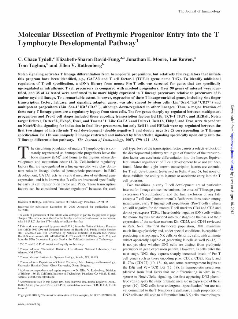
Molecular Dissection of Prethymic Progenitor Entry into the TLymphocyte Developmental Pathway1
C. Chace Tydell,2 Elizabeth-Sharon David-Fung,2,3 Jonathan E. Moore, Lee Rowen,4
Tom Taghon,5 and Ellen V. Rothenberg6
Notch signaling activates T lineage differentiation from hemopoietic progenitors, but relatively few regulators that initiatethis program have been identified, e.g., GATA3 and T cell factor-1 (TCF-1) (gene name Tcf7). To identify additionalregulators of T cell specification, a cDNA library from mouse Pro-T cells was screened for genes that are specificallyup-regulated in intrathymic T cell precursors as compared with myeloid progenitors. Over 90 genes of interest were iden-tified, and 35 of 44 tested were confirmed to be more highly expressed in T lineage precursors relative to precursors of Band/or myeloid lineage. To a remarkable extent, however, expression of these T lineage-enriched genes, including zinc fingertranscription factor, helicase, and signaling adaptor genes, was also shared by stem cells (Lin�Sca-1�Kit�CD27�) andmultipotent progenitors (Lin�Sca-1�Kit�CD27�), although down-regulated in other lineages. Thus, a major fraction ofthese early T lineage genes are a regulatory legacy from stem cells. The few genes sharply up-regulated between multipotentprogenitors and Pro-T cell stages included those encoding transcription factors Bcl11b, TCF-1 (Tcf7), and HEBalt, Notchtarget Deltex1, Deltex3L, Fkbp5, Eva1, and Tmem131. Like GATA3 and Deltex1, Bcl11b, Fkbp5, and Eva1 were dependenton Notch/Delta signaling for induction in fetal liver precursors, but only Bcl11b and HEBalt were up-regulated between thefirst two stages of intrathymic T cell development (double negative 1 and double negative 2) corresponding to T lineagespecification. Bcl11b was uniquely T lineage restricted and induced by Notch/Delta signaling specifically upon entry into theT lineage differentiation pathway. The Journal of Immunology, 2007, 179: 421– 438.
T he circulating population of mature T lymphocytes is con-stantly regenerated as hemopoietic progenitors leave thebone marrow (BM)7 and home to the thymus where de-
velopment and maturation occur (1–3). Cell-intrinsic regulatoryfactors that are up-regulated in a lineage-specific way play domi-nant roles in lineage choice of hemopoietic precursors. In RBCdevelopment, GATA1 acts as a central mediator of erythroid geneexpression, and it is known that B cells are instructed specificallyby early B cell transcription factor and Pax5. These transcriptionfactors can be considered “master regulators” because, for each
cell type, loss of the transcription factor causes a selective block ofthe developmental pathway while gain of function of the transcrip-tion factor can accelerate differentiation into the lineage. Equiva-lent “master regulators” of T cell development have not yet beenfound. More than eight known transcription factors are essentialfor T cell development (reviewed in Refs. 4 and 5), but none ofthese exhibits the ability to instruct or accelerate entry into the Tcell program.
Two transitions in early T cell development are of particularinterest for lineage choice mechanisms: the onset of T lineage geneexpression (“specification”), and the final exclusion of any fateexcept a T cell fate (“commitment”). Both transitions occur amongintrathymic, early T lineage cell populations (Pro-T cells), whichare still negative for the mature T cell markers CD4 and CD8 anddo not yet express TCRs. These double-negative (DN) cells withinthe mouse thymus are divided into four stages on the basis of theirexpression of the surface markers Kit, CD25, and CD44 reviewedin Refs. 6–8. The first thymocyte population, DN1, maintainsmuch lineage plasticity and, under special conditions, is capable ofproducing macrophages, NK cells, or dendritic cells, with a minutesubset apparently capable of generating B cells as well (9–12). Itis not yet clear whether DN1 cells are distinct from prethymicprecursors in gene expression pattern. However, as cells enter thenext stage, DN2, they express sharply increased levels of Pro-Tcell genes such as those encoding pT�, CD3�, CD25, Rag1, andIL-7R� (CD127) (10, 13–16), and some rearrangement begins atthe DJ� and VJ� TCR loci (17, 18). In hemopoietic precursors(derived from fetal liver) that are differentiating in vitro in re-sponse to Notch/Delta signaling, the first-appearing DN2 pheno-type cells display the same dramatic increase in expression of thesegenes (19). DN2 cells have undergone “specification” but are notyet committed to the T lymphocyte pathway; a high proportion ofDN2 cells are still able to differentiate into NK cells, macrophages,
Division of Biology, California Institute of Technology, Pasadena, CA 91125
Received for publication December 18, 2006. Accepted for publication April16, 2007.
The costs of publication of this article were defrayed in part by the payment of pagecharges. This article must therefore be hereby marked advertisement in accordancewith 18 U.S.C. Section 1734 solely to indicate this fact.1 This work was supported by grants (to E.V.R.) from the National Science Founda-tion (MCB-9983129) and National Institutes of Health U.S. Public Health Service(R01 CA90233 and R01 CA98925), by National Institutes of Health U.S. PublicHealth Service awards K08 AI054699 (to C.C.T.) and F32 AI068366 (to J.E.M.); andfrom the DNA Sequencer Royalty Fund at the California Institute of Technology.2 C.C.T. and E.-S.D.-F. contributed equally to this study.3 Current address: Theoretical Division, Los Alamos National Laboratory, LosAlamos, NM 87544.4 Current address: Institute for Systems Biology, Seattle, WA 98103.5 Current address: Department of Clinical Chemistry, Microbiology and Immunology,University Hospital Ghent, Ghent University, Ghent, Belgium.6 Address correspondence and reprint requests to Dr. Ellen V. Rothenberg, Divisionof Biology 156-29, California Institute of Technology, Pasadena, CA 91125. E-mailaddress: [email protected] Abbreviations used in this paper: BM, bone marrow; DN, double negative; Dtx3L,Deltex3-like; pT�, pre-TCR�; qRT-PCR, quantitative real-time PCR; TCF-1, T cellfactor-1.
Copyright © 2007 by The American Association of Immunologists, Inc. 0022-1767/07/$2.00
The Journal of Immunology
www.jimmunol.org
by guest on August 10, 2015
http://ww
w.jim
munol.org/
Dow
nloaded from

or dendritic cells (10, 20–23). At the DN3 stage, thymocytes stopdividing, further increase expression of the Pro-T differentiationgenes as well as Notch target genes (24), and undergo extensiveTCR rearrangements. Only at this stage do they become committedto a T cell fate in vivo. Cells only progress beyond the DN3 stagethrough successful TCR gene rearrangement and TCR-dependentselection, at which time they graduate from Pro-T cell status, togive rise to up to five types of T cells: �� CD4, �� CD8, ��, NKT,or regulatory T cells.
Of all these developmental transitions, surprisingly little isknown about the stages encompassing “T lineage specification,”that is, the DN1 to DN2 transition. The regulatory participants inthese early stages have not been sufficiently characterized to ex-plain the outcome, although Notch/Delta signaling plays a role (19,25). Traditional methods to identify all the transcription factorsthat play key roles in early stages of T lineage specification havefound limited success, in part because transcription factors are typ-ically present in low copy numbers. T cell precursors at the earlieststages are represented in vivo at any one time by tiny numbers ofcells, providing very limiting material for standard microarrayanalysis. In addition, small fold changes in transcription factorabundance or changes in transcription factor ratios may generatedramatic shifts in cell state. Identification of truly novel regulatoryfactors and novel isoforms of known factors has also been limitedby the comprehensiveness of the microarrays used (26–29) and bythe microarray’s nucleotide probe design.
To circumvent these problems, we used a subtractive hybrid-ization technique (30) to probe a mouse Pro-T cell cDNA mac-roarray library. The subtractive technique allowed us to enrich aPro-T cell probe for message not shared by progenitor/premyeloidcells and to identify genes (known and previously unknown) thatmight be specifically up-regulated during the initiation of the Tlineage program. Clones selected by the subtraction were se-quenced and their patterns of expression characterized in detailusing sensitive, quantitative real-time PCR (qRT-PCR) on a rangeof highly purified cell populations. The Pro-T cell library was gen-erated by random priming of mRNA from SCID thymocytes,which consist of DN1–3 stage Pro-T cells and NK precursors (31).The resulting macroarray library represents the actual spectrum oftranscripts in the DN2 and DN3 cell populations (those immedi-ately preceding lineage commitment) and also contains multipleclones of genes with abundant transcripts, providing opportunitiesto sample alternate splice variants.
Our subtraction protocol has identified genes that are specifi-cally enriched in T-lineage as opposed to early B or myeloid lin-eage precursors. Enriched genes include novel transcription factorcandidates, chromatin remodeling factors, RNA binding moleculesand helicases, a select group of signaling molecules and adaptors,and novel or functionally uncharacterized genes. In this study, wepresent the resulting expression profiles of �35 of these genesexpressed during T lineage specification. A remarkable feature ofthe whole ensemble of these Pro-T cell genes is the high frequencyof “legacy” genes that are expressed strongly in both stem cellsand Pro-T cells, although down-regulated elsewhere. In this con-text, the very select group of regulatory genes that are specificallyinduced coincidentally with T lineage specification takes on anunexpected significance.
Materials and MethodsMice
C57BL/6J, B6.CB17-Prkdcscid/SzJ (B6-scid) (The Jackson Laboratory)and B6-Rag2null mice (originally from E. Palmer, Basel Institute, Basel,Switzerland) were bred and maintained in specific pathogen-free facilitiesat Caltech.
Thymus and BM samples were taken from animals 5–7 wk old. Theanimals used were bred and maintained under sterile conditions at Caltech.
cDNA library
The C.B-17-scid thymocyte, random-primed, cDNA library was con-structed in the pSPORT1 vector (Invitrogen Life Technologies), andwas arrayed and spotted at high density onto Hybond-N� nylon filters(Amersham Biosciences) using the Q-BOT robot (Genetix) as describedpreviously (31).
Cell populations for library generation and library screening
The two types of cells used as sources of RNA for the subtraction protocolwere a bulk population of Pro-T cells and a population of progenitor/premyeloid cells. To obtain large numbers of Pro-T cells in the DN1-DN3stages, we took advantage of the Rag2 knockout mouse, in which thymo-cyte development arrests at DN3. In the wild-type mouse thymus, DN3cells account for only 1% of thymocytes, but even without sorting, Rag2knockout thymocytes consist of 90% DN3 Pro-T cells, with the remainingcells being DN1, DN2, NK, or thymic stromal cells. Because these cellswere not pure Pro-T cells, we refer to them in the text as “Pro-T plus.” Aprogenitor/premyeloid population was obtained from Lin�Kit� BM cellsmatured in culture toward a myeloid fate. Specifically, BM cells that wereKit�Gr1�CD11b�Ter119�CD19� from Rag2ko mice were cultured for42 h at 37°C in 5% CO2 in IMDM with 10% heat-inactivated FCS sup-plemented with IL-3 (100 �l of WEHI-3B cell supernatant/ml medium), stemcell factor (Kit ligand) (100 �l of BHK-MKL cell supernatant/ml medium),and 10 ng/ml rIL-6 (PeproTech). At the time of harvest, the morphologicalappearance of the cells ranged from undifferentiated, blast-like cells to maturegranulocytes.
Cell populations for quantitative RNA expression analysis
In addition to the subtraction populations, sorted cell populations wereobtained for analysis by qRT-PCR. Two hemopoietic progenitor popula-tions, a Pro-B population, and a population of myeloid cells were all sortedfrom the BM of Rag2ko mice. LSK CD27� cells were Kit�Sca-1�CD27�
Gr1�CD11b�Ter119�CD19�. LSK CD27� cells were Kit�Sca-1�CD27�
Gr1�CD11b�Ter119�CD19�. Pro-B cells were CD19�Kit�/�Gr1�CD11b�
Ter119�. Sorted BM myeloid cells were Gr1�CD11b�Kit�Ter119�.Initial expression screening made use of the unsorted Rag2ko thymocytes
called Pro-T plus. For more detailed analysis, five populations of DN Pro-Tcells were purified from C57BL/6 mouse thymi by cell sorting, essentially asdescribed previously (13, 24). Each of these DN subsets is CD4�CD8�CD3��
Ter119�F4/80� and Gr1�. DN1 cells are Kit�CD44�CD25�. DN2 cells areKit�CD44�CD25�. DN3a cells are KitlowCD44�CD25�CD27�. DN3b cellsare CD44�CD25�CD27�, and DN4 cells are CD44�CD25�. All Abs used inthis study were obtained from eBioscience or BD Pharmingen.
Thymocytes and BM cells were obtained from animals immediatelyafter euthanasia. Cells were incubated in CBSS (5.4 mM KCl, 0.3 mMNa2HPO4, 0.4 mM KH2PO4, 4.2 mM NaHCO3, 137 mM NaCl, and 5.6mM D-glucose (pH 7.4))/1% BSA with clone 2.4G2 anti-CD32/CD16(Fc�RIII/II) supernatant on ice for 10 min, followed by washing and ad-dition of Abs for staining. Cells stained with biotin-conjugated Abs werewashed through a layer of FCS before staining with streptavidin-PECy5(eBioscience). Stained cells were sorted using FACS Aria cell sorter (BDImmunocytometry Systems). All sorted fractions were reanalyzed imme-diately for purity and all fractions used here were at least 96% pure.
Preparation of fetal liver cells as input for OP9-DL1 and OP9-controlcultures is described below.
Generation of subtractive probe
A Pro-T plus unselected cDNA probe and a T lineage-enriched subtractedprobe were generated from the pool of enriched Pro-T cells (�1 � 107
cells; Pro-T plus) and the population of myeloid and multipotent progen-itors (�9 � 106 cells) described above. RNA was isolated from each popu-lation of cells by the Qiagen RNeasy Lipid Minikit (Qiagen), and the RNA wasanalyzed for purity and integrity by Agilent Bioanalyzer and RNA 6000 Nano-chips (Agilent). Messenger RNA was isolated from total RNA by the AmbionPoly(A) Purist Kit (Ambion). The mRNA was also evaluated for purity andconcentration by the Bioanalyzer.
The subtraction method was adapted from Rast et al. (30). cDNA wassynthesized using enzymes and buffers from a Clontech Marathon cDNAsynthesis kit (BD Biosciences), but with the LT7 random-BT primer: 5�-(biotin)-CGGAGGTAATACGACTCACTATAGGGAGNNNNNN-3� (34nt). Qiagen PCR purification columns were used to purify samples betweenfirst-strand and second-strand synthesis stages.
422 GENE DISCOVERY FOR T CELL SPECIFICATION IN VIVO AND IN VITRO
by guest on August 10, 2015
http://ww
w.jim
munol.org/
Dow
nloaded from

Different linkers are used for Selectate (the Pro-T plus-derived cDNA)or Driver (the progenitor/myeloid-derived cDNA) to avoid nonspecificsubtraction. Linkers contain a 3� dideoxy residue to prevent filling in ofoverhang, and a 5� phosphate for blunt ended ligation to the cDNA. TheSelectate linker sequences were 5�-GGGTGCTGTATTGTGTACTTGAACGGGCGGCCGCA-3� and 3�-dideoxy-CGCCCGCCGGCGT-P-5�. TheDriver linker sequences were 5�-GCCAACGTATGTAAGGTTGAGTTCCGGGCAGGT-3� and 3�-dideoxy-CCCGTCCA-P-5�. Linkers were an-nealed by placing the linker pairs in a 1:1 molar ratio at a concentrationtotaling 1 �g/�l in 10 mM Tris (pH 7.9) and 100 mM NaCl, heating in aheating block to 95°C for 5 min, then turning off the block and allowing thelinkers to anneal as the block cools to room temperature (�30 min). Li-gation efficiency was evaluated by comparing their electrophoresis migra-tion on a 2% agarose gel relative to the untreated mixture. These linkerswere ligated to either the Selectate or Driver cDNA for 16 h at 16°C withDNA ligase.
cDNA with linkers attached was PCR amplified with LT7 primer (5�-CGGAGGTAATACGACTCACTATAGG-3�) and a primer specific for ei-ther the Selectate linker (5�-GGGTGCTGTATTGTGTACTTGAACG-3�)or for the Driver linker (5�-GCCAACGTATGTAAGGTTGAGTTCC-3�)to produce 600 ng of product. The resulting Selectate was size selected for300- to 500-bp product by electrophoresis of the PCR in an agarose gel,excising the appropriate region of the gel, and electroeluting cDNA fromthe gel. The electroeluate was precipitated and resuspended in 50 �l ofwater or T low E (10 mM Tris and 0.1 mM EDTA, (pH 7.8). Driverproduct was precipitated and resuspended in 16 �l of water.
Size-selected Selectate was amplified by PCR (primers listed above),and 1 �g of Selectate was set aside for production of the unsubtractedlibrary probe. Selectate (3 �g) was subjected to single-strand purificationby Dynal Streptavidin beads, according to the manufacturer’s instructions(Invitrogen Life Technologies). RNase-free technique was used from thispoint forward for both Selectate and Driver. The Ambion MEGAshortscript kit(Ambion) was used according to the manufacturer’s instructions to trans-late the Driver cDNA to RNA. Single-stranded Selectate DNA (200 ng)from Pro-T plus cells was mixed with Driver RNA (30 �g) in a 10-�l totalvolume, denatured at 95°C, iced, then hybridized at 65°C for 40 h. Double-stranded and single-stranded products were separated by hydroxylapatitechromatography (30, 32), and the eluate containing the single-strandedproduct was desalted and concentrated. The single-stranded product was usedto manufacture the subtracted, radioactive probe by Ambion Maxiscript kitusing Amersham 800 Ci/mM 32P-UTP.
Subtractive hybridization protocol
Macroarray filters were sequentially hybridized with cDNA from Pro-Tplus Rag2ko thymocytes, stripped of probe, then hybridized with subtractedprobe, i.e., probe enriched for mRNA that was not shared by the progen-itor/premyeloid population. Hybridization intensity for each probe wasmeasured by a PhosphorImager (Molecular Dynamics, GE Healthcare) usingBioArray software (Genetix). Representative data are shown in Fig. 1.
On the macroarray blot, each clone is applied in duplicate as a “spotpair” with unique position and arrangement (see Fig. 1, A and B). Pixelintensity for each spot pair was averaged, and systematic differences inhybridization intensity across the blots were minimized by the applicationof a whitening filter, much like the linear optimal filter or Wiener filter (33,34). Its formula is:
�� f � S� f 2 / �S� f 2 � N� f 2)
where �( f) is the whitening filter, S( f) is the signal in Fourier space, andN( f) is the noise in Fourier space. A Wiener filter is often used to filterrandom, usually small-scale, noise from data leaving mostly the large-scalecorrelations; this is its normal use in image analysis. However, because ourclones were randomly placed on the blots, the only correlations expected inour data are the relatively small-scale ones between spot pairs. Hence, alllarge-scale correlations are likely due to systematic noise such as inhomo-geneous probing or washing of the blots. Therefore, we used the whiteningfilter to remove such correlations (J. E. Moore, unpublished results).
The ratio of the average spot-pair hybridization intensity before andafter subtraction was termed enrichment: (intensity for subtracted probe) (intensity for the unsubtracted probe) � enrichment. The logarithms of theenrichments for 73,728 spot pairs were calculated, and a clone was deemedto be significantly enriched when the logarithm of its enrichment was morethan three SDs above the mode of its blot (Fig. 1C). Clones more than fourSDs above the mode were selected for special attention (see Table I). Themodes were calculated by a process called “estimating the rate of an in-homogeneous Poisson process by Jth waiting times” (33), briefly outlinedhere. For each blot, the logarithms of the enrichments were ordered. Awindow size, J, was chosen, which for these calculations was (1)/24 the
number of spot pairs on a blot, or 768; other reasonable values of J do notappreciably change the estimates. An integer, I, was chosen so as to min-imize the difference between the Ith and (I � J)th logarithms, and the modewas estimated by averaging these.
Clones selected by this method were sequenced from both the 5� and the3� ends at the Institute for Systems Biology using standard proceduresdesigned for the Applied Biosystems 3730XL sequencer. Each sequencewas filtered by RepeatMasker and analyzed by Blastn, Blastx, and BLATsearches of National Center for Biotechnology Information (NCBI), Uni-versity of California, Santa Cruz (UCSC), and Ensembl databases to iden-tify matches to known genes or genomic sequences (latest searches: buildm36 of mouse genome). Of the 1164 clones submitted, 1046 good se-quences were obtained.
GOToolBox analysis
The GO-Stats function of the GOToolBox website (35) was used to per-form a hypergeometric analysis of statistically relevant over- or under-represented terms within our data set as compared with the Mouse GenomeInformatics database of genes. The Benjamini & Hochberg correction formultiple testing was applied. Selected results of searches in the categoriesof Biological Processes and Cellular Components are reported herein.
Bioinformatic databases
The following databases were used: www.ncbi.nlm.nih.gov, http://genome.ucsc.edu, www.ensembl.org/Mus_musculus, http://crfb.univ-mrs.fr/GOToolBox/index.php, and www.informatics.jax.org.
Coculture of fetal liver cells with OP9 cells
Hemopoietic progenitors cocultured with BM stromal cells (OP9 cell line)will develop into B lymphocytes in vitro. When OP9 stromal cells aretransfected to stably express the Notch ligand Delta-like 1 (OP9-DL1),progenitor cells will develop into T lymphocytes in coculture (36). Mousefetal liver cells (containing hemopoietic progenitors) were cocultured withOP9-control or OP9-DL1 cells exactly as described previously (19). Inshort, Kit�Lin� (Lin � Gr1, Ter119, F4/80, CD19) cells from day 14 to14.5 mouse embryo livers were obtained by FACS sorting. Kit�Lin� fetalliver cells were cocultured with OP9 control cells or with OP9-DL1 cells(36). Cocultures were harvested for RNA analysis by forceful pipetting atindicated time points. In some experiments, to test the effect of delayedaddition or withdrawal of Notch signals, progenitor cells were transferredto secondary cultures at day 4. OP9-control and OP9-DL1 cocultures wereharvested, Kit�CD27�Lin� cells were isolated from each culture by sort-ing, and these were each split and used to seed fresh monolayers of OP9control and OP9-DL1 cells, to be harvested at the indicated later timepoints.
To compare the time courses of T lineage differentiation from distinct pre-cursor subsets, Kit�Lin� fetal liver cells were fractionated into Kit�Sca-1�
(“LSK”), Kit�Sca-1lowCD27�Flk2/Flt3 (CD135)�IL-7R�(CD127)� (“Flk�”),and Kit�Sca-1lowCD27�CD135�CD127� (“CLP-like”) subsets, as de-scribed elsewhere (T. Taghon, M. A. Yui, and E. V. Rothenberg, submittedfor publication). These subsets were then cocultured with OP9-control orOP9-DL1 cells for 1–7 days before harvesting for RNA.
Quantitative real-time PCR
qRT-PCR was performed on diluted samples of cDNA using SYBR GreenPCR Master Mix in an ABI PRISM 7700 Sequence Detector (AppliedBiosystems). In all figures comparing expression levels of multiple genes,the measurements for all genes shown were conducted on the same cDNAsamples. The �Ct method was used for all expression measurements, witha fixed threshold to enable direct comparison between test genes and theGAPDH standard. Primers were designed, using Primer3 software (37), tohave optimal melting temperatures and to cross introns. The primers wereBLAST tested for gene specificity before being synthesized (OperonBiotechnologies). Each primer pair was evaluated for acceptable dose-response titration slopes and amplification. Primer sequences are asfollows: Ablim, forward, CTGGCAGCTCAGAGGAGTTC, and re-verse, CGCAGCTGGGATGATAATG; Aff3, forward, CAACAGAGAGCAGCGCAACA, and reverse, CCCGTCTCCATATTGCACACTT;AI449175, forward, GCTCCTTCCCAGAAGACTCTC, and reverse, TCAGGCTCTTCAAAATGGTCTT; Akap8, forward, AAATTGAGAAACGGCGTCAG, and reverse, AATGTGCGGCTTCAATCTTT; �-ac-tin, forward, ACACCCGCCACCAG, and reverse, TACAGCCCGGGGAG; Bat2, forward, ATACTGCCACAAGCCGAAAG, and reverse, TCAGGTCCACTCCACTGTCA; Bcl11a, forward, GTCTGCACACGGAGCTCTAA, and reverse, CACTGGTGAATGGCTGTTTG; Bcl11b,forward, GGGCGATGCCAGAATAGAT, and reverse, GGTAGCCTC
423The Journal of Immunology
by guest on August 10, 2015
http://ww
w.jim
munol.org/
Dow
nloaded from

Table I. Other zinc finger proteins
TotalClones Enrichment qRT-PCR Comments
Exon matchedGenes known to be up-regulated in T development
IL-2ra 124 clones ‡ IL-2R, � chain. regulation of T cell proliferationCD3g 61 clones ‡ CD3 Ag, � chain. Component of T cell complexTcrg 53 clones ‡ TCR � chainCD3e 34 clones ‡ CD3 chainTcrb 22 clones ‡ TCR � chainTcf7 (TCF-1) 18 clones ‡ A,b B Transcription factor 7, aka TCF-1, T cell specificThy1 17 clones ‡ Thymus cell Ag 1, Lat 5 clones ‡ Linker for activation of T cells, receptor-signaling proteinDntt 5 clones ‡ Deoxynucleotidyltransferase, terminal. Also known as TdtLy6a 5 clones ‡ Lymphocyte Ag 6 complex, locus A, aka Sca-1.Lck 3 clones ‡ Lymphocyte protein tyrosine kinase is involved in
regulation of the TCR-signaling pathwayDpp4 2 clones ‡ Dipeptidylpeptidase 4, aka CD26Cd24a ‡ HSA or Ly52
RNA bindingDdx17 † B DEAD box helicase protein 17, p72, reported to regulate
transcriptionDdx19b ‡ B, C DEAD box helicase protein 19bFUS 2 clones † A, B Pigpen or Fusion derived from t(12;16) malignant
liposarcoma, transcriptional activatorHelz † B, C Helicase with zinc finger domain
Other zinc finger proteinsRad18 ‡ Ring-type zinc finger protein, DNA repair activityTrim44 ‡ A, B Tripartite motif-containing 44, B-box domain
Lim-related proteinsAblim1 ‡ B Actin-binding LIM protein 1, deletions are linked to
cancerCtdsp1 ‡ A, B A small phosphatase with a NLI, nuclear LIM interacting,
domainLdb1 2 clones ‡ A, B LIM domain-binding 1, may interact with Scl in
thrombopoiesisProtein metabolism, signaling, kinases, and ubiquitination
Akap8 ‡ B A kinase (PRKA) anchor protein 8Apoe 2 clones ‡ Apolipoprotein E, lipid transport and Ca2� homeostasisAtxn2l † A, B Ataxin 2 spinocerebellar ataxia type II, may be part of
cytokine-signaling systemCcnk † Cyclin K, may play a dual role in regulating CDK and
RNA polymerase II activitiesDdef1 † Development and differentiation enhancing 1, predicted to
have transcriptional activityDpp8 ‡ Dipeptidylpeptidase 8Dtx3l 2 clones ‡ C Deltex3L, RING finger protein, similar to rhysin2Fgfrl1 † B Fibroblast growth factor receptor-like 1 is involved in the
negative regulation of cell proliferation.Fkbp5 † A, B, C FK506 binding protein 5, a novel T cell-specific
immunophilin capable of calcineurin inhibition. Low inmature T cells.
Gpr56 3 clones ‡ B An atypical G protein-coupled receptor, binds to tissuetransglutaminase and acts in brain cortical development
Gps1 ‡ G protein pathway suppressor 1, subunit 1 of the COP9signalosome
Grap † A, B GRB2-related adaptor protein, GRB2 may interact withLAT by the SH2 domain. It is a negative regulator ofthe Erk pathway.
Huwe1 ‡ A, B HECT, UBA and WWE domain containing 1 (aka Ureb1)is an E3 ubiquitin protein ligase that inhibits theactivity of tumor suppressor p53 protein
Prkag3 † Protein kinase, AMP-activated, �3 noncatalytic subunit,regulates skeletal muscle glycogen content
Prpf4b † Pre-mRNA processing factor 4 homolog B, a putativekinase
Prss16 # 2 clones ‡ B# Thymus-specific serine protease precursor Prss16, aprotein of the thymic stroma
Ptpn7 ‡ B Protein tyrosine phosphatase, non-receptor type 7, aMAPK-specific protein tyrosine phosphatase. Same asHEPTP
Rab2 ‡ A, B A G protein of the RAS oncogene family(Table continues)
424 GENE DISCOVERY FOR T CELL SPECIFICATION IN VIVO AND IN VITRO
by guest on August 10, 2015
http://ww
w.jim
munol.org/
Dow
nloaded from

Table I. (Continued)
TotalClones Enrichment qRT-PCR Comments
Rabgap1 † A, B RAB GTPase-activating protein 1Senp2 ‡ B SUMO/sentrin-specific protease, enhancer of WntTgfbr2 ‡ TGF, � receptor IITrappc2l † B Trafficking protein particle complex 2-like, formerly
1810017G16RikUbe2l3 2 clones ‡ Ubiquitin-conjugating enzyme E2L3, an E2 ubiquitin
protein ligaseUble1a ‡ Ubiquitin-like 1-activating enzyme E1A (SUMO-1-
activating enzyme)Was (Wasp) † A Wiskott-Aldrich syndrome protein, may function as a
signal transduction adaptor downstream of Cdc42Wbp11 † WW domain binding protein 11, has protein phosphatase
type 1 regulator activity, and is involved in RNAsplicing
Zcchc11 ‡ B Zinc finger, CCHC domain containing 11, reportedinteract with TIFA to modulate TLR signaling
Regulators of transcriptionAI449175 ‡ A, B Kruppel-like zinc finger protein that has a KRAB domainAff3 ‡ A, B Laf4, implicated in T cell leukemiaCrsp7 ‡ A, B Cofactor required for Sp1 transcriptional activationLef1 ‡ Lymphoid enhancer binding factor 1, positive regulator of
transcriptionMll1 ‡ A, B Myeloid/lymphoid or mixed-lineage leukemia 1, trithorax
family proteinMll2 ‡ A, B Myeloid/lymphoid or mixed-lineage leukemia 2, trithorax
family proteinMxd4 ‡ B Max dimerization protein 4, bHLH protein and predicted
transcription factorNotch1 8 clones ‡ Notch gene homolog 1 is a positive regulator of
transcriptionNotch3 3 clones ‡ Notch gene homolog 3 is involved in the negative
regulation of cell differentiationRunx1 ‡ Runt-related transcription factor 1, AML1, has DNA-
dependent transcriptional activityTardbp 2 clones † TAR DNA-binding protein, predicted to regulate
transcriptionTcf12 (HEB) 4 clones ‡ A, B, C HEB, bHLH protein, transcriptional activatorZfp109 † A, B, C KRAB box zinc finger protein, predicted to regulate
transcriptionZfp27 † A, B KRAB box zinc finger protein, predicted to regulate
transcriptionZfp30 † A, B, C KRAB box zinc finger protein, predicted to regulate
transcription1700021K02Rik # 2 clones ‡ A, B# A stromal protein also known as Spatial, stromal protein
associated with thymi and lymph nodes, a possibletranscription factor
Nucleotide bindingExosc10 ‡ Exosome component 10, a nucleolar protein with
exonuclease activityGcc1 ‡ Golgi-coiled coil 1 contains a GRIP domain
Electron transportTxn2 ‡ Thioredoxin 2, a mitochondrial protein involved in
electron transportMolecular function unknown
Bat2 4 clones ‡ B HLA-B associated transcript 2, a novel MHC class III-encoded protein
Eva1 18 clones ‡ A, B Epithelial V-like Ag 1, possible homotypic adhesionmolecule, also expressed on thymic stroma.
Mpv17 ‡ May be integral to the mitochondrial membraneTmem131 3 clones ‡ A, B RW1 or Neg, predicted to contain 2 transmembrane
domains. On chromosome 1, immediately adjacent toZap70.
6030458C11Rik ‡ A RIKEN cDNA of unknown function
Intron with or without exonRegulators of transcription and other zinc fingers
Baz2a ‡ Bromodomain adjacent to zinc finger domain 2A or TIP5,transcription termination factor I-interacting protein 5
Bcl11b (CTIP2) † B, C CTIP2, transcription factor required for T celldevelopment
(Table continues)
425The Journal of Immunology
by guest on August 10, 2015
http://ww
w.jim
munol.org/
Dow
nloaded from

CACATGGTCAG; Crsp7, forward, ATGGTGGCAGTGTTGGAAGT,and reverse, GGTTTTCTTGCGGACATCAT; Ctdsp1, forward, CCAGTGAACAATGCGGACTT, and reverse, CCCATTCGCTGTAGGAACTC; Ddx17, forward, AGACAAAGAGGCGCTGTGAT, and reverse,CCTTTCCAGATCGGAACTCA; Ddx19b, forward, GCCAAGTAGAGCCTGCAAAC, and reverse, ACTTGCCCATCTGCTCAATC; Deltex1,forward, GAGGATGTGGTTCGGAGGTA, and reverse, CCCTCATAGCCAGATGCTGT; Deltex3L, forward, CGGACACCTACGAGGTGAAG, and reverse, TTTCCAGGACAATGGTCACA; Eva1, forward,TCACAGCCCTTTGTCCTACA, and reverse, AGTTAGCGCATCTCCCACAG; FgfrL1, forward, TGCAAATACCATGGGCTACA, and re-verse, GCTTGTGGATGACGATGAAG; Fkbp5, forward, AACGAAGGAGCAACGGTAAA, and reverse, AATCGGAATGTCGTGGTCTT;FUS, forward, CAGCAACGAGCTGGAGACTG, and reverse, TCTGGCTTAGGTGCCTTACACTG; GAPDH, forward, ACTCCACTCACGGCAAATTCA, and reverse, GCCTCACCCCATTTGATGTT; GATA3,forward, GAGGTGGTGTCGCATTCCAA, and reverse, TTTCACAGCACTAGAGACCCTGTTA; Gpr56, forward, TTGCAGCAGCTTAGCAGGTA, and reverse, GTCTCCCAGGAAGCTCACAG; Grap, forward,GTGTGACGAGCAACCACTGA, and reverse, TCCACAACTTCCACGATGTC; Heb-alternate, forward, GTGCTTATCCTGTCCCTGGAATG, and reverse, TGGCTTGGGAGATGGGTAAC; Heb-canonical, for-ward, GAGAAGAAGACCGCTCCATGAT, and reverse, TGGCTTGGGAGATGGGTAAC; Helz, forward, TGATGGGCTATTTGGGTGTT,and reverse, CTGGAGGGCCATGTCATAGT; Huwe1, forward, GGT
TGCTGCCACAGCTATTT, and reverse, CACCAACCTTTGCTGGAGAT; Ldb1, forward, TGAAGTTGGCTCCACCTTAGT, and reverse, GCTCCTTCGGCGAGTACAG; MLL1, forward, TGCCCATAGCCCAT, and reverse, TCTGTGAATGAGGC; MLL2, forward, GTGCAGCAGAAGATGGTGAA, and reverse, AGAGCAGCCAGCAGGTCTAA;Myb, forward, AGCGGGAATCGGATGAATCT, and reverse, GAGCAGAAGAAGTTTCCCGATTT; Mxd4, forward, CCGAACAACAGGTCTTCACA, and reverse, CGCTTCAGAAGGCTCAGAGT; Prss16,forward, CCCAAACAAGGGTGGTTAGA, and reverse, CTTGGCCAGTTCTGTGTTGA; Ptpn7, forward, CTTACACGCTGGACGCTACA,and reverse, TCCAGGTCTTCAGGGTTGAC; PU.1, forward, GCGCTGGCACCTTTTTGTAT, and reverse, CAATAATTTTACTTGTCTTTAGTGGTTA; Rab2, forward, TGCCAAGACTGCGTCTAATG, andreverse, GCTGAGGGCCAATTTTAATG; Rabgap1, forward, CCTCCCAGTGGTTCCTTACA, and reverse, GGGCGACATTAAAGATGACAC; Scl, forward, CAACAACAACCGGGTGAAGA, and reverse, ATTCTGCTGCCTCCATCGTT; Senp2, forward, TAAGGTTCTCGGCACCATTC, and reverse, GGCTGGGATCTCATCAGTGT; Spatial,forward, GACACAAGAGGCAGCCTACAG, and reverse, GGATGCACCAGGAGGACTT; Tcf7 (aka T cell factor-1 (TCF-1)), forward, CAAGGCAGAGAAGGAGGCTAAG, and reverse, GGCAGCGCTCTCCTTGAG; Tmem13, forward, GCCCTCCCTAGACCCAACTG, andreverse, GCTTCCAAGTAGGCTGTTCCA; Trim44, forward, TCTGTGTCCTGTGTCCAGTCATT, and reverse, CAGTCCACCGGAATCTTTGC; Zcchc11, forward, TGACAGTGCTTCAGGGATTG, and reverse,
Table I. (Continued)
TotalClones Enrichment qRT-PCR Comments
Foxp1 † Forkhead box protein P1, a possible transcriptionalrepressor
Lass5 ‡ A Longevity assurance homolog 5, homeobox domainMyb ‡ A, B, C Myeloblastosis oncogene, Myb is a SANT domain
transcription factorNcor1 † Nuclear receptor corepressor 1, transcriptional repressor
reported to bind Runx1Runx1 ‡ B Runt-related transcription factor 1, AML1Rbm4 ‡ Reported to be involved in nuclear mRNA splicing, via
spliceosome.Trim39 ‡ Tripartite motif protein 39, has a four-helical cytokine
domainCalcium channel proteins
Cacnb2 † Calcium channel, voltage-dependent, �2 subunit has highvoltage-gated calcium channel activity
Kcnn4 ‡ Potassium intermediate/small conductance calcium-activated channel, subfamily N, member 4, positiveregulator of protein secretion.
Protein metabolism, signaling, kinases, and ubiquitinationArpp21 † Protein phophatase type I regulator activityCsnk1e ‡ Casein kinase 1, , involved in Wnt signaling by DVL1Fbxw4 ‡ F-box and WD-40 domain protein 4, domain structure
suggests it may be involved in ubiquitin cycle and/orWnt signaling
Gna11 ‡ Guanine nucleotide binding protein, �11. Possiblyinvolved in G protein-coupled receptor protein complex
Saps1 † SAPS domain family, member 1, formerly KIAA1115Taok2 ‡ TAO kinase 2, contains a serine/threonine protein kinase
domainMolecular function unknown
5730419I09RIK ‡ May have synaptotagmin and calcium-dependent lipid-binding domains
Tm6sf1 ‡ Transmembrane 6 superfamily member 1, integral to themembrane
Malat1 ‡ A, B, C Metastasis associated lung adenocarcinoma transcript 1(noncoding RNA)
a Genes identified by the subtraction protocol as enriched in the Pro-T plus population relative to a myeloid/progenitor population are listed in Table I. Genes identified byclones with canonical exon matches are listed separately from genes identified by clones with matches to noncoding or intron and exon regions. Two genes selected by thesubtraction that were found in thymic stroma but not in developing T cells are indicated by #. One clone was identified for each gene unless otherwise noted. Most genes listedwere enriched by 4 SD above the geometric mean, ‡, and some were enriched by 3 SD, †. Selected genes were analyzed by qRT-PCR in various cell population sets, coded A,B, and C. Population set A is comprised of nonsorted Pro-T plus cells, sorted Pro-B cells, and nonsorted progenitor/premyeloid cells. Set B is sorted DN3 cells, stem-likeprogenitor cells (LSK CD27�), later multipotent progenitor (LSK CD27�), sorted Pro-B cells, and sorted BM myeloid cells (see also Fig. 2). Set C includes LSK CD27� andLSK CD27� progenitors as in set B, as well as sorted DN thymocyte subsets from earliest DN1 stage through �-selection to the DP stage (see Fig. 5).
b Key: A, unsorted Pro-T plus, cultured progenitor/premyeloid and sorted Pro-B cells (data not shown); B, sorted DN3, LSK CD27�, LSK CD27�, Pro-B, and BM myeloidpopulations (Fig. 2); and C, B populations with sorted DN1, DN2, DN3a, DN3b, and DN4 Pro-T subpopulations (Fig. 5). #, Present in thymic stroma (see Fig. 4); ‡, genesup-regulated by four SDs; and †, 3 SD up-regulation.
426 GENE DISCOVERY FOR T CELL SPECIFICATION IN VIVO AND IN VITRO
by guest on August 10, 2015
http://ww
w.jim
munol.org/
Dow
nloaded from

TAGCCTCTGCTCAGGTGTCA; Zfp27, forward, TTTTTGCCAGCAGCAGATAG, and reverse, CTGCACCACATCCCGATAG; Zfp30, for-ward, TGCCTACGAGAGGGATCTGT, and reverse, CCTTGTTCCAACAGGGTGA; and Zfp109, forward, GCTGCTCAGAGGAAGCTGTA,and reverse, CCCCAGTGAAAGGCATCTTA.
Data display as heat maps
The heat maps were generated in the Excel program by arranging expres-sion data in a table with the genes forming the rows and the conditionsforming the columns. For each gene, its expression data is normalized bydividing by the geometric mean of that gene’s maximum and minimumexpressions. All of the normalized values between 1/ 3 and 3 are as-signed to the middle bin, yellow. Each step from dark blue (lowest ex-pression) to red (highest expression) in the rainbow represents a 3-folddecrease or increase, respectively, in the boundaries of the bin, except forthe final bins; dark blue ranges from 0 to 1/27 3, and red ranges from27 3 to infinity.
ResultsSubtractive screening of arrayed library
To identify previously uncharacterized genes that might act duringthe earliest stages of the T cell developmental program, we per-formed a hybridization screen for T-lineage-enriched transcripts ina macroarrayed cDNA library of �70,000 clones from mousePro-T (DN1-DN3) and pre-NK cells. This library, generated in ourlab (31), had yielded novel and informative Pro-T cell transcriptsbefore (38) and provided an opportunity to recover unannotatedgenes as well as alternative transcripts that might not be repre-sented in microarrays. To establish a baseline, the library was ini-tially probed with Rag2ko thymocyte cDNA (Pro-T plus; becausethis mutation prevents �-selection, this population is primarilyDN3 cells). It was then probed with “subtracted probe,” consistingof Rag2ko thymocyte cDNA from which message shared by a my-eloid-biased progenitor population was subtracted. T lineage spe-cific cDNAs were those that hybridized with specifically increasedintensities to the subtraction-enriched probe (Fig. 1). Clones thusidentified as enriched (see Materials and Methods) were se-quenced and mapped to their coordinates in the mouse genomicsequence. More than 1000 sequences were analyzed to retrievegenes specific to early T lineage cells.
An early indication of the robustness of the subtraction wasevidenced by the fact that 348 clones, one third of the enrichedclones, were found to represent genes already known to be up-regulated in or unique to Pro-T cells (Table I). One of these genes,Tcf7 (TCF-1), encodes a transcription factor with known essentialroles in T cell development (39–41) while the others encode pre-TCR and TCR components, signaling molecules (Lck and LAT),the mutagenic DNA polymerase DNTT (terminal deoxynucleoti-dyl transferase), and distinctive cell surface markers of Pro-T cells.We excluded from consideration 217 clones that represented ribo-somal RNA, 51 clones of mitochondrial origin, and 120 cloneswith significant alignments to short or long interspersed nuclearelements (SINEs or LINEs). Also, 154 of the clones (24%) alignedto unidentified RIKEN sequences in the databases or were notsignificantly similar to any known sequence in the bacterial oranimal NCBI database. (These sequences have been reported andare presented in Supplementary Table I.)8 The 92 genes repre-sented by the remaining clones are the focus of this report.
Candidate genes for early T cell function
Table I lists the transcripts that were identified as enriched in Pro-Tplus cells relative to premyeloid cells by 3 (†) or 4 (‡) SDs abovethe mode. These genes were identified by high-quality sequence-matches (typically �500 bp, all were �100 bp) to documented
exons. In addition, select matches that include intronic or imme-diately flanking sequences are listed, as long as they did not in-clude SINE or LINE homologies, because novel alternative splic-ing, polyadenylation, and promoter use isoforms would also be ofinterest.
Ninety of the 92 genes listed in Table I were submitted toGOToolBox (35) for classification by Gene Ontology. Hypergeo-metric statistical analysis was performed using the GOToolBoxGO-Stats function. Only Spatial and Prss16 were omitted from thisanalysis, for reasons described below. Select GO-Toolbox resultsare listed in Table II. Among the subtraction-enriched transcripts,those encoding transcriptional regulators were markedly over-rep-resented relative to the Mouse Genome Informatics (MGI) data-base ( p � 4 � 10�6). Also significantly overrepresented in theenriched data set were transcripts predicted to encode componentsof the ubiquitin cycle ( p � 0.0012), Wnt receptor signaling com-ponents ( p � 3 � 10�6), and proteins with a nuclear localization( p � 4 � 10�7). Wnt signaling and Notch signaling componentsas well as transcriptional regulators generally were of interest be-cause of the critical roles of these signaling pathways in early Tcell development (42–45). These results suggested that our sub-traction-enriched clones could be a rich source of potential regu-latory genes for the early stages of T cell development.
To verify the enrichment predicted by the subtractive screen, aselection of genes identified as up-regulated by the screen was8 The online version of this article contains supplemental material.
FIGURE 1. Subtractive hybridization to macroarrays to identify Pro-Tcell enriched cDNAs. A and B, Phosphorimages of a single region of amacroarray blot are shown, demonstrating enrichment of two cDNAs insubtracted probe. A was hybridized by unsubtracted Pro-T plus probe. Bwas hybridized with subtracted probe. cDNA clones are each representedby spot pairs. Note that two spot pairs, indicated by arrows, are muchdarker in the subtracted probe relative to the mostly unchanged backgroundclones. The enriched clones represented by these spot pairs were sequencedand identified as linker for activation of T cells (LAT) and IL-2 receptor �(IL2Ra). C, The results of the subtractive enrichment of one of the fourblots that comprise the whole arrayed library are shown here on a log scale.Each dot on the graph represents the change in hybridization intensitybetween unsubtracted and subtracted probes for a single clone in the mac-roarray library. Note the skew of the data reflects the ability of this methodto measure enrichment but not depletion between two probe samples. Threegeometric SDs from the mode are indicated by the black lines and fourgeometric SDs by the gray lines. The number of clones in each region ofthe graph is denoted on the right.
427The Journal of Immunology
by guest on August 10, 2015
http://ww
w.jim
munol.org/
Dow
nloaded from

analyzed for expression by qRT-PCR analysis of a sorted Pro-Bcell population from Rag2ko mice and the two populations of cellsused in the subtractive hybridization, i.e., Rag2ko thymocytes(Pro-T plus), and cultured Rag2ko progenitor/premyeloid cells. Ex-pression analyses also included Nulp and Nfe2 (data not shown),identified early in the study by a less stringent criterion. Of 23genes tested from the set shown in Table I (qRT-PCR “A”), for allexcept Ctdsp1, Was (Wasp), Rab2, and Senp2, the qRT-PCR re-sults showed higher expression in the Pro-T plus population ascompared with progenitor/premyeloid cells (data not shown).These results justified a higher resolution analysis of the expres-sion patterns of the enriched genes.
Comparison of gene expression patterns between Pro-T andPro-B cells and multilineage progenitors
The significance of new candidate regulatory genes for T celllineage determination could be quite different depending ontheir expression pattern in the above cell populations. We iden-tified three general categories of expression: 1) inherited from astem-cell precursor, a category we termed “legacy”; 2) ex-pressed in a general “pan-lymphoid” pattern; and 3) actuallyinduced in developing precursors through a T lineage-specificprocess. We analyzed the patterns of expression of 43 genesfrom the screen in sorted populations of wild-type mouse he-mopoietic cells. By using gene-specific qRT-PCR, we were ableto compare expression quantitatively in highly purified, sortedcells from very small populations. Pro-T plus cell populations
FIGURE 2. Quantitative real-time PCR comparison of selected gene expression levels in prethymic progenitors, Pro-T cells, and other hemopoieticlineages. The patterns of expression of 38 genes selected by the subtractive screen were analyzed in five hemopoietic cell populations as indicated by thekey at the bottom of the figure. Gene expression relative to GAPDH, averaged from three biological replicates, is graphed on a log scale. Error bars indicateplus and minus one geometric SD. The patterns of expression of subtraction-identified genes (B and C) are compared with those of six landmark genes (A).(See Materials and Methods for primers and details of acquisition of cell populations.) The same samples were used for all measurements shown.
Table II. Statistical overrepresentation of genes in the subtractiona
Regulation of transcription ( p � 4 � 10�6)Aff3 Foxp1 Ncor1 TardbpBaz2a Lass5 Notch1 Tcf12Bcl11b Lef1 Notch3 Tcf7Ccnk Mxd4 Rab2 Zfp109Ddef1 Myb Runx1 Zfp30
Ubiquitin cycle ( p � 0.0012)Fbxw4 Rad18 Trim39 Uble1aHuwe1 Senp2 Ube2l3
Wnt receptor signaling ( p � 3 � 10�6)Csnk1e Ldb1 Senp2 Tcf7Fbxw4 Lef1
Nuclear localization ( p � 4 � 10�7)Aff3 Exosc10 Mxd4 TardbpAkap8 Fkbp5 Myb Tcf12Bat2 Foxp1 Ncor1 Tcf7Baz2a Gps1 Prpf4b Uble1aBcl11b Huwe1 Rad18 Wbp11Ctdsp1 Lass5 Rbm4 Zcchc11Dde1 Ldb1 Runx1 Zfp27Ddx19b Lef1 Senp2 Zfp30
a The 90 genes selected by the subtraction protocol were analyzed by the GO-Statsfunction of the GOToolBox web site for statistically significant overrepresentation ofBiological Processes or Cellular Components relative to the Mouse Genome Infor-matics database.
428 GENE DISCOVERY FOR T CELL SPECIFICATION IN VIVO AND IN VITRO
by guest on August 10, 2015
http://ww
w.jim
munol.org/
Dow
nloaded from

were compared not only with sorted Gr-1�Mac-1� myeloidcells and CD19� Pro-B cells from Rag-knockout BM, but alsowith sorted populations of enriched hemopoietic stem/progenitorcells (Lin�Kit�Sca-1�CD27�) and multipotent lymphomyeloidprogenitors (Lin�Kit�Sca-1�CD27�) (19, 46). These results arepresented in Fig. 2 as qRT-PCR graphs and in Fig. 3 as a clusteredheat map.
The genes that we selected from Table I to test for expressionpattern included those encoding known transcription factors andchromatin modifying proteins Bcl11b, HEB (aka Tcf12, two dis-tinct promoter isoforms tested), MLL1, MLL2, Mxd4, Myb, andfour likely zinc finger transcriptional repressors (Zfp109, Zfp27,Zfp30, and AI449175), as well as known or suspected transcrip-tional modulatory factors Aff3, Crsp7, Ablim, Ctdsp1, and Ldb1.We also tested genes encoding RNA-binding proteins and heli-cases Ddx17, Ddx19, FUS, and Helz; zinc finger factors with otherless-characterized roles such as Trim44 and Zcchc11; signalingmolecules and adaptors such as Gpr56, Grap, Rabgap1, Rab2,Akap8, and Fkbp5; the E3 ubiquitin ligase Huwe1 (Ureb1); po-
tential Wnt signaling modulator Senp2, the protein tyrosine phos-phatase Ptpn7 (He-PTP), and Deltex3-like (Dtx3L) (B lymphomaand BAL-associated protein, Rhysin-2), a RING finger ubiquitin li-gase related to the Notch-induced protein Deltex1 (Dtx1). Severalother genes of unknown function, such as Bat2, Tmem131 (RW1 andNeg), and Eva1, were also included in the analysis based on theirhigh representation among the subtraction-enriched cDNA clones.
The populations used for this analysis were validated by anal-yses of regulatory landmarks for the stem cell to T cell transition(Fig. 2A), namely, the genes encoding the stem cell transcriptionfactor SCL/Tal1, the T cell transcription factor GATA3, the my-eloid transcription factor PU.1, the Bcl11b relative that is requiredfor B cell development, Bcl11a, and the direct Notch target geneDeltex1, which encodes an E3 ubiquitin ligase (47) (Fig. 2A).These showed the expected patterns of expression for the cell pop-ulations. SCL was expressed highly in the progenitor subsets butnot the others; PU.1 was expressed highly in the progenitors andPro-B cells and was further enriched in myeloid cells, but down-regulated in the Pro-T cells; GATA3 was up-regulated specificallyin the Pro-T cells; and Bcl11a was highest in the Pro-B cells butspecifically down-regulated in the Pro-T and myeloid cells.
As shown in Fig. 2, the majority of the subtraction-selectedgenes was verified to show at least 2-fold more expression in Pro-Tcells than in the sorted BM myeloid cells (yellow or white vs pinkbars, Fig. 2), and in most cases the difference was at least 10-fold(please note log scale in Fig. 2). The exceptions were Senp2,Ctdsp1, and Rab2, which showed weak enrichment if any.Trappc2l had �3-fold enrichment. The genes that were T-enrichedshowed various patterns of expression relative to Pro-B cells (bluebars) and to the stem and multipotent progenitor cells (green bars).However, as will be described below, remarkably few of thesegenes were truly T lineage restricted.
Dominance of “legacy genes” and pan-lymphoid genes in Pro-Tvs premyeloid-enriched gene set
Many of the genes that were differentially expressed betweenPro-T and premyeloid cells were expressed at similar levels inPro-T cells and in Pro-B cells (�2� difference and/or within er-ror), implying functions shared in early T and B lineage develop-ment. These pan-lymphoid genes include Aff3 (LAF4), Crsp7,Mll1, Mll2, Mxd4, Zfp27, Ddx17, Trim44, Zcchc11, Gpr56, Grap,and Akap8. Although boundaries between classes are not sharplydefined, Myb, FUS, Ablim, Huwe1, and Bat2 could be consideredpan-lymphoid as well. All of these genes except Ablim and Grapwere also expressed at similar levels in the multilineage LSKCD27� precursors, implying that their lymphoid function may beinherited from a pluripotent precursor. These similarities are evi-dent in the heat map shown in Fig. 3.
Genes specifically up-regulated as part of the T lineage devel-opmental choice would be expected to be expressed more highly inPro-T cells than in either Pro-B or BM myeloid cells (�2�), anda number of genes were found to have this pattern. However, evenwithin this set, the majority had expression levels in one or both ofthe progenitor populations (LSK CD27� and LSK CD27�) similarto (within 2�) that found in the Pro-T cell population. These genesinclude Zfp109, Zfp30, Ldb1, Rabgap1, Ptpn7, and Ddx19b. Genessuch as Myb, AI449175, and Helz were also expressed most highlyin the Pro-T cell samples, but the magnitudes of their up-regulationrelative to stem and progenitor cells were only 2–3�. None ofthese transcripts were as T lineage enriched as GATA3 or Tcf7(Fig. 2A) or even as the canonical form of HEB (HEBcan). Thesenewfound genes therefore are not specifically induced during Tlineage specification, but instead represent multipotent precursor
FIGURE 3. Heat map of gene expression levels in hemopoietic lin-eages. The data depicted in graphs in Fig. 2 were used to construct aclustered heat map. The log average of the maximum and minimum ex-pression level for each gene was set to mid-range (yellow). Each color stepindicates a 3-fold change in expression level from blue (lowest expressionlevel) to red (highest expression level). Population averages with SDs areshown in Fig. 2.
429The Journal of Immunology
by guest on August 10, 2015
http://ww
w.jim
munol.org/
Dow
nloaded from

legacy genes that T lineage cells continue to express, even whileother lineages down-regulate them.
Genes specifically up-regulated in T lineage precursors
Against this background, the T lineage specificity of a select groupof genes from our screen stood out (Figs. 2 and 3). These includedtranscripts of three genes encoding transcription factors, Tcf7(TCF-1), Bcl11b and HEBalt (the alternative promoter use form ofHEB); the RING finger protein Dtx3L; the signaling adaptor pro-tein Fkbp5; and two products of unknown function, Tmem131(RW1, Neg) and Eva1 (epithelial V Ag). Tmem131 and Fkbp5were up-regulated by slightly less than an order of magnitude (9-fold) from precursors (Fig. 3, green to gold). HEBalt levels inPro-T cells were much higher than those in LSK cells, but werealso up-regulated substantially in Pro-B cells, in agreement withprevious report (48). Bcl11b was unusual for the magnitude andspecificity of its up-regulation (Fig. 3, dark blue to red), evengreater than the up-regulation of known T lineage factor Tcf7(TCF-1, Fig. 3, blue to orange) and comparable to that of Deltex1(Fig. 3, dark blue to red). These genes are investigated in moredetail below.
Subtraction-enriched thymic stromal genes and a gene withshared lymphoid and stromal expression
Because mRNAs for the library construction and the subtractionprotocol were obtained from nonsorted Rag2ko and SCID thymo-cytes with some contamination by stromal epithelial cells, ourscreen would be expected to enrich for stromal-specific cDNAs aswell as for thymic lymphocyte-specific ones. Two genes selectedby the subtraction, encoding the serine protease Prss16 and Spatial(Titest, 1700021K02Rik), were also specifically expressed in thePro-T plus population (Fig. 2C and data not shown). However,their transcripts were not found in sorted hemopoietic populations(Fig. 4A), in accord with their annotation as stromal specific genes.Unlike Spatial or Prss16, a third “stromal” annotated gene, Eva1 (ep-ithelial V-like Ag 1), was verified to be expressed in sort-purifiedDN3 cells (Fig. 4A). Eva1, thought to be a homotypic adhesion mol-ecule and previously found only in thymic stroma, liver, and otherepithelial tissues (49), was expressed within the T lineage in a stage-specific and transient way, beginning at the DN2 stage and peaking atthe preselection DN3a (24) stage (Fig. 4B). It is possible that Eva1mediates homotypic adhesion interactions between thymocytes andthymic stroma. Expression of Eva1 by DN3 cells would have easilybeen overlooked in immunohistochemical assays (49) because the
FIGURE 4. Stromal-specific genes and a gene shared by stromal cells and thymic lymphocytes. A, Expression of three genes previously considered tobe exclusive to the thymic stroma was measured in sort-purified DN3 thymocytes (denoted DN3) and in unsorted DN3-enriched cells with stromalcontamination, Pro-T plus. Although Pro-T plus samples indicate high levels of expression of Eva1, Spatial, and Prss16, only Eva1 expression is expressedequally in sort-purified DN3 cells. B, The pattern of Eva1 expression was measured in LSK prethymic cells, Pro-B and myeloid cells, and in DN thymocytesubsets from the earliest stages through �-selection (see text and Fig. 4 for details). Up-regulation of Eva1 occurs at DN2, peaks at DN3a, and declinesafter �-selection. Eva1 mRNA is present at low levels in Pro-B cells and is found at background levels in myeloid cells. C, Structures of novel Eva1transcripts identified in this study and aligned with the mouse genome on chromosome 9 by BLASTn (red blocks identified as “Non-canonical sequences”in Ensembl genome browser). Nine of them are depicted in this Ensembl alignment. The GenBank accession numbers for these expressed sequence tagsare EL773010, EL773011, EL773012, EL773013, EL773014, EL773015, EL773016, EL773017, and EL773018. These novel isoforms all affect promoteruse and/or splicing patterns at the 5� end of the Eva1 gene. For sequences of these novel Eva1 cDNAs, see Supplemental Table I.8
430 GENE DISCOVERY FOR T CELL SPECIFICATION IN VIVO AND IN VITRO
by guest on August 10, 2015
http://ww
w.jim
munol.org/
Dow
nloaded from

percentage of DN3 cells among thymocytes in the wild-type thymusis low (�1%). We found nine noncanonical transcripts for Eva1, abenefit of the macroarray library, that appear to encode at least fournovel transcripts with previously unreported exons or promoter re-gions (Fig. 4C and Supplemental Table I).8 Regulation of Eva1 ispotentially interesting because the Eva1 gene is located on chromo-some 9 in the only significant physical cluster of Pro-T cell genesidentified in our study. It is immediately adjacent to cd3e(within 32 kb) and within 161 kb of Mll1, which flanks thecd3g/cd3d/cd3e cluster on the other side (data not shown).
Stage-specific onsets of regulatory gene expression in T lineageprecursors
The potential roles of the few T lineage-specific transcription fac-tors and signaling molecules identified in Fig. 2 should depend onthe developmental stages at which they are induced. Tcf7 (TCF-1)has been extensively studied (29–31), and its expression shown toincrease gradually through the DN1–DN4 progression (8), but theother genes are less well characterized. To determine the timing ofup-regulation of these T lineage-biased genes, we analyzed theirexpression in 5 subpopulations of T cell precursors sorted fromwild-type mouse thymus (DN1, DN2, DN3a, DN3b, and DN4), indirect comparison with the two subpopulations of hemopoieticprogenitors (Lin�Kit�Sca-1�CD27� and Lin�Kit�Sca-1�
CD27�), Pro-B cells, and the sorted BM myeloid population usedabove. This comparison spans the range of early T lineage mile-stones: entry into the thymus during the transition to the DN1
FIGURE 5. Distinct stage-specific regulation of different Pro-T-specific genes: fine-scale developmental regulation of Pro-T cell-enriched genes en-coding Bcl11b (J ), HEBalt (K), Dtx3L (F ), and Fkbp5 (E) and novel zinc finger factor genes Zfp109 (D) and Zfp30 (C) is compared with that of referencegenes encoding GATA3 (T lineage) (A), Myb (legacy) (B), PU.1 (G), SCL (H), and Bcl11a (progenitor- and non-T lineage) (I), and Dtx1 (Notch target)(L). Gene expression was analyzed by qRT-PCR in nine hemopoietic cell populations as described in Fig. 4B and graphed on a log scale relative to GAPDHexpression. Cell populations and their relationships to T cell development are shown in the schematic (bottom). They include LSK CD27� stem cell-likehemopoietic progenitors (dark green bars), LSK CD27� multipotent progenitors (light green), sorted DN T cells from earliest, DN1 stage through�-selection to DN4 stage (orange, gold, yellow, pale yellow, and white), and Pro-B cells (blue) and BM myeloid cells (pink). Populations of each cell typewere purified on three separate dates, and the expression levels for the genes analyzed are reported here as the average � geometric SD among the threeindependent biological replicates.
FIGURE 6. Heat maps of LSK and/or DN subset expression of selectgenes. The gene expression heat maps were generated as was the mapshown in Fig. 3. The expression data shown as graphs in Fig. 5 werecombined with data from additional genes and are presented here as a heatmap. B, Expression of 10 additional genes in DN subsets (relative to �-ac-tin) is shown as a heat map with the classic Notch target gene HES1 as areference (geometric mean levels from two independent biological series).The sorted DN populations used for this panel were from different prepa-rations than those used for A and Fig. 5 (E.-S. David-Fung, data notshown).
431The Journal of Immunology
by guest on August 10, 2015
http://ww
w.jim
munol.org/
Dow
nloaded from

stage; “specification” at the DN1 to DN2 transition; “commit-ment” and proliferation arrest at the DN2 to DN3a transition;and �-selection or ��-selection, via DN3b and DN4 intermedi-ates (13, 24, 50).
The results are shown in Fig. 5 as qRT-PCR graphs, and Fig. 6shows DN subsets � LSK population expression results for a moreextensive set of genes in heat map form. For developmental ref-erence standards, we measured GATA3 expression as a model Tlineage-specific positive regulator (Fig. 5A); Myb expression as akey regulator used by both multipotent progenitor cells and Pro-Tcells (Fig. 5B); and PU.1 and SCL as progenitor-cell regulatorsthat are shut off precipitously between the DN1 and DN3 stages(Fig. 5, G and H). The Notch signaling target gene Deltex1 (Dtx1)was also analyzed (Fig. 5L). Deltex1 was not transcribed in the twohemopoietic progenitor populations (see Fig. 5M, detection thresh-olds), but its expression was up-regulated �100-fold above back-ground levels at the DN1 and DN2 stages, in agreement with thecritical role of Notch signaling in T cell specification. Interest-ingly, it showed a further up-regulation at the DN3a stage to�2000-fold over the background (Fig. 5L), suggesting a seconddiscrete phase of Notch activity (51).
Only two of the regulatory factors in our study were primarilyinduced during the DN1 to DN2 transition. One was the promoter-use variant of HEB known as HEBalt (Fig. 5K). Its dramatic up-regulation was followed by an �10-fold decline after the DN3astage, in agreement with previous report (48), and consistent withthe early hit-and-run positive function this basic helix-loop-helixfactor variant appears to play in T cell development (48).
The gene with the most singular pattern of expression in ouranalysis encodes Bcl11b, a zinc finger factor that usually acts as atranscriptional repressor (52–54). Bcl11b appeared strictly T lin-eage-specific relative to stem and progenitor populations and, incontrast to HEBalt, is expressed at only trace levels in Pro-B andmyeloid cells (Fig. 5J). In addition, unlike all the other T lineagegenes, Bcl1b transcripts showed little expression in DN1 cells, butincreased 500-fold between the DN1 and DN2 stages, with only afewfold further up-regulation to the DN3a stage (Fig. 5J). Themagnitude of this increase dwarfed the increase seen in GATA3expression over the same interval (Fig. 5A). Unlike HEBalt,Bcl11b expression then remained fairly level through the DN4stage, and its expression continued in peripheral T cells (52) (datanot shown). Bcl11b up-regulation was accompanied by the recip-rocal down-regulation of its relative, Bcl11a, which was stronglyexpressed in progenitor cells and non-T cells but down-regulatedby two orders of magnitude between the DN1 and DN3 stages(Fig. 5I). This analysis implies that the ratio of Bcl11b to Bcl11ain thymocytes shifts dramatically during progression from DN1 toDN4, by over four orders of magnitude, a finding that is particu-larly relevant in light of reports that chromosomal translocationsaffecting Bcl11b expression are found in �20% of pediatric, T cellacute lymphoblastic leukemias (55, 56).
Other signaling genes and transcription factor genes selected bythe screen showed less dramatic increases with earlier or laterpeaks of expression. Dtx3l, encoding a putative interaction partnerof Deltex1, roughly paralleled Deltex1 (Fig. 5, F and L) and Eva1(Fig. 4B) in expression in T lineage populations. Showing littleexpression in the stem/progenitor cells, Dtx3L was already detect-ably up-regulated at the DN1 stage and increased to a peak in theDN3a stage. Like several Notch pathway targets (24), Dtx3L wassharply down-regulated after �-selection in the DN3b and DN4stages. FK506-binding protein5, Fkbp5, was already expressed atsignificant levels in the multilineage precursor populations, but itsT lineage-specific up-regulation also reached a peak at the DN3stage, albeit with changes of lower amplitude (Fig. 5E). Fkbp5 is
a modulator of the glucocorticoid receptor (57). These signalingmolecules and cell surface receptors thus appear to be moststrongly expressed at a stage coinciding with T lineage commit-ment, cell cycle arrest, and TCR gene rearrangement, but afterinitial T lineage specification has begun.
The genes with similar levels in prethymic progenitor cells andPro-T plus cell fractions (Fig. 2) confirmed their “legacy” patternsof expression by the continuity and constancy of their expressionpatterns throughout the early DN stages. Myb expression showedlittle change from prethymic stages throughout the Pro-T stages,with only a gentle increase from DN1 to DN3 and a steeper dropafter �-selection (Fig. 5B). Two relatively novel KRAB-domainzinc finger transcription factors with “legacy” patterns of expres-sion, Zfp30 and Zfp109, were up-regulated between the LSKCD27� and LSK CD27� stages of prethymic differentiation andcontinued their expression through the DN1 to DN3 stages with adecrease after �-selection (Fig. 5, C and D).
Gene expression analyses in these DN and BM subsets werealso conducted for Helz, Ddx19b, Tmem131 (Fig. 6A), and furtheranalysis of DN thymocyte subsets was performed on Aff3, Grap,Ldb1, Mll1, Mll2, Trim44, Atxn2l, Tcf7, and FUS, in comparisonwith the Notch target gene HES1 (Fig. 6B). None of these matchedthe T lineage specification-associated induction of Bcl11b. Aff3actually declined steadily from the DN2 to the DN4 stage after anearly plateau. Tcf7, FUS, Grap, Helz, Ldb1, Mll1, Mll2, Tmem131,and Trim44 remained steady or increased gently to the DN3 stage,with a decline thereafter; but the range of expression was narrow.Ddx19b followed the same pattern, after an initial drop betweenthe LSK CD27� prethymic stage and DN1 stage. In a companionstudy of �80 Pro-T cell-expressed transcription factors (E.-S.David, G. Buzi, L. Rowen, R. Butler, R. A. Diamond, M. K.Anderson, and E. V. Rothenberg, manuscript in preparation), onlyBcl11b demonstrated T lineage specificity and �100-fold up-reg-ulation at the DN1 to DN2 transition.
Bcl11b induction by Notch/Delta signaling
Notch/Delta signaling induces expression of the known T lineageregulatory genes GATA3 and Tcf7 with a characteristic timecourse in fetal liver-derived hemopoietic precursors (19, 58, 59).OP9 stromal cells normally support B cell differentiation of he-mopoietic precursors, but OP9 cells engineered to express theNotch ligand Delta-like1 (OP9-DL1 cells) support T cell develop-ment. Time course analysis of hemopoietic precursor cells in co-culture with OP9 control or OP9-DL1 cells provides a second wayto look at the earliest events involved in T lineage specification,separable from any technical issues about the correct identificationand purification of precursor subsets. We therefore cultured fetalliver-derived hemopoietic precursors on OP9-DL1 or OP9-controlstroma and compared the expression kinetics of Bcl11b with thoseof Tcf7, Deltex1, the T lineage gene CD3�, Eva1, and legacy orpan-lymphoid transcription factor genes as shown in Fig. 7. Sam-ples were obtained as described previously (19), representing2-day intervals in a time course of 10 days of culture overall.
Fig. 7 shows that Bcl11b was strongly and specifically up-reg-ulated in fetal liver cells in response to culture with OP9-DL1(navy blue line) but not when cultured with OP9-control cells thatdo not express Notch ligand (turquoise line). The kinetics ofBcl11b induction were strictly dependent on the timing of expo-sure to DL1. When cells were initiated in culture on OP9-controland then shifted to OP9-DL1 after a delay of 4 days (see Materialsand Methods), Bcl11b induction was also delayed (Fig. 7, yellowline). In contrast, induction of Bcl11b through these stages de-pended on continued signaling from Notch/DL1 interaction, be-cause when the cells were removed from OP9-DL1 to OP9-control
432 GENE DISCOVERY FOR T CELL SPECIFICATION IN VIVO AND IN VITRO
by guest on August 10, 2015
http://ww
w.jim
munol.org/
Dow
nloaded from

after 4 days, Bcl11b expression was down-regulated (Fig. 7, ma-genta line). The dependence of Bcl11b induction on Notch/Deltasignaling was comparable to that of Deltex1 (Fig. 7), but its re-sponses were temporally blunted. Deltex1 reached a plateau within4 days of stimulation (Fig. 7, navy line) and was immediatelyturned on or off in response to the addition or removal of DL1 (Fig.7, yellow and magenta lines), whereas Bcl11b required more than6 days of stimulation to reach a plateau and showed slower induc-tion and de-induction in response to changes in DL1 stimulation(Fig. 7). This pattern would be consistent with more complex reg-ulatory requirements than Notch signaling alone, or a longermRNA half-life than Deltex1, or both. Bcl11b showed the same
temporal pattern as Tcf7 (Fig. 7) (19), but with even greater Tlineage restriction of expression. Thus, Bcl11b is an integral partof a T lineage-specific regulatory program that is induced in thefirst stages of response to Notch/Delta signaling in hemopoieticprogenitors.
For most of the other genes identified as T cell specific in thisscreen, the OP9-DL1 response kinetics of fetal liver progenitors (Fig.7) gave results consistent with their steady-state expression patterns inadult prethymic cells and DN thymocytes (Figs. 5 and 6). Fkbp5 (Fig.7) and Tmem131 (data not shown) were up-regulated in response toNotch/Delta signaling, in agreement with their identification as Notchtarget genes (28), but with a very shallow change in magnitude. Eva1
FIGURE 7. Temporal regulation of Pro-T and legacy genes during induction of T cell specification by Notch/Delta signaling. Time courses of geneexpression were analyzed by qRT-PCR in fetal liver cells cocultured with either OP9 control cells or OP9 cells expressing the Notch ligand DL1. The resultsof four culture conditions are shown. Top panels, Schematic of the experiment. Lower panels, Quantitation of gene expression in time course samples byqRT-PCR. Fetal liver-derived precursors (day 0) were cultured for up to 10 days total with OP9-control stromal cells (nonpermissive for T lineage) orOP9-DL1 stroma (T lineage inducing). To distinguish gene expression responses that respond to continuous Notch/DL1 signaling from those that do not,cells retaining progenitor phenotype (Kit�CD27�) after 4 days of culture were repurified from both kinds of cultures, then each sample was split and usedto seed both OP9-control and OP9-DL1 secondary cultures. Samples from days 6–10 were generated either from cells that were replated on the originaltype of stroma or from duplicate samples that were switched to the opposite stromal type, as indicated by the color code: DL1 to DL1 (navy blue �continuous T lineage inducing), DL1 to control (magenta � abortive exposure to T lineage inducing), control to control (aqua � T lineage nonpermissive,B lineage inducing), and control to DL1 (yellow � delayed exposure to T lineage inducing). Expression levels of the indicated genes are graphed relativeto GAPDH levels on a log scale.
433The Journal of Immunology
by guest on August 10, 2015
http://ww
w.jim
munol.org/
Dow
nloaded from

was also induced in a Notch/Delta dependent way. The legacy genesMyb and Mll1 showed virtually unchanging expression from fetalliver progenitor throughout the differentiation time course, in the pres-ence or absence of DL1, in keeping with their shared use in T, B, andstem cells.
The OP9 kinetic assays did discriminate between Bcl11b andHEBalt usage in T vs B lineage differentiation. HEBalt is ex-pressed in B lineage as well as T lineage precursors (48), but in theadult, in vivo-derived populations its expression appeared to bestrongly biased toward the T lineage (Fig. 2B). In fetal liver pre-cursors differentiating in vitro, however (Fig. 7), HEBalt was in-duced in the absence of DL1 (turquoise line, magenta line; B cellconditions) only a fewfold less strongly than in the presence ofDL1 (navy line, yellow line; T cell conditions). It also appeared tobe expressed at substantial levels in the fetal liver-derived startingpopulations (Fig. 7), as in the BM LSK populations (Fig. 2B). Incontrast, Bcl11b expression was completely dependent on the Tlineage differentiation conditions. Thus, our screen identifiesBcl11b as a singularly specific early component of the T cell pro-gram in vivo and in vitro.
Kinetics of Bcl11b induction depend on developmental state ofprethymic precursors
The exponential increase of Bcl11b RNA expression over a 4- to6-day period, as shown in Fig. 7, suggested that in addition to theNotch-dependent process inducing Bcl11b transcription, the fre-quency of cells competent to express the gene may also be in-creasing or that Bcl11b may exert a positive feedback effect on itsown expression. We therefore tested whether the kinetics ofBcl11b induction under these conditions were dependent on thedevelopmental status of the input cells. We took advantage of thefact that distinct subsets of prethymic precursors in the fetal liverprogress to the DN2 stage with faster or slower kinetics in theOP9-DL1 system: CLP-like (Lin�Kit�CD27�CD135�CD127�)cells differentiate faster, while stem-like LSK cells (Lin�Kit�
Sca-1�) show a lag (T. Taghon, M. A. Yui, and E. V. Rothenberg,submitted for publication), and “Flk�” cells (Lin�Kit�
Sca-1lowCD27�CD135�CD127�) cells give intermediate re-
sponses. Fig. 8 shows that none of these populations expressdetectable Bcl11b initially (“0” time points). When cocultured withOP9-DL1, Bcl11b is rapidly up-regulated in the CLP-like cells, tolevels approaching maximal within 2 days, while the LSK cellstake 7 days to reach the same level and the Flk� cells require atleast 3 days (Fig. 8, �). These kinetics are in excellent agreementwith the time it takes for each population to generate DN2 and laterstage cells in vitro (Fig. 8, line graphs) (T. Taghon, M. A. Yui, andE. V. Rothenberg, submitted for publication). Thus, the duration ofNotch-Delta signaling required to turn on Bcl11b depends on theinitial developmental state of the responding cells.
DiscussionIn this study, we have used a gene discovery approach to search fornew regulatory factors that participate with GATA3, Tcf7 (TCF-1), and Notch signaling in initiating T lineage specification. Themethod used was focused on broad-scale de novo identification ofgene transcripts that are specifically up-regulated in early T lineagecells relative to other hemopoietic progenitors. This screen yieldeda number of candidate transcription factors and signaling mole-cules that, previously, have been uncharacterized or only circum-stantially linked with T cell development. Using quantitative RT-PCR with highly purified cell populations, we have trackedexpression of many of the candidate regulatory genes in detailacross the transition from stem cell to committed T lineage cell.This analysis has generated two main results. First, against expec-tation, it has established the preponderance of legacy genes in earlyT cell development. Most of the regulatory genes that showedpreferential expression in T lineage precursors, as compared withB or myeloid lineage cells, actually represent a direct, continuous,quantitatively stable inheritance from pluripotent hemopoietic pro-genitors. Second, just two new regulatory factors have emergedfrom this screen with early T lineage up-regulation comparable tothat of Tcf7: HEBalt, a functionally distinct promoter variant ofHEB, which is also used in early B cells, and Bcl11b, the zincfinger transcription factor and tumor suppressor, which is fully Tlineage specific.
FIGURE 8. Impact of developmental preprogramming on kinetics of Bcl11b induction by Notch/Delta signaling. Expression of Bcl11b in cells differ-entiating from three populations of hemopoietic progenitors was compared by qRT-PCR at time points for up to 7 days on either OP9 control stroma (f)or OP9-DL1 stroma (�). Bcl11b expression relative to GAPDH is graphed on a log scale (left axis). For each progenitor population, the percentage of cellscocultured on OP9 DL1 (�) that had progressed passed the DN1 stage of T cell development at the indicated time points is also graphed on a linear scale(line graphs, right axis). CLP-like � common lymphoid progenitor-like cells. LSK � stem-like cells. Flk�: Lin�Kit�Sca-1�CD27�Flk2/Flt3� multipotentprogenitors. Bcl11b expression under conditions that promote B cell development (f, OP9 control) remained at very low/background levels, whereasBcl11b expression in cells cultured under T cell-promoting conditions (�, OP9-DL1) up-regulated dramatically by day 2 of coculture. Results shown arefrom one of two independent experiments that gave similar results. The rate at which each progenitor population matures down the T lineage is most rapidfor CLP-like cells, slowest for LSKs, and intermediate for Flk� cells. The rate at which each population proceeds down the T cell pathway in responseto Notch signaling, as indicated by the appearance of DN2 and later cells, is tightly correlated to the level of Bcl11b expression.
434 GENE DISCOVERY FOR T CELL SPECIFICATION IN VIVO AND IN VITRO
by guest on August 10, 2015
http://ww
w.jim
munol.org/
Dow
nloaded from

This study is distinguished from related studies of early T celldevelopment in part by its focus on the transition from prethymicprogenitors (specifically LSK CD27� and LSK CD27� from BMand Lin�Kit�CD27� fetal liver precursors) into the first intrathy-mic stages. Although several other studies have compared popu-lations from DN2 through �-selection, the earlier transitions haveremained more obscure. Furthermore, the de novo, gene cloningapproach of our work identified a number of genes not previously
studied in T cell development, including those encoding transcrip-tional regulators Aff3, MLL2, Zfp27, Zfp109, and Zfp30, RNA-binding proteins Helz, FUS and Ddx19b, signaling componentGrap, the immunophilin Fkbp5, the Lim-binding protein Ldb1, aswell as uncharacterized products like Eva1, Tmem131, andTrim44 (Table III). A comparison of our findings and the results ofmicroarray studies by Hoffmann et al. (27), Tabrizifard et al. (26),Dik et al. (28), and Lee et al. (29) shows general agreement where
Table III. Summary of microarray results from recent reportsa
Tabrizifard et al.Petrie lab (24)
Hoffmann et al.Melchers lab (25)
Dik et al.Staal lab (26)
Lee et al.McCune lab (27)
Affymetrix MouseMG-U74A chip
Affymetrix MouseMu11k A&B
Affymetrix HumanU95Av2
Affymetrix HumanU133 A&B
Ablim —b — — n/rAff3 — n/r n/r n/rAI449175 — — — —Akap8 n/r n/r n/r n/rBat2 n/r n/r Flat n/rBcl11b — — Peaks at an equivalent
developmental stageEnriched vs stroma but no
up-regulation betweenITTP and DP
Bcl11a — — Peaks at ISP CD4� FlatCrsp7 — — — n/rCtdsp1 — — — n/rDdx17 n/r n/r Flat n/rDdx19b — n/r n/r n/rDeltex1 n/r n/r ? n/rDtx3L — — — n/rEva1 n/r — n/r n/rFgfrl1 — — — n/rFkbp5 n/r n/r Higher in ISP CD4� Slow rise from ITTP to
CB4FUS n/r n/r n/r n/rGATA3 Peaks at DN3 Rises through CD4� High at SP CD4� Rises from ITTP to peak
at DPGpr56 n/r — Drops at SP CD4� Drops from ITTP to DPGrap — — n/r n/rHEBalt — — — —HEBcan Rises through smDP n/r Highest at DP CD3� n/rHelz — n/r n/r n/rHuwe1 n/r n/r n/r n/rLdb1 Peaks at smDP n/r n/r n/rMLL1 Peaks at DN2 n/r n/r n/rMLL2 — — n/r n/rMxd4 n/r n/r Only high at UCB n/rMyb Peak at DN2 and
again at smDPPeak at DN3 Peaks at CD34�38�1a� 6-fold drop between DP
and SP CD4�Ptpn7 — — High at DP CD3- and again
at SP CD8�n/r
Rab2 Peaks at smDP n/r High at DP CD3� n/rRabgap1 — — n/r n/rSenP2 — — — n/rTcf7 Rises through smDP n/r Peaks at DP CD3� and DP
CD3�Enriched vs stroma but flat
between ITTP and DPTmem131 n/r — n/r n/rTrappc2l n/r — — n/rTrim44 — — Flat n/rZcchc11 — — n/r n/rZfp109 — — — —Zfp27 — n/r — —Zfp30 n/r n/r — n/r
a Expression of the panel of genes identified in Fig. 2 as reported by four recent microarray-based bioinformatics articles. The first two columns ofthe table summarize investigations of mouse hematopoiesis. Small double-positive thymocytes (smDP) represent the development stage subsequent toDN4. CD4� indicates a population of naive CD4� thymocytes. The third and fourth columns summarize analyses of human T lymphopoiesis. HumanISP CD4� cells are roughly equivalent to the mouse DN3 stage (78, 79). Single-positive CD4� (SP CD4�) are naive CD4� thymocytes. DP CD3� andDP CD3� human thymocytes are similar to mouse double-positive thymocytes. UCB in the Dik et al. study refers to “stem cell-like” CD34� cells fromumbilical cord blood, presumed to correspond to a mouse LSK CD27� population. The ITTP population referred to in the Lee et al. article are similarto the human ISP CD4� poulation (and to mouse DN3 cells), and CB4 indicates a population of naive CD4� T cells obtained from umbilical cord blood.
b —, The Affymetrix microarray used by the authors does not have a probe for the gene; n/r, the authors did not report expression for the gene eitherbecause it was not the focus of the study or because expression fell below the limits of detection; Flat, gene expression was reported but is consistentthroughout the populations evaluated by the authors.
435The Journal of Immunology
by guest on August 10, 2015
http://ww
w.jim
munol.org/
Dow
nloaded from

results for comparable samples were given, but also highlights theimpact of microarray chip comprehensiveness and probe design.Microarray data from these sources regarding the genes in Fig. 2are presented in Table III. Hoffmann et al. (27) and Tabrizifard etal. (26) studied T cell development in the mouse, but the Hoffmannwork did not examine prethymic or DN1 populations and Tabriz-ifard et al. (26) started only with DN1 cells and reported resultsonly for transcription factors. These studies were constrained bythe limits of their microarrays as shown by the dashes in Table III(not included on the chip used) and possibly also by detectionthreshold issues (Table III – “n/r”, gene expression not reported).Dik et al. (28) evaluated gene expression in human hemopoieticcells roughly equivalent to the LSK CD27� through DN transi-tions and extended to the late CD4 and CD8 single-positive T cellstages. These authors also noted selective up-regulation of Bcl11bat a point in human T cell development similar to our murine data.However, they found Bcl11a expression reinduced in CD4� ISPcells, representing a later T lineage population, and this was notsupported by our findings. The recent work by Lee et al. (29) useda highly comprehensive human microarray chip but their analysisfocuses on the later transitions, from �-selection to naive CD4� Tcell. Without early thymocyte and hemopoietic populations forcomparison, the Lee paper answers different questions about geneexpression in T cell development.
Identification of additional T lineage-specific regulators is sig-nificant because the known T lineage-specific transcription factors,GATA3, Tcf7, and HEB (canonical form), do not appear to in-crease in mRNA expression sharply enough to account for thedramatic onset of T lineage differentiation gene expression, duringthe DN1 to DN2 “specification” transition (10, 13, 15). In vitro, a100-fold up-regulation of differentiation genes encoding CD3� andpT� at the DN1 to DN2 transition is accompanied by only a 2- to4-fold greater expression of GATA3 and Tcf7, with no detectableincrease in known Notch target gene expression to suggest en-hanced Notch signaling (24). Therefore, any additional factors thatmight provide a gain in T lineage-specific regulatory function dur-ing the DN1 to DN2 transition would be of great interest as com-binatorial participants in the lineage specification process. A priori,it was assumed that many transcription factors would be up-regulated in this interval as the cells began T lineage differen-tiation, and that the challenge would be to detect a subset withfunctional importance. The results instead revealed remarkablecontinuities between the multilineage stem and progenitor cellsand the Pro-T cells.
The strategy we used to select the genes of interest was delib-erately designed to enable us to recover legacy genes as well asstrictly T lineage-specific ones. This decision was based on theevidence for low-level “multilineage priming” of stem cells forexpression of genes used in other hemopoietic lineages (60, 61).The surprise was that the Pro-T cell-enriched genes identified bythis approach were so dominated by legacy genes, many of themvirtually unchanging in their levels of expression from the multi-potent progenitor to the Pro-T cell state. It is tempting to speculatethat this inherited assemblage of regulatory factors may contributeto the remarkable maintenance of developmental plasticity in in-trathymic Pro-T cells until just before �-selection (10, 12, 17, 22,62–65) (T. Taghon, M. A. Yui, and E. V. Rothenberg, submittedfor publication). Where genetic evidence is available, it confirmsthat legacy genes like Myb and Mll1 are indeed required for Tlineage differentiation (66–71), but they are not induced de novo inthis process. In addition to transcription factors, transcripts encod-ing signaling molecules, adaptors, phosphatases, predicted RNA-binding molecules, helicases, and others identified in our screenwere all found to be shared with prethymic BM LSK cells (sum-
marized in Fig. 9). These legacy genes include many moleculesthat could play roles in signaling cascades that trigger differenti-ation as well as in functions shared by multipotent progenitors andearly T cells, and they will be of substantial interest for furtherstudy.
The only two genes that emerged from this screen showingsharp up-regulation during T lineage specification both turned outto encode transcription factors. The alternate promoter use form ofHEB, HEBalt, was previously identified in this arrayed cDNA li-brary on the basis of its enrichment in Pro-T cells relative to pe-ripheral lymphocytes (4, 31, 48). In the T lineage, its expression isconfined to stages before �-selection, it is positively regulated byboth Notch signaling and canonical HEB, and its expression isdown-regulated sharply if lymphoid precursors are diverted to amyeloid fate (48, 72, 73). Most recently, HEBalt has been shownto play a hit-and-run accelerating role in early T cell development,distinct from that of canonical HEB (48). There is circumstantialevidence from conditional expression studies with the basic helix-loop-helix transcription factor E2A that increases in effective E-protein activity could provide some of the rate-limiting inductivefunction for T lineage differentiation genes (69, 71). E2A itselfappears to be regulated mostly through a dramatic increase at theprotein level between the DN1 and DN2 stages (74), even thoughits RNA levels remain relatively constant. The basis for this effectis not yet known. Thus, it is intriguing that the sharp increase inHEBalt mRNA expression could provide a functionally distinctiveinteraction partner (48) for E2A protein, or a decoy partner for itsId-family antagonist, in the same developmental interval. An E2Astabilization role would be consistent with the accelerating func-tion that HEBalt has recently been shown to provide for entry intothe T lineage pathway (48).
The highly lineage-specific, �100-fold up-regulation of Bcl11bin the DN2 stage (Fig. 5 and Ref. 55) stands alone as the mostremarkable regulatory discontinuity we found to mark the entryinto the T lineage program. In a study of human lymphopoiesis,
T development
Dtx3L Ddx19b AI449175 Eva1 Ablim HUWE1 Fkbp5
Trappc2l Heb-alt Legacy
Myeloid lineage
Aff3 Akap8 Tcf7 Tmem131 MLL2 Ptpn7
Bat2 Crsp7 Ddx17 FUS (Fgfrl1) Grap Gpr56 MLL1 Mxd4 Rabgap1
Ldb1
Trim44 Zfp109 Zfp27 Zfp30 Zcchc11
Progenitor
Scl
PU.1
Bcl11b Deltex1
FIGURE 9. Legacy genes as well as T lineage-specific genes con-tribute to the early T cell regulatory state. This Venn diagram representslegacy genes as those genes that are maintained with similar expressionin hemopoietic progenitors and in early T cells, but which are down-regulated in early myeloid development. A selection of legacy genes islisted in the overlapping area between progenitor and T development.The genes found in this study that are most specifically up-regulated ata part of the T lineage program (i.e., expression in Pro-T plus is at least2-fold that of LSK CD27� and the SDs do not overlap) are listed in theyellow region of the Venn.
436 GENE DISCOVERY FOR T CELL SPECIFICATION IN VIVO AND IN VITRO
by guest on August 10, 2015
http://ww
w.jim
munol.org/
Dow
nloaded from

Dik et al. (28) also report dramatic up-regulation of human Bcl11bat a stage similar to the murine LSK CD27� to DN1 transition.Although already known to be expressed in a T lineage-specificway, Bcl11b was not expected to represent such a rarity. The tightcoupling between the timing of Bcl11b up-regulation and the ca-pability of a precursor cell to begin T cell differentiation indicatesthat this gene responds to developmental stage-specific regulatoryinputs in addition to Notch signaling. Continued Bcl11b expressionis also closely correlated with maintenance of T lineage identity:when DN2 and DN3 stage thymocytes are diverted to the myeloidpathway by forced expression of PU.1, Bcl11b is one of the genesthat is sharply down-regulated (72). This makes the regulatorysystem controlling Bcl11b a uniquely powerful indicator for themechanism of T lineage specification. The key question raised byour findings is what role Bcl11b may play during T lineage spec-ification as this gene undergoes its own rapid induction. Steady-state analysis of germline Bcl11b knockout mice has shown onlythat it is essential for normal passage from the DN3 to the DP stage(75) and has not identified any function yet in the DN1-DN2 tran-sition. It is notable, however, that in the absence of Bcl11b, DN3cells are quite abnormal and incapable of differentiation to the DPstage even when a TCR transgene is supplied or p53 checkpointcontrol is removed (76, 77). Indeed, it is now known that abnor-malities of Bcl11b expression are found in �20% of childhood Tcell acute lymphoblastic leukemias (55, 56). Thus, Bcl11b activityduring the DN2 stage, when it is first up-regulated, may guide thegeneration of normal DN3 cells. Bcl11b could contribute to normalT cell development either by acting directly on T lineage genesthrough its newly reported positive regulatory activity (52), or byparticipating in the repression of residual antagonists of the T cellprogram, such as PU.1 or SCL/Tal-1, to enforce T lineagecommitment.
In summary, this study has defined about ninety genes enrichedin early T cell precursors as compared with precursors embarkingon myeloid differentiation, but of �40 tested, the majority arealready highly expressed in prethymic multilineage precursors andmerely sustained in a T lineage-specific way. Using detailed quan-titative expression profiling in phenotypically defined subsets andin kinetic analyses of differentiation triggered by Notch/Delta sig-naling, one gene emerges as most intimately linked with the Tlineage specification process. This gene, Bcl11b, has had its func-tions only hinted at in previous work and should provide a richfield for future study.
AcknowledgmentsWe thank Drs. Mark Leid (Oregon State University) and Dorina Avram(Albany Medical College) for stimulating discussions about Bcl11b and forgenerously sharing data before publication; John Cortese (Georgia TechResearch Institute) for helpful advice on terminology; Mary Yui (Califor-nia Institute of Technology) for providing independent, confirmatory RNAsamples; Rochelle Diamond and Stephanie Adams (California Institute ofTechnology) for expert advice and help with flow cytometry; Scott Bloom(Institute for Systems Biology) for excellent sequencing; Rob Butler,Robin Condie, Natasha Bouey, and Ruben Bayon (California Institute ofTechnology) for excellent mouse care; and Shirley Pease and the Geneti-cally Engineered Mouse Service staff (California Institute of Technology)for timed pregnant mice.
DisclosuresThe authors have no financial conflicts of interest.
References1. Petrie, H. T., and P. W. Kincade. 2005. Many roads, one destination for T cell
progenitors. J. Exp. Med. 202: 11–13.
2. Pelayo, R., R. Welner, S. S. Perry, J. Huang, Y. Baba, T. Yokota, and P. W. Kincade.2005. Lymphoid progenitors and primary routes to becoming cells of the immunesystem. Curr. Opin. Immunol. 17: 100–107.
3. Bhandoola, A., A. Sambandam, D. Allman, A. Meraz, and B. Schwarz. 2003.Early T lineage progenitors: new insights, but old questions remain. J. Immunol.171: 5653–5658.
4. Anderson, M. K. 2006. At the crossroads: diverse roles of early thymocyte tran-scriptional regulators. Immunol. Rev. 209: 191–211.
5. Rothenberg, E. V., and T. Taghon. 2005. Molecular genetics of T cell develop-ment. Annu. Rev. Immunol. 23: 601–649.
6. Borowski, C., C. Martin, F. Gounari, L. Haughn, I. Aifantis, F. Grassi, andH. von Boehmer. 2002. On the brink of becoming a T cell. Curr. Opin. Immunol.14: 200–206.
7. Hayday, A. C., and D. J. Pennington. 2007. Key factors in the organized chaosof early T cell development. Nat. Immunol. 8: 137–144.
8. Ceredig, R., and T. Rolink. 2002. A positive look at double-negative thymocytes.Nat. Rev. Immunol. 2: 888–897.
9. Benz, C., and C. C. Bleul. 2005. A multipotent precursor in the thymus maps tothe branching point of the T versus B lineage decision. J. Exp. Med. 202: 21–31.
10. Balciunaite, G., R. Ceredig, and A. G. Rolink. 2005. The earliest subpopulationof mouse thymocytes contains potent T, significant macrophage, and naturalkiller cell but no B lymphocyte potential. Blood 105: 1930–1936.
11. Sambandam, A., I. Maillard, V. P. Zediak, L. Xu, R. M. Gerstein, J. C. Aster,W. S. Pear, and A. Bhandoola. 2005. Notch signaling controls the generation anddifferentiation of early T lineage progenitors. Nat. Immunol. 6: 663–670.
12. Porritt, H. E., L. L. Rumfelt, S. Tabrizifard, T. M. Schmitt, J. C. Zuniga-Pflucker,and H. T. Petrie. 2004. Heterogeneity among DN1 prothymocytes reveals mul-tiple progenitors with different capacities to generate T cell and non-T cell lin-eages. Immunity 20: 735–745.
13. David-Fung, E. S., M. A. Yui, M. Morales, H. Wang, T. Taghon, R. A. Diamond,and E. V. Rothenberg. 2006. Progression of regulatory gene expression states infetal and adult pro-T cell development. Immunol. Rev. 209: 212–236.
14. Tremblay, M., S. Herblot, E. Lecuyer, and T. Hoang. 2003. Regulation of pT�gene expression by a dosage of E2A, HEB, and SCL. J. Biol. Chem. 278:12680–12687.
15. Gounari, F., I. Aifantis, C. Martin, H. J. Fehling, S. Hoeflinger, P. Leder,H. von Boehmer, and B. Reizis. 2002. Tracing lymphopoiesis with the aid of apT�-controlled reporter gene. Nat. Immunol. 3: 489–496.
16. Wang, H., R. A. Diamond, J. A. Yang-Snyder, and E. V. Rothenberg. 1998.Precocious expression of T cell functional response genes in vivo in primitivethymocytes before T lineage commitment. Int. Immunol. 10: 1623–1635.
17. King, A. G., M. Kondo, D. C. Scherer, and I. L. Weissman. 2002. Lineageinfidelity in myeloid cells with TCR gene rearrangement: a latent developmentalpotential of pro-T cells revealed by ectopic cytokine receptor signaling. Proc.Natl. Acad. Sci. USA 99: 4508–4513.
18. Capone, M., R. D. Hockett, Jr., and A. Zlotnik. 1998. Kinetics of T cell receptor�, �, and � rearrangements during adult thymic development: T cell receptorrearrangements are present in CD44�CD25� pro-T thymocytes. Proc. Natl.Acad. Sci. USA 95: 12522–12527.
19. Taghon, T. N., E. S. David, J. C. Zuniga-Pflucker, and E. V. Rothenberg. 2005.Delayed, asynchronous, and reversible T lineage specification induced by Notch/Delta signaling. Genes Dev. 19: 965–978.
20. Wu, L., C. L. Li, and K. Shortman. 1996. Thymic dendritic cell precursors:relationship to the T lymphocyte lineage and phenotype of the dendritic cellprogeny. J. Exp. Med. 184: 903–911.
21. Shen, H. Q., M. Lu, T. Ikawa, K. Masuda, K. Ohmura, N. Minato, Y. Katsura,and H. Kawamoto. 2003. T/NK bipotent progenitors in the thymus retain thepotential to generate dendritic cells. J. Immunol. 171: 3401–3406.
22. Schmitt, T. M., M. Ciofani, H. T. Petrie, and J. C. Zuniga-Pflucker. 2004. Main-tenance of T cell specification and differentiation requires recurrent notch recep-tor-ligand interactions. J. Exp. Med. 200: 469–479.
23. Ikawa, T., S. Fujimoto, H. Kawamoto, Y. Katsura, and Y. Yokota. 2001. Com-mitment to natural killer cells requires the helix-loop-helix inhibitor Id2. Proc.Natl. Acad. Sci. USA 98: 5164–5169.
24. Taghon, T., M. A. Yui, R. Pant, R. A. Diamond, and E. V. Rothenberg. 2006.Developmental and molecular characterization of emerging �- and ��-selectedpre-T cells in the adult mouse thymus. Immunity 24: 53–64.
25. Maillard, I., T. Fang, and W. S. Pear. 2005. Regulation of lymphoid development,differentiation, and function by the Notch pathway. Annu. Rev. Immunol. 23:945–974.
26. Tabrizifard, S., A. Olaru, J. Plotkin, M. Fallahi-Sichani, F. Livak, and H. T.Petrie. 2004. Analysis of transcription factor expression during discrete stages ofpostnatal thymocyte differentiation. J. Immunol. 173: 1094–1102.
27. Hoffmann, R., L. Bruno, T. Seidl, A. Rolink, and F. Melchers. 2003. Rules forgene usage inferred from a comparison of large-scale gene expression profiles ofT and B lymphocyte development. J. Immunol. 170: 1339–1353.
28. Dik, W. A., K. Pike-Overzet, F. Weerkamp, D. de Ridder, E. F. de Haas,M. R. Baert, P. van der Spek, E. E. Koster, M. J. Reinders, J. J. van Dongen, et al.2005. New insights on human T cell development by quantitative T cell receptorgene rearrangement studies and gene expression profiling. J. Exp. Med. 201:1715–1723.
29. Lee, M. S., K. Hanspers, C. S. Barker, A. P. Korn, and J. M. McCune. 2004. Geneexpression profiles during human CD4� T cell differentiation. Int. Immunol. 16:1109–1124.
30. Rast, J. P., G. Amore, C. Calestani, C. B. Livi, A. Ransick, and E. H. Davidson.2000. Recovery of developmentally defined gene sets from high-density cDNAmacroarrays. Dev. Biol. 228: 270–286.
437The Journal of Immunology
by guest on August 10, 2015
http://ww
w.jim
munol.org/
Dow
nloaded from

31. Anderson, M. K., G. Hernandez-Hoyos, R. A. Diamond, and E. V. Rothenberg.1999. Precise developmental regulation of Ets family transcription factors duringspecification and commitment to the T cell lineage. Development 126:3131–3148.
32. Britten, R. J., D. E. Graham, and B. R. Neufeld. 1974. Analysis of repeating DNAsequences by reassociation. Methods Enzymol. 29: 363–418.
33. Press, W. H. 1986. Numerical Recipes: The Art of Scientific Computing. Cam-bridge University Press, Cambridge.
34. Wiener, N. 1948. Cybernetics or Control and Communication in the Animal andthe Machine. J. Wiley, New York.
35. Martin, D., C. Brun, E. Remy, P. Mouren, D. Thieffry, and B. Jacq. 2004.GOToolBox: functional analysis of gene datasets based on gene ontology. Ge-nome Biol. 5: R101.
36. Schmitt, T. M., and J. C. Zuniga-Pflucker. 2002. Induction of T cell developmentfrom hematopoietic progenitor cells by Delta-like-1 in vitro. Immunity 17:749–756.
37. Rozen, S., and H. Skaletsky. 2000. Primer3 on the WWW for general users andfor biologist programmers. Methods Mol. Biol. 132: 365–386.
38. Chen, F., L. Rowen, L. Hood, and E. V. Rothenberg. 2001. Differential tran-scriptional regulation of individual TCR V� segments before gene rearrange-ment. J. Immunol. 166: 1771–1780.
39. Verbeek, S., D. Izon, F. Hofhuis, E. Robanus-Maandag, H. te Riele,M. van de Wetering, M. Oosterwegel, A. Wilson, H. R. MacDonald, andH. Clevers. 1995. An HMG-box-containing T cell factor required for thymocytedifferentiation. Nature 374: 70–74.
40. Ioannidis, V., F. Beermann, H. Clevers, and W. Held. 2001. The �-catenin-TCF-1pathway ensures CD4�CD8� thymocyte survival. Nat. Immunol. 2: 691–697.
41. Goux, D., J. D. Coudert, D. Maurice, L. Scarpellino, G. Jeannet, S. Piccolo,K. Weston, J. Huelsken, and W. Held. 2005. Cooperating pre-T cell receptor andTCF-1-dependent signals ensure thymocyte survival. Blood 106: 1726–1733.
42. Zediak, V. P., I. Maillard, and A. Bhandoola. 2005. Closer to the source: Notchand the nature of thymus-settling cells. Immunity 23: 245–248.
43. Wu, L. 2006. T lineage progenitors: the earliest steps en route to T lymphocytes.Curr. Opin. Immunol. 18: 121–126.
44. Weerkamp, F., M. R. Baert, B. A. Naber, E. E. Koster, E. F. de Haas,K. R. Atkuri, J. J. van Dongen, L. A. Herzenberg, and F. J. Staal. 2006. Wntsignaling in the thymus is regulated by differential expression of intracellularsignaling molecules. Proc. Natl. Acad. Sci. USA 103: 3322–3326.
45. Staal, F. J., and H. C. Clevers. 2005. WNT signalling and haematopoiesis: aWNT-WNT situation. Nat. Rev. Immunol. 5: 21–30.
46. Igarashi, H., S. C. Gregory, T. Yokota, N. Sakaguchi, and P. W. Kincade. 2002.Transcription from the RAG1 locus marks the earliest lymphocyte progenitors inbone marrow. Immunity 17: 117–130.
47. Liu, W. H., and M. Z. Lai. 2005. Deltex regulates T cell activation by targeteddegradation of active MEKK1. Mol. Cell. Biol. 25: 1367–1378.
48. Wang, D., C. L. Claus, G. Vaccarelli, M. Braunstein, T. M. Schmitt, J. C.Zuniga-Pflucker, E. V. Rothenberg, and M. K. Anderson. 2006. The basic helix-loop-helix transcription factor HEBAlt is expressed in pro-T cells and enhancesthe generation of T cell precursors. J. Immunol. 177: 109–119.
49. Guttinger, M., F. Sutti, M. Panigada, S. Porcellini, B. Merati, M. Mariani,T. Teesalu, G. G. Consalez, and F. Grassi. 1998. Epithelial V-like antigen (EVA),a novel member of the immunoglobulin superfamily, expressed in embryonicepithelia with a potential role as homotypic adhesion molecule in thymus histo-genesis. J. Cell Biol. 141: 1061–1071.
50. Rothenberg, E. V., and C. J. Dionne. 2002. Lineage plasticity and commitment inT cell development. Immunol. Rev. 187: 96–115.
51. Ciofani, M., and J. C. Zuniga-Pflucker. 2005. Notch promotes survival of pre-Tcells at the �-selection checkpoint by regulating cellular metabolism. Nat. Im-munol. 6: 881–888.
52. Cismasiu, V. B., S. Ghanta, J. Duque, D. I. Albu, H. M. Chen, R. Kasturi, andD. Avram. 2006. BCL11B participates in the activation of IL2 gene expression inCD4� T lymphocytes. Blood 108: 2695–2702.
53. Avram, D., A. Fields, K. Pretty On Top, D. J. Nevrivy, J. E. Ishmael, andM. Leid. 2000. Isolation of a novel family of C2H2 zinc finger proteins implicatedin transcriptional repression mediated by chicken ovalbumin upstream promotertranscription factor (COUP-TF) orphan nuclear receptors. J. Biol. Chem. 275:10315–10322.
54. Avram, D., A. Fields, T. Senawong, A. Topark-Ngarm, and M. Leid. 2002.COUP-TF (chicken ovalbumin upstream promoter transcription factor)-interact-ing protein 1 (CTIP1) is a sequence-specific DNA binding protein. Biochem J.368: 555–563.
55. Su, X. Y., V. Della-Valle, I. Andre-Schmutz, C. Lemercier, I. Radford-Weiss,P. Ballerini, M. Lessard, M. Lafage-Pochitaloff, F. Mugneret, R. Berger, et al.2006. HOX11L2/TLX3 is transcriptionally activated through T-cell regulatoryelements downstream of BCL11B as a result of the t(5;14)(q35;q32). Blood 108:4198–4201.
56. Przybylski, G. K., W. A. Dik, J. Wanzeck, P. Grabarczyk, S. Majunke,J. I. Martin-Subero, R. Siebert, G. Dolken, W. D. Ludwig, B. Verhaaf, et al. 2005.
Disruption of the BCL11B gene through inv(14)(q11.2q32.31) results in the ex-pression of BCL11B-TRDC fusion transcripts and is associated with the absenceof wild-type BCL11B transcripts in T-ALL. Leukemia 19: 201–208.
57. Wochnik, G. M., J. Ruegg, G. A. Abel, U. Schmidt, F. Holsboer, and T. Rein.2005. FK506-binding proteins 51 and 52 differentially regulate dynein interactionand nuclear translocation of the glucocorticoid receptor in mammalian cells.J. Biol. Chem. 280: 4609–4616.
58. Schmitt, T. M., R. F. de Pooter, M. A. Gronski, S. K. Cho, P. S. Ohashi, andJ. C. Zuniga-Pflucker. 2004. Induction of T cell development and establishmentof T cell competence from embryonic stem cells differentiated in vitro. Nat.Immunol. 5: 410–417.
59. Hoflinger, S., K. Kesavan, M. Fuxa, C. Hutter, B. Heavey, F. Radtke, andM. Busslinger. 2004. Analysis of Notch1 function by in vitro T cell differentiationof Pax5 mutant lymphoid progenitors. J. Immunol. 173: 3935–3944.
60. Hu, M., D. Krause, M. Greaves, S. Sharkis, M. Dexter, C. Heyworth, andT. Enver. 1997. Multilineage gene expression precedes commitment in the he-mopoietic system. Genes Dev. 11: 774–785.
61. Miyamoto, T., H. Iwasaki, B. Reizis, M. Ye, T. Graf, I. L. Weissman, andK. Akashi. 2002. Myeloid or lymphoid promiscuity as a critical step in hemato-poietic lineage commitment. Dev. Cell 3: 137–147.
62. Carlyle, J. R., A. M. Michie, C. Furlonger, T. Nakano, M. J. Lenardo, C. J. Paige,and J. C. Zuniga-Pflucker. 1997. Identification of a novel developmental stagemarking lineage commitment of progenitor thymocytes. J. Exp. Med. 186:173–182.
63. Shortman, K., D. Vremec, L. M. Corcoran, K. Georgopoulos, K. Lucas, andL. Wu. 1998. The linkage between T cell and dendritic cell development in themouse thymus. Immunol. Rev. 165: 39–46.
64. Lee, C.-K., J. K. Kim, Y. Kim, M.-K. Lee, K. Kim, J.-K. Kang, R. Hofmeister,S. K. Durum, and S. S. Han. 2001. Generation of macrophages from early Tprogenitors in vitro. J. Immunol. 166: 5964–5969.
65. Weerkamp, F., M. R. Baert, M. H. Brugman, W. A. Dik, E. F. de Haas,T. P. Visser, C. J. de Groot, G. Wagemaker, J. J. van Dongen, and F. J. Staal.2006. Human thymus contains multipotent progenitors with T/B lymphoid, my-eloid, and erythroid lineage potential. Blood 107: 3131–3137.
66. Ernst, P., J. Wang, and S. J. Korsmeyer. 2002. The role of MLL in hematopoiesisand leukemia. Curr. Opin. Hematol. 9: 282–287.
67. Ernst, P., J. K. Fisher, W. Avery, S. Wade, D. Foy, and S. J. Korsmeyer. 2004.Definitive hematopoiesis requires the mixed-lineage leukemia gene. Dev. Cell 6:437–443.
68. Fidanza, V., P. Melotti, T. Yano, T. Nakamura, A. Bradley, E. Canaani,B. Calabretta, and C. M. Croce. 1996. Double knockout of the ALL-1 gene blockshematopoietic differentiation in vitro. Cancer Res. 56: 1179–1183.
69. Emambokus, N., A. Vegiopoulos, B. Harman, E. Jenkinson, G. Anderson, andJ. Frampton. 2003. Progression through key stages of haemopoiesis is dependenton distinct threshold levels of c-Myb. EMBO J. 22: 4478–4488.
70. Lieu, Y. K., A. Kumar, A. G. Pajerowski, T. J. Rogers, and E. P. Reddy. 2004.Requirement of c-myb in T cell development and in mature T cell function. Proc.Natl. Acad. Sci. USA 101: 14853–14858.
71. Allen, R. D., III, T. P. Bender, and G. Siu. 1999. c-Myb is essential for early Tcell development. Genes Dev. 13: 1073–1078.
72. Franco, C. B., D. D. Scripture-Adams, I. Proekt, T. Taghon, A. H. Weiss,M. A. Yui, S. L. Adams, R. A. Diamond, and E. V. Rothenberg. 2006. Notch/Delta signaling constrains reengineering of pro-T cells by PU.1. Proc. Natl. Acad.Sci. USA 103: 11993–11998.
73. Dionne, C. J., K. Y. Tse, A. H. Weiss, C. B. Franco, D. L. Wiest, M. K. Anderson,and E. V. Rothenberg. 2005. Subversion of T lineage commitment by PU.1 in aclonal cell line system. Dev. Biol. 280: 448–466.
74. Engel, I., C. Johns, G. Bain, R. R. Rivera, and C. Murre. 2001. Early thymocytedevelopment is regulated by modulation of E2A protein activity. J. Exp. Med.194: 733–745.
75. Wakabayashi, Y., H. Watanabe, J. Inoue, N. Takeda, J. Sakata, Y. Mishima,J. Hitomi, T. Yamamoto, M. Utsuyama, O. Niwa, et al. 2003. Bcl11b is requiredfor differentiation and survival of �� T lymphocytes. Nat. Immunol. 4: 533–539.
76. Inoue, J., T. Kanefuji, K. Okazuka, H. Watanabe, Y. Mishima, and R. Kominami.2006. Expression of TCR �� partly rescues developmental arrest and apoptosisof �� T cells in Bcl11b�/� mice. J. Immunol. 176: 5871–5879.
77. Okazuka, K., Y. Wakabayashi, M. Kashihara, J. Inoue, T. Sato, M. Yokoyama,S. Aizawa, Y. Aizawa, Y. Mishima, and R. Kominami. 2005. p53 prevents mat-uration of T cell development to the immature CD4�CD8� stage in Bcl11b�/�
mice. Biochem. Biophys. Res. Commun. 328: 545–549.78. Spits, H. 2002. Development of �� T cells in the human thymus. Nat. Rev.
Immunol. 2: 760–772.79. Carrasco, Y. R., C. Trigueros, A. R. Ramiro, V. G. de Yebenes, and
M. L. Toribio. 1999. �-Selection is associated with the onset of CD8� chainexpression on CD4�CD8�� pre-T cells during human intrathymic development.Blood 94: 3491–3498.
438 GENE DISCOVERY FOR T CELL SPECIFICATION IN VIVO AND IN VITRO
by guest on August 10, 2015
http://ww
w.jim
munol.org/
Dow
nloaded from
Copyright © 2022 FDOKUMEN