Genome mapping and characterization of the Anopheles gambiae heterochromatin
Upload
independentCategory
view
2download
0
Genetic Dissection of Anopheles gambiae Gut EpithelialResponses to Serratia marcescensStavros Stathopoulos1, Daniel E. Neafsey2, Mara K. N. Lawniczak1, Marc A. T. Muskavitch3,
George K. Christophides1,4*
1 Department of Life Sciences, Imperial College London, London, United Kingdom, 2 Broad Institute, Cambridge, Massachusetts, United States of America, 3 Boston
College, Chestnut Hill, Massachusetts, United States of America, 4 The Cyprus Institute, Nicosia, Cyprus
Abstract
Genetic variation in the mosquito Anopheles gambiae profoundly influences its ability to transmit malaria. Mosquito gutbacteria are shown to influence the outcome of infections with Plasmodium parasites and are also thought to exert a strongdrive on genetic variation through natural selection; however, a link between antibacterial effects and genetic variation isyet to emerge. Here, we combined SNP genotyping and expression profiling with phenotypic analyses of candidate genesby RNAi-mediated silencing and 454 pyrosequencing to investigate this intricate biological system. We identified 138 An.gambiae genes to be genetically associated with the outcome of Serratia marcescens infection, including the peptidoglycanrecognition receptor PGRPLC that triggers activation of the antibacterial IMD/REL2 pathway and the epidermal growthfactor receptor EGFR. Silencing of three genes encoding type III fibronectin domain proteins (FN3Ds) increased the Serratiaload and altered the gut microbiota composition in favor of Enterobacteriaceae. These data suggest that natural geneticvariation in immune-related genes can shape the bacterial population structure of the mosquito gut with high specificity.Importantly, FN3D2 encodes a homolog of the hypervariable pattern recognition receptor Dscam, suggesting thatpathogen-specific recognition may involve a broader family of immune factors. Additionally, we showed that silencing thegene encoding the gustatory receptor Gr9 that is also associated with the Serratia infection phenotype drastically increasedSerratia levels. The Gr9 antibacterial activity appears to be related to mosquito feeding behavior and to mostly rely onchanges of neuropeptide F expression, together suggesting a behavioral immune response following Serratia infection. Ourfindings reveal that the mosquito response to oral Serratia infection comprises both an epithelial and a behavioral immunecomponent.
Citation: Stathopoulos S, Neafsey DE, Lawniczak MKN, Muskavitch MAT, Christophides GK (2014) Genetic Dissection of Anopheles gambiae Gut EpithelialResponses to Serratia marcescens. PLoS Pathog 10(3): e1003897. doi:10.1371/journal.ppat.1003897
Editor: David S. Schneider, Stanford University, United States of America
Received June 13, 2013; Accepted December 9, 2013; Published March 6, 2014
Copyright: � 2014 Stathopoulos et al. This is an open-access article distributed under the terms of the Creative Commons Attribution License, which permitsunrestricted use, distribution, and reproduction in any medium, provided the original author and source are credited.
Funding: SS was supported by a Wellcome Trust PhD studentship (086723/Z/08/A). The work was additionally supported by the BBSRC grants BB/K009338/1 andBB/E002641/1. The generation of the DNA microarrays used here was financed by the EU-funded TransMalariaBloc project (HEALTH-F3-2008-223736). The fundershad no role in study design, data collection and analysis, decision to publish, or preparation of the manuscript.
Competing Interests: The authors have declared that no competing interests exist.
* E-mail: [email protected]
Introduction
Genetic variation within populations of the An. gambiae
mosquito, especially with regard to genes encoding immune
factors, is believed to play an important role in the mosquito
susceptibility to infection by the malaria parasite Plasmodium
falciparum [1–3]. Many immune factors exhibit both anti-Plasmo-
dium and antibacterial activities, such as those involved in the
IMD/REL2 pathway, which is triggered by bacteria through the
peptidoglycan recognition receptor PGRPLC [4,5]. Bacterial
infections can affect mosquito survival [6] and are thought to
constitute a major evolutionary drive [7] as opposed to Plasmodium
infections the impact of which on mosquito fitness is unclear [8].
An example is the segregation of TEP1 alleles between the M and
S molecular forms of An. gambiae in west Africa, which differentially
affect Plasmodium infections, and is thought to be largely driven by
bacterial pathogen pressure in larval habitats [2]. Therefore,
genetic associations related to the outcome of bacterial infections
may, directly or indirectly, influence mosquito vectorial capacity.
The adult mosquito gut harbors a wide spectrum of bacterial
populations, mainly Gram-negative enterobacteria [9–11]. The
broad variation in gut microbiota composition observed both at the
individual and population levels is probably the result of an interplay
between the environmental bacterial diversity and the mosquito
genetic makeup [12–14]. Moreover, a precipitous bacterial increase
after a blood meal, whose peak coincides with midgut invasion by
Plasmodium [15], can affect the Plasmodium infection load both
indirectly, by triggering PGRPLC-mediated mosquito immune
responses [5,16] or through generation of immune memory [17],
and directly, through the generation of reactive oxygen species by
specific enterobacteria that compromise malaria parasites [18].
Epithelial responses against Gram-negative bacteria have been
extensively studied in Drosophila [19]. They involve recognition of
peptidoglycan [20,21] that triggers a finely-tuned immune
response mainly through the Imd pathway, resulting in the
expression of antimicrobial peptides that limit bacterial popula-
tions [22,23]. Production of reactive oxygen species, which target
bacteria, through the Dual Oxidase (DUOX) pathway, has also
been reported [24]. Gut stem cell proliferation and epithelial cell
renewal following tissue damage due to bacterial infection are
regulated by the EGFR and JAK/STAT pathways [25–27].
However, the mechanisms involved in achieving gut homeostasis
PLOS Pathogens | www.plospathogens.org 1 March 2014 | Volume 10 | Issue 3 | e1003897
remain poorly understood. It has been suggested that regulation of
Imd responses can influence the microbiota composition in Drosophila
[28]. Further discrimination between commensal and pathogenic
bacteria can be provided by recognition of pathogen-derived uracil,
most likely by unidentified G protein-coupled receptors (GPCRs),
which triggers the DUOX pathway [29], but the possibility of more
specific responses that shape the gut microbiota remains open.
One unexplored aspect of antibacterial immunity is the
behavioral immune responses that limit or disrupt the intake of
pathogens, thus making an infection more controllable by the
immune system. Feeding behavior in Drosophila is known to be
finely regulated through an interplay between allatostatin A and
neuropeptide F (NPF) [30] while feeding suppression is shown to
occur following an immune challenge [31–33]. Gustatory recep-
tors are shown to modulate feeding behavior by acting as nutrient
sensors [34] and may also be involved in aversion circuits [35] or
antibacterial responses through recognition of bacterial-derived
metabolites as in mammalian chemoattractant receptors [36].
Here we set out to examine the genetic basis of bacterial
infection in the mosquito gut using An. gambiae infections with the
Gram-negative enterobacterium Serratia marcescens that is prevalent
in both lab-reared and field collected mosquitoes and is shown to
affect the Plasmodium infection load [10,12,37]. To achieve this, we
used an Affymetrix 400 k SNP genotyping array to identify genetic
variation associated with the outcome of oral S. marcescens infection
in a recently established M form An. gambiae colony. The results
identify 138 genes associated with the outcome of infection,
including the gene encoding the major IMD/REL2 receptor
PGRPLC and the epidermal growth factor receptor EGFR, and
further suggest that epithelial immune responses against gut
bacteria are more complex than previously thought. We identify a
set of three type III fibronectins that modulate homeostasis of the
gut microbiota with specificity mainly against Enterobacteriaceae. We
also present evidence that behavioral responses following S.
marcescens infection can modulate the bacterial load. These data
could be further exploited in mosquito microbiota-based inter-
ventions aiming to limit malaria transmission.
Results
S. marcescens infection of the mosquito gutAn. gambiae female adults were treated with antibiotics to reduce
their natural gut microbiota load (Figure S1) and subsequently fed
with fluorescently labeled S. marcescens (Db11-GFP) added to the
sugar meal. The bacterial levels in the gut of sugar-fed mosquitoes
(henceforth referred to as infection) were monitored from day 2 to
6 post infection and showed considerable variation including
highly and lowly infected mosquitoes as well as mosquitoes that
despite ingesting bacteria-containing sugar showed no sign of
fluorescence in their gut (Figure 1A). While the proportion of lowly
infected mosquitoes remained rather constant at approximately
50% throughout the course of the experiment, the relative
proportions of highly and non-infected mosquitoes changed
between days 2 and 3 in favor of highly infected mosquitoes and
remained stable thereafter until day 5 (Figure 1B). At day 6, highly
infected mosquitoes decreased by ca. 15% with a parallel increase
of non-infected mosquitoes.
Identification of SNP divergence associated with theoutcome of S. marcescens infection
To investigate whether genetic variation could partly explain
the observed S. marcescens infection phenotype, single nucleotide
polymorphism (SNP) divergence between the highly and non-
infected phenotypic pools was interrogated using a 400 k SNP
genotyping array. Mosquitoes were orally infected with S.
marcescens, and gut infection levels were determined at day 5 post
infection. The results were similar to those obtained in the
previous replicate experiments: 38.4% of mosquitoes could be
classified as highly infected, 48.6% lowly infected and 13% non-
infected (Figure 1C). Pools of equimolar amounts of genomic DNA
(gDNA) prepared from carcasses of 15 highly infected and 15 non-
infected mosquitoes out of 139 and 47 mosquitoes in each
phenotypic group, respectively, were hybridized onto two
Affymetrix SNP genotyping arrays. These SNP chips interrogate
genetic variation at ,400,000 variable positions in the An. gambiae
genome (Table S1) [38], and were previously shown to provide
useful quantitative information regarding divergence between
pooled mosquito samples [39].
Allele calls for each SNP locus were used to determine the
minor allele frequency (MAF) differences between highly and non-
infected gDNA pools. Two approaches were used to assess
genotypic association with the S. marcescens infection phenotype.
The first included MAF difference at a SNP locus between highly
infected and non-infected pools .0.5, suggesting a preponderance
of different genotypes between the two pools for the respective
locus. The second involved a permutation analysis in which the
average MAF difference of 10 adjacent SNP loci (SNPs) was
compared with that of 10 random SNPs. Statistical significance
was assessed for each of the ,40,000 non-overlapping 10-SNP
windows (Table S2) and those showing a p-value,1025, following
a Bonferroni correction for the number of tests conducted, were
considered as being associated with the S. marcescens infection
phenotype.
The two approaches detected 140 SNPs with MAF difference
.0.5 and 44 10-SNP windows with significant p-values, respec-
tively. As shown in Figure 2, these SNPs and 10-SNP windows
together formed distinctive clusters along the An. gambiae genome
that were designated as peaks so that they are discerned from each
other, although assessed association was limited to genes within a
5 kb radius of highlighted SNPs or within genomic areas
delineated by significant 10-SNP windows. Overall, 118 genes
were found to reside within a 5 kb radius of highlighted SNPs
(Table S3), while 27 genes fell within significant 10-SNP windows
(Table S4). The two approaches combined detected 138 genes
(Table S5), as there was an overlap of 7 genes between the two
sets, including the highly relevant CLIPE6 and EGFR as discussed
below.
Author Summary
In malaria vector mosquitoes, the presence of bacteria andmalaria parasites is tightly linked. Bacteria that are part ofthe mosquito gut ecosystem are critical modulators of theimmune response elicited during infection with malariaparasites. Furthermore, responses against oral bacterialinfections can affect malaria parasites. Here, we combinedmosquito gut infections with the enterobacterium Serratiamarcescens with genome-wide discovery and phenotypicanalysis of genes involved in antibacterial responses tocharacterize molecular processes that control gut bacterialinfections thus possibly affecting the mosquito suscepti-bility to infection by malaria parasites. Our data revealcomplex genetic networks controlling the gut bacterialinfection load and ecosystem homeostasis. These networksappear to exhibit much higher specificity toward specificclasses of bacteria than previously thought and includebehavioral response circuits involved in antibacterialimmunity.
Mosquito Gut Epithelial Response to Serratia
PLOS Pathogens | www.plospathogens.org 2 March 2014 | Volume 10 | Issue 3 | e1003897

In peak 2L-5 (chromosome 2L, peak 5), the gene encoding
PGRPLC is found within a 5 kb radius of a highlighted SNP.
PGRPLC recognizes peptidoglycan and activates the IMD/REL2
NF-kappaB signaling pathway, thus eliciting antibacterial respons-
es [21,40,41]. This pathway is constitutively triggered by mosquito
gut bacteria maintaining an elevated level of antimicrobial peptide
production [5,42]. The association of PGRPLC with the S.
marcescens infection phenotype suggests that genetic variation
within the mosquito population may influence the ability to
mount an antibacterial response via the IMD/REL2 pathway.
Adjacent to PGRPLC, in peak 2L-5, is another peptidoglycan
recognition protein encoding gene, PGRPLA.
Of the remaining genes, several exhibit homologies suggesting
involvement in antibacterial immune responses, especially in
recognition of pathogen or host derived signals as well as in signal
transduction and regulation of immune responses (Table 1). The
permutation analysis revealed 3 genes, out of a total of 27,
encoding proteins with type III fibronectin domains (FN3D) in
different peaks: FN3D1 in peak 2L-4, which was also in the
proximity of a highlighted SNP, FN3D2 in 2L-14 and FN3D3 in
2R-4. A total of 65 An. gambiae genes contain FN3 domains,
including the hypervariable pattern recognition receptor AgDscam,
the insulin receptor INR and the JAK/STAT receptor DOME.
FN3D2 and FN3D3 additionally possess immunoglobulin and
putative transmembrane domains, while FN3D2 is an ortholog of
Drosophila Dscam4. Drosophila Dscam is shown to bind bacteria and
influence the efficiency of phagocytosis [43], while its An. gambiae
ortholog, AgDscam, is also shown to bind bacteria and mediate
antibacterial and anti-Plasmodium responses [44]. Importantly,
Dscam genes in various organisms generate a diverse repertoire of
isoforms, suggestive of challenge-specific pattern recognition
through alternative splicing [43,45,46], with particular AgDscam
isoforms specifically targeting P. berghei, P. falciparum or commensal
bacteria [47,48].
Several putative transcription factors with homeobox-like or
DNA-binding domains were found in the identified peaks.
AGAP005096 in 2L-4 and AGAP005244 in 2L-5 (together with
PGRPLC and PGRPLA) encode homeodomains. The homeobox
gene, Caudal, has been previously implicated in the regulation of
epithelial immune responses and shown to influence the gut
bacterial population structure in Drosophila [28], while its mosquito
homolog has been shown to regulate the IMD/REL2 pathway
Figure 1. Gut infection with S. marcescens varies between individual An. gambiae mosquitoes. Mosquitoes were antibiotic treated for 5days and subsequently fed on sugar containing the Db11-GFP strain of S. marcescens. Bacteria-fed mosquitoes were selected 2 days post infectionand the prevalence of fluorescent bacteria in their gut was monitored from day 2 to 6 post infection. 1A: The level of S. marcescens infection in themosquito gut showed considerable variation: mosquitoes with intense fluorescence in most of the gut were characterized as highly infected (leftpanel), mosquitoes in which fluorescence was evident but confined to a part of the gut were characterized as lowly infected (middle panel) andmosquitoes with no sign of fluorescence were characterized as non-infected (right panel). 1B: S. marcescens infected mosquitoes were dissected eachday, from day 2 to 6 post infection, and the proportions of highly, lowly and non-infected mosquitoes were determined over 4 independentinfections. The average percentage 6SEM for each level of infection is indicated for each day post infection, with the total number of mosquitoesdissected each day in all 4 infections shown over each bar. 1C: In 2 independent infections used for SNP genotyping, mosquitoes were dissected 5days post infection and the percentage of highly, lowly and non-infected mosquitoes, pooled from both infections, can be seen beside the respectivepart of the bar representing each level of infection.doi:10.1371/journal.ppat.1003897.g001
Mosquito Gut Epithelial Response to Serratia
PLOS Pathogens | www.plospathogens.org 3 March 2014 | Volume 10 | Issue 3 | e1003897

[49]. Thus, these putative transcription factors could play similar
regulatory roles. AGAP002492 in peak 2R-7 encodes a DNA-
binding domain, while its Drosophila ortholog ewg is involved in the
Wnt/Wingless pathway [50]. AGAP005156, in peak 2L-4 encodes
an ARID/BRIGHT DNA-binding domain, with its Drosophila
ortholog, retained, is involved in behavioral modulations and
repression of male courtship [51,52]. AGAP005661, in peak 2L-7,
a putative ligand-regulated transcription factor, is an ortholog of
the Drosophila nuclear receptor FTZ-F1, involved in juvenile
hormone mediated gene expression [53].
Genes encoding alpha-glucosidase and alpha-mannosidase
homologs were detected in peaks 2R-1 and 2R-13, respectively.
These genes possess glycoside hydrolase domains that are also
present in the conserved chitinase gene family [54], involved in
bacterial clearance and host tolerance [55].
The gene encoding the epidermal growth factor receptor,
EGFR, was identified in the prominent peak 3R-6 both by both
the permutation and the individual SNP analysis. The Drosophila
EGFR pathway has been implicated in gut remodeling following
oral bacterial infection [27], suggesting that the EGFR pathway
may influence the outcome of S. marcescens infection in Anopheles,
possibly through synergistic functions in gut homeostasis.
CLIPE6 and CLIPE7, found in peak 3L-16, belong to the non-
catalytic E sub-family of CLIP-type serine proteases, a family
known to participate in proteolytic cascades in antibacterial and
anti-Plasmodium responses [56,57], with SPCLIP1, another E sub-
family member, involved in anti-Plasmodium responses by regulat-
ing complement recruitment [58,59]. Several leucine-rich repeat
containing genes were also detected, including LRIM15 (peak 2L-
13), a transmembrane member of the LRIM family of immune
Figure 2. Mapping of An. gambiae genetic variation associated with the S. marcescens infection phenotype. SNPs with MAF difference.0.5 and 10-SNP windows with Bonferroni-corrected significance (p-value,1025) are shown in their respective chromosomal position as red Xcrosses and dots, respectively. Non-significant 10-SNP windows are shown as blue dots. Genomic areas with highlighted SNPs and/or significant 10-SNP windows in close proximity are referred to as peaks and are numbered. Each peak is referred to using the chromosomal arms it resides on and itsrespective assigned number. The genomic positions of genes of interest found within a 5 kb radius of highlighted SNPs or within genomic areasdelineated by 10-SNP windows with a significant p-value are indicated by vertical arrows.doi:10.1371/journal.ppat.1003897.g002
Mosquito Gut Epithelial Response to Serratia
PLOS Pathogens | www.plospathogens.org 4 March 2014 | Volume 10 | Issue 3 | e1003897
proteins [60]. LRIMs have also been implicated in complement
anti-Plasmodium responses [61–63].
Two Toll-like receptors, TOLL1A and a previously unchar-
acterized paralog of TOLL5B, were found in peak X-4. Little is
known about the role of Toll-like receptors in Anopheles immunity,
however, cross-talk between the REL1 and REL2 signaling
pathways in the yellow fever mosquito Aedes aegypti [64] and
synergistic interactions between the Toll and Imd pathway in
Drosophila [65], leave open the possibility for involvement of Toll-
like receptors in defenses against Gram-negative bacteria, also in
Anopheles [66].
A gene encoding a protein with a ricin B lectin domain was
found in peak 2R-15. Lectins bind oligosaccharides and have been
shown to modulate mosquito immune responses [6,63], while
mammalian lectins modulate host and gut microbiota interactions
[67]. Genes belonging to other families of putative pattern
recognition receptors were also found to be associated with the
S. marcescens infection phenotype, including a fibrinogen-related
protein (FBN or FREP) and a galectin in peak 3L-10 and an MD2-
like receptor in 2L-16 [59,68,69].
Five annotated or putative GPCRs were found to be associated
with the S. marcescens infection phenotype, including three putative
neurotransmitter-triggered receptors: the serotonin receptor
GPR5HT7 in peak 2R-14, the GABA-B family receptor GPRGBB1
in peak 3R-15 and the neuropeptide receptor GPRNPR2 in 3R-5.
GPCRs have been previously implicated in modulation of P.
falciparum infection in An. gambiae [70], but the mechanism by
which this is accomplished remains unclear. NPR-1, a neuro-
transmitter-triggered GPCR of Caenorhabditis elegans, has been
shown to modulate antibacterial defenses in a behavior dependent
or independent manner, and NPR-1 genetic polymorphisms are
suggested to be major determinants of bacterial susceptibility
[71,72]. Serotonin is a major modulator of mammalian intestinal
inflammation [73,74], in an interplay between the nervous and
immune system [75]. The Drosophila ortholog of GPR5HT7 is
involved in various behavioral processes [76,77], including
Table 1. Genes of interest associated with the S. marcescens infection phenotype.
Gene ID Name/Description SNP/Permutation analysis Peak (Figure 2)
AGAP001111 alpha-glucosidase Permutation 2R-1
AGAP004032 alpha-mannosidase SNP 2R-13
AGAP011785 CLIPE6 SNP, Permutation 3L-16
AGAP011786 CLIPE7 SNP, Permutation 3L-16
AGAP002492 DNA-binding Permutation 2R-7
AGAP005661 DNA-binding SNP 2L-7
AGAP005156 DNA-binding SNP 2L-4
AGAP008819 EGFR SNP, Permutation 3R-6
AGAP005147 FN3D1 SNP, Permutation 2L-4
AGAP007092 FN3D2 Permutation 2L-14
AGAP001824 FN3D3 Permutation 2R-4
AGAP011277 FREP6 SNP 3L-10
AGAP011278 GALE4 SNP 3L-10
AGAP004223 GPR5HT7 SNP 2R-14
AGAP010281 GPRGBB1 SNP 3R-15
AGAP008702 GPRNPR2 SNP 3R-5
AGAP009804 Gr10 SNP 3R-11
AGAP009805 Gr9 SNP 3R-11
AGAP005096 Homeobox Permutation 2L-4
AGAP005244 Homeodomain SNP 2L-5
AGAP007291 IAP4 SNP 2L-15
AGAP007292 IAP5 SNP 2L-15
AGAP007045 LRIM15 SNP 2L-13
AGAP007415 ML12 SNP 2L-16
AGAP005205 PGRPLA SNP 2L-5
AGAP005203 PGRPLC SNP 2L-5
AGAP012252 Protein C kinase 53E SNP 3L-19
AGAP004375 Ricin B lectin Permutation 2R-15
AGAP001004 TOLL1A SNP X-4
AGAP001002 Toll-like receptor SNP X-4
The Gene ID is shown along with its assigned name, if any, or a homology description (Name/Description column). The SNP/Permutation analysis column indicateswhether association is based on the presence of the gene within a 5 kb radius of a SNP with MAF difference .0.5 (SNP) or within a significant 10-SNP window(Permutation). The peak each gene is found corresponds to the designation shown in Figure 2.doi:10.1371/journal.ppat.1003897.t001
Mosquito Gut Epithelial Response to Serratia
PLOS Pathogens | www.plospathogens.org 5 March 2014 | Volume 10 | Issue 3 | e1003897

aggressive behavior, a process also modulated by NPF [78].
Interestingly, the Drosophila ortholog of GPRGBB1 has been
implicated in behavioral responses to alcohol sensitivity [79], a
process in which NPF is also a major modulator [80,81].
Two gustatory receptor genes, Gr9 and Gr10, encoding 7-
transmembrane chemoreceptor domains, were associated with the
outcome of S. marcescens infection (Figure 2, peak 3R-11). Gr9 and
Gr10 are paralogs and show co-orthologous relationships with the
Drosophila Gr32a, Gr39a and Gr68a [82]. Gr32a and Gr68a act as
pheromone receptors in modulating mating behavior [83,84],
while Gr39a has been implicated, through 4 splice variants, in
sustaining courtship behavior [85]. Gr32a is also involved in
regulating aggressive behavior through recognition of small non-
volatile hydrocarbons [86], or feeding suppression triggered by
DEET or other antifeedants [87].
Gustatory receptor family members have also been implicated
in aversive taste [35,88], CO2 responses [89,90] and sugar
recognition [91–94]. A Drosophila gustatory receptor, Gr43a, has
been shown to recognize fructose and act as a nutrient sensor,
promoting or suppressing feeding [34]. Since enhanced or
suppressed feeding of bacteria-containing sugar can decisively
influence the abundance of S. marcescens that the mosquito takes in
and its immune system can handle, it is possible that Gr9 or Gr10
variants linked to altered mosquito feeding behavior can affect the
outcome of infection. Furthermore, GPR43, a mammalian
chemoattractant receptor, has been shown to recognize short-
chain fatty acids of bacterial origin and participate in antibacterial
responses [36], while other mammalian chemoattractant receptors
regulate inflammatory responses by recognizing endogenous
factors [95]. Recognition of bacterial-derived uracil has recently
been shown to modulate Drosophila antibacterial responses through
the DUOX pathway [29]. Therefore, another possibility is that
Gr9 or Gr10 recognize bacterial-derived metabolites or infection-
induced mosquito molecules and mediate antibacterial responses.
Several other genes with no known or unrelated to immune
responses homologies were also associated with the S. marcescens
infection phenotype such as AGAP013684 in peak 2R-8, encoding
a putative miRNA. MiRNAs are known to modulate gene
regulation in processes that include epithelial immunity [96,97].
AGAP006405 in peak 2L-10 encodes a tyrosine protein kinase,
while its Drosophila ortholog, derailed2, is involved in Wnt5 signaling
and establishment of olfactory circuits [98]. In peak 2L-15 the
inhibitors of apoptosis IAP4 and IAP5 were found. The Drosophila
IAP2 is known to regulate Imd signaling [99], suggesting that the
An. gambiae IAP4 or IAP5 may also play similar roles.
AGAP012252, in peak 3L-19, encodes the ortholog of Drosophila
PKC53E, implicated in NPF-mediated alcohol sensitivity
[100,101]. AGAP011363, in peak 3L-11, encodes the ortholog
of Drosophila rab6, implicated in phagocytosis [102] but also
trafficking of Grk, the EGFR ligand [103,104]. AGAP010503, in
peak 3L-4, encodes the ortholog of the Drosophila SK channel,
implicated in behavioral courtship memory [105]. AGAP005216,
in peak 2L-5, encodes the ortholog of Drosophila fab1, involved in
autophagy but also the lysosomal degradation of necrotic, a
modulator of the Toll pathway [106–109].
Candidate gene prioritization for further phenotypic analysis
was based on homologies with genes known to be involved in
species-specific antibacterial responses, e.g. FN3D2 and Dscam [43]
or demonstrably regulating the response to gut microbiota in other
systems, e.g. Gr9 and the mammalian chemoattractant receptor
GPR43 [36], with the aim of the identification of novel functions of
genes or gene families in antibacterial responses.
Serratia infection phenotypic analysis of FN3D1-3The involvement of the three FN3D genes in shaping the
outcome of An. gambiae gut infection with S. marcescens was
investigated by RNAi-mediated gene silencing (Figure 3). Antibi-
otic treated mosquitoes were orally infected with S. marcescens
following knockdown (kd) of each of the FN3Ds (Figure S2). The
bacterial load in mosquito guts was determined 5 days post
infection by quantitative RT-PCR (qRT-PCR), using both broad
range bacterial 16S and Serratia-specific primers. Highly significant
and robust increase of the S. marcescens load was observed after
silencing any of the three genes compared to dsLacZ-treated
controls: 21 to 53-fold in FN3D1 (Figure 3A), 41 to 60-fold in
FN3D2 (Figure 3B) and 13 to 29-fold in FN3D3 kd (Figure 3C).
We also assessed the role of FN3Ds in shaping the load of Serratia
naturally found in the mosquito gut. Mosquitoes reared in
Figure 3. Silencing of FN3D1–3 increases Serratia levels in orally infected mosquitoes or mosquitoes retaining their natural gutmicrobiota. Antibiotic treated and subsequently orally infected with S. marcescens (Ab+Sm+) or non-treated mosquitoes retaining their naturalmidgut microbiota (Ab2Sm2), were dsRNA treated to silence FN3D1 (3A), FN3D2 (3B) or FN3D3 (3C) or treated with the LacZ dsRNA control. Thebacterial load in the midguts of surface sterilized mosquitoes was determined 5 days post S. marcescens infection for Ab+Sm+ mosquitoes, or 5 dayspost dsRNA treatment for Ab2Sm2 mosquitoes. Bacterial load was determined using broad range bacterial 16S or Serratia-specific primers usingqRT-PCR, in which relative to the endogenous AgS7 control bacterial abundance was determined for each sample and then normalized to the relativeabundance of the dsLacZ treated control. For Ab+Sm+ mosquitoes, the average 6SEM of the fold-change in bacterial load is shown as determinedover 7 independent infections for FN3D1 (3A) and FN3D2 (3B), or 8 independent infections for FN3D3 (3C), with the qRT-PCR in each infectionreplicated at least twice. For Ab2Sm2 mosquitoes, the average 6SEM of the fold-change in bacterial load is shown as determined over 4independent assays for FN3D1 and FN3D3, or 5 independent assays for FN3D2. Asterisks indicate significance in an one-sample t-test against zerousing the log2-transformed fold-change values so that zero corresponds to no difference from dsLacZ treatment. Two asterisks indicate a p-value,0.005 while three asterisks indicate a p-value,0.0005.doi:10.1371/journal.ppat.1003897.g003
Mosquito Gut Epithelial Response to Serratia
PLOS Pathogens | www.plospathogens.org 6 March 2014 | Volume 10 | Issue 3 | e1003897
standard conditions, without antibiotic treatment or infection with
S. marcescens, were treated with dsRNA against each of FN3D1–3
and the level of commensal Serratia was determined 5 days later
(Figure 3 A–C, last bar in each panel). Silencing any of the three
genes resulted in a significant 4 to 8-fold increase in the levels of
commensal Serratia compared to dsLacZ-treated controls. These
data indicate the involvement of FN3D1–3 in constitutive
antibacterial effects that shape the load and composition of the
mosquito natural gut microbiota.
FN3D1–3 kd alters the gut microbiota composition infavor of Enterobacteriaceae
When the effect of FN3D1–3 kd was assessed on the total
bacterial load in the gut of mosquitoes that retained their natural
gut microbiota, a non-uniform effect was observed between 4
independent replicate assays (Figure S3). In some cases, FN3D
silencing resulted in moderate increases of both Serratia and total
bacterial load, while in other cases the total bacterial load showed
no or marginal increase while Serratia showed a strong increase.
This variability suggested that the FN3D effect on total bacteria
may depend on the initial Serratia load and that FN3Ds may
function in shaping the population structure of the gut microbiota
by affecting a subset of bacteria inhabiting the mosquito gut,
including Serratia.
To further investigate these hypotheses, we carried out a
microbiome analysis using 454 pyrosequencing of samples from
two of the replicate assays in which FN3D1–3 kd increased Serratia
but not total bacteria abundance (Figure 4A–B) and from a
replicate assay in which FN3D3 kd increased both Serratia and total
bacterial load (Figure 4C). The resulting sequence reads were
assigned to their respective bacterial family. Reads aligning to
Serratia reference sequences were categorized separately from other
Enterobacteriaceae (Table S6).
Considerable variation in bacterial composition was observed in
control gut pools between the three assays (Figure 4). This
variation is consistent with previously reported metagenomic
analyses in lab-reared and field-collected mosquitoes, which
revealed extensive gut microbiota diversity at both the individual
and population levels [12–14]. Total Enterobacteriaceae (Serratia and
other Enterobacteriaceae) were highly prevalent in all pools corre-
sponding to 83.2%, 44.2% and 47.5% of total reads, respectively,
while significant variation was observed in the specific represen-
tation of Serratia that corresponded to 1.9%, 24.5% and 9.5% of
total sequence reads, respectively. This natural Serratia variation is
consistent with the variation observed following oral infection with
Db11-GFP S. marcescens (see Figure 1) and may be related to the
underlying genetic variation. Acetobacteriaceae was a prominent
family in all assays, while Flavobacteriaceae was the prevailing family
in the second assay.
In the first assay, FN3D1 or FN3D2 kd increased the
representation of total Enterobacteriaceae to 87.6% and 89.6%,
respectively (Figure 4A). Remarkably, silencing FN3D1 or FN3D2
resulted in a dramatic increase in Serratia representation from 1.9%
in the dsLacZ-treated control to 30% and 39.3% of total sequence
reads, respectively, in agreement with the qRT-PCR analysis of
the same samples (Figure S3). Similar results were obtained in the
second assay whereby silencing FN3D2 or FN3D3 resulted in an
increase in total Enterobacteriaceae representation, from 44.2% to
83.1% and 69.7%, respectively (Figure 4B). In both cases, Serratia
representation showed a precipitous increase from an initial
intermediate level of 24.5%, to almost all Enterobacteriaceae sequence
reads aligning to Serratia reference sequences, again in consistence
with the qRT-PCR analysis (Figure S3). Although non-Enterobac-
teriaceae representation decreased in both FN3D2 and FN3D3 kd,
Flavobacteriaceae persisted following FN3D2 kd but were completely
eliminated following FN3D3 kd, indicating a difference in the
effect between the two FN3Ds related to non-Enterobacteriaceae
strains.
Taken together, these data indicate that FN3Ds indeed play a
major role in shaping the population structure of the mosquito gut
microbiota, as silencing any of FN3D1–3 led to increased Serratia
abundance but also shifted the composition of the mosquito gut
microbiota in favor of Enterobacteriaceae, mainly Serratia or strains
that show similarity to Serratia reference sequences. This shift may
be a result of a specific FN3D function against Serratia or a subset
of gut bacteria. Alternatively, bacterial interactions or differential
growth potential of different bacterial strains may account for the
observed shift following a uniform FN3D antibacterial effect.
We tested this hypothesis by examining whether FN3D1–3
silencing could affect the levels of gut infection with non-
Enterobacteriaceae. Antibiotic treated dsLacZ treated controls and
FN3D1–3 kd An. gambiae mosquitoes were orally infected with
bacteria of the genus Asaia, a member of the Acetobacteriaceae family,
common in both field and laboratory-reared An. gambiae [9–11]
and present in all of our sequenced samples. FN3D1–3 silencing
resulted in moderate, non-significant increases in bacterial load,
compared to controls (Figure S4), distinguishably lower than
following oral S. marcescens infection (Figure 3). These data suggest
that the observed FN3D antibacterial effect is not uniform across
all Gram-negative bacteria and may be specific to a subset of the
gut bacterial population including Enterobacteriaceae.
The observed shift in favor of Enterobacteriaceae representation
when both Serratia and total bacterial abundance increased
following FN3D3 kd was also confirmed by microbiome sequenc-
ing that showed an increase of Serratia from 9.5% to 33.7% of total
sequence reads and of total Enterobacteriaceae from 47.5% to 66%
(Figure 4C). Remarkably, FN3D3 kd also increased the represen-
tation of bacteria of the genus Burkholderia, from an initial 0.71% to
15.1% of total reads (Figure 4C). Burkholderia were not traced in the
dsLacZ treated control pool in which the effect of FN3D3 kd was
also assayed (Figure 4B). These data suggest that FN3D3 limits a
subset of the mosquito gut bacterial community including
Enterobacteriaceae but also bacteria of the genus Burkholderia.
Gr9 modulates S. marcescens infection levelsThe genomic area encompassing genes encoding the gustatory
receptors Gr9 and Gr10 was associated with the outcome of S.
marcescens infection. As alternative splicing of Gr9 has been
previously suggested [82], with Gr9 possessing 13 splice variants
compared to one for the adjacent Gr10, we considered Gr9 genetic
variation more likely to influence the outcome of S. marcescens
infection, leading to the observed SNP divergence. Gr9 has shown
significant upregulation compared to other tissues in the midgut of
blood-fed adult mosquitoes [110] and also in the midgut of adult
mosquito tissues [111]. The Gr9 midgut expression was also
confirmed here (Figure S2). Furthermore, comparison of tran-
scription profiles between antennae or maxillary palps and whole
body transcriptomes in female mosquitoes has previously shown a
non-significant upregulation of Gr9 in those two tissues (1.42 for
antennae and 1.15 for maxillary palps) [112].
We carried out RNAi-mediated silencing of Gr9 in adult
mosquitoes and examined the outcome of oral S. marcescens Db11-
GFP infection. Gr9 knockdown resulted in a precipitous 36 to 48-
fold increase in S. marcescens levels compared to dsLacZ treated
controls, as determined using both broad range 16S and Serratia-
specific primers (Figure 5A). These data suggest that Gr9 exerts an
antibacterial effect that influences the outcome of S. marcescens
infection.
Mosquito Gut Epithelial Response to Serratia
PLOS Pathogens | www.plospathogens.org 7 March 2014 | Volume 10 | Issue 3 | e1003897

Figure 4. FN3D1–3 silencing changes the composition of the mosquito gut microbiota in favor of Enterobacteriaceae. The 16S V4–V6hypervariable regions of gut bacterial populations from mosquitoes retaining their natural gut microbiota without antibiotic treatment or S.marcescens infection (Ab2Sm2, Figure S3) were sequenced using 454 pyrosequencing (Table S6). cDNA pools from guts of FN3D1–3 dsRNA treatedmosquitoes or dsLacZ treated controls, surface sterilized and dissected 5 days post dsRNA treatment, were PCR amplified and sequenced over 3independent assays (panels A to C). The gut microbiota composition of the FN3D1–3 dsRNA treated pools or the dsLacZ-treated control in eachindependent assay can be seen in the respective pie charts, with the dsRNA treatment indicated below each pie chart. The color legend indicates thebacterial family corresponding to each pie chart color. Modulation of total bacteria or Serratia abundance can be seen for each sequenced pool inFigure S3, with FN3D1 kd corresponding to replicate 1 in panel 4A, FN3D2 kd corresponding to replicate 1 in panel 4A and 3 in panel 4B and FN3D3 kdcorresponding to replicate 2 in panel 4B and 4 in panel 4C.doi:10.1371/journal.ppat.1003897.g004
Mosquito Gut Epithelial Response to Serratia
PLOS Pathogens | www.plospathogens.org 8 March 2014 | Volume 10 | Issue 3 | e1003897

We next examined the possibility that the Gr9 antibacterial
effect is related to changes in mosquito feeding behavior. Based on
the Gr9 many-to-many orthologous relationship with the Drosophila
Gr32a, Gr39a and Gr68a, it would be more likely that any Gr9
effects on mosquito behavior would be exerted through the
recognition of mosquito-induced or bacterial-derived molecules
rather than nutrient sensing [83–85,87].Therefore we first
examined the possibility that Gr9 mediates aversion to bacteria-
containing sugar thus limiting sugar meal uptake upon oral S.
marcescens infection. A two-choice preference assay, in which
mosquitoes were offered to feed from a capillary that contained S.
marcescens and another that contained only sugar, indicated that
there was no significant difference due to Gr9 silencing in
consumption between the two capillaries (Figure S5).
Another possibility that could explain the Gr9 antibacterial
effect is that Gr9 modulates meal size irrespective of the presence
of bacteria. Antibiotic treated mosquitoes were starved and then
offered a sugar meal. Consumption was determined 16 hours later
in LacZ or Gr9 dsRNA treated mosquitoes (Figure 5B). Indeed, Gr9
silencing resulted in a significant 1.45-fold increase in meal size
compared to dsLacZ treated mosquitoes. An increased meal size
could result in higher S. marcescens uptake following oral infection,
thus contributing to the precipitous increase in S. marcescens load
following Gr9 silencing. Our data suggest that Gr9 influences
feeding behavior by triggers that do not rely on the presence of
bacteria. As the presence of S. marcescens does not seem to affect
sugar uptake following Gr9 silencing, there is no reason to assume
that the presence of S. marcescens influences the Gr9 effect on meal
size, although Gr9-independent aversion circuits could conceiv-
ably taper overall consumption.
Transcriptional responses following S. marcescensinfection
To examine the relationship between genes identified in the
population genetics analysis to be associated with the S. marcescens
infection outcome and infection-induced transcriptional responses,
we used DNA microarrays to monitor the transcriptional profile of
mosquito guts 3 days post infection with S. marcescens added to the
sugar meal. Uninfected mosquitoes, which were also treated with
antibiotics, were used as controls. Three independent replicate
infections were performed. Overall, 55 and 44 transcripts were
found to be up and down regulated by at least 1.75-fold,
respectively, with 38 and 28 respective up or down regulated
transcripts yielding a significant p-value in a t-test against zero,
where zero corresponds to no transcriptional regulation (Figure 6A
and Table S7). Functional classification of all 97 differentially
regulated genes, accounting for multiple transcripts of the same
gene, identified serine-type endopeptidases and protein/receptor
binding as the most represented classes (Figure 6B). The protein/
receptor binding functional class comprised 12 members, includ-
ing several up or downregulated FREPs, zinc finger containing
proteins, PGRPLC and the complement factor regulator LRIM1,
which has been previously shown to be regulated by the IMD/
REL2 pathway [113]. The oxidoreductase class comprised 7
members, including two P450 cytochromes, possibly involved in
detoxification [114], the hydrolase class included a glycoside
hydrolase and the nucleotide metabolic process class included 5
heat shock proteins, likely to be involved in stress responses [115].
The antimicrobial peptide LYSC2, showing the highest 3.44-fold
upregulation of all genes, has been previously shown to be
upregulated following a bacterial challenge [116].
A hypergeometric test followed by Benjamini-Hochberg
correction was used to determine enriched GO terms in the set
of 97 genes. The results identified 16 GO terms that were
significantly overrepresented, most of which were related to just
two functional classes: serine-type endopeptidases and chitin-
binding genes (Figure S6 and Table S8). In total, 16 serine-type
endopeptidase genes were differentially regulated, including
CLIPE6, which was also associated with the outcome of infection,
CLIPB14 that has been implicated in defense against Gram-
negative bacteria [57,117], CLIPB17 and CLIPB20. The group of
chitin-binding genes comprised 5 members, including the gene
encoding the scavenger receptor SCRASP1, previously shown to
be upregulated following bacterial infection and bind chitin
[117,118] and two downregulated peritrophic matrix components
identified by a previous proteomic analysis [119]. Chitin-binding
genes are upregulated following oral bacterial infection in
Drosophila [26], with one member participating in barrier
formation that protects against oral S. marcescens infection [120],
while their suggested role in mosquitoes is recognition of danger
signals following tissue remodeling due to a bacterial infection
[118].
Several transcriptionally regulated genes suggested a mosquito
behavioral response following S. marcescens infection (Table 2).
Among these genes, NPF was downregulated after infection. NPF is
expressed in the midgut of Drosophila [121] and Aedes aegypti [122],
and has been implicated in modulation of feeding behavior in
Drosophila [30], aversion to noxious food [123] as well as in alcohol
sensitivity [80] and regulation of reward systems [81]. It has been
also linked to food signaling by integrating sugar gustatory stimuli
[124] and behavioral immune responses against endoparasitoid
wasps, by mediating oviposition behavior [125].
Additional behavior-related genes that were transcriptionally
regulated following S. marcescens infection included the gustatory
receptor Gr13 with two downregulated transcripts, three upregulated
Figure 5. Gr9 silencing increases S. marcescens levels in orallyinfected mosquitoes. 5A: The bacterial load of antibiotic treatedmosquitoes orally infected with S. marcescens (Ab+Sm+), treated eitherwith Gr9 dsRNA or the dsLacZ control was determined at day 5 postinfection either with broad range 16S or Serratia-specific primers. Theaverage 6SEM of the bacterial fold-increase is shown, compared todsLacZ treated mosquitoes over 5 independent infections, with the qRT-PCR performed at least twice for each infection. 5B: Antibiotic treatedmosquitoes were starved overnight and then offered a sugar mealthrough a 5 ml capillary. Sugar meal consumption was determined16 hours later for 38 LacZ and 55 Gr9 dsRNA treated mosquitoes. Theaverage 6SEM percentage of sugar consumption through the capillaryfor each mosquito is shown for LacZ and Gr9 dsRNA treatments. Inpanel 5A, asterisks indicate significance in a one-sample t-test againstzero using the log2-transformed fold-change values while in panel 5Basterisks indicate significance in the non-parametric Mann-Whitney test.Two asterisks indicate a p-value,0.005 while three asterisks indicate ap-value,0.0005.doi:10.1371/journal.ppat.1003897.g005
Mosquito Gut Epithelial Response to Serratia
PLOS Pathogens | www.plospathogens.org 9 March 2014 | Volume 10 | Issue 3 | e1003897

juvenile hormone-inducible kinases, two downregulated genes
encoding a pheromone and a juvenile hormone binding protein
and the downregulated odorant binding protein genes OBP13 and
OBP54. Juvenile hormone circuits are known to affect gustatory
perception and feeding behavior in various organisms including Ae.
aegypti [126–129], while pheromone and olfaction circuits are also
Figure 6. Transcriptional regulation following S. marcescens infection using DNA microarrays. Antibiotic treated mosquitoes were orallyinfected with S. marcescens and, 3 days post infection, transcriptional regulation in the gut of bacteria-fed mosquitoes was determined using DNAmicroarrays, compared to uninfected mosquitoes further antibiotic treated for 3 days. 6A: Volcano plot of transcriptional regulation as determinedover 3 independent infections. The log2-transformed fold-change values for each transcript, as determined by two probes for each of the threearrays, were used for a one-sample t-test against zero, where zero corresponds to no regulation. Transcripts with more than 1.75-fold regulation areindicated either by black dots if the p-value of the t-test is .0.05 or red dots if the p-value is ,0.05. Transcripts corresponding to LYSC2, PGRPLC,CLIPE6, CLIPB14 and NPF are indicated by arrows. 6B: Functional classification of more than 1.75-fold regulated genes. The 97 genes with more than1.75-fold regulation were assigned to a functional class based on assigned GO terms, InterPro-predicted domains or Drosophila orthologs. The piechart shows the proportion of genes assigned to each functional class. Functional classes corresponding to significantly overrepresented GO termsare indicated by asterisks.doi:10.1371/journal.ppat.1003897.g006
Mosquito Gut Epithelial Response to Serratia
PLOS Pathogens | www.plospathogens.org 10 March 2014 | Volume 10 | Issue 3 | e1003897
known to affect mosquito behavior [130,131]. The gene encoding
the juvenile hormone binding protein TO2 (takeout2), was also
upregulated following S. marcescens infection and may also participate
in behavioral responses, as its Drosophila homolog, takeout, is known
to regulate feeding behavior [132,133].
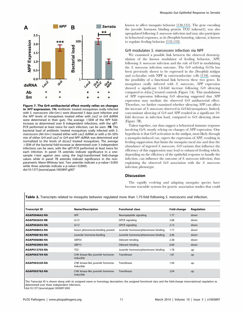
Gr9 modulates S. marcescens infection via NPFWe examined a possible link between the observed downreg-
ulation of the known modulator of feeding behavior, NPF,
following S. marcescens infection and the role of Gr9 in modulating
the S. marcescens infection outcome. The Gr9 ortholog Gr39a has
been previously shown to be expressed in the Drosophila midgut
and co-localize with NPF in enteroendocrine cells [134], raising
the possibility of a functional link between these two genes. In
mosquitoes orally infected with S. marcescens, NPF expression
showed a significant 1.8-fold increase following Gr9 silencing
compared to dsLacZ treated controls (Figure 7A). This modulation
of NPF expression following Gr9 silencing suggested that NPF
expression may mediate the observed Gr9 antibacterial effect.
Therefore, we further examined whether silencing NPF can affect
the increase of S. marcescens observed in Gr9 kd mosquitoes. Indeed,
concomitant silencing of Gr9 and NPF resulted in a significant 10-
fold decrease in infection load, compared to Gr9 silencing alone
(Figure 7B).
Taken together, our data suggest a behavioral immune response
involving Gr9, mostly relying on changes of NPF expression. One
hypothesis is that Gr9 activation in the midgut, most likely through
a mosquito-induced cue, tapers the expression of NPF, resulting in
feeding suppression that limits the mosquito meal size and thus the
abundance of ingested S. marcescens. Gr9 variants that influence the
efficiency of this suppression may lead to enhanced feeding which,
depending on the efficiency of the epithelial response to handle the
infection, can influence the outcome of S. marcescens infection, thus
explaining the observed Gr9 association with the S. marcescens
infection phenotype.
Discussion
The rapidly evolving and adapting mosquito species have
become tractable systems for genetic association studies that could
Table 2. Transcripts related to mosquito behavior regulated more than 1.75-fold following S. marcescens oral infection.
Transcript ID Name/Description Functional class Fold-change Regulation
AGAP004642-RA NPF Neuropeptide signaling 1.77 down
AGAP002635-RB Gr13 GPCR signaling 2.68 down
AGAP002635-RA Gr13 GPCR signaling 2.13 down
AGAP008052-RA Insect pheromone-binding protein Juvenile hormone/pheromone binding 1.77 down
AGAP008182-RA Juvenile hormone-binding Juvenile hormone/pheromone binding 2.06 down
AGAP006080-RA OBP54 Odorant binding 2.38 down
AGAP002905-RA OBP13 Odorant binding 6.00 down
AGAP012703-RA TO2 Juvenile hormone/pheromone binding 1.78 up
AGAP003759-RA CHK kinase-like juvenile hormone-inducible
Transferase 1.81 up
AGAP003220-RA CHK kinase-like juvenile hormone-inducible
Transferase 1.94 up
AGAP003762-RA CHK kinase-like juvenile hormone-inducible
Transferase 2.04 up
The Transcript ID is shown along with its assigned name or homology description, the assigned functional class and the fold-change transcriptional regulation asdetermined over three independent infections.doi:10.1371/journal.ppat.1003897.t002
Figure 7. The Gr9 antibacterial effect mostly relies on changesin NPF expression. 7A: Antibiotic treated mosquitoes orally infectedwith S. marcescens (Ab+Sm+) were dissected 5 days post infection andthe NPF levels of mosquitoes treated either with LacZ or Gr9 dsRNAwere determined in their guts. The average 6SEM of the NPF fold-increase as determined over 8 independent infections, with the qRT-PCR performed at least twice for each infection, can be seen. 7B: Thebacterial load of antibiotic treated mosquitoes orally infected with S.marcescens (Ab+Sm+) treated either with LacZ dsRNA or with a 50–50%mix of either Gr9 and LacZ or Gr9 and NPF dsRNA was determined andnormalized to the levels of dsLacZ treated mosquitoes. The average6SEM of the bacterial fold-increase as determined over 3 independentinfections can be seen, with the qRT-PCR performed at least twice foreach infection. In panel 7A asterisks indicate significance in a one-sample t-test against zero using the log2-transformed fold-changevalues while in panel 7B asterisks indicate significance in the non-parametric Mann-Whitney test. Two asterisks indicate a p-value,0.005while three asterisks indicate a p-value,0.0005.doi:10.1371/journal.ppat.1003897.g007
Mosquito Gut Epithelial Response to Serratia
PLOS Pathogens | www.plospathogens.org 11 March 2014 | Volume 10 | Issue 3 | e1003897
yield important information about vector/parasite interactions
leading to malaria transmission [135]. Previous studies have
focused on the outcome of Plasmodium infections, using laboratory
or field mosquitoes and genetic tools such as microsatellite markers
and targeted SNP loci genotyping [1,3,136]. These studies have
not considered the effect of gut bacteria on the outcome of
Plasmodium infections, which has been revealed recently [5,16–18].
Furthermore, the influence of associated complement factors on
natural P. falciparum infections remains questionable [137]. Indeed,
the presence of Enterobacteriaceae, such as S. marcescens, a common
member of the mosquito gut flora, has been correlated with P.
falciparum susceptibility in field mosquito populations [12], while
intraspecific variation within S. marcescens populations also is shown
to affect the Plasmodium infection load [37]. Therefore, genome-
wide studies to determine factors that modulate the levels of
mosquito gut bacteria can provide novel insights into how midgut
bacteria affect the outcome of Plasmodium infection and hence
malaria transmission.
The unprecedented level of detail achieved in the population
genetics analysis presented here in identifying SNPs associated
with the outcome of S. marcescens infection is a result of the strong
evolutionary drive exerted by gut bacteria on mosquito genetic
variation, the use of a high-resolution SNP genotyping array and
the use of a recently established laboratory colony of An. gambiae
which retains genetic variation found in field populations but also
shows elevated linkage disequilibrium due to colonization bottle-
necks. This population homogeneity can facilitate gene discovery
as shown in human genome-wide association studies in isolated
populations [138,139].
A dual implication can be inferred for genes associated with the
S. marcescens infection phenotype; they are putatively involved in
shaping the infection outcome, while their level of involvement
may also be affected by genetic variation within the mosquito
population. It is possible that identified associations are the result
of causal polymorphisms such as gain or loss of function mutations
in coding or regulatory sequences or the result of allele
combination in several genetic loci which shapes the outcome of
infection through synergism, epistatic interactions or redundant
function. In any of the latter cases, a reverse genetics approach
may not be capable of capturing such interactions.
The involvement of the three FN3Ds in the outcome of Serratia
infection reveals a novel function of this family in modulating the
load and composition of the mosquito gut microbiota and opens
new avenues in investigating the complexity of such responses and
possible synergisms with known antibacterial pathways such as the
IMD/REL2. The three FN3D genes identified here emerge as
major modulators of the bacterial population structure in the
mosquito gut, limiting the representation of Enterobacteriaceae,
mainly Serratia or strains with similarity to Serratia reference
sequences, but also, for FN3D3, bacteria of the genus Burkholderia.
As shifts in gut microbiota population structure can elicit gut
pathology [28,140], while Serratia can influence the outcome of
Plasmodium infection [37], FN3Ds can play critical roles in gut
homeostatic interactions and malaria transmission dynamics.
Further insights into the FN3D mode of action remain to be
determined. Our data showing that the knockdown effects of
FN3Ds may be limited to Serratia or to a fraction of the microbiota
raise intriguing questions about the specificity of bacterial
recognition in the mosquito gut. The homology of FN3D2 with
the hypervariable pattern recognition receptor Dscam opens the
possibility that the specific pathogen recognition shown for
AgDscam [47] concerns a broader family of FN3Ds, equipping
mosquitoes with the capacity for specific recognition resembling
that of animals possessing adaptive immune systems. The
phylogenetically unrelated FN3D2 and FN3D3 share a similar
domain architecture comprising immunoglobulin and FN3
domains, as is the case with Dscam. The identification of FN3D2
and FN3D3 as being both associated with the outcome of S.
marcescens infection and exhibiting discrete but similar phenotypic
characteristics in modulating the bacterial population structure in
the mosquito gut, parallels the discrete but similar functions of the
phylogenetically unrelated Dscam, Frazzled and Roundabout in
Drosophila axon guidance, with all three receptors sharing
immunoglobulin and FN3 domains [141–144]. FN3D1 has a
distinct domain architecture with an FN3 domain, while its
orthologous relationship with Drosophila windei [145] and sequence
similarity with the activating transcription factor 7- interacting
protein [146], suggest a role in regulating gene expression.
The identification of An. gambiae genes involved in immune
responses against bacteria and/or Plasmodium has been largely
based to date on studies that combine bioinformatic identification
of known immunity gene homologs and transcriptional profiling of
genes following a pathogen challenge. This approach, however,
has the limitation of the a priori assumption that genes of interest
show significant change in transcriptional regulation, mostly
induction, which is true for most effectors, but not all genes, for
example pattern recognition receptors or transcription factors. In
addition, it is possible that even strong changes in transcriptional
regulation are the consequence of the infection rather than part of
the response. Especially for quantitative traits within mosquito
populations, such as Plasmodium infection intensity, different
infection intensities can correlate with variable transcriptional
responses [70], while the underlying genetic variation further
complicates the observed transcriptional regulation.
The microarray approach adopted here has identified a limited
set of 99 differentially regulated transcripts following oral S.
marcescens infection. The number of regulated transcripts is
consistent with that of a previous microarray-based comparison
of antibiotic treated and untreated mosquitoes, which showed
differential expression for 185 transcripts [16], attributing this
limited transcriptional regulation to symbiotic relationships that
have led to adaptation of commensal bacteria. A much broader set
of differentially expressed genes has been identified following oral
bacterial infections in Drosophila [26,32]. This is most likely due to
differences in gene pool diversity between the genetically
homogeneous fly lines and the recently established mosquito
laboratory colony used here, which retains considerable genetic
variation thus enabling the SNP genotyping analysis. The different
levels of infection seen between mosquitoes (high, low and no
infection), which are largely attributed to genetic variation within
the colony population, are most likely linked to differences in the
mosquito transcription profiles that are averaged out in our study
design. Therefore, our analysis identifies transcripts with the most
pronounced and consistent differential expression, comprising the
core response to S. marcescens infection. Future studies investigating
the transcription profile of highly, lowly or non-infected mosqui-
toes are most likely to reveal components of transcriptional
regulation that lead to the respective outcome of infection. Indeed,
genes identified to show prominent differential expression after
bacterial challenge in previous studies also showed transcriptional
regulation following oral S. marcescens infection, including CLIPB14
[117], LRIM1 [63,113], LYSC2 [116] and SCRASP1 [117,118].
The identification of diverse transcriptional responses to
different bacteria in Drosophila [32] along with the specificity of
mosquito responses to a subset of bacteria, as suggested by the
SNP genotyping analysis presented here, may explain the
surprisingly little overlap between differentially expressed genes
following S. marcescens infection and antibiotic treated vs. untreated
Mosquito Gut Epithelial Response to Serratia
PLOS Pathogens | www.plospathogens.org 12 March 2014 | Volume 10 | Issue 3 | e1003897
mosquitoes [16]. Remarkably, however, consistency is seen in gene
families present in both datasets, including CLIPs, chitin-binding
genes, homeobox genes, PGRPs and FREPs, suggesting that similar
defense strategies are employed, which are customized for each
type of infection through utilization of different gene family
members.
The approach we adopted here to identify genes involved in
mosquito gut infection with S. marcescens combines transcriptional
profiling of infected guts with the identification of SNPs
segregating between phenotypic pools, whereby an association
implies contribution to the outcome of infection, while the study
design incorporates variation that leads to different observed
phenotypes. This approach addresses some of the aforementioned
shortcomings but introduces others, as it cannot capture genes
with redundant functions, genes with additional housekeeping
functions or a role during development, of which variants are
eliminated from the population, or genes with rare variants that
are not in the variation pool of our colony. Furthermore, an
association may be the result of a selective sweep in the proximity
of the gene that creates linkage disequilibrium and leads to SNP
divergence between the phenotypic pools. Therefore, although
each of the approaches cannot provide by itself a complete picture,
the combination of the two can provide novel insights into the
mosquito gut responses to S. marcescens.
The comparison between the datasets of transcriptionally
regulated genes and genes associated with the outcome of S.
marcescens infection shows limited overlap, with only PGRPLC and
CLIPE6 found in both datasets. Again, considerable overlap is
detected in identified gene families, which are represented by
different members in each dataset. These include acyl-transferase,
glycoside hydrolase, kinase, GPCR, LRIM, homeobox, zinc-
finger, PGRP, peptidase, FREP, MD2-like and chitin-binding
genes. Interestingly, a previous study investigating differential
expression following a bacterial challenge in mosquito immuno-
globulin-containing genes failed to identify significant regulation
for FN3D2 or FN3D3 [147], strengthening the case for the
complementarity of the SNP genotyping and expression analysis
approaches. The specific role of gene family members, especially
those showing considerable expansion in Anopheles, e.g. FREPs
[68,148], remains unclear. Therefore, SNP genotyping reveals a
different set of candidate genes involved in antibacterial immunity
while at the same time it is intriguing to postulate whether this
divergence between associated and differentially expressed genes
within each gene family constitutes a functional divergence
between them.
A novel finding stemming from this combinatorial approach is a
mosquito behavioral response to S. marcescens infection that
involves Gr9 signaling and is mediated by changes of NPF
expression. Although Gr9 orthologs in Drosophila recognize
chemosensory cues and mediate aversive behaviors [83,84,87],
surprisingly, Gr9 appears to suppress feeding irrespective of the
presence of bacteria. One explanation is that the Gr9 antibacterial
effect relies on its expression in the midgut rather than external
sensory organs, where the role of its Drosophila counterparts has
been studied. The role of gustatory receptor midgut expression
[134] remains poorly understood and could involve detection of
nutrients or host-derived molecules that triggers downstream
responses. The role of NPF midgut expression [121,122] also
remains poorly understood. NPF downregulation following S.
marcescens infection implies its involvement in an aversion circuit
triggered by the presence of S. marcescens, with a possible NPF role
in integrating aversion and satiation signals that lead to feeding
suppression. Such NPF involvement remains to be further
investigated, in conjunction with the involvement of other genes
related to mosquito behavior which were either associated with the
outcome of S. marcescens infection or were transcriptionally
regulated following infection. These include Gr13, downregulated
following S. marcescens infection but also three neurotransmitter-
triggered GPCRs, associated with the outcome of infection,
pointing to complex behavioral circuits involved in antibacterial
responses, which are yet to be revealed.
The identification of FN3Ds as well as Gr9 and NPF in responses
affecting the outcome of S. marcescens infection, in addition to
known responses including the IMD/REL2 and DUOX path-
ways, suggests that the response to gut infection is the result of a
complex molecular interplay. Both the SNP genotyping and
expression analysis suggest that the mosquito response to oral S.
marcescens infection involves two discrete but inextricably linked
modes of defense, a behavioral and an epithelial immune response.
A behavioral immune response involving Gr9 and NPF can limit
or disrupt pathogen intake, a defense conceptually similar to
barrier responses that inhibit pathogen contact with triggers of
epithelial or systemic immune responses. An impaired behavioral
response, e.g. due to Gr9 variants that affect feeding behavior, can
decisively influence the efficacy of the epithelial response and thus
the infection outcome. This implies a threshold after which
epithelial immunity cannot efficiently handle the pathogen load,
an aspect of immunity that remains poorly understood. Never-
theless, in mosquito infections with Plasmodium parasites, the
intensity of infection has been correlated with the efficacy of
different components of the IMD/REL2 pathway, suggesting that
different effectors may be deployed in low, mid or high intensity
parasite infections [4].
As pathogen abundance most likely relies on feeding behavior,
the interplay between behavioral and epithelial immunity can
shape both responses. Our implementation of a model of natural
bacterial infections through the oral route integrated both
behavioral and epithelial responses and not only revealed the
previously unknown behavioral component but also allowed the
study of aspects of epithelial immunity that, by being infection
intensity dependent, possibly rely on the behavioral component.
This integrative approach to behavioral and epithelial immunity
can be further employed to reveal aspects of this interplay that
may involve regulation of behavioral responses by host-derived
factors induced by the epithelial component. This implies that the
study of behavioral immunity alone may be insufficient to uncover
some aspects of its biological consequences.
In Drosophila, a balance between immune response and
tolerance, achieved by various Imd regulators, largely shapes the
gut microbiota population structure, although the only known
elicitor of such responses is DAP-type peptidoglycan, common to
all Gram-negative bacteria [28,149,150]. A similar mechanism has
been suggested for mosquitoes through alternative splicing of the
modular IMD/REL2 pathway receptor PGRPLC that leads to
production of positive and negative pathway modulators [5,42].
Indeed, utilization of alternative splicing as a mechanism to derive
new immune functions and increase the specificity of pathogen
recognition by the mosquito innate immune system has been
described for the FN3D2 homolog, Dscam [43,44,47]. Whether the
Enterobacteriaceae-specific effect of FN3D2 knockdown is due to
specific recognition and activation of highly specialized or targeted
effector reactions remains to be investigated. Furthermore, the
significance of alternative splicing suggested for Gr9 [82] remains
to be determined along with the cue that triggers its antibacterial
effect, and could also involve recognition of, most likely, host-
derived signals. In addition, recognition of differentially produced
metabolites after infection as shown for the DUOX pathway [29]
could further increase the specificity in antibacterial responses.
Mosquito Gut Epithelial Response to Serratia
PLOS Pathogens | www.plospathogens.org 13 March 2014 | Volume 10 | Issue 3 | e1003897

Whether PAMPs (bacterial-derived) or DAMPs (host-derived),
such metabolites can be recognized by gustatory receptors
triggering specific antibacterial responses, which together with
FN3Ds and the rather generalist response of the IMD/REL2
pathway can shape the load and composition of the mosquito gut
microbiota. In conclusion, our findings suggest that mosquitoes
can mount a much more complex and specific antibacterial
response than previously thought, which not only contributes to
fending off intestinal bacterial infections but also to achieving
homeostasis of the complex gut ecosystem.
Materials and Methods
Ethics statementThis study was carried out in strict accordance with the United
Kingdom Animals (Scientific Procedures) Act 1986. The protocols
for maintenance of mosquitoes by blood feeding were approved
and carried out under the UK Home Office License PPL70/7170.
The procedures are of mild to moderate severity and the numbers
of animals used are minimized by incorporation of the most
economical protocols. Opportunities for reduction, refinement and
replacement of animal experiments are constantly monitored and
new protocols are implemented following approval by the Imperial
College Ethical Review Committee.
Mosquito rearing and maintenanceThe N’gousso strain of An. gambiae was used. This is an M form
strain colonized in 2006 [1] and kept in large numbers to retain
genetic variation. Rearing and maintenance of the strain was
performed as described previously [151]. Mosquitoes were
collected after emergence and kept on a cocktail of 25 mg/ml
gentamicin, 10 mg/ml penicillin and 10 units/ml streptomycin,
diluted in 10% D-(-)-Fructose (Sigma). This antibiotic treatment
regime was used for 5 days, with the antibiotic solution refreshed
every 24 hours. At day 5 post emergence, the antibiotic solution
was replaced by dH2O and mosquitoes were starved overnight
prior to oral bacterial infection.
Mosquito oral infection with S. marcescens or AsaiaWe used the S. marcescens Db11-GFP strain, modified to be GFP-
fluorescent and resistant to tetracycline and carbenicillin [152]. S.
marcescens glycerol stock was grown in 5 ml LB cultures containing
50 mg/ml tetracycline and carbenicillin (Sigma) at 37uC. Follow-
ing overnight incubation, the cultures were expanded to 100 ml
and further incubated overnight at 37uC. OD600 and GFP
fluorescence (excitation/emission at 485/520 nm) were then
measured to ensure cultures maintained GFP fluorescence, using
the Fluostar Omega spectrophotometer (BMG Labtech). Bacterial
pellets following centrifugation at 2500 rpm for 5 minutes were
washed twice with PBS and resuspended in such volume of 10%
D-(-)-Fructose, so that 1 ml of the bacteria-containing sugar
solution corresponded to OD600 = 0.1 of the initial 100 ml culture.
The sugar solution was further diluted 1:12 in a 10% D-(-)-
Fructose solution that contained tetracycline and carbenicillin at
50 mg/ml and 5% v/v of scarlet dye (Langdale). Mosquitoes were
fed with this solution for 2 days. Subsequently, mosquitoes fed with
bacteria-containing sugar were separated based on the presence of
the dye in their gut and kept on 10% D-(-)-Fructose containing
tetracycline and carbenicillin at 50 mg/ml.
Oral infections with Asaia were conducted in a similar
manner. The Asaia SF2.1 (GFP) strain was used, grown as
previously described [153] and maintained in 50 mg/ml kanamy-
cin (Sigma).
Fluorescence microscopyThe levels of S. marcescens infection were determined by
microscopic observation of dissected midguts immersed in
Vectashield mounting medium (Vecta), immediately after dissection.
The Zeiss Axiophot fluorescence microscope was used, equipped
with light and GFP filters while photos of observed midguts were
taken with the Axiocam HRc and Axiovision software (Zeiss).
SNP genotyping arraysAll carcasses corresponding to midguts of S. marcescens infected
mosquitoes were kept numbered in 96-well plates immersed in
75% ethanol at 280uC. Carcasses from selected midguts were
used for gDNA extraction using the QIAquick Blood and Tissue kit
(QIAGEN). Subsequently, gDNA concentrations were determined
using the Picogreen dsDNA kit (Invitrogen) and equimolar gDNA
quantities from each mosquito were pooled. The design and
validation of the SNP genotyping array used along with the
treatment of gDNA pools, hybridization, calling of SNP genotypes
and measurement of differentiation in each pooled hybridization
between allele A and B have been described previously [39,154].
The frequency of designated allele A was considered as the minor
allele frequency and was used to measure the difference between
pooled hybridizations. The permutation analysis used has been
described previously [39], with a modified length of non-
overlapping 10-SNP windows. Determination of genes residing
in identified genomic areas and homology analysis was performed
using Biomart 0.7 and the AgamP3.7 An. gambiae gene annotation
[155]. The SNP genotyping array datasets have been deposited to
ArrayExpress under the experiment name Serratia_SNP1 and
accession number E-MEXP-3951.
DNA microarraysTotal RNA was extracted from midguts using the Trizol reagent
(Invitrogen), and treated with Turbo DNAse I (Ambion). Samples
were further purified using the RNeasy kit (QIAGEN). Quantifi-
cation was performed using the Nanodrop 1000 spectrophotometer
(Thermo Scientific) and RNA integrity was assessed using the RNA
6000 Pico Chip kit (Agilent). Labeling and hybridization were
performed using the Low Input Quick Amp Labeling kit for two-color
microarray based expression analysis (Agilent). We used Agilent
custom 4644 k gene expression microarrays. The microarray
design Pfalcip_Agamb2009 (A-MEXP-2324) comprises oligonucleo-
tide probes encompassing all An. gambiae annotated transcripts of
the AgamP3.6 release along with P. falciparum probes, with each
probe represented in duplicate. Slides were scanned using the
Genepix 4000B scanner equipped with the Genepix Pro 6.1 software
(Axon instruments).
All dataset files were normalized using the Genespring 11.0 GX
software (Agilent). The Lowess normalization method was used
while the threshold of raw signals was set to 5, which was sufficient
to eliminate background regulation of P. falciparum probes. Further
analysis of transcriptionally regulated genes and GO analysis was
performed using the Genespring 11.0 GX software. For GO
analysis, GO accession numbers for all An. gambiae transcripts were
obtained using Biomart 0.7 and a hypergeometric test with
Benjamini-Hochberg correction was performed on the set of more
than 1.75-fold regulated genes. The corrected p-value for testing
multiple GO accession numbers for their significance was set to 0.1.
The log2-transformed transcriptional regulation for each transcript
was extracted from the normalized datasets for each of the two
probes corresponding to each transcript and the obtained values
from all three independent infections were used in a t-test against
zero, with a p-value cut-off of 0.05. The DNA microarray datasets
Mosquito Gut Epithelial Response to Serratia
PLOS Pathogens | www.plospathogens.org 14 March 2014 | Volume 10 | Issue 3 | e1003897
have been deposited to ArrayExpress under the experiment name
Serratia_infections and accession number E-MEXP-3952.
Bacterial load following RNAi-mediated silencingMosquitoes were treated with the respective dsRNA at the day
of emergence, as described previously [156]. For each targeted
gene or the dsLacZ control, dsRNA was synthesized using the T7
Megascript kit (Invitrogen) and further purified using the RNeasy kit
(QIAGEN) to a concentration of 3 mg/ml. For each T7 Megascript
reaction, 1 mg of purified PCR product was added, derived using
the T7 primer sets shown in Table S9, using An. gambiae cDNA as
template.
Mosquitoes were surface sterilized by immersing them in 70%
ethanol for 30 seconds and washing them twice in PBS and
midguts were dissected in RNA later (Invitrogen). Total RNA from
mosquito midguts was extracted after homogenization with a
pestle motor in RNA later using the RNeasy kit (QIAGEN). cDNA
was synthesized from total RNA using the QuantiTect Reverse
Transcription kit (QIAGEN).
Quantification of bacterial load or the efficiency of RNAi-
mediated silencing was performed using qRT-PCR with the
respective primers shown in Table S9. In a 20 ml reaction of Fast
SYBR Green Master Mix (Applied Biosystems), 1 ml of cDNA
template and 2 ml of each respective primer at a 0.5 to 9 mM
concentration, optimized for each primer set, were added. The
7500 Real-Time PCR System (Applied Biosystems) was used with
its respective software to perform the reaction and any further
analysis. The relative abundance of each sample was determined
using the standard curve method as described in User Bulletin #2
for the ABI Prism 7700 Sequence Detection system (Applied
Biosystems) in which the housekeeping AgS7 gene was used as an
endogenous control.
454 pyrosequencingcDNA pools were amplified with the GO Taq DNA polymerase
(Promega) using the 16S V4–V6 primers shown in Table S9 and
suitable barcode sequences and purified using PCR purification and
Gel Extraction kits (QIAGEN). PCR products were sequenced by
Beckman Genomics (Grenoble, France) using the Roche 454 GS
FLX+ and standard procedures. The resulting FASTA files were
filtered to a minimum read length of 250 bp using Galaxy [157]
and blasted against the NCBI 16SMicrobial database using BLAST+and prfectBLAST [158] with standard blastn algorithm settings and
10 maximum target sequences. Further analysis was performed
using MEGAN4 [159].
Meal size and two-choice preference assaysSugar meal size was determined through a modified capillary
feeder assay [160]. Mosquitoes treated with LacZ or Gr9 dsRNA
were antibiotic treated for 5 days, starved overnight and,
subsequently, individual mosquitoes were fed on a 5 ml glass
capillary (VWR) containing 10% D-(-)-Fructose and 5% v/v
scarlet dye. For alive mosquitoes, sugar consumption was
determined 16 hours later through the reduction of sugar solution
in each capillary. The two-choice preference assay was also
conducted based on a previously described capillary feeder assay
[160]. Mosquitoes treated with LacZ or Gr9 dsRNA were antibiotic
treated for 5 days, starved overnight and placed in pools of 8–11
mosquitoes. Mosquitoes were offered to feed from two capillaries,
one containing a sugar solution as above and one also containing
S. marcescens, prepared as described above for oral infection. Water-
containing cotton pads were also used and pools with mosquito
mortality were disregarded. 16 hours later, consumption for each
capillary was determined based on the reduction of the sugar
solution.
Supporting Information
Figure S1 Efficacy of antibiotic treatment in reducingthe presence of gut bacteria in treated mosquitoes. An.
gambiae mosquitoes were antibiotic treated for 5 days with a
cocktail of gentamicin, penicillin and streptomycin. Subsequently,
10–15 mosquitoes were surface sterilized and their guts were
dissected, homogenized and total RNA was extracted and further
used for cDNA synthesis. Mosquitoes kept untreated were
processed in the same way. Subsequently, cDNA from antibiotic
treated or untreated mosquito pools was used in a qRT-PCR using
broad range bacterial 16S primers while AgS7 primers were used
as controls. The bacterial load 6SEM in 3 independent assays,
with the qRT-PCR performed at least twice for each assay, can be
seen. Asterisks indicate significance, with a p-value,0.0005, in a
Mann-Whitney non-parametric test between the bacterial load of
untreated and antibiotic treated mosquitoes.
(TIF)
Figure S2 Efficacy of RNAi-mediated silencing ofFN3D1–3, Gr9 and NPF. Mosquitoes were treated with dsRNA
targeting the FN3D1, FN3D2, FN3D3, Gr9 or NPF transcripts or
with the dsLacZ control. The Gr9 dsRNA targets the 59 exon 1,
which mainly limits the RA and RG splice variants. The relative
expression of each transcript in the mosquito gut was determined 5
to 6 days post dsRNA treatment, normalized to the endogenous
AgS7 control, in mosquitoes treated with the respective dsRNA by
qRT-PCR and primers targeting the respective transcript in a
region not targeted by the respective dsRNA. Silencing efficiency
was determined by further normalizing the relative expression of
each transcript in mosquitoes treated with the respective dsRNA to
the relative expression in the dsLacZ treated control. The average
6SEM of relative expression is shown for at least 3 independent
assays, with the qRT-PCR performed at least twice for each assay.
(TIF)
Figure S3 FN3D1–3 silencing modulates total bacteriaand Serratia in a non-uniform way in mosquitoesretaining their natural gut microbiota. Mosquitoes retain-
ing their natural gut microbiota (Ab2Sm2) were treated with
FN3D1 (S3A), FN3D2 (S3B) or FN3D3 (S3C) dsRNA and bacterial
load was normalized to the respective dsLacZ treated control.
Bacterial load using broad range 16S or Serratia-specific primers is
shown for 4 independent assays (Replicates 1–4 as indicated above
each pair of bars).
(TIF)
Figure S4 Oral infection with Asaia following FN3D1–3silencing. Antibiotic treated mosquitoes were orally infected with
bacteria of the genus Asaia following treatment with FN3D1–3
dsRNA or the dsLacZ control. Bacterial load was determined in the
guts of surface sterilized mosquitoes dissected 5 days post infection,
using qRT-PCR with 16S broad range bacterial primers and the
AgS7 control. The bacterial load 6SEM in 3 independent assays,
with the qRT-PCR reaction performed at least twice for each
assay, can be seen.
(TIF)
Figure S5 Two-choice preference assay between sugarsolutions containing or not S. marcescens. Antibiotic
treated mosquitoes treated either with LacZ or Gr9 dsRNA were
starved overnight and, subsequently, pools of 8–11 mosquitoes
were offered a choice of two meals in separate 5 ml capillaries, one
containing a sugar solution containing a dye used to measure
Mosquito Gut Epithelial Response to Serratia
PLOS Pathogens | www.plospathogens.org 15 March 2014 | Volume 10 | Issue 3 | e1003897
consumption and ensure uptake from the mosquitoes and one also
containing S. marcescens, at the same concentration as used for oral
infection. 16 hours later, consumption was measured for each
capillary and, for each mosquito pool, the ratio of the %
consumption in the Serratia-containing capillary to the %
consumption in the sugar-only capillary was determined. Overall,
the consumption percentage ratio of Serratia-containing versus
sugar-only capillaries was determined for 6 LacZ and 11 Gr9
dsRNA treated mosquito pools. The average 6SEM percentage
ratio for each dsRNA treatment can be seen. Significant
differences were assessed using the non-parametric Mann-Whitney
test resulting in a p-value of 0.7395, indicated as non-significant
(NS). A percentage ratio of ,100% would indicate aversion to the
Serratia-containing solution while a percentage ratio of .100%
would indicate attraction to the Serratia-containing solution for
mosquitoes treated with the respective dsRNA, as indicated.
(TIF)
Figure S6 Significantly overrepresented GO terms inthe set of more than 1.75-fold regulated genes followingS. marcescens infection. A hypergeometric test with Benja-
mini-Hochberg correction was used to compare the representation
of genes corresponding to the same GO term in the set of 97 more
than 1.75-fold regulated genes following S. marcescens infection to
the respective representation in the An. gambiae genome, as
annotated in the Pfalcip_Agamb2009 microarray design. 16 GO
terms corresponded to significantly overrepresented groups of
genes, shown in orange type in the GO directed acyclic graph,
with GO terms in the same path that did not meet the p-value cut-
off shown in black type. For significantly overrepresented GO
terms, the number of corresponding genes in the set of more than
1.75-fold regulated genes is shown in parenthesis. A table with the
regulated transcripts corresponding to GO terms at each final leaf
node is shown, including, for each transcript, the Transcript ID,
an assigned name or description based on Interpro-predicted
domains or homologies with Drosophila counterparts and the
observed transcriptional regulation following S. marcescens infection.
(TIF)
Table S1 Complete list of SNP loci assayed for associ-ation with the S. marcescens infection phenotype. SNP
loci interrogated in a 400 k Affymetrix SNP genotyping array. The
MAF for the highly infected and non-infected phenotypic pools
and the calculated MAF difference can be seen for each
interrogated SNP locus.
(RAR)
Table S2 Complete list of 10-SNP loci windows assayedfor association with the S. marcescens infection pheno-type. The 40,007 sliding 10-SNP windows assayed for association
with the S. marcescens infection phenotype are shown along with the
genomic position of each 10-SNP window, the calculated average
MAF difference and the assigned p-value of the permutation
analysis conducted.
(XLSX)
Table S3 Individual SNP loci associated with the S.marcescens infection phenotype. A total of 140 out of
400,071 SNP loci that showed MAF difference .0.5 between the
highly infected and non-infected S. marcescens phenotypic pools are
shown. Each column contains for each SNP locus: the chromo-
some it resides, the MAF for the highly infected and non-infected
phenotypic pools and the calculated MAF difference, the genomic
position of the SNP locus and the calculated 5 kb radius (position
25 kb and position +5 kb columns), the residing peak for each
SNP locus as assigned in Figure 2 and genes found within a 5 kb
radius of this SNP locus based on the AgamP3.7 annotation of the
An. gambiae genome.
(XLSX)
Table S4 10-SNP loci windows associated with the S.marcescens infection phenotype. A total of 44 out of 40,007
10-SNP loci windows showed a significant p-value of ,1025 in a
permutation analysis following Bonferroni correction, when
assayed for association with the S. marcescens infection phenotype.
Each column contains for each 10-SNP locus window: The
chromosome it resides, the genomic position (Center-Start-Stop
columns), the average MAF difference calculated, the assigned p-
value of the permutation analysis, the peak it resides as assigned in
Figure 2 and the genes residing within the 10-SNP locus window
based on the AgamP3.7 annotation of the An. gambiae genome.
(XLSX)
Table S5 Complete list of An. gambiae genes associatedwith the outcome of S. marcescens infection. 138 An.
gambiae genes were associated with the outcome of S. marcescens
infection, either by residing within a 5 kb radius of SNP loci with
MAF difference .0.5 (SNP) or by residing within 10-SNP loci
windows with a ,1025 p-value in a permutation analysis
(Permutation). For genes identified by both approaches, both
SNP and Permutation are noted. For each gene, the Description
column includes the assigned name, if any, or a description based
on homologs or Interpro-predicted domains.
(XLSX)
Table S6 16S metagenomic profiling in FN3D1–3 orLacZ dsRNA treated mosquitoes retaining their naturalgut microbiota. cDNA pools from Ab2Sm2 mosquitoes
following FN3D1–3 or LacZ dsRNA treatment were sequenced
using 454 pyrosequencing targeting the V4–V6 region of the
bacterial 16S rRNA transcript. Results are shown for each cDNA
pool corresponding to a dsRNA treatment shown in Figure 4. In
each case, taxonomic identification and alignment of reference
sequences for each identified taxon is shown along with the
number of sequence reads assigned. For each bacterial genus, the
reference sequence with most alignment hits (sequence reads) is
shown. For Serratia, the reference sequence with most alignment
hits is highlighted and was used for categorization between Serratia
and other Enterobacteriaceae.
(XLSX)
Table S7 An. gambiae transcripts with more than 1.75-fold regulation following S. marcescens infection. 99 An.
gambiae transcripts showing more than 1.75-fold up or down
regulation following S. marcescens infection. For each transcript, the
Transcript ID is shown along with its designated name or
functional description, the assigned functional class, the fold-
change regulation and the p-values obtained by a t-test against
zero using the log2-transformed values for each interrogated
probe.
(XLSX)
Table S8 GO terms significantly overrepresented in theset of more than 1.75-fold regulated genes following S.marcescens infection. 16 enriched GO terms as determined by
a hypergeometric test followed by Benjamini-Hochberg correction
using a 0.1 p-value cut-off are shown. The GO ID, accession
number and description of each GO term is shown along with the
results of the test for each GO term.
(XLSX)
Table S9 Sequences of primers used for qRT-PCR,dsRNA synthesis or 454 pyrosequencing. For each forward
Mosquito Gut Epithelial Response to Serratia
PLOS Pathogens | www.plospathogens.org 16 March 2014 | Volume 10 | Issue 3 | e1003897
or reverse primer, its use is indicated along with the respective
sequence.
(XLSX)
Acknowledgments
We thank Dina Vlachou and Amanda Jackson for help and advice in
carrying out the DNA microarray experiments, Katarzyna Sala for helping
with mosquito rearing, Dominique Ferrandon for sharing the S. marcescens
stock and Guido Favia for sharing the Asaia stock. The authors are grateful
to Fotis C. Kafatos for his multifaceted and invaluable support.
Author Contributions
Conceived and designed the experiments: GKC SS. Performed the
experiments: SS. Analyzed the data: SS. Contributed reagents/materials/
analysis tools: GKC MKNL DEN MATM. Wrote the paper: SS GKC.
References
1. Harris C, Lambrechts L, Rousset F, Abate L, Nsango SE, et al. (2010)Polymorphisms in Anopheles gambiae immune genes associated with natural
resistance to Plasmodium falciparum. PLoS Pathog 6: e1001112.
2. White BJ, Lawniczak MK, Cheng C, Coulibaly MB, Wilson MD, et al. (2011)
Adaptive divergence between incipient species of Anopheles gambiae increasesresistance to Plasmodium. Proc Natl Acad Sci U S A 108: 244–249.
3. Riehle MM, Markianos K, Niare O, Xu J, Li J, et al. (2006) Natural malariainfection in Anopheles gambiae is regulated by a single genomic control region.
Science 312: 577–579.
4. Garver LS, Bahia AC, Das S, Souza-Neto JA, Shiao J, et al. (2012) Anopheles
imd pathway factors and effectors in infection intensity-dependent anti-Plasmodium action. PLoS Pathog 8: e1002737.
5. Meister S, Agianian B, Turlure F, Relogio A, Morlais I, et al. (2009) Anophelesgambiae PGRPLC-mediated defense against bacteria modulates infections with
malaria parasites. PLoS Pathog 5: e1000542.
6. Schnitger AK, Yassine H, Kafatos FC, Osta MA (2009) Two C-type lectins
cooperate to defend Anopheles gambiae against Gram-negative bacteria. J Biol
Chem 284: 17616–17624.
7. Grossman SR, Andersen KG, Shlyakhter I, Tabrizi S, Winnicki S, et al. (2013)
Identifying recent adaptations in large-scale genomic data. Cell 152: 703–713.
8. Sangare I, Michalakis Y, Yameogo B, Dabire R, Morlais I, et al. (2013)
Studying fitness cost of Plasmodium falciparum infection in malaria vectors:validation of an appropriate negative control. Malar J 12: 2.
9. Rani A, Sharma A, Rajagopal R, Adak T, Bhatnagar RK (2009) Bacterialdiversity analysis of larvae and adult midgut microflora using culture-
dependent and culture-independent methods in lab-reared and field-collectedAnopheles stephensi-an Asian malarial vector. BMC Microbiol 9: 96.
10. Gonzalez-Ceron L, Santillan F, Rodriguez MH, Mendez D, Hernandez-AvilaJE (2003) Bacteria in midguts of field-collected Anopheles albimanus block
Plasmodium vivax sporogonic development. J Med Entomol 40: 371–374.
11. Gendrin M, Christophides GK (2013) The Anopheles Mosquito Microbiota
and Their Impact on Pathogen Transmission. In Manguin S, Ed. Anopheles
mosquitoes - New insights into malaria vectors
12. Boissiere A, Tchioffo MT, Bachar D, Abate L, Marie A, et al. (2012) Midgut
Microbiota of the Malaria Mosquito Vector Anopheles gambiae andInteractions with Plasmodium falciparum Infection. PLoS Pathogens 8:
e1002742.
13. Wang Y, Gilbreath TM, 3rd, Kukutla P, Yan G, Xu J (2011) Dynamic Gut
Microbiome across Life History of the Malaria Mosquito Anopheles gambiae inKenya. PLoS One 6: e24767.
14. Osei-Poku J, Mbogo CM, Palmer WJ, Jiggins FM (2012) Deep sequencingreveals extensive variation in the gut microbiota of wild mosquitoes from
Kenya. Mol Ecol 21: 5138–5150.
15. Kumar S, Molina-Cruz A, Gupta L, Rodrigues J, Barillas-Mury C (2010) A
peroxidase/dual oxidase system modulates midgut epithelial immunity inAnopheles gambiae. Science 327: 1644–1648.
16. Dong Y, Manfredini F, Dimopoulos G (2009) Implication of the mosquitomidgut microbiota in the defense against malaria parasites. PLoS Pathog 5:
e1000423.
17. Rodrigues J, Brayner FA, Alves LC, Dixit R, Barillas-Mury C (2010) Hemocyte
differentiation mediates innate immune memory in Anopheles gambiae
mosquitoes. Science 329: 1353–1355.
18. Cirimotich CM, Dong Y, Clayton AM, Sandiford SL, Souza-Neto JA, et al.
(2011) Natural microbe-mediated refractoriness to Plasmodium infection inAnopheles gambiae. Science 332: 855–858.
19. Lemaitre B, Hoffmann J (2007) The host defense of Drosophila melanogaster.Annu Rev Immunol 25: 697–743.
20. Kaneko T, Goldman WE, Mellroth P, Steiner H, Fukase K, et al. (2004)Monomeric and polymeric gram-negative peptidoglycan but not purified LPS
stimulate the Drosophila IMD pathway. Immunity 20: 637–649.
21. Choe KM, Lee H, Anderson KV (2005) Drosophila peptidoglycan recognition
protein LC (PGRP-LC) acts as a signal-transducing innate immune receptor.Proc Natl Acad Sci U S A 102: 1122–1126.
22. Maillet F, Bischoff V, Vignal C, Hoffmann J, Royet J (2008) The Drosophilapeptidoglycan recognition protein PGRP-LF blocks PGRP-LC and IMD/JNK
pathway activation. Cell Host Microbe 3: 293–303.
23. Lhocine N, Ribeiro PS, Buchon N, Wepf A, Wilson R, et al. (2008) PIMS
modulates immune tolerance by negatively regulating Drosophila innate
immune signaling. Cell Host Microbe 4: 147–158.
24. Ha EM, Oh CT, Bae YS, Lee WJ (2005) A direct role for dual oxidase in
Drosophila gut immunity. Science 310: 847–850.
25. Cronin SJ, Nehme NT, Limmer S, Liegeois S, Pospisilik JA, et al. (2009)
Genome-wide RNAi screen identifies genes involved in intestinal pathogenicbacterial infection. Science 325: 340–343.
26. Buchon N, Broderick NA, Poidevin M, Pradervand S, Lemaitre B (2009)
Drosophila intestinal response to bacterial infection: activation of host defense
and stem cell proliferation. Cell Host Microbe 5: 200–211.
27. Buchon N, Broderick NA, Kuraishi T, Lemaitre B (2010) Drosophila EGFRpathway coordinates stem cell proliferation and gut remodeling following
infection. BMC Biol 8: 152.
28. Ryu JH, Kim SH, Lee HY, Bai JY, Nam YD, et al. (2008) Innate immune
homeostasis by the homeobox gene caudal and commensal-gut mutualism inDrosophila. Science 319: 777–782.
29. Lee KA, Kim SH, Kim EK, Ha EM, You H, et al. (2013) Bacterial-derived
uracil as a modulator of mucosal immunity and gut-microbe homeostasis inDrosophila. Cell 153: 797–811.
30. Hergarden AC, Tayler TD, Anderson DJ (2012) Allatostatin-A neurons inhibitfeeding behavior in adult Drosophila. Proc Natl Acad Sci U S A 109: 3967–
3972.
31. Adamo SA (2005) Parasitic suppression of feeding in the tobacco hornworm,
Manduca sexta: parallels with feeding depression after an immune challenge.Arch Insect Biochem Physiol 60: 185–197.
32. Chakrabarti S, Liehl P, Buchon N, Lemaitre B (2012) Infection-induced host
translational blockage inhibits immune responses and epithelial renewal in theDrosophila gut. Cell Host Microbe 12: 60–70.
33. Liehl P, Blight M, Vodovar N, Boccard F, Lemaitre B (2006) Prevalence oflocal immune response against oral infection in a Drosophila/Pseudomonas
infection model. PLoS Pathog 2: e56.
34. Miyamoto T, Slone J, Song X, Amrein H (2012) A Fructose ReceptorFunctions as a Nutrient Sensor in the Drosophila Brain. Cell 151: 1113–1125.
35. Moon SJ, Lee Y, Jiao Y, Montell C (2009) A Drosophila gustatory receptoressential for aversive taste and inhibiting male-to-male courtship. Curr Biol 19:
1623–1627.
36. Maslowski KM, Vieira AT, Ng A, Kranich J, Sierro F, et al. (2009) Regulationof inflammatory responses by gut microbiota and chemoattractant receptor
GPR43. Nature 461: 1282–1286.
37. Bando H, Okado K, Guelbeogo WM, Badolo A, Aonuma H, et al. (2013)
Intra-specific diversity of Serratia marcescens in Anopheles mosquito midgutdefines Plasmodium transmission capacity. Sci Rep 3: 1641.
38. Lawniczak MK, Emrich SJ, Holloway AK, Regier AP, Olson M, et al. (2010)
Widespread divergence between incipient Anopheles gambiae species revealed
by whole genome sequences. Science 330: 512–514.
39. Neafsey DE, Lawniczak MK, Park DJ, Redmond SN, Coulibaly MB, et al.(2010) SNP genotyping defines complex gene-flow boundaries among African
malaria vector mosquitoes. Science 330: 514–517.
40. Gottar M, Gobert V, Michel T, Belvin M, Duyk G, et al. (2002) The
Drosophila immune response against Gram-negative bacteria is mediated by apeptidoglycan recognition protein. Nature 416: 640–644.
41. Choe KM, Werner T, Stoven S, Hultmark D, Anderson KV (2002)
Requirement for a peptidoglycan recognition protein (PGRP) in Relish
activation and antibacterial immune responses in Drosophila. Science 296:359–362.
42. Lin H, Zhang L, Luna C, Ho NT, Zheng L (2007) A splice variant of PGRP-
LC required for expression of antimicrobial peptides in Anopheles gambiae.Insect Science 14: 185–192.
43. Watson FL, Puttmann-Holgado R, Thomas F, Lamar DL, Hughes M, et al.(2005) Extensive diversity of Ig-superfamily proteins in the immune system of
insects. Science 309: 1874–1878.
44. Dong Y, Taylor HE, Dimopoulos G (2006) AgDscam, a hypervariableimmunoglobulin domain-containing receptor of the Anopheles gambiae innate
immune system. PLoS Biol 4: e229.
45. Watthanasurorot A, Jiravanichpaisal P, Liu H, Soderhall I, Soderhall K (2011)
Bacteria-Induced Dscam Isoforms of the Crustacean, Pacifastacus leniusculus.PLoS Pathog 7: e1002062.
46. Wojtowicz WM, Wu W, Andre I, Qian B, Baker D, et al. (2007) A vastrepertoire of Dscam binding specificities arises from modular interactions of
variable Ig domains. Cell 130: 1134–1145.
47. Dong Y, Cirimotich CM, Pike A, Chandra R, Dimopoulos G (2012) AnophelesNF-kappaB-Regulated Splicing Factors Direct Pathogen-Specific Repertoires
Mosquito Gut Epithelial Response to Serratia
PLOS Pathogens | www.plospathogens.org 17 March 2014 | Volume 10 | Issue 3 | e1003897
of the Hypervariable Pattern Recognition Receptor AgDscam. Cell Host
Microbe 12: 521–530.
48. Smith PH, Mwangi JM, Afrane YA, Yan G, Obbard DJ, et al. (2011)
Alternative splicing of the Anopheles gambiae Dscam gene in diverse
Plasmodium falciparum infections. Malar J 10: 156.
49. Clayton AM, Cirimotich CM, Dong Y, Dimopoulos G (2012) Caudal is a
negative regulator of the Anopheles IMD Pathway that controls resistance to P.
falciparum infection. Dev Comp Immunol 39(4):323–32.
50. Xin N, Benchabane H, Tian A, Nguyen K, Klofas L, et al. (2011) Erect Wing
facilitates context-dependent Wnt/Wingless signaling by recruiting the cell-
specific Armadillo-TCF adaptor Earthbound to chromatin. Development 138:
4955–4967.
51. Ditch LM, Shirangi T, Pitman JL, Latham KL, Finley KD, et al. (2005)
Drosophila retained/dead ringer is necessary for neuronal pathfinding, female
receptivity and repression of fruitless independent male courtship behaviors.
Development 132: 155–164.
52. Shirangi TR, Taylor BJ, McKeown M (2006) A double-switch system regulates
male courtship behavior in male and female Drosophila melanogaster. Nat
Genet 38: 1435–1439.
53. Dubrovsky EB, Dubrovskaya VA, Bernardo T, Otte V, DiFilippo R, et al.
(2011) The Drosophila FTZ-F1 nuclear receptor mediates juvenile hormone
activation of E75A gene expression through an intracellular pathway. J Biol
Chem 286: 33689–33700.
54. Funkhouser JD, Aronson NN, Jr. (2007) Chitinase family GH18: evolutionary
insights from the genomic history of a diverse protein family. BMC Evol Biol 7:
96.
55. Dela Cruz CS, Liu W, He CH, Jacoby A, Gornitzky A, et al. (2012) Chitinase
3-like-1 Promotes Streptococcus pneumoniae Killing and Augments Host
Tolerance to Lung Antibacterial Responses. Cell Host Microbe 12: 34–46.
56. Volz J, Muller HM, Zdanowicz A, Kafatos FC, Osta MA (2006) A genetic
module regulates the melanization response of Anopheles to Plasmodium. Cell
Microbiol 8: 1392–1405.
57. Volz J, Osta MA, Kafatos FC, Muller HM (2005) The roles of two clip domain
serine proteases in innate immune responses of the malaria vector Anopheles
gambiae. J Biol Chem 280: 40161–40168.
58. Povelones M, Bhagavatula L, Yassine H, Tan LA, Upton LM, et al. (2013) The
CLIP-Domain Serine Protease Homolog SPCLIP1 Regulates Complement
Recruitment to Microbial Surfaces in the Malaria Mosquito Anopheles
gambiae. PLoS Pathog 9: e1003623.
59. Dong Y, Aguilar R, Xi Z, Warr E, Mongin E, et al. (2006) Anopheles gambiae
immune responses to human and rodent Plasmodium parasite species. PLoS
Pathog 2: e52.
60. Waterhouse RM, Povelones M, Christophides GK (2010) Sequence-structure-
function relations of the mosquito leucine-rich repeat immune proteins. BMC
Genomics 11: 531.
61. Povelones M, Upton LM, Sala KA, Christophides GK (2011) Structure-
Function Analysis of the Anopheles gambiae LRIM1/APL1C Complex and its
Interaction with Complement C3-Like Protein TEP1. PLoS Pathog 7:
e1002023.
62. Povelones M, Waterhouse RM, Kafatos FC, Christophides GK (2009)
Leucine-rich repeat protein complex activates mosquito complement in defense
against Plasmodium parasites. Science 324: 258–261.
63. Osta MA, Christophides GK, Kafatos FC (2004) Effects of mosquito genes on
Plasmodium development. Science 303: 2030–2032.
64. Kazura JW, Zou Z, Souza-Neto J, Xi Z, Kokoza V, et al. (2011) Transcriptome
Analysis of Aedes aegypti Transgenic Mosquitoes with Altered Immunity.
PLoS Pathogens 7: e1002394.
65. Valanne S, Wang JH, Ramet M (2011) The Drosophila Toll signaling pathway.
J Immunol 186: 649–656.
66. Garver LS, Dong Y, Dimopoulos G (2009) Caspar controls resistance to
Plasmodium falciparum in diverse anopheline species. PLoS Pathog 5:
e1000335.
67. Vaishnava S, Yamamoto M, Severson KM, Ruhn KA, Yu X, et al. (2011) The
antibacterial lectin RegIIIgamma promotes the spatial segregation of
microbiota and host in the intestine. Science 334: 255–258.
68. Dong Y, Dimopoulos G (2009) Anopheles fibrinogen-related proteins provide
expanded pattern recognition capacity against bacteria and malaria parasites.
J Biol Chem 284: 9835–9844.
69. Pace KE, Baum LG (2004) Insect galectins: roles in immunity and
development. Glycoconj J 19: 607–614.
70. Mendes AM, Awono-Ambene PH, Nsango SE, Cohuet A, Fontenille D, et al.
(2011) Infection intensity dependent responses of Anopheles gambiae to African
malaria parasites Plasmodium falciparum. Infect Immun 79(11):4708–15.
71. Styer KL, Singh V, Macosko E, Steele SE, Bargmann CI, et al. (2008) Innate
immunity in Caenorhabditis elegans is regulated by neurons expressing NPR-
1/GPCR. Science 322: 460–464.
72. Reddy KC, Andersen EC, Kruglyak L, Kim DH (2009) A polymorphism in
npr-1 is a behavioral determinant of pathogen susceptibility in C. elegans.
Science 323: 382–384.
73. Cirillo C, Vanden Berghe P, Tack J (2011) Role of serotonin in gastrointestinal
physiology and pathology. Minerva Endocrinol 36: 311–324.
74. Khan WI, Ghia JE (2010) Gut hormones: emerging role in immune activation
and inflammation. Clin Exp Immunol 161: 19–27.
75. Baganz NL, Blakely RD (2013) A dialogue between the immune system andbrain, spoken in the language of serotonin. ACS Chem Neurosci 4: 48–63.
76. Becnel J, Johnson O, Luo J, Nassel DR, Nichols CD (2011) The serotonin 5-HT7Dro receptor is expressed in the brain of Drosophila, and is essential for
normal courtship and mating. PLoS One 6: e20800.
77. Johnson O, Becnel J, Nichols CD (2011) Serotonin receptor activity is necessaryfor olfactory learning and memory in Drosophila melanogaster. Neuroscience
192: 372–381.
78. Dierick HA, Greenspan RJ (2007) Serotonin and neuropeptide F have opposite
modulatory effects on fly aggression. Nat Genet 39: 678–682.
79. Dzitoyeva S, Dimitrijevic N, Manev H (2003) Gamma-aminobutyric acid B
receptor 1 mediates behavior-impairing actions of alcohol in Drosophila: adult
RNA interference and pharmacological evidence. Proc Natl Acad Sci U S A100: 5485–5490.
80. Wen T, Parrish CA, Xu D, Wu Q, Shen P (2005) Drosophila neuropeptide Fand its receptor, NPFR1, define a signaling pathway that acutely modulates
alcohol sensitivity. Proc Natl Acad Sci U S A 102: 2141–2146.
81. Shohat-Ophir G, Kaun KR, Azanchi R, Heberlein U (2012) Sexual
deprivation increases ethanol intake in Drosophila. Science 335: 1351–1355.
82. Hill CA, Fox AN, Pitts RJ, Kent LB, Tan PL, et al. (2002) G protein-coupledreceptors in Anopheles gambiae. Science 298: 176–178.
83. Bray S, Amrein H (2003) A putative Drosophila pheromone receptor expressedin male-specific taste neurons is required for efficient courtship. Neuron 39:
1019–1029.
84. Miyamoto T, Amrein H (2008) Suppression of male courtship by a Drosophilapheromone receptor. Nat Neurosci 11: 874–876.
85. Watanabe K, Toba G, Koganezawa M, Yamamoto D (2011) Gr39a, a highlydiversified gustatory receptor in Drosophila, has a role in sexual behavior.
Behav Genet 41: 746–753.
86. Wang L, Han X, Mehren J, Hiroi M, Billeter JC, et al. (2011) Hierarchical
chemosensory regulation of male-male social interactions in Drosophila. Nat
Neurosci 14: 757–762.
87. Lee Y, Kim SH, Montell C (2010) Avoiding DEET through insect gustatory
receptors. Neuron 67: 555–561.
88. Moon SJ, Kottgen M, Jiao Y, Xu H, Montell C (2006) A taste receptor
required for the caffeine response in vivo. Curr Biol 16: 1812–1817.
89. Jones WD, Cayirlioglu P, Kadow IG, Vosshall LB (2007) Two chemosensory
receptors together mediate carbon dioxide detection in Drosophila. Nature
445: 86–90.
90. Erdelyan CN, Mahood TH, Bader TS, Whyard S (2011) Functional validation
of the carbon dioxide receptor genes in Aedes aegypti mosquitoes using RNAinterference. Insect Mol Biol.
91. Chyb S, Dahanukar A, Wickens A, Carlson JR (2003) Drosophila Gr5aencodes a taste receptor tuned to trehalose. Proc Natl Acad Sci U S A 100
Suppl 2: 14526–14530.
92. Ueno K, Ohta M, Morita H, Mikuni Y, Nakajima S, et al. (2001) Trehalosesensitivity in Drosophila correlates with mutations in and expression of the
gustatory receptor gene Gr5a. Curr Biol 11: 1451–1455.
93. Dahanukar A, Foster K, van der Goes van Naters WM, Carlson JR (2001) A
Gr receptor is required for response to the sugar trehalose in taste neurons ofDrosophila. Nat Neurosci 4: 1182–1186.
94. Jiao Y, Moon SJ, Montell C (2007) A Drosophila gustatory receptor required
for the responses to sucrose, glucose, and maltose identified by mRNA tagging.Proc Natl Acad Sci U S A 104: 14110–14115.
95. Serhan CN, Chiang N, Van Dyke TE (2008) Resolving inflammation: dualanti-inflammatory and pro-resolution lipid mediators. Nat Rev Immunol 8:
349–361.
96. Lucas KJ, Myles KM, Raikhel AS (2013) Small RNAs: a new frontier in
mosquito biology. Trends Parasitol 29: 295–303.
97. Zhou R, Hu G, Liu J, Gong AY, Drescher KM, et al. (2009) NF-kappaB p65-dependent transactivation of miRNA genes following Cryptosporidium parvum
infection stimulates epithelial cell immune responses. PLoS Pathog 5:e1000681.
98. Sakurai M, Aoki T, Yoshikawa S, Santschi LA, Saito H, et al. (2009)Differentially expressed Drl and Drl-2 play opposing roles in Wnt5 signaling
during Drosophila olfactory system development. J Neurosci 29: 4972–4980.
99. Gesellchen V, Kuttenkeuler D, Steckel M, Pelte N, Boutros M (2005) An RNAinterference screen identifies Inhibitor of Apoptosis Protein 2 as a regulator of
innate immune signalling in Drosophila. EMBO Rep 6: 979–984.
100. Chen J, Zhang Y, Shen P (2008) A protein kinase C activity localized to
neuropeptide Y-like neurons mediates ethanol intoxication in Drosophila
melanogaster. Neuroscience 156: 42–47.
101. Chen J, Zhang Y, Shen P (2010) Protein kinase C deficiency-induced alcohol
insensitivity and underlying cellular targets in Drosophila. Neuroscience 166:34–39.
102. Ye T, Tang W, Zhang X (2012) Involvement of Rab6 in the regulation ofphagocytosis against virus infection in invertebrates. J Proteome Res 11: 4834–
4846.
103. Coutelis JB, Ephrussi A (2007) Rab6 mediates membrane organization anddeterminant localization during Drosophila oogenesis. Development 134:
1419–1430.
104. Tian AG, Tamori Y, Huang YC, Melendez NT, Deng WM (2013) Efficient
EGFR signaling and dorsal-ventral axis patterning requires syntaxin dependentGurken trafficking. Dev Biol 373: 349–358.
Mosquito Gut Epithelial Response to Serratia
PLOS Pathogens | www.plospathogens.org 18 March 2014 | Volume 10 | Issue 3 | e1003897
105. Abou Tayoun AN, Pikielny C, Dolph PJ (2012) Roles of the Drosophila SK
channel (dSK) in courtship memory. PLoS One 7: e34665.106. Rusten TE, Vaccari T, Lindmo K, Rodahl LM, Nezis IP, et al. (2007) ESCRTs
and Fab1 regulate distinct steps of autophagy. Curr Biol 17: 1817–1825.
107. Soukup SF, Culi J, Gubb D (2009) Uptake of the necrotic serpin in Drosophilamelanogaster via the lipophorin receptor-1. PLoS Genet 5: e1000532.
108. Levashina EA (1999) Constitutive Activation of Toll-Mediated AntifungalDefense in Serpin-Deficient Drosophila. Science 285: 1917–1919.
109. Pelte N, Robertson AS, Zou Z, Belorgey D, Dafforn TR, et al. (2006) Immune
challenge induces N-terminal cleavage of the Drosophila serpin Necrotic. InsectBiochem Mol Biol 36: 37–46.
110. Marinotti O, Calvo E, Nguyen QK, Dissanayake S, Ribeiro JM, et al. (2006)Genome-wide analysis of gene expression in adult Anopheles gambiae. Insect
Mol Biol 15: 1–12.111. Baker DA, Nolan T, Fischer B, Pinder A, Crisanti A, et al. (2011) A
comprehensive gene expression atlas of sex- and tissue-specificity in the malaria
vector, Anopheles gambiae. BMC Genomics 12: 296.112. Pitts RJ, Rinker DC, Jones PL, Rokas A, Zwiebel LJ (2011) Transcriptome
profiling of chemosensory appendages in the malaria vector Anophelesgambiae reveals tissue- and sex-specific signatures of odor coding. BMC
Genomics 12: 271.
113. Meister S, Kanzok SM, Zheng XL, Luna C, Li TR, et al. (2005) Immunesignaling pathways regulating bacterial and malaria parasite infection of the
mosquito Anopheles gambiae. Proc Natl Acad Sci U S A 102: 11420–11425.114. Felix RC, Silveira H (2011) The Interplay between Tubulins and P450
Cytochromes during Plasmodium berghei Invasion of Anopheles gambiaeMidgut. PLoS One 6: e24181.
115. Benoit JB, Lopez-Martinez G, Patrick KR, Phillips ZP, Krause TB, et al. (2011)
From the Cover: Drinking a hot blood meal elicits a protective heat shockresponse in mosquitoes. Proc Natl Acad Sci U S A 108: 8026–8029.
116. Li B, Calvo E, Marinotti O, James AA, Paskewitz SM (2005) Characterizationof the c-type lysozyme gene family in Anopheles gambiae. Gene 360: 131–139.
117. Christophides GK, Zdobnov E, Barillas-Mury C, Birney E, Blandin S, et al.
(2002) Immunity-related genes and gene families in Anopheles gambiae.Science 298: 159–165.
118. Danielli A, Loukeris TG, Lagueux M, Muller HM, Richman A, et al. (2000) Amodular chitin-binding protease associated with hemocytes and hemolymph in
the mosquito Anopheles gambiae. Proc Natl Acad Sci U S A 97: 7136–7141.119. Dinglasan RR, Devenport M, Florens L, Johnson JR, McHugh CA, et al.
(2009) The Anopheles gambiae adult midgut peritrophic matrix proteome.
Insect Biochem Mol Biol 39: 125–134.120. Kuraishi T, Binggeli O, Opota O, Buchon N, Lemaitre B (2011) Genetic
evidence for a protective role of the peritrophic matrix against intestinalbacterial infection in Drosophila melanogaster. Proc Natl Acad Sci U S A 108:
15966–15971.
121. Brown MR, Crim JW, Arata RC, Cai HN, Chun C, et al. (1999) Identificationof a Drosophila brain-gut peptide related to the neuropeptide Y family.
Peptides 20: 1035–1042.122. Stanek DM, Pohl J, Crim JW, Brown MR (2002) Neuropeptide F and its
expression in the yellow fever mosquito, Aedes aegypti. Peptides 23: 1367–1378.
123. Wu Q, Zhao Z, Shen P (2005) Regulation of aversion to noxious food by
Drosophila neuropeptide Y- and insulin-like systems. Nat Neurosci 8: 1350–1355.
124. Shen P, Cai HN (2001) Drosophila neuropeptide F mediates integration ofchemosensory stimulation and conditioning of the nervous system by food.
J Neurobiol 47: 16–25.
125. Kacsoh BZ, Lynch ZR, Mortimer NT, Schlenke TA (2013) Fruit flies medicateoffspring after seeing parasites. Science 339: 947–950.
126. Wang Y, Brent CS, Fennern E, Amdam GV (2012) Gustatory perception andfat body energy metabolism are jointly affected by vitellogenin and juvenile
hormone in honey bees. PLoS Genet 8: e1002779.
127. Xu J, Sheng Z, Palli SR (2013) Juvenile hormone and insulin regulate trehalosehomeostasis in the red flour beetle, Tribolium castaneum. PLoS Genet 9:
e1003535.128. Noriega FG (2004) Nutritional regulation of JH synthesis: a mechanism to
control reproductive maturation in mosquitoes? Insect Biochem Mol Biol 34:687–693.
129. Perez-Hedo M, Rivera-Perez C, Noriega FG (2013) The insulin/TOR signal
transduction pathway is involved in the nutritional regulation of juvenilehormone synthesis in Aedes aegypti. Insect Biochem Mol Biol 43(6):495–500.
130. Sharon G, Segal D, Ringo JM, Hefetz A, Zilber-Rosenberg I, et al. (2010)Commensal bacteria play a role in mating preference of Drosophila
melanogaster. Proc Natl Acad Sci U S A 107: 20051–20056.
131. Carey AF, Wang G, Su CY, Zwiebel LJ, Carlson JR (2010) Odorant receptionin the malaria mosquito Anopheles gambiae. Nature 464: 66–71.
132. Meunier N, Belgacem YH, Martin JR (2007) Regulation of feeding behaviourand locomotor activity by takeout in Drosophila. J Exp Biol 210: 1424–1434.
133. Vanaphan N, Dauwalder B, Zufall RA (2012) Diversification of takeout, amale-biased gene family in Drosophila. Gene 491: 142–148.
134. Park JH, Kwon JY (2011) Heterogeneous expression of Drosophila gustatory
receptors in enteroendocrine cells. PLoS One 6: e29022.135. Niare O, Markianos K, Volz J, Oduol F, Toure A, et al. (2002) Genetic loci
affecting resistance to human malaria parasites in a West African mosquito
vector population. Science 298: 213–216.136. Blandin SA, Wang-Sattler R, Lamacchia M, Gagneur J, Lycett G, et al. (2009)
Dissecting the genetic basis of resistance to malaria parasites in Anophelesgambiae. Science 326: 147–150.
137. Molina-Cruz A, Garver LS, Alabaster A, Bangiolo L, Haile A, et al. (2013) The
human malaria parasite Pfs47 gene mediates evasion of the mosquito immunesystem. Science 340: 984–987.
138. Heutink P, Oostra BA (2002) Gene finding in genetically isolated populations.Hum Mol Genet 11: 2507–2515.
139. Gudmundsson J, Sulem P, Gudbjartsson DF, Masson G, Agnarsson BA, et al.(2012) A study based on whole-genome sequencing yields a rare variant at 8q24
associated with prostate cancer. Nat Genet 44: 1326–1329.
140. Lee K-A, Kim S-H, Kim E-K, Ha E-M, You H, et al. (2013) Bacterial-DerivedUracil as a Modulator of Mucosal Immunity and Gut-Microbe Homeostasis in
Drosophila. Cell 153: 797–811.141. Meijers R, Puettmann-Holgado R, Skiniotis G, Liu JH, Walz T, et al. (2007)
Structural basis of Dscam isoform specificity. Nature 449: 487–491.
142. Kolodziej PA, Timpe LC, Mitchell KJ, Fried SR, Goodman CS, et al. (1996)frazzled encodes a Drosophila member of the DCC immunoglobulin subfamily
and is required for CNS and motor axon guidance. Cell 87: 197–204.143. Kidd T, Brose K, Mitchell KJ, Fetter RD, Tessier-Lavigne M, et al. (1998)
Roundabout controls axon crossing of the CNS midline and defines a novelsubfamily of evolutionarily conserved guidance receptors. Cell 92: 205–215.
144. Evans TA, Bashaw GJ (2010) Axon guidance at the midline: of mice and flies.
Curr Opin Neurobiol 20: 79–85.145. Koch CM, Honemann-Capito M, Egger-Adam D, Wodarz A (2009) Windei,
the Drosophila homolog of mAM/MCAF1, is an essential cofactor of theH3K9 methyl transferase dSETDB1/Eggless in germ line development. PLoS
Genet 5: e1000644.
146. Liu L, Ishihara K, Ichimura T, Fujita N, Hino S, et al. (2009) MCAF1/AM isinvolved in Sp1-mediated maintenance of cancer-associated telomerase
activity. J Biol Chem 284: 5165–5174.147. Garver LS, Xi Z, Dimopoulos G (2008) Immunoglobulin superfamily members
play an important role in the mosquito immune system. Dev Comp Immunol32: 519–531.
148. Zdobnov EM, von Mering C, Letunic I, Torrents D, Suyama M, et al. (2002)
Comparative genome and proteome analysis of Anopheles gambiae andDrosophila melanogaster. Science 298: 149–159.
149. Bosco-Drayon V, Poidevin M, Boneca IG, Narbonne-Reveau K, Royet J, et al.(2012) Peptidoglycan Sensing by the Receptor PGRP-LE in the Drosophila
Gut Induces Immune Responses to Infectious Bacteria and Tolerance to
Microbiota. Cell Host Microbe 12: 153–165.150. Paredes JC, Welchman DP, Poidevin M, Lemaitre B (2011) Negative
Regulation by Amidase PGRPs Shapes the Drosophila Antibacterial Responseand Protects the Fly from Innocuous Infection. Immunity 35: 770–779.
151. Crampton JM, Beard CB, Louis C, World Health Organization. (1997) Themolecular biology of insect disease vectors : a methods manual. London:
Chapman & Hall. xxv, 578 p.
152. Nehme NT, Liegeois S, Kele B, Giammarinaro P, Pradel E, et al. (2007) Amodel of bacterial intestinal infections in Drosophila melanogaster. PLoS
Pathog 3: e173.153. Favia G, Ricci I, Damiani C, Raddadi N, Crotti E, et al. (2007) Bacteria of the
genus Asaia stably associate with Anopheles stephensi, an Asian malarial
mosquito vector. Proc Natl Acad Sci U S A 104: 9047–9051.154. Reidenbach KR, Neafsey DE, Costantini C, Sagnon N, Simard F, et al. (2012)
Patterns of genomic differentiation between ecologically differentiated M and Sforms of Anopheles gambiae in West and Central Africa. Genome Biol Evol
4(12):1202–12.
155. Lawson D, Arensburger P, Atkinson P, Besansky NJ, Bruggner RV, et al.(2009) VectorBase: a data resource for invertebrate vector genomics. Nucleic
Acids Res 37: D583–587.156. Blandin S, Moita LF, Kocher T, Wilm M, Kafatos FC, et al. (2002) Reverse
genetics in the mosquito Anopheles gambiae: targeted disruption of theDefensin gene. EMBO Rep 3: 852–856.
157. Goecks J, Nekrutenko A, Taylor J, Galaxy T (2010) Galaxy: a comprehensive
approach for supporting accessible, reproducible, and transparent computa-tional research in the life sciences. Genome Biol 11: R86.
158. Santiago-Sotelo P, Ramirez-Prado JH (2012) prfectBLAST: a platform-independent portable front end for the command terminal BLAST+ stand-
alone suite. Biotechniques 53: 299–300.
159. Huson DH, Mitra S, Ruscheweyh HJ, Weber N, Schuster SC (2011)Integrative analysis of environmental sequences using MEGAN4. Genome
Res 21: 1552–1560.160. Ja WW, Carvalho GB, Mak EM, de la Rosa NN, Fang AY, et al. (2007)
Prandiology of Drosophila and the CAFE assay. Proc Natl Acad Sci U S A 104:8253–8256.
Mosquito Gut Epithelial Response to Serratia
PLOS Pathogens | www.plospathogens.org 19 March 2014 | Volume 10 | Issue 3 | e1003897
Copyright © 2022 FDOKUMEN