The accumulation of specific mRNAs following multiple blood meals in Anopheles gambiae
11
eScholarship provides open access, scholarly publishing services to the University of California and delivers a dynamic research platform to scholars worldwide. University of California Peer Reviewed Title: The accumulation of specific mRNAs following multiple blood meals in Anopheles gambiae Author: Xavier, Nirmala , University of California Irvine Marinotti, Osvaldo , University of California Irvine James, Anthony A , University of California Irvine Publication Date: 01-01-2005 Publication Info: Postprints, UC Irvine Permalink: http://escholarship.org/uc/item/3gx4s33c Additional Info: This is an electronic version of an Article published in Insect Molecular Biology (2005) 14 (1), 95– 103. DOI: doi:10.1111/j.1365-2583.2005.00535.x Original Citation: X. Nirmala, O. Marinotti and A. A. James. The accumulation of specific mRNAs following multiple blood meals in Anopheles gambiae Insect Molecular Biology 14(1):95-103, 2005. Published Web Location: http://www.blackwell-synergy.com/doi/abs/10.1111/j.1365-2583.2005.00535.x Keywords: Anopheles, mosquito, gene expression Abstract: One approach to genetic control of transmission of the parasites that cause human malaria is based on expressing effector genes in mosquitoes that disable the pathogens. Endogenous mosquito promoter and other cis-acting DNA sequences are needed to direct the optimal tissue-, stage- and sex-specific expression of the effector molecules. The mRNA accumulation profiles of eight different genes expressed specifically in the midgut, salivary glands or fat body tissues of the malaria vector, Anopheles gambiae, were characterized as a measure of their suitability to direct the expression of effector molecules designed to disable specific stages of the parasites. RT- PCR techniques were used to determine the abundance of the gene products and their duration following multiple blood meals. Transcription from the midgut-expressed carboxypeptidase- encoding gene, AgCP, follows a cyclical, blood-inducible expression pattern with maximum accumulation every 3 h post blood meal. Other midgut-expressed genes encoding a trypsin and
-
Upload
independent -
Category
Documents
-
view
0 -
download
0
Transcript of The accumulation of specific mRNAs following multiple blood meals in Anopheles gambiae
eScholarship provides open access, scholarly publishingservices to the University of California and delivers a dynamicresearch platform to scholars worldwide.
University of California
Peer Reviewed
Title:The accumulation of specific mRNAs following multiple blood meals in Anopheles gambiae
Author:Xavier, Nirmala, University of California IrvineMarinotti, Osvaldo, University of California IrvineJames, Anthony A, University of California Irvine
Publication Date:01-01-2005
Publication Info:Postprints, UC Irvine
Permalink:http://escholarship.org/uc/item/3gx4s33c
Additional Info:This is an electronic version of an Article published in Insect Molecular Biology (2005) 14 (1), 95–103.
DOI:doi:10.1111/j.1365-2583.2005.00535.x
Original Citation:X. Nirmala, O. Marinotti and A. A. James. The accumulation of specific mRNAs following multipleblood meals in Anopheles gambiae Insect Molecular Biology 14(1):95-103, 2005.
Published Web Location:http://www.blackwell-synergy.com/doi/abs/10.1111/j.1365-2583.2005.00535.x
Keywords:Anopheles, mosquito, gene expression
Abstract:One approach to genetic control of transmission of the parasites that cause human malariais based on expressing effector genes in mosquitoes that disable the pathogens. Endogenousmosquito promoter and other cis-acting DNA sequences are needed to direct the optimal tissue-,stage- and sex-specific expression of the effector molecules. The mRNA accumulation profiles ofeight different genes expressed specifically in the midgut, salivary glands or fat body tissues of themalaria vector, Anopheles gambiae, were characterized as a measure of their suitability to directthe expression of effector molecules designed to disable specific stages of the parasites. RT-PCR techniques were used to determine the abundance of the gene products and their durationfollowing multiple blood meals. Transcription from the midgut-expressed carboxypeptidase-encoding gene, AgCP, follows a cyclical, blood-inducible expression pattern with maximumaccumulation every 3 h post blood meal. Other midgut-expressed genes encoding a trypsin and

eScholarship provides open access, scholarly publishingservices to the University of California and delivers a dynamicresearch platform to scholars worldwide.
chymotrypsin, Antryp2 and Anchym1, respectively, and the fat body-expressed genes, Vg1 andCathepsin, also show a blood-inducible pattern of expression with maximum accumulation 24 hafter every blood meal. Expression of the Lipophorin gene in the fat body and apyrase and D7-related genes (AgApy and D7r2) in the salivary glands is constitutive and not significantly affectedby blood meals. Promoters of the midgut- and fat body-expressed genes may lead to maximumaccumulation of antiparasite effector molecule transcripts after multiple blood meals. The multiplefeeding behaviour of An. gambiae thus can be an advantage to express high levels of antiparasiteeffector molecules to counteract the parasites throughout most of adult development.

Insect Molecular Biology (2005)
14
(1), 95–103
© 2005 The Royal Entomological Society
95
Blackwell Publishing, Ltd.
The accumulation of specific mRNAs following multiple blood meals in
Anopheles gambiae
X. Nirmala*, O. Marinotti* and A. A. James*†
Departments of
*
Molecular Biology & Biochemistry, and
†
Microbiology & Molecular Genetics, University of California, Irvine, CA, USA
Abstract
One approach to genetic control of transmission ofthe parasites that cause human malaria is based onexpressing effector genes in mosquitoes that disablethe pathogens. Endogenous mosquito promoter andother
cis
-acting DNA sequences are needed to directthe optimal tissue-, stage- and sex-specific expressionof the effector molecules. The mRNA accumulationprofiles of eight different genes expressed specificallyin the midgut, salivary glands or fat body tissues of themalaria vector,
Anopheles gambiae
, were character-ized as a measure of their suitability to direct theexpression of effector molecules designed to disablespecific stages of the parasites. RT-PCR techniqueswere used to determine the abundance of the geneproducts and their duration following multiple bloodmeals. Transcription from the midgut-expressed car-boxypeptidase-encoding gene,
AgCP
, follows a cycli-cal, blood-inducible expression pattern with maximumaccumulation every 3 h post blood meal. Other midgut-expressed genes encoding a trypsin and chymot-rypsin,
Antryp2
and
Anchym1
, respectively, and the fatbody-expressed genes,
Vg1
and
Cathepsin
, also showa blood-inducible pattern of expression with maximumaccumulation 24 h after every blood meal. Expressionof the
Lipophorin
gene in the fat body and
apyrase
and
D7-related
genes (
AgApy
and
D7r2
) in the salivaryglands is constitutive and not significantly affectedby blood meals. Promoters of the midgut- and fat body-expressed genes may lead to maximum accumulationof antiparasite effector molecule transcripts after
multiple blood meals. The multiple feeding behaviour of
An. gambiae
thus can be an advantage to express highlevels of antiparasite effector molecules to counteractthe parasites throughout most of adult development.
Keywords:
Anopheles gambiae
,
Plasmodium
, multipleblood feeding, transmission blocking, antiparasiteeffector molecules, gene expression.
Introduction
Successful germline transformation of anophelines hasincreased the prospect of using genetically modifiedmosquitoes for blocking malaria transmission (Catteruccia
et al
., 2000; Grossman
et al
., 2001; Ito
et al
., 2002). Antipara-site effector molecules such as monoclonal and single-chainantibodies, antiparasite toxins, molecules involved in immuneresponses and synthetic peptides are candidates for inter-fering with parasite development (Nirmala & James, 2003).These molecules target different stages of the parasitesas they traverse the mosquito midgut, haemolymph andsalivary glands. The efficiency of an effector molecule dependsdirectly on its intrinsic toxicity and/or ability to block
Plasmo-dium
development, and also on its spatial and temporalexpression pattern, which maximizes contact with the targetstages of the parasite. Furthermore, continuous and abundantexpression of antiparasite molecules is desirable in orderto challenge the parasite for an extended period of time.Hence, considerable efforts have been invested to identifyand characterize promoters of mosquito genes that areexpressed in the primary tissues of vector–parasite inter-action (James, 2002).
In
Anopheles gambiae
midguts, genes encoding productssuch as the peritrophic matrix protein (
Ag-Aper1
, Shen& Jacobs-Lorena, 1998) and carboxypeptidase (
AgCP
,Edwards
et al
., 1997) are expressed at maximum levels earlyafter a blood meal [3 h post blood meal (PBM)] whereasgenes encoding
An. gambiae
trypsins (
Antryp1
and
Antryp2
,Muller
et al
., 1993) and chymotrypsins (
Anchym1
and
Anchym2
, Vizioli
et al.
, 2001) are expressed at maximumlevels much later (24 and 30 h PBM, respectively). DNAfragments containing the promoter elements of these genesare ideal to drive the expression of effector moleculestargeting the early and later developmental stages of
doi: 10.1111/j.1365-2583.2005.00535.x
Received 11 August 2004; accepted after revision 1 October 2004. Corre-spondence: Dr Anthony A. James, Departments of Molecular Biology &Biochemistry and Microbiology & Molecular Genetics, 3205 McGaugh Hall,University of California, Irvine, CA 92697-3900, USA. Tel.: +1 949 824 5930;fax: +1 949 824 2814; e-mail: [email protected]

96
X. Nirmala, O. Marinotti and A. A. James
© 2005 The Royal Entomological Society,
Insect Molecular Biology
,
14
, 95–103
Plasmodium
in the lumen of the mosquito midgut. The
AgCP
gene promoter was used to drive the expression of the SM1peptide (Ito
et al
., 2002) and bee venom phospholipase A2(Moreira
et al
., 2002) in the midgut of transgenic
Anophelesstephensi.
In both cases the promoter efficiently expressedthe transgenes and the effector molecules inhibited
Plas-modium berghei
oocyst formation.The promoters of genes expressed in other mosquito
tissues also can be used to drive the expression of antiparasitemolecules. Fat-body-specific promoters derived from the
Vitellogenin
(
Vg
) genes of
Aedes aegypti
and
An. gambiae
can be used to synthesize and secrete effector moleculesdesigned to counteract sporozoites as they migrate throughthe haemolymph from the oocysts to the salivary glands.The
An. gambiae Vg
promoter has yet to be used for theexpression of exogenous genes in mosquitoes, but the
Ae.aegypti Vg
promoter directed efficiently the expressionof defensin in the haemolymph of transgenic mosquitoes,conferring greater resistance to bacterial infection (Kokoza
et al
., 2000). Other genes expressed in mosquito fat body,which are expressed in a constitutive (
Lipophorin
, vanHeusden
et al
., 1998) and blood-inducible pattern (
Cathepsin
,Cho
et al
., 1999), have been cloned and characterized,providing alternative promoters for the expression of antipar-asite molecules. None of these has been used yet to drivethe expression of effector molecules in transgenic mosquitoes.Similarly, genes expressed in the mosquito salivary glandscan provide promoters to control the expression of moleculeswith antisporozoite activity. Several of these genes, suchas
Maltase-like 1
(
Mal1
),
Apyrase
(
Apy
and
AgApy
) and
D7
-related (
D7r2
), as well as their promoters have beencharacterized in
Ae. aegypti
and
An. gambiae
(Arca
et al
.,1999; Coates
et al
., 1999; Lombardo
et al
., 2000).Most of the previous studies describing genes expressed
in adult mosquitoes focused on variations in gene expres-sion following a single blood meal. However, studies haveshown that anophelines in the field may take multipleblood meals in a single gonotrophic cycle (Briegel & Horler,1993; Koella
et al
., 1998). In order to understand better theexpression patterns of selected genes in adult
An. gambiae
in conditions that resemble feeding behaviour in the field, wedetermined variations in mRNA accumulation of eight genesby quantitative RT-PCR after multiple blood-fed regimens.
Results
Multiple blood meals
Blood meals were given to three different cohorts of 200–400
An. gambiae
females at intervals of 48 h (Fig. 1). Thenumber of days elapsed before a blood-fed mosquitoconsumed a second blood meal was determined by trans-ferring them to a new cage and providing access every 24 hto a blood meal (Fig. 2). Nearly 70% of the female mosquitoestook two blood meals within 24 h and 100% of them fedtwice within a week (Fig. 3).
Figure 1. Experimental design for the multiple blood feeding of Anopheles gambiae.
Figure 2. Experimental design to study the time taken for blood-fed Anopheles gambiae to ingest a second blood meal.

Accumulation of mRNAs following blood meals in
An. gambiae 97
© 2005 The Royal Entomological Society,
Insect Molecular Biology
,
14
, 95–103
Quantitative RT-PCR
The mRNA accumulation profiles were determined for
AgCP
,
Antryp2
,
Anchym1
,
Vg1
,
Cathepsin
,
Lipophorin
,
AgApy
and
D7r2
using RT-PCR
.
Reactions for each of the genes wereperformed using gene-specific primers and total mosquitoRNA as template (yield equal to 5–13
µ
g per mosquito).Differences among individual mosquitoes in a populationwere determined by using RNA obtained from singlemosquitoes. The gene products amplified by RT-PCR wereanalysed by gel electrophoresis and sequenced (data notshown). Their sizes and the absence of introns in the PCRproducts support the conclusion that the RNA sampleswere devoid of any contaminating genomic DNA.
Accumulation of mRNAs of genes expressed in the midgut
AgCP
,
Antryp2
and
Anchym1
are transcribed in the midgutand showed expression profiles that were modulated by ablood meal (Fig. 4). The amount of
AgCP
transcript inmosquitoes increased significantly (
P
< 0.01), 2.5- to three-fold, 3 h PBM compared with levels detected in sugar-fedmosquitoes (Fig. 4A). The amount of mRNA droppedsubsequently to basal levels at 24 h PBM and remainedconstant until at least 96 h PBM unless another blood mealwas taken. Following a second blood meal,
AgCP
mRNAaccumulation increased again to high levels 3 h PBM anddropped subsequently to sugar-fed levels. Thus,
AgCP
hada cyclical, inducible expression pattern if multiple bloodmeals were taken.
In contrast to
AgCP
,
Antryp2
and
Anchym1
had lowerlevels of mRNA accumulation in sugar-fed mosquitoeswhen normalized to
S17
mRNA abundance (Fig. 4B,C).
Antryp2
transcript levels in the midgut increased signific-
antly (
P
< 0.001), five- to six-fold, 24 h PBM. The level ofmRNA decreased at 48 h PBM and remained at the basallevel until at least 96 h PBM. If a second blood meal wastaken,
Antryp2
mRNA accumulated again to the maximumat 24 h PBM and dropped subsequently to sugar-fed levels.
Anchym1
gene expression was elevated sixty-three-fold(
P
< 0.001) 24 h PBM and subsequently decreased tobasal levels. The results indicated that if mosquitoes weregiven multiple blood meals,
AgCP
transcript accumulationincreased to a maximum 3 h PBM whereas that of
Antryp2
and
Anchym1
transcripts increased 24 h PBM resulting incontinuous crests and troughs in their accumulation.
Accumulation of mRNAs from genes expressed in the fat body
The mRNAs of the fat body-expressed genes
Vg1
and
Cathepsin
accumulated significantly after each blood mealand this was interpreted to indicate the induction of noveltranscription. Levels of
Vg1
mRNA increased 260-fold (
P
<0.01) whereas those of
Cathepsin
were elevated thirty-five-to seventy-four-fold (
P
< 0.01) 24 h PBM, and subsequentlydropped back to basal levels (Fig. 5A,B). Peak accumulationof mRNA was observed every 24 h PBM in adult mosquitoestaking multiple blood meals every 48 h. After each peak,mRNA levels decreased but increased again 24 h afterthe next blood meal. Accumulation of
Lipophorin
mRNAwas not affected significantly by blood meals, with a 0.5-foldincrease (P < 0.2) 24 h PBM and a subsequent decline tosugar-fed levels (Fig. 5C).
Accumulation of mRNAs of genes expressed in the salivary glands
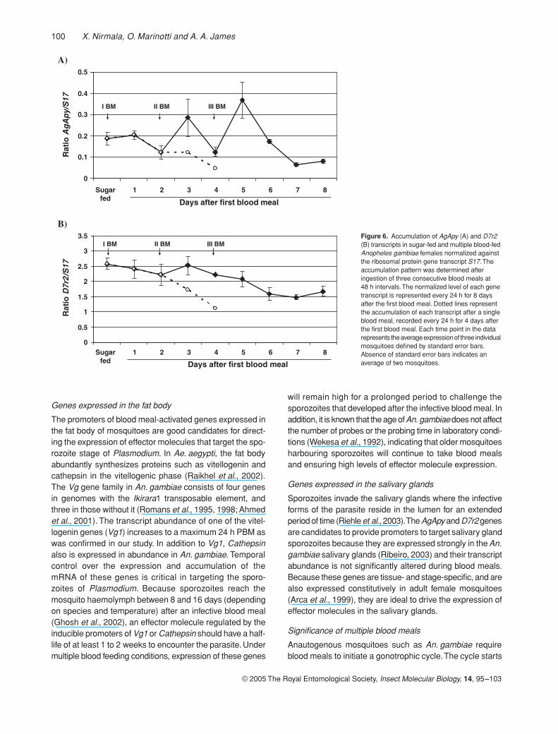
Expression levels of the salivary gland genes AgApy andD7r2 differed from each other and from the genes expressedin the midgut and fat body. AgApy mRNA accumulationremained almost unaltered after the first blood meal (Fig. 6A).This level increased 0.5- and one-fold 24 h after the secondand third blood meals, respectively. However, if mosquitoesdid not feed again, AgApy mRNA abundance dropped tolevels even lower than that observed in sugar-fed females.In contrast, the levels of D7r2 transcript decreased slightlyafter the first blood meal and continued to decline aftersubsequent blood meals (Fig. 6B). Overall, there was nostatistically significant change in the levels of AgApy andD7r2 accumulation.
Discussion
The development of transgenic mosquitoes expressingantiparasite effector molecules, and their ability to block orreduce malaria transmission under laboratory conditions,has fostered enthusiasm for releasing these insects as atool to increase the efficiency of integrated malaria vectorcontrol programmes. Selection criteria for genes for which
Figure 3. Number of days taken for blood-fed mosquitoes to take a second blood meal. Mosquitoes were fed using anaesthetized mice and blood-fed mosquitoes were removed to a separate cage. Blood meals were given to the fed mosquitoes every day until all the mosquitoes consumed a blood meal. A blood meal was not provided on day 5. Each time point in the data represents the average of three experiments defined by standard error bars.

98 X. Nirmala, O. Marinotti and A. A. James
© 2005 The Royal Entomological Society, Insect Molecular Biology, 14, 95–103
promoters could drive sustainable expression of transgenesinclude those that have a high level of constitutive expres-sion. However, such expression patterns and product accu-mulation may have a negative physiological effect on larvaland pupal stages where effector molecule expression is notneeded. Hence, the likely choices are blood meal-induciblegenes that are specifically expressed in adult females(James et al., 1999). In the experiments described here, weinterpret differences in accumulation profiles to reflectchanges in transcriptional activity of genes. In the absenceof specific knowledge of mRNA stability in mosquitoes and
the relatively short time periods over which we observe adifference, we argue that this is a reasonable interpretation.
Genes expressed in the midgut
Midgut stages of Plasmodium have been the target of effec-tor molecules in transgenic mosquitoes in an effort to curtailthe parasite at the initial stages of infection. Promoters ofgenes from the carboxypeptidase, trypsin and chymo-trypsin family whose products are involved in the proteolyticdigestion of a blood meal are good candidates for thispurpose. In An. gambiae midgut, AgCP is an ‘early’ gene
Figure 4. Accumulation of AgCP (A), Antryp2 (B) and Anchym1 (C) transcripts in sugar-fed and multiple blood-fed Anopheles gambiae females normalized against the ribosomal protein gene transcript S17. The accumulation pattern was determined after ingestion of three consecutive blood meals at 48 h intervals. The normalized level of AgCP (A) is represented at 3 h PBM and every 24 h thereafter for 8 days after the first blood meal. Ratios of Antryp2/S17 (B) and Anchym1/S17 (C) are represented every 24 h PBM. Dotted lines represent the accumulation of each transcript after a single blood meal, recorded every 24 h for 4 days after the first blood meal. Each time point in the data represents the average expression of three individual mosquitoes defined by standard error bars. Absence of standard error bars indicates an average for two mosquitoes. Asterisks indicate P < 0.01 (A,C) and P < 0.001 (B).

Accumulation of mRNAs following blood meals in An. gambiae 99
© 2005 The Royal Entomological Society, Insect Molecular Biology, 14, 95–103
whose transcription product accumulates to maximumlevels 3 h PBM (Edwards et al., 1997). Expression of trypsingenes Antryp1 and Antryp2, and chymotrypsin genesAnchym1 and Anchym2, is inducible following a bloodmeal,and reaches a maximum accumulation between 24 and30 h (Muller et al., 1995; Vizioli et al., 2001). An advantageof using an ‘early’ promoter in transgenesis is the expres-sion of effector molecules in the midgut before the forma-tion of peritrophic matrix (PM), which is formed around theblood bolus within 2 h of blood meal ingestion (Edwardset al., 1997). As diffusion of macromolecules across thePM is speculated to be rate-limiting (Villalon et al., 2003),
effector molecules expressed by Antryp2 or Anchym1 genepromoters may not gain access into the food bolus to inter-act with the ookinetes. In addition, these gene transcriptsreach maximum accumulation 24 h PBM, whereas the ook-inetes form 20 h PBM in the midgut lumen of An. gambiae(Ahmed et al., 2001), providing a small window for the ook-inetes to escape interaction with the effector molecule.However, because An. gambiae takes multiple blood mealsat 24-h intervals (Klowden & Briegel, 1994), effector mole-cules driven by Antryp2 and Anchym1 may accumulate inabundance after first blood meal to interact with the para-sites ingested during the second or third blood meal.
Figure 5. Accumulation of Vg1 (A), Cathepsin (B) and Lipophorin (C) transcripts in sugar-fed and multiple blood-fed Anopheles gambiae females normalized against the ribosomal protein gene transcript S17. The accumulation pattern was determined after ingestion of three consecutive blood meals at 48 h intervals. The normalized level of each gene transcript is represented every 24 h for 8 days after the first blood meal. Dotted lines represent the accumulation of each transcript after a single blood meal, recorded every 24 h for 4 days after the first blood meal. Each time point in the data represents the average expression of three individual mosquitoes defined by standard error bars. Absence of standard error bars indicates an average of two mosquitoes. Asterisks indicate P < 0.01 (A,B) and P < 0.2 (C).
100 X. Nirmala, O. Marinotti and A. A. James
© 2005 The Royal Entomological Society, Insect Molecular Biology, 14, 95–103
Genes expressed in the fat body
The promoters of blood meal-activated genes expressed inthe fat body of mosquitoes are good candidates for direct-ing the expression of effector molecules that target the spo-rozoite stage of Plasmodium. In Ae. aegypti, the fat bodyabundantly synthesizes proteins such as vitellogenin andcathepsin in the vitellogenic phase (Raikhel et al., 2002).The Vg gene family in An. gambiae consists of four genesin genomes with the Ikirara1 transposable element, andthree in those without it (Romans et al., 1995, 1998; Ahmedet al., 2001). The transcript abundance of one of the vitel-logenin genes (Vg1) increases to a maximum 24 h PBM aswas confirmed in our study. In addition to Vg1, Cathepsinalso is expressed in abundance in An. gambiae. Temporalcontrol over the expression and accumulation of themRNA of these genes is critical in targeting the sporo-zoites of Plasmodium. Because sporozoites reach themosquito haemolymph between 8 and 16 days (dependingon species and temperature) after an infective blood meal(Ghosh et al., 2002), an effector molecule regulated by theinducible promoters of Vg1 or Cathepsin should have a half-life of at least 1 to 2 weeks to encounter the parasite. Undermultiple blood feeding conditions, expression of these genes
will remain high for a prolonged period to challenge thesporozoites that developed after the infective blood meal. Inaddition, it is known that the age of An. gambiae does not affectthe number of probes or the probing time in laboratory condi-tions (Wekesa et al., 1992), indicating that older mosquitoesharbouring sporozoites will continue to take blood mealsand ensuring high levels of effector molecule expression.
Genes expressed in the salivary glands
Sporozoites invade the salivary glands where the infectiveforms of the parasite reside in the lumen for an extendedperiod of time (Riehle et al., 2003). The AgApy and D7r2 genesare candidates to provide promoters to target salivary glandsporozoites because they are expressed strongly in the An.gambiae salivary glands (Ribeiro, 2003) and their transcriptabundance is not significantly altered during blood meals.Because these genes are tissue- and stage-specific, and arealso expressed constitutively in adult female mosquitoes(Arca et al., 1999), they are ideal to drive the expression ofeffector molecules in the salivary glands.
Significance of multiple blood meals
Anautogenous mosquitoes such as An. gambiae requireblood meals to initiate a gonotrophic cycle. The cycle starts
Figure 6. Accumulation of AgApy (A) and D7r2 (B) transcripts in sugar-fed and multiple blood-fed Anopheles gambiae females normalized against the ribosomal protein gene transcript S17. The accumulation pattern was determined after ingestion of three consecutive blood meals at 48 h intervals. The normalized level of each gene transcript is represented every 24 h for 8 days after the first blood meal. Dotted lines represent the accumulation of each transcript after a single blood meal, recorded every 24 h for 4 days after the first blood meal. Each time point in the data represents the average expression of three individual mosquitoes defined by standard error bars. Absence of standard error bars indicates an average of two mosquitoes.

Accumulation of mRNAs following blood meals in An. gambiae 101
© 2005 The Royal Entomological Society, Insect Molecular Biology, 14, 95–103
with the search for a host and is followed by blood ingestionand digestion, maturation of ovaries and oviposition (Klow-den & Briegel, 1994). However, previous studies conductedwith laboratory colonies and field-caught mosquitoes showthat some anopheline species, including An. gambiae, takeseveral blood meals during each gonotrophic cycle (Bore-ham & Garrett-Jones, 1973; Burkot et al., 1988; Klowden &Briegel, 1994). Those mosquitoes taking a second bloodmeal could correspond to a small proportion of the popula-tion that was not successful in complete engorgementduring the first feeding bout, and therefore needed toacquire more nutrients via another blood meal to completeoogenesis. Our observations show that most mosquitoeswill seek blood meals every 24 h, even when fully engorgedafter a first blood meal. Because laboratory- and field-based studies are consistent with a multiple feedingbehaviour, sustainable expression of effector moleculescan be achieved by using promoters of blood meal-inducedor up-regulated genes. In addition, the likelihood of aPlasmodium-infected mosquito taking multiple bloodmeals is higher than that in uninfected mosquito (Koellaet al., 1998), indicating that multiple blood-feeding behav-iour can be an advantage to drive the expression of effectormolecules.
Conclusion
The availability of genetic resources and technology formodifying mosquitoes has advanced the transmissionblocking efforts to focus on the introduction of geneticallymodified mosquitoes in the field. Challenges facing therelease of transgenic mosquitoes include fitness costs ofrefractoriness, possible resistance to antiparasite genesand development of a drive mechanism to sustain thetransgene in the population (Riehle et al., 2003). Sustain-able expression of antiparasite effector molecules is onesuch challenge that can be overcome by choosing appro-priate promoters for transgenesis.
In transgenic mosquitoes generated so far, no singleeffector molecule has inhibited Plasmodium transmissioncompletely (Nirmala & James, 2003). Therefore, it is likelythat more than one kind of effector molecule driven by dif-ferent promoters should be used for effective transmissionblocking. An ideal vector-incompetent mosquito will targetparasite ligands in all stages of Plasmodium develop-ment in the midgut, haemolymph and salivary glands.Ookinetes that escape effector molecules in the midgutcan be targeted as sporozoites by genes expressed inthe fat body and salivary glands. Blood-meal-induciblegenes AgCP, Antryp2 and Anchym1 in the midgut, Vg1 andCathepsin in the fat body are ideal candidates for use intransgenic mosquitoes because of their tissue- and stage-specific expression pattern. AgApy and D7r2 are notaffected by blood meal ingestion but are strong promoters
that could be used to drive effector molecule expression inthe salivary glands.
Experimental procedures
Mosquitoes
The Pink-eye strain of An. gambiae (Githeko et al., 1992) wasmaintained at 25 °C, 75–85% relative humidity and 18/6-hlight–dark cycles. Larvae were fed on finely powdered fishfood (Tetramin) mixed 1 : 1 with yeast powder. Adults were fedon raisins and water ad libitum. Adult female mosquitoes were fedusing anaesthetized mice.
The first blood meal was provided to 3- to 4-day-old sugar-fedmosquitoes and those that had fed to repletion were separatedfrom the cohort and transferred to a new cage. A second bloodmeal was given after 48 h and the females that fed a second timewere transferred to a new cage. Similarly, a third blood meal wasprovided 48 h later and the blood-fed females were separated.After each blood-meal, 20–25 blood-fed females were removed toa new cage to analyse gene expression after each meal. Fromthese groups, three to four females were collected at 24, 48, 72and 96 h for analysis of gene expression by one-step RT-PCR.Care was taken to create a laboratory model to mimic naturalconditions for mosquitoes. During the entire procedure, a cupfilled with water was placed in all the cages to facilitate ovipositionand males were added to each cage of blood-fed females.
Determination of mRNA abundance by RT-PCR
mRNA abundance was quantified according to Justice et al. (2003).In brief, total RNA was extracted from whole bodies of female mos-quitoes using Trizol (Gibco/BRL). Oligonucleotide primers for eachgene of interest were designed from the An. gambiae database(Table 1). Wherever possible, primer pairs were derived from twodifferent exons with an intervening intron in order to detect ampli-fication products from genomic DNA that may have precipitatedduring the RNA preparation. Primers were designed to yield ampli-fication products ranging in size between 500 bp and 1.0 kb. Thegene encoding the ribosomal protein, S17, was used as a stand-ard in the amplification reactions, and its primer pair produced a250 bp fragment from its RNA.
Cycle-controlled one-step RT-PCR reactions were performedusing the one-step RT-PCR kit (Qiagen) in 50 µl volumes containing100–200 ng of total RNA. Each amplification reaction contained20 pmol of each of the gene-specific and S17 primers. Reactionswere performed in an MJ Research thermal cycler as follows:reverse transcription at 50 °C for 30 min, activation of Taq polymeraseat 95 °C for 15 min, followed by thirty-six cycles of denaturation at94 °C for 30 s, annealing at 66 °C for 30 s and extension at 72 °Cfor 1 min. In order to quantify the amplified products in a phos-phorimager, 1 µCi each of α32P-dATP and α32P-dCTP (3000 Ci/mM, Amersham Pharmacia) were added to each 50 µl RT-PCRreaction. Ten-microlitre aliquots were removed from each of thereactions during the annealing phase at cycles 24, 28, 32 and 36and mixed with the gel-loading buffer containing sucrose andbromophenol blue. Ten-microlitre samples were loaded on 3%agarose gels, which then were dried after electrophoresis on toZeta-probe GT Genomic Tested Blotting Membranes (Bio-Rad)and exposed on Kodak BioMax film. For quantification of thesignals, dried gels were exposed in phosphorimager cassettes andscanned in a phosphorimager (Molecular Dynamics). Quantification

102 X. Nirmala, O. Marinotti and A. A. James
© 2005 The Royal Entomological Society, Insect Molecular Biology, 14, 95–103
was performed with ImageQuant software (Molecular Dynamics)and tabulated in Microsoft Excel. Only values of signal intensitiesthat were increasing linearly in proportion to the amplification cyclewere used for further analysis. Signal intensities that reached asaturation point during amplification were not used. The ratio ofsignal intensities between the product of each gene and S17 wascalculated for individual mosquitoes and values are representedas average of three mosquitoes. Standard error and t-tests wereperformed in Microsoft Excel.
Acknowledgements
We thank Dr Harald Biessmann for advice on quantitativeRT-PCR and critical comments on the manuscript, Dr EricCalvo and Lisa Coleman for help in the blood feedings,Lynn Olson for help in preparing the manuscript and ourlaboratory colleagues for comments. The work reportedhere was supported by a grant from the National Institutesof Health, AI 29746 to A.A.J.
References
Ahmed, A.M., Maingon, R., Romans, P. and Hurd, H. (2001)Effects of malaria infection on vitellogenesis in Anophelesgambiae during two gonotrophic cycles. Insect Mol Biol 10:347–356.
Arca, B., Lombardo, F., Capurro, M., della Torre, A., Spanos, L.,Dimopoulos, G., Louis, C., James, A.A. and Coluzzi, M. (1999)Salivary gland-specific gene expression in the malaria vectorAnopheles gambiae. Parassitologia 41: 483–487.
Boreham, P.F. and Garrett-Jones, C. (1973) Prevalence of mixedblood meals and double feeding in a malaria vector (Anophelessacharovi Favre). Bull World Health Organ 48: 605–614.
Briegel, H. and Horler, E. (1993) Multiple blood meals as a repro-ductive strategy in Anopheles (Diptera: Culicidae). J Med Ento-mol 30: 975–985. Erratum. J. Med Entomol 1994, 31: following321.
Burkot, T.R., Graves, P.M., Paru, R. and Lagog, M. (1988) Mixedblood feeding by the malaria vectors in the Anopheles punc-tulatus complex (Diptera: Culicidae). J Med Entomol 25: 205–213.
Catteruccia, F., Nolan, T., Loukeris, T.G., Blass, C., Savakis, C.,Kafatos, F.C. and Crisanti, A. (2000) Stable germline transfor-mation of the malaria mosquito Anopheles stephensi. Nature405: 959–962.
Cho, W.L., Tsao, S.M., Hays, A.R., Walter, R., Chen, J.S., Snigi-revskaya, E.S. and Raikhel, A.S. (1999) Mosquito cathepsin B-like protease involved in embryonic degradation of vitellin isproduced as a latent extraovarian precursor. J Biol Chem 274:13311–13321.
Coates, C.J., Jasinskiene, N., Pott, G.B. and James, A.A. (1999)Promoter-directed expression of recombinant fire-fly luciferasein the salivary glands of Hermes-transformed Aedes aegypti.Gene 226: 317–325.
Edwards, M.J., Lemos, F.J., Donnelly-Doman, M. and Jacobs-Lorena, M. (1997) Rapid induction by a blood meal of a carbo-xypeptidase gene in the gut of the mosquito Anophelesgambiae. Insect Biochem Mol Biol 27: 1063–1072.
Ghosh, A.K., Moreira, L.A. and Jacobs-Lorena, M. (2002)Plasmodium–mosquito interactions, phage display libraries andtransgenic mosquitoes impaired for malaria transmission.Insect Biochem Mol Biol 32: 1325–1331.
Githeko, A.K., Brandling-Bennett, A.D., Beier, M., Atieli, F., Owaga, M.and Collins, F.H. (1992) The reservoir of Plasmodium falciparummalaria in a holoendemic area of western Kenya. Trans R SocTrop Med Hyg 86: 355–358.
Grossman, G.L., Rafferty, C.S., Clayton, J.R., Stevens, T.K.,Mukabayire, O. and Benedict, M.Q. (2001) Germline
Table 1. Genes, oligonucleotide primers, and specifics of gene sequences
GenePrimer name Primer sequence (5′−3′)
NCBI accession no. ENSANGT no. bp amplified
Antryp2 T2 For CCTCCTGCTGGCTGTTCTGGTTGCGG Z18890 615T2 Rev GGTGATTTCTCCGAACCACATGTAGGC 00000027737
Anchym1 C1 For CTCCTGGTAGTAAGTGCAGCCAAGGTGCC Z18887 715C1 Rev CCCAATCATGATAGTACGAGACACGGGC 00000023046
AgCP CBP For ATGCGAAGGTGCGTGCACTGGCCG AF000953 716CBP Rev GTAGCGCTAAGCCACCAACCCGC 00000021052
Vg1 Vg1 For GGTGACCGACTTGTTGCTCATGTCC AF281078 1077Vg1 Rev CTACCATCCATCTTGCGCTGTGTCG 00000012892
Cathepsin Cat For GGGATGTTGCGACTCGAGCTACGCCG Not available 585Cat Rev CCCCACTGTGCACCCCAGGAATTGGC 00000012222
Lipophorin LIP For CGCTAGTGCTGATTCAAAGTGTGTCCG Not available 504LIP Rev GGGTCACGAAGCTGTTCGAGAGCG 00000018348
AgApy Apy For TGGCACTGCGCATTCTTGCACCG AJ237705 970Apy Rev GTGAGCAATGCGAATGGGCG 00000015382
D7r2 D72 For CTGAGCGTTGGACTGGTCTGGTG AY045760 624D72 Rev GATTTCCTTGATCAGCGCTGCC 00000018371
S17 S17 For GGGTCGTGTACGTACCAAGACG AF164153 250S17 R2 CATAGTTATCGCGACGCTCACG 00000013205

Accumulation of mRNAs following blood meals in An. gambiae 103
© 2005 The Royal Entomological Society, Insect Molecular Biology, 14, 95–103
transformation of the malaria vector, Anopheles gambiae,with the piggyBac transposable element. Insect Mol Biol 10:597–604.
van Heusden, M.C., Thompson, F. and Dennis, J. (1998) Biosyn-thesis of Aedes aegypti lipophorin and gene expression of itsapolipoproteins. Insect Biochem Mol Biol 28: 733–738.
Ito, J., Ghosh, A., Moreira, L.A., Wimmer, E.A. and Jacobs-Lorena, M.(2002) Transgenic Anopheline mosquitoes impaired in trans-mission of a malaria parasite. Nature 417: 452–455.
James, A.A. (2002) Engineering mosquito resistance to malariaparasites: the avian malaria model. Insect Biochem Mol Biol32: 1317–1323.
James, A.A., Beerntsen, B.T., de Capurro, M.L., Coates, C.J.,Coleman, J., Jasinskiene, N. and Krettli, A.U. (1999) Control-ling malaria transmission with genetically-engineered,Plasmodium-resistant mosquitoes: milestones in a modelsystem. Parassitologia 41: 461–471.
Justice, R.W., Dimitratos, S., Walter, M.F., Woods, D.F. and Biess-mann, H. (2003) Sexual dimorphic expression of putativeantennal carrier protein genes in the malaria vector Anophelesgambiae. Insect Mol Biol 12: 581–594.
Klowden, M.J. and Briegel, H. (1994) Mosquito gonotrophic cycleand multiple feeding potential: contrasts between Anophelesand Aedes (Diptera: Culicidae). J Med Entomol 31: 618–622.
Koella, J.C., Sorensen, F.L. and Anderson, R.A. (1998) Themalaria parasite, Plasmodium falciparum, increases the fre-quency of multiple feeding of its mosquito vector, Anophelesgambiae. Proc R Soc Lond B Biol Sci 265: 763–768.
Kokoza, V., Ahmed, A., Cho, W.L., Jasinskiene, N., James, A.A.and Raikhel, A. (2000) Engineering blood meal-activatedsystemic immunity in the yellow fever mosquito, Aedes aegypti.Proc Natl Acad Sci USA 97: 9144–9149.
Lombardo, F., Di Cristina, M., Spanos, L., Louis, C., Coluzzi, M.and Arca, B. (2000) Promoter sequences of the putativeAnopheles gambiae apyrase confer salivary gland expressionin Drosophila melanogaster. J Biol Chem 275: 23861–23868.
Moreira, L.A., Ito, J., Ghosh, A., Devenport, M., Zieler, H.,Abraham, E.G., Crisanti, A., Nolan, T., Catteruccia, F. andJacobs-Lorena, M. (2002) Bee venom phospholipase inhibitsmalaria parasite development in transgenic mosquitoes.J Biol Chem 277: 40839–40843.
Muller, H.M., Catteruccia, F., Vizioli, J., della Torre, A. and Crisanti, A.
(1995) Constitutive and blood meal-induced trypsin genes inAnopheles gambiae. Exp Parasitol 81: 371–385.
Muller, H.M., Vizioli, I., della Torre, A. and Crisanti, A. (1993) Temporaland spatial expression of serine protease genes in Anophelesgambiae. Parasitology 35 (Suppl.): 73–76.
Nirmala, X. and James, A.A. (2003) Engineering Plasmodium-refractory phenotypes in mosquitoes. Trends Parasitol 19:384–387.
Raikhel, A.S., Kokoza, V.A., Zhu, J., Martin, D., Wang, S.F., Li, C.,Sun, G., Ahmed, A., Dittmer, N. and Attardo, G. (2002) Molec-ular biology of mosquito vitellogenesis: from basic studies togenetic engineering of antipathogen immunity. Insect BiochemMol Biol 32: 1275–1286.
Ribeiro, M.C. (2003) A catalogue of Anopheles gambiae tran-scripts significantly more or less expressed following a bloodmeal. Insect Biochem Mol Biol 33: 865–882.
Riehle, M.A., Srinivasan, P., Moreira, C.K. and Jacobs-Lorena, M.(2003) Towards genetic manipulation of wild mosquito popula-tions to combat malaria: advances and challenges. J Exp Biol206: 3809–3816.
Romans, P., Tu, Z., Ke, Z. and Hagedorn, H.H. (1995) Analysis ofa vitellogenin gene of the mosquito, Aedes aegypti and com-parisons to vitellogenins from other organisms. Insect BiochemMol Biol 25: 939–958.
Romans, P., Bhattacharyya, R.K. and Colavita, A. (1998) Ikirara, anovel transposon family from the malaria vector mosquito,Anopheles gambiae. Insect Mol Biol 7: 1–10.
Shen, Z. and Jacobs-Lorena, M. (1998) A type I peritrophic matrixprotein from the malaria vector Anopheles gambiae binds tochitin. Cloning, expression, and characterization. J Biol Chem273: 17665–17670.
Villalon, J.M., Ghosh, A. and Jacobs-Lorena, M. (2003) The peri-trophic matrix limits the rate of digestion in adult Anophelesstephensi and Aedes aegypti mosquitoes. J Insect Physiol 49:891–895.
Vizioli, J., Catteruccia, F., della Torre, A., Reckmann, I. and Muller,H.M. (2001) Blood digestion in the malaria mosquito Anophe-les gambiae: molecular cloning and biochemical characteriza-tion of two inducible chymotrypsins. Eur J Biochem 268: 4027–4035.
Wekesa, J.W., Copeland, R.S. and Mwangi, R.W. (1992) Effect ofPlasmodium falciparum on blood feeding behavior of naturallyinfected Anopheles mosquitoes in western Kenya. Am J TropMed Hyg 47: 484–488.